An LRE (Leucine-Arginine-Glutamate)-dependent Adhesion of Neurons to S-laminin Mechanism

Nutrients 2012, 4, 1851-1867; doi:10.3390/nu4121851
nutrients ISSN 2072-6643
www.mdpi.com/journal/nutrients
Article
Dose and Latency Effects of Leucine Supplementation in
Modulating Glucose Homeostasis: Opposite Effects in Healthy
and Glucocorticoid-Induced Insulin-Resistance States
Nelo Eidy Zanchi 1,2,3,
*, Lucas Guimarães-Ferreira 2, Mário Alves de Siqueira-Filho
2,
Vitor Felitti 1, Humberto Nicastro
1, Carlos Bueno Jr.
4, Fábio Santos Lira
3,
Marshall Alan Naimo 5, Patrícia Campos-Ferraz
1, Maria Tereza Nunes
2, Marília Seelaender
3,
Carla Roberta de Oliveira Carvalho 2, François Blachier
6 and Antonio Herbert Lancha Jr.
1
1 Laboratory of Applied Nutrition and Metabolism, School of Physical Education and Sports,
University of Sao Paulo, Sao Paulo, 05508-030, Brazil; E-Mails: [email protected] (V.F.);
[email protected] (H.N.); [email protected] (P.C.-F.); [email protected] (A.H.L.) 2 Department of Physiology and Biophysics, Institute of Biomedical Sciences, University of Sao
Paulo, Sao Paulo, 05508-900, Brazil; E-Mails: [email protected] (L.G.-F.);
[email protected] (M.A.S.-F.); [email protected] (M.T.N.); [email protected] (C.R.O.C.) 3 Cancer Metabolism Research Group, Institute of Biomedical Sciences, University of São Paulo, Sao
Paulo, 05508-900, Brazil; E-Mails: [email protected] (F.S.L.); [email protected] (M.S.) 4 Human Genome Research Center, Institute of Biosciences, University of São Paulo, São Paulo,
05508-900, Brazil; E-Mail: [email protected] 5 Department of Health Sciences and Human Performance, The University of Tampa, Tampa,
FL 33606, USA; E-Mail: [email protected] 6 INRA, CRNH-IdF, UMR 914, Nutrition Physiology and Ingestive Behavior, Paris, 78350, France;
E-Mail: [email protected]
* Author to whom correspondence should be addressed; E-Mail: [email protected];
Tel.: +55-11-3091-7247.
Received: 28 September 2012; in revised form: 1 November 2012 / Accepted: 14 November 2012 /
Published: 27 November 2012
Abstract: Dexamethasone (DEXA) is a potent immunosupressant and anti-inflammatory
agent whose main side effects are muscle atrophy and insulin resistance in skeletal
muscles. In this context, leucine supplementation may represent a way to limit the DEXA
side effects. In this study, we have investigated the effects of a low and a high dose of
leucine supplementation (via a bolus) on glucose homeostasis, muscle mass and muscle
strength in energy-restricted and DEXA-treated rats. Since the leucine response may also
OPEN ACCESS

Nutrients 2012, 4 1852
be linked to the administration of this amino acid, we performed a second set of
experiments with leucine given in bolus (via gavage) versus leucine given via drinking
water. Leucine supplementation was found to produce positive effects (e.g., reduced
insulin levels) only when administrated in low dosage, both via the bolus or via drinking
water. However, under DEXA treatment, leucine administration was found to significantly
influence this response, since leucine supplementation via drinking water clearly induced a
diabetic state, whereas the same effect was not observed when supplied via the gavage.
Keywords: leucine supplementation; glucose homeostasis; skeletal muscle mass
1. Introduction
Branched-chain amino acids (BCAA—leucine, isoleucine and valine) supplementation, especially
leucine, has been described as a potential therapeutic tool capable to attenuate skeletal muscle atrophy
induced by several catabolic conditions, such as cancer, sepsis, muscular diseases [1] and
glucocorticoid treatment [2]. Since leucine is considered as the second more potent insulin
secretagogue amongst all the amino acids (AA) [3], it has been studied for its capacity to modulate
whole body glucose homeostasis [4].
Indeed, leucine supplementation might exert positive systemic effects in conditions characterized
by increases in glucose homeostasis disturbance, such as high fat diet (HFD) induced insulin
resistance, but the effects are controversial. For example, Zhang and coworkers [5] found increases in
the glucose metabolism of leucine supplemented mice, whereas Lynch and coworkers [6] did not
observe improvements nor decreases in the glucose homeostasis. However, this may have been due to
the lack of standardization of doses and forms of administration. On the other hand, in healthy rats and
humans, oral leucine feeding has shown to rapidly inhibit skeletal muscle protein degradation [7,8] and
also promote robust increases in skeletal muscle protein synthesis [9], demonstrating overall a
potential capacity to handle disturbances in glucose metabolism and spare skeletal muscle mass,
especially under atrophic conditions [8]. However, its chronic effects remain elusive, specifically
during insulin resistant states from different physiopathological backgrounds, such as glucocorticoid
treatment (and even HFD treatment).
Dexamethasone (DEXA) is a synthetic glucocorticoid form of the endogenous hormone cortisone,
which exhibits potent immunosupressant and anti-inflammatory properties [10]. The successful
therapeutic benefits of this drug in a wide range of inflammatory diseases is, however, limited as it
presents several side effects [11], such as insulin resistance and skeletal muscle atrophy [12,13].
Therefore, in order to benefit from the desired effects of long term DEXA treatment, the deleterious
responses must be reduced. In this context, leucine supplementation may represent an interesting
intervention with a clinical perspective, due to the reasons justified from above.
Since there is still a lack of evidence regarding the effects of leucine supplementation on
DEXA-induced insulin resistance and skeletal muscle atrophy, we decided to investigate: (1) whether a
supplementation, with a low dose of leucine (not capable of increasing insulin levels or muscle protein
synthesis) or a high dose of leucine (capable of maximally increasing both insulin concentration and

Nutrients 2012, 4 1853
muscle protein synthesis) given through gavage or drinking water is able to improve glucose
metabolism, as well as spare the muscle mass and, consequently, voluntary muscular strength in
healthy (control pair-fed and energy restricted) rats; (2) investigate to see if leucine supplementation
via gavage or via drinking water exerts some positive effect on glucose metabolism and muscle
sparing/strength effects under DEXA treatment; in other words, besides the dosage effect, would
frequent nutritional stimuli (leucine provided through drinking water) be different from that provided
by a pulsatile pattern (leucine provided through gavage) with both groups consuming the same
daily dosage?
2. Materials and Methods
2.1. Animals
The experiments were conducted in accordance with the National Research Council’s Guidelines
for the Care and Use of Laboratory Animals. All methods used were approved by the Ethical
Committee for Animal Research of the Physical Education and Sport of the University of Sao Paulo
(protocol 2008/45). Adult male Wistar rats (mean body weight 440 g) were housed in individual cages
under controlled environmental conditions (temperature, 22 °C; 12-h dark period) with a standard diet
(Nuvilab, Brazil) and water provided ad libitum.
2.2. Study 1
Groups
Animals were randomly divided into the following groups: control non-supplemented (CON-NS;
n = 10), control + leucine low-dose (CON-LL; n = 10), control + leucine high-dose (CON-LH; n = 10),
DEXA (DEX; n = 10), DEX + leucine low-dose (DEX-LL; n = 10) and DEXA + leucine high-dose
(DEX-LH). During the duration of the experiment, which lasted seven days, DEXA (a synthetic
glucocorticoid analogue that does not bind to plasma binding proteins) was given daily (at 9:00 a.m.)
through intraperitoneal injection (5 mg/kg/day); control groups received an equivalent volume of
saline (0.9% NaCl). As DEXA was reported to decrease food intake, all groups were fed the same
amount of food (in terms of caloric intake) equal to the DEX group. Thus, differences among groups
did not originate from different food intakes. We measured the caloric content of our standard chow
(16.32 kJ/g) as well as leucine (25 kJ/g) in a calorimetric bomb (FTT Oxygen Bomb Calorimeter) in
order to avoid differences in the caloric ingestion between experimental groups and observed that the
total caloric consumption was not statistically different among groups. A suspension of 54.0 g of
L-leucine/L in water was prepared according to Crozier and coworkers [9]. Rats of
leucine-supplemented groups received 0.068 g/kg/day (low-dose) or 1.35 g/kg/day (high-dose) twice a
day (8:00 a.m. and 2:00 p.m.) through gavage during seven days [9]. Importantly, the high dosage
were capable of maximally increasing muscle protein synthesis and insulin plasmatic levels (in a well
defined pulsatile form), whereas the low dosage was not capable of increasing either muscle protein
synthesis or insulin plasmatic levels [9,14]. Non-supplemented groups received 0.155 mol/L of NaCl
at a volume of 2.5 mL/100 g of body weight twice a day. This volume of saline is equivalent to the

Nutrients 2012, 4 1854
volume of leucine suspension administered to leucine-supplemented groups and was chosen in order to
take into account any possible volume-induced effects of oral gavage, i.e., gastric expansion-induced
signaling. We chose to administer two daily doses of leucine in order to maintain plasma increased
concentration throughout the day and counteract DEXA-induced effects. Animals were euthanized
after 13 h fasting by decapitation. Soleus and extensor digitorius longus (EDL) muscles of each limb
were isolated, weighed and frozen at −80 °C for analysis. To assess the dry over total weight ratio, a
small portion of each muscle was weighed and then dried for 48 h.
2.3. Study 2
Since we observed that control groups responded equally to leucine supplementation via bolus or
drinking water (data not shown), we additionally investigated whether or not leucine supplementation
would be different via a bolus or drinking water in the presence of DEXA treatment.
2.3.1. Groups
Animals were randomly divided into the following groups: DEXA + leucine low-dose (DEX-LL;
n = 8); DEXA + leucine low-dose drinking water (DEX-LL-H2O; n = 8); DEXA + leucine high-dose
(DEX-LH; n = 8); and DEXA + leucine high-dose drinking water (DEX-LH-H2O; n = 8). The groups
received the same dosage of DEXA administered in study 1. All groups were the same amount of food
(in terms of caloric intake) equal to the DEXA group, and no statistically differences among groups
were observed. DEX-LL and DEX-LH groups were supplemented via gavage (0.068 and
1.35 g/kg/day, respectively) twice a day and fed the same diet with regular tap water as drinking water,
as previously described in study 1. DEX-LL-H2O and DEX-LH-H2O groups were supplemented with
leucine via drinking water, and the leucine dose was adjusted every day on the basis of drinking water
intake the day before. The water or liquid leucine supplement was provided by means of graduated
cylinders topped with a 1-hole rubber stopper holding a metal drinking nipple. Leucine in water
solution was used as crystals grounded to a fine powder with a ceramic mortar and pestle to optimize
solubility [15]. In this study, we added leucine in drinking water to compare leucine provided through
gavage (which results in a well defined pulsatile pattern [9,14]) versus leucine provided through
drinking water (rendering less fluctuations in the leucine levels) with both groups consuming the same
daily dosage. Animals were euthanized by decapitation and soleus, and EDL muscles of each limb
were isolated, weighed and frozen at −80 °C for analysis.
2.3.2. Basal Fasting Glucose, Insulin and Tryacilglycerol (TAG) Levels
Basal and fed glycemia were measured through blood collected from the caudal vein after an
overnight fast (13 h) using a digital glucometer (ACCU-CHEK Performa, Roche) before euthanasia.
Immediately after euthanasia (13 h fasting), blood was collected and serum samples were prepared on
ice. Serum was frozen and stored at −80 °C for analysis. Basal TAG was measured using a commercial
kit (Biolab, Brazil). Serum insulin concentration was quantified using the commercial kits RIA
(DPC®, Brazil). The homeostase model for assessment of insulin resistance index (HOMA-IR) was
calculated as follows:

Nutrients 2012, 4 1855
HOMA-IR index (mmol·mU/L2) = fasting insulin (mU/L) × serum glucose (mmol/L)/22.5 [16].
2.3.3. Motor Performance Tests
In order to evaluate skeletal muscle function, two evaluations were carried out. Such evaluations are
widely adopted as measures of muscle function in dystrophic mice. The first one, the Grip Strength
System Test (model: DFE-002, San Diego Instruments, San Diego, CA, USA) is a condition where
animals are let to grab onto the system with the forepaws as the experimenter gently pulls on their
tails. This allows the experimenter to determine the maximal strength before the animal releases the
bar [17]. Importantly, all measurements of maximal strength were performed by the same investigator,
who was highly experienced with performing this test.
The second motor performance test is the ambulation test. This test allows the determination of the
mean length of a step measured in hindfoot ink prints and is normalized by the animal’s length.
Briefly, rats were allowed to freely run in a corridor (length, 100 cm; width, 10.5 cm; height of lateral
walls, 20 cm) three different times. Before the test, the animals were permitted to explore the
apparatus [18,19]. Mean values were individually calculated for each test through the mean of three
consecutive tests performed during one minute.
2.3.4. RNA Isolation and Realt-Time PCR
Total RNA was extracted from homogenized soleus and EDL muscles with the Trizol reagent
(Invitrogen) according to the manufacturer instructions. One microgram of total RNA was
retranscribed with MMLV enzyme (Invitrogen), and an aliquot was used to measure real-time PCR.
All reactions were conducted in a volume of 25 µL containing 4 mM MgCl2 (Invitrogen), 0.25 mM
dNTPs (Invitrogen), 1.2 U of Taq polymerase (Invitrogen), 1/30000 Sybr Green (Invitrogen) and
specific oligonucleotides for each gene with the Rotor Gene 3000 sequence detector (Quiagen Inc.;
Hilden, Germany). Primers utilized for real-time PCR analysis was: GLUT-4 sense:
5′-GGGCTGTGAGTGAGTGCTTTC-3′; antisense: 5′-CAGCGAGGCAAGGCTAGA-3′; GAPDH
sense: 5′-GATGGGTGTGAACCACGAGAAA-3′; antisense: 5′-ACGGATACATTGGGGGTAGGA-3′.
Reactions were run for 40 cycles under the following conditions: 40 s at 95 °C, 40 s at 65 °C and 40 s
at 72 °C. The amplification of unique products in each reaction was verified by melting curve and
ethidium bromide (Sigma Aldrich) stained agarose gel electrophoresis. Each sample was run in
triplicate. The expression level of each gene was normalized to housekeeping gene (GAPDH)
expression level using the standard curve method. Mean and standard errors were calculated and are
expressed as fold changes relative to the control group.
2.3.5. Statistical Analysis
The dependent variables were tested by either one-way or two-way ANOVA, as appropriate.
A post-hoc test with a Tukey adjustment was performed for multiple comparison purposes. The
significance level was set at p < 0.05. The results are expressed as means ± S.E.M.

Nutrients 2012, 4 1856
3. Results
3.1. Study 1
The effects of DEXA treatment and leucine supplementation on body weight and muscle
morphological parameters: as shown in Table 1, the baseline body weight was similar among groups.
All groups were characterized by a significant body weight reduction at the end of the experimental
protocol (p < 0.05). DEXA-treated groups showed significantly reduced body weight when compared
with the control groups (p < 0.05). Thus, leucine supplementation at both low and high doses did not
counteract body weight loss in both food restricted (control groups) and DEXA-treated animals. Soleus
muscle mass did not differ among groups. Leucine supplementation at high doses attenuated food
restriction-induced EDL muscle loss (CON-LH group) when compared with the CON-NS group
(p < 0.05). All DEXA-treated animals presented reduced EDL muscle mass when compared with the
CON-NS group (p < 0.05), and leucine supplementation at both low and high doses of amino acid did
not attenuate it (total n = 60).
Table 1. Body and muscle morphological parameters of the experimental groups.
Variable Group
CON-NS CON-LL CON-LH DEX-NS DEX-LL DEX-LH
Initial BW (g) 442.7 ± 5.90 442.2 ± 4.91 441.0 ± 4.35 443.8 ± 6.19 443.3 ± 4.19 444.8 ± 6.16
Final BW (g) 381.9 ± 22.1 b 386.3 ± 20.32
b 375.0 ± 10.21
b 343.4 ± 11.53
a,b 345.79 ± 15.89
a,b 339.9 ± 11.73
a,b
Delta BW (g) −60.8 ± 4.64 −55.9 ± 5.36 −65.17 ± 4.98 −100.8 ± 2.40 a −103.6 ± 2.08
a −104.5 ± 3.35
a
Soleus (mg) 214.8 ± 5.99 223.3 ± 5.45 221.0 ± 6.13 214.4 ± 4.29 210.8 ± 2.25 212.7 ± 5.45
EDL (mg) 189.4 ± 2.31 197.3 ± 2.08 200.1 ± 1.49 a 179.6 ± 3.37
a 174.8 ± 2.80
a 174.6 ± 4.12
a
Values are expressed as mean ± SE. Control non-supplemented group (CON-NS; n = 10); Control leucine supplemented
group with low dose via gavage (CON-LL; n = 10); Control leucine supplemented group with high dose via gavage
(CON-LH; n = 10); DEXA non supplemented group (DEX-NS; n = 10); DEXA treated group plus low dose of leucine
supplementation via gavage (DEX-LL; n = 10); DEXA treated group plus high dose of leucine supplementation via
gavage (DEX-LH; n = 10). BW—body weight; EDL—extensor digitorum longus. a p < 0.05 vs. CON-NS; b p < 0.05 vs.
Initial BW.
The effects of DEXA treatment and leucine supplementation on water intake: In the control groups,
water intake was significantly increased in CON-LL group when compared with the CON-NS group at
day 3 (38.33 ± 0.57 in CON-LL group vs. 29.00 ± 2.93 in CON-NS group; p < 0.05). At day 6 of
treatment, DEX-NS group showed increased water intake when compared with day 1 (36.77 ± 2.34 in
DEX-NS group at day 6 vs. 28.09 ± 2.05 at day 1; p < 0.05) and with the CON-NS group at day 6
(27.53 ± 2.68 in CON-NS group at day 6; p < 0.05). DEX-LL group presented reduced water intake at
day 3 when compared with day 1 (23.14 ± 1.04 in DEX-LL group at day 3 vs. 31.2 ± 2.05 at day 1;
p < 0.05) and increased when compared with the CON-NS group at day 6 (p < 0.05) (total n = 60).
The effects of DEXA treatment and leucine supplementation on serum glucose, insulin and
triacylglycerol (TAG): fasting glycemia and tryacilglycerol were significantly higher in the DEX-NS
group when compared with the CON-NS group (Figure 1A; 166.6 ± 20.8 mg/dL in DEX-NS vs.
118.5 ± 7.5 mg/dL in CON-NS group; p < 0.05; Figure 1C; 114.4 ± 14.4 mg/dL in DEX-NS vs.
68.3 ± 9.7 mg/dL in CON-NS group; p < 0.05), suggesting a DEXA-induced increase in blood glucose

Nutrients 2012, 4 1857
and TAG. Leucine supplementation at a low dose decreased fasting insulin and triacylglycerol when
compared to the CON-NS and CON-LH groups (0.15 ± 0.01 mg/dL in CON-LL vs. 2.95 ± 0.66 mg/dL
in CON-NS and 2.68 ± 0.79 mg/dL in CON-LH groups; p < 0.05; Figure 1B; 27.56 ± 2.89 mg/dL in
CON-LL vs. 68.31 ± 9.74 mg/dL in CON-NS group; p < 0.05; Figure 1C). DEX-LL and DEX-LH
groups presented increased fasting glycemia (157.6 ± 29.8 mg/dL in DEX-LL vs. 103.3 ± 4.0 mg/dL in
CON-LL group; p < 0.05; 190.1 ± 27.2 mg/dL in DEX-LH vs. 96.7 ± 3.2 mg/dL in CON-LH group;
p < 0.05; Figure 1A), insulin (3.54 ± 0.74 mg/dL in DEX-LL vs. 0.15 ± 0.01 mg/dL in CON-LL group;
p < 0.05; 4.42 ± 0.51 mg/dL in DEX-LH vs. 2.68 ± 0.79 mg/dL in CON-LH group; p < 0.05;
Figure 1B) and triacylglycerol (122.4 ± 16.9 mg/dL in DEX-LL vs. 27.6 ± 2.9 mg/dL in CON-LL
group; p < 0.05; 119.7 ± 32.7 mg/dL in DEX-LH vs. 38.5 ± 4.7 mg/dL in CON-LH group; p < 0.05;
Figure 1C) when compared to its respective control groups. Thus, it is possible to emphasize that these
effects were meditated by leucine since both groups had the same food intake (n = 10 per group).
Figure 1. Fasting (A) blood glucose, (B) insulin, and (C) triacylglycerol levels and
(D) HOMA index. Values are expressed as mean ± S.E.M. a Different from CON-NS
(p < 0.05); b Different from CON-LL (p < 0.05); and
c Different from CON-LH (p < 0.05).
In the fed state, control groups did not show any significant alteration in serum glucose during the
experimental protocol. However, DEXA-treated groups showed markedly increased serum glucose
levels at days 3 and 6 when compared with day 1 (p < 0.05) and with the CON-NS group at days 3 and
6 (p < 0.05). No significant differences were observed among DEXA-treated groups (Table 2)
(n = 10).

Nutrients 2012, 4 1858
Table 2. Fed serum glucose (mg/dL) of the experimental groups on days 1, 3 and 6 of the study.
Day Group
CON-NS CON-LL CON-LH DEX-NS DEX-LL DEX-LH
1 110.5 ± 5.73 107.3 ± 0.76 115.5 ± 1.47 120.1 ± 5.06 104.8 ± 4.07 114.7 ± 4.78
3 109.8 ± 5.26 103.5 ± 1.16 129.6 ± 1.28 215.6 ± 29.08 a,b
196.5 ± 17.27 a,b
178.1 ± 15.18 a,b
6 109.8 ± 5.26 110.0 ± 1.64 130.7 ± 2.25 267.3 ± 31.51 a,b
255.2 ± 23.44 a,b
215.6 ± 22.33 a,b
Values are expressed as mean ± SE. Control non-supplemented group (CON-NS; n = 10); Control leucine supplemented
group with low dose via gavage (CON-LL; n = 10); Control leucine supplemented group with high dose via gavage
(CON-LH; n = 10); DEXA non supplemented group (DEX-NS; n = 10); DEXA treated group plus low dose of leucine
supplementation via gavage (DEX-LL; n = 10); DEXA treated group plus high dose of leucine supplementation via
gavage (DEX-LH; n = 10). a p < 0.05 vs. CON-NS in the same day of treatment; b p < 0.05 vs. day 1 in the same group.
The effects of DEXA treatment and leucine supplementation on GLUT-4 gene expression in
skeletal muscle: in soleus muscle, GLUT-4 gene expression was not significantly affected by DEXA
or leucine treatment. In EDL muscle, however, GLUT-4 mRNA content was significantly lower in
DEX-NS, DEX-LL and DEX-LH groups (0.69 ± 0.38, 0.63 ± 0.18 and 0.54 ± 0.17, respectively) when
compared to CON-NS group (1.00 ± 0.15; p < 0.05; Figure 2A). In control supplemented groups, only
CON-LL was lower than CON-NS (0.60 ± 0.16 vs. 1.00 ± 0.15; p < 0.05; Figure 2B) (n = 6–8 animals
per group).
Figure 2. GLUT-4 gene expression in (A) soleus and (B) EDL muscles Values are
expressed as mean ± S.E.M. a Different from CON-NS (p < 0.05).
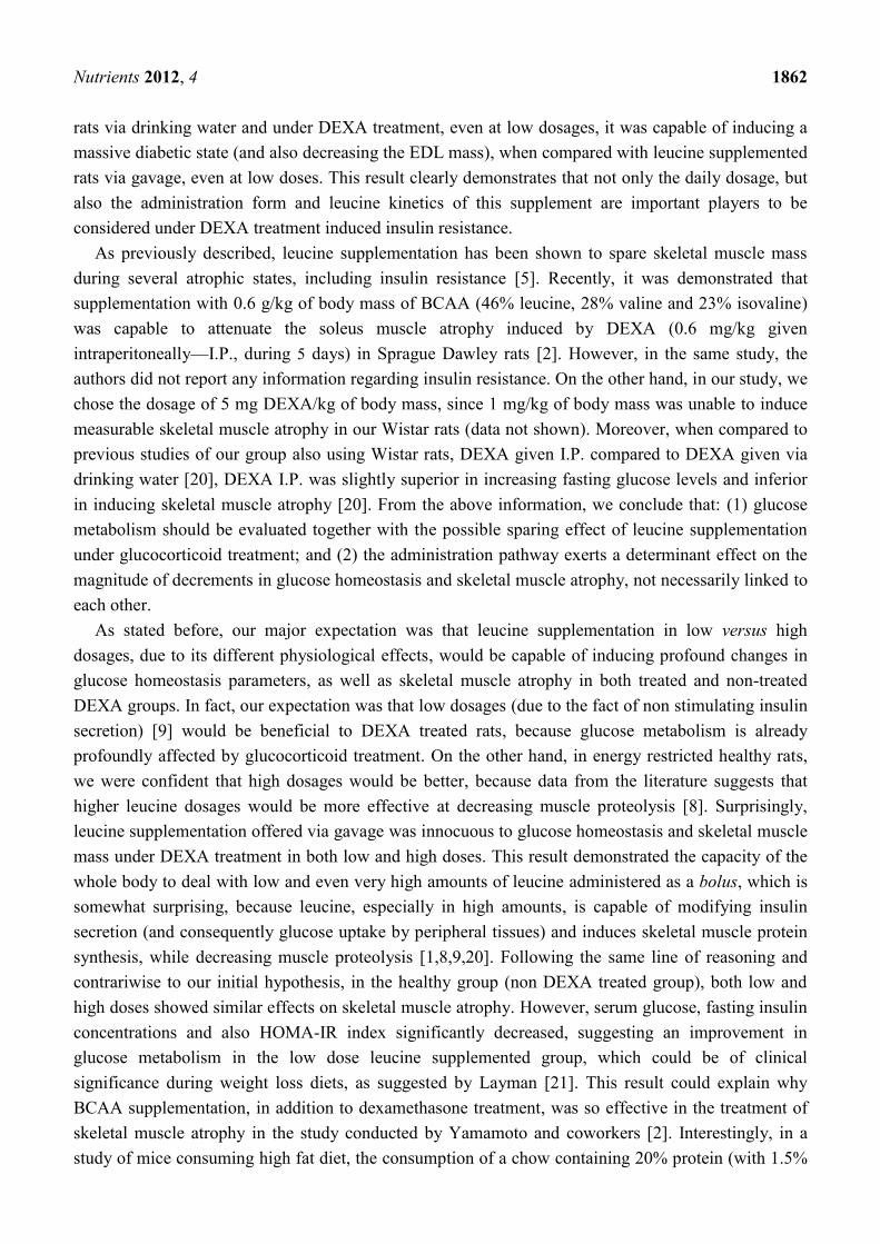
The effects of DEXA treatment and leucine supplementation on muscle functional parameters: We
observed that DEX-LH animals presented a modest but significant deficit in mean ambulation when
compared to the CON-LH group (0.54 ± 0.03 cm in DEX-LH vs. 0.59 ± 0.02 cm in CON-LH group;
p < 0.05; Figure 3B). The CON-LH group also presented less mean grip strength when compared with
the CON-LL group (0.46 ± 0.04 N in CON-LH vs. 0.66 ± 0.02 N in CON-LL; p < 0.05; Figure 3D)
(n = 8–10 per group).

Nutrients 2012, 4 1859
Figure 3. Muscle functional parameters. (A) Maximum ambulation (cm); (B) Mean
ambulation (cm); (C) Maximum grip strength (N); (D) Mean grip strength (N). Values are
expressed as mean ± S.E.M. a Different from CON-NS (p < 0.05); b
Different from CON-LL
(p < 0.05); and c Different from CON-LH (p < 0.05).
3.2. Study 2
The effects of DEXA treatment and leucine supplementation on body weight and muscle
morphological parameters: baseline body weight did not differ among groups. Leucine supplementation
with a high dose via drinking water (DEX-LH (H2O) group) significantly attenuated body weight loss
when compared with the DEX-LL and DEX-LH (bolus) groups (p < 0.05). Soleus muscle mass was
significantly reduced in DEX-LL (H2O) group when compared with the DEX-LL (bolus) and DEX-LH
groups (p < 0.05). EDL muscle mass did not significantly differ among groups (Table 3).
Table 3. Body and muscle morphological parameters of the experimental groups.
Variable Group
DEX-LL DEX-LL (H2O) DEX-LH DEX-LH (H2O)
Initial BW (g) 442.3 ± 1.40 441.8 ± 1.58 444.9 ± 1.49 446.7 ± 1.27
Final BW (g) 338.9 ± 1.83 346.5 ± 3.58 340.0 ± 2.93 361.0 ± 4.13 a,b,c
Delta BW (g) −103.6 ± 2.07 −95.27 ± 3.14 −104.5 ± 3.35 −85.67 ± 6.77 a,b
Soleus (mg) 222.5 ± 5.28 205.0 ± 2.39 a,b
222.9 ± 5.54 207.4 ± 3.22
EDL (mg) 174.8 ± 2.79 170.5 ± 1.99 174.6 ± 4.12 169.4 ± 1.27
Values are expressed as mean ± SE. DEXA treated group plus low dose of leucine supplementation via gavage (DEX-LL;
n = 10); DEXA treated group plus low dose of leucine supplementation via drinking water (DEX-LL (H2O); n = 10);
DEXA treated group plus high dose of leucine supplementation via gavage (DEX-LH; n = 10). DEXA treated group plus
high dose of leucine supplementation via drinking water (DEX-LH (H2O); n = 10). BW—body weight; EDL—extensor
digitorum longus. a p < 0.05 vs. DEX-LL; b p < 0.05 vs. DEX-LH; c p < 0.05 vs. DEX-LL (H2O).

Nutrients 2012, 4 1860
The effects of DEXA treatment and leucine supplementation on water intake: both DEX-LL (H2O)
and DEX-LH (H2O) presented increased water intake at day 6 when compared with their intake at
day 1 (48.80 ± 3.45 in DEX-LL (H2O) group at day 6 vs. 31.44 ± 3.09 at day 1; p < 0.05; 45.29 ± 1.84
in DEX-LH (H2O) group at day 6 vs. 34.25±2.93 at day 1; p < 0.05) and with the DEX-LL and DEX-LH
at day 6 (34.67 ± 2.34 in DEX-LL group and 26.56 ± 3.98 in DEX-LH group at day 6; p < 0.05). The
DEX-LH (H2O) group showed increased water intake when compared with the DEX-LL and DEX-LH
groups at day 3 (33.08 ± 1.7 in DEX-LH (H2O) group vs. 23.14 ± 1.04 in DEX-LL group and
22.00 ± 1.98 in DEX-LH group; p < 0.05).
The effects of DEXA treatment and leucine supplementation on serum glucose, insulin and
triacylglycerol (TAG): fasting serum glucose was significantly increased in the DEX-LL (H2O) group
when compared with the DEX-LL group (320.3 ± 68.4 mg/dL in DEX-LL (H2O) group vs.
173.6 ± 28.2 mg/dL in DEX-LL group; p < 0.05; Figure 4A) and in the DEX-LH (H2O) when
compared with the DEX-LH group (338.7 ± 41.9 mg/dL in DEX-LH (H2O) group vs. 176.1 ± 21.4 mg/dL
in DEX-LH group; p < 0.05; Figure 4A). These results suggest a dose-response and administration
route action of leucine. The DEX-LH (H2O) group also presented decreased fasting serum insulin
when compared with the DEX-LH group (35.24 ± 18.94 mg/dL in DEX-LL (H2O) group vs.
109.3 ± 12.00 mg/dL in DEX-LL group; p < 0.05; Figure 4B). Regarding fasting triacylglycerol,
DEX-LL (H2O) presented increased level when compared with the DEX-LL group (182.3 ± 37.1 mg/dL
in DEX-LL (H2O) group vs. 72.2 ± 31.6 mg/dL in DEX-LL group; p < 0.05; Figure 4C). HOMA-IR
index did not significantly differ among groups (Figure 4D).
Figure 4. Fasting (A) blood glucose, (B) insulin and (C) triacylglycerol levels and
(D) HOMA index. Values are expressed as mean ± S.E.M. a Different from DEX-LL
(Bolus) (p < 0.05); b Different from DEX-LL (H2O) (p < 0.05); and c
Different from DEX-LH
(Bolus) (p < 0.05).

Nutrients 2012, 4 1861
At days 3 and 6 of the experiment (with the notable exception of the DEX-LH group at day 3), all
groups presented markedly increased serum glucose level in the fed state when compared with their
respective day 1 (p < 0.05). DEX-LL (H2O) and the DEX-LH (H2O) groups showed higher serum
glucose level in the fed state when compared with the DEX-LH group at day 6 (p < 0.05; Table 4),
suggesting that the route of administration promoted distinct results on blood glucose (n = 10).
Table 4. Fed serum glucose (mg/dL) of the experimental groups on days 1, 3 and 6 of the study.
Day Group
DEX-LL DEX-LL (H2O) DEX-LH DEX-LH (H2O)
1 104.8 ± 4.07 100.2 ± 4.14 114.7 ± 4.78 100.3 ± 4.95
3 196.5 ± 17.27 b 175.4 ± 9.49
b 178.1 ± 15.18 177.4 ± 10.16
b
6 255.2 ± 23.44 b 286.1 ± 37.34
a,b 215.6 ± 22.33
b 314.8 ± 39.34
a,b
Values are expressed as mean ± SE. DEXA treated group plus low dose of leucine supplementation via
gavage (DEX-LL; n = 10); DEXA treated group plus low dose of leucine supplementation via drinking water
(DEX-LL (H2O); n = 10); DEXA treated group plus high dose of leucine supplementation via gavage
(DEX-LH; n = 10). DEXA treated group plus high dose of leucine supplementation via drinking water
(DEX-LH (H2O); n = 10). a p < 0.05 vs. DEX-LH in the same day of treatment;
b p < 0.05 vs. day 1 in the
same group.
The effects of DEXA treatment and leucine supplementation on GLUT-4 gene expression in skeletal muscle:
no significant differences were recorded in GLUT-4 gene expression among groups (Figure 5; p > 0.05).
Figure 5. GLUT-4 gene expression in (A) soleus and (B) EDL muscles. Values are
expressed as mean ± S.E.M.
The effects of DEXA treatment and leucine supplementation on muscle functional parameters: no
significant difference was observed in maximum ambulation, mean ambulation, maximum grip
strength and mean grip strength among groups (n = 8–10).
4. Discussion
The major findings of the present study are that under DEXA treatment, leucine supplementation
through gavage in both low and high doses was not capable of changing metabolic parameters
(i.e., triacylglycerol, fasting insulin levels and fasting glucose levels), but was capable of decreasing
maximal voluntary strength function. On the other hand, when administered to leucine supplementated
Nutrients 2012, 4 1862
rats via drinking water and under DEXA treatment, even at low dosages, it was capable of inducing a
massive diabetic state (and also decreasing the EDL mass), when compared with leucine supplemented
rats via gavage, even at low doses. This result clearly demonstrates that not only the daily dosage, but
also the administration form and leucine kinetics of this supplement are important players to be
considered under DEXA treatment induced insulin resistance.
As previously described, leucine supplementation has been shown to spare skeletal muscle mass
during several atrophic states, including insulin resistance [5]. Recently, it was demonstrated that
supplementation with 0.6 g/kg of body mass of BCAA (46% leucine, 28% valine and 23% isovaline)
was capable to attenuate the soleus muscle atrophy induced by DEXA (0.6 mg/kg given
intraperitoneally—I.P., during 5 days) in Sprague Dawley rats [2]. However, in the same study, the
authors did not report any information regarding insulin resistance. On the other hand, in our study, we
chose the dosage of 5 mg DEXA/kg of body mass, since 1 mg/kg of body mass was unable to induce
measurable skeletal muscle atrophy in our Wistar rats (data not shown). Moreover, when compared to
previous studies of our group also using Wistar rats, DEXA given I.P. compared to DEXA given via
drinking water [20], DEXA I.P. was slightly superior in increasing fasting glucose levels and inferior
in inducing skeletal muscle atrophy [20]. From the above information, we conclude that: (1) glucose
metabolism should be evaluated together with the possible sparing effect of leucine supplementation
under glucocorticoid treatment; and (2) the administration pathway exerts a determinant effect on the
magnitude of decrements in glucose homeostasis and skeletal muscle atrophy, not necessarily linked to
each other.
As stated before, our major expectation was that leucine supplementation in low versus high
dosages, due to its different physiological effects, would be capable of inducing profound changes in
glucose homeostasis parameters, as well as skeletal muscle atrophy in both treated and non-treated
DEXA groups. In fact, our expectation was that low dosages (due to the fact of non stimulating insulin
secretion) [9] would be beneficial to DEXA treated rats, because glucose metabolism is already
profoundly affected by glucocorticoid treatment. On the other hand, in energy restricted healthy rats,
we were confident that high dosages would be better, because data from the literature suggests that
higher leucine dosages would be more effective at decreasing muscle proteolysis [8]. Surprisingly,
leucine supplementation offered via gavage was innocuous to glucose homeostasis and skeletal muscle
mass under DEXA treatment in both low and high doses. This result demonstrated the capacity of the
whole body to deal with low and even very high amounts of leucine administered as a bolus, which is
somewhat surprising, because leucine, especially in high amounts, is capable of modifying insulin
secretion (and consequently glucose uptake by peripheral tissues) and induces skeletal muscle protein
synthesis, while decreasing muscle proteolysis [1,8,9,20]. Following the same line of reasoning and
contrariwise to our initial hypothesis, in the healthy group (non DEXA treated group), both low and
high doses showed similar effects on skeletal muscle atrophy. However, serum glucose, fasting insulin
concentrations and also HOMA-IR index significantly decreased, suggesting an improvement in
glucose metabolism in the low dose leucine supplemented group, which could be of clinical
significance during weight loss diets, as suggested by Layman [21]. This result could explain why
BCAA supplementation, in addition to dexamethasone treatment, was so effective in the treatment of
skeletal muscle atrophy in the study conducted by Yamamoto and coworkers [2]. Interestingly, in a
study of mice consuming high fat diet, the consumption of a chow containing 20% protein (with 1.5%

Nutrients 2012, 4 1863
leucine in w/v) increased oxygen consumption (and increasing resting energy expenditure) [5], and in
C2C12 myocytes, leucine (0.5 mM) increased mitonchondrial mass by 30% and stimulated genes
related to mitochondrial biogenesis [22]. Additionally, only leucine supplementation was able to
protect animals from the deleterious effects of a high fat diet, such as insulin resistance and increased
LDL cholesterol [5]. Although not directly measured in our study, we also observed a decrease in the
blood TAG concentration in the low dose control group.
We then undertook a second study comparing the effects of DEXA plus leucine treatment with low
and high doses of this amino acid used via bolus, as previously described, against the same daily
concentration offered in the drinking water. Since the dosage effect was not different when comparing
rats presenting insulin resistance mediated by DEXA treatment as shown in Figure 1A, would frequent
nutritional stimuli be different from that provided by a pulsatile pattern to aggravate insulin resistance
caused by DEXA treatment?
To test this hypothesis, we supplemented four groups of DEXA-treated rats, which consumed the
same daily dose: the first two groups consumed the low dose of leucine in a pulsatile form (via gavage)
versus a non-pulsatile form (via drinking water), and the second two groups followed the same
schedule but consumed the high dosage. Our results were notable: rats supplemented through short
periods of time (offered in drinking water), in a non-pulsatile form presented a markedly higher fasting
glycemia compared with rats supplemented with the same daily dosage, in a pulsatile form
(Figure 4A). These results suggest that tissues need time to terminate the leucine signal. Moreover,
these results show that the continuous presence of this AA in the whole body, in an DEXA-induced
insulin resistant state, would be capable of transforming to a clear diabetes state even with such a small
leucine dose (i.e., not capable of affecting glucose homeostasis when supplied via bolus). Importantly,
this outcome also occurred in the high dosage group, which proves that the threshold of leucine
supplementation capable of inducing diabetes, in a previous DEXA-induced insulin resistance, is
extremely low when supplied via drinking water; this would be a completely novel result. On the
contrary, the results clearly demonstrated that leucine supplementation with low dose via drinking
water did not modify muscle mass of DEXA-treated animals when compared with gavage and the
same pattern was observed with a high dose of leucine. This would mean that skeletal muscle at our
end point, would not suffer the effects of this disturbance on glucose homeostasis. In fact, as shown
below, GLUT-4 gene expression was unaltered in the muscles analyzed in this study. Interestingly,
leucine supplementation (1.5% in drinking water for eight months) carried out in the polygenic mouse
model NONcNZO10/LtJ (RCS10), which is predisposed to beta cell failure and type 2 diabetes, is able
to improve the glycemic control that was associated with an increased insulin response to food
challenge in control mice [23]. In our study, in the presence of DEXA plus high doses of leucine in the
drinking water, we observed a significant decrease in the insulin level measured in fasted animals.
Such a decrease may be associated with a failure of the beta cells to respond to high leucine
concentration for insulin secretion in this group of animals. However, such an effect was not observed
in the control animals supplemented with high leucine dose. Although insulin levels decreased in both
situations, these results would indicate diametrically opposite situations. Under healthy conditions, a
low dose of supplemented leucine would be capable of increasing glucose homeostasis and reducing
insulin plasmatic levels. On the other hand, when given chronically at low and high doses in the
presence of DEXA-induced insulin resistance, leucine supplementation promotes a clear diabetic state,

Nutrients 2012, 4 1864
and the diminishment of insulin levels observed with high doses would indicate a beta cell failure
function. However, this hypothesis needs further research in order to be confirmed.
In muscle cells, glucose transport is mainly controlled by the stimulation of insulin, leading to the
translocation of GLUT-4 from late and early endosome vesicles to the plasmatic membrane, as well as
through control of gene expression. Indeed, multiple and complex mechanisms control the GLUT-4
transporter function [24]. In addition, it is acknowledged that DEXA treatment affects several steps of
the insulin signaling cascade, leading to impaired glucose transport inside muscle cells [13]. However,
there is very little information about the involvement of leucine supplementation in DEX-treated
animals on GLUT-4 gene expression in different muscle tissues.
In study 1, we detected in EDL muscles a marked impairment of GLUT-4 gene expression in
DEXA treated groups (Figure 2). EDL muscles are primarily composed of fast twitch muscle
fibers [25]. This result may be linked with the modification of the genomic expression that can lead to
impaired glucose transport [10,24]. This would mean that during conditions of supplementation with
high doses of leucine and in short term periods, GLUT-4 expression is strongly controlled by hormonal
inputs. In order to test such a concept, we compared in study 2 the effect of leucine supplementation in
DEXA treated animals (Figure 5). The results obtained are compatible with the idea that with high
doses of leucine in short-term periods, GLUT-4 expression is mainly controlled by hormonal inputs,
not genetic ones. For example, Hu and coworkers [12] recently showed that under insulin resistant
conditions (e.g., stressed rats showing increased glucocorticoid levels), the cortisol receptor binds to
and inactivates the insulin receptor, demonstrating the strong impact of glucocorticoids during periods
of insulin resistance on cell signaling. In another study by Doi and coworkers [26] examining the
effects of isoleucine, the investigators found in C2C12 cells that the isoleucine effect on glucose uptake
was mediated by phosphatidylinositol 3-kinase (PI3K). These results suggest that isoleucine stimulates
the insulin-independent glucose uptake in skeletal muscle cells, which may contribute to the plasma
glucose-lowering effect of isoleucine in normal rats. Collectively, our results suggest that healthy adult
rats are capable of metabolizing very high amounts of leucine, and that the threshold of leucine
supplementation needed to transform a protein synthesis signal into an insulin-resistant one is very
high during normal states, but abnormal under glucocorticoid induced insulin resistance states,
especially when supplied via drinking water.
The role of leucine supplementation in this scenario is uncertain because, when compared with
Control-NS group, only Control-LL group showed a decreased GLUT-4 expression, and this decrease
is evidenced only in the EDL muscle (Figure 2B). This result points out that GLUT-4 gene expression
in EDL muscles may be altered not only by DEX treatment, but also by leucine supplementation,
although this parameter is not predictive of changes in the whole body glucose metabolism and
additional measurements, such as total GLUT-4, membrane-bound and glucose uptake in isolated
muscles should provide more conclusive results.
Finally, when we evaluated the muscle function of these animals, we observed that animals treated
with DEXA and receiving high dose of leucine presented a significant reduction in mean ambulation
when compared with the control group. Surprisingly, control animals supplemented with high doses of
leucine also presented lower mean grip strength when compared with the low dose group, suggesting
that a high leucine dose, applied via bolus, is not innocuous in this experimental model after seven
days of treatment.

Nutrients 2012, 4 1865
5. Conclusions
These results show that the continuous presence of this AA in the whole body, in an insulin
resistant state (DEXA-induced), would have several clinical implications: (1) will the results of
prolonged leucine plus DEXA treatment in muscle cells (in a non-pulsatile form) apply to the whole
body measurement? (2) Will patients receiving intravenous nutrition, but suffering from
insulin-resistant states (induced by glucocorticoid treatment), benefit from AA supplementation?
(3) Will skeletal muscle cells treated with glucocorticoids and essential AA have the same capacity to
metabolize AA and glucose? (4) Are subjects that have resistance to insulin (due to overstress and
hypercortisolemia induced stress) as showed by Hu and coworkers [12] and are ingesting several meals
containing high amounts of protein (and leucine) per day capable of maintaining muscle mass and
strength during these conditions? The answers to such questions are still unknown, and until further
research clarifies the issue, the pros and cons will have to be weighed for each individual case.
Acknowledgments
Contract grant sponsor: Brazilian Funding Agency (FAPESP- Fundação de Amparo a Pesquisa do
Estado de São Paulo); Contract grant number: 08/51090-1; CNPq (Conselho Nacional de
Desenvolvimento Científico e Tecnológico) grant number: 142095/2009-5.
Conflict of Interest
The authors declare that they have no conflict of interest.
References
1. Nicastro, H.; Zanchi, N.E.; da Luz, C.R.; de Moraes, W.M.A.M.; Ramona, P.; de Siqueira Filho,
M.A.; Chaves, D.F.S.; Medeiros, A.; Brum, P.C.; Dardevet, D.; Lancha, A.H., Jr. Effects of
leucine supplementation and resistance exercise on dexamethasone-induced muscle atrophy and
insulin resistance in rats. Nutrition 2012, 28, 465–471.
2. Yamamoto, D.; Maki, T.; Herningtyas, E.H.; Ikeshita, N.; Shibahara, H.; Sugiyama, Y.;
Nakanishi, S.; Iida, K.; Iguchi, G.; Takahashi, Y.; Kaji, H.; Chihara, K.; Okimura, Y.
Branched-chain amino acids protect against dexamethasone-induced soleus muscle atrophy in
rats. Muscle Nerve 2010, 41, 819–827.
3. Nair, K.S.; Short, K.R. Hormonal and signaling role of branched-chain amino acids. J. Nutr.
2005, 135, 1547S–1552S.
4. Deshmukh, A.; Salehzadeh, F.; Metayer-Coustard, S.; Fahlman, R.; Nair, K.S.; Al-Khalili, L.
Post-transcriptional gene silencing of ribosomal protein S6 kinase 1 restores insulin action in
leucine-treated skeletal muscle. Cell. Mol. Life Sci. 2009, 66, 1457–1466.
5. Zhang, Y.; Guo, K.; LeBlanc, R.E.; Loh, D.; Schwartz, G.J.; Yu, Y.-H. Increasing dietary leucine
intake reduces diet-induced obesity and improves glucose and cholesterol metabolism in mice via
multimechanisms. Diabetes 2007, 56, 1647–1654.
Nutrients 2012, 4 1866
6. Lynch, C.J.; Hutson, S.M.; Patson, B.J.; Vaval, A.; Vary, T.C. Tissue-specific effects of chronic
dietary leucine and norleucine supplementation on protein synthesis in rats. Am. J. Physiol.
Endocrinol. Metab. 2002, 283, E824–E835.
7. Nagasawa, T.; Kido, T.; Yoshizawa, F.; Ito, Y.; Nishizawa, N. Rapid suppression of protein
degradation in skeletal muscle after oral feeding of leucine in rats. J. Nutr. Biochem. 2002, 13,
121–127.
8. Zanchi, N.E.; Nicastro, H.; Lancha, A.H., Jr. Potential antiproteolytic effects of L-leucine:
Observations of in vitro and in vivo studies. Nutr. Metab. (Lond.) 2008, 5, 20.
9. Crozier, S.J.; Kimball, S.R.; Emmert, S.W.; Anthony, J.C.; Jefferson, L.S. Oral leucine
administration stimulates protein synthesis in rat skeletal muscle. J. Nutr. 2005, 135, 376–382.
10. Zanchi, N.E.; Filho, M.A.; Felitti, V.; Nicastro, H.; Lorenzeti, F.M.; Lancha, A.H., Jr.
Glucocorticoids: Extensive physiological actions modulated through multiple mechanisms of gene
regulation. J. Cell. Physiol. 2010, 224, 311–315.
11. Stahn, C.; Buttgereit, F. Genomic and nongenomic effects of glucocorticoids. Nat. Clin. Pract.
Rheumatol. 2008, 4, 525–533.
12. Hu, Z.; Wang, H.; Lee, I.H.; Du, J.; Mitch, W.E. Endogenous glucocorticoids and impaired
insulin signaling are both required to stimulate muscle wasting under pathophysiological
conditions in mice. J. Clin. Invest. 2009, 119, 3059–3069.
13. Saad, M.J.; Folli, F.; Kahn, J.A.; Kahn, C.R. Modulation of insulin receptor, insulin receptor
substrate-1, and phosphatidylinositol 3-kinase in liver and muscle of dexamethasone-treated rats.
J. Clin. Invest. 1993, 92, 2065–2072.
14. Anthony, J.C.; Lang, C.H.; Crozier, S.J.; Anthony, T.G.; MacLean, D.A.; Kimball, S.R.;
Jefferson, L.S. Contribution of insulin to the translational control of protein synthesis in skeletal
muscle by leucine. Am. J. Physiol. Endocrinol. Metab. 2002, 282, E1092–E1101.
15. Nairizi, A.; She, P.; Vary, T.C.; Lynch, C.J. Leucine supplementation of drinking water does not
alter susceptibility to diet-induced obesity in mice. J. Nutr. 2009, 139, 715–719.
16. Adami, G.F.; Cordera, R.; Andraghetti, G.; Camerini, G.B.; Marinari, G.M.; Scopinaro, N.
Changes in serum ghrelin concentration following biliopancreatic diversion for obesity. Obes.
Res. 2004, 12, 684–687.
17. Anderson, K.D.; Abdul, M.; Steward, O. Quantitative assessment of deficits and recovery of
forelimb motor function after cervical spinal cord injury in mice. Exp. Neurol. 2004, 190,
184–191.
18. Kennel, P.F.; Fonteneau, P.; Martin, E.; Schmidt, J.M.; Azzouz, M.; Borg, J.; Guenet, J.L.;
Schmalbruch, H.; Warter, J.M.; Poindron, P. Electromyographical and motor performance studies
in the pmn mouse model of neurodegenerative disease. Neurobiol. Dis. 1996, 3, 137–147.
19. Vieira, N.M.; Bueno, C.R., Jr.; Brandalise, V.; Moraes, L.V.; Zucconi, E.; Secco, M.;
Suzuki, M.F.; Camargo, M.M.; Bartolini, P.; Brum, P.C.; Vainzof, M.; Zatz, M. SJL dystrophic
mice express a significant amount of human muscle proteins following systemic delivery of
human adipose-derived stromal cells without immunosuppression. Stem Cells 2008, 26,
2391–2398.

Nutrients 2012, 4 1867
20. Nicastro, H.; Artioli, G.G.; Costa Ados, S.; Solis, M.Y.; da Luz, C.R.; Blachier, F.;
Lancha, A.H., Jr. An overview of the therapeutic effects of leucine supplementation on skeletal
muscle under atrophic conditions. Amino Acids 2011, 40, 287–300.
21. Layman, D.K. The role of leucine in weight loss diets and glucose homeostasis. J. Nutr. 2003,
133, 261S–267S.
22. Sun, X.; Zemel, M.B. Leucine modulation of mitochondrial mass and oxygen consumption in
skeletal muscle cells and adipocytes. Nutr. Metab. (Lond.) 2009, 6, 26.
23. Guo, K.; Yu, Y.-H.; Hou, J.; Zhang, Y. Chronic leucine supplementation improves glycemic
control in etiologically distinct mouse models of obesity and diabetes mellitus. Nutr. Metab.
(Lond.) 2010, 7, 57.
24. Huang, S.; Czech, M.P. The GLUT4 glucose transporter. Cell Metab. 2007, 5, 237–252.
25. Almon, R.R.; Dubois, D.C. Fiber-type discrimination in disuse and glucocorticoid-induced
atrophy. Med. Sci. Sports Exerc. 1990, 22, 304–311.
26. Doi, M.; Yamaoka, I.; Fukunaga, T.; Nakayama, M. Isoleucine, a potent plasma glucose-lowering
amino acid, stimulates glucose uptake in C2C12 myotubes. Biochem. Biophys. Res. Commun.
2003, 312, 1111–1117.
© 2012 by the authors; licensee MDPI, Basel, Switzerland. This article is an open access article
distributed under the terms and conditions of the Creative Commons Attribution license
(http://creativecommons.org/licenses/by/3.0/).
Copyright © 2022 FDOKUMEN