DNA Packaging Motor Assembly Intermediate of Bacteriophage ϕ29
34
DNA Packaging Motor Assembly Intermediate of Bacteriophage ϕ29 Jaya S. Koti 1,5 , Marc C. Morais 3,† , Raj Rajagopal 1 , Barbara A. L. Owen 4 , Cynthia T. McMurray 4 , and Dwight L. Anderson 1,2,* 1Department of Diagnostic/Biological Sciences, University of Minnesota, Minneapolis, MN 55455, USA 2Department of Microbiology, University of Minnesota, Minneapolis, MN 55455, USA 3Department of Biological Sciences, Purdue University, West Lafayette, IN 47907, USA 4Departments of Biochemistry and Molecular Biology, Department of Medical Pharmacology and Experimental Therapeutics, Mayo College of Medicine, Rochester, MN 55905, USA. Abstract Unraveling the structure and assembly of the DNA packaging ATPases of the tailed, double-stranded DNA bacteriophages is integral to understanding the mechanism of DNA translocation. Here the bacteriophage ϕ29 packaging ATPase, gene product gp16 (gp16), was over-expressed in soluble form in Bacillus subtilis (pSAC), purified to near homogeneity, and assembled to the ϕ29 precursor capsid (prohead) to produce a packaging motor intermediate that was fully active in in vitro DNA packaging. The formation of higher oligomers of the gp16 from monomers was concentration dependent and was characterized by analytical ultracentrifugation, gel filtration and electron microscopy. The binding of multiple copies of gp16 to the prohead was dependent upon the presence of an oligomer of 174- or 120-base prohead RNA (pRNA) fixed to the head-tail connector at the unique portal vertex of the prohead. The use of mutant pRNAs demonstrated that gp16 bound specifically to the A-helix of pRNA, and ribonuclease footprinting of gp16 on pRNA showed that gp16 protected the CC residues of the CCA bulge (residues 18–20) of the A-helix. The binding of gp16 to the prohead/pRNA to constitute the complete and active packaging motor was confirmed by cryo-electron microscopy three-dimensional reconstruction of the prohead/pRNA/gp16 complex. The complex was capable of supercoiling DNA-gp3 as observed previously for gp16 alone, and therefore the binding of gp16 to the prohead, rather than first to DNA-gp3, represents an alternative packaging motor assembly pathway. Keywords DNA packaging; ATPase gp16; phage assembly; bacteriophage ϕ29; RNA binding *Corresponding author: Telephone, 612-624-7989; Fax, 612-626-1108; E-mail, [email protected]. 5 Current address: Instrument Systems Development Center, Beckman Coulter Inc., Chaska, MN 55318, USA. † Current address: Department of Biochemistry and Molecular Biology, University of Texas Medical Branch, Galveston, TX 77555, USA Publisher's Disclaimer: This is a PDF file of an unedited manuscript that has been accepted for publication. As a service to our customers we are providing this early version of the manuscript. The manuscript will undergo copyediting, typesetting, and review of the resulting proof before it is published in its final citable form. Please note that during the production process errors may be discovered which could affect the content, and all legal disclaimers that apply to the journal pertain. NIH Public Access Author Manuscript J Mol Biol. Author manuscript; available in PMC 2009 September 19. Published in final edited form as: J Mol Biol. 2008 September 19; 381(5): 1114–1132. doi:10.1016/j.jmb.2008.04.034. NIH-PA Author Manuscript NIH-PA Author Manuscript NIH-PA Author Manuscript
-
Upload
independent -
Category
Documents
-
view
1 -
download
0
Transcript of DNA Packaging Motor Assembly Intermediate of Bacteriophage ϕ29
DNA Packaging Motor Assembly Intermediate of Bacteriophageϕ29
Jaya S. Koti1,5, Marc C. Morais3,†, Raj Rajagopal1, Barbara A. L. Owen4, Cynthia T.McMurray4, and Dwight L. Anderson1,2,*
1Department of Diagnostic/Biological Sciences, University of Minnesota, Minneapolis, MN 55455, USA
2Department of Microbiology, University of Minnesota, Minneapolis, MN 55455, USA
3Department of Biological Sciences, Purdue University, West Lafayette, IN 47907, USA
4Departments of Biochemistry and Molecular Biology, Department of Medical Pharmacology andExperimental Therapeutics, Mayo College of Medicine, Rochester, MN 55905, USA.
AbstractUnraveling the structure and assembly of the DNA packaging ATPases of the tailed, double-strandedDNA bacteriophages is integral to understanding the mechanism of DNA translocation. Here thebacteriophage ϕ29 packaging ATPase, gene product gp16 (gp16), was over-expressed in solubleform in Bacillus subtilis (pSAC), purified to near homogeneity, and assembled to the ϕ29 precursorcapsid (prohead) to produce a packaging motor intermediate that was fully active in in vitro DNApackaging. The formation of higher oligomers of the gp16 from monomers was concentrationdependent and was characterized by analytical ultracentrifugation, gel filtration and electronmicroscopy. The binding of multiple copies of gp16 to the prohead was dependent upon the presenceof an oligomer of 174- or 120-base prohead RNA (pRNA) fixed to the head-tail connector at theunique portal vertex of the prohead. The use of mutant pRNAs demonstrated that gp16 boundspecifically to the A-helix of pRNA, and ribonuclease footprinting of gp16 on pRNA showed thatgp16 protected the CC residues of the CCA bulge (residues 18–20) of the A-helix. The binding ofgp16 to the prohead/pRNA to constitute the complete and active packaging motor was confirmed bycryo-electron microscopy three-dimensional reconstruction of the prohead/pRNA/gp16 complex.The complex was capable of supercoiling DNA-gp3 as observed previously for gp16 alone, andtherefore the binding of gp16 to the prohead, rather than first to DNA-gp3, represents an alternativepackaging motor assembly pathway.
KeywordsDNA packaging; ATPase gp16; phage assembly; bacteriophage ϕ29; RNA binding
*Corresponding author: Telephone, 612-624-7989; Fax, 612-626-1108; E-mail, [email protected] address: Instrument Systems Development Center, Beckman Coulter Inc., Chaska, MN 55318, USA.†Current address: Department of Biochemistry and Molecular Biology, University of Texas Medical Branch, Galveston, TX 77555,USAPublisher's Disclaimer: This is a PDF file of an unedited manuscript that has been accepted for publication. As a service to our customerswe are providing this early version of the manuscript. The manuscript will undergo copyediting, typesetting, and review of the resultingproof before it is published in its final citable form. Please note that during the production process errors may be discovered which couldaffect the content, and all legal disclaimers that apply to the journal pertain.
NIH Public AccessAuthor ManuscriptJ Mol Biol. Author manuscript; available in PMC 2009 September 19.
Published in final edited form as:J Mol Biol. 2008 September 19; 381(5): 1114–1132. doi:10.1016/j.jmb.2008.04.034.
NIH
-PA Author Manuscript
NIH
-PA Author Manuscript
NIH
-PA Author Manuscript

IntroductionThe packaging of the genomes of the tailed, double-stranded DNA (dsDNA) bacteriophagesinto preformed protein capsids (proheads) is a remarkable process in which the highly chargedDNA is compacted to near crystalline density.1, 2 Molecular motors assembled at the uniqueportal vertex of the prohead drive packaging, converting energy obtained from ATP hydrolysisto power DNA translocation. The motor in bacteriophage ϕ29 is among the most powerfulbiological motors known, generating forces in the range of 60–100 picoNewtons.3, 4 Thepackaging machines include a ring of ATPases (terminases), the head-tail connector of theprohead, the prohead and the DNA substrate. These components show many biochemical andstructural similarities, suggesting a common mechanism for DNA translocation.5–7Terminases belong to a large family of ATPases involved in DNA transactions such as celldivision, chromosome segregation, recombination, strand separation, and conjugation.8, 9Each terminase has large and small subunits: the large subunit has prohead and ATP bindingactivities, potential magnesium binding motifs and endonuclease activity.9–14 The terminasesmall subunit has DNA binding and packaging stimulation activities. Unraveling the structure,assembly and function of packaging ATPases is essential in gaining an understanding of thepackaging mechanism.
In the DNA packaging system of the Bacillus subtilis bacteriophage ϕ29, gene product 16(gp16) corresponds to the large terminase subunit, and gp3 terminally bound to the 5’ DNAends (DNA-gp3) represents the small subunit.15 In vitro and in vivo encapsidation of ϕ29DNA-gp3 into proheads requires gp16,16, 17 a 39 kDa protein with the Walker A and WalkerB motifs needed for ATP hydrolysis. Although ϕ29 is one of the best-characterized systemsfor study of the dsDNA packaging mechanism, and the structure of the head-tail connector isdetermined,18, 19 limited structural information is available for gp16 and its assembly toconstitute the packaging motor. Detailed physical and biological characterization of gp16 hasbeen challenged by its insolubility after over-expression and purification. Expression of gp16in E.coli yielded inclusion bodies, and because the bulk of the protein was insoluble afterrenaturation, about 100 copies of protein were needed to package each ϕ29 DNA in thecompletely defined in vitro system.20
The unique features of ϕ29 DNA-gp3 packaging are i) the DNA-gp3 packaging substrate issupercoiled by the concerted action of gp16 and gp3 in the absence of the prohead, and thepreferential packaging of this DNA/gp3/gp16 complex suggests that gp16 constitutes thepackaging motor while bound to DNA;21 ii) a 174-base ϕ29-encoded RNA (or its 120-basederivative) (prohead RNA, pRNA) is an essential packaging motor component;22 iii) pRNAbinds to the connector of the prohead,23 and cryoEM reconstructions show that gp16 binds tothe pRNA18 iv) the ATPase activity of gp16 is pRNA-dependent,24 such that the ATPase“subunits” of the packaging motor may be gp16-pRNA heterodimers; and v) in vitro packagingrivals in vivo packaging in efficiency,2, 16, 20 facilitating study of the packaging mechanism.
pRNA binds to the prohead in a specific, rapid, irreversible, and magnesium-dependentreaction.25–31 Although pRNA and gp16 are essential to DNA packaging, their presence istransitory during phage assembly, as both components are displaced during neck/tail assemblyand are absent in the mature phage.32 Two functional regions of pRNA, a prohead bindingregion and a region involved in DNA translocation, have been identified.33–36 Bases 22–84of the pRNA were identified to be the prohead binding domain by ribonuclease footprinting,and this was confirmed by mutant studies and competitive binding experiments.26, 33, 34While the prohead binding domain is well characterized, knowledge of the gp16-bindingdomain has been rudimentary.
Koti et al. Page 2
J Mol Biol. Author manuscript; available in PMC 2009 September 19.
NIH
-PA Author Manuscript
NIH
-PA Author Manuscript
NIH
-PA Author Manuscript

A mechanistic understanding of the DNA packaging process in ϕ29 and other dsDNA phagesis lacking in spite of extensive biochemical, structural, biophysical and theoretical studies.2,14 A model for how ϕ29 DNA is translocated into the capsid was proposed in which ATPbinding, hydrolysis, and release of products induce conformational changes in the ATPasesthat are directly involved in the translocation of the DNA.37 DNA translocation is triggeredby or performed by the ring of ATPases, possibly in concert with other motor components suchas the head-tail connector, via a conformational change that likely follows release of phosphateafter ATP hydrolysis.37 Many of the packaging mechanism models have been based on rotationof the head-tail connector.2, 38 However, recent evidence suggested that the T4 connector doesnot rotate during DNA translocation,39 and a direct test by following the orientation of singlefluorophores attached to the connector provided evidence that the ϕ29 connector does notrotate.40 In phage SPP1, amino acid loops in the connector channel that tightly embrace DNAare proposed to form an undulating ‘wave’ of conformational change as DNA is translocated,which may coordinate ATP hydrolysis whether force is applied by the ATPase itself or byATPase-induced conformational change in the connector.41 On the basis of a crystal structureof the ATPase gp17 of phage T4 at 1.8Å and the similarity of this structure to monomerichelicases, it was proposed that DNA is translocated by the gp17 itself, with each monomer inthe ring of ATPases sequentially detecting the same DNA structural component aftertranslocation of DNA by two base pairs.9
Here, over-expression of gp16 in its natural Bacillus subtilis host is described. The protein hasbeen purified to greater than 99% homogeneity to obtain soluble and highly active protein inmilligram quantities needed for structural and functional studies. The physical state of theprotein in solution is described as well as its pRNA- and DNA-binding properties. Analternative pathway of gp16 assembly to the prohead prior to interaction with DNA-gp3 isdocumented. Multiple copies of gp16 interact with the prohead-bound pRNA oligomer toconstitute the DNA packaging motor, and the isolated prohead/pRNA/gp16 complex is activein DNA packaging without the aid of additional gp16. The pRNA-gp16 interaction is ATPindependent, and the gp16 binding is shown to be A-helix specific by the use of pRNA mutantsand RNase footprinting. Cryo-EM reconstruction demonstrates that the pRNA-gp16 oligomersare both five-fold symmetric and interfaced such that the motor subunits may be gp16-pRNAheterodimers.
ResultsProduction of soluble gp16, purification, and biological activity
Production of gp16 in E. coli (pPLc2833) yields inclusion bodies, and the bulk of the highlyhydrophobic protein is insoluble after treatment of isolated inclusion bodies with guanidiniumchloride, followed by urea,20 making detailed physical and biological characterization of thisDNA packaging ATPase impossible. To circumvent this, gene 16 was expressed in B.subtilis, yielding soluble gp16 that has long been the source of ATPase for packaging assays.21 However, the details of plasmid construction and the purification of milligram amounts ofprotein to near homogeneity have not been published. Briefly, the EcoRI E/D segment of ϕ29DNA-gp3 that includes gene 16,42 obtained by partial digestion, was cloned into the plasmidpUB18. The sucrose inducible promoter SACB43 was inserted to drive expression, and theresulting plasmid pSACB-gp16 was transformed into B. subtilis WB30, an asporogenic,protease-defective strain. After induction of gene 16 expression with sucrose (Methods), about70% of the gp16 was soluble. The 39 kDa gp16 was purified to near homogeneity by cationexchange followed by hydroxyapatite chromatography (Figure 1(a)). The pI of the purifiedprotein was estimated at 10.2 by isoelectrofocussing, and its identity was confirmed by Westernblotting with polyclonal anti-gp16 serum.
Koti et al. Page 3
J Mol Biol. Author manuscript; available in PMC 2009 September 19.
NIH
-PA Author Manuscript
NIH
-PA Author Manuscript
NIH
-PA Author Manuscript

The activity of purified gp16 in in vitro DNA-gp3 packaging was determined by a standardDNase protection assay (Figure. 1(b)).44 Purified gp16 was highly active, and multiple copieswere required per prohead for DNA-gp3 packaging (Figure 1(b)). No DNA-gp3 packagingwas detected in the absence of ATP or gp16 (Figure 1(b) lanes 2 and 10). A 4-fold reductionin gp16 resulted in a 40-fold reduction in packaging. These results are consistent with the invitro DNA-gp3 packaging in extracts, which shows a first order concentration dependence forthe prohead and DNA-gp3, while multiple copies of the packaging ATPase gp16 are required.45
Monomeric/multimeric state of gp16No large aggregates of the purified gp16 at concentrations of 0.8 mg/ml were detected bydynamic light scattering, and the hydrodynamic radius of 6 to 9 nm (data not shown) suggestedthat the protein was soluble and formed an oligomer. To determine the oligomeric state ofgp16, sedimentation equilibrium centrifugation was performed. The calculated monomermolecular weight of gp16 based on the amino acid sequence is ~39 kDa. Sedimentationequilibrium centrifugation confirmed that gp16 self-associates, giving molecular weightsgreater than monomer and an apparent molecular mass that varied with the proteinconcentration (Table 1). The apparent molecular mass of gp16 at various concentrations andspeeds was derived by fitting the data to a self-association model using NONLIN.46 Figure 2(a) shows the A280 versus r2/2 plots for gp16 at three protein concentrations at 11,000 rpm.80–85% of the protein was a trimer and the remainder was a monomer as determined by speciesanalysis using the sedimentation analysis software, SEDPHAT.47 A trimer of gp16 wouldhave a molecular weight of ~117 kDa, and this is the most abundant species at 0.2 mg/ml orat 11,000 rpm (Table1). However, fits of the data consistent with only monomer to trimermodels were unsatisfactory, indicating that additional species may be present. Furthermore,the data at higher protein concentrations and lower centrifugation speeds suggested that specieslarger than trimer were present. SEDPHAT47 was used to determine the relative concentrationsof additional species. Figure 2(b) shows a plot of A280 versus the radial position at 0.4 mg/mland at 8,000 rpm. The data was fit with four possible species, monomer, trimer, hexamer anddodecamer. The best fit of the data yielded 22, 68 and 10% of monomer, trimer and dodecamer,respectively. The relatively small and random distribution of the residuals attests to thegoodness of this fit. Indeed, other models tried, such as monomer, dimer, tetramer, yieldedpoor fits to the data. A kd for the oligomerization could not be obtained because gp16 wasrelatively difficult to maintain in a soluble state. At protein concentrations of 0.2–0.4 mg/ml,approximately 40% of the total protein was irreversibly lost even at speeds as low as 5,000rpm. This loss increased to 65% at 0.8 mg/ml, but additional loss was not seen at the highercentrifugation speeds of 8,000 and 11,000 rpm. Non-reducing PAGE was done after thecentrifugation to look for degradation and oxidized cysteine-driven oligomerization, and noprotein degradation was detected.
Secondary plots of the sedimentation equilibrium primary data (data not shown) indicated thatgp16 was monomeric at protein concentrations below 100 µg/ml. To confirm these findings,gel filtration chromatography was performed at gp16 concentrations between 10 and 100 µg/ml. Buffer and temperature conditions were exactly the same as those used in the sedimentationequilibrium experiments. Figure 2(c) shows the elution profile of 100 µg/ml gp16 on asuperdex-200 column (dashed line) with molecular weight markers (solid line). gp16 elutedslightly ahead of a protein standard having a molecular weight of 44,000, consistent with gp16being monomeric at concentrations less than 100 µg/ml as suggested by the analyticalultracentrifugation results.
Transmission electron microscopy of the purified gp16 at 50 ug/ml was done to determine thesize and shape of gp16. Figure 2 (d) and (e) show purified gp16 negatively stained with uranyl
Koti et al. Page 4
J Mol Biol. Author manuscript; available in PMC 2009 September 19.
NIH
-PA Author Manuscript
NIH
-PA Author Manuscript
NIH
-PA Author Manuscript
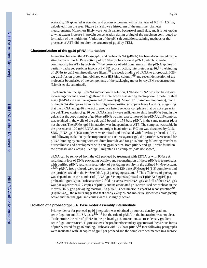
acetate. gp16 appeared as rounded and porous oligomers with a diameter of 9.5 +/− 1.5 nm,calculated from the area. Figure 2 (f) shows a histogram of the multimer diametermeasurements. Monomers likely were not visualized because of small size, and it is not knownto what extent increase in protein concentration during drying of the specimen contributed toformation of the multimers. Variation of the pH, salt conditions, staining methods or thepresence of ATP did not alter the structure of gp16 by TEM.
Characterization of the gp16-pRNA interactionInteraction between the ATPase gp16 and prohead RNA (pRNA) has been documented by thestimulation of the ATPase activity of gp16 by prohead-bound pRNA, which is neededcontinuously for ATP hydrolysis;24 the presence of additional mass on the pRNA spokes ofpartially packaged particles in a cryo-EM 3D reconstruction, interpreted as gp16;18 the bindingof pRNA to gp16 on nitrocellulose filters;48 the weak binding of pRNA to thioredoxin HIS-tag gp16 fusion protein immobilized on a HIS-bind column;49 and recent delineation of themolecular boundaries of the components of the packaging motor by cryoEM reconstruction(Morais et al., submitted).
To characterize the gp16-pRNA interaction in solution, 120-base pRNA was incubated withincreasing concentrations of gp16 and the interaction assessed by electrophoretic mobility shiftassay (EMSA) in a native agarose gel (Figure 3(a)). Mixed 1:1 (based on monomers), muchof the pRNA disappears from its fast migration position (compare lanes 1 and 2), suggestingthat the pRNA and gp16 interact to produce heterogeneous complexes that do not appear inthe gel. Three copies of gp16 per pRNA (lane 3) were sufficient to shift the pRNA band in thegel, and as the copy number of gp16 per pRNA was increased, more of the pRNA/gp16 complexwas retained in the wells of the gel. gp16 bound to 174-base pRNA in the same manner (datanot shown). The pRNA-gp16 interaction was independent of ATP. The complex was stable inthe presence of 100 mM EDTA and overnight incubation at 4°C but was disrupted by 0.1%SDS. pRNA-gp16(1:3) complexes were mixed and incubated with fiberless proheads (10:1),and following isolation by electrophoresis on a native agarose gel, the particles were tested forpRNA binding by staining with ethidium bromide and for gp16 binding following transfer tonitrocellulose and development with anti-gp16 serum. Both pRNA and gp16 were found onthe prohead, and excess pRNA/gp16 migrated as a complex (data not shown).
pRNA can be removed from the ϕ29 prohead by treatment with EDTA or with RNase A,resulting in loss of DNA packaging activity, and reconstitution of these pRNA-free proheadswith purified pRNA results in restoration of packaging activity in the defined in vitro system.22,50 pRNA-free proheads were reconstituted with 120-base pRNA/gp16 (1:3) complexes andthe particles tested in the in vitro DNA-gp3 packaging system.44 The efficiency of packagingwas dependent on the number of pRNA/gp16 complexes (mixed as 1 pRNA: 3 gp16) perprohead (Figure 3(b)). Proheads were 2-fold in excess over DNA-gp3, and all of the DNA-gp3was packaged when 5–7 copies of pRNA and its associated gp16 were used per prohead in thein vitro DNA-gp3 packaging reaction. As pRNA is pentameric in cryoEM reconstruction30(Figure 7(d)), the results suggested that nearly every pRNA molecule added was biologicallyactive and that the gp16 molecules were also highly active.
Isolation of a prohead/gp16 ATPase motor assembly intermediatePrior evidence for prohead-gp16 interaction was obtained by sucrose density gradientcentrifugation and ELISA tests,15, 48 but the role of pRNA in the interaction was not clear.To determine the role of pRNA in the prohead-gp16 interaction, sucrose density gradientcentrifugation was used. Figure 4 shows the predicted secondary structures of the various formsof pRNA tested for gp16 binding. Proheads with 174-base pRNA51 (see following paragraph)were incubated with 20 copies of gp16 per prohead and the complexes sedimented in a sucrose
Koti et al. Page 5
J Mol Biol. Author manuscript; available in PMC 2009 September 19.
NIH
-PA Author Manuscript
NIH
-PA Author Manuscript
NIH
-PA Author Manuscript

density gradient (Figure 5(a)). There was a clear shift in the position of proheads incubatedwith gp16, and the fast-sedimenting particles were isolated and shown by SDS-PAGE tocontain gp16 (Figure 6(a), lane 2). The copies of gp16 per prohead from densitometric analysiswas about one-half of that of the head-tail connector, which is known to contain 12 copies ofgp10.18 This prohead/174-base pRNA/gp16 interaction is Mg2+ dependent and ATPindependent. Proheads and gp16 interacted at high ionic strength (100 mM NaCl), but lowionic strength (10 mM) was needed to retain the bound gp16. The prohead/174-base pRNA/gp16 complexes isolated from the sucrose gradient were tested for in vitro DNA-gp3 packagingactivity upon addition of DNA-gp3 and ATP (Figure 6(b)). The isolated prohead/174-pRNA/ATPase intermediate packaged DNA-gp3 without further addition of gp16 (lane 5), and withthe same efficiency as proheads with 174-base pRNA mixed with gp16 in the in vitro reaction(lane 3), as the concentration of proheads used in the reaction of lane 3 was ~2-fold higher thanthat represented in lane 5. Figure 6(b), lane 7 shows that packaging remained about the sameupon addition of more gp16, and thus the isolated prohead/174-base pRNA/gp16 complex isa true intermediate with an intact and active packaging motor. The intermediate is stable andactive in packaging after overnight storage at 4°C. Also, just as EDTA treatment releases pRNAfrom the prohead,22 EDTA releases pRNA-gp16 as a complex from the prohead (data notshown).
174-base pRNA (with domains I and II)51 is cleaved to a 120-base form (domain I) byadventitious ribonucleases during prohead isolation, and both forms are active in reconstitutingpRNA-free proheads for DNA-gp3 packaging.25 RNA-free proheads were reconstituted with120-base pRNA (Figure 4(a)), purified by sucrose density gradient centrifugation, incubatedwith gp16 and run again on a sucrose gradient to determine the role of the 54-base domain IIin the prohead-gp16 interaction. gp16 co-sediments with proheads containing 120-base pRNAjust as with proheads having 174-base pRNA, and the isolated prohead/120-base pRNA/gp16complex is functional in DNA-gp3 packaging (data not shown). Thus domain II of pRNA wasconfirmed to be dispensable for in vitro packaging as shown previously.25 Figure 6(c), lane 2shows the composition of the prohead/120-base pRNA/gp16 complexes on a native agarosegel. A Western blot of the particle developed with anti-gp16 serum confirmed the presence ofgp16 on these proheads (data not shown).
pRNA dependency of the prohead-gp16 interaction and identification of the gp16 bindingsite on pRNA
The prohead-gp16 interaction is pRNA dependent, as pRNA-free proheads incubated withgp16 did not show the characteristic faster sedimentation demonstrated for the prohead/pRNA/gp16 particles on a sucrose gradient (Figure 5 (b)), and gp16 was not found on the isolatedparticles by SDS-PAGE (data not shown). Thus, stable binding of gp16 to proheads requiredpRNA.
Two functional domains of pRNA have been identified, a prohead binding domain and asegment involved in DNA translocation.33–35 The prohead binding domain consists of bases22–84 and was identified by ribonuclease footprinting and competitive binding experiments,while the A-helix that contains bases 1–28 and 117-92 was predicted to interact with gp16(Figure 4(a)).18 To further define the gp16 binding site on the A helix, mutant pRNAs withtruncations of the A-helix were employed (Figure 4(b)–(d)). 71-base pRNA is an A-helixdeletion mutant, which retains the prohead binding activity of 120-base pRNA but cannotsupport in vitro DNA-gp3 packaging.34 pRNA-free proheads reconstituted with 71-basepRNA and purified were incubated with gp16 and the mixture sedimented in a sucrose densitygradient (Figure 5(c)). The sedimentation profiles of particles reconstituted with 71-basepRNA, without and with gp16 incubation, were similar, and no gp16 was detected on particles
Koti et al. Page 6
J Mol Biol. Author manuscript; available in PMC 2009 September 19.
NIH
-PA Author Manuscript
NIH
-PA Author Manuscript
NIH
-PA Author Manuscript

by SDS-PAGE. These results confirm that the prohead-gp16 interaction is mediated by pRNAand show directly that the A-helix is the site of gp16 binding.
To further define the sequences and elements of the pRNA A-helix needed for gp16 binding,pRNA-free proheads were reconstituted with two different pRNA deletion mutants and studiedfor gp16 binding by use of sucrose gradient centrifugation and SDS-PAGE of the isolatedparticles. Mutants lacking the CCA bulge at positions 18–20 of pRNA (R7 mutant, Figure 4(d)), or the 14 residues at the 3′end of the A-helix (106-base pRNA, Figure 4(b)) have proheadbinding competitor activity but no DNA-gp3 packaging activity.33 pRNA-free proheadsreconstituted with these mutant pRNAs were able to bind gp16, indicating that the CCA bulgeor the 14 bases at the 3′end of the A-helix were not required for the initial gp16-pRNAinteraction, although particles so constituted were not active in DNA packaging (Table 2).
RNase footprinting of gp16 on pRNATo define the nucleotides of pRNA protected by gp16, ribonuclease footprinting analysis of 5′end-labeled [32P]120-base pRNA was performed with the RNases A, T1 and V1. Proheadsreconstituted with [32P]120-base pRNA were separated from unbound labeled pRNA bycentrifugation and used to produce footprints. In addition, free [32P]120-base pRNA was alsocomplexed with gp16 for mapping of the gp16 interactive domain. Both free pRNA andprohead-bound pRNA complexed with gp16 were protected from RNase A digestion atresidues 8 to 19, 5′ to 3 ′(Figure 7(a), (b)). RNase T1 did not produce cleavage outside theprohead binding domain and was not useful for the current studies. Footprints produced withRNase V1 showed additional protection at residues 5 and 31 (data not shown). The presentresults confirmed the ribonuclease footprint of the prohead on pRNA, which demonstratedprotection of residues 22–84 by the prohead with RNase V1 and enhanced cleavage of pRNAat residues 38–40 (data not shown)26.
Cryo-EM 3D-reconstruction of the prohead/pRNA/ gp16 packaging intermediateThe electron dense pRNA-spokes that radiate from the head-tail connector in cryo-EMreconstructions were postulated to be A-helices.18 A cryo-EM reconstruction of pRNA-freeproheads reconstituted with 71-base pRNA lacking the A-helix has confirmed this hypothesis(Morais et al., submitted). Cryo-EM 3D reconstruction of the prohead/174-base pRNA/gp16motor assembly intermediate showed mass that bridged the pRNA spokes and was interpretedas gp16 (Figure 7(d)–(g)). The localization of gp16 on the A-helix of pRNA was consistentwith the sucrose density gradient sedimentation (Figure 5) and ribonuclease footprinting(Figure 7(a)–(b)) results.
The presence of gp16 on the DNA-filled head was also demonstrated by incubating proheads/pRNA for 10 min (too short a time for tail assembly) in an extract of cells from a restrictiveinfection with the ϕ29 mutant sus7(614)-sus8(769)-sus14(1241), which is defective for theprohead scaffolding and major capsid proteins but that served as a source of DNA-gp3 andexcess gp16. The filled heads were isolated on a sucrose density gradient and analyzed by SDS-PAGE (Figure 8) and Western blotting (data not shown). gp16 remains on the filled head,apparently until displaced by subsequent neck/tail assembly. These results are consistent withthe presence of gp16 on filled heads produced by in vitro complementation of extracts providingproheads and gp16.17 Also, DNA-gp3 packaged into purified proheads in the defined invitro system yielded filled heads containing gp16 following sucrose gradient purification (datanot shown).
Koti et al. Page 7
J Mol Biol. Author manuscript; available in PMC 2009 September 19.
NIH
-PA Author Manuscript
NIH
-PA Author Manuscript
NIH
-PA Author Manuscript

Binding and supercoiling of DNA-gp3 by the prohead/pRNA/gp16 motor assemblyintermediate
gp16 binds and supercoils DNA-gp3 in vitro, and each coiled DNA retains about 3 copies ofgp16 when isolated in a sucrose density gradient.21 This interaction of gp16 with DNA-gp3is independent of the prohead, and the DNA-gp3/gp16 complex is packaged preferentially. Toreconcile these results with the present demonstration of the prohead/pRNA/gp16 intermediateand to gain insight on the order of assembly of gp16 in the initiation of DNA packaging invitro, the DNA-gp3 sedimentation assay was performed as described.21 [3H]DNA-gp3sedimented in the sucrose gradient as a peak centering on fraction 16 (Figure 9(a), black line).When DNA-gp3 was preincubated with gp16, the sedimentation rate of more than one-half ofthe molecules increased to give a new peak at fraction 13 (Figure 9(a), pink line). A Westerndot-blot of the sucrose gradient fractions, developed with anti-gp16 serum, confirmed the co-sedimentation of gp16 with DNA-gp3 (Figure 9(c)) [Figure 9(b) shows the position of gp16alone in the gradient]. Addition of proheads/pRNA did not alter the sedimentation state ofDNA-gp3 (Figure 9(d), green line). The prohead/pRNA/gp16 intermediate (Figure 9(d)),yellow line) produced a faster-sedimenting DNA-gp3, just as did gp16 (Figure 9(d), pink line).The presence of proheads and gp16 on the fast sedimenting DNA-gp3 was confirmed byWestern blotting with anti-gp10 and anti-gp16 sera, respectively. These results suggested thatgp16, assembled as a component of the DNA packaging motor onto proheads/pRNA, was ableto bind and supercoil the DNA-gp3 to produce an initiation complex for DNA packaging. Whilethis interaction and formation of the higher order complex is gp3-dependent, EMSA showedthat gp16 binds non-specifically to all HpaI fragments of proteinase K-treated DNA-gp3(Figure 9(e)).
DiscussionThe ATPase gp16 is a central component in packaging of the ϕ29 genome, interacting withpRNA and DNA-gp3 to constitute the packaging motor at the capsid portal vertex and thenproviding the energy for DNA translocation. The ATPase (terminase) large subunits of thedsDNA phages and Herpesvirus tend to be insoluble,1, 52 and production of the highlyhydrophobic gp16 in soluble form for study of structure and function has been problematic.Inclusion bodies of gp16 produced in E. coli (pPLc 2833) could only be partially solubilizedby the use of guanidinium chloride followed by urea, and therefore about 100 molecules wereused to package each DNA-gp3 in vitro.20 Co-expression of gp16 with GROESL in E. coliwith the intent of producing soluble protein resulted in an excess of GROESL which had to beremoved.48 The production of gp16 with an N-terminal thioredoxin and His-Tag in E. coliresulted in insoluble and inactive protein.53 Treatment with acetone or PEG was needed toimprove the solubility of the gp16-His-thioredoxin, and the protein was inactive in the acetone/PEG after the thioredoxin was cleaved away, making structural or functional studies of thenative protein impossible. The present preparation of gp16 in highly purified and soluble formin Bacillus subtilis (pSAC) will facilitate studies of gp16 structure and function.
The number of copies of the large terminase ATPase subunit required for optimal packagingactivity is not fully understood, although in T3 biochemical data suggests that 6 copies areneeded,11 and in T4, gp17 is predicted to form a ring surrounding the DNA on the procapsid.54 All the known terminase large subunits with ATPase activity exist predominantly asmonomers in solution.55–59 Self-association of terminase monomers into oligomers has beenreported by sedimentation velocity in λ and by chemical crosslinking in T4 phage.13,14 gp16exists in solution predominantly in a monomeric state at concentrations of 0.1 mg/ml, withtrimers and higher order structures at concentrations of 0.2–0.8 mg/ml as seen by gel filtration,sedimentation equilibrium studies, and electron microscopy (Figure 2). Early in vitropackaging studies demonstrated higher order concentration dependence for gp16, showing that
Koti et al. Page 8
J Mol Biol. Author manuscript; available in PMC 2009 September 19.
NIH
-PA Author Manuscript
NIH
-PA Author Manuscript
NIH
-PA Author Manuscript

multiple copies of gp16 were needed, while concentration dependence for proheads and DNA-gp3 was first order.16 The present study of gp16 produced in Bacillus subtilis (pSAC) showsthat the quantity of gp16 that was found on the isolated, fully active prohead/pRNA/gp16complex by SDS-PAGE was about one-half that of the dodecameric connector (Figure 6(a)).Similarly, terminase large subunits of phages T3, T7 and P22 bind the procapsid.60–62 Inphage λ, packaging is initiated by docking of the DNA-terminase (complex I) at the portalvertex, and amino acids of the large subunit C terminus are responsible for the capsid binding.63, 64
Amino acid sequence analysis indicates that the N-termini of many terminase large subunitshave a conserved ATPase center.8 All known and predicted members of DNA packagingATPase large subunits belong to the P-loop NTPase fold which have conserved nucleotidebinding (Walker A) and Mg+2 binding motifs (Walker B).8, 15 The ATPase domains of T4gp17 and ϕ29 gp16 appear to be similar. For example, the Walker B mutant D118E/E119D ingp16 of ϕ29 is inactive in DNA packaging (data not shown) as is the mutant D255E/E256D inT4 gp17, which binds ATP but is unable to precisely orient the ATP molecule.9 Also, thecrystal structures of P4 of ϕ12 and gp17 of T4 show a common ATPase domain.9,65 However,comparative genomic studies indicate that gp16 of ϕ29 may be a diverging independent lineagefrom the HerA/FtsK ATPases.66, 67 Sequence alignment and mutation data suggest that theadenine-binding motif54 is not conserved among the gp16 analogs and is different from thatof T4 gp17 (data not shown). Massey et al.68 have shown that FtsK ATPase exists as amonomer in solution, and formation of hexameric rings around DNA was dependent only onthe presence of DNA and was independent of nucleotide or magnesium ions. The FtsK ATPase,like gp16, differs from terminases in lacking domain fusions with their nuclease partners.
The ϕ29 DNA packaging motor differs primarily from its counterparts among the dsDNAphages by having an essential RNA component, pRNA. To date, pRNA has been confirmedonly in ϕ29 and its relatives,51 although Acidianus bottle shaped virus (ABV), an archaealphage, produces RNA with a predicted secondary structure similar to pRNA.69 Binding ofpRNA to the head-tail connector requires the basic residues RKR at the gp10 N terminus23and is the last step in morphogenesis of the mature prohead. Then gp16 binds to the pRNAoligomer to constitute the active packaging motor, and this binding was strictly dependent uponand mediated by the presence of pRNA (Figure 5 and Figure 6). Ribonuclease footprints ofgp16 on the prohead/pRNA showed protection of residues of the A-helix, extending to residues18–19 which correspond to CC of the CCA bulge (Figure 7). This result was confirmed bydemonstration that mutant pRNA lacking the A-helix did not bind gp16 (Figure 5(c)) and bycryo-EM reconstruction of the prohead-gp16 motor assembly intermediate which showed thatgp16 was bound to the A-helix spokes of pRNA (Figure 7). The 120-base pRNA ΔCCA bulgemutant has prohead binding competitor activity but no DNA-gp3 packaging activity.33 Eventhough the CC residues in the pRNA CCA bulge contribute to the gp16 binding, they were notrequired for the initial pRNA-gp16 interaction (Figure 4). This is similar to a previous reportthat the CCA bulge is dispensable for gp16 binding.49 The His-gp16-thioredoxin was foundto bind DNA nonspecifically and also to bind the pRNA/prohead more strongly than the pRNA-free capsid, although none of these complexes were tested for DNA packaging activity.49 TheRNase footprinting of gp16 on the prohead (Figure 7) should help model the gp16-pRNAinteraction. Endonuclease activity possessed by other phage large terminase subunits is notneeded in ϕ29 as the DNA is unit length and not packaged from a DNA concatemer.
In cryoEM reconstruction the pRNA and gp16 subunits have five-fold symmetry and areintimately associated (Figure 7). This and the fact that gp16 is a pRNA-dependentATPase24 lend credence to the idea that the ATPase subunits of the motor may be gp16/pRNAheterodimers. Once the gp16 has bound to pRNA, the pRNA subunits have been shown bycryoEM reconstruction to make contacts with the capsid, which rationalizes the finding that
Koti et al. Page 9
J Mol Biol. Author manuscript; available in PMC 2009 September 19.
NIH
-PA Author Manuscript
NIH
-PA Author Manuscript
NIH
-PA Author Manuscript

the ring of motor subunits is five-fold symmetric.30 Thus, pRNA ultimately links the gp16 tothe capsid, and whether and how tightly pRNA remains associated with the connector isunknown. The connector is shown by cryoEM reconstruction to be essentially situated in abasket formed by pRNA.30 In conflict with the above scenario, Ibarra et al.,48 reported that,by the use of ELISA and immuno-precipitation experiments, the connector N-terminus canbind gp16, as V8-treated connectors that lack the N-terminus did not interact with gp16; pRNA-gp16 interaction was also reported in this study. The gp16 used was in high excess in theseexperiments (3137/prohead, 115/connector and 3.3 × 10⁴/RNA), and none of these complexeswere shown to be active in DNA packaging.
It is not known if the pRNA molecules retain their novel intermolecular base pairinginteractions70, 71 in the active packaging motor, which might serve as a means ofcommunication among the ATPases in the coordination of motor function. The pRNA subunitshave been hypothesized to function sequentially, as incorporation of one inactive subunit of amutant pRNA has been reported to be sufficient to stop DNA packaging.27, 72 Tests of thesequential mode of action of gp16 (and the proposed pRNA/gp16 heterodimers) are an aim ofsingle molecule packaging studies in process.
ϕ29 gp3, covalently bound at the DNA 5’ termini (DNA-gp3), is needed for DNA packagingand has been viewed as a small terminase protein.10 gp3 also greatly enhances the efficiencyand selectivity of DNA packaging in bulk in vitro.44 In phage P22, the small terminase subunitgp3 is shown to bind DNA in a sequence independent manner and is a globular decamer insolution with a central annulus that can fit the DNA.59 In the phage λ small terminase subunitgpNu1, the NMR structure shows a HTH motif for DNA binding,73 and the T4 gp16 smallterminase also has a predicted HTH motif, whereas gp3 of ϕ29 lacks a HTH motif.74 Thegeneral strategy for genome packaging is thus retained in ϕ29 despite the apparent divergencefrom the conventional model with active participation of pRNA in the packaging motorfunction.
gp16 supercoils the DNA-gp3 packaging substrate in a reaction that is dependent upon gp3covalently bound at the DNA termini.21 This supercoiled DNA-gp3/gp16 complex ispreferentially packaged into proheads in vitro. Thus, the description here of a stable and activeprohead/pRNA/gp16 complex that can package DNA without additional gp16 (Figure 6)represents an alternative pathway of gp16 assembly and packaging initiation. Notably,however, this prohead/pRNA/gp16 intermediate was also capable of supercoiling DNA-gp3,just as demonstrated earlier with gp16 on free DNA-gp3 in the absence of proheads (Figure9).21 Figure 10 illustrates the alternative pathways for producing the prohead/pRNA/gp16/DNA-gp3 packaging initiation complex. To directly observe the events of initiation andprogression of DNA-gp3 packaging in vitro, various mixtures of the proheads/pRNA, gp16and DNA-gp3, without or with ATP, or complexes stalled in the packaging process with γS-ATP at different times, were prepared in BAC films,75 mounted on support films, rotary metalshadowed and observed by transmission electron microscopy (data not shown). DNA in theprocess of packaging appeared to be supercoiled and condensed, but the provisional resultshave not yet provided a clear picture of the structure of the initiation complex or the progressionof packaging. In another attempt to determine the pathway and events in packaging initiation,proheads and DNA-gp3 were isolated at times ranging from 45 to 90 min from a lysate of arestrictive infection of B. subtilis with the ϕ29 mutant sus14 (1241) that provides delayed lysis.gp16 was found in multiple copies on both the DNA-gp3 and the proheads (data not shown).The wrapping of supercoiled DNA by the free ϕ29 head-tail connector to remove negativesupercoils76 is a provocative reminder that the initiation of packaging may involve DNA-protein transactions within the motor that have not been contemplated. Further experimentationis needed to dissect the details of packaging initiation.
Koti et al. Page 10
J Mol Biol. Author manuscript; available in PMC 2009 September 19.
NIH
-PA Author Manuscript
NIH
-PA Author Manuscript
NIH
-PA Author Manuscript
Isolation of the prohead-pRNA-gp16 motor assembly intermediate has facilitated singlemolecule studies that allow DNA translocation to be measured from initiation to completion,allowing study of the previously uncharacterized early steps of packaging and thedemonstration of packaging forces >100 pN.4, 77 Prohead/pRNA/gp16 assemblyintermediates were attached to one bead, biotinylated DNA molecules were tethered tostreptavadin-coated microspheres, and packaging was initiated by bringing the beads intocontact in the presence of ATP. In these single molecule studies, gp3 is not required, whereasDNA without gp3 is packaged an order of magnitude less efficiently in bulk.44 The prohead/pRNA/gp16 complex and DNA are rapidly brought into close physical contact in the lasertweezers, possibly bypassing the need for gp3. It is also noteworthy that use of DNA-gp3 inthe single molecule studies results in great variation in DNA tether length upon initiation,indicating that the DNA-gp3 molecules have a higher order structure.4
Materials and MethodsPurification of DNA-gp3
DNA-gp3 and [3H]DNA-gp3 were isolated from lysates of Bacillus subtilis RD2 (sup−)infected with the mutant sus4(369)-sus8(22), defective for late transcription and production ofthe major capsid protein, respectively, as described.21 Proteinase K-treated DNA, furtherpurified in an isopycnic cesium chloride density gradient, was used to generate a HpaI digest,which was phenol extracted and used for the EMSA determination of gp16 binding.
Preparation of proheadsProheads with 174-base pRNA were purified from a lysate of Bacillus subtilis SpoOA12(sup−) cells infected with the mutant sus16(300)-sus14(1241) that is defective for the DNApackaging ATPase gp16 as described previously.30
Production and purification of soluble gp16 from Bacillus subtilisThe purified E/D segment of ϕ29 DNA-gp3 containing gene 16 was obtained by partialdigestion with EcoRI42 and cloned into the plasmid pUB18 that encodes kanamycin resistance.The sucrose inducible promoter SACB43 was inserted before gp16 gene and the resultingpSACB-gp16 plasmid moved into Bacillus subtilis WB30 (asporogenic and protease negative)by protoplast transformation78. The maximum yield of the soluble protein (10 mg/liter) wasobtained after induction of gene16 expression with 2% sucrose for 3 hr at 37°C. The expressionof gp16 in B. subtilis did not result in inclusion body formation as in E coli,20 and about 70%of the protein was found in the soluble fraction (supernatant). Sucrose-induced cells werepassed through a French press twice in a buffer containing 50 mM Tris-HCl (pH 7.8), 200 mMNaCl, 5% glycerol and 2 mM TCEP. The lysate was clarified by centrifugation at 10,000 rpmfor 15 min at 4°C in a SS-34 rotor. The supernatant was subjected to P11 phosphocellulose(Whatman) cation-exchange chromatography. The P11 column was equilibrated with lysisbuffer, washed with 8 bed volumes of 300 mM NaCl in 50 mM Tris-HCl (pH 7.8), 5% glyceroland 2 mM TCEP, and gp16 was eluted with 350 mM NaCl in the same buffer. The P11 eluatecontaining gp16 was further purified on a hydroxyapatite column (Biorad), which wasequilibrated with 300 mM NaCl in 50 mM Tris-HCl (pH 7.8), 5% glycerol and 2 mM TCEP.The column was washed with 3 bed volumes of 100 mM sodium phosphate (pH 6.8), 5%glycerol and 2 mM TCEP, followed by two bed volumes of 50 mM sodium phosphate (pH 6.8)in the same buffer. The protein was eluted by a ten-bed volume gradient of 100 mM to 900mM NaCl in 50 mM sodium phosphate (pH 6.8), 5% glycerol and 2 mM TCEP. Fractions wereanalyzed by SDS-PAGE, and peak fractions containing pure gp16 were stored in aliquots at−70°C. The identity of the protein was confirmed by a Western blot developed with polyclonalanti-gp16 serum. The average yield of the purified gp16 was 8 mg/liter of culture.
Koti et al. Page 11
J Mol Biol. Author manuscript; available in PMC 2009 September 19.
NIH
-PA Author Manuscript
NIH
-PA Author Manuscript
NIH
-PA Author Manuscript

In vitro DNA-gp3 packagingPackaging in the defined in vitro system was performed as described.44 DNA-gp3 (1µg) andproheads were mixed in the ratio of 1:2 in a total volume of 20 µl in 0.5X TMS buffer [25 mMTris-HCl (pH 7.8), 5 mM MgCl2 and 50 mM NaCl] containing 0.5 mM ATP. Generally 15copies (molecules) of gp16 per prohead were added and the mixture incubated at ambienttemperature for 15 min. The gp16 copy number varied as indicated. DNase I was added to 1µg/ml to digest the unpackaged DNA, while the packaged DNA was protected in the head. TheDNase was inhibited and the packaged DNA extracted from the filled heads by adding EDTAto 25 mM and proteinase K to 500 µg/ml, and the efficiency of packaging was quantifiedfollowing electrophoresis on a 0.8% agarose gel run in TBE buffer [89 mM Tris (pH 8.3), 89mM Boric acid and 2.5 mM EDTA].
Sedimentation equilibriumSedimentation equilibrium experiments were performed with purified gp16 equilibrated in abuffer containing 50 mM sodium phosphate (pH 6.8), 400 mM NaCl and 2 mM TCEP at 4°C.Two samples at each concentration of 0.2, 0.4 and 0.8 mg/ml were analyzed in an An50Ti rotorin a Beckman Optima XL-I analytical centrifuge. Data was collected at 280 nm and at threerotor speeds of 5,000, 8,000 and 11,000 rpm. Equilibrium was reached when scans taken 4 hrapart were super-imposable. Each data curve was the result of at least three averaged scans.Partial specific volume (υ) for gp16 calculated from the primary amino acid sequence usingSEDNTERP79 was 0.7373 ml/g. Based on the primary amino acid sequence, the calculatedmonomer molecular weight of gp16 was 38,965 daltons. Using SEDNTERP, the calculateddensity (σ) of the buffer was 1.02859 g/ml and the viscosity (η) was 0.0167043.
Data were initially fit to a self-association model using NONLIN,46 which yielded molecularweights suggestive of a trimer. Additional species that were included as fits were not good fora simple monomer to trimer model. The data at lower centrifugation speeds and higher proteinconcentrations were fit using SEDPHAT.47 gp16 forms larger molecular weight species thatare not reversible and are lost to the bottom of the cell. To determine the amount of loss, thearea of absorbance for each cell was determined at each centrifuge speed relative to the initialabsorbance at 3,000 rpm. This amounted to loss of 40% of the gp16 at 0.2–0.4 mg/ml and 65%loss at 0.8 mg/ml. The protein remaining in solution was fit as described above.
Gel filtrationGel filtration was performed using a Superdex 200 HR 10/30 (Amersham Pharmacia Biotech)column at 4°C in a buffer containing 50 mM sodium phosphate (pH 6.8), 400 mM NaCl and2 mM TCEP on an Akta-FPLC system (Amersham Pharmacia Biotech). Molecular weightstandards were analyzed in the same buffer and contained, in kDa, thyroglobulin (669), humanIgG (158), ovalbumin (44), myoglobin (17.6) and vitamin B12 (1.4).
Transmission electron microscopyGlow discharge treated holey carbon films on 400 mesh copper grids were floated on samplesof purified gp16 [50 µg/ml in 50 mM PIPES (pH 6.8) and 400 mM NaCl], negatively stainedwith 2% (w/v) uranyl acetate and imaged at 57,000 X magnification. Micrographs were madewith a Philips EM301 electron microscope. The diameter of gp16 particles from the electronmicrographs was determined by the use of Image J (NIH). Calibration was done frommicrographs of a carbon-grating replica (Fullam, NY).
Preparation of pRNA120-base and 106-base pRNA forms were generated by in vitro T7 transcription from pRT72plasmid linearized with DdeI and ApaLI and (Invitrogen), respectively, as described.33
Koti et al. Page 12
J Mol Biol. Author manuscript; available in PMC 2009 September 19.
NIH
-PA Author Manuscript
NIH
-PA Author Manuscript
NIH
-PA Author Manuscript
Alternatively, 120-base pRNA was produced from in vitro transcription of the BamHI-linearized pH120RNAH plasmid (Atz et al., unpublished results). The 71-base pRNAexpression construct pH71RNAH was generated by two rounds of PCR from the pRNA geneand by adding hammerhead ribozyme sequences to both ends of the template to generateuniform pRNA ends. The plasmid was cloned into pUC19 and transformed into E.coliDH5α The purified plasmid was linearized with SmaI and transcribed in vitro to obtain the 71-base pRNA. R7-pRNA was generated from Bacillus subtilis 12A (pUM102) as described.33All forms of pRNA were purified by electrophoresis in denaturing urea-acrylamide gels.80Individual pRNA bands were located by UV shadowing over polyethyleneimine UV254-cellulose plates with a 254 nm light source. pRNA bands were excised and the pRNA elutedtwice by diffusion into TEN buffer [10mM Tris-HCl (pH 8.0), 1mM EDTA and 100 mM NaCl]for 3 hr at 4°C.
Electrophoretic mobility shift assay120-base pRNA was incubated with increasing concentrations of gp16 in 50 mM Tris- HCl(pH 7.8), 10 mM MgCl2 and 100 mM NaCl for 20 min at room temperature. 5% glycerol,0.05% bromophenol blue and 0.05% xylene cyanol were added to each reaction, the complexeswere resolved by electrophoresis in a 0.8% agarose gel in TB buffer [89 mM Tris-HCl (pH8.3) and 89 mM Boric acid] for 3 hr, and the gels were stained with ethidium bromide. EMSAwas also performed with HpaI-digested ϕ29 DNA fragments and gp16 in a similar manner.
Preparation of RNA-free proheadsPurified proheads were diluted 10-fold in TMS buffer and digested with 1µg/ml of RNase Afor 15 min at room temperature.44 These RNA-free proheads were purified on a 10–40%sucrose density gradient in TMS buffer (50 mM Tris-HCl (pH 7.8), 10 mM MgCl2 and 100mM NaCl) by centrifugation for 3hr at 35,000 rpm in a SW55 rotor. The prohead band wascollected and the particles pelleted in TMS buffer.
Reconstitution of pRNA-free proheads with pRNATen copies of the pRNA produced from in vitro transcription were used per prohead and themixture incubated for 10 min at room temperature for reconstitution. These reconstitutedproheads were repurified on a sucrose gradient to remove excess pRNA.
Isolation of the prohead/pRNA/gp16 ATPase motor assembly intermediateProheads with 174-base pRNA and gp16 were incubated in TMS buffer without ATP atambient temperature for 20 min at a ratio of 20 gp16 molecules per prohead. After incubation,the reaction mixture was loaded on a 5–20% sucrose gradient in TM buffer [50 mM Tris-HCl(pH 7.6) and 5 mM MgCl2] and centrifuged at 35,000 rpm for 30 min in a SW55 rotor at 4°C.The peak band was collected with a syringe and 20 µl used for testing the in vitro DNA-gp3packaging activity upon addition of DNA-gp3 and ATP. 100 µl of the fraction was precipitatedwith 10% TCA and subjected to SDS-PAGE to determine the composition of the prohead/pRNA/gp16 intermediate. pRNA-free proheads and proheads reconstituted with various formsof pRNA with A-helix deletions (71-base, 106-base and R7-pRNA (Fig4(b)–(d)) were used toscreen for binding of gp16 as described above.
RNase footprinting120-base pRNA was 5′-labeled with Optikinase™ (USB Corp) and [γ-32P]ATP after removalof the 5′ terminal phosphate with shrimp alkaline phosphatase. The end-labeled pRNA wasfurther purified by electrophoresis in denaturing urea-acrylamide gels. The pRNA bands wereeluted from the gel into TEN buffer and concentrated by isopropanol precipitation. pRNA
Koti et al. Page 13
J Mol Biol. Author manuscript; available in PMC 2009 September 19.
NIH
-PA Author Manuscript
NIH
-PA Author Manuscript
NIH
-PA Author Manuscript

obtained after precipitation was used for footprinting and reconstitution of pRNA-free proheadsfor footprinting.
140 µg of pRNA-free proheads were reconstituted with 2.8 µg of 5′ end-labeled 120-basepRNA (6 pRNA/prohead; 7 × 10⁵ CPM/µg of pRNA) in TM buffer for 20 min at roomtemperature. Proheads were separated from the unbound pRNA by dilution into 5 ml TM bufferand pelleting at 35,000 rpm for 5 hr in the SW55 rotor at 4°C as described.26
Prohead-bound [32P]pRNA, [32P]pRNA alone (2 × 10⁴ CPM) and [32P]pRNA complexed withgp16 were digested with varying concentrations of RNase A (Ambion), RNase T1 or RNaseV1(Ambion) in 10 µl reactions for 15 min at ambient temperature. pRNA was extracted with200 µl of a mixture containing 1 part 50 mM Tris-HCl (pH 7.6), 10 mM EDTA, 20µg/ml yeasttRNA (Ambion) and 1 part phenol. The aqueous phases were precipitated with 0.6 volumes ofisopropanol containing 20 µg/ml glycogen. Pellets were resuspended in 4 M urea, 20%glycerol, 0.05% xylene cyanol and 0.05% bromophenol blue and run on a 10% denaturing 8.3M urea polyacrylamide gel.
Cryo-electron microscopyProheads with 174-base pRNA and prohead/174-base pRNA/gp16 particles were flashfrozenon holey grids in liquid ethane. Images were recorded at 39,000X magnification with a CM200FEG microscope, with electron dose levels of approximately 20 e−/Å2. All micrographs weredigitized at 4.24 Å pixel−1 using a Zeiss SCAI scanner.
Individual particle images were boxed, floated, and preprocessed to normalize mean intensitiesand variances and to remove linear background gradients. Reference projections of a previouslypublished prohead reconstruction30 were used to initially classify particles for three-dimensional reconstruction. The resulting model was used to recalculate reference projectionsfor better particle classification. Several cycles of iterative particle classification andreconstruction were performed until convergence had been reached. Structure factor phasesand amplitudes were modified as indicated by the parameters of the contrast transfer function.All steps of the reconstruction process, including determination of the contrast transfer functionparameters, were performed with the program EMAN.81 Five-fold symmetry was assumed inthe reconstruction. The number of particles incorporated in the final reconstruction was 1705,and the final resolution of the reconstruction was 18.5 Å, as determined by the Fourier shellcorrelation method using a correlation coefficient of 0.5 between independent half data sets asthe cut-off criterion.
Sedimentation assay of DNA-gp16 and DNA-prohead/pRNA/gp16[3H]DNA-gp3 (1µg) was diluted in water and heated for 5 min at 37°C. A 0.1 volume of 10xTM buffer and 12 copies of gp16 per DNA were added to give a final reaction volume of 100µl. The mixture was incubated at ambient temperature for 10 min and then centrifuged in a 5–20% linear sucrose density gradient containing TM [50 mM Tris-HCl (pH 7.6), 5 mM NaCl]buffer in the SW55 rotor at 35,000 rpm for 2 hr at 20°C. Fractions were collected and the[3H]DNA-gp3 quantified by liquid scintillation counting. Also, samples of the fractions weretreated with 5µg/ml DNase I at room temperature for 20 min, and dot-blots of the samples weredeveloped with gp16-antiserum. As a control, gp16 alone was sedimented on a sucrose gradientand samples of the fractions used for dot-blots and gp16 quantification. Prohead/pRNA/gp16complexes (12 copies of gp16 per prohead) were prepared, incubated with [3H]DNA-gp3, andsedimented in a sucrose gradient to study their activity in binding and supercoiling DNA-gp3as described.21
Koti et al. Page 14
J Mol Biol. Author manuscript; available in PMC 2009 September 19.
NIH
-PA Author Manuscript
NIH
-PA Author Manuscript
NIH
-PA Author Manuscript

Extract preparationB. subtilis SpoOA12 (sup−) was infected with the mutant sus7(614)-sus8(769)-sus14(1241)that is defective for the prohead scaffold and capsid proteins and is a source of gp16, andextracts of these infected cells were prepared as described.16
Abbreviations usedpRNA, prohead RNA; gp, gene product; cryo-EM, cryo-electron microscopy; EMSA,electrophoretic mobility shift assay..
AcknowledgementsWe thank Siddhartha Jena for DLS analysis, Steve Erickson and Rockney Atz for technical assistance, Daniel Cohenand Charlene Peterson for expertise with figures. M.C.M was supported by NSF grant MCB-0443899. This researchwas supported by NIH grants DE-003606 and GM-059604.
References1. Catalano, CE. Viral genome packaging machines: an overview. In: Catalano, CE., editor. Viral genome
packaging machines: Genetics, structure and mechanism. Georgetown, TX: 2005. p. 1-4.LandesBioscience/Eurekah.com
2. Jardine, PJ.; Anderson, DL. DNA packaging in double-stranded DNA phages. In: Calendar, R., editor.The bacteriophages. New York: oxford press; 2006. p. 49-65.
3. Smith DE, Tans SJ, Smith SB, Grimes S, Anderson DL, Bustamante C. The bacteriophage phi29 portalmotor can package DNA against a large internal force. Nature (London) 2001;413:748–752. [PubMed:11607035]
4. Rickgauer JP, Fuller N, Grimes S, Jardine PJ, Anderson DL, Smith DE. Portal motor velocity andinternal force resisting viral DNA packaging in bacteriophage phi29. Biophys. J 2008;94:159–167.[PubMed: 17827233]
5. Murialdo H, Becker A. Head morphogenesis of complex double-stranded deoxyribonucleic acidbacteriophages. Microbiol. Rev 1978;42:529–576. [PubMed: 362149]
6. Earnshaw WC, Casjens SR. DNA packaging by the double-stranded DNA phages. Cell 1980;21:319–331. [PubMed: 6447542]
7. Feiss M. Terminase, a viral enzyme involved in the recognition, cutting and packaging of bacteriophageλ chromosomes. Trends genet 1986;2:100–104.
8. Mitchell MS, Matsuzaki S, Imai S, Rao VB. Sequence analysis of bacteriophage T4 DNA packaging/terminase genes 16 and 17 reveals a common ATPase center in the large subunit of viral terminases.Nucl. Acids. Res 2002;30:4009–4021. [PubMed: 12235385]
9. Sun S, Kondabagil K, Gentz PM, Rossmann MG, Rao VB. The structure of the ATPase that powersDNA packaging into bacteriophage T4 procapsids. Mol. Cell 2007;25:943–949. [PubMed: 17386269]
10. Guo P, Peterson C, Anderson DL. Prohead and DNA-gp3 dependent ATPase activity of the DNApackaging protein gp16 of bacteriophage ϕ29. J. Mol. Biol 1987;197:229–236. [PubMed: 2960820]
11. Fujisawa H, Shibata H, Kato H. Analysis of interactions among factors involved in the bacteriophageT3 DNA packaging reaction in a defined in vitro system. Virol 1991;185:788–794.
12. Kanamaru S, Kondabagil K, Rossmann MG, Rao VB. The functional domains of bacteriophage T4terminase. J. Biol. Chem 2004;279:40795–40801. [PubMed: 15265872]
13. Kondabagil K, Zhang Z, Rao VB. The DNA translocating ATPase of bacteriophage T4 packagingmotor. J. Mol. Biol 2006;363:786–799. [PubMed: 16987527]
14. Feiss, M.; Catalano, CE. Bacteriophage Lambda terminase and the mechanism of viral DNApackaging. In: Catalano, CE., editor. Viral genome packaging machines: Genetics, structure andmechanism. Georgetown, TX: 2005. p. 5-39.Landes Bioscience/Eurekah.com
15. Guo P, Peterson C, Anderson DL. Initiation events in in-vitro packaging of bacteriophage ϕ29 DNA-gp3. J. Mol. Biol 1987;197:219–228. [PubMed: 3119862]
Koti et al. Page 15
J Mol Biol. Author manuscript; available in PMC 2009 September 19.
NIH
-PA Author Manuscript
NIH
-PA Author Manuscript
NIH
-PA Author Manuscript

16. Bjornsti MA, Reilly BE, Anderson DL. In vitro assembly of the Bacillus subtilis bacteriophage ϕ29.Proc. Natl.Acad. Sci. USA 1981;78:5861–5865. [PubMed: 6795639]
17. Bjornsti MA, Reilly BE, Anderson DL. Morphogenesis of bacteriophage ϕ29 of Bacillus subtilis:oriented and quantized in vitro packaging of DNA-gp3. J. Virol 1983;45:383–396. [PubMed:6185695]
18. Simpson AA, Tao Y, Leiman PG, Badasso MO, He Y, Jardine PJ, Olson NH, Morais MC, GrimesS, Anderson DL, et al. Structure of the bacteriophage phi29 DNA packaging motor. Nature2000;408:745–750. [PubMed: 11130079]
19. Gausch A, Pous J, Ibarra B, Gomis-Ruth FX, Valpuesta JM, Sousa N, Carrascosa JL, Coll M. Detailedarchitecture of a DNA translocating machine: the high-resolution structure of the bacteriophage ϕ29connector particle. J. Mol. Biol 2002;315:663–676. [PubMed: 11812138]
20. Guo P, Grimes S, Anderson DL. A defined system for in vitro packaging of DNA-gp3 of the Bacillussubtilis bacteriophage ϕ29. Proc. Natl. Acad. Sci. USA 1986;83:3505–3509. [PubMed: 3458193]
21. Grimes S, Anderson DL. The bacteriophage ϕ29 packaging proteins supercoil the DNA ends. J. Mol.Biol 1997;266:901–914. [PubMed: 9086269]
22. Guo P, Erickson S, Anderson DL. A small viral RNA is required for in vitro packaging ofbacteriophage ϕ29 DNA. Science 1987a;236:690–694. [PubMed: 3107124]
23. Atz R, Ma S, Gao J, Anderson DL, Grimes S. Alanine scanning and FE-BABE probing of thebacteriophage ϕ29 prohead RNA-connector interaction. J. Mol. Biol 2007;369:239–248. [PubMed:17433366]
24. Grimes S, Anderson DL. RNA dependence of the bacteriophage ϕ29 DNA packaging ATPase. J.Mol. Biol 1990;215:559–566. [PubMed: 1700132]
25. Wichitwechkarn J, Bailey S, Bodley JW, Anderson DL. Prohead RNA of bacteriophage ϕ29: size,stoichiometry and biological activity. Nucl Acids Res 1989;17:3459–3468. [PubMed: 2498842]
26. Reid RJ, Bodley JW, Anderson DL. Characterization of the prohead-pRNA interaction ofbacteriophage ϕ29. J. Biol. Chem 1994;269:5157–5162. [PubMed: 8106496]
27. Trottier M, Guo P. Approaches to determine stoichiometry of viral assembly components. J. Virol1997;71:487–494. [PubMed: 8985375]
28. Ibarra B, Caston JR, Llorca O, Valle M, Valpuesta JM, Carrascosa JL. Topology of the componentsof the DNA packaging machinery in the phage ϕ29 prohead. J. Mol. Biol 2000;298:807–815.[PubMed: 10801350]
29. Morais MC, Tao Y, Olson NH, Grimes S, Jardine PJ, Anderson DL, Baker TS, Rossmann MG.Cryoelectron-microscopy image reconstruction of symmetry mismatches in bacteriophage ϕ29. J.Struct. Biol 2001;135:38–46. [PubMed: 11562164]
30. Morais MC, Choi KH, Koti JS, Chipman PR, Anderson DL, Rossmann MG. Conservation of capsidstructure in tailed dsDNA bacteriophages: the pseudoatomic structure of ϕ29. Mol. Cell 2005;18:149–159. [PubMed: 15837419]
31. Shu D, Zhang H, Jin J, Guo P. Counting of six pRNAs of phi29 DNA packaging motor with customizedsingle-molecule dual-view system. EMBO. J 2007;26:527–537. [PubMed: 17245435]
32. Grimes S, Jardine PJ, Anderson DL. Bacteriophage phi29 DNA packaging. Adv. Virus Res2002;58:255–294. [PubMed: 12205781]
33. Reid RJ, Bodley JW, Anderson DL. Identification of bacteriophage ϕ29 prohead RNA domainsnecessary for in vitro DNA-gp3 packaging. J. Biol. Chem 1994;269:9084–9089. [PubMed: 8132646]
34. Reid RJ, Zhang F, Benson S, Anderson DL. Probing the structure of bacteriophage ϕ29 prohead RNAwith specific mutations. J. Biol. Chem 1994;269:18656–18661. [PubMed: 8034614]
35. Zhang C, Lee CS, Guo P. The proximate 5′ and 3′ ends of the 120-base viral RNA (pRNA) are crucialfor the packaging of bacteriophage phi29. Virol 1994;201:77–85.
36. Zhang C, Garver K, Guo P. Inhibition of phage phi29 assembly by antisense oligonucleotides targetingviral pRNA essential for DNA packaging. Virol 1995;211:568–676.
37. Chemla YR, Aathavan K, Michaelis J, Grimes S, Jardine PJ, Anderson DL, Bustamante C. Mechanismof force generation of a viral DNA packaging motor. Cell 2005;122:683–692. [PubMed: 16143101]
38. Hendrix RW. Symmetry mismatch and DNA packaging in large bacteriophages. Proc. Natl. Acad.Sci. USA 1978;75:4779–4783. [PubMed: 283391]
Koti et al. Page 16
J Mol Biol. Author manuscript; available in PMC 2009 September 19.
NIH
-PA Author Manuscript
NIH
-PA Author Manuscript
NIH
-PA Author Manuscript

39. Baumann RG, Mullaney J, Black LW. Portal fusion protein constraints on function in DNA packagingof bacteriophage T4. Mol. Microbiol 2006;61:16–32. [PubMed: 16824092]
40. Hugel T, Michaelis J, Hetherington CL, Jardine PJ, Grimes S, Walter JM, Falk W, Anderson DL,Bustamante C. Experimental test of connector rotation during DNA packaging into bacteriophagephi29 capsids. PLoS Biol 2007;5:e59. [PubMed: 17311473]
41. Lebedev AE, Krause MH, Isidro AL, Vagin AA, Orlova EV, Turner J, Dodson EJ, Tavares P, AntsonAA. Structural framework for DNA translocation via the viral portal protein. EMBO. J2007;26:1984–1994. [PubMed: 17363899]
42. Garvey KJ, Saedi MS, Ito J. The complete sequence of Bacillus phage ϕ29 gene16: a protein requiredfor the genome encapsidation reaction. Gene 1985;40:311–316. [PubMed: 3879485]
43. Li M, Wong SL. Cloning and characterization of the GROESL operon from Bacillus subtilis. J.Bacteriol 1992;174:3981–3992. [PubMed: 1350776]
44. Grimes S, Anderson DL. In vitro packaging of bacteriophage ϕ29 DNA restriction fragments and therole of the terminal protein gp3. J. Mol. Biol 1989;209:91–100. [PubMed: 2530357]
45. Bjornsti MA, Reilly BE, Anderson DL. Bacteriophage ϕ29 proteins required for in vitro DNA-gp3packaging. J. Virol 1984;50:766–772. [PubMed: 6427474]
46. Johnson M, Correia JJ, Yphantis DA, Halvorson H. Analysis of data from the analytical ultracentrifugeby nonlinear least-squares techniques. Biophys. J 1981;36:575–588. [PubMed: 7326325]
47. Vistica J, Dam J, Balbo A, Yikilmaz E, Mariuzza RA, Rouault TA, Schuck P. Sedimentationequilibrium analysis of protein interactions with global implicit mass conservation constraints andsystematic noise decomposition. Anal. Biochem 2004;326:234–256. [PubMed: 15003564]SEDPHAT: freeware from www.analyticalultracentrifugation.com
48. Ibarra B, Valpuesta J, Carrascosa JL. Purification and functional characterization of p16, the ATPaseof the bacteriophage ϕ29 packaging machinery. Nucl. Acid. Res 2001;29:4264–4273.
49. Lee T, Guo P. Interaction of gp16 with pRNA and DNA for genome packaging by the motor ofbacterial virus phi29. J. Mol. Biol 2006;356:589–599. [PubMed: 16376938]
50. Guo P, Bailey S, Bodley JW, Anderson DL. Characterization of the small RNA of the bacteriophageϕ29 DNA packaging machine. Nucl. Acid. Res 1987d;15:7081–7090.
51. Bailey S, Wichitwechkarn J, Johnson D, Reilly BE, Anderson DL, Bodley JW. Phylogenetic analysisand secondary structure of the Bacillus subtilis bacteriophage RNA required for DNA packaging. J.Biol. Chem 1990;265:22365–22370. [PubMed: 2125049]
52. Przech AJ, Yu D, Weller SK. Point mutations in exon I of the herpes simplex virus putative terminasesubunit, UL15, indicate that the most conserved residues are essential for cleavage and packaging.J.Virol 2003;77:9613–9621. [PubMed: 12915573]
53. Huang LP, Guo P. Use of PEG to acquire highly soluble DNA-packaging enzyme gp16 of bacterialvirus phi29 for stoichiometry quantification. J. Virol. Methods 2003;109:235–244. [PubMed:12711068]
54. Draper B, Rao BV. An ATP hydrolysis sensor in the DNA packaging motor from bacteriophage T4suggests an inch-type translocation mechanism. J. Mol. Biol 2007;369:79–94. [PubMed: 17428497]
55. Gual A, Camacho AG, Alonso CJ. Functional analysis of the terminase large subunit, G2P, of Bacillussubtilis Bacteriophage SPP1. J. Biol.Chem 2000;275:35311–35319. [PubMed: 10930407]
56. Leffers G, Rao VB. Biochemical characterization of an ATPase activity associated with the largepackaging subunit gp17 from bacteriophage T4. J. Biol. Chem 2000;275:37127–37136. [PubMed:10967092]
57. Paris W, Rubinchik S, Yang Y, Gold M. A new procedure for the purification of bacteriophage λterminase enzyme and its subunits. Properties of gene product A the large subunit. J. Biol. Chem1994;269:13564–13574. [PubMed: 8175792]
58. Casjens, S.; Weigele, P. DNA packaging by bacteriophage P22. In: Catalano, CE., editor. Viralgenome packaging machines: Genetics, structure and mechanism. Georgetown, TX: 2005. p.80-88.Landes Bioscience/Eurekah.com
59. Nemecek D, Gilcrease EB, Kang S, Prevelige PE Jr, Casjens S, Thomas GJ Jr. Subunit conformationsand assembly states of a DNA-translocating motor: The terminase of bacteriophage P22. J. Mol. Biol2007;374:817–836. [PubMed: 17945256]
Koti et al. Page 17
J Mol Biol. Author manuscript; available in PMC 2009 September 19.
NIH
-PA Author Manuscript
NIH
-PA Author Manuscript
NIH
-PA Author Manuscript

60. Serwer P. Evolution and the complexity of bacteriophages. Virol. J 2007;4:30–39. [PubMed:17355641]
61. Morita M, Tasaka M, Fujisawa H. Structure and functional domains of the large subunit of thebacteriophage T3 DNA packaging enzyme: importance of the C-terminal region in prohead binding.J. Mol. Biol 1995;245:635–644. [PubMed: 7844832]
62. Eppler K, Wyckoff E, Goates J, Parr R, Casjens S. Nucleotide sequence of the bacteriophage P22genes required for DNA packaging. Virol 1991;183:519–538.
63. Yeo A, Feiss M. Specific interaction of terminase, the DNA packaging enzyme of bacteriophagelambda, with the portal protein of the prohead. J. Mol. Biol 1995;245:141–150. [PubMed: 7799432]
64. Sippy J, Feiss M. Analysis of a mutation affecting the specificity domain for the prohead binding ofthe bacteriophage lambda terminase. J. Bacteriol 1992;174:850–856. [PubMed: 1531050]
65. Kainov DE, Pirttimaa M, Tuma R, Butcher SJ, Thomas GJJ, Bamford DH, Makeyev EV. RNApackaging device of double-stranded RNA bacteriophages, possibly as simple as hexamer of P4protein. J. Biol. Chem 2003;278:48084–48091. [PubMed: 12966097]
66. Burroughs AM, Iyer LM, Aravind L. Comparative genomics and evolutionary trajectories of viralATP dependent DNA-packaging systems. Gene and Prot. Evol 2007;3:48–65.
67. Iyer LM, Makarova KS, Koonin EV, Aravind L. Comparative genomics of the FtsK HerA superfamilyof pumping ATPases: implications for the origins of chromosome segregation, cell division and viralcapsid packaging. Nucl. Acid. Res 2004;32:5260–5279.
68. Massey TH, Mercogliano CP, Yates J, Sherratt DJ, Lowe J. Double-stranded DNA translocation:Structure and mechanism of hexameric FtsK. Mol. Cell 2006;23:457–469. [PubMed: 16916635]
69. Peng X, Basta T, Haring M, Garrett RA, Prangishvili D. Genome of Acidianus bottle-shaped virusand insights into the replication and packaging mechanisms. Virol 2007;364:237–243.
70. Zhang F, Lemieux S, Wu X, Arnaud SD, McMurray CT, Major F, Anderson DL. Function ofhexameric RNA in packaging of bacteriophage ϕ29 DNA in vitro. Mol. Cell 1998;2:141–147.[PubMed: 9702201]
71. Guo P, Zhang C, Chen C, Garver K, Trottier M. Inter-RNA interaction of phage ϕ29 pRNA to forma hexameric complex for viral DNA transportation. Mol. Cell 1998;2:149–155. [PubMed: 9702202]
72. Chen C, Guo P. Sequential action of six virus-encoded DNA-packaging RNAs during phage ϕ29genomic DNA translocation. J. Virol 1997;71:3864–3871. [PubMed: 9094662]
73. de Beer TD, Fang J, Ortega M, Yang Q, Maes L, Duffy C, Berton N, Sippy J, Overduin M, Feiss M,Catalano CE. Insights into specific DNA recognition during the assembly of a viral genomepackaging machine. Mol. Cell 2002;9:981–991. [PubMed: 12049735]
74. Kamtekar S, Berman JA, Wang J, Lazaro MJ, Devega M, Blanco L, Salas M, Steitz TA. The ϕ29DNA polymerase: protein-primer structure suggests a model for the initiation to elongation transition.EMBO J 2006;25:1335–1343. [PubMed: 16511564]
75. Vollenweider H, Sogo JM, Koller T. A routine method for protein free spreading of double and singlestranded nucleic acid molecules. Proc. Natl. Acad. Sci. USA 1975;72:83–87. [PubMed: 164030]
76. Turnquist S, Simon M, Egelman E, Anderson D. Supercoiled DNA wraps around the bacteriophageϕ29 head-tail connector. Proc. Natl. Acad. Sci. USA 1992;89:10479–10483. [PubMed: 1438237]
77. Fuller DN, Rickgauer JP, Jardine PJ, Grimes S, Anderson DL, Smith DE. Ionic effects on viral DNApackaging and portal motor function in bacteriophage ϕ29. Proc. Natl. Acad. Sci. USA2007;104:11245–11250. [PubMed: 17556543]
78. Chang S, Cohen SNC. High frequency transformation of Bacillus subtilis protoplasts by plasmidDNA. Mol. Gen. Genetics 1979;168:111–115.
79. Laue, TM.; Shah, BD.; Ridgeway, TM.; Pelletier, SL. Analytical Ultracentrifugation in Biochemistryand Polymer Science. Harding, S.; Rowe, A.; Horton, J., editors. Cambridge: Royal Society ofChemistry; 1992. SEDNTERP: freeware from ww.jphilo.mailway.com
80. Wichitwechkarn J, Johnson D, Anderson DL. Mutant prohead RNAs in the in vitro packaging ofbacteriophage ϕ29 DNA-gp3. J. Mol. Biol 1992;223:991–998. [PubMed: 1538407]
81. Ludtke SJ, Baldwin PR, Chiu W. EMAN: Semiautomated software for high resolution single-particlereconstructions. J. Struct.Biol 1999;128:82–97. [PubMed: 10600563]
Koti et al. Page 18
J Mol Biol. Author manuscript; available in PMC 2009 September 19.
NIH
-PA Author Manuscript
NIH
-PA Author Manuscript
NIH
-PA Author Manuscript

Figure 1.Purification and in vitro DNA-gp3 packaging activity of the ATPase gp16. (a) SDS-PAGE ofsamples from steps in the purification of gp16 following expression in Bacillus subtilis(pSACB-gp16). The lanes show: 1) the supernatant from the cell lysate; 2–3) P11 cationexchange column peak fractions; 4–8) hydroxyapatite column peak fractions. (b) Packagingactivity of purified gp16 determined by the in vitro DNA-gp3 packaging assay (Methods). Thelanes show: 1) the input DNA-gp3 added to the reaction; 3–9) DNA that was packaged in thepresence of 15, 10, 8, 6, 4, 3 and 2 copies (molecules) of gp16 per prohead, respectively; 2 and10) negative controls in which ATP and gp16 were omitted, respectively, from the packagingreaction.
Koti et al. Page 19
J Mol Biol. Author manuscript; available in PMC 2009 September 19.
NIH
-PA Author Manuscript
NIH
-PA Author Manuscript
NIH
-PA Author Manuscript
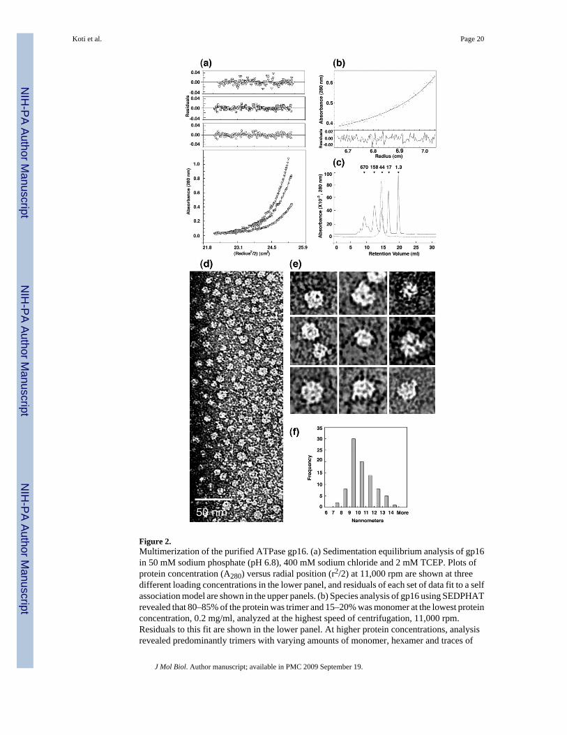
Figure 2.Multimerization of the purified ATPase gp16. (a) Sedimentation equilibrium analysis of gp16in 50 mM sodium phosphate (pH 6.8), 400 mM sodium chloride and 2 mM TCEP. Plots ofprotein concentration (A280) versus radial position (r2/2) at 11,000 rpm are shown at threedifferent loading concentrations in the lower panel, and residuals of each set of data fit to a selfassociation model are shown in the upper panels. (b) Species analysis of gp16 using SEDPHATrevealed that 80–85% of the protein was trimer and 15–20% was monomer at the lowest proteinconcentration, 0.2 mg/ml, analyzed at the highest speed of centrifugation, 11,000 rpm.Residuals to this fit are shown in the lower panel. At higher protein concentrations, analysisrevealed predominantly trimers with varying amounts of monomer, hexamer and traces of
Koti et al. Page 20
J Mol Biol. Author manuscript; available in PMC 2009 September 19.
NIH
-PA Author Manuscript
NIH
-PA Author Manuscript
NIH
-PA Author Manuscript

dodecamer (text). (c) Gel filtration elution profiles of proteins in the same buffer used forsedimentation equilibrium studies, plotted as protein concentration (A280) versus retentionvolume (ml). The dashed line shows the elution profile of gp16 at approximately 100 µg/ml atthe peak. The solid line shows the elution profile of MW standards with molecular weight(kDa) indicated across the top of the profile. (d) and (e) Transmission electron micrographs ofgp16 negatively stained with 2% (w/v) uranyl acetate (Methods). (f) Histogram showing themeasurement of the diameter of gp16 from electron micrographs (n = 88).
Koti et al. Page 21
J Mol Biol. Author manuscript; available in PMC 2009 September 19.
NIH
-PA Author Manuscript
NIH
-PA Author Manuscript
NIH
-PA Author Manuscript

Figure 3.Binding of gp16 to pRNA, and the activity of the pRNA-gp16 complex in in vitro DNA-gp3packaging. (a) EMSA of 120-base pRNA incubated with increasing concentrations of gp16.The lanes show: 1) 120-base pRNA alone; 2–6) shift of pRNA in the presence of 1, 3, 5, 8 and10 copies (molecules) of gp16 per pRNA molecule, respectively; 7) empty; 8) 10 copies ofgp16 alone. (b) 120-base pRNA/gp16 (1:3) was added in increasing amounts to pRNA-freeproheads and the mixtures tested for activity in the in vitro DNA-gp3 packaging assay. Thelanes show: 1) input DNA-gp3 added to the reaction; 3) empty; 4–8) packaged DNA in thepresence of pRNA:gp16 copies per prohead of 1:3, 2:6, 5:15, 7:21 and 10:30, respectively; 2)negative control without ATP
Koti et al. Page 22
J Mol Biol. Author manuscript; available in PMC 2009 September 19.
NIH
-PA Author Manuscript
NIH
-PA Author Manuscript
NIH
-PA Author Manuscript

Figure 4.Secondary structure prediction of various forms of pRNA tested for gp16 binding. (a) 120-base pRNA. (b) 106-base pRNA with a 14-base deletion at the 3′ end of the A-helix. (c) 71-base pRNA with a deletion of the A-helix. (d) R7-pRNA with deletion of the CCA bulge (bases18–20), indicated by an arrow.
Koti et al. Page 23
J Mol Biol. Author manuscript; available in PMC 2009 September 19.
NIH
-PA Author Manuscript
NIH
-PA Author Manuscript
NIH
-PA Author Manuscript

Figure 5.The sedimentation rate of proheads/pRNA increased in the presence of gp16 and was dependenton the presence of the pRNA A-helix. (a) Sucrose density gradient centrifugation profiles ofproheads/174-base pRNA (solid line) and proheads/174-base pRNA incubated with gp16(dashed line). Sedimentation is from left to right. gp16 was bound to proheads containing 174-base pRNA in the faster sedimenting peak as revealed by SDS-PAGE (Figure 6 (a)) and by invitro DNA-gp3 packaging (Figure 6(b)). (b) Sucrose density gradient profile of pRNA-freeproheads (solid line) and pRNA-free proheads incubated with gp16 (dashed line), showing thatgp16 did not alter the sedimentation rate of the pRNA-free proheads; gp16 was not found onthese proheads by SDS-PAGE. (c) Sucrose density gradient profile of proheads /71-base pRNA
Koti et al. Page 24
J Mol Biol. Author manuscript; available in PMC 2009 September 19.
NIH
-PA Author Manuscript
NIH
-PA Author Manuscript
NIH
-PA Author Manuscript

(solid line) and proheads/71-base pRNA incubated with gp16 (dashed line), showing that gp16did not alter the sedimentation rate of these proheads; gp16 was not found on the particles. Thegradients in a), b) and c) were run at different times and under different conditions, and thepeak positions are not comparable.
Koti et al. Page 25
J Mol Biol. Author manuscript; available in PMC 2009 September 19.
NIH
-PA Author Manuscript
NIH
-PA Author Manuscript
NIH
-PA Author Manuscript
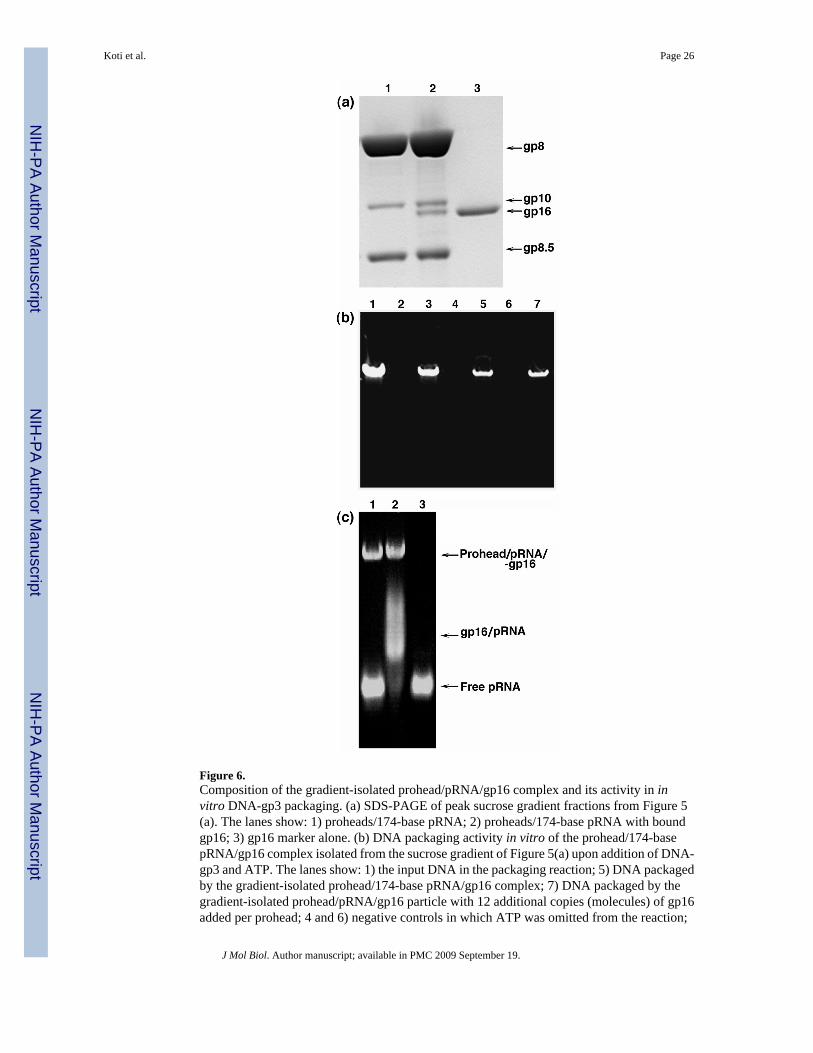
Figure 6.Composition of the gradient-isolated prohead/pRNA/gp16 complex and its activity in invitro DNA-gp3 packaging. (a) SDS-PAGE of peak sucrose gradient fractions from Figure 5(a). The lanes show: 1) proheads/174-base pRNA; 2) proheads/174-base pRNA with boundgp16; 3) gp16 marker alone. (b) DNA packaging activity in vitro of the prohead/174-basepRNA/gp16 complex isolated from the sucrose gradient of Figure 5(a) upon addition of DNA-gp3 and ATP. The lanes show: 1) the input DNA in the packaging reaction; 5) DNA packagedby the gradient-isolated prohead/174-base pRNA/gp16 complex; 7) DNA packaged by thegradient-isolated prohead/pRNA/gp16 particle with 12 additional copies (molecules) of gp16added per prohead; 4 and 6) negative controls in which ATP was omitted from the reaction;
Koti et al. Page 26
J Mol Biol. Author manuscript; available in PMC 2009 September 19.
NIH
-PA Author Manuscript
NIH
-PA Author Manuscript
NIH
-PA Author Manuscript

3) packaging in which proheads/pRNA and gp16 were mixed and not sedimented; 2) ATPnegative control for the reaction of lane 3. The prohead concentration was 2 times higher inthe reaction of lane 3 than in the reactions of lanes 4–7 where the prohead/pRNA/gp16 complexwas isolated from the sucrose gradient. (c) EMSA analysis of proheads/120-base pRNAincubated with gp16. The lanes show: 1) proheads/120-base pRNA, where the bulk of thepRNA dissociates from the proheads during electrophoresis; 2) the prohead/120-base pRNA/gp16 complex, revealing release of pRNA/gp16 as a complex; 3) free 120-base pRNA marker.
Koti et al. Page 27
J Mol Biol. Author manuscript; available in PMC 2009 September 19.
NIH
-PA Author Manuscript
NIH
-PA Author Manuscript
NIH
-PA Author Manuscript

Figure 7.Localization of gp16 on pRNA. (a)–(c) Ribonuclease footprinting of gp16 on 120-base pRNA.[32P]120-base pRNA and prohead-bound [32P]120-base pRNA were incubated with gp16, themixtures treated with RNase A and the products separated by denaturing gel electrophoresis(Methods). An alkaline hydrolysis ladder was generated from 5′ end-labeled pRNA. (a) and(b) RNase A digestion. The lanes show: 1–4) treatment with 10−2 µg/ml RNase A; 5–8)treatment with 10−3 µg/ml RNase A; 1 and 5) free [32P]120-base pRNA; 2 and 6) [32P]120-base pRNA with gp16; 3 and 7) prohead-bound [32P]120-base pRNA; and 4 and 8) prohead-bound [32P]120-base pRNA with gp16. (c) gp16 and prohead composite footprint on pRNA.Shaded regions represent residues protected by gp16, the open box represents residues
Koti et al. Page 28
J Mol Biol. Author manuscript; available in PMC 2009 September 19.
NIH
-PA Author Manuscript
NIH
-PA Author Manuscript
NIH
-PA Author Manuscript

protected by the prohead, and regions shown in the box bounded by the discontinuous linerepresent enhanced cleavages by RNase V1 when pRNA is bound to the proheads26. (d)–(g)Cryo-EM 3D reconstruction of the prohead/174-base pRNA/gp16 packaging intermediate. (d)prohead with 174 base pRNA. (e) Cross section of the prohead/pRNA, same view as in (d). (f)Prohead/174-base pRNA/gp16. (g) Cross section of the prohead/pRNA/gp16, same view as in(f).
Koti et al. Page 29
J Mol Biol. Author manuscript; available in PMC 2009 September 19.
NIH
-PA Author Manuscript
NIH
-PA Author Manuscript
NIH
-PA Author Manuscript

Figure 8.The isolated DNA filled head retains gp16. SDS-PAGE analysis of DNA-filled heads isolatedin a sucrose density gradient. The gradient sample was prepared by incubating proheads/pRNAfor 10 min with a head-defective extract of cells from a restrictive infection with the ϕ29 mutantsus7(614)-sus8(769)-sus14(1241), which served as a source of DNA-gp3 and excess gp16.The lanes show: 1) filled heads consisting of capsid (gp8), connector (gp10), gp16 and headfibers (gp8.5); 2) gp16 marker; and 3) prohead marker.
Koti et al. Page 30
J Mol Biol. Author manuscript; available in PMC 2009 September 19.
NIH
-PA Author Manuscript
NIH
-PA Author Manuscript
NIH
-PA Author Manuscript

Figure 9.Binding and supercoiling of DNA-gp3 by both gp16 and the prohead/pRNA/gp16 intermediate.(a) The sedimentation rate of DNA-gp3 was increased by binding of gp16, previously shownto be due to gp3- and gp16-dependent supercoiling of DNA-gp321. The black line representsthe gradient profile of DNA-gp3 and the pink line DNA-gp3 incubated with gp16.Sedimentation is from right to left. (b) Free gp16 (gradient not shown) was found primarily inthe sucrose gradient fractions 23-20 in the dot-blot developed with polyclonal gp16 antiserum.(c) Dot-blot of fractions from the sucrose gradient of DNA-gp3 incubated with gp16 (Figure9(a)), shows co-sedimentation of gp16 with DNA-gp3, notably from fraction 16 to the bottomof the gradient. (d) gp16 on the prohead-pRNA supercoils DNA-gp3. Sucrose gradient profilesof DNA-gp3 (black line), DNA-gp3 + proheads (green line), DNA-gp3 + gp16 (pink line),DNA-gp3 + gp16 + proheads (sky blue line), DNA-gp3 + gp16 complex incubated withproheads (blue line), prohead/gp16 complex incubated with DNA-gp3 (yellow line). Allproheads contained 174-base pRNA. There was no alteration in the sedimentation rate of DNA-gp3 by proheads (green line). (e) Native agarose gel stained with ethidium bromide,demonstrating EMSA analysis of ϕ29 gp3-free DNA HpaI fragments (6784, 2549, 2341, 1781,1777, 1714, 1608 and 731bp) in the presence of gp16. The lanes show: 1) 0.5 µg of HpaI DNAfragments; 2–6) the same DNA incubated with 6, 12, 24, 48, 48 copies of gp16 per DNA,respectively; 7) empty lane; 8) 48 copies of gp16 alone. gp16 binds to all of the HpaI fragmentsin a sequence independent manner.
Koti et al. Page 31
J Mol Biol. Author manuscript; available in PMC 2009 September 19.
NIH
-PA Author Manuscript
NIH
-PA Author Manuscript
NIH
-PA Author Manuscript

Figure 10.Alternative pathways for producing the prohead/pRNA/gp16/DNA-gp3 packaging initiationcomplex. (a) DNA-gp3 forms a lariat by interaction of the terminal gp3 with DNA, independentof sequence, and gp16 binds to the lariat loop junction to effect supercoiling of the DNA inconcert with gp3; this supercoiled DNA-gp3-gp16 complex is preferentially packaged21. (b)gp16 binds the prohead/pRNA to constitute a packaging intermediate that can be isolated andis fully active without additional gp16. This prohead/pRNA/gp16 complex is hypothesized tobind and supercoil DNA-gp3 at the prohead portal vertex. Scaffolding protein exits aspackaging proceeds upon hydrolysis of ATP, and the tail components are sequentiallyassembled on the DNA-filled head to yield ϕ29.
Koti et al. Page 32
J Mol Biol. Author manuscript; available in PMC 2009 September 19.
NIH
-PA Author Manuscript
NIH
-PA Author Manuscript
NIH
-PA Author Manuscript

NIH
-PA Author Manuscript
NIH
-PA Author Manuscript
NIH
-PA Author Manuscript
Koti et al. Page 33
Table 1Apparent molecular mass (kDa) of gp16 by sedimentation equilibrium
gp16 (mg/ml) 5000 rpm 8000 rpm 11000 rpm
0.2 100.5 104.7 96.50.4 157.7 128.7 107.50.8 156.2 126.8 104.4
Values are calculated from the fit of 2 cells at each concentration using Nonlin46. Partial specific volume (υ) of 0.7375, density (σ) of 1.02859, and
viscosity (η) of 0.00167 were calculated using SEDNTERP79. See methods for details.
J Mol Biol. Author manuscript; available in PMC 2009 September 19.

NIH
-PA Author Manuscript
NIH
-PA Author Manuscript
NIH
-PA Author Manuscript
Koti et al. Page 34
Table 2Wild-type and truncated forms of pRNA tested for gp16 binding
pRNA Type Prohead Binding gp16 Binding Packaging Activity
174-base Wild-type + + +120-base Wild-type + + +106-base 3′ A-helix deletion + + −71-base A-helix deletion + − −
R7 A-helix CCA deletion + + −
J Mol Biol. Author manuscript; available in PMC 2009 September 19.