Diversity and biological significance of sex hormone-binding ...
43
HAL Id: hal-00550179 https://hal.archives-ouvertes.fr/hal-00550179 Submitted on 24 Dec 2010 HAL is a multi-disciplinary open access archive for the deposit and dissemination of sci- entific research documents, whether they are pub- lished or not. The documents may come from teaching and research institutions in France or abroad, or from public or private research centers. L’archive ouverte pluridisciplinaire HAL, est destinée au dépôt et à la diffusion de documents scientifiques de niveau recherche, publiés ou non, émanant des établissements d’enseignement et de recherche français ou étrangers, des laboratoires publics ou privés. Diversity and biological significance of sex hormone-binding globulin in fish, an evolutionary perspective Julien Bobe, yann Guiguen, Alexis Fostier To cite this version: Julien Bobe, yann Guiguen, Alexis Fostier. Diversity and biological significance of sex hormone- binding globulin in fish, an evolutionary perspective. Molecular and Cellular Endocrinology, Elsevier, 2009, 316 (1), pp.66. 10.1016/j.mce.2009.09.017. hal-00550179
-
Upload
khangminh22 -
Category
Documents
-
view
0 -
download
0
Transcript of Diversity and biological significance of sex hormone-binding ...
HAL Id: hal-00550179https://hal.archives-ouvertes.fr/hal-00550179
Submitted on 24 Dec 2010
HAL is a multi-disciplinary open accessarchive for the deposit and dissemination of sci-entific research documents, whether they are pub-lished or not. The documents may come fromteaching and research institutions in France orabroad, or from public or private research centers.
L’archive ouverte pluridisciplinaire HAL, estdestinée au dépôt et à la diffusion de documentsscientifiques de niveau recherche, publiés ou non,émanant des établissements d’enseignement et derecherche français ou étrangers, des laboratoirespublics ou privés.
Diversity and biological significance of sexhormone-binding globulin in fish, an evolutionary
perspectiveJulien Bobe, yann Guiguen, Alexis Fostier
To cite this version:Julien Bobe, yann Guiguen, Alexis Fostier. Diversity and biological significance of sex hormone-binding globulin in fish, an evolutionary perspective. Molecular and Cellular Endocrinology, Elsevier,2009, 316 (1), pp.66. 10.1016/j.mce.2009.09.017. hal-00550179
Accepted Manuscript
Title: Diversity and biological significance of sexhormone-binding globulin in fish, an evolutionary perspective
Authors: Julien Bobe, Yann Guiguen, Alexis Fostier
PII: S0303-7207(09)00500-0DOI: doi:10.1016/j.mce.2009.09.017Reference: MCE 7329
To appear in: Molecular and Cellular Endocrinology
Received date: 26-5-2009Revised date: 15-9-2009Accepted date: 16-9-2009
Please cite this article as: Bobe, J., Guiguen, Y., Fostier, A., Diversity and biologicalsignificance of sex hormone-binding globulin in fish, an evolutionary perspective,Molecular and Cellular Endocrinology (2008), doi:10.1016/j.mce.2009.09.017
This is a PDF file of an unedited manuscript that has been accepted for publication.As a service to our customers we are providing this early version of the manuscript.The manuscript will undergo copyediting, typesetting, and review of the resulting proofbefore it is published in its final form. Please note that during the production processerrors may be discovered which could affect the content, and all legal disclaimers thatapply to the journal pertain.

Page 1 of 41
Accep
ted
Man
uscr
ipt
1
Diversity and biological significance of sex hormone-
binding globulin in fish, an evolutionary perspective
Julien Bobe, Yann Guiguen, Alexis Fostier
INRA, UR1037 SCRIBE, IFR140, Ouest-Genopole, F-35000 Rennes, France
Corresponding author:
J Bobe
INRA, UR1037 SCRIBE, Fish Reproduction Group, Campus de Beaulieu, F-35000 Rennes,
France
Email : [email protected]
Tel : (+33) 2 23 48 57 24
Fax : (+33) 2 23 48 50 20
*Manuscript

Page 2 of 41
Accep
ted
Man
uscr
ipt
2
Keywords
Shbga, Shbgb, Estradiol, Steroids, teleosts, Trout
Abtract
In fish, two different genes, shbga and shbgb, exist that encode for very different proteins.
Shbga is the ortholog of mammalian Shbg and was found in all investigated teleosts. In
contrast, Shbgb is highly divergent and appears to be a salmonid-specific protein. Here, we
review existing data on fish Shbga and Shbgb that have been obtained in chondrichthyes and
osteichtyes. Even though other significant expression sites exist, existing data indicate that
Shbga is mainly expressed in liver and subsequently secreted into the blood as a homodimer.
In contrast, Shbgb is mainly expressed in the ovary, probably secreted as a monomer, and
could contribute to the regulation of local steroid action. Binding studies indicate a
specialization of circulating Shbg during evolution towards the preferential binding of
estradiol and testosterone in teleosts. In contrast, specific fish steroids such as 11-oxo-
androgens and oocyte maturation-inducing steroids that are crucial for reproduction are poorly
bound by either form of Shbg.

Page 3 of 41
Accep
ted
Man
uscr
ipt
3
Introduction
Sex hormone-binding globulins (Shbg) are mainly known as carrier blood proteins involved
in the transport of sex steroids in plasma and in the regulation of their bioavailability to target
organs. The Shbg protein has initially been identified in the beta-globulin fraction of the
human serum (Rosner et al., 1969). This field has subsequently benefited from many
molecular studies of the genes and transcripts encoding Shbg in many mammalian species
(Joseph et al., 1991;Kahn et al., 2002;Rosner, 2006;Nakhla et al., 2009) (see also reviews in
the present issue). In fish, Shbg has been found in the plasma of an elasmobranch, the skate
(Raja radiata) (Freeman and Idler, 1969), and of a teleost, the rainbow trout (Oncorhynchus
mykiss) (Fostier and Breton, 1975) a few decades ago, and subsequently identified and studied
in a wide variety of fish species as reviewed here. However, in contrast to mammals, the
molecular analysis of teleost shbg genes was not carried out until recently (Miguel-Queralt et
al., 2004;Miguel-Queralt et al., 2005;Bobe et al., 2008b) and available data remain scarce.
Here, after providing a phylogenetic characterization of Shbg proteins in fish, we first aim at
reviewing existing molecular and biochemical data that have been obtained not only in
osteichtyes, but also in chondrichthyes. The recent molecular data on shbg genes will be
reviewed and discussed in the light of existing biochemical data that have previously been
gathered in a wide variety of species. In addition, we will take advantage of the basal
evolutionary position of teleosts within the vertebrate lineage to discuss the evolution of
binding characteristics of vertebrate Shbg proteins. Finally, although several alternatively
spliced transcripts have been identified in mammals (Nakhla et al., 2009), it should be
stressed that, in contrast to mammals, two highly divergent Shbg proteins exist in fish. Shbg
alpha form (Shbga) is clearly the ortholog of the mammalian Shbg and has been found in all
investigated teleost species. In contrast, the Shbg beta form (Shbgb) initially identified in
Page 4 of 41
Accep
ted
Man
uscr
ipt
4
rainbow trout (Bobe et al., 2008b) has subsequently been found in other salmonid species
(Miguel-Queralt et al., 2009) but could not be identified in any other non-salmonid teleost
species investigated to date. This offers a great opportunity to discuss the evolution of Shbgb
binding characteristics and putative biological functions with regards to sequence divergence
and conserved sequence features.
For clarity reasons, Shbga and Shbgb symbols will be used in the text when proteins with
known sequences were specifically studied while Shbg symbol will be used for Shbg proteins
in general or when binding or biochemical studies were carried out directly from plasma or
tissues.
Shbg genes, structure and evolutionary history
Evolution of fish Shbg genes
To date, Shbg proteins have only been characterized molecularly in bony fish (osteichtyes
class), more specifically in the teleost superorder that includes all modern ray-finned fish
species (actinopterygians fishes). The phylogenetic reconstruction of the evolutionary history
of Shbg proteins in these teleost fish (Figure. 1) indicates that most species have only one
Shbg, designated as the Shbg alpha form (Shbga), that is closely related to mammalian Shbg
proteins. However, in some fish species, such as the common carp (Cyprinus carpio), two
duplicated genes are found (shbga1 and shbga2) that are probably the result of the recent
whole genome duplication reported in this species (Larhammar and Risinger, 1994). In
contrast to these recent gene duplications that resulted in highly similar Shbg proteins, a
highly-divergent Shbg beta form, designated as Shbgb, has been found in the salmonid
lineage (Bobe et al., 2008b;Miguel-Queralt et al., 2009). The phylogenetic position of Shbgb
proteins is intriguing as they all branched together with high bootstrap values at the root of the
tetrapod Shbg branch (Figure. 1). This topology suggests an ancient duplication followed by a
Page 5 of 41
Accep
ted
Man
uscr
ipt
5
subsequent lost in all non-salmonid vertebrate species (Miguel-Queralt et al., 2009).
However, it is also possible that shbga and shbgb genes are the result of a duplication of a
common shbg ancestor gene that occurred after salmonid radiation. The additional whole
genome duplication (3R) that occurred in salmonids would be consistent with this hypothesis
(Allendorf and Thorgaard, 1984). The topology of the phylogenetic tree could thus be
explained by a long-branch attraction artifact that is classically observed for highly divergent
sequences (Delsuc et al., 2005). It should also be stressed that accelerated molecular evolution
appears to be quite frequent following fish genome duplications (Steinke et al., 2006). The
high divergence between Shbga and Shbgb would thus suggest a functional shift through
either sub- or neo-functionalization that would be consistent with the very different
expression patterns observed (Bobe et al., 2008b;Miguel-Queralt et al., 2009).
Structure of Shbg-alpha (Shbga) in teleosts
In all species studied to date the classical Shbg (Shbga) exists as a homodimeric glycoprotein
characterized by a common structure composed by two laminin G (LG) like domains that
contains two sets of conserved cysteines that form intramolecular disulphide bridges. As
secreted proteins, a signal peptide sequence is cleaved from the precursor protein to give rise
to the mature protein. Apart from its role as a steroid carrier protein, there is some evidence
that this protein can mediate steroid signaling by binding to membrane associated proteins
(Hryb et al., 2002). Accordingly, a putative N-terminal membrane receptor-binding domain
that could bind to specific receptors on cell membranes has been characterized in humans
(Khan et al., 1990). Plasma Shbg is normally glycosylated and this is thought to influence the
biological half-life of the protein (Cousin et al., 1999). Classical Shbg (Shbga) sequences in
fish have been characterized recently but only in a very few number of fish species including
zebrafish (Danio rerio) (Miguel-Queralt et al., 2004), Atlantic sea bass (Dicentrarchus
labrax) (Miguel-Queralt et al., 2005), rainbow trout (Bobe et al., 2008b) and coho salmon
Page 6 of 41
Accep
ted
Man
uscr
ipt
6
(Miguel-Queralt et al., 2009). Other information on fish Shbg sequences can also be deduced
from published cDNA, expressed sequence tags (ESTs) databases, and from the analysis of
genomic sequences available in some fish species. The analysis of these sequences and their
deduced proteins indicates that the fish classical Shbga proteins are typically 380-400 amino
acid long. These proteins are poorly conserved at the amino-acid level as they only share 40 to
80 % identity with each other, depending on their evolutionary proximity, and around 25 %
identity with the human sequence (Table 1). Despite this relatively poor sequence identity, all
fish Shbga have classical Shbg features including, the two well conserved LG-domains (N
and C-terminal) with the conserved cysteine residues that participate in the disulphide bridges,
a signal peptide sequence that is cleaved from the precursor protein to produce the mature
protein, and a putative N-terminal membrane receptor-binding domain (Figure 2). Fish Shbga
also contain three N-glycolysation sites that are relatively well conserved among fish
sequences (Figure 2) with the exception of the Japanese medaka (Oryzias latipes) that does
not share the third site of N-glycosylation (Miguel-Queralt et al., 2005). However, these N-
glycosylation sites are unique to fish Shbga and are not conserved in mammalian SHBG
sequences (Miguel-Queralt et al., 2005).
A few biochemical determinations of fish Shbg molecular weight have been performed on
native or recombinant proteins using gel filtration and/or polyacrylamide gel electrophoresis:
80 Kda in skate (Freeman and Idler, 1969), 150-180 Kda in spiny dogfish (Squalus Acanthias)
(Ho et al., 1980), 64 Kda in Japanese eel (Anguilla japonica) (Chang et al., 1994), 194 Kda in
common carp (Chang and Lee, 1992), 105±8.7 Kda in zebrafish (Miguel-Queralt et al., 2004),
118.3±11.5 Kda in Atlantic sea bass (Miguel-Queralt et al., 2005), and 153 Kda for the coho
salmon Shbg alpha form (Miguel-Queralt et al., 2009). With the exception of the Japanese eel
these observations are in agreement with the presence of a circulating homodimer made from
glycosylated subunits (Miguel-Queralt et al., 2005). Besides species differences, variability in
Page 7 of 41
Accep
ted
Man
uscr
ipt
7
molecular weights may also be due to the occurrence of Shbg aggregation / polymerization
which is less pronounced when dithiothreitol or 2-mercaptoethanol was used as reducing
agents in buffer preventing disulfide bonds (Freeman and Idler, 1969;Ho et al., 1980). These
differences could also be due to different numbers of oligosaccharides chains since, as
indicated above, three N-glycosylation sites may be found in several species (Miguel-Queralt
et al., 2004). In mammals, observed isoforms have been considered to be the result of partial
use of the two N-glycosylation sites on each Shbg subunit (Danzo et al., 1989;Bocchinfuso et
al., 1992;Cousin et al., 1999).
The structure of the shbga gene has been initially described in the zebrafish (Miguel-Queralt
et al., 2004) in which the transcription unit encoding the Shbga protein has been found to span
close to 13 kb in length and to contain 8 exons. This gene organization with 8 exons is well
conserved in teleost fish; at least within shbga genes deduced from species with an available
genome sequence (Figure 3). Despite important differences in gene length (from 2.2 kb for
the Japanese pufferfish (Takifugu rubipres) to more than 13 kb in the zebrafish) all intron-
exon boundaries are conserved (data not shown). This highly conserved structure of the shbga
gene among teleost species was not totally unexpected as the zebrafish gene organization was
already found to be well conserved with the cognate human sequence, including a perfect
conservation of the intron-exon boundaries (Miguel-Queralt et al., 2004). In contrast, none of
the regulatory regions, that are conserved in human and rat genes, have been identified in the
1 kb proximal promoter of zebrafish and Japanese pufferfish shbga genes (Miguel-Queralt et
al., 2004).
Structure of the salmonid-specific Shbg beta form (Shbgb)
The salmonid-specific Shbgb proteins are highly divergent Shbg proteins (Bobe et al., 2008b)
as they share less identity (around 20%) with the classical fish Shbg (Shbga) than the human
SHBG with fish Shbga (around 25%). However, despite these important sequence
Page 8 of 41
Accep
ted
Man
uscr
ipt
8
divergences (see Table 1 and Figure 2) with Shbga proteins, they still display a good
structural conservation of the tandem repeats of laminin G-like domains that are crucial for
steroid-binding and dimer formation, or of the putative N-terminal amino-acid stretch that
could bind to a Shbg membrane receptor. This suggests some well conserved functionality
even though major structural differences have been found between Shbga and Shbgb proteins.
For instance, and in contrast to Shbga, the Chinook salmon Shbgb has an estimated molecular
weight of 68 kda and is only found as a monomer in plasma or when expressed as a
recombinant protein (Miguel-Queralt et al., 2009). The fact that Shbgb would not
homodimerize spontaneously has been correlated with the lack of the highly conserved Val89
and Leu122 (Miguel-Queralt et al., 2009) that have been found to be crucial for dimerization
in human (Avvakumov et al., 2001). Other hypotheses explaining this absence of dimerization
include major differences in N-glycosylation sites, as none of these sites are conserved with
the Shbga form (Figure 2), or the lack of essential cation biding sites required for this
dimerization (Miguel-Queralt et al., 2009). To date, no gene structure has been reported for
these highly divergent salmonid-specific shbgb genes. The gene structure information would,
however, provide important information as the comparison of the vicinity of shbga and shbgb
genes in salmonids would probably help to conclude whether or not these genes arise from a
duplication that took place following the salmonid-specific genome duplication or following a
more ancient duplication. Comparison of regulatory sequences between the proximal
promoter sequences of shbga and shgbb genes may also be of great interest with regards of
the important tissues specific expression of these two shbg genes.
Page 9 of 41
Accep
ted
Man
uscr
ipt
9
Tissue distribution and expression sites
Shbga
The tissue distribution of shbga transcript has been studied in a limited number of teleost
species. In zebrafish, shbga mRNA was detected by in situ hybridization and RT-PCR in
digestive tract and hepatopancreas (Miguel-Queralt et al., 2004). In addition, a low expression
was detected in testis using RT-PCR (Miguel-Queralt et al., 2004). Using
immunocytochemistry, zebrafish Shbga protein was immunodetected in hepatopancreas,
intestine, and testis (Miguel-Queralt et al., 2004). In Atlantic sea bass, northern blot analysis
showed that shbga mRNA was strongly expressed in liver but could not be detected in brain,
digestive tract, kidney, or gonads (Miguel-Queralt et al., 2007). Using immunocytochemistry,
seabass Shbga protein was detected in liver, intestine, and testis. Shbga immunoreactivity was
also detected in the connective tissue around the ovary and a positive signal was observed
around the post-vitellogenic oocytes of a mature female (Miguel-Queralt et al., 2007). This
would be in agreement with the finding of a Shbg-type binding in ovarian interstitial fluids of
another perciformes species, the spotted weakfish Cynoscion nebulosus (Laidley and Thomas,
1994). In salmonids, shbga is also strongly expressed in liver (Bobe et al., 2008b;Miguel-
Queralt et al., 2009). In rainbow trout, a strong mRNA expression was also observed in spleen
while low expression levels could be detected in gills, stomach, and pituitary using
quantitative RT-PCR (Bobe et al., 2008b). In coho salmon, a low expression could also be
detected in gills, stomach, and brain (Miguel-Queralt et al., 2009). Together, these data
indicate, in agreement with mammalian literature, that liver is the main expression site of
Shbga in teleost fish. It is thus likely that the hepatic expression of Shbga, followed by
secretion into the blood stream, is resulting in plasma Shbga. This is fully supported by
existing data demonstrating that Shbg present in the culture medium of rainbow trout

Page 10 of 41
Accep
ted
Man
uscr
ipt
10
hepatocytes and in plasma had similar steroid binding characteristics and electrophoretic
mobility (Foucher et al., 1991). However, the significant expression found in several other
tissues (e.g. spleen) suggests that, at least in some species, circulating Shbga could also be
from extra-hepatic origin. In mammals, a local expression of Shbg in several target organs has
also been evidenced and associated with a modulation of the steroidogenic signal (Hryb et al.,
2002;Kahn et al., 2002). Similarly, non-hepatic expression sites of Shbga in teleosts could
also be associated with local action in target organs. However, further investigation would be
necessary in fish to identify potential transcripts variants given that the transcriptional
regulation of vertebrate shbg genes could be somewhat complex. For instance, 19 unique
transcripts exhibiting differential tissular expression and deriving from 3 different promoters
have been recently identified in humans (Nakhla et al., 2009).
In mammals, a protein initially named androgen-binding protein (Abp) due to its binding
affinity for androgen was found in the male reproductive tract of several species (Joseph,
1994). Further investigations have however demonstrated, in several mammalian species, that
both Shbg and the so-called Abp were in fact encoded by a single copy Shbg gene (Joseph et
al., 1987;Hammond et al., 1989;Hammond and Bocchinfuso, 1996). Numerous mammalian
studies have further confirmed that testis was an important expression site of Shbg (Selva and
Hammond, 2006;Nakhla et al., 2009). In fish, Shbg binding was found in cytosol originating
from trout testis that had been previously perfused to avoid blood contamination, in seminal
plasma, and in the incubation medium of testicular explants (Foucher and Le Gac, 1989). It
was thus hypothesized by these authors that shbg expression could occur in the rainbow trout
testis. The binding found in testis was very similar to the binding found in plasma with
regards to electrophoretic mobility and relative binding affinity for testosterone (T) and E2
(Foucher and Le Gac, 1989). In contrast, the recombinant Shbgb protein corresponding to the
expressed form in the trout ovary has a similar affinity for T and E2 (Bobe et al., 2008b), thus

Page 11 of 41
Accep
ted
Man
uscr
ipt
11
suggesting that the testicular Shbg corresponded to Shbga rather than to Shbgb. In agreement
with this hypothesis made in trout was the Shbg immunoreactivity observed in zebrafish testis
around the seminiferous tubules as well as the testicular expression of shbga mRNA (Miguel-
Queralt et al., 2004). Surprisingly, no testicular mRNA expression of shbga or shbgb was
detected in the two salmonid species investigated so far (Bobe et al., 2008b;Miguel-Queralt et
al., 2009). Similarly, no mRNA expression was seen in Atlantic sea bass testis by Northern
blot analysis, even though Shbga immunoreactivity could be observed in the interstitial spaces
between testicular lobules (Miguel-Queralt et al., 2007). Together, data obtained in fish
indicate that testicular expression of shbga mRNA is much lower than what has been
evidenced in mammals. In addition, the discrepancy between the lack of testicular shbga
expression and the presence of the corresponding Shbga protein observed at least in some
species does not rule out uptake and accumulation of it from blood by testicular tissues
(Foucher and Le Gac, 1989). Further investigations are required to clarify the expression of
shbga gene and corresponding protein in the fish testis.
During development, zebrafish shbga mRNA was detected in larvae 5 and 6 days after
fertilization (Miguel-Queralt et al., 2004) while Atlantic sea bass shbga mRNA was detected
at all assayed stages between 8 and 150 days post-fertilization (Miguel-Queralt et al., 2007).
In both species, it was assumed that the expression occurred in the hepatopancreas or the
liver. In mammals, Shbg mRNA is detected as early as 11 days of gestation in rabbit fetal
liver and its expression increases dramatically at day 30 to remain high until parturition (Ng et
al., 2005) while the transcript is also detected in fetal rat liver (Sullivan et al., 1991).
Together, these available data indicate that shbga expression occurs relatively early during
fish development, in agreement to what is observed in some mammals.
Page 12 of 41
Accep
ted
Man
uscr
ipt
12
Shbgb
To date, the tissue distribution of shbgb, the highly divergent paralog of shbga that has only
been found in salmonids was only studied in rainbow trout (Bobe et al., 2008b) and, more
recently, coho salmon (Miguel-Queralt et al., 2009), two species belonging to the
Oncorhynchus genus. In rainbow trout females, a strong ovarian expression was observed
while a lower expression was evidenced in muscle and stomach. In contrast, no expression
could be detected in brain, pituitary, gills, heart, liver, spleen, intestine, kidney, and skin.
Similarly, no expression was found in rainbow trout testis (Bobe et al., 2008b). In coho
salmon, the strong ovarian expression of shbgb mRNA was evidenced in pre-smolt females. A
significant expression was also observed in gills, stomach, and muscle. It should however be
stressed that differences were observed for these expression sites depending on the sex and
the sexual maturity of the fish (Miguel-Queralt et al., 2009). Within the ovary, Shbgb is
expressed in the granulosa cells as shown by both in situ hybridization and
immunohistochemistry (Bobe et al., 2008b). Based on the binding characteristics of coho
salmon Shbga and Shbgb recombinant proteins (Shbga binds androstenedione and
ethinylestradiol with high affinity, whereas Shbgb binds E2 preferentially), it was concluded
that Shbgb was present in the plasma in both immature and mature fish. However, the protein
has never been immunodetected, to date, in salmon or trout plasma. In addition, the estimated
amount of Shbgb was much lower than the quantity of Shbga present in the plasma of both
pre-smolts and mature fish of both sexes (Miguel-Queralt et al., 2009).
Binding characteristics of Fish Shbg proteins
The specificity of steroid binding has mostly been investigated in teleost fish. While the
amount of information remains limited, available data are consistent with differences in
binding characteristics that correspond to the species position in the phylogenetic tree. In

Page 13 of 41
Accep
ted
Man
uscr
ipt
13
addition to studies of the proteins in plasma, the binding characteristics of some fish Shbg has
been studied in hepatocyte cell culture medium (Foucher et al., 1991), testis extracts (Foucher
and Le Gac, 1989), and as recombinant proteins (Miguel-Queralt et al., 2004;Miguel-Queralt
et al., 2005;Miguel-Queralt et al., 2009). As discussed above, their characteristics should all
correspond to Shbga and are listed in Table 2.
Circulating shbg
The existence of sex steroids and sex steroid receptors is clearly established in lampreys
(Bryan et al., 2008) but to our knowledge, studies on circulating steroid binding proteins are
very scarce in these species. Two steroid binding proteins, a α1 and a β globulin, have been
reported in the sea lamprey (Petromyzon marinus). Both proteins bind progesterone (P4) and
E2 but very poorly testosterone (T), the α1 globulin having the highest specificity for P4 and
the β globulin for E2 (Boffa et al., 1972). They showed lower affinities for T and
corticosterone. Further analyses in agnathan species would be valuable due to their
evolutionary position in the vertebrate lineage. In gnathostoma, data are unfortunately missing
in the primitive ray-finned bony fishes, and to our knowledge, only one preliminary study
indicated that testosterone was bound in blood of a chondrostean fish, the Siberian sturgeon
(Acipenser baerii, Acipenseriformes) (Bennetau-Pelissero et al., 1998).
Shbg has been, in contrast, characterized in the blood of both chondrichthyes (eslasmobranch:
sharks and rays), and osteichtyes (mostly teleostei) (Table 2). Plasma Shbg shows a lower
specificity in the first fish group than in the second one since it binds C18 (E2), C19 (T) and
C21 (P4, corticosterone) steroids while C18 and C19 steroids are the main steroids able to
bind Shbg in teleosts (Freeman and Idler, 1969;Freeman and Idler, 1971;Martin, 1975;Ho et
al., 1980). Thus, the dissociation constant of female spiny dogfish plasma for P4 (Kd=23.8
nM) was similar for T and a little lower than for E2. Furthermore, P4 showed also a similar
competition to T against E2 binding for the spiny dogfish testicular Shbg (Mak and Callard,
Page 14 of 41
Accep
ted
Man
uscr
ipt
14
1987), while it competed only a little against T binding for the trout testicular Shbg (Foucher
and Le Gac, 1989). Furthermore, in almost all studies performed in teleosts, P4 has been
shown to be a relatively poor competitor against E2 or T for the circulating Shbga. This is
also true for the Shbgb paralog recently found in rainbow trout ovary (Bobe et al., 2008b) and
coho salmon blood (Miguel-Queralt et al., 2009). Surprisingly, P4 has been claimed to be a
high competitor against E2 in the common carp (Kloas et al., 2000), although the other two
studied cyprinidae species, the goldfish, Carassius auratus (Van der Kraak and Biddiscombe,
1999) and the tench, Tinca tinca (Scott et al., 2005), lacked this characteristics.
Species differences in binding affinities are also apparent since osteichtyes show a 5 to 10
fold higher affinity for E2 and T than chondrichthyes, with the exception of Scyliorhinus
canicula in which affinity for E2 was similar to teleosts (Martin, 1975). In most teleosts, the
dissociation constant for E2 or T ranged from 1 to 10 nM, which is similar to human Kd for
E2 (Petra et al., 2001). In addition, both association and dissociation rates are rapid in several
teleosts, i.e. t1/2 < 5 min (Pasmanik and Callard, 1986;Laidley and Thomas, 1994;Hobby et al.,
2000) while the dissociation rate was found to be slower in the spiny dogfish (i.e: t1/2= 100
min). Finally, it should be stressed that a rapid dissociation rate is one of the characteristics
classically used to distinguish Shbg proteins from steroid receptors (Table 3).
Differences between studies may be due to methodological discrepancies, especially for the
separation of bound and free steroids (Fostier and Breton, 1975) or to the analysis of either
complete or partially purified serum (Martin, 1975). Other binding systems, with lower
affinities but higher capacities (Cortiscosteroid Binding Globulin like protein, albumin) may
exist and interfere in non-purified or non-diluted plasma or serum (Laidley and Thomas,
1994). Thus, blood binding capacities can hardly be compared between species when different
methods have been used to estimate bound and unbound steroid fractions (Table 4). For
instance, several authors have stressed the possible underestimation of binding capacities due
Page 15 of 41
Accep
ted
Man
uscr
ipt
15
to over exposure to dextran-coated charcoal as the separation reagent (DCC) (Foucher and Le
Gac, 1989;Laidley and Thomas, 1994;Hobby et al., 2000). Nevertheless, when comparing
data obtained with similar methods, estimated Shbg binding capacities for T are close among
studied species, including between elasmobranches and teleosts (Table 4). However,
differences could also be explained by physiological species specificities depending on
natural circulating steroid levels (Laidley and Thomas, 1997;Hobby et al., 2000), or
differences between sexual stages, although binding characteristics are generally similar
between sexes (Tollefsen et al., 2004). Nutritional factors such as non-esterified fatty acids
(Van der Kraak and Biddiscombe, 1999) or environmental estrogens (Tollefsen, 2002) may
also modulate Shbg affinity properties.
The progestins 17,20β-dihydroxy-4-pregnen-3-one (17,20ß-P) and 17,20β,21-trihydroxy-4-
pregnen-3-one have a major physiological role in fish as they are involved in teleost oocyte
maturation(Bobe et al., 2008a). Interestingly, these progestins compete even less than P4
against E2 or T. However, a dimeric 110 kDa protein able to bind 17,20β-P has been purified
from female rainbow trout plasma (Yoshikuni et al., 1994). Its overall amino acid
composition was similar to that of human SHBG, but its binding characteristics were not
classical for a Shbg-like protein. Although its affinity for 17,20β-P appears to be moderate
(Kd=21 nM), T and E2 did not compete strongly against 17,20β-P, showing relative binding
affinity values (RBA) of 79 and 31, respectively. Furthermore, 17-hydroxyprogesterone and
progesterone had higher and similar RBA than E2; i.e. 65 and 32 respectively. Although
salmon Shbga binds progesterone and 17,20β-P better than Shbgb, this is at a lower level than
androgens and estrogens (Table 5). These features do not fit with the properties of other
Shbgs and are closer to the binding characteristics of another binding protein found in trout
plasma and able to bind C-18, C-19 and C-21 steroids, including some corticosteroids like
cortisone (that has not been tested by Yoshikuni et al.) but not cortisol (Fostier and Breton,

Page 16 of 41
Accep
ted
Man
uscr
ipt
16
1975). However, the amino acid composition and the dimeric structure of this purified
17,20ß-P binding protein would exclude it from the Cbg protein family. Finally, direct
measurement of E2 and T affinities would have been helpful to better compare this protein to
other Shbgs.
It is also noteworthy that another physiologically important steroid, 11-ketotestosterone, the
fish-specific androgen (Borg, 1994), binds poorly to plasmatic Shbg. Finally, estriol and
estrone, which have low biological activity in fish, have also a low affinity for Shbg.
Altogether, existing data indicate a specialization of circulating Shbg during evolution
towards the preferential binding of E2 and its precursor, T. Both steroids show biological
activities in fish. P4 binding, which is still observed in the ovoviviparous elasmobranches,
was lost in teleosts. Finally, specific fish steroids as 11-oxo-androgens and oocyte maturation-
inducing steroids which are actively involved in reproduction are poorly bound by fish Shbg.
Testis Shbg binding
A counterpart of the mammalian Abp, which is, as indicated above, encoded by a single copy
Shbg gene, has been first described in the spiny dogfish (Mak and Callard, 1987). The authors
claimed that it was identical to the circulating Shbg in its broad specificity but differed by a
longer rate of dissociation (T1/2 = 160 min vs <30 min) and a faster electrophoretic mobility
on polyacrylamide gel. However, such differences were not found for the rainbow trout
testicular Shbg which has been found to be secreted by testicular explants (Foucher and Le
Gac, 1989). Several Shbg characteristics, especially for tissular Shbg, are classically used for
discriminating Shbg binding from T or E2 receptor binding, e.g. higher association and
dissociation rates, lower affinity (higher Kd), higher capacity, absence of binding to DNA
(Table 3). However, except for the last one, such differences between Shbg and receptors are
not always valid as previously stressed (Bryan et al., 2007).

Page 17 of 41
Accep
ted
Man
uscr
ipt
17
Shbg recombinant proteins (RPs)
Binding studies using native purified or recombinant proteins (RP) give the opportunity to
perform the characterization of binding features without possible interference due to other
binding proteins present in the blood such as Corticosteroid Binding Globulin like protein
(Cbg) (Fostier and Breton, 1975) or albumin (Baker, 1998), even though fish Cbg still
remains to be further characterized (Mommsen et al., 1999;Fast et al., 2008).
Plasma and RP binding studies (Table 5) gave similar affinity constant estimations in Atlantic
sea bass (Kd = 6.8 and 8.8 nM, respectively; (Miguel-Queralt et al., 2005) and similar
electrophoretic mobilities in Atlantic sea bass and zebrafish (Miguel-Queralt et al., 2004).
Interestingly, in salmonids, RPs have been used to specifically characterize the two Shbg
forms (Bobe et al., 2008b;Miguel-Queralt et al., 2009). Altogether, data interpretation is made
difficult by the diversity of tritiated steroids used to estimate affinity constants and relative
binding affinities. E2 and T show high affinities for both Shbga and Shbgb with a possible
higher affinity of Shbgb for E2. In contrast, estriol, cortisol and progestins have low or very
low affinities. Androstenedione (A4), 5α-dihydrotestosterone (DHT), 17α-ethynyl-estradiol
and, to a lesser extent, estrone and 11-ketotestosterone show a higher affinities for Shbga than
for Shbgb. Finally, Shbgb binds 2-methoxy-estradiol with a higher affinity than Shbga.
Binding to xenobiotics
The potential binding of xenobiotics to Shbg is of particular interest because that could be a
mechanism of disturbing action for these molecules. Concerning pharmaceutical steroids,
17α-ethynylestradiol and diethylstilbestrol showed low or high affinity for Shbg depending on
the species, but other xenobiotics like phytotoxins, pesticides and industrial estrogenic
compounds compete poorly for Shbg E2 binding (McPherson et al., 1988;Milligan et al.,
1998;Tollefsen et al., 2004;Miguel-Queralt et al., 2005). However, these compounds could
Page 18 of 41
Accep
ted
Man
uscr
ipt
18
interfere with native steroid binding to Shbg because they may be found at high
concentrations (Milligan et al., 1998). Estrogenic pollutants could thus modulate Shbg
synthesis by the liver and change the blood binding capacities (Pryce-Hobby et al., 2003).
Interestingly, a recent study explored the possibility to screen in silico the binding
potentialities of 80,000 commercial chemicals listed by the European Chemical Bureau and
Environment Canada by using a zebrafish Shbg model (Thorsteinson et al., 2009). Six non-
steroidal substances of the top hits were shown to displace effectively 3H-5α-DHT bound to a
zebrafish recombinant Shbga, and three of them bound in the micromolar range.
Putative functional roles of Shbg proteins
As in mammals, fish Shbg proteins are considered to be involved in sex steroid transport,
regulation, and action. However, to our knowledge, no study in fish is clearly showing a direct
action of Shbg on specific biological mechanisms. In addition, circulating Shbg has received
most of the attention so far, but it may be reasonably hypothesized that the function of this
circulating form, or forms, could be very different from the functions of Shbg proteins
expressed locally in target organs and tissues.
Protective and transport functions of Shbg
Shbg-type binding fluctuated in blood in parallel to sex steroids levels in spotted weakfish
(Laidley and Thomas, 1997) and Indian major carp, Labeo rohita (Suresh et al., 2008) but not
in common carp (Chang and Chen, 1990) and brown trout, Salmo trutta (Pottinger, 1988). A
direct relationship may be due to the stimulation of liver Shbg synthesis by E2 (Foucher et al.,
1991). However, when studied in details in spotted weakfish, no significant correlation could
be found between Shbg binding and E2 or T plasma levels (Laidley and Thomas, 1997). In
contrast, the hypothesis of a protective role of Shbg for biologically active plasma steroids
would be consistent with Shbg binding characteristics found in teleosts and elasmobranches.

Page 19 of 41
Accep
ted
Man
uscr
ipt
19
As already known, E2 and T have a peripheral action in both Elasmobranches and Teleosts
and are both able to bind Shbg. In contrast, ovarian progestins have mainly a local action in
teleosts (Bobe et al., 2008a) while P4 has a peripheral action in Elasmobranches. Indeed, it
can be noted that Elasmobranch Shgb can bind P4 (Jones and Baxter, 1991) while Shbg has
very little affinity for progestins in teleosts species (Table 5). Thus, the associated loss of both
Shbg binding and peripheral action of progestins during evolution would be in favor of a
major protective and transport role of Shbg for plasma steroids.
As evidenced in Atlantic sea bass, circulating Shbga levels decrease in both males and
females during reproductive season (Miguel-Queralt et al., 2007). Similarly, circulating Shbg
levels decrease in male rainbow trout towards the end of the reproductive cycle, e.g. during
spermiation (Foucher et al., 1992) and the plasma-binding capacity of both mature and
immature brown trout declined during the spawning period (Pottinger, 1988). Such a decrease
in blood E2 at the end of the female cycle (Fostier et al., 1978;Goetz et al., 1987;Bobe et al.,
2003) is necessary for the completion vitellogenesis but also to release an E2 inhibition on the
maturation inducing steroid synthesis by granulosa, as shown in rainbow trout (Jalabert and
Fostier, 1984a;Jalabert and Fostier, 1984b;Fostier and Baek, 1993). Such blood E2 decrease is
related to the decrease of ovarian aromatase activity (Young et al., 1983) and gene expression
(cyp19a1a) (Bobe et al., 2008b), but is amplified also by a higher metabolic clearance of E2
(Baroiller et al., 1987). This higher clearance could be due to a decrease of Shbg-protected E2
levels. In conclusion, blood Shbg might play a major role in fish for the regulation of E2 and
T catabolism since binding capacities reach levels (110-3500 nM; Table 4) sufficient to bind a
large part, if not all, of the circulating sex steroids (roughly in the range of 3 to 300 nM,
according to sexual stages and species). However, the protective function of Shbg against
steroid catabolism is not completely accepted. After comparing radioactive steroids affinities
for tench Shbg and their clearances, which have been estimated by the accumulation of their
Page 20 of 41
Accep
ted
Man
uscr
ipt
20
radioactivity in the gale bladder, it was suggested that Shbg could enhance the ability of the
steroid to be metabolized by the liver (Scott et al., 2005). In fact, peripheral metabolization of
steroids is a multi-compartments system in which metabolizing enzymes levels and affinities,
with their own regulation, have to be taken into account. Shbg should be considered only as a
part of this complex system.
Facilitation of liphophilic steroid release into the environment
In fish, the regulation of steroid metabolism and excretion in the aquatic environment has a
particular biological significance because some free and conjugated steroids play an important
pheromonal role in both males and females (Stacey et al., 2003). Hydrophilic conjugated
steroid are not bound by Shbg (Hobby et al., 2000;Tollefsen, 2002) and can be release freely
into urine. Interestingly, no significant shbga or shbgb expression could be detected in the
kidney, as indicated above. In contrast, liphophilic non-conjugated steroidal pheromones can
be released by the gills (Vermeirssen and Scott, 1996) and Shbg may regulate their diffusion
(or uptake) between water and blood through the gill membranes, depending on their affinities
for the protein (Scott et al., 2005). Considering a protective function of Shbg, the protein
might also help in providing unmetabolized active steroids to the fish environment. These
various functions need to be provided at the gills level and Shbg, which has been found to be
expressed in gills, could have such local physiological functions, regardless of its potentiality
to uptake xenobiotics from the aquatic environment (Miguel-Queralt and Hammond, 2008).
At the local ovarian level, salmonid shbgb and cyp19a1a are expressed in the same follicular
compartment, the granulosa layer, as shown by in situ hybridization in trout (Bobe et al.,
2008b) and the exchange of T and E2 between Shbg and the aromatase enzyme is worth
considering. As suggested by Pasmanik and Callard (1986), aromatase and Shbg affinities for
T are close (aromatase Km usually in the range 5-50 nM according to Piferrer.and Blazquez,
2005), thus facilitating the transfer of T from Shbg to the enzyme. After T aromatization, E2

Page 21 of 41
Accep
ted
Man
uscr
ipt
21
may be quickly bound by Shbg for a protected transportation to its targets. In that case, a
cellular local couple Shbgb-Aromatase might be a fully functional system to maintain a
proper E2 production. Together, these observations suggest a local participation of Shbgb in
steroidogenesis and/or steroid action during late oogenesis in salmonids, possibly in follicle-
enclosed maturational competence acquisition. Given the remarkably high levels of brain
aromatase found in fish (Piferrer and Blazquez, 2005), the transfer of T to aromatase might
also occur in pituitary and brain where Shbga transcripts have been found to be expressed in
salmonids (Bobe et al., 2008b; Miguel-Queralt et al., 2009).
Shbga Shbgb and tissular receptors: 3 actors involved in E2/T
availability
Finally, a finely tuned equilibrium may also occur, in teleosts, for E2 and T between
circulating or local Shbg and their nuclear or membrane receptors. In fact, affinities of E2 and
T for their receptors are close to their affinities for Shbg (Table 3) and range from 1 to 5 nM
(Lazier et al., 1985;McPherson et al., 1988;Pottinger and Pickering, 1990;Kloas et al.,
2000;Tollefsen et al., 2002;Gale et al., 2004;Thomas et al., 2006).
Conclusion
In fish, two different genes, shbga and shbgb, exist that encode for Shbga and Shbgb proteins
respectively. Shbga is the ortholog of mammalian Shbg and has been found in all teleost
species investigated thus far. In contrast, Shbgb has only been characterized from salmonid
species and it remains unclear whether or not this highly divergent paralog results from
salmonid-specific duplication or from a more ancient duplication followed by a subsequent
loss in all non-salmonid vertebrate species. The genomic sequences obtained from species
with an available genome sequence suggest that shbga genomic organization is well
conserved in teleosts. In contrast, the genomic sequence of shbgb remains currently unknown.
Page 22 of 41
Accep
ted
Man
uscr
ipt
22
Shbga is mainly expressed in the liver where it is subsequently secreted into the blood. Other
important sites of Shbga expression exist that could also contribute to plasma Shbga levels
and/or support a local function of this protein in target organs. In contrast, Shbgb is mainly
expressed in the ovary where it could contribute to the regulation of local steroid action. In
coho salmon, Shbgb is expressed in both males and females and is also detected in plasma,
thus suggesting possible species-specific differences among salmonids.
In the blood of sea lamprey, an agnathan vertebrate that occupies a key phylogenetic position
between cephalochordates and gnathostomes, two globulins are able to bind E2 and P4, but
not T. Testosterone binding is established in elasmobranches and is maintained in teleosts
while P4 binding, which is still observed in elasmobranches, was lost in teleosts. Altogether,
existing binding studies indicate a specialization of circulating Shbg during evolution towards
a preferential and more specific binding of E2 and its precursor, T. As previously stressed,
specific fish steroids such as 11-oxo-androgens and oocyte maturation-inducing steroids
which are actively involved in reproduction are poorly bound by Shbg. In addition, several
bodies of evidence indicate that circulating Shbg might play a major role in the regulation of
E2 and T transport and availability for metabolization or target organs, as binding capacities
reach levels sufficient to bind a large part, if not all, of the circulating sex steroids in
investigated fish species.
Further studies are needed to better understand the evolutionary history of Shbg proteins in
vertebrates. This is especially true for the salmonid-specific Shbgb form. New insights in the
local expression and potential action of both Shbga and Shbgb protein in target organs are
also required to better understand the biological significance of each protein and the possible
contribution of the non-hepatic expression sites to circulating Shbga, and possibly Shbgb,
levels. In addition, the striking dynamic co-expression of cyp19a1a and shbgb in the rainbow
trout ovarian follicle during late oogenesis raises the question of the participation of Shbgb in
Page 23 of 41
Accep
ted
Man
uscr
ipt
23
some key steps of fish reproduction, in which E2 plays a key role. In the light of existing
mammalian literature on the local role of Shbg in target organs, this suggests a possible
participation of Shbg in the local modulation of reproductive functions by steroids that will
require future investigations.
References
Allendorf, F.W., Thorgaard, G.H., 1984. Tetraploidy and the evolution of salmonid fishes. In:
Turner, B.J. (Ed.), The Evolutionary Genetics of Fishes Plenum Press, New York, pp. 1-53.
Avvakumov, G.V., Grishkovskaya, I., Muller, Y.A., Hammond, G.L., 2001. Resolution of the
human sex hormone-binding globulin dimer interface and evidence for two steroid-binding
sites per homodimer. J. Biol. Chem. 276, 34453-34457.
Baker, M.E., 1998. Albumin's role in steroid hormone action and the origins of vertebrates: is
albumin an essential protein? FEBS Lett. 439, 9-12.
Baroiller, J.F., Fostier, A., Zohar, Y., Marcuzzi, O., 1987. The metabolic clearance rate of
estradiol-17 beta in rainbow trout, Salmo gairdneri R., estimated by both single injection and
constant infusion methods: increase during oocyte maturation. Gen. Comp. Endocrinol. 66,
85-94.
Bennetau-Pelissero, C., Kaushik, S., Sumpter, J.P., Fostier, A., Le Gac, F., Valotaire, Y.,
Davail-Cuisset, B., Le Menn, F., 1998. Effets du soja et des phyto-oestrogènes sur la
vitellogénèse et l'endocrinologie de la truite arc-en-ciel et de l'esturgeon sibérien. Approches
in vivo et in vitro. Bulletin Français de la Pêche et Pisciculture 350-351, 571-583.
Bobe, J., Jalabert, B., Fostier, A., 2008a. Oogenesis: post-vitellogenic events leading to a
fertilizable oocyte. In: Rocha, M.J., Arukwe, A., Kapoor, B.G. (Eds.), Fish Reproduction
Science Publishers, Enfield, pp. 1-36.
Bobe, J., Mahe, S., Nguyen, T., Rime, H., Vizziano, D., Fostier, A., Guiguen, Y., 2008b. A
novel, functional, and highly divergent Sex Hormone-Binding Globulin that may participate
in the local control of ovarian functions in salmonids. Endocrinology 149, 2980-2989.
Bobe, J., Maugars, G., Nguyen, T., Jalabert, B., 2003. Specific gene expression profiles are
associated with follicular maturational competence acquisition in rainbow trout
(Oncorhynchus mykiss). Fish Physiology and Biochemistry 28, 309-311.
Bocchinfuso, W.P., Ma, K.L., Lee, W.M., Warmels-Rodenhiser, S., Hammond, G.L., 1992.
Selective removal of glycosylation sites from sex hormone-binding globulin by site-directed
mutagenesis. Endocrinology 131, 2331-2336.
Boffa, G.A., Martin, B., Winchenne, J.J., Ozon, R., 1972. Interactions stéroïdes-protéines
dans le sérum de l'homme, d'un amphibien et d'un cyclostome. Biochimie 54, 1137-1145.
Page 24 of 41
Accep
ted
Man
uscr
ipt
24
Borg, B., 1994. Androgens in teleost fishes. Comp. Biochem. Physiol. C. 109, 219-245.
Bryan, M.B., Scott, A.P., Li, W., 2007. The sea lamprey (Petromyzon marinus) has a receptor
for androstenedione. Biol. Reprod. 77, 688-696.
Bryan, M.B., Scott, A.P., Li, W., 2008. Sex steroids and their receptors in lampreys. Steroids
73, 1-12.
Callard, G.V., Mak, P., 1985. Exclusive nuclear location of estrogen receptors in Squalus
testis. Proc. Natl. Acad. Sci U. S. A 82, 1336-1340.
Chang, C.F., Chen, M.R., 1990. Fluctuation in sex steroids and sex steroid-binding protein
during the development and annual cycle of the male common carp, Cyprinus carpio. Comp.
Biochem. Physiol. 97A, 565-568.
Chang, C.F., Lee, Y.H., 1992. Purification of the sex steroid-binding protein from common
carp (Cyprinus carpio) plasma. Comp Biochem. Physiol B 101, 587-590.
Chang, C.F., Lee, Y.H., Yoshida, T., Sun, L.T., 1994. Characterization of the plasma sex
steroid-binding protein in eel (Anguilla japonica). Comp. Biochem. Physiol. B. Biochem.
Mol. Biol. 108B, 189-197.
Cousin, P., Dechaud, H., Grenot, C., Lejeune, H., Hammond, G.L., Pugeat, M., 1999.
Influence of glycosylation on the clearance of recombinant human sex hormone-binding
globulin from rabbit blood. J. Steroid Biochem. Mol. Biol. 70, 115-121.
Danzo, B.J., Black, J.H., Bell, B.W., 1989. The microheterogeneity of rabbit testosterone-
binding globulin is due to differential glycosylation of its single protomer. Biol. Reprod. 41,
957-965.
Delsuc, F., Brinkmann, H., Philippe, H., 2005. Phylogenomics and the reconstruction of the
tree of life. Nat Rev Genet 6, 361-375.
Fast, M.D., Hosoya, S., Johnson, S.C., Afonso, L.O., 2008. Cortisol response and immune-
related effects of Atlantic salmon (Salmo salar Linnaeus) subjected to short- and long-term
stress. Fish Shellfish. Immunol. 24, 194-204.
Fostier, A., Baek, H., 1993. Inhibition of production of maturation inducing steroid in
rainbow trout granulosa cells : effect of oestradiol on gonadotropin stimulated 20-beta-
hydroxysteroid dehydrogenase activity. Reprod. Nutr. Dev. 33, 81-82.
Fostier, A., Breton, B., 1975. Binding of steroids by plasma of a teleost: the rainbow trout,
Salmo gairdneri. J Steroid Biochem 6, 345-51.
Fostier, A., Weil, C., Terqui, M., Breton, B., Jalabert, B., 1978. Plasma estradiol 17 beta and
gonadotropin during ovulation in rainbow trout Salmo Gairdneri. Ann. Biol. Anim. Biochim.
Biophys. 18, 929-936.
Foucher, J.L., Le Bail, P.Y., Le Gac, F., 1992. Influence of hypophysectomy, castration,
fasting, and spermiation on SBP concentration in male rainbow trout (Oncorhynchus mykiss).
Gen. Comp Endocrinol. 85, 101-110.
Page 25 of 41
Accep
ted
Man
uscr
ipt
25
Foucher, J.L., Le Gac, F., 1989. Evidence for an androgen binding protein in the testis of a
teleost fish (Salmo gairdneri R.): a potential marker of Sertoli cell function. J. Steroid
Biochem. 32, 545-552.
Foucher, J.L., Niu, P.D., Mourot, B., Vaillant, C., Le Gac, F., 1991. In vivo and in vitro
studies on sex steroid binding protein (SBP) regulation in rainbow trout (Oncorhynchus
mykiss): influence of sex steroid hormones and of factors linked to growth and metabolism. J.
Steroid Biochem. Mol. Biol. 39, 975-986.
Freeman, H.C., Idler, D.R., 1969. Sex hormone binding proteins. II. Isolation from serum of
an elasmobranch (Raja radiata). Gen. Comp Endocrinol. 13, 83-91.
Freeman, H.C., Idler, D.R., 1971. Binding affinities of blood protens for sex hormones and
corticosteroids in fish. Steroids 17, 233-250.
Gale, W.L., Patino, R., Maule, A.G., 2004. Interaction of xenobiotics with estrogen receptors
alpha and beta and a putative plasma sex hormone-binding globulin from channel catfish
(Ictalurus punctatus). Gen. Comp Endocrinol. 136, 338-345.
Goetz, F.W., Fostier, A.Y., Breton, B., Jalabert, B., 1987. Hormonal changes during meiotic
maturation and ovulation in the brook trout (Salvelinus fontinalis). Fish Physiology and
Biochemistry 3, 203-211.
Gouret, P., Vitiello, V., Balandraud, N., Gilles, A., Pontarotti, P., Danchin, E.G., 2005.
FIGENIX: intelligent automation of genomic annotation: expertise integration in a new
software platform. BMC. Bioinformatics. 6, 198.
Hammond, G.L., Bocchinfuso, W.P., 1996. Sex hormone-binding globulin: gene organization
and structure/function analyses. Horm. Res. 45, 197-201.
Hammond, G.L., Underhill, D.A., Rykse, H.M., Smith, C.L., 1989. The human sex hormone-
binding globulin gene contains exons for androgen-binding protein and two other testicular
messenger RNAs. Mol. Endocrinol. 3, 1869-1876.
Ho, S.M., Tsang, P., Callard, I.P., 1980. Some properties of a steroid-binding protein in the
plasma of an ovoviviparous dogfish, Squalus acanthias, at different stages of the life cycle.
Biol. Reprod. 23, 281-289.
Hobby, A.C., Geraghty, D.P., Pankhurst, N.W., 2000. Differences in binding characteristics
of sex steroid binding protein in reproductive and nonreproductive female rainbow trout
(Oncorhynchus mykiss), black bream (Acanthopagrus butcheri), and greenback flounder
(Rhombosolea tapirina). Gen. Comp Endocrinol. 120, 249-259.
Hryb, D.J., Nakhla, A.M., Kahn, S.M., St George, J., Levy, N.C., Romas, N.A., Rosner, W.,
2002. Sex hormone-binding globulin in the human prostate is locally synthesized and may act
as an autocrine/paracrine effector. J. Biol. Chem. 277, 26618-26622.
Jalabert, B., Fostier, A., 1984a. The follicular sensitivity in vitro to maturation-inducing
hormones in rainbow trout, Salmo gairdneri: role of oestradiol-17 . Aqua 43, 1-11.
Jalabert, B., Fostier, A., 1984b. The modulatory effect in vitro of oestradiol-17 , testosterone
or cortisol on the output of 17 -hydroxy-20 -dihydroprogesterone by rainbow trout (Salmo
Page 26 of 41
Accep
ted
Man
uscr
ipt
26
gairdneri) ovarian follicles stimulated by the maturational gonadotropin s-GtH. Reprod Nutr
Develop 24, 127-136.
Jones, R.E., Baxter, D.C., 1991. Gestation, with emphasis on corpus luteum biology,
placentation, and parturition. In: Pang, P.K.T., Schreibman, M.P. (Eds.), Vertebrate
endocrinology: fundamentals and biomedical implications Academic Press, pp. 205-302.
Joseph, D.R., 1994. Structure, function, and regulation of androgen-binding protein/sex
hormone-binding globulin. Vitam. Horm. 49, 197-280.
Joseph, D.R., Hall, S.H., French, F.S., 1987. Rat androgen-binding protein: evidence for
identical subunits and amino acid sequence homology with human sex hormone-binding
globulin. Proc. Natl. Acad. Sci. U. S. A 84, 339-343.
Joseph, D.R., Sullivan, P.M., Wang, Y.M., Millhorn, D.E., Bayliss, D.M., 1991. Complex
structure and regulation of the ABP/SHBG gene. J Steroid Biochem. Mol. Biol 40, 771-775.
Kahn, S.M., Hryb, D.J., Nakhla, A.M., Romas, N.A., Rosner, W., 2002. Sex hormone-binding
globulin is synthesized in target cells. J. Endocrinol. 175, 113-120.
Khan, M.S., Hryb, D.J., Hashim, G.A., Romas, N.A., Rosner, W., 1990. Delineation and
synthesis of the membrane receptor-binding domain of sex hormone-binding globulin. J. Biol.
Chem. 265, 18362-18365.
Kloas, W., Schrag, B., Ehnes, C., Segner, H., 2000. Binding of xenobiotics to hepatic estrogen
receptor and plasma sex steroid binding protein in the teleost fish, the common carp (Cyprinus
carpio). Gen. Comp Endocrinol. 119, 287-299.
Laidley, C.W., Thomas, P., 1994. Partial characterization of a sex-steroid binding protein in
the spotted seatrout (Cynoscion nebulosus). Biol. Reprod. 51, 982-992.
Laidley, C.W., Thomas, P., 1997. Changes in plasma sex steroid-binding protein levels
associated with ovarian recrudescence in the spotted seatrout (Cynoscion nebulosus). Biol.
Reprod. 56, 931-937.
Larhammar, D., Risinger, C., 1994. Molecular genetic aspects of tetraploidy in the common
carp Cyprinus carpio. Mol. Phylogenet. Evol. 3, 59-68.
Lazier, C.B., Lonergan, K., Mommsen, T.P., 1985. Hepatic estrogen receptors and plasma
estrogen-binding activity in the Atlantic salmon. Gen. Comp Endocrinol. 57, 234-245.
Mak, P., Callard, G.V., 1987. A novel steroid-binding protein in the testis of the dogfish
Squalus acanthias. Gen. Comp Endocrinol. 68, 104-112.
Martin, B., 1975. Steroid-protein interactions in nonmammalian vertebrates. I. Elasmobranch
steroids-binding protein (E. SBP) in dogfish serum (Scyliorhinus canicula). Gen. Comp
Endocrinol. 25, 42-51.
McPherson, R., Hannum, J., Greco, T., 1988. An investigation of an estrogen-binding
component in the liver and plasma of brook char, Salvelinus fontinalis. Comp Biochem.
Physiol A Comp Physiol 89, 615-619.

Page 27 of 41
Accep
ted
Man
uscr
ipt
27
Miguel-Queralt, S., Avvakumov, G.V., Blazquez, M., Piferrer, F., Hammond, G.L., 2005. Sea
bass (Dicentrarchus labrax) sex hormone binding globulin: molecular and biochemical
properties and phylogenetic comparison of its orthologues in multiple fish species. Mol. Cell
Endocrinol. 229, 21-29.
Miguel-Queralt, S., Blazquez, M., Piferrer, F., Hammond, G.L., 2007. Sex hormone-binding
globulin expression in sea bass (Dicentrarchus labrax L.) throughout development and the
reproductive season. Mol. Cell Endocrinol. 276, 55-62.
Miguel-Queralt, S., Hammond, G.L., 2008. Sex hormone-binding globulin in fish gills is a
portal for sex steroids breached by xenobiotics. Endocrinology 149, 4269-4275.
Miguel-Queralt, S., Knowlton, M., Avvakumov, G.V., Al Nouno, R., Kelly, G.M., Hammond,
G.L., 2004. Molecular and functional characterization of sex hormone binding globulin in
zebrafish. Endocrinology 145, 5221-5230.
Miguel-Queralt, S., Underhill, C., Devlin, R.H., Hammond, G.L., 2009. Characterization and
measurement of the plasma alpha- and beta-sex hormone-binding globulin paralogs in
salmon. Endocrinology 150, 366-375.
Milligan, S.R., Khan, O., Nash, M., 1998. Competitive binding of xenobiotic oestrogens to rat
alpha-fetoprotein and to sex steroid binding proteins in human and rainbow trout
(Oncorhynchus mykiss) plasma. Gen. Comp Endocrinol. 112, 89-95.
Mommsen, T., Vijayan, M.M., Moon, T.W., 1999. Cortisol in teleosts: dynamics,
mechanisms of action, and metabolic regulation. Reviews in Fish Biology and Fisheries 9,
211-268.
Nakhla, A.M., Hryb, D.J., Rosner, W., Romas, N.A., Xiang, Z., Kahn, S.M., 2009. Human
sex hormone-binding globulin gene expression- multiple promoters and complex alternative
splicing. BMC. Mol. Biol. 10, 37.
Ng, K.M., So, M.T., Lee, W.M., 2005. Expression of rabbit sex hormone-binding globulin
during pregnancy and prenatal development and identification of a novel isoform.
Endocrinology 146, 1965-1972.
Pasmanik, M., Callard, G., 1986. Characteristics of a testosterone-estradiol binding globulin
(TEBG) in goldfish serum. Biol. Reprod. 35, 838-845.
Petra, P.H., Adman, E.T., Orr, W.R., Woodcock, K.T., Groff, C., Sui, L.M., 2001. Arginine-
140 and isoleucine-141 determine the 17beta-estradiol-binding specificity of the sex-steroid-
binding protein (SBP, or SHBG) of human plasma. Protein Sci 10, 1811-1821.
Piferrer, F., Blazquez, M., 2005. Aromatase distribution and regulation in fish. Fish
Physiology and Biochemistry 31, 215-226.
Pottinger, T.G., 1988. Seasonal variation in specific plasma- and target-tissue binding of
androgens, relative to plasma steroid levels, in the brown trout, Salmo trutta L. Gen. Comp
Endocrinol. 70, 334-344.

Page 28 of 41
Accep
ted
Man
uscr
ipt
28
Pottinger, T.G., Pickering, A.D., 1990. The effect of cortisol administration on hepatic and
plasma estradiol-binding capacity in immature female rainbow trout (Oncorhynchus mykiss).
Gen. Comp Endocrinol. 80, 264-273.
Pryce-Hobby, A.C., McMaster, M.E., Hewitt, L.M., Van Der, K.G., 2003. The effects of pulp
mill effluent on the sex steroid binding protein in white sucker (Catostomus commersoni) and
longnose sucker (C catostomus). Comp Biochem. Physiol C Toxicol. Pharmacol. 134, 241-
250.
Querat, B., Hardy, A., Leloup-Hatey, J., 1983. Etude de la liaison plasmatique des stéroïdes
sexuels et du cortisol chez l'anguille femelle (Anguilla anguilla L.). Comptes Rendus de
l'Académie des Sciences, série III 297, 119-122.
Rosner, W., 2006. Sex steroids and the free hormone hypothesis. Cell 124, 455-456.
Rosner, W., Christy, N.P., Kelly, W.G., 1969. Partial purification and preliminary
characterization of estrogen-binding globulins from human plasma. Biochemistry (Mosc). 8,
3100-3108.
Scott, A.P., Pinillos, M.L., Huertas, M., 2005. The rate of uptake of sex steroids from water
by Tinca tinca is influenced by their affinity for sex steroid binding protein in plasma. Journal
of Fish Biology 67, 182-200.
Selva, D.M., Hammond, G.L., 2006. Human sex hormone-binding globulin is expressed in
testicular germ cells and not in sertoli cells. Horm. Metab Res. 38, 230-235.
Stacey, N.E., Chojnacki, A., Narayanan, A., Cole, T.B., Murphy, C.A., 2003. Hormonally
derived sex pheromones in fish: exogenous cues and signals from gonad to brain. Can. J.
Physiol. Pharmacol. 81, 329-341.
Steinke, D., Salzburger, W., Braasch, I., Meyer, A., 2006. Many genes in fish have species-
specific asymmetric rates of molecular evolution. BMC. Genomics 7, 20.
Sullivan, P.M., Petrusz, P., Szpirer, C., Joseph, D.R., 1991. Alternative processing of
androgen-binding protein RNA transcripts in fetal rat liver. Identification of a transcript
formed by trans splicing. J Biol Chem. 266, 143-154.
Suresh, D.V., Baile, V.V., Prasada Rao, P.D., 2008. Annual reproductive phase-related profile
of sex steroids and their carrier, SHBG, in the Indian major carp, Labeo rohita. Gen. Comp
Endocrinol. 159, 143-149.
Thomas, P., Dressing, G., Pang, Y., Berg, H., Tubbs, C., Benninghoff, A., Doughty, K., 2006.
Progestin, estrogen and androgen G-protein coupled receptors in fish gonads. Steroids 71,
310-316.
Thorsteinson, N., Ban, F., Santos-Filho, O., Tabaei, S.M., Miguel-Queralt, S., Underhill, C.,
Cherkasov, A., Hammond, G.L., 2009. In silico identification of anthropogenic chemicals as
ligands of zebrafish sex hormone binding globulin. Toxicol. Appl. Pharmacol. 234, 47-57.
Tollefsen, K.E., 2002. Interaction of estrogen mimics, singly and in combination, with plasma
sex steroid-binding proteins in rainbow trout (Oncorhynchus mykiss). Aquat. Toxicol. 56,
215-225.
Page 29 of 41
Accep
ted
Man
uscr
ipt
29
Tollefsen, K.E., Meys, J.F., Frydenlund, J., Stenersen, J., 2002. Environmental estrogens
interact with and modulate the properties of plasma sex steroid-binding proteins in juvenile
Atlantic salmon (Salmo salar). Mar. Environ. Res. 54, 697-701.
Tollefsen, K.E., Ovrevik, J., Stenersen, J., 2004. Binding of xenoestrogens to the sex steroid-
binding protein in plasma from Arctic charr (Salvelinus alpinus L.). Comp Biochem. Physiol
C Toxicol. Pharmacol. 139, 127-133.
Van der Kraak, G., Biddiscombe, S., 1999. Polyunsaturated fatty acids modulate the
properties of the sex steroid binding protein in goldfish. Fish Physiology and Biochemistry
20, 115-123.
Vermeirssen, E.L., Scott, A.P., 1996. Excretion of free and conjugated steroids in rainbow
trout (Oncorhynchus mykiss): evidence for branchial excretion of the maturation-inducing
steroid, 17,20 beta-dihydroxy-4-pregnen-3-one. Gen. Comp Endocrinol. 101, 180-194.
Yoshikuni, M., Matsushita, H., Shibata, N., Nagahama, Y., 1994. Purification and
characterization of 17 alpha,20 beta-dihydroxy-4-pregnen-3-one binding protein from plasma
of rainbow trout, Oncorhynchus mykiss. Gen. Comp Endocrinol. 96, 189-196.
Young, G., Kagawa, H., Nagahama, Y., 1983. Evidence for a decrease in aromatase activity
in the ovarian granulosa cells of amago salmon (Oncorhynchus rhodurus) associated with
final oocyte maturation. Biol. Reprod. 29, 310-315.
Page 30 of 41
Accep
ted
Man
uscr
ipt
30
Tables
Table 1. Cross-species amino acid sequence identities (in % of identity) among vertebrate
Shbg proteins.
Fish Shbga SHBG Shbgb
Species D. rerio C. carpio I C. carpio II O. mykiss D. labrax H. sapiens O. mykiss
Fis
h S
hb
ga
D. rerio 100 86.3 80.2 51.8 42 25.9 21.2
C. carpio I 86.3 100 88 51.4 42.5 26.5 21.6
C. carpio II 80.2 88 100 49.3 39.9 24.6 20.7
O. mykiss 51.8 51.4 49.3 100 50.1 27.7 20.8
D. labrax 42 42.5 39.9 50.1 100 25.6 22
SHBG H. sapiens 25.9 26.5 24.6 27.7 25.6 100 19.8
Shbgb O. mykiss 21.2 21.6 20.7 20.8 22 19.8 100

Page 31 of 41
Accep
ted
Man
uscr
ipt
31
Table 2. Shbg binding characteristics in gnathostoma fish. The sex and sexual stage (M: mature, IMM: immature) are indicated.
(1) Samples used for the binding studies might be serum, plasma, a partially purified fraction (P.P.F) or a recombinant protein (R.P.). Various methods might be used to separate bound and free
steroids, i.e. equilibrium dialysis (Dial.), gel filtration (GF), DEAE (gel of filter), dextran-coated charcoal (DCC). Temperature at which incubation was performed separation is mentioned. The
duration of incubations were usually 16h or ‘overnight’.
(2) Except when indicated, Relative binding affinity values (RBA) are estimated by the ratio of the concentration of radioinert T, E2 or DHT to those of tested steroids resulting in a 50%
reduction in the specific binding of tritiated T, E2 or DHT.
(3) Steroids are abreviated as follow: E2= estradiol-17β; E1= estrone; E3= estriol; T= testosterone; A4= androstenedione; DHT= 5αdihydrotestosterone; 11KT= 11-keto-testosterone; P4=
progesterone; 17,20P= 17,20β-dihydroxy-4-pregnen-3-one; 20βS= 17,20β,21-trihydroxy-4-pregnen-3-one; F= cortisol
(4) These values are non competitive binding relative to testosterone = % bound of 0.1µM (Idler and Freeman, 1969) or 0.1µM (Freeman and Idler, 1969) of cold steroid / % bound of the same
concentration of cold T.
(5) In that case, 100 ng of displacing steroid has been used in competition with 0.1 ng 3H--testosterone.
(6) The relative ability of various steroid to displace 3H-T, 3H-E2 or 3H-P4 has been estimated by the only competition of competing steroid at the concentration of 1 x 10⁻⁷M against 3H-steroid at
the concentration of 1.2-1.7 x 10⁻¹⁰M.
(7) These values have not been given by the authors but have been roughly estimated according to their graphical data in order to indicate the rank of competition.
(8) RBA was estimated from the estimated Kd of each competing steroid relatively to E2 Kd.
(9) The relative ability of various steroid to displace 3H-T, 3H-E2 or 3H-P4 has been estimated by the only competition of competing steroid at the concentration of 2 x 10⁻⁷ M against 3H-steroid
at the concentration of 2 x 10⁻⁹M.
(10) Media of primary culture of hepatocytes
Page 32 of 41
Accep
ted
Man
uscr
ipt
32
CLASS Order
Species
Sample and
method(1) Sex
Sexual
stage
Kd (nM) Relative Binding Affinities(2)
References Bound 3H-St
Competitive steroids against 3H-St binding(3)
T E2 C18 steroids C19 steroids C21 steroids
E2 E1 T A4 DHT 11KT P4 17,20P 20βS F
CHONDRICHTHYES (elasmobranchs)
Rajiformes
Raja radiata Serum, Dial. 3°C
M M 101 100 46 130 63 Freeman and Idler, 1969 4
M IMM 17.4 Freeman and Idler, 1971
F IMM 12.3
P.P.F., Dial. 3°C M M T 110 76 100 2 118 19 Freeman and Idler, 1969 4,5
Carcharhiniformes
Scyliorhinus canicula
Serum, Dial. 4°C
F IMM 10.9 2.1
Martin, 1975 6
F M 13.3 2.9
M M 13.3 2.8
T 275 100 413
E2 100 35 50
P4 133 64 100
P.P.F., Dial. 4°C P4 104 76 51 8.5 98 100 15
Squaliformes
Squalus acanthias Plasma, DCC, 4°C
M M 39 Ho et al., 1980 7
F M 25 36 E2 100 45 1100 245 100 20
Testis, G.F., 4°C M M 2.2 2.5 E2 100 <10 80 15 55 Mak and Callard, 1987 7
OSTEICHTHYES(teleosts)
Anguilliformes
Anguilla anguilla Plasma, Dial. F IMM 1.9 5 T 66 37 100 90 100 47 36 0.6
Quérat et al., 1983 E2 100 25 72 26 55 51 18 0
Anguilla japonica P.P.F, DCC,4°C mixed IMM T 193 0.5 100 0.6 25 <0.1 <0.1 <0.1
Chang et al., 1994
E2 100 <0.1 73.9 0.9 14.9 <0.1 <0.1 <0.1
Cypriniformes
Carassius. auratus
Serum, DCC, 4°C M&F M 1.9 2.1 T 20 1 100 5 <1 6 <0.1 Pasmanik and Callard, 1986
7 E2 100 <0.1 75 20 2 3 <0.1
Serum, DCC, 4°C M&F Various
stages 2.1 1.9 E2 100 20 100 60 5 0.5
Van Der Kraak and
Biddiscombe, 1999 7
Cyprinus carpio Plasma, DCC, 4°C T 17 7 100 39 4 8 3 0.2 Chang and Lee, 1992
Plasma, DCC, 4°C M&F IMM E2 100 104 111 51 0.9 Kloas et al., 2000
Tinca tinca Plasma, DCC, 4°C M&F IMM 3.4 4.0 T 19 100 162 7 8 <0.1
Scott et al., 2005 A4 10 45 100 4 3 <0.1
Danio rerio R.P. DCC, 4°C 1.8 2.2 T 37 10 100 106 39 11 3 2 <0.3 Miguel-Queralt et al., 2004
Catostomus commersoni
Plasma, DCC, 4°C 0.9 2.7 E2 100 15 313 87 15 0.3
Pryce-Hobby et al., 2003 8 Catostomus
catostomus Plasma, DCC, 4°C 1.9 3.1 E2 100 9 165 82 11 0.2
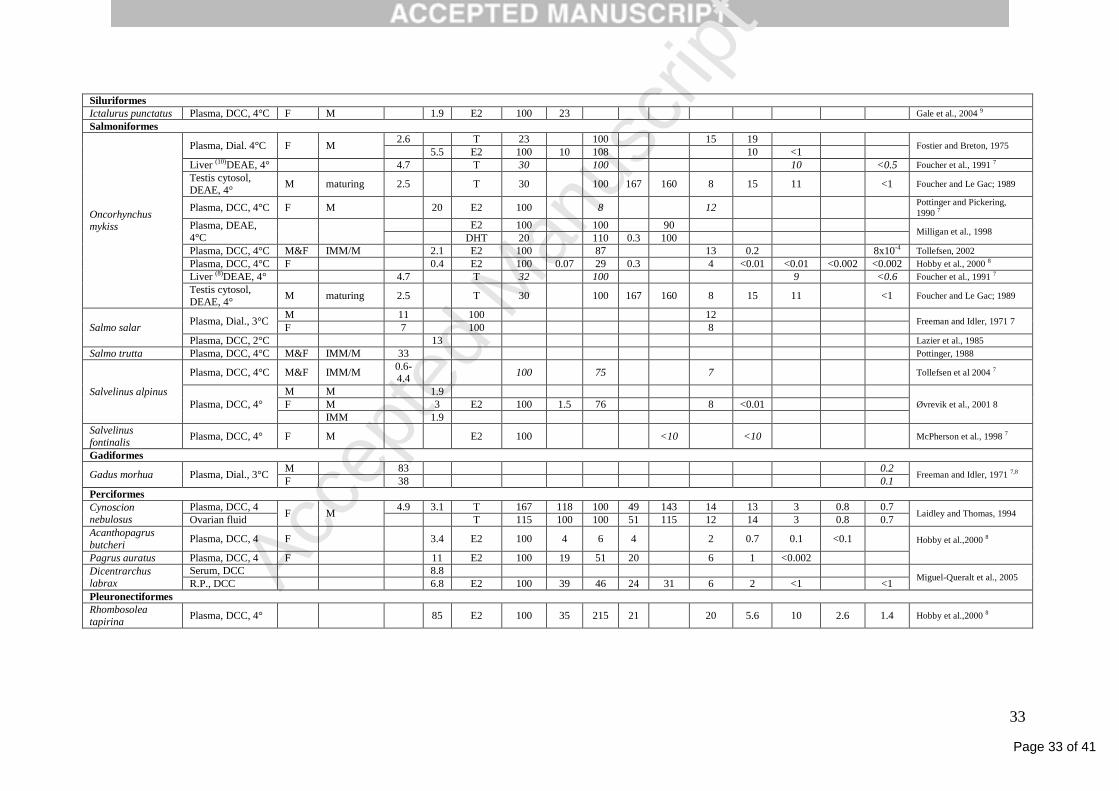
Page 33 of 41
Accep
ted
Man
uscr
ipt
33
Siluriformes
Ictalurus punctatus Plasma, DCC, 4°C F M 1.9 E2 100 23 Gale et al., 2004 9
Salmoniformes
Oncorhynchus mykiss
Plasma, Dial. 4°C F M 2.6 T 23 100 15 19
Fostier and Breton, 1975 5.5 E2 100 10 108 10 <1
Liver (10)DEAE, 4° 4.7 T 30 100 10 <0.5 Foucher et al., 1991 7
Testis cytosol,
DEAE, 4° M maturing 2.5 T 30 100 167 160 8 15 11 <1 Foucher and Le Gac; 1989
Plasma, DCC, 4°C F M 20 E2 100 8 12 Pottinger and Pickering,
1990 7
Plasma, DEAE,
4°C
E2 100 100 90 Milligan et al., 1998
DHT 20 110 0.3 100
Plasma, DCC, 4°C M&F IMM/M 2.1 E2 100 87 13 0.2 8x10⁻⁴ Tollefsen, 2002
Plasma, DCC, 4°C F 0.4 E2 100 0.07 29 0.3 4 <0.01 <0.01 <0.002 <0.002 Hobby et al., 2000 8
Liver (8)DEAE, 4° 4.7 T 32 100 9 <0.6 Foucher et al., 1991 7
Testis cytosol,
DEAE, 4° M maturing 2.5 T 30 100 167 160 8 15 11 <1 Foucher and Le Gac; 1989
Salmo salar Plasma, Dial., 3°C
M 11 100 12 Freeman and Idler, 1971 7
F 7 100 8
Plasma, DCC, 2°C 13 Lazier et al., 1985
Salmo trutta Plasma, DCC, 4°C M&F IMM/M 33 Pottinger, 1988
Salvelinus alpinus
Plasma, DCC, 4°C M&F IMM/M 0.6-4.4
100 75 7 Tollefsen et al 2004 7
Plasma, DCC, 4°
M M 1.9
Øvrevik et al., 2001 8 F M 3 E2 100 1.5 76 8 <0.01
IMM 1.9
Salvelinus fontinalis
Plasma, DCC, 4° F M E2 100 <10 <10 McPherson et al., 1998 7
Gadiformes
Gadus morhua Plasma, Dial., 3°C M 83 0.2
Freeman and Idler, 1971 7,8 F 38 0.1
Perciformes
Cynoscion nebulosus
Plasma, DCC, 4 F M
4.9 3.1 T 167 118 100 49 143 14 13 3 0.8 0.7 Laidley and Thomas, 1994
Ovarian fluid T 115 100 100 51 115 12 14 3 0.8 0.7
Acanthopagrus
butcheri Plasma, DCC, 4 F 3.4 E2 100 4 6 4 2 0.7 0.1 <0.1 Hobby et al.,2000 8
Pagrus auratus Plasma, DCC, 4 F 11 E2 100 19 51 20 6 1 <0.002
Dicentrarchus labrax
Serum, DCC 8.8 Miguel-Queralt et al., 2005
R.P., DCC 6.8 E2 100 39 46 24 31 6 2 <1 <1
Pleuronectiformes
Rhombosolea
tapirina Plasma, DCC, 4° 85 E2 100 35 215 21 20 5.6 10 2.6 1.4 Hobby et al.,2000 8
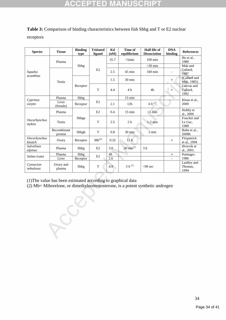
Page 34 of 41
Accep
ted
Man
uscr
ipt
34
Table 3: Comparison of binding characteristics between fish Shbg and T or E2 nuclear
receptors
Species Tissue Binding
type
Tritiated
ligand
Kd
(nM)
Time of
equilibrium
Half-life of
Dissociation
DNA
binding References
Squalus
acanthias
Plasma
Shbg
E2
35.7 <5min 100 min Ho et al.,
1980
<30 min Mak and
Callard,
1987
Testis
2.5 45 min 160 min -
Receptor
1.5 30 min + (Callard and
Mak, 1985)
T 4.4 4 h 4h +
Cuevas and
Callard,
1992
Cyprinus
carpio
Plasma Shbg
E2
15 min Kloas et al.,
2000 Liver
(female) Receptor 2.1 12h 6 h (1)
Oncorhynchus
mykiss
Plasma
Shbga
E2 0.4 15 min <5 min Hobby et
al., 2000
Testis T 2.5 2 h 1-2 min
Foucher and
Le Gac,
1989
Recombinant
protein Shbgb T 0.8 30 min 3 min
Bobe et al.,
2008b
Oncorhynchus
kisutch Ovary Receptor Mb(2) 0.32 12 h +
Fitzpatrick
et al., 1994
Salvelinus
alpinus Plasma Shbg E2 3.0 30 min(1) 3 h
Øvrevik et
al., 2001.
Salmo trutta Plasma Shbg
E2 48 + Pottinger,
1986 Liver Receptor 2.6 -
Cynoscion
nebulosus
Ovary and
plasma Shbg T 4.9 5 h (1) <90 sec
Laidley and
Thomas,
1994
(1)The value has been estimated according to graphical data
(2) Mb= Miborelone, or dimethylnortestosterone, is a potent synthetic androgen
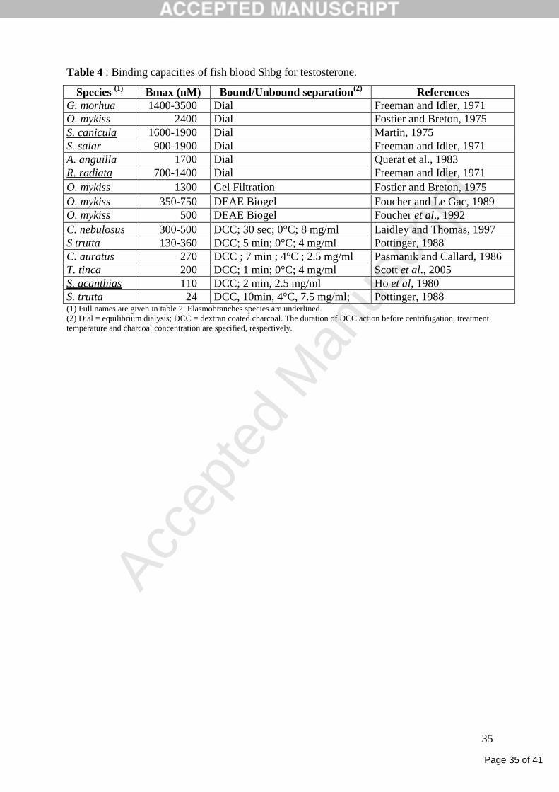
Page 35 of 41
Accep
ted
Man
uscr
ipt
35
Table 4 : Binding capacities of fish blood Shbg for testosterone.
Species (1)
Bmax (nM) Bound/Unbound separation(2)
References
G. morhua 1400-3500 Dial Freeman and Idler, 1971
O. mykiss 2400 Dial Fostier and Breton, 1975
S. canicula 1600-1900 Dial Martin, 1975
S. salar 900-1900 Dial Freeman and Idler, 1971
A. anguilla 1700 Dial Querat et al., 1983
R. radiata 700-1400 Dial Freeman and Idler, 1971
O. mykiss 1300 Gel Filtration Fostier and Breton, 1975
O. mykiss 350-750 DEAE Biogel Foucher and Le Gac, 1989
O. mykiss 500 DEAE Biogel Foucher et al., 1992
C. nebulosus 300-500 DCC; 30 sec; 0°C; 8 mg/ml Laidley and Thomas, 1997
S trutta 130-360 DCC; 5 min; 0°C; 4 mg/ml Pottinger, 1988
C. auratus 270 DCC ; 7 min ; 4°C ; 2.5 mg/ml Pasmanik and Callard, 1986
T. tinca 200 DCC; 1 min; 0°C; 4 mg/ml Scott et al., 2005
S. acanthias 110 DCC; 2 min, 2.5 mg/ml Ho et al, 1980
S. trutta 24 DCC, 10min, 4°C, 7.5 mg/ml; Pottinger, 1988 (1) Full names are given in table 2. Elasmobranches species are underlined.
(2) Dial = equilibrium dialysis; DCC = dextran coated charcoal. The duration of DCC action before centrifugation, treatment
temperature and charcoal concentration are specified, respectively.
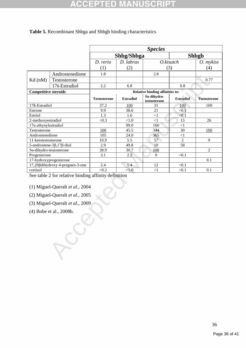
Page 36 of 41
Accep
ted
Man
uscr
ipt
36
Table 5. Recombinant Shbga and Shbgb binding characteristics
Species Shbg/Shbga Shbgb
D. rerio
(1)
D. labrax
(2)
O.kisutch
(3)
O. mykiss
(4)
Kd (nM)
Androstenedione 1.8 2.8
Testosterone 0.77
17ß-Estradiol 2.2 6.8 0.8
Competitive steroids Relative binding affinities to:
Testosterone Estradiol 5α-dihydro-
testosterone Estradiol Testosterone
17ß-Estradiol 37.2 100 31 100 100
Estrone 9.9 38.6 21 <0.1
Estriol 1.3 1.6 <1 <0.1
2-methoxyestradiol <0.3 <1.0 <1 15 26
17α ethynylestradiol 99.0 160 <1
Testosterone 100 45.5 344 30 100
Androstenedione 105 24.0 365 <1
11-ketotestosterone 10.9 5.5 57 2 9
5-androstene-3β,17β-diol 2.9 49.8 10 58
5α-dihydro-testosterone 38.9 30.7 100 2
Progesterone 3.1 2.2 9 <0.1
17-hydroxyprogesterone 0.1
17,20βdihydroxy 4-pregnen-3-one 2.4 1.4 12 <0.1
cortisol <0.2 <1.0 <1 <0.1 0.1
See table 2 for relative binding affinity definition
(1) Miguel-Queralt et al., 2004
(2) Miguel-Queralt et al., 2005
(3) Miguel-Queralt et al., 2009
(4) Bobe et al., 2008b.

Page 37 of 41
Accep
ted
Man
uscr
ipt
37
Figure legends
Figure 1. Evolutionary relationships of teleost fishes sex hormone binding proteins (Shbg).
This consensus n/p/l tree (fusion of neighbor joining (n), maximum parsimony (p), and
maximum likelihood (l) trees) was computed using the FIGENIX platform (Gouret et al.,
2005) with the D. rerio growth arrest-specific 6 (Gas6) protein (AAH53117) as an outgroup
and the following Shbg proteins dataset: human and mouse Shbg (CAA34398 and
AAI20788); Shbg alpha (Shbga) from zebrafish (AAU14174); barfin flounder, Verasper
Moseri (BAG84605); Atlantic sea bass, Dicentrartchus labrax (AAW23033); spotted green
pufferfish, Tetraodon nigroviridis (CAF93818); three-spined stickleback, Gasterosteus
aculeatus (ENSORLP00000008096); Japanese medaka, Oryzias latipes
(ENSORLP00000008096); Japanese pufferfish, Takifugu rubipres (ENSTRUP00000031810);
common carp, Cyprinus carpio ShbgaI and ShbgaII (BAE48780, BAE48781); salmonids
Shbg alpha and Shbg beta (Shbgb) from rainbow trout, Oncorhynchus mykiss (BAE48779,
ABQ45411); Chinook salmon, Oncorhynchus tshawytscha (ACJ25981, ACJ25982); coho
salmon, Oncorhynchus kisutch (ACJ25979, ACJ25980). The Atlantic salmon, Salmo salar
Shbg partial proteins were deduced from Expressed Sequence Tags matching Shbga
(DV106329, CK888851 and CK888675) and Shbgb (DY735728, DY694623, DY719210,
DW561629, DY699232, CK884863, CA056042, DY699233, and DY719211). Significant
bootstrap values are given in percentage at the main nodes of the consensus tree for each of
the n_p_l method used (*: non significant).
Figure 2. Comparison of the structure of different teleost fishes shbga genes. Exons are
boxed and lines indicate the introns. Out of scale introns are indicated by broken lines and
their lengths are given in bp. Exon numbers are given above exon boxes. All intron-exon
boundaries were predicted according to the Ensembl gene prediction for three-spined

Page 38 of 41
Accep
ted
Man
uscr
ipt
38
stickleback, Gasterosteus aculeatus (ENSORLP00000008096); Japanese medaka, Oryzias
latipes (ENSORLP00000008096) and Japanese pufferfish, Takifugu rubipres
(ENSTRUP00000031810).
Figure 3. Alignment of Shbga sequences from zebrafish (AAU14174), rainbow trout
(BAE48779), sea bass (AAW23033) with human SHBG (CAA34398) and rainbow trout
Shbgb (ABQ45411). Amino-acids sequences conserved in at least 4 sequences are shaded.
Amino acid residues specific of the rainbow trout Shbgb are shown below the alignment
respectively with a hyphen (-). The conserved cysteine residues are identified below the
alignment with a bold type C letter and the two intra-molecular disulfide bridges (IMDB1 and
IMDB2) linking these cysteines are represented. Consensus sites for N-glycosylation are
indicated in bold italics. Amino acids corresponding to the signal peptides are underlined and
have been obtained from the following published sequences (Miguel-Queralt et al.,
2004;Miguel-Queralt et al., 2005).
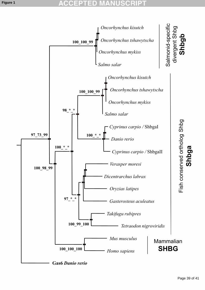
Page 39 of 41
Accep
ted
Man
uscr
ipt
Oncorhynchus kisutch
Oncorhynchus tshawytscha
Oncorhynchus mykiss
Salmo salar
Cyprinus carpio / ShbgaI
Danio rerio97_73_99
100_100_99
98_*_*
100_100_99
100_*_*
Oncorhynchus kisutch
Oncorhynchus mykiss
Salmo salar
Oncorhynchus tshawytscha
Sal
mon
id-s
peci
fic
dive
rgen
t Shb
g
Sh
bg
bF
ish
cons
erve
d or
thol
ogS
hbg
Cyprinus carpio / ShbgaII
Verasper moresi
Dicentrarchus labrax
Oryzias latipes
Gasterosteus aculeatus
Takifugu rubipres
Tetraodon nigroviridis
100_98_99
100_*_*
97_*_*
100_99_100
100_100_100
Mus musculus
Gas6 Danio rerio
Homo sapiens
Fis
h co
nser
ved
orth
olog
Sh
bg
a
Mammalian
SHBG
Figure 1
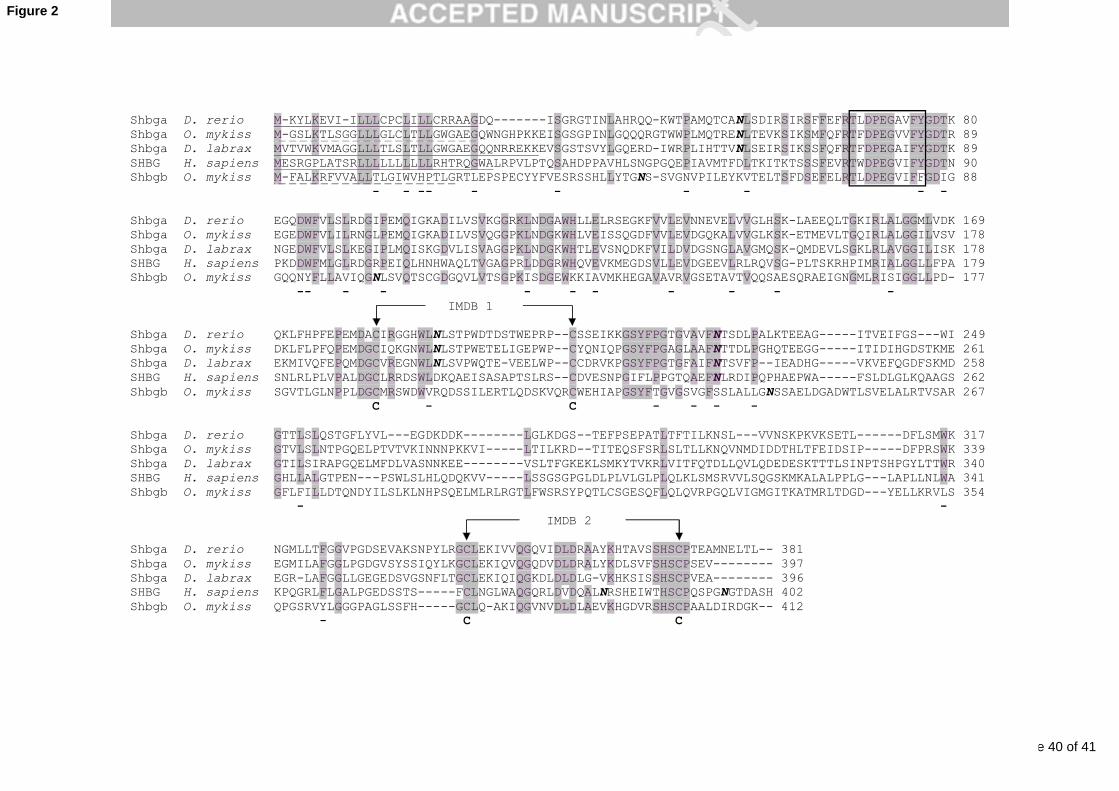
Page 40 of 41
Accep
ted
Man
uscr
ipt
Shbga D. rerio M-KYLKEVI-ILLLCPCLILLCRRAAGDQ-------ISGRGTINLAHRQQ-KWTPAMQTCANLSDIRSIRSFFEFRTLDPEGAVFYGDTK 80 Shbga O. mykiss M-GSLKTLSGGLLLGLCLTLLGWGAEGQWNGHPKKEISGSGPINLGQQQRGTWWPLMQTRENLTEVKSIKSMFQFRTFDPEGVVFYGDTR 89 Shbga D. labrax MVTVWKVMAGGLLLTLSLTLLGWGAEGQQNRREKKEVSGSTSVYLGQERD-IWRPLIHTTVNLSEIRSIKSSFQFRTFDPEGAIFYGDTK 89 SHBG H. sapiens MESRGPLATSRLLLLLLLLLLRHTRQGWALRPVLPTQSAHDPPAVHLSNGPGQEPIAVMTFDLTKITKTSSSFEVRTWDPEGVIFYGDTN 90 Shbgb O. mykiss M-FALKRFVVALLTLGIWVHPTLGRTLEPSPECYYFVESRSSHLLYTGNS-SVGNVPILEYKVTELTSFDSEFELRTLDPEGVIFFGDIG 88 - - -- - - - - - -
Shbga D. rerio EGQDWFVLSLRDGIPEMQIGKADILVSVKGGRKLNDGAWHLLELRSEGKFVVLEVNNEVELVVGLHSK-LAEEQLTGKIRLALGGMLVDK 169 Shbga O. mykiss EGEDWFVLILRNGLPEMQIGKADILVSVQGGPKLNDGKWHLVEISSQGDFVVLEVDGQKALVVGLKSK-ETMEVLTGQIRLALGGILVSV 178 Shbga D. labrax NGEDWFVLSLKEGIPLMQISKGDVLISVAGGPKLNDGKWHTLEVSNQDKFVILDVDGSNGLAVGMQSK-QMDEVLSGKLRLAVGGILISK 178 SHBG H. sapiens PKDDWFMLGLRDGRPEIQLHNHWAQLTVGAGPRLDDGRWHQVEVKMEGDSVLLEVDGEEVLRLRQVSG-PLTSKRHPIMRIALGGLLFPA 179 Shbgb O. mykiss GQQNYFLLAVIQGNLSVQTSCGDGQVLVTSGPKISDGEWKKIAVMKHEGAVAVRVGSETAVTVQQSAESQRAEIGNGMLRISIGGLLPD- 177 -- - - - - - - - - - IMDB 1
Shbga D. rerio QKLFHPFEPEMDACIRGGHWLNLSTPWDTDSTWEPRP--CSSEIKKGSYFPGTGVAVFNTSDLPALKTEEAG-----ITVEIFGS---WI 249 Shbga O. mykiss DKLFLPFQPEMDGCIQKGNWLNLSTPWETELIGEPWP--CYQNIQPGSYFPGAGLAAFNTTDLPGHQTEEGG-----ITIDIHGDSTKME 261 Shbga D. labrax EKMIVQFEPQMDGCVREGNWLNLSVPWQTE-VEELWP--CCDRVKPGSYFPGTGFAIFNTSVFP--IEADHG-----VKVEFQGDFSKMD 258 SHBG H. sapiens SNLRLPLVPALDGCLRRDSWLDKQAEISASAPTSLRS--CDVESNPGIFLPPGTQAEFNLRDIPQPHAEPWA-----FSLDLGLKQAAGS 262 Shbgb O. mykiss SGVTLGLNPPLDGCMRSWDWVRQDSSILERTLQDSKVQRCWEHIAPGSYFTGVGSVGFSSLALLGNSSAELDGADWTLSVELALRTVSAR 267 Shbgb O. mykiss SGVTLGLNPPLDGCMRSWDWVRQDSSILERTLQDSKVQRCWEHIAPGSYFTGVGSVGFSSLALLGNSSAELDGADWTLSVELALRTVSAR 267 C - C - - - -
Shbga D. rerio GTTLSLQSTGFLYVL---EGDKDDK--------LGLKDGS--TEFPSEPATLTFTILKNSL---VVNSKPKVKSETL------DFLSMWK 317 Shbga O. mykiss GTVLSLNTPGQELPTVTVKINNNPKKVI-----LTILKRD--TITEQSFSRLSLTLLKNQVNMDIDDTHLTFEIDSIP-----DFPRSWK 339 Shbga D. labrax GTILSIRAPGQELMFDLVASNNKEE--------VSLTFGKEKLSMKYTVKRLVITFQTDLLQVLQDEDESKTTTLSINPTSHPGYLTTWR 340 SHBG H. sapiens GHLLALGTPEN---PSWLSLHLQDQKVV-----LSSGSGPGLDLPLVLGLPLQLKLSMSRVVLSQGSKMKALALPPLG---LAPLLNLWA 341 Shbgb O. mykiss GFLFILLDTQNDYILSLKLNHPSQELMLRLRGTLFWSRSYPQTLCSGESQFLQLQVRPGQLVIGMGITKATMRLTDGD---YELLKRVLS 354 - - IMDB 2
Shbga D. rerio NGMLLTFGGVPGDSEVAKSNPYLRGCLEKIVVQGQVIDLDRAAYKHTAVSSHSCPTEAMNELTL-- 381 Shbga O. mykiss EGMILAFGGLPGDGVSYSSIQYLKGCLEKIQVQGQDVDLDRALYKDLSVFSHSCPSEV-------- 397 Shbga D. labrax EGR-LAFGGLLGEGEDSVGSNFLTGCLEKIQIQGKDLDLDLG-VKHKSISSHSCPVEA-------- 396 SHBG H. sapiens KPQGRLFLGALPGEDSSTS-----FCLNGLWAQGQRLDVDQALNRSHEIWTHSCPQSPGNGTDASH 402 Shbgb O. mykiss QPGSRVYLGGGPAGLSSFH-----GCLQ-AKIQGVNVDLDLAEVKHGDVRSHSCPAALDIRDGK-- 412 - C C
Figure 2

Page 41 of 41
Accep
ted
Man
uscr
ipt
Fugu Fugu shbgashbga (2.2 kb, 392 aa)(2.2 kb, 392 aa)
Stickleback Stickleback shbgashbga (4 kb, 394 aa)(4 kb, 394 aa)
1 2 3 4 5 6 7 8
1 2 3 4 5 6 7 8
500 bp
: Introns
: Exons
Medaka Medaka shbga shbga (6.8 kb, 385 aa)(6.8 kb, 385 aa)1 2 3 4 5 6 7 8
Zebrafish Zebrafish shbgashbga (13.3 kb, 381 aa)(13.3 kb, 381 aa)
3071 bp 5108 bp 3192 bp1 2 3 4 5 6 7 8
1517 bp 2278 bp
Figure 3