Distraction affects frontal alpha rhythms related to expectancy of pain: An EEG study
10
Distraction affects frontal alpha rhythms related to expectancy of pain: An EEG study Claudio Del Percio, a,b,c,d, * Domenica Le Pera, e Lars Arendt-Nielsen, b Claudio Babiloni, a,c Alfredo Brancucci, a,b,d Andrew C.N. Chen, f Liala De Armas, e Roberto Miliucci, g Domenico Restuccia, g Massimiliano Valeriani, h and Paolo Maria Rossini c,i a Dipartimento di Fisiologia Umana e Farmacologia, Universita ` ‘‘La Sapienza’’, Rome, Italy b Human Brain Mapping and Cortical Imaging Laboratory/SMI, University of Aalborg, Denmark c A.Fa.R. Dip.Neuroscienze-Ospedale Fatebenefratelli Isola Tiberina, Rome, Italy d Instituto di Medicina e Scienza dello Sport, CONI Servizi, Rome, Italy e Casa di Cura San Raffaele Pisana, Tosinvest Sanita ` , Rome, Italy f Center for Functional Human Brain Mapping, Capital University of Medical Sciences, Beijing, China g Dipartimento di Neurologia, Universita ` Cattolica del Sacro Cuore, Rome, Italy h Divisione di Neurologia, Ospedale Pediatrico Bambino Gesu ` IRCCS, Rome, Italy i IRCCS ‘‘S. Giovanni di Dio-FBF’’, Brescia and Clinica Neurologica, Universita ` ‘‘Campus Biomedico’’, Rome, Italy Received 30 March 2005; revised 13 October 2005; accepted 20 January 2006 Available online 10 March 2006 Previous electroencephalographic (EEG) evidence has shown event- related desynchronization (ERD) of alpha rhythms before predictable painful stimuli, as a possible neural concomitant of attentional preparatory processes (Babiloni, C., Brancucci, A., Babiloni, F., Capotosto, P., Carducci, F., Cincotti, F., Arendt-Nielsen, L., Chen, A.C., Rossini, P.M., 2003. Anticipatory cortical responses during the expectancy of a predictable painful stimulation. A high-resolution electroencephalography study. Eur. J. Neurosci. 18 (6) 1692–700). This study tested the hypothesis that alpha ERD before predictable painful stimuli is reduced as an effect of distraction. A visual warning stimulus preceded a laser painful stimulation, which was strictly followed by visual imperative stimuli. In the Pain (control) condition, no task was required after the imperative stimuli. In the Pain + Movement condition, subjects had to perform a movement of the right index finger. In the Pain + Cognition condition, they had to mentally perform an arithmetical task. EEG data were recorded in 10 subjects from 30 electrodes. Artifact-free recordings were spatially enhanced by surface Laplacian transformation. Alpha ERD was computed at three alpha sub-bands according to subjects’ individual alpha frequency peak (i.e., about 6 – 8 Hz, 8 – 10 Hz, 10 – 12 Hz). Compared to the control condition, the subjects reported a significantly lower stimulus intensity perception and unpleasantness in the Pain + Movement and Pain + Cognition conditions. In addition, there was a cancellation of the alpha 3 ERD (i.e., about 10 – 12 Hz) in Pain + Cognition condition and even a generation of a statistically significant alpha 3 ERS in Pain + Movement condition. These effects were maximum over fronto-central midline. These results suggest that distraction during the expectancy of pain is related to a reduced neural desynchronization of fronto-central midline alpha rhythms (i.e., reduced cortical activation) towards an overt hyper-synchronization (cortical idling). D 2006 Elsevier Inc. All rights reserved. Keywords: Electroencephalography (EEG); Alpha rhythms; Event-related desynchronization/synchronization (ERD/ERS); Pain; Sensorimotor inter- actions; Cognition; Distraction Introduction Investigations measuring regional cerebral blood flow (rCBF) have shown that activity of nociceptive cortical areas was observed not only during pain experience but also during the expectancy of predictable pain stimulations (Ingvar, 1999; Treede et al., 1999; Hsieh et al., 1999; Petrovic et al., 2000). Among these areas, primary somatosensory cortex would be mainly devoted to the anticipation of sensory discrimination (Porro et al., 1998, 2002; Treede et al., 1999; Schnitzler and Ploner, 2000; Petrovic and Ingvar, 2002). Conversely, midline cingulate and frontal areas would be engaged in the anticipation of affective/cognitive aspects of pain experience, selection of proper viscero-sensorimotor/ behavioral responses, and event memorization (Rainville et al., 1997; Porro et al., 1998, 2002; Hsieh et al., 1999; Ingvar, 1999; Treede et al., 1999; Ploghaus et al., 1999; Petrovic and Ingvar, 2002; Petrovic et al., 2000; Schnitzler and Ploner, 2000; Garcia- Larrea et al., 2003). 1053-8119/$ - see front matter D 2006 Elsevier Inc. All rights reserved. doi:10.1016/j.neuroimage.2006.01.013 * Corresponding author. Dipartimento di Fisiologia Umana e Farm- acologia, Universita ` degli Studi di Roma ‘‘La Sapienza’’, P.le Aldo Moro, 5, 00185 Rome, Italy. Fax: +39 06 4991 0989 0917. E-mail address: [email protected] (C. Del Percio). Available online on ScienceDirect (www.sciencedirect.com). www.elsevier.com/locate/ynimg NeuroImage 31 (2006) 1268 – 1277
-
Upload
independent -
Category
Documents
-
view
0 -
download
0
Transcript of Distraction affects frontal alpha rhythms related to expectancy of pain: An EEG study

www.elsevier.com/locate/ynimg
NeuroImage 31 (2006) 1268 – 1277
Distraction affects frontal alpha rhythms related to expectancy
of pain: An EEG study
Claudio Del Percio,a,b,c,d,* Domenica Le Pera,e Lars Arendt-Nielsen,b Claudio Babiloni,a,c
Alfredo Brancucci,a,b,d Andrew C.N. Chen,f Liala De Armas,e Roberto Miliucci,g
Domenico Restuccia,g Massimiliano Valeriani,h and Paolo Maria Rossini c,i
aDipartimento di Fisiologia Umana e Farmacologia, Universita ‘‘La Sapienza’’, Rome, ItalybHuman Brain Mapping and Cortical Imaging Laboratory/SMI, University of Aalborg, DenmarkcA.Fa.R. Dip.Neuroscienze-Ospedale Fatebenefratelli Isola Tiberina, Rome, ItalydInstituto di Medicina e Scienza dello Sport, CONI Servizi, Rome, ItalyeCasa di Cura San Raffaele Pisana, Tosinvest Sanita, Rome, ItalyfCenter for Functional Human Brain Mapping, Capital University of Medical Sciences, Beijing, ChinagDipartimento di Neurologia, Universita Cattolica del Sacro Cuore, Rome, ItalyhDivisione di Neurologia, Ospedale Pediatrico Bambino Gesu IRCCS, Rome, ItalyiIRCCS ‘‘S. Giovanni di Dio-FBF’’, Brescia and Clinica Neurologica, Universita ‘‘Campus Biomedico’’, Rome, Italy
Received 30 March 2005; revised 13 October 2005; accepted 20 January 2006
Available online 10 March 2006
Previous electroencephalographic (EEG) evidence has shown event-
related desynchronization (ERD) of alpha rhythms before predictable
painful stimuli, as a possible neural concomitant of attentional
preparatory processes (Babiloni, C., Brancucci, A., Babiloni, F.,
Capotosto, P., Carducci, F., Cincotti, F., Arendt-Nielsen, L., Chen,
A.C., Rossini, P.M., 2003. Anticipatory cortical responses during the
expectancy of a predictable painful stimulation. A high-resolution
electroencephalography study. Eur. J. Neurosci. 18 (6) 1692–700). This
study tested the hypothesis that alpha ERD before predictable painful
stimuli is reduced as an effect of distraction. A visual warning stimulus
preceded a laser painful stimulation, which was strictly followed by
visual imperative stimuli. In the Pain (control) condition, no task was
required after the imperative stimuli. In the Pain + Movement
condition, subjects had to perform a movement of the right index
finger. In the Pain + Cognition condition, they had to mentally perform
an arithmetical task. EEG data were recorded in 10 subjects from 30
electrodes. Artifact-free recordings were spatially enhanced by surface
Laplacian transformation. Alpha ERD was computed at three alpha
sub-bands according to subjects’ individual alpha frequency peak (i.e.,
about 6–8 Hz, 8–10 Hz, 10–12 Hz). Compared to the control
condition, the subjects reported a significantly lower stimulus intensity
perception and unpleasantness in the Pain + Movement and Pain +
Cognition conditions. In addition, there was a cancellation of the alpha
3 ERD (i.e., about 10–12 Hz) in Pain + Cognition condition and even a
generation of a statistically significant alpha 3 ERS in Pain +
Movement condition. These effects were maximum over fronto-central
1053-8119/$ - see front matter D 2006 Elsevier Inc. All rights reserved.
doi:10.1016/j.neuroimage.2006.01.013
* Corresponding author. Dipartimento di Fisiologia Umana e Farm-
acologia, Universita degli Studi di Roma ‘‘La Sapienza’’, P.le Aldo Moro, 5,
00185 Rome, Italy. Fax: +39 06 4991 0989 0917.
E-mail address: [email protected] (C. Del Percio).
Available online on ScienceDirect (www.sciencedirect.com).
midline. These results suggest that distraction during the expectancy of
pain is related to a reduced neural desynchronization of fronto-central
midline alpha rhythms (i.e., reduced cortical activation) towards an
overt hyper-synchronization (cortical idling).
D 2006 Elsevier Inc. All rights reserved.
Keywords: Electroencephalography (EEG); Alpha rhythms; Event-related
desynchronization/synchronization (ERD/ERS); Pain; Sensorimotor inter-
actions; Cognition; Distraction
Introduction
Investigations measuring regional cerebral blood flow (rCBF)
have shown that activity of nociceptive cortical areas was observed
not only during pain experience but also during the expectancy of
predictable pain stimulations (Ingvar, 1999; Treede et al., 1999;
Hsieh et al., 1999; Petrovic et al., 2000). Among these areas,
primary somatosensory cortex would be mainly devoted to the
anticipation of sensory discrimination (Porro et al., 1998, 2002;
Treede et al., 1999; Schnitzler and Ploner, 2000; Petrovic and
Ingvar, 2002). Conversely, midline cingulate and frontal areas
would be engaged in the anticipation of affective/cognitive aspects
of pain experience, selection of proper viscero-sensorimotor/
behavioral responses, and event memorization (Rainville et al.,
1997; Porro et al., 1998, 2002; Hsieh et al., 1999; Ingvar, 1999;
Treede et al., 1999; Ploghaus et al., 1999; Petrovic and Ingvar,
2002; Petrovic et al., 2000; Schnitzler and Ploner, 2000; Garcia-
Larrea et al., 2003).
C. Del Percio et al. / NeuroImage 31 (2006) 1268–1277 1269
Anticipatory cortical processes can also be probed by electro-
magnetic brain signals named event-related potentials (ERPs),
which might denote affective-motivational cortical processes and
preparatory sensorimotor processes (Bocker et al., 2001; Bocker
and Van Boxtel, 1997). These potentials are termed contingent
negative variation (CNV) when a motor response is required after a
warned imperative stimulus (Brunia, 1999; Brunia and van Boxtel,
2001). On the whole, the CNV can be considered as a global sign of
anticipatory cortical excitability (Brunia, 1999; Bastiaansen and
Brunia, 2001). Before nonpainful or painful somatosensory stimuli,
no ERP has been shown despite its biological significance (Babiloni
et al., 2003). In contrast, there was a CNV before nonpainful and
painful stimuli (left hand) concomitant with a Go/NoGo motor task,
presumably elicited by inter-hemispheric flow of information
involving both primary (Babiloni et al., 2004a, 2005a) and
secondary (Babiloni et al., 2005b) sensorimotor areas.
In parallel, anticipatory cortical processes can be investigated by
electroencephalographic (EEG) rhythms at an extended alpha band
ranging about from 6 to 12 Hz (Neuper and Pfurtscheller, 2001;
Bastiaansen et al., 2001; Brunia and van Boxtel, 2001). During
sensorimotor or cognitive events, the lower the amplitude of alpha
rhythms (event-related desynchronization, ERD), the better the
information transfer (‘‘gating’’) through sensorimotor thalamo-
cortical and cortico-cortical pathways (Pfurtscheller and Lopes da
Silva, 1999; Klimesch, 1999). In contrast to the negligible CNV
before painful stimuli, anticipatory alpha ERD has been found to be
clearly evident over primary somatosensory cortex contralateral to
the stimulation (Babiloni et al., 2003). Furthermore, that anticipa-
tory alpha ERD increased in magnitude over primary sensorimotor
areas before nonpainful and painful stimuli to the left hand
concomitant with a Go/NoGo motor task for the right hand
(Babiloni et al., 2004a, 2005b). This was true even if painful
stimulation was concomitant with a working memory task replacing
the Go/NoGo motor one (Babiloni et al., 2004c). According to
previous EEG evidence (Rockstroh et al., 1989), these findings
have been interpreted as an anticipatory facilitation of nociceptive
thalamo-cortical and cortico-cortical channels. However, it is still
an open issue whether and how such anticipatory modulation of
nociceptive channels is related to attentional processes as reflected
by trial-by-trial stimulus evaluation.
Previous studies have investigated how cortical processes
linked with pain are affected by distraction due to concomitant
sensorimotor and cognitive demands (Nakata et al., 2004a,b,
2005). Distraction reduced subjective pain sensation (Eccleston,
1995). Furthermore, pain or anticipation of pain reduced cognitive
performance, probably as a result of subtraction of attentional
resources from cognitive operations (De Pascalis et al., 1995;
Kuhajda et al., 2002; Lieberman et al., 2002). That reduction of
cognitive performance occurred as a function of task complexity,
age of the subjects, endogenous variations in hormone level (i.e.,
menstrual cycle), and individual differences of reactivity to
stressful agents (Pauli et al., 1993; Kumari and Corr, 1998).
Relationships between pain, distraction, and cortical activity
have been investigated by neuroimaging studies estimating rCBF.
Pain sensation (cold pressure test) and simultaneous attention-
demanding maze task induced an increase of orbitofrontal rCBF
(Petrovic et al., 2000). Pain stimuli associated with attentional
(Stroop) demands increased activity of perigenual anterior cingu-
late (ACC) and orbitofrontal cortices, while the opposite was true
at thalamus, insular cortex and caudal parts of ACC (Bantick et al.,
2002). A similar modulation of attention also increased rCBF in
periaqueductal gray, which modulates descending pain inhibitory
systems (Tracey et al., 2002). Taken together, these neuroimaging
studies suggest that distraction tasks reduce subjective pain
sensation and decrease rCBF in cerebral areas such as perigenual
ACC, orbitofrontal, and insular cortices (Petrovic and Ingvar,
2002; Petrovic et al., 2000; Bantick et al., 2002; Frankenstein et al.,
2001). A reasonable hypothesis is that alpha rhythms are
implicated in the attentional ‘‘gating’’ of painful stimuli across
the mentioned node of the nociceptive circuits.
The present EEG study aims at investigating relationships
among pain, distraction, and cortical alpha rhythms in order to test
the hypothesis that concurrent motor or cognitive demands reduce
both the evaluation of pain (distraction effects) and alpha ERD
preceding predictable pain stimuli.
Materials and methods
Subjects
Ten healthy volunteers (mean age = 24 years, range: 20–32
years) participated to the present study. All subjects gave their
written informed consent according to the Declaration of Helsinki
and could freely request an interruption of the investigation at any
time. The local Institutional Ethics Committee approved the
research aims and procedures.
Experimental design
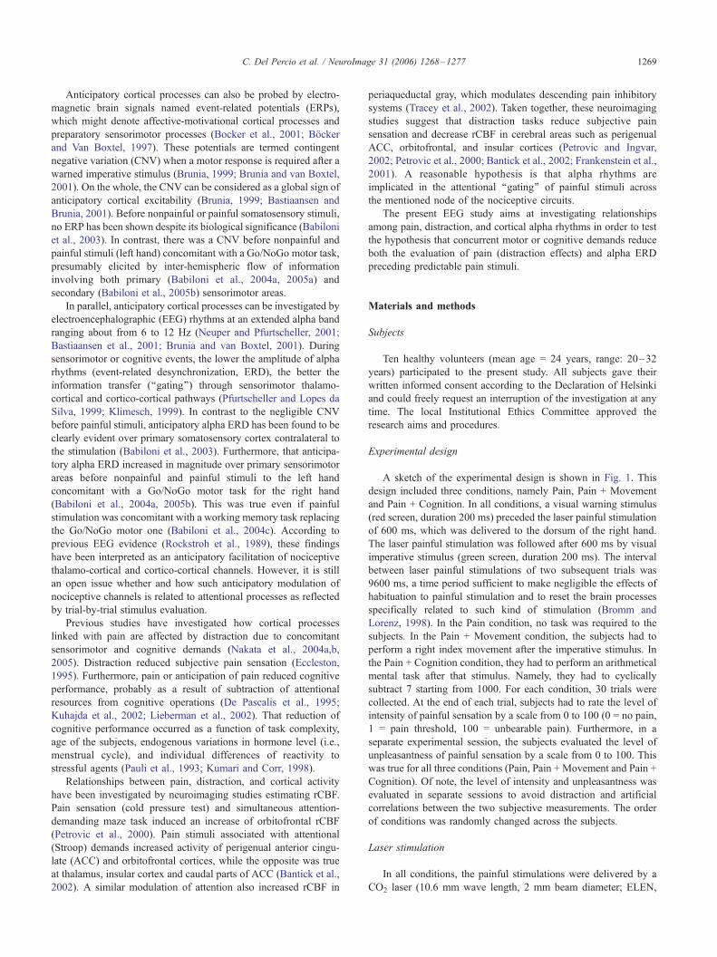
A sketch of the experimental design is shown in Fig. 1. This
design included three conditions, namely Pain, Pain + Movement
and Pain + Cognition. In all conditions, a visual warning stimulus
(red screen, duration 200 ms) preceded the laser painful stimulation
of 600 ms, which was delivered to the dorsum of the right hand.
The laser painful stimulation was followed after 600 ms by visual
imperative stimulus (green screen, duration 200 ms). The interval
between laser painful stimulations of two subsequent trials was
9600 ms, a time period sufficient to make negligible the effects of
habituation to painful stimulation and to reset the brain processes
specifically related to such kind of stimulation (Bromm and
Lorenz, 1998). In the Pain condition, no task was required to the
subjects. In the Pain + Movement condition, the subjects had to
perform a right index movement after the imperative stimulus. In
the Pain + Cognition condition, they had to perform an arithmetical
mental task after that stimulus. Namely, they had to cyclically
subtract 7 starting from 1000. For each condition, 30 trials were
collected. At the end of each trial, subjects had to rate the level of
intensity of painful sensation by a scale from 0 to 100 (0 = no pain,
1 = pain threshold, 100 = unbearable pain). Furthermore, in a
separate experimental session, the subjects evaluated the level of
unpleasantness of painful sensation by a scale from 0 to 100. This
was true for all three conditions (Pain, Pain + Movement and Pain +
Cognition). Of note, the level of intensity and unpleasantness was
evaluated in separate sessions to avoid distraction and artificial
correlations between the two subjective measurements. The order
of conditions was randomly changed across the subjects.
Laser stimulation
In all conditions, the painful stimulations were delivered by a
CO2 laser (10.6 mm wave length, 2 mm beam diameter; ELEN,

Fig. 1. Sketch of the experimental design including 3 conditions: Pain, Pain +
Movement and Pain + Cognition. In all conditions, a visual warning stimulus
(red screen, duration 200 ms) preceded the laser painful stimulation of 600
ms, which was delivered to the dorsum of the right hand. The painful
stimulation was followed after 600 ms by a visual imperative stimulus (green
screen, duration 200 ms). The interval between two painful stimulations was
9600 ms. The number of painful stimulations was 30 for each conditions.
In the Pain condition, no task was required to the subjects. In the Pain +
Movement condition, subjects had to perform a movement of the right
hand after the imperative stimulus. In the Pain + Cognition condition,
subjects had to perform an arithmetical mental tasks after the imperative
stimulus. The arithmetical tasks consisted in subtracting cyclically 7
starting from 1000. (For interpretation of the references to colour in this
figure legend, the reader is referred to the web version of this article.)
C. Del Percio et al. / NeuroImage 31 (2006) 1268–12771270
Florence, Italy) on the dorsum of the right hand. The stimulation site
was visualized by a He–Ne laser beam. The location of the impact
on the skin was moved slightly between two successive stimula-
tions, to avoid skin burns and nociceptor fatigue (Schwarz et al.,
2000).
Magnitude of the painful stimulation was determined at the
beginning of each recording. During the setting of the stimulus
intensity, subjects had to indicate verbally when the stimulus
reached a moderate pain level, that is 30 in a scale ranging from 0
(no pain) to 100 (unbearable pain). The same laser pulse intensity,
corresponding to the pain threshold, was used in all conditions
(Pain, Pain + Movement and Pain + Cognition).
Recordings
The subjects were seated in a comfortable chair in front of a
computer monitor (about 1 m). A brief training session served to
minimize blinking and eye movements and to make stable and
reproducible the motor performance during the Pain + Movement
condition.
Bio-signals were recorded from 30 electroencephalographic
(EEG) electrodes placed according to an augmented 10–20
system. The reference electrode was on the nose and the ground
on the forehead. The electrode impedance was kept around 5
KOhm. To monitor blinking and eye movements, vertical EOG
electrode was recorded. To monitor also involuntary and
voluntary hand motor responses (during the Pain + Movement
condition), electromyographic activity was collected from exten-
sor digitorum muscle. Recording parameters for all data were:
real bandpass of 0.3–20 Hz, sampling rate of 64 Hz, and
acquisition time from �4.8 s to +4.8 s after the onset of laser
painful stimulation. Thirty EEG trials were collected for each
condition and subject. At the end of each EEG trial, as
aforementioned, subjects rated the level of intensity of painful
sensation by a scale from 0 to 100 (level of unpleasantness was
evaluated by a scale from 0 to 100 in a separate experimental
session with no EEG recording).
To avoid contamination of the pain-related anticipatory alpha
ERD by the visual warning stimulus evoked potential, we
preliminarily removed EEG phase-locked activity related to the
first visual stimulation (i.e., warning stimulus). To this purpose, a
correction factor was calculated for each EEG individual trial by
cross-correlation between the evoked potential and the on-going
EEG of that trial. This factor was used to weight the subtraction of
the visual evoked potential related to the information flow through
the screen from that EEG single trial. A similar technique has been
successfully used in previous studies (Kalcher and Pfurtscheller,
1995; Babiloni et al., 2004b).
Off-line preliminary data analysis
Two experimenters rejected EEG single trials associated with
mirror or involuntary finger movements (less than 5%), slight
muscle activation, and instrumental artifacts.
The spatial resolution of the artifact-free EEG data was
enhanced by surface Laplacian estimation (regularized 3-D spline
function; Babiloni et al., 1996, 1998). The surface Laplacian
estimation acts as a spatial filter that reduces head volume
conductor effects and annuls electrode reference influence (Nunez,
1995; Babiloni et al., 1996). In some cases, the Laplacian values at
the border electrodes were zeroed because of unreliability of the
spline Laplacian estimate for these electrodes.
The single trial analysis was carefully repeated on the
Laplacian-transformed EEG data, to discard the single trials
contaminated by residual computational artifacts. The mean of
the individual artifact-free EEG data was of 26.1 (T0.7 standard
error, SE) single trials for the Pain condition, of 25.4 (T0.9 SE)
single trials for the Pain + Movement condition, and of 26.2 (T0.3SE) for the Pain + Cognition condition (no statistical difference
among the conditions; F2,9 = 0.6; P = 0.55).
The final statistical data analysis considered the electrodes
roughly overlying prefrontal and anterior cingulate (Fz site);
supplementary motor area and mid cingulate (Cz site); superior
parietal cortex and posterior cingulate (Pz site); and sensorimotor
cortex contralateral (C3 site) and ipsilateral (C4 site) to the
painful stimulation. These areas were of interest for their role in
sensorimotor interactions and related attentional processes
(Ingvar, 1999; Treede et al., 1999; Hsieh et al., 1999; Petrovic
and Ingvar, 2002; Petrovic et al., 2000; Babiloni et al., 2004a,
2005a). Of note, F3 and F4 electrodes overlying frontal lateral
regions were not considered here, although previous EEG studies

Fig. 2. Mean values subjective evaluation of pain intensity and pain
unpleasantness.
C. Del Percio et al. / NeuroImage 31 (2006) 1268–1277 1271
using anticipation of an aversive outcome have consistently
described an anterior negativity (Regan and Howard, 1995; Flor
et al., 1996; Baas et al., 2002; Pizzagalli et al., 2003). This was
due to the need for a data reduction before statistical analysis.
Indeed, the experimental group was formed by 10 subjects, so a
proper statistical design including three conditions (Pain, Pain +
Movement, Pain + Cognition) could just include a limited
number of levels for the spatial factor (i.e., Fz, C3, Cz C4, and
Pz). Of note, the analysis focused on selected electrodes did not
make it useless multi-channel recordings. Indeed, the surface
Laplacian estimation used the spatial information of all 30 EEG
electrodes to spatially enhance the potentials at the mentioned
five electrodes of interest (Babiloni et al., 1996).
Determination of individual alpha bands
The power spectrum analysis was based on a standard FFT
approach using Welch technique and Hanning windowing
function. For the determination of the individual alpha bands,
an anchor frequency was selected according to literature guide-
lines. It is called the individual alpha frequency (IAF) peak
(Klimesch, 1996, 1999; Klimesch et al., 1998). Practically, the
IAF was defined as the frequency showing the higher power
density in the 6–12 Hz spectrum. With reference to the IAF, the
frequency bands of interest were as follows: alpha 1 as IAF-4 Hz
to IAF-2 Hz, alpha 2 as IAF-2 Hz to IAF, and alpha 3 as IAF to
IAF + 2 Hz. Of note, the mean IAF peak was 9 Hz (T0.3 SE) for
each condition.
Computation of alpha event-related desynchronization/
synchronization (ERD/ERS)
To quantify the event-related changes of EEG power, we used
the well-tested procedure called event-related desynchronization/
synchronization (ERD/ERS; Pfurtscheller and Aranibar, 1979;
Pfurtscheller and Neuper, 1994; Pfurtscheller and Lopes da Silva,
1999; Pfurtscheller et al., 1997). The ERD/ERS of the individual
alpha 1, alpha 2 and alpha 3 bands was computed by:
ERD=ERS% ¼ E � Rð ÞR
4100;
where E indicates the power density at the ‘‘event’’ period
(lasting 500 ms) and R the power density at the ‘‘baseline or rest’’
period (lasting 500 ms). Negative values of event-related
percentage changes of alpha band power represented the ERD
(Pfurtscheller and Lopes da Silva, 1999; Pfurtscheller et al., 1997,
1999). On the contrary, positive values indicated the so-called
event-related synchronization (ERS; Pfurtscheller and Lopes da
Silva, 1999). The ‘‘rest’’ period was defined as the period of 500
ms before the visual warning stimulus, whereas the ‘‘event’’
period was defined as the period of 500 ms before the painful
stimulation.
Topographic mapping
Topographic maps (256 hues) of the alpha ERD/ERS were
calculated on a 3-D cortical model by a spline interpolating
function (Babiloni et al., 1995, 1996). This model is based on
the magnetic resonance data of 152 subjects digitized at Brain
Imaging Center of the Montreal Neurological Institute (SPM96,
www.mni.mcgill.ca) and is commonly considered as an
acceptable template for the rendering of group neuroimaging
data.
Statistical analysis
Statistical comparisons were performed by ANOVAs for
repeated measures. With the ANOVA analysis, Mauchly’s test
evaluated the sphericity assumption when necessary. Correction
of the degrees of freedom was made by Greenhouse–Geisser
procedure and Duncan test was used for post hoc comparisons
(P < 0.05). In total, five statistical ANOVA analyses were
performed.
According to the working hypothesis of the present study, the
following ANOVA analyses were performed. Firstly, an ANOVA
analysis tested the ‘‘distraction’’ hypothesis that subjective evalu-
ation of the painful stimulus intensity (dependent variable) was
different between Pain, Pain + Movement and Pain + Cognition
conditions. The ANOVA factor was Condition (Pain, Pain +
Movement, Pain + Cognition).
Secondly, an ANOVA analysis tested the ‘‘distraction’’
hypothesis that subjective evaluation of the painful stimulus
unpleasantness (dependent variable) was different between the
three experimental conditions. The ANOVA factor was Condition
(Pain, Pain + Movement, Pain + Cognition). Of note, instru-
mental problems prevented the estimation of the stimulus
unpleasantness in 2 out of 10 subjects. Consequently, subjective
reports of only 8 subjects were used for such a statistical
analysis.
Thirdly, 3 ANOVA analyses tested the ‘‘distraction’’ hypothesis
that amplitude of anticipatory alpha ERD (dependent variable)
presented differences among the Pain, Pain + Movement and Pain +
Cognition conditions. Namely, an ANOVA analysis for each alpha
sub-band (alpha 1, alpha 2, and alpha 3). The ANOVA factors were

Fig. 3. Across subjects mean three-dimensional color maps of alpha 1,
alpha 2 and alpha 3 event-related desynchronization/synchronization (ERD/
ERS) during the period (500 ms) preceding the painful stimulation for the
three experimental conditions (Pain, Pain + Movement, Pain + Cognition).
Color scale: maximum ERD and ERS are coded in white and violet,
respectively. The maximal % value of the ERD/ERS is reported.
C. Del Percio et al. / NeuroImage 31 (2006) 1268–12771272
Condition (Pain, Pain + Movement, Pain + Cognition) and
Electrode of interest (Fz, C3, Cz, C4, Pz).
Results
Subjective perception of stimulus intensity and unpleasantness
The ANOVA analysis for the subjective evaluation of the
painful stimulus intensity showed a main statistical effect for
Condition (F2,9 = 13.6; P < 0.001). Duncan post hoc indicated that
Table 1
Electrodes of interest (Fz, Cz, Pz, C3, C4) showing the maximum alpha ERD fo
Subject Alpha 1 Alpha 2
Pain Pain + Movement Pain + Cognition Pain Pain + Move
s1 C3 C3 C3 C3 C3
s2 Fz Pz Fz Fz Cz
s3 C3 Pz Pz Cz Pz
s4 Cz C3 Cz C3 C3
s5 Cz Cz C3 Cz Cz
s6 C3 C3 C4 C3 C3
s7 Pz Pz Fz C3 Pz
s8 Pz Cz C4 C3 C3
s9 C3 C3 Cz Cz C3
s10 Cz Fz C3 Pz Pz
The electrodes in questions were reported for the three conditions (Pain, Pain +
alpha 3).
intensity level was stronger in Pain (mean T SE = 27.7 T 4; Fig. 2)
than Pain + Movement (18.3 T 4; P < 0.05) and Pain + Cognition
(12.8 T 3; P < 0.001) conditions. Furthermore, the level of painful
stimulus was stronger in Pain + Movement than Pain + Cognition
condition (P < 0.01).
Similarly, the ANOVA analysis for the evaluation of un-
pleasantness level of the painful stimulus showed a main
statistical effect for Condition (F2,9 = 3.8; P = 0.04). Duncan
post hoc testing indicated that level of unpleasantness was
stronger in Pain (25.2 T 6; Fig. 2) than Pain + Movement
(13.9 T 3; P < 0.05) and Pain + Cognition (12.9 T 4; P < 0.05)
conditions. Therefore, the statistical results are in line with the
‘‘distraction’’ hypothesis, which predicted higher values of
subjective stimulus intensity in Pain than Pain + Movement
and Pain + Cognition conditions.
Topography of the alpha ERD/ERS
Fig. 3 shows the topographical maps of alpha 1, alpha 2, and
alpha 3 ERD/ERS during 500 ms period preceding the laser painful
stimulation. The topographical maps refer to the three experimental
conditions, i.e., Pain, Pain + Movement, and Pain + Cognition. In
general, anticipatory alpha ERD was observed at all alpha sub-
bands. For the Pain condition, the maximum anticipatory alpha
ERD (all band) was observed in central and parietal areas.
Anticipatory alpha 1 ERD was more evident at left than right
central areas. Finally, a sustained alpha 3 ERD was observed also
in medial frontal area. Compared to the Pain condition, the maps of
Pain + Movement and Pain + Cognition conditions showed a
decrease of anticipatory alpha 3 ERD and even an occurrence of
alpha 3 ERS at medial frontal and central areas.
Table 1 reports for each subject (i.e., 10 subjects) and for each
condition (i.e., Pain, Pain + Movement, and Pain + Cognition) the
electrodes of interest (Fz, Cz, Pz, C3, C4) showing the maximum
alpha ERD value.
Statistical comparison of alpha ERD/ERS among the three
conditions
There was no statistically significant differences in ERD
amplitude among the three conditions (Pain, Pain + Movement,
Pain + Cognition) as revealed by two separate ANOVA analyses
for alpha 1 and alpha 2 sub-bands (P > 0.4).
r each subject
Alpha 3
ment Pain + Cognition Pain Pain + Movement Pain + Cognition
C3 Pz C3 C3
Fz Cz C4 C3
Pz Cz C3 C4
C3 Cz C3 Pz
C3 Fz C4 C4
C4 C4 C3 C4
Cz Fz Pz C3
C4 C4 Pz C4
Fz Cz C3 Fz
C3 C4 C4 C3
Movement, Pain + Cognition) and for the three bands (alpha 1, alpha 2,

Fig. 4. Across subject means (Tstandard error) of the alpha 3 ERD/ERS
amplitudes as provided by the ANOVA design. In particular, these means
refer to a statistical interaction between the factors Condition (Pain, Pain +
Movement, Pain + Cognition) and Electrode of interest (Fz, C3, Cz, C4,
Pz). The results of the Duncan post hoc testing are indicated by one asterisk
if P < 0.05.
C. Del Percio et al. / NeuroImage 31 (2006) 1268–1277 1273
ANOVA analysis of alpha 3 ERD showed a statistically
significant interaction (F8,72 = 2.13, P = 0.04) between the factors
Condition (Pain, Pain + Movement, Pain + Cognition) and
Electrode of interest (Fz, C3, Cz, C4, Pz). Fig. 4 illustrates the
means (TSE) of alpha 3 ERD. Duncan post hoc testing indicated
that alpha 3 ERD amplitude was stronger in Pain than Pain +
Movement and Pain + Cognition conditions at Fz (P < 0.02) and
Cz (P < 0.02) electrodes. Therefore, the statistical results are in line
with the ‘‘distraction’’ hypothesis, which predicted higher ampli-
tude of anticipatory alpha ERD in Pain than Pain + Movement and
Pain + Cognition conditions.
Control analyses
A first control analysis aimed at investigating possible
hemispheric asymmetries in the activation pattern during the
expectancy. As aforementioned, the topographical mapping
showed an anticipatory alpha 1 ERD stronger in left than right
central areas. An ANOVA analysis was carried out to verify
whether such a lateralization of alpha 1 ERD was statistically
significant. The ANOVA factors were Condition (Pain, Pain +
Movement, Pain + Cognition) and Electrode of interest (C3, C4).
There was a statistical main effect for the factor Electrode of
interest (F1,9 = 1.8; P < 0.012), indicating that the alpha 1 ERD
was stronger in left (at C3 electrode site) than right central area
regardless the specific condition. This indicated that the inter-
individual variance of the alpha 1 ERD did not allow to disclose
statistically significant differences in asymmetry of the alpha 1
ERD among the experimental conditions, at least with the present
group size.
A second control analysis was aimed at verifying whether the
alpha 3 ERD for Pain condition and the alpha 3 ERS for Pain +
Movement and Pain + Cognition conditions over frontocentral
midline electrodes were true statistically significant effects, i.e.,
whether ERD/ERS was significantly different in the baseline and
expectancy periods. Three ANOVA analyses were carried out for
Pain, Pain + Movement, and Pain + Cognition conditions,
respectively. The absolute amplitude of alpha 3 power density
was used as a dependent variable. The ANOVA factors were Event
(Baseline, Expectancy) and Electrode of interest (Fz, Cz). The
ANOVA analysis for Pain condition showed a statistical main
effect for the factor Event (F1,9 = 16.5; P < 0.002) indicating that,
regardless from the electrode site (Fz and Cz), the amplitude of
alpha 3 power density was lower at expectancy than baseline
period (namely, a significant alpha ERD). The results for Pain +
Movement condition pointed to a statistical interaction between the
factors Event and Electrode of interest (F1,9 = 7.9; P < 0.02).
Duncan post hoc indicated that the amplitude of alpha 3 power
density at Fz and Cz electrode was stronger (P < 0.05) at
expectancy than baseline period (namely, a statistically significant
alpha ERS). Finally, the results of the ANOVA analysis carried out
for the Pain + Cognition condition showed no significant effects.
The lack of a statistically significant ERS in the Pain + Cognition
condition (Fz and Cz) means that from a statistical point of view,
the effect of the distraction in that condition is the cancellation of
the alpha ERD rather than the generation of a statistically
significant alpha ERS.
Discussion
Distraction reduces pain-related anticipatory alpha ERD
The results of the present study showed that, during the
expectancy of painful stimuli, alpha ERD was maximum over
contralateral primary sensorimotor cortex in all experimental
conditions, in line with previous evidence (Babiloni et al., 2003).
As a novelty, there was a cancellation of the alpha ERD when the
painful stimulation was immediately followed by cognitive
demands and even a generation of a statistically significant alpha
ERS when the painful stimulation was immediately followed by
movements. These effects were maximum over fronto-central
midline. That cancellation of anticipatory alpha ERD or generation
of alpha ERS in fronto-central midline was paralleled by a
reduction of subjective pain intensity and unpleasantness, possibly
reflecting a decline of subject’s attention to the stimulation. In line
with the common meaning of alpha ERD/ERS (Hari and Salmelin,
1997; Pfurtscheller and Lopes da Silva, 1999; Pfurtscheller et al.,
1997, 1999), the distraction from pain would be related not only to
reduced frontocentral cortical excitation (alpha ERD) but also to
increased frontocentral cortical idling (alpha ERS). The explana-
tion of alpha ERS as cortical idling is supported by the results of
previous studies using transcranial magnetic stimulations showing
that cortical excitability increases during sensorimotor events
usually inducing alpha ERD and it decreases after these events
when usually ERS replaces ERD (Chen et al., 1998, 1999). The
present alpha ERD/ERS modulation suggests a deactivation of
frontal and anterior cingulate areas as a reflection of distraction
from pain (Petrovic and Ingvar, 2002; Petrovic et al., 2000; Bantick
et al., 2002; Frankenstein et al., 2001). These results confirm the
importance of underlying anterior cingulate cortex in the evalua-
tion of biologically relevant stimuli such as painful ones
(Derbyshire et al., 1997; Price, 2000; Sawamoto et al., 2000;
Ferretti et al., 2003; Bentley et al., 2003; Valet et al., 2004) and
disclose its modulatory role in the generation of alpha rhythms
preceding painful stimuli.
The present study also extends previous findings showing
prominent high-band alpha ERD during the anticipation of
sensorimotor events (Foxe et al., 1998; Bastiaansen et al., 1999;

C. Del Percio et al. / NeuroImage 31 (2006) 1268–12771274
Pfurtscheller et al., 1999; Babiloni et al., 2003, 2004a,d) and
confirms that modulation of high-band alpha ERD tends to be more
localized at those cortical areas playing a task-specific role
(Pfurtscheller and Klimesch, 1992; Klimesch, 1999). According
to a modern view of alpha rhythms, alpha 3 ERD reflects an
opening of thalamo-cortical channel as operated by reticular
thalamic neurons (Steriade and Llinas, 1988; Bastiaansen et al.,
1999; Brunia, 1999; Pfurtscheller and Lopes da Silva, 1999).
However, a contribution of cortico-cortical functional connections
cannot be excluded.
The evaluation of pain intensity was stronger in the motor than
cognitive task. It can be speculated that during the motor task,
attention is still in part directed towards the sensorimotor channel
of the body district that receives the painful stimulation. On the
contrary, attention is mainly dismissed from the body district
stimulated with pain during cognitive information processing
(Petrovic et al., 2000).
Functional meaning of anticipatory alpha rhythm (Klimesch et
al., 1996; Klimesch, 1996, 1997, 1999) and the fact that here the
effects of distraction on alpha ERD were localized over frontal
regions suggest a top-down attentional mechanism effective during
diversion from pain (Hopfinger et al., 2001; Babiloni et al., 2004c).
Top-down attentional mechanisms sub-serve the direction of
attention and brain information processing to an upcoming
important task by means of an involvement of higher-order frontal
cortex (Miyashita, 2004). The process then would extend to
multiple prefrontal, parietal, and cingulate cortical areas (Mesulam,
1981, 1998). Such a model of attentional networks has recently
received substantial evidence by functional neuroimaging studies
(Luks et al., 2002). Taken together these data and concepts, it can
be speculated that here the sensorimotor or cognitive tasks would
interfere with frontal alpha rhythms and associated top-down
attentional processes directed towards incoming painful stimulus.
This would cause a reduction of alpha ERD and an underestimation
of pain intensity and its emotional impact (i.e., unpleasantness), in
line with previous findings (Levine et al., 1982; Suls and Fletcher,
1985; Miron et al., 1989; Arntz and de Jong, 1993). Involvement of
frontal alpha ERD during the expectancy of pain complements
previous neuroimaging evidence showing that attentional modula-
tion accompanying noxious heat pain stimuli reduced the
activation of anterior cingulate cortex (Bantick et al., 2002; Hsieh
et al., 1999; Treede et al., 1999; Schnitzler and Ploner, 2000; Porro
et al., 2002). Indeed, previous functional neuroimaging studies
have shown that cingulate areas are engaged in the anticipation of
affective/cognitive aspects of pain experience, selection of proper
viscero-sensorimotor/behavioral responses, and event memoriza-
tion (Rainville et al., 1997; Porro et al., 1998, 2002; Hsieh et al.,
1999; Ingvar, 1999; Treede et al., 1999; Ploghaus et al., 1999;
Petrovic and Ingvar, 2002; Petrovic et al., 2000; Schnitzler and
Ploner, 2000). Furthermore, it has been demonstrated that these
cortical regions are important for the control of the motor Go/
action and NoGo/inhibition together with prefrontal, and posterior
parietal areas (Bokura et al., 2001; Garavan et al., 2002, Yamanaka
et al., 2002). Finally, anterior cingulate and medial orbitofrontal
areas are up-regulated during the anticipation of an unlearned and
unpredictable painful stimulus, possibly to recruit resources for the
management of the incoming event (Hsieh et al., 1999). Of note, it
should be mentioned that distraction could impinge not only upon
the reduction of the midline alpha ERD but also upon the decrease
of the midline theta ERS. Indeed, previous studies have reported
that cognitive demands including working memory and feedback
processing modulate the midline theta rhythms as a function of the
mental workload (Gevins et al., 1997; Luu et al., 2003).
It is conceivable that distraction due to motor preparation
concomitant to pain expectancy is based on a physiological
mechanism similar to that acting during sensorimotor interactions
due to simultaneous ongoing sensory and motor events. This
mechanism would be a consequence of corollary discharge from
the acting motor area to the sensory areas, and would result in an
attenuation (gating) cortical somatosensory responses during
movements. Similar results are those found with median-nerve
somatosensory evoked potentials recorded during the movement of
the stimulated limb (Rossini et al., 1991). This effect results from
interfering influences of the motor cortex on cortical, brainstem,
and spinal somatosensory relays (centrifugal gating; Cheron and
Borenstein, 1991; Rossini et al., 1996) or from interfering effects
of somatosensory inputs (centripetal gating; Cheron and Boren-
stein, 1991; Rossini et al., 1989). These findings suggest a cortical
sensorimotor interaction between anticipatory processes related to
the encoding of the incoming painful events and motor preparation.
At first glance, the present results on sensorimotor interactions
can appear at variance with those of a previous EEG study carried
out by our group (Babiloni et al., 2005a). Indeed, in that study, an
increase rather than a decrease of anticipatory high-band alpha
ERD was found when preparatory motor activity was added to pain
expectancy. However, it should be noted that, in the previous study
(Babiloni et al., 2005a), the painful stimulus was applied to left
index finger and the movement was performed by the opposite
(right) hand. Therefore, the increase of alpha ERD during the
sensorimotor interaction implied facilitatory physiological mecha-
nisms based on inter-hemispheric callosal connections as previ-
ously described (Buchner et al., 1995; Rossini et al., 1999). At
variance, painful stimulus and movement regarded the same hand
in the present study.
Clinical implications
The present results may have implications for the understanding
of central chronic pain. It should be remarked that, at this research
stage, the link between modulation of anticipatory processes and
chronic pain is speculative and requires specific experimental
evaluation. However, there are some pieces of evidence showing
the important role of distraction or hyperattention in the attentional/
cognitive variables on chronic pain (Logan et al., 1996; Al-Obaidi
et al., 2000; Pfingsten et al., 2001) linked with appraisal of harm
(Eccleston, 1994, 1995) and/or fear of pain (Roelofs et al., 2002).
According to this view, chronic pain is maintained through
abnormal anticipatory attentional processes towards painful sensa-
tions and subsequent avoidance. In this vein, patients with chronic
pain would selectively attend to sensory aspects of nociceptive
stimuli (Dehghani et al., 2003). Furthermore, a loss of inhibition of
expectancy processes from attentional fronto-thalamic systems is
suggested by a recent study using structural magnetic resonance
imaging (Apkarian, 2004), which showed a reduction of gray
matter density in bilateral dorsolateral prefrontal cortex and right
thalamus in chronic pain patients.
Conclusions
This high-resolution EEG study tested the hypothesis that alpha
ERD before predictable painful stimulation is reduced when pain is
accompanied by motor or cognitive task, as an effect of distraction.

C. Del Percio et al. / NeuroImage 31 (2006) 1268–1277 1275
Compared to the control condition, the subjects reported a
significantly lower stimulus intensity perception and unpleasant-
ness in the Pain + Movement and Pain + Cognition conditions. In
these conditions, there was also there was a cancellation of the
alpha 3 ERD (i.e., about 10–12 Hz) in Pain + Cognition condition
and even a generation of a statistically significant alpha 3 ERS in
Pain + Movement condition. These effects were maximum over
fronto-central midline. These results suggest that distraction during
the expectancy of pain is related to a reduced neural desynchro-
nization of fronto-central midline alpha rhythms (i.e., reduced
cortical activation) towards an overt hyper-synchronization (corti-
cal idling).
Acknowledgment
This research was granted by AFaR (Associazione Fatebene-
fratelli per la Ricerca).
References
Al-Obaidi, S.M., Nelson, R.M., Al-Awadhi, S., Al-Shuwaie, N., 2000.
The role of anticipation and fear of pain in the persistence of
avoidance behavior in patients with chronic low back pain. Spine 25
(9), 1126–1131.
Apkarian, A.V., 2004. Cortical pathophysiology of chronic pain. Novartis
Found. Symp. 261, 239–245 (discussion 245–61).
Arntz, A., de Jong, P., 1993. Anxiety, attention and pain. J. Psychosom.
Res. 37 (4), 423–431.
Baas, J.M., Kenemans, J.L., Bocker, K.B., Verbaten, M.N., 2002. Threat-
induced cortical processing and startle potentiation. NeuroReport 13 (1),
133–137.
Babiloni, F., Babiloni, C., Fattorini, L., Carducci, F., Onorati, P., Urbano,
A., 1995. Performances of surface Laplacian estimators: a study of
simulated and real scalp potential distributions. Brain Topogr. 8 (1),
35–45.
Babiloni, F., Babiloni, C., Carducci, F., Fattorini, L., Onorati, P., Urbano, A.,
1996. Spline Laplacian estimate of EEG potentials over a realistic mag-
netic resonance-constructed scalp surface model. Electroencephalogr.
Clin. Neurophysiol. 98 (4), 363–373.
Babiloni, F., Carducci, F., Babiloni, C., Urbano, A., 1998. Improved
realistic Laplacian estimate of highly-sampled EEG potentials by
regularization techniques. Electroencephalogr. Clin. Neurophysiol. 106
(4), 336–343.
Babiloni, C., Brancucci, A., Babiloni, F., Capotosto, P., Carducci, F.,
Cincotti, F., Arendt-Nielsen, L., Chen, A.C., Rossini, P.M., 2003.
Anticipatory cortical responses during the expectancy of a predictable
painful stimulation. A high-resolution electroencephalography study.
Eur. J. Neurosci. 18 (6), 1692–1700.
Babiloni, C., Brancucci, A., Arendt-Nielsen, L., Del Percio, C., Babiloni,
F., Pascual-Marqui, R.D., Sabbatini, G., Rossini, P.M., Chen, A.C.,
2004a. Cortical sensorimotor interactions during the expectancy of a
go/no-go task: effects of painful stimuli. Behav. Neurosci. 118 (5),
925–935.
Babiloni, C., Miniussi, C., Babiloni, F., Carducci, F., Cincotti, F., Del
Percio, C., Sirello, G., Sosta, K., Nobre, A.C., Paolo, M., Rossini, P.M.,
2004b. Sub-second ‘‘temporal attention’’ modulates alpha rhythms. A
high-resolution EEG study. Cogn. Brain Res. 19 (3), 259–268.
Babiloni, C., Brancucci, A., Arendt-Nielsen, L., Babiloni, F., Capotosto, P.,
Carducci, F., Cincotti, F., Del Percio, C., Petrini, L., Rossini, P.M.,
Chen, A.C., 2004c. Attentional processes and cognitive performance
during expectancy of painful galvanic stimulations: a high-resolution
EEG study. Behav. Brain Res. 152 (1), 137–147.
Babiloni, C., Brancucci, A., Arendt-Nielsen, L., Babiloni, F., Capotosto, P.,
Carducci, F., Cincotti, F., Romano, L., Chen, A.C., Rossini, P.M.,
2004d. Alpha event-related desynchronization preceding a go/no-go
task: a high-resolution EEG study. Neuropsychology 18 (4), 719–728.
Babiloni, C., Brancucci, A., Capotosto, P., Romani, G.L., Arendt-Nielsen,
L., Chen, A.C.N., Rossini, P.M., 2005a. Slow cortical potential shifts
preceding sensorimotor interactions. Brain Res. Bull. 65 (4), 309–316.
Babiloni, C., Brancucci, A., Pizzella, V., Romani, G.L., Tecchio, F.,
Torquati, K., Zappasodi, F., Arendt-Nielsen, L., Chen, A.C.N., Rossini,
P.M., 2005b. Contingent negative variation of parasylvian cortex
increases during the expectancy of painful sensorimotor events. A
magnetoencephalography study. Behav. Neurosci. 119 (2), 491–502.
Bantick, S.J., Wise, R.G., Ploghaus, A., Clare, S., Smith, S.M., Tracey, I.,
2002. Imaging how attention modulates pain in humans using
functional MRI. Brain 125 (Pt. 2), 310–319.
Bastiaansen, M.C., Brunia, C.H., 2001. Anticipatory attention: an event-
related desynchronization approach. Int. J. Psychophysiol. 43 (1),
91–107.
Bastiaansen, M.C., Bocker, K.B., Cluitmans, P.J., Brunia, C.H., 1999.
Event-related desynchronization related to the anticipation of a stimulus
providing knowledge of results. Clin. Neurophysiol. 110 (2), 250–260.
Bastiaansen, M.C., Bocker, K.B., Brunia, C.H., de Munck, J.C., Spekreijse,
H., 2001. Event-related desynchronization during anticipatory attention
for an upcoming stimulus: a comparative EEG/MEG study. Clin.
Neurophysiol. 112 (2), 393–403.
Bentley, D.E., Derbyshire, S.W., Youell, P.D., Jones, A.K., 2003. Caudal
cingulate cortex involvement in pain processing: an inter-individual
laser evoked potential source localisation study using realistic head
models. Pain 102 (3), 265–271.
Bocker, K.B.E., Van Boxtel, G.J.M., 1997. Stimulus-preceding negativity: a
class of anticipatory slow potentials. In: Van Boxtel, G.J.M., Bocker,
K.B.E. (Eds.), Brain and Behavior: Past, Present, and Future. Tilburg
Univ. Press, Tilburg, pp. 105–116.
Bocker, K.B., Baas, J.M., Kenemans, J.L., Verbaten, M.N., 2001. Stimulus-
preceding negativity induced by fear: a manifestation of affective
anticipation. Int. J. Psychophysiol. 43 (1), 77–90.
Bokura, H., Yamaguchi, S., Kobayashi, S., 2001. Electrophysiological
correlates for response inhibition in a go/nogo task. Clin. Neurophysiol.
112, 2224–2232.
Bromm, B., Lorenz, J., 1998. Neurophysiological evaluation of pain.
Electroencephalogr. Clin. Neurophysiol. 107 (4), 227–253.
Brunia, C.H.M., 1999. Neural aspects of anticipatory behavior. Acta
Psychol. (Amst.) 101 (2–3), 213–242.
Brunia, C.H., van Boxtel, G.J., 2001. Wait and see. Int. J. Psychophysiol.
43 (1), 59–75.
Buchner, H., Adams, L., Muller, A., Ludwig, I., Knepper, A., Thron, A.,
Niemann, K., Scherg, M., 1995. Somatotopy of human somatosensory
cortex revealed by dipole source analysis of early somatosensory
evoked potentials and 3D-MR tomography. Electroenceph. Clin.
Neurophysiol. 96, 121–134.
Chen, A.CN., Arendt-Nielsen, L., Plaghki, L., 1998. Laser-evoked
potentials in human pain: II. Cerebral generators. Pain Forum 7,
201–211.
Chen, R., Corwell, B., Hallett, M., 1999. Modulation of motor cortexexcit-
ability by median nerve and digit stimulation. Exp. Brain Res. 129,
77–86.
Cheron, G., Borenstein, S., 1991. Gating of the early components of the
frontal and parietal SEPs in different sensory–motor interference
modalities. Electroencephalogr. Clin. Neurophysiol. 80, 522–530.
De Pascalis, V., Barry, R.J., Sparita, A., 1995. Decelerative changes in heart
rate during recognition of visual stimuli: effects of psychological stress.
Int. J. Psychophysiol. 20 (1), 21–31.
Dehghani, M., Sharpe, L., Nicholas, M.K., 2003. Selective attention to
pain-related information in chronic musculoskeletal pain patients. Pain
105 (1–2), 37–46.
Derbyshire, S.W., Jones, A.K., Gyulai, F., Clark, S., Townsend, D.,
Firestone, L.L., 1997. Pain processing during three levels of noxious

C. Del Percio et al. / NeuroImage 31 (2006) 1268–12771276
stimulation produces differential patterns of central activity. Pain 73 (3),
431–445.
Eccleston, C., 1994. Chronic pain and attention: a cognitive approach. Br. J.
Clin. Psychol. 33 (Pt. 4), 535–547.
Eccleston, C., 1995. Chronic pain and distraction: an experimental
investigation into the role of sustained and shifting attention in the
processing of chronic persistent pain. Behav. Res. Ther. 33 (4),
391–405.
Ferretti, A., Babiloni, C., Gratta, C.D., Caulo, M., Tartaro, A., Bonomo, L.,
Rossini, P.M., Romani, G.L., 2003. Functional topography of the
secondary somatosensory cortex for nonpainful and painful stimuli: an
fMRI study. NeuroImage 20 (3), 1625–1638.
Flor, H., Birbaumer, N., Roberts, L.E., Feige, B., Lutzenberger, W.,
Hermann, C., Kopp, B., 1996. Slow potentials, event-related potentials,
‘‘gamma-band’’ activity, and motor responses during aversive condi-
tioning in humans. Exp. Brain Res. 112 (2), 298–312.
Foxe, J.J., Simpson, G.V., Ahlfors, S.P., 1998. Parieto-occipital approxi-
mately 10 Hz activity reflects anticipatory state of visual attention
mechanisms. NeuroReport 9 (17), 3929–3933.
Frankenstein, U.N., Richter, W., McIntyre, M.C., Remy, F., 2001.
Distraction modulates anterior cingulate gyrus activations during the
cold pressor test. NeuroImage 14 (4), 827–836.
Garavan, H., Ross, T.J., Murphy, K., Roche, R.A., Stein, E.A., 2002.
Dissociable executive functions in the dynamic control of behavior:
inhibition, error detection, and correction. NeuroImage 17, 1820–1829.
Garcia-Larrea, L., Frot, M., Valeriani, M., 2003. Brain generators of laser-
evoked potentials: from dipoles to functional significance. Neuro-
physiol. Clin. 33 (6), 279–292.
Gevins, A., Smith, M.E., McEvoy, L., Yu, D., 1997. High-resolution EEG
mapping of cortical activation related to working memory: effects of task
difficulty, type of processing, and practice. Cereb. Cortex 7 (4), 374–385.
Hari, R., Salmelin, R., 1997. Human cortical oscillations: a neuromagnetic
view through the skull. Trends Neurosci. 20 (1), 44–49.
Hsieh, J.C., Stone-Elander, S., Ingvar, M., 1999. Anticipatory coping of
pain expressed in the human anterior cingulate cortex: a positron
emission tomography study. Neurosci. Lett. 262 (1), 61–64.
Hopfinger, J.B., Woldorff, M.G., Fletcher, E.M., Mangun, G.R., 2001.
Dissociating top-down attentional control from selective perception and
action. Neuropsychologia 39 (12), 1277–1291.
Ingvar, M., 1999. Pain and functional imaging. Philos. Trans. R. Soc.
London, Ser. B Biol. Sci. 54, 1347–1358.
Kalcher, J., Pfurtscheller, G., 1995. Discrimination between phase-locked
and non-phase-locked event-related EEG activity. Electroencephalogr.
Clin. Neurophysiol. 94 (5), 381–384.
Klimesch, W., 1996. Memory processes, brain oscillations and EEG
synchronization. Int. J. Psychophysiol. 24 (1–2), 61–100.
Klimesch, W., 1997. EEG-alpha rhythms and memory processes. Int. J.
Psychophysiol. 26 (1–3), 319–340.
Klimesch, W., 1999. Event-related band power changes and memory
performance. In: Pfurtscheller, G., Lopes da Silva, FH. (Eds.), Event-
related desynchronization, Handb. Electroencephalogr. Clin. Neuro-
physiol., vol. 6. Elsevier, Amsterdam, pp. 161–178 (revised series).
Klimesch, W., Doppelmayr, M., Schimke, H., Pachinger, T., 1996. Alpha
frequency, reaction time, and the speed of processing information.
J. Clin. Neurophysiol. 13 (6), 511–518.
Klimesch, W., Doppelmayr, M., Russegger, H., Pachinger, T., Schwaiger,
J., 1998. Induced alpha band power changes in the human EEG and
attention. Neurosci. Lett. 244 (2), 73–76.
Kuhajda, M.C., Thorn, B.E., Klinger, M.R., Rubin, N.J., 2002. The effect of
headache pain on attention (encoding) and memory (recognition). Pain
97 (3), 213–221.
Kumari, V., Corr, P.J., 1998. Trait anxiety, stress and the menstrual cycle:
effects on raven’s standard progressive matrices test. Pers. Individ.
Differ. 24 (5), 615–623.
Levine, J.D., Gordon, N.C., Smith, R., Fields, H.L., 1982. Post-operative
pain: effect of extent of injury and attention. Brain Res. 234 (2),
500–504 (Feb. 25).
Lieberman, H.R., Tharion, W.J., Shukitt-Hale, B., Speckman, K.L., Tulley,
R., 2002. Effects of caffeine, sleep loss, and stress on cognitive
performance and mood during U.S. Navy SEAL training. Psychophar-
macology (Berlin) 164 (3), 250–261.
Logan, H.L., Risner, A., Muller, P., 1996. Anticipatory stress reduction
among chronic pain patients. Spec. Care Dent. 16 (1), 8–14.
Luks, T.L., Simpson, G.V., Feiwell, R.J., Miller, W.L., 2002. Evidence for
anterior cingulate cortex involvement in monitoring preparatory
attentional set. NeuroImage 17 (2), 792–802.
Luu, P., Tucker, D.M., Derryberry, D., Reed, M., Poulsen, C., 2003.
Electrophysiological responses to errors and feedback in the process of
action regulation. Psychol. Sci. 14 (1), 47–53.
Mesulam, M.M., 1981. A cortical network for directed attention and
unilateral neglect. Ann. Neurol. 10 (4), 309–325.
Mesulam, 1998. From sensation to cognition. Brain 121 (Pt. 6), 1013–1052
(Jun.).
Miron, D., Duncan, G.H., Bushnell, M.C., 1989. Effects of attention on the
intensity and unpleasantness of thermal pain. Pain 39 (3), 345–352.
Miyashita, Y., 2004. Cognitive memory: cellular and network machineries
and their top-down control. Science 306 (5695), 435–440.
Nakata, H., Inui, K., Nishihira, Y., Hatta, A., Sakamoto, M., Kida, T.,
Wasaka, T., Kakigi, R., 2004a. Effects of a go/nogo task on event-
related potentials following somatosensory stimulation. Clin. Neuro-
physiol. 115 (2), 361–368.
Nakata, H., Inui, K., Wasaka, T., Tamura, Y., Tran, T.D., Qiu, Y., Wang, X.,
Nguyen, T.B., Kakigi, R., 2004b. Movements modulate cortical
activities evoked by noxious stimulation. Pain 107 (1–2), 91–98.
Nakata, H., Inui, K., Wasaka, T., Tamura, Y., Kida, T., Kakigi, R., 2005.
Effects of ISI and stimulus probability on event-related go/nogo
potentials after somatosensory stimulation. Exp. Brain Res. 162 (3),
293–299.
Neuper, C., Pfurtscheller, G., 2001. Event-related dynamics of cortical
rhythms: frequency-specific features and functional correlates. Int. J.
Psychophysiol. 43 (1), 41–58.
Nunez, P., 1995. Neocortical Dynamics and Human EEG Rhythms. Oxford
Univ. Press, New York.
Pauli, P., Schwenzer, M., Brody, S., Rau, H., Birbaumer, N., 1993. Hypo-
chondriacal attitudes, pain sensitivity, and attentional bias. J. Psychosom.
Res. 37 (7), 745–752.
Petrovic, P., Ingvar, M., 2002. Imaging cognitive modulation of pain
processing. Pain 95 (1–2), 1–5.
Petrovic, P., Petersson, K.M., Ghatan, P.H., Stone-Elander, S., Ingvar, M.,
2000. Pain-related cerebral activation is altered by a distracting
cognitive task. Pain 85 (1–2), 19–30.
Pfingsten, M., Leibing, E., Harter, W., Kroner-Herwig, B., Hempel, D.,
Kronshage, U., Hildebrandt, J., 2001. Fear-avoidance behavior and
anticipation of pain in patients with chronic low back pain: a
randomized controlled study. Pain Med. 2 (4), 259–266.
Pfurtscheller, G., Aranibar, A., 1979. Evaluation of event-related desynch-
ronization (ERD) preceding and following voluntary self-paced move-
ment. Electroencephalogr. Clin. Neurophysiol. 46, 138–146.
Pfurtscheller, G., Klimesch, W. 1992. Functional topography during a
visuoverbal judgment task studied with event-related desynchronization
mapping. J. Clin. Neurophysiol. 9, 120–131.
Pfurtscheller, G., Lopes da Silva, F.H., 1999. Event-related EEG/MEG
synchronization and desynchronization: basic principles. Clin. Neuro-
physiol. 110, 1842–1857.
Pfurtscheller, G., Neuper, C., 1994. Event-related synchronization of mu
rhythm in the EEG over the cortical hand area in man. Neurosci. Lett.
174 (1), 93–96.
Pfurtscheller, G., Neuper, C., Flotzinger, D., Pregenzer, M., 1997.
EEG-based discrimination between imagination of right and left
hand movement. Electroencephalogr. Clin. Neurophysiol. 103 (6),
642–651.
Pfurtscheller, G., Pichler-Zalaudek, K., Neuper, C., 1999. ERD and ERS in
voluntary movement of different limbs. In: Pfurtscheller, G., Lopes da
Silva, FH. (Eds.), Event-Related Desinchronization/Synchronization,

C. Del Percio et al. / NeuroImage 31 (2006) 1268–1277 1277
(Revised edition). Handb. Electroencephalogr. Clin. Neurophysiol., vol.
6. Elsevier, pp. 245–268.
Pizzagalli, D.A., Greischar, L.L., Davidson, R.J., 2003. Spatio-temporal
dynamics of brain mechanisms in aversive classical conditioning: high-
density event-related potential and brain electrical tomography analyses.
Neuropsychologia 41 (2), 184–194.
Ploghaus, A., Tracey, I., Gati, J.S., Clare, S., Menon, R.S., Matthews, P.M.,
Rawlins, J.N., 1999. Dissociating pain from its anticipation in the
human brain. Science 284 (5422), 1979–1981.
Porro, C.A., Cettolo, V., Francescato, M.P., Baraldi, P., 1998. Temporal and
intensity coding of pain in human cortex. Neurophysiology 80 (6),
3312–3320.
Porro, C.A., Baraldi, P., Pagnoni, G., Serafini, M., Facchin, P., Maieron, M.,
Nichelli, P., 2002. Does anticipation of pain affect cortical nociceptive
systems? J. Neurosci. 22 (8), 3206–3214.
Price, D.D., 2000. Psychological and neural mechanisms of the affective
dimension of pain. Science 288 (5472), 1769–1772.
Rainville, P., Duncan, G.H., Price, D.D., Carrier, B., Bushnell, M.C., 1997.
Pain affect encoded in human anterior cingulate but not somatosensory
cortex. Science 277 (5328), 968–971.
Regan, M., Howard, R., 1995. Fear conditioning, preparedness, and the
contingent negative variation. Psychophysiology 32 (3), 208–214.
Rockstroh, G., Lieberenz, S., Straubel, U., 1989. Quality control in
computed tomography. Radiol. Diagn. (Berl.) 30 (3), 339–345.
Roelofs, J., Peters, M.L., Zeegers, M.P., Vlaeyen, J.W., 2002. The modified
Stroop paradigm as a measure of selective attention towards pain-
related stimuli among chronic pain patients: a meta-analysis. Eur. J.
Pain 6 (4), 273–281.
Rossini, P.M., Narici, L., Romani, G.L., Peresson, M., Torrioli, G.,
Traversa, R., 1989. Simultaneous motor output and sensory input:
cortical interference site resolved in humans via neuromagnetic
measurements. Neurosci. Lett. 96, 300–305.
Rossini, P.M., Paradiso, C., Zarola, F., Mariorenzi, R., Traversa, R., Martino,
G., et al., 1991. Bit-mapped somatosensory evoked potentials and
muscolar reflex responses in man: comparative analysis in different
experimental protocols. Electroencephalogr. Clin. Neurophysiol. 81,
454–465.
Rossini, P.M., Caramia, M.D., Bassetti, M.A., Pasqualetti, P., Tecchio, F.,
Bernardi, G., 1996. Somatosensory evoked potentials during the
ideation and execution of individual finger movements. Muscle Nerve
19, 191–202.
Rossini, PM., Babiloni, C., Babiloni, F., Ambrosini, A., Onorati, P.,
Carducci, F., Urbano, A., 1999. ‘‘Gating’’ of human short-latency
somatosensory evoked cortical responses during execution of move-
ment. A high resolution electroencephalography study. Brain Res. 843
(1–2), 161–170.
Sawamoto, N., Honda, M., Okada, T., Hanakawa, T., Kanda, M.,
Fukuyama, H., Konishi, J., Shibasaki, H., 2000. Expectation of pain
enhances responses to nonpainful somatosensory stimulation in the
anterior cingulate cortex and parietal operculum/posterior insula: an
event-related functional magnetic resonance imaging study. J. Neurosci.
20 (19), 7438–7445.
Schnitzler, A., Ploner, M., 2000. Neurophysiology and functional
neuroanatomy of pain perception. J. Clin. Neurophysiol. 17 (6),
592–603.
Schwarz, S., Greffrath, W., Busselberg, D., Treede, R.D., 2000. Inactivation
and tachyphylaxis of heat-evoked inward currents in nociceptive
primary sensory neurones of rats. J. Physiol. 528, 539–549.
Steriade, M., Llinas, R.R., 1988. The functional states of the thalamus and
the associated neuronal interplay. Physiol. Rev. 68 (3), 649–742.
Suls, J., Fletcher, B., 1985. The relative efficacy of avoidant and
nonavoidant coping strategies: a meta-analysis. Health Psychol. 4 (3),
249–288.
Tracey, I., Ploghaus, A., Gati, J.S., Clare, S., Smith, S., Menon, R.S.,
Matthews, P.M., 2002. Imaging attentional modulation of pain in the
periaqueductal gray in humans. J. Neurosci. 22 (7), 2748–2752.
Treede, R.D., Kenshalo, D.R., Gracely, R.H., Jones, A.K., 1999. The
cortical representation of pain. Pain 79, 105–111.
Valet, M., Sprenger, T., Boecker, H., Willoch, F., Rummeny, E., Conrad, B.,
Erhard, P., Tolle, T.R., 2004. Distraction modulates connectivity of the
cingulo-frontal cortex and the midbrain during pain—An fMRI
analysis. Pain 109 (3), 399–408.
Yamanaka, K., Kimura, T., Miyazaki, M., Kawashima, N., Nozaki, D.,
Nakazawa, K., Yano, H., Yanamoto, Y., 2002. Human cortical activities
during go/nogo tasks with opposite motor control paradigms. Exp.
Brain Res. 42, 301–307.