Distinct Contributions of Conserved Modules to Runt Transcription Factor Activity
12
Molecular Biology of the Cell Vol. 21, 2315–2326, July 1, 2010 Distinct Contributions of Conserved Modules to Runt Transcription Factor Activity Pegine B. Walrad,* †‡ Saiyu Hang,* Genevieve S. Joseph,* ¶ Julia Salas,* and J. Peter Gergen* *Department of Biochemistry and Cell Biology and the Center for Developmental Genetics, Graduate Programs in † Molecular and Cellular Biology and Biochemistry and Structural Biology, Stony Brook University, Stony Brook, NY 11794-5215 Submitted November 16, 2009; Revised April 20, 2010; Accepted May 3, 2010 Monitoring Editor: William P. Tansey Runx proteins play vital roles in regulating transcription in numerous developmental pathways throughout the animal kingdom. Two Runx protein hallmarks are the DNA-binding Runt domain and a C-terminal VWRPY motif that mediates interaction with TLE/Gro corepressor proteins. A phylogenetic analysis of Runt, the founding Runx family member, identifies four distinct regions C-terminal to the Runt domain that are conserved in Drosophila and other insects. We used a series of previously described ectopic expression assays to investigate the functions of these different conserved regions in regulating gene expression during embryogenesis and in controlling axonal projections in the developing eye. The results indicate each conserved region is required for a different subset of activities and identify distinct regions that participate in the transcrip- tional activation and repression of the segmentation gene sloppy-paired-1 (slp1). Interestingly, the C-terminal VWRPY- containing region is not required for repression but instead plays a role in slp1 activation. Genetic experiments indicating that Groucho (Gro) does not participate in slp1 regulation further suggest that Runt’s conserved C-terminus interacts with other factors to promote transcriptional activation. These results provide a foundation for further studies on the molecular interac- tions that contribute to the context-dependent properties of Runx proteins as developmental regulators. INTRODUCTION The Runx transcription factors comprise a family of vital developmental regulators that participate in multiple path- ways extending from pattern formation and sex determina- tion in Drosophila to blood, bone, neural, and stomach de- velopment in mammals (Duffy and Gergen, 1994; Komori, 2002, 2003; Lian et al., 2003; de Bruijn and Speck, 2004; Enomoto et al., 2004; Ito, 2004). Runx genes have been iden- tified in all animals, with single gene family members in basal metazoans such as sponges and sea anemones, as well as in nematodes, the spider Cupiennius salei and in sea ur- chins (Damen et al., 2000; Robertson et al., 2002; Sullivan et al., 2008). Most vertebrates contain three Runx family mem- bers (Levanon and Groner, 2004; Ito, 2008), whereas four family members are conserved in the genomes of arthropod insects such as Drosophila, mosquitoes, beetles, bees, and wasps (Bao and Friedrich, 2008). The hallmark of the Runx proteins is the highly conserved DNA-binding Runt domain (Kagoshima et al., 1993). In ad- dition to interacting with DNA, this domain also mediates interaction with a conserved and unrelated partner protein, referred to as CBF/Bro. The CBF/Bro proteins do not bind DNA or make DNA contacts as a component of the resulting heterodimeric complex, but instead stabilize a con- formation of the Runt domain that has enhanced DNA- binding affinity (Tang et al., 2000; Bravo et al., 2001; Zhang et al., 2003). The Runt domain has also been shown to mediate functional interactions with a variety of factors that are involved in transcription regulation, including other se- quence specific DNA-binding proteins as well as non-DNA– binding cofactors (for review see Ito, 2004). A second conserved characteristic shared among Runx proteins is a C-terminal pentapeptide VWRPY motif that mediates interactions with the TLE/Gro family of corepres- sor proteins (Aronson et al., 1997; Levanon et al., 1998; Javed et al., 2000, 2001) In Drosophila, the Runt VWRPY motif is required for repressing specific stripes of expression of the pair-rule genes hairy (h) and even-skipped (eve; Aronson et al., 1997). The VWRPY motif and Gro both also participate in the maintenance, but are not required for the initial establish- ment of Runt-dependent repression of the en segment- polarity gene (Wheeler et al., 2002). In mammals, the VWRPY- TLE interaction is similarly involved in a subset of Runx protein functions. Although a Runx1 derivative lacking the VWRPY motif supports the development of hematopoietic progenitors from either embryonic stem cells or from fetal liver cells, in both cases there are defects in CD4 silencing and abnormal thymocyte development (Nishimura et al., 2004; Kawazu et al., 2005). Likewise, although the VWRPY motif is not required for the Runx3-dependent development of either sensory neurons or dendritic cells, there is a failure to properly regulate dendritic cell maturation in Runx3 [VWRPY] mice (Yarmus et al., 2006). The C-terminal VWRPY motif is conserved in basal metazoans, including This article was published online ahead of print in MBoC in Press (http://www.molbiolcell.org/cgi/doi/10.1091/mbc.E09 –11– 0953) on May 12, 2010. Present addresses: ‡ Institute of Immunology and Infection Research, University of Edinburgh, West Mains Road, Edinburgh, EH9 3JT, United Kingdom; ¶ Laboratory of Apoptosis and Cancer Biology, Rockefeller University, 1230 York Avenue, New York, NY 10065. Address correspondence to: J. Peter Gergen ([email protected]. sunysb.edu). © 2010 by The American Society for Cell Biology 2315
-
Upload
independent -
Category
Documents
-
view
0 -
download
0
Transcript of Distinct Contributions of Conserved Modules to Runt Transcription Factor Activity

Molecular Biology of the CellVol. 21, 2315–2326, July 1, 2010
Distinct Contributions of Conserved Modules to RuntTranscription Factor ActivityPegine B. Walrad,*†‡ Saiyu Hang,*� Genevieve S. Joseph,*¶ Julia Salas,*and J. Peter Gergen*
*Department of Biochemistry and Cell Biology and the Center for Developmental Genetics, GraduatePrograms in †Molecular and Cellular Biology and �Biochemistry and Structural Biology, Stony BrookUniversity, Stony Brook, NY 11794-5215
Submitted November 16, 2009; Revised April 20, 2010; Accepted May 3, 2010Monitoring Editor: William P. Tansey
Runx proteins play vital roles in regulating transcription in numerous developmental pathways throughout the animalkingdom. Two Runx protein hallmarks are the DNA-binding Runt domain and a C-terminal VWRPY motif that mediatesinteraction with TLE/Gro corepressor proteins. A phylogenetic analysis of Runt, the founding Runx family member, identifiesfour distinct regions C-terminal to the Runt domain that are conserved in Drosophila and other insects. We used a series ofpreviously described ectopic expression assays to investigate the functions of these different conserved regions in regulatinggene expression during embryogenesis and in controlling axonal projections in the developing eye. The results indicate eachconserved region is required for a different subset of activities and identify distinct regions that participate in the transcrip-tional activation and repression of the segmentation gene sloppy-paired-1 (slp1). Interestingly, the C-terminal VWRPY-containing region is not required for repression but instead plays a role in slp1 activation. Genetic experiments indicating thatGroucho (Gro) does not participate in slp1 regulation further suggest that Runt’s conserved C-terminus interacts with otherfactors to promote transcriptional activation. These results provide a foundation for further studies on the molecular interac-tions that contribute to the context-dependent properties of Runx proteins as developmental regulators.
INTRODUCTION
The Runx transcription factors comprise a family of vitaldevelopmental regulators that participate in multiple path-ways extending from pattern formation and sex determina-tion in Drosophila to blood, bone, neural, and stomach de-velopment in mammals (Duffy and Gergen, 1994; Komori,2002, 2003; Lian et al., 2003; de Bruijn and Speck, 2004;Enomoto et al., 2004; Ito, 2004). Runx genes have been iden-tified in all animals, with single gene family members inbasal metazoans such as sponges and sea anemones, as wellas in nematodes, the spider Cupiennius salei and in sea ur-chins (Damen et al., 2000; Robertson et al., 2002; Sullivan etal., 2008). Most vertebrates contain three Runx family mem-bers (Levanon and Groner, 2004; Ito, 2008), whereas fourfamily members are conserved in the genomes of arthropodinsects such as Drosophila, mosquitoes, beetles, bees, andwasps (Bao and Friedrich, 2008).
The hallmark of the Runx proteins is the highly conservedDNA-binding Runt domain (Kagoshima et al., 1993). In ad-dition to interacting with DNA, this domain also mediatesinteraction with a conserved and unrelated partner protein,
referred to as CBF�/Bro. The CBF�/Bro proteins do notbind DNA or make DNA contacts as a component of theresulting heterodimeric complex, but instead stabilize a con-formation of the Runt domain that has enhanced DNA-binding affinity (Tang et al., 2000; Bravo et al., 2001; Zhang etal., 2003). The Runt domain has also been shown to mediatefunctional interactions with a variety of factors that areinvolved in transcription regulation, including other se-quence specific DNA-binding proteins as well as non-DNA–binding cofactors (for review see Ito, 2004).
A second conserved characteristic shared among Runxproteins is a C-terminal pentapeptide VWRPY motif thatmediates interactions with the TLE/Gro family of corepres-sor proteins (Aronson et al., 1997; Levanon et al., 1998; Javedet al., 2000, 2001) In Drosophila, the Runt VWRPY motif isrequired for repressing specific stripes of expression of thepair-rule genes hairy (h) and even-skipped (eve; Aronson et al.,1997). The VWRPY motif and Gro both also participate in themaintenance, but are not required for the initial establish-ment of Runt-dependent repression of the en segment-polarity gene (Wheeler et al., 2002). In mammals, the VWRPY-TLE interaction is similarly involved in a subset of Runxprotein functions. Although a Runx1 derivative lacking theVWRPY motif supports the development of hematopoieticprogenitors from either embryonic stem cells or from fetalliver cells, in both cases there are defects in CD4 silencingand abnormal thymocyte development (Nishimura et al.,2004; Kawazu et al., 2005). Likewise, although the VWRPYmotif is not required for the Runx3-dependent developmentof either sensory neurons or dendritic cells, there is a failureto properly regulate dendritic cell maturation in Runx3[�VWRPY] mice (Yarmus et al., 2006). The C-terminalVWRPY motif is conserved in basal metazoans, including
This article was published online ahead of print in MBoC in Press(http://www.molbiolcell.org/cgi/doi/10.1091/mbc.E09–11–0953)on May 12, 2010.
Present addresses: ‡Institute of Immunology and Infection Research,University of Edinburgh, West Mains Road, Edinburgh, EH9 3JT,United Kingdom; ¶Laboratory of Apoptosis and Cancer Biology,Rockefeller University, 1230 York Avenue, New York, NY 10065.
Address correspondence to: J. Peter Gergen ([email protected]).
© 2010 by The American Society for Cell Biology 2315

the sea anemone Nematostella vectensis and the freshwaterhydra Hydra magnipapillata (Sullivan et al., 2008), indicatingthis is an ancient aspect of Runx protein function.
Functional studies with all three mammalian Runx proteinshave identified regions outside of the Runt domain and theVWRPY motif that contribute to the regulatory activities ofthese proteins. This includes regions that mediate interactionswith a number of other transcription factors and different co-activators and corepressors (for review see Ito, 2004). All threemammalian Runx proteins contain regions C-terminal to theRunt domain that contribute to transactivation in cell-basedtranscription assays (Kanno et al., 1998; Thirunavukkarasu etal., 1998; Ito, 1999; Pande et al., 2008). Two other notable prop-erties shared by these C-terminal regions are the unique abilityto interact with both BMP-responsive as well as TGF�-respon-sive SMAD proteins (Miyazono et al., 2004), and a conservednuclear matrix–targeting signal (NMTS; Zeng et al., 1997). In-terestingly, the Runx1-dependent repression of CD4 in thymo-cytes requires the NMTS, but appears to be independent ofinteractions with the Sin3 or Groucho/TLE corepressors (Telferet al., 2004). The NMTS is also important for the activity of theRunx2 and Runx3 proteins (Zaidi et al., 2001; Pande et al., 2008).
The goal of this work is to identify regions that contribute tothe regulatory properties of the Drosophila Runt protein, thefounding Runx family member. The runt gene was initiallyidentified based on the pair-rule segmentation defects in mu-tant embryos (Nusslein-Volhard and Wieschaus, 1980) andwas subsequently found to participate in other developmentalpathways in the fly, including sex determination and neuro-genesis (Duffy and Gergen, 1991; Duffy et al., 1991; Kaminker etal., 2002). As found for other Runx proteins, Runt is capable ofeither activating or repressing gene transcription in a context-dependent manner. These dual regulatory properties are ex-emplified by the parasegment-specific regulation of the slp1segmentation gene. Activation of slp1 in the two posterior-mostcells of each odd-numbered parasegment in the late blasto-derm embryo requires Runt in concert with the Zn-finger tran-scription factor encoded by the pair-rule gene odd-paired (opa).These same two transcription factors are also expressed inadjacent cells that comprise the anterior half of the even-num-bered parasegments at this stage, but in these cells the presenceof the homeodomain transcription factor Fushi-tarazu (Ftz)converts Runt from an activator to a repressor of slp1 transcrip-tion (Swantek and Gergen, 2004). Although these combinato-rial rules are well established and can indeed be used to ma-nipulate slp1 expression in all cells in the late blastodermembryo in a Runt-dependent manner, the specific molecularrequirements for Runt-dependent activation versus repressionhave yet to be elucidated.
Regions outside of the conserved Runt domain clearlycontribute to the specificity of Runt function. Neither themammalian Runx1 protein, nor Lozenge, a second Drosoph-ila Runx family member are effective in altering the expres-sion of different Runt target genes in ectopic expressionassays in the early Drosophila embryo (Pepling and Gergen,1995; Tracey et al., 2000). However, a chimeric protein thatcontains the Runt domain of mammalian Runx1 in the con-text of the flanking N- and C-terminal regions of the Dro-sophila protein mimics Runt function in these assays (Peplingand Gergen, 1995). Taken together, these observations indi-cate that regions of Runt outside of the Runt domain aredistinct from those of these two other Runx family membersand that these regions are functionally relevant.
A previous alignment of runt sequences from Drosophilamelanogaster, D. pseudoobscura, and D. virilis identified eightregions of high sequence homology that are likely to contributeto the regulatory functions of Runt, including of course the
Runt domain and the C-terminal VWRPY motif (Pepling andGergen, 1995). In this article, an extended phylogenetic analysisreveals that these eight homology regions are maintained inother drosophilid species, but that conservation in some re-gions dissipates when the comparison is widened to includeother insects. We investigate the importance of four of the mostwell-conserved regions for Runt function during Drosophiladevelopment. We find that deletion derivatives lacking thesedifferent conserved regions all retain activity in vivo, but withdifferential effects on different activities of Runt. Indeed, theresults indicate that Runt’s conserved C-terminus contributesto the activation, rather than the repression of slp1 and identifya distinct conserved module that is required for repression ofthis target. These findings provide compelling evidence for themodular architecture of the Runt transcription factor and laygroundwork for identifying the molecular interactions thatcontribute to the context-dependent regulatory properties ofthis protein.
MATERIALS AND METHODS
Drosophila Mutations and Runt Deletion Transgene LinesThe maternally expressed P{GAL4-nos.NGT} Gal4-drivers have been describedpreviously (Tracey et al., 2000; Wheeler et al., 2002), as have the P{UAS-runt.T}232,P{UAS-runt.T}15, P{UAS-runt[CK].L}77, P{UAS-runt[�8].S}4-3, P{UAS-opa.VZ}10,P{UAS-opa.VZ}12, and P{UAS-opa.VZ}14 transgenic lines (Li and Gergen,1999; Tracey et al., 2000; Wheeler et al., 2002; Swantek and Gergen, 2004).P{UAS-ftz}263 is a third chromosome-linked transgene obtained from LesliePick (University of Maryland) that is comparable in activity to the previouslydescribed second chromosome-linked P{UAS-ftz}261 (Swantek and Gergen,2004; Lohr and Pick, 2005). The GroBX22, GroE48, and Rpd304456 mutations arealso as described previously (Wheeler et al., 2002).
Excite PCR was performed with primers to generate in-frame deletions ofdifferent conserved regions of Runt essentially as described for the previouslygenerated Runt[CK], Runt[FLAG] and Runt[FLAG�8] expression constructs(Kramer et al., 1999; Wheeler et al., 2000). pB:Runt[FLAG-�3] was createdusing the primers 5�-GCCAAGTCCTCGGCCTCC-3� and 5�-TCTTGGCTC-CCGTGGCCCGTC-3�. pB:Runt[FLAG-�6] was created using the primers 5�-GTGGCGGATTACAAGGATGACG-3� and 5�-CTGGGTGGGCGAGGAGC-TGG-3�. (Bases that transcribe the FLAG epitope-tag are underlined.) pB:Runt[FLAG-�7] was created using the primers 5�-GGTCCTGGAGCGGTAGCC-3�and 5�-CTTATCGTCGTCATCCTTGTAATC-3�. The products lack aminoacids S233-L284, H410-D453, and P456-S476 of the normal protein, respectively.The deletions were confirmed by sequencing the plasmid and a StyI/BstEIIfragment (Runt [�3]) or a BbsI/ApaI fragment (Runt [�6], Runt [�7]) span-ning the deletion was cloned into pB:ED(Bam-8�KS)Runt[FLAG] and di-gested accordingly. The p[UAS:Runt[FLAG�3]], p[UAS:Runt[FLAG�6]], andp[UAS:Runt[FLAG�7]] germline transformation constructs were generatedby insertion of BamHI fragments from the appropriate pB:Runt[FLAG] con-struct into BglII-linearized pUAS:T vector and sequenced to confirm theorientation. Transformant lines were recovered by standard P-element germ-line transformation. The UAS-Runt[�8]79 and UAS-Runt[�8]49 lines wereobtained by P-element–mediated mobilization of the previously describedand weaker UAS-Runt[�8]4-3 line (Wheeler et al., 2000).
Immunofluorescence Detection of Protein Expression andNuclear LocalizationSalivary gland expression of the different Runt deletions was obtained bymating the appropriate UAS constructs with the salivary gland GAL4 driverP{w[�mc] � Sgs3-GAL4.PD}TP1 (Tweedie et al., 2009). Larvae were grown at18°C. Flat-bottom wells were blocked with PBT (phosphate-buffered saline [PBS]and 10.1% Tween) and 2% BSA an hour before larval salivary gland dissectionsin PBS at 4°C. The glands were fixed in PBS, 2%BSA, and 3% formaldehyde for5 min; washed in PBT and 2% BSA; incubated with anti-FLAG M2 antibody(Sigma, St. Louis, MO) for 30 min; washed in PBT and 2% BSA; incubated withFITC-conjugated anti-mouse antibody (Invitrogen, Carlsbad, CA) for 30 min;washed in PBT; and mounted in PBS, 50% glycerol, and 2% n-propyl gallate.
Viability AssayFemales homozygous for NGT11 or NGT40 were crossed to males heterozy-gous for each transformed UAS-Runt, UAS-Runt[CK], UAS-Runt[FLAG],UAS-Runt[�3], UAS-Runt[�6], UAS-Runt[�7], and UAS-Runt[�8] line. Viabil-ity measurements for transgenes on chromosome II were determined usingmales heterozygous for the UAS transgene and the CyO balancer. Viabilitymeasurements with transgenes on chromosome III were determined using malesheterozygous for the UAS transgene and a chromosome carrying the dominant
P. B. Walrad et al.
Molecular Biology of the Cell2316

Pr and Dr mutations due to the reduced fitness of flies heterozygous for either theTM3 or TM6 third chromosome balancers. The relative viability is the percent ofprogeny that inherit the UAS transgene relative to their CyO (or Pr Dr) sibs,rounded to the nearest decile, except for values between 0 and 10%, which arerounded to the nearest fifth percentile.
Axonal RedirectionThe MT14-Gal4 driver was used to drive expression of different UAS-Runtconstructs in third-instar larval photoreceptor neurons R2 and R5 as de-scribed previously (Kaminker et al., 2002). Homozygous MT14-Gal4 virginfemales were mated to males homozygous for the different UAS-Runt trans-genes, and larvae were grown at 18°C before dissection of third-instar eyeimaginal disk optic lobe preparations. Axonal projections within the opticlobes were detected using a 1:50 dilution of 24B10 (Developmental StudiesHybridoma Bank, University of Iowa, Iowa City, IA), an mAb that recognizesthe photoreceptor membrane protein chaoptin (Van Vactor et al., 1988). Thebiotinylated secondary antibody was blocked with an acetone-washed pow-der of ground larvae. The signal was amplified and visualized using theVectastain DAB kit (Vector Laboratories, Burlingame, CA).
Embryo Manipulation and In Situ HybridizationEctopic expression of the different UAS-Runt deletion derivatives was ob-tained using the maternally expressed NGT GAL4 drivers. Experiments in-vestigating en repression involved matings between homozygous NGT40females and the different UAS-Runt males. Initial experiments to screen for
the ability of the different Runt deletions to perturb slp1 expression werecarried out using females homozygous for both NGT40 and NGTA thatproduce slightly higher levels of ectopic expression. Experiments investigat-ing the maternal dose-dependent effects of Gro and Rpd3 mutations on slp1repression and activation involved matings between females heterozygousfor these different mutations that were also heterozygous for NGT40 andNGTA to homozygous UAS-Runt15; UAS-Ftz263 males or to homozygousUAS-Runt15 UAS-Opa14 males, respectively. In this case the increased potencyof UAS-Runt15 compensates for the reduced levels of maternally providedGAL4, giving a level of ectopic Runt expression that gives clear effects on slp1expression while remaining in a range that is sensitive to changes in the levelsof runt activity (Swantek and Gergen, 2004). Embryos were collected andprocessed for in situ hybridization with digoxigenin-labeled (BoehringerMannheim, Indianapolis, IN) anti-sense RNA probes for en and slp1 as de-scribed previously (Swantek and Gergen, 2004).
RESULTS
Evolutionary Conservation of Runt in DrosophilaA comparison of runt sequences of D. melanogaster, D.pseudoobscura, and D. virilis previously identified eightblocks of high sequence homology that were separated bynonconserved spacers (Pepling and Gergen, 1995). This ob-servation was interpreted to reflect a modular architecture.
Figure 1. Conservation of the Runt proteinin Drosophila. The figure shows a ClustalW2-generated alignment of Runt protein se-quences from 12 different Drosophila species.The D. melanogaster amino acid sequence (sin-gle -letter code) is given at the top of eachsegment of the alignment with the other spe-cies listed in the order of their increasing di-vergence from D. melanogaster. The top fivespecies (D. melanogaster, D. simulans, D. sech-ellia, D. yakuba and D. erecta) comprise themelanogaster subgroup. The melanogastergroup includes these five plus D. ananassae.The color-coding of conserved regions in thealignment is as provided by ClustalW2: Hy-drophobic (A, F, L, M, V, W), light blue; Basic(K, R), red; Acidic (D, E), purple; Polar (N, Q,S, T), green; C, pink; G, salmon; and H and Y,blue. The limits of the eight conserved regionsidentified in the initial three-way alignmentare indicated above the D. melanogaster se-quence. These initial limits were used toguide the generation of the deletion con-structs used to investigate the in vivo func-tions of the different conserved regions. AClustalW2-generated plot of sequence conser-vation is provided across the bottom for eachof the different sequence segments. Positionsthat are conserved with sequence identity inall species are indicated in yellow in this plot,with an asterisk (�) below the amino acidposition. The limits of the Runt domain areindicated within the extended block of se-quence conservation revealed in the plot forregion III. The region I alignment shown inthe figure fails to identify a conserved pen-tapeptide motif (S/T)QVL(Q/A) that pre-cedes a homopolymeric run of eight (D. will-istoni) to 12 (all of the others except D.ananassae, D. psuedoobscura, and D. persimilis)alanine residues.
Distinct Functions of Runt Modules
Vol. 21, July 1, 2010 2317

Alignment of Runt sequences from 12 different Drosophilaspecies reveals that the eight previously identified regionsare present and for the most part intact in all of the speciesexcept for a clear divergence of regions IV and V and theN-terminal half of region VI in D. willistoni (Figure 1). Re-gion III is the largest conserved block and encompasses theentire 128-amino acid Runt domain with conserved N-ter-minal and C-terminal extensions of 14 and 54 amino acids,respectively. The Runt domain sequence is identical in all 12species, with perfect identity extending contiguously for 12residues to the N-terminus and 39 residues to the C-termi-nus (Figure 1). The little variation that is observed in regionIII is consistent with the generally accepted phylogeneticrelationships of these species. The entire 196-amino acidregion is identical in all six species within the melanogastergroup. The two species within the obscura group, D.pseudoobscura and D. persimilis are nearly the same but sharethe deletion of a single glutamine residue in the C-terminalextension. This same deletion is shared by the more distantlyrelated species D. mojavenesis, D. virilis, and D. grimshawi, allthree of which also share a nearby alanine-to-glutaminesubstitution.
Three of the regions outside of the Runt domain that wereidentified in the initial three-way alignment are not intact inD. willistoni. The 12-way alignment also reveals that four ofthe regions outside of the Runt domain are subject to asequence interruption (Table 1). The one region identified inthe initial alignment that clearly does not survive as a dis-crete functional module in the 12-way alignment is regionIV. This region corresponds to a 57-residue, alanine- andproline-rich region of the D. melanogaster protein that alsocontained a single site of sequence disruption in the initialthree-way alignment. Even excluding D. willistoni from thealignment, this region is disrupted by sequence breaks atfour different positions that identify five distinctive smallerregions of homology, the largest of which is an alanine-richsequence that is identical at 16 of 17 positions (excluding D.willistoni). Based on this divergence, region IV does notcorrespond to a single functional module that is under highselective pressure. In contrast, the conservation observedacross the Drosophilidae for other regions, especially thecontiguous blocks of very high sequence homology identi-fies regions of the Runt protein that are under selectivepressure and thus likely to be functionally important.
Conservation of Runt in Nondrosophilid InsectsRunt protein sequences are available for nondrosophilidinsects including the mosquitoes Aedes aegypti and Anophelesgambia, the silkworm Bombyx mori, the flour beetle Triboliumcastaneum, the bee Apis mellifera and the wasp Nasonia vitrip-ennis. We compared these sequences to the D. melanogastersequence in order to determine whether the conserved re-gions observed in the Drosophilidae extended to other in-sects. There are substitutions at a total of 42 different posi-tions in the Runt domain relative to that in Drosophila andeven an amino acid insertion in Tribolium (not shown). Thereare seven amino acids that are conserved within the Runtdomain of these other insects that differ from a residue inDrosophila. Five of these seven substitutions involve a re-placement also found in the vertebrate Runx proteins.
None of the regions N-terminal to the Runt domain ap-pear to be conserved in these other insects. Indeed, thenondrosophilid proteins are smaller than those in Drosophilaand contain only short (6–25 amino acid) regions N-terminalto the Runt domain. Except for A. aegypti, the N-termini ofthe insect Runt proteins start with MHLP (data not shown).It is interesting to note that a similar sequence, MRIP isfound at the N-terminus of the vertebrate Runx proteins.
There is evidence of sequence conservation C-terminal to theRunt domain. The two most prominent regions of conservationare the C-terminal extension of the Runt domain and the hall-mark VWRPY motif. The wasp sequence is unusual in thatthere are an additional two amino acids that follow theVWRPY sequence. In addition to the VWRPY motif there areseveral other residues from region VIII that are conserved inother insects, including a lysine and a somewhat further up-stream SP-TK(I/L) sequence (Figure 2). The conservation of theC-terminal extension to the Runt domain is more extensivespanning a region of 37–44 amino acids. The nine-residuesegment immediately C-terminal to the Runt domain includestwo amino acids that are identical in all of these species withconservative substitutions at most of the other positions (Fig-ure 2). Perhaps even more striking is a 15-amino acid regionthat begins 13 amino acids C-terminal to the Drosophila Runtdomain. This segment is demarcated by conserved tyrosineand phenylalanine residues and includes four other absolutelyconserved positions (Figure 2). A similar architecture, with twoshort conserved motifs located immediately adjacent and some20 amino acids C-terminal to the Runt domain is found inseveral vertebrate Runx proteins and is thought to modulate
Table 1. Conserved regions of Runt proteins in Drosophila and other insects
Originalregion Sizea
Sequencebreaksb
Contiguous blocks of homology(identity) in all 12 Drosophila Homology in other insects Comments
I 25 1 10 (8); 14 (11) No N-terminus, poly-AlaII 16 1 9 (3); 7 (7) No Ser, Thr rich
III 196 1 187 (182); 8 (7) Runt domain and C-terminalextension
Runt domain with 14-amino acid(N-terminus) and 54-aminoacid (C-terminus) extensions
IV 62 4 — No 2 Ala rich regions, His/ProV 15 0 — Mosquito Ser (7), Pro (3) rich
VI 44 0 23 (19) Yes Acidic patchVII 20 0 20 (17) Yes RCDLKAP motif
VIII 25 1 8 (6); 17 (13) Yes VWRPY motif
a In D. melanogaster; number of residues.b The sequence breaks are for an alignment that excludes D. willistoni because regions IV and V and the N-terminal half of region VI are notintact in this species.
P. B. Walrad et al.
Molecular Biology of the Cell2318

DNA-binding activity (Ito, 1999). However there is no appar-ent sequence homology between the C-terminal Runt domainextensions of the vertebrate and insect proteins. In vertebratesthis region is rich in basic and acidic residues, whereas thecorresponding conserved regions of the insect proteins arepredominantly hydrophobic, including aromatic residues atsix positions and include only single charged lysine and aspar-tic acid residues.
All of the insect proteins have sizable regions (from 134 to238 amino acids) located between the region III and regionVIII homologies noted above. The three subregions of regionIV that are conserved in several Drosophila species are notevident in these other insects, although all of the proteins arerich in proline in the region downstream of region III (datanot shown). The homology to region V is evident in the moreclosely related mosquito sequences, but not in the bee andthe wasp, although all of the proteins have serine- andproline-rich regions N-terminal to a block of homology withregion VI (data not shown). The homology to region VI iscomprised of several acidic residues followed by a hydro-
phobic patch and a basic lysine or arginine, a feature of theblock of sequence identity shared by the Drosophila Runtproteins (Figure 2). Region VII is less well conserved, al-though there is homology centered on the 12-amino acidblock that is identical in the Drosophila proteins (Figure 2).Although the sequence divergence is greater when the anal-ysis is extended to these other insects, the conservation thatis observed for the C-terminal extension of the Runt domainas well as for regions VI, VII, and VIII provides furtherevidence that these regions are functionally important.
In Vivo Activity of Runt Derivatives Lacking ConservedModules Outside the Runt DomainDeletion derivatives were generated to investigate the func-tional importance of the four regions that showed evidenceof conservation in nondrosophilid insects. Not surprisingly,deletion of the Runt domain eliminates in vivo function(Kramer et al., 1999). Indeed, a mutation in the Runt domainthat abrogates the interaction with the Bro protein fullyeliminates Runt activity in a number of different in vivo
Figure 2. Conservation of RUNT C-terminalmodules in other insects. The figure shows analignment of the regions of Runt proteinsfrom six nondrosophilid insects with corre-sponding intervals from conserved regionsIII, VI, VII, and VIII of the Drosophila proteins.The top line of sequence information in eachsegment is from D. melanogaster, with resi-dues that are conserved with identity in all 12Drosophila species indicated by a yellow barand an asterisk (�) above the sequence. Spe-cies identification is provided to the left ofeach of the other sequence segments. Resi-dues that are conserved with identity in all ofthe sequences in this alignment are indicatedbelow the alignment with an asterisk, con-served substitutions are indicated with a co-lon, and similarities are indicated with a pe-riod. A legend for the color coding used toidentify basic, acidic, aromatic, and hydro-phobic amino acids is provided at the bottomof the figure.
Distinct Functions of Runt Modules
Vol. 21, July 1, 2010 2319

assays (Li and Gergen, 1999). In contrast, the Runt[CK]derivative, which contains two point mutations in the Runtdomain (C127S, K199A) that perturb DNA-binding (Krameret al., 1999), retains the ability to establish repression of thesegment-polarity gene en (Vander Zwan et al., 2003), indi-cating DNA-binding independent activities of Runt. The roleof the conserved C-terminal extension of the Runt domainwas not tested in these previous experiments. Therefore wegenerated a deletion derivative, termed Runt[�3] that re-tains the Runt domain but that lacks this conserved C-terminal extension, from amino acids S233 to L284 inclusive(Figure 3). To test the functional importance of regions VI,VII, and VIII, we also generated deletions that remove thefull extent of each of these conserved regions. All of thedifferent deletion derivatives were generated in the contextof a UAS-Runt expression construct that also contains aFLAG epitope tag inserted between amino acids A455 andP456, i.e., in the linker region between conserved regions VIand VII (Figure 3). Therefore we also generated a constructcontaining the FLAG epitope tag inserted in this same po-sition in the full-length Runt protein as a control. Theseseveral different Runt derivatives were all inserted into thepUAS-T germline transformation vector, and transgeniclines were generated for each of the UAS-Runt constructs.The activities of the different deletions were examined in anumber of different in vivo assays and compared with re-sults obtained with the full-length wild-type protein as wellas the DNA-binding defective Runt[CK] derivative.
We used a GAL4 driver that is expressed in larval salivaryglands to examine the effects of the different deletions onprotein expression and subcellular localization. The Runt-FLAG protein and all four of the different deletions showaccumulation within nuclei, indicating that none of the de-letions dramatically affect protein stability or nuclear local-ization (Figure 4). The Runt-FLAG, Runt[�6], and Runt[�8]derivatives all show a punctate pattern within the nucleussimilar to that described for the nuclear matrix-associatedmammalian Runx proteins (Zeng et al., 1997; Zaidi et al.,2001). In contrast, the Runt[�3] and Runt[�7] proteins showmore uniform expression throughout the nucleus (Figure 4,C and E). This result suggests these two regions contributeto subnuclear localization, potentially mediating associationwith the nuclear matrix. We have not examined whether thepunctate expression observed in these salivary gland prep-arations corresponds to bona fide association with the nu-
clear matrix and further note that there is no obvious ho-mology of either region III or region VII with the conservedNMTS of the vertebrate Runx proteins.
We used ectopic expression assays to investigate the invivo functional activity of the different deletion derivatives.As an initial test that also provides information on therelative strength of different insertions of the same UAStransgene constructs, we measured the lethality producedby ectopic expression at the blastoderm stage in response tomaternally expressed GAL4 (Tracey et al., 2000). All of thedifferent UAS-Runt transgene insertions for every constructshow evidence of lethality using the strong NGT40 maternalGAL4 driver (Table 2), indicating that all of the differentdeletion derivatives retain activity in vivo. As expected,
Figure 3. Runt deletion constructs. Schematic diagram indicatingthe regions removed in different UAS-Runt deletion constructs andthe location of the FLAG epitope tag between conserved regions VIand VII. The solid horizontal line represents the Runt protein, withboxed regions on this line indicating the relative locations of differ-ent conserved regions. Regions removed in different deletion con-structs are indicated by dashed lines that connect the regions flank-ing the deletions. Runt[�3] removes the segment of conservedregion III that is immediately C-terminal to the Runt domain (DBD).
Figure 4. Expression and nuclear localization of the Runt deletionderivatives. Expression of different Runt deletion constructs in lar-val salivary glands as detected by the anti-FLAG M2 mAb. (A–F)Background antibody control of salivary glands that are not ex-pressing a FLAG-tagged protein (A), FLAG-tagged full-length Runt(B), Runt[�3] (C), Runt[�6] (D), Runt[�7] (E), and Runt[�8] (F). Theimages in A, C, and E have higher background fluorescence due todifferences in the antibody used for detection and the imaginginstrumentation when the experiment was extended to includeRunt[�3]. Images for Runt[�7] were acquired using both sets ofconditions and indicate the difference is due to background, and notdifferences in expression levels of the Runt deletion derivatives. Theimages with higher background are used for the control in A andthe Runt[�3] and Runt[�7] proteins as this best demonstrates theirsimilarity in this assay.
P. B. Walrad et al.
Molecular Biology of the Cell2320
there is less lethality in crosses using the NGT11 driver,which has �40% of the activity of NGT40 (Tracey et al.,2000), and males are more sensitive to the lethality associ-ated with ectopic Runt expression. The differences in lethal-ity obtained with different insertions of the same constructprovide an indication of the relative expression levels andallowed us to identify strong representative lines for eachconstruct for use in subsequent studies (bold in Table 2). Ineach case these representative lines are completely lethalwhen expressed using the NGT40 driver but produce escap-ers in crosses with NGT11.
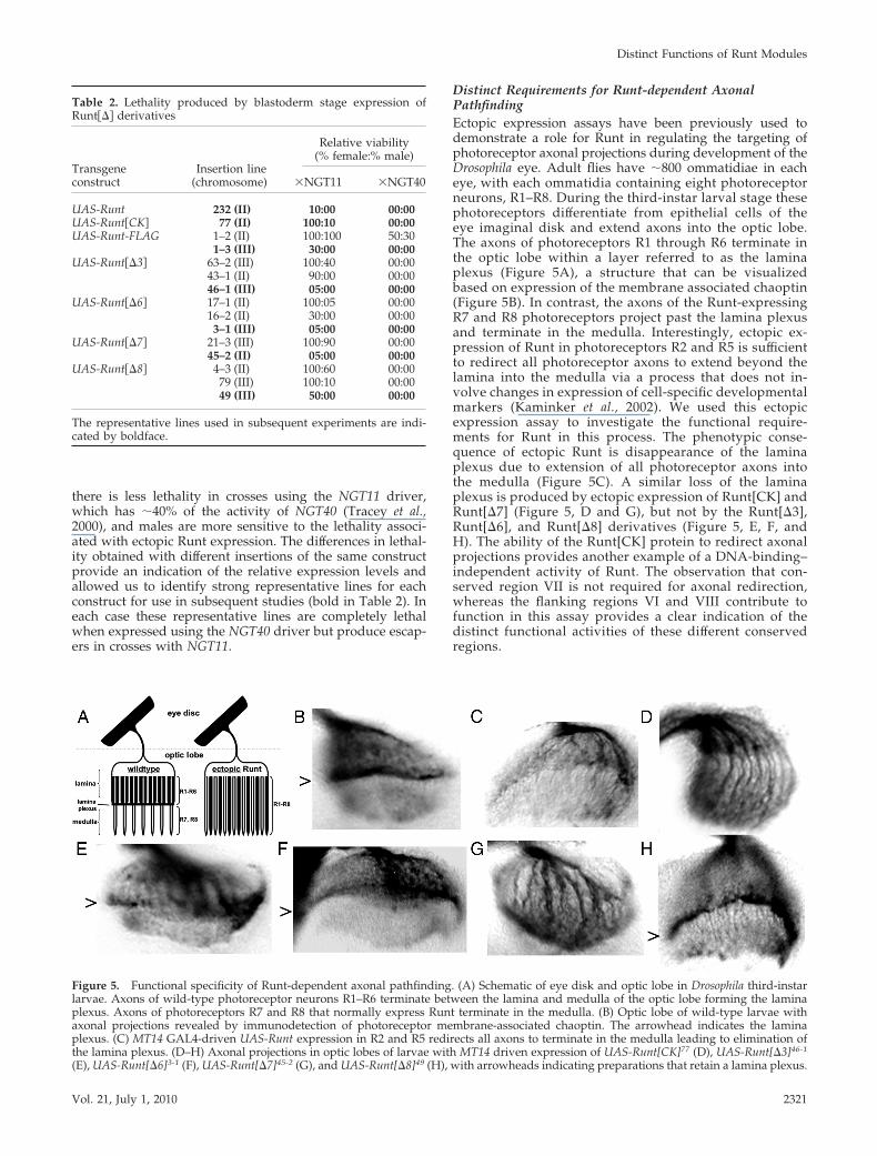
Distinct Requirements for Runt-dependent AxonalPathfindingEctopic expression assays have been previously used todemonstrate a role for Runt in regulating the targeting ofphotoreceptor axonal projections during development of theDrosophila eye. Adult flies have �800 ommatidiae in eacheye, with each ommatidia containing eight photoreceptorneurons, R1–R8. During the third-instar larval stage thesephotoreceptors differentiate from epithelial cells of theeye imaginal disk and extend axons into the optic lobe.The axons of photoreceptors R1 through R6 terminate inthe optic lobe within a layer referred to as the laminaplexus (Figure 5A), a structure that can be visualizedbased on expression of the membrane associated chaoptin(Figure 5B). In contrast, the axons of the Runt-expressingR7 and R8 photoreceptors project past the lamina plexusand terminate in the medulla. Interestingly, ectopic ex-pression of Runt in photoreceptors R2 and R5 is sufficientto redirect all photoreceptor axons to extend beyond thelamina into the medulla via a process that does not in-volve changes in expression of cell-specific developmentalmarkers (Kaminker et al., 2002). We used this ectopicexpression assay to investigate the functional require-ments for Runt in this process. The phenotypic conse-quence of ectopic Runt is disappearance of the laminaplexus due to extension of all photoreceptor axons intothe medulla (Figure 5C). A similar loss of the laminaplexus is produced by ectopic expression of Runt[CK] andRunt[�7] (Figure 5, D and G), but not by the Runt[�3],Runt[�6], and Runt[�8] derivatives (Figure 5, E, F, andH). The ability of the Runt[CK] protein to redirect axonalprojections provides another example of a DNA-binding–independent activity of Runt. The observation that con-served region VII is not required for axonal redirection,whereas the flanking regions VI and VIII contribute tofunction in this assay provides a clear indication of thedistinct functional activities of these different conservedregions.
Table 2. Lethality produced by blastoderm stage expression ofRunt��� derivatives
Transgeneconstruct
Insertion line(chromosome)
Relative viability(% female:% male)
�NGT11 �NGT40
UAS-Runt 232 (II) 10:00 00:00UAS-Runt�CK� 77 (II) 100:10 00:00UAS-Runt-FLAG 1–2 (II) 100:100 50:30
1–3 (III) 30:00 00:00UAS-Runt��3� 63–2 (III) 100:40 00:00
43–1 (II) 90:00 00:0046–1 (III) 05:00 00:00
UAS-Runt��6� 17–1 (II) 100:05 00:0016–2 (II) 30:00 00:003–1 (III) 05:00 00:00
UAS-Runt��7� 21–3 (III) 100:90 00:0045–2 (II) 05:00 00:00
UAS-Runt��8� 4–3 (II) 100:60 00:0079 (III) 100:10 00:0049 (III) 50:00 00:00
The representative lines used in subsequent experiments are indi-cated by boldface.
Figure 5. Functional specificity of Runt-dependent axonal pathfinding. (A) Schematic of eye disk and optic lobe in Drosophila third-instarlarvae. Axons of wild-type photoreceptor neurons R1–R6 terminate between the lamina and medulla of the optic lobe forming the laminaplexus. Axons of photoreceptors R7 and R8 that normally express Runt terminate in the medulla. (B) Optic lobe of wild-type larvae withaxonal projections revealed by immunodetection of photoreceptor membrane-associated chaoptin. The arrowhead indicates the laminaplexus. (C) MT14 GAL4-driven UAS-Runt expression in R2 and R5 redirects all axons to terminate in the medulla leading to elimination ofthe lamina plexus. (D–H) Axonal projections in optic lobes of larvae with MT14 driven expression of UAS-Runt[CK]77 (D), UAS-Runt[�3]46-1
(E), UAS-Runt[�6]3-1 (F), UAS-Runt[�7]45-2 (G), and UAS-Runt[�8]49 (H), with arrowheads indicating preparations that retain a lamina plexus.
Distinct Functions of Runt Modules
Vol. 21, July 1, 2010 2321
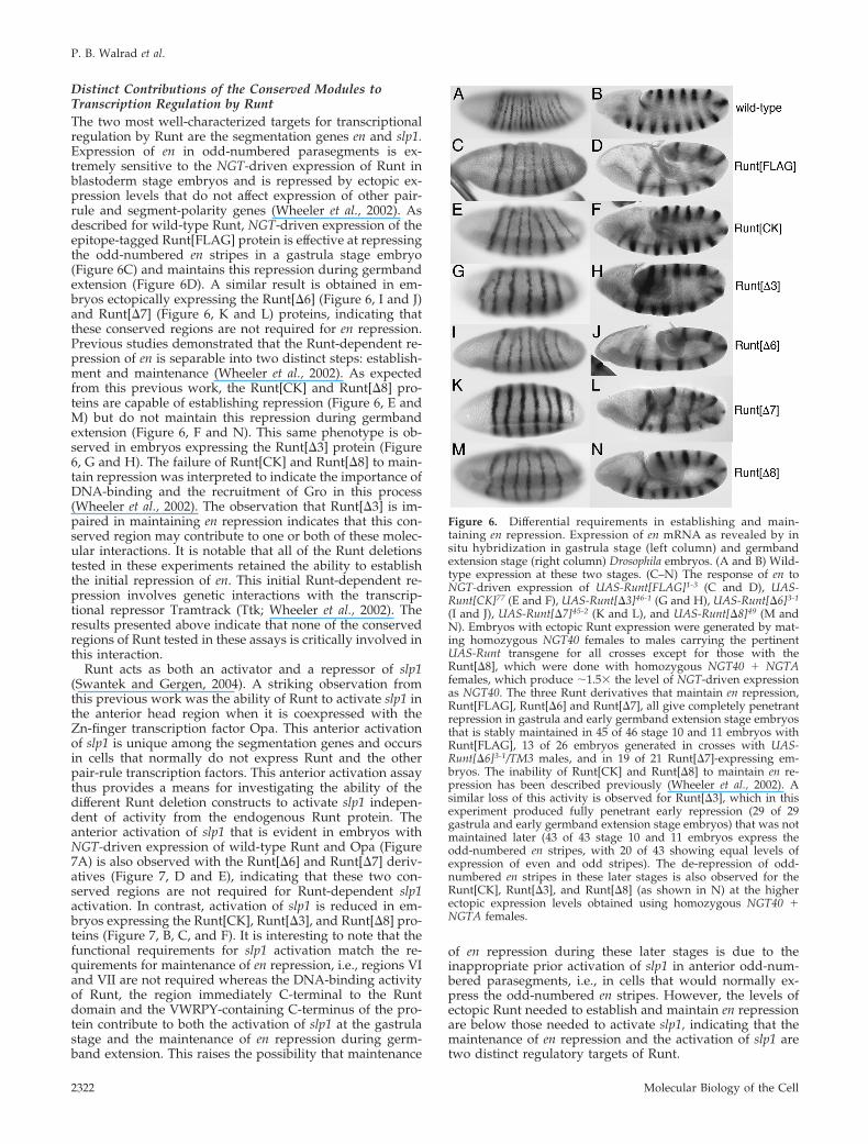
Distinct Contributions of the Conserved Modules toTranscription Regulation by RuntThe two most well-characterized targets for transcriptionalregulation by Runt are the segmentation genes en and slp1.Expression of en in odd-numbered parasegments is ex-tremely sensitive to the NGT-driven expression of Runt inblastoderm stage embryos and is repressed by ectopic ex-pression levels that do not affect expression of other pair-rule and segment-polarity genes (Wheeler et al., 2002). Asdescribed for wild-type Runt, NGT-driven expression of theepitope-tagged Runt[FLAG] protein is effective at repressingthe odd-numbered en stripes in a gastrula stage embryo(Figure 6C) and maintains this repression during germbandextension (Figure 6D). A similar result is obtained in em-bryos ectopically expressing the Runt[�6] (Figure 6, I and J)and Runt[�7] (Figure 6, K and L) proteins, indicating thatthese conserved regions are not required for en repression.Previous studies demonstrated that the Runt-dependent re-pression of en is separable into two distinct steps: establish-ment and maintenance (Wheeler et al., 2002). As expectedfrom this previous work, the Runt[CK] and Runt[�8] pro-teins are capable of establishing repression (Figure 6, E andM) but do not maintain this repression during germbandextension (Figure 6, F and N). This same phenotype is ob-served in embryos expressing the Runt[�3] protein (Figure6, G and H). The failure of Runt[CK] and Runt[�8] to main-tain repression was interpreted to indicate the importance ofDNA-binding and the recruitment of Gro in this process(Wheeler et al., 2002). The observation that Runt[�3] is im-paired in maintaining en repression indicates that this con-served region may contribute to one or both of these molec-ular interactions. It is notable that all of the Runt deletionstested in these experiments retained the ability to establishthe initial repression of en. This initial Runt-dependent re-pression involves genetic interactions with the transcrip-tional repressor Tramtrack (Ttk; Wheeler et al., 2002). Theresults presented above indicate that none of the conservedregions of Runt tested in these assays is critically involved inthis interaction.
Runt acts as both an activator and a repressor of slp1(Swantek and Gergen, 2004). A striking observation fromthis previous work was the ability of Runt to activate slp1 inthe anterior head region when it is coexpressed with theZn-finger transcription factor Opa. This anterior activationof slp1 is unique among the segmentation genes and occursin cells that normally do not express Runt and the otherpair-rule transcription factors. This anterior activation assaythus provides a means for investigating the ability of thedifferent Runt deletion constructs to activate slp1 indepen-dent of activity from the endogenous Runt protein. Theanterior activation of slp1 that is evident in embryos withNGT-driven expression of wild-type Runt and Opa (Figure7A) is also observed with the Runt[�6] and Runt[�7] deriv-atives (Figure 7, D and E), indicating that these two con-served regions are not required for Runt-dependent slp1activation. In contrast, activation of slp1 is reduced in em-bryos expressing the Runt[CK], Runt[�3], and Runt[�8] pro-teins (Figure 7, B, C, and F). It is interesting to note that thefunctional requirements for slp1 activation match the re-quirements for maintenance of en repression, i.e., regions VIand VII are not required whereas the DNA-binding activityof Runt, the region immediately C-terminal to the Runtdomain and the VWRPY-containing C-terminus of the pro-tein contribute to both the activation of slp1 at the gastrulastage and the maintenance of en repression during germ-band extension. This raises the possibility that maintenance
of en repression during these later stages is due to theinappropriate prior activation of slp1 in anterior odd-num-bered parasegments, i.e., in cells that would normally ex-press the odd-numbered en stripes. However, the levels ofectopic Runt needed to establish and maintain en repressionare below those needed to activate slp1, indicating that themaintenance of en repression and the activation of slp1 aretwo distinct regulatory targets of Runt.
Figure 6. Differential requirements in establishing and main-taining en repression. Expression of en mRNA as revealed by insitu hybridization in gastrula stage (left column) and germbandextension stage (right column) Drosophila embryos. (A and B) Wild-type expression at these two stages. (C–N) The response of en toNGT-driven expression of UAS-Runt[FLAG]1-3 (C and D), UAS-Runt[CK]77 (E and F), UAS-Runt[�3]46-1 (G and H), UAS-Runt[�6]3-1
(I and J), UAS-Runt[�7]45-2 (K and L), and UAS-Runt[�8]49 (M andN). Embryos with ectopic Runt expression were generated by mat-ing homozygous NGT40 females to males carrying the pertinentUAS-Runt transgene for all crosses except for those with theRunt[�8], which were done with homozygous NGT40 � NGTAfemales, which produce �1.5� the level of NGT-driven expressionas NGT40. The three Runt derivatives that maintain en repression,Runt[FLAG], Runt[�6] and Runt[�7], all give completely penetrantrepression in gastrula and early germband extension stage embryosthat is stably maintained in 45 of 46 stage 10 and 11 embryos withRunt[FLAG], 13 of 26 embryos generated in crosses with UAS-Runt[�6]3-1/TM3 males, and in 19 of 21 Runt[�7]-expressing em-bryos. The inability of Runt[CK] and Runt[�8] to maintain en re-pression has been described previously (Wheeler et al., 2002). Asimilar loss of this activity is observed for Runt[�3], which in thisexperiment produced fully penetrant early repression (29 of 29gastrula and early germband extension stage embryos) that was notmaintained later (43 of 43 stage 10 and 11 embryos express theodd-numbered en stripes, with 20 of 43 showing equal levels ofexpression of even and odd stripes). The de-repression of odd-numbered en stripes in these later stages is also observed for theRunt[CK], Runt[�3], and Runt[�8] (as shown in N) at the higherectopic expression levels obtained using homozygous NGT40 �NGTA females.
P. B. Walrad et al.
Molecular Biology of the Cell2322

Runt is converted from an activator to a repressor of slp1by the homeodomain transcription factor Ftz (Swantek andGergen, 2004). Endogenous Runt and Ftz are expressed inthe anterior half of the even-numbered parasegments whereboth are required to prevent slp1 expression. Ectopic expres-sion of Ftz alone leads to repression of slp1 in the posteriorhalf of the odd-parasegments where endogenous Runt isalso present. More important for this work is the observationthat NGT-driven coexpression of Runt and Ftz also leads torepression of slp1 in the posterior half of the even-numberedparasegments, resulting in the elimination of expressionthroughout the presegmental region of the embryo (Figure7G). This coexpression assay thus provides an approach toinvestigating the ability of different Runt deletion constructsto repress slp1 independent of the activity of endogenousRunt protein. Two of the deletions, Runt[�6] and Runt[�8],retain activity as repressors (Figure 7, J and L), whereas theRunt[CK], Runt[�3], and Runt[�7] proteins fail to repress
the even-numbered slp1 stripes in this assay (Figure 7, H, I,and K). The observation that these last three Runt deriva-tives fail to repress slp1, yet are competent in the initialestablishment of en repression at this stage (Figure 6, E, G,and K) indicates a clear distinction in the molecular require-ments for Runt-dependent repression of these two targets. Itwas noted above that the Runt[�8] protein is effective atestablishing en repression at the gastrula stage, but thisrepression is not maintained during germband extension(Figure 6, M and N). In contrast, the Runt[�8]-dependentrepression of slp1 is maintained during these later stages(Figure 7R). This observation provides a further indicationfor differences in the Runt-dependent repression of en andslp1. There are some differences in early versus late slp1 ex-pression in embryos that coexpress Ftz and the Runt[CK],Runt[�3], and Runt[�7] proteins (Figure 7, N, O, and Q). How-ever, it is difficult to interpret these changes without examiningthe response of other Runt targets in these embryos.
Figure 7. Differential requirements for slp1activation and repression. Expression of slp1mRNA as revealed by in situ hybridization.(A–F) gastrula stage expression of slp1 in re-sponse to NGT-driven coexpression of Opaand different Runt deletion derivatives. In allcases ectopic expression was obtained usingfemales homozygous for both the NGT40 andNGTA GAL4 drivers. (A) UAS-Runt232 andUAS-Opa12, 60 of 65 gastrula stage embryosscored in this experiment showed ectopic an-terior slp1 activation comparable or strongerthan that shown in this panel. The remainingfive embryos had weaker anterior activationwith incomplete fusion of stripes within thesegmented region of the embryo. (B) UAS-Runt[CK]77 and UAS-Opa12, 32 of 39 gastrulastage embryos showed incomplete fusion ofslp1 stripes similar to that depicted in thispanel, and six of seven the remaining embryosshowed evidence of weak anterior activation,with one embryo showing clear evidence ofectopic anterior activation. (C) UAS-Runt[�3]46-1
and UAS-Opa14, 34 of 37 embryos showedincomplete fusion as depicted, with theother three showing evidence of weak ante-rior activation. (D) UAS-Runt[�6]3-1/TM3and UAS-Opa14, 13 of 25 gastrula stage em-bryos showed strong anterior activation sim-ilar to that shown in this panel. As expected ina cross with males heterozygous for the UAS-Runt[�6] construct, 12 of 25 showed minoralterations in the spacing of slp1 stripes pro-duced by NGT-driven expression of Opa alone.(E) UAS-Runt[�7]21-3/TM3 and UAS-Opa14, 13of 28 gastrula stage embryos in crosses with these heterozygous males showed strong anterior activation. (F) UAS-Runt[�8]49 andUAS-Opa10, 28 of 38 gastrula stage embryos showed abnormal spacing of slp1 stripes, whereas 10 of 38 showed loss of specific stripessimilar to that shown, presumably due to repression by Runt[�8] (see below). None of the embryos in this cross showed strong anterioractivation. Arrows indicate regions of anterior slp1 activation in response to Runt, Runt[�6], and Runt[�7]. The potent activity ofRunt[�7] in slp1 activation is underscored by the use of the weaker UAS-Runt[�7]21-3 line in this coexpression assay. Similarly, theinability of the Runt[�8] derivative to activate slp1 is underscored by the use of UAS-Opa10 as this line is stronger than the UAS-Opalines used for the other Runt constructs (Swantek and Gergen, 2004). The slp1 response to NGT-driven coexpression of Ftz and thesedifferent Runt deletion derivatives in gastrula (G–L) and germband extension stages (M–R). In all cases ectopic expression was obtainedby mating females homozygous for both NGT40 and NGTA to males homozygous for UAS-Ftz263 and the pertinent Runt transgene: (Gand M) UAS-Runt232, 15 of 22 gastrula stage embryos show partial to complete repression of the even-numbered slp1 stripes; (H andN) UAS-Runt[CK]77, 0 of 23 gastrula stage embryos showed any evidence of repression of the even-numbered stripes; (I and O)UAS-Runt[�3]46-1, 0 of 8 gastrula stage embryos show repression of all of the even-numbered stripes, although there is a region specificreduction in stripe 10 expression in several embryos; (J and P) UAS-Runt[�6]16-2, 10 of 10 gastrula stage embryos show evidence ofrepression¤ with four of these having nearly complete repression similar to that shown in this panel. Note that the Runt[�6] line usedin this experiment is slightly weaker than the line used in the slp1 activation assay described above; (K and Q) UAS-Runt[�7]45-2, 0 of17 gastrula stage embryos show repression of all even-numbered stripes, although expression of stripe 10 is reduced in 16 of these 17embryos; (L and R) UAS-Runt[�8]49/TM3, 17 of 33 gastrula and early germband extension stage embryos scored in the cross with theseheterozygous males showed partial to complete repression of the even-numbered stripes. The odd-numbered slp1 stripes are repressedin all of the embryos in these different crosses due to ectopic Ftz expression in cells expressing endogenous Runt.
Distinct Functions of Runt Modules
Vol. 21, July 1, 2010 2323

The Role of Groucho and Rpd3 in slp1 RegulationThe results presented above indicating that Runt’s con-served C-terminal region VIII has no apparent role in slp1repression, but instead contributes to Runt-dependent slp1activation are somewhat surprising. The Runt[�8] protein
lacks the C-terminal VWRPY motif that mediates interactionwith the corepressor protein Gro (Aronson et al., 1997). Themaintenance of Runt-dependent en repression is sensitive tothe maternal dosage of Gro and the Gro-interacting histonedeacetylase Rpd3 (Wheeler et al., 2002). We used assayssimilar to those used in this previous work to investigatewhether Gro and Rpd3 have roles in Runt-dependent slp1regulation. NGT-driven coexpression of Runt and Ftz is aseffective at repressing slp1 in embryos from females het-erozygous for either the GroBX22 or GroE48 mutations as inembryos with wild-type Gro dosage (Figure 8, A–C). Thisresult is consistent with observations above indicating thatthe Gro-interacting C-terminus is not required for slp1 re-pression. The maternal dosage of Rpd3 also has no effect onslp1 repression (Figure 8D).
The more interesting question is whether the requirementfor region VIII in slp1 activation reflects a role for Gro in thisprocess. We used the anterior activation assay describedabove to investigate to investigate whether the dosage ofeither Gro or Rpd3 influences Runt-dependent slp1 activa-tion. In this case the activation of slp1 in anterior headregions in response to NGT-driven coexpression of Runt andOpa is not reduced in embryos from females that are het-erozygous for mutations in either Gro or Rpd3 (Figure 8,E–H). The extent of anterior activation obtained with thespecific combination of NGT drivers and UAS-Runt andUAS-Opa lines used for these experiments is strong, but notmaximal. Importantly, slight reductions (less than twofold)in the level of ectopic Runt activity results in significantlyweaker anterior activation (Swantek and Gergen, 2004).These results thus provide strong evidence that the Runt-dependent activation of slp1 is not sensitive to Gro dosage.On the basis of these observations, we conclude that therequirement for region VIII in slp1 activation does not in-volve interactions with Gro but instead should involve in-teractions of Runt’s conserved C-terminus with other factorsthat are involved in transcriptional activation.
DISCUSSION
Functionally Distinct Roles for Different ConservedRegions of RuntThe experiments presented above use several different as-says to investigate the functional contributions of differentconserved regions of Runt. Each of the four deletion deriv-atives affects a different set of properties (Table 3). Thefunctional specificity demonstrated by the different dele-tions is consistent with the notion that different conservedregions correspond to functional modules that participate in
Figure 8. Runt-dependent slp1 regulation is insensitive to Gro andRpd3 dosage. Expression of slp1 mRNA in response to NGT-drivencoexpression of UAS-Runt15 and UAS-Ftz263 (A–D) or UAS-Runt15
and UAS-Opa14 (E–H) transgenes. (A) Sixteen of 20 gastrula andearly germband extension stage embryos from crosses involvingfemales heterozygous for both the NGT40 and NGTA drivers thatare wild-type for Gro and Rpd3 show partial to complete repressionof slp1. Reducing the strength of either the NGT driver or theUAS-Runt line reduces the efficiency of the repression of slp1 ob-served in response to coexpression of Runt and Ftz (Swantek andGergen, 2004). (B) Nineteen of 23 embryos from females heterozy-gous for NT40, NGTA, and the GroBX22 mutation show repression ofslp1. (C) Nineteen of 25 embryos from heterozygous NGT40, NGTAfemales that are also heterozygous for GroE48 show slp1 repression.(D) Thirty of 36 embryos from females heterozygous for NGT40,NGTA, and the Rpd304556 mutation show evidence of repression. (E)Anterior activation in response to of Runt and Opa is observed in 57of 58 embryos from females heterozygous for NGT40 and NGTAthat are otherwise wild type. Strong anterior activation is alsoobserved in crosses with females that are also heterozygous for (F)GroBX22, 20 of 23 embryos; (G) GroE48, 30 of 39 embryos; and (H)Rpd304556, 46 of 55 gastrula and early germband extension stageembryos.
Table 3. Functional specificity of Runt’s conserved regions
Runt derivatives
Functional assay Runt�CK� Runt��3� Runt��6� Runt��7� Runt��8�
Nuclear localization n.d. Diffuse Punctate Diffuse PunctateEmbryo lethality � � � � �Axonal redirection � � � � �Repress en (est.) � � � � �Repress en (maint.) � � � � �Activate slp1 � � � � �Repress slp1 � � � � �
n.d., not determined.
P. B. Walrad et al.
Molecular Biology of the Cell2324

distinct molecular interactions of the Runt protein. It isinteresting to note that similar patterns of functional require-ments for Runt are observed for two different pairs of assays.The observations that NGT-driven expression of all of thedifferent Runt constructs is lethal and that all of the con-structs also retain the ability to establish en repression areconsistent with the idea that this initial repression of en is theprincipal basis for the lethality associated with ectopic Runtexpression. Perhaps more interesting are the common func-tional deficits of the Runt[CK], Runt[�3], and Runt[�8] pro-teins in both slp1 activation and the maintenance of en re-pression (Table 3). As mentioned above, the levels of Runtneeded to maintain en repression in anterior odd paraseg-ments are less than those needed for activation of slp1 inthese same cells, indicating that these are two distinct targetsthat have coincidental requirements for DNA-binding byRunt, the conserved region C-terminal to the Runt domainand the VWRPY-containing C-terminus.
The inactivity of Runt[CK] in maintaining en repressionand in both the activation and repression of slp1 stronglysuggests that DNA binding by Runt is critical for these threeaspects of Runt function. Runt[�3], which is also defectivefor these same three functions lacks a conserved region thatis located just C-terminal of the DNA-interacting “tail re-gion” loop of the Runt domain (Bravo et al., 2001). Intramo-lecular interactions with regions C-terminal to the Runtdomain of Runx1 modulate DNA binding in vitro (Ka-goshima et al., 1996; Kanno et al., 1998) and can influencecooperative interactions with other DNA-binding factors(Gu et al., 2000). Our results are consistent with the idea thatthe region C-terminal to the Runt domain makes importantcontributions to the in vivo DNA-binding activity of theRunt transcription factor.
Runt[�8] is similar to Runt[CK] and Runt[�3] in that itis defective in maintaining en repression and in activatingslp1. A key difference between these three is the ability ofthe Runt[�8] to repress slp1. This result indicates that theC-terminal VWRPY motif is not required for slp1 repres-sion, a finding also consistent with the results of ourgenetic experiments indicating that Runt-dependent slp1repression is insensitive to Gro dosage. The observationthat conserved region VIII contributes to slp1 activation issomewhat surprising, especially given the previouslydocumented physical interaction between the C-terminalVWRPY motif and the Gro corepressor (Aronson et al.,1997). On the basis of these results, we propose thatconserved region VIII also mediates interactions with aseparate factor that participates in transcription activa-tion. One issue is whether this proposed interaction alsoinvolves the VWRPY motif. Conserved region VIII con-tains two contiguous blocks of sequence conservation sep-arated by a variable linker, suggesting that the proposedinteraction with an activator may involve these otherconserved amino acids. In any event, the relatively com-pact size of region VIII makes it likely that interactionswith Gro and the proposed cofactor involved in Runt-dependent activation will be mutually exclusive.
The unique activities of Runt[�6] and Runt[�7] under-score the modular architecture and context-dependent activ-ities of Runt. These two conserved regions are separated bytwo amino acids in the five species that comprise the mela-nogaster subgroup (Figure 1). In the set of assays we used,the only activity that is disrupted by deletion of region VI isthe redirection of axonal projections in the developing eye(Table 3). Region VII is not required for this activity of Runt,but appears to contribute to the punctate subnuclear local-ization of Runt and is required for slp1 repression. These last
two properties are shared with Runt[�3]. Nuclear matrixassociation of Runx1 is important for CD4 repression (Telferet al., 2004), suggesting a potential parallel in the mecha-nisms of slp1 repression by Runt and the repression of CD4by Runx1. The repression of slp1 by Runt also requires theactivity of the homeodomain protein Ftz as well as theFtz-interacting orphan nuclear receptor protein Ftz-F1(Swantek and Gergen, 2004; Hou et al., 2009). It is reasonableto propose that regions III and VII are involved in molecularinteractions with Ftz, Ftz-F1 and/or the nuclear matrix thatare involved in converting Runt from an activator to arepressor of slp1 transcription. Taken all together, our resultsprovide compelling evidence for the functional modularityof Runt and lay groundwork for identifying molecular in-teractions that contribute to the regulatory properties of thisconserved family of transcriptional regulators.
ACKNOWLEDGMENTS
Xiangqun Ning played an important role in initiating functional studies onthe different conserved regions of Runt. Comments from Xiaoling Wang andother members of the Gergen lab are appreciated. This research was sup-ported by grants from the National Science Foundation (MCB 0344486 andMCB 0721430). We dedicate this work to the memory of Kevin King.
REFERENCES
Aronson, B. D., Fisher, A. L., Blechman, K., Caudy, M., and Gergen, J. P.(1997). Groucho-dependent and -independent repression activities of Runtdomain proteins. Mol. Cell. Biol. 17, 5581–5587.
Bao, R., and Friedrich, M. (2008). Conserved cluster organization of insectRunx genes. Dev. Genes Evol. 218, 567–574.
Bravo, J., Li, Z., Speck, N. A., and Warren, A. J. (2001). The leukemia-associated AML1 (Runx1)–CBF beta complex functions as a DNA-inducedmolecular clamp. Nat. Struct. Biol. 8, 371–378.
Damen, W.G.M., Weller, M., and Tautz, D. (2000). Expression patterns ofhairy, even-skipped and runt in the spider Cupiennius salei imply that thesegenes were segmentation genes in a basal arthropod. Proc. Natl. Acad. Sci.USA 97, 4515–4519.
de Bruijn, M. F., and Speck, N. A. (2004). Core-binding factors in hematopoi-esis and immune function. Oncogene 23, 4238–4248.
Duffy, J. B., and Gergen, J. P. (1991). The Drosophila segmentation gene runtacts as a position-specific numerator element necessary for the uniform ex-pression of the sex-determining gene Sex-lethal. Genes Dev. 5, 2176–2187.
Duffy, J. B., and Gergen, J. P. (1994). Sex, segments, and the central nervoussystem: common genetic mechanisms of cell fate determination. Adv. Genet.31, 1–28.
Duffy, J. B., Kania, M. A., and Gergen, J. P. (1991). Expression and function ofthe Drosophila gene runt in early stages of neural development. Development113, 1223–1230.
Enomoto, H., Furuichi, T., Zanma, A., Yamana, K., Yoshida, C., Sumitani, S.,Yamamoto, H., Enomoto-Iwamoto, M., Iwamoto, M., and Komori, T. (2004).Runx2 deficiency in chondrocytes causes adipogenic changes in vitro. J. CellSci. 117, 417–425.
Gu, T. L., Goetz, T. L., Graves, B. J., and Speck, N. A. (2000). Auto-inhibitionand partner proteins, core-binding factor beta (CBFbeta) and Ets-1, modulateDNA binding by CBFalpha2 (AML1). Mol. Cell. Biol. 20, 91–103.
Hou, H. Y., Heffer, A., Anderson, W. R., Liu, J., Bowler, T., and Pick, L. (2009).Stripy Ftz target genes are coordinately regulated by Ftz-F1. Dev. Biol. 335,442–453.
Ito, Y. (1999). Molecular basis of tissue-specific gene expression mediated bythe runt domain transcription factor PEBP2/CBF. Genes Cell. 4, 685–696.
Ito, Y. (2004). Oncogenic potential of the RUNX gene family: “overview.”Oncogene 23, 4198–4208.
Ito, Y. (2008). RUNX genes in development and cancer: regulation of viralgene expression and the discovery of RUNX family genes. Adv. Cancer Res.99, 33–76.
Javed, A., Barnes, G. L., Jasanya, B. O., Stein, J. L., Gerstenfeld, L., Lian, J. B.,and Stein, G. S. (2001). runt homology domain transcription factors (Runx,Cbfa, and AML) mediate repression of the bone sialoprotein promoter: evi-
Distinct Functions of Runt Modules
Vol. 21, July 1, 2010 2325

dence for promoter context-dependent activity of Cbfa proteins. Mol. Cell.Biol. 21, 2891–2905.
Javed, A., et al. (2000). Groucho/TLE/R-esp proteins associate with the nu-clear matrix and repress RUNX (CBF(alpha)/AML/PEBP2(alpha)) dependentactivation of tissue-specific gene transcription. J. Cell Sci. 113, 2221–2231.
Kagoshima, H., Akamatsu, Y., Ito, Y., and Shigesada, K. (1996). Functionaldissection of the alpha and beta subunits of transcription factor PEBP2 and theredox susceptibility of its DNA binding activity. J. Biol. Chem. 271, 33074–33082.
Kagoshima, H., Shigesada, K., Satake, M., Ito, Y., Miyoshi, H., Ohki, M.,Pepling, M., and Gergen, P. (1993). The Runt domain identifies a new familyof heteromeric transcriptional regulators. Trends Genet. 9, 338–341.
Kaminker, J. S., Canon, J., Salecker, I., and Banerjee, U. (2002). Control ofphotoreceptor axon target choice by transcriptional repression of Runt. Nat.Neurosci. 5, 746–750.
Kanno, T., Kanno, Y., Chen, L. F., Ogawa, E., Kim, W. Y., and Ito, Y. (1998).Intrinsic transcriptional activation-inhibition domains of the polyomavirusenhancer binding protein 2/core binding factor alpha subunit revealed in thepresence of the beta subunit. Mol. Cell. Biol. 18, 2444–2454.
Kawazu, M., et al. (2005). Functional domains of Runx1 are differentiallyrequired for CD4 repression, TCRbeta expression, and CD4/8 double-nega-tive to CD4/8 double-positive transition in thymocyte development. J. Im-munol. 174, 3526–3533.
Komori, T. (2002). [Cbfa1/Runx2, an essential transcription factor for theregulation of osteoblast differentiation]. Jpn. J. Clin. Med. [Nippon Rinsho]60(suppl 3), 91–97.
Komori, T. (2003). Requisite roles of Runx2 and Cbfb in skeletal development.J. Bone Mineral Metab. 21, 193–197.
Kramer, S. G., Jinks, T. M., Schedl, P., and Gergen, J. P. (1999). Directactivation of Sex-lethal transcription by the Drosophila runt protein. Develop-ment 126, 191–200.
Levanon, D., Goldstein, R. E., Bernstein, Y., Tang, H., Goldenberg, D., Stifani,S., Paroush, Z., and Groner, Y. (1998). Transcriptional repression by AML1and LEF-1 is mediated by the TLE/Groucho corepressors. Proc. Natl. Acad.Sci. USA 95, 11590–11595.
Levanon, D., and Groner, Y. (2004). Structure and regulated expression ofmammalian RUNX genes. Oncogene 23, 4211–4219.
Li, L. H., and Gergen, J. P. (1999). Differential interactions between Brotherproteins and Runt domain proteins in the Drosophila embryo and eye. Devel-opment 126, 3313–3322.
Lian, J. B., et al. (2003). Runx1/AML1 hematopoietic transcription factorcontributes to skeletal development in vivo. J. Cell. Physiol. 196, 301–311.
Lohr, U., and Pick, L. (2005). Cofactor-interaction motifs and the cooption ofa homeotic Hox protein into the segmentation pathway of Drosophila melano-gaster. Curr. Biol. 15, 643–649.
Miyazono, K., Maeda, S., and Imamura, T. (2004). Coordinate regulation ofcell growth and differentiation by TGF-beta superfamily and Runx proteins.Oncogene 23, 4232–4237.
Nishimura, M., Fukushima-Nakase, Y., Fujita, Y., Nakao, M., Toda, S., Kitamura,N., Abe, T., and Okuda, T. (2004). VWRPY motif-dependent and -independentroles of AML1/Runx1 transcription factor in murine hematopoietic develop-ment. Blood 103, 562–570.
Nusslein-Volhard, C., and Wieschaus, E. (1980). Mutations affecting segmentnumber and polarity in Drosophila. Nature 287, 795–801.
Pande, S., et al. (2008). Subnuclear targeting of the Runx3 tumor suppressorand its epigenetic association with mitotic chromosomes. J. Cell. Physiol. 218,473–479.
Pepling, M. E., and Gergen, J. P. (1995). Conservation and function of thetranscriptional regulatory protein Runt. Proc. Natl. Acad. Sci. USA 92, 9087–9091.
Robertson, A. J., Dickey, C. E., McCarthy, J. J., and Coffman, J. A. (2002). Theexpression of SpRunt during sea urchin embryogenesis. Mech. Dev. 117, 327–330.
Sullivan, J. C., Sher, D., Eisenstein, M., Shigesada, K., Reitzel, A. M., Marlow,H., Levanon, D., Groner, Y., Finnerty, J. R., and Gat, U. (2008). The evolution-ary origin of the Runx/CBFbeta transcription factors–studies of the mostbasal metazoans. BMC Evol. Biol. 8, 228.
Swantek, D., and Gergen, J. P. (2004). Ftz modulates Runt-dependent activa-tion and repression of segment-polarity gene transcription. Development 131,2281–2290.
Tang, Y. Y., Crute, B. E., Kelley, J. J., Huang, X., Yan, J., Shi, J., Hartman, K. L.,Laue, T. M., Speck, N. A., and Bushweller, J. H. (2000). Biophysical charac-terization of interactions between the core binding factor alpha and betasubunits and DNA. FEBS Lett. 470, 167–172.
Telfer, J. C., Hedblom, E. E., Anderson, M. K., Laurent, M. N., and Rothenberg,E. V. (2004). Localization of the domains in Runx transcription factors requiredfor the repression of CD4 in thymocytes. J. Immunol. 172, 4359–4370.
Thirunavukkarasu, K., Mahajan, M., McLarren, K. W., Stifani, S., andKarsenty, G. (1998). Two domains unique to osteoblast-specific transcriptionfactor Osf2/Cbfa1 contribute to its transactivation function and its inability toheterodimerize with Cbfbeta. Mol. Cell. Biol. 18, 4197–4208.
Tracey, W. D., Jr., Ning, X., Klingler, M., Kramer, S. G., and Gergen, J. P.(2000). Quantitative analysis of gene function in the Drosophila embryo. Ge-netics 154, 273–284.
Tweedie, S., et al. (2009). FlyBase: enhancing Drosophila Gene Ontology anno-tations. Nucleic Acids Res. 37, D555–D559.
Van Vactor, D., Jr., Krantz, D. E., Reinke, R., and Zipursky, S. L. (1988).Analysis of mutants in chaoptin, a photoreceptor cell-specific glycoprotein inDrosophila, reveals its role in cellular morphogenesis. Cell 52, 281–290.
Vander Zwan, C. J., Wheeler, J. C., Li, L. H., Tracey, W. D., and Gergen, J. P.(2003). A DNA-binding-independent pathway of repression by the DrosophilaRunt protein. Blood Cells Mol. Dis. 30, 207–222.
Wheeler, J. C., Shigesada, K., Gergen, J. P., and Ito, Y. (2000). Mechanisms oftranscriptional regulation by Runt domain proteins. Semin. Cell Dev. Biol. 11,369–375.
Wheeler, J. C., VanderZwan, C., Xu, X., Swantek, D., Tracey, W. D., andGergen, J. P. (2002). Distinct in vivo requirements for establishment versusmaintenance of transcriptional repression. Nat. Genet. 29, 29.
Yarmus, M., Woolf, E., Bernstein, Y., Fainaru, O., Negreanu, V., Levanon, D.,and Groner, Y. (2006). Groucho/transducin-like Enhancer-of-split (TLE)-de-pendent and -independent transcriptional regulation by Runx3. Proc. Natl.Acad. Sci. USA 103, 7384–7389.
Zaidi, S. K., Javed, A., Choi, J. Y., van Wijnen, A. J., Stein, J. L., Lian, J. B., andStein, G. S. (2001). A specific targeting signal directs Runx2/Cbfa1 to sub-nuclear domains and contributes to transactivation of the osteocalcin gene.J. Cell Sci. 114, 3093–3102.
Zeng, C., et al. (1997). Identification of a nuclear matrix targeting signal in theleukemia and bone-related AML/CBF-alpha transcription factors. Proc. Natl.Acad. Sci. USA 94, 6746–6751.
Zhang, L., Li, Z., Yan, J., Pradhan, P., Corpora, T., Cheney, M. D., Bravo, J.,Warren, A. J., Bushweller, J. H., and Speck, N. A. (2003). Mutagenesis of theRunt domain defines two energetic hot spots for heterodimerization with thecore binding factor beta subunit. J. Biol. Chem. 278, 33097–33104.
P. B. Walrad et al.
Molecular Biology of the Cell2326