dissertation v1- prelim pages - eScholarship
130
UC San Diego UC San Diego Electronic Theses and Dissertations Title The role of cGMP-dependent protein kinases in bone cell growth and survival Permalink https://escholarship.org/uc/item/3t9842q4 Author Marathe, Nisha Madhav Publication Date 2011 Peer reviewed|Thesis/dissertation eScholarship.org Powered by the California Digital Library University of California
-
Upload
khangminh22 -
Category
Documents
-
view
2 -
download
0
Transcript of dissertation v1- prelim pages - eScholarship

UC San DiegoUC San Diego Electronic Theses and Dissertations
TitleThe role of cGMP-dependent protein kinases in bone cell growth and survival
Permalinkhttps://escholarship.org/uc/item/3t9842q4
AuthorMarathe, Nisha Madhav
Publication Date2011 Peer reviewed|Thesis/dissertation
eScholarship.org Powered by the California Digital LibraryUniversity of California

UNIVERSITY OF CALIFORNIA, SAN DIEGO
The Role of cGMP-dependent Protein Kinases in Bone Cell Growth and Survival
A dissertation submitted in partial satisfaction of the requirements for the degree Doctor
of Philosophy
in
Biomedical Sciences
by
Nisha Madhav Marathe
Committee in Charge:
Professor Renate Pilz, ChairProfessor Gerry Boss, Co-chairProfessor Laurence BruntonProfessor Mark GinsbergProfessor Pamela Mellon
2011

Copyright
Nisha Madhav Marathe, 2011
All rights reserved

iii
The Dissertation of Nisha Madhav Marathe is approved and it is acceptable in quality and
form for publication on microfilm and electronically:
_________________________________________________
_________________________________________________
_________________________________________________
_________________________________________________Co-chair
_________________________________________________Chair
University of California, San Diego
2011

iv
DEDICATION
To my parents and my sister.
For all your love and support.
I couldn’t have done this without you.
and
To my grandparents.

iv
TABLE OF CONTENTS
Signature Page............................................................................................................... iii
Dedication......................................................................................................................iv
Table of Contents ...........................................................................................................iv
List of Figures ................................................................................................................vi
List of Tables .................................................................................................................ix
Acknowledgements ........................................................................................................xi
Vita...................................................................................................................................xiii
Abstract of The Dissertation .........................................................................................xiv
Chapter 1: Introduction....................................................................................................1References .........................................................................................................11
Chapter 2: The NO/cGMP Pathway Mediates the Osteocyte-protective Effects ofEstrogen via Differential Actions of cGMP-dependent Protein Kinases I and II .20Abstract .............................................................................................................21Introduction .......................................................................................................22Material and Methods ........................................................................................24Results...............................................................................................................27Discussion .........................................................................................................33Figures...............................................................................................................37References .........................................................................................................43
Chapter 3: Apoptosis in BoneAbstract .............................................................................................................49Introduction .......................................................................................................50Materials and Methods.......................................................................................52Results...............................................................................................................54Discussion .........................................................................................................56Figures...............................................................................................................59References .........................................................................................................63
Chapter 4: Type II cGMP-dependent Protein Kinase Mediates OsteoblastMechanotransductionAbstract .............................................................................................................66Type II cGMP-dependent Protein Kinase Mediates OsteoblastMechanotransduction.........................................................................................68

v
Cyclic GMP and Protein Kinase G Control a Src-Containing Mechanosome inOsteoblasts ........................................................................................................86
Chapter 5: Discussion..................................................................................................110References .......................................................................................................114

vi
LIST OF FIGURES
Chapter 2:
Figure 1: Serum starvation- and etoposide-induced apoptosis in prevented by E2 and
cGMP and is mediated through PKG. ................................................................37
Figure 2: PKGIα and PKGII are required for E2- and cGMP-mediated protection from
etoposide-induced apoptosis ..............................................................................38
Figure 3: E2-induced Akt and ERK phosphorylation and ERK nuclear translocalization is
mediated by PKGII ............................................................................................39
Figure 4: BAD phosphorylation at Ser155 is mediated by PKGIα and is necessary to
prevent apoptosis ...............................................................................................40
Supplemental Figure 1: Knockdown and viral reconstitution of PKGI and II .................41
Supplemental Figure 2: Transfection efficiency of MLO-Y4 cells .................................42
Chapter 3:
Figure 1: T3 mediates cell survival through the NO/cGMP pathway and Src .................59
Figure 2: cGMP regulates mRNA expression of bim, bad, and bax. ...............................60
Figure 3: Dexamethasone-induced cell death.................................................................61
Figure 4: The NO/cGMP pathway prevents dexamethasone-induced cell death .............62
Chapter 4:
Figure 1: Effect of fluid shear stress on osteoblast c-fos, fra-1, fra-2, and fosB/ΔfosB
mRNA expression..............................................................................................70

vii
Figure 2: Effect of fluid shear stress on osteoblast NO and cGMP production................71
Figure 3: Inhibition of NO/cGMP signaling prevents shear-induced c-fos, fra-1, fra-2,
and fosB/ΔfosB mRNA expression .....................................................................72
Figure 4: cGMP partly mimics the effects of fluid shear stress son osteoblast c-fos, fra-1,
fra-2, and fosB/ΔfosB mRNA expression ...........................................................73
Figure 5: Effect of siRNA-mediated PKGI or PKGII knockdown on shear- or cGMP-
induced c-fos, fra-1, fra-2, and fosB/ΔfosB mRNA expression ...........................74
Figure 6: Rescue of PKGII siRNA-transfected cells with a virus encoding siRNA-
resistant PKGII ..................................................................................................75
Figure 7: Induction of c-fos, fra-1, and fra-2 mRNA expression by fluid shear stress is
MEK/ERK dependent ........................................................................................76
Figure 8: Fluid shear stress-induced ERK activation required NO/cGMP/PKGII
signaling........................................................................................................................76
Figure 9: NO/cGMP activation of PKGII is sufficient to activate ERK1/2 .....................77
Figure 10: NO/cGMP and calcium signaling in osteoblast mechanotransduction ...........78
Supplemental Figure 1: Fluid shear stress induction of fos family genes, NO production
and VASP phosphorylation................................................................................82
Supplemental Figure 2: Effect of calcium and cGMP on fos family gene expression and
ERK phosphorylation.........................................................................................83
Supplemental Figure 3: Semi-quantitative RT-PCR of fos family mRNA expression in
response to cGMP and PKGII ............................................................................84
Supplemental Figure 4: Pharmacological inhibition of the NO/cGMP pathway prevents
fluid shear stress-, NO- and cGMP- induced ERK phosphorylation....................85

viii
Figure 1: Fluid shear stress- and cGMP- induced osteoblast proliferation and ERK
activation ...........................................................................................................87
Figure 2: Src activation by membrane-bound PKG........................................................88
Figure 3: Fluid shear stress- and cGMP- induced Src activtion mediated by SHP-1 and
SHP-2................................................................................................................89
Figure 4: PKGII phosphorylation of SHP-1 and SHP-2 and regulation of PTP
activity ..........................................................................................................................90
Figure 5: Integrin dependence of Src activation by cGMP and PKGII............................91
Figure 6: Characterization of a Src-containing mechanosensitive complex in
osteoblasts.....................................................................................................................92
Figure 7: Signaling defect in PKGII-null osteoblasts. ....................................................93
Supplemental Figure 1: siRNA knockdown of PKGI or PKGII; effects of fluid shear
stress and cGMP on Src and ERK phosphorylation in MLO-Y4 and MC3T3...100
Supplemental Figure 2: PKGII does not directly phosphorylate Src .............................101
Supplemental Figure 3: Effect of vanadate- and phosphotase-specific siRNAs on Src
phosphorylation and dephosphorylation ...........................................................102
Supplemental Figure 4: Analysis of SHP-1 phosphorylation and function in MC3T3 cells
........................................................................................................................103
Supplemental Figure 5: cGMP activation of Src requires ligation of β3 integrins .........104
Supplemental Figure 6: Colocalization of β3 integrins, PKGII and Src with SHP-2-
containing membrane complexes .....................................................................105
Supplemental Figure 7: Interactions among PKGII, SHP-2, Src, and β3 integrins ........106

ix
LIST OF TABLES
Chapter 3:
Table 1: qRT-PCR primers ............................................................................................58
Chapter 4:
Supplemental Table 1: PCR Primers Used for RT-PCR.................................................81
Supplemental Table 1: siRNA target sequences ...........................................................107

x
ACKNOWLEDGEMENTS
I would like to first thank my advisor, Dr. Renate Pilz, for the giving me the
opportunity to do my graduate research in her lab. She has helped me through all the
bumps in the road and her passion for science has been a great motivator. I can’t express
how much her support has meant to me. In her lab, I was able to grow both as a scientist
and as a person, and I can’t thank her enough for that. I also thank Dr. Gerry Boss for all
his support, insights, and opportunities.
The camaraderie of all the current and former members of the Boss Pilz lab has
made my graduate experience enjoyable. I appreciate the time I have spent with them;
their knowledge and experience has helped guide me along the way. My time would have
definitely been less enjoyable without them. I would especially like to thank Darren
Casteel, Hema Rangaswami, Raphaela Schwappacher, and Tong Zhang for their
invaluable support, guidance, and friendship. I would also like to take the opportunity to
thank my friends and family who have been with me through every step of this journey.
They have helped shape who I am and, without them, I would not have reached so far.
I also appreciate the time and guidance of my thesis committee; Drs Larry
Brunton, Mark Ginsburg, and Pamela Mellon.
Chapter 2 is currently being prepared for submission for publication of the
material. Marathe N, Rangaswami H, Zhuang S, Boss GR, Pilz RB. The NO/cGMP
pathway mediates the osteocyte-protective effects of estrogen via differential actions of
cGMP-dependent protein kinases I and II. The dissertation author was the primary
investigator and author of this material.

xi
Chapter 4, in part, is material which appears in Rangaswami H, Marathe N,
Zhuang S, Chen Y, Yeh JC, Frangos JA, Boss GR, Pilz RB. Type II cGMP-dependent
protein kinase mediates osteoblast mechanotransduction J Biol Chem 2009, 284:14796-
14808. The dissertation author was the second author and investigator of this material.
The remaining part of Chapter 4 is material which appears in Rangaswami H,
Schwappacher R, Marathe N, Zhuang S, Casteel DE, Haas B, Chen Y, Pfeifer A, Kato H,
Shattil S, Boss GR, Pilz RB. Cyclic GMP and protein kinase G control a Src-containing
mechanosome in osteoblasts. Sci Signal 2010, 3:ra91. The dissertation author was the
third investigator and author of this material.
This work was supported, in part, by the Hypertension Training Grant T32-
HL007261.

xii
VITA
2005 Bachelor of Science, Microbiology,Immunology, and Molecular GeneticsUniversity of California, Los AngelesLos Angeles, California
2006 Teaching Assistant, Department of BiologyUniversity of California, San DiegoLa Jolla, California
2008-2011 Institutional Kirschstein-NRSA ResearchTraining GrantUniversity of California, San DiegoLa Jolla, California
2011 Doctor of Philosophy, Biomedical SciencesUniversity of California, San DiegoLa Jolla, California
PUBLICATIONSRangaswami H, Marathe N, Zhuang S, Chen Y, Yeh JC, Frangos JA, Boss GR, Pilz RB:Type II cGMP-dependent protein kinase mediates osteoblast mechanotransduction.J Biol Chem 2009.
Rangaswami H, Schwappacher R, Marathe N, Zhuang S, Casteel DE, Haas B, Chen Y,Pfeifer A, Kato H, Shattil S, Boss GR, Pilz RB: Cyclic GMP/Protein Kinase G Controla Src-containing Mechanosome in Osteoblasts. Sci Signal. 2010.

xiii
ABSTRACT OF THE DISSERTATION
The Role of cGMP-dependent Protein Kinases in Bone Cell Growth and Survival
by
Nisha Madhav Marathe
Doctor of Philosophy in Biomedical Sciences
University of California, San Diego, 2011
Professor Renate Pilz, Chair
Professor Gerry Boss, Co-Chair
Skeletal integrity is preserved by continuous bone remodeling by osteoblasts and
osteoclasts. While the careful regulation between bone formation and resorption is known
to be a hallmark of bone maintenance, the signaling mechanisms which drive each of
these processes are incompletely understood. Estrogens and fluid shear stress are known
to exert positive effects on osteoblast proliferation and osteocyte survival. Both

xiv
stimuli have been shown to rapidly increase nitric oxide (NO) production in a number of
cell types, suggesting an activation of the NO/cGMP/PKG pathway. NO is involved in
regulating cell survival and proliferation in different cell types.
In the first part of the dissertation, we show that estrogen-mediated osteocytes
survival is regulated through the NO/cGMP pathway. Trypan blue, TUNEL, and cleaved
caspase 3 staining show the inability of estrogen to protect osteocytes from etoposide-
induced apoptosis in the presence of pharmacological inhibitors of NOS, soluble
guanylate cyclase, and PKG, or siRNA targeting PKG. The pro-survival effects of
estrogen were mimicked by a membrane-permeable analog of cGMP. We show that
PKGs function in a dual mechanism to prevent apoptosis; PKGII activates Akt and ERK,
and PKGIα directly phosphorylates the Bcl-2 family member, BAD. cGMP is also
involved in regulating mRNA expression of pro-apoptotic genes.
The second part of this dissertation focuses on the role of PKGII in
mechanotransduction. We first analyzed the effects of the NO/cGMP pathway on
downstream fos family gene expression. We determined that fos genes were induced
upon fluid shear stress through a NO/cGMP/PKGII mechanism activating MEK and
ERK. We found that PKGII is recruited to a complex containing SHP-1, SHP-2, Src, and
αvβ3 integrins in response to fluid shear stress. SHP-1 is directly phosphorylated by
PKGII, and in turn, dephosphorylates and activates Src, allowing for downstream ERK
signaling.
In conclusion, we reveal a integral role for NO/cGMP/PKG signaling in the
maintenance of bone integrity.

1
Chapter 1:
Introduction

2
Bone Structure
Bones are comprised of a hard outer shell, known as cortical bone, with a porous
interior, which is referred to as trabecular bone. Together, both parts of the bone provide
a structure and protection for the body as well as a number of metabolic and storehouse
functions [1]. Bones contain three main types of cells: osteoblasts, osteocytes, and
osteoclasts. Osteoblasts are immature bone cells but are responsible for creating all bone
tissue. They produce the osteoid— the unmineralized, organic component of the bone
matrix composed mainly of type I collegen, osteocalcin, and chondroitin sulfate— which
eventually mineralizes to form the bone matrix. Osteocytes are mature bone cells; they
are differentiated from osteoblasts which have become trapped in the bone matrix.
Osteocytes no longer produce the bone matrix but are involved in bone homeostasis.
Osteoclasts are large multinucleated cells derived from monocytes and are responsible for
bone resorption [2, 3].
Bone is a dynamic organ; it is constantly being formed and resorped. Bone
remodeling is required to maintain the bone integrity by repairing microcracks in the
bone caused weight strain and impact from movement [2]. A number of factors are
known to induce bone formation resulting from osteoblast proliferation. Osteoblasts
proliferate and differentiate in response to mechanical fluid shear stress [4-7], growth
factors such as bone morphogenic proteins (BMPs) [8-12] and Wnts [13-16], and
hormones such as estrogens [17-21] and androgens [22-25]. Bone formation is a tightly
controlled process with paracrine signaling between osteoblasts and osteoclasts. For
instance, osteoblasts produce Fas ligand (FasL) in response to estrogens. FasL can then
bind to the Fas receptor on osteoclasts, resulting in osteoclast apoptosis [26]. Conversely,

3
osteoclasts release chemokines and growth factors which attract pre-osteoblasts to a
newly resorped site and promote differentiation [2, 27]. Thus, both osteoblasts and
osteoclasts help regulate each other to keep the process of bone formation in check.
Osteoporosis: an overview
Osteoporosis is a chronic disorder diagnosed by low bone mineral density. The
disease results in increased bone fractures, most frequently in the vertebral column, hip or
wrist [28]. Lifestyle choices and age, as well as genetics, impact osteoporosis risk. Poor
diet, lack of exercise, taking steroid medications, family history, and low circulating
estrogen or testosterone levels are all important contributors [1, 28]. As life expectancy
continues to increase, the prevalence of osteoporosis is also increasing. The National
Osteoporosis Foundation estimates that about 10 million individuals in the United States
are currently diagnosed with osteoporosis and as many as 34 million more are at high risk
for developing osteoporosis. Women have a higher risk than men of becoming
osteoporotic; prevalence in postmenopausal women is four times higher than in men,
with one in five women over 50 having osteoporosis [28].
Osteoporosis is caused by an imbalance between osteoblast proliferation and
osteoclast resorbtion. As noted above, bone turnover is occurring constantly. However,
when bone resorption outpaces bone formation, osteoporosis occurs. This can be caused
by increased bone resorption, decreased bone formation, or an inadequate peak bone
mass [1]. Studies have long shown that estrogens are able to help alleviate osteoporosis
[29, 30]; indeed, low estrogen levels after menopause are a major cause of osteoporosis.
However, the mechanism(s) by which estrogens protect against osteoporosis are largely

4
unknown. A more complete understanding of the estrogen-mediated prevention of
osteoporosis is imperative to the development of better therapies against the disease.
Estrogens and Estrogen Receptors
Estrogens are steroid hormones which modulate reproductive development and
sexual maintenance; they are also involved in neuronal development [31], neuronal
protection [32, 33], hepatoprotection [34, 35], cadiovascular protection [36], osteoblast
proliferation and differentiation [37], and prevention of osteoblast and osteocyte
apoptosis [38]. Being hydrophobic, estrogens are able to diffuse through the plasma
membrane and bind to cytosolic estrogen receptors. The receptor-ligand complex can
then translocate to the nucleus and regulate gene expression, either by directly binding
DNA or through interactions with transactivators or transrepressors [39]. Additionally,
estrogens can also modulate extragenomic signaling cascades through interaction with
membrane-bound estrogen receptors [39].
Three forms of estrogen are present in humans: estrone (E1), estradiol (E2, 17β-
estradiol) and estriol (E3). Of the three, the effects of estradiol are the best studied and the
most relevant to osteoblastic studies. Estradiol is more prevalent and a more potent
hormone than either estrone or estriol. Estradiol is produced by the adrenal cortex,
adipocytes and the brain in addition to the ovaries and testes. Estrone is the least
abundant, present primarily during pregnancy and in post- menopausal women and is
synthesized by the ovaries [40]. Synthesized by the placenta, estriol is only produced
during pregnancy [41].

5
Estradiol has three known receptors in ostecytes: estrogen receptor α (ERα),
estrogen receptor β (ERβ) and G protein-coupled receptor 30 (GPR30). In contrast to the
well-defined roles of ERα and ERβ in estradiol signaling, the contribution of GPR30
remains controversial. GPR30 binds estradiol with nanomolar affinity and can trigger
rapid signaling [42] with increased adenyl cyclase activity [43]. Additionally, GPR30
knockout mice exhibit shortened femurs caused by reduced growth plate height [44] and
show metabolic problems [45]. However, overexpression or knockout studies in vivo and
in intact cells reveal inconclusive, and of contradictory, results [46, 47].
ERα and ERβ are encoded by two different genes on chromosomes six and
fourteen, respectively. The structure of both receptors is similar to other nuclear receptors
with two transcriptional activation domains, a DNA-binding domain and a ligand-binding
domain. Three splice variants exist for ERα and five isoforms for ERβ [48]. Original
gene knockouts for ERα generated by Lubahn et al [49] in 1993 and for ERβ generated
by Dupont et al [50] showed reduced fertility but otherwise normal sexual development.
However, the ERα knockout was created by targeting exon 2, allowing the presence of
the other two splice variants of ERα to contribute to the maintenance of estrogen
responses. A total ERα knockout mouse was subsequently generated by deleting exon 3,
which encodes the first zinc finger of the DNA binding domain [50]. The total ERα
knockout mice show overall decreased bone density [51] and both ERα knockout mice
are unable to respond to mechanical stimulation, as measured by strain-induced
proliferation, NO production and transcriptional response [52-54]. ERβ knockout mice
show increased bone density [55]. The response of these mice to mechanical strain is

6
controversial with some groups reporting decreased ERK activation [52] while others
report increased bone formation [56]. The double knockout mouse is sterile with normal
sexual development [50] and decreased bone mineral density [57]. In the following, we
will refer to ERα as ER.
The estrogen receptor must be properly posttranslationally modified to ensure
successful downstream signaling. Palmitoylation occurs within the ligand binding domain
at cysteine 447 on ERα [58] and at cysteine 399 on ERβ [59]. This modification is
required for ER localization to specific plasma membrane domains, called caveolae, and
interaction with caveolin-1 [60, 61]. In addition, phosphorylation at six sites on the ERα
receptor have been reported [62]. The phosphorylation of the five serine residues have
varying effects on ER-dependent transcriptional regulation [62] but phosphorylation at
tyrosine 537 induces conformational changes which enhance estradiol binding and ER
dimerization [63]. Src family kinases phosphorylate ERα tyrosine 537 in vitro [63]. It is
speculated that phosphorylation at this site enhances binding to SH2 domains of other
proteins. This could lead to interactions with Src or dimerization by interactions with the
SH2 domain on a second ER molecule to modulate downstream signaling.
Extragenomic Estrogen Signaling
Membrane-bound ligand-free ERs are localized to caveolae —a subset of
cholesterol-sphingolipid-enriched lipid rafts within the plasma membrane containing high
levels of caveolin— which are centers of signal transduction. Previous reports have
shown that ER interacts with a number of cellular proteins, including caveolin-1 [60], Src
[64, 65], modulator of non-genomic activity of ER (MNAR) [66] and endothelial nitric

7
oxide synthase (eNOS) [67]. Physiological effects of these protein interactions are are
increased cell proliferation and decreased apoptosis [68, 69].
Previous work has demonstrated that the extragenomic effects of estradiol
mediated by membrane-bound estrogen receptors are, in part, dependent on increased NO
synthesis [70-72]. Nitric oxide (NO) is a gaseous second messenger molecule generated
from L-arginine by nitric oxide synthases (NOS). Among other effects, NO binds to and
activates soluble guanylate cyclase (sGC), generating cGMP, an activator of a number of
proteins including phosphodiesterases and cGMP-dependent protein kinases (PKGs) [73].
Furthermore, estradiol has been shown to activate extracellular signal-regulated
kinases (ERKs) and Akt in a variety of cell types [74-84]. Estradiol also causes activation
of protein kinase A [84] and protein kinase C [85] pathways as well as induce rapid
intracellular calcium release [86], induce ion channel fluxes [87-89] and generate G-
protein coupled receptor mediated second messengers such as cAMP [20, 89].
Caspase-dependent Apoptosis
Apoptosis is the process of programmed cell death and allows for natural cell
turnover. It may also be triggered in response to a number of internal or external signals,
such as starvation or drug treatments. Apoptosis is characterized by membrane blebbing,
loss of attachment, nuclear fragmentation, chromatin condensation, chromosomal DNA
fragmentation, cell shrinkage, and the formation of apoptotic bodies [3]. There are two
forms of apoptosis: a caspase-dependent mechanism and a caspase-independent
mechanism.

8
Caspases are a family of cysteine proteases with important roles in apoptosis.
Upon stimulation by apoptotic factors, initiator caspases (caspases 2, 8, 9, 10) process
effector procaspases (caspases 3, 6, 7) into their active form. Activated effector caspases
then cleave downstream target proteins to complete cell death. Caspases are regulated
posttranslationally to ensure a quick response to apoptotic stimuli.
As the cleavage activity of caspases can lead to dire consequences for a cell,
caspase activation is a tightly regulated process. For intrinsic apoptotic signals, a family
of apoptotic regulatory proteins, known as the Bcl-2 family, regulate caspase activation.
The Bcl-2 family is categorized into three subfamilies: multi-domain anti-apoptotic
proteins, multi-domain pro-apoptotic proteins, and BH3-only pro-apoptotic proteins.
Together, the Bcl-2 family members are sensors of cell stress and control apoptosis based
on the ratio of active pro-apoptotic proteins to active anti-apoptotic proteins. The current
cell death model for the role of Bcl-2 family-mediated apoptosis suggests that the pro-
apoptotic proteins, mainly the BH3-only proteins, bind and inactivate the anti-apoptotic,
thereby preventing the anti-apoptotic members from exerting their survival actions and
shifting the ratio toward more active apoptotic proteins [90, 91]. This allows for Bax and
Bak, two pro-apoptotic proteins, to form a pore in the mitochondrial membrane and
release cytochrome c from the intermembrane space into the cytoplasm. Cytochrome c
then binds to and activates the adaptor protein, apoptotic protease activating factor
(APAF) which, in turn, activates the initator caspase 9. Active caspase 9 can then cleave
caspases 3 and 7, resulting in cell death [92].
The caspase-independent cell death (CICD) is a process that is not completely
understood. It was discovered after scientists noticed that inhibition of caspases and

9
transgenic mice with knockouts of key apoptosis factors did not prevent apoptosis. It is
suggested that mitochondrial outer membrane permeabilization (MOMP) is required but
there are instances where CICD occurs without MOMP. This releases a number of
factors, such as apoptosis-inducing factor (AIF), Endonuclease G, Smac/Diablo, and
Htr2A/Omi. Some of these factors translocate to the nucleus and cause chromotin
condensation and DNA fragmentation [93].
cGMP-dependent Protein Kinases
cGMP-dependent protein kinases (PKGs) are activated by the NO/cGMP
pathway. NO is produced by nitric oxide synthases (NOS). NO then binds to the heme
group of soluble guanylyl cyclase (sGC) to activate the enzyme and allow for the
conversion of guanosine triphosphate to 3’,5’- cyclic monophosphate (cGMP). cGMP
acts as the activator for PKG. There are two isoforms of PKG; PKGI, a cytosolic isoform
expressed highly in smooth muscle cells, and PKGII, a membrane-bound isoform
expressed in brain. There are two splice variants of PKGI: PKGIα and PKGIβ. These
isoforms share a common C-terminus and differ only in the leucine zipper region at the
N-terminus. To date, there are no known differential binding partners of each of the
splice variants. The PKGI knockout mouse has a reduced life span and the bone
phenotype has yet to be characterized [95]. PKGII knockout mice exhibit dwarfism
caused by a defect in endochondral ossification at the growth plates [94], indicating a
functional role of PKGII in bone development.

10
The work presented in the second chapter of this dissertation will demonstrate the
role of PKGs in estrogen protection from apoptosis. Importantly, we show that both
PKGIα and PKGII are independently required to protect cells against an apoptotic
stimuli. Both PKGs function through different mechanisms with PKGIα directly
inhibiting the pro-apoptotic machinery and with PKGII activating cell survival pathways.
The third chapter provides further evidence that PKGs are involved in pro-survival
signaling. We use multiple apoptotic stimuli and multiple steroid hormones to validate
that the pro-survival signaling is mediated via PKG. The fourth and fifth chapters of this
dissertation will focus on the PKGII mediated response to fluid shear stress. We first
analyze the PKGII-mediated activation of fos genes and ERK in response to fluid shear
stress. Second, we define a mechanosome which converts the extracellular fluid shear
into an intracellular signaling cascade. Finally, we begin to characterize the bone
phenotype of PKGII knockout mice. The results from this study reveal an integral role of
PKGII in the formation of bone.

11
References:
1. Bone Health and Osteoporosis: A Report of the Surgeon General. Rockville, MD:U.S. Department of Health and Human Services, Office of the Surgeon General;2004.
2. Whitfield JF: Growing Bone. Second edn. Austin, TX: Landes Bioscience andSpringer Science+Business Media; 2007.
3. Alberts B JA, Lewis J, et al.: Molecular Biology of the Cell. 4th edn. New York:Garland Science; 2002.
4. Jiang GL, White CR, Stevens HY, Frangos JA: Temporal gradients in shearstimulate osteoblastic proliferation via ERK1/2 and retinoblastoma protein.Am J Physiol Endocrinol Metab 2002, 283:E383-389.
5. Kapur S, Baylink DJ, Lau KH: Fluid flow shear stress stimulates humanosteoblast proliferation and differentiation through multiple interacting andcompeting signal transduction pathways. Bone 2003, 32:241-251.
6. Taylor AF, Saunders MM, Shingle DL, Cimbala JM, Zhou Z, Donahue HJ:Mechanically stimulated osteocytes regulate osteoblastic activity via gapjunctions. Am J Physiol Cell Physiol 2007, 292:C545-552.
7. Lee DY, Li YS, Chang SF, Zhou J, Ho HM, Chiu JJ, Chien S: Oscillatory flow-induced proliferation of osteoblast-like cells is mediated by alphavbeta3 andbeta1 integrins through synergistic interactions of focal adhesion kinase andShc with phosphatidylinositol 3-kinase and the Akt/mTOR/p70S6K pathway.J Biol Chem 2010, 285:30-42.
8. Yamaguchi A, Katagiri T, Ikeda T, Wozney JM, Rosen V, Wang EA, Kahn AJ,Suda T, Yoshiki S: Recombinant human bone morphogenetic protein-2stimulates osteoblastic maturation and inhibits myogenic differentiation invitro. J Cell Biol 1991, 113:681-687.
9. Tang DZ, Hou W, Zhou Q, Zhang M, Holz J, Sheu TJ, Li TF, Cheng SD, Shi Q,Harris SE, et al: Osthole stimulates osteoblast differentiation and boneformation by activation of beta-catenin-BMP signaling. J Bone Miner Res2010, 25:1234-1245.
10. Wang M, Jin H, Tang D, Huang S, Zuscik MJ, Chen D: Smad1 Plays anEssential Role in Bone Development and Postnatal Bone Formation.Osteoarthritis Cartilage 2011.

12
11. Kim IS, Song YM, Cho TH, Kim JY, Weber FE, Hwang SJ: Synergistic actionof static stretching and BMP-2 stimulation in the osteoblast differentiation ofC2C12 myoblasts. J Biomech 2009, 42:2721-2727.
12. Thies RS, Bauduy M, Ashton BA, Kurtzberg L, Wozney JM, Rosen V:Recombinant human bone morphogenetic protein-2 induces osteoblasticdifferentiation in W-20-17 stromal cells. Endocrinology 1992, 130:1318-1324.
13. Yang Y, Topol L, Lee H, Wu J: Wnt5a and Wnt5b exhibit distinct activities incoordinating chondrocyte proliferation and differentiation. Development2003, 130:1003-1015.
14. Kato M, Patel MS, Levasseur R, Lobov I, Chang BH, Glass DA, 2nd, HartmannC, Li L, Hwang TH, Brayton CF, et al: Cbfa1-independent decrease inosteoblast proliferation, osteopenia, and persistent embryonic eyevascularization in mice deficient in Lrp5, a Wnt coreceptor. J Cell Biol 2002,157:303-314.
15. Canalis E: Growth factor control of bone mass. J Cell Biochem 2009, 108:769-777.
16. Bodine PV, Zhao W, Kharode YP, Bex FJ, Lambert AJ, Goad MB, Gaur T, SteinGS, Lian JB, Komm BS: The Wnt antagonist secreted frizzled-related protein-1 is a negative regulator of trabecular bone formation in adult mice. MolEndocrinol 2004, 18:1222-1237.
17. Komm BS, Terpening CM, Benz DJ, Graeme KA, Gallegos A, Korc M, GreeneGL, O'Malley BW, Haussler MR: Estrogen binding, receptor mRNA, andbiologic response in osteoblast-like osteosarcoma cells. Science 1988, 241:81-84.
18. Gray TK, Flynn TC, Gray KM, Nabell LM: 17 beta-estradiol acts directly onthe clonal osteoblastic cell line UMR106. Proc Natl Acad Sci U S A 1987,84:6267-6271.
19. Ernst M, Schmid C, Froesch ER: Enhanced osteoblast proliferation andcollagen gene expression by estradiol. Proc Natl Acad Sci U S A 1988, 85:2307-2310.
20. Levin ER: Plasma membrane estrogen receptors. Trends Endocrinol Metab2009, 20:477-482.
21. Imai Y, Kondoh S, Kouzmenko A, Kato S: Regulation of bone metabolism bynuclear receptors. Mol Cell Endocrinol 2009, 310:3-10.
22. Krum SA: Direct transcriptional targets of sex steroid hormones in bone. JCell Biochem 2011, 112:401-408.

13
23. Kasperk CH, Wergedal JE, Farley JR, Linkhart TA, Turner RT, Baylink DJ:Androgens directly stimulate proliferation of bone cells in vitro.Endocrinology 1989, 124:1576-1578.
24. Takeuchi M, Kakushi H, Tohkin M: Androgens directly stimulatemineralization and increase androgen receptors in human osteoblast-likeosteosarcoma cells. Biochem Biophys Res Commun 1994, 204:905-911.
25. Chiang C, Chiu M, Moore AJ, Anderson PH, Ghasem-Zadeh A, McManus JF, MaC, Seeman E, Clemens TL, Morris HA, et al: Mineralization and boneresorption are regulated by the androgen receptor in male mice. J BoneMiner Res 2009, 24:621-631.
26. Krum SA, Miranda-Carboni GA, Hauschka PV, Carroll JS, Lane TF, FreedmanLP, Brown M: Estrogen protects bone by inducing Fas ligand in osteoblasts toregulate osteoclast survival. EMBO J 2008, 27:535-545.
27. Bernhardt A, Thieme S, Domaschke H, Springer A, Rosen-Wolff A, Gelinsky M:Crosstalk of osteoblast and osteoclast precursors on mineralized collagen--towards an in vitro model for bone remodeling. J Biomed Mater Res A 2010,95:848-856.
28. National Osteoporosis Foundation
29. Stubelius A, Andreasson E, Karlsson A, Ohlsson C, Tivesten A, Islander U,Carlsten H: Role of 2-methoxyestradiol as inhibitor of arthritis andosteoporosis in a model of postmenopausal rheumatoid arthritis. ClinImmunol 2011.
30. Kawai M, Modder UI, Khosla S, Rosen CJ: Emerging therapeuticopportunities for skeletal restoration. Nat Rev Drug Discov 2011, 10:141-156.
31. McCarthy MM, Schwarz JM, Wright CL, Dean SL: Mechanisms mediatingoestradiol modulation of the developing brain. J Neuroendocrinol 2008,20:777-783.
32. Pozzi S, Benedusi V, Maggi A, Vegeto E: Estrogen action in neuroprotectionand brain inflammation. Ann N Y Acad Sci 2006, 1089:302-323.
33. Marin R, Ramirez C, Morales A, Gonzalez M, Alonso R, Diaz M: Modulation ofAbeta-induced neurotoxicity by estrogen receptor alpha and other associatedproteins in lipid rafts. Steroids 2008, 73:992-996.
34. Sun B, Karin M: NF-kappaB signaling, liver disease and hepatoprotectiveagents. Oncogene 2008, 27:6228-6244.

14
35. Liu WH, Yeh SH, Lu CC, Yu SL, Chen HY, Lin CY, Chen DS, Chen PJ:MicroRNA-18a Prevents Estrogen Receptor-alpha Expression, PromotingProliferation of Hepatocellular Carcinoma Cells. Gastroenterology 2008.
36. Xing D, Nozell S, Chen YF, Hage F, Oparil S: Estrogen and mechanisms ofvascular protection. Arterioscler Thromb Vasc Biol 2009, 29:289-295.
37. Dai Z, Li Y, Quarles LD, Song T, Pan W, Zhou H, Xiao Z: Resveratrolenhances proliferation and osteoblastic differentiation in humanmesenchymal stem cells via ER-dependent ERK1/2 activation. Phytomedicine2007, 14:806-814.
38. Gu G, Hentunen TA, Nars M, Harkonen PL, Vaananen HK: Estrogen protectsprimary osteocytes against glucocorticoid-induced apoptosis. Apoptosis 2005,10:583-595.
39. Cheskis BJ, Greger JG, Nagpal S, Freedman LP: Signaling by estrogens. J CellPhysiol 2007, 213:610-617.
40. Lipsett MB: Hormones, nutrition, and cancer. Cancer Res 1975, 35:3359-3361.
41. Coelingh Bennink HJ: Are all estrogens the same? Maturitas 2004, 47:269-275.
42. Revankar CM, Cimino DF, Sklar LA, Arterburn JB, Prossnitz ER: Atransmembrane intracellular estrogen receptor mediates rapid cell signaling.Science 2005, 307:1625-1630.
43. Thomas P, Pang Y, Filardo EJ, Dong J: Identity of an estrogen membranereceptor coupled to a G protein in human breast cancer cells. Endocrinology2005, 146:624-632.
44. Windahl SH, Andersson N, Chagin AS, Martensson UE, Carlsten H, Olde B,Swanson C, Moverare-Skrtic S, Savendahl L, Lagerquist MK, et al: The role ofthe G protein-coupled receptor GPR30 in the effects of estrogen inovariectomized mice. Am J Physiol Endocrinol Metab 2009, 296:E490-496.
45. Haas E, Bhattacharya I, Brailoiu E, Damjanovic M, Brailoiu GC, Gao X, Mueller-Guerre L, Marjon NA, Gut A, Minotti R, et al: Regulatory role of G protein-coupled estrogen receptor for vascular function and obesity. Circ Res 2009,104:288-291.
46. Pedram A, Razandi M, Levin ER: Nature of functional estrogen receptors atthe plasma membrane. Mol Endocrinol 2006, 20:1996-2009.
47. Maggiolini M, Picard D: The unfolding stories of GPR30, a new membrane-bound estrogen receptor. J Endocrinol 2010, 204:105-114.

15
48. Heldring N, Pike A, Andersson S, Matthews J, Cheng G, Hartman J, Tujague M,Strom A, Treuter E, Warner M, Gustafsson JA: Estrogen receptors: how dothey signal and what are their targets. Physiol Rev 2007, 87:905-931.
49. Lubahn DB, Moyer JS, Golding TS, Couse JF, Korach KS, Smithies O:Alteration of reproductive function but not prenatal sexual developmentafter insertional disruption of the mouse estrogen receptor gene. Proc NatlAcad Sci U S A 1993, 90:11162-11166.
50. Dupont S, Krust A, Gansmuller A, Dierich A, Chambon P, Mark M: Effect ofsingle and compound knockouts of estrogen receptors alpha (ERalpha) andbeta (ERbeta) on mouse reproductive phenotypes. Development 2000,127:4277-4291.
51. Laboratory J: Mouse Genome Informatics.
52. Aguirre JI, Plotkin LI, Gortazar AR, Millan MM, O'Brien CA, Manolagas SC,Bellido T: A novel ligand-independent function of the estrogen receptor isessential for osteocyte and osteoblast mechanotransduction. J Biol Chem2007, 282:25501-25508.
53. Armstrong VJ, Muzylak M, Sunters A, Zaman G, Saxon LK, Price JS, LanyonLE: Wnt/beta-catenin signaling is a component of osteoblastic bone cell earlyresponses to load-bearing and requires estrogen receptor alpha. J Biol Chem2007, 282:20715-20727.
54. Jessop HL, Suswillo RF, Rawlinson SC, Zaman G, Lee K, Das-Gupta V,Pitsillides AA, Lanyon LE: Osteoblast-like cells from estrogen receptor alphaknockout mice have deficient responses to mechanical strain. J Bone MinerRes 2004, 19:938-946.
55. Ke HZ, Brown TA, Qi H, Crawford DT, Simmons HA, Petersen DN, Allen MR,McNeish JD, Thompson DD: The role of estrogen receptor-beta, in the earlyage-related bone gain and later age-related bone loss in female mice. JMusculoskelet Neuronal Interact 2002, 2:479-488.
56. Saxon LK, Robling AG, Castillo AB, Mohan S, Turner CH: The skeletalresponsiveness to mechanical loading is enhanced in mice with a nullmutation in estrogen receptor-beta. Am J Physiol Endocrinol Metab 2007,293:E484-491.
57. McCauley LK, Tozum TF, Kozloff KM, Koh-Paige AJ, Chen C, Demashkieh M,Cronovich H, Richard V, Keller ET, Rosol TJ, Goldstein SA: Transgenic modelsof metabolic bone disease: impact of estrogen receptor deficiency on skeletalmetabolism. Connect Tissue Res 2003, 44 Suppl 1:250-263.

16
58. Marino M, Ascenzi P, Acconcia F: S-palmitoylation modulates estrogenreceptor alpha localization and functions. Steroids 2006, 71:298-303.
59. Marino M, Ascenzi P: Membrane association of estrogen receptor alpha andbeta influences 17beta-estradiol-mediated cancer cell proliferation. Steroids2008, 73:853-858.
60. Acconcia F, Ascenzi P, Bocedi A, Spisni E, Tomasi V, Trentalance A, Visca P,Marino M: Palmitoylation-dependent estrogen receptor alpha membranelocalization: regulation by 17beta-estradiol. Mol Biol Cell 2005, 16:231-237.
61. Galluzzo P, Caiazza F, Moreno S, Marino M: Role of ERbeta palmitoylation inthe inhibition of human colon cancer cell proliferation. Endocr Relat Cancer2007, 14:153-167.
62. Lannigan DA: Estrogen receptor phosphorylation. Steroids 2003, 68:1-9.
63. Arnold SF, Melamed M, Vorojeikina DP, Notides AC, Sasson S: Estradiol-binding mechanism and binding capacity of the human estrogen receptor isregulated by tyrosine phosphorylation. Mol Endocrinol 1997, 11:48-53.
64. Migliaccio A, Di Domenico M, Castoria G, de Falco A, Bontempo P, Nola E,Auricchio F: Tyrosine kinase/p21ras/MAP-kinase pathway activation byestradiol-receptor complex in MCF-7 cells. Embo J 1996, 15:1292-1300.
65. Manolagas SC, Kousteni S, Jilka RL: Sex steroids and bone. Recent Prog HormRes 2002, 57:385-409.
66. Cheskis BJ, Greger J, Cooch N, McNally C, McLarney S, Lam HS, Rutledge S,Mekonnen B, Hauze D, Nagpal S, Freedman LP: MNAR plays an importantrole in ERa activation of Src/MAPK and PI3K/Akt signaling pathways.Steroids 2008, 73:901-905.
67. Chambliss KL, Yuhanna IS, Mineo C, Liu P, German Z, Sherman TS,Mendelsohn ME, Anderson RG, Shaul PW: Estrogen receptor alpha andendothelial nitric oxide synthase are organized into a functional signalingmodule in caveolae. Circ Res 2000, 87:E44-52.
68. Mann V, Huber C, Kogianni G, Jones D, Noble B: The influence of mechanicalstimulation on osteocyte apoptosis and bone viability in human trabecularbone. J Musculoskelet Neuronal Interact 2006, 6:408-417.
69. Plotkin LI, Mathov I, Aguirre JI, Parfitt AM, Manolagas SC, Bellido T:Mechanical stimulation prevents osteocyte apoptosis: requirement ofintegrins, Src kinases, and ERKs. Am J Physiol Cell Physiol 2005, 289:C633-643.

17
70. Cherney A, Edgell H, Krukoff TL: NO mediates effects of estrogen on centralregulation of blood pressure in restrained, ovariectomized rats. Am J PhysiolRegul Integr Comp Physiol 2003, 285:R842-849.
71. Gingerich S, Krukoff TL: Estrogen modulates endothelial and neuronal nitricoxide synthase expression via an estrogen receptor beta-dependentmechanism in hypothalamic slice cultures. Endocrinology 2005, 146:2933-2941.
72. O'Shaughnessy MC, Polak JM, Afzal F, Hukkanen MV, Huang P, MacIntyre I,Buttery LD: Nitric oxide mediates 17beta-estradiol-stimulated human androdent osteoblast proliferation and differentiation. Biochem Biophys ResCommun 2000, 277:604-610.
73. Kemp-Harper B, Schmidt HH: cGMP in the vasculature. Handb Exp Pharmacol2009:447-467.
74. Kang HY, Cho CL, Huang KL, Wang JC, Hu YC, Lin HK, Chang C, Huang KE:Nongenomic androgen activation of phosphatidylinositol 3-kinase/Aktsignaling pathway in MC3T3-E1 osteoblasts. J Bone Miner Res 2004, 19:1181-1190.
75. Wade CB, Robinson S, Shapiro RA, Dorsa DM: Estrogen receptor (ER)alphaand ERbeta exhibit unique pharmacologic properties when coupled toactivation of the mitogen-activated protein kinase pathway. Endocrinology2001, 142:2336-2342.
76. Marino M, Acconcia F, Bresciani F, Weisz A, Trentalance A: Distinctnongenomic signal transduction pathways controlled by 17beta-estradiolregulate DNA synthesis and cyclin D(1) gene transcription in HepG2 cells.Mol Biol Cell 2002, 13:3720-3729.
77. Singh M, Setalo G, Jr., Guan X, Warren M, Toran-Allerand CD: Estrogen-induced activation of mitogen-activated protein kinase in cerebral corticalexplants: convergence of estrogen and neurotrophin signaling pathways. JNeurosci 1999, 19:1179-1188.
78. Bulayeva NN, Gametchu B, Watson CS: Quantitative measurement ofestrogen-induced ERK 1 and 2 activation via multiple membrane-initiatedsignaling pathways. Steroids 2004, 69:181-192.
79. Zivadinovic D, Watson CS: Membrane estrogen receptor-alpha levels predictestrogen-induced ERK1/2 activation in MCF-7 cells. Breast Cancer Res 2005,7:R130-144.
18
80. Nuedling S, Kahlert S, Loebbert K, Meyer R, Vetter H, Grohe C: Differentialeffects of 17beta-estradiol on mitogen-activated protein kinase pathways inrat cardiomyocytes. FEBS Lett 1999, 454:271-276.
81. Honda K, Shimohama S, Sawada H, Kihara T, Nakamizo T, Shibasaki H, AkaikeA: Nongenomic antiapoptotic signal transduction by estrogen in culturedcortical neurons. J Neurosci Res 2001, 64:466-475.
82. Huang A, Sun D, Wu Z, Yan C, Carroll MA, Jiang H, Falck JR, Kaley G:Estrogen elicits cytochrome P450--mediated flow-induced dilation ofarterioles in NO deficiency: role of PI3K-Akt phosphorylation in genomicregulation. Circ Res 2004, 94:245-252.
83. Koga M, Hirano K, Hirano M, Nishimura J, Nakano H, Kanaide H: Akt plays acentral role in the anti-apoptotic effect of estrogen in endothelial cells.Biochem Biophys Res Commun 2004, 324:321-325.
84. Jessop HL, Sjoberg M, Cheng MZ, Zaman G, Wheeler-Jones CP, Lanyon LE:Mechanical strain and estrogen activate estrogen receptor alpha in bonecells. J Bone Miner Res 2001, 16:1045-1055.
85. Migliaccio S, Wetsel WC, Fox WM, Washburn TF, Korach KS: Endogenousprotein kinase-C activation in osteoblast-like cells modulates responsivenessto estrogen and estrogen receptor levels. Mol Endocrinol 1993, 7:1133-1143.
86. Ekstein J, Nasatzky E, Boyan BD, Ornoy A, Schwartz Z: Growth-platechondrocytes respond to 17beta-estradiol with sex-specific increases in IP3and intracellular calcium ion signalling via a capacitative entry mechanism.Steroids 2005, 70:775-786.
87. Okabe K, Okamoto F, Kajiya H, Takada K, Soeda H: Estrogen directly acts onosteoclasts via inhibition of inward rectifier K+ channels. NaunynSchmiedebergs Arch Pharmacol 2000, 361:610-620.
88. Valverde MA, Rojas P, Amigo J, Cosmelli D, Orio P, Bahamonde MI, Mann GE,Vergara C, Latorre R: Acute activation of Maxi-K channels (hSlo) by estradiolbinding to the beta subunit. Science 1999, 285:1929-1931.
89. Moriarty K, Kim KH, Bender JR: Minireview: estrogen receptor-mediatedrapid signaling. Endocrinology 2006, 147:5557-5563.
90. Lodish H BA, Zipursky SL, et al.: Molecular Cell Biology. 4th edn. New York:W. H. Freeman; 2000.
91. Marc Pellegrini AS: Caspases: Their Role in Cell Death and Cell Survival.Austin, TX: Landes Bioscience and Springer Science+Business Media; 2003.

19
92. Kelly PN, Strasser A: The role of Bcl-2 and its pro-survival relatives intumourigenesis and cancer therapy. Cell Death Differ 2011.
93. Tait SW, Green DR: Caspase-independent cell death: leaving the set withoutthe final cut. Oncogene 2008, 27:6452-6461.
94. Pfeifer A, Aszodi A, Seidler U, Ruth P, Hofmann F, Fassler R: Intestinalsecretory defects and dwarfism in mice lacking cGMP-dependent proteinkinase II. Science 1996, 274:2082-2086.
95. Pfeifer A, Klatt P, Massberg S, Ny L, Sausbier M, Hirneiss C, Wang GX, KorthM, Aszodi A, Andersson KE, et al: Defective smooth muscle regulation incGMP kinase I-deficient mice. EMBO J 1998, 17:3045-3051.

20
Chapter 2:
The NO/cGMP Pathway Mediates the Osteocyte-protective
Effects of Estrogen via Differential Actions of cGMP-
dependent Protein Kinases I and II

21
ABSTRACT
Estrogens promote bone health in part by increasing osteocyte survival, an effect
that requires activation of the protein kinases Akt and ERK1/2, but the molecular
mechanisms involved are only partly understood. Since estrogens also increase nitric
oxide (NO) synthesis and NO can have anti-apoptotic effects, we examined the role of
NO/cGMP signaling in estrogen regulation of osteocyte survival. In MLO-Y4 osteocyte-
like cells, etoposide-induced cell death, assessed by trypan blue staining, caspase-3
cleavage, and TUNEL assays, was completely prevented when cells were pre-treated
with estradiol. This protective effect was mimicked when cells were pre-treated with a
membrane-permeable cGMP analog, and blocked in the presence of pharmacological
inhibitors of NO synthase, soluble guanylate cyclase, or cGMP-dependent protein kinases
(PKGs), supporting a requirement for NO/cGMP/PKG signaling downstream of estrogen.
siRNA-mediated knock-down and viral reconstitution of individual PKG isoforms
demonstrated that the anti-apoptotic effects of estradiol and cGMP were mediated by
PKG Iα and PKG II. Akt and ERK1/2 activation by estradiol required PKG II, and cGMP
mimicked the effects of estradiol on Akt and ERK, including induction of ERK nuclear
translocation. cGMP induced BAD phosphorylation on several sites, and experiments
with phosphorylation-deficient BAD mutants demonstrated that the anti-apoptotic effects
of cGMP and estradiol required BAD phosphorylation on Ser136 and Ser155, which are
targeted by Akt and PKGI, respectively, and regulate BAD interaction with Bcl-2. In
conclusion, estradiol protects osteocytes against apoptosis by activating the
NO/cGMP/PKG cascade; PKG II is required for estradiol-induced activation of ERK and
Akt, and PKG Iα contributes to pro-survival signaling by directly phosphorylating BAD.

22
INTRODUCTION
Skeletal integrity and maintenance of bone mass require continuous bone
remodeling through resorption by osteoclasts and new bone formation by osteoblasts.
Normal aging and estrogen deficiency are associated with progressive bone loss and
increased bone fragility; both conditions are characterized by decreasing osteoblast
numbers and increased apoptosis of osteoblasts and mature osteocytes [1, 2]. Estrogens
prevent bone loss by prolonging the life span of osteoblasts and osteocytes, while
shortening the life span of osteoclasts [2]. Previous work has shown that estradiol
protects osteoblasts and osteocytes from apoptosis by activating the protein kinases Akt
and ERK via a plasma membrane-bound estrogen receptor; these effects do not require
nuclear localization of the estrogen receptor, but nuclear translocation of ERK [3-6].
In different cell types, including osteoblasts, estrogens increase the synthesis of
NO through transcriptional and post-transcriptional regulation of endothelial NO
synthase (eNOS), which generates NO from L-arginine [7-10]. Among other effects, NO
binds to and activates soluble guanylate cyclase (sGC), generating cGMP, which in turn
regulates cGMP-dependent protein kinases (PKGs) and phosphodiesterases [11]. The
PKG I gene encodes two splice variants differing in the N-terminal ~100 amino acids,
PKG Iα and Iβ, which are largely cytosolic enzymes, and the PKG II gene encodes a
membrane-bound enzyme [11]. PKG I and II differ in their tissue distribution, but both
genes are expressed in osteoblasts and osteocytes [11, 12]. We recently showed that
stimulation of osteoblasts and osteocytes by fluid shear stress increases NO and cGMP
levels, leading to PKG activation, and found that PKGII mediates Src and ERK
activation, induction of fos family genes, and increased osteoblast proliferation in

23
response to fluid shear stress [12, 13]. PKGII-null mice show defective Src and ERK
signaling in osteoblasts, and decreased c-fos expression in bone [13]; these mice also
exhibit dwarfism caused by defective chondroblast differentiation [14].
NO/cGMP signaling has been implicated in the regulation of apoptosis in
different cell types [15]. Fluid shear stress and NO donors protect osteocytes and
osteoblasts from tumor necrosis factor (TNF)-α-induced apoptosis but the downstream
targets of NO are unclear [16, 17]. NO donors counteract estrogen deficiency-induced
osteopenia in ovarectomized rats, and show promise in ameliorating osteoporosis in post-
menopausal women [18-20]. Experiments in eNOS-deficient mice suggest that at least
some of the bone-protective effects of estrogens are mediated by the NO pathway [21].
We, therefore, decided to determine the role of NO/cGMP signaling in estrogen-
regulation of osteocyte survival. We found that the protective effect of estradiol against
osteocyte apoptosis required NO/cGMP stimulation of both PKGIα and PKGII, and
involved activation of Akt and ERK via PKG II, and BAD phosphorylation by Akt and
PKG Iα.

24
MATERIALS AND METHODS
Reagents. 17β-estradiol and etoposide were purchased from Sigma. Antibodies
specific for phospho-ERK1 (Tyr204), total ERK 1/2, α-tubulin and β-actin were from
Santa Cruz Biotechnology. Antibodies specific for phospho-Akt (Ser473), total Akt,
cleaved caspase-3, phospho-BAD (Ser112, Ser136, or Ser155) and total BAD were from Cell
Signaling. The HA-epitope antibody was from Roche. The cGMP agonist 8-(4-
chlorophenylthio)-cGMP (8-pCPT-cGMP) and antagonist 8-(4-chlorophenylthio)- β -
phenyl-1,N2-ethenoguanosine-3’,5’-cyclic phosphothioate, Rp isomer [(Rp)-8-pCPT-
PET-cGMPS] were from Biolog. The NOS inhibitor N-nitro-L-arginine methyl ester (L-
NAME), and the sGC inhibitor 1H-[1,2,4]oxadiazolo[4,3-a]quinoxalin-1-one (ODQ),
were from Cayman.
Cell Culture. The murine osteocyte-like cell line MLO-Y4 was a gift from Dr.
Linda Bonewald (University of Missouri, Kansas City), and was maintained on collagen-
coated plates in αMEM supplemented with 2.5% enriched calf serum and 2.5% heat-
inactivated fetal bovine serum [22]. For most experiments, cells were incubated overnight
in phenol red-free αMEM with 0.1% charcoal-stripped fetal calf serum prior to
stimulation with estradiol.
DNA and siRNA Transfections and RT-PCR. MLO-Y4 cells were transiently
transfected using Lipofectamine 2000 (Invitrogen). HA-tagged BAD constructs were a
gift from Dr. Tom Chittenden (ImmunoGen Inc, MA). Experimental treatments occurred
48 h post-transfection for DNA-transfected cells and 24-48 hours post-transfection for
siRNA-transfected cells. siRNA oligoribonucleotides were purchased from Qiagen. The
target sequence for PKGIα/β was 5’-CCGGACAUUUAAAGACAGCAA-3’ (PKGI) and

25
for PKGII was 5’-CTGCTTGGAAGTGGAATACTA-3’ (PKGIIa) and 5’-
CCGGGTTTCTTGGGGTAGTCAA- 3’ (PKGIIb). mRNA knockdown and protein
depletion were quantified by real-time RT-PCR and Western blotting, respectively, at 24
h after transfection as described [12].
PCR pr imer sequences for PKGI were ( forward) 5’-
GTCACTAGGGATTCTGATGTATGA-3’ and (reverse) 5’-AGAATTTCCAAAGAAG-
ATTGCAAA-3’. The mRNA levels of PKGII were measured using the primers (forward)
5’-GTGACACAGCGCGGTTGTT-3’ and (reverse) 5’-TGGGAATGGAAAAG-
GACAAC- 3’. PKG mRNA levels were normalized to gapdh mRNA levels as described
[12].
Quantification of Cell Death. MLO-Y4 cells were treated with vehicle or 100µM
Rp-8-pCPT-PET-cGMPS for 1 h prior to stimulation with 100nM estradiol or 100µM 8-
pCPT-cGMP for 1 h. Subsequently, cells were treated with 50µM etoposide for 8 h. A
minumum of 200 cells for each condition were examined by trypan blue uptake, and
etoposide-induced cell death was calculated by subtracting the percentage of trypan-blue
positive cells observed in vehicle-treated samples from the experimental sample.
Apoptosis was quantified similarly by TUNEL staining after 6 h of etoposide treatment,
using the DeadEnd™ Colorimetric TUNEL System according to the manufacturer’s
instructions (Promega). Apoptotic cells were also visualized by immunofluorescence
staining with an antibody specific for cleaved caspase-3 (Cell Signaling). With both
assays, a minimum of 100 cells from three randomly selected fields were assessed for
each condition and percentage of apoptotic cells was calculated by subtracting the

26
percentage of TUNEL- or cleaved caspase-3 positive cells quantified in the control
samples from the experimental sample.
In Vitro Phosphorylation of BAD. 293T cells were transiently transfected with
vectors encoding HA-tagged wildtype or mutant BAD constructs. Cells were lysed in 50
mM Tris-HCl, pH 7.5, 137 mM NaCl, 1% Triton X-100, 1 mM EDTA, 1 mM EGTA, 1
mM DTT, and protease inhibitors at 24 h after transfection. Cell lysates were subjected to
immunoprecipitation using anti-HA-agarose beads (Sigma) and incubated in the presence
of 5 µCi [γ32P]ATP and purified catalytic domain of PKGI. Phosphorylation was
analysed by SDS-polyacrylamide gel eletrophoresis (SDS-PAGE) and autoradiography.
Western Blot Analyses. Western blots were developed with horseradish
peroxidase- conjugated secondary antibody and enhanced chemiluminescence as
described [23]. Bar graphs were generated by densitometry scanning using ImageJ
software.
Statistical Analyses. Data were analyzed by one-way analysis of variance
(ANOVA) with Bonferroni post-test. A p value of <0.05 was considered statistically
significant. Bar graphs are shown as the mean ± SEM of at least three independent
experiments. All other results are shown as representative results of at least three
independent experiments.
27
RESULTS
Estradiol and cGMP Protect Osteocytes from Apoptosis. Since estradiol can increase NO
synthesis in various cell types, leading to increased intracellular cGMP concentrations
[24-26], and since NO/cGMP have been implicated in the regulation of apoptosis [15],
we questioned if cGMP could mimic the effects of estradiol on osteocyte apoptosis.
When MLO-Y4 osteocyte-like cells were exposed to either serum starvation or the DNA-
damaging agent etoposide, the number of trypan blue-positive, dead cells increased by
5.8% and 7.7%, respectively, over basal levels measured in control cells growing in full
serum. We used TUNEL staining to identify early apoptotic cells containing fragmented
DNA, and found that etoposide treatment increased the number of apoptotic cells by
12.6%. When cells were pre-treated with 100 nM estradiol or 100 µM 8-pCPT-cGMP
prior to serum starvation or etoposide exposure, the number of trypan blue- or TUNEL-
positive cells was reduced to near basal levels (Fig. 1A and B). Inhibition of cGMP-
dependent protein kinases with the pharmacological inhibitor Rp-8-pCPT-PET-cGMPS
(abbreviated Rp-cGMPS) prevented the pro-survival effects of both estradiol and 8-
pCPT-cGMP (Fig. 1B and C), as quantified by trypan blue uptake and TUNEL staining.
These data suggest that the cGMP/PKG pathway is integral in protecting osteocytes from
apoptosis.
PKGIα and PKGII are Independently Required for Estradiol- and cGMP-mediated
Protection from Apoptosis. Since the global PKG inhibitor Rp-8-pCPT-PET-cGMPS
prevented the anti-apoptotic effects of estradiol and cGMP, we used an siRNA approach
to determine which PKG isoform was involved. MLO-Y4 cells were transfected with

28
siRNAs targeting either the common C-terminal region of PKGIα and Iβ or PKGII, with
an siRNA targeting green fluorescent protein (GFP) serving as a control. Quantitative
RT-PCR showed that PKGI mRNA levels were reduced by 71% in cells transfected with
PKGI siRNA while PKGII mRNA levels were unaffected. Cells transfected with siRNA
targeting PKGII showed a 76% reduction in PKGII mRNA levels without change in
PKGI mRNA (Fig. 2A). Expression of PKGI or PKGII protein was reduced
correspondingly in PKGI or PKGII siRNA-transfected cells (Supplemental Fig. 1 and C).
In control siRNA-transfected MLO-Y4 cultures, etoposide increased the number
of trypan blue-positive cells by 12%, and the number of apoptotic cells staining positive
for cleaved caspase-3 by 20% (Fig. 2B and C). The effects of etoposide were abrogated
with estradiol or 8-pCPT-cGMP pre-treatment, similar to results obtained for trypan blue
staining and TUNEL assay in untransfected cells. However, in cells transfected with
either PKGI- or PKGII-specific siRNA, etoposide increased cell death regardless of the
presence or absence of estradiol or 8-pCPT-cGMP, indicating necessary and non-
redundant roles for both PKG isoforms in estradiol- and cGMP-induced protection from
cell death (Fig. 2B shows etoposide-induced increases in trypan blue-positive cells, and
Fig. 2C and D show etoposide-induced apoptosis quantified by immunofluorescence
staining for cleaved caspase-3).
To ensure that these results were not due to off-target effects of the siRNAs, we
reconstituted expression of each PKG isoform in siRNA-transfected cells using
adenoviral vectors encoding siRNA-resistant PKGIα, PKGIβ, or PKGII. While
reconstituting PKGIα in PKGI-depleted cells, and PKGII in PKGII-depleted cells
restored the ability of estradiol and 8-pCPT-cGMP to reverse etoposide-induced cell

29
death, reconstituting PKGIβ in PKGI-depleted cells had no effect (Fig. 2E and F show
viral infection of PKGI- and PKGII-depleted cells, respectively, with adenovirus
encoding LacZ serving as a negative control). Similar results were obtained when
apoptotic cells were quantified by immunofluorescence staining for cleaved caspase-3
(data not shown). Infection of PKGI-depleted MLO-Y4 cells with adenoviral vectors
encoding PKGIα or Iβ resulted in similar levels of PKGI protein measured by Western
blotting with an antibody specific for the common C-terminus of both PKGI isoforms
(Supplemental Fig. 1A), and cGMP treatment of these cells effected similar levels of
PKGI activity, as shown by phosphorylation of vasodilator-stimulated protein on Ser259 (a
site specifically phosphorylated by PKG, Supplemental Fig. 1B). The viral vectors
produced levels of PKGI or PKGII above those of the endogenous enzymes
(Supplemental Fig. 1A and C). We conclude that estradiol and cGMP require both
PKGIα and PKGII to protect osteocytes from apoptosis.
Estradiol-induced Akt and ERK Activation in Osteocytes is Mediated by cGMP and
PKGII. We have recently shown that fluid shear stress-induced ERK activation in
osteoblasts and osteocytes is mediated by NO/cGMP activation of PKGII [12]. Since the
anti-apoptotic effects of estradiol in osteocytes require Akt and ERK activation [27], we
wanted to explore the role of NO/cGMP/PKG signaling in estradiol-induced Akt and
ERK activation. We found that pharmacological inhibition of NO synthase, sGC, or PKG
in MLO-Y4 cells largely prevented estradiol-induced Akt and ERK phosphorylation on
activating sites (Fig. 3A). Using the above-described siRNAs, we determined that only
PKGII, and but not PKGI, was required for activation of Akt and ERK by estrogen (Fig.

30
3B). Reconstitution of PKGII by adenoviral infection restored both Akt and ERK
phosphorylation in PKGII-depleted cells (Fig. 3C). Previous work has shown that the
anti-apoptotic effects of estradiol require transient nuclear localization of ERK; we found
that cGMP mimicked this effect of estradiol and induced nuclear translocation of ERK in
a PKGII-dependent manner (Fig. 3D and E). We conclude that NO/cGMP activation of
PKGII is necessary for estradiol-induced Akt and ERK activation.
Osteocyte Protection from Apoptosis by Estradiol or cGMP Requires BAD
Phosphorylation on Ser136 and Ser155, but Not Ser112. Osteoblast and osteocyte survival is
regulated by the balance of pro- and anti-apoptotic Bcl-2 family proteins [28, 29]. The
pro-apoptotic family member BAD sensitizes cells to apoptosis by binding and
inactivating anti-apoptotic Bcl-2 family members, and BAD function is regulated by
phosphorylation on serines 112, 136 and 155. BAD phosphorylation on Ser112 or Ser136
enables binding of 14-3-3 proteins and allows for the subsequent phosphorylation of
Ser155 to fully disassociate BAD from the anti-apoptotic Bcl-2 family member [30]. To
determine the role of BAD phosphorylation in the anti-apoptotic effects of estradiol and
cGMP, we used mutant BAD constructs containing serine to alanine substitutions in each
of the three phosphorylation sites. Etoposide-treated osteocytes expressing a BAD S112A
mutant showed similar protection by estradiol and 8-pCPT-cGMP as cells expressing
wild type BAD: etoposide induced 12.7% and 13.6% cell death in wild type and BAD
S112A-expressing cells, respectively, which was reduced to basal levels by estradiol or 8-
pCPT-cGMP (Fig. 4A). However, etoposide-treated MLO-Y4 cells expressing BAD
S136A or BAD S155A continued to show high levels of apoptosis despite treatment with

31
estradiol or 8-pCPT-cGMP (Fig. 4A). Similar results were obtained when probing for
cleaved caspase-3 in cells transfected with wild type or S155A-mutant BAD (Fig. 4B).
Transfection efficiency of MLO-Y4 cells was approximately 60-70% (Suppl. Fig. 2), and
all three BAD mutants were expressed at levels slightly lower than wild type BAD (Fig.
4A). The mutant BAD S136A and S155A constructs appear to act in a dominant-negative
fashion likely by dimerizing with endogenous Bcl-2 or Bcl-XL, and preventing estradiol-
and cGMP-induced protection from cell death. These results suggest that BAD
phosphorylation on Ser136 and Ser155 are crucial for the effects of estradiol and cGMP on
osteocyte apoptosis.
PKG I Phosphorylates BAD on Ser155. Previous in vitro studies suggest that PKGIα is
able to phosphorylate BAD on Ser155 [31]. Using wild type and mutant BAD constructs
isolated from 293T cells, we confirmed that PKGI directly phosphorylated BAD Ser155,
since alanine substitution for Ser155 completely prevented in vitro phosphorylation of the
immunoprecipitated BAD protein by the purified kinase (Fig. 4C). Mutation of Ser112 or
Ser136 slightly reduced 32PO4 incorporation, possibly because phosphorylation of these
sites in wildtype BAD by other kinases in 293T cells may enhance the efficiency of
Ser155 phosphorylation by PKG, as has been shown for BAD Ser155 phosphorylation by
cAMP-dependent protein kinase [32]. To examine the kinetics of BAD Ser155
phosphorylation in intact cells, we used a phospho-specific antibody. We confirmed
specificity of the antibody for BAD phosphorylated on Ser155 (data not shown), but
detection of BAD phosphorylation required transfection of wild type BAD; cells were
infected with the PKGIα adenovirus to enhance PKG activity corresponding to the

32
increase in substrate level. Using this system, we found that BAD Ser155 phosphorylation
was increased within 10 min after adding 8-CPT-cGMP to MLO-Y4 cells, and remained
elevated at 2 hours (Fig. 4D). Only adenoviral expression of PKGIα, but not PKGIβ,
enhanced cGMP-induced BAD Ser155 phosphorylation in intact cells (Fig. 4E).
Treatment of MLO-Y4 cells with cGMP also increased BAD phosphorylation on
Ser112 and Ser136 (Fig. 4F); this was most likely through cGMP/PKGII-mediated
activation of ERK and Akt (Fig. 3B), since BAD Ser112 and Ser136 are targets of RSK
(acting downstream of ERK) and Akt, respectively [33]. Consistent with this hypothesis,
siRNA-mediated depletion of PKGII prevented Ser112 and Ser136 phosphorylation of BAD
(Fig. 4E). Ser155 phosphorylation was also undetectable in PKGII siRNA-transfected
cells, consistent with previous studies that indicate BAD must be phosphorylated on
Ser136 and bound to 14-3-3 proteins, before final phosphorylation of Ser155 by cAMP-
dependent protein kinase can occur [30].
Since BAD phosphorylation on both Ser136 and Ser155 were required for estradiol
and cGMP-mediated protection from apoptosis, and both PKGIα and PKGII were
necessary for the effects of estradiol and cGMP, we conclude that PKGI and PKGII
signaling converges on BAD to regulate apoptosis, with PKGII necessary for BAD Ser136
phosphorylation by Akt, which precedes direct phosphorylation of Ser155 by PKGIα.

33
DISCUSSION
Increased numbers of apoptotic osteocytes are observed with aging, as well as in
animal models of estrogen deficiency after gonadectomy, and in humans after
pharmacological induction of a hypoestrogenic state through administration of
gonadotropin releasing hormone. In transgenic mice with osteocyte-specific expression of
the diphtheria toxin receptor, induction of osteocyte death through injection of the toxin
leads to bone loss and fragility; thus, osteocyte viability is a key determinant of bone
strength and bone mineral density [34]. Estrogens enhance bone formation by increasing
osteoblast proliferation and protecting osteoblasts and osteocytes from apoptosis [35, 36].
A more complete understanding of the mechanisms whereby estrogens protect osteocytes
from apoptosis is important for improving pharmacological treatment of osteoporosis.
Previous work in osteocytes, osteoblasts, and other cell types has established that
estrogens protect cells against a number of apoptosis-inducing stimuli [36-38]. Estrogens
activate signaling pathways involving Akt and ERK through extra-nuclear actions of a
membrane-bound receptor [39]. The anti-apoptotic effects of estradiol in osteocyte-like
cells are blocked by pharmacological or genetic inhibition of the ERK and Akt pathways,
and require nuclear translocation of ERK as well as the regulation of Bcl-2 family
proteins [4, 27]. We found that protection of MLO-Y4 cells from etoposide- or serum
starvation-induced apoptosis by estradiol required NO and cGMP synthesis and
activation of PKG. cGMP mimicked the anti-apoptotic effects of estradiol, and we found
that PKGIα and PKGII performed independent anti-apoptotic functions converging on
the regulation of BAD. While PKGIα directly phosphorylated BAD on Ser155, PKGII
indirectly increased BAD phosphorylation on Ser112 and Ser136 by activating ERK and

34
Akt. We previously showed that PKGII activates ERK in osteoblasts and osteocytes via
activation of Src, and that membrane-bound PKGII is uniquely situated to stimulate Src
through activation of a Shp-1/2 phosphatase complex [12, 13]. Estradiol-induced ERK
and Akt activation are dependent on Src activation [39, 40], and we found that Akt
activation by PKGII also requires Src (manuscript in preparation). Akt is upstream of
several pro-survival pathways, but one of the most prominent pro-survival effects of Akt
is phosphorylation of BAD on Ser136, which is required for binding of 14-3-3 proteins
and a pre-requisite for full BAD phosphorylation and dissociation from Bcl-2 [30].
The NO/cGMP/PKG pathway can regulate apoptosis in a positive or negative
fashion, dependent on the cell type. For example, NO donors and cGMP analogs protect
neuronal and myocardial cells from cell death induced by ischemia/reperfusion injury,
growth factor withdrawal, or tumor necrosis factor-α exposure [41, 42]. In contrast, PKG
activation promotes apoptosis in intestinal epithelial cells. MC3T3 osteoblast-like cells
are protected from TNF-α-induced apoptosis by NO, but the authors of the study
concluded that this effect was not cGMP/PKG-mediated, because of lack of reversal by a
pharmacological inhibitor of PKG which has shown varied efficacy in other studies [17].
MLO-Y4 osteocytes are protected from TNF-α-induced apoptosis by exposure to fluid
shear stress; this effect is NO-dependent, but downstream effects of NO were not
examined [16]. While this manuscript was in preparation, Wong and Fiscus described a
role for cGMP/PKGI in protecting OP9 bone marrow stromal osteoprogenitor cells from
spontaneous apoptosis [43].
Using siRNA-mediated depletion of PKGI and reconstitution with adenoviral
vectors encoding either PKG Iα or Iβ, we found that only PKG Iα, but not Iβ mediates

35
the anti-apoptotic effects of cGMP, despite similar expression levels of both kinases and
comparable activities toward the shared substrate VASP. We are not aware of other
examples of PKG Iα-specific functions for which PKG Iβ cannot substitute; in PKGI null
mice, it appears that either PKGI isoform alone can rescue basic vascular and intestinal
smooth muscle functions [44]. However, due to their unique N-terminal
dimerization/leucine zipper domain, PKG Iα and Iβ bind to some isoform-specific G-
kinase interaction proteins (GKIPs), which may target the kinases to unique subcellular
localizations and substrates. For example, PKGIα specifically binds to the regulator of G-
protein signaling and the myosin-targeting subunit of myosin phosphatase [45, 46],
whereas PKGIβ specifically binds to the inositol 1,4,5-trisphosphate receptor-associated
G-kinase substrate IRAG and the transcriptional regulator TFII-I [47, 48]. PKGIα may be
targeted to a subcellular compartment that favors its ability to phosphorylate BAD Ser155
in intact cells.
In conclusion, we identified PKG Iα and PKG II as important mediators of the
anti-apoptotic effects of estradiol in osteocytes. These results help explain why
ovarectomized eNOS-deficient mice exhibit a defective anabolic response to exogenous
estrogens, L-NAME reduces estradiol-induced bone formation in intact mice, and NO
donors alleviate ovarectomy-induced bone loss in rats [19, 49, 50]. Several
epidemiological studies and clinical trials suggest that NO donors may be as effective as
estrogens in preventing bone loss in post-menopausal women [20]. However, NO donors
may have detrimental effects due to their ability to generate oxidative stress [51], and our
results provide a rationale for the use of direct activators of sGC or PKG as bone-
protective agents.

36
Acknowledgements: The work presented in this chapter is being prepared for
publication. Marathe N, Rangaswami H, Zhuang S, Boss GR, Pilz RB. The NO/cGMP
pathway mediates the osteocyte-protective effects of estrogen via differential actions of
cGMP-dependent protein kinases I and II. The dissertation author was the primary
investigator and author of this material. Other contributors include Drs. Hema
Rangaswami and Shunhui Zhuang.

37
Figure 1: Serum starvation- and etoposide-induced apoptosis is prevented by E2 andcGMP and is mediated through PKG. A. MLO-Y4 cells were treated with vehicle, 100nM E2, or 100 µM 8-pCPT-cGMP (cGMP) and serum starved for 18 h. Trypan blueuptake was measured as described in “Materials and Methods”. B. MLO-Y4 cells werepretreated with 100 µM Rp-8-CPT-PET-cGMPS (Rp-cGMPS) for 1 h where indicatedfollowed by 100 nM E2 or 100 µM cGMP for 1 h and then treated with etoposide for 8 h(trypan blue, left graph) or 6 h (TUNEL assay, right graph). C. Representative images ofTUNEL staining in MLO-Y4 cells. Cells were treated as in B. *, p< 0.05 compared toapoptotic stimulus alone. **, p< 0.05 compared between presence and absence of Rp-cGMPS.

38
Figure 2: PKGIα and PKGII are required for E2- and cGMP-mediated protectionfrom etoposide-induced apoptosis. MLO-Y4 cells were transfected as described in“Materials and Methods” with siRNA targeting GFP, PKGI or PKGII as indicated. A.mRNA levels of PKGI and PKGII were measured 24 h after transfection. PKG mRNAlevels measured in GFP (control) siRNA-transfected cells were considered 100%(internal normalization to gapdh). B. Twnety-four h after transfection, MLO-Y4 cellswere treated with 100 nM E2 or 100 µM cGMP for 1 h followed by an 8 h treatment with50 µM etoposide. Trypan blue uptake was measured as described in “Materials andMethods”. C and D . MLO-Y4 cells were treated as in B but cells were fixed,permeablized, and stained for cleaved caspase-3 and Hoechst to mark apoptotic cells. Cshows representative pictures, D shows quantification of apoptotic cells. E. MLO-Y4cells were infected with siRNA-resistant PKGIα or PKGIβ adenovirus 24 h post-transfection. Cells were treated with 100 nM E2 or 100 µM cGMP for 1 h prior totreatment for 8 h with 50 µM etoposide. Trypan blue uptake was measured as in B. F.Cells were treated as in E except for transfection with PKGII siRNA and subsequentinfection with a PKGII adenovirus. *, p< 0.05 compared to control.

39
Figure 3:E2-induced Akt and ERK phosphorylation and ERK nuclear translocationis mediated by PKGII. A. Serum-starved MLO-Y4 cells were pretreated with 4 mM L-NAME, 10 µM ODQ, or 100 µM Rp-8-CPT-PET-cGMPS (Rp-cGMPS) for 1 h thenstimulated with 100 nM E2 for 5 min. B. MLO-Y4 cells were transfected with siRNAtargeting GFP, PKGI, or PKGII (PKGIIa and PKGIIb). Twenty-four h post-transfection,cells were serum-starved for 12 h then stimulated with 100 nM E2 for 5 min. C. Twenty-four h after GFP or PKGII siRNA transfection, MLO-Y4 cells were infected withadenoviruses encoding β-galactosidase (LacZ) or PKGII as indicated, for 10 h and thenserum-starved for 18 h. Cells were then treated with 100 nM E2 for 5 min. D. Cells weretreated with 100 nM E2 or 100 µM cGMP for 1 h and stained for ERK (red) and Hoechst(blue). E. MLO-Y4 cells were transfected with GFP or PKGII siRNA. Twenty-four hafter transfection, cells were treated with 100 nM E2 or 100 µM cGMP for 1 h. Cells werefractionated by differential centrifugation and the nuclear fraction was analyzed byWestern blotting using antibodies specific for ERK and proliferating cell nuclear antigen(PCNA). *, p< 0.05 compared to control. **, p< 0.05 compared to cells transfected withPKGII siRNA and infected with the LacZ virus.

40
Figure 4: BAD phosphorylation at Ser155 is mediated by PKGIα and is necessary toprevent apoptosis. A. MLO-Y4 cells were transfected with HA epitope-tagged wildtypeor mutant BAD constructs as indicated. Forty-eight h post-transfection, cells were treatedwith 100 nM E2 or 100 µM cGMP prior to treatment with 50 µM etoposide for 8 h.Trypan blue uptake was measured as described in “Materials and Methods”. The lowerpanel shows expression of wildtype and mutant expression of BAD constructs in MLO-Y4 cells. B. MLO-Y4 cells were treated as in A. Cells were lysed and analyzed byWestern blot using an antibody specific for cleaved caspase-3 and BAD expression wasshown using an anti-HA antiboy. C. 293T cells were transfected with wildtype or mutantBAD constructs, immunoprecipitated with an anti-HA epitope antibody, and subjected toin vitro phosphorylation with purified PKGI in the presence of [32P]-γ-ATP. D. MLO-Y4cells were transfected with empty vector (EV) or wildtype BAD as indicated. Twenty-four h after transfection, cells were infected with PKGIα adenovirus. Twenty-four h post-infection, cells were treated with 100 µM cGMP for the indicated times. BADphosphorylation was on Ser155 was detected using a phospho-specific antibody. E. MLO-Y4 cells were transfected as in D. Twenty-four h later, cells were infected with either theLacZ, PKGIα, or PKGIβ adenoviruses. Twenty-four h post-infection, cells were treatedwith 100 µM cGMP for 1 h. Note that the endogenous PKGIα is no sufficient forphosphorylation of over-expressed BAD. F. MLO-Y4 cells were transfected with siRNAtargeting GFP or PKGII. Twenty-four h later, cells were transfected with wildtype BAD.Twenty-four h post DNA-transfection, cells were infected with PKGIα virus for 24 hprior to 100 µM cGMP treatment for 1 h. *, p< 0.05 compared to control.

41
Supplemental Figure 1:Knockdown and viral reconstitution of PKGI and II. A. MLO-Y4 cells were transfected with GFP or PKGI siRNA as indicated. Twenty-four h later,cells were harvested (left panels) and fractionated. Cytosolic fractions are analyzed forendogenous PKGI expression. (right panels). Twenty-four h after siRNA transfection,cells were infected with adenoviruses encoding β-galactosidase (LacZ), PKGIα , orPKGIβ for another 24 h. Cells were harvested and fractionated by differentialcentrifugation; cytosolic fractions were analyzed for endogenous and viral PKGI. B.Serum-starved MLO-Y4 cells were treated with 100 µM cGMP for 30 min and blottedwith an antibody specific for pVASP259, a PKG specific phosphorylation site. C. MLO-Y4 cells were transfected with GFP or PKGII siRNA. Twenty-four h later, cells wereharvested and fractionated; membrane fractions were examined for endogenous PKGIIexpression (left panels). Cells were infected with LacZ or PKGII adenovirus 24 h post-transfection. Cells were harvested 24 h later, fractionated, and membrane fractions wereanalyzed for endogenous and viral PKGII levels (right panels).

42
Supplemental Figure 2: Transfection efficiency of MLO-Y4 cells. MLO-Y4 cells wereleft untransfected or transfected with GFP. Forty-eight h post-transfection, cells wereanalyzed for GFP-positive cells.

43
REFERENCES
1. Boskey AL, Coleman R: Aging and bone. J Dent Res 2010, 89:1333-1348.
2. Manolagas SC: From estrogen-centric to aging and oxidative stress: a revisedperspective of the pathogenesis of osteoporosis. Endocr Rev 2010, 31:266-300.
3. Wessler S, Otto C, Wilck N, Stangl V, Fritzemeier KH: Identification ofestrogen receptor ligands leading to activation of non-genomic signalingpathways while exhibiting only weak transcriptional activity. J SteroidBiochem Mol Biol 2006, 98:25-35.
4. Chen JR, Plotkin LI, Aguirre JI, Han L, Jilka RL, Kousteni S, Bellido T,Manolagas SC: Transient versus sustained phosphorylation and nuclearaccumulation of ERKs underlie anti-versus pro-apoptotic effects ofestrogens. J Biol Chem 2005, 280:4632-4638.
5. Kousteni S, Han L, Chen JR, Almeida M, Plotkin LI, Bellido T, Manolagas SC:Kinase-mediated regulation of common transcription factors accounts for thebone-protective effects of sex steroids. J Clin Invest 2003, 111:1651-1664.
6. Jessop HL, Sjoberg M, Cheng MZ, Zaman G, Wheeler-Jones CP, Lanyon LE:Mechanical strain and estrogen activate estrogen receptor alpha in bonecells. J Bone Miner Res 2001, 16:1045-1055.
7. Armour KE, Ralston SH: Estrogen upregulates endothelial constitutive nitricoxide synthase expression in human osteoblast-like cells. Endocrinology 1998,139:799-802.
8. Huang A, Sun D, Koller A, Kaley G: 17beta-estradiol restores endothelialnitric oxide release to shear stress in arterioles of male hypertensive rats.Circulation 2000, 101:94-100.
9. Sakamoto M, Uen T, Nakamura T, Hashimoto O, Sakata R, Kin M, Ogata R,Kawaguch T, Torimura T, Sata M: Estrogen upregulates nitric oxide synthaseexpression in cultured rat hepatic sinusoidal endothelial cells. J Hepatol 2001,34:858-864.
10. van't Hof RJ, Ralston SH: Nitric oxide and bone. Immunology 2001, 103:255-261.
11. Francis SH, Busch JL, Corbin JD, Sibley D: cGMP-dependent protein kinasesand cGMP phosphodiesterases in nitric oxide and cGMP action. PharmacolRev 2010, 62:525-563.

44
12. Rangaswami H, Marathe N, Zhuang S, Chen Y, Yeh JC, Frangos JA, Boss GR,Pilz RB: Type II cGMP-dependent protein kinase mediates osteoblastmechanotransduction. J Biol Chem 2009, 284:14796-14808.
13. Rangaswami H, Schwappacher R, Marathe N, Zhuang S, Casteel DE, Haas B,Chen Y, Pfeifer A, Kato H, Shattil S, et al: Cyclic GMP and protein kinase Gcontrol a Src-containing mechanosome in osteoblasts. Sci Signal 2010, 3:ra91.
14. Pfeifer A, Aszodi A, Seidler U, Ruth P, Hofmann F, Fassler R: Intestinalsecretory defects and dwarfism in mice lacking cGMP-dependent proteinkinase II. Science 1996, 274:2082-2086.
15. Blaise GA, Gauvin D, Gangal M, Authier S: Nitric oxide, cell signaling and celldeath. Toxicology 2005, 208:177-192.
16. Cheung WY, Liu C, Tonelli-Zasarsky RM, Simmons CA, You L: Osteocyteapoptosis is mechanically regulated and induces angiogenesis in vitro. JOrthop Res 2011, 29:523-530.
17. Chae HJ, Chin HY, Lee GY, Park HR, Yang SK, Chung HT, Pae HO, Kim HM,Chae SW, Kim HR: Carbon monoxide and nitric oxide protect against tumornecrosis factor-alpha-induced apoptosis in osteoblasts: HO-1 is necessary tomediate the protection. Clin Chim Acta 2006, 365:270-278.
18. Wang J, Shang F, Mei Q, Wang J, Zhang R, Wang S: NO-donating genisteinprodrug alleviates bone loss in ovariectomised rats. Swiss Med Wkly 2008,138:602-607.
19. Wimalawansa SJ, De Marco G, Gangula P, Yallampalli C: Nitric oxide donoralleviates ovariectomy-induced bone loss. Bone 1996, 18:301-304.
20. Jamal SA, Cummings SR, Hawker GA: Isosorbide mononitrate increases boneformation and decreases bone resorption in postmenopausal women: arandomized trial. J Bone Miner Res 2004, 19:1512-1517.
21. Grassi F, Fan X, Rahnert J, Weitzmann MN, Pacifici R, Nanes MS, Rubin J:Bone re/modeling is more dynamic in the endothelial nitric oxide synthase(-/-) mouse. Endocrinology 2006, 147:4392-4399.
22. Kato Y, Windle JJ, Koop BA, Mundy GR, Bonewald LF: Establishment of anosteocyte-like cell line, MLO-Y4. J Bone Miner Res 1997, 12:2014-2023.
23. Gudi T, Lohmann SM, Pilz RB: Regulation of gene expression by cyclic GMP-dependent protein kinase requires nuclear translocation of the kinase:identification of a nuclear localization signal. Mol Cell Biol 1997, 17:5244-5254.
45
24. Cherney A, Edgell H, Krukoff TL: NO mediates effects of estrogen on centralregulation of blood pressure in restrained, ovariectomized rats. Am J PhysiolRegul Integr Comp Physiol 2003, 285:R842-849.
25. Gingerich S, Krukoff TL: Estrogen modulates endothelial and neuronal nitricoxide synthase expression via an estrogen receptor beta-dependentmechanism in hypothalamic slice cultures. Endocrinology 2005, 146:2933-2941.
26. O'Shaughnessy MC, Polak JM, Afzal F, Hukkanen MV, Huang P, MacIntyre I,Buttery LD: Nitric oxide mediates 17beta-estradiol-stimulated human androdent osteoblast proliferation and differentiation. Biochem Biophys ResCommun 2000, 277:604-610.
27. Kousteni S, Bellido T, Plotkin LI, O'Brien CA, Bodenner DL, Han L, Han K,DiGregorio GB, Katzenellenbogen JA, Katzenellenbogen BS, et al:Nongenotropic, sex-nonspecific signaling through the estrogen or androgenreceptors: dissociation from transcriptional activity. Cell 2001, 104:719-730.
28. Chipuk JE, Green DR: How do BCL-2 proteins induce mitochondrial outermembrane permeabilization? Trends Cell Biol 2008, 18:157-164.
29. Wong WW, Puthalakath H: Bcl-2 family proteins: the sentinels of themitochondrial apoptosis pathway. IUBMB Life 2008, 60:390-397.
30. Datta SR, Katsov A, Hu L, Petros A, Fesik SW, Yaffe MB, Greenberg ME: 14-3-3 proteins and survival kinases cooperate to inactivate BAD by BH3 domainphosphorylation. Mol Cell 2000, 6:41-51.
31. Johlfs MG, Fiscus RR: Protein kinase G type-Ialpha phosphorylates theapoptosis-regulating protein Bad at serine 155 and protects against apoptosisin N1E-115 cells. Neurochem Int 2010, 56:546-553.
32. Lizcano JM, Morrice N, Cohen P: Regulation of BAD by cAMP-dependentprotein kinase is mediated via phosphorylation of a novel site, Ser155.Biochem J 2000, 349:547-557.
33. Danial NN: BAD: undertaker by night, candyman by day. Oncogene 2008, 27Suppl 1:S53-70.
34. Tatsumi S, Ishii K, Amizuka N, Li M, Kobayashi T, Kohno K, Ito M, TakeshitaS, Ikeda K: Targeted ablation of osteocytes induces osteoporosis withdefective mechanotransduction. Cell Metab 2007, 5:464-475.
35. Dai Z, Li Y, Quarles LD, Song T, Pan W, Zhou H, Xiao Z: Resveratrolenhances proliferation and osteoblastic differentiation in human

46
mesenchymal stem cells via ER-dependent ERK1/2 activation. Phytomedicine2007, 14:806-814.
36. Gu G, Hentunen TA, Nars M, Harkonen PL, Vaananen HK: Estrogen protectsprimary osteocytes against glucocorticoid-induced apoptosis. Apoptosis 2005,10:583-595.
37. Sun B, Karin M: NF-kappaB signaling, liver disease and hepatoprotectiveagents. Oncogene 2008, 27:6228-6244.
38. Xing D, Nozell S, Chen YF, Hage F, Oparil S: Estrogen and mechanisms ofvascular protection. Arterioscler Thromb Vasc Biol 2009, 29:289-295.
39. Cheskis BJ, Greger JG, Nagpal S, Freedman LP: Signaling by estrogens. J CellPhysiol 2007, 213:610-617.
40. Moriarty K, Kim KH, Bender JR: Minireview: estrogen receptor-mediatedrapid signaling. Endocrinology 2006, 147:5557-5563.
41. Gorbe A, Giricz Z, Szunyog A, Csont T, Burley DS, Baxter GF, Ferdinandy P:Role of cGMP-PKG signaling in the protection of neonatal rat cardiacmyocytes subjected to simulated ischemia/reoxygenation. Basic Res Cardiol2010, 105:643-650.
42. Kim YM, Chung HT, Kim SS, Han JA, Yoo YM, Kim KM, Lee GH, Yun HY,Green A, Li J, et al: Nitric oxide protects PC12 cells from serum deprivation-induced apoptosis by cGMP-dependent inhibition of caspase signaling. JNeurosci 1999, 19:6740-6747.
43. Wong JC, Fiscus RR: Essential roles of the nitric oxide (no)/cGMP/proteinkinase G type-Ialpha (PKG-Ialpha) signaling pathway and the atrialnatriuretic peptide (ANP)/cGMP/PKG-Ialpha autocrine loop in promotingproliferation and cell survival of OP9 bone marrow stromal cells. J CellBiochem 2011, 112:829-839.
44. Weber S, Bernhard D, Lukowski R, Weinmeister P, Worner R, Wegener JW,Valtcheva N, Feil S, Schlossmann J, Hofmann F, Feil R: Rescue of cGMP kinaseI knockout mice by smooth muscle specific expression of either isozyme. CircRes 2007, 101:1096-1103.
45. Tang KM, Wang GR, Lu P, Karas RH, Aronovitz M, Heximer SP, KaltenbronnKM, Blumer KJ, Siderovski DP, Zhu Y, Mendelsohn ME: Regulator of G-protein signaling-2 mediates vascular smooth muscle relaxation and bloodpressure. Nat Med 2003, 9:1506-1512.

47
46. Surks HK, Mochizuki N, Kasai Y, Georgescu SP, Tang KM, Ito M, Lincoln TM,Mendelsohn ME: Regulation of myosin phosphatase by a specific interactionwith cGMP- dependent protein kinase Ialpha. Science 1999, 286:1583-1587.
47. Casteel DE, Zhuang S, Gudi T, Tang J, Vuica M, Desiderio S, Pilz RB: cGMP-dependent protein kinase I beta physically and functionally interacts with thetranscriptional regulator TFII-I. J Biol Chem 2002, 277:32003-32014.
48. Schlossmann J, Ammendola A, Ashman K, Zong X, Huber A, Neubauer G, WangGX, Allescher HD, Korth M, Wilm M, et al: Regulation of intracellular calciumby a signalling complex of IRAG, IP3 receptor and cGMP kinase Ibeta.Nature 2000, 404:197-201.
49. Armour KE, Armour KJ, Gallagher ME, Godecke A, Helfrich MH, Reid DM,Ralston SH: Defective bone formation and anabolic response to exogenousestrogen in mice with targeted disruption of endothelial nitric oxide synthase.Endocrinology 2001, 142:760-766.
50. Samuels A, Perry MJ, Gibson RL, Colley S, Tobias JH: Role of endothelialnitric oxide synthase in estrogen-induced osteogenesis. Bone 2001, 29:24-29.
51. Wimalawansa SJ: Rationale for using nitric oxide donor therapy forprevention of bone loss and treatment of osteoporosis in humans. Ann N YAcad Sci 2007, 1117:283-297.

48
Chapter 3:
Apoptosis in Bone

49
ABSTRACT
Estradiols are known to decrease apoptosis in osteocytes. In chapter 2, we showed
that this effect was mediated through the NO/cGMP pathway. In this chapter, we
examine another hormone, triiodothyronine (T3), which also rapidly increases NO
production and protects cells from undergoing apoptosis. We found that T3 prevented
etoposide-induced apoptosis in osteocytes in a NO/cGMP/PKG- dependent manner.
Furthermore, treatment with 8-pCPT-cGMP, a membrane-permeable cGMP analog,
prevented etoposide-induced upregulation of bim, bad, and bax mRNA transcription.
Lastly, UMR106 cells were also rescued from apoptosis with 8-pCPT-cGMP when
challenged with the glucocorticoid, dexamethasone, suggesting that the effects of the
NO/cGMP/PKG pathway can be generalized as an anti-apoptotic mechanism in bone
cells.

50
INTRODUCTION
We show in Chapter 2 that estradiol signaling, mediated through cGMP and
PKGs, is able to prevent apoptosis in osteocytes. However, this phenomenon is not
limited to estradiol-mediated signal transduction in osteocytes or to apoptosis induced by
etoposide.
Triiodothyronine (T3) is a thyroid hormone that is also known to elevate nitric
oxide (NO) production [1]. T3 binding to the thyroid hormone receptor (TR-α or TR-β)
allows for the receptor association with the p85 subunit of phosphoinositide-3-kinase
(PI3K) and subsequent phosphorylation and activation of endothelial nitric oxide
synthase (eNOS) [1]. Extragenomic signaling through the TR is also shown to prevent
apoptosis in neuronal cells, hepatocytes and pancreatic islets ex vivo cultures [2-4].
Genetic knockout mice of each of the TRs show significant, albeit differing, skeletal
defects. TR-α null mice show a transient growth delay, and as adults have osteosclerosis
with increased trabecular bone volume due to impaired osteoclast resorption [5]. TR-β
knockout mice have short stature but are osteoporotic because of increased bone
resorption [5]. The double knockout mice have brittle bones and a reduced bone
formation rate [5]. These data indicate that effects of T3 are important in bone
development and skeletal maintenance.
Estradiol and T3 may also protect osteocytes from apoptosis through
transcriptional regulation of anti-apoptotic genes. Both the estrogen receptor and the TR
can translocate to the nucleus and control gene expression by either binding directly to
the DNA or by forming a complex with other co-activators or co-repressors.

51
Additionally, the rapid signaling cascades induced by the hormones may also lead to
transcriptional regulation of apoptotic genes.
Dexamethasone is a synthetic corticosteroid used in the treatment of inflammatory
and autoimmune diseases [6]. However, a major side effect of long term dexamethasone
(or any glucocorticoid) is glucocorticoid-induced osteoporosis [7]. It has been shown that
dexamethasone induces osteoblast and osteocyte apoptosis, prevents osteoblast
proliferation and differentiation and may increase osteoclastogenesis [8]. Glucocorticoids
exert their effects by diffusing into the cell and binding to the cytosolic glucocorticoid
receptors (GRs). These receptors then translocate to the nucleus and control transcription
of target genes. A number of pro-inflammatory genes are down-regulated in response to
dexamethasone and other glucocorticoids, including interleukins (IL)-3 and IL-5,
chemokines, cytokines, granulocyte-macrophage colony stimulating factor (GM-CSF),
and tumor necrosis factor alpha (TNFα ) genes [9]. Additionally, glucocorticoids
negatively regulate transcription of osteocalcin and stimulate expression of RANKL, a
hormone secreted by osteoblasts to enhance resorption by osteoclasts [10].
In this chapter, we show that, similar to estradiol, the T3-induced NO production
activated the NO/cGMP/PKG pathway, resulting in an anti-apoptotic effect. In addition
to the rapid signaling effects, treatment with cGMP prevented the etoposide-induced
increases in pro-apoptotic gene transcription. Furthermore, we demonstrate that the anti-
apoptotic effects are not limited to protection against etoposide but can be generalized to
other apoptosis-stimulating agents.

52
MATERIALS AND METHODS
Materials- 17β-estradiol, etoposide, and dexamethasone were purchased from Sigma.
The cGMP agonist 8-(4-chlorophenylthio)-GMP (8-pCPT-cGMP) and cGMP antagonist
8 - ( 4 - c h l o r o p h e n y l t h i o ) - β - p h e n y l - 1,N2-ethenoguanosine-3’,5’-cyclic
monophosphorothioate, Rp isomer (Rp-8-pCPT-PET-cGMPS) were from Biolog. The
NOS inhibitor N-nitro-L-arginine methyl ester (L-NAME), the sGC inhibitor 1H-
[1,2,4]oxadiazolo[4,3-a]quinoxalin-1-one (ODQ), were from Cayman.
Cell Culture- The murine osteocytic cell line MLO-Y4 was maintained in αMEM
supplemented with 2.5% enriched calf serum and 2.5% heat-inactivated fetal bovine
serum. The UMR106 rat osteosarcoma cell lime was maintained in Dulbecco’s modified
Eagle’s medium with 10% fetal bovine serum.
Quantification of Apoptotic Cells- MLO-Y4 or UMR106 cells were treated with vehicle
or inhibitors for 1 hour prior to a 1 hour stimulation with 1 nM T3 or 100 µM 8-pCPT-
cGMP. Subsequently, cells were treated with 50 µM etoposide or 10 µM dexamethasone
for 8 hours. A minumum of 200 cells for each condition were examined by trypan blue
uptake for cell death. Induced cell death was calculated by subtracting the percentage of
trypan-blue positive cells observed in vehicle-treated samples from the experimental
sample.
RT-PCR- RNA was extracted using TriReagent (Molecular Research Center, Inc.) and
1µg was reversed transcribed using Superscript reverse transcriptase (Invitrogen). gapdh
was used as an internal control. qRT-PCR values were analyzed using the 2–ΔΔCt method.
Primer sequences are given in Table 1.

53
Statistical Analyses- Data were analyzed by one- way analysis of variance (ANOVA)
with Bonferroni post-test. A p value of <0.05 was considered statistically significant. Bar
graphs are shown as the mean± SEM of at least two independent experiments.

54
RESULTS
T3 Mediates Osteocyte Apoptosis Through the NO/cGMP Pathway- T3 is known to
increase NO production through the activation of eNOS and prevent apoptosis in a
number of cell types [1-4]. We therefore examined if this protective effect extended to
bone cells and was mediated through the NO/cGMP pathway. MLO-Y4 osteocytes were
pretreated with vehicle, 4 mM L-NAME (NOS inhibitor), 10 µM ODQ (sGC inhibitor),
100 µM Rp-8-pCPT-PET-cGMPS (PKG inhibitor), 10 µM PP2 (Src family inhibitor), or
10 µM PP3 (inactive analog of PP2) for 1 hour. Cells were then stimulated for 1 hour
with 1 nM T3 prior to an 8 hour etoposide treatment. Cell death was measured by trypan
blue uptake. Etoposide induced cell death in about 5% of the cell population. Etoposide-
induced cell death was abolished with T3 treatment but remained high in the presence of
the inhibitors listed above. PP3 did not affect the T3-mediated prevention of apoptosis
(Fig. 1). These data suggest that the NO/cGMP/PKG pathway and a Src family kinase are
integral in T3-mediated apoptosis protection in osteocytes.
Etoposide and cGMP Regulate Pro-apoptotic Gene Transcription- Since cGMP, through
Akt and ERK (as described in Chapter 2), can ultimately influence a number of
downstream transcription factors and co-regulators, we sought to determine if cGMP
regulated mRNA expression of Bcl-2 family genes. We examined three pro-apoptotic
genes: Bim, BAD, and Bax. Bim and BAD are BH3-domain only proteins which function
sequestering the anti-apoptotic Bcl-2 family members [11]. Bax is a multi-domain pro-
apoptotic protein which is an integral part of the mitochondrial outer membrane
permeabilization pore [12]. Etoposide increased the mRNA expression level of each of

55
these genes by as much as 3-fold while pretreatment with cGMP prevented this increase
in pro-apoptotic gene expression (Fig. 2). gapdh was used as an internal control and its
expression was not affected by either treatment. These results indicate that the pro-
survival effects cGMP occur, in part, through repression of pro-apoptotic genes.
Dexamethasone Induces Cell Death which is Prevented Through the Activation of
NO/cGMP/PKG- We further tested whether activation of the NO/cGMP/PKG pathway
could protect UMR106 rat osteoblastic cells from death induced by another apoptotic
stimulus. The UMR106 cells needed to be sensitized to apoptosis by pretreatment with a
sub-lethal concentration of PP2. One µM dexamethasone treatment alone for 8 hours did
not affect cell death but, in conjunction with 1 µM PP2, it induced cell death in over 15%
of the cell population (Fig. 3). 8-pCPT-cGMP decreased cell death to basal levels. PP3
did not sensitize the UMR106 cells to dexamethasone treatment.
To further examine the role of the NO/cGMP pathway, UMR106 cells were
additionally pretreated with either ODQ or Rp-8-pCPT-PET-cGMPS and stimulated with
either 3 µM DETA-NONOate (an NO donor) or 100 µM 8-pCPT-cGMP prior to
dexamethasone treatment. Both DETA-NONOate and 8-pCPT-cGMP were able to
reduce apoptosis. However, ODQ prevented the protective effect of NO, while Rp-8-
pCPT-PET-cGMPS prevented the cGMP-mediated pro-survival effect (Fig. 4).
Therefore, we conclude that NO and cGMP, acting through PKG, can prevent
dexamethasone-induced apoptosis.

56
DISCUSSION
We found that T3 had a similar mechanism for promoting cell survival as
estradiol. We also observed that the NO/cGMP pathway regulated dexamethasone-
induced cell death in osteosarcoma cells. This supports the concept of the NO/cGMP
pathway as a general pro-survival pathway in bone-forming since similar results were
obtained using two different NO-elevating agents, two apoptosis-inducing agents, and
two cell lines.
The anti-apoptotic effect may be mediated, in part, by lowering the etoposide-
induced mRNA transcription of bim, bad, and bax. Expression of bim, bad, and bax
mRNA, as well as protein, has been previously been shown to increase following
etoposide treatment in C2C12 skeletal myoblast cells [13]. The effect of cGMP on
mRNA expression may be the result of downstream signaling from PKGII-activated Akt
and ERK since both kinases exhibit such strong pro-survival effects. Experiments in
neuronal cells indicate that stimulating PKG activity attenuated a serum-deprivation
induced increase in BAX protein expression [14]. Hsu et al also describe an increase in
Bcl-2 protein which we did not observe in the etoposide-induced osteocytes [14].
Furthermore, transcription of bim mRNA has been shown to be controlled by the
forkhead transcription factor, FOXO3a, in a number of cell types [15-17]. The FOXO
family of transcription factors modulates cell fate by controlling genes involved in
apoptosis, cell cycle, and DNA repair [18]. FOXO3a is inactivated by Akt
phosphorylation [18, 19]. Thus, bim mRNA expression could be regulated by a
mechanism whereby PKGII-activation of Akt leads to inactivation of FOXO3a, resulting
in no increase in bim mRNA expression. Additionally, both bad and bax gene expression

57
has been shown to be controlled by the tumor suppressor, p53, in lung cancer cells [20].
Since p53 is also regulated downstream of Akt [21], it is possible that Akt activation by
PKGII and cGMP leads to downstream repression of bim, bad and bax gene expression.
To test this hypothesis, pharmacological inhibition of PI3K or Akt, or siRNA targeting
Akt should result in an etoposide-induced increase in bim, bad, and bax mRNA
expression, regardless of the presence or absence of cGMP pretreatment. cGMP
stimulation of MLO-Y4 cells should also reveal FOXO3a phosphorylation and p53
degradation, which would be prevented by siRNA targeting Akt or PKGII. Taken
together, these data could uncover a role of PKGII activation of Akt in regulating
apoptotic genes.
Acknowledgements:
The work presented in this chapter was performed by the dissertation author.

58
Table 1: qRT-PCR primers
Gene Primer (5’-3’)
gapdh sense AGGTCGGTGTGAACGGATTTG
antisense TGTAGACCATGTAGTTGAGGTCA
bim sense CGACAGTCTCAGGAGGAACC
antisense CCTTCTCCATACCAGACGGA
bad sense AAGTCCGATCCCGGAATCC
antisense GCTCACTCGGCTCAAACTCT
bax sense TGAAGACAGGGGCCTTTTTG
antisense AATTCGCCGGAGACACTCG
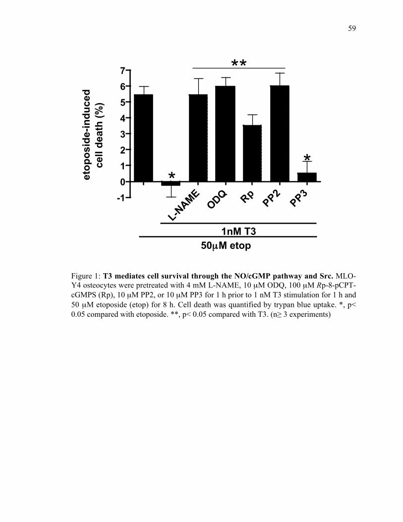
59
L-NAME
ODQ RpPP2
PP3-101234567
**
1nM T350µM etop
etop
osid
e-in
duce
dce
ll de
ath
(%)
**
Figure 1: T3 mediates cell survival through the NO/cGMP pathway and Src. MLO-Y4 osteocytes were pretreated with 4 mM L-NAME, 10 µM ODQ, 100 µM Rp-8-pCPT-cGMPS (Rp), 10 µM PP2, or 10 µM PP3 for 1 h prior to 1 nM T3 stimulation for 1 h and50 µM etoposide (etop) for 8 h. Cell death was quantified by trypan blue uptake. *, p<0.05 compared with etoposide. **, p< 0.05 compared with T3. (n≥ 3 experiments)

60
0
1
2
3
4BimBADBax
*
**
control
fold
indu
ctio
n
etop etop +cGMP
Figure 2: cGMP regulates mRNA expression of bim, bad, and bax. Where indicated,MLO-Y4 cells were pretreated with 100 µM 8-pCPT-cGMP for 1 h prior to treatmentwith 50 µM etoposide (etop) for 5 h. RNA was harvested and subjected to qRT-PCR asdescribed in “Materials and Methods”. Gene expression was standardized to gapdh.Sham-treated cells were considered as control and normalized as 1. *, p< 0.05 comparedto control. **, p< 0.05 compared to etop. (n=3 experiments)

61
0
5
10
15
20
drug
-indu
ced
cell
deat
h (%
)
** *
PP2 – + + + – –dex + – + + – +
PP3 – – – – + +cGMP – – – – –+
Figure 3: Dexamethasone-induced cell death. UMR106 cells were subjected to 100 µM8-pCPT-cGMP (cGMP) pretreatment for 1 h, sensitization with 1 µM PP2 or PP3 for 30min, and treatment with 1 µM dexamethasone (dex) for 8 h. Cell death was measured bytrypan blue uptake. *, p< 0.05. (n≥2 experiments)

62
Figure 4: The NO/cGMP pathway prevents dexamethasone-induced cell death. Asindicated, some UMR106 cells were pretreated with 10 µM ODQ or 100 µM Rp-8-pCPT-cGMPS (Rp) prior to stimulation with 3 µM DETA-NONOate or 100 µM 8-pCPT-cGMPand treatment with 1 µM PP2 and 1 µM dexamethasone (dex). Cell death was measuredby trypan blue uptake. *, p< 0.05. (n≥ 2 experiments)

63
REFERENCES
1. Hiroi Y, Kim HH, Ying H, Furuya F, Huang Z, Simoncini T, Noma K, Ueki K,Nguyen NH, Scanlan TS, et al: Rapid nongenomic actions of thyroid hormone.Proc Natl Acad Sci U S A 2006, 103:14104-14109.
2. Cao X, Kambe F, Yamauchi M, Seo H: Thyroid-hormone-dependent activationof the phosphoinositide 3-kinase/Akt cascade requires Src and enhancesneuronal survival. Biochem J 2009, 424:201-209.
3. Verga Falzacappa C, Mangialardo C, Raffa S, Mancuso A, Piergrossi P, MoriggiG, Piro S, Stigliano A, Torrisi MR, Brunetti E, et al: The thyroid hormone T3improves function and survival of rat pancreatic islets during in vitroculture. Islets 2010, 2:96-103.
4. Sukocheva OA, Carpenter DO: Anti-apoptotic effects of 3,5,3'-tri-iodothyronine in mouse hepatocytes. J Endocrinol 2006, 191:447-458.
5. Gogakos AI, Duncan Bassett JH, Williams GR: Thyroid and bone. ArchBiochem Biophys 2010, 503:129-136.
6. D e x a m e t h a s o n e O r a l[http://www.ncbi.nlm.nih.gov/pubmedhealth/PMH0000773/]
7. Limbourg FP, Liao JK: Nontranscriptional actions of the glucocorticoidreceptor. J Mol Med 2003, 81:168-174.
8. Caplan L, Saag KG: Glucocorticoids and the risk of osteoporosis. Expert OpinDrug Saf 2009, 8:33-47.
9. Newton R: Molecular mechanisms of glucocorticoid action: what isimportant? Thorax 2000, 55:603-613.
10. Ferron M, Wei J, Yoshizawa T, Del Fattore A, DePinho RA, Teti A, Ducy P,Karsenty G: Insulin signaling in osteoblasts integrates bone remodeling andenergy metabolism. Cell 2010, 142:296-308.
11. Lomonosova E, Chinnadurai G: BH3-only proteins in apoptosis and beyond:an overview. Oncogene 2008, 27 Suppl 1:S2-19.
12. Danial NN: BAD: undertaker by night, candyman by day. Oncogene 2008, 27Suppl 1:S53-70.
13. Biswas G, Guha M, Avadhani NG: Mitochondria-to-nucleus stress signaling inmammalian cells: nature of nuclear gene targets, transcription regulation,and induced resistance to apoptosis. Gene 2005, 354:132-139.

64
14. Hsu YY, Liu CM, Tsai HH, Jong YJ, Chen IJ, Lo YC: KMUP-1 attenuatesserum deprivation-induced neurotoxicity in SH-SY5Y cells: roles of PKG,PI3K/Akt and Bcl-2/Bax pathways. Toxicology 2010, 268:46-54.
15. Meng J, Fang B, Liao Y, Chresta CM, Smith PD, Roth JA: Apoptosis inductionby MEK inhibition in human lung cancer cells is mediated by Bim. PLoS One2010, 5:e13026.
16. Gilley J, Coffer PJ, Ham J: FOXO transcription factors directly activate bimgene expression and promote apoptosis in sympathetic neurons. J Cell Biol2003, 162:613-622.
17. Sunters A, Fernandez de Mattos S, Stahl M, Brosens JJ, Zoumpoulidou G,Saunders CA, Coffer PJ, Medema RH, Coombes RC, Lam EW: FoxO3atranscriptional regulation of Bim controls apoptosis in paclitaxel-treatedbreast cancer cell lines. J Biol Chem 2003, 278:49795-49805.
18. Huang H, Tindall DJ: Dynamic FoxO transcription factors. J Cell Sci 2007,120:2479-2487.
19. Huang H, Tindall DJ: FOXO factors: a matter of life and death. Future Oncol2006, 2:83-89.
20. Jiang P, Du W, Heese K, Wu M: The Bad guy cooperates with good cop p53:Bad is transcriptionally up-regulated by p53 and forms a Bad/p53 complex atthe mitochondria to induce apoptosis. Mol Cell Biol 2006, 26:9071-9082.
21. Ogawara Y, Kishishita S, Obata T, Isazawa Y, Suzuki T, Tanaka K, MasuyamaN, Gotoh Y: Akt enhances Mdm2-mediated ubiquitination and degradationof p53. J Biol Chem 2002, 277:21843-21850.

65
Chapter 4:
Type II cGMP-dependent Protein Kinase Mediates Osteoblast
Mechanotransduction

66
ABSTRACT
Mechanical stimulation of bone is crucial for bone growth and differentiation.
Interstitial fluid flow ultimately promotes osteoblastic and osteocytic proliferation
through a number of second messengers including nitric oxide (NO) but the mechanisms
by which fluid shear stress regulates these anabolic responses is incompletely understood.
We found that primary human osteoblasts and MC3T3-E1 murine osteoblast-like cells
responded to fluid shear stress by inducing expression of genes encoding transcription
factors essential for skeletal integrity, namely c-fos, fra-1, fra-2, and fosB/ΔfosB, in a NO/
cGMP/PKG-dependent mechanism. Increased NO production resulting from fluid shear
stress elevated intracellular cGMP concentrations and activated PKG, leading to
activation of the MEK/ERK pathway and downstream fos family gene regulation.
Inhibition of this pathway by pharmacological inhibitors or siRNA targeting PKGII
prevented fluid shear stress-induced ERK1/2 phosphorylation and stimulation of all four
fos genes. Loss of PKGI did not affect either ERK1/2 phosphorylation or fos family
mRNA expression. A membrane permeable analog of cGMP was able to partially mimic
the effects of fluid shear stress; cGMP combined with a calcium ionophore was more
completely able to reproduce the fos family gene induction observed in fluid shear
stressed cells. Fluid shear stress- and cGMP-dependent osteoblast proliferation was also
discovered to occur in a Src-dependent manner. PKGII was found to indirectly activate
Src through a mechanism whereby PKGII directly phosphorylates and activates SHP-1.
SHP-1, in turn, dephosphorylates the inhibitory phosphorylation site on Src. This process
also required the interaction of Src with β3 integrins and SHP-2; fluid shear stress
triggered the recruitment of PKGII, Src, SHP-1, and SHP-2 to β3 integrins and the

67
formation of a mechanosome to convert extracellular fluid shear stress into intracellular
signaling promoting cell growth.

68

69

70
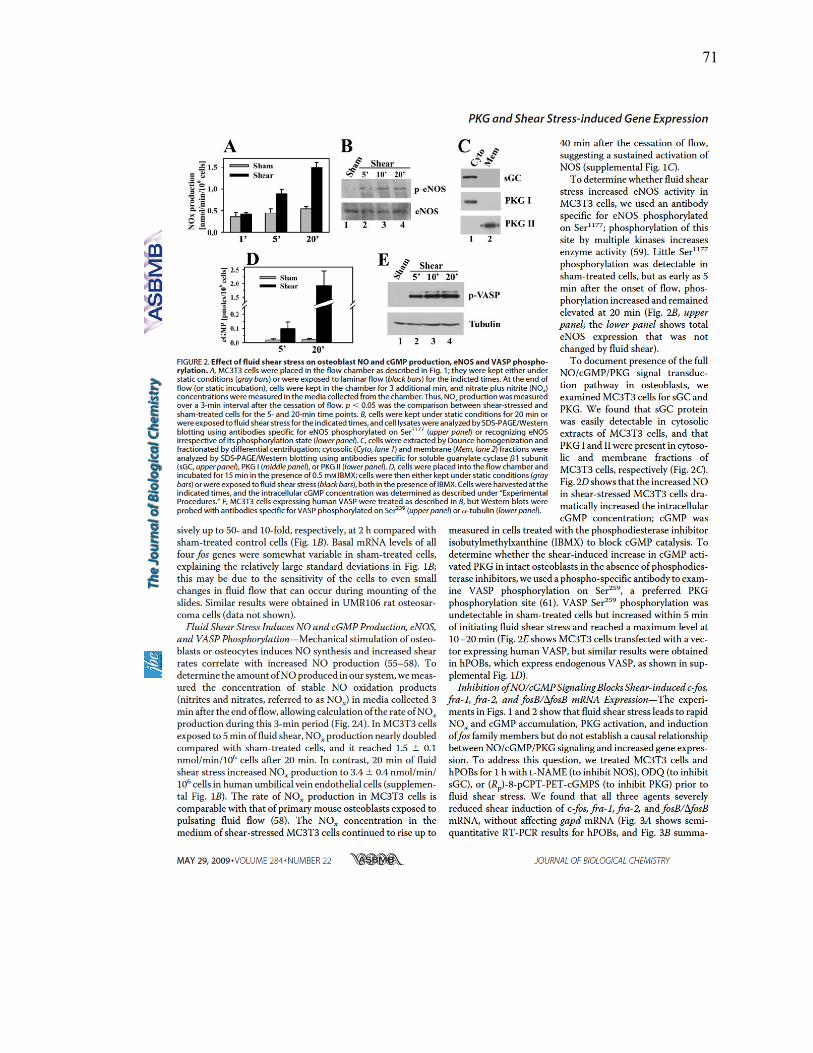
71

72

73

74

75

76

77

78

79

80

81
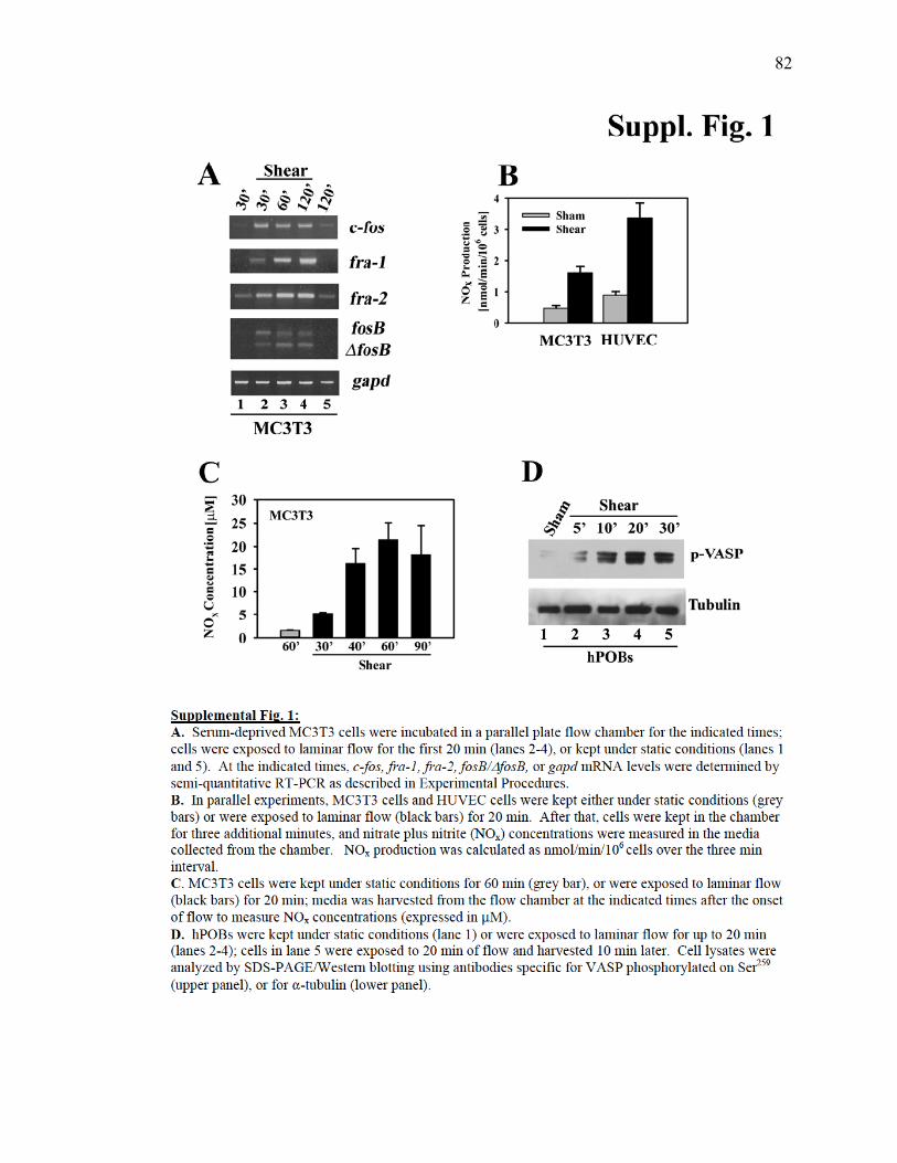
82

83

84

85

86
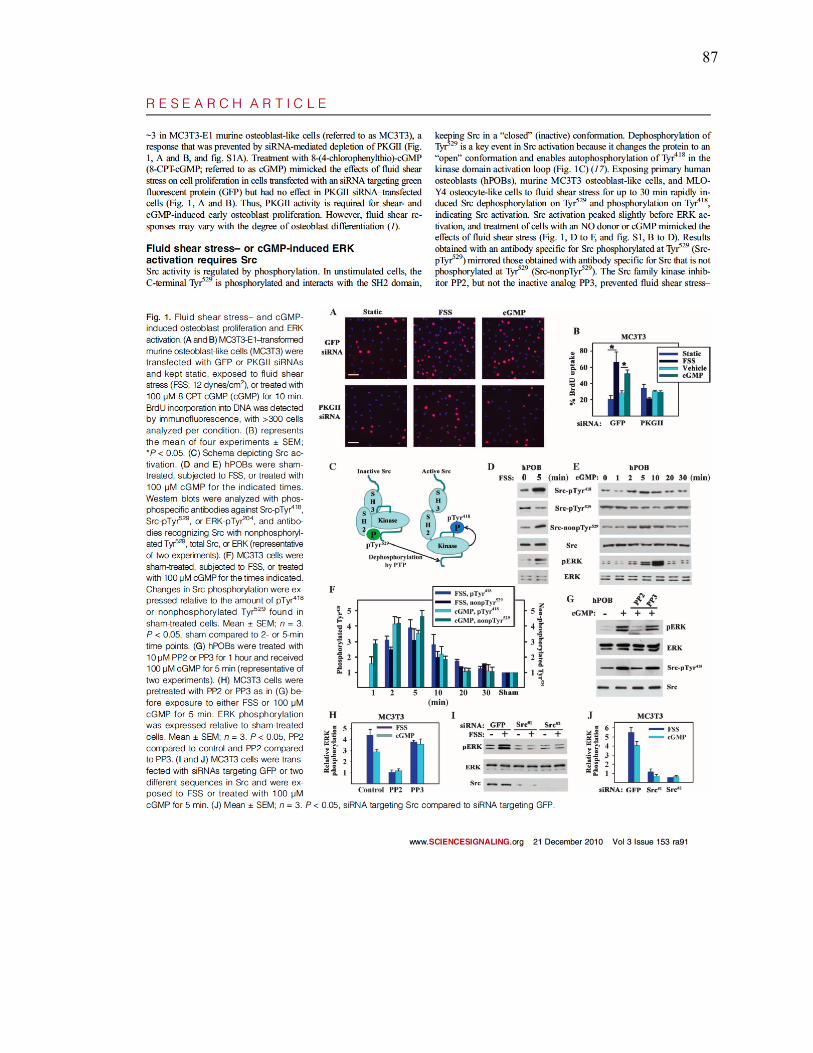
87

88

89

90

91

92

93

94

95

96

97

98

99

100

101

102

103

104

105

106

107

108

109
Acknowledgements: The work presented in this chapter was published in two
papers: Rangaswami H, Marathe N, Zhuang S, Chen Y, Yeh JC, Frangos JA, Boss GR,
Pilz RB. Type II cGMP-dependent protein kinase mediates osteoblast
mechanotransduction J Biol Chem 2009, 284:14796-14808. and Rangaswami H,
Schwappacher R, Marathe N, Zhuang S, Casteel DE, Haas B, Chen Y, Pfeifer A, Kato H,
Shattil S, Boss GR, Pilz RB. Cyclic GMP and protein kinase G control a Src-containing
mechanosome in osteoblasts. Sci Signal 2010, 3:ra91. The dissertation author was the
second and third author on the papers, respectively, and was involved in the following
experiments: RNA extraction and qPCR, establishment of the qPCR primers, primary
human osteoblast establishment and characterization, and cloning and generation of
various plasmid and viral constructs. Other contributors were Drs. Hema Rangaswami,
Shunhui Zhuang, Yongchang Chen, Jiunn-Chern Yeh, John Frangos, Raphaela
Schwappacher, Darren Casteel, Bodo Haas, Yong Chen, Alexander Pfeifer, Hisashi Kato,
Sanford Shattill, Gerry Boss, and Renate Pilz.

110
Chapter 5:
Discussion
111
This dissertation has investigated the molecular mechanisms whereby cGMP-
dependent protein kinases regulate cell survival and proliferation in osteoblasts and
osteocytes. We found that PKGIα and PKGII are essential for estradiol-mediated cell
survival and the PKGII is responsible for Src activation in response to fluid shear stress.
Estrogens and load-bearing exercise have long been known to help support healthy bone
function but the mechanisms by which they act were incompletely understood [1]. Here,
we fill a gap in knowledge of osteoporosis prevention.
In the second chapter, we analyzed the role of PKGs in extragenomic estrogen
signaling. cGMP was shown to mimic the estradiol-protective effects on osteocyte
survival and the protection was abolished using NO/cGMP/PKG pathway inhibitors. This
indicated a role for the pathway in estrogen signaling. We additionally identified PKGII
and PKGIα as having differential effects on osteocyte survival; PKGII stimulated the Akt
and ERK pro-survival pathways while PKGIα inhibited BAD through direct
phosphorylation at Ser155. Interestingly, PKGIβ was unable to phosphorylate BAD in
intact cells, most likely due to lack of proper subcellular localization.
In future studies in our laboratory, this finding may be further investigated to
identify one or more binding partners which allow for the association of PKGIα with
BAD or prevent the interaction of PKGIβ with BAD. These interacting proteins may
influence the subcellular localization allowing for PKGIα to be in the optimal placement
for BAD phosphorylation. Alternately, PKGIβ may be sequestered away from active
BAD or it may be otherwise inhibited by associated proteins. By purifying PKGIα and
PKGIβ from intact cells and identifying unique interacting proteins with mass

112
spectrometry, we may determine novel binding partners of PKGIα versus PKGIβ. This
will allow a better understanding of how PKGIα, and not PKGIβ, is able to regulate BAD
phosphorylation.
Characterization of the bone phenotype in PKGI knockout mice would provide
further evidence of the importance of PKGI in osteocyte cell survival. However, total
PKGI knockout mice have a severely shortened life span due to intestinal dysfunction
and their bone phenotype has not yet been characterized [2]. Therefore, generation of
osteoblast-specific PKGI or PKGIα knockout mice would be crucial to examine bone
characteristics. The PKGIα osteoblast-specific knockout would also determine if the
observed differential effects of PKGIα and PKGIβ from cultured osteocytes translate into
physiological significance.
In Chapter 4, we investigated the role of PKGII in mechanotransduction. We
characterized a novel complex containing PKGII, Src, SHP-1, SHP-2, and αvβ3 integrins
required to translate the mechanical energy of external fluid shear stress into internal
biochemical signals. PKGII, through its indirect activation of Src via SHP-1, controlled
downstream ERK activation and fos family gene expression.
In light of these findings, it would be of value to examine the bone
characterization of PKGII-deficient mice. In preliminary studies, we show that PKGII-
deficient primary osteoblasts isolated from calvariae are unable to activate Src or ERK in
response to fluid shear stress or cGMP. Additionally, mRNA examined from the tibial
diaphyses showed reduced levels of c-fos and fra-2 mRNA, as compared to wildtype
littermates. This indicates that PKGII is required for c-fos and fra-2 expression in resting
cells as well as fluid shear-stimulated cells. Further characterization of bone morphology,

113
including calcein labeling of calvariae to determine the rate of new bone formation and
mRNA expression of other osteoblast specific genes, is currently being examined in the
laboratory.
Furthermore, osteoblast-specific PKGII knockout mice will help evaluate the
physiological role of PKGII in a mechanical loading model. Mechanical loading is
imperative to retaining bone mass as evidenced by both weightlessness and bed rest [3-5].
Unloaded limbs lose bone mass due to a decrease in osteoblast bone formation and
increased osteoclast resorption [3]. Hindlimb suspension studies could demonstrate the
role of PKGII in osteoblast mechanotransduction.
Taken together, the results of this dissertation help understand the role of PKGs in
estradiol- and fluid shear stress-mediated signaling pathways in osteocytes and
osteoblasts. Further exploration of the roles of these proteins in skeletal biology will
advance our knowledge of the genesis and progression of osteoporosis. Ultimately,
research in this area may lead to improved therapeutics for the increasing number of
individuals affected by these disorder.

114
REFERENCES
1. National Osteoporosis Foundation
2. Pfeifer A, Klatt P, Massberg S, Ny L, Sausbier M, Hirneiss C, Wang GX, KorthM, Aszodi A, Andersson KE, et al: Defective smooth muscle regulation incGMP kinase I-deficient mice. EMBO J 1998, 17:3045-3051.
3. Aguirre JI, Plotkin LI, Stewart SA, Weinstein RS, Parfitt AM, Manolagas SC,Bellido T: Osteocyte apoptosis is induced by weightlessness in mice andprecedes osteoclast recruitment and bone loss. J Bone Miner Res 2006, 21:605-615.
4. LeBlanc A, Schneider V, Shackelford L, West S, Oganov V, Bakulin A, VoroninL: Bone mineral and lean tissue loss after long duration space flight. JMusculoskelet Neuronal Interact 2000, 1:157-160.
5. Sievanen H: Immobilization and bone structure in humans. Arch BiochemBiophys 2010, 503:146-152.

![Download Full Proceedings [~20M] - prelim pages.pmd - Tata ...](https://static.fdokumen.com/doc/165x107/631773d871e3f2062906ca05/download-full-proceedings-20m-prelim-pagespmd-tata-.jpg)