Disruption of structural connectivity along the dorsal and ventral language pathways in patients...
10
Disruption of structural connectivity along the dorsal and ventral language pathways in patients with nonfluent and semantic variant primary progressive aphasia: A DT MRI study and a literature review Federica Agosta a , Sebastiano Galantucci a,b , Elisa Canu a , Stefano F. Cappa c , Giuseppe Magnani b , Massimo Franceschi e , Andrea Falini d , Giancarlo Comi b , Massimo Filippi a,b,⇑ a Neuroimaging Research Unit, Institute of Experimental Neurology, Division of Neuroscience, San Raffaele Scientific Institute, Vita-Salute San Raffaele University, Milan, Italy b Department of Neurology, Institute of Experimental Neurology, Division of Neuroscience, San Raffaele Scientific Institute, Vita-Salute San Raffaele University, Milan, Italy c Department of Clinical Neurosciences, San Raffaele Scientific Institute, Vita-Salute San Raffaele University, Milan, Italy d Department of Neuroradiology and CERMAC, San Raffaele Scientific Institute, Vita-Salute San Raffaele University, Milan, Italy e Department of Neurology, IRCCS Multimedica, Castellanza, Italy article info Article history: Available online 26 July 2013 Keywords: Primary progressive aphasia Nonfluent Semantic Dorsal stream Ventral stream Magnetic resonance imaging (MRI) Diffusion tensor MRI Tractography Tract-based spatial statistics abstract Nonfluent (NFV) and semantic (SV) variants of primary progressive aphasia (PPA) are associated with dis- tinct patterns of focal cortical atrophy and underlying pathology. Previous diffusion tensor (DT) MRI stud- ies showed a more ventral white matter (WM) involvement in SV patients and a more widespread frontal involvement in NFV. Aim of this manuscript is twofold. First, we wished to provide a brief state-of-the-art review on WM damage in PPA. Second, we used DT MRI to assess the topography of WM microstructural damage along dorsal and ventral language pathways and corpus callosum in patients with NFV and SV. Our findings show that the two PPA variants share an overlapping pattern of dorsal and ventral pathway abnormalities. In addition to these common abnormalities, variant-specific WM changes were also found, with NFV patients having a more severe damage to the dorsal (fronto-parietal) WM connections within the left superior longitudinal fasciculus/arcuate and SV patients showing a greater left ventral tract involvement (inferior longitudinal and uncinate fasciculi). These findings offer evidence that both dorsal and ventral language networks may contribute to the relatively selective deficits in NFV and SV patients. Ó 2013 Elsevier Inc. All rights reserved. 1. Introduction Primary progressive aphasia (PPA) is a clinical syndrome in which degeneration of language-related structures of the domi- nant hemisphere is associated with progressive deficits of speech and/or language function (Mesulam, 1982; Mesulam, 2001). PPA has several distinct presentations, including the nonfluent (NFV), semantic (SV), and logopenic variants (Gorno-Tempini et al., 2011; Grossman, 2012; Hodges & Patterson, 2007; Neary et al., 1998). The hallmarks of NFV are effortful, nonfluent speech, agram- matism and limited comprehension of grammatically complex sentences (Gorno-Tempini et al., 2011; Grossman, 2012; Josephs et al., 2006). Anatomically, NFV is associated with atrophy of the left posterior fronto-insular regions (Gorno-Tempini et al., 2004; Rohrer et al., 2009). The major clinical features of SV include con- frontation naming difficulty, impaired comprehension of single words, and degraded object knowledge (Gorno-Tempini et al., 2011; Hodges & Patterson, 2007). Comprehension of language is markedly diminished in patients with SV because of the degrada- tion of semantic representations (Patterson, Nestor, & Rogers, 2007). SV is associated with atrophy of the ventral and lateral por- tions of the anterior temporal lobes bilaterally, although tissue damage is usually more severe in the left hemisphere (Gorno-Tem- pini, Dronkers et al., 2004; Rohrer et al., 2009). The logopenic var- iant is characterized by slow speech, sentence repetition and comprehension deficits, and relative sparing of motor speech, grammar, and single-word comprehension (Gorno-Tempini et al., 2011). Brain atrophy in the logopenic variant was found in the left posterior temporoparietal regions (Gorno-Tempini, Murray, Ran- kin, Weiner, & Miller, 2004; Gorno-Tempini et al., 2008). As the dis- ease progresses, PPA can be complicated by behavioral disorders, such as reduced insight, loss of empathy or disinhibition (Gross- man, 2012; Hodges & Patterson, 2007; Josephs et al., 2006). Clinicopathological series suggest that the majority of NFV and SV cases are due to underlying frontotemporal lobar degenera- tion (FTLD) pathology. FTLD pathology can be broadly classified 0093-934X/$ - see front matter Ó 2013 Elsevier Inc. All rights reserved. http://dx.doi.org/10.1016/j.bandl.2013.06.003 ⇑ Corresponding author. Address: Neuroimaging Research Unit, Institute of Experimental Neurology, Division of Neuroscience, San Raffaele Scientific Institute, Vita-Salute San Raffaele University, Via Olgettina, 60, 20132 Milan, Italy. Fax: +39 02 26435972. E-mail address: m.fi[email protected] (M. Filippi). Brain & Language 127 (2013) 157–166 Contents lists available at SciVerse ScienceDirect Brain & Language journal homepage: www.elsevier.com/locate/b&l
-
Upload
independent -
Category
Documents
-
view
5 -
download
0
Transcript of Disruption of structural connectivity along the dorsal and ventral language pathways in patients...

Brain & Language 127 (2013) 157–166
Contents lists available at SciVerse ScienceDirect
Brain & Language
journal homepage: www.elsevier .com/locate /b&l
Disruption of structural connectivity along the dorsal and ventrallanguage pathways in patients with nonfluent and semantic variantprimary progressive aphasia: A DT MRI study and a literature review
0093-934X/$ - see front matter � 2013 Elsevier Inc. All rights reserved.http://dx.doi.org/10.1016/j.bandl.2013.06.003
⇑ Corresponding author. Address: Neuroimaging Research Unit, Institute ofExperimental Neurology, Division of Neuroscience, San Raffaele Scientific Institute,Vita-Salute San Raffaele University, Via Olgettina, 60, 20132 Milan, Italy. Fax: +3902 26435972.
E-mail address: [email protected] (M. Filippi).
Federica Agosta a, Sebastiano Galantucci a,b, Elisa Canu a, Stefano F. Cappa c, Giuseppe Magnani b,Massimo Franceschi e, Andrea Falini d, Giancarlo Comi b, Massimo Filippi a,b,⇑a Neuroimaging Research Unit, Institute of Experimental Neurology, Division of Neuroscience, San Raffaele Scientific Institute, Vita-Salute San Raffaele University, Milan, Italyb Department of Neurology, Institute of Experimental Neurology, Division of Neuroscience, San Raffaele Scientific Institute, Vita-Salute San Raffaele University, Milan, Italyc Department of Clinical Neurosciences, San Raffaele Scientific Institute, Vita-Salute San Raffaele University, Milan, Italyd Department of Neuroradiology and CERMAC, San Raffaele Scientific Institute, Vita-Salute San Raffaele University, Milan, Italye Department of Neurology, IRCCS Multimedica, Castellanza, Italy
a r t i c l e i n f o
Article history:Available online 26 July 2013
Keywords:Primary progressive aphasiaNonfluentSemanticDorsal streamVentral streamMagnetic resonance imaging (MRI)Diffusion tensor MRITractographyTract-based spatial statistics
a b s t r a c t
Nonfluent (NFV) and semantic (SV) variants of primary progressive aphasia (PPA) are associated with dis-tinct patterns of focal cortical atrophy and underlying pathology. Previous diffusion tensor (DT) MRI stud-ies showed a more ventral white matter (WM) involvement in SV patients and a more widespread frontalinvolvement in NFV. Aim of this manuscript is twofold. First, we wished to provide a brief state-of-the-artreview on WM damage in PPA. Second, we used DT MRI to assess the topography of WM microstructuraldamage along dorsal and ventral language pathways and corpus callosum in patients with NFV and SV.Our findings show that the two PPA variants share an overlapping pattern of dorsal and ventral pathwayabnormalities. In addition to these common abnormalities, variant-specific WM changes were also found,with NFV patients having a more severe damage to the dorsal (fronto-parietal) WM connections withinthe left superior longitudinal fasciculus/arcuate and SV patients showing a greater left ventral tractinvolvement (inferior longitudinal and uncinate fasciculi). These findings offer evidence that both dorsaland ventral language networks may contribute to the relatively selective deficits in NFV and SV patients.
� 2013 Elsevier Inc. All rights reserved.
1. Introduction
Primary progressive aphasia (PPA) is a clinical syndrome inwhich degeneration of language-related structures of the domi-nant hemisphere is associated with progressive deficits of speechand/or language function (Mesulam, 1982; Mesulam, 2001). PPAhas several distinct presentations, including the nonfluent (NFV),semantic (SV), and logopenic variants (Gorno-Tempini et al.,2011; Grossman, 2012; Hodges & Patterson, 2007; Neary et al.,1998). The hallmarks of NFV are effortful, nonfluent speech, agram-matism and limited comprehension of grammatically complexsentences (Gorno-Tempini et al., 2011; Grossman, 2012; Josephset al., 2006). Anatomically, NFV is associated with atrophy of theleft posterior fronto-insular regions (Gorno-Tempini et al., 2004;Rohrer et al., 2009). The major clinical features of SV include con-
frontation naming difficulty, impaired comprehension of singlewords, and degraded object knowledge (Gorno-Tempini et al.,2011; Hodges & Patterson, 2007). Comprehension of language ismarkedly diminished in patients with SV because of the degrada-tion of semantic representations (Patterson, Nestor, & Rogers,2007). SV is associated with atrophy of the ventral and lateral por-tions of the anterior temporal lobes bilaterally, although tissuedamage is usually more severe in the left hemisphere (Gorno-Tem-pini, Dronkers et al., 2004; Rohrer et al., 2009). The logopenic var-iant is characterized by slow speech, sentence repetition andcomprehension deficits, and relative sparing of motor speech,grammar, and single-word comprehension (Gorno-Tempini et al.,2011). Brain atrophy in the logopenic variant was found in the leftposterior temporoparietal regions (Gorno-Tempini, Murray, Ran-kin, Weiner, & Miller, 2004; Gorno-Tempini et al., 2008). As the dis-ease progresses, PPA can be complicated by behavioral disorders,such as reduced insight, loss of empathy or disinhibition (Gross-man, 2012; Hodges & Patterson, 2007; Josephs et al., 2006).
Clinicopathological series suggest that the majority of NFV andSV cases are due to underlying frontotemporal lobar degenera-tion (FTLD) pathology. FTLD pathology can be broadly classified
158 F. Agosta et al. / Brain & Language 127 (2013) 157–166
according to the major constituents of the cellular inclusions: tau(FTLD-tau), transactive response DNA binding protein of 43 kD(TDP-43, FTLD-TDP), or fused-in-sarcoma protein (Mackenzieet al., 2010). The majority of NFV patients exhibit FTLD-tau or,less frequently, FTLD-TDP pathology, while most SV cases havea TDP-43 proteinopathy (Grossman, 2010; Josephs et al., 2011;Kertesz, McMonagle, Blair, Davidson, & Munoz, 2005; Knibb,Xuereb, Patterson, & Hodges, 2006; Mesulam et al., 2008; Snow-den, Neary, & Mann, 2007). Alzheimer’s disease is the most likelyunderlying pathology of the logopenic variant (Mesulam et al.,2008).
Current theories on brain organization suggest that cognitivefunctions, such as language, are organized as distributed, segre-gated, and overlapped networks (Hickok & Poeppel, 2004; Hickok& Poeppel, 2007; Mesulam, 2009). Anatomically, such large-scalenetworks comprise specialized brain grey matter (GM) areas (net-work nodes) and their interconnecting white matter (WM) fibertracts (network connections). This network-based approach arguesthat not only cortical lesions, but also damage to the WM connec-tions between cortical areas can lead to a neural dysfunction and,hence, cognitive disorders (Mesulam, 2009). These pieces of evi-dence, together with the availability of new MRI techniques thatenable the in vivo detection of WM damage are changing the clas-sical understanding of the architecture of the language networkand the pathophysiology of PPA.
Aim of this manuscript is twofold. First, we wished to providea brief state-of-the-art review on WM damage in PPA variantswhich are most likely caused by FTLD pathology, i.e., NFV andSV, and the use of diffusion tensor (DT) MRI in these patients. Sec-ond, we present a detailed analysis of the patterns of WM micro-structural abnormalities along individual dorsal and ventrallanguage WM pathways and corpus callosum in patients withNFV and SV.
1.1. White matter damage in PPA cases with FTLD pathology
WM abnormalities have been detected in PPA but their origin isincompletely understood and may vary according to underlyingpathology. In some instances, WM damage results from axonaldegeneration following the injury/death of cell bodies in the af-fected GM regions and in other instances it may be due to a directeffect of FTLD pathology. Indeed, seminal pathologic studies ofFTLD patients showed that, although the regional extent of WMalterations roughly parallels that of cortical degeneration, thereare also mild WM abnormalities that extend beyond the confinesof the GM involvement (Englund & Brun, 1987).
Primary WM damage in FTLD may involve the accumulation ofpathological proteins in WM regions as well as microglia activa-tion. A significant abnormal tau pathology has been shown to occurin the WM of patients with corticobasal degeneration (Dicksonet al., 2002) and progressive supranuclear palsy pathology (Zhu-kareva et al., 2006), accompanied by variable, sometimes sparse,oligodendroglial inclusions (Dickson et al., 2002). Pick’s disease isassociated with the presence of tau-positive glial inclusions,threads, and multiple small, dotlike inclusions in the WM (Zhukar-eva et al., 2002). In addition, in FTLD-tau pathologies a significantproportion of the activated WM microglia is tau-immunoreactive,suggesting an association with axonal degeneration, possibly viaimmune processes (Schofield, Kersaitis, Shepherd, Kril, & Halliday,2003). In the FTLD subtypes associated with TDP-43 preoteinopa-thy, WM pathology, which is characterized by glial cytoplasmicinclusions and thread-like inclusions, is abundant in frontal andtemporal lobes, but it is also detectable in the brainstem and spinalcord from patients with concomitant motor neuron involvement(Neumann et al., 2007).
1.2. Diffusion tensor MRI
Information on the microstructural integrity of the WM path-ways connecting the different GM structures of the human braincan be obtained in vivo with DT MRI (Basser, Mattiello, & LeBihan,1994; Pierpaoli, Jezzard, Basser, Barnett, & Di Chiro, 1996). At pres-ent, there is no consensus on the best way to analyze quantitativeDT MRI data in a clinical research setting, and this remains anactive area of technical development. Manually placed regions-of-interest (ROI) have been widely used, but this strategy ischaracterized by high intrarater and inter-rater variabilities andis time-consuming, especially when many WM tracts are to be ana-lyzed in large cohorts of subjects. Another important flaw is thatROI-based approaches are generally inaccurate on tracts that aredifficult to be directly visualized, in particular in disease groupswhere the presence of atrophy can be associated with even a subtlesystematic ROI misplacement and, thus, lead to significant, butspurious, DT MRI changes. In addition, such an approach allowsto assess only a portion of each tract of interest ignoring the partsof the tract that are not included in the ROI.
The general strategy behind DT tractography methods is totranslate DT information into a vector field. When vectors thathave the same orientation are combined, the course of WM tractscan be visualized and their intrinsic tissue damage quantified(Mori & van Zijl, 2002). DT MR images can also be used to detectgroup-wise abnormalities of diffusion metrics from the WM ofthe entire brain. TBSS is an automated method which allows to car-ry out multi-subject, localized statistical testing of fractionalanisotropy (FA- and other diffusion-related data), and which hassolved crucial issues of cross-subject data alignment (Smith et al.,2006). TBSS combines the strengths of voxel-based analyses (beingable to analyze the whole brain without predefining voxels ortracts of interest) with the strengths of tractography-based analy-ses (ideally, being confident that the estimates of FA are truly takenfrom relevant voxels). Furthermore, while DT MRI tractographymeasurements are derived by averaging values over all voxelswithin an individual WM tract, which could wash out discreteareas of difference, TBSS can disclose abnormalities of DT MRI indi-ces throughout the entire WM of the brain without any a priorihypothesis, and at the same time detect different patterns ofabnormality within an individual tract.
1.3. Diffusion tensor MRI in PPA
Using ROI-based or tractography DT MRI to investigate the WMlanguage tracts in PPA patients, a few studies showed that NFV ischaracterized by an involvement of all the left superior longitudi-nal fasciculus (SLF) components (Galantucci et al., 2011; Whitwellet al., 2011). In contrast, the ventral tracts connecting the temporallobe with the occipital and the oribitofrontal cortices (i.e., the infe-rior longitudinal [ILF] and uncinate fasciculi) were relativelyspared (Galantucci et al., 2011; Whitwell et al., 2011). Studiesusing a voxel-wise approach showed that NFV patients have notonly a damage to the fronto-parieto-temporal connections but alsoan involvement of the corpus callosum, cingulum bundle, externalcapsule, several regions of the prefrontal and orbitofrontal WM,and parietotemporal WM and temporal regions, mainly in the lefthemisphere (Agosta et al., 2011; Schwindt et al., 2013). A recentTBSS study found a damage to portions of the ILF in NFV patients(Schwindt et al., 2013).
In SV, abnormalities of WM tract diffusivity were identified inthe major inferior and superior temporal connections of the lefthemisphere, thus mirroring the severe atrophy affecting the sameregions: the ILF, inferior-fronto-occipital fasciculus and the unci-nate fasciculus within the ventral stream, and the arcuate andthe temporo-parietal component of the SLF within the dorsal
F. Agosta et al. / Brain & Language 127 (2013) 157–166 159
stream (Acosta-Cabronero et al., 2011; Agosta et al., 2010; Agostaet al., 2011; Galantucci et al., 2011; Schwindt et al., 2013; Whitwellet al., 2011). Although tractography studies have argued that thefronto-parietal connections are relatively spared in SV (Agostaet al., 2010; Galantucci et al., 2011), a finding which couples withthe absence of syntactic deficits in these patients, a few studiesusing TBSS have provided evidence for an involvement of the leftprefrontal and parieto-frontal WM contributing to the SLF and cor-ona radiata (Agosta et al., 2011; Schwindt et al., 2013).
In summary, a few studies to date have investigated the pat-terns of WM abnormalities in small samples of NFV and SV patientsusing DT MRI. The most consistent findings among studies are amore ventral WM involvement in SV patients and a more wide-spread frontal involvement in NFV. However, recent studies usinga voxel-based approach suggested a spreading of diffusion abnor-malities beyond the sites of local damage in both PPA variants. Fur-thermore, none of the previous studies has compared directly thepatterns of WM damage between NFV and SV cases. In this study,we combined the unique ability of probabilistic DT tractography toreconstruct WM structural connections with that of TBSS to per-form a voxel-wise analysis in order to capture the subset of voxelsspecifically abnormal in individual language tracts of NFV and SVpatients as well as the common areas of WM damage in thesetwo conditions.
2. Materials and methods
Right-handers (Oldfield, 1971) native Italian-speaking patientswith NFV and SV (Gorno-Tempini et al., 2011; Neary et al., 1998)were recruited at the Department of Neurology and Departmentof Clinical Neurosciences, Scientific Institute University Vita-SaluteHospital San Raffaele, Milan, and Department of Neurology, IRCCSMultimedica, Castellanza, Italy. Patients received a comprehensiveevaluation including neurological history and examination, neuro-psychological testing, and neuroimaging. Clinical assessmentswere performed by experienced neurologists blinded to the MRIresults. History was taken with a structured interview from pa-tients’ relatives. Right-handers (Oldfield, 1971), native Italian-speaking, age-matched healthy controls were recruited amongspouses of patients and by word of mouth. Healthy controls under-went a neurological and neuropsychological assessment, and wereincluded in the study only when no abnormality was found. Sub-jects were excluded if they had: (1) a family history of dementiaor FTLD-related disorders; (2) significant medical illnesses or sub-stance abuse that could interfere with cognitive functioning; (3)any other major systemic, psychiatric, or neurological illnesses;and (4) other causes of focal or diffuse brain damage, includinglacunae, and extensive cerebrovascular disorders at routine MRI.
We studied 26 PPA patients, 13 NFV (mean age 69 ± 7 years; 7women) and 13 SV (mean age 66 ± 8 years; 6 women), and 35age- and sex-matched healthy controls (mean age 64 ± 9 years;16 men, 19 women). Approval was obtained from the local ethicalstandards committee on human experimentation and written in-formed consent from all subjects before enrolment.
2.1. Cognitive assessment
Neuropsychological assessment was performed by experiencedneuropsychologists unaware of the MRI results, and evaluated: (i)global cognitive functioning with the Mini Mental State Examina-tion (Folstein, Folstein, & McHugh, 1975); (ii) memory functionwith verbal and spatial span (Orsini et al., 1987) and Rey’s figuredelayed recall test (Caffarra, Vezzadini, Dieci, Zonato, & Venneri,2002); (iii) visuo-spatial abilities with the Rey’s Figure Copy Test(Caffarra et al., 2002); (iv) attention and executive functions with
the attentive matrices (Spinnler & Tognoni, 1987) and the Raven’scoloued progressive matrices (Basso, Capitani, & Laiacona, 1987);(v) language with the phonemic and semantic fluency (Novelliet al., 1986). PPA patients also underwent a comprehensive speechevaluation including the examination of the confrontation namingwith naming subtests of the Battery for Analysis of Aphasic Deficits(BADA) (Miceli, Laudanna, Burani, & Capasso, 1994), CaGi test(Catricala, Della Rosa, Ginex, et al., 2012), or Aachener Aphasie Test(AAT) (Luzzatti et al., 1994); single word comprehension with theauditory and/or visual comprehension subtests of the BADA, CaGior AAT; object knowledge with the Pyramids and Palm Trees Test(Gamboz, Coluccia, Iavarone, & Brandimonte, 2009); dyslexia/dys-graphia with the reading and writing subtests of the BADA or AAT;repetition with the repetition subtests of the BADA or AAT; andcomprehension of syntactically complex sentences with the audi-tory and visual grammatical comprehension subtests of the BADA,and with the Token test (De Renzi & Vignolo, 1962; Spinnler & Tog-noni, 1987). Scores on neuropsychological tests were age-, sex-,and education-corrected by using related normative values, when-ever available. To evaluate fluency, we recorded speech samplesfrom each of our patients while they described the image of thepicnic picture subtest of the Western Aphasia Battery (Kertesz,1982) and analyzed them according to Wilson et al. (2010). Sucha test, however, has not been standardized yet in the Italian popu-lation. As a consequence, the recorded spontaneous speech sam-ples were not used for quantitative analysis.
2.2. MRI acquisition
Brain MRI scans were obtained using a 3.0 T scanner (Intera,Philips Medical Systems, Best, the Netherlands). The following se-quences were acquired from all subjects: (i) T2-weighted spin echo(SE) (repetition time [TR] = 3500 ms; echo time [TE] = 85 ms; echotrain length = 15; flip angle = 90�; 22 contiguous, 5-mm-thick, axialslices; matrix size = 512 � 512; field of view [FOV] = 230 �184 mm2); (ii) fluid-attenuated inversion recovery (FLAIR)(TR = 11.000 ms; TE = 120 ms; flip angle = 90�; 22 contiguous, 5-mm-thick, axial slices; matrix size = 512 � 512; FOV = 230 mm2);(iii) pulsed-gradient SE echo planar with sensitivity encoding(acceleration factor = 2.5; TR = 8986 ms; TE = 80 ms; 55 contigu-ous, 2.5-mm-thick, axial slices; number of acquisitions = 2; afterSENSE reconstruction, the matrix dimension of each slice was256 � 256, with an in-plane pixel size = 0.94 � 0.94 mm and aFOV = 240 � 240 mm2) and diffusion gradients applied in 32 non-collinear directions using a gradient scheme which is standard onthis system (gradient over-plus) and optimized to reduce echotime as much as possible. The b factor used was 1000 s/mm2. Fatsaturation was performed to avoid chemical shift artifacts. Allslices were positioned to run parallel to a line that joins the mostinferoanterior and inferoposterior parts of the corpus callosum.
2.3. MRI analysis
MRI analysis was performed by an experienced observerblinded to subjects’ identity.
2.3.1. DT MRI preprocessingDT MRI analysis was performed using the FMRIB software li-
brary (FSL) tools (http://www.fmrib.ox.ac.uk/fsl/fdt/index.html)and the JIM5 software (Version 5.0, Xinapse Systems, Northants,UK, http://www.xinapse.com). The diffusion-weighted data wereskull-stripped using the Brain Extraction Tool implemented inFSL. Using FMRIB’s Linear Image Registration Tool (FLIRT), thetwo diffusion-weighted scans were coregistered by applying the ri-gid transformation needed to correct for position between the twob0 images (T2-weighted, but not diffusion-weighted). The rotation
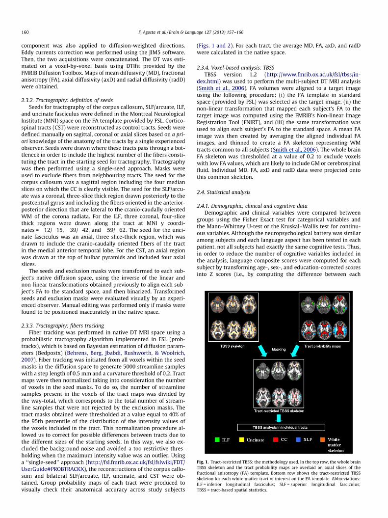
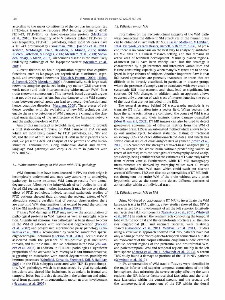
Fig. 1. Tract-restricted TBSS: the methodology used. In the top row, the whole brainTBSS skeleton and the tract probability maps are overlaid on axial slices of thefractional anisotropy (FA) template. Bottom row shows the tract-restricted TBSSskeleton for each white matter tract of interest on the FA template. Abbreviations:ILF = inferior longitudinal fasciculus; SLF = superior longitudinal fasciculus;TBSS = tract-based spatial statistics.
160 F. Agosta et al. / Brain & Language 127 (2013) 157–166
component was also applied to diffusion-weighted directions.Eddy currents correction was performed using the JIM5 software.Then, the two acquisitions were concatenated. The DT was esti-mated on a voxel-by-voxel basis using DTIfit provided by theFMRIB Diffusion Toolbox. Maps of mean diffusivity (MD), fractionalanisotropy (FA), axial diffusivity (axD) and radial diffusivity (radD)were obtained.
2.3.2. Tractography: definition of seedsSeeds for tractography of the corpus callosum, SLF/arcuate, ILF,
and uncinate fasciculus were defined in the Montreal NeurologicalInstitute (MNI) space on the FA template provided by FSL. Cortico-spinal tracts (CST) were reconstructed as control tracts. Seeds weredefined manually on sagittal, coronal or axial slices based on a pri-ori knowledge of the anatomy of the tracts by a single experiencedobserver. Seeds were drawn where these tracts pass through a bot-tleneck in order to include the highest number of the fibers consti-tuting the tract in the starting seed for tractography. Tractographywas then performed using a single-seed approach. Masks wereused to exclude fibers from neighbouring tracts. The seed for thecorpus callosum was a sagittal region including the four medianslices on which the CC is clearly visible. The seed for the SLF/arcu-ate was a coronal, three-slice thick region drawn posteriorly to thepostcentral gyrus and including the fibers oriented in the anterior-posterior direction that are lateral to the cranio-caudally orientedWM of the corona radiata. For the ILF, three coronal, four-slicethick regions were drawn along the tract at MNI y coordi-nates = �12/�15, �39/�42, and �59/�62. The seed for the unci-nate fasciculus was an axial, three slice-thick region, which wasdrawn to include the cranio-caudally oriented fibers of the tractin the medial anterior temporal lobe. For the CST, an axial regionwas drawn at the top of bulbar pyramids and included four axialslices.
The seeds and exclusion masks were transformed to each sub-ject’s native diffusion space, using the inverse of the linear andnon-linear transformations obtained previously to align each sub-ject’s FA to the standard space, and then binarized. Transformedseeds and exclusion masks were evaluated visually by an experi-enced observer. Manual editing was performed only if masks werefound to be positioned inaccurately in the native space.
2.3.3. Tractography: fibers trackingFiber tracking was performed in native DT MRI space using a
probabilistic tractography algorithm implemented in FSL (prob-trackx), which is based on Bayesian estimation of diffusion param-eters (Bedpostx) (Behrens, Berg, Jbabdi, Rushworth, & Woolrich,2007). Fiber tracking was initiated from all voxels within the seedmasks in the diffusion space to generate 5000 streamline sampleswith a step length of 0.5 mm and a curvature threshold of 0.2. Tractmaps were then normalized taking into consideration the numberof voxels in the seed masks. To do so, the number of streamlinesamples present in the voxels of the tract maps was divided bythe way-total, which corresponds to the total number of stream-line samples that were not rejected by the exclusion masks. Thetract masks obtained were thresholded at a value equal to 40% ofthe 95th percentile of the distribution of the intensity values ofthe voxels included in the tract. This normalization procedure al-lowed us to correct for possible differences between tracts due tothe different sizes of the starting seeds. In this way, we also ex-cluded the background noise and avoided a too restrictive thres-holding when the maximum intensity value was an outlier. Usinga ‘‘single-seed’’ approach (http://fsl.fmrib.ox.ac.uk/fsl/fslwiki/FDT/UserGuide#PROBTRACKX), the reconstructions of the corpus callo-sum and bilateral SLF/arcuate, ILF, uncinate, and CST were ob-tained. Group probability maps of each tract were produced tovisually check their anatomical accuracy across study subjects
(Figs. 1 and 2). For each tract, the average MD, FA, axD, and radDwere calculated in the native space.
2.3.4. Voxel-based analysis: TBSSTBSS version 1.2 (http://www.fmrib.ox.ac.uk/fsl/tbss/in-
dex.html) was used to perform the multi-subject DT MRI analysis(Smith et al., 2006). FA volumes were aligned to a target imageusing the following procedure: (i) the FA template in standardspace (provided by FSL) was selected as the target image, (ii) thenon-linear transformation that mapped each subject’s FA to thetarget image was computed using the FMRIB’s Non-linear ImageRegistration Tool (FNIRT), and (iii) the same transformation wasused to align each subject’s FA to the standard space. A mean FAimage was then created by averaging the aligned individual FAimages, and thinned to create a FA skeleton representing WMtracts common to all subjects (Smith et al., 2006). The whole brainFA skeleton was thresholded at a value of 0.2 to exclude voxelswith low FA values, which are likely to include GM or cerebrospinalfluid. Individual MD, FA, axD and radD data were projected ontothis common skeleton.
2.4. Statistical analysis
2.4.1. Demographic, clinical and cognitive dataDemographic and clinical variables were compared between
groups using the Fisher Exact test for categorical variables andthe Mann–Whitney U-test or the Kruskal–Wallis test for continu-ous variables. Although the neuropsychological battery was similaramong subjects and each language aspect has been tested in eachpatient, not all subjects had exactly the same cognitive tests. Thus,in order to reduce the number of cognitive variables included inthe analysis, language composite scores were computed for eachsubject by transforming age-, sex-, and education-corrected scoresinto Z scores (i.e., by computing the difference between each

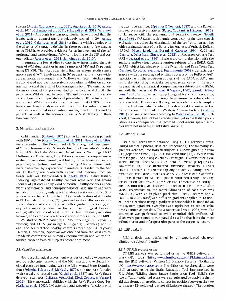
Fig. 2. Tract-restricted TBSS analysis in nonfluent variant (NFV) primary progres-sive aphasia patients. In the top row (gray background), probability maps of thecorpus callosum (red), superior longitudinal fasciculus (SLF)/arcuate (light blue),inferior longitudinal fasciculus (ILF; green), and uncinate fasciculus (yellow) areshown. Bottom row (black background) shows areas of significant decreasedfractional anisotropy (FA) in each white matter tract in NFV patients compared withhealthy controls (red = corpus callosum; light blue = SLF/arcuate; green = ILF;yellow = uncinate fasciculus). The results are overlaid on a 3D FA template in theMontreal Neurological Institute standard space in neurological convention (left isleft), and displayed at p < 0.05, Family Wise Error-corrected.
F. Agosta et al. / Brain & Language 127 (2013) 157–166 161
subject score and the average score, which was then divided by thestandard deviation of the score). Composite scores were obtainedfor confrontation naming, single-word comprehension, objectknowledge, dyslexia/dysgraphia, repetition, and comprehensionof syntactically complex sentences by averaging Z scores of testsobtained for each of these language features. These analyses wererun using SAS Release 9.1, SAS Institute, Cary, NC, USA.
2.4.2. Tract-restricted TBSSTract probability maps obtained from subjects included in each
contrast were used to mask the whole brain MD, FA, axD and radDskeletons in order to obtain ‘‘tract-restricted’’ skeletons. Then, a‘‘tract-restricted’’ TBSS analysis was performed to compare groups(Fig. 1). DT MRI voxelwise statistics were performed using a per-mutation-based inference tool for nonparametric statistical thres-holding (‘‘randomise’’, part of FSL (Nichols & Holmes, 2002)). Thenumber of permutations was set at 5000 (Nichols & Holmes,2002). Between-group comparisons were performed using two-sample t tests, adjusting for subject’s age. The patients vs. controlsstatistical maps were thresholded at p < 0.05 corrected for multiplecomparisons (family-wise error, FWE) at a cluster level using thethreshold-free cluster enhancement (TFCE) option (Smith and Nic-hols, 2009). The comparison between patient groups was testedalso using a statistical threshold of p < 0.05 uncorrected to avoidfalse-negatives that can occur in the direct comparison betweensmall patient populations.
3. Results
3.1. Demographic, clinical and cognitive data
Subject groups did not differ in terms of age at MRI, gender andyears of education (Table 1). Patients with the two PPA variantshad similar Clinical Dementia Rating score-Sum of boxes and MiniMental State Examination scores, but SV patients had a longer dis-ease duration compared with NFV patients (p = 0.01).
3.2. WM tract damage
3.2.1. NFV patients vs. healthy controls (p < 0.05 corrected Fig. 2)3.2.1.1. Corpus callosum. NFV patients showed a decreased FA andan increased MD in the anterior portion of the body and genu of
the corpus callosum, with a left-side predominance. The spleniumfibers projecting to the superior temporal regions were also in-volved, bilaterally. The pattern of increased radD resembled thoseof altered MD and FA, while axD was significantly increased in theanterior body of the corpus callosum and only in a few regions ofthe splenium.
3.2.1.2. SLF/arcuate. In NFV patients relative to controls, there werea decreased FA and an increased MD in the entire left SLF/arcuate.MD was also increased in the frontal regions of the right SLF/arcu-ate, while no FA abnormality was found in tracts of the right hemi-sphere. RadD increase resembled the pattern of MD increase. AxDrevealed a more asymmetric pattern of abnormalities and was in-creased in the entire left SLF/arcuate and only in a minimal portionof the right tract.
3.2.1.3. ILF. No ILF DT MRI differences were found between NFV pa-tients and healthy controls.
3.2.1.4. Uncinate fasciculus. NFV patients had a decreased FA in theportion of the uncinate fasciculus that runs through the anteriorportion of the external capsule. MD and radD were found to be in-creased in the entire uncinate fasciculus. There was no axD abnor-mality in NFV patients compared with controls.
3.2.1.5. CST. No CST FA and MD abnormalities were found in NFVpatients compared with controls. Increased radD and axD werefound in the cranial portion of the corona radiata bilaterally, witha left-side predominance for the radD pattern of abnormalities.
3.2.2. SV patients vs. healthy controls (p < 0.05 corrected Fig. 3)3.2.2.1. Corpus callosum. SV patients showed a decreased FA in thegenu and anterior half of the body of the corpus callosum, with aleft-side predominance. On the left hemisphere, there was a de-creased FA was also found in the splenial fibers projecting to thesuperior temporal gyrus. MD and radD were found to be increasedin the entire corpus callosum, with a relative prevalence in thegenu and the anterior half of the body for radD. AxD was signifi-cantly increased in the splenium and the posterior portion of thebody of the corpus callosum.
3.2.2.2. SLF/arcuate. In SV patients, there was a decreased FA in theentire left SLF/arcuate, while no FA abnormality was found in theright hemisphere. MD, radD and axD were found to be increasedin the entire left SLF/arcuate and in the frontal portion of the righttract.
3.2.2.3. ILF. SV patients relative to controls showed a decreased FAin the anterior third of the left ILF and in a small, anterior region ofthe right ILF. MD, radD and axD were found to be increased in theanterior third of the tract bilaterally, with a greater involvement ofthe left ILF.
3.2.2.4. Uncinate fasciculus. All DT MRI metrics were significantlyaltered in the entire uncinate fasciculus bilaterally in SV patientscompared with controls.
3.2.2.5. CST. There were no difference in CST FA and radD whencomparing SV patients to controls. SV patients showed some voxelsof increased MD and axD in the cranial portions of the corona rad-iata bilaterally, with a prevalence on the left side.
3.2.3. NFV vs. SV patients (p < 0.05 uncorrected Figs. 4 and 5)No difference was found in the DT MRI values of the corpus cal-
losum and CST between NFV and SV patients.

Table 1Demographic, clinical and neuropsychological data from patients with nonfluent and semantic primary progressive aphasia variants.
NFV SV p
Number 13 13Age 69 ± 7 66 ± 8 0.39Gender (women/men) 7/6 6/7 1.00Education (years) 8.1 ± 5.2 10.5 ± 5.3 0.35Disease duration [years] 2.1 ± 1.3 3.8 ± 1.7 0.01CDR-SB 2.8 ± 1.3 3.2 ± 2.6 0.89MMSE – co 24 21.1 ± 6.1 21.3 ± 7.1 0.74
Verbal & Spatial MemoryDigit span – co 3.75 3.6 ± 0.8 (63%) 5.3 ± 1.2 (8%) 0.001Spatial span – co 3.75 4.0 ± 0.8 (10%) 4.2 ± 0.9 (25%) 0.87Figure rey recall – co 9.47 11.2 ± 8.3 (10%) 10.3 ± 7.0 (8%) 0.81
PraxisFigure Rey copy – co 28.88 23.9 ± 11.0 (50%) 28.5 ± 5.6 (42%) 0.58
Attention & executive functionsAttentive matrices – co 31 31.7 ± 10.2 (44%) 39.8 ± 11.2 (18%) 0.11Raven’s colored progressive matrices – co 18 20.7 ± 6.4 (8%) 22.6 ± 8.5 (9%) 0.45
LanguageSemantic fluency – co 25 14.7 ± 4.8 (70%) 8.5 ± 8.7 (92%) 0.05Phonemic fluency – co 17 6.0 ± 4.5 (100%) 10.6 ± 9.7 (50%) 0.38Token test – co 26.5 22.7 ± 7.0 (64%) 21.5 ± 8.4 (75%) 0.70Confrontation naming, Z score 0.5 ± 0.7 �0.6 ± 0.8 0.01Single-word comprehension, Z score 0.6 ± 0.6 �0.5 ± 1.1 0.005Object knowledge, Z score 0.3 ± 1.2 �0.2 ± 0.8 0.28Dyslexia/dysgraphia, Z score �0.4 ± 0.9 0.4 ± 0.6 0.27Repetition, Z score �0.4 ± 1.0 0.8 ± 0.1 0.003Comprehension of syntactically complex sentences, Z score �0.2 ± 0.9 �0.1 ± 1.2 0.90
Numbers are mean ± standard deviation (% of subjects with abnormal test score compared with normative data of reference). P refer to Fisher exact test or Mann–Whitney Utest, as appropriate. Abbreviations: CDR-SB = Clinical Dementia Rating score-Sum of boxes; co = cut-off obtained from the normative data of reference; NFV = nonfluentvariant PPA; MMSE = Mini Mental State Examination; SV = semantic variant PPA. Z scores were computed according to the following formula: Z = mean score/standarddeviation. Z scores of tests assessing a specific language feature were then averaged to obtain Z scores of each language feature (see text for further details).
162 F. Agosta et al. / Brain & Language 127 (2013) 157–166
3.2.3.1. SLF/arcuate. Compared with SV patients, NFV patientsshowed an increased MD in the fronto-parietal portion of the leftSLF/arcuate (Fig. 4). On the contrary, SV patients had an increasedMD in the portion of the tract located in the temporal lobe (Fig. 4).NFV and SV patients had similar MD values in the right SLF/arcu-ate. No difference in the other DT MRI indices was found betweenthe two patient groups.
3.2.3.2. ILF. In the ILF, SV patients relative to those with NFVshowed an increased MD in the anterior third of the tract on the
Fig. 3. Tract-restricted TBSS analysis in semantic variant (SV) primary progressiveaphasia patients. In the top row (gray background), probability maps of the corpuscallosum (red), superior longitudinal fasciculus (SLF)/arcuate (light blue), inferiorlongitudinal fasciculus (ILF; green), and uncinate fasciculus (yellow) are shown.Bottom row (black background) shows areas of significant decreased fractionalanisotropy (FA) in each white matter tract in SV patients compared with healthycontrols (red = corpus callosum; light blue = SLF/arcuate; green = ILF; yellow = unci-nate fasciculus). The results are overlaid on a 3D FA template in the MontrealNeurological Institute standard space in neurological convention (left is left), anddisplayed at p < 0.05, Family Wise Error-corrected.
left side and in a small, anterior region on the right side (Fig. 5).No difference in the other DT MRI indices was found betweenthe two patient groups.
3.2.3.3. Uncinate fasciculus. SV patients showed an increased MD inthe entire left uncinate fasciculus and in the anterior temporal por-tion of the right tract compared with NFV patients (Fig. 5).
Fig. 4. Superior longitudinal fasciculus (SLF)/arcuate comparison between nonflu-ent (NFV) and semantic (SV) primary progressive aphasia variants. Areas ofsignificant increased mean diffusivity (MD) in NFV patients compared with SVpatients are shown in orange; regions of increased MD in SV patients comparedwith those with NFV are shown in pink. The results are overlaid on the left SLF/arcuate probability map (light blue) displayed on a 3D T1 template in the MontrealNeurological Institute standard space in neurological convention (left is left), atp < 0.05 uncorrected.

Fig. 5. Inferior longitudinal fasciculus (ILF) and uncinate fasciculus comparisonsbetween nonfluent (NFV) and semantic (SV) primary progressive aphasia variants.Areas of significant increased mean diffusivity (MD) in SV patients compared withNFV patients are shown in red. The results are overlaid on the ILF and uncinateprobability maps (green = ILF; yellow = uncinate fasciculus) displayed on a 3D T1template in the Montreal Neurological Institute standard space in neurologicalconvention (left is left), at p < 0.05 uncorrected.
F. Agosta et al. / Brain & Language 127 (2013) 157–166 163
4. Discussion
This study shows that the two major variants of PPA share anoverlapping pattern of dorsal and ventral language pathway abnor-malities. In addition to these common abnormalities, variant-spe-cific WM changes were also found. Indeed, NFV patients showeda more severe damage to the dorsal (fronto-parietal) WM connec-tions (SLF/arcuate) and those with SV showed a greater ventraltract involvement (both ILF and uncinate) in the left hemisphere.We discuss our findings in the framework of the dual stream modelfor language processing and the neural basis of language deficits inPPA.
Built on an analogy between the visual and auditory systems,dorsal and ventral pathways have been suggested for language(Hickok & Poeppel, 2004; Hickok & Poeppel, 2007; Wise, 2003).From the superior temporal gyrus, which is engaged in early corti-cal stages of speech perception, the system diverges into two pro-cessing streams. The dorsal stream projects dorso-posteriorly tothe inferior parietal and posterior frontal cortices and is involvedin auditory-motor integration by mapping acoustic speech soundsto articulatory representations. The ventral stream projects ventro-laterally to the middle and inferior temporal cortices and serves asa sound-to-meaning interface by mapping sound-based represen-tations of speech to widely distributed conceptual representations.There is also a recent piece of evidence that suggests that the dor-sal pathway is involved in syntactic processing, especially whensentences are complex (Friederici, Bahlmann, Heim, Schubotz, &Anwander, 2006; Saur et al., 2008). Recent DT MRI studies showedthat the dorsal stream includes not only the arcuate fasciculus, butalso other branches of the SLF, which link the posterior superiortemporal gyrus and adjacent inferior parietal areas to the dorsolat-eral prefrontal cortex (Friederici, 2012). The ventral pathway clas-sically refers to the extreme capsule system, which includes the ILFand the inferior fronto-occipital fasciculus, and connects the infe-rior frontal cortex through the temporal cortex to the occipital cor-tex (Friederici, 2012). In addition to this classic ventral languagepathway, another ventral connection between the temporal lobelanguage areas and the ventral frontal lobe through the uncinate
fasciculus has been proposed (Friederici, 2012; Griffiths, Marslen-Wilson, Stamatakis, & Tyler, 2012; Papagno et al., 2011; Papoutsi,Stamatakis, Griffiths, Marslen-Wilson, & Tyler, 2011; Rolheiser,Stamatakis, & Tyler, 2011; Weiller, Musso, Rijntjes, & Saur, 2009).
NFV patients showed a damage to the dorsal fronto-parietal-temporal WM connections within the left SLF/arcuate and anteriorinterhemispheric fibers. The dorsal diffusivity abnormalities wefound in NFV patients agree with results of previous DT MRI stud-ies (Agosta et al., 2011; Galantucci et al., 2011; Schwindt et al.,2013; Whitwell et al., 2011). These observations fit with the notionthat the regions experiencing the most severe atrophy in NFV pa-tients are centered around the left inferior frontal gyrus, precentralgyrus, frontal operculum and insula (Gorno-Tempini, Dronkerset al., 2004; Rohrer et al., 2009). Hypometabolism is also mostprominent in left frontal regions of these patients (Josephs et al.,2010; Nestor et al., 2003). Syntactic processing depends primarilyon the left frontal cortex (Amici et al., 2007; Gunawardena et al.,2010; Rogalski et al., 2011; Wilson et al., 2010) and dorsal languagetracts (Wilson et al., 2011). A strong correlation between decreasedFA in the SLF/arcuate and deficits in syntactic comprehension andproduction was found in PPA patients (Wilson et al., 2011). Fur-thermore, the superior SLF is also known to contain fibers that con-nect frontal motor and premotor regions (Makris et al., 2005).Indeed, vascular (Ogar et al., 2006) and neurodegenerative (Wilsonet al., 2010) lesions involving the SLF/arcuate tract have been asso-ciated with motor speech deficits. More recently, a DT MRI studyshowed that a decreased FA in the SLF/arcuate is also associatedwith motor speech deficits in PPA patients (Wilson et al., 2011).In addition to damage to the left dorsal language pathway, the pre-frontal portion of the right SLF/arcuate was also damaged in ourNFV patients. Furthermore, NFV patients showed a distributed in-jury to the anterior callosal fibers which link the bilateral ventraland dorsolateral prefrontal cortices. Although additional work isneeded to define the precise role of this network in language dys-function of PPA patients, previous stroke studies suggested a role ofthe right frontal cortex in recovery from aphasia (van Oers et al.,2009; Winhuisen et al., 2005), and poor fluency in PPA patientswas found to be associated with neuronal loss in right dorsal fron-tal regions (Rogalski, Cobia, Harrison, Wieneke, Thompson et al.,2011).
In NFV patients, diffusivity abnormalities were also found tospread to the temporal portion of the SLF/arcuate and the spleniumof the corpus callosum, including callosal fibers projecting to theposterior superior temporal regions. Several studies have reportedatrophy of the superior temporal gyrus and temporal-parietal junc-tion in NFV patients, although it was considerably less extensivecompared with that of the frontal lobe (Rogalski et al., 2011; Roh-rer et al., 2009; Wilson et al., 2010). Longitudinal studies have alsoshown that posterior perisylvian regions are increasingly impactedas the disease progresses (Rogalski, Cobia, Harrison, Wieneke,Weintraub et al., 2011; Rohrer et al., 2009). Structural MRI studieshave highlighted that grammatical difficulties can be associatednot only with atrophy of the left inferior frontal cortex but alsoto damage of the superior temporal cortex (Gunawardena et al.,2010; Peelle et al., 2008; Rogalski, Cobia, Harrison, Wieneke,Thompson et al., 2011; Wilson et al., 2010). Taken together, thesefindings suggest that the involvement of the posterior perisylvianregions and corresponding WM connections may play an impor-tant role in language function deficits of NFV patients (Wilson, Gal-antucci, Tartaglia, & Gorno-Tempini, 2012).
Although the WM damage of NFV patients was more pro-nounced in the dorsal pathways, the frontal portion of the leftuncinate fasciculus was also involved. Uncinate damage is one ofthe most consistent diffusivity abnormalities reported in bvFTD(Agosta et al., 2011; Matsuo et al., 2008; Whitwell et al., 2011;Zhang et al., 2009) and SV patients (Acosta-Cabronero et al.,
164 F. Agosta et al. / Brain & Language 127 (2013) 157–166
2011; Agosta et al., 2010; Agosta et al., 2011; Galantucci et al.,2011; Schwindt et al., 2013; Whitwell et al., 2011), but previouswork also suggested an involvement of this tract in NFV patients(Agosta et al., 2011; Schwindt et al., 2013; Whitwell et al., 2011).Given uncinate anatomy, injury to this tract is likely to be respon-sible for the behavioral symptoms, which are shared by all FTLDsyndromes (Rohrer & Warren, 2010). An association betweenorbitofrontal damage and behavioral symptoms has been shownin PPA patients (Rohrer & Warren, 2010). The specific functionsof ventral tracts in language processing are still a matter of debate.However, some authors have suggested that the ventral tracts mayplay a role in syntactic processing (Friederici, 2012; Griffiths et al.,2012; Papoutsi et al., 2011; Rolheiser et al., 2011; Weiller et al.,2009), and in retrieving people’s names (Papagno et al., 2011).For instance, three months after surgery for a left frontal or tempo-ral glioma, patients with and without uncinate fasciculus removaldiffered in naming of object and famous faces (Papagno et al.,2011). In addition, removal of the temporal pole and temporaluncinate fasciculus resulted in the worst outcome (Papagnoet al., 2011).
In SV patients, the ventral pathways comprising the ILF anduncinate fasciculus were severely altered bilaterally. This findingis in line with previous reports showing that left ventral temporalWM areas are selectively vulnerable in patients with SV (Acosta-Cabronero et al., 2011; Agosta et al., 2010; Galantucci et al.,2011). Our study confirms that the uncinate fasciculus is damagedin both its temporal and frontal portions, and that the ILF involve-ment is located in the anterior temporal areas with its occipitalportion being relatively spared (Acosta-Cabronero et al., 2011;Agosta et al., 2011). Neuropsychological studies suggest that thetemporal lobe represents a ‘‘hub’’ that links all the disparate ele-ments of a semantic concept, thus the temporal lobe lesion of SVpatients causes deficits in all modalities of the semantic processing(Patterson et al., 2007). Previous PPA studies have demonstratedconsistently relationships between semantic deficits and left ante-rior temporal lobe atrophy (Amici et al., 2007; Butler, Brambati,Miller, & Gorno-Tempini, 2009; Mummery et al., 2000).
SV patients also showed a distributed pattern of abnormalitiesinvolving the anterior callosal fibers as well as the fronto-parie-tal-temporal WM connections of the left SLF/arcuate. Furthermore,as in NFV, patients with SV experienced diffusivity abnormalities inthe fibers of the splenium of the corpus callosum that project to theleft posterior superior temporal regions. Most of the previous DTMRI studies found a damage to the SLF/arcuate in SV (Acosta-Cabronero et al., 2011; Agosta et al., 2010; Agosta et al., 2011; Gal-antucci et al., 2011; Schwindt et al., 2013; Whitwell et al., 2011).This could be due to the fact that the ventrorostral temporal lobesends projections into the arcuate fasciculus (Ture, Yasargil, Fried-man, & Al-Mefty, 2000). Furthermore, it is well known that diseaseprogression in SV patients is associated with a spread of damageanteriorly to the frontal lobe and posteriorly to the temporo-pari-etal junction (Brambati et al., 2009; Rogalski, Cobia, Harrison, Wie-neke, Weintraub et al., 2011; Rohrer et al., 2009). As aconsequence, considering that mean disease duration in our cohortwas of about 4 years, the involvement of posterior WM connec-tions is not surprising. It is also worth noting that, despite the ro-bust body of evidence for preservation of syntax in SV patients(Gorno-Tempini et al., 2011), subtle syntactic deficits do neverthe-less emerge as the disease progresses (Benedet, Patterson, Gomez-Pastor, Garcia, & de la Rocha, 2006; Rochon, Kave, Cupit, Jokel, &Winocur, 2004) In our study, although syntactic measures weremore altered in NFV patients, several SV patients also had lowscores at such tests. Longitudinal structural MRI studies suggestthat some syntactic deficits in SV patients might reflect a spreadof atrophy to posterior perisylvian regions, which are importantfor syntactic processing (Bright, Moss, Stamatakis, & Tyler, 2008;
Peelle et al., 2008). Our findings indicate that the involvement ofthe dorsal pathway in moderately advanced SV patients might con-tribute to the development of syntactic deficits.
This study is not without limitations. Disease duration waslonger in SV compared with NFV patients, suggesting that theymay be at a more advanced stage of the disease. Although thiscould, at least partially, explain the differences between the twoPPA variants, patients were nonetheless well matched on generalmeasures of cognitive status and dementia severity. In addition,PPA group differences did not survive the multiple comparison cor-rection, and therefore, some of the significances might have beenoverestimated. An additional shortcoming of this study is that wereported a comprehensive cognitive and language evaluation,including recorded speech samples, but we did not obtain anyquantitative measure of apraxia of speech and agrammatism inlanguage production. Future, larger studies are warranted to iden-tify the contribution of WM pathways to these disturbances in PPApatients.
Acknowledgments
This study was partially supported by a grant from the ItalianMinistry of Health (Grant #GR-2010-2303035). We thank Drs.Alessandra Marcone, Chiara Cerami, Monica Falautano and StefaniaCastiglioni for their help in selecting and evaluating patients.
References
Acosta-Cabronero, J., Patterson, K., Fryer, T. D., Hodges, J. R., Pengas, G., Williams, G.B., et al. (2011). Atrophy, hypometabolism and white matter abnormalities insemantic dementia tell a coherent story. Brain, 134(Pt 7), 2025–2035.
Agosta, F., Henry, R. G., Migliaccio, R., Neuhaus, J., Miller, B. L., Dronkers, N. F., et al.(2010). Language networks in semantic dementia. Brain, 133(Pt 1), 286–299.
Agosta, F., Scola, E., Canu, E., Marcone, A., Magnani, G., Sarro, L., et al. (2011). WhiteMatter Damage in Frontotemporal Lobar Degeneration Spectrum. Cereb Cortex,20(4), 203–211.
Amici, S., Ogar, J., Brambati, S. M., Miller, B. L., Neuhaus, J., Dronkers, N. L., et al.(2007). Performance in specific language tasks correlates with regional volumechanges in progressive aphasia. Cognitive and Behavioral Neurology, 20(4),203–211.
Basser, P. J., Mattiello, J., & LeBihan, D. (1994). Estimation of the effective self-diffusion tensor from the NMR spin echo. Journal of Magnetic Resonance Series B,103(3), 247–254.
Basso, A., Capitani, E., & Laiacona, M. (1987). Raven’s coloured progressive matrices:Normative values on 305 adult normal controls. Functional Neurology, 2(2),189–194.
Behrens, T. E., Berg, H. J., Jbabdi, S., Rushworth, M. F., & Woolrich, M. W. (2007).Probabilistic diffusion tractography with multiple fibre orientations: What canwe gain? Neuroimage, 34(1), 144–155.
Benedet, M., Patterson, K., Gomez-Pastor, I., Garcia, Luisa., & de la Rocha, M. (2006).‘Non-semantic’ aspects of language in semantic dementia: As normal as they’resaid to be? Neurocase, 12(1), 15–26.
Brambati, S. M., Rankin, K. P., Narvid, J., Seeley, W. W., Dean, D., Rosen, H. J., et al.(2009). Atrophy progression in semantic dementia with asymmetric temporalinvolvement: A tensor-based morphometry study. Neurobiology of Aging, 30(1),103–111.
Bright, P., Moss, H. E., Stamatakis, E. A., & Tyler, L. K. (2008). Longitudinal studies ofsemantic dementia: The relationship between structural and functionalchanges over time. Neuropsychologia, 46(8), 2177–2188.
Butler, C. R., Brambati, S. M., Miller, B. L., & Gorno-Tempini, M. L. (2009). The neuralcorrelates of verbal and nonverbal semantic processing deficits inneurodegenerative disease. Cognitive and Behavioral Neurology, 22(2), 73–80.
Caffarra, P., Vezzadini, G., Dieci, F., Zonato, F., & Venneri, A. (2002). Rey-Osterriethcomplex figure: Normative values in an Italian population sample. NeurologicalScience, 22(6), 443–447.
Catricala, E., Della Rosa, P. A., Ginex, V., Mussetti, Z., Plebani, V., & Cappa, S. F. (2012).An Italian battery for the assessment of semantic memory disorders.Neurological Science, 2, 58–89.
De Renzi, E., & Vignolo, L. A. (1962). The token test: A sensitive test to detectreceptive disturbances in aphasics. Brain, 85, 665–678.
Dickson, D. W., Bergeron, C., Chin, S. S., Duyckaerts, C., Horoupian, D., Ikeda, K., et al.(2002). Office of rare diseases neuropathologic criteria for corticobasaldegeneration. Journal of Neuropathology and Experimental Neurology, 61(11),935–946.
Englund, E., & Brun, A. (1987). Frontal lobe degeneration of non-Alzheimer type. IV.White matter changes. Archives of Gerontology and Geriatrics, 6(3), 235–243.
F. Agosta et al. / Brain & Language 127 (2013) 157–166 165
Folstein, M. F., Folstein, S. E., & McHugh, P. R. (1975). ‘‘Mini-mental state’’. Apractical method for grading the cognitive state of patients for the clinician.Journal of Psychiatric Research, 12(3), 189–198.
Friederici, A. D. (2012). The cortical language circuit: From auditoryperception to sentence comprehension. Trends in Cognitive Sciences, 16(5),262–268.
Friederici, A. D., Bahlmann, J., Heim, S., Schubotz, R. I., & Anwander, A. (2006). Thebrain differentiates human and non-human grammars: Functional localizationand structural connectivity. Proceedings of the National Academy of Sciences ofthe United States of America, 103(7), 2458–2463.
Galantucci, S., Tartaglia, M. C., Wilson, S. M., Henry, M. L., Filippi, M., Agosta, F., et al.(2011). White matter damage in primary progressive aphasias: A diffusiontensor tractography study. Brain, 134(Pt 10), 3011–3029.
Gamboz, N., Coluccia, E., Iavarone, A., & Brandimonte, M. A. (2009). Normative datafor the pyramids and palm trees test in the elderly Italian population.Neurological Science, 30(6), 453–458.
Gorno-Tempini, M. L., Brambati, S. M., Ginex, V., Ogar, J., Dronkers, N. F., Marcone, A.,et al. (2008). The logopenic/phonological variant of primary progressiveaphasia. Neurology, 71(16), 1227–1234.
Gorno-Tempini, M. L., Dronkers, N. F., Rankin, K. P., Ogar, J. M., Phengrasamy, L.,Rosen, H. J., et al. (2004). Cognition and anatomy in three variants of primaryprogressive aphasia. Annals of Neurology, 55(3), 335–346.
Gorno-Tempini, M. L., Hillis, A. E., Weintraub, S., Kertesz, A., Mendez, M., Cappa, S. F.,et al. (2011). Classification of primary progressive aphasia and its variants.Neurology, 76(11), 1006–1014.
Gorno-Tempini, M. L., Murray, R. C., Rankin, K. P., Weiner, M. W., & Miller, B. L.(2004). Clinical, cognitive and anatomical evolution from nonfluent progressiveaphasia to corticobasal syndrome: A case report. Neurocase, 10(6), 426–436.
Griffiths, J. D., Marslen-Wilson, W. D., Stamatakis, E. A., & Tyler, L. K. (2012).Functional Organization of the Neural Language System: Dorsal and VentralPathways Are Critical for Syntax. Cereb Cortex, 2, 1–2.
Grossman, M. (2010). Primary progressive aphasia: Clinicopathological correlations.Nature Reviews Neurology, 6(2), 88–97.
Grossman, M. (2012). The non-fluent/agrammatic variant of primary progressiveaphasia. Lancet Neurology, 11(6), 545–555.
Gunawardena, D., Ash, S., McMillan, C., Avants, B., Gee, J., & Grossman, M. (2010).Why are patients with progressive nonfluent aphasia nonfluent? Neurology,75(7), 588–594.
Hickok, G., & Poeppel, D. (2004). Dorsal and ventral streams: A framework forunderstanding aspects of the functional anatomy of language. Cognition, 92(1–2), 67–99.
Hickok, G., & Poeppel, D. (2007). The cortical organization of speech processing.Nature Reviews Neuroscience, 8(5), 393–402.
Hodges, J. R., & Patterson, K. (2007). Semantic dementia: A uniqueclinicopathological syndrome. Lancet Neurology, 6(11), 1004–1014.
Josephs, K. A., Duffy, J. R., Fossett, T. R., Strand, E. A., Claassen, D. O., Whitwell, J. L.,et al. (2010). Fluorodeoxyglucose F18 positron emission tomography inprogressive apraxia of speech and primary progressive aphasia variants.Archives of Neurology, 67(5), 596–605.
Josephs, K. A., Duffy, J. R., Strand, E. A., Whitwell, J. L., Layton, K. F., Parisi, J. E., et al.(2006). Clinicopathological and imaging correlates of progressive aphasia andapraxia of speech. Brain, 129(Pt 6), 1385–1398.
Josephs, K. A., Hodges, J. R., Snowden, J. S., Mackenzie, I. R., Neumann, M., Mann, D.M., et al. (2011). Neuropathological background of phenotypical variability infrontotemporal dementia. Acta Neuropathologica, 122(2), 137–153.
Kertesz, A. (1982). The western aphasia battery. New York: Grune & Stratton.Kertesz, A., McMonagle, P., Blair, M., Davidson, W., & Munoz, D. G. (2005). The
evolution and pathology of frontotemporal dementia. Brain, 128(Pt 9),1996–2005.
Knibb, J. A., Xuereb, J. H., Patterson, K., & Hodges, J. R. (2006). Clinical andpathological characterization of progressive aphasia. Annals of Neurology, 59(1),156–165.
Luzzatti, C., Willmes, K., De Bleser, R., Bianchi, A., Chiesa, G., De Tanti, A., et al.(1994). New normative data for the Italian version of the Aachen Aphasia Test(A.A.T.). Archivio di Psicologia, Neurologia e Psichiatria, 55(6), 10–86 (1131).
Mackenzie, I. R., Neumann, M., Bigio, E. H., Cairns, N. J., Alafuzoff, I., Kril, J., et al.(2010). Nomenclature and nosology for neuropathologic subtypes offrontotemporal lobar degeneration: An update. Acta Neuropathologica, 119(1),1–4.
Makris, N., Kennedy, D. N., McInerney, S., Sorensen, A. G., Wang, R., & Caviness, V. S.Jr., (2005). Segmentation of subcomponents within the superior longitudinalfascicle in humans: A quantitative, in vivo, DT-MRI study. Cerebral Cortex, 15(6),854–869.
Matsuo, K., Mizuno, T., Yamada, K., Akazawa, K., Kasai, T., Kondo, M., et al. (2008).Cerebral white matter damage in frontotemporal dementia assessed bydiffusion tensor tractography. Neuroradiology, 50(7), 605–611.
Mesulam, M. M. (1982). Slowly progressive aphasia without generalized dementia.Annals of Neurology, 11(6), 592–598.
Mesulam, M. M. (2001). Primary progressive aphasia. Annals of Neurology, 49(4),425–432.
Mesulam, M. (2009). Defining neurocognitive networks in the BOLD new world ofcomputed connectivity. Neuron, 62(1), 1–3.
Mesulam, M. M., Wicklund, A., Johnson, N., Rogalski, E., Leger, G. C., Rademaker, A.,et al. (2008). Alzheimer and frontotemporal pathology in subsets of primaryprogressive aphasia. Annals of Neurology, 63(6), 709–719.
Miceli, G., Laudanna, A., Burani, C., & Capasso, R. (1994). Batteria per l’Analisi delDeficit Afasico. B.A.D.A. [B.A.D.A. A Battery for the Assessment of Aphasic Disorders].Roma: CEPSAG.
Mori, S., & van Zijl, P. C. (2002). Fiber tracking: Principles and strategies – Atechnical review. NMR in Biomedicine, 15(7–8), 468–480.
Mummery, C. J., Patterson, K., Price, C. J., Ashburner, J., Frackowiak, R. S., & Hodges, J.R. (2000). A voxel-based morphometry study of semantic dementia:Relationship between temporal lobe atrophy and semantic memory. Annals ofNeurology, 47(1), 36–45.
Neary, D., Snowden, J. S., Gustafson, L., Passant, U., Stuss, D., Black, S., et al. (1998).Frontotemporal lobar degeneration: A consensus on clinical diagnostic criteria.Neurology, 51(6), 1546–1554.
Nestor, P. J., Graham, N. L., Fryer, T. D., Williams, G. B., Patterson, K., & Hodges, J. R.(2003). Progressive non-fluent aphasia is associated with hypometabolismcentred on the left anterior insula. Brain, 126(Pt 11), 2406–2418.
Neumann, M., Kwong, L. K., Truax, A. C., Vanmassenhove, B., Kretzschmar, H. A., VanDeerlin, V. M., et al. (2007). TDP-43-positive white matter pathology infrontotemporal lobar degeneration with ubiquitin-positive inclusions. Journalof Neuropathology and Experimental Neurology, 66(3), 177–183.
Nichols, T. E., & Holmes, A. P. (2002). Nonparametric permutation tests forfunctional neuroimaging: A primer with examples. Human Brain Mapping,15(1), 1–25.
Novelli, G., Papagno, C., Capitani, E., Laiacona, M., Cappa, S. F., & Vallar, G. (1986).Three clinical tests for the assessment of verbal long-term memory function:Norms from 320 normal subjects. Archivio di Psicologia, Neurologia e Psichiatria,47(2), 278–296.
Ogar, J., Willock, S., Baldo, J., Wilkins, D., Ludy, C., & Dronkers, N. (2006). Clinical andanatomical correlates of apraxia of speech. Brain and Language, 97(3), 343–350.
Oldfield, R. C. (1971). The assessment and analysis of handedness: The Edinburghinventory. Neuropsychologia, 9(1), 97–113.
Orsini, A., Grossi, D., Capitani, E., Laiacona, M., Papagno, C., & Vallar, G. (1987).Verbal and spatial immediate memory span: Normative data from 1355 adultsand 1112 children. Italian Journal of Neurological Sciences, 8(6), 539–548.
Papagno, C., Miracapillo, C., Casarotti, A., Romero Lauro, L. J., Castellano, A., Falini, A.,et al. (2011). What is the role of the uncinate fasciculus? Surgical removal andproper name retrieval. Brain, 134(Pt 2), 405–414.
Papoutsi, M., Stamatakis, E. A., Griffiths, J., Marslen-Wilson, W. D., & Tyler, L. K.(2011). Is left fronto-temporal connectivity essential for syntax? Effectiveconnectivity, tractography and performance in left-hemisphere damagedpatients. Neuroimage, 58(2), 656–664.
Patterson, K., Nestor, P. J., & Rogers, T. T. (2007). Where do you know what youknow? The representation of semantic knowledge in the human brain. NatureReviews Neuroscience, 8(12), 976–987.
Peelle, J. E., Troiani, V., Gee, J., Moore, P., McMillan, C., Vesely, L., et al. (2008).Sentence comprehension and voxel-based morphometry in progressivenonfluent aphasia, semantic dementia, and nonaphasic frontotemporaldementia. Journal of Neurolinguistics, 21(5), 418–432.
Pierpaoli, C., Jezzard, P., Basser, P. J., Barnett, A., & Di Chiro, G. (1996). Diffusiontensor MR imaging of the human brain. Radiology, 201(3), 637–648.
Rochon, E., Kave, G., Cupit, J., Jokel, R., & Winocur, G. (2004). Sentencecomprehension in semantic dementia: A longitudinal case study. CognitiveNeuropsychology, 21(2), 317–330.
Rogalski, E., Cobia, D., Harrison, T. M., Wieneke, C., Thompson, C. K., Weintraub, S.,et al. (2011). Anatomy of language impairments in primary progressive aphasia.Journal of Neuroscience, 31(9), 3344–3350.
Rogalski, E., Cobia, D., Harrison, T. M., Wieneke, C., Weintraub, S., & Mesulam, M. M.(2011). Progression of language decline and cortical atrophy in subtypes ofprimary progressive aphasia. Neurology, 76(21), 1804–1810.
Rohrer, J. D., & Warren, J. D. (2010). Phenomenology and anatomy of abnormalbehaviours in primary progressive aphasia. Journal of the Neurological Sciences,293(1–2), 35–38.
Rohrer, J. D., Warren, J. D., Modat, M., Ridgway, G. R., Douiri, A., Rossor, M. N., et al.(2009). Patterns of cortical thinning in the language variants of frontotemporallobar degeneration. Neurology, 72(18), 1562–1569.
Rolheiser, T., Stamatakis, E. A., & Tyler, L. K. (2011). Dynamic processing in thehuman language system: Synergy between the arcuate fascicle and extremecapsule. Journal of Neuroscience, 31(47), 16949–16957.
Saur, D., Kreher, B. W., Schnell, S., Kummerer, D., Kellmeyer, P., Vry, M. S., et al.(2008). Ventral and dorsal pathways for language. Proceedings of the NationalAcademy of Sciences of the United States of America, 105(46), 18035–18040.
Schofield, E., Kersaitis, C., Shepherd, C. E., Kril, J. J., & Halliday, G. M. (2003). Severityof gliosis in Pick’s disease and frontotemporal lobar degeneration: Tau-positiveglia differentiate these disorders. Brain, 126(Pt 4), 827–840.
Schwindt, G. C., Graham, N. L., Rochon, E., Tang-Wai, D. F., Lobaugh, N. J., Chow, T.W., et al. (2013). Whole-brain white matter disruption in semantic andnonfluent variants of primary progressive aphasia. Human Brain Mapping,34(4), 973–984.
Smith, S. M., Jenkinson, M., Johansen-Berg, H., Rueckert, D., Nichols, T. E., Mackay, C.E., et al. (2006). Tract-based spatial statistics: Voxelwise analysis of multi-subject diffusion data. Neuroimage, 31(4), 1487–1505.
Smith, S. M., & Nichols, T. E. (2009). Threshold-free cluster enhancement:addressing problems of smoothing, threshold dependence and localisation incluster inference. Neuroimage, 44, 83–98.
Snowden, J., Neary, D., & Mann, D. (2007). Frontotemporal lobar degeneration:Clinical and pathological relationships. Acta Neuropathologica, 114(1), 31–38.
166 F. Agosta et al. / Brain & Language 127 (2013) 157–166
Spinnler, H., & Tognoni, G. (1987). Standardizzazione e taratura italiana di testneuropsicologici. Italian Journal of Neurological Sciences, 6, 1–120.
Ture, U., Yasargil, M. G., Friedman, A. H., & Al-Mefty, O. (2000). Fiber dissectiontechnique: lateral aspect of the brain. Neurosurgery, 47(2), 417–426 (discussion426–417).
van Oers, C. A., Vink, M., van Zandvoort, M. J., van der Worp, H. B., de Haan, E. H.,Kappelle, L. J., et al. (2009). Contribution of the left and right inferior frontalgyrus in recovery from aphasia. A functional MRI study in stroke patients withpreserved hemodynamic responsiveness. Neuroimage, 49(1), 885–893.
Weiller, C., Musso, M., Rijntjes, M., & Saur, D. (2009). Please don’t underestimate theventral pathway in language. Trends in Cognitive Sciences, 13(9), 369–370 (370–361).
Whitwell, J. L., Avula, R., Senjem, M. L., Kantarci, K., Weigand, S. D., Samikoglu, A.,et al. (2011). Gray and white matter water diffusion in the syndromic variantsof frontotemporal dementia. Neurology, 74(16), 1279–1287.
Wilson, S. M., Galantucci, S., Tartaglia, M. C., & Gorno-Tempini, M. L. (2012). Theneural basis of syntactic deficits in primary progressive aphasia. Brain andLanguage.
Wilson, S. M., Galantucci, S., Tartaglia, M. C., Rising, K., Patterson, D. K., Henry, M. L.,et al. (2011). Syntactic processing depends on dorsal language tracts. Neuron,72(2), 397–403.
Wilson, S. M., Henry, M. L., Besbris, M., Ogar, J. M., Dronkers, N. F., Jarrold, W., et al.(2010). Connected speech production in three variants of primary progressiveaphasia. Brain, 133(Pt 7), 2069–2088.
Winhuisen, L., Thiel, A., Schumacher, B., Kessler, J., Rudolf, J., Haupt, W. F., et al.(2005). Role of the contralateral inferior frontal gyrus in recovery of languagefunction in poststroke aphasia: A combined repetitive transcranial magneticstimulation and positron emission tomography study. Stroke, 36(8),1759–1763.
Wise, R. J. (2003). Language systems in normal and aphasic human subjects:Functional imaging studies and inferences from animal studies. British MedicalBulletin, 65, 95–119.
Zhang, Y., Schuff, N., Du, A. T., Rosen, H. J., Kramer, J. H., Gorno-Tempini, M. L., et al.(2009). White matter damage in frontotemporal dementia and Alzheimer’sdisease measured by diffusion MRI. Brain, 132(Pt 9), 2579–2592.
Zhukareva, V., Joyce, S., Schuck, T., Van Deerlin, V., Hurtig, H., Albin, R., et al. (2006).Unexpected abundance of pathological tau in progressive supranuclear palsywhite matter. Annals of Neurology, 60(3), 335–345.
Zhukareva, V., Mann, D., Pickering-Brown, S., Uryu, K., Shuck, T., Shah, K., et al.(2002). Sporadic Pick’s disease: A tauopathy characterized by a spectrum ofpathological tau isoforms in gray and white matter. Annals of Neurology, 51(6),730–739.