Disease and genetic contributions toward local tissue volume disturbances in schizophrenia: A...
11
r Human Brain Mapping 000:000–000 (2011) r Disease and Genetic Contributions Toward Local Tissue Volume Disturbances in Schizophrenia: A Tensor-Based Morphometry Study Yaling Yang, 1,2 * Keith H. Nuechterlein, 2 Owen R. Phillips, 1 Boris Gutman, 1 Florian Kurth, 1 Ivo Dinov, 1 Paul M. Thompson, 1 Robert F Asarnow, 3 Arthur W. Toga, 1 and Katherine L. Narr 1 1 Laboratory of Neuro Imaging, Geffen School of Medicine at UCLA, Los Angeles, California 2 Department of Psychology, UCLA, Los Angeles, California 3 The Jane and Terry Semel Institute for Neuroscience and Human Behavior, Geffen School of Medicine at UCLA, Los Angeles, California r r Abstract: Structural brain deficits, especially frontotemporal volume reduction and ventricular enlargement, have been repeatedly reported in patients with schizophrenia. However, it remains unclear whether brain structural deformations may be attributable to disease-related or genetic factors. In this study, the structural magnetic resonance imaging data of 48 adult-onset schizophrenia patients, 65 first-degree nonpsychotic rela- tives of schizophrenia patients, 27 community comparison (CC) probands, and 73 CC relatives were exam- ined using tensor-based morphometry (TBM) to isolate global and localized differences in tissue volume across the entire brain between groups. We found brain tissue contractions most prominently in frontal and temporal regions and expansions in the putamen/pallidum, and lateral and third ventricles in schizophrenia patients when compared with unrelated CC probands. Results were similar, though less prominent when patients were compared with their nonpsychotic relatives. Structural deformations observed in unaffected patient relatives compared to age-similar CC relatives were suggestive of schizophrenia-related genetic liability and were pronounced in the putamen/pallidum and medial temporal regions. Schizophrenia and genetic liability effects for the putamen/pallidum were confirmed by regions-of-interest analysis. In conclu- sion, TBM findings complement reports of frontal, temporal, and ventricular dysmorphology in schizophre- nia and further indicate that putamen/pallidum enlargements, originally linked mainly with medication exposure in early studies, also reflect a genetic predisposition for schizophrenia. Thus, brain deformation profiles revealed in this study may help to clarify the role of specific genetic or environmental risk factors to- ward altered brain morphology in schizophrenia. Hum Brain Mapp 00:000–000, 2011. V C 2011 Wiley-Liss, Inc. Key words: MRI; schizophrenia; relatives r r Additional Supporting Information may be found in the online version of this article. Contract grant sponsor: NIMH; Contract grant numbers: T32 MH014584, MH049716, MH037705, MH066286, and MH073990; Contract grant sponsor: NIH/National Center for Research Resources [Center for Computational Biology (CCB)]; Contract grant numbers: P41 RR013642, and U54 RR021813. *Correspondence to: Yaling Yang, Laboratory of NeuroImaging, University of California, Los Angeles, Los Angeles, CA 90024, USA. E-mail: [email protected] Received for publication 5 January 2011; Revised 22 March 2011; Accepted 11 April 2011 DOI: 10.1002/hbm.21349 Published online in Wiley Online Library (wileyonlinelibrary. com). V C 2011 Wiley-Liss, Inc.
Transcript of Disease and genetic contributions toward local tissue volume disturbances in schizophrenia: A...

r Human Brain Mapping 000:000–000 (2011) r
Disease and Genetic Contributions Toward LocalTissue Volume Disturbances in Schizophrenia:
A Tensor-Based Morphometry Study
Yaling Yang,1,2* Keith H. Nuechterlein,2 Owen R. Phillips,1 Boris Gutman,1
Florian Kurth,1 Ivo Dinov,1 Paul M. Thompson,1 Robert F Asarnow,3
Arthur W. Toga,1 and Katherine L. Narr1
1Laboratory of Neuro Imaging, Geffen School of Medicine at UCLA, Los Angeles, California2Department of Psychology, UCLA, Los Angeles, California
3The Jane and Terry Semel Institute for Neuroscience and Human Behavior, Geffen School of Medicineat UCLA, Los Angeles, California
r r
Abstract: Structural brain deficits, especially frontotemporal volume reduction and ventricular enlargement,have been repeatedly reported in patients with schizophrenia. However, it remains unclear whether brainstructural deformations may be attributable to disease-related or genetic factors. In this study, the structuralmagnetic resonance imaging data of 48 adult-onset schizophrenia patients, 65 first-degree nonpsychotic rela-tives of schizophrenia patients, 27 community comparison (CC) probands, and 73 CC relatives were exam-ined using tensor-based morphometry (TBM) to isolate global and localized differences in tissue volumeacross the entire brain between groups. We found brain tissue contractions most prominently in frontal andtemporal regions and expansions in the putamen/pallidum, and lateral and third ventricles in schizophreniapatients when compared with unrelated CC probands. Results were similar, though less prominent whenpatients were compared with their nonpsychotic relatives. Structural deformations observed in unaffectedpatient relatives compared to age-similar CC relatives were suggestive of schizophrenia-related geneticliability and were pronounced in the putamen/pallidum and medial temporal regions. Schizophrenia andgenetic liability effects for the putamen/pallidum were confirmed by regions-of-interest analysis. In conclu-sion, TBM findings complement reports of frontal, temporal, and ventricular dysmorphology in schizophre-nia and further indicate that putamen/pallidum enlargements, originally linked mainly with medicationexposure in early studies, also reflect a genetic predisposition for schizophrenia. Thus, brain deformationprofiles revealed in this study may help to clarify the role of specific genetic or environmental risk factors to-ward altered brain morphology in schizophrenia. Hum Brain Mapp 00:000–000, 2011. VC 2011Wiley-Liss, Inc.
Keywords: MRI; schizophrenia; relatives
r r
Additional Supporting Information may be found in the onlineversion of this article.
Contract grant sponsor: NIMH; Contract grant numbers: T32MH014584, MH049716, MH037705, MH066286, and MH073990;Contract grant sponsor: NIH/National Center for ResearchResources [Center for Computational Biology (CCB)]; Contractgrant numbers: P41 RR013642, and U54 RR021813.
*Correspondence to: Yaling Yang, Laboratory of NeuroImaging,University of California, Los Angeles, Los Angeles, CA 90024,USA. E-mail: [email protected]
Received for publication 5 January 2011; Revised 22 March 2011;Accepted 11 April 2011
DOI: 10.1002/hbm.21349Published online in Wiley Online Library (wileyonlinelibrary.com).
VC 2011 Wiley-Liss, Inc.

INTRODUCTION
Prior evidence supports that the majority of patientswith schizophrenia show detectable morphological altera-tions in the brain, especially during the chronic stages ofillness. Using magnetic resonance imaging (MRI), studieshave convincingly demonstrated enlargement of the thirdand lateral ventricles and of basal ganglia substructures,and gray matter (GM) volume deficits in the frontal andtemporal lobe, particularly within medial frontal, dorsolat-eral prefrontal, medial temporal (including hippocampusand amygdala), and superior temporal regions [Brandtand Bonelli, 2008; Ellison-Wright et al., 2008; Kikinis et al.,2010; Lopez-Garcia et al., 2006; Pantelis et al., 2005; Shen-ton et al., 2001]. These findings complement neuropatho-logical findings reported in postmortem studies [Fornitoet al., 2009].
Several potential risk factors including family history,low socioeconomic status, and pre/postnatal complicationshave been identified for schizophrenia [Bromet and Fen-nig, 1999]. Family history appears one of the strongest riskfactors, with the overall heritability for disease liabilityestimated at � 60–70%. However, it remains unclearwhether particular structural deficits observed in schizo-phrenia are familial biomarkers or secondary to environ-mental or illness effects [Prasad and Keshavan, 2008].Studies of discordant/concordant schizophrenia twinssupport that volume reductions in frontal subregions andmedial temporal and sensory-motor cortices indicategenetic risk for schizophrenia [Brans et al., 2008; Cannonet al., 2005], although contradictory findings exist [Borg-wardt et al., 2009]. Investigations of nontwin healthyfirst-degree biological relatives also provide evidence fordisease-related genetic influences toward morphologicalalterations. In general, unaffected first-degree relatives ofschizophrenia are shown to share some structural brainabnormalities with patient probands in several frontal andthe temporal regions [Boos et al., 2007; Cannon et al., 1998;Diwadkar et al., 2006; Goldman et al., 2008,2009; Honeaet al., 2008; Job et al., 2002; Lawrie, 2004; Lawrie et al.,2001; Mclntosh et al., 2006; Rosso et al., 2010; Staal et al.,2000; Yang et al., 2010], with unaffected relatives showingregional changes intermediate to those observed inpatients and controls. However, some studies failed tofind such structural deficits in patients’ relatives [Goldmanet al., 2009a,b; Schulze et al., 2003].
Despite the accumulating evidence, findings remaininconsistent in terms of which and to what degree brainabnormalities in schizophrenia are attributable to geneticvulnerability factors. Furthermore, a growing amount ofdata supports that schizophrenia is a disorder of dissemi-nated brain networks rather than of discrete localized neu-ropathology [Fornito et al., 2009; Stephan et al., 2009].Discrepancies in findings may thus at least partially reflectprior focus on only specific regions, the study of small andheterogeneous samples and other methodological con-straints. The recently developed method of tensor-based
morphometry (TBM) offers promise for clarifying the struc-tural correlates of schizophrenia by allowing automatedand relatively unbiased voxel-wise comparisons of braintissue-specific changes [GM, white matter (WM), and cere-brospinal fluid (CSF)] across the entire brain. Specifically,global and regional differences in brain tissue volume areestimated by applying localized deformations to adjust theanatomy of each individual to match a population-specificgroup-average template and then by comparing these de-formation fields at the voxel-level between groups. Thus,TBM provides a new opportunity to characterize local var-iations in brain morphology associated with schizophreniaand biological risk for the disorder with higher accuracyand sensitivity. To date, TBM has been applied to examinestructural brain deformations in several disorders [Brunet al., 2009; Gogtay et al., 2008], however has yet to beapplied to examine patterns of brain tissue deformations inadult-onset schizophrenia and their relatives.
In this study, we applied TBM methods to the structuralimaging data of a large sample of 213 subjects, which over-lapped with the sample of a prior report focused on assess-ing cortical thickness [Yang et al., 2010], to examine moresubtle and extensive changes in characteristics of brain mor-phology between groups defined by diagnosis or geneticrisk. Specifically, by examining the cortical and subcorticalstructures of adult-onset schizophrenia patients, first-degree biological relatives of schizophrenia patients, com-munity comparison (CC) probands, and their first-degreerelatives, we were able to further determine effects of schiz-ophrenia, genetic liability for schizophrenia and effects spe-cific to disease-related processes on local and global braintissue deformations. Based on the findings discussed men-tioned earlier and of our prior report [Yang et al., 2010], wespeculated that schizophrenia patients would show lateraland third ventricle expansions and deformations indicatingtissue volume reductions in frontal and temporal regionscompared to CC probands. We hypothesized that patientrelatives would show ventricular and regional brain tissuechanges intermediate to those observed between schizo-phrenia patients and CC participants, thus suggesting thepresence of genetic-liability effects.
MATERIALS AND METHODS
Subjects
This investigation was conducted as part of the largerUCLA Family Study, which includes schizophrenia and CCprobands and first-degree relatives of both proband groups[Asarnow et al., 2000; Nuechterlein et al., 2002]. Subjectsincluded 48 adult-onset schizophrenia patients and 65 non-psychotic biological relatives of patients (24 siblings and 41parents), 27 CC probands, and 73 nonpsychotic relatives ofCC probands (37 siblings and 36 parents) from 96 nuclearfamilies (see Table I]. Schizophrenia patients, recruited fromamongst current and former patients of the UCLA AftercareResearch Program [Narr et al., 2009], were receiving
r Yang et al. r
r 2 r

standard antipsychotic medication treatments at the time ofassessment (risperidone: n ¼ 20, olanzapine: n ¼ 7, ziprasi-done: n ¼ 3, aripiprazole: n ¼ 8, haloperidol: n ¼ 2, cloza-pine: n ¼ 7, quetiapine: n ¼ 5, and fluphenazine: n ¼ 2). TheCC probands were recruited to have demographic charac-teristics similar to schizophrenia probands using lists pro-vided by a survey research company and telephone contact.Exclusion criteria for all participants included neurologicaldisorders (e.g., temporal lobe epilepsy), mental retardation,and a history of drug abuse or alcoholism in the 6 monthsbefore the assessment. An additional exclusion criterion forthe schizophrenia patients was substance abuse that mayhave triggered the psychotic episode or interfered with adefinite diagnosis. Schizophrenia spectrum disorder was anexclusion criterion for CC probands. Relatives of probandswith psychotic disorders were also excluded. The UCLAInstitutional Review Board approved all research proce-dures, and informed written consent was obtained from allsubjects.
Diagnostic Evaluation
Schizophrenia diagnosis was confirmed by consensus asdetermined by DSM-IV criteria using the Structured Clini-
cal Interview for DSM-IV—Patient version and by inform-ant information [Narr et al., 2009; Nuechterlein et al.,2002]. Clinical symptoms were assessed using theexpanded 24-item Brief Psychiatric Rating Scale (BPRS)and clustered into withdrawal (negative symptoms) factorand thinking disorder (positive symptoms) factor scores[Narr et al., 2009; Nuechterlein et al., 2002]. CC probandsand relatives and first-degree relatives of patients werescreen using the Structured Clinical Interview for DSM-IV—Non Patient. A radiologist reviewed any suspectedbrain abnormalities identified in the MRI images, and sub-jects with incidental findings (such as benign cysts or calci-fication) were excluded from analysis.
Image Acquisition and Preprocessing
High-resolution T1-weighted MRI scans were collectedon a Siemens 1.5 T Sonata system using a 3D MPRAGEsequence (TR ¼ 1,900 ms; TE ¼ 4.28 ms; TI ¼ 1,100; flipangle: 15�; field of view ¼ 256 � 256; matrix ¼ 256 � 256� 160; voxel size ¼ 1 � 1 � 1 mm3). Image preprocessingincluded correction of signal intensity and magnetic fieldinhomogeneity artifacts [Sled and Pike, 1998], correctionfor head tilt and alignment by using a three-translation
TABLE I. Demographic, clinical, and MRI measures for schizophrenia patients, nonpsychotic relatives of patients,
CC probands, and nonpsychotic relatives of CC probands
Schizophre-nia patients(N ¼ 48)
Patient relatives
CC probands(N ¼ 27)
Relatives of CC probands
Siblings(N ¼ 24)
Parents(N ¼ 41)
Siblings(N ¼ 37)
Parents(N ¼ 36)
Demographic measures Mean SD Mean SD Mean SD Mean SD Mean SD Mean SDAge (years) 31.8 9.0 31.8 13.3 55.0 9.0 26.4 7.1 27.1 9.5 55.5 7.5Handedness (left/right) 4/44 2/22 3/38 2/25 1/36 2/34Gender (male/female) 35/13 13/11 14/27 19/8 17/20 15/21Current socioeconomic statusa Mean SD Mean SD Mean SD Mean SD Mean SD Mean SD
29.8 12.7 38.5 20.1 42.3 20.0 39.0 17.4 43.2 17.6 36.9 20.4Diagnostic measures Mean SD RangeAge of onset (years) 22.2 4.4 14–35Duration of illness (years) 8.7 7.4 0–26BPRS total scoreb 38.2 9.6 18–63Withdrawal factor score 1.6 0.8 0.4–4.4Thinking disorder factor score 1.7 0.7 0.8–3.8
MRI measures Mean SD Mean SD Mean SD Mean SD Mean SD Mean SDTotal brain volume (cm3) 1427.3 135.9 1408.7 153.6 1331.4 150.2 1398.4 114.5 1411.6 162.2 1375.3 150.0Total gray matter volume (cm3)c 667.1 42.4 677.0 42.0 629.3 36.6 707.3 38.1 709.5 41.8 618.3 45.5Total white matter volume (cm3)c 537.5 35.0 532.8 39.7 541.4 32.2 517.0 24.1 509.3 32.3 540.1 31.7Total cerebrospinal fluid (cm3)c 188.5 42.5 183.3 51.3 222.4 45.4 168.8 30.2 174.4 36.3 234.7 43.8Right putamen volume (mm3)c 5886.2 542.0 5778.8 673.9 5322.7 613.5 5744.5 459.9 5735.0 490.6 5073.5 592.0Left putamen volume (mm3)c 5587.5 601.9 5366.0 745.1 5047.8 550.0 5478.6 492.6 5418.4 477.8 4796.6 502.8Right pallidum volume (mm3)c 1850.9 199.6 1755.5 262.3 1650.5 158.0 1766.9 257.9 1716.0 195.4 1515.8 245.0Left pallidum volume (mm3)c 1747.2 216.9 1709.3 284.8 1570.3 188.0 1702.7 191.4 1598.1 207.6 1403.7 191.7
aScores were available for 159 subjects (27 schizophrenia patients, 19 patient siblings, 35 patient parents, 23 CC probands, 28 CC sib-lings, and 27 CC parents).bScores were available for 46 schizophrenia patients.cVolumes were corrected for total brain volumes.
AQ1r Local Tissue Volume Disturbances in Schizophrenia r
r 3 r

and three-rotation rigid-body transformation [Woodset al., 1998a,b], and automated removal of extra-corticaltissue using FSL’s Brain Extraction Tool [Smith, 2002] withmanual correction of errors performed on a slice-by-slicebasis on each brain slice through each image volume.Scalp editing was performed to exclude scalp andmeninges and to include brain tissue, sulcal, and subar-achnoid CSF. Brain volume estimates were obtained fromthese manually corrected scalp-edited image volumes.
TBM relies on matching structures with similar intensitypatterns where the gradients of the nonlinear deformationfields required to align individual images to an anatomicaltemplate determine group differences [Brun et al., 2008,2009; Chiang et al., 2007; Gogtay et al., 2008; Hua et al.,2009; Lee et al., 2007; Leow et al., 2007; Lepore et al., 2007,2008; Sowell et al., 2010; Thompson et al., 2007]. To detectlocal differences in brain tissue structure between groups,TBM processing streams were implemented in the LONIPipeline environment [Dinov et al., 2009; Rex et al., 2003]using methods similar to those described in prior investi-gations [e.g., Brun et al., 2009; Gogtay et al., 2008; Sowellet al., 2010]. Specifically, TBM processing included opti-mized nonrigid registration models that allow unbiasedimage registration by quantifying the symmetric Kullback–Leibler-distance between the anatomical template and theresulting deformation. Processing steps are summarized asfollows: (1) each preprocessed image volume was first reg-istered to a single image using a nine-parameter registra-tion to adjust for global brain scale and head tilt andalignment, (2) images from the CC and CC relativesgroups (N ¼ 100) were then used to create an anatomicaltemplate or minimal deformation target (MDT). This stepmatches each 3D volume to all other volumes using a mu-tual information-based inverse-consistent algorithm, fol-lowed by applying the inverse of the mean displacementfield from all subjects to the MDT, and (3) image volumesfrom all subjects were each subsequently aligned to theMDT by nonlinearly deforming the anatomy of each indi-vidual image to match the anatomical template. The Jaco-bian operator was then applied to the deformation fieldsto produce univariate Jacobian determinants (i.e., Jacobianmaps) at each voxel that index the extent of local expan-sion or contraction required to nonlinearly warp eachbrain to match each subject’s anatomy to the MDT. These3D Jacobian maps represent relative tissue volume differ-ences between each individual and the MDT and may becompared at each voxel across the whole brain to reveallocal changes in brain tissue structure between groups.
Statistical Analysis
The Statistics Online Computational Resource (www.SOCR.ucla.edu) was used to compute voxel-wise differen-ces in the deformation fields between groups across theentire brain using the general linear model [Che et al.,2009a; Dinov et al., 2008]. Specifically, the Jacobian valuesat each voxel were compared between (1) schizophrenia
probands and unrelated CC probands to establish the over-all effects of schizophrenia, (2) schizophrenia probands andtheir nonpsychotic relatives to determine effects specificallyassociated with the illness, (3) nonpsychotic patient siblingsand CC probands and their siblings, and (4) patientsparents versus CC parents to establish potential geneticliability effects. Because the age spread and shared environ-mental factors are expected to be different for parents andsiblings, patient siblings were compared to CC probandsand CC siblings, whereas patient parents were comparedto CC parents. Because comparisons were made at thou-sands of voxels, results were thresholded using FalseDiscovery Rate (FDR) (q-value ¼ 0.05) [Che et al., 2009b].By setting the FDR to 5%, this study was able to control theexpected proportion of incorrectly rejected null hypotheses,so that, in all maps reported, 95% of the findings areexpected to be true positive irrespective of how many con-trasts were conducted. FDR-thresholded probability valuesfrom each comparison were mapped onto the MDT atlascolor encoding regionally significant volumetric differencesbetween the respective groups.
For descriptive purposes and to confirm the location ofthe observed effects, we further extracted the ICBM atlascoordinate locations for regions showing significant struc-tural deformations for each group comparison describedearlier using the Anatomy Toolbox V1.5 [Eickhoff et al.,2005] of Statistic Parametric Mapping (SPM8; http://www.fil.ion.ucl.ac.uk/spm/software/spm8) executed inMATLAB (Mathworks, Sherborn, MA). The anatomiclocations of clusters with >3,000 voxels are provided inSupporting Information Table I (see the Supporting Infor-mation). The same statistical models described earlier wereused to identify group differences in total intracranial vol-ume and tissue volume across the hemispheres includingsex and age as covariates and subject relatedness as a ran-dom factor for statistical tests including related individuals.Brain volume was used as an additional covariate for com-parisons of brain tissue volumes. Finally, because effects ofage may be nonlinear in some regions and may also inter-act with biological sex, for example, [Good et al., 2001;Sowell et al., 2003], group comparisons were additionallyperformed including age squared and age by sex interac-tions as covariates in the statistical models to ensure thatthe results were not attributable to such effects.
Post Hoc Analyses
To determine whether the structural deformationsobserved in patients are also associated with duration ofillness, these relationships were examined in a separateanalysis. Finally, to confirm that local putamen/pallidumexpansions observed in the TBM analyses reflect volumet-ric changes between biological risk groups, putamen andpallidum volumes were measured by employing com-pletely independent image preprocessing streams usingFreesurfer v4.3.0 (http://surfer.nmr.mgh.harvard.edu) thathave been detailed elsewhere [Fischl et al., 2002]. For these
r Yang et al. r
r 4 r
procedures, any small topographical errors in segmenta-tion were corrected manually on a case-by-case basis.
RESULTS
Demographic and Clinical Assessments
Table I shows demographic and clinical details of sub-jects. Groups differed significantly in age [F(5, 207) ¼ 78.51,P < 0.001] and gender [v2 (5, 207) ¼ 19.38, P ¼ 0.002], butnot handedness [v2 (5, 207) ¼ 1.39, P ¼ 0.93] or currentsocioeconomic status [F(5, 153) ¼ 1.97, P ¼ 0.09]. Specifi-cally, schizophrenia probands were on average older thanCC probands (P ¼ 0.017). However, both sibling and par-ent groups of schizophrenia and CC probands did not dif-fer from one another (P > 0.05). Age and sex wereincluded in all analyses as covariates.
Total Intracranial and Brain Tissue Volume
When compared with CC probands, schizophrenia pro-bands showed significantly reduced total GM [F(1, 70) ¼8.15, P ¼ 0.006] and increased WM volume [F(1, 70) ¼4.32, P ¼ 0.041], but did not differ significantly in overallintracranial size or intracranial CSF (both P > 0.10). Com-parisons between patient relatives and schizophrenia pro-bands revealed no significant differences for total orcompartmentalized intracranial volumes (all P > 0.10).Patient siblings showed significant volumetric GM reduc-tions [F(1, 83) ¼ 6.25, P ¼ 0.014] and WM increases [F(1,83) ¼ 4.18, P ¼ 0.044] when compared with CC probandsand CC siblings, but did not differ in intracranial or CSFvolume (P > 0.10). Parents of patients and of CC probandsdid not differ statistically for any intracranial volume mea-surement (all P > 0.10). The Kolmogorov�Smirnov Z testshowed that all estimated volumes were normally distrib-uted across the samples (all P > 0.20).
TBM Analysis
Schizophrenia effects
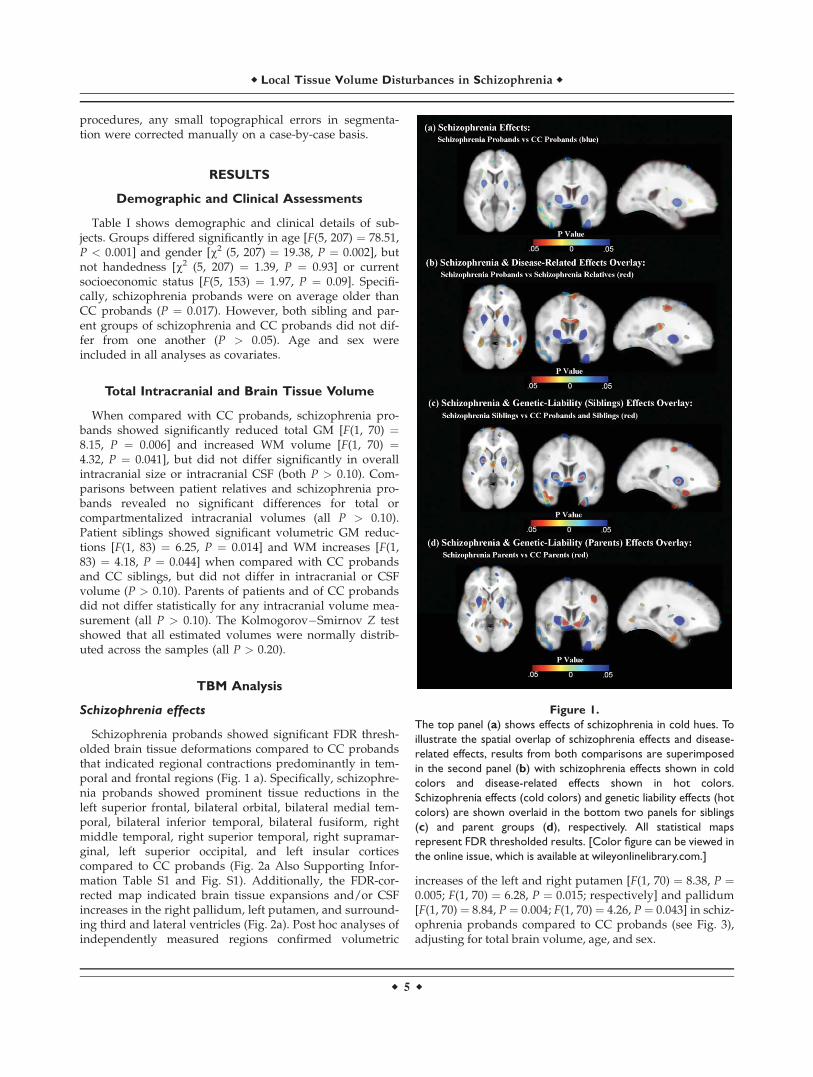
Schizophrenia probands showed significant FDR thresh-olded brain tissue deformations compared to CC probandsthat indicated regional contractions predominantly in tem-poral and frontal regions (Fig. 1 a). Specifically, schizophre-nia probands showed prominent tissue reductions in theleft superior frontal, bilateral orbital, bilateral medial tem-poral, bilateral inferior temporal, bilateral fusiform, rightmiddle temporal, right superior temporal, right supramar-ginal, left superior occipital, and left insular corticescompared to CC probands (Fig. 2a Also Supporting Infor-mation Table S1 and Fig. S1). Additionally, the FDR-cor-rected map indicated brain tissue expansions and/or CSFincreases in the right pallidum, left putamen, and surround-ing third and lateral ventricles (Fig. 2a). Post hoc analyses ofindependently measured regions confirmed volumetric
increases of the left and right putamen [F(1, 70) ¼ 8.38, P ¼0.005; F(1, 70) ¼ 6.28, P ¼ 0.015; respectively] and pallidum[F(1, 70) ¼ 8.84, P ¼ 0.004; F(1, 70) ¼ 4.26, P ¼ 0.043] in schiz-ophrenia probands compared to CC probands (see Fig. 3),adjusting for total brain volume, age, and sex.
Figure 1.
The top panel (a) shows effects of schizophrenia in cold hues. To
illustrate the spatial overlap of schizophrenia effects and disease-
related effects, results from both comparisons are superimposed
in the second panel (b) with schizophrenia effects shown in cold
colors and disease-related effects shown in hot colors.
Schizophrenia effects (cold colors) and genetic liability effects (hot
colors) are shown overlaid in the bottom two panels for siblings
(c) and parent groups (d), respectively. All statistical maps
represent FDR thresholded results. [Color figure can be viewed in
the online issue, which is available at wileyonlinelibrary.com.]
r Local Tissue Volume Disturbances in Schizophrenia r
r 5 r

Disease-related effects
Comparisons between schizophrenia probands and non-psychotic first-degree relatives of patients revealed several
spatially similar, but less prominent, regional deformationsto those described earlier for patient/CC probandscomparisons (Fig. 1b) that implicate the influences of dis-ease processes specifically. Brain tissue reductions in schiz-ophrenia probands were most prominent in the bilateralsuperior frontal, precentral, postcentral, superior parietal,right middle cingulate, left hippocampus, left amygdala,left inferior parietal, left angular, left fusiform, and rightinferior frontal cortices compared to their unaffected rela-tives (Fig 2b; Also see Supporting Information Table S1and Fig. S2), whereas tissue/CSF expansions wereobserved most prominently in the bilateral caudate nuclei,right thalamus, and lateral and third ventricles. To furtherensure the age differences between schizophrenia and CC,probands did not influence results, a post hoc analysison an age-matched subsample of schizophrenia and CCprobands was performed, which revealed similar signifi-cant findings.
Genetic-liability effects
Comparisons between nonpsychotic relatives of schizo-phrenia and CC probands and relatives showed significantregional deformations, suggesting genetic or shared envi-ronmental factors may influence these local changes inbrain tissue structure (Fig. 1c,d). Specifically, nonpsychoticsiblings of schizophrenia showed prominent tissue reduc-tions in the bilateral inferior frontal, left rectal, and left in-ferior temporal, and left medial temporal, and tissue/CSFexpansion in the left middle cingulate, right pallidum, andlateral and third ventricles compared to CC probands andsiblings (Fig 2c; Also see Supporting Information Table S1and Fig. S3). Although post hoc analyses comparing pal-lidum volumes in patient siblings were not significant (P> 0.29), means were intermediate to those observed inpatients and CC subjects. Nonpsychotic parents of schizo-phrenia exhibited most prominent brain tissue reductionsin the right inferior temporal, right superior temporal, andbilateral fusiform; and tissue/CSF increases in the left an-terior cingulate, left middle cingulate and left parahippo-campal gyri, and bilateral thalamus, bilateral putamen,right pallidum, and left caudate nucleus compared toparents of CC probands (Fig 2d; Also see Supporting In-formation Table S1 and Fig. S4). Post hoc analyses con-firmed increased volume of the right putamen [F(1, 74) ¼4.74, P ¼ 0.033] and left and right pallidum [F(1, 74) ¼14.19, P < 0.001; F(1, 74) ¼ 21.14, P < 0.001; respectively]in patient parents compared to CC parents (see Fig. 3).Additional analyses were performed including age-squared and age by sex interactions as covariates, whichrevealed very similar results (Fig. S6).
Duration of illness
Duration of illness was found to correlate positivelywith ventricular enlargement [Supporting Information Fig.S5(a,b)], particularly in the occipital horns.
Figure 2.
Beta maps that correspond to the probability maps (see Fig. 1)
show tissue volume changes in (a) schizophrenia probands com-
pared to CC probands; (b) schizophrenia probands compared to
schizophrenia relatives; (c) schizophrenia siblings compared to
CC probands and siblings; and (d) schizophrenia parents
compared to CC parents. Negative beta values index regional
reductions of tissue volume in schizophrenia patients or in their
nonpsychotic relatives when compared with CC subjects. [Color
figure can be viewed in the online issue, which is available at
wileyonlinelibrary.com.]
r Yang et al. r
r 6 r

DISCUSSION
This study is the first to our knowledge to use TBM
methods, which may capture unique patterns of global
and local brain dysmorphology, to cross-sectionally exam-
ine the effects of schizophrenia and disease-related genetic
predisposition toward altered brain tissue structure in
schizophrenia. The principal findings of this study are thatschizophrenia probands showed significant brain tissuereduction in frontal and temporal regions and tissue/CSFexpansion in the putamen/pallidum, lateral, and thirdventricles compared to CC probands. Comparisonsbetween schizophrenia probands and their relativesshowed disease-related effects indicating reduced tissue
Figure 3.
Left and right volumes of the putamen and the pallidum of schizophrenia (SZ) probands and rela-
tives (blue-colored bars), and CC probands and relatives (green-colored bars), standardized and
corrected for total brain volumes, age, and gender. [Color figure can be viewed in the online
issue, which is available at wileyonlinelibrary.com.]
r Local Tissue Volume Disturbances in Schizophrenia r
r 7 r

volume most prominently in the superior frontal cortexand enlargement of the lateral and third ventricles. Geneticliability effects that were examined by comparing to CCprobands to their nonpsychotic siblings and CC parents topatient parents showed significant deformations indexinglocal tissue reductions in medial temporal regions and tis-sue volume expansions in the putamen/pallidum. Thus,these findings suggest that genetic-liability effects may bemost associated with reduced temporal and increasedputamen/pallidum volume in schizophrenia, while frontalvolume reductions and ventricular enlargement appearlargely illness related. Taken together, these results indi-cate that distinct patterns of brain tissue deformation mayserve as biomarkers for distinguishing both individualswith schizophrenia and individuals with a genetic predis-position for the disorder.
Schizophrenia effects
Consistent with our hypotheses, TBM comparisonsbetween schizophrenia and CC probands produced resultsthat are largely in line with observations from numerousstudies using different methodological approaches [Elli-son-Wright et al., 2008; Pantelis et al., 2005; Shenton et al.,2001] and which have reported frontal and temporal volu-metric reductions and lateral and third ventricular enlarge-ment in schizophrenia. Tissue volume deformationsrevealed here may thus likewise reflect frontotemporalneuropathology that may account for widely reported ex-ecutive function and declarative memory processing defi-cits observed in the disorder [Wright et al., 2000]. Theadditional observations between illness duration and ven-tricular enlargement suggest a possible progressive neuro-pathological process in schizophrenia involving CSFexpansions [Raz and Raz, 1990]. Findings of tissue volumeexpansions in the putamen/pallidum in schizophreniaprobands compared to CC probands are also consistentwith several prior studies showing enlargements of basalganglia substructures, both in association with or inde-pendent of medication effects [Brandt and Bonelli, 2008;Simpson et al., 2010].
Findings of increased volume in the putamen/pallidumare of particular interest given the evidence suggesting arole for striatal dysfunction and altered striatal–cortical cir-cuitry in schizophrenia [Kegeles et al., 2010]. Increasedvolume in the putamen/pallidum in schizophreniapatients may reflect increased neuronal size and/or num-ber, dendrites, or the intracellular structures such as glialcells, which may result in increased striatal dopaminergicactivity observed in previous studies of schizophrenia[Kegeles et al., 2010; Kreczmanski et al., 2007]. Alterna-tively, tissue volume expansions in the putamen/pallidummay indicate decreases in cellular number and/or density,such that local tissue expansions as detected by TBMcould reflect increased extracellular space. Despite previ-ous studies suggesting that basal ganglia (including the
putamen/pallidum) volume increases are secondary to an-tipsychotic treatment [Chua et al., 2009; Diwadkar et al.,2006; Shenton et al., 2001], such effects are usually associ-ated with exposure to typical medications [Brandt andBonelli, 2008], whereas all patients in this study werereceiving standard atypical medication treatment. Further-more, regional expansions in the putamen/pallidum werealso observed in unaffected relatives of patients in thisstudy, indicating that medication treatments most likelydo not account for the observed effects and that enlargedvolumes in these regions and may instead represent a bio-marker for schizophrenia.
Disease-related effects
Focal changes in brain structure observed betweenschizophrenia patients and their relatives, particularlybrain tissue contractions within the frontal, cingulate, andtemporal gyri and expansions within the lateral ventricles,indicate the additional contributions of disease-related fac-tors toward altered brain morphology. Consistent withprevious findings of several patient-relative comparisons[Lawrie et al., 2001; Shenton et al., 2001], schizophreniapatients showed significant volume reduction in the fron-tal and temporal cortex compared to their nonpsychoticrelatives, especially the superior frontal cortex. Findingsare also consistent with previous studies documenting no-table changes in the lateral ventricles between patientsand their nonpsychotic relatives, although disease-relatedeffects appear less pronounced for the third ventricle[Lawrie, 2004; Staal et al., 2000]. Although patients werelargely asymptomatic at the time of assessment (meanBPRS score: 38.9), suggesting TBM-related changes inbrain structure are independent of state, given the narrowrange of symptom scores we could not directly addressthe hypotheses of whether structural deformationsobserved in patients are associated with other clinicalcharacteristics.
Genetic liability effects
The examination of both nonpsychotic first-degree sib-lings and parents of schizophrenia patients showed braintissue contractions commensurate with medial temporalvolume reductions and expansions in the putamen/pal-lidum compared to CC probands and their relatives, sug-gesting an effect of genetic-liability on structuraldeformations in these regions. The presence of reduced vol-umes in the medial temporal regions in both siblings andparents of schizophrenia is consistent with findings of pre-vious studies [Lawrie, 2004] and supports the hypothesisthat brain structural abnormalities occurring in schizophre-nia patients may be at least partially attributable to geneticinfluences. In particular, findings of increased volume in thebasal ganglia, most prominently in the putamen/pallidumbut also observed in the vicinity of the subthalamic nucleusand the substantia nigra, are in line with some prior studies,
r Yang et al. r
r 8 r

which suggested that morphological changes in this regionmay be a biophysiological risk factor for schizophrenia[Mamah et al., 2008] and could be associated with reportedchanges in cellular number or density, increased presynap-tic dopamine synthesis, impaired striatal activation, andhigher striatal dopamine upregulation in nonpsychotic rela-tives of schizophrenia [Huttunen et al., 2008].
Limitations
It is possible that exposure to antipsychotic medicationmay contribute to volume variations in the frontal and tem-poral regions and CSF increases in the ventricles in schizo-phrenia, especially with regard to relationships with theduration of illness. For example, it has been found that dif-ferent medication treatments may influence the trajectory ofbrain tissue loss associated with ongoing disease processes[Lieberman et al., 2005; Thompson et al., 2005]. However,findings in patient relatives and reports of similar structuraldeformations in patients with little or no medication expo-sure [Narr et al., 2005a,b; Nesvag et al., 2008] argue thatmedication effects are not central to the observed findings.TBM provides an unbiased approach to identify the pres-ence of localized alterations in brain tissue structurebetween individuals. However, because the surface of thecortex is highly variable between individuals, the registra-tion procedures used by TBM are less sensitive for detectingchanges at edges of the brain and the cortex specifically[Cahn et al., 2002], and partial volume effects may stillpotentially influence results. In spite of these caveats, schiz-ophrenia-related expansions observed toward the outer pe-rimeter of the brain observed in the b-maps are thought toreflect sulcal widening and increased subarachnoid/extra-cortical CSF, rather than to indicate increased brain tissue.These findings are thus in line with numerous studies indi-cating increased sulcal and/or extra-cortical CSF in schizo-phrenia, for example, [Narr et al., 2006; Schwarz and Bahn,2008], and are consistent with our prior observations ofwidespread cortical thinning in this sample [Yang et al.,2010]. However, because TBM is not focused on a particulartissue type or region, other approaches such as corticalthickness assessments may be more sensitive for determin-ing some specific structural deficits in schizophrenia
In summary, TBM provides both complementary and aunique characterization of genetic and disease-related con-tributions toward local brain tissue structure. Tissue vol-ume contractions in the frontal and temporal regions andexpansions in the putamen/pallidum and ventricles distin-guish schizophrenia patients from controls. Structuraldeformations in medial temporal regions and the puta-men/pallidum are associated most prominently withgenetic-liability effects while effects in frontal regions andthe lateral ventricles appear more associated with disease-related factors. These findings are in line with the mount-ing evidence, suggesting schizophrenia pathophysiologyaffects multiple brain systems and that some changes in
brain morphometry confer a genetic predisposition for thedisorder [Fornito et al., 2009; Lewis and Sweet, 2009].
ACKNOWLEDGMENTS
The authors thank David Fogelson, M.D., Heidi Kup-pinger, Ph.D., Sun Hwang, M.S., and Joseph Ventura,Ph.D., for their contributions to recruitment, diagnosis,and assessment of the participants in this study.
REFERENCES
Asarnow RF, Nuechterlein KH, Fogelson DL, Subotnik KL, PayneDL, Russell D, Asamen J, Kuppinger H, Kendler KS (2000):Schizophrenia and schizophrenia-spectrum personality disor-ders in the first-degree relatives of children with schizophre-nia: The UCLA family study. Arch Gen Psychiatry 58:581–588.
Brandt GN, Bonelli RM (2008): Structural neuroimaging of the ba-sal ganglia in schizophrenic patients: A review. Wien MedWochenschr 158:84–90.
Brans RG, van Haren NE, van Baal GC, Schnack HG, Kahn RS,Hulshoff Pol HE (2008): Heritability of changes in brain vol-ume over time in twin pairs discordant for schizophrenia.Arch Gen Psychiatry 65:1259–1268.
Borgwardt SJ, Picchioni MM, Ettinger U, Toulopoulou T, MurrayR, McGuire PK (2009): Regional gray matter volume in mono-zygotic twins concordant and discordant for schizophrenia.Biol Psychiatry 67:956–964.
Boos HB, Aleman A, Cohn W, Hulshoff Pol H, Kahn RS (2007):Brain volumes in relatives of patients with schizophrenia: Ameta-analysis. Arch Gen Psychiatry 64:297–304.
Bromet EI, Fennig S (1999): Epidemiology and natural history ofschizophrenia. Biol Psychiatry 46:871–881.
Brun C, Lepore N, Pennec X, Chou YY, Lee AD, Barysheva M, deZubicaray G, Meredith M, McMahon K, Wright MJ, Toga AW,Thompson PM (2008): A tensor-based morphometry study ofgenetic influences on brain structure using a new fluid regis-tration method. Med Image Comput Comput Assist Interv11:914–921.
Brun CC, Nicolson R, Lepore N, Chou YY, Vidal CN, DeVito TJ,Drost DJ, Williamson PC, Rajakumar N, Toga AW, ThompsonPM (2009): Mapping brain abnormalities in boys with autism.Hum Brain Mapp 30:3887–3900.
Cahn W, Hulshoff Pol HE, Bongers M, Schnack HG, Mandl RC,van Haren NE, Durston S, Koning H, Van Der Linden JA,Kahn RS (2002): Brain morphology in antipsychotic-naıveschizophrenia: A study of multiple brain structures. Br J Psy-chiatry 181:S66–S72.
Cannon TD, van Erp TG, Huttunen M, Lonnqvist J, Salonen O,Valanne L, Poutanen VP, Standertskjold-Nordenstam CG, GurRE, Yan M (1998): Regional gray matter, white matter, and cer-ebrospinal fluid distributions in schizophrenic patients, theirsiblings, and controls. Arch Gen Psychiatry 55:1084–1091.
Cannon TD, Hennah W, van Erp TG, Thompson PM, Lonnqvist J,Huttunene M, Gasperoni T, Tuulio-Henriksson A, Pirkola T,Toga AW, Kaprio J, Mazziotta J, Peltonen L (2005): Associationof DISC1/TRAX haplotypes with schizophrenia, reduced pre-frontal gray matter, and impaired short- and long-term mem-ory. Arch Gen Psychiatry 62:1205–1213.
r Local Tissue Volume Disturbances in Schizophrenia r
r 9 r

Che A, Cui J, Ivo D (2009a): SOCR analysis: Implementation anddemonstration of a new graphical statistical educational tool-kit. J Stat Software 30:1–19.
Che A, Cui J, Dinov ID (2009b): SOCR analyses—An InstructionalJava Web-based Statistical Analysis Toolkit. J Online LearnTeach 5:1–19.
Chiang MC, Reiss AL, Lee AD, Bellugi U, Calaburda AM, Koren-berg JR, Mills DL, Toga AW, Thompson PM (2007): 3D patternof brain abnormalities in Williams syndrome visualized usingtensor-based morphometry. Neuroimage 36:1096–1109.
Chua SE, Deng Y, Chen EY, Law CW, Chiu CP, Cheung C, WongJC, Lienenkaemper N, Cheung V, Suckling J, McAlonan GM(2009): Early striatal hypertrophy in first-episode psychosiswithin 3 weeks of initiating antipsychotic drug treatment. Psy-chol Med 39:793–800.
Dinov I, Van Horn JD, Lozev KM, Magsipoc R, Petrosyan P, LiuZ, MacKenzie-Graha A, Eggert P, Parker DS, Toga AW (2009):Efficient, distributed and interactive neuroimaging data analy-sis using the LONI Pipeline. Front Neuroinform 3:22.
Dinov ID, Sanchez J, Christou N (2008): Pedagogical utilizationand assessment of the statistic online computational resourcein introductory probability and statistics courses. ComputEduc 50:284–300.
Diwadkar VA, Montrose DM, Dworakowski D, Sweeney JA,Keshavan MS (2006): Genetically predisposed offspring withschizotypal features: An ultra high-risk group for schizophre-nia? Prog Neuropsychopharmacol Biol Psychiatry 30:230–238.
Eickhoff SB, Stephan KE, Mohlberg H, Grefkes C, Fink GR,Amunts K, Zilles K (2005): 3 new SPM toolbox for combiningprobabilistic sytoarchitectonic maps and functional imagingdata. Neuroimage 25:1325–1335.
Ellison-Wright I, Glahn DC, Laird AR, Thelen SM, Bullmore E(2008): The anatomy of first-episode and chronic schizophre-nia: An anatomical likelihood estimation meta-analysis. Am JPsychiatry 165:1015–1023.
Fischl B, Salat DH, Busa E, Albert M, Dieterich M, Haselgrove C,van der Kouwe A, Killiany R, Kennedy D, Klaveness S, Mon-tillo A, Makris N, Rosen B, Dale AM (2002): Whole brain seg-mentation: Automated labeling of neuroanatomical structuresin the human brain. Neuron 33:341–355.
Fornito A, Yucel M, Pantelis C (2009): Reconciling neuroimagingand neuropathological findings in schizophrenia and bipolardisorder. Curr Opin Psychiatry 22:312–319.
Gogtay N, Lu A, Leow AD, Klunder AD, Lee AD, Chavez A,Greenstein D, Giedd JN, Toga AW, Rapoport JL, ThompsonPM (2008): Three-dimensional brain growth abnormalities inchildhood-onset schizophrenia visualized by using tensor-based morphometry. Proc Natl Acad Sci USA 105:15979–15984.
Goldman AL, Pezawas L, Mattay VS, Fischl B, Verchinski BA,Chen Q, Weinberger DR, Meyer-Lindenberg A (2008): Wide-spread reductions of cortical thickness in schizophrenia andspectrum disorders and evidence of heritability. Arch Gen Psy-chiatry 66:467–477.
Goldman AL, Pezawas L, Mattay VS, Fischl B, Verchinski BA,Zoltick B, Weinberger DR, Meyer-Lindenberg A (2009): Herit-ability of brain morphology related to schizophrenia: A large-scale automated magnetic resonance imaging segmentationstudy. Biol Psychiatry 63:475–483.
Good CD, Johnsrude IS, Ashburner J, Henson RN, Friston KJ,Frackowiak RS (2001): A voxel-based morphometric study ofaging in 465 normal adult human brains. Neuroimage 14:21–36.
Honea RA, Meyer-Lindenberg A, Hobbs KB, Pezawas L, MattayVS, Egan MF, Verchinski B, Passingham RE, Weinberger DR,Callicott JH (2008): Is gray matter volume an intermediate phe-notype for schizophrenia? A voxel-based morphometry studyof patients with schizophrenia and their healthy siblings. BiolPsychiatry 63:465–474.
Hua X, Leow AD, Levitt JG, Gaplan R, Thompson PM, Toga AW(2009): Detecting brain growth patterns in normal childrenusing tensor-based morphometry. Hum Brain Mapp 30:209–219.
Huttunen J, Heinimaa M, Svirskis T, Nyman M, Kajander J, Fors-back S, Solin O, Lionen T, Korkeila J, Ristkari T, McGlashan T,Salokangas RK, Hietala J (2008): Striatal dopamine synthesis infirst-degree relatives of patients with schizophrenia. Biol Psy-chiatry 63:114–117.
Job DE, Whalley HC, McConnell S, Glabus M, Johnstone EC, Law-rie SM (2002): Structural gray matter differences between first-episode schizophrenics and normal controls on voxel-basedmorphometry. Neuroimage 17:880–889.
Kegeles LS, Abi-Dargham A, Frankle WG, Gil R, Cooper TB, Slif-stein M, Hwang DR, Huang Y, Haber SN, Laruelle M (2010):Increased synaptic dopamine function in associative regions othe striatum in schizophrenia. Arch Gen Psychiatry 67:231–239.
Kikinis Z, Fallon JH, Niznikiewicz M, Nestor P, Davidson C,Bobrow L, Pelavin PE, Fischl B, Tendiki A, McCarley RW,Kikinis R, Kubicki M, Shenton ME (2010): Gray matter volumereduction in rostral middle frontal gyrus in patients withchronic schizophrenia. Schizophr Res 123:153–159.
Kreczmanski P, Heinsen H, Mantua V, Woltersdorf F, Masson T,Ulfig N, Schmidt-Kastner R, Korr H, Steinbusch HW, Hof PR,Schmitz C (2007): Volume, neuron density and total neuronnumber in five subcortical regions in schizophrenia. Brain130:678–692.
Lawrie SM (2004): Premorbid structural abnormalities in schizo-phrenia. In: Keshavan M, Kennedy J, Murray R, editors. Neu-rodevelopment and Schizophrenia. Cambridge, UK:Cambridge University Press. pp 347–372.
Lawrie SM, Whalley HC, Abukmeil SS, Kestelman JN, Donnelly L,Miller P, Best JJ, Owens DG, Johnstone EC (2001): Brain structure,genetic liability and psychotic symptoms in subjects at high riskof developing schizophrenia. Biol Psychiatry 49:811–823.
Lee AD, Leow AD, Lu A, Reiss AL, Hall S, Chiang MC, TogaAW, Thompson PM (2007): 3D pattern of brain abnormalitiesin Fragile X syndrome visualized using tensor-based mor-phometry. Neuroimage 34:924–938.
Leow AD, Yanovsky I, Chiang MC, Lee AD, Klunder AD, Lu A,Becker JT, Davis SW, Toga AW, Thompson PM (2007): Statisti-cal properties of Jacobian maps and the realization of unbiasedlarge-deformation nonlinear image registration. IEEE TransMed Imaging 26:822–832.
Lepore N, Brun C, Pennec X, Chou YY, Lopez OL, Aizenstein HJ,Becker JT, Toga AW, Thompson PM (2007): Mean template fortensor-based morphometry using deformation tensors. MedImage Comput Comput Assist Interv 10:826–833.
Lepore N, Brun C, Chou YY, Chiang MC, Dutton RA, HayashiKM, Luders E, Lopez OL, Alzenstein HJ, Toga AW, Becker JT,Thompson PM (2008): Generalized tensor-based morphometryof HIV/AIDS using multivariate statistics on deformation ten-sors. IEEE Trans Med Imaging 27:129–141.
Lewis DA, Sweet RA (2009): Schizophrenia from a neural circuitryperspective: Advancing toward rational pharmacological thera-pies. J Clin Invest 119:706–716.
r Yang et al. r
r 10 r

Lopez-Garcia P, Alzenstein HJ, Snitz BE, Walter RP, Carter CS(2006): Automated ROI-based brain parcellation analysis offrontal and temporal brain volumes in schizophrenia. Psychia-try Res 147:153–161.
Mamah D, Harms MP, Wang L, Barch D, Thompson P, Kim J,Miller MI, Csernansky JG (2008): Basal ganglia shape abnor-malities in the unaffected siblings of schizophrenia patients.Biol Psychiatry 64:111–120.
McIntosh AM, Job DE, Moorhead WJ, Harrison LK, Whalley HC,Johnstone EC, Lawrie SM (2006): Genetic liability to schizophreniaor bipolar disorder and its relationship to brain structure. Am JMed Genet B Neuropsychiatr Genet 141:76–83.
Narr KL, Bilder RM, Toga AW, Woods RP, Szeszko PR, RobinsonD, Sevy S, Gunduz-Bruce H, Wang YP, DeLuca H, ThompsonPM (2005a): Mapping cortical thickness and gray matter con-centration in first episode schizophrenia. Cereb Cortex 15:708–719.
Narr KL, Toga AW, Szeszko P, Thompson PM, Woods RP, Robin-son D, Sevy S, Wang Y, Schrock K, Bilder RM (2005b): Corticalthinning in cingulate and occipital cortices in first episodeschizophrenia. Biol Psychiatry 58:32–40.
Narr KL, Bilder RM, Woods RP, Thompson PM, Szeszko P, Robin-son D, Ballmaier M, Messenger B, Wang Y, Toga AW (2006):Regional specificity of cerebrospinal fluid abnomalities in firstepisode schizophrenia. Psychiatry Res 146:21–33.
Narr KL, Hageman N, Woods RP, Hamilton LS, Clark K, PhillipsO, Shattuck DW, Asarnow RF, Toga AW, Nuechterlein KH(2009): Mean diffusivity: A biomarker for CSF-related diseaseand genetic liability effects in schizophrenia. Psychiatry Res171:20–32.
Nesvag R, Lawyer G, Vernas K, Fjell AM, Walhovd KB, FrigessiA, Jonsson EG, Agartz I (2008): Regional thinning of the cere-bral cortex in schizophrenia: Effects of diagnosis, age, and anti-psychotic medication. Schizophr Res 98:16–28.
Nuechterlein KH, Asarnow RF, Subotnik KL, Fogelson DL, PayneDL, Kendler KS, Neale MC, Jacobson KC, Mintz J (2002): Thestructure of schizotypy: Relationships between neurocognitiveand personality disorder features in relatives of schizophrenicpatients in the UCLA Family Study. Schizophr Res 54:121–130.
Pantelis C, Yucel M, Wood SJ, Velakoulis D, Sun D, Berger G,Stuart GW, Yung A, Phillips L, McGorry PD (2005): Structuralbrain imaging evidence for multiple pathological processes atdifferent stages of brain development in schizophrenia. Schiz-ophr Bull 31:672–696.
Prasad KM, Keshavan MS (2008): Structural cerebral variations asuseful endophenotypes in schizophrenia. Schizophr Bull34:774–790.
Raz S, Raz N (1990): Structural brain abnormalities in the majorpsychoses: A quantitative review of the evidence from compu-terized imaging. Psychol Bull 108:93–108.
Rex DE, Ma JQ, Toga AW (2003): The LONI pipeline processingenvironment. Neuroimage 19:1033–1048.
Rosso IM, Makris N, Thermenos HW, Hodge SM, Brown A, Ken-nedy D, Caviness VS, Faraone SV, Tsuang MT, Seldman LJ
(2010): Regional prefrontal cortex gray matter volumes inyouth at familial risk for schizophrenia from the Harvard Ado-lescent High Risk Study. Schizophr Res 123:15–21.
Schulze K, McDonald C, Frangou S, Sham P, Grech A, Toulopou-lous T, Walshe M, Shama T, Sigmundsson T, Taylor M, Mur-ray RM (2003): Hippocampal volume in familial andnonfamilial schizophrenic probands and their unaffected rela-tives. Biol Psychiatry 53:562–570.
Schwarz E, Bahn S (2008): Cerebrospinal fluid: Identification ofdiagnostic markers for schizophrenia. Expert Rev Mol Diagn8:209–216.
Shenton ME, Dickey CC, Frumin M, McCarley RW (2001): Areview of MRI findings in schizophrenia. Schizophr Res 49:1–52.
Simpson EH, Kellendonk C, Kandel E (2010): A possible role forthe striatum in the pathogenesis of the cognitive symptoms ofschizophrenia. Neuron 65:585–596.
Sled JG, Pike GB (1998): Standing-wave and RF penetration arti-facts caused by elliptic geometry: An electrodynamic analysisof MRI. IEEE Trans Med Imaging 17:653–662.
Smith SM (2002): Fast robust automated brain extraction. HumBrain Mapp 17:145–155.
Sowell ER, Peterson BS, Thompson PM, Welcome SE, HenkeniusAL, Toga AW (2003): Mapping cortical change across thehuman life span. Nat Neurosci 27:27.
Sowell ER, Leow AD, Bookheimer SY, Smith LM, O’Connor MJ,Kan E, Rosso C, Houston S, Dinov ID, Thompson PM (2010):Differentiating prenatal exposure to methamphetamine andalcohol versus alcohol and not methamphetamine using ten-sor-based brain morphometry and discriminant analysis. JNeurosci 30:3876–3885.
Staal WG, Hulshoff Pol HE, Schnack HG, Hoogendoorn MLC, Jel-lema K, Kahn RS (2000): Structural brain abnormalities inpatients with schizophrenia and their healthy siblings. Am JPsychiatry 157:416–421.
Stephan KE, Friston KJ, Frith CD (2009): Dysconnection in schizo-phrenia: From abnormal synaptic plasticity to failures of self-monitoring. Schizophr Bull 35:509–527.
Woods RP, Grafton ST, Watson JD, Sicotte NL, Mazziotta JC(1998a): Automated image registration. II. Intersubject valida-tion of linear and nonlinear models. J Comput Assist Tomogr22:153–165.
Woods RP, Grafton ST, Holmes CJ, Cherry SR, Mazziotta JC(1998b): Automated image registration. I. General methods andintrasubject, intramodality validation. J Comput Assist Tomogr22:139–152.
Wright IC, Rabe-Hesketh S, Woodruff PW, David AS, MurrayRM, Bullmore ET (2000): Meta-analysis of regional brain vol-umes in schizophrenia. Am J Psychiatry 157:16–25.
Yang Y, Nuechterlein KH, Phillips O, Hamilton LS, Subotnik KL,Asarnow RF, Toga AW, Narr KL (2010): The contribution ofdisease and genetic factors towards regional cortical thinningin schizophrenia: The UCLA Family Study. Schizophr Res123:116–125.
r Local Tissue Volume Disturbances in Schizophrenia r
r 11 r