Discovery of defense-and neuropeptides in social ants by genome-mining
12
Discovery of Defense- and Neuropeptides in Social Ants by Genome-Mining Christian W. Gruber 1 *, Markus Muttenthaler 2 1 Medical University of Vienna, Center for Physiology and Pharmacology, Vienna, Austria, 2 Departments of Chemistry and Cell Biology, The Scripps Research Institute, La Jolla, California, United States of America Abstract Natural peptides of great number and diversity occur in all organisms, but analyzing their peptidome is often difficult. With natural product drug discovery in mind, we devised a genome-mining approach to identify defense- and neuropeptides in the genomes of social ants from Atta cephalotes (leaf-cutter ant), Camponotus floridanus (carpenter ant) and Harpegnathos saltator (basal genus). Numerous peptide-encoding genes of defense peptides, in particular defensins, and neuropeptides or regulatory peptide hormones, such as allatostatins and tachykinins, were identified and analyzed. Most interestingly we annotated genes that encode oxytocin/vasopressin-related peptides (inotocins) and their putative receptors. This is the first piece of evidence for the existence of this nonapeptide hormone system in ants (Formicidae) and supports recent findings in Tribolium castaneum (red flour beetle) and Nasonia vitripennis (parasitoid wasp), and therefore its confinement to some basal holometabolous insects. By contrast, the absence of the inotocin hormone system in Apis mellifera (honeybee), another closely-related member of the eusocial Hymenoptera clade, establishes the basis for future studies on the molecular evolution and physiological function of oxytocin/vasopressin-related peptides (vasotocin nonapeptide family) and their receptors in social insects. Particularly the identification of ant inotocin and defensin peptide sequences will provide a basis for future pharmacological characterization in the quest for potent and selective lead compounds of therapeutic value. Citation: Gruber CW, Muttenthaler M (2012) Discovery of Defense- and Neuropeptides in Social Ants by Genome-Mining. PLoS ONE 7(3): e32559. doi:10.1371/ journal.pone.0032559 Editor: Jason Mulvenna, James Cook University, Australia Received September 23, 2011; Accepted January 29, 2012; Published March 20, 2012 Copyright: ß 2012 Gruber, Muttenthaler. This is an open-access article distributed under the terms of the Creative Commons Attribution License, which permits unrestricted use, distribution, and reproduction in any medium, provided the original author and source are credited. Funding: This work was financially supported by the Austrian Science Fund (FWF): P22889-B11 and from the European Union Seventh Framework Programme (FP7/2007–2013) under grant agreement no. [254897]. The funders had no role in study design, data collection and analysis, decision to publish, or preparation of the manuscript. Competing Interests: The authors have declared that no competing interests exist. * E-mail: [email protected] Introduction Natural peptides of great number and diversity occur in all organisms from microbes to plants to animals and exhibit biological activity often against unrelated targets. This provides researchers with excellent starting points for drug discovery [1], given that it is possible to isolate and characterize these natural peptides in adequate quantities or to retrieve their amino acid sequence genetically for synthetic production and biological testing. Peptidomics, using state-of-the-art liquid chromatography and mass spectrometry technologies, is generally the method-of- choice to identify and characterize peptides on protein level, whereas this technique yet fails to accurately identify the ‘peptidome’ from complex sample mixtures [2,3,4] or when the sample amount is limited or difficult to obtain, for instance peptides that are produced by mandibular- or venom glands of some insect species [5,6,7]. This applies in particular to ants, which are, due to their limited body and organ size, difficult to screen by analytical instrumentation unless many thousand individuals are sacrificed or laborious venom sac dissection is being used [5,8]. Other problems associated with peptidomics is the retrieval of low abundant peptides in complex mixtures and the detection of pseudo-gene products, i.e. peptide coding genes that have been switched off during evolution, but which may encode bioactive drug leads [9,10]. Genome-mining, a term that has been used to describe the exploitation of genomic information for the discovery of new processes, targets, and products [11], may be a useful alternative or complement to peptide discovery by peptidomics. This technique seems in particular valuable in the genomic era, since the number of available genomes is steadily increasing as whole genome sequencing is becoming affordable and achievable. Following the footsteps of the human genome initiative [12] and many other successful genome-sequencing efforts in animals, plants and microbes, recently the genomes of seven ant species have been reported. These include the invasive Argentine ant Linepithema humile [13], the red harvester ant Pogonomyrmex barbatus [14], the fire ant Solenopsis invicta [15], the carpenter ant Camponotus floridanus and a basal ant Harpegnathos saltator [16], as well as the leaf-cutter/farming ants Atta cephalotes [17] and Acromyrmex echinatior [18], respectively. The aim of this study was to analyze three representative ant genomes from the subfamilies of Myrmicinae (A.cephalotes), Formicinae (C.floridanus) and Ponerinae (H.saltator) for the discovery of peptide encoding genes and their sequences using an array of publicly available tools, including tBLASTn similarity search, GeneWise gene structure prediction and ClustalW sequence alignments (Figure 1). Using this methodology it was possible to identify numerous putative peptides as partial, full-length precur- sor and mature amino acid sequences of ant defense- and neuropeptides. These genes were characterized by similarity to PLoS ONE | www.plosone.org 1 March 2012 | Volume 7 | Issue 3 | e32559
Transcript of Discovery of defense-and neuropeptides in social ants by genome-mining

Discovery of Defense- and Neuropeptides in Social Antsby Genome-MiningChristian W. Gruber1*, Markus Muttenthaler2
1 Medical University of Vienna, Center for Physiology and Pharmacology, Vienna, Austria, 2 Departments of Chemistry and Cell Biology, The Scripps Research Institute, La
Jolla, California, United States of America
Abstract
Natural peptides of great number and diversity occur in all organisms, but analyzing their peptidome is often difficult. Withnatural product drug discovery in mind, we devised a genome-mining approach to identify defense- and neuropeptides inthe genomes of social ants from Atta cephalotes (leaf-cutter ant), Camponotus floridanus (carpenter ant) and Harpegnathossaltator (basal genus). Numerous peptide-encoding genes of defense peptides, in particular defensins, and neuropeptidesor regulatory peptide hormones, such as allatostatins and tachykinins, were identified and analyzed. Most interestingly weannotated genes that encode oxytocin/vasopressin-related peptides (inotocins) and their putative receptors. This is the firstpiece of evidence for the existence of this nonapeptide hormone system in ants (Formicidae) and supports recent findingsin Tribolium castaneum (red flour beetle) and Nasonia vitripennis (parasitoid wasp), and therefore its confinement to somebasal holometabolous insects. By contrast, the absence of the inotocin hormone system in Apis mellifera (honeybee),another closely-related member of the eusocial Hymenoptera clade, establishes the basis for future studies on the molecularevolution and physiological function of oxytocin/vasopressin-related peptides (vasotocin nonapeptide family) and theirreceptors in social insects. Particularly the identification of ant inotocin and defensin peptide sequences will provide a basisfor future pharmacological characterization in the quest for potent and selective lead compounds of therapeutic value.
Citation: Gruber CW, Muttenthaler M (2012) Discovery of Defense- and Neuropeptides in Social Ants by Genome-Mining. PLoS ONE 7(3): e32559. doi:10.1371/journal.pone.0032559
Editor: Jason Mulvenna, James Cook University, Australia
Received September 23, 2011; Accepted January 29, 2012; Published March 20, 2012
Copyright: � 2012 Gruber, Muttenthaler. This is an open-access article distributed under the terms of the Creative Commons Attribution License, which permitsunrestricted use, distribution, and reproduction in any medium, provided the original author and source are credited.
Funding: This work was financially supported by the Austrian Science Fund (FWF): P22889-B11 and from the European Union Seventh Framework Programme(FP7/2007–2013) under grant agreement no. [254897]. The funders had no role in study design, data collection and analysis, decision to publish, or preparation ofthe manuscript.
Competing Interests: The authors have declared that no competing interests exist.
* E-mail: [email protected]
Introduction
Natural peptides of great number and diversity occur in all
organisms from microbes to plants to animals and exhibit
biological activity often against unrelated targets. This provides
researchers with excellent starting points for drug discovery [1],
given that it is possible to isolate and characterize these natural
peptides in adequate quantities or to retrieve their amino acid
sequence genetically for synthetic production and biological
testing. Peptidomics, using state-of-the-art liquid chromatography
and mass spectrometry technologies, is generally the method-of-
choice to identify and characterize peptides on protein level,
whereas this technique yet fails to accurately identify the
‘peptidome’ from complex sample mixtures [2,3,4] or when the
sample amount is limited or difficult to obtain, for instance
peptides that are produced by mandibular- or venom glands of
some insect species [5,6,7]. This applies in particular to ants,
which are, due to their limited body and organ size, difficult to
screen by analytical instrumentation unless many thousand
individuals are sacrificed or laborious venom sac dissection is
being used [5,8]. Other problems associated with peptidomics is
the retrieval of low abundant peptides in complex mixtures and
the detection of pseudo-gene products, i.e. peptide coding genes
that have been switched off during evolution, but which may
encode bioactive drug leads [9,10].
Genome-mining, a term that has been used to describe the
exploitation of genomic information for the discovery of new
processes, targets, and products [11], may be a useful alternative
or complement to peptide discovery by peptidomics. This
technique seems in particular valuable in the genomic era, since
the number of available genomes is steadily increasing as whole
genome sequencing is becoming affordable and achievable.
Following the footsteps of the human genome initiative [12] and
many other successful genome-sequencing efforts in animals,
plants and microbes, recently the genomes of seven ant species
have been reported. These include the invasive Argentine ant
Linepithema humile [13], the red harvester ant Pogonomyrmex barbatus
[14], the fire ant Solenopsis invicta [15], the carpenter ant Camponotus
floridanus and a basal ant Harpegnathos saltator [16], as well as the
leaf-cutter/farming ants Atta cephalotes [17] and Acromyrmex echinatior
[18], respectively.
The aim of this study was to analyze three representative ant
genomes from the subfamilies of Myrmicinae (A.cephalotes),
Formicinae (C.floridanus) and Ponerinae (H.saltator) for the discovery
of peptide encoding genes and their sequences using an array of
publicly available tools, including tBLASTn similarity search,
GeneWise gene structure prediction and ClustalW sequence
alignments (Figure 1). Using this methodology it was possible to
identify numerous putative peptides as partial, full-length precur-
sor and mature amino acid sequences of ant defense- and
neuropeptides. These genes were characterized by similarity to
PLoS ONE | www.plosone.org 1 March 2012 | Volume 7 | Issue 3 | e32559

other insect and non-insect species and for the first time we report
the sequences of inotocin peptides (oxytocin/vasopressin-related
neuropeptides) in social ants. The presented results offer the
possibility to interpret the phylogenetic relationship and evolution
of insect defense molecules and peptide hormone systems, but
most importantly the predicted mature peptide sequences could
provide novel drug leads or tools to study the similar and preserved
receptor systems in humans.
Results and Discussion
Genome-mining is a powerful technique to discover novel
putative bioactive peptides, in particular when the peptidome of
interest is not readily accessible by modern analytical instrumen-
tation. This study was designed to explore the genomes of three
recently reported social ant species for the discovery of defense-
and neuropeptide sequences, which are listed in Table 1.
Identification of ant defense peptidesAnts belong to the class of eusocial insects and live in crowded
nests. Millions of individuals are in close interaction and hence it is
not surprising that they have evolved a highly developed system of
immune response to fight pathogen infections [16]. One of the best
studied class of innate immune molecules are the so-called defensin
peptides, which occur in many if not all organisms [19]. Several
ant defensins have been reported so far [19,20,21,22,23], mainly
by genomic sequencing [23], since it seems extremely difficult to
isolate and identify these peptides by tandem mass spectrometry or
Edman degradation. Using the proposed genome-mining meth-
odology, it was possible to identify sequences of defensins and
several related defense peptides and peptide toxins in all three ant
species (Table 1, Table S1).
Structural characteristics of putative ant defensin
precursors and mature peptides. Using the amino acid
sequences of the putative ant defensin genes we were able to
compare their molecular characteristics to known insect defensins.
Figure 2 shows the defensin sequences in alignment with selected
ant and insect defensins from Apis mellifera and Drosophila
melanogaster (Figure 2).
The peptides share common molecular characteristics with
other insect defensins, i.e. (i) a similar length of precursor protein
and mature peptide (ranging from 40–43 amino acids; Table 1), (ii)
a conserved network of six cysteine residues and (iii) a strong
positive net-charge of the mature peptides (A.cephalotes DEF = +6,
C.floridanus DEF1 = +5, C.floridanus DEF2 = +3, H.saltator
DEF1 = +5) to interact with and disrupt negatively-charged
microbial membranes. This strong positive charge is ‘neutralized’
by an anionic pro-domain (A.cephalotes DEF = 24, C.floridanus
DEF1 = 23, C.floridanus DEF2 = 23, H.saltator DEF1 = 24) to
prevent toxic effects to the cells during defensin biosynthesis. This
charge correlation between the prodomains and the mature
defensin is well known to exist for many defensins, including
mammalian defensins and it appears to be conserved throughout
this class of peptides [24].
Besides the cysteine residues there are at least two more residues
that appear highly conserved amongst ant defensins, namely the
negatively-charged aspartic acid residue at the beginning (pos. 4)
and the positively-charged arginine residue (pos. 42) at the end of
the mature domain (Figure 2C). Similarly, mammalian a-defensins
contain oppositely-charged residues that form a highly conserved
salt-bridge interaction, which is critical for the formation of the
disulfide bonds, structural rigidity, and biological function [25,26].
To gain further insight into the role these conserved residues play
in folding and stability of ant defensins, we prepared a homology
structure model of the representative A.cephalotes defensin. The
peptide sequence was modeled by energy minimization on
distance restraints to the closely related structures of the insect
defensin phormicin [27] and a synthetic defensin analogue (DEF-
Figure 1. Flowchart of genome-mining for the discovery of ant peptides used in this study. Whole genome shotgun data, in this instancefrom the three ant species Atta cephalotes, Camponotus floridanus and Harpegnathos saltator, and amino acid sequences of precursor proteins frombioactive peptides of interest (e.g. defense and/or regulatory neuropeptides) were used for database analysis. This included similarity analysis oftarget DNA sequence and query protein sequence using tBLASTn, DNA to protein translation of discovered hit sequences and identification of openreading frames and coding sequence. The obtained automated results were refined and confirmed manually and used for gene structure predictionusing the GeneWise algorithm. Database analysis yielded precursor protein and peptide sequences that were further annotated and analyzed bysequence alignments and similarity comparison to identify signal sequences, propeptides and mature peptide chains. Using this genome-miningmethodology it was possible to predict the amino acid sequences of bioactive peptides in ants.doi:10.1371/journal.pone.0032559.g001
Ant Peptide Sequences as Leads in Drug Discovery?
PLoS ONE | www.plosone.org 2 March 2012 | Volume 7 | Issue 3 | e32559

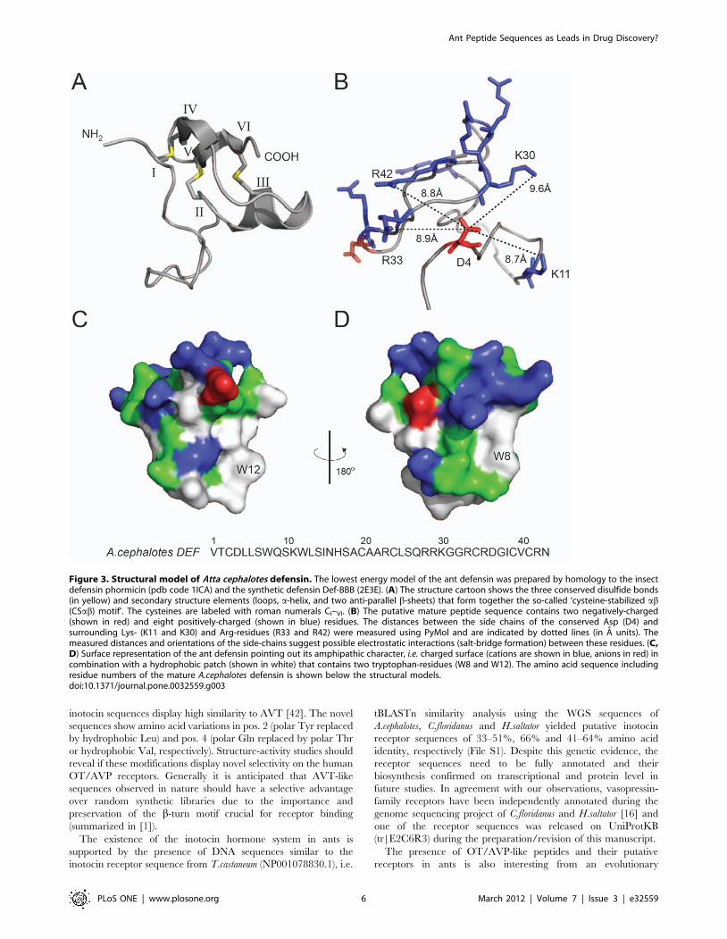
BBB, [28]). The model strongly suggests the presence of a so-called
‘cysteine-stabilized ab (CSab) motif’ whose CI–CIV, CII–CV and
CIII–CVI pairing forms three intramolecular disulfide bonds,
which is a characteristic for other insect defensins (Figure 3A)
[27]. Furthermore, according to the NMR structures (pdb codes
1ICA and 2E3E, respectively) that were used as templates, the
loop between the first two cysteine residues (CI and CII, see
Figure 3A) is mainly disordered. To analyze the possible formation
of a functionally important salt-bridge interaction, we calculated
the distances of the side-chain of the conserved aspartic acid
residue to neighboring side-chains of positively-charged residues.
The charged side-chains of Asp 4 and Arg 42 are ,8.8 A apart
from each other (Figure 3B) and although the conserved Asp 4
does not form a salt bridge in the homology model or in the
template structure on which it was based, several positively-
charged residues (Lys 11, Lys 30, Arg 33 and Arg 42) are within
close enough proximity (,10 A) to be potentially important for the
formation of electrostatic interactions. However, structural studies
would be required to confirm whether this is actually the case. In
addition to the charge distribution and potential electrostatic
interactions, we analyzed the surface characteristics and it is
obvious (Figure 3C and D) that the ant defensin contains many
hydrophobic residues (,30%), which seem to form a hydrophobic
surface patch; in particular, the model points out two tryptophan
residues (Trp 8 and Trp 12) that appear on the surface of the
molecule. The overall amphipathic character of the mature
peptide, i.e. combination of charged and hydrophobic surface, is
common to many defensins and presumably contributes to their
ability to insert into and disrupt microbial cell walls. In summary,
the sequence and surface characteristics (charge distribution,
Table 1. Selected defense- and neuropeptides from ant species characterized by genome-mining.
peptide class species/peptide namelength amino acids (precursor/mature) no. of Cys evidence
precursormaturepeptide partial
Abaecin+ A.cephalotes ABA-like 52/33 n.a.* ! !
C.floridanus ABA-like - n.a. !
H.saltator ABA-like 50/31 n.a. ! !
Allatostatin A.cephalotes AST 193/8, 8, 8, 7, 30, 9, 28# n.a. ! !
C.floridanus AST 193/8, 8, 8, 7, 30, 9, 27# n.a. ! !
H.saltator AST 193/8, 8, 8, 7, 30, 9, 25# n.a. ! !
Defensin+ A.cephalotes DEF 97/43 6 ! !
C.floridanus DEF1 97/43 6 ! !
C.floridanus DEF2 98/40 6 ! !
H.saltator DEF1 100/43 6 ! !
H.saltator DEF1a 77/43 6 ! !
H.saltator DEF2 -/41 5$ ! !
Eclosion hormone+ A.cephalotes EH-like+ - n.a. !
Diuretic-hormone+ A.cephalotes DH-like - n.a. !
C.floridanus DH-like -/31 n.a. ! !
H.saltator DH-like - n.a. !
Inotocin/AVP-like+ A.cephalotes INT/AVP-like& 148/9 2+12 ! ! !
C.floridanus INT/AVP-like& -/9 2 ! !
H.saltator INT/AVP 150/9 2+12 ! !
Ion-transport and CHH-like peptide+ A.cephalotes ITP-like -/72 6 ! !
C.floridanus ITP-like - - !
C.floridanus CHH-like - - !
H.saltator ITP-like 157/72 6 ! !
Neuroparsin-A+ A.cephalotes NP-like - n.a. !
Pilosulin C.floridanus PIL-like 87/multiple mature peptides** ! ! !
Tachykinin-like A.cephalotes TRP-like 484/9## n.a. ! ! !
*not applicable, peptide class generally contains no cysteines in the mature peptides;#multiple mature allatostatin peptides are encoded by the same precursor protein, order of presented length of peptides are in order as presented in Figure 4;$5 cysteines were identified in the mature form of H.saltator DEF2, which likely indicates a partial sequence;&GeneWise prediction was no successful, but precursor sequences could be established manually from tBLASTn results;+during the preparation/revision of this manuscript the following peptide-/receptor sequences (partial or complete) were released on UniProtKB: diuretic hormones(C.floridanus: tr|E2AZE8; H.saltator: tr|E2C6V6, tr|E2B7W2), vasotocin-neurophysin (H.saltator: EFN79183), eclosion hormones (C.floridanus: tr|E2AXD4; H.saltator:tr|E2BSX6), ion-transport peptides (C.floridanus: tr|E2AP65; H.saltator: tr|E2BEL2), neuroparsins-A (C.floridanus: tr|E1ZXL4; H.saltator: tr|E2BLJ9), abaecin (H.saltator:tr|E2B7M5) and defensins (C.floridanus: tr|E2AKI0, tr|E2AVT3; H.saltator: tr|E2BDP6), for reference see [16];**multiple mature peptides can be cleaved from the same precursor protein, see [33,35,36];##eight tachykinin peptides of equal length are encoded by the precursor peptide, for order see Figure S5.doi:10.1371/journal.pone.0032559.t001
Ant Peptide Sequences as Leads in Drug Discovery?
PLoS ONE | www.plosone.org 3 March 2012 | Volume 7 | Issue 3 | e32559

Ant Peptide Sequences as Leads in Drug Discovery?
PLoS ONE | www.plosone.org 4 March 2012 | Volume 7 | Issue 3 | e32559

hydrophobic patch, and amphipathic surface) and structural
characteristics (CSab-motif, potential salt-bridge interaction) of
the putative ant defensin appear to be in agreement with known
defensins and it would be interesting to assess their biological
activity in future studies.
Evolutionary analysis of putative ant defensins. Viljakainen
and Pamilo have recently analysed ant defensin precursor sequences
and identified two positions in the mature peptide domain that show
positive evolutionary selection (green box in Figure 2A) [23]. These
positions are characterized by non-synonymous substitutions to yield a
higher rate of amino acid variation, as compared to the remaining
protein sequence. The defensin sequences from A.cephalotes (DEF) and
C.floridanus (DEF1) have a glutamine in pos. 60 (named according to
[23]) and A.cephalotes (DEF), C.floridanus (DEF1) and H.saltator (DEF1)
contain a serine or valine, respectively, in pos. 73. This sequence
variation has been highlighted in a sequence logo (Figure 2C). These
residues represent novel amino acid variations and hence support the
hypothesis by Viljakainen and Pamilo that the immune system of social
ants and dipteran insects may have responded differently to selection
pressure caused by microbes and pathogens [23]. Another feature of
the putatively identified ant defensins is their gene structure, which
differs in the length and position of introns and exons compared to
other insect defensins from A.mellifera and D.melanogaster (Figure 2B). For
example, the A.mellifera DEF1 and C.floridanus DEF2 share the same
position and a similar length of the first intron, but the honeybee gene
contains an additional second proximal intron, whereas no introns
could be identified for the D.melanogaster DEF gene. Similarly, A.mellifera
DEF2, A.cephalotes DEF, C.floridanus DEF1 and H.saltator DEF1 share a
similar intron position, whereas the ant defensin introns tend to be
much longer (e.g. 4702 bases in leaf-cutter ant vs. 335 bases in
honeybee). These differences support the suggestion that defensin
evolution may be taxon specific [23,29].
Discovery of related ant defense peptides. Besides the
discovery of defensin genes we have analyzed the three ant genomes
for the presence of many other defense peptides and peptide toxins
using tBLASTn and found genetic evidence of at least five different
classes of ant/insect defense and defense-related peptides (see Table
S1). Amongst those are defensins (as discussed above), abaecins,
apidaecin-related peptides, hymenoptaecins and pilosulins.
Exemplarily, molecular structures of pilosulin and abaecin defense
peptides were analyzed in more detail. Pilosulins are allergenic
peptides with immunoglobulin-binding activity that are commonly
found in venoms of Myrmecia spp. [30,31,32,33]. The present
genomic screen identified a pilosulin-like peptide in C.floridanus
(Figure S1A). Pilosulins share a high sequence similarity in their first
47 amino acid residues (including the signal peptide), but the mature
peptide domain varies substantially amongst pilosulins
[34,35,36,37]. This similarity supports the results of our analysis;
however, at this stage it has to be considered with caution and needs
to be confirmed on transcriptional level, since C.floridanus, like all
Formicinae species, has a significantly reduced sting and venom
reservoir exclusively for the production of formic acid.
Another class of defense peptides that have been identified in
this screen are abaecins. Abaecin peptides are considered to be
major antibacterial response peptides that have been originally
discovered in honeybee [38] and occur in several ant species that
have been analyzed, including the putative abaecin peptide
sequences found in A.cephalotes, C.floridanus and H.saltator (Figure
S1B). All sequences share the common proline-rich characteristic,
i.e. they contain between 9 and 10 Pro-residues (,30% proline
content). Besides the reported genetic evidence of putative ant
defense peptides, the main focus of this study was the analysis of
the three ant genomes for the presence of neuropeptides and
regulatory peptide hormones.
Identification of ant neuropeptide- and regulatorypeptide hormone-encoding genes
Neuropeptides and regulatory peptide hormones control many,
if not all important developmental, physiological and behavioral
processes in animals, including insects [39]. In the following we
describe the characterization of several ant neuropeptides and
regulatory peptide hormones, in particular oxytocin (OT) and
arginine-vasopressin (AVP)-related peptides and interpret the
importance of these findings on a molecular and phylogenetic
level.
Genetic evidence for the existence of an oxytocin/
vasopressin-related hormone system in social ants. The
origination of the OT/AVP peptide hormone system is considered
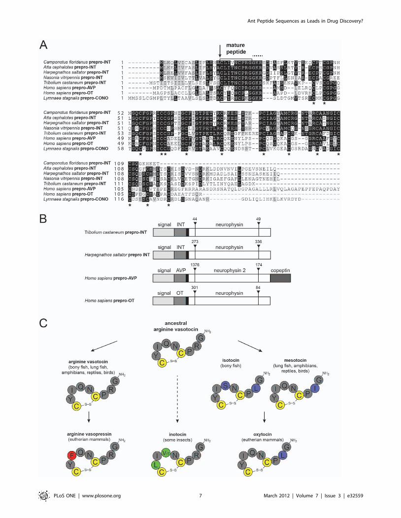
to date back 640–760 million years ago [40,41]. All vertebrate
OT/AVP-like peptides are considered to have evolved from the
ancestral nonapeptide arginine-vasotocin (AVT) [42] and are
today present in many different species, including non-mammalian
vertebrates, fish, mammals and humans [43,44] (Figure 4C). AVT
is structurally similar to a variety of invertebrate nonapeptides
[45], suggesting that the AVT-like invertebrate nonapeptides are
much more ancient than AVT itself [46,47]. AVT-like
nonapeptides are present in several invertebrate species,
including molluscs and annelids and they have been
characterized in the arthropods Locusta migratoria, in the red flour
beetle Tribolium castaneum and the parasitic wasp Nasonia vitripennis
[41,48].
The ant genome analysis revealed the presence of prepro-
inotocin proteins in all three species (Figure 4). The precursor
proteins of the ant inotocins described here, other insect inotocin
proteins, snail conopressin and human OT/AVP precursor
proteins all share molecular features. Following the mature
nonapeptides, they all contain the canonical GRK amidation
signal and they all contain 12 conserved Cys-residues in the
neurophysin domain (Figure 4A). This similarity is supported by
their gene structure, since T.castaneum, H.saltator and the human
prepro-OT gene share identical intron sites and similar lengths
(Figure 4B). The mature peptides have the same length and
position of Cys-residues, but the molecular sequence is slightly
different between species (Figure 4C). Both newly identified ant
Figure 2. Sequence and gene structure of novel ant defensins. (A) Identified ant defensin precursor sequences from Atta cephalotes,Camponotus floridanus and Harpegnathos saltator were used for similarity alignment (ClustalW2) and compared to known defensins from Formicaaquilonia (UniProtKB Q5BU36), Lasius niger (B9TXS0), Myrmecia ruginodis (B9TXS6), Drosophila melanogaster (P36192), Apis mellifera (P17722, Q5MQL3)and Acromyrmex echinatior (F4WLL3). The signal peptide cleavage site (identified by similarity) is shown as an arrow. Mature defensin peptides areindicated in the black box and the conserved cysteine residues are indicated with asterisks. Residue positions of positive evolutionary selection [23]are indicated with a green box. The sequence alignment was prepared using Boxshade. (B) Gene structure of novel ant and known insect defensingenes (GenBank D.melanogaster NT033778.3 and A.mellifera NC007085.3, NC007075.3) was predicted using the GeneWise algorithm. Signalsequences are indicated in light grey, pre-regions in dark grey and the mature peptide domain in white. Intron sequences (including their base pairlength) are indicated with upside-down arrow heads. (C) The sequences of 22 known [23] and the novel ant defensins have been compared using asequence logo to highlight their amino acid variation. Conserved cysteines are colored in yellow, the positions of positive evolutionary selection arecolored in green and the conserved negatively-charged Asp and positively-charged Arg are colored in red and blue, respectively.doi:10.1371/journal.pone.0032559.g002
Ant Peptide Sequences as Leads in Drug Discovery?
PLoS ONE | www.plosone.org 5 March 2012 | Volume 7 | Issue 3 | e32559
inotocin sequences display high similarity to AVT [42]. The novel
sequences show amino acid variations in pos. 2 (polar Tyr replaced
by hydrophobic Leu) and pos. 4 (polar Gln replaced by polar Thr
or hydrophobic Val, respectively). Structure-activity studies should
reveal if these modifications display novel selectivity on the human
OT/AVP receptors. Generally it is anticipated that AVT-like
sequences observed in nature should have a selective advantage
over random synthetic libraries due to the importance and
preservation of the b-turn motif crucial for receptor binding
(summarized in [1]).
The existence of the inotocin hormone system in ants is
supported by the presence of DNA sequences similar to the
inotocin receptor sequence from T.castaneum (NP001078830.1), i.e.
tBLASTn similarity analysis using the WGS sequences of
A.cephalotes, C.floridanus and H.saltator yielded putative inotocin
receptor sequences of 33–51%, 66% and 41–64% amino acid
identity, respectively (File S1). Despite this genetic evidence, the
receptor sequences need to be fully annotated and their
biosynthesis confirmed on transcriptional and protein level in
future studies. In agreement with our observations, vasopressin-
family receptors have been independently annotated during the
genome sequencing project of C.floridanus and H.saltator [16] and
one of the receptor sequences was released on UniProtKB
(tr|E2C6R3) during the preparation/revision of this manuscript.
The presence of OT/AVP-like peptides and their putative
receptors in ants is also interesting from an evolutionary
Figure 3. Structural model of Atta cephalotes defensin. The lowest energy model of the ant defensin was prepared by homology to the insectdefensin phormicin (pdb code 1ICA) and the synthetic defensin Def-BBB (2E3E). (A) The structure cartoon shows the three conserved disulfide bonds(in yellow) and secondary structure elements (loops, a-helix, and two anti-parallel b-sheets) that form together the so-called ‘cysteine-stabilized ab(CSab) motif’. The cysteines are labeled with roman numerals CI–VI. (B) The putative mature peptide sequence contains two negatively-charged(shown in red) and eight positively-charged (shown in blue) residues. The distances between the side chains of the conserved Asp (D4) andsurrounding Lys- (K11 and K30) and Arg-residues (R33 and R42) were measured using PyMol and are indicated by dotted lines (in A units). Themeasured distances and orientations of the side-chains suggest possible electrostatic interactions (salt-bridge formation) between these residues. (C,D) Surface representation of the ant defensin pointing out its amphipathic character, i.e. charged surface (cations are shown in blue, anions in red) incombination with a hydrophobic patch (shown in white) that contains two tryptophan-residues (W8 and W12). The amino acid sequence includingresidue numbers of the mature A.cephalotes defensin is shown below the structural models.doi:10.1371/journal.pone.0032559.g003
Ant Peptide Sequences as Leads in Drug Discovery?
PLoS ONE | www.plosone.org 6 March 2012 | Volume 7 | Issue 3 | e32559
Ant Peptide Sequences as Leads in Drug Discovery?
PLoS ONE | www.plosone.org 7 March 2012 | Volume 7 | Issue 3 | e32559

perspective, since the homologues sequences are absent in the
genomes of the fruit fly (Brachycera), mosquito (Nematocera),
silkworm (Lepidoptera) and honeybee (Hymenoptera) [41,48],
whereas they are present in the red flour beetle (Coleoptera), the
parasitic wasp and the three ant species (both Hymenoptera) as
described above (Figure 5). The novel discoveries confirm the
recent findings of the existence of the OT/AVP receptor system in
arthropods and their confinement to some basal holometabolous
insects (i.e. Hymenoptera and Coleoptera species). As indicated in
Figure 5 the OT/AVP peptide hormone system has been lost at
least two times during holometabolous insect evolution. However,
the question of how the (predicted) existence of this neuropeptide
system in ants (Formicidae) and its absence in honeybees (Apidae)
can be explained remains, since both insect families belong to the
eusocial Hymenoptera clade (monophyletic lineage Aculeata). The
answer to this is beyond the scope of this paper, but needs to be
studied in detail after the peptides and their receptors have been
confirmed and their function tested.
Identification and analysis of other ant neuropeptide- and
peptide hormone genes. As mentioned above, ants as well as
honeybees belong to the eusocial Hymenoptera clade and are
considered as being advanced over other insects in terms of
learning, navigation and behavior [49,50]. To reveal genes that
are involved in higher brain functions and sociality, Hauser et al.
compared neuropeptide and protein hormone genes and their
receptors from honeybee and fruit fly [50]. Although there are
differences in (i) the overall number of identified ligands and
receptors for each species and (ii) the presence/absence of certain
ligand/receptor-systems in one versus another species, the general
conclusion of the comparison is coevolution of neuropeptide and
protein hormone ligands and their receptors in both species.
Besides OT/AVP-related peptides and their putative receptors,
we analyzed the three ant genomes for the presence of several
other neuropeptides and peptide hormones (Table S2). From the
genome sequencing project of C.floridanus and H.saltator [16]
genetic evidence of the following neuropeptide ligands amongst
Figure 4. Sequences, gene- and peptide structures of ant inotocins. (A) Sequences of prepro-inotocin/neurophysin proteins from Attacephalotes, Camponotus floridanus and Harpegnathos saltator were compared by similarity alignment to known inotocin/oxytocin/vasopressinprepro-proteins from Tribolium castaneum (UniProtKB A3RE83), Nasonia vitripennis (GenBank XP001606547.1), Homo sapiens (P01185 and P01178)and Lymnaea stagnalis (Q00945). The signal peptide cleavage site (identified by similarity) is shown with an arrow. Mature vasopressin/oxytocin/inotocin peptides are indicated in the box, followed by the canonical GRK amidation signal (dotted line above the sequences). The conservedcysteine residues in the neurophysin domain are indicated with asterisks. The sequence alignment was prepared using Boxshade. (B) Gene structureof novel H.saltator inotocin and known vasopressin-family prepro-protein genes (GenBank H.sapiens NC000020.10 and T.castaneum NC007423.2) waspredicted using the GeneWise algorithm. Signal sequences are indicated in light grey, the mature peptide hormone chains (INT, inotocin; AVP,vasopressin; OT, oxytocin) in dark grey, pre-regions in black and the neurophysin domains in white. For the AVP prepro-protein the copeptin region isalso marked. Intron sequences (including their base pair length) are indicated with upside-down arrow heads. (C) Evolution of the vasotocinnonapeptide family (simplified illustration for clarity, see also [42,46]) is indicated by solid arrows. Arginine-vasotocin is the presumed ancestralpeptide of oxytocin and vasopressin. Mammalian oxytocin evolved via intermediate forms of isotocin (bony fish) and mesotocin (lung fish,amphibians, reptiles and birds). It is yet to be determined whether invertebrate oxytocin/vasopressin-related peptides in insects or snails (e.g.conopressins, not shown) have also evolved from ancestral vasotocin (indicated as dashed line) [42]. The peptide sequences are shown in one-letteramino acid code. The highly conserved cysteine-residues and disulfide bonds are colored in yellow. Residues in the ancestral arginine-vasotocin andthose that are identical to vasotocin are colored in dark grey. Residues that have changed during vasopressin evolution are colored in red, residuesthat have changed during oxytocin evolution are colored in purple and residues that are unique to insect/ant inotocins are colored in green.doi:10.1371/journal.pone.0032559.g004
Figure 5. Phylogenetic relationship of selected ant and insect species. The phylogenetic relationship of the ant species (Formicidae) used forthis study (Atta cephalotes, Camponotus floridanus and Harpegnathos saltator; shown as grey box) is indicated as a phylogram tree (adapted andmodified from [16,66]) in comparison to two Hymenoptera species Apis mellifera (honeybee) and Nasonia vitripennis (parasitoid wasp), the Coleopteraspecies Tribolium castaneum (red flour beetle), the Nematocera species Anopheles gambiae (mosquitoes), the Brachycera species Drosophilamelanogaster (fruit fly) and human as representative vertebra species (Homo sapiens). The absence of the oxytocin/vasopressin peptide hormonesystem in a specific lineage is indicated with an X.doi:10.1371/journal.pone.0032559.g005
Ant Peptide Sequences as Leads in Drug Discovery?
PLoS ONE | www.plosone.org 8 March 2012 | Volume 7 | Issue 3 | e32559
others has been reported: Pro-corazonin (UniProtKB tr|E2ARW3
and tr|E2B7L4), FMRFamide-related peptide (tr|E2A009), orco-
kinin (tr|E1ZVK3 and tr|E2BU65), pheromone biosynthesis-
activating peptide (tr|E2B2R9), eclosion-hormone (tr|E2AXD4
and tr|E2BSX6), neuroparsin-A (tr|E1ZXL4 and tr|E2BLJ9) and
latrophilin-3 (tr|E2A464). A tBLASTn analysis (using different
insect query sequences) in our study indicated the presence of
those peptide-encoding genes also in A.cephalotes; furthermore, we
independently found genetic evidence of several neuropeptides
and peptide hormones, such as allatostatin, bombyxin, diuretic
hormone, ecdysis-triggering hormone, hypertrehalosaemic hor-
mone, ion-transport peptide, myosupressin, short neuropeptide F,
neuropeptide Y-like, Nogo-B like peptide, queen-brain selective
protein-1, parathyroid hormone-related peptide, pigment dispers-
ing factor, prohormones 1–4, sulfakinin and tachykinin. (Table
S2). These findings are generally in agreement with those from
Bonasio et al. [16].
There are a few neuropeptides that have been lost in ants
compared to other insects. Analysis of the three ant WGS
sequences (A.cephalotes, C.floridanus and H.saltator) did not yield
genetic evidence for precursor proteins of allatotropin, neuropep-
tide F, sialokinin, protein hugin, cardioactive/cardio-acceleratory
peptide and prothoracicotropic hormone (see Table S2, [16]). The
lack of genetic evidence for the above mentioned neuropeptide
ligands may have several reasons, i.e. the current peptide hormone
system does not exist in ants or the genome-mining approach is
not sensitive enough to detect the genes for those ligands due to
lack of similarity or contig arrangement. For example, sialokinin is
a tachykinin-like peptide, which has only been reported in Aedes
aegypti and it is therefore not surprising that this ligand does neither
exist in ants nor in honeybee or fruit fly. On the other hand,
neuropeptide F and proctolin have also not been found in
honeybee, another Hymenoptera species, but they do exist in fruit
fly (Brachycera). To obtain more detailed information about the
evolutionary conservation of certain neuropeptides in insects, it
will be necessary to perform a thorough comparative genomics
analysis of several insect species once the annotations of ligands
and receptors are available.
Exemplarily, the amino acid sequences and/or gene structures
of allatostatins (Figure S2), diuretic hormone and ion-transport-
like peptides (Figure S3), neuroparsin- and eclosion hormone-like
peptides (Figure S4) and tachykinin peptides (Figure S5) were
analyzed in more detail. The molecular features of these peptides
have been summarized in Table 1; generally, they share high
sequence similarity to their insect orthologs. Allatostatins and
tachykinins are short amidated peptides and act similarly to OT/
AVP on G-protein coupled receptors (GPCR). For both peptide
ligands we provide genetic evidence (tBLASTn) for the respective
receptors in ants (File S1) and confirmed earlier findings [16].
Allatostatins are neuropeptides found in insects and other
invertebrates, which function inter alia as inhibitors of juvenile
hormone synthesis and hence are important regulators of
development and reproduction [51]. On the other hand,
tachykinins represent one of the largest neuropeptide families
and are widely distributed in animals from the lowest invertebrates
to humans. It has been recognized that tachykinins have a variety
of effects in physiological and pathological conditions [52]. Not
only due to their suggested intrinsic neuroprotective and
neurodegenerative properties, many tachykinin peptides are under
investigation as templates in drug discovery and development for
neurological disorders [53].
Other examples of physiologically important insect peptides are
diuretic hormones and ion-transport peptides. Diuretic hormone
peptides regulate water balance in insects and belong to either of
three families, namely corticotropin-releasing factor-related pep-
tides, calcitonin-like peptides or kinin peptides [39]. It occurs that all
three classes of diuretic hormone peptides are genetically present in
the three ant species (Table 1, Table S2, [16]) and based on the
comparison of the putative mature peptide sequences to Drosophila
melanogaster (Figure S3), they appear conserved within insects. More
evidence for the presence of the diuretic hormone system in ants has
been added with the report of the sequences for the diuretic
hormone receptors from H.saltator (tr|E2C6V6, tr|E2BIN7) and
C.floridanus (tr|E2B0Y7). On the contrary, ion-transport peptides
stimulate ion and water reabsorption from the ileum and act as anti-
diuretic hormones [39]. The annotated putative mature ion-
transport peptides (Figure S3) share high similarity to other insect
peptides from Orthoptera, Lepidoptera and Hymenoptera and
hence appear evolutionary conserved within insects.
We are aware that this information is preliminary until the
discovered genes have been confirmed on a transcriptional and/or
peptide level. Furthermore, the receptors for all reported ant
peptides need to be annotated and analyzed in more detail since
the identification of the receptors constitutes an essential step in
the definition of a ligand/hormone-receptor system. Nevertheless,
due to the highly topical interest in ant genome research [54] and
peptide drug discovery from nature [1], it is intriguing to interpret
the findings from an evolutionary and drug discovery point of
view; these putative peptide ligands may provide blueprints to
devise novel tools for neuroscientists, maybe even initial drug
leads, and it is anticipated that genome-mining of evolutionary
selected species will turn out more efficient than random synthetic
chemical library approaches.
Opportunities for peptide drug discovery and medicinalchemistry
Nature’s diversity has long been and still is one of the biggest
resources of pharmaceutical lead compounds and many natural
products often exhibit biological activity against unrelated
biological targets, thus providing starting points for drug
development [1]. In particular, natural peptides of great number
and diversity occur in all organisms from microbes to insects to
vertebrates [1] and some of them have already successfully made it
into the clinic, including PrialtH, a cone snail venom peptide for
the treatment of chronic pain, and ByettaH, an anti-diabetic
glucagon-like peptide isolated from glia monster.
The discovery of OT/AVP-like neuropeptides in these ant
species is of special interest due to an ongoing program on OT/
AVP drug discovery in our lab. OT and AVP are closely related,
highly conserved, multifunctional neurohypophyseal peptides. In
humans and other mammalian species, these nonapeptides
mediate a range of peripheral and central functions (summarized
in [1]) by signaling through four GPCRs (OTR, V1aR, V1bR and
the V2R). The high extracellular receptor homology and
ubiquitous receptor distribution constitute a major hurdle for the
development of selective ligands and therapeutics [55,56]. Low
receptor correlation between mammalian species complicates the
situation further and several compounds selective in rat or mice
turn out to be unspecific at the human receptors restricting
translation into the clinic significantly [57]. Nevertheless, OT is
still the ligand of choice in the clinic and for most OTR studies,
although it is well established that OT also signals via the AVP
receptors [58]. A selective OT/AVP ligand has hence enormous
potential for therapeutic development and it is intriguing to
synthesize and analyze the novel ant inotocin peptides (and their
modifications) for selectivity and potency on the human receptors
in the future.
Ant Peptide Sequences as Leads in Drug Discovery?
PLoS ONE | www.plosone.org 9 March 2012 | Volume 7 | Issue 3 | e32559
A similar peptide discovery approach from nature has already
been successfully reported for the AVP-like conopressins [59].
Their discovery and characterization of conopressin-T in
comparison with the human OT and AVP led to the identification
of an agonist/antagonist switch, which is currently investigated
towards antagonist design for the human receptors [59]. Besides
future opportunities for investigating the structure-activity rela-
tionship of ant inotocin peptides and other neuropeptides (Table 1,
Tables S1 and S2) similar strategies could be applied to defensins
and other defense peptides as novel antimicrobial and cytolytic
agents.
ConclusionGenome-mining can be considered as an efficient alternative to
peptidomics analysis for the discovery of defense- and neuropep-
tide genes, in particular when the peptide sample amount is
limited or difficult to obtain. Although the approach lacks to
provide conclusive evidence for the biosynthesis of the peptides or
information on post-translational modifications, the genetic
information can be utilized to analyze the putative peptide
sequences in molecular detail. That this can yield therapeutic drug
leads was shown with conotoxin Vc1.1, which was identified via
DNA sequencing, where surprisingly only the non-modified
sequence and not the native, expressed and post-translational
modified sequences was active in chronic pain models [60,61,62].
In this work we were able to annotate, analyze and discover
encoding genes for many antimicrobial defense peptides, in
particular ant defensins, and regulatory neuropeptides, such as
allatostatin and tachykinin peptides and many others in three ant
species A.cephalotes (leaf-cutter ant), C.floridanus (carpenter ant) and
H.saltator (basal genus). It was also possible to identify and analyze
OT/AVP-related peptides, so-called inotocins, and their putative
receptors in social ants. Peptide sequences identified from nature
should provide evolutionary advantage over random chemical
libraries and future structure-activity relationship studies will show
if some of these sequences can provide novel lead compounds for
therapeutic drug design.
Analysis
Whole genome sequence data sets and GenBankaccession numbers
The ant whole genome shotgun (WGS) sequences from
A.cephalotes Ac (ADTU00000000) [17], C.floridanus Cf
(AEAB00000000) [16] and H.saltator Hs (AEAC00000000) [16]
were accessed via GenBank [63]. The following GenBank entries
have been used for DNA annotation of novel coding sequences:
Cf_pilosulin-like (AEAB00001185.1), Cf_defensin-1 (AEAB0101
8225.1), Cf_defensin-2 (AEAB01026853.1), Ac_defensin (ADTU
01021145.1), Hs_defensin-1 (AEAC01007503.1), Hs_defensin-1a
(AEAC01007503.1), Hs_defensin-2 (AEAC01015843.1), Cf_
diuretic-hormone-like (AEAB01029697.1), Ac_diuretic-hormone-
like (ADTU01016305.1), Hs_diuretic-hormone-like (AEAC010
24560.1), Cf_abaecin-like (AEAB01017814.1), Ac_abaecin-like
(ADTU01009315.1), Hs_abaecin-like (AEAC01003437.1), Ac_e-
closion-hormone-like (ADTU01024031.1), Ac_neuroparsin-like
(ADTU01005355.1), Cf_allatostatin (AEAB01013854.1), Hs_alla-
tostatin (AEAC01017332.1), Ac_allatostatin (ADTU01011027.1),
Hs_inotocin-vasopressin (AEAC01019310.1), Cf_inotocin-vaso-
pressin-like (AEAB01028364.1), Ac_inotocin-vasopressin-like (AD
TU01000445.1), Cf_ion-transport-peptide-like (AEAB01021
789.1), Cf_CHH-like (AEAB01021790.1), Ac_ion-transport-pep-
tide-like (ADTU01018997.1), Hs_ion-transport-peptidee (AEAC
01008001.1) and Ac_tachykinin-related-peptide (ADTU010
00331.1). The annotated sequences are presented in File S2.
Nucleotide sequence data reported are available in the Third
Party Annotation Section of the DDBJ/EMBL/GenBank data-
bases under the accession numbers TPA: BK008403-BK008420.
Genome-mining using tBLASTn similarity searchQuery peptide sequences from related insect species were used
for initial tBLASTn search [64] of WGS entries via GenBank
(blosum62 matrix). Similar DNA (genomic) hits from the three ant
species were collected and translated into their respective amino
acid sequence (six-frame translation). The open reading frames
were identified via manual sequence assignments. The identified
amino acid sequences were confirmed by another tBLASTn
alignment against the WGS database of each species and manual
refinement. Final peptide sequences were collected and used for
further identification and annotation.
Identification and annotation of novel peptide genesAnnotation of tBLASTn genome hits was performed using the
GeneWise2 algorithm [65] and manual refinement when neces-
sary. The automated database query yielded full- or partial peptide
sequences, their open reading frames and the corresponding DNA
sequences (see File S3). The following settings were used: version
2.1.20, algorithm 6:23, matrix blosum62. The predicted protein
coding sequence as well as the intron/exon-structure from
genomic data (without considering 59 and 39 untranslated regions)
was used for further annotation and similarity alignments.
Similarity alignments and feature annotation of peptidesequences
The web tool ClustalW2 was used to generate sequence
alignments for comparison of novel ant peptides and precursor
protein sequences to known and related UniProtKB entries. The
position of signal peptide cleavage sites, pro- and prepro-protein
regions as well as the mature peptide chains were defined by
similarity with related proteins using the alignments. The sequence
alignment graphs were prepared using the Boxshade 3.21 server.
Peptide structure modelingThe model of A.cephalotes defensin is based on homology to the
insect defensin phormicin (pdb code 1ICA, [27]) and the synthetic
defensin analogue Def-BBB (pdb code 2E3E, [28]). The software
modeller 9v9 was used to create sequence/structure alignments and
energy minimization on distance restraints; 100 models were prepared
and the most energetically favorable (according to its DOPE score)
were selected. PyMol was used for graphical illustration of the
structure model and distance measurement between negatively- and
positively-charged side chains of the lowest energy model.
Supporting Information
Figure S1 Ant abaecin- and pilosulin-like defense pep-tides. Similarity alignment of novel ant (A) abaecin-like (ABA)
and (B) pilosulin-like (PIL) defense peptides with known insect
peptides (UniProtKB C7AHU0, P15450, F4WAL0, Q26464,
Q68Y23, Q68Y22 and A9CM07). The alignments were prepared
with ClustalW2 and Boxshade. The mature peptides are presented
in the box and the signal peptide cleavage site (identified by
similarity) is indicated by an arrow.
(PDF)
Figure S2 Alignment and evolutionary relationship ofnovel ant allatostatin peptides. (A) Identified ant allatostatin
(AST) precursor sequences from Atta cephalotes, Camponotus floridanus
Ant Peptide Sequences as Leads in Drug Discovery?
PLoS ONE | www.plosone.org 10 March 2012 | Volume 7 | Issue 3 | e32559

and Harpegnathos saltator were used for similarity alignment
(ClustalW2) and compared to known allatostatins from Apis
mellifera (UniProtKB P85797), and Acromyrmex echinatior (F4X8T3).
The signal peptide cleavage site (identified by similarity) is shown
as arrow. Mature allatostatin peptides are indicated in the boxes
and are numbered by similarity to the A.mellifera precursor. The
sequence alignment was prepared using Boxshade. (B) Gene
structure of novel ant was predicted with the GeneWise algorithm
and is presented in comparison to the Drosophila melanogaster
drostatin (allatostatin homolog, GenBank NT033777.2) and
A.mellifera (NC007084.3) precursor genes. Signal sequences are
indicated in light grey, propeptide-regions in white and the mature
peptide domains in dark grey. Intron sequences (including their
base pair length) are indicated with upside-down arrow heads.
(PDF)
Figure S3 Ant diuretic hormone-like and ion-transport-like/CHH-like peptides. Similarity alignment of novel ant (A)
diuretic hormone-like (DH) and (B) ion-transport-like peptides
(ITP) with known insect peptides (UniProtKB Q9VLK4, F4X307,
F4WGA3, Q1XAU6, Q1XAU8, Q9NL55, F4WAC6, Q26491
and Q1XAU8). The alignments were prepared with ClustalW2
and Boxshade. The mature peptides are presented by the box.
(PDF)
Figure S4 Ant neuroparsin- and eclosion hormone-likepeptides. Similarity alignments of novel ant (A) neuroparsin-like
(NP) and (B) eclosion hormone-like (EH) peptides with known insect
peptides (UniProtKB Q07892, F4WBP0 and P10776). The align-
ments were prepared with ClustalW2 and Boxshade. The signal
peptide cleavage site (identified by similarity) is indicated by an arrow.
(PDF)
Figure S5 Ant tachykinin-related peptides. Similarity
alignments of novel ant (A) tachykinin-like peptides with known
insect peptides (UniProtKB Q868G6, Q9VGE8 and F4WJJ0).
The alignment was prepared with ClustalW2 and Boxshade. (B)
The mature tachykinin-related peptide (TRP) sequences from Atta
cephalotes are listed in comparison with Drosophila melanogaster TRPs.
(C) Known human (Homo sapiens) tachykinin peptides contain the
common F(x)GLM-NH2 motif with C-terminal amidation.
Exemplarily neurokinin-A and B, endokinin A/B and C,
substance P and neuropeptide K and c are shown with their
respective amino acid sequence.
(PDF)
Table S1 tBLASTn summary of defense peptides andpeptide toxins.
(PDF)
Table S2 tBLASTn summary of neuropeptides andregulatory peptide hormones.
(PDF)
File S1 tBLASTn results of (putative) ant oxytocin/vasopressin, allatostatin and tachykinin receptors.
(PDF)
File S2 Annotated DNA and translated (putative) pre-cursor proteins from ants.
(PDF)
File S3 GeneWise raw data for prediction of genestructure.
(PDF)
Acknowledgments
The author would like to thank M. Freissmuth (Medical University of
Vienna) for useful comments and discussions and Q. Kaas (University of
Queensland) for preparing the defensin model.
Author Contributions
Conceived and designed the experiments: CWG. Performed the
experiments: CWG. Analyzed the data: CWG MM. Contributed
reagents/materials/analysis tools: CWG. Wrote the paper: CWG MM.
References
1. Gruber CW, Muttenthaler M, Freissmuth M (2010) Ligand-based peptide
design and combinatorial peptide libraries to target G protein-coupled receptors.Curr Pharm Des 16: 3071–3088.
2. Gruber CW (2010) Global cyclotide adventure: a journey dedicated to the
discovery of circular peptides from flowering plants. Biopolymers 94: 565–572.
3. Gruber CW, Elliott AG, Ireland DC, Delprete PG, Dessein S, et al. (2008)Distribution and evolution of circular miniproteins in flowering plants. Plant Cell
20: 2471–2483.
4. Ueberheide BM, Fenyo D, Alewood PF, Chait BT (2009) Rapid sensitive
analysis of cysteine rich peptide venom components. Proc Natl Acad Sci U S A106: 6910–6915.
5. Davies NW, Wiese MD, Browne SGA (2004) Characterisation of major peptides
in ‘jack jumper’ ant venom by mass spectrometry. Toxicon 43: 173–183.
6. Escoubas P, Sollod B, King GF (2006) Venom landscapes: Mining thecomplexity of spider venoms via a combined cDNA and mass spectrometric
approach. Toxicon 47: 650–663.
7. Wiese MD, Chataway TK, Davies NW, Milne RW, Brown SGA, et al. (2006)
Proteomic analysis of Myrmecia pilosula (jack jumper) ant venom. Toxicon 47:208–217.
8. Laenen B, Verhaert P, Schoofs L, Steels P, Van Kerkhove E (1999) Partial
identification of a peptide that stimulates the primary urine production of singleisolated Malpighian tubules of the forest ant, Formica polyctena. J Insect Physiol
45: 743–753.
9. Cole AM, Hong T, Boo LM, Nguyen T, Zhao C, et al. (2002) Retrocyclin: a
primate peptide that protects cells from infection by T- and M-tropic strains ofHIV-1. Proc Natl Acad Sci U S A 99: 1813–1818.
10. Daly NL, Chen YK, Rosengren KJ, Marx UC, Phillips ML, et al. (2007)
Retrocyclin-2: structural analysis of a potent anti-HIV theta-defensin. Biochem-istry 46: 9920–9928.
11. Challis GL (2008) Genome mining for novel natural product discovery. J Med
Chem 51: 2618–2628.
12. Lander ES, Linton LM, Birren B, Nusbaum C, Zody MC, et al. (2001) Initial
sequencing and analysis of the human genome. Nature 409: 860–921.
13. Smith CD, Zimin A, Holt C, Abouheif E, Benton R, et al. (2011) Draft genome
of the globally widespread and invasive Argentine ant (Linepithema humile).Proc Natl Acad Sci U S A 108: 5673–5678.
14. Smith CR, Smith CD, Robertson HM, Helmkampf M, Zimin A, et al. (2011)
Draft genome of the red harvester ant Pogonomyrmex barbatus. Proc Natl AcadSci U S A 108: 5667–5672.
15. Wurm Y, Wang J, Riba-Grognuz O, Corona M, Nygaard S, et al. (2011)
The genome of the fire ant Solenopsis invicta. Proc Natl Acad Sci U S A 108:
5679–5684.
16. Bonasio R, Zhang G, Ye C, Mutti NS, Fang X, et al. (2010) Genomiccomparison of the ants Camponotus floridanus and Harpegnathos saltator.
Science 329: 1068–1071.
17. Suen G, Teiling C, Li L, Holt C, Abouheif E, et al. (2011) The genome sequenceof the leaf-cutter ant Atta cephalotes reveals insights into its obligate symbiotic
lifestyle. PLoS Genet 7: e1002007.
18. Nygaard S, Zhang G, Schiott M, Li C, Wurm Y, et al. (2011) The genome of the
leaf-cutting ant Acromyrmex echinatior suggests key adaptations to advancedsocial life and fungus farming. Genome Res 21: 1339–1348.
19. Jenssen H, Hamill P, Hancock RE (2006) Peptide antimicrobial agents. Clin
Microbiol Rev 19: 491–511.
20. Ratzka C, Liang C, Dandekar T, Gross R, Feldhaar H (2011) Immune responseof the ant Camponotus floridanus against pathogens and its obligate mutualistic
endosymbiont. Insect Biochem Mol Biol 41: 529–536.
21. Taguchi S, Bulet P, Hoffmann JA (1998) A novel insect defensin from the ant
Formica rufa. Biochimie 80: 343–346.
22. Viljakainen L, Pamilo P (2005) Identification and molecular characterizationof defensin gene from the ant Formica aquilonia. Insect Mol Biol 14: 335–
338.
23. Viljakainen L, Pamilo P (2008) Selection on an Antimicrobial Peptide Defensinin Ants. J Mol Evol 67: 643–652.
24. Figueredo SM, Weeks CS, Young SK, Ouellette AJ (2009) Anionic amino acids
near the pro-alpha-defensin N terminus mediate inhibition of bactericidal
activity in mouse pro-cryptdin-4. J Biol Chem 284: 6826–6831.
Ant Peptide Sequences as Leads in Drug Discovery?
PLoS ONE | www.plosone.org 11 March 2012 | Volume 7 | Issue 3 | e32559
25. Rajabi M, de Leeuw E, Pazgier M, Li J, Lubkowski J, et al. (2008) The
conserved salt bridge in human alpha-defensin 5 is required for its precursorprocessing and proteolytic stability. J Biol Chem 283: 21509–21518.
26. Rosengren KJ, Daly NL, Fornander LM, Jonsson LM, Shirafuji Y, et al. (2006)
Structural and functional characterization of the conserved salt bridge inmammalian paneth cell alpha-defensins: solution structures of mouse CRYPT-
DIN-4 and (E15D)-CRYPTDIN-4. J Biol Chem 281: 28068–28078.27. Cornet B, Bonmatin JM, Hetru C, Hoffmann JA, Ptak M, et al. (1995) Refined
three-dimensional solution structure of insect defensin A. Structure 3: 435–448.
28. Landon C, Barbault F, Legrain M, Guenneugues M, Vovelle F (2008) Rationaldesign of peptides active against the gram positive bacteria Staphylococcus
aureus. Proteins 72: 229–239.29. Bulmer MS, Crozier RH (2004) Duplication and diversifying selection among
termite antifungal peptides. Mol Biol Evol 21: 2256–2264.30. Donovan GR, Street MD, Tetaz T, Smith AI, Alewood D, et al. (1996)
Expression of jumper ant (Myrmecia pilosula) venom allergens: Post-transla-
tional processing of allergen gene products. Biochem Mol Biol Int 39: 877–885.31. Ford SA, Baldo BA, Weiner J, Sutherland S (1991) Identification of jack-jumper
ant (Myrmecia pilosula) venom allergens. Clin Exp Allergy 21: 167–171.32. Wiese MD, Brown SGA, Chataway TK, Davies NW, Milne RW, et al. (2007)
Myrmecia pilosula (Jack Jumper) ant venom: identification of allergens and
revised nomenclature. Allergy 62: 437–443.33. Wu QX, King MA, Donovan GR, Alewood D, Alewood P, et al. (1998)
Cytotoxicity of pilosulin 1, a peptide from the venom of the jumper antMyrmecia pilosula. Biochim Biophys Acta 1425: 74–80.
34. Donovan GR, Baldo BA, Sutherland S (1993) Molecular cloning andcharacterization of a major allergen (MYR PI) from the venom of the Australian
jumper ant, Myrmecia pilosula. Biochim Biophys Acta 1171: 272–280.
35. Inagaki H, Akagi M, Imai HT, Taylor RW, Kubo T (2004) Molecular cloningand biological characterization of novel antimicrobial peptides, pilosulin 3 and
pilosulin 4, from a species of the Australian ant genus Myrmecia. Arch BiochemBiophys 428: 170–178.
36. Inagaki H, Akagi M, Imai HT, Taylor RW, Wiese MD, et al. (2008) Pilosulin 5,
a novel histamine-releasing peptide of the Australian ant, Myrmecia pilosula(Jack Jumper Ant). Arch Biochem Biophys 477: 411–416.
37. Street MD, Donovan GR, Baldo BA, Sutherland S (1994) Immediate allergicreactions to Myrmecia ant stings - immunochemical analysis of Myrmecia
venoms. Clin Exp Allergy 24: 590–597.38. Casteels P, Ampe C, Riviere L, Van Damme J, Elicone C, et al. (1990) Isolation
and characterization of abaecin, a major antibacterial response peptide in the
honeybee (Apis mellifera). FEBS J 187: 381–386.39. Gade G (2004) Regulation of intermediary metabolism and water balance of
insects by neuropeptides. Annu Rev Entomol 49: 93–113.40. Douzery EJ, Snell EA, Bapteste E, Delsuc F, Philippe H (2004) The timing of
eukaryotic evolution: does a relaxed molecular clock reconcile proteins and
fossils? Proc Natl Acad Sci U S A 101: 15386–15391.41. Stafflinger E, Hansen KK, Hauser F, Schneider M, Cazzamali G, et al. (2008)
Cloning and identification of an oxytocin/vasopressin-like receptor and itsligand from insects. Proc Natl Acad Sci U S A 105: 3262–3267.
42. Goodson JL (2008) Nonapeptides and the evolutionary patterning of sociality.Prog Brain Res 170: 3–15.
43. Donaldson ZR, Young LJ (2008) Oxytocin, vasopressin, and the neurogenetics
of sociality. Science 322: 900–904.44. Hoyle CH (1999) Neuropeptide families and their receptors: evolutionary
perspectives. Brain Res 848: 1–25.45. Fujino Y, Nagahama T, Oumi T, Ukena K, Morishita F, et al. (1999) Possible
functions of oxytocin/vasopressin-superfamily peptides in annelids with special
reference to reproduction and osmoregulation. J Exp Zool 284: 401–406.
46. Acher R, Chauvet J (1995) The neurohypophysial endocrine regulatory cascade:
precursors, mediators, receptors, and effectors. Front Neuroendocrinol 16:
237–289.
47. Acher R, Chauvet J, Chauvet MT (1995) Man and the chimaera. Selective
versus neutral oxytocin evolution. Adv Exp Med Biol 395: 615–627.
48. Aikins MJ, Schooley DA, Begum K, Detheux M, Beeman RW, et al. (2008)
Vasopressin-like peptide and its receptor function in an indirect diuretic
signaling pathway in the red flour beetle. Insect Biochem Mol Biol 38: 740–748.
49. Elekonich MM, Roberts SP (2005) Honey bees as a model for understanding
mechanisms of life history transitions. Comp Biochem Physiol 141: 362–371.
50. Hauser F, Cazzamali G, Williamson M, Blenau W, Grimmelikhuijzen CJ (2006)
A review of neurohormone GPCRs present in the fruitfly Drosophila
melanogaster and the honey bee Apis mellifera. Prog Neurobiol 80: 1–19.
51. Stay B, Tobe SS (2007) The role of allatostatins in juvenile hormone synthesis in
insects and crustaceans. Annu Rev Entomol 52: 277–299.
52. Severini C, Improta G, Falconieri-Erspamer G, Salvadori S, Erspamer V (2002)
The tachykinin peptide family. Pharmacol Rev 54: 285–322.
53. Holmes A, Heilig M, Rupniak NM, Steckler T, Griebel G (2003) Neuropeptide
systems as novel therapeutic targets for depression and anxiety disorders. Trends
Pharmacol Sci 24: 580–588.
54. Gadagkar R (2011) The birth of ant genomics. Proc Natl Acad Sci U S A 108:
5477–5478.
55. Chini B, Mouillac B, Balestre MN, Trumpp-Kallmeyer S, Hoflack J, et al. (1996)
Two aromatic residues regulate the response of the human oxytocin receptor to
the partial agonist arginine vasopressin. FEBS Lett 397: 201–206.
56. Kimura T, Makino Y, Saji F, Takemura M, Inoue T, et al. (1994) Molecular
characterization of a cloned human oxytocin receptor. Eur J Endocrinol 131:
385–390.
57. Saito M, Tahara A, Sugimoto T (1997) 1-desamino-8-D-arginine vasopressin
(DDAVP) as an agonist on V1b vasopressin receptor. Biochem Pharmacol 53:
1711–1717.
58. Muttenthaler M, Andersson A, de Araujo AD, Dekan Z, Lewis RJ, et al. (2010)
Modulating oxytocin activity and plasma stability by disulfide bond engineering.
J Med Chem 53: 8585–8596.
59. Dutertre S, Croker D, Daly NL, Andersson A, Muttenthaler M, et al. (2008)
Conopressin-T from Conus tulipa reveals an antagonist switch in vasopressin-
like peptides. J Biol Chem 283: 7100–7108.
60. Clark RJ, Fischer H, Nevin ST, Adams DJ, Craik DJ (2006) The synthesis,
structural characterization, and receptor specificity of the alpha-conotoxin
Vc1.1. J Biol Chem 281: 23254–23263.
61. Sandall DW, Satkunanathan N, Keays DA, Polidano MA, Liping X, et al.
(2003) A novel alpha-conotoxin identified by gene sequencing is active in
suppressing the vascular response to selective stimulation of sensory nerves in
vivo. Biochemistry 42: 6904–6911.
62. Satkunanathan N, Livett B, Gayler K, Sandall D, Down J, et al. (2005) Alpha-
conotoxin Vc1.1 alleviates neuropathic pain and accelerates functional recovery
of injured neurones. Brain Res 1059: 149–158.
63. Benson DA, Karsch-Mizrachi I, Lipman DJ, Ostell J, Sayers EW (2011)
GenBank. Nucleic Acids Res 39: D32–37.
64. Altschul SF, Gish W, Miller W, Myers EW, Lipman DJ (1990) Basic local
alignment search tool. J Mol Biol 215: 403–410.
65. Birney E, Clamp M, Durbin R (2004) GeneWise and Genomewise. Genome
Res 14: 988–995.
66. Brady SG, Schultz TR, Fisher BL, Ward PS (2006) Evaluating alternative
hypotheses for the early evolution and diversification of ants. Proc Natl Acad
Sci U S A 103: 18172–18177.
Ant Peptide Sequences as Leads in Drug Discovery?
PLoS ONE | www.plosone.org 12 March 2012 | Volume 7 | Issue 3 | e32559