Cover-UnlimitedDistributionA_2016 Shiozawa - Defense ...
73
AWARD NUMBER: W81XWH-14-1-0403 TITLE: The Roles of the Bone Marrow Microenvironment in Controlling Tumor Dormancy PRINCIPAL INVESTIGATOR: Yusuke Shiozawa, M.D., Ph.D. CONTRACTING ORGANIZATION: Wake Forest University Health Sciences Winston Salem, NC 27157 REPORT DATE: October 2017 TYPE OF REPORT: Annual PREPARED FOR: U.S. Army Medical Research and Materiel Command Fort Detrick, Maryland 21702-5012 DISTRIBUTION STATEMENT: Approved for Public Release; Distribution Unlimited The views, opinions and/or findings contained in this report are those of the author(s) and should not be construed as an official Department of the Army position, policy or decision unless so designated by other documentation.
-
Upload
khangminh22 -
Category
Documents
-
view
1 -
download
0
Transcript of Cover-UnlimitedDistributionA_2016 Shiozawa - Defense ...

AWARD NUMBER: W81XWH-14-1-0403
TITLE: The Roles of the Bone Marrow Microenvironment in Controlling Tumor Dormancy
PRINCIPAL INVESTIGATOR: Yusuke Shiozawa, M.D., Ph.D.
CONTRACTING ORGANIZATION: Wake Forest University Health SciencesWinston Salem, NC 27157
REPORT DATE: October 2017
TYPE OF REPORT: Annual
PREPARED FOR: U.S. Army Medical Research and Materiel Command Fort Detrick, Maryland 21702-5012
DISTRIBUTION STATEMENT: Approved for Public Release; Distribution Unlimited
The views, opinions and/or findings contained in this report are those of the author(s) and should not be construed as an official Department of the Army position, policy or decision unless so designated by other documentation.

REPORT DOCUMENTATION PAGE Form Approved
OMB No. 0704-0188 Public reporting burden for this collection of information is estimated to average 1 hour per response, including the time for reviewing instructions, searching existing data sources, gathering and maintaining the data needed, and completing and reviewing this collection of information. Send comments regarding this burden estimate or any other aspect of this collection of information, including suggestions for reducing this burden to Department of Defense, Washington Headquarters Services, Directorate for Information Operations and Reports (0704-0188), 1215 Jefferson Davis Highway, Suite 1204, Arlington, VA 22202-4302. Respondents should be aware that notwithstanding any other provision of law, no person shall be subject to any penalty for failing to comply with a collection of information if it does not display a currently valid OMB control number. PLEASE DO NOT RETURN YOUR FORM TO THE ABOVE ADDRESS. 1. REPORT DATEOctober 2017
2. REPORT TYPEAnnual
3. DATES COVERED8 Sep 2016 - 7 Sep 2017
4. TITLE AND SUBTITLE 5a. CONTRACT NUMBER
The Roles of the Bone Marrow Microenvironment in Controlling Tumor Dormancy5b. GRANT NUMBER W81XWH-14-1-0403 5c. PROGRAM ELEMENT NUMBER
6. AUTHOR(S)
5d. PROJECT NUMBER
Yusuke Shiozawa, MD, PhD 5e. TASK NUMBER
E-Mail: [email protected]
5f. WORK UNIT NUMBER
7. PERFORMING ORGANIZATION NAME(S) AND ADDRESS(ES)
AND ADDRESS(ES)
8. PERFORMING ORGANIZATION REPORTNUMBER
Wake Forest University Health Sciences Medical Center Blvd. Winston-Salem NC 27157
9. SPONSORING / MONITORING AGENCY NAME(S) AND ADDRESS(ES) 10. SPONSOR/MONITOR’S ACRONYM(S)
U.S. Army Medical Research and Materiel Command Fort Detrick, Maryland 21702-5012 11. SPONSOR/MONITOR’S REPORT
NUMBER(S)
12. DISTRIBUTION / AVAILABILITY STATEMENT
Approved for Public Release; Distribution Unlimited
13. SUPPLEMENTARY NOTES
14. ABSTRACT
The purpose of this study is to identify the mechanisms whereby the bone marrow microenvironment is involved in the regulation of tumor dormancy. Aim1 will identify and explore how disseminated tumor cells (DTCs) stay dormant for long periods of time. We postulate that DTCs drive the bone marrow niche into dormancy through the GAS6 pathway. Aim2 will determine how DTCs escape dormancy, consequently rendering them more susceptible to the chemotherapy.
During this period, we found that a fast growing less differentiated osteoblast may contribute to growth of bone metastatic prostate cancer. In addition, we developed a new and creative in vitro cell culture system that allows us to evaluate the effects of osteoblastic activation on the growth of disseminated prostate cancer cells.
15. SUBJECT TERMSProstate Cancer; Bone metastasis; Disseminated tumor cells; Bone marrow microenvironment; Tumor dormancy; GAS6; BMP2 16. SECURITY CLASSIFICATION OF: 17. LIMITATION
OF ABSTRACT 18. NUMBEROF PAGES
19a. NAME OF RESPONSIBLE PERSON USAMRMC
a. REPORT
Unclassified
b. ABSTRACT
Unclassified
c. THIS PAGE
Unclassified Unclassified 73
19b. TELEPHONE NUMBER (include area code)
Standard Form 298 (Rev. 8-98) Prescribed by ANSI Std. Z39.18

Table of Contents
Page
1. Introduction…………………………………………………………. 4
2. Keywords……………………………………………………………. 4
3. Accomplishments………..…………………………………………... 5
4. Impact…………………………...…………………………………… 11
5. Changes/Problems...….……………………………………………… 12
6. Products…………………………………….……….….……………. 13
7. Participants & Other Collaborating Organizations………………. 16
8. Special Reporting Requirements…………………………………… 16
9. Appendices…………………………………………………………… 17
AWARD: PC130359 TITLE: The Role of the Bone Marrow Microenvironment in Controlling Tumor Dormancy PI: Yusuke Shiozawa, M.D., Ph.D.
________________________________________________________________________________ Page 4
1. INTRODUCTION: Despite improvements in treatments for primary prostate cancer (PCa), bone metastasis remains a major cause of death in PCa patients. Several studies have shown that disseminated tumor cells (DTCs) shed from a primary tumor may lie dormant in distant tissues for long periods of time, retaining the potential for activation resulting in metastatic growth. Understanding the underlying mechanisms of metastasis is therefore crucial for effective treatment of this disease. Bone marrow has been well established as a regulatory site for hematopoietic function. In the marrow, hematopoietic stem cells (HSCs) are believed to localize to a specific microenvironment, the “niche”, where they reside in a dormant state. Likewise, growing evidence has suggested that disseminated PCa also resides within the marrow niche. In fact, disseminated PCa uses similar mechanisms as HSCs in order to gain access to the marrow microenvironment, and DTCs target and displace HSCs, establishing metastatic foci within the hematopoietic niche. As a result, these cells parasitize the niche to become dormant, utilizing the mechanisms that keep HSCs in a dormant state. Although bone marrow is known as a fertile microenvironment (“soil”) for metastatic tumor cells (“seed”), little is known about how dormancy is established or what leads to re-activation of the dormant cells. Therefore, we hypothesize that once DTCs become dormant within the bone marrow niche, they stay dormant by stimulating the niche to remain dormant, and eventually escape from dormancy when the niche matures. To address our hypothesis the following aims are proposed: Aim1: Determine the mechanisms whereby DTCs control the dormancy of the niche cells. Sub hypothesis: DTCs drive the niche into dormancy via GAS6 signaling. Aim2: Determine if the differentiation of the niche cells triggers the regrowth of DTCs. Sub hypothesis: Dormant DTCs exit from dormancy when the niche is differentiated via BMP2 signaling. The proposed studies will provide significant insight into the mechanisms whereby the bone marrow microenvironment is involved in regulation of tumor dormancy. Aim 1 allows us to identify and explore how DTCs stay dormant for long periods of time. We postulate that DTCs drive the bone marrow niche into dormancy through the GAS6 pathway. Aim2 will determine how DTCs escape dormancy, consequently rendering them more susceptible to the chemotherapy. Results from this work will lead to a greater understanding of niche aging effects on metastatic growth, and could result in valuable new treatment approaches. 2. KEYWORDS: Prostate Cancer; Bone metastasis; Disseminated tumor cells; Bone marrow microenvironment; Tumor dormancy; GAS6; BMP2

AWARD: PC130359 TITLE: The Role of the Bone Marrow Microenvironment in Controlling Tumor Dormancy PI: Yusuke Shiozawa, M.D., Ph.D.
________________________________________________________________________________ Page 5
3. ACCOMPLISHMENTS: What were the major goals and objectives of the project? The goal of this project is to understand the mechanisms of tumor dormancy and metastatic outgrowth of disseminated prostate cancer within the bone marrow microenvironment. Task 1: Complete the grant transfer from University of Michigan to Wake Forest School of Medicine. Months 1-3.
• Upon arrival at Wake Forest School of Medicine, the PI will seek to obtain the necessary approvals (IACUC, IRB, IBC) to complete the grant transfer, and then will initiate the proposed research as soon as possible (Months 1-3).
Task 2: Determine the mechanisms whereby DTCs control the dormancy of the niche cells. Months 4-18.
• To determine the effects of GAS6 on the dormancy of niche cells in vitro, co-culture of bone marrow
stromal cells (BMSCs) (pre-stained with DiD fluorescent dye) with either GAS6-downregulated PCa cells (PCashGAS6) or control PCa (PCaControl) will be performed. At the termination of experiments, BMSCs will be harvested, and the retention of DiD dye will be measured with FACS (Months 4-7). To further characterize the difference, gene and protein expression of proliferation markers and cell cycle status will be analyzed using those isolated BMSCs (Months 7-9).
• To determine the effects of GAS6 on the dormancy of niche cells in vivo, we will perform a vertebral body implant (vossicle) experiment. We will implant BrdU-incorporated vossicles directly injected with PCashGAS6 or PCaControl into immunocompromized mice, and then will determine the effects of GAS6 on the dormancy of the microenvironment by immunohistochemistry for BrdU (Months 9-14). Additionally, using immunohistochemistry we will also visualize co-localization of PCa cells with the dormant microenvironment cells using these vossicles (Months 14-19).
Task 3: Determine if the differentiation of the niche cells triggers the regrowth of DTCs. Months 19-36.
• To determine if the differentiation of the niche following exogenous BMP2 treatment stimulates the regrowth of DTCs in vitro, co-culture of BMSCs with G1-Red and SG2M-Cyan co-infected PCa cells will be performed. The differentiation of the niche, and the dormancy, proliferation, and cell

AWARD: PC130359 TITLE: The Role of the Bone Marrow Microenvironment in Controlling Tumor Dormancy PI: Yusuke Shiozawa, M.D., Ph.D.
________________________________________________________________________________ Page 6
cycle status of PCa cells after treatment with recombinant mouse (rm) BMP2 will be analyzed (Months 19-22).
• To determine if the differentiation of the niche following the exogenous BMP2 treatment stimulates the regrowth of DTCs in vivo, we will implant vossicles directly injected with G1-Red and SG2M-Cyan co-infected PCa cells into immunocompromized mice. The differentiation of the niche, and the dormancy, proliferation, and cell cycle status of PCa cells after treatment with rm BMP2 will be analyzed (Months 22-26).
• To determine whether BMP2 expressed by DTCs is crucial for metastatic progression in vitro, co-culture of BMSCs with BMP2-downregulated PCa (PCashBMP2), upregulated PCa (PCaBMP2OE), or control PCa (PCaControl) will be performed. Thereafter, the differentiation of the niche, and the dormancy, proliferation, and cell cycle status of PCa cells will be analyzed (Months 27-30)
• To determine whether BMP2 expressed by DTCs is crucial for metastatic progression in vivo, we
will implant vossicles directly injected with PCashBMP2, PCaBMP2OE, or PCaControl. Thereafter, the differentiation of the niche, and the dormancy, proliferation, and cell cycle status of PCa cells will be analyzed (Months 31-36).
What was accomplished under these goals?
(~2016)
The Award transfer. As of 03/01/15, thanks to receiving this Idea Development Award for Young Investigators, the PI, Dr. Yusuke Shiozawa started an independent faculty job as an Assistant Professor at Wake Forest School of Medicine. Upon his arrival at Wake Forest School of Medicine, the PI obtained the necessary institutional approvals (IACUC, IRB, IBC) and submitted the grant transfer request (06/11/2015) to gain approval from the Department of Defense for a transfer of the award from the University of Michigan to Wake Forest School of Medicine. As of 07/01/16, a transfer of the award from the University of Michigan to Wake Forest School of Medicine was completed. The development of a mouse model to measure tumor growth and bone remodeling. To evaluate the interaction between the bone marrow niche and disseminated PCa cells, we must be able to measure (i) the growth of bone metastatic PCa and (ii) bone remodeling within the same animal. To address this concern, we first attempted to establish an innovative and powerful mouse model. For this experiment, PCa cells (DU145) were inoculated intrafemorally into severe combined immunodeficient (SCID) mice to establish bone metastases. Thereafter, we measured changes in tumor growth [bioluminescent imaging (BLI), immunohistochemistry (IHC)] and bone remodeling (microCT, IHC). Using this strategy, we found (i) tumor growth by BLI, (ii) tumor burden in the marrow by histology, and decreased bone volume density and connective density in tumor-burdened mice on microCT.
The neuropeptide CGRP expressed by sensory neurons around bone influences PCa proliferation through CRLR/JNK pathway. We recently reported that PCa cells parasitize the mechanisms whereby HSCs home to the marrow to gain

AWARD: PC130359 TITLE: The Role of the Bone Marrow Microenvironment in Controlling Tumor Dormancy PI: Yusuke Shiozawa, M.D., Ph.D.
________________________________________________________________________________ Page 7
access to bone. Nerves are a major component of the microenvironment for HSCs, and are also involved in the metastatic process of PCa to bone. Yet whether the interactions between DTCs and sensory neurons in the bone play a crucial role in controlling later bone metastatic progression remains unclear. Interestingly, our preliminary data demonstrated that bone metastatic PCa increases neuronal hypertrophy of calcitonin-gene related peptide (CGRP)-expressing sensory nerves in the periosteum in a time-dependent manner. It has been demonstrated that levels of CGRP are increased in the serum of patients with advanced PCa compared to low-grade PCa, and that CGRP induces osteoblastic differentiation. Next, we wondered if CGRP affects PCa progression. We found that CGRP enhances proliferation of PCa cells in vitro. PCa patients with metastases express higher levels of CGRP receptor Calcitonin receptor like receptor (CRLR) (gene name CALCRL), compared to PCa patients without metastases. Additionally, CGRP activates JNK in PCa. (2016-2017) The fast growing less differentiated osteoblasts may contribute more to tumor growth, compared to mature osteoblasts in vitro. To test the effects of differentiation status on osteoblast proliferation we used an MTT assay, as well as a live cell imaging device (Incucyte). We used the mouse calvarial preosteoblast cell line MC3T3-E1 transduced to constitutively express green fluorescent protein (eGFP) for these studies. We found that BMP2 stimulated a modest increase in osteoblast proliferation by (Fig. 1A) as well as expression of the osteoblast differentiation marker osteocalcin (Ocn) (Fig. 1C). We used bone mineralization media (BMM) consisting of 0.5mM L-Ascorbic acid and 2mM β-Glycerophosphate as a positive control for osteoblast differentiation and found that only this treatment increased osteoblast proliferation, when measured by Incucyte live cell imaging (Fig. 1B). We were unable to test this by MTT as ascorbic acid interferes with this assay. We also tested the effect of histamine dihydrochloride (HC) and found little to no effect on osteoblast proliferation and differentiation (Fig. 1). Strangely, we found that BMM had no effect on Ocn expression (Fig. 1C). Also, we were surprised to find that another osteoblast differentiation marker, osteopontin (Opn), actually decreased after treatment with BMM, BMP2 and HC (Fig. 1D).
Fig. 1: The effects of differentiation on osteoblast growth MC3T3-eGFP were seeded into 96-well plates and serum starved for 24 hours before treatment with 25 ng/mL BMP2, 100nM histamine dihydrochloride (HC), or 0.1% DMSO (Vehicle). (A) Relative cell numbers measured at 48 and 72 hours with MTT (500µg/mL) by optical density (OD) at 560nm minus background at 650nm. (B) Confluency change measured for 72 hours using the Incucyte after addition of bone mineralization media (BMM) supplemented with 0.5mM L-Ascorbic acid and 2mM β-Glycerophosphate, or BMP2, HC, or Vehicle. (C) Osteocalcin expression levels were tested by qPCR after 14 day treatments of BMM, BMP2, HC, or Vehicle in complete media (10% FBS). (D) Osteopontin levels were also measured (data presented as fold change using the ∆∆CT method with Gapdh as reference gene). All data presented as mean ± SD, *P<0.05, **P<0.005, ***P<0.0005, ****P<0.0005 (Two-way ANOVA with Bonferroni multiple comparisons)

AWARD: PC130359 TITLE: The Role of the Bone Marrow Microenvironment in Controlling Tumor Dormancy PI: Yusuke Shiozawa, M.D., Ph.D.
________________________________________________________________________________ Page 8
Next we tested the effects of these treatments on the proliferation of the PCa cell lines PC3 and DU145 alone and in coculture with MC3T3-E1. We used a luciferase reporter to monitor tumor cell growth via bioluminescent imaging (BLI) using the IVIS system. Not surprisingly, we found that PC3 and DU145 grew slower in coculture with osteoblasts (Fig. 2A&D). Additionally, we found that BMM, BMP2 and HC had no effect on PC3 growth when cultured alone (Fig. 2B), whereas BMM slightly increased DU145 growth in the same conditions (Fig. 2E). A different trend was revealed when the same treatments were tested on the cocultures. There was a significant reduction in PC3 and DU145 growth only when treated with BMM (Fig. 2C&E). We performed the test again using the DiD membrane dye method of tracking cellular divisions and tumor cell dormancy by flow cytometry. We found that coculture of PC3 and DU145 with MC3T3-E1 led to higher retention of DiD, indicating slower cancer cell growth (Fig. 2G&H). Treatment with BMM resulted in even higher retention of DiD in PC3 and DU145 (Fig. 2G&H). Treatment of BMP2 caused a similar effect only in DU145 (Fig. 2H). In addition, we found that the percentage of PC3 and DU145 cells in the cocultures decreased after treatments with BMM and BMP2 (Fig. 2I), although we cannot say whether this is because cancer cell growth was slowed, or osteoblast proliferation was increased. These findings made us reconsider the initial hypothesis as the differentiation status or maturity of osteoblasts may actually slow tumor growth, contributing to dormancy. However, the unexpected result of BMM having no significant effect on Ocn expression, as well as decreasing Opn expression made us question the validity of MC3T3-E1 as a reliable model of osteoblast differentiation. We thought back to an interesting observation when we perform intracardiac injection of tumor cells as a model of metastasis. Often times these animals develop tumors in the mandible, and sometimes only in the
Fig. 2: The effects of osteoblast differentiation on prostate cancer growth DiD-labelled, luciferase expressing PC3 (PC3-luc) and DU145 (DU145-luc) cells were sparsely seeded on top of a monolayer of MC3T3-E1-eGFP (Coculture), or directly into 24-well tissue culture plates (Alone). PCa cell growth was measured by BLI and media replaced with BMM, BMP2, HC, or Vehicle on Day 0, 3, and 5. Cultures harvested on Day 6. (A&D) Luciferase signals from the Vehicle treated PCa Alone and Coculture plates (One-way ANOVA). (B&E) Treatment effects from the Alone and (C&F) Coculture plates (Two-way ANOVA with Bonferroni multiple comparisons). (G&H) BD Accuri flow cytometry for mean fluorescence intensity of DiD in the eGFP negative cell population and (I) Percent DiD positive, eGFP negative PCa cells in Coculture (Two-way ANOVA with Bonferroni multiple comparisons). All data presented as mean ± SD, *P<0.05, **P<0.005, ***P<0.0005, ****P<0.0005
AWARD: PC130359 TITLE: The Role of the Bone Marrow Microenvironment in Controlling Tumor Dormancy PI: Yusuke Shiozawa, M.D., Ph.D.
________________________________________________________________________________ Page 9
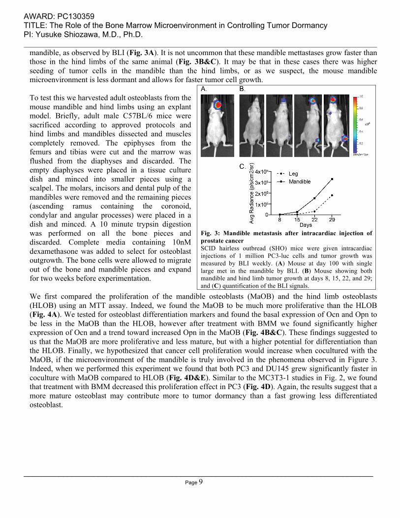
mandible, as observed by BLI (Fig. 3A). It is not uncommon that these mandible mettastases grow faster than those in the hind limbs of the same animal (Fig. 3B&C). It may be that in these cases there was higher seeding of tumor cells in the mandible than the hind limbs, or as we suspect, the mouse mandible microenvironment is less dormant and allows for faster tumor cell growth. To test this we harvested adult osteoblasts from the mouse mandible and hind limbs using an explant model. Briefly, adult male C57BL/6 mice were sacrificed according to approved protocols and hind limbs and mandibles dissected and muscles completely removed. The epiphyses from the femurs and tibias were cut and the marrow was flushed from the diaphyses and discarded. The empty diaphyses were placed in a tissue culture dish and minced into smaller pieces using a scalpel. The molars, incisors and dental pulp of the mandibles were removed and the remaining pieces (ascending ramus containing the coronoid, condylar and angular processes) were placed in a dish and minced. A 10 minute trypsin digestion was performed on all the bone pieces and discarded. Complete media containing 10nM dexamethasone was added to select for osteoblast outgrowth. The bone cells were allowed to migrate out of the bone and mandible pieces and expand for two weeks before experimentation. We first compared the proliferation of the mandible osteoblasts (MaOB) and the hind limb osteoblasts (HLOB) using an MTT assay. Indeed, we found the MaOB to be much more proliferative than the HLOB (Fig. 4A). We tested for osteoblast differentiation markers and found the basal expression of Ocn and Opn to be less in the MaOB than the HLOB, however after treatment with BMM we found significantly higher expression of Ocn and a trend toward increased Opn in the MaOB (Fig. 4B&C). These findings suggested to us that the MaOB are more proliferative and less mature, but with a higher potential for differentiation than the HLOB. Finally, we hypothesized that cancer cell proliferation would increase when cocultured with the MaOB, if the microenvironment of the mandible is truly involved in the phenomena observed in Figure 3. Indeed, when we performed this experiment we found that both PC3 and DU145 grew significantly faster in coculture with MaOB compared to HLOB (Fig. 4D&E). Similar to the MC3T3-1 studies in Fig. 2, we found that treatment with BMM decreased this proliferation effect in PC3 (Fig. 4D). Again, the results suggest that a more mature osteoblast may contribute more to tumor dormancy than a fast growing less differentiated osteoblast.
Fig. 3: Mandible metastasis after intracardiac injection of prostate cancer SCID hairless outbread (SHO) mice were given intracardiac injections of 1 million PC3-luc cells and tumor growth was measured by BLI weekly. (A) Mouse at day 100 with single large met in the mandible by BLI. (B) Mouse showing both mandible and hind limb tumor growth at days 8, 15, 22, and 29; and (C) quantification of the BLI signals.

AWARD: PC130359 TITLE: The Role of the Bone Marrow Microenvironment in Controlling Tumor Dormancy PI: Yusuke Shiozawa, M.D., Ph.D.
________________________________________________________________________________ Page 10
What opportunities for training and professional development did the project provide?
(~2016) Thanks to receiving an Idea Development Award for Young Investigators, the PI obtained independent status at Wake Forest School of Medicine with lab space, office space, and start-up costs provided. Thanks to the Department of Defense, the PI attended IMPaCT Young Investigator Meeting, Baltimore, MD, USA, August 4-5, 2016. The PI was chosen for the 2016-2018 cohort of the Wake Forest Clinical and Translational Science Institute’s Translational Scholar Academy, which supports the scientific and career development of early-stage investigators. (2016-2017) The PI was invited to attend The U.S. Bone and Joint Initiative (USBJI) and Bone and Joint Canada (BJC) Young Investigator Initiative Program will be held at Toronto, Canada on 11/10/17-11/12-17.
How were the results disseminated to communities of interest?
There is nothing to report at this time. What do you plan to do during the next reporting period to accomplish the goals and objectives? In the next year of the award, we will characterize the differences between the mandible and hind limb osteoblasts in order to determine the detailed molecular mechanisms whereby active osteoblasts influence the progression of bone metastatic prostate cancer (Aim 2). We will also further elucidate the role of osteoblastic activation in the progression of prostate cancer bone metastasis in vivo (Aim 2). In addition, we will continue to pursue the effects of the disseminated tumor cells on the dormancy of the bone marrow niche (Aim 1).
Fig. 4: Mandible osteoblasts promote prostate cancer growth Mandible (MaOB) and hind limb osteoblast (HLOB) primary cell cultures were seeded into 96-well plates and (A) relative cell numbers measured at Day 1, 3 and 5 with MTT (500µg/mL) by optical density (OD) at 560nm minus background at 650nm, normalized to Day 1. (B) Osteocalcin expression levels were tested by qPCR after 14 day treatments of BMM or Vehicle in complete media (10% FBS). (C) Osteopontin levels were also measured (data presented as fold change using the ∆∆CT method with Gapdh as reference gene). (D&E) PC3-luc and DU145-luc were sparsely seeded on top of a monolayer of MaOB or HLOB in a 96-well plate and PCa cell growth was measured by BLI and media replaced with BMM, BMP2, HC, or Vehicle on Day 1, 3, and 5. All data presented as mean ± SD, *P<0.05, **P<0.005, ***P<0.0005, ****P<0.0005 (Two-way ANOVA with Bonferroni multiple comparisons).

AWARD: PC130359 TITLE: The Role of the Bone Marrow Microenvironment in Controlling Tumor Dormancy PI: Yusuke Shiozawa, M.D., Ph.D.
________________________________________________________________________________ Page 11
4. IMPACT: What was the impact on the development of the principal discipline(s) of the project?
(~2016) We developed an innovative and powerful mouse models that enable us to measure within the same animal: (i) the growth of bone metastatic PCa and (ii) bone remodeling. (2016-2017) We developed a new and creative tool (mandible vs. hind limb osteoblast) to test the hypothesis that active osteoblasts enhance the re-growth of cancer in the bone. Using this technique, we found that a fast growing less differentiated osteoblast may contribute to growth of bone metastatic prostate cancer.
What was the impact on other disciplines?
There is nothing to report at this time. What was the impact on technology transfer?
There is nothing to report at this time. What was the impact on society beyond science and technology?
There is nothing to report at this time.

AWARD: PC130359 TITLE: The Role of the Bone Marrow Microenvironment in Controlling Tumor Dormancy PI: Yusuke Shiozawa, M.D., Ph.D.
________________________________________________________________________________ Page 12
5. CHANGES/PROBLEMS: Changes in approach and reasons for change
Nothing to report. Actual or anticipated problems or delays and actions or plans to resolve them
Nothing to report. Changes that have a significant impact on expenditures
Nothing to report. Significant changes in use or care of human subjects, vertebrate animals, biohazards, and/or select agents
Nothing to report.

AWARD: PC130359 TITLE: The Role of the Bone Marrow Microenvironment in Controlling Tumor Dormancy PI: Yusuke Shiozawa, M.D., Ph.D.
________________________________________________________________________________ Page 13
6. PRODUCTS: Publications, conference papers, and presentations
Journal Publications Peer reviewed journal
(~2016)
1. Seib FP, Berry JE, Shiozawa Y, Taichman RS, Kaplan DL. Tissue engineering a surrogate niche for metastatic cancer cells. Biomaterials. 2015;51:313-9. PMID: 25771021. PMCID: PMC4367489. Status of Publication: Published Acknowledgement of federal support: Yes
2. Zalucha JL, Jung Y, Joseph J, Wang J, Berry JE, Shiozawa Y, Taichman RS. The Role of
Osteoclasts in Early Dissemination of Prostate Cancer Tumor Cells. J Cancer Stem Cell Res. 2015;3:e1005. PMID: 26097863. PMCID: PMC4469294. Status of Publication: Published Acknowledgement of federal support: Yes
3. Shiozawa Y, Berry JE, Eber MR, Jung Y, Yumoto K, Cackowski FC, Yoon HJ, Parsana P, Mehra
R, Wang J, McGee S, Lee E, Nagrath S, Pienta KJ, Taichman RS. The Marrow Niche Controls The Cancer Stem Cell Phenotype Of Disseminated Prostate Cancer. Oncotarget. In Press. PMID: 27172799. PMCID: PMC5173053. Status of Publication: Published Acknowledgement of federal support: Yes
4. Sharma S, Xing F, Liu Y, Wu K, Said N, Pochampally R, Shiozawa Y, Lin HK, Balaji KC, Watabe K. Secreted Protein Acidic and Rich in Cysteine (SPARC) Mediates Metastatic Dormancy of Prostate Cancer in the Bone. J Biol Chem. 2016;291:19351-63. PMID: 27422817. PMCID: PCM5016675. Status of Publication: Published Acknowledgement of federal support: No (2016-2017)
5. Cackowski F, Eber MR, Rhee J, Decker A, Yumoto K, Berry JE, Lee E, Shiozawa Y, Jung Y, Aguirre-Ghiso JA, Taichman RS. Mer Tyrosine Kinase Regulates Disseminated Prostate Cancer Cellular Dormancy. J Cell Biochem. 2017;118:891-902. PMID: 27753136. PMCID: PMC5296338. Status of Publication: Published Acknowledgement of federal support: Yes

AWARD: PC130359 TITLE: The Role of the Bone Marrow Microenvironment in Controlling Tumor Dormancy PI: Yusuke Shiozawa, M.D., Ph.D.
________________________________________________________________________________ Page 14
6. Kast RE, Skuli N, Cos S, Karpel-Massler G, Shiozawa Y, Goshen R, Halatsch ME. The ABC7 regimen: a new approach to metastatic breast cancer using seven common drugs to inhibit epithelial-to-mesenchymal transition and augment capecitabine efficacy. Breast Cancer (Dove Med Press). 2017;9:495-514. PMID: 28744157. PMCID: PMC5513700 Status of Publication: Published Acknowledgement of federal support: Yes
Invited reviews
(~2016)
1. Shiozawa Y*, Eber MR, Berry JE, Taichman RS*. Bone marrow as a metastatic niche for disseminated tumor cells from solid tumors. BoneKEy Rep. 2015;4:689. PMID: 26029360. PMCID: PMC4440229. (* Co-corresponding authors) Status of Publication: Published Acknowledgement of federal support: Yes
2. Dai J, Hensel J, Wang N, Kruithof-de Julio M, Shiozawa Y. Mouse models for studying prostate cancer bone metastasis. BoneKEy Rep. 2016;5:777. PMID: 26916039. PMCID: PMC4757481. Status of Publication: Published Acknowledgement of federal support: Yes
3. Tsuzuki S, Park SH, Eber MR, Peters CM, Shiozawa Y. Skeletal complications in cancer patients
with bone metastases. Int J Urol. In Press. PMID: 27488133. PMCID: In Progress. Status of Publication: Accepted Acknowledgement of federal support: Yes • The Figure 1 is chosen as the Cover Figure of Int J Urol Vol. 23 No. 10
Book
(~2016)
1. Miler SF, Thomas CY, Shiozawa Y. (2016) Molecular involvement of the bone marrow microenvironment in bone metastasis. In Ahmad A (Ed.), Introduction to Cancer Metastasis. In Press. Philadelphia, Elsevier. Status of Publication: Accepted Acknowledgement of federal support: Yes
Presentation
(~2016)
1. Tsuzuki S, Eber MR, Miler SF, Park SH, Widner DB, Shiozawa Y. The effects of neuropeptides on the prostate cancer progression. IMPaCT Young Investigator Meeting, Baltimore, MD, USA, August 4-5, 2016. Poster.

AWARD: PC130359 TITLE: The Role of the Bone Marrow Microenvironment in Controlling Tumor Dormancy PI: Yusuke Shiozawa, M.D., Ph.D.
________________________________________________________________________________ Page 15
(2016-2017)
2. Mamo M, Park SH, Eber MR, Widner DB, Tsuzuki S, Warren CC, Strickland TB, Peters CM, Shiozawa Y. The Interaction between Disseminated Prostate Cancer Cells and Sensory Nerves. the 2016 Annual Biomedical Research Conference for Minority Students (ABRCMS), Tampa, FL, USA, November 9-12, 2016. Poster.
Website(s) or other Internet site(s)
Nothing to report. Technologies or techniques
Nothing to report. Inventions, patent applications, and/or licenses
Nothing to report. Other products
Nothing to report.

AWARD: PC130359 TITLE: The Role of the Bone Marrow Microenvironment in Controlling Tumor Dormancy PI: Yusuke Shiozawa, M.D., Ph.D.
________________________________________________________________________________ Page 16
7. PARTICIPANTS & OTHER COLLABORATING ORGANIZATIONS What individuals have worked on the project?
Name: Yusuke Shiozawa Project Role: PI Researcher Identifier (e.g. ORCID ID): orcid.org/0000-0001-9814-9230 Nearest person month worked: 2.4 Contribution to Project: Dr. Shiozawa provides oversight of the entire program and development and implementation of all policies, procedures, and processes. In this role, Dr. Shiozawa is responsible for the implementation of the specific aims, and for ensuring that systems are in place to guarantee institutional compliance with US laws, including biosafety and animal research, data and facilities. Dr. Shiozawa supervises other personnel on the project to ensure timely and effective studies. Funding Support: National Cancer Institution, Department of Defense
Has there been a change in the other active support of the PD/PI(s) or senior/key personnel since the last reporting period?
Nothing to report. What other organizations have been involved as partners?
Nothing to report. 8. SPECIAL REPORTING REQUIREMENTS:
N/A.
9. APPENDICES: The original copies of manuscript and a curriculum vitae are attached.

Mer Tyrosine Kinase Regulates Disseminated ProstateCancer Cellular DormancyFrank C. Cackowski,1,2 Matthew R. Eber,1,3 James Rhee,1 Ann M. Decker,1
Kenji Yumoto,1 Janice E. Berry,1§ Eunsohl Lee,1 Yusuke Shiozawa,3 Younghun Jung,1
Julio A. Aguirre-Ghiso,4 and Russell S. Taichman1*1Department of Periodontics and Oral Medicine, University of Michigan School of Dentistry, Ann Arbor, Michigan2Division of Hematology and Oncology, Department of Medicine, University of Michigan School of Medicine, AnnArbor, Michigan
3Department of Cancer Biology and Comprehensive Cancer Center, Wake Forest University School of Medicine,Winston-Salem, North Carolina
4Division of Hematology and Oncology, Tisch Cancer Institute, Departments of Medicine, Otolaryngology, and BlackFamily Stem Cell Institute, Icahn School of Medicine at Mount Sinai, New York, New York
ABSTRACTMany prostate cancer (PCa) recurrences are thought to be due to reactivation of disseminated tumor cells (DTCs). We previously found a role ofthe TAM family of receptor tyrosine kinases TYRO3, AXL, and MERTK in PCa dormancy regulation. However, the mechanism andcontributions of the individual TAM receptors is largely unknown. Knockdown of MERTK, but not AXL or TYRO3 by shRNA in PCa cellsinduced a decreased ratio of P-Erk1/2 to P-p38, increased expression of p27, NR2F1, SOX2, and NANOG, induced higher levels of histoneH3K9me3 and H3K27me3, and induced a G1/G0 arrest, all of which are associated with dormancy. Similar effects were also observed withsiRNA. Most importantly, knockdown of MERTK in PCa cells increased metastasis free survival in an intra-cardiac injection mouse xenograftmodel. MERTK knockdown also failed to inhibit PCa growth in vitro and subcutaneous growth in vivo, which suggests that MERTK hasspecificity for dormancy regulation or requires a signal from the PCa microenvironment. The effects of MERTK on the cell cycle and histonemethylation were reversed by p38 inhibitor SB203580, which indicates the importance of MAP kinases for MERTK dormancy regulation.Overall, this study shows that MERTK stimulates PCa dormancy escape through a MAP kinase dependent mechanism, also involving p27,pluripotency transcription factors, and histone methylation. J. Cell. Biochem. 118: 891–902, 2017. © 2016 Wiley Periodicals, Inc.
KEY WORDS: MERTK; AXL; TYRO3; PROSTATE CANCER; DORMANCY; DISSEMINATED TUMOR CELL
Prostate cancer patients often have long time periods betweencurative intent surgery or radiation therapy until the time of
biochemical recurrence or metastatic disease visible with currentimaging, which marks incurable disease with current treatmentoptions. For example, in a large series of patients treated with radicalprostatectomy, nearly 20% recurrences occurred at least 5 years aftersurgery [Amling et al., 2000]. Greater than half of prostate cancerpatients with no evidence of disease, soon after radical prostatec-tomy were found to have disseminated prostate tumor cells (DTCs) intheir bone marrow, which are thought to be a major source of distant
recurrences [Morgan et al., 2009]. This finding implies that many ofthese tumor cells die, never grow, or grow very slowly. Manyinvestigators refer to this ability of cancer cells to remain viable butnot have detectable growth as “cellular dormancy.”
There is significant interest in regulators of cancer cellulardormancy. Several studies have identified a low ratio of phosphory-lated MAPK3/MAPK1 (Erk 1/2) to phosphorylated MAPK14 (p38) asmarking dormant tumor cells. TGFB2 (TGF-b2) was proposed to bethe major ligand responsible for dormant behavior of head and necksquamous cell carcinoma cells. The cell cycle inhibitor CDKN1B
§Deceased on February 4, 2016.Grant sponsor: NIH/NCI; Grant number: PO1-CA093900; Grant sponsor: NIH/NCI TumorMicroenvironment Network;Grant number: U54-CA163124; Grant sponsor: Department of Defense; Grant numbers: W81XWH-14-1-0403,W81XWH-15-1-0637, W81XWH-15-1-0413; Grant sponsor: NIH/NCI T32; Grant number: 5T32CA009357-32;Grant sponsor: Prostate Cancer Foundation Grant (PCF); Grant number: 2016CHAL1503.*Correspondence to: Russell S. Taichman, D.M.D., D.M.Sc., University of Michigan School of Dentistry, 1011 NorthUniversity Avenue, Ann Arbor, MI 48109. E-mail: [email protected] Received: 17 October 2016; Manuscript Accepted: 17 October 2016Accepted manuscript online in Wiley Online Library (wileyonlinelibrary.com): 17 October 2016DOI 10.1002/jcb.25768 � © 2016 Wiley Periodicals, Inc.
891
ARTICLEJournal of Cellular Biochemistry 118:891–902 (2017)

(p27) and transcription factor BHLHE41 (DEC2) were implicated asnuclear signals [Bragado et al., 2013]. More recently, pluripotencyassociated transcription factors NR2F1, SOX2, SOX9, NANOG, andRARB were identified as transcriptional regulators of dormancy inhead and neck, prostate and breast cancers [Sosa et al., 2015].Similarly, others have shown that TGF-b family member BMP7maintains prostate cancer dormancy through autocrine SPARC[Kobayashi et al., 2011; Sharma et al., 2016]. A role of the epigenomein regulating cellular dormancy is also becoming apparent. HistoneH3 tri-methylated lysine 9 and tri-methylated lysine 27 were shownto identify and to be important for dormant cells, primarily in headand neck cancer [Sosa et al., 2015].
Our group has established a role of the TYRO3, AXL, and MERTK(TAM) family of receptors and one of their ligands, growth arrest-specific 6 (GAS6), in regulation of prostate cancer cell dormancy inthe bone marrow [Shiozawa et al., 2010; Jung et al., 2012; Taichmanet al., 2013]. We also found that GAS6 and MERTK are important forcancer stem like cell formation [Jung et al., 2016; Shiozawa et al.,2016]. This receptor family has an established role in the regulationof the innate immune system, but more recently has been shown tobe important for cancer growth and metastasis as well. For example,MERTK was recently identified in a screen of wild-type kinases as amediator of prostate cancer metastasis [Faltermeier et al., 2015]. TheTAM family of receptors have a high degree of homology, but havebeen shown to have different functions, which might relate todifferences in ligand binding affinities and downstream pathways[Graham and DeRyckere, 2014]. There are at least four vitamin Kdependent g-carboxyalated protein ligands that bind to at least oneof the TAM receptors including GAS6, Protein S (PROS1), Tubby(TUB), and Tubby like protein 1 (TULP1) [Caberoy, 2010]. We foundthat GAS6 decreased prostate cancer proliferation and protected thecells from chemotherapy induced apoptosis [Shiozawa et al., 2010;Lee et al., 2016]. We also found that prostate cancer bone metastasesgrew larger in the absence of GAS6 [Jung et al., 2012].
However, these studies did not identify which of the TAMreceptors are responsible for the ability of GAS6 to slow prostatecancer growth, while also preventing apoptosis—findings which areconsistent with cellular dormancy. However, the role of TYRO3 andMERTK remained unclear. To begin to answer this question, wepreviously studied the relative expression level TYRO3 and AXL inprostate cancer primary tumors, DTCs, and in gross metastases.However, MERTK was not included in these studies. We found thatTYRO3 was expressed highly in the primary tumors, but that AXLwas expressed highly in disseminated but dormant disease[Taichman et al., 2013]. Based on these results, we hypothesizedthat TYRO3 might play a role when prostate cancer was activelygrowing but that AXLmight play a role when it is dormant. However,these studies did not include experiments to test this hypothesisfurther than gene expression. Most recently, in work that is currentlyin press, we reported that AXL is required for TGF-ß2 to induceprostate cancer dormancy [Yumoto et al., 2016]. However, thecontributions of MERTK and TYRO3 remain unclear.
In the current study, we took an unbiased approach to discernwhich of the TAM receptors, including MERTK, are required forprostate cancer dormancy escape. With shRNA and siRNA technol-ogy, we knocked down the expression of each of the three receptors in
three different prostate cancer cell lines.We found that loss ofMERTK,but not the other receptors decreased the ratio of P-Erk to P-p38,increased the expression of p27 and pluripotency associatedtranscription factors, increased the levels of dormancy associatedhistone H3 and caused accumulation of cells in the G1 and G0 phasesof the cell cycle, and decreased apoptosis, all of which characterizedormant cells. Importantly, the effect of MERTK on the cell cycle andhistone H3 post-translational modifications was reversed by alteringMAPkinase signalingwith a p38 inhibitor.We also found thatMERTKknockdown increased metastasis free survival in an intra-cardiacinjection mouse prostate cancer xenograft model, but did not inhibitin vitro cell growth or subcutaneous tumor growth showing that it didnot compromise global growth characteristics. Thus, our studiesimplicate MERTK in stimulation of prostate cancer dormancy escapeby a mechanism particular to the metastatic microenvironment andinvolving MAP kinases.
MATERIALS AND METHODS
CELL CULTUREHuman PCa cell lines, PC3, Du145, and LNCaP C4-2B (C4-2B) wereobtained from American Type Culture Collection (Rockville, MD[PC3 and Du145]) and UroCor (Oklahoma City, OK [C4-2B]). PCa cellswere maintained in RPMI 1640 with 10% fetal bovine serum (FBS)and 1% penicillin/streptomycin (P/S) in a humidified incubator with5% CO2. For in vitro assays, unless indicated otherwise, cells wereseeded at a density 1� 105/ml, allowed to rest for 1 day in 10%serum and then changed to reduced serum concentrations asindicated. For the p38 inhibitor experiments, cells were first culturedfor 14 days under routine conditions with 10% serum and 5mMSB203580 (EMD Millipore #A8254) dissolved in DMSO or 0.05%DMSO control.
TAM RECEPTOR STABLE shRNA KNOCKDOWNSGFP and luciferase expressing PCa cell lines (PC3GFP, Du145GFP, andC4-2B GFP cells) were first established by lentiviral transduction.Stable knockdowns of the TAM receptors (TYRO3, AXL, andMERTK)were then generated by lentiviral infection. Lentiviruses wereconstructed by the University of Michigan Vector Core using pGIPZlentiviral vectors containing either a shRNA targeting one of theTAM receptors or a nonsilencing (shControl) shRNA (OpenBiosystems). Stable lines were selected with puromycin. Knockdownof greater than 80% was verified by Western blotting and qRT-PCR.qRT-PCR gene expression data are presented as mean� SEM ofindependent cultures.
MerTK TRANSIENT siRNA KNOCKDOWNSsiRNAs targeting MerTK (# s20474, s20473, and s20472) and controlsiRNA (siControl) (# 4390843) were purchased from Thermo-FisherScientific. Transient transfection in C4-2B and PC3 cells wasperformed using 10mM of each siRNA with LipofectamineRNAiMAX reagent (Thermo-Fisher) using the reverse transfectionprotocol, followed by 3 days incubation. Knockdownwas verified byreal time qRT-PCR. Data are presented as mean� SEM of triplicatePCR reactions.
JOURNAL OF CELLULAR BIOCHEMISTRY892 MERTK AND PROSTATE CANCER DORMANCY
WESTERN BLOTTINGCells were serum starved overnight unless indicated otherwise.Lysates were prepared in cOmplete lysis M (Roche #04 719 956 001)supplemented with proteinase inhibitor Mini cOmplete Tablets(Roche #04705378) and phosphatase inhibitor PhosSTOP EASYpackTablets (Roche #04 906 837 001). Protein concentration wasdetermined by the BCA method. Twenty micrograms of total proteinwas added per lane of 4–20% reducing SDS polyacrylamide Tris-Glycine gels after sample preparation in Laemmli sample buffer. Thesamples were transferred to PVDF membranes and blocked for 1 h in5% dry milk in TBS with 0.1% Tween-20 (TBST). Antibodies forphosphorylated proteins were applied at 4°C overnight in 5% BSATBST, washed, and visualized with a horseradish peroxidaseconjugated anti-rabbit IgG secondary antibody (Cell Signalling#7074S) and SuperSignal West Dura Chemiluminescent Substrate(Thermo Scientific #34075). Images were acquired with a ChemiDocTouch imager (BioRad). Membranes were stripped with Restore PLUSstripping buffer (Thermo Scientific #46430). They were blocked andre-probed for antibodies to total proteins, and again striped and re-probed for GAPDH or b-actin to normalize for protein loading. Allprimary antibodies were monoclonal rabbit from Cell SignalingTechnology. Catalog numbers and dilutions were as follows;Phosphorylated-Erk 1/2 (P-Erk) Y204(#4377S, diluted 1:500), totalErk (#4695, 1:500), Phospho-p38 (P-p38) T190/Y182 (#4511, 1:500),total p38, (#9212, total Axl (#4939, 1:500), total Tyro3 (#5585,1:500), total MerTK (#4319, 1:500), Sox2 (# 3579, 1:500), Caspase-9(#9502, 1:1000), b-actin (#4970, 1:2000), and GAPDH (#2118,1:2000). Images representative of biological replicates are shownand cropped for presentation. For P-Erk, P-p38, and p27quantification, images from five independent experiments werequantified relative to each vehicle treated scrambled shRNA controlwith BioRad ImageLab software and then normalized to housekeep-ing gene expression. The P-Erk to P-p38 ratio was obtained bydividing the normalized P-Erk and P-p38 values for eachindependent experiment. All data are shown as fold change fromcontrol.
REAL TIME REVERSE TRANSCRIPTASE PCR (qRT-PCR)Cells were lysed and RNAwas harvested using the Qiagen RNeasy kitfollowed by reverse transcription using Invitrogen SuperScript IIReverse Transcriptase. Real time qPCR was performed using TaqManUniversal PCRMaster Mix and Gene Expression Assays on a AppliedBiosystems ViiA 7 instrument. TaqMan MGB probes (AppliedBiosystems) were as follows: MERTK (Hs00179024_m1),p27/CDKN1B (Hs00153277), SOX2 (Hs01053049_s1), and NANOG(Hs02387400). We designed primers and a probe to specificallydetect NR2F1/TFCOUP1: forward; CAAAGCCATCGTGCTGTTCAC,reverse; CCTGCAGGCTCTCGATGT, and probe; TCA-GACGCCTGTGGCCTG. b-actin (Hs01060665_g1) was used as aninternal control for the normalization of target gene expression.
FLOW CYTOMETRY FOR HISTONE POST-TRANSLATIONALMODIFICATIONS AND Ki67Cell pellets were fixed and permeabilized with dropwise addition of1ml of cold 70% ethanol and then incubated overnight. All stepswere at 4°C or on ice. Samples were then washed, blocked, and
incubated for 1 h in flow buffer (PBS with 2% FBS and 2mM EDTA)with each of the following antibodies; Alexa 647 conjugated rabbitanti histone H3 tri-methylated lysine 27 diluted 1:50 (Cell SignalingTechnology #12158), unconjugated rabbit polyclonal anti histoneH3 tri-methylated lysine 9 (Abcam #8898), or APC conjugated rabbitanti-human Ki-67 antibody (Biolegend #350513). Cells were washedtwice with flow buffer. The unconjugated histone H3 tri-methylatedlysine 9 antibody was detected with an Alexa 647 conjugated anti-rabbit IgG diluted 1:250 (Cell Signaling Technology #4414). Datawere acquired with a three laser (405, 488, and 640 nm) BectonDickinson FACS Aria IIu flow cytometer. Gating was forward scatterversus side scatter, single cells (linear on FSC-A vs. FSC-H), then the670/30 filter (APC or Alexa 647) versus forward scatter or histogram.An isotype control antibody was used for setting the gates. Negativeand dim cells were selected for methylated histones. Negative cellswere selected for Ki67. Data are presented as representative plots ormean� SEM of triplicate wells from replicate experiments.
CELL CYCLE ANALYSISCells were cultured for 3 days as indicated and pulsed with 10mMbromodeoxyuridine (BrdU) for 30min. The cells were collected withtrypsin as necessary and then fixed and stained for total DNA with7-AAD and BrdU incorporated into DNA using the Becton DickinsonAPC BrdU flow kit (#552598). Data were acquired with a BectonDickinson FACS Aria IIu flow cytometer. Gating was forward scatterversus side scatter, single cells (linear on FSC-A vs. FSC-H), thenAPC(BrdU) versus 7-AAD (DNA).
LEFT VENTRICLE INTRACARDIAC INJECTION XENOGRAFT MODEL OFPROSTATE CANCER METASTASISStable shRNA infected PC3GFP (1� 106) or Du145GFP (2� 105)prostate cancer cells were suspended in 100ml of PBS and injectedinto male CB.17. SCID mice (6–8 weeks of age: Charles River Labs) byleft ventricle intracardiac injection. For analysis of metastasis freesurvival, bioluminescence images were acquired after injection ofluciferin twiceweekly using a PerkinElmer IVIS 2000 system.Animalsthat had a large portion of the signal in the lungs (indicative of a rightventricle injection) were removed from analysis a priori. Afterremoving mice that had a right ventricular injections or did notsurvive the procedure, the following numbers of animals wereanalyzed; PC3 shControl; 6, PC3 shMER; 7, Du145 shControl; 20, andDu145 shMER; 18. Time to metastasis formation visible bybioluminescence (or death in rare cases) was then determined fromthe images. The data were analyzed by Kaplan–Meier analysis. Foranalysis of transit to the bone marrow, different mice, five mice pergroup, were sacrificed 24h after tumor cell injection, and their pelvis,femora, and tibiae were harvested. The bones were crushed with amortar and pestle and strained to remove debris. All steps used PBSbufferwith 2%FBS unless otherwise noted. Cellswerefirst depleted ofmouse cells with aMouse Cell Depletion Kit magnetic labeling system(Miltenyi Biotec # 130-104-694) and anti-Biotin MicroBeads and anAutoMACS machine (Miltenyi Biotec). The enriched cells wereincubated with an APC-Cy7 conjugated anti-HLA-ABC antibody(BioLegend #311426) and a PerCP-Cy5.5 conjugated anti-mouselineage cocktail (CD3e, CD11b, B220, Ter-119, Ly-6G, and Ly-6C) (BDBiosciences #561317), for an hour at 4°C, washed and resuspended in
JOURNAL OF CELLULAR BIOCHEMISTRY MERTK AND PROSTATE CANCER DORMANCY 893

PBS with 2% FBS, 2mM EDTA and 0.5mg/ml DAPI. Thereafter, thepercentage of disseminated prostate cancer cells (DTCs) wasdetermined by gating on single, viable, lineage negative, HLAþ cellswith a FACSAria IIu flow cytometer. Mice injected with PBS ratherthanPCacellswereusedas anegative control forflowcytometry.Datarepresents three independent experiments.
PROSTATE CANCER SUBCUTANEOUS TUMOR MODELOne million prostate cancer cells suspended in 50ml complete mediawere mixed with an equal volume of cold collagen solution and thenslowly injected under the skin of the back of SCIDmice; fivemice pergroup. Bioluminescence images were acquired weekly. Animals weresacrificed before tumors grew to 1 cm3. All experimental procedureswere approved by the University of Michigan Committee for the Useand Care of Animals.
PROSTATE CANCER CELL VIABILITY/MTS ASSAYProstate cancer cells were seeded at 2000 cells per well in 96 wellplates and rested for 1 day in 10% FCS RPMI media. The media wassubsequently changed to the indicated serum concentrations and thecells were cultured for an additional 3 days. The total viable cellnumber was then assayed with the Cell Titer Aqueous One Solution
MTS Proliferation Assay System (Promega #G3580) by absorbanceat 490 nm. Data represent means of three independent experiments.
STATISTICAL ANALYSESThe type I error rate (a) was set to 0.05 for all analyses. Two-sample,two-tailed Student’s t-tests were used to comparemeans of two groups.One-way repeated measures analysis of variance (ANOVA) withBonferroni post-hoc testing was used for data normalized tohousekeeping genes (blots and PCR). Standard one-way ANOVAwith Tukey’s Honest Significant Difference post-hoc testing was usedfor multiple comparisons in other experiments. The Log-rank test wasused for Kaplan–Meier survival analyses. Growth curves for subcuta-neous tumors were analyzed with a mixed design (split plot) ANOVAwith repeated measures. All analyses were conducted with SPSSsoftware, except for t-tests, which were performed in Microsoft Excel.
RESULTS
MERTK KNOCKDOWN CAUSES DORMANCY ASSOCIATED CHANGESIN MAPK ACTIVITY AND p27 EXPRESSIONTo study the importance of TAM signaling on dormancy, each TAMreceptor was stably knocked down with shRNA in PC3, Du145, and
Fig. 1. TAM receptor knockdown and dormancy associated pathways in prostate cancer cells. (A) Representative Western blots of PC3 cells with each of the TAM receptorsknocked down by shRNA probed for phosphorylated or total p38 and Erk 1/2 or housekeeping proteins. (B) Quantification of the samples in panel A for P-Erk, P-p38 or the ratio ofP-Erk to P-p38 relative to the values for the scrambled shRNA control and normalized to the housekeeping proteins. (C) Representative Western blot for p27 of the PC3 cells. (D)Quantification of the data in C. All data is presented normalized to control. Error bars represent mean� SEM. �Represents P< 0.05 compared to shRNA control cells.
JOURNAL OF CELLULAR BIOCHEMISTRY894 MERTK AND PROSTATE CANCER DORMANCY
C4-2B prostate cancer cell lines. Protein expression of each receptorwas decreased by at least 80% (Fig. S1). A decreased ratio of P-Erk1/2 to P-p38 MAPK marks cellular dormancy in prostate and othercancers [Kobayashi et al., 2011; Bragado et al., 2013; Chery et al.,2014]. Therefore, we first examined P-Erk1/2 and P-p38 levels inPC3 cells with each of the TAM receptors knocked down. The ratio ofP-Erk 1/2 to P-p38 was significantly decreased in serum starvedMERTK knockdown cells, but not in TYRO3 or AXL knockdown cells(Fig. 1A and B). Similarly, the cell cycle inhibitor p27 was alsopreviously found to be a dormancy marker [Kobayashi et al., 2011;Bragado et al., 2013]. In agreement with the Erk and p38 data, wefound higher basal p27 protein expression in theMERTK knockdowncells (Fig. 1C and D). These data are consistent with a dormantphenotype in prostate cancer cells as a result of chronically reducedexpression of MERTK.
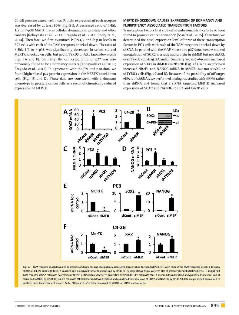
MERTK KNOCKDOWN CAUSES EXPRESSION OF DORMANCY ANDPLURIPOTENCY ASSOCIATED TRANSCRIPTION FACTORSTranscription factors first studied in embryonic stem cells have beenfound to promote cancer dormancy [Sosa et al., 2015]. Therefore, wedetermined the basal expression level of three of these transcriptionfactors in PC3 cells with each of the TAM receptors knocked down byshRNA. In parallel with theMAP kinase and p27 data, we sawmarkedupregulation of SOX2 message and protein in shMER but not shAXLor shTYRO3cells (Fig. 2AandB). Similarly,wealsoobserved increasedexpression of SOX2 in shMER C4-2B cells (Fig. 2A). We also observedincreased NR2F1 and NANOG mRNA in shMER, but not shAXL orshTYRO3 cells (Fig. 2C and D). Because of the possibility of off targeteffects of shRNAs, we performed analogous studies with siRNA ratherthan shRNA and found that a siRNA targeting MERTK increasedexpression of SOX2 and NANOG in PC3 and C4-2B cells.
Fig. 2. TAM receptor knockdown and expression of dormancy and pluripotency associated transcription factors. (A) PC3 cells with each of the TAM receptors knocked down byshRNA or C4-2B cells with MERTK knocked down, analyzed for SOX2 expression by qPCR. (B) Representative SOX2Western blot of shControl and shMER PC3 cells. (C and D) PC3TAM receptor shRNA cells with expression of NR2F1 or NANOG respectively, quantified by qPCR. (E) PC3 cells withMerTK knocked down by siRNA and quantified for expression ofSOX2 and NANOG by qPCR. (F) C4-2B cells withMERTK knocked down by siRNA and quantified for expression of SOX2 and NANOG by qPCR. All data are presented normalized tocontrol. Error bars represent mean� SEM. �Represents P< 0.05 compared to shRNA or siRNA control cells.
JOURNAL OF CELLULAR BIOCHEMISTRY MERTK AND PROSTATE CANCER DORMANCY 895

MERTK KNOCKDOWN INDUCES CELL CYCLE CHANGES ASSOCIATEDWITH CELLULAR DORMANCYCellular dormancy anddecreasedErk1/2 activity are also characterizedby arrest in the G1 andG0 phases of the cell cycle [Aguirre-Ghiso et al.,2004]. Therefore, we compared cell cycle characteristics of TAMreceptor knockdown PC3 cells cultured in 0.1% serum using flowcytometry todetect antibody labeledpulsedbromodeoxyuridine (BrdU)incorporated into DNA and 7-AAD to quantify total DNA content. Thisassay identifies BrdU positive cells as S-phase, less than 2n DNA asapoptotic or necrotic cells, 2nBrdU negative cells as G1 andG0 phases,4n BrdU negative cells as G2 andM phases and cells with>4n DNA aspolyploid (Fig. 3A). In agreement with our other data, MERTKknockdown cells showed a pattern consistent with dormancy, with ahigher percentage of cells in G0/G1 and lower percentage in G2/Mcompared to control (Fig. 3B). Curiously, TYRO3 knockdown cells
showed the opposite patternwith fewer cells inG0/G1 andmore cells inthe G2 and M phases. We did not convincingly see other analogousresults for TYRO3 in our other experiments. We did not see differencesbetween the different TAM knockdown cells in the percentage ofcells in S-phase, but note that there are very few cells in S-phase in thisstudy because of the low serum conditions. However, we did observe areduction in the sub-G0 (apoptotic and necrotic) population in theshMER cells. Further,Western blots showed decreased levels of cleavedCaspase-9 in the shMER cells, thus suggesting that this reduced sub-G0population represented reduced apoptosis. There is precedence in theliterature for correlation of reduced apoptosis with a dormantphenotype [Aguirre-Ghiso et al., 2004]. Further, p38 stimulated cellulardormancy and reduced apoptosis have been proposed to be adaptiveresponses to allow DTCs to survive when conditions are not conduciveto growth [Ranganathan et al., 2006].
Fig. 3. Cell cycle analysis of TAM receptor knockdown PC3 cells by flow cytometry with BrdU and total DNA labeling. (A) Example flow plots for each cell type. Top: histogramsof total DNA content labeled with7-AAD. Bottom: BrdU versus total DNA plots. The significance of each population is as follows:<2n DNA and BrdU negative; Sub-G0 (apoptoticand necrotic), 2n DNA and BrdU negative; G0 and G1 phases, 4n DNA and BrdU negative; G2 and M phases. BrdU positive; S-phase,>4n DNA; polyploid cells. (B) Quantificationof the above cell cycle data. The table lists P values for each cell type compared to control with significant comparisons marked with an asterisk. Error bars are shown for the G0G1and G2M populations and represent mean� SEM. �Represents P< 0.05 compared to shRNA control cells. (C) Western blots for total caspase-9 to verify the changes in Sub-G0cells observed by flow cytometry.
JOURNAL OF CELLULAR BIOCHEMISTRY896 MERTK AND PROSTATE CANCER DORMANCY
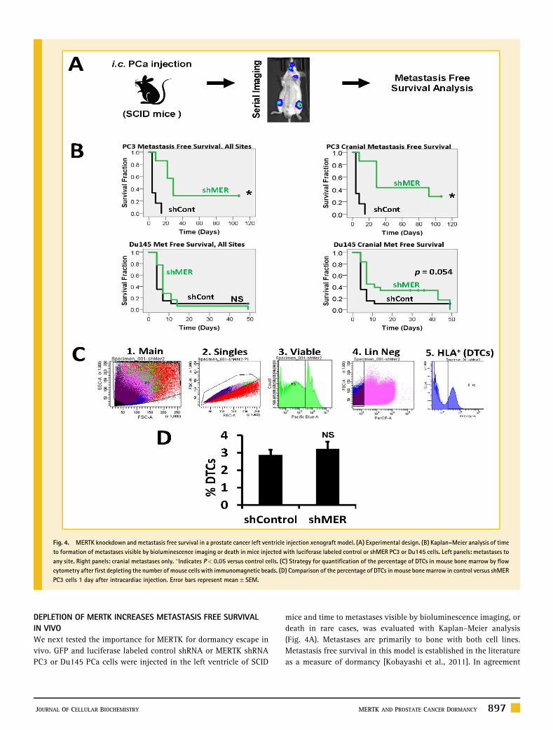
DEPLETION OF MERTK INCREASES METASTASIS FREE SURVIVALIN VIVOWe next tested the importance for MERTK for dormancy escape invivo. GFP and luciferase labeled control shRNA or MERTK shRNAPC3 or Du145 PCa cells were injected in the left ventricle of SCID
mice and time to metastases visible by bioluminescence imaging, ordeath in rare cases, was evaluated with Kaplan–Meier analysis(Fig. 4A). Metastases are primarily to bone with both cell lines.Metastasis free survival in this model is established in the literatureas a measure of dormancy [Kobayashi et al., 2011]. In agreement
Fig. 4. MERTK knockdown and metastasis free survival in a prostate cancer left ventricle injection xenograft model. (A) Experimental design. (B) Kaplan–Meier analysis of timeto formation of metastases visible by bioluminescence imaging or death in mice injected with luciferase labeled control or shMER PC3 or Du145 cells. Left panels: metastases toany site. Right panels: cranial metastases only. �Indicates P< 0.05 versus control cells. (C) Strategy for quantification of the percentage of DTCs in mouse bone marrow by flowcytometry after first depleting the number of mouse cells with immunomagnetic beads. (D) Comparison of the percentage of DTCs in mouse bone marrow in control versus shMERPC3 cells 1 day after intracardiac injection. Error bars represent mean� SEM.
JOURNAL OF CELLULAR BIOCHEMISTRY MERTK AND PROSTATE CANCER DORMANCY 897
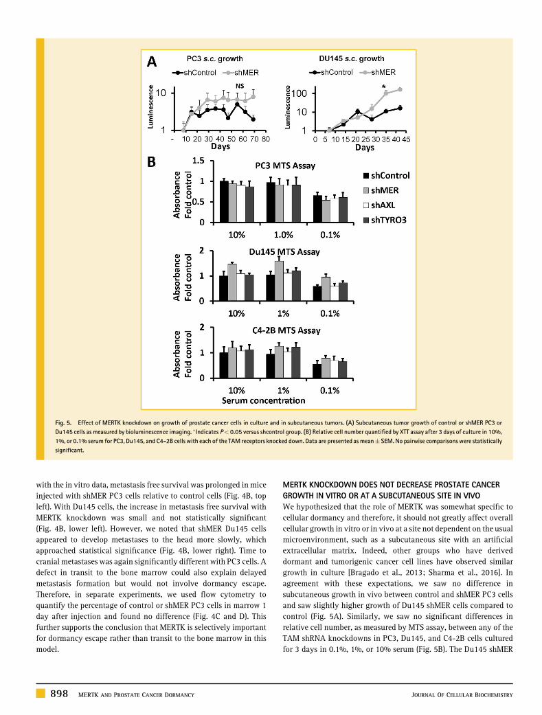
with the in vitro data, metastasis free survival was prolonged in miceinjected with shMER PC3 cells relative to control cells (Fig. 4B, topleft). With Du145 cells, the increase in metastasis free survival withMERTK knockdown was small and not statistically significant(Fig. 4B, lower left). However, we noted that shMER Du145 cellsappeared to develop metastases to the head more slowly, whichapproached statistical significance (Fig. 4B, lower right). Time tocranial metastases was again significantly different with PC3 cells. Adefect in transit to the bone marrow could also explain delayedmetastasis formation but would not involve dormancy escape.Therefore, in separate experiments, we used flow cytometry toquantify the percentage of control or shMER PC3 cells in marrow 1day after injection and found no difference (Fig. 4C and D). Thisfurther supports the conclusion that MERTK is selectively importantfor dormancy escape rather than transit to the bone marrow in thismodel.
MERTK KNOCKDOWN DOES NOT DECREASE PROSTATE CANCERGROWTH IN VITRO OR AT A SUBCUTANEOUS SITE IN VIVOWe hypothesized that the role of MERTK was somewhat specific tocellular dormancy and therefore, it should not greatly affect overallcellular growth in vitro or in vivo at a site not dependent on the usualmicroenvironment, such as a subcutaneous site with an artificialextracellular matrix. Indeed, other groups who have deriveddormant and tumorigenic cancer cell lines have observed similargrowth in culture [Bragado et al., 2013; Sharma et al., 2016]. Inagreement with these expectations, we saw no difference insubcutaneous growth in vivo between control and shMER PC3 cellsand saw slightly higher growth of Du145 shMER cells compared tocontrol (Fig. 5A). Similarly, we saw no significant differences inrelative cell number, as measured by MTS assay, between any of theTAM shRNA knockdowns in PC3, Du145, and C4-2B cells culturedfor 3 days in 0.1%, 1%, or 10% serum (Fig. 5B). The Du145 shMER
Fig. 5. Effect of MERTK knockdown on growth of prostate cancer cells in culture and in subcutaneous tumors. (A) Subcutaneous tumor growth of control or shMER PC3 orDu145 cells as measured by bioluminescence imaging. �Indicates P< 0.05 versus shcontrol group. (B) Relative cell number quantified by XTT assay after 3 days of culture in 10%,1%, or 0.1% serum for PC3, Du145, and C4-2B cells with each of the TAM receptors knocked down. Data are presented as mean� SEM. No pairwise comparisons were statisticallysignificant.
JOURNAL OF CELLULAR BIOCHEMISTRY898 MERTK AND PROSTATE CANCER DORMANCY

cells trended toward higher relative cell number but did not reachstatistical significance after multiple comparison testing.
CELL CYCLE CHANGES OF MERTK KNOCKDOWN ARE MAP KINASEDEPENDENTBecause of the well-established role of MAP kinases in regulation ofcancer cellular dormancy, we investigated if p38 was required for thedormancy associated cell cycle changes induced by knockdown ofMERTK. In appreciation of the known role of epigenetic changes indormancy regulation and the timeperiod required for these changes tooccur, we cultured control and MERTK shRNA PC3 and Du145 cellswith p38 inhibitor SB203580 or 0.05% DMSO solvent control for 2weeks before performing experiments [Sosa et al., 2015]. The expectedcompensatory increase in P-p38 in response to p38 active siteinhibition was observed by Western blot (Fig. S2A). We againobserved an increased percentage of G0/G1 and decreased percentage
of G2/M cells with MERTK knockdown both in PC3 and Du145 cells(Fig. 6). This change induced by MERTK knockdown was completelyreversed by p38 inhibition, thus showing involvement of MAPkinases. Similarly, we also observed a increased percentage of Ki67negative cells (non-cycling) with MERTK knockdown in PC3 cells,whichwas also reversedbyp38 inhibition (Fig. S2C). Curiously,wedidnot observe reversal of shMER induced p27 upregulation with p38inhibition (Fig. S2B). This suggests that not all of the effects ofMERTKknockdown are MAP kinase dependent.
MERTK KNOCKDOWN INDUCES DORMANCY ASSOCIATED CHANGESIN HISTONE H3 METHYLATION BY A MAP KINASE DEPENDENTMECHANISMLastly, we used flow cytometry with specific antibodies to determinethe effect of shRNA knockdown in PC3 cells of each of the TAMreceptors on histone H3 tri-methylated lysine 9 (H3 K9 me3) and
Fig. 6. Reversal of the shMER cell cycle phenotype with long term p38 inhibition. (A) Control or shMER PC3 cells cultured with 5mMSB203580 or 0.05%DMSO solvent controlfor 14 days in 10% serum and 3 days in 0.1% serum followed by cell cycle analysis with total DNA and BrdU labeling as described for Figure 3. Top: Total DNA flow cytometryhistograms. Bottom: Quantified data. Error bars are shown for the G0G1 and G2M populations and represent mean� SEM. �Represents P< 0.05 for comparisons of interest.(B) Cell cycle analysis as above of Du145 cells grown for 17 days in 10% serum with or without SB203580.
JOURNAL OF CELLULAR BIOCHEMISTRY MERTK AND PROSTATE CANCER DORMANCY 899

histone H3 tri-methylated lysine 27 (H3 K27 me3). Both of thesehistone marks are increased in dormant cells [Sosa et al., 2015].Because the majority of cells were positive, we gated on the negativeand dim populations rather than the positive population (Fig. 7A).We observed the expected dormancy associated change in H3K9me3, and a trend toward significance for H3 K27me3 with MERTKknockdown but no significant differences for AXL or TYRO3knockdown (Fig. 7B). In cells treated with the p38 inhibitorSB203580 or solvent control, we saw significant dormancyassociated changes with MERTK knockdown, which were partiallyreversed by p38 inhibition (Fig. 7C).
DISCUSSION
Overall, these studies implicate MERTK in prostate cancer dormancyescape through a MAP kinase dependent mechanism linked totranscriptional and epigenetic regulation. Knockdown of MERTKconsistently induced the changes expected for dormant cells; adecreased ratio of P-Erk to P-p38, increased p27 expression,
expression of dormancy, and pluripotency associated transcriptionfactors, and G0/G1 arrest. Further, these findings translated to anincreased metastasis free survival in vivo. This identifies MERTK asbeing important for the process whereby one or a few cancer cellsprogress to a small tumor (i.e., escape from cellular dormancy). Asexpected, MERTK knockdown did not inhibit growth in culture orgrowth of subcutaneous tumors implanted in an artificial matrix.This lack of a general growth inhibitory effect of MERTK knockdownsuggests specificity for dormancy regulation in bone and arequirement for a signal from the microenvironment rather thandysregulation of multiple cellular processes.
Our data do not identify which signal(s) from the microenviron-ment interact with MERTK to regulate dormancy. The four otherTAM receptor ligands, other than GAS6 (Tubby, Tubby like protein 1,Galectin 2, and Protein S), may also play a role. Although not aMERTK ligand, retinoic acid may be indirectly involved as wellbecause it has been shown to stimulate cancer dormancy throughNR2F1 and also interacts indirectly with MERTK in immune cells[Garabuczi, 2015; Sosa et al., 2015]. Because no recombinant proteinor other treatment other than low serum conditions was required for
Fig. 7. Presence and MAP kinase dependence of dormancy associated histone H3 post-translational modifications in TAM receptor knockdown prostate cancer cells.(A) Example plots for percentage of cells negative or dim for histone H3 tri-methylated lysine 9 and tri-methylated lysine 27 evaluated byflow cytometry. (B) Percent of PC3 cellswith each TAM receptor knocked down by shRNA negative for each histone H3 tri-methylation. (C) Control or shMER PC3 cells cultured for 14 days with or without p38 inhibitorSB203580 (as in Fig. 6) negative or dim for tri-methylated histone H3 lysine 9 or lysine 27. Error bars represent mean� SEM. �Represents P< 0.05 compared to control for panelB, or for comparisons of interest for panel C.
JOURNAL OF CELLULAR BIOCHEMISTRY900 MERTK AND PROSTATE CANCER DORMANCY
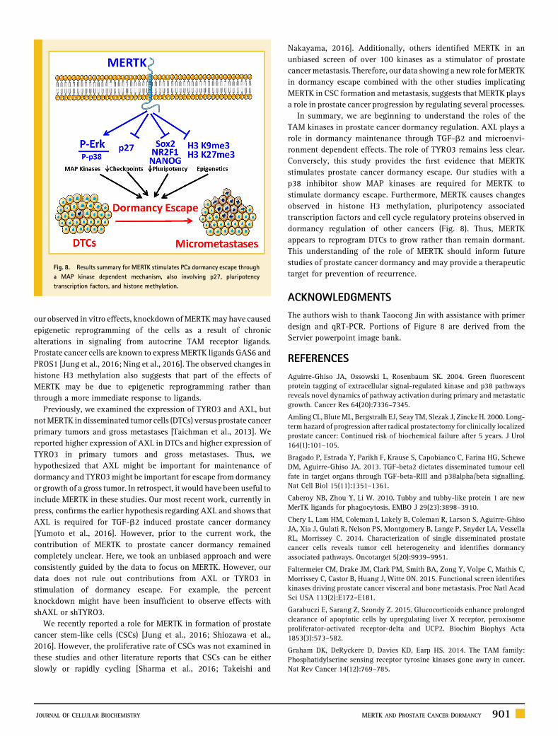
our observed in vitro effects, knockdown ofMERTKmay have causedepigenetic reprogramming of the cells as a result of chronicalterations in signaling from autocrine TAM receptor ligands.Prostate cancer cells are known to express MERTK ligands GAS6 andPROS1 [Jung et al., 2016; Ning et al., 2016]. The observed changes inhistone H3 methylation also suggests that part of the effects ofMERTK may be due to epigenetic reprogramming rather thanthrough a more immediate response to ligands.
Previously, we examined the expression of TYRO3 and AXL, butnotMERTK in disseminated tumor cells (DTCs) versus prostate cancerprimary tumors and gross metastases [Taichman et al., 2013]. Wereported higher expression of AXL in DTCs and higher expression ofTYRO3 in primary tumors and gross metastases. Thus, wehypothesized that AXL might be important for maintenance ofdormancy and TYRO3might be important for escape from dormancyor growth of a gross tumor. In retrospect, it would have been useful toinclude MERTK in these studies. Our most recent work, currently inpress, confirms the earlier hypothesis regarding AXL and shows thatAXL is required for TGF-b2 induced prostate cancer dormancy[Yumoto et al., 2016]. However, prior to the current work, thecontribution of MERTK to prostate cancer dormancy remainedcompletely unclear. Here, we took an unbiased approach and wereconsistently guided by the data to focus on MERTK. However, ourdata does not rule out contributions from AXL or TYRO3 instimulation of dormancy escape. For example, the percentknockdown might have been insufficient to observe effects withshAXL or shTYRO3.
We recently reported a role for MERTK in formation of prostatecancer stem-like cells (CSCs) [Jung et al., 2016; Shiozawa et al.,2016]. However, the proliferative rate of CSCs was not examined inthese studies and other literature reports that CSCs can be eitherslowly or rapidly cycling [Sharma et al., 2016; Takeishi and
Nakayama, 2016]. Additionally, others identified MERTK in anunbiased screen of over 100 kinases as a stimulator of prostatecancermetastasis. Therefore, our data showing a new role forMERTKin dormancy escape combined with the other studies implicatingMERTK in CSC formation andmetastasis, suggests that MERTK playsa role in prostate cancer progression by regulating several processes.
In summary, we are beginning to understand the roles of theTAM kinases in prostate cancer dormancy regulation. AXL plays arole in dormancy maintenance through TGF-b2 and microenvi-ronment dependent effects. The role of TYRO3 remains less clear.Conversely, this study provides the first evidence that MERTKstimulates prostate cancer dormancy escape. Our studies with ap38 inhibitor show MAP kinases are required for MERTK tostimulate dormancy escape. Furthermore, MERTK causes changesobserved in histone H3 methylation, pluripotency associatedtranscription factors and cell cycle regulatory proteins observed indormancy regulation of other cancers (Fig. 8). Thus, MERTKappears to reprogram DTCs to grow rather than remain dormant.This understanding of the role of MERTK should inform futurestudies of prostate cancer dormancy and may provide a therapeutictarget for prevention of recurrence.
ACKNOWLEDGMENTSThe authors wish to thank Taocong Jin with assistance with primerdesign and qRT-PCR. Portions of Figure 8 are derived from theServier powerpoint image bank.
REFERENCESAguirre-Ghiso JA, Ossowski L, Rosenbaum SK. 2004. Green fluorescentprotein tagging of extracellular signal-regulated kinase and p38 pathwaysreveals novel dynamics of pathway activation during primary and metastaticgrowth. Cancer Res 64(20):7336–7345.
Amling CL, BluteML, Bergstralh EJ, Seay TM, Slezak J, Zincke H. 2000. Long-term hazard of progression after radical prostatectomy for clinically localizedprostate cancer: Continued risk of biochemical failure after 5 years. J Urol164(1):101–105.
Bragado P, Estrada Y, Parikh F, Krause S, Capobianco C, Farina HG, ScheweDM, Aguirre-Ghiso JA. 2013. TGF-beta2 dictates disseminated tumour cellfate in target organs through TGF-beta-RIII and p38alpha/beta signalling.Nat Cell Biol 15(11):1351–1361.
Caberoy NB, Zhou Y, Li W. 2010. Tubby and tubby-like protein 1 are newMerTK ligands for phagocytosis. EMBO J 29(23):3898–3910.
Chery L, Lam HM, Coleman I, Lakely B, Coleman R, Larson S, Aguirre-GhisoJA, Xia J, Gulati R, Nelson PS, Montgomery B, Lange P, Snyder LA, VessellaRL, Morrissey C. 2014. Characterization of single disseminated prostatecancer cells reveals tumor cell heterogeneity and identifies dormancyassociated pathways. Oncotarget 5(20):9939–9951.
Faltermeier CM, Drake JM, Clark PM, Smith BA, Zong Y, Volpe C, Mathis C,Morrissey C, Castor B, Huang J, Witte ON. 2015. Functional screen identifieskinases driving prostate cancer visceral and bone metastasis. Proc Natl AcadSci USA 113(2):E172–E181.
Garabuczi E, Sarang Z, Szondy Z. 2015. Glucocorticoids enhance prolongedclearance of apoptotic cells by upregulating liver X receptor, peroxisomeproliferator-activated receptor-delta and UCP2. Biochim Biophys Acta1853(3):573–582.
Graham DK, DeRyckere D, Davies KD, Earp HS. 2014. The TAM family:Phosphatidylserine sensing receptor tyrosine kinases gone awry in cancer.Nat Rev Cancer 14(12):769–785.
Fig. 8. Results summary for MERTK stimulates PCa dormancy escape througha MAP kinase dependent mechanism, also involving p27, pluripotencytranscription factors, and histone methylation.
JOURNAL OF CELLULAR BIOCHEMISTRY MERTK AND PROSTATE CANCER DORMANCY 901

Jung Y, Decker AM, Wang J, Lee E, Kana LA, Yumoto K, Cackowski FC, RheeJ, Carmeliet P, Buttitta L, Morgan TM, Taichman RS. 2016. Endogenous GAS6and Mer receptor signaling regulate prostate cancer stem cells in bonemarrow. Oncotarget 7(18):25698–25711.
Jung Y, Shiozawa Y, Wang J, McGregor N, Dai J, Park SI, Berry JE, HavensAM, Joseph J, Kim JK, Patel L, Carmeliet P, Daignault S, Keller ET,McCauley LK, Pienta KJ, Taichman RS. 2012. Prevalence of prostate cancermetastases after intravenous inoculation provides clues into the molecularbasis of dormancy in the bone marrow microenvironment. Neoplasia14(5):429–439.
Kobayashi A, OkudaH, Xing F, Pandey PR,WatabeM, Hirota S, Pai SK, LiuW,Fukuda K, Chambers C, Wilber A, Watabe K. 2011. Bone morphogeneticprotein 7 in dormancy and metastasis of prostate cancer stem-like cells inbone. J Exp Med 208(13):2641–2655.
Lee E, Decker AM, Cackowski FC, Kana LA, Yumoto K, Jung Y, Wang J,Buttitta L, Morgan TM, Taichman RS. 2016. Growth arrest-specific 6 (GAS6)promotes prostate cancer survival by G1 Arrest/S phase delay and inhibitionof apoptotic pathway during chemotherapy in bone marrow. J Cell Biochem117(12):2815–2824.
Morgan TM, Lange PH, Porter MP, Lin DW, Ellis WJ, Gallaher IS, Vessella RL.2009. Disseminated tumor cells in prostate cancer patients after radicalprostatectomy and without evidence of disease predicts biochemicalrecurrence. Clin Cancer Res 15(2):677–683.
Ning P, Zhong JG, Jiang F, Zhang Y, Zhao J, Tian F, Li W. 2016. Role ofprotein S in castration-resistant prostate cancer-like cells. Endocr RelatCancer 23(8):595–607.
Ranganathan AC, Adam AP, Zhang L, Aguirre-Ghiso JA. 2006. Tumor celldormancy induced by p38SAPK and ER-stress signaling: An adaptiveadvantage for metastatic cells? Cancer Biol Ther 5(7):729–735.
Sharma S, Xing F, Liu Y, Wu K, Said N, Pochampally R, Shiozawa Y, Lin HK,Balaji KC, Watabe K. 2016. Secreted protein acidic and rich in cysteine
(SPARC) mediates metastatic dormancy of prostate cancer in the bone. J BiolChem 291(37):19351–19363.
Shiozawa Y, Berry JE, Eber MR, Jung Y, Yumoto K, Cackowski FC, Yoon HJ,Parsana P, Mehra R, Wang J, McGee S, Lee E, Nagrath S, Pienta KJ, TaichmanRS. 2016. The marrow niche controls the cancer stem cell phenotype ofdisseminated prostate cancer. Oncotarget. doi: 10.18632/oncotarget.9251[Epub ahead of print].
Shiozawa Y, Pedersen EA, Patel LR, Ziegler AM, Havens AM, Jung Y,Wang J,Zalucha S, Loberg RD, Pienta KJ, Taichman RS. 2010. GAS6/AXL axisregulates prostate cancer invasion, proliferation, and survival in the bonemarrow niche. Neoplasia 12(2):116–127.
Sosa MS, Parikh F, Maia AG, Estrada Y, Bosch A, Bragado P, Ekpin E, GeorgeA, Zheng Y, LamHM,Morrissey C, Chung CY, Farias EF, Bernstein E, Aguirre-Ghiso JA. 2015. NR2F1 controls tumour cell dormancy via SOX9- andRARbeta-driven quiescence programmes. Nat Commun 6:6170.
Taichman RS, Patel LR, Bedenis R, Wang J, Weidner S, Schumann T,Yumoto K, Berry JE, Shiozawa Y, Pienta KJ. 2013. GAS6 receptor status isassociated with dormancy and bone metastatic tumor formation. PLoS ONE8(4):e61873.
Takeishi S, Nakayama KI. 2016. To wake up cancer stem cells, or to let themsleep, that is the question. Cancer Sci 107(7):875–881.
Yumoto K, Eber M, Wang J, Cackowski F, Lee E, Nobre AR, Aguirre-Ghiso J,Jung Y, Taichman R. 2016. Axl is required for TGF-b2-induced dormancy ofprostate cancer cells in the bone marrow. Sci Rep 6:36520. doi: 10.1038./srep36520
SUPPORTING INFORMATION
Additional supporting information may be found in the onlineversion of this article at the publisher’s web-site.
JOURNAL OF CELLULAR BIOCHEMISTRY902 MERTK AND PROSTATE CANCER DORMANCY
© 2017 Kast et al. This work is published and licensed by Dove Medical Press Limited. The full terms of this license are available at https://www.dovepress.com/terms. php and incorporate the Creative Commons Attribution – Non Commercial (unported, v3.0) License (http://creativecommons.org/licenses/by-nc/3.0/). By accessing the work
you hereby accept the Terms. Non-commercial uses of the work are permitted without any further permission from Dove Medical Press Limited, provided the work is properly attributed. For permission for commercial use of this work, please see paragraphs 4.2 and 5 of our Terms (https://www.dovepress.com/terms.php).
Breast Cancer - Targets and Therapy 2017:9 495–514
Breast Cancer - Targets and Therapy Dovepress
submit your manuscript | www.dovepress.com
Dovepress 495
P E R S P E C T I V E S
open access to scientific and medical research
Open Access Full Text Article
http://dx.doi.org/10.2147/BCTT.S139963
The ABC7 regimen: a new approach to metastatic breast cancer using seven common drugs to inhibit epithelial-to-mesenchymal transition and augment capecitabine efficacy
Richard E Kast1 Nicolas Skuli2 Samuel Cos3 Georg Karpel-Massler4 Yusuke Shiozawa5 Ran Goshen6 Marc-Eric Halatsch4
1IIAIGC Study Center, Burlington, VT, USA; 2INSERM, Centre de Recherches en Cancérologie de Toulouse – CRCT, UMR1037 Inserm/Université Toulouse III – Paul Sabatier, Toulouse, France; 3Department of Physiology and Pharmacology, School of Medicine, University of Cantabria and Valdecilla Research Institute (IDIVAL), Santander, Spain; 4Department of Neurosurgery, Ulm University Hospital, Ulm, Germany; 5Department of Cancer Biology, Comprehensive Cancer Center, Wake Forest School of Medicine, Winston-Salem, NC, USA; 6Eliaso Consulting Ltd., Tel Aviv-Yafo, Israel
Abstract: Breast cancer metastatic to bone has a poor prognosis despite recent advances in
our understanding of the biology of both bone and breast cancer. This article presents a new
approach, the ABC7 regimen (Adjuvant for Breast Cancer treatment using seven repurposed
drugs), to metastatic breast cancer. ABC7 aims to defeat aspects of epithelial-to-mesenchymal
transition (EMT) that lead to dissemination of breast cancer to bone. As add-on to current
standard treatment with capecitabine, ABC7 uses ancillary attributes of seven already-marketed
noncancer treatment drugs to stop both the natural EMT process inherent to breast cancer and
the added EMT occurring as a response to current treatment modalities. Chemotherapy, radia-
tion, and surgery provoke EMT in cancer generally and in breast cancer specifically. ABC7 uses
standard doses of capecitabine as used in treating breast cancer today. In addition, ABC7 uses
1) an older psychiatric drug, quetiapine, to block RANK signaling; 2) pirfenidone, an anti-fibrosis
drug to block TGF-beta signaling; 3) rifabutin, an antibiotic to block beta-catenin signaling;
4) metformin, a first-line antidiabetic drug to stimulate AMPK and inhibit mammalian target
of rapamycin, (mTOR); 5) propranolol, a beta-blocker to block beta-adrenergic signaling; 6)
agomelatine, a melatonergic antidepressant to stimulate M1 and M2 melatonergic receptors; and
7) ribavirin, an antiviral drug to prevent eIF4E phosphorylation. All these block the signaling
pathways – RANK, TGF-beta, mTOR, beta-adrenergic receptors, and phosphorylated eIF4E –
that have been shown to trigger EMT and enhance breast cancer growth and so are worthwhile
targets to inhibit. Agonism at MT1 and MT2 melatonergic receptors has been shown to inhibit
both breast cancer EMT and growth. This ensemble was designed to be safe and augment
capecitabine efficacy. Given the expected outcome of metastatic breast cancer as it stands today,
ABC7 warrants a cautious trial.
Keywords: ABC7, breast cancer, agomelatine, capecitabine, metformin, pirfenidone, proprano-
lol, quetiapine, repurposing, ribavirin, rifabutin, TGF-beta
Plain language summaryThis article presents the rationale and thinking behind the ABC7 regimen for metastatic breast
cancer. Since there is currently no cure for breast cancer once it has spread to bone and other
organs beyond the breast itself, the ABC7 regimen was designed to take advantage of ancillary
attributes of seven common and readily available noncancer treatment drugs that, in theory,
should make current traditional cytotoxic chemotherapy with capecitabine more effective. The
ABC7 regimen has not been shown to be safe or effective yet. In the current article, we discuss
an untested proposal for a new treatment approach to metastatic breast cancer.
Correspondence: Richard E Kast IIAIGC Study Center, 22 Church Street, Burlington, VT 05401, USA Tel +1 802 557 7278 Email [email protected]
Journal name: Breast Cancer - Targets and TherapyArticle Designation: PERSPECTIVESYear: 2017Volume: 9Running head verso: Kast et alRunning head recto: The ABC7 regimenDOI: http://dx.doi.org/10.2147/BCTT.S139963
B
reas
t Can
cer:
Tar
gets
and
The
rapy
dow
nloa
ded
from
http
s://w
ww
.dov
epre
ss.c
om/ b
y 15
2.11
.97.
88 o
n 27
-Sep
-201
7F
or p
erso
nal u
se o
nly.
Powered by TCPDF (www.tcpdf.org)
1 / 1
Breast Cancer - Targets and Therapy 2017:9submit your manuscript | www.dovepress.com
Dovepress
Dovepress
496
Kast et al
IntroductionEstrogen-positive metastatic breast cancer cannot be cured
currently.1 One major metastatic site of breast cancer is
bone. Once breast cancer metastasizes to bone, the survival
rate declines despite recent advances in local treatments of
breast cancer. Current treatment strategies for bone metas-
tasis, including bone-targeted agents (bisphosphonate and
denosumab), provide only palliation. New and effective
therapeutic strategies for this still incurable disease are
therefore urgently needed.
This article reviews the attributes of seven currently mar-
keted drugs that, as indicated by prior research data, will block
or partially block the escape pathways from current traditional
treatments. The seven drugs of ABC7 were chosen by first
identifying the basic pathways by which EMT is initiated and
maintained. We then reviewed 1000 of the most commonly used
drugs2 for which we have both usual plasma levels and pub-
lished data showing potential inhibitory interaction with these
pathways. The resulting list was reduced by semi- subjective
evaluation of the strength of data on their EMT inhibition
benefit versus the drugs’ expected tolerability. The better the
tolerability, the weaker the data had to be for inclusion.
This ABC7 regimen is designed to block several core
breast cancer growth signals in a coordinated manner, thereby
augmenting the cytotoxicity of a currently used cytotoxic
chemotherapy drug, capecitabine. Figure 1 shows an over-
view schematic of the biochemistry that ABC7 is designed
to influence. This is explained in detail in the respective drug
discussions in the “Drugs to inhibit EMT” section. Table 1
gives an overview of the ABC7 drugs and their intended
targets in treating breast cancer.
The ABC7 regimen follows the approach of previous
cancer treatment regimens, for example, MTZ regimen,3
COMBAT regimen,4 MEMMAT regimen, a current trial of
Peyrl et al’s seven-drug cocktail (ClinicalTrials.gov Identifier:
NCT01356290), and CUSP9 regimen.5,6 In all of the studies,
extensive use is made of drugs not primarily marketed to treat
cancer but that have ancillary attributes that research data
indicate would enhance the anticancer effect of a cytotoxic,
traditional cancer treatment drug. The ancillary drugs exert
anticancer effects by blocking various growth-enhancing sur-
vival pathways used by the target cancer or as for agomelatine
are agonists at growth-retarding receptors.
Similar to other cancers, breast cancer has heteroge-
neous regions within the same tumor – different areas that
depend on or use different growth-signaling pathways. This
is related to but distinct from the idea of clonal evolution
driven by cytotoxic chemotherapy selection. Both forms of
heterogeneity exist in a typical breast cancer, proteomic and
genomic. ABC7 aims to inhibit breast cancer by pharmaco-
logical manipulation of what genes are expressed and what
genes are not, as well as by targeting different clonal variants
of the original breast cancer clone.
Because of these limitations, we do not expect testing
for molecular markers to be predictive or useful. In addition,
further intensifying the spatial and temporal diversity of
the molecular status, particularly for EMT markers, are the
diversity-driving effects of chemotherapy7 and discussed in
greater detail in the following sections.
Cytotoxic chemotherapy also induces important receptor
status changes in a large minority of breast cancer cases.8,9
Typical findings are as follows: 13% changed from HR+ to
HR−, 5% changed from HR− to HR+, 6% changed from
HER2+ to HER2, 3% changed from HER2 to HER2+, and
13% changed to triple negative.8
Multiple signaling systems have been identified that
drive metastatic breast cancer.1,9–11 These growth-driving
receptors can cross cover for each other.1,9–11 When one is
pharmacologically blocked, several parallel growth-driving
pathways can become active, taking the place of the blocked
pathways. Growth factor signaling converges from a wide
variety of outer membrane receptors to more restricted, fewer,
intracellular pathways. This is, more elegantly stated, the
spatial–temporal genomic and proteomic range, the “genetic
collectives [that] dominate the landscape of advanced-stage
(malignant) disease.”11,12 We see this as mandating an inte-
grated, coordinated polypharmacy to successfully address
these malignancy attributes.
Capecitabine is intracellularly metabolized to 5-FU;
the details are given in the “Capecitabine: 359 Da, half-life
<1 hour” section. ABC7 drugs are designed to make 5-FU
more effective.
The results of several recent ER+ metastatic breast cancer
studies are listed in Table 2. These studies cannot be judged
simply by overall survival in that entry requirements were
different, with different kinds and number of prior treat-
ments. These numbers in Table 2 are for general idea only.1
One cannot conclude that one of these is better than another.
In general, post-progression survival durations in recent
Phase III studies of combination therapy ranged from
approximately 16 to 33 months.1
EMT is a feature of cancers generally13 and breast cancer
specifically.14,15
Table 3 lists several features and behaviors associated
with the two (epithelial and mesenchymal) states. Interest-
ingly, a transcription factor ZEB1, known to control EMT,
B
reas
t Can
cer:
Tar
gets
and
The
rapy
dow
nloa
ded
from
http
s://w
ww
.dov
epre
ss.c
om/ b
y 15
2.11
.97.
88 o
n 27
-Sep
-201
7F
or p
erso
nal u
se o
nly.
Powered by TCPDF (www.tcpdf.org)
1 / 1

Breast Cancer - Targets and Therapy 2017:9 submit your manuscript | www.dovepress.com
Dovepress
Dovepress
497
The ABC7 regimen
contributes to breast cancer osteolytic bone metastasis, but
not brain or lung metastasis.16,17
EMT is a phase transition, where flat, sessile, mutually
adherent epithelioid cells take on a rounded, non-adherent,
motile mesenchymal shape and behavior.18–20 The reverseless
transient state and process, MET, also occurs and is also a
feature of robust or aggressive cancer growth.21 Post-EMT
cells tend to be invasive but proliferation restricted. Post-MET
cells tend to be proliferative but have limited invasiveness.18–20
Breast cancers develop in proximity to adipose tissue.
Adipocytes are a further trigger to EMT.22 The relationship
between stem cell subpopulations within a cancer and EMT
Figure 1 The basic biochemical, intracellular, and receptor pathways relating to interventions of the ABC7 regimen for advanced breast cancer.Notes: This is the basic breast cancer intracellular circuitry that the ABC7 regimen attempts to address. The two major controllers or stimuli for eIF4E activation are indicated by yellow arrows, the p-mTOR and the MNK paths.
Metformin AMP ATP
LKB1
elF4E 4E-BP
elF4E
elF4ERifabutin
Beta-catenin
Beta-catenin Active, to nucleus
NFkBAKT
mTORmTOR
AMPK
AMPK TSC2
Decreased
Increased
Phosphorylated
mRNAtranscription
Inactive, degraded in proteasome
Surfacereceptors
AKT
PI3K
4E-BPelF4E
Ribavirin
Propranolol
Melatonin
QuetiapinePirfenidone
RANK/RANKL
MNK
TGF-beta
Beta-AR
p38 p38
ERK
ERK
Table 1 The drugs of ABC7, their targets during treatment of breast cancer, and suggested doses
Drug Target in breast cancer treatment Starting dose Target dose
Capecitabine DNA synthesis 600 mg/m2 twice daily.7 days on, 7 days off
1250 mg/m2 twice daily. 7 days on, 7 days off or 14 days on, 7 off
Quetiapine RANK/RANKL 50 mg once at bedtime 300–600 mg once at bedtimePirfenidone TGF-beta 200 mg three times daily 600 mg three times dailyRifabutin BCL-6; beta-catenin 150 mg/day 300 mg/dayMetformin AMPK*; mTOR; mitochondria oxphos 500 mg once daily 1000 mg twice dailyPropranolol Beta-adrenergic receptors 10 mg twice daily Uptitrate as toleratedAgomelatine Melatonergic receptors* 25 mg once at bedtime 50 mg once at bedtimeRibavirin** eIF4E; MNK; IMPDH 600 mg/day 1200 mg/day
Notes: The drugs are listed in a suggested order of addition. Pace of drug addition is individualized per patient and physician estimations of risk/benefit. *Note that all entries denote inhibition of named target except for metformin that activates AMPK and agomelatine that stimulates melatonin receptors. All drugs, except capecitabine, are given continuously without interruption. Capecitabine is given on 7 days on, 7 days off, or 14 days on, 7 days off cycles. **Ribavirin is likely to give unpleasant side effects and depressed mood but is potentially a beneficial enough drug to try.
Table 2 Representative recent trials in metastatic HR+ breast cancer
Intervention Months survival Trial
Anastrozole + fulvestrant 48 SWOG
Anastrozole + fulvestrant 38 FACT
Anastrozole + fulvestrant 21 SoFEA
Letrozole + fulvestrant 52 LEA
Everolimus + exemestane 31 BOLERO 2
B
reas
t Can
cer:
Tar
gets
and
The
rapy
dow
nloa
ded
from
http
s://w
ww
.dov
epre
ss.c
om/ b
y 15
2.11
.97.
88 o
n 27
-Sep
-201
7F
or p
erso
nal u
se o
nly.
Powered by TCPDF (www.tcpdf.org)
1 / 1

Breast Cancer - Targets and Therapy 2017:9submit your manuscript | www.dovepress.com
Dovepress
Dovepress
498
Kast et al
process is also unclear,23 and the two populations probably
largely overlap.
Perhaps our deepest insight into the EMT process in breast
cancer came from a study by Bulfoni et al.24 They showed
that all patients with metastatic breast cancer had CTCs.
These circulating cancer cells split into four groups: those
with epithelial, those with mesenchymal, those with both,
and those with neither marker. Patients with higher numbers
of circulating cancer cells that expressed both markers had
shorter overall survival.
Survival as a function of E-cadherin expression, repre-
sentative of epithelial state, and fibronectin, representative of
mesenchymal state, was examined by immunohistochemistry
on 1495 breast cancer biopsies.25 More E-cadherin and less
fibronectin are associated with longer survival.25 Breast
cancer patients whose tissue expresses greater EMT-related
protein have shorter survival.26 EMT drives chemotherapy
resistance and other poor prognosis features in breast
cancer.27–29 In addition, a greater degree of metabolic changes
characteristic of EMT in breast cancer predicts shorter overall
survival.30
Medical research discusses vimentin, fibronectin, and
N-cadherin as markers of EMT process, but these proteins
would be more accurately viewed as mediators of the attri-
butes we designate EMT.
As indicated in Table 3, fibronectin is a characteristic
marker of EMT. Higher breast cancer tissue expression of
fibronectin correlates with shorter survival.31,32 A range of
other characteristic behavioral and morphological attributes
of EMT and MET states is also outlined in Table 3.
Chemotherapy triggers EMTPaclitaxel triggers EMT in breast cancer, increasing mesen-
chymal markers, vimentin and fibronectin, and decreasing
epithelial marker, ZO-1.33 Experimental inhibitors of TGF-
beta signaling block paclitaxel-induced EMT and suppress
paclitaxel-induced CSC properties.33 Paclitaxel also increases
EMT markers in mouse breast cancer cell line, MCF-7/PAX.34
Doxorubicin exposure enhances gastric cancer’s EMT marker
expression.35
These reports, combined with similar findings in other
cancers, allow a general statement of a core principle of
oncology: cytotoxic chemotherapies tend to provoke EMT.
Such a conjecture is amply supported by the recent work
of Yoshimasu et al36 who reported that cisplatin, 5-FU,
gemcitabine, paclitaxel, and vinorelbine show hormesis when
tested individually.
Surgical trauma or fine-needle biopsy triggers EMTOf concern in current medical practice, there is a tendency
for cancers generally, and breast cancer specifically, to be
triggered by any kind of tissue disruption – including fine-
needle biopsy – to undergo EMT with consequent cancer
cell shedding to circulation. Such hematogenous tumor cell
dissemination could be the origin of later overt metastases.
Breast cancers in mice release a flood of CTCs after
simple fine-needle biopsy.37 Clinical needle biopsy of breast
cancer triggers recruitment of inflammatory cells to the biopsy
site and causes increased tumor cell mitoses in the biopsied
area.38 In a second murine breast cancer study, both fine-
needle biopsy and surgical resection resulted in the release
of a flood of CTCs, but noteworthy in this work is that biopsy
resulted in greater and longer lasting appearance of circulat-
ing cancer cells than did surgical resection.39 These murine
data were replicated by Kaigorodova et al40 who showed that
simple fine-needle biopsy of human breast cancers releases
a flood of breast cancer cells into the general circulation.
These authors found that although some released CTCs had
CSC markers and attributes and some did not, none of them
had the particular EMT markers for which they were tested.
This worrisome situation in breast cancer is similar to
data collected in other cancers. For example, treatment with
radioactive needle insertion into prostate cancer results in
significant hematological shedding of tumor cells post pro-
cedure.41 Standard transrectal ultrasound-guided prostate
needle biopsy results in detectable prostate cancer cells in
the circulation in half of patients.42 Oral squamous cell car-
cinoma biopsies result in 16% of patients having post-biopsy
Table 3 Characteristic protein markers and mediators of EMT in breast cancer
Marker Epithelial state Mesenchymal state
E-cadherin Increased DecreasedN-cadherin Decreased IncreasedZO-1 Increased DecreasedOccludin Increased DecreasedVimentin Decreased IncreasedFibronectin Decreased IncreasedMMP-2 Decreased IncreasedMMP-9 Decreased IncreasedPhenotype Epithelial state Mesenchymal stateMotility Sessile MotileShape Elongated RoundedAdherence Adherent to neighbors Non-adherent to neighborsInvasion Noninvasive InvasiveProliferation Higher proliferation Lower proliferationMicrotentacles Absent Present
Abbreviation: EMT, epithelial-to-mesenchymal transition.
B
reas
t Can
cer:
Tar
gets
and
The
rapy
dow
nloa
ded
from
http
s://w
ww
.dov
epre
ss.c
om/ b
y 15
2.11
.97.
88 o
n 27
-Sep
-201
7F
or p
erso
nal u
se o
nly.
Powered by TCPDF (www.tcpdf.org)
1 / 1

Breast Cancer - Targets and Therapy 2017:9 submit your manuscript | www.dovepress.com
Dovepress
Dovepress
499
The ABC7 regimen
CTCs.43 Simple wide excision that does not disrupt the tumor
tissue integrity did not result in postoperative CTCs, whereas
incisional biopsy did.44
A study is therefore required comparing the long-term
outcome potential difference between those having fine-
needle biopsy versus those having initial wide lesion excision.
If initial excision that leaves the suspicious mass intact does
result in fewer later disseminated metastases, it might be
worth the iatrogenic morbidity incurred by the consequent
excision of some benign masses.
Radiation triggers EMTAbove we reviewed some evidence that chemotherapy and
mechanical tissue disruption give rise to CTC and EMT in
surviving cells. Below we review data showing radiation
causes CTC and EMT as well.45,46 For a few specific examples,
as in other cancers,47 breast cancers synthesize GM-CSF
that then functions as a growth factor for them.48 Clinically
used, radiation treatment not only kills breast cancer cells
and prolongs survival in breast cancer but also triggers
exposed residual cells that are not killed to undergo EMT,
to start migrating, and to synthesize increased amounts of
autocrine growth factor, GM-CSF.49 Radiation also increases
IL-6, migration, and EMT markers in murine and human
breast cancer cell lines.50 The subject of radiation-induced
EMT and radiation-induced increase in CTCs was recently
reviewed by Lee et al.51
Clinically, finding greater post-EMT CTCs confers a
worse prognosis with more aggressive disease course and
greater metastatic proclivity in colon cancer52 and finding
circulating clusters of vimentin-positive gastric cancer cells
confers a worse prognosis53 as did finding circulating cancer
cell clusters and vimentin-positive CTC in colon cancer.54
Surgery for epithelial ovarian cancer causes an increase in
both EMT-positive and EMT-negative CTCs, but there is a
disproportionate increase in EMT positive. The increase in
EMT-positive CTCs was even stronger after platinum-based
chemotherapy.7 As Kolbl et al55 point out, EMT precedes the
release of CTCs but after entering circulation CTC can revert
to epithelial or partial epithelial phenotype.
Based on all these evidences, it seems that inhibiting
EMT is a worthwhile goal during breast cancer treatment
and that current common cancer treatments have elements
of cancer growth stimulation inherent to them, or as Niccolo
Machiavelli (born 1469–died 1527) said in 1513:
People should either be caressed or crushed. If you do
them minor damage they will get their revenge; but if you
cripple them there is nothing they can do. If you need to
injure someone, do it in such a way that you do not have to
fear their vengeance.
ABC7 regimen was crafted with that in mind.
Drugs to inhibit EMTQuetiapine: 384 Da, cyp3A4 to norquetiapine, 6-hour half-lifeThe RANK, its ligand (RANKL), and the soluble decoy
receptor OPG (or bone protector) are central elements in
breast cancer’s establishment of metastases to bone.56 Early
indications are that quetiapine inhibits the RANK/RANKL
signaling system.57
Several forms of pro-RANKL are expressed on osteo-
blasts. After proteolytic release, RANKL binds to RANK
leading to osteoclast syncytium formation then osteoclasts’
resorption of bone. Osteopetrosis results when RANK/
RANKL system is nonfunctional. RANK/RANKL also
functions in guiding normal breast gland ontogeny. There
occurs an ebb and flow of RANK expression in mammary
duct epithelial cells during the menstrual cycle, the increase
occurring in late luteal phase. RANK/RANKL function is
essential to the luminal epithelial proliferation seen particu-
larly where ducts branch.56 Higher levels of RANK/RANKL
in human breast cancer biopsy tissue correlate with higher
metastasis likelihood and shorter survival.58
PR-negative cells are affected through RANKL-induced
paracrine actions leading to proliferation of mammary epi-
thelial PR-negative cells.59
RANK/RANKL is a core physiologic signaling system
allowing circulating breast cancer cells to metastasize to
bone.60,61 RANK/RANKL is a principal part of the complex
signaling giving rise to breast cancer’s propensity to metas-
tasize to bone.
Since breast cancer commonly metastasizes to bone
with consequent bone pain, pathological fractures, ver-
tebrae compressions, and hypercalcemia, this process is
important to block. Breast cancer cells are continuously
shed into circulation from the primary and metastatic
sites. Why then do these CTCs choose to establish growth
preferentially in bone? This is because these CTCs can
establish growth-enhancing communication with bone
cells, specifically osteoblasts, and they do so primarily via
RANK/RANKL.62 Muscle, skin, liver, spleen, fat, and other
sites of less common breast cancer metastasis cannot so
reciprocally communicate.
Osteoblasts receiving RANKL signaling transform to
syncytial osteoclasts that resorb bone and increase TGF-beta.
TGF-beta is also stored in bone, then released with any bone
B
reas
t Can
cer:
Tar
gets
and
The
rapy
dow
nloa
ded
from
http
s://w
ww
.dov
epre
ss.c
om/ b
y 15
2.11
.97.
88 o
n 27
-Sep
-201
7F
or p
erso
nal u
se o
nly.
Powered by TCPDF (www.tcpdf.org)
1 / 1
Breast Cancer - Targets and Therapy 2017:9submit your manuscript | www.dovepress.com
Dovepress
Dovepress
500
Kast et al
dissolution. This creates room for the CTCs to grow and free
TGF-beta signaling prompting them to do so,62,63 making the
TGF-beta blocking drug pirfenidone, which is discussed in
the following sections, an ideal partner drug for quetiapine
during metastatic breast cancer treatment.
An initial dose can be 50 mg once at bedtime, uptitrating
to a target dose of quetiapine 300 mg or more as tolerated,
given once at bedtime. Tiredness for a few hours on awaken-
ing is common upon starting quetiapine. It then abates after
a week or so but reappears after each dose increase. Some
weight increase due to increased appetite can be expected.
Otherwise, side effects are not common.
Pirfenidone: 185 Da, cyp1A2, 3-hour half-lifePirfenidone is a 185 Da drug approved and marketed to treat
idiopathic pulmonary fibrosis.64 Mild-to-moderate, reversible,
nausea, dyspepsia, and rash are side effects in about one-third
of treated patients, but these often resolve with continued
use. Approximately 2403 mg/day divided into three equal
doses is a common pirfenidone dose in treating its marketed
indication, idiopathic pulmonary fibrosis.64,65
Pirfenidone blocks TGF-beta signaling.66–70 TGF-beta
is a 25 kDa signaling protein proteolytically clipped from
a larger precursor protein. Carboplatin induces elevation
of TGF-beta and triggers EMT in NSCLC, as given in the
“Chemotherapy triggers EMT” section,71 both effects blocked
by coadministration with pirfenidone.71
TGF-beta signaling is a major driver of EMT in cancer
generally72–74 and in breast cancer EMT specifically.75–78 TGF-
beta is a facilitating element of many cancers by promoting
angiogenesis and differentiation, by immune suppression,
by promoting loss of cell-to-cell contact, and particularly
by promoting EMT. Pirfenidone inhibits TGF-beta-induced
phosphorylation of SMAD3, p38, and AKT. TGF-beta pro-
vides a “get up and go” signal for breast cancer.79 In a murine
breast cancer model, TGF-beta exposure also enhances
normal lung’s ability to better support establishment of breast
cancer metastases.80 TGF-beta drives breast cancer’s EMT
and various biochemical, morphological, and behavioral
changes characteristic of EMT.10,81–83 The manifold paths by
which TGF-beta signaling leads to or enhances EMT spe-
cifically in breast cancer were outlined by Tan et al,18 Chen
et al,81 Nooshinfar et al,84 and Felipe Lima et al.75
TGF-beta dependency for taking on typical mesenchy-
mal morphology, increased motility, and increased vimentin
expression after radiation exposure was shown in breast,
colon, and lung adenocarcinoma cell lines.85
Preclinical studies have shown activity in pirfenidone’s
enhancing cisplatin cytotoxicity to NSCLCs.86 In addition,
pirfenidone enhances radiation and sunitinib cytotoxicity in
Lewis lung cancer cells87 and reduces desmoplasia in pan-
creatic cancer.88 Growth of human TNBC tissue (ER nega-
tive, PR negative, HER2 negative) xenografted to nude mice
was inhibited more by pirfenidone and doxorubicin than by
doxorubicin alone.89 In another murine breast cancer model,
pirfenidone reduced intratumoral collagen and hyaluronan
by TGF-beta inhibition with consequent improvement of
doxorubicin efficacy.90
Pirfenidone disrupts Hh signaling in parallel with TGF-
beta inhibition, a worthwhile added benefit during breast
cancer treatment.91
The starting dose of pirfenidone is 267 mg three times a
day. This is gradually increased at 14-day intervals as toler-
ated to 801 mg three times daily. Pirfenidone at 400 mg three
times daily (1200 mg/day) used to treat potential progression
of hepatitis C-related fibrosis reduced circulating TGF-beta
and IL-6.92 Abdominal pain, rash, and nausea were seen in a
half of treated patients, but these side effects tended to subside
within a month or two and no patient dropped out due to them.
Ribavirin: 244 Da, 6-day half-life for a single oral dose, up to 12 days after continuous useSince its introduction to clinical practice in the late 1970s,
ribavirin had been used to treat various viral infections, later
becoming central to a now-outmoded hepatitis C treatment.
Ribavirin remains useful in treating human respiratory syn-
cytial virus infections and selected other rarer virus infec-
tions such as those of the hantavirus group.93 Ribavirin is
currently being investigated in numerous clinical trials for
its therapeutic activity in various cancers, particularly acute
myeloid leukemia (NCT02109744, NCT02073838), head
and neck cancer (NCT01268579), and notably for ABC7,
metastatic breast cancer (NCT01056757).
Although ribavirin’s mechanisms of antiviral and anti-
cancer action are uncertain and probably will vary between
viruses, several potential mechanisms of action have been
identified. One proposes that ribavirin enters the cell via a
nucleoside transport mechanism, intermingling itself within
the viral RNA, thus inhibiting/altering viral RNA synthesis.
However, ribavirin, particularly when paired with interferon-
alpha, activates anti-inflammatory responses in various other
ways. Alternatively, due to the fact that ribavirin is structur-
ally analogous to GTP, a purine nucleoside, ribavirin can be
incorporated into the cell passively, thereafter competitively
B
reas
t Can
cer:
Tar
gets
and
The
rapy
dow
nloa
ded
from
http
s://w
ww
.dov
epre
ss.c
om/ b
y 15
2.11
.97.
88 o
n 27
-Sep
-201
7F
or p
erso
nal u
se o
nly.
Powered by TCPDF (www.tcpdf.org)
1 / 1
Breast Cancer - Targets and Therapy 2017:9 submit your manuscript | www.dovepress.com
Dovepress
Dovepress
501
The ABC7 regimen
binding to, and inhibiting, RNA polymerase, and RNA syn-
thesis as a whole; ribavirin often achieves this via blocking the
IMPDH pathway, among other pathways such as the eIF4E
pathway. Ultimately, five major mechanisms of action have
been proposed:94–96
1. Immunostimulation by upregulating cytokines to shift
Th1/2 cell balance to Th1 dominance.
2. Inhibition of 24 kDa eIF4E function, thereby inhibiting
mRNA capping and translation initiation.
3. Modulation of interferon-alpha-related gene expression.
4. Direct inhibition of IMPDH with consequent depletion
of intracellular GTP.
5. After triphosphorylation, ribavirin triphosphate is incor-
porated into replicating RNA viral RNA polymerases with
consequent induction of viral mutagenesis.
How ribavirin acts vis-a-vis eIF4E is as follows:
eIF4E forms part of the multimeric cap-dependent mRNA
translation initiation complex. Mammalian cap-dependent
translation starts with that complex binding to an RNA
methyl-nucleotide. eIF4E has many positive and negative
control points, two of which are 1) posttranslational phos-
phorylation and 2) 4E-BPs.97–99 There are several variants of
4E-BP protein, hereafter designated simply as 4E-BP. 4E-BP
is in turn controlled by its phosphorylation status.
eIF4E non-covalently bound to 4E-BP is inactive in
translation initiation. Both currently recognized complexes
of mTOR (mTORC1, loosely associated with growth and
mTORC2, loosely associated with cell survival and apoptosis
resistance) can phosphorylate 4E-BP.100 Unphosphorylated
4E-BP has non-covalent affinity to and prevents transcription
initiation activity of eIF4E. When phosphorylated, 4E-BP1
loses that affinity and separates from eIF4E, thereby allowing
eIF4E to function in cap-dependent mRNA translation.97,99–101
MAP kinase interacting kinases (hereafter referred to
as MNK) can also phosphorylate 4E-BP, releasing it from
eIF-4E.99,102–105 A wide variety of internal and extracellular
events converge on mTOR and/or MNK to enhance or inhibit
their activity. Development of resistance to mTOR inhibitors
such as everolimus is often caused by eIF4E amplification
or MNK upregulation.97,105
eIF4E overexpression has been identified in 30% of
human cancers generally,97,106–110 including in invasive
breast cancer where the degree of eIF4E, both gene and
protein overexpression, has been positively correlated with
occurrence, recurrence, and metastasis.111–118 eIF4E protein
expression was associated with shorter survival, higher tumor
mitotic index, and higher-grade breast cancer.115 Increased
phosphorylated 4E-BP confers a worse prognosis and faster
disease progression in breast, ovary, and prostate cancers.103
A crucially important oddity of eIF4E in breast cancer
is the homogenous spatial uniformity of phosphorylated
eIF4E protein overexpression in breast cancer tissues, both
metastatic and primary.116 This is particularly notable given
the spatial heterogeneity of ER, PR, HER2, mTOR, and other
commonly overexpressed markers in breast cancer.
In addition, in 200 patients with Stage 4 breast cancer,
immunohistochemistry analysis revealed that greater increase
in eIF4E phosphorylation in response to chemotherapy with
doxorubicin, cyclophosphamide, or FU was correlated with
shorter median overall survival,114 4.7 years in patients with
a two- to fourfold increase in eIF4E phosphorylation versus
3.1 years in patients with a 9–11-fold increase. A second
study a few years later found similar results.118 Among
patients undergoing primary debulking for a node-positive
breast cancer when nodes were positive, after 4-year follow-
up, systemic recurrence occurred in 22% of women with low
eIF4E protein expression, 27% of the intermediate group,
and in 49% expressing large amounts of eIF4E.119 Even more
serious was the presence of multiple distant metastases in
60% of women whose primary expressed large amounts of
eIF4E but in 15% of women whose primary expressed low
amounts of eIF4E, again after 4-year follow-up.119
In an unusually exciting and instructive study, Li et al120
studied breast cancer biopsy tissue by immunohistochemistry
both before and after chemotherapy. After cytotoxic chemo-
therapy with doxorubicin, or cyclophosphamide or 5-FU, the
expression of phosphorylated eIF4E increased in the posttreat-
ment biopsy material, as given in the “Chemotherapy triggers
EMT” section, and chemotherapy-activated Wnt/beta-catenin,
as given in the “Rifabutin: 847 Da, 2-day half-life” section,
signaling in a phosphorylated eIF4E-dependent manner.120
Although the significance of eIF4E phosphorylation or
its range of functions is not fully understood, some aspects
are predominantly the empirical data in the abovementioned
paragraph. Regulation of eIF4E function is partly achieved
through this phosphorylation process. Untreated GBMs show
an excess of phosphorylated (unbound) 4E-BP.98,102 Inhibi-
tion of 4E-BP phosphorylation with consequent retention
of its association with 4E-BP leads to inhibition of protein
synthesis, inhibition of glioma cell proliferation in vitro,
and tumor growth in vivo, in an orthotopic GBM mouse
model.98,102 We know ribavirin gets good brain tissue levels
based on the psychiatric morbidities associated with its use in
treating hepatitis C. Volpin et al121 suggested using ribavirin
to treat GBM based on these considerations.
B
reas
t Can
cer:
Tar
gets
and
The
rapy
dow
nloa
ded
from
http
s://w
ww
.dov
epre
ss.c
om/ b
y 15
2.11
.97.
88 o
n 27
-Sep
-201
7F
or p
erso
nal u
se o
nly.
Powered by TCPDF (www.tcpdf.org)
1 / 1

Breast Cancer - Targets and Therapy 2017:9submit your manuscript | www.dovepress.com
Dovepress
Dovepress
502
Kast et al
That metformin inhibits 4E-BP1 phosphorylation via
mTOR inhibition122 makes metformin a good coordinated
partner drug to ribavirin. That ribavirin also inhibits MNK and
since MNK phosphorylation of eIF4E is an alternate eIF4E
activation pathway particularly used during the development
of resistance to the mTOR inhibitor everolimus,98,99,102,123,124
ribavirin might be combined with everolimus or metformin
to advantage. This would be a good example of the phenom-
enon mentioned in the “Introduction” section that when one
growth pathway is pharmacologically blocked other paral-
lel growth-driving pathways can become active, taking the
place of blocked paths. mTOR phosphorylates 4E-BP1, or if
mTOR is inhibited then MNK can take over, phosphorylat-
ing 4E-BP1. This would also explain why and how mTOR
inhibitors have not been successful in treating some tumors
such as GBM even though they express an overabundance
of mTOR. MNK simply takes over when mTOR is blocked.
TGF-beta promotion of EMT that occurs largely through
phosphorylation of eIF4E by MNK (with multiple intermedi-
ates between the two)103 makes pirfenidone a good partner
drug for both metformin and ribavirin.
In addition, experimental MNK inhibitors decrease eIF4E
phosphorylation levels in breast cancers,120 and GBM,98
where MNK inhibition enhanced temozolomide cytotoxic-
ity. In parallel fashion, in 103 cases of astrocytomas, high
expression of phosphorylated eIF4E was significantly cor-
related with shorter overall survival rates.107
All treated breast cancers were found to overexpress phos-
phorylated (activated) eIF4E,116,120 a remarkable and unique
finding in any cancer. Decreased eIF4E phosphorylation in
breast cancer also resulted in increased E-cadherin and beta-
catenin protein levels125 reflecting a shift from mesenchymal
toward epithelial attributes. The abovementioned combined
data suggest that ribavirin could be of potential benefit by
inhibiting eIF4E in breast cancer. Kentsis et al126,127 have
demonstrated that ribavirin inhibits m7G mRNA cap binding
to eIF4E. Ribavirin directly bound to eIF4E with a micro-
molar affinity at the functional site used by m7G mRNA cap,
reducing eIF4E/mRNA binding and disrupting the translation
process. Of note, not all mRNA translation is eIF4E depen-
dent, but important mRNAs in breast cancer are, for instance,
the one coding cyclin D1/3, c-Myc, VEGF, FGF2/4, and
MCL-1. Some preclinical studies in several murine models
of breast cancer revealed that ribavirin inhibits breast cancer
cell proliferation through eIF4E blockage.128,129 Moreover, in
these studies, multiple-aspect characteristics of EMT were
reversed or diminished by ribavirin.128,129
More recently, two studies demonstrated significant
glioma cell killing by ribavirin,121,130 confirming a 2014 study
showing that ribavirin induced G0/G1 arrest in seven glioma
cell lines at a median 55 µM IC50
(range 28–664).131 This lat-
ter study positively correlated mRNA expression of PDGF
receptor-alpha, a major driver of GBM growth, with better
glioma cell sensitivity (lower IC50
) to ribavirin. That PDGF
receptor is also a major driver of breast cancer132,133 and can
cross cover for the ER134,135 forming one of the many escape
paths from aromatase inhibitor suppression of breast cancer
growth. This fact favors the possibility of this path contribut-
ing to ribavirin’s inhibitory effect in breast cancer as well.
Similar to the abovementioned data on breast cancer,
targeting eIF4E using ribavirin to block migration and EMT
in NSCLC has been highlighted.136 In this study, inhibition
of eIF4E after ribavirin treatment led to decreased migra-
tion, differentiation, and expression of several EMT-related
genes such as ERa, SMAD5, NF-kB, cyclin D1, c-MYC, or
HIF-1a.136 As we expect to do but using ribavirin, an engi-
neered short hairpin RNA interfering with eIF4E transcrip-
tion inhibited breast cancer cell migration, primary tumor
growth, and metastasis establishment.137
TGF-beta-induced eIF4E phosphorylation enhanced
metastases, invasion, and EMT in a mouse breast cancer model,
all of which were inhibited when an un-phosphorylatable
eIF4E was present.138
IMPDH is a pivotal enzyme for biosynthesis of GTP and
is frequently increased in tumor cells.139 It has been shown
that ribavirin via IMPDH inhibition was effective against
chronic lymphocytic leukemia cells.140 Recently, Isakovic
et al130 demonstrated in glioma cells that ribavirin inhibits
IMPDH activity and induces autophagy inhibiting the activity
of mTORC1 and the SRC/AKT pathway.
Of deep significance for understanding breast cancer
growth and the ABC7 regimen to inhibit it, is the study by
Decarlo et al,141 where they demonstrated a feed-forward
amplification loop between TGF-beta and eIF4E (that we
intend to block with pirfenidone and ribavirin, respectively).
In addition, TGF-beta agonism drives eIF4E activation138
confirming pirfenidone as a good partner drug for ribavirin.
Ribavirin has also been shown to inhibit mTOR/eIF4E
signaling increasing paclitaxel and imatinib activity in squa-
mous cell carcinoma142 and leukemic cells,124 respectively.
Discussion here of ribavirin strikes at the heart of why
pharmaceutical mTOR inhibitors such as everolimus have
not been as clinically useful as the biochemistry of cancer
indicates it should be. The data in this section paint a con-
sistent picture of eIF4E as a central element in breast cancer
malignancy degree and as such a worthwhile target to inhibit.
Ribavirin can be expected to do this effectively but it will be
the most difficult of the ABC7 drugs to tolerate. Ribavirin is
B
reas
t Can
cer:
Tar
gets
and
The
rapy
dow
nloa
ded
from
http
s://w
ww
.dov
epre
ss.c
om/ b
y 15
2.11
.97.
88 o
n 27
-Sep
-201
7F
or p
erso
nal u
se o
nly.
Powered by TCPDF (www.tcpdf.org)
1 / 1

Breast Cancer - Targets and Therapy 2017:9 submit your manuscript | www.dovepress.com
Dovepress
Dovepress
503
The ABC7 regimen
the problematic drug of ABC7. When used over months to
treat hepatitis C, 1000 mg/day would have been a common
dose. Depressed mood, anemia, weight loss, and a severe but
ill-defined malaise were common side effects and not rarely
required dose reduction or even stopping ribavirin entirely.143
Given ribavirin’s propensity to give unpleasant side effects,
it should be increased with caution from starting dose of
100 mg once daily with frequent mood and CBC evaluations.
Rifabutin: 847 Da, 2-day half-lifeRifabutin is an old antibiotic closely related to the even
older drug rifampin (same as rifampicin). Rifabutin is active
against Mycobacterium tuberculosis, atypical mycobacteria,
staphylococci, group A streptococci, Neisseria gonor-
rhoeae, Neisseria meningitidis, Haemophilus influenzae,
Haemophilus ducreyi, Campylobacter spp., Helicobacter
pylori, chlamydia, and Toxoplama gondii.144,145
In 2016, rifabutin was reported to have blunted the growth
of a patient’s NSCLC, subsequently studied in vitro and
found to be active in inhibiting lung cancer cell growth and
suppressing Ki67 staining.146 Rifabutin suppressed eIF4E
phosphorylation with consequent decreased beta-catenin
phosphorylation and increased beta-catenin destruction
consequent to that.146 Thus, rifabutin could coordinate to
advantage with ribavirin to thoroughly block eIF4E.
Erlotinib inhibits epidermal growth factor receptors
(HER1, EGFR) and is effective initially in stopping some
lung cancers’ growth. As resistance to erlotinib develops,
EGFR mutations resulting in EGFR affinity to beta-catenin,
thereby shifting growth drive to beta-catenin system.147,148
eIF4E–beta-catenin axis is inhibited by several of the ABC7
drugs.
BCL6 is a 95 kDa protein transcription factor of selected
genes, inhibiting expression of some and triggering transcrip-
tion of others’ in cancers generally149 and in breast cancer
specifically.150–152 The result is a BCL-6-mediated anti-
apoptosis effect. Breast cancer cells’ survival is enhanced by
BCL6.151 Rifabutin binds to BCL6, preventing its function
in translation inhibition.153 This would be expected to be of
benefit on multiple accounts during breast cancer treatment.
Interestingly, miR-544 inhibition of BCL6 in TNBC cells
inhibited proliferation, migration, and invasion in vitro.154
BCL6 promoted invasion, migration, and EMT marker
expression in breast cancer with indication that greater
expression of BCL-6 correlates with shorter overall survival
in breast cancer.150
Not all malignant cells within a strongly ER+ breast can-
cer will express ERs. The minority population not expressing
ERs is relatively chemotherapy resistant with some of that
extra chemotherapy resistance mediated by upregulated
BCL-6 specifically in that subpopulation.155
Metformin: 129 Da, not metabolized, 6-hour half-lifeMetformin is the most prescribed initial drug treatment
for type 2 diabetes worldwide. Despite 60 years of use,
the mechanism of action in lowering average glucose is
not entirely clear.156 Hepatic gluconeogenesis is decreased
by metformin and insulin sensitivity is increased but how
that occurs is uncertain. Metformin in vivo and in vitro
increases AMPK, a major regulator of energy homeostasis,
metabolism, and protein synthesis.156 Thus, activated AMPK
results in inhibition of mTOR. Breast cancer cell expression
of beta-catenin was decreased by metformin concomitantly
and proportionately to AMPK phosphorylation.157
Decreased insulin/insulin-like growth factor-I signaling
and inhibition of mitochondrial electron transport chain
complex are other documented actions of metformin. Across
many cancers, a large chart review has shown decreased mor-
tality in patients treated with metformin.158 Experimental data
support the notions that increased lactate secretion, reduced
oxygen consumption, and activated AMPK signaling are
plausible mechanisms for metformin’s anticancer effects.159
Metformin also decreased breast cancer cells’ intracellular
adenosine triphosphate, viability, and anti-apoptotic protein
BCL6 concomitant with increased intracellular ROS,160 the
conclusion being that metformin acts primarily on mito-
chondria, other effects being secondary to that. That work
confirmed a related earlier breast cancer study where the
mode of viability loss mediated by metformin was found to
be by oxidative stress increase and BCL-2 decrease.161
Silvestri et al162 showed that metformin was indeed
cytotoxic to breast cancer cells but 1) only in low glucose
conditions – high glucose in vitro could subvert metformin’s
growth inhibition and 2) although AMPK activation was a
requirement for cytotoxicity, mTOR was not. However, Wu
et al163 showed that metformin both increased lifespan of
the nematode Caenorhabditis elegans and showed growth
inhibition of pancreatic cancer and melanoma cells by an
AMPK-independent interference with mitochondrial respira-
tion mechanism. Likewise, Ben Sahra et al164 demonstrated
that metformin cytotoxicity to androgen-sensitive human
prostate adenocarcinoma cells was AMPK independent but
mTOR inactivation dependent. Furthering complicating
delineation of metformin’s mechanism of action in treating
cancer, Gui et al165 showed that metformin’s anticancer effect
B
reas
t Can
cer:
Tar
gets
and
The
rapy
dow
nloa
ded
from
http
s://w
ww
.dov
epre
ss.c
om/ b
y 15
2.11
.97.
88 o
n 27
-Sep
-201
7F
or p
erso
nal u
se o
nly.
Powered by TCPDF (www.tcpdf.org)
1 / 1

Breast Cancer - Targets and Therapy 2017:9submit your manuscript | www.dovepress.com
Dovepress
Dovepress
504
Kast et al
was by inhibiting mitochondrial regeneration of oxidized
NAD+ regeneration and lowering aspartate levels.
Just in 2016, five extensive reviews appeared recounting
evidence favoring the use of metformin as treatment adjunct
in cancer generally.166–170
In a study particularly relevant to ABC7 regimen consid-
ering that capecitabine metabolizes into 5-FU within cancer
cells, Qu et al171 showed that breast cancer cells that had
become resistant to 5-FU regained cytotoxic sensitivity to
5-FU by simultaneous exposure with metformin. Metformin
synergy with 5-FU could also be demonstrated to breast
cancer cells in both the stem and non-stem subpopulations.172
Of central importance to the ABC7 regimen, IC50
of 5-FU
to esophageal cancer cells was lowered by metformin173 and
correlated with increased AMPK activation and decreased
mTOR function and lactate production. Metformin plus
5-FU combination was also active in slowing esophageal
cancer growth in a xenotransplant model more than either
agent alone.174
YAP is a small protein transcription factor promoting the
growth of many cancers. When phosphorylated, it is retained
in cytoplasm and therefore nonfunctional in promoting
growth or inhibiting apoptosis. Metformin treatment of hepa-
tocellular carcinoma patients increased YAP phosphorylation
via AMPK phosphorylation and prolonged survival, half
deceased at ~31 months without compared to ~44 months
with metformin.175 Adding metformin to exemestane also
increased survival in ER+ breast cancers that overexpressed
IGF1R.176
An ongoing trial (ClinicalTrials.gov Identif ier
NCT01589367) is studying potential survival benefits of
adding metformin 2000 mg/day to standard antiestrogen
aromatase inhibitor, letrozole 2.5 mg/day, in nondiabetic
postmenopausal women with ER+ breast cancer.
Preoperative treatment of breast cancer patients with met-
formin has given mixed results. Some studies showed reduced
mitotic rate after metformin 2000 mg/day177 and 1500 mg/
day,178 while others showed no reduction using 1500 mg/
day.179 A similarly designed study using 1700 mg/day found
marginally lower Ki67 only in women with increased insulin
resistance.180
A pivotal study supporting metformin use during the
treatment of breast cancer was reported back in 2011. In
women undergoing primary resection for breast cancer, 1 g
twice daily metformin was given 14 days prior to surgery.177
By immunohistochemistry, the diagnostic biopsy was com-
pared to resected tissue for p-AMPK, p-AKT, insulin recep-
tor, cleaved caspase-3, and Ki67. In metformin pretreated,
increased p-AMPK and decreased p-AKT were seen com-
pared to those not treated with metformin in the interval
between biopsy and surgery. Ki67 and cleaved caspase-3
were diminished in metformin-treated women compared to
those not so treated. These changes were not large but were
statistically significant and large enough to expect some
clinical benefit.177
Although metformin decreases breast cancer cell survival
in vitro,160–162,181 the clinical benefit would seem small given
the equivocal human trials and evidence that the small benefit
seen tended to be restricted to diabetic/prediabetic people.
However, small benefit is not no benefit.
Metformin despite being hydrophilic achieves approxi-
mately equal plasma and brain tissue levels. In rats, after
single-dose oral metformin administration, 28 µmol/L plasma
and 14 nmol/g brain tissue (14 µM) were seen.182 Average
metformin plasma levels typically seen in asymptomatic
diabetes patients were 2.7 ± 7.3 mg/L, ~3 µM. The unusu-
ally wide drug range seen, ± 7.3 mg/L (± 57 µM), reflects
metformin’s safety.183 Metformin’s side effects are limited to
diarrhea, nausea, and vomiting. Some cases of lactic acidosis
could occur but at a low frequency and when metformin is
implicated as the cause of lactic acidosis, metformin plasma
levels greater than 5 µg/mL are generally found. Target dose
of metformin is the standard dose used in past breast cancer
studies of metformin – 1700–2000 mg/day.
Propranolol: 259 Da, cyp 1A2, 2D6, 9-hour half-lifePropranolol was the first beta-blocker introduced to clinical
practice. Introduced in the 1960s, it is still in wide use to
treat hypertension, migraine, angina, selected arrhythmias,
essential tremor, resolution of infantile hematomas, and
in reducing the cardiac effects (tachycardia) due to acute
anxiety. Propranolol’s general cancer process inhibiting
attributes were recently reviewed.184 Below are selected data
supporting propranolol’s use specifically as adjunct in breast
cancer treatment.
A study of 404 breast cancer patients to compare the
proliferation rates of breast cancers in women who had
taken beta-blockers compared to those who had not found a
clear reduction in Ki67 only in those with Stage 1 disease.185
A single ER+, HER2− patient was treated with 25 days of
propranolol 1.5 mg/kg per day after diagnostic biopsy but
before resection. Resection of tumor tissue showed a 23%
reduction in Ki67 staining compared to biopsy tissue 25 days
earlier, before any propranolol.185 Of important note, beta-
1-selective beta-blockers did not work to reduce Ki67, only
B
reas
t Can
cer:
Tar
gets
and
The
rapy
dow
nloa
ded
from
http
s://w
ww
.dov
epre
ss.c
om/ b
y 15
2.11
.97.
88 o
n 27
-Sep
-201
7F
or p
erso
nal u
se o
nly.
Powered by TCPDF (www.tcpdf.org)
1 / 1

Breast Cancer - Targets and Therapy 2017:9 submit your manuscript | www.dovepress.com
Dovepress
Dovepress
505
The ABC7 regimen
nonselective beta-blockers did. However, a large European
epidemiological study found no survival benefit from pro-
pranolol use after a breast cancer diagnosis.186
Of particular interest to ABC7 regimen, Rico et al187
examined the effects of metformin and propranolol singly
and combined in several preclinical TNBC models, finding
additive to synergistic growth-inhibiting effects.
In a cohort of 800 women with early TNBC, 9% used
beta-blockers. The beta-blocker use and nonuse groups were
well matched. At 5 years, 19% of the nonusers had died of
breast cancer while 8% of beta-blocker users had died of
breast cancer.188
In examining a cohort of 1971 multiple myeloma patients,
those who took any beta-blocker, had a 24% disease-specific
mortality at 5 years. Those who took a beta-blocker plus other
cardiac drugs had 32% while those on no cardiac or blood
pressure medicines had 41% myeloma-specific mortality at
5 years.189
An interesting study from Choy et al190 showed that among
1000 breast cancer patients those on a beta-blocker had a
lower recurrence rate, and specifically TNBC expressed par-
ticularly high levels of beta-adrenergic receptors. Their brain
metastases expressed more beta-adrenergic receptors per cell
than did the primary tumors.190 This study also gave evidence
of propranolol’s inhibition of proliferation and migration in
breast cancer cells expressing the beta-adrenergic receptor.
In reviewing seven epidemiological studies prior to
2015 on beta-blocker use in breast cancer, Childers et al191
concluded that, although results were mixed between these
studies, slightly lower risk of death was associated with beta-
blocker use. Beta-blocker use is associated with improved
relapse-free survival (but not in overall survival) also in
patients with TNBC.192
Bone is richly supplied with sympathetic nerve endings.
When specifically osteoblasts’ beta-adrenergic receptor is
stimulated by norepinephrine from these nerve endings, the
osteoblasts secrete RANKL.193 Thus, propranolol should
harmonize with quetiapine (vide supra) in treating and
preventing bone metastases in breast cancer. CA125 is a
high molecular weight mucin commonly elevated in ovarian
cancer. Patients given perioperative propranolol showed an
83% CA125 decrease on postoperative day 7 when those
given placebo had a 72% decrease.194
Although the data were mixed, a review of 10 studies com-
pleted by 2015 of beta-blocker use in breast cancer concluded
that specifically propranolol use was indeed associated with
slightly reduced breast cancer-specific mortality.195
Propranolol-blocked beta-adrenergic agonist induced
increased migration and decreased breast cancer cell-to-cell
adhesion.196 Propranolol inhibited breast cancer cell migration
in vitro.197 Breast cancer cells express beta-adrenergic receptors.
Blocking these with propranolol lowers their glucose uptake.198
Campbell et al199 demonstrated that beta-adrenergic
stimulation of bone increased osteoblasts’ RANKL expres-
sion. That induced RANKL increased breast cancer establish-
ment of metastases in bone.199 Thus, the combination with
quetiapine might be particularly beneficial.
Beta-adrenergic stimulation did not change the growth
of an orthotopic murine breast cancer but did induce a
remarkable 30-fold increase in metastases, an effect partially
blocked by propranolol.200 Of clinical importance to ABC7,
Shaashua et al201 showed that combining propranolol with
a COX-2 inhibitor in perioperative breast cancer decreased
EMT, serum IL-6, and C-reactive protein levels.
There is risk of symptomatic iatrogenic hypotension with
propranolol. The propranolol dose must therefore be slowly
uptitrated as tolerated, monitoring blood pressure.
Capecitabine: 359 Da, half-life <1 hourCapecitabine is a 359 Da pro-drug giving rise to intracellular
release of 130 Da 5-FU.202,203 5-FU inhibits thymidylate syn-
thase, which mediates the synthesis of thymidine monophos-
phate, the active form of thymidine required DNA synthesis.
Despite ~20 years of clinical use in treating breast cancer,
there remains some unclarity on the ideal dosing schedule for
capecitabine.204–206 A comparison of cycles of 1000 mg/m2
twice daily for 14 days, 7 days off with 1250 mg/m2 twice
daily for 14 days, and 7 days off indicated lower side effect
burden with 1000 mg/m2 twice daily.207,208
Several reports indicate that dosing capecitabine at just
high enough level to generate palmar–plantar erythrodyses-
thesia might be most effective dosing regimen.206,209 This
would be analogous to erlotinib dosing where titrating to
rash might be most effective.210
Capecitabine is best given with Coke™ or fresh squeezed
lemon juice to assure low enough gastric pH for adequate and
uniform absorption. This would be particularly important for
those on proton pump inhibitors.
Principle toxicity is palmar–plantar erythrodysesthesia
(synonyms hand-foot syndrome or chemotherapy induced
acral erythema), diarrhea, and nausea, although cytopenias,
fatigue, dyspnea, or cardiac abnormalities can be seen.211 The
common dose for capecitabine in breast cancer is 1250 mg/m2
orally twice daily for 14 days, none for 7 days, every 21 days.
B
reas
t Can
cer:
Tar
gets
and
The
rapy
dow
nloa
ded
from
http
s://w
ww
.dov
epre
ss.c
om/ b
y 15
2.11
.97.
88 o
n 27
-Sep
-201
7F
or p
erso
nal u
se o
nly.
Powered by TCPDF (www.tcpdf.org)
1 / 1

Breast Cancer - Targets and Therapy 2017:9submit your manuscript | www.dovepress.com
Dovepress
Dovepress
506
Kast et al
Agomelatine: 243 Da, 2-hour half-lifeAgomelatine is a 243 Da pharmaceutical melatonergic ago-
nist at both melatonin’s receptors, M1 and M2.212 It has many
advantages over the use of melatonin itself213,214 In short,
these advantages are: 1) agomelatine is Health Canada and
EMA approved and marketed as an antidepressant. As such,
it is a well-standardized product, as opposed to over-the-
counter melatonin preparations which are exempt from the
strict standards of approved medicines; 2) agomelatine has
considerably tighter affinity to both M1 and M2 receptors
than does the natural ligand (melatonin); 3) agomelatine has
a much longer dwell time in the body than does melatonin,
and; 4) absorption is more uniform and reliable than is
absorption of melatonin.
Although agomelatine is available for import into the
USA, it is not FDA approved. Ramelteon is an equally
potent melatonergic agonist at M1 and M2 as is agomelatine.
Ramelteon is FDA approved and marketed in the USA. It
has similar actions and advantages over melatonin as does
agomelatine213,215 and can be substituted for agomelatine in
the ABC7 regimen.
Elevation of hepatic transaminases is of potential concern
when using agomelatine. This requires regular monitoring.
Elevation is dose dependent, occurring in ~3% of those
receiving 50 mg once at bedtime.216 It is usually reversible.
Work pointing to diminished breast cancer cell malignant
behavior during exposure to melatonin dates back at least
3 decades.217 There are numerous studies about oncostatic
effects of melatonin on several tumors as well as recent
reviews summarizing the different mechanisms of cancer
inhibition by melatonin.84,218–221 These include regulation of
estrogen pathway, melatonin as SERM and SEEM, modula-
tion of the cell cycle, differentiation and the induction of
apoptosis, inhibition of telomerase activity, inhibition of
oxidative stress, inhibition of angiogenesis, regulation
of circadian rhythms, avoidance of circadian disruption,
inhibition of tumor metastasis, invasiveness and motility
decline, and enhancement of immune system and epigenetic
regulation.218,221
Briefly and empirically, melatonin has readily demonstrable
growth-inhibiting effects in both in vivo animal models, with
chemically induced mammary tumors in rodents, and in vitro
assays in estrogen-positive human breast cancer cells.221–224
Melatonin inhibits invasive and metastatic properties of human
breast cancer cells in different xenograft models.225–228 Due to
the broad spectrum of melatonin’s actions, the mechanisms
through which it interferes with metastases are varied. These
include modulation of cell–cell and cell–matrix interaction,
extracellular matrix remodeling by matrix metalloproteinases,
cytoskeleton reorganization, EMT, and angiogenesis.229
Melatonin shifts human breast cancer cells to a lower
invasive status by upregulating E-cadherin and β1-integrin
expression and decreasing OCT4, N-cadherin, and vimen-
tin.219,227,228,230 These findings suggest that melatonin modu-
lates both cell–cell and cell–matrix interactions in breast
cancer and reduces the metastatic potential of the tumor.
Melatonin also has regulatory actions on matrix metallopro-
teinases in breast cancer. It has been described that melatonin
inhibits the induction, catalytic activity, and expression of
MMP-9 and MMP-2.229,231 In addition to modulating the
metalloproteinase activity, melatonin reduces cancer cell
migration through the downregulation of ROCK-1 and
MCLCK, two kinases that control the cytoskeletal rearrange-
ment associated with cell–cell and cell–matrix adhesion.229,232
The attenuation of HER2-Rsk2 signaling by melatonin plays
a main role in the melatonin-mediated suppression of EMT
and late-staged metastasis in breast cancer cells.226,229
In tumor angiogenesis, there is a crosstalk between
cancer cells and surrounding endothelial cells. Melatonin
interferes in the paracrine interactions between malignant
epithelial cells and proximal endothelial cells through a
downregulatory action on VEGF expression in human breast
cancer cells, which decrease the levels of VEGF around
endothelial cells.230,231 In addition, melatonin directly exerts
antiangiogenic actions by reducing endothelial cell prolif-
eration, invasion, migration, and tube formation, through
a downregulation of VEGF expression.219,233–235 Melatonin
also impedes the EMT process and cancer cell dissemination
through downregulatory actions of the p38 pathway227 and
interferences with NF-κB signaling in tumor cells.217,229,236
Recently, a review of the effects of melatonin and che-
motherapeutic agents in combination in cancer treatments
has been published.237 Although the information available is
limited, the results obtained suggest that melatonin sensitizes
tumor cells to the cytotoxic effects of chemotherapeutic
agents.
In addition, in a rat ER+ breast cancer model, melatonin
reduced tumor weight, prolonged survival, and increased
E-cadherin without giving apparent side effects.238 In this
model, doxorubicin cytotoxicity to the breast cancers was
augmented by giving simultaneous melatonin.238 Melatonin
reduced in vitro migration and in vivo growth, proliferation
index, and metastases in a murine xenograft model.232
Of particular relevance to ABC7, earlier in year 2017,
melatonin was shown to increase 5-FU inhibition of colon
cancer cell proliferation, in vitro colony formation, migration,
B
reas
t Can
cer:
Tar
gets
and
The
rapy
dow
nloa
ded
from
http
s://w
ww
.dov
epre
ss.c
om/ b
y 15
2.11
.97.
88 o
n 27
-Sep
-201
7F
or p
erso
nal u
se o
nly.
Powered by TCPDF (www.tcpdf.org)
1 / 1

Breast Cancer - Targets and Therapy 2017:9 submit your manuscript | www.dovepress.com
Dovepress
Dovepress
507
The ABC7 regimen
and invasion, showing a corresponding in vivo synergy with
5-FU in colon cancer tumor growth inhibition in a xenograft
model.239
Similarly, melatonin moderately enhanced cytotoxicity
to cisplatin and doxorubicin, while slightly but significantly
enhancing 5-FU cytotoxicity to HeLa cells.240 In an in
vitro study, in rat pancreatic adenocarcinoma, melatonin
augmented cytotoxicity of 5-FU, cisplatin, and doxorubi-
cin.241 Melatonin decreased pancreas cancers in hamsters
given a carcinogen (N-nitrosobis (2-oxopropyl) amine), as
did capecitabine. Giving both melatonin and capecitabine
decreased this incidence further.242 Melatonin augmented
doxorubicin cytotoxicity to lymphocytic leukemia cells
without having cytotoxicity to normal lymphocytes.243
Melatonin sensitized human breast cancer cells to radiation
via 1) reduction in estrogen-synthesizing proteins, and 2) induc-
tion of a twofold change in p53 expression, and 3) downregula-
tion of proteins involved in double-strand DNA break repair,
such as RAD51 and DNA-PKcs.244 Melatonin enhanced cyto-
toxicity of 5-FU to esophageal squamous carcinoma cells both
in vitro and in a xenograft model.245 These authors used 20 mg/
kg per day melatonin in the xenograft model, corresponding to
a nominal 1400 mg/day for a 70 kg adult human. The common
over-the-counter melatonin used is 3–20 mg once at bedtime.
The tighter affinity to melatonin receptors and much longer
half-life of agomelatine compared to melatonin would go some
of the way toward generating a stronger agonist signal to M1
and M2 than today’s commonly used melatonin doses. Another
felicitous aspect of melatonergic agonism is a potential increase
in NK cell numbers and function.246
A remarkable epidemiological study of cancer-free
postmenopausal women showed that higher urinary mela-
tonin levels were associated with a slightly reduced risk of
later developing breast cancer,247 although these data are not
uncontested. A review of all studies on urinary melatonin
would indicate that this matter remains unsettled.248
The suggested dose of agomelatine is 50 mg once at bed-
time, twice the EMA and Health Canada recommended dose
for treating depression. If ramelteon is used instead 16 mg
at bedtime, twice the FDA-approved dose is recommended.
ConclusionOnce breast cancer has metastasized to bone, liver, or lungs,
the prognosis becomes poor. No current treatment has a reli-
able and robust disease control rate at that point.
Animal study of the complete ABC7 regimen would be
advisable. Based on clinical experience with these drugs
individually and in pairs in general medical practice, the
predicted safety and tolerability of the ABC7 regimen should
be safe. As a further safety measure, the ABC7 drugs should
be added one at a time at weekly intervals, thereby catching
any unwanted interactions early and the offending drug more
easily identified.
In this article, we propose that seven common and already
FDA-approved drugs, such as agomelatine (or ramelteon),
metformin, pirfenidone, propranolol, quetiapine, ribavi-
rin, and rifabutin, can have the ability to reduce EMT and
breast cancer cell tumorigenesis. These ancillary drugs have
demonstrated that attributes that we have reason to believe
will inhibit EMT and enhance capecitabine’s efficacy. The
predicted safety and tolerability of the ABC7 regimen is
good. A clinical trial is warranted given the fatal outcome
of metastatic breast cancer as things now stand.
AbbreviationsABC7, Adjuvant for Breast Cancer treatment using seven
repurposed drugs; AMPK, AMP-activated protein kinase;
BCL-6, B-cell lymphoma-6; CBC, complete blood count;
COMBAT regimen, combined oral metronomic biodifferen-
tiating antiangiogenic treatment regimen; CSCs, cancer stem
cells; CTCs, circulating tumor cells; CUSP9 regimen, coor-
dinated undermining of survival paths with nine regimens;
EGFR, epidermal growth factor receptor; EMA, European
Medicines Agency; EMT, epithelial-to-mesenchymal transi-
tion; ER, estrogen receptor; ER+, ER positive; 4E-BP, eIF4E-
binding protein; 4E-BP1, eIF4E-binding protein-1; FDA,
Food and Drug Administration; 5-FU, 5-fluorouracil; GBM,
glioblastoma; GM-CSF, granulocyte–monocyte colony-
stimulating factor; GTP, guanosine-5′-triphosphate; HER2,
human epidermal growth factor receptor 2; Hh, hedgehog;
HR, hormone receptor; HR+, hormone receptor positive;
HR−, hormone receptor negative; IGF1R, insulin-like growth
factor type 1 receptor; IL-6, interleukin-6; IMPDH, inosine
monophosphate dehydrogenase; MCLCK, myosin light-
chain kinase; MEMMAT regimen, metronomic and targeted
anti-angiogenesis therapy regimen; MET, mesenchymal-to-
epithelial transition; MNK, MAP kinase-interacting kinase;
mTOR, mammalian target of rapamycin; MTZ regimen,
minocycline telmisartan and zoledronic acid regimen; m7G,
7-methylguanosine; NAD+, nicotinamide adenine dinucleo-
tide; NCATS, National Center for Advancing Translational
Sciences; NK, natural killer; NSCLC, non-small cell lung
cancer; OPG, osteoprotegerin; PDGF, platelet-derived growth
factor; PR, progesterone receptor; RANK, receptor activa-
tor of nuclear factor-κB; RANKL, RANK ligand; ROCK-1,
rho-associated protein kinase; ROS, reactive oxygen species;
B
reas
t Can
cer:
Tar
gets
and
The
rapy
dow
nloa
ded
from
http
s://w
ww
.dov
epre
ss.c
om/ b
y 15
2.11
.97.
88 o
n 27
-Sep
-201
7F
or p
erso
nal u
se o
nly.
Powered by TCPDF (www.tcpdf.org)
1 / 1

Breast Cancer - Targets and Therapy 2017:9submit your manuscript | www.dovepress.com
Dovepress
Dovepress
508
Kast et al
SEEM, selective estrogen enzyme modulator; SERM, selec-
tive estrogen receptor modulator; TGF-beta, transforming
growth factor-beta; Th, T helper; TNBC, triple-negative
breast cancer; ZO-1, zonula occludens-1.
AcknowledgmentsSC was supported by grants from the Spanish Economy and
Competitiveness Ministry (SAF2016-77103-P) and from
Instituto de Investigación Sanitaria Valdecilla (IDIVAL)
(APG/12). YS was supported by the Department of Defense
(W81XWH-14-1-0403), the Wake Forest School of Medicine
Internal Pilot Funding, and the Translational Research Acad-
emy which is supported by the NCATS, National Institutes
of Health, through Grant Award Number UL1TR001420.
Disclosure The authors report no conflicts of interest in this work.
References 1. Jerusalem G, Bachelot T, Barrios C, et al. A new era of improving
progression-free survival with dual blockade in postmenopausal HR(+), HER2(-) advanced breast cancer. Cancer Treat Rev. 2015;41(2):94–104.
2. Schulz M, Iwersen-Bergmann S, Andresen H, Schmoldt A. Therapeutic and toxic blood concentrations of nearly 1,000 drugs and other xeno-biotics. Crit Care. 2012;16(4):R136.
3. Salacz ME, Kast RE, Saki N, Brüning A, Karpel-Massler G, Halatsch ME. Toward a noncytotoxic glioblastoma therapy: blocking MCP-1 with the MTZ regimen. Onco Targets Ther. 2016;9:2535–2545.
4. Zapletalova D, Andre N, Deak L, et al. Metronomic chemotherapy with the COMBAT regimen in advanced pediatric malignancies: a multicenter experience. Oncology. 2012;82(5):249–260.
5. Kast RE, Boockvar JA, Brüning A, et al. A conceptually new treat-ment approach for relapsed glioblastoma: coordinated undermining of survival paths with nine repurposed drugs (CUSP9) by the Interna-tional Initiative for Accelerated Improvement of Glioblastoma Care. Oncotarget. 2013;4(4):502–530.
6. Kast RE, Karpel-Massler G, Halatsch ME. CUSP9* treatment pro-tocol for recurrent glioblastoma: aprepitant, artesunate, auranofin, captopril, celecoxib, disulfiram, itraconazole, ritonavir, sertraline augmenting continuous low dose temozolomide. Oncotarget. 2014;5(18):8052–8082.
7. Chebouti I, Kasimir-Bauer S, Buderath P, et al. EMT-like circulating tumor cells in ovarian cancer patients are enriched by platinum-based chemotherapy. Oncotarget. Epub 2017 Mar 14. doi: 10.18632/oncotarget.16179.
8. Jin X, Jiang YZ, Chen S, Yu KD, Shao ZM, Di GH. Prognostic value of receptor conversion after neoadjuvant chemotherapy in breast cancer patients: a prospective observational study. Oncotarget. 2015;6(11):9600–9611.
9. Toss A, Venturelli M, Peterle C, Piacentini F, Cascinu S, Cortesi L. Molecular biomarkers for prediction of targeted therapy response in metastatic breast cancer: trick or treat? Int J Mol Sci. 2017;18(1):E85. doi: 10.3390/ijms18010085.
10. Sambi M, Haq S, Samuel V, et al. Alternative therapies for metastatic breast cancer: multimodal approach targeting tumor cell heterogeneity. Breast Cancer (Dove Med Press). 2017;9:85–93.
11. Jonsson VD, Blakely CM, Lin L, et al. Novel computational method for predicting polytherapy switching strategies to overcome tumor heterogeneity and evolution. Sci Rep. 2017;7:44206.
12. Blakely CM, Watkins TBK, Wu W, et al. Evolution and clinical impact of genetic epistasis within EGFR-mutant lung cancers. bioRxiv. 2017. Available from: http://biorxiv.org/content/biorxiv/early/2017/03/16/117291.full.pdf. Accessed March 16, 2017.
13. Yeung KT, Yang J. Epithelial-mesenchymal transition in tumor metas-tasis. Mol Oncol. 2017;11(1):28–39.
14. Creighton CJ, Gibbons DL, Kurie JM. The role of epithelial- mesenchymal transition programming in invasion and metastasis: a clinical perspective. Cancer Manag Res. 2013;5:187–195.
15. Wu Y, Sarkissyan M, Vadgama JV. Epithelial-mesenchymal transition and breast cancer. J Clin Med. 2016;5(2):E13.
16. Mock K, Preca BT, Brummer T, Brabletz S, Stemmler MP, Brabletz T. The EMT-activator ZEB1 induces bone metastasis associated genes including BMP-inhibitors. Oncotarget. 2015;6(16):14399–14412.
17. Preca BT, Bajdak K, Mock K, et al. A novel ZEB1/HAS2 posi-tive feedback loop promotes EMT in breast cancer. Oncotarget. 2017;8(7):11530–11543.
18. Tan EJ, Olsson AK, Moustakas A. Reprogramming during epithelial to mesenchymal transition under the control of TGFβ. Cell Adh Migr. 2015;9(3):233–246.
19. Sato R, Semba T, Saya H, Arima Y. Concise review: stem cells and epithelial-mesenchymal transition in cancer: biological implications and therapeutic targets. Stem Cells. 2016;34(8):1997–2007.
20. Chaffer CL, San Juan BP, Lim E, Weinberg RA. EMT, cell plasticity and metastasis. Cancer Metastasis Rev. 2016;35(4):645–654.
21. Yeung SF, Tong JH, Law PP, et al. Profiling of oncogenic driver events in lung adenocarcinoma revealed MET mutation as independent prognostic factor. J Thorac Oncol. 2015;10(9):1292–1300.
22. Lee Y, Jung WH, Koo JS. Adipocytes can induce epithelial- mesenchymal transition in breast cancer cells. Breast Cancer Res Treat. 2015;153(2):323–335.
23. Ingthorsson S, Briem E, Bergthorsson JT, Gudjonsson T. Epithelial plasticity during human breast morphogenesis and cancer progression. J Mammary Gland Biol Neoplasia. 2016;21(3–4):139–148.
24. Bulfoni M, Gerratana L, Del Ben F, et al. In patients with metastatic breast cancer the identification of circulating tumor cells in epithelial-to-mesenchymal transition is associated with a poor prognosis. Breast Cancer Res. 2016;18(1):30.
25. Bae YK, Choi JE, Kang SH, Lee SJ. Epithelial-mesenchymal transition phenotype is associated with clinicopathological factors that indicate aggressive biological behavior and poor clinical outcomes in invasive breast cancer. J Breast Cancer. 2015;18(3):256–263.
26. Jin MS, Hyun CL, Park IA, et al. SIRT1 induces tumor invasion by targeting epithelial mesenchymal transition-related pathway and is a prognostic marker in triple negative breast cancer. Tumour Biol. 2016;37(4):4743–4753.
27. Kotiyal S, Bhattacharya S. Epithelial mesenchymal transition and vascular mimicry in breast cancer stem cells. Crit Rev Eukaryot Gene Expr. 2015;25(3):269–280.
28. Wu ZH, Tao ZH, Zhang J, et al. MiRNA-21 induces epithelial to mesenchymal transition and gemcitabine resistance via the PTEN/AKT pathway in breast cancer. Tumour Biol. 2016;37(6): 7245–7254.
29. Scimeca M, Antonacci C, Colombo D, Bonfiglio R, Buonomo OC, Bonanno E. Emerging prognostic markers related to mesenchymal characteristics of poorly differentiated breast cancers. Tumour Biol. 2016;37(4):5427–5435.
30. Bhowmik SK, Ramirez-Pena E, Arnold JM, et al. EMT-induced metabolite signature identifies poor clinical outcome. Oncotarget. 2015;6(40):42651–42660.
31. Fernandez-Garcia B, Eiro N, Marin L, et al. Expression and prognostic significance of fibronectin and matrix metalloproteases in breast cancer metastasis. Histopathology. 2014;64:512–522.
32. Zhou X, Zhang J, Yun H, et al. Alterations of biomarker profiles after neoadjuvant chemotherapy in breast cancer: tumor heteroge-neity should be taken into consideration. Oncotarget. 2015;6(34): 36894–36902.
B
reas
t Can
cer:
Tar
gets
and
The
rapy
dow
nloa
ded
from
http
s://w
ww
.dov
epre
ss.c
om/ b
y 15
2.11
.97.
88 o
n 27
-Sep
-201
7F
or p
erso
nal u
se o
nly.
Powered by TCPDF (www.tcpdf.org)
1 / 1

Breast Cancer - Targets and Therapy 2017:9 submit your manuscript | www.dovepress.com
Dovepress
Dovepress
509
The ABC7 regimen
33. Park SY, Kim MJ, Park SA, et al. Combinatorial TGF-beta attenuation with paclitaxel inhibits the epithelial-to-mesenchymal transition and breast cancer stem-like cells. Oncotarget. 2015;6(35):37526–37543.
34. Choi HS, Cho SG, Kim MK, et al. SH003 enhances paclitaxel che-mosensitivity in MCF-7/PAX breast cancer cells through inhibition of MDR1 activity. Mol Cell Biochem. 2017;426(1–2):1–8.
35. Xu J, Liu D, Niu H, et al. Resveratrol reverses doxorubicin resistance by inhibiting epithelial-mesenchymal transition (EMT) through modulat-ing PTEN/Akt signaling pathway in gastric cancer. J Exp Clin Cancer Res. 2017;36(1):19.
36. Yoshimasu T, Ohashi T, Oura S, et al. A theoretical model for the hormetic dose-response curve for anticancer agents. Anticancer Res. 2015;35(11):5851–5855.
37. Mathenge EG, Dean CA, Clements D, et al. Core needle biopsy of breast cancer tumors increases distant metastases in a mouse model. Neoplasia. 2014;16(11):950–960.
38. Szalayova G, Ogrodnik A, Spencer B, et al. Human breast cancer biopsies induce eosinophil recruitment and enhance adjacent cancer cell proliferation. Breast Cancer Res Treat. 2016;157(3):461–474.
39. Juratli MA, Siegel ER, Nedosekin DA, et al. In vivo long-term monitor-ing of circulating tumor cells fluctuation during medical interventions. PLoS One. 2015;10(9):e0137613.
40. Kaigorodova EV, Tarabanovskaya NA, Staheeva MN, et al. Effect of small and radical surgical injury on the level of different populations of circulating tumor cells in the blood of breast cancer patients. Neo-plasma. 2017;64(3):437–443. doi: 10.4149/neo_2017_315.
41. Tsumura H, Satoh T, Ishiyama H, et al. Perioperative search for circulat-ing tumor cells in patients undergoing prostate brachytherapy for clini-cally nonmetastatic prostate cancer. Int J Mol Sci. 2017;18(1):E128.
42. Hara N, Kasahara T, Kawasaki T, et al. Frequency of PSA-mRNA-bearing cells in the peripheral blood of patients after prostate biopsy. Br J Cancer. 2001;85(4):557–562.
43. Dyavanagoudar S, Kale A, Bhat K, Hallikerimath S. Reverse tran-scriptase polymerase chain reaction study to evaluate dissemination of cancer cells into circulation after incision biopsy in oral squamous cell carcinoma. Indian J Dent Res. 2008;19(4):315–319.
44. Kusukawa J, Suefuji Y, Ryu F, Noguchi R, Iwamoto O, Kameyama T. Dissemination of cancer cells into circulation occurs by inci-sional biopsy of oral squamous cell carcinoma. J Oral Pathol Med. 2000;29(7):303–307.
45. Moncharmont C, Levy A, Guy JB, et al. Radiation-enhanced cell migration/invasion process: a review. Crit Rev Oncol Hematol. 2014;92(2):133–142.
46. Lee HM, Hwang KA, Choi KC. Diverse pathways of epithelial mes-enchymal transition related with cancer progression and metastasis and potential effects of endocrine disrupting chemicals on epithelial mesenchymal transition process. Mol Cell Endocrinol. Epub 2016 Dec 29.
47. Kast RE, Hill QA, Wion D, et al. Glioblastoma-synthesized G-CSF and GM-CSF contribute to growth and immunosuppression: potential ther-apeutic benefit from dapsone, fenofibrate, and ribavirin. Tumour Biol. 2017;39(5):1010428317699797. doi: 10.1177/1010428317699797.
48. Leyland-Jones B, Bondarenko I, Nemsadze G, et al. A randomized, open-label, multicenter, phase III study of epoetin alfa versus best stan-dard of care in anemic patients with metastatic breast cancer receiving standard chemotherapy. J Clin Oncol. 2016;34(11):1197–1207.
49. Vilalta M, Rafat M, Giaccia AJ, Graves EE. Recruitment of circu-lating breast cancer cells is stimulated by radiotherapy. Cell Rep. 2014;8(2):402–409.
50. Kim RK, Kaushik N, Suh Y, et al. Radiation driven epithelial- mesenchymal transition is mediated by Notch signaling in breast cancer. Oncotarget. 2016;7(33):53430–53442.
51. Lee SY, Jeong EK, Ju MK, et al. Induction of metastasis, cancer stem cell phenotype, and oncogenic metabolism in cancer cells by ionizing radiation. Mol Cancer. 2017;16(1):10.
52. Zhao R, Cai Z, Li S, et al. Expression and clinical relevance of epithelial and mesenchymal markers in circulating tumor cells from colorectal cancer. Oncotarget. 2017;8(6):9293–9302.
53. Zheng X, Fan L, Zhou P, et al. Detection of circulating tumor cells and circulating tumor microemboli in gastric cancer. Transl Oncol. 2017;10(3):431–441.
54. Zhang D, Zhao L, Zhou P, et al. Circulating tumor microemboli (CTM) and vimentin+ circulating tumor cells (CTCs) detected by a size-based platform predict worse prognosis in advanced colorectal cancer patients during chemotherapy. Cancer Cell Int. 2017;17:6.
55. Kolbl AC, Jeschke U, Andergassen U. The significance of epithelial-to-mesenchymal transition for circulating tumor cells. Int J Mol Sci. 2016;17(8):E1308.
56. Kiesel L, Kohl A. Role of the RANK/RANKL pathway in breast cancer. Maturitas. 2016;86:10–16.
57. Wang H, Shen W, Hu X, et al. Quetiapine inhibits osteoclastogenesis and prevents human breast cancer-induced bone loss through suppres-sion of the RANKL-mediated MAPK and NF-κB signaling pathways. Breast Cancer Res Treat. 2015;149(3):705–714.
58. Palafox M, Ferrer I, Pellegrini P, et al. RANK induces epithelial-mesenchymal transition and stemness in human mammary epithe-lial cells and promotes tumorigenesis and metastasis. Cancer Res. 2012;72(11):2879–2888.
59. Beleut M, Rajaram RD, Caikovski M, et al. Two distinct mechanisms underlie progesterone-induced proliferation in the mammary gland. Proc Natl Acad Sci U S A. 2010;107(7):2989–2994.
60. Kan C, Vargas G, Pape FL, Clézardin P. Cancer cell colonisation in the bone microenvironment. Int J Mol Sci. 2016;17(10):E1674.
61. Yoneda T, Tanaka S, Hata K. Role of RANKL/RANK in primary and secondary breast cancer. World J Orthop. 2013;4(4):178–185.
62. Futakuchi M, Fukamachi K, Suzui M. Heterogeneity of tumor cells in the bone microenvironment: mechanisms and therapeutic targets for bone metastasis of prostate or breast cancer. Adv Drug Deliv Rev. 2016;99(pt B):206–211.
63. Lamora A, Talbot J, Mullard M, Brounais-Le Royer B, Redini F, Verrecchia F. TGF-β signaling in bone remodeling and osteosarcoma progression. J Clin Med. 2016;5(11):E96.
64. King TE Jr, Bradford WZ, Castro-Bernardini S, et al; ASCEND Study Group. A phase 3 trial of pirfenidone in patients with idiopathic pul-monary fibrosis. N Engl J Med. 2014;370(22):2083–2092.
65. Nathan SD, Albera C, Bradford WZ, et al. Effect of continued treatment with pirfenidone following clinically meaningful declines in forced vital capacity: analysis of data from three phase 3 trials in patients with idiopathic pulmonary fibrosis. Thorax. 2016;71(5):429–435.
66. Conte E, Gili E, Fagone E, Fruciano M, Iemmolo M, Vancheri C. Effect of pirfenidone on proliferation, TGF-β-induced myofibroblast differentiation and fibrogenic activity of primary human lung fibro-blasts. Eur J Pharm Sci. 2014;58:13–19.
67. Du J, Paz K, Flynn R, et al. Pirfenidone ameliorates murine chronic GVHD through inhibition of macrophage infiltration and TGF-beta production. Blood. 2017;129(18):2570–2580.
68. Stahnke T, Kowtharapu BS, Stachs O, et al. Suppression of TGF-beta pathway by pirfenidone decreases extracellular matrix deposition in ocular fibroblasts in vitro. PLoS One. 2017;12(2):e0172592.
69. Didiasova M, Singh R, Wilhelm J, et al. Pirfenidone exerts antifibrotic effects through inhibition of GLI transcription factors. FASEB J. 2017;31(5):1916–1928.
70. Yu W, Guo F, Song X. Effects and mechanisms of pirfenidone, pred-nisone and acetylcysteine on pulmonary fibrosis in rat idiopathic pulmonary fibrosis models. Pharm Biol. 2017;55(1):450–455.
71. Fujiwara A, Shintani Y, Funaki S, et al. Pirfenidone plays a biphasic role in inhibition of epithelial-mesenchymal transition in non-small cell lung cancer. Lung Cancer. 2017;106:8–16.
72. Cantelli G, Crosas-Molist E, Georgouli M, Sanz-Moreno V. TGFbeta-induced transcription in cancer. Semin Cancer Biol. 2017;42:60–69.
B
reas
t Can
cer:
Tar
gets
and
The
rapy
dow
nloa
ded
from
http
s://w
ww
.dov
epre
ss.c
om/ b
y 15
2.11
.97.
88 o
n 27
-Sep
-201
7F
or p
erso
nal u
se o
nly.
Powered by TCPDF (www.tcpdf.org)
1 / 1
Breast Cancer - Targets and Therapy 2017:9submit your manuscript | www.dovepress.com
Dovepress
Dovepress
510
Kast et al
73. Lin RL, Zhao LJ. Mechanistic basis and clinical relevance of the role of transforming growth factor-beta in cancer. Cancer Biol Med. 2015;12(4):385–393.
74. Bellomo C, Caja L, Moustakas A. Transforming growth factor beta as regulator of cancer stemness and metastasis. Br J Cancer. 2016;115(7):761–769.
75. Felipe Lima J, Nofech-Mozes S, Bayani J, Bartlett JM. EMT in breast carcinoma-A review. J Clin Med. 2016;5(7):E65.
76. Li L, Qi L, Liang Z, et al. Transforming growth factor-β1 induces EMT by the transactivation of epidermal growth factor signaling through HA/CD44 in lung and breast cancer cells. Int J Mol Med. 2015;36(1):113–122.
77. Chen W, Zhou S, Mao L, et al. Crosstalk between TGF-beta sig-naling and miRNAs in breast cancer metastasis. Tumour Biol. 2016;37(8):10011–10019.
78. Li Z, Liu X, Wang B, et al. Pirfenidone suppresses MAPK signaling pathway to reverse epithelial-mesenchymal transition and renal fibro-sis. Nephrology (Carlton). Epub 2016 Jun 1. doi: 10.1111/nep.12831.
79. Pang MF, Georgoudaki AM, Lambut L, et al. TGF-β1-induced EMT promotes targeted migration of breast cancer cells through the lym-phatic system by the activation of CCR7/CCL21-mediated chemotaxis. Oncogene. 2016;35(6):748–760.
80. Ye Y, Liu S, Wu C, Sun Z. TGFβ modulates inflammatory cytokines and growth factors to create premetastatic microenvironment and stimulate lung metastasis. J Mol Histol. 2015;46(4–5):365–375.
81. Chen L, Fu H, Luo Y, et al. cPLA2α mediates TGF-β-induced epithelial-mesenchymal transition in breast cancer through PI3k/Akt signaling. Cell Death Dis. 2017;8(4):e2728.
82. Ruff M, Leyme A, Le Cann F, et al. The disintegrin and metallopro-tease ADAM12 is associated with TGF-beta-induced epithelial to mesenchymal transition. PLoS One. 2015;10(9):e0139179.
83. Desai K, Aiyappa R, Prabhu JS, et al. HR+HER2- breast cancers with growth factor receptor-mediated EMT have a poor prognosis and lapatinib downregulates EMT in MCF-7 cells. Tumour Biol. 2017;39(3):1010428317695028. doi: 10.1177/1010428317695028.
84. Nooshinfar E, Safaroghli-Azar A, Bashash D, Akbari ME. Melatonin, an inhibitory agent in breast cancer. Breast Cancer. 2017;24(1):42–51.
85. Zhou YC, Liu JY, Li J, et al. Ionizing radiation promotes migration and invasion of cancer cells through transforming growth factor-beta-mediated epithelial-mesenchymal transition. Int J Radiat Oncol Biol Phys. 2011;81(5):1530–1537.
86. Mediavilla-Varela M, Boateng K, Noyes D, Antonia SJ. The anti-fibrotic agent pirfenidone synergizes with cisplatin in killing tumor cells and cancer associated fibroblasts. BMC Cancer. 2016;16:176.
87. Choi SH, Nam JK, Jang J, Lee HJ, Lee YJ. Pirfenidone enhances the efficacy of combined radiation and sunitinib therapy. Biochem Biophys Res Commun. 2015;462(2):138–143.
88. Kozono S, Ohuchida K, Eguchi D, et al. Pirfenidone inhibits pan-creatic cancer desmoplasia by regulating stellate cells. Cancer Res. 2013;73(7):2345–2356.
89. Takai K, Le A, Weaver VM, Werb Z. Targeting the cancer-associated fibroblasts as a treatment in triple-negative breast cancer. Oncotarget. 2016;7(50):82889–82901.
90. Polydorou C, Mpekris F, Papageorgis P, Voutouri C, Stylianopoulos T. Pirfenidone normalizes the tumor microenvironment to improve chemotherapy. Oncotarget. 2017;8(15):24506–24517.
91. Wang X, Wei S, Zhao Y, et al. Anti-proliferation of breast cancer cells with itraconazole: Hedgehog pathway inhibition induces apoptosis and autophagic cell death. Cancer Lett. 2017;385:128–136.
92. Flores-Contreras L, Sandoval-Rodríguez AS, Mena-Enriquez MG, et al. Treatment with pirfenidone for two years decreases fibrosis, cytokine levels and enhances CB2 gene expression in patients with chronic hepatitis C. BMC Gastroenterol. 2014;14:131.
93. Szabó R. Antiviral therapy and prevention against hantavirus infections. Acta Virol. 2017;61(1):3–12.
94. Kast RE. Ribavirin in cancer immunotherapies: controlling nitric oxide augments cytotoxic lymphocyte function. Neoplasia. 2003; 5(1):3–8.
95. Dixit NM, Layden-Almer JE, Layden TJ, Perelson AS. Modelling how ribavirin improves interferon response rates in hepatitis C virus infection. Nature. 2004;432(7019):922–924.
96. Nakatsuka K, Atsukawa M, Shimizu M, Takahashi H, Kawamoto C. Ribavirin contributes to eradicate hepatitis C virus through polariza-tion of T helper 1/2 cell balance into T helper 1 dominance. World J Hepatol. 2015;7(25):2590–2596.
97. Siddiqui N, Sonenberg N. Signalling to eIF4E in cancer. Biochem Soc Trans. 2015;43(5):763–772.
98. Kerekatte V, Smiley K, Hu B, Smith A, Gelder F, De Benedetti A. The proto-oncogene/translation factor eIF4E: a survey of its expression in breast carcinomas. Int J Cancer. 1995;64(1):27–31.
99. Qin X, Jiang B, Zhang Y. 4E-BP1, a multifactor regulated multifunc-tional protein. Cell Cycle. 2016;15(6):781–786.
100. Wang R, Ganesan S, Zheng XF. Yin and yang of 4E-BP1 in cancer. Cell Cycle. 2016;15(11):1401–1402.
101. Muller D, Lasfargues C, El Khawand S, et al. 4E-BP restrains eIF4E phosphorylation. Translation (Austin). 2013;1(2):e25819.
102. Grzmil M, Huber RM, Hess D, et al. MNK1 pathway activity main-tains protein synthesis in rapalog-treated gliomas. J Clin Invest. 2014;124(2):742–754.
103. Armengol G, Rojo F, Castellví J, et al. 4E-binding protein 1: a key molecular “funnel factor” in human cancer with clinical implications. Cancer Res. 2007;67(16):7551–7555.
104. Ramalingam S, Gediya L, Kwegyir-Afful AK, et al. First MNKs degrading agents block phosphorylation of eIF4E, induce apopto-sis, inhibit cell growth, migration and invasion in triple negative and Her2-overexpressing breast cancer cell lines. Oncotarget. 2014;5(2):530–543.
105. Pyronnet S. Phosphorylation of the cap-binding protein eIF4E by the MAPK-activated protein kinase Mnk1. Biochem Pharmacol. 2000;60(8):1237–1243.
106. Lu C, Makala L, Wu D, Cai Y. Targeting translation: eIF4E as an emerging anticancer drug target. Expert Rev Mol Med. 2016;18:e2.
107. Fan W, Wang W, Mao X, et al. Elevated levels of p-Mnk1, p-eIF4E and p-p70S6K proteins are associated with tumor recurrence and poor prognosis in astrocytomas. J Neurooncol. 2017;131(3):485–493.
108. Grzmil M, Seebacher J, Hess D, et al. Inhibition of MNK pathways enhances cancer cell response to chemotherapy with temozolomide and targeted radionuclide therapy. Cell Signal. 2016;28(9):1412–1421.
109. Li S, Fu J, Lu C, et al. Elevated translation initiation factor eIF4E is an attractive therapeutic target in multiple myeloma. Mol Cancer Ther. 2016;15(4):711–719.
110. Karaki S, Andrieu C, Ziouziou H, Rocchi P. The eukaryotic transla-tion initiation factor 4E (eIF4E) as a therapeutic target for cancer. Adv Protein Chem Struct Biol. 2015;101:1–26.
111. Hu A, Sun M, Yan D, Chen K. Clinical significance of mTOR and eIF4E expression in invasive ductal carcinoma. Tumori. 2014;100(5):541–546.
112. DeFatta RJ, Turbat-Herrera EA, Li BD, Anderson W, De Benedetti A. Elevated expression of eIF4E in confined early breast cancer lesions: possible role of hypoxia. Int J Cancer. 1999;80(4):516–522.
113. Sorrells DL, Black DR, Meschonat C, et al. Detection of eIF4E gene amplification in breast cancer by competitive PCR. Ann Surg Oncol. 1998;5(3):232–237.
114. Norton KS, McClusky D, Sen S, et al. TLK1B is elevated with eIF4E overexpression in breast cancer. J Surg Res. 2004;116(1):98–103.
115. Heikkinen T, Korpela T, Fagerholm R, et al. Eukaryotic translation initiation factor 4E (eIF4E) expression is associated with breast cancer tumor phenotype and predicts survival after anthracycline chemo-therapy treatment. Breast Cancer Res Treat. 2013;141(1):79–88.
116. Ramon Y, Cajal S, De Mattos-Arruda L, Sonenberg N, Cortes J, Peg V. The intra-tumor heterogeneity of cell signaling factors in breast cancer: p4E-BP1 and peIF4E are diffusely expressed and are real potential targets. Clin Transl Oncol. 2014;16(11):937–941.
117. Li BD, Gruner JS, Abreo F, et al. Prospective study of eukaryotic initiation factor 4E protein elevation and breast cancer outcome. Ann Surg. 2002;235(5):732–738.
B
reas
t Can
cer:
Tar
gets
and
The
rapy
dow
nloa
ded
from
http
s://w
ww
.dov
epre
ss.c
om/ b
y 15
2.11
.97.
88 o
n 27
-Sep
-201
7F
or p
erso
nal u
se o
nly.
Powered by TCPDF (www.tcpdf.org)
1 / 1

Breast Cancer - Targets and Therapy 2017:9 submit your manuscript | www.dovepress.com
Dovepress
Dovepress
511
The ABC7 regimen
118. McClusky DR, Chu Q, Yu H, et al. A prospective trial on initiation fac-tor 4E (eIF4E) overexpression and cancer recurrence in node-positive breast cancer. Ann Surg. 2005;242(4):584–590.
119. Yin X, Kim RH, Sun G, Miller JK, Li BD. Overexpression of eukary-otic initiation factor 4E is correlated with increased risk for systemic dissemination in node positive breast cancer patients. J Am Coll Surg. 2014;218(4):663–671.
120. Li Z, Sun Y, Qu M, Wan H, Cai F, Zhang P. Inhibiting the MNK-eIF4E-beta-catenin axis increases responsiveness of aggressive breast cancer cells to chemotherapy. Oncotarget. 2017;8(2):2906–2915.
121. Volpin F, Casaos J, Sesen J, et al. Use of an anti-viral drug, ribavirin, as an anti-glioblastoma therapeutic. Oncogene. 2017;36(21):3037–3047.
122. Bhat M, Yanagiya A, Graber T, et al. Metformin requires 4E-BPs to induce apoptosis and repress translation of Mcl-1 in hepatocel-lular carcinoma cells. Oncotarget. Epub 2016 Jul 18. doi: 10.18632/oncotarget.10671.
123. Pons B, Peg V, Vázquez-Sánchez MA, et al. The effect of p-4E-BP1 and p-eIF4E on cell proliferation in a breast cancer model. Int J Oncol. 2011;39(5):1337–1345.
124. Shi F, Len Y, Gong Y, et al. Ribavirin inhibits the activity of mTOR/eIF4E, ERK/Mnk1/eIF4E signaling pathway and synergizes with tyro-sine kinase inhibitor imatinib to impair Bcr-Abl mediated proliferation and apoptosis in Ph+ leukemia. PLoS One. 2015;10(8):e0136746.
125. Zhang Y, Zheng XF. mTOR-independent 4E-BP1 phosphorylation is associated with cancer resistance to mTOR kinase inhibitors. Cell Cycle. 2012;11(3):594–603.
126. Kentsis A, Volpon L, Topisirovic I, et al. Further evidence that ribavirin interacts with eIF4E. RNA. 2005;11(12):1762–1766.
127. Kentsis A, Topisirovic I, Culjkovic B, Shao L, Borden KL. Ribavirin suppresses eIF4E-mediated oncogenic transformation by physical mimicry of the 7-methyl guanosine mRNA cap. Proc Natl Acad Sci U S A. 2004;101(52):18105–18110.
128. Pettersson F, Yau C, Dobocan MC, et al. Ribavirin treatment effects on breast cancers overexpressing eIF4E, a biomarker with prognos-tic specificity for luminal B-type breast cancer. Clin Cancer Res. 2011;17(9):2874–2884.
129. Pettersson F, Del Rincon SV, Emond A, et al. Genetic and pharmaco-logic inhibition of eIF4E reduces breast cancer cell migration, invasion, and metastasis. Cancer Res. 2015;75(6):1102–1112.
130. Isakovic AM, Dulovic M, Markovic I, et al. Autophagy suppression sensitizes glioma cells to IMP dehydrogenase inhibition-induced apoptotic death. Exp Cell Res. 2017;350(1):32–40.
131. Ogino A, Sano E, Ochiai Y, et al. Efficacy of ribavirin against malignant glioma cell lines. Oncol Lett. 2014;8(6):2469–2474.
132. Criscitiello C, Gelao L, Viale G, Esposito A, Curigliano G. Investiga-tional platelet-derived growth factor receptor kinase inhibitors in breast cancer therapy. Expert Opin Investig Drugs. 2014;23(5):599–610.
133. Yu JM, Sun W, Hua F, et al. BCL6 induces EMT by promoting the ZEB1-mediated transcription repression of E-cadherin in breast cancer cells. Cancer Lett. 2015;365(2):190–200.
134. Pinto MP, Dye WW, Jacobsen BM, Horwitz KB. Malignant stroma increases luminal breast cancer cell proliferation and angiogen-esis through platelet-derived growth factor signaling. BMC Cancer. 2014;14:735.
135. Weigel MT, Banerjee S, Arnedos M, et al. Enhanced expression of the PDGFR/Abl signaling pathway in aromatase inhibitor-resistant breast cancer. Ann Oncol. 2013;24(1):126–133.
136. Attar-Schneider O, Drucker L, Gottfried M. Migration and epi-thelial to mesenchymal transition of lung cancer can be targeted via translation initiation factors eIF4E and eIF4GI. Lab Invest. 2016;96(9):1004–1015.
137. Nasr Z, Robert F, Porco JA Jr, Muller WJ, Pelletier J. eIF4F suppres-sion in breast cancer affects maintenance and progression. Oncogene. 2013;32(7):861–871.
138. Robichaud N, del Rincon SV, Huor B, et al. Phosphorylation of eIF4E promotes EMT and metastasis via translational control of SNAIL and MMP-3. Oncogene. 2015;34(16):2032–2042.
139. Cuny GD, Suebsuwong C, Ray SS. Inosine-5’-monophosphate dehy-drogenase (IMPDH) inhibitors: a patent and scientific literature review. Expert Opin Ther Pat. 2017;27(6):677–690.
140. Guidicelli G, Chaigne-Delalande B, Dilhuydy MS, et al. The necrotic signal induced by mycophenolic acid overcomes apoptosis-resistance in tumor cells. PLoS One. 2009;4(5):e5493.
141. Decarlo L, Mestel C, Barcellos-Hoff MH, Schneider RJ. Eukaryotic translation initiation factor 4E is a feed-forward translational coactiva-tor of transforming growth factor β early pro-transforming events in breast epithelial cells. Mol Cell Biol. 2015;35(15):2597–2609.
142. Dai D, Chen H, Tang J, Tang Y. Inhibition of mTOR/eIF4E by anti-viral drug ribavirin effectively enhances the effects of paclitaxel in oral tongue squamous cell carcinoma. Biochem Biophys Res Commun. 2017;482(4):1259–1264.
143. McHutchison JG, Gordon SC, Schiff ER, et al. Interferon alfa-2b alone or in combination with ribavirin as initial treatment for chronic hepatitis C. Hepatitis Interventional Therapy Group. N Engl J Med. 1998;339(21):1485–1492.
144. Crabol Y, Catherinot E, Veziris N, Jullien V, Lortholary O. Rifabutin: where do we stand in 2016? J Antimicrob Chemother. 2016;71(7): 1759–1771.
145. Kunin CM. Antimicrobial activity of rifabutin. Clin Infect Dis. 1996;22(suppl 1):S3–S13.
146. Li J, Huang Y, Gao Y, Wu H, Dong W, Liu L. Antibiotic drug rifabutin is effective against lung cancer cells by targeting the eIF4E-β-catenin axis. Biochem Biophys Res Commun. 2016;472(2):299–305.
147. Nakayama S, Sng N, Carretero J, et al. β-catenin contributes to lung tumor development induced by EGFR mutations. Cancer Res. 2014;74(20):5891–5902.
148. Togashi Y, Hayashi H, Terashima M, et al. Inhibition of β-Catenin enhances the anticancer effect of irreversible EGFR-TKI in EGFR-mutated non-small-cell lung cancer with a T790M mutation. J Thorac Oncol. 2015;10(1):93–101.
149. Wei Z, Gao W, Wu Y, Ni B, Tian Y. Mutual interaction between BCL6 and miRNAs contributing to the pathogenesis of various cancers. Clin Transl Oncol. 2015;17(11):841–846.
150. Wu Q, Liu X, Yan H, et al. B-cell lymphoma 6 protein stimulates oncogenicity of human breast cancer cells. BMC Cancer. 2014;14:418.
151. Walker SR, Frank DA. Targeting BCL6 and STAT3 in triple negative breast cancer: the one-two punch? Oncoscience. 2015;2(11):912.
152. Chang CC, Ye BH, Chaganti RS, Dalla-Favera R. BCL-6, a POZ/zinc-finger protein, is a sequence-specific transcriptional repressor. Proc Natl Acad Sci U S A. 1996;93(14):6947–6952.
153. Evans SE, Goult BT, Fairall L, et al. The ansamycin antibiotic, rifamycin SV, inhibits BCL6 transcriptional repression and forms a complex with the BCL6-BTB/POZ domain. PLoS One. 2014;9(3): e90889.
154. Zhu Z, Wang S, Zhu J, Yang Q, Dong H, Huang J. MicroRNA-544 down-regulates both Bcl6 and Stat3 to inhibit tumor growth of human triple negative breast cancer. Biol Chem. 2016;397(10):1087–1095.
155. Goodman CR, Sato T, Peck AR, et al. Steroid induction of therapy-resistant cytokeratin-5-positive cells in estrogen receptor positive breast cancer through a BCL6-dependent mechanism. Oncogene. 2016;35(11):1373–1385.
156. An H, He L. Current understanding of metformin effect on the control of hyperglycemia in diabetes. J Endocrinol. 2016;228(3):R97–R106.
157. Zou YF, Xie CW, Yang SX, Xiong JP. AMPK activators suppress breast cancer cell growth by inhibiting DVL3-facilitated Wnt/beta-catenin signaling pathway activity. Mol Med Rep. 2017;15(2):899–907.
158. Xu H, Aldrich MC, Chen Q, et al. Validating drug repurposing signals using electronic health records: a case study of metformin associated with reduced cancer mortality. J Am Med Inform Assoc. 2015;22(1): 179–191.
159. Sośnicki S, Kapral M, Węglarz L. Molecular targets of metformin antitumor action. Pharmacol Rep. 2016;68(5):918–925.
B
reas
t Can
cer:
Tar
gets
and
The
rapy
dow
nloa
ded
from
http
s://w
ww
.dov
epre
ss.c
om/ b
y 15
2.11
.97.
88 o
n 27
-Sep
-201
7F
or p
erso
nal u
se o
nly.
Powered by TCPDF (www.tcpdf.org)
1 / 1
Breast Cancer - Targets and Therapy 2017:9submit your manuscript | www.dovepress.com
Dovepress
Dovepress
512
Kast et al
160. Gongol B, Marin T, Peng IC, et al. AMPK exerts its anti- inflammatory effects through PARP-1 and Bcl-6. Proc Natl Acad Sci U S A. 2013;110(8): 3161–3166.
161. Queiroz EA, Puukila S, Eichler R, et al. Metformin induces apoptosis and cell cycle arrest mediated by oxidative stress, AMPK and FOXO3a in MCF-7 breast cancer cells. PLoS One. 2014;9(5):e98207.
162. Silvestri A, Palumbo F, Rasi I, et al. Metformin induces apoptosis and downregulates pyruvate kinase M2 in breast cancer cells only when grown in nutrient-poor conditions. PLoS One. 2015;10(8):e0136250.
163. Wu L, Zhou B, Oshiro-Rapley N, et al. An ancient, unified mecha-nism for metformin growth inhibition in C. elegans and cancer. Cell. 2016;167(7):1705.e13–1718.e13.
164. Ben Sahra I, Regazzetti C, Robert G, et al. Metformin, independent of AMPK, induces mTOR inhibition and cell-cycle arrest through REDD1. Cancer Res. 2011;71(13):4366–4372.
165. Gui DY, Sullivan LB, Luengo A, et al. Environment dictates depen-dence on mitochondrial complex I for NAD+ and aspartate produc-tion and determines cancer cell sensitivity to metformin. Cell Metab. 2016;24(5):716–727.
166. Chae YK, Arya A, Malecek MK, et al. Repurposing metfor-min for cancer treatment: current clinical studies. Oncotarget. 2016;7(26):40767–40780.
167. Coyle C, Cafferty FH, Vale C, Langley RE. Metformin as an adjuvant treatment for cancer: a systematic review and meta-analysis. Ann Oncol. 2016;27(12):2184–2195.
168. Gadducci A, Biglia N, Tana R, Cosio S, Gallo M. Metformin use and gynecological cancers: a novel treatment option emerging from drug repositioning. Crit Rev Oncol Hematol. 2016;105:73–83.
169. Zhang HH, Guo XL. Combinational strategies of metformin and chemotherapy in cancers. Cancer Chemother Pharmacol. 2016;78(1): 13–26.
170. Cazzaniga M, Bonanni B. Breast cancer metabolism and mitochondrial activity: the possibility of chemoprevention with metformin. Biomed Res Int. 2015;2015:972193.
171. Qu C, Zhang W, Zheng G, Zhang Z, Yin J, He Z. Metformin reverses multidrug resistance and epithelial-mesenchymal transition (EMT) via activating AMP-activated protein kinase (AMPK) in human breast cancer cells. Mol Cell Biochem. 2014;386(1–2):63–71.
172. Soo JS, Ng CH, Tan SH, et al. Metformin synergizes 5-fluorouracil, epirubicin, and cyclophosphamide (FEC) combination therapy through impairing intracellular ATP production and DNA repair in breast cancer stem cells. Apoptosis. 2015;20(10):1373–1387.
173. Honjo S, Ajani JA, Scott AW, et al. Metformin sensitizes chemotherapy by targeting cancer stem cells and the mTOR pathway in esophageal cancer. Int J Oncol. 2014;45(2):567–574.
174. Harada K, Ferdous T, Harada T, Ueyama Y. Metformin in combina-tion with 5-fluorouracil suppresses tumor growth by inhibiting the Warburg effect in human oral squamous cell carcinoma. Int J Oncol. 2016;49(1):276–284.
175. Tian Y, Tang B, Wang C, et al. Metformin mediates resensitivity to 5-fluorouracil in hepatocellular carcinoma via the suppression of YAP. Oncotarget. 2016;7(29):46230–46241.
176. Engels CC, de Glas NA, Sajet A, et al. The influence of insulin-like Growth Factor-1 Receptor expression and endocrine treatment on clinical outcome of postmenopausal hormone receptor positive breast cancer patients: a Dutch TEAM substudy analysis. Mol Oncol. 2016;10(4):509–516.
177. Hadad S, Iwamoto T, Jordan L, et al. Evidence for biological effects of metformin in operable breast cancer: a pre-operative, window-of-oppor-tunity, randomized trial. Breast Cancer Res Treat. 2011;128(3):783–794.
178. Niraula S, Dowling RO, Ennis M, et al. Metformin in early breast cancer: a prospective window of opportunity neoadjuvant study. Breast Cancer Res Treat. 2012;135(3):821–830.
179. Kalinsky K, Crew KD, Refice S, et al. Presurgical trial of metformin in overweight and obese patients with newly diagnosed breast cancer. Cancer Invest. 2014;32(4):150–157.
180. Bonanni B, Puntoni M, Cazzaniga M, et al. Dual effect of metformin on breast cancer proliferation in a randomized presurgical trial. J Clin Oncol. 2012;30(21):2593–2600.
181. Cabello P, Pineda B, Tormo E, Lluch A, Eroles P. The antitumor effect of metformin is mediated by miR-26a in breast cancer. Int J Mol Sci. 2016;17(8):E1298.
182. Łabuzek K, Suchy D, Gabryel B, Bielecka A, Liber S, Okopień B. Quantification of metformin by the HPLC method in brain regions, cerebrospinal fluid and plasma of rats treated with lipopolysaccharide. Pharmacol Rep. 2010;62(5):956–965.
183. Lalau JD, Lemaire-Hurtel AS, Lacroix C. Establishment of a database of metformin plasma concentrations and erythrocyte levels in normal and emergency situations. Clin Drug Investig. 2011;31(6):435–438.
184. Pantziarka P, Bouche G, Sukhatme V, Meheus L, Rooman I, Sukhatme VP. Repurposing drugs in oncology (ReDO)-propranolol as an anti-cancer agent. Ecancermedicalscience. 2016;10:680.
185. Montoya A, Amaya CN, Belmont A, et al. Use of non-selective beta-blockers is associated with decreased tumor proliferative indices in early stage breast cancer. Oncotarget. 2017;8(4):6446–6460.
186. Cardwell CR, Pottegård A, Vaes E, et al. Propranolol and survival from breast cancer: a pooled analysis of European breast cancer cohorts. Breast Cancer Res. 2016;18(1):119.
187. Rico M, Baglioni M, Bondarenko M, et al. Metformin and propranolol combination prevents cancer progression and metastasis in different breast cancer models. Oncotarget. 2017;8(2):2874–2889.
188. Botteri E, Munzone E, Rotmensz N, et al. Therapeutic effect of β-blockers in triple-negative breast cancer postmenopausal women. Breast Cancer Res Treat. 2013;140(3):567–575.
189. Hwa YL, Shi Q, Kumar SK, et al. Beta-blockers improve survival outcomes in patients with multiple myeloma: a retrospective evalua-tion. Am J Hematol. 2017;92(1):50–55.
190. Choy C, Raytis JL, Smith DD, et al. Inhibition of β2-adrenergic receptor reduces triple-negative breast cancer brain metastases: the potential benefit of perioperative β-blockade. Oncol Rep. 2016;35(6): 3135–3142.
191. Childers WK, Hollenbeak CS, Cheriyath P. β-blockers reduce breast cancer recurrence and breast cancer death: a meta-analysis. Clin Breast Cancer. 2015;15(6):426–431.
192. Melhem-Bertrandt A, Chavez-Macgregor M, Lei X, et al. Beta-blocker use is associated with improved relapse-free survival in patients with triple-negative breast cancer. J Clin Oncol. 2011;29(19):2645–2652.
193. Elefteriou F. Role of sympathetic nerves in the establishment of metastatic breast cancer cells in bone. J Bone Oncol. 2016;5(3): 132–134.
194. Jang HI, Lim SH, Lee YY, et al. Perioperative administration of pro-pranolol to women undergoing ovarian cancer surgery: a pilot study. Obstet Gynecol Sci. 2017;60(2):170–177.
195. Wang T, Li Y, Lu HL, Meng QW, Cai L, Chen XS. β-Adrenergic receptors: new target in breast cancer. Asian Pac J Cancer Prev. 2015;16(18):8031–8039.
196. Wilson JM, Lorimer E, Tyburski MD, Williams CL. β-Adrenergic receptor suppress Rap1B prenylation and promote the metastatic phenotype in breast cancer cells. Cancer Biol Ther. 2015;16(9): 1364–1374.
197. Işeri OD, Sahin FI, Terzi YK, Yurtcu E, Erdem SR, Sarialioglu F. beta-Adrenoreceptor antagonists reduce cancer cell proliferation, invasion, and migration. Pharm Biol. 2014;52(11):1374–1381.
198. Kang F, Ma W, Ma X, et al. Propranolol inhibits glucose metabolism and 18F-FDG uptake of breast cancer through posttranscriptional downregulation of hexokinase-2. J Nucl Med. 2014;55(3):439–445.
199. Campbell JP, Karolak MR, Ma Y, et al. Stimulation of host bone mar-row stromal cells by sympathetic nerves promotes breast cancer bone metastasis in mice. PLoS Biol. 2012;10(7):e1001363.
200. Sloan EK, Priceman SJ, Cox BF, et al. The sympathetic nervous system induces a metastatic switch in primary breast cancer. Cancer Res. 2010;70(18):7042–7052.
B
reas
t Can
cer:
Tar
gets
and
The
rapy
dow
nloa
ded
from
http
s://w
ww
.dov
epre
ss.c
om/ b
y 15
2.11
.97.
88 o
n 27
-Sep
-201
7F
or p
erso
nal u
se o
nly.
Powered by TCPDF (www.tcpdf.org)
1 / 1

Breast Cancer - Targets and Therapy 2017:9 submit your manuscript | www.dovepress.com
Dovepress
Dovepress
513
The ABC7 regimen
201. Shaashua L, Shabat-Simon M, Haldar R, et al. Perioperative COX-2 and & beta-adrenergic blockade improves metastatic biomarkers in breast cancer patients in a phase-II randomized trial. Clin Cancer Res. Epub 2017 May 10. doi: 10.1158/1078-0432.CCR-17-0152.
202. Wagstaff AJ, Ibbotson T, Goa KL. Capecitabine: a review of its phar-macology and therapeutic efficacy in the management of advanced breast cancer. Drugs. 2003;63(2):217–236.
203. Walko CM, Lindley C. Capecitabine: a review. Clin Ther. 2005;27(1):23–44.
204. Daniele G, Gallo M, Piccirillo MC, et al. Pharmacokinetic evaluation of capecitabine in breast cancer. Expert Opin Drug Metab Toxicol. 2013;9(2):225–235.
205. Rossi D, Alessandroni P, Catalano V, et al. Safety profile and activity of lower capecitabine dose in patients with metastatic breast cancer. Clin Breast Cancer. 2007;7(11):857–860.
206. Zielinski C, Gralow J, Martin M. Optimising the dose of capecitabine in metastatic breast cancer: confused, clarified or confirmed? Ann Oncol. 2010;21(11):2145–2152.
207. Ambros T, Zeichner SB, Zaravinos J, et al. A retrospective study evaluating a fixed low dose capecitabine monotherapy in women with HER-2 negative metastatic breast cancer. Breast Cancer Res Treat. 2014;146(1):7–14.
208. Nishijima TF, Suzuki M, Muss HB. A comparison of toxicity profiles between the lower and standard dose capecitabine in breast cancer: a systematic review and meta-analysis. Breast Cancer Res Treat. 2016;156(2):227–236.
209. Azuma Y, Hata K, Sai K, et al. Significant association between hand-foot syndrome and efficacy of capecitabine in patients with metastatic breast cancer. Biol Pharm Bull. 2012;35(5):717–724.
210. Kast RE. Erlotinib augmentation with dapsone for rash mitigation and increased anti-cancer effectiveness. Springerplus. 2015;4:638.
211. Harbeck N, Saupe S, Jäger E, et al; PELICAN Investigators. A random-ized phase III study evaluating pegylated liposomal doxorubicin versus capecitabine as first-line therapy for metastatic breast cancer: results of the PELICAN study. Breast Cancer Res Treat. 2017;161(1):63–72.
212. Emet M, Ozcan H, Ozel L, Yayla M, Halici Z, Hacimuftuoglu A. A review of melatonin, its receptors and drugs. Eurasian J Med. 2016;48(2):135–141.
213. Kast RE. Agomelatine or ramelteon as treatment adjuncts in glioblas-toma and other M1- or M2-expressing cancers. Contemp Oncol (Pozn). 2015;19(2):157–162.
214. Liu J, Clough SJ, Hutchinson AJ, Adamah-Biassi EB, Popovska-Gore-vski M, Dubocovich ML. MT1 and MT2 melatonin receptors: a thera-peutic perspective. Annu Rev Pharmacol Toxicol. 2016;56:361–383.
215. Hardeland R, Poeggeler B. Melatonin and synthetic melatonergic agonists: actions and metabolism in the central nervous system. Cent Nerv Syst Agents Med Chem. 2012;12(3):189–216.
216. Perlemuter G, Cacoub P, Valla D, et al. Characterisation of agomelatine-induced increase in liver enzymes: frequency and risk factors deter-mined from a pooled analysis of 7605 treated patients. CNS Drugs. 2016;30(9):877–888.
217. Vriend J, Reiter RJ. Breast cancer cells: modulation by melatonin and the ubiquitin-proteasome system – a review. Mol Cell Endocrinol. 2015;417:1–9.
218. Hill SM, Belancio VP, Dauchy RT, et al. Melatonin: an inhibitor of breast cancer. Endocr Relat Cancer. 2015;22(3):R183–R204.
219. Cos S, Alvarez-García V, González A, Alonso-González C, Martínez-Campa C. Melatonin modulation of crosstalk among malignant epithelial, endothelial and adipose cells in breast cancer. Oncol Lett. 2014;8:487–492.
220. Sánchez-Barceló EJ, Cos S, Mediavilla D, Martínez-Campa C, González A, Alonso-González C. Melatonin-estrogen interactions in breast cancer. J Pineal Res. 2005;38(4):217–222.
221. Mediavilla MD, Sánchez-Barceló EJ, Tan DX, Manchester L, Reiter RJ. Basic mechanisms involved in the anti-cancer effects of melatonin. Curr Med Chem. 2010;17:4462–4481.
222. Lopes J, Arnosti D, Trosko JE, Tai MH, Zuccari D. Melatonin decreases estrogen receptor binding to estrogen response elements sites on the OCT4 gene in human breast cancer stem cells. Genes Cancer. 2016;7(5–6):209–217.
223. Nooshinfar E, Bashash D, Safaroghli-Azar A, et al. Melatonin promotes ATO-induced apoptosis in MCF-7 cells: proposing novel therapeutic potential for breast cancer. Biomed Pharmacother. 2016;83:456–465.
224. Lopes JR, da Silva Kavagutti M, Medeiros FA, De Campos Zuccari DA. Evaluation of melatonin effect on human breast cancer stem cells using a three-dimensional growth method of mammospheres. Anticancer Agents Med Chem. Epub 2016 Sep 22.
225. Cos S, Fernández R, Güezmes A, Sánchez-Barceló EJ. Influence of melatonin on invasive and metastatic properties of MCF-7 human breast cancer cells. Cancer Res. 1998;58(19):4383–4390.
226. Mao L, Summers W, Xiang S, et al. Melatonin represses metastasis in Her2-positive human breast cancer cells by suppressing RSK2 expression. Mol Cancer Res. 2016;14(11):1159–1169.
227. Mao L, Yuan L, Slakey LM, Jones FE, Burow ME, Hill SM. Inhibition of breast cancer cell invasion by melatonin is mediated through regula-tion of the p38 mitogen-activated protein kinase signaling pathway. Breast Cancer Res. 2010;12(6):R107.
228. Jardim-Perassi BV, Arbab AS, Ferreira LC, et al. Effect of melatonin on tumor growth and angiogenesis in xenograft model of breast cancer. PLoS One. 2014;9(1):e85311.
229. Su SC, Hsieh MJ, Yang WE, Chung WH, Reiter RJ, Yang SF. Cancer metastasis: mechanisms of inhibition by melatonin. J Pineal Res. 2017;62(1). doi: 10.1111/jpi.12370.
230. Gonçalves Ndo N, Colombo J, Lopes JR, et al. Effect of melatonin in epithelial mesenchymal transition markers and invasive properties of breast cancer stem cells of canine and human cell lines. PLoS One. 2016;11(3):e0150407.
231. Rudra DS, Pal U, Maiti NC, Reiter RJ, Swarnakar S. Melatonin inhib-its matrix metalloproteinase-9 activity by binding to its active site. J Pineal Res. 2013;54(4):398–405.
232. Borin TF, Arbab AS, Gelaleti GB, et al. Melatonin decreases breast cancer metastasis by modulating Rho-associated kinase protein-1 expression. J Pineal Res. 2016;60(1):3–15.
233. Alvarez-Garcia V, Gonzalez A, Martinez-Campa C, Alonso-Gonzalez C, Cos S. Melatonin modulates aromatase activity and expression in endothelial cells. Oncol Rep. 2013;29(5):2058–2064.
234. Alvarez-Garcia V, Gonzalez A, Alonso-Gonzalez C, Marti nez-Campa C, Cos S. Antiangiogenic effects of melatonin in endothelial cell cultures. Microvasc Res. 2013;87:25–33.
235. Cos S, Alvarez-Garci a V, Gonzalez A, Alonso-Gonzalez C, Martinez-Campa C. Melatonin modulation of crosstalk among malignant epithelial, endothelial and adipose cells in breast cancer (Review). Oncol Lett. 2014;8(2):487–492.
236. Wang J, Xiao X, Zhang Y, et al. Simultaneous modulation of COX-2, p300, Akt, and Apaf-1 signaling by melatonin to inhibit prolif-eration and induce apoptosis in breast cancer cells. J Pineal Res. 2012;53(1):77–90.
237. Martínez-Campa C, Menéndez-Menéndez J, Alonso-González C, González A, Alvarez-García V, Cos S. What is known about melatonin, chemotherapy and altered gene expression in breast cancer. Oncol Lett. 2017;13(4):2003–2014.
238. Ma C, Li LX, Zhang Y, et al. Protective and sensitive effects of mela-tonin combined with adriamycin on ER+ (estrogen receptor) breast cancer. Eur J Gynaecol Oncol. 2015;36(2):197–202.
239. Gao Y, Xiao X, Zhang C, et al. Melatonin synergizes the chemothera-peutic effect of 5-fluorouracil in colon cancer by suppressing PI3K/AKT and NF-κB/iNOS signaling pathways. J Pineal Res. 2017;62(2). doi: 10.1111/jpi.12380.
240. Pariente R, Pariente JA, Rodríguez AB, Espino J. Melatonin sensitizes human cervical cancer HeLa cells to cisplatin-induced cytotoxicity and apoptosis: effects on oxidative stress and DNA fragmentation. J Pineal Res. 2016;60(1):55–64.
B
reas
t Can
cer:
Tar
gets
and
The
rapy
dow
nloa
ded
from
http
s://w
ww
.dov
epre
ss.c
om/ b
y 15
2.11
.97.
88 o
n 27
-Sep
-201
7F
or p
erso
nal u
se o
nly.
Powered by TCPDF (www.tcpdf.org)
1 / 1
Breast Cancer - Targets and Therapy 2017:9submit your manuscript | www.dovepress.com
Dovepress
Dovepress
Breast Cancer - Targets and Therapy
Publish your work in this journal
Submit your manuscript here: https://www.dovepress.com/breast-cancer---targets-and-therapy-journal
Breast Cancer - Targets and Therapy is an international, peer- reviewed open access journal focusing on breast cancer research, identification of therapeutic targets and the optimal use of preven-tative and integrated treatment interventions to achieve improved outcomes, enhanced survival and quality of life for the cancer patient.
The manuscript management system is completely online and includes a very quick and fair peer-review system, which is all easy to use. Visit http://www.dovepress.com/testimonials.php to read real quotes from published authors.
Dovepress
514
Kast et al
241. Uguz AC, Cig B, Espino J, et al. Melatonin potentiates chemotherapy-induced cytotoxicity and apoptosis in rat pancreatic tumor cells. J Pineal Res. 2012;53(1):91–98.
242. Ruiz-Rabelo J, Vázquez R, Arjona A, et al. Improvement of capecitabine antitumoral activity by melatonin in pancreatic cancer. Pancreas. 2011;40(3):410–414.
243. Zhelev Z, Ivanova D, Bakalova R, Aoki I, Higashi T. Synergistic cytotoxicity of melatonin and new-generation anticancer drugs against leukemia lymphocytes but not normal lymphocytes. Anticancer Res. 2017;37(1):149–159.
244. Alonso-González C, González A, Martínez-Campa C, et al. Melatonin enhancement of the radiosensitivity of human breast cancer cells is associated with the modulation of proteins involved in estrogen bio-synthesis. Cancer Lett. 2016;370(1):145–152.
245. Lu JJ, Fu L, Tang Z, et al. Melatonin inhibits AP-2beta/hTERT, NF-kB/COX-2 and Akt/ERK and activates caspase/Cyto C signaling to enhance the antitumor activity of berberine in lung cancer cells. Oncotarget. 2016;7(3):2985–3001.
246. Calvo JR, González-Yanes C, Maldonado MD. The role of melatonin in the cells of the innate immunity: a review. J Pineal Res. 2013;55(2): 103–120.
247. Devore EE, Warner ET, Eliassen AH, et al. Urinary melatonin in relation to postmenopausal breast cancer risk according to melatonin 1 receptor status. Cancer Epidemiol Biomarkers Prev. 2017;26(3):413–419.
248. Basler M, Jetter A, Fink D, Seifert B, Kullak-Ublick GA, Trojan A. Urinary excretion of melatonin and association with breast cancer: meta-analysis and review of the literature. Breast Care (Basel). 2014;9(3):182–187.
B
reas
t Can
cer:
Tar
gets
and
The
rapy
dow
nloa
ded
from
http
s://w
ww
.dov
epre
ss.c
om/ b
y 15
2.11
.97.
88 o
n 27
-Sep
-201
7F
or p
erso
nal u
se o
nly.
Powered by TCPDF (www.tcpdf.org)
1 / 1

Yusuke Shiozawa, MD, PhD Curriculum Vitae

Yusuke Shiozawa, M.D., Ph.D. Department of Cancer Biology
WAKE FOREST SCHOOL OF MEDICINE
Curriculum Vitae Elements NAME: Yusuke Shiozawa, M.D., Ph.D. CURRENT ACADEMIC TITLE: Assistant Professor ADDRESS: Department of Cancer Biology
Wake Forest School of Medicine Medical Center Boulevard Winston-Salem, NC 27157-1010 (336) 716-8743 (office) (336) 716-0255 (fax) E-mail: [email protected]
EDUCATION:
1994-2000 Juntendo University School of Medicine Tokyo, Japan M.D.
2002-2006 Juntendo University School of Medicine
Department of Pediatrics Tokyo, Japan Ph.D.
Research Advisor: Yuichiro Yamashiro, M.D., Ph.D. Nobutaka Kiyokawa, M.D., Ph.D.
Thesis: Human osteoblasts support hematopoietic cell development in vitro
2016-2017 Wake Forest School of Medicine Clinical and Translational Science Institute Translational Research Academy 2016 Cohort
POSTDOCTORAL TRAINING:
2000-2002 Resident, (Department of Pediatrics) Juntendo University School of Medicine Tokyo, Japan
2002-2006 Staff Doctor, (Pediatric Hematology/Oncology)
Juntendo University School of Medicine Tokyo, Japan
2004-2006 Predoctoral Research Fellow, National Research Institute for Child Health and
Development, Department of Pediatric Hematology and Oncology Research Research Advisor: Nobutaka Kiyokawa, M.D., Ph.D. Research Project: Human osteoblasts support hematopoietic
cell development in vitro
2006-2010 Postdoctoral Research Fellow, University of Michigan School of Dentistry,

Yusuke Shiozawa, M.D., Ph.D. Department of Cancer Biology
Department of Periodontics & Oral Medicine.
Research Advisor: Russell S. Taichman, D.M.D, D.M.Sc. Research Project: Human prostate cancer metastases target
the hematopoietic stem cell niche to establish footholds in mouse bone marrow
PROFESSIONAL LICENSURE:
2000-present Japanese Medical License EMPLOYMENT:
Academic Appointments: Wake Forest School of Medicine:
2015-Present Assistant Professor, Department of Cancer Biology
University of Michigan School of Dentistry:
2010-2014 Research Investigator, Department of Periodontics & Oral Medicine 2014-2015 Assistant Research Scientist, Department of Periodontics & Oral Medicine
OTHER PROFESSIONAL APPOINTMENTS AND INSTITUTIONAL SERVICE:
Institutional Committee Service:
Wake Forest School of Medicine:
2016, 2017 The Cancer Center Protocol Review Committee 2016-2019 The Radiation Safety Committee 2017 Application reviewer for Medical Student Research Program University of Michigan School of Dentistry:
2014, 2015 Judge for Dental School Research day
Departmental Committee Service: Wake Forest School of Medicine: 2015, 2016, 2017 Molecular and Cellular Bioscience PhD Track 4 Recruiting
committee (Interview, Application Review) 2016, 2017 Molecular and Cellular Bioscience MD/PhD Track 4 Recruiting
committee (Interview, Application Review)
Yusuke Shiozawa, M.D., Ph.D. Department of Cancer Biology
University of Michigan School of Dentistry:
2011, 2012 Research track faculty promotion committee
EXTRAMURAL APPOINTMENTS AND SERVICE:
Journal Reviewer: 2008-Present Journal of Gene Medicine
2008-Present Biochemical Pharmacology (reviewed 16 times) 2010-Present Journal of Bone and Mineral Research (reviewed 4 times) 2010-2016 Journal of Pediatric Biochemistry (reviewed 1 time)
2010-Present Journal of Pediatric Epilepsy 2010-Present Journal of Pediatric Neuroradiology
2010-Present Journal of Pediatric Intensive Care 2011-Present Journal of Dental Research (reviewed 1 time)
2012-Present Asian Journal of Andrology (reviewed 1 time) 2012-Present Clinical Cancer Research (reviewed 1 time) 2013-Present Pediatric Oncall
2014-Present The journal Minerva Pediatrica (reviewed 4 times) 2014-Present Oncotarget (reviewed 5 times)
2015-Present PLOS ONE 2015-Present Cancer Research Frontiers (reviewed 1 time) 2015-Present International Journal of Urology 2017-Present Journal of Child Science
Ad-hoc Journal Reviewer: 2009 Blood 2009 American Journal of Pathology 2009 Journal of Clinical Investigation 2009 Acta Pediatrica 2011 International Medical Case Reports Journal 2012 Clinical & Experimental Metastasis 2012 Current Psychiatry Reviews 2012 Molecular Cancer Therapeutics 2013 African Journal of Biotechnology 2013 Expert Opinion On Biological Therapy 2013 Hematology/Oncology and Stem Cell Therapy 2013 International Journal of Nutrition and Metabolism 2013 Letters in Drug Design & Discovery 2013 Journal of Experimental & Clinical Cancer Research 2013 Tissue Engineering 2013 Journal of Immunoassay and Immunochemistry 2013 Topics in Anticancer Research (BSP-Patent Book Series) 2014 Expert Opinion On Biological Therapy 2014 International Journal of Endocrinology 2014 BioMed Research International 2014 Oncotarget 2014 Current Pharmaceutical Design

Yusuke Shiozawa, M.D., Ph.D. Department of Cancer Biology
2014 Stem Cells International 2014 International Journal of Nanomedicine 2014 Cancer Informatics
2015 Current Cancer Drug Targets (reviewed 2 times this year) 2015 Frontiers in Drug Design and Discovery (eBook Series) 2015 Evidence-Based Complementary and Alternative Medicine
2015 Medical Journals-Journal of Bone Marrow Research 2015 Expert Opinion On Drug Discovery 2015 BBA - Molecular Cell Research
2015 Medical Journals-Angiology (reviewed 2 times this year) 2015 Medical Journals-Integrative Oncology 2015 Cancers (reviewed 2 times this year) 2015 Calcified Tissue International and Musculoskeletal Research 2015 Cancer Biology & Medicine 2015 Medical Journals-Palliative Care & Medicine 2015 Jacobs Journal of Dentistry and Research 2015 Neoplasia 2015 Medical Journals-Journal of Pain Management & Medicine 2015 Cancer Growth and Metastasis 2015 Medical Journals-Journal of Pain Management & Medicine 2015 Gastroenterology Research and Practice 2015 Stem Cells and Cloning: Advances and Applications 2016 Medical Journals-Integrative Oncology 2016 Anesthesiology 2016 Medical Journals-Nuclear Medicine & Radiation Therapy
2016 The Journal of Cellular Biochemistry 2016 Medical Journals-Nuclear Medicine & Radiation Therapy 2016 Medical Journals-Journal of Prostate Cancer (reviewed 2 times this year)
2016 Stem Cell International (reviewed 2 times this year) 2016 International Journal of Molecular Sciences (reviewed 2 times this year)
2016 Cancer Growth and Metastasis 2016 Journal of Urology and Renal Diseases 2016 Journal of Spine & Neurosurgery 2017 International Journal of Molecular Sciences (reviewed 3 times this year) 2017 Nature Communications 2017 Journal of Clinical & Experimental oncology (reviewed 3 times this year) 2017 Marine Drugs 2017 Molecular Medicine Reports 2017 Urologic Oncology: Seminars and Original Investigations
2017 BBA - Molecular Cell Research Funding Agency Reviewer: The NY Stem Cell Research Program, Washington DC
(Ad hoc Reviewer, October 15-17, 2008) Cancer Research UK Program (Ad hoc Reviewer (remote, online), February, 2009) The Fund for Scientific Research (F.R.S.-FNRS)

Yusuke Shiozawa, M.D., Ph.D. Department of Cancer Biology
(Ad hoc Reviewer (remote, online), September, 2013) The Fund for Scientific Research (F.R.S.-FNRS) (Ad hoc Reviewer (remote, online), September, 2014) The Fund for Scientific Research (F.R.S.-FNRS) (Ad hoc Reviewer (remote, online), September, 2015) Prostate Cancer Foundation, 2017 Young Investigator Award (Ad hoc Reviewer (remote, online), April, 2017)
Editorial Boards: 2011-Present Journal of Clinical Medicine and Research 2011-Present Case Reports in Hematology (edited 61 times)
2012-Present International Journal of Experimental Dental Science 2012-Present Case Reports in Pediatrics (edited 13 times) 2013-Present JSM Dentistry 2013-Present Genetic Disorders & Gene Therapy 2013-Present Cancer Studies 2014-Present Pharmacologia 2014-Present Austin Journal of Dentistry 2014-Present Journal of Stem Cell Research and Transplantation (reviewed 4 times) 2014-Present Journal of Hematology, Blood Transfusion and Disorders 2014-Present Journal of Stem Cells Research, Development and Therapy 2014-Present Journal of Clinical Studies and Medical Case Reports 2014-Present Journal of Cancer Science and Clinical Research 2014-Present Journal of Dentistry and Clinical Research 2014-Present Journal of Stem Cells Research, Reviews & Reports 2014-Present Sarcoma Research - International (reviewed 5 times) 2014-Present International Journal of Hematology Research 2015-Present Medical Oncology 2015-2016 Journal of Molecular Cytotherapy 2016-Present Journal of Cancer Biology and Treatment (edited 2 times) 2016-Present JSM Bone Marrow Research
Ad-hoc Journal Editor for Special Issue:
2014 BioMed Research International
Session Chair: 19th Annual Prostate Cancer Foundation Scientific Retreat
Session 1: Field Cancerization & the Tumor Microenvironment October 25-27, 2012 San Diego, CA
PROFESSIONAL MEMBERSHIPS: 2008-Present American Association for Cancer Research 2008-2016 American Society of Hematology 2011-Presnet International Bone and Mineral Society

Yusuke Shiozawa, M.D., Ph.D. Department of Cancer Biology
2015-Presnet Metastasis Research Society 2017-Present Cancer and Bone Society 2017-Present Society for Basic Urologic Research HONORS AND AWARDS:
2006 Pediatric Oncology Research Fellowship
2007 Best Paper Award from the Japan Cytometry Society
2009 Department of Defense Prostate Cancer Physician Research Training Award
2009 4th place in the 2009 University of Michigan Cancer Center Fall
Research Symposium Poster Session (out of 78 posters) 2011 Prostate Cancer Foundation Young Investigator Award
2017 Accepted Participant of The U.S. Bone and Joint Initiative (USBJI) and Bone and Joint Canada (BJC) Young Investigator Initiative Program
INVITED PRESENTATIONS AND SEMINARS:
1. Invited Presentation. Annexin II regulates prostate cancer metastasis. Joint prostate cancer P01 meeting. Atlanta, GA. January 28, 2007.
2. Invited Presentation. How metastatic prostate cancer parasitize the osteoblastic hematopoietic
stem cell niche. Joint prostate cancer P01 meeting. Seattle, WA. January 27, 2010.
3. Invited Seminar. The hematopoietic stem cell niche is the target of metastatic cells from solid tumors. Case Western Reserve University, Cleveland, OH. February 23, 2012.
4. Invited Presentation. Prevalence of prostate cancer metastasis following intravenous inoculation
provides clues into the molecular basis of dormancy in the bone marrow microenvironment. 5th Annual Prostate Cancer Program Retreat. Fort Lauderdale, FL. March 20, 2012.
5. Invited Presentation. The bone marrow microenvironment controls the phenotype of disseminated
tumor cells. Joint prostate cancer P01 meeting. Ann Arbor, MI. April 12, 2012.
6. Invited Seminar. Solid tumors target hematopoietic stem cell niche is during metastasis. University of South Florida, Tampa, FL. July 9, 2012.
7. Invited Seminar. Solid tumors target hematopoietic stem cell niche is for metastasis. University of
Oklahoma Health Science Center, Oklahoma City, OK. July 31, 2012.
8. Invited Seminar. Prostate cancer target hematopoietic stem cell niche during metastasis. The Cedars-Sinai medical center, Los Angeles, CA. Dec 3, 2012.

Yusuke Shiozawa, M.D., Ph.D. Department of Cancer Biology
9. Invited Presentation. The bone marrow niche controls the “stemness” of disseminated tumor cells. 6th Annual Prostate Cancer Program Retreat. Fort Lauderdale, FL. March 20, 2013.
10. Invited Seminar. The critical roles of bone marrow microenvironment in prostate cancer bone
metastasis. Oregon Health & Science University, Portland, OR. April 22, 2013.
11. Invited Presentation. The bone marrow niche controls the "stemness" of disseminated tumor cells. 13th International Conference on Cancer-Induced Bone Disease. Miami, FL. November 8, 2013.
12. Invited Seminar. Bone as a Target: Solving the Mystery of Cancer Metastasis. Penn State Hershey Children’s Hospital, Hershey, PA. July 9, 2014.
13. Invited Seminar. Bone as a Target: Solving the Mystery of Prostate Cancer Metastasis. Wake
Forest School of Medicine, Winston-Salem, NC. July 15, 2014.
14. Invited Seminar. Bone as a Target: Solving the Mystery of Prostate Cancer Metastasis. Cleveland Clinic, Cleveland, OH. July 28, 2014.
15. Invited Seminar. My Ongoing Journey To Uncover The Mystery Of Prostate Cancer Bone
Metastasis. Jikei University School of Medicine, Tokyo, Japan. January 5, 2015.
Yusuke Shiozawa, M.D., Ph.D. Department of Cancer Biology
GRANTS:
Current:
W81XWH-14-1-0403, Idea Development Award (Shiozawa, PI, 25% effort) 03/01/2015-02/28/2018
Department of Defense $75,000/year direct cost The roles of the bone marrow microenvironment in controlling tumor dormancy The goal of this project is to provide significant insight into the mechanisms whereby the bone marrow microenvironment is involved in regulation of tumor dormancy. Aim 1 allows us to identify and explore how disseminated tumor cells (DTCs) stay dormant for long periods of time within the bone marrow microenvironment through the GAS6 pathway. Aim 2 will determine whether exogenous BMP2 triggers the activation of dormant DTCs through the bone marrow microenvironment. This work will inform greater understanding of microenvironmental aging’s effects on metastatic growth, and could result in valuable new treatment approaches. Cancer Center, Tumor Progression and Recurrence Program Pilot Funding (Shiozawa, PI) 06/01/2017-05/31/2018 Wake Forest Baptist Comprehensive Cancer Center $10,000/year direct cost The roles of renin-angiotensin system in cancer-induced bone pain In this pilot project we will generate preliminary data for future external grant applications, which will determine the mechanisms of cancer-induced bone pain development.
Multi-Investigator Prostate Cancer Research Internal Pilot Funding (Shiozawa, PI) 06/01/2017-05/31/2018 Wake Forest Baptist Comprehensive Cancer Center $20,000/year direct cost Development of theranostic strategies for prostate cancer bone metastases In this pilot project we will develop a theranostic strategy that targets pain signaling in bone metastatic disease.
W81XWH-17-1-0541, Idea Development Award (Shiozawa, PI, 20% effort, Peters, Partnering PI) 09/01/2017-08/31/2020
Department of Defense $333,333/year direct cost Molecular crosstalk: bone metastatic prostate cancer and nociceptive neurons The proposed studies will focus on fundamental mechanisms behind the novel concept that nerves in the bone marrow microenvironment have a crucial role in prostate cancer bone metastasis. Aim 1 will provide the framework to identify the extent to which the interaction between disseminated tumor cells (DTCs) and nociceptive neurons affects cancer-induced bone pain. Aim 2 will determine the molecular mechanisms of cancer-induced bone pain. Aim 3 will define how DTC/nociceptive neuron interactions influence tumor outgrowth. We believe that the insights derived from our investigations will lead to new strategies for reducing cancer-induced bone pain and also the outgrowth of DTCs. Cancer Center, Tumor Progression and Recurrence Program Push Pilot Funding (Shiozawa, PI) 09/01/2017-08/31/2018 Wake Forest Baptist Comprehensive Cancer Center $50,000/year direct cost The roles of the stem cell factor/its receptor c-Kit axis in cancer-induced bone pain In this pilot project we will generate preliminary data for future external grant applications, which will determine the mechanisms whereby the stem cell factor/its receptor c-Kit axis controls cancer-induced bone pain development.
Yusuke Shiozawa, M.D., Ph.D. Department of Cancer Biology
Wake Forest School of Medicine CTSI Ignition Pilot Funding (Shiozawa, PI) 09/20/2017-12/31/2017 Wake Forest School of Medicine CTSI $5,000/year direct cost Feasibility study for in vivo gene manipulation with adeno-associated viral vectors In this pilot project, we will optimize and demonstrate the feasibility of the generation of nerve-specific c-Kit knockdown mice using adeno-associated virus (AAV) vectors in vivo. Our other studies will continue to investigate the influence of SCF on neurite outgrowth and neuropeptide expression in sensory nerves. Pending: None
PAST GRANT HISTORY:
Pediatric Oncology Research Fellowship (Shiozawa, PI) 2006-2007 (¥4,000,000/year direct cost) Department of Defense Role and Function of the Chemokine CXCL16 and its Receptor CXCR6 in Prostate Cancer (Shiozawa, PI) 01/01/2009-07/31/2011 ($60,000/year direct cost) Prostate Cancer Foundation Molecular dissection of the premetastatic niche in prostate cancer (Shiozawa, PI) 01/01/2012-12/31/2014 ($75,000/year direct cost) National Cancer Institution 1U54CA163124, Tumor Microenvironment Network U54 Mechanisms of Prostate Cancer Dormancy In The Bone Marrow Niche (Taichman, PI: Shiozawa, Co-PI) 09/16/2011-07/31/2016 ($87,233/year direct cost to YS) Wake Forest School of Medicine CTSI Wake Forest School of Medicine Internal Pilot Funding Molecular Crosstalk of Bone Metastatic Prostate Cancer and Nociceptive Neurons (Shiozawa, PI) 07/01/2016-06/30/2017 ($10,000/year direct cost)
PATENTS:
1. Publication number: WO2012139131 A1, Application number: PCT/US2012/032807, “Autologous human adult pluripotent very small embryonic-like (HVSEL) stem cell regeneration of bone and cartilage”, Taichman RS, Krebsbach PH, Havens AM, Mishra A, Rodgerson DO, Wang J, Shiozawa Y, Jung Y., Publication date: October 11, 2012. The invention provides methods and compositions for the repair or regeneration of osteochondral tissue. The methods and compositions provide an effective amount of isolated differentiable human Very Small Embryonic Like Stem Cells (hVSELs) sufficient for regeneration or repair of an osteochondral tissue. The compositions can be administered directly to the tissue or administered systemically.

Yusuke Shiozawa, M.D., Ph.D. Department of Cancer Biology
BIBLIOGRAPHY: Peer-Reviewed Journal Articles:
1. Shiozawa Y, Fujita H, Fujimura J, Suzuki K, Sato H, Saito M, Shimizu T, Yamashiro Y. A fetal case of transient abnormal myelopoiesis with severe liver failure in Down syndrome: Prognostic value of serum markers. Pediatr Hematol Oncol. 2004;21:273-278. PMID: 15202167. PMCID: N/A.
2. Taguchi T, Kiyokawa N, Takenouch H, Matsui J, Tang WR, Nakajima H, Suzuki K, Shiozawa Y, Saito M, Katagiri YU, Takahashi T, Karasuyama H, Matsuo Y, Okita H, Fujimoto J. Deficiency of BLNK hampers PLC-gamma2 phosphorylation and Ca2+ influx induced by the pre-B-cell receptor in human pre-B cells. Immunology. 2004;112:575-82. PMID: 15270728. PMCID: PMC1782531.
3. Matsui J, Kiyokawa N, Takenouchi H, Taguchi T, Suzuki K, Shiozawa Y, Saito M, Tang WR,
Katagiri YU, Okita H, Fujimoto J. Dietary bioflavonoids induce apoptosis in human leukemia cells. Leuk Res. 2005;29:573-81. PMID: 15755510. PMCID: N/A.
4. Shiozawa Y, Kiyokawa N, Saito M, Fujimoto J, Hata JI, Yamashiro Y. Granulocytic sarcoma of
the spine in a child without bone marrow involvement: a case report and literature review. Eur J Pediatr. 2005;164:616-20. PMID: 16012854. PMCID: N/A.
5. Shiozawa Y, Kiyokawa N, Fujimura J, Suzuki K, Yarita Y, Fujimoto J, Saito M, Yamashiro Y.
Primary Malignant Lymphoma of the Central Nervous System in an Immunocompetent Child A Case Report. J Pediatr Hematol Oncol. 2005;27:561-4. PMID: 16217262. PMCID: N/A.
6. Taguchi T, Takenouchi H, Matsui J, Tang WR, Itagaki M, Shiozawa Y, Suzuki K, Sakaguchi S,
Ktagiri YU, Takahashi T, Okita H, Fujimoto J, Kiyokawa N. Involvement of insulin-like growth factor-I and insulin-like growth factor binding proteins in pro-B-cell development. Exp Hematol. 2006;34:508-18. PMID: 16569597. PMCID: N/A.
7. Jung Y, Wang J, Song J, Shiozawa Y, Wang J, Havens A, Wang Z, Sun YX, Emerson SG,
Krebsbach PH, Taichman RS. Annexin II expressed by osteoblasts and endothelial cells regulates stem cell adhesion, homing, and engraftment following transplantation. Blood. 2007;110:82-90. PMID: 17360942. PMCID: PMC1896130.
8. Taguchi T, Takenouchi H, Shiozawa Y, Matsui J, Kitamura N, Miyagawa Y, Katagiri YU, Takahashi
T, Okita H, Fujimoto J, Kiyokawa N. Interleukin-7 contributes to human pro-B-cell development in a mouse stromal cell-dependent culture system. Exp Hematol. 2007;35:1398-407. PMID: 17656007. PMCID: N/A.
9. Wang J, Shiozawa Y, Wang J, Wang Y, Jung Y, Pienta KJ, Mehra R, Loberg R, Taichman RS.
The role of CXCR7/RDC1 as a chemokine receptor for CXCL12/ SDF-1 in prostate cancer. J Biol Chem. 2008;283:4283-94. PMID: 18057003. PMCID: N/A.
10. Shiozawa Y, Havens AM, Pienta KJ, Taichman RS. The Bone Marrow Niche: Habitat to
hematopoietic and mesenchymal stem cells, and unwitting host to molecular parasites. Leukemia. 2008;22:941-50. PMID: 18305549. PMCID: N/A.
11. Havens AM, Pedersen EA, Shiozawa Y, Ying C, Jung Y, Sun YX, Neeley C, Wang J, Mehra R,
Keller ET, McCauley LK, Loberg RD, Pienta KJ, Taichman RS. An In Vivo Mouse Model for Human Prostate Cancer Metastasis. Neoplasia. 2008;10:371-80. PMID: 18392141. PMCID: PMC2288546.

Yusuke Shiozawa, M.D., Ph.D. Department of Cancer Biology
12. Havens AM, Chiu E, Taba Jr. M, Wang J, Shiozawa Y, Jung Y, Taichman LS, D'Silva NJ,
Gopalakrishnan R, Wang CY, Giannobile WV, Taichman RS. Stromal Derived Factor-1alpha (CXCL12) Levels Increase in Periodontal Disease. J Periodontol. 2008;79:845-53. PMID: 18454663. PMCID: PMC2582372.
13. Shiozawa Y, Sakaguchi S, Sakakibara O, Yagishita K, Saito M, Yamashiro Y. Urolithiasis in an
acute lymphoblastic leukemia child during induction chemotherapy. Pediatr Hematol Oncol. 2008;25:359-63. PMID: 18484482. PMCID: N/A.
14. Jung Y, Song J, Shiozawa Y, Wang J, Wang Z, Williams B, Havens A, Schneider A, Ge C,
Francaschi RT, McCauley LK, Krebsbach PH, Taichman RS. Hematopoietic Stem Cells Regulate Mesenchymal Stromal Cell Induction into Osteoblasts Thereby Participating in The Formation of the Stem Cell Niche. Stem Cells. 2008;26:2042-51. PMID: 18499897. PMCID: PMC3513687.
15. Sun YX, Pedersen EA, Shiozawa Y, Havens AM, Jung Y, Wang J, Pienta KJ, Taichman RS.
CD26/dipeptidyl peptidase IV regulates prostate cancer metastasis by degrading SDF-1/CXCL12. Clin Exp Metastasis. 2008;25:765-76. PMID: 18563594. PMCID: N/A.
16. Shiozawa Y, Havens AM, Jung Y, Ziegler AM, Pedersen EA, Wang J, Wang J, Lu G, Roodman GD,
Loberg RD, Pienta KJ, Taichman RS. Annexin II/Annexin II receptor axis regulates adhesion, migration, homing, and growth of prostate cancer. J Cell Biochem. 2008;105:370-80. PMID: 18636554. PMCID: PMC3614912.
17. Shiozawa Y, Takenouchi H, Taguchi T, Saito M, Katagiri YU, Okita H, Shimizu T, Yamashiro Y,
Fujimoto J, Kiyokawa N. Human Osteoblasts Support Hematopoietic Cell Development in vitro. Acta Haematol. 2008;120:134-45. PMID: 19039204. PMCID: N/A.
18. Jung Y*, Shiozawa Y*, Wang J*, Wang J, Wang Z, Pedersen EA, Lee CH, Hall CL, Hogg PJ,
Krebsbach PH, Keller ET, Taichman RS. Expression of PGK1 By Prostate Cancer Cells Induces Bone Formation. Mol Cancer Res. 2009;7:1595-604. PMID: 19825988. PMCID: PMC2771860. (*
Authors contributed equally) 19. Shiozawa Y, Pedersen EA, Taichman RS. GAS6/Mer axis regulates the homing and survival of
the E2A/PBX1 positive B-cell precursor acute lymphoblastic leukemia in the bone marrow niche. Exp Hematol. 2010;38:132-40. PMID: 19922767. PMCID: PMC2815170.
20. Shiozawa Y, Pedersen EA, Patel LR, Ziegler AM, Havens AM, Jung Y, Wang J, Zalucha S, Loberg
RD, Pienta KJ, Taichman RS. GAS6/AXL axis regulates prostate cancer invasion, proliferation, and survival in the bone marrow niche. Neoplasia. 2010;12:116-27. PMID: 20126470. PMCID: PMC2814350.
21. Taichman RS, Wang Z*, Shiozawa Y*, Jung Y, Song J, Balduino A, Wang J, Havens A, Kucia M,
Ratajczak MZ, Krebsbach PH. Prospective Identification and Skeletal Localization of Mesenchymal Stem Cells Capable of Multilineage Differentiation In Vivo. Stem Cells Dev. 2010;19:1557-70. PMID: 20446812. PMCID: PMC3089003. (* Authors contributed equally)
22. Shiozawa Y, Jung Y, Ziegler AM, Pedersen EA, Wang J, Wang Z, Song J, Wang J, Lee CH, Sud S,
Pienta KJ, Krebsbach PH, Taichman RS. Erythropoietin Couples Hematopoiesis with Bone Formation. PLoS One. 2010;27:e10853. PMID: 20523730. PMCID: PMC2877712.
23. Jung Y, Shiozawa Y, Wang J, Patel LR, Havens AM, Song J, Krebsbach PH, Roodman GD,
Taichman RS. Annexin 2 is a Regulator of SDF-1/CXCL12 Function in the Hematopoietic Stem Cell Endosteal Niche. Exp Hematol. 2011;39:151-66. PMID: 21108988. PMCID: PMC3026087.
Yusuke Shiozawa, M.D., Ph.D. Department of Cancer Biology
24. Shiozawa Y, Pedersen EA, Havens AM, Jung Y, Mishra A, Joseph J, Kim JK, Patel LR, Ying C,
Ziegler AM, Pienta MJ, Song J, Wang J, Loberg RD, Krebsbach PH, Pienta KJ, Taichman RS. Human prostate cancer metastases target the hematopoietic stem cell niche to establish footholds in mouse bone marrow. J Clin Invest. 2011;121:1298-312. PMID: 21436587. PMCID: PMC3069764. • Comment in
Payton S. Prostate cancer: molecular insights into bone metastasis. Nat Rev Urol. 2011 Jun 10;8(6):291. Keller ET. Prostate cancer cells metastasize to the hematopoietic stem cell niche in bone. Asian J Androl. 2011 Jul;13(4):622-3. Schuettpelz LG, Link DC. Niche competition and cancer metastasis to bone. J Clin Invest. 2011 Apr;121(4):1253-5.
25. Kim J, Jung Y, Sun H, Joseph J, Mishra A, Shiozawa Y, Wang J, Krebsbach PH, Taichman RS.
Erythropoietin mediated bone formation is regulated by mTOR signaling. J Cell Biochem. 2012;113:220-8. PMID: 21898543. PMCID: PMC3237787.
26. D'Souza S, Kurihara N, Shiozawa Y, Joseph J, Taichman R, Galson DL, Roodman GD. Annexin
II interactions with the annexin II receptor enhance multiple myeloma cell adhesion and growth in the bone marrow microenvironment. Blood. 2012;119:1888-96. PMID: 22223826. PMCID: PMC3293642.
27. Joseph J, Shiozawa Y, Jung Y, Kim JK, Pedersen E, Mishra A, Zalucha JL, Wang J, Keller ET,
Pienta KJ, Taichman RS. Disseminated Prostate Cancer Cells Can Instruct Hematopoietic Stem and Progenitor Cells To Regulate Bone Phenotype. Mol Cancer Res. 2012;10:282-92. PMID: 22241219 . PMCID: PMC3307952. (Highlighted in this issue) • Comment in
Weckermann D. Words of wisdom. Re: Disseminated prostate cancer cells can instruct hematopoietic stem and progenitor cells to regulate bone phenotype. Eur Urol. 2012 Jul;62(1):181.
28. Mishra A, Wang J, Shiozawa Y, McGee S, Kim JK, Jung Y, Joseph J, Berry JE, Havens AM,
Pienta KJ, Taichman RS. Hypoxia stabilizes GAS6/AXl signaling in metastatic prostate cancer. Mol Cancer Res. 2012;10:703-12. PMID: 22516347. PMCID: PMC3378814.
29. Shiozawa Y*, Taichman RS*. Getting Blood from Bone: An Emerging Understanding of the Role
That Osteoblasts Play In Regulating Hematopoietic Stem Cells Within Their Niche. Exp Hematol. 2012;40:685-94. PMID: 22640993. PMCID: PMC3419274. (* Co-corresponding authors)
30. Sun HL, Jung Y, Shiozawa Y, Taichman RS, Krebsbach PH. Erythropoietin Modulates the
Structure of Bone Morphogenetic Protein 2-Engineered Bone. Tissue Eng Part A. 2012;18:2095-105. PMID: 22703029. PMCID: PMC3463277.
31. Jung Y*, Shiozawa Y*, Wang J, McGregor N, Dai J, Park SI, Berry JE, Havens AM, Joseph J, Kim
JK, Patel LR, Carmeliet P, Daignault S, Keller ET, McCauley LK, Pienta KJ, Taichman RS. Prevalence of Prostate Cancer Metastases Following Intravenous Inoculation Provides Clues into the Molecular Basis of Dormancy in the Bone Marrow Microenvironment. Neoplasia. 2012;14:429-39. PMID: 22745589. PMCID: PMC3384430. (* Authors contributed equally)
32. Havens AM*, Shiozawa Y*, Jung Y*, Sun H*, Wang J, McGee S, Mishra A, Taichman LS, Danciu T,
Yavanian G, Leary E, Krebsbach PH, Rodgerson D, Taichman RS. Human Very Small Embryonic-Like (VSEL) Cells Generate Skeletal Structures, In Vivo. Stem Cells Dev. 2013;22:622-30. PMID: 23020187. PMCID: PMC3564465. (* Authors contributed equally)

Yusuke Shiozawa, M.D., Ph.D. Department of Cancer Biology
33. Taichman RS, Patel LR, Bedenis R, Wang J, Weidner S, Schumann T, Yumoto K, Berry JE,
Shiozawa Y, Pienta KJ. GAS6 Receptor Status Is Associated With Dormancy And Bone Metastatic Tumor Formation. PLoS One. 2013;8:e61873. PMID: 23637920. PMCID: PMC3634826.
34. Jung Y, Kim JK, Shiozawa Y, Wang J, Mishra A, Joseph J, Berry JE, McGee S, Lee E, Sun H,
Wang J, Jin T, Zhang H, Dai J, Krebsbach PH, Keller ET, Pienta KJ, Taichman RS. Recruitment of mesenchymal stem cells into prostate tumours promotes metastasis. Nat Commun. 2013;4:1795. PMID: 23653207. PMCID: PMC3649763.
35. Shiozawa Y, McGee S, Pienta MJ, McGregor N, Jung Y, Wang J, Yumoto K, Berry JE, Pienta KJ,
Taichman RS. Erythropoietin supports the survival of prostate cancer, but not growth and bone metastasis. J Cell Biochem. 2013;114:2471-8. PMID: 23696192. PMCID: PMC3978144.
36. Kim JK, Jung Y, Wang J, Joseph J, Mishra A, Krebsbach PH, Pienta KJ, Shiozawa Y, Taichman
RS. TBK1 mediates prostate cancer dormancy through mTOR inhibition. Neoplasia. 2013;15:1064-74. PMID: 24027431. PMCID: PMC3769885.
37. Havens AM, Sun H, Shiozawa Y, Jung Y, Wang J, Mishra A, Jiang Y, O’Neill D, Krebsbach PH,
Rodgerson DO, Taichman RS. Human and Murine Very Small Embryonic-Like (VSEL) Cells Represent Multipotent Tissue Progenitors, In Vitro and In Vivo. Stem Cells Dev. 2014;23:689-701. PMID: 24372153. PMCID: PMC3967374.
38. Yumoto K, Berry JE, Taichman RS, Shiozawa Y. A novel method for monitoring tumor proliferation
in vivo using fluorescent dye DiD. Cytometry Part A. 2014;85:548-55. PMID: 24700602. PMCID: PMC4143457.
39. Jung Y, Wang J, Lee E, McGee S, Berry JE, Yumoto K, Dai J, Keller ET, Shiozawa Y, Taichman
RS. Annexin 2-CXCL12 Interactions Regulate Prostate Cancer Growth in the Bone Marrow. Mol Cancer Res. 2015;13:197-207. PMID: 25139998. PMCID: PMC4297714.
40. Seib FP, Berry JE, Shiozawa Y, Taichman RS, Kaplan DL. Tissue engineering a surrogate niche
for metastatic cancer cells. Biomaterials. 2015;51:313-9. PMID: 25771021. PMCID: PMC4367489.
41. Zalucha JL, Jung Y, Joseph J, Wang J, Berry JE, Shiozawa Y, Taichman RS. The Role of
Osteoclasts in Early Dissemination of Prostate Cancer Tumor Cells. J Cancer Stem Cell Res. 2015;3:e1005. PMID: 26097863. PMCID: PMC4469294.
42. Shiozawa Y, Berry JE, Eber MR, Jung Y, Yumoto K, Cackowski FC, Yoon HJ, Parsana P, Mehra R, Wang J, McGee S, Lee E, Nagrath S, Pienta KJ, Taichman RS. The Marrow Niche Controls The Cancer Stem Cell Phenotype Of Disseminated Prostate Cancer. Oncotarget. 2016;7:41217-32. PMID: 27172799. PMCID: PMC5173053.
43. Sharma S, Xing F, Liu Y, Wu K, Said N, Pochampally R, Shiozawa Y, Lin HK, Balaji KC, Watabe K. Secreted Protein Acidic and Rich in Cysteine (SPARC) Mediates Metastatic Dormancy of Prostate Cancer in the Bone. J Biol Chem. 2016;291:19351-63. PMID: 27422817. PMCID: PMC5016675.
44. Cackowski F, Eber MR, Rhee J, Decker A, Yumoto K, Berry JE, Lee E, Shiozawa Y, Jung Y, Aguirre-Ghiso JA, Taichman RS. Mer Tyrosine Kinase Regulates Disseminated Prostate Cancer Cellular Dormancy. J Cell Biochem. 2017;118:891-902. PMID: 27753136. PMCID: PMC5296338.
45. Kast RE, Skuli N, Cos S, Karpel-Massler G, Shiozawa Y, Goshen R, Halatsch ME. The ABC7 regimen: a new approach to metastatic breast cancer using seven common drugs to inhibit

Yusuke Shiozawa, M.D., Ph.D. Department of Cancer Biology
epithelial-to-mesenchymal transition and augment capecitabine efficacy. Breast Cancer (Dove Med Press). 2017;9:495-514. PMID: 28744157. PMCID: PMC5513700.
Invited Reviews:
1. Havens AM, Pedersen EA, Shiozawa Y, Taichman RS. Innovative Mouse Models for Metastatic Disease. Drug Discov Today Dis Models. 2009;6:27-31. PMID: N/A. PMCID: N/A.
2. Pedersen EA, Shiozawa Y, Taichman RS. Hematopoietic Stem Cells, Cancer Stem Cells, and
the Hematopoietic Stem Cell Niche. IBMS BoneKEy. 2010;7:32-46. PMID: N/A. PMCID: N/A.
3. Shiozawa Y, Pienta KJ, Taichman RS. Hematopoietic stem cell niche is a potential therapeutic target for bone metastatic tumors. Clin Cancer Res. 2011;17:5553-8. PMID: 21676926. PMCID: PMC3593121.
4. Mishra A, Shiozawa Y, Pienta KJ, Taichman RS. Homing of Cancer Cells to the bone. Cancer
Microenviron. 2011;4:221-35. PMID: 21826451. PMCID: PMC3234327.
5. Patel LR, Camacho DF, Shiozawa Y, Pienta KJ, Taichman RS. Mechanisms of cancer cell metastasis to the bone: a multistep process. Future Oncol. 2011;7:1285-97. PMID: 22044203. PMCID: PMC3258525.
6. McGee SJ, Havens AM, Shiozawa Y, Jung Y, Taichman RS. Effects of erythropoietin on the bone
microenvironment. Growth Factors. 2012;30:22-8. PMID: 22117584. PMCID: PMC3295609.
7. Pedersen EA, Shiozawa Y, Mishra A, Taichman RS. Structure and function of the solid tumor niche. Front Biosci (Schol Ed). 2012;4:1-15. PMID: 22202039. PMCID: PMC3594847.
8. Shiozawa Y*, Taichman RS*. Cancer stem cells and the bone marrow microenvironment.
BoneKEy Rep. 2012;1:48. PMID: 23745167. PMCID: PMC3671623. (* Co-corresponding authors)
9. Pedersen EA, Shiozawa Y, Pienta KJ, Taichman RS. The Prostate Cancer Bone Marrow Niche: More than Just "Fertile Soil". Asian J Androl. 2012;14:423-7. PMID: 22367179. PMCID: PMC3602965.
10. Yu C, Shiozawa Y, Taichman RS, McCauley LK, Pienta KJ, Keller ET. Prostate cancer and
parasitism of the bone hematopoietic stem cell niche. Crit Rev Eukaryot Gene Expr. 2012;22:131-148. PMID: 22856431. PMCID: PMC3615416.
11. Shiozawa Y*, Nie B, Pienta KJ, Morgan TM, Taichman RS*. Cancer Stem Cells and their Role in
Metastasis. Pharmacol Ther. 2013;138:285-93. PMID: 23384596. PMCID: PMC3602306. (* Co-corresponding authors)
12. Noda K, Shiozawa Y, Komatsu Y. Molecular etiology of craniosynostosis. Genet Disord Gene
Ther. 2013;1:101. PMID: N/A. PMCID: N/A.
13. Yumoto K, Eber MR, Berry JE, Taichman RS, Shiozawa Y. Molecular Pathways: Niches in metastatic dormancy. Clin Cancer Res. 2014;20:3384-9. PMID: 24756372. PMCID: PMC4079737.
14. Eber MR, Yumoto K, Berry JE, Taichman RS, Shiozawa Y. The roles of microenvironment in tumor
dormancy. Cancer Stud Open Access. 2014;1:103. PMID: N/A. PMCID: N/A.
Yusuke Shiozawa, M.D., Ph.D. Department of Cancer Biology
15. Shiozawa Y*, Eber MR, Berry JE, Taichman RS*. Bone marrow as a metastatic niche for
disseminated tumor cells from solid tumors. BoneKEy Rep. 2015;4:689. PMID: 26029360. PMCID: PMC4440229. (* Co-corresponding authors)
16. Dai J, Hensel J, Wang N, Kruithof-de Julio M, Shiozawa Y. Mouse models for studying prostate
cancer bone metastasis. BoneKEy Rep. 2016;5:777. PMID: 26916039. PMCID: PMC4757481.
17. Tsuzuki S, Park SH, Eber MR, Peters CM, Shiozawa Y. Skeletal complications in cancer patients with bone metastases. Int J Urol. 2016;23:825-32. PMID: 27488133. PMCID: In Progress. • The Figure 1 is chosen as the Cover Figure of Int J Urol Vol. 23 No. 10
Invited Book Chapters:
1. Havens AM, Shiozawa Y, Taichman RS. (2009) Blood-Bone Axis and Bone Marrow Microenvironment. In Bronner F and Farach-Carson MC (Eds.), Bone and Cancer, Topics in Bone Biology volume 5 (pp. 1-17). London: Springer. Topics in Bone Biology. 2009;5:1-17.
2. Forest AE, Shiozawa Y, Pienta KJ, Taichman RS. (2013) The Hematopoietic Stem Cell Niche
and Bone Metastasis. In Jandial R (Ed.), Metastatic Cancer: Clinical and Biological Perspectives, Molecular biology intelligence unit. Texas, Landes Bioscience.
3. Mehta G, Shiozawa Y, Taichman RS. (2015) Hematopoietic Stem Cells and their Niches. In Ma
PX (Ed.), Biomaterials and Regenerative Medicine Book. (pp. 44-63). Cambridge: Cambridge University Press.
4. Shiozawa Y*, Taichman RS, Keller ET*. (2013) Detection and Isolation of Human Disseminated
Tumor Cells in the Murine Bone Marrow Stem Cell Niche. In Turksen K (Ed.), Stem cells and Niche: Methods and Protocols, Methods in Molecular Biology volume 1035 (pp. 207-15). New York: Humana Press. Methods Mol Biol. 2013;1035:207-15. PMID: 23959994. PMCID: N/A. (* Co-corresponding authors)
5. Kawahara R, Shiozawa Y. (2015) Hematopoiesis. Reference Module in Biomedical Sciences. (pp.
1-4). Elsevier. 6-Mar-15 doi: 10.1016/B978-0-12-801238-3.05054-6.
6. Schrader J, White M, Silberstein P, Shiozawa Y. (2015) Hemophilia. Reference Module in Biomedical Sciences. (pp. 1-3). Elsevier. 9-Apr-15 doi: 10.1016/B978-0-12-801238-3.05056-X.
7. Miler SF, Thomas CY, Shiozawa Y. (2016) Molecular involvement of the bone marrow
microenvironment in bone metastasis. In Ahmad A (Ed.), Introduction to Cancer Metastasis. In Press. Philadelphia, Elsevier.
Miscellaneous: Letters to the Editor
1. Shiozawa Y, Taichman RS. Erythropoietin and Bone Remodeling. [e-Letter], Blood
(http://bloodjournal.hematologylibrary.org/content/117/21/5631.full/reply#bloodjournal_el_2584). Published July 25, 2011.

Yusuke Shiozawa, M.D., Ph.D. Department of Cancer Biology
Editorial
1. Shiozawa Y, Taichman RS. Dysfunctional Niches as a Root of Hematopoietic Malignancy. Cell
Stem Cell. 2010;6:399-400. PMID: 20452309. PMCID: N/A. 2. Shiozawa Y, Morgan TM, Taichman RS. Welcome to Journal of Cancer Stem Cell Research.
Journal of Cancer Stem Cell Research. 2013;1:e1001. PMID: N/A. PMCID: N/A.
3. Shiozawa Y, Taichman RS. HSCs & Niche Relations Marked by CD166. Blood. 2014;124:471-2. PMID: 25061166. PMCID: N/A.
4. Shiozawa Y, Taichman RS. Elucidating which cell erythropoietin targets in bone. Nat Rev
Endocrinol. 2015;11:263-4. PMID: 25752274. PMCID: N/A. Abstracts/Scientific exhibits/Presentations at national meetings:
1. Shiozawa Y, Saito M, Miyata Y, Saito Y, Sakaguchi S, Sakakibara O, Fujimura J, Suzuki K,
Yamashiro Y. Clinical evaluation of Cefpirome as an empiric therapy for febrile neutropenia among pediatric patients. Pediatr Blood Cancer. 2004;43:462-463. 36th Congress of the International Society of Pediatric Oncology. Oslo, Norway, September 16-19, 2004. Poster.
2. Shiozawa Y, Saito M, Kiyokawa N, Fujimoto J, Hata J-I, Yamashiro Y, Yanai T, Yamataka A. Three
cases of mesoblastic nephroma: two prenatal diagnosed cases and an adolscent case. Pediatr Blood Cancer. 2004;45:519. 37th Congress of the International Society of Pediatric Oncology. Vancouver, Canada, September 21-24, 2005. Poster.
3. Shiozawa Y, Havens AM, Zalucha S, Ziegler AM, Pederson EA, Wang Y, Jung Y, Wang JH, Ying
C, Wang J, Lu G, Roodman GD, Pienta KJ, Loberg RD, Taichman RS. Prostate cancer cells as parasites of the hematopoietic stem cell niche. Clin Exp Metastasis. 2008;25 (suppl):22. The symposium Skeletal Complications of Malignancy V, Philadelphia, PA, USA, October 25–27, 2007. Oral.
4. Sakakibara O, Saito M, Sakaguchi S, Shiozawa Y, Fujimura J, Nagata S, Shimizu T, Yamashiro
Y, Asahara T, Takahashi A, Nomoto K. Ribosomal RNA-targeted reverse transcription-PCR is sensitive and useful system in pediatric febrile neutropenia: farewell to empiric therapy. Pediatr Blood Cancer. 2007;49:428. 39th Congress of the International Society of Pediatric Oncology. Mumbai, India, October 31- November 3, 2007. Oral.
5. Wang Z, Jung Y, Kucia M, Song J, Shiozawa Y, Ratajczak MZ, Krebsbach PH, Taichman RS.
Prospective In Vivo Identification of Osteogenic Stem/Progenitor Cells from Bone Marrow-Derived Lin–/Sca-1+/CD45– Cells. Blood. 2007;110:1409. The American Society of Hematology 49th Annual Meeting, Atlanta, GA, USA, December 8-11, 2007. Poster.
6. Del Prete D, Lu G, Esteve, Shiozawa Y, Taichman RS, Roodman GD. Role of Annexin II and
Annexin II Receptor in Homing Multiple Myeloma Cells in the Bone Marrow. JBMR. 2008;23:s118. The American Society for Bone and Mineral Research (ASBMR) 30th Annual Meeting, Montreal, Canada, September 12-16, 2008. Poster.
7. Saito M, Takata O, Onda K, Saito Y, Sakaguchi S, Shiozawa Y, Suzuki K, Fujimura J, Takanashi
T, Shimizu T. The effectiveness of dose-escalated piperacillin-tazobactam monotherapy for pediatric febrile neutropenia in Japan. 40th Congress of the International Society of Pediatric Oncology. Berlin, Germany, October 2-6, 2008. Poster.
Yusuke Shiozawa, M.D., Ph.D. Department of Cancer Biology
8. Jung Y, Ziegler AM, Shiozawa Y, Wang Z, Song J, Wang J, Wang J, Pienta MJ, Lee CH, Havens
AM, Pedersen EA, Krebsbach PH, Taichman RS. Erythropoietin Stimulates Bone Formation In Vivo By Targeting Both HSCs and MSCs. Blood. 2008;112:123-24. The American Society of Hematology 50th Annual Meeting, San Francisco, CA, USA, December 6-9, 2008. Oral.
9. Shiozawa Y, Pedersen EA, Havens AM, Jung Y, Ying C, Ziegler AM, Pienta MJ, Song J, Wang J,
Loberg RD, Krebsbach PH, Pienta KJ, Taichman RS. Solid Tumors Target the HSC Niche to Establish Metastatic Footholds in the Marrow. Blood. 2008;112:206. The American Society of Hematology 50th Annual Meeting, San Francisco, CA, USA, December 6-9, 2008. Oral.
10. Taichman RS, Shiozawa Y, Pedersen EA, Havens AM, Jung Y, Ying C, Ziegler AM, Pienta MJ,
Song J, Wang J, Loberg RD, Krebsbach PH, Pienta KJ. Solid Tumors Target the Hematopoietic Stem Cell Niche for Metastasis. Bone. 2010;47:S265. The IX International Meeting on Cancer Induced Bone Disease, Arlington, VA, USA, October 28-31, 2009. Oral.
11. Shiozawa Y, Pedersen EA, Havens AM, Jung Y, Ziegler AM, Pienta MJ, Song J, Wang J, Loberg
RD, Krebsbach PH, Pienta KJ, Taichman RS. Metastases From Solid Tumors Target the Hematopoietic Stem Cell Niche to Establish Footholds in Marrow. Bone. 2010;47:S289. The IX International Meeting on Cancer Induced Bone Disease, Arlington, VA, USA, October 28-31, 2009. Poster.
12. Pedersen EA, Shiozawa Y, Havens AM, Jung Y, Ying C, Ziegler AM, Pienta MJ, Song J, Wang J,
Loberg RD, Krebsbach PH, Pienta KJ, Taichman RS. The role of the Hematopoietic Stem Cell Niche in Bone Metastasis Regulation. Bone. 2010;47:S288-9. The IX International Meeting on Cancer Induced Bone Disease, Arlington, VA, USA, October 28-31, 2009. Poster.
13. D’Souza S, Shiozawa Y, Galson DL, Taichman RS, Roodman DG. Annexin II and Annexin II
Receptor Interaction Enhance Multiple Myeloma Growth in the Bone Marrow Microenvironment. Bone. 2010;47:S284-5. The IX International Meeting on Cancer Induced Bone Disease, Arlington, USA, October 28-31, 2009. Poster.
14. Patel LR, Shiozawa Y, Taichman RS, Pienta KJ. CD133+/CD44+ Cancer Stem Cells Represent
a Disproportionately Large Fraction of Early Bone Disseminated Prostate Cancer Tumor Cells. The AACR 101st Annual Meeting, Washington DC, USA, April 17-21, 2010. Poster.
15. D'Souza S, Kurihara N, Shiozawa Y, Taichman RS, Galson D, Roodman DG. Annexin II
Interactions with the Annexin II Receptor Enhance Multiple Myeloma Cell Adhesion and Growth In the Bone Marrow Microenvironment. Blood. 2010;116:130. The American Society of Hematology 52th Annual Meeting, Orlando, FL, USA, December 4-7, 2010. Oral.
16. Ziegler A, Jung Y, Shiozawa Y, Wang J, Krebsbach P, Taichma RS. Erythropoietin Couples
Hamatopoiesis with Bone Formation. IADR/AADR/CADR 89th general session and exhibition, San Diego, CA, USA, March 16-19, 2011. Poster.
17. Shiozawa Y, Jung Y, Mishra A, Joseph J, Kim JK, Wang J, Iura A, Marotte H, Koch AE, Taichman
RS. CXCL16/CXCR6 chemokine axis participates in the growth of prostate cancer by recruiting mescenchymal stem-like cells from bone marrow. Congressionally Directed Medical Research Programs IMPaCT 2011, Orlando, FL, USA, March 9-12, 2011. Oral & Poster.
18. Shiozawa Y, Taichman RS, Jung Y, Wang J, Wang J, Wang Z, Pedersen EA, Lee CH, Hall CL,
Hogg PJ, Krebsbach PH, Keller ET. Expression of PGK1 By Prostate Cancer Cells Induces Bone Formation. Congressionally Directed Medical Research Programs IMPaCT 2011, Orlando, FL, USA, March 9-12, 2011. Poster.

Yusuke Shiozawa, M.D., Ph.D. Department of Cancer Biology
19. Shiozawa Y, Taichman RS, Joseph J, Jung Y, Mishra A, Pedersen EA, Kim JK, Wang J, Pienta
KJ. The Bone Phenotype Of Prostate Cancer May Be Due To The Ability Of Cancer To Alter Hematopoietic Stem Cell Function. Congressionally Directed Medical Research Programs IMPaCT 2011, Orlando, FL, USA, March 9-12, 2011. Oral & Poster.
20. Shiozawa Y, Patel LR, Mishra A, Jung Y, Joseph J, Kim JK. Wang J, Pienta KJ, Taichman RS.
The CD133+/CD44+ population is Enriched in Disseminated Human Prostate Cancer Cells Isolated from Murine Marrow. The XI International Conference on Cancer-Induced Bone Disease, Chicago, IL, USA, November 30-December 3, 2011. Poster.
21. Taichman RS, Shiozawa Y, Pedersen EA, Havens AM, Jung Y, Lalit LR, Ying C, Pienta MJ, Song
J, Wang J, Krebsbach PH, Pient KJ. Hematopoietic Stem Cell Niche is the Target of Metastatic Cells from Solid Tumors. The XI International Conference on Cancer-Induced Bone Disease, Chicago, IL, USA, November 30-December 3, 2011. Oral.
22. D'Souza S, Kurihara N, Shiozawa Y, Joseph J, Taichman RS, Galson D, Roodman DG. Annexin
II Interactions with the Annexin II Receptor Enhance Multiple Myeloma Cell Adhesion and Growth in the Bone Marrow Microenvironment. Blood. 2011;118:3942. The American Society of Hematology 53th Annual Meeting, San Diego, CA, USA, December 10-13, 2011. Poster.
23. Zalucha JL, Joseph J, Shiozawa Y, Jung Y, Wang J, Taichman RS. Determining osteoclasts’ role
in hematopoietic mobilization and metastasis. The AADR/CADR 2012 Annual Meeting, Tampa, FL, USA, March 21-24, 2012. Oral.
24. Havens AM, Sun H, Shiozawa Y, Jung Y, Wang J, McGee S, Mishra A, Taichman LS, Danciu T,
Jiang Y, Yavanian G, Leary E, Krebsbach PH, Rodgerson D, Taichman RS. Multilineage Tissue Regeneration by Human Very Small Embryonic-Like Cells. IADR/AADR/CADR General Session & Exhibition, Seattle, WA, USA, March 20-23, 2013. Poster.
25. Zalucha JL, Joseph J, Jung Y, Berry J, Wang J, Shiozawa Y, Taichman RS. The Role of
Osteoclasts in Early Dissemination of Tumor Cells. The AADR/CADR 2014 Annual Meeting, Charlotte, NC, USA, March 19-22, 2014. Oral.
26. Jung Y, Berry JE, Shiozawa Y, Eber M, Yumoto K, Wang J, Jiang Y, Rodgerson D, Taichman RS.
Efficacy of Human VSELs to Reverse Bone Loss in Osteoporosis. The 2015 IADR/AADR/CADR General Session in Boston, MA, USA, March 11-14, 2015. Oral.
27. Tsuzuki S, Eber MR, Miler SF, Park SH, Widner DB, Shiozawa Y. The effects of neuropeptides
on the prostate cancer progression. IMPaCT Young Investigator Meeting, Baltimore, MD, USA, August 4-5, 2016. Poster.
28. Mamo M, Park SH, Eber MR, Widner DB, Tsuzuki S, Warren CC, Strickland TB, Peters CM, Shiozawa Y. The Interaction between Disseminated Prostate Cancer Cells and Sensory Nerves. The 2016 Annual Biomedical Research Conference for Minority Students (ABRCMS), Tampa, FL, USA, November 9-12, 2016. Poster.
29. Williams SN, Eber MR, Tsuzuki S, Park SH, Shiozawa Y. Does histamine influence the osteoblastic activities that help regulate the progression of bone metastatic prostate cancer? The 2017 Annual Biomedical Research Conference for Minority Students (ABRCMS), Phoenix, AZ, USA, November 1-4, 2017. Poster.
Yusuke Shiozawa, M.D., Ph.D. Department of Cancer Biology
GRADUATE STUDENTS/RESIDENTS/FELLOWS ADVISED: Wake Forest School of Medicine:
Courses taught: • Metastasis, Cancer Cell Biology (CABI 705), Lecture, (2015: 3h, 2016: 3h, 2017: 3h) (directed by
Dr. William Gmeiner)
• Prostate Cancer: Oncogenesis and Progression, Carcinogenesis, DNA Damage and Repair (MCB721), Lecture, (2015: 1.5h, 2016: 1.5h, 2017: 1.5h) (directed by Dr. Hui-Wen Lo)
• Prostate Cancer Etiology and Treatment, Cancer Cell Biology (CABI 705), Lecture, (2015: 3h,
2016: 3h, 2017: 3h) (directed by Dr. William Gmeiner)
• Prostate cancer / Bone metastatic niche / Cancer dormancy, Advanced Topics in Cancer Biology (CABI 711), Facilitator, (2015: 1h, 2016: 1h) (directed by Dr. Ravi Singh (2015, 2016), and by Dr. Neveen Said (2017))
• Prostate cancer / Bone metastatic niche / Cancer dormancy, Advanced Topics in Cancer Biology
(CABI 712), Facilitator, (2016: 1h, 2017: 1h) (directed by Dr. Ravi Singh)
• Prostate cancer diagnosis and treatment, North Carolina Summer Undergraduate Prostate Cancer Research Program, Lecture, (2016: 1h, 2017: 1h) (directed by Dr. Kounosuke Watabe)
• A small group discussion using the Problem-Based Learning (PBL) case-based method, Scientific
Integrity (Grad 713), Facilitator, (2016: 8h) (directed by Dr. J. Charles Eldridge)
• A small group discussion using the Problem-Based Learning (PBL) case-based method, Scientific Integrity (Grad 714), Facilitator, (2017: 18h) (directed by Dr. J. Charles Eldridge)
• Bone metastasis and treatment, Molecular Pathogenesis of Cancer (MCB722), Lecture, (2017:
1.5h) (directed by Drs. Thaddeus J. Wadas and Bethany Kerr)
• Tumor Metastasis, Oncology Core Curriculum Lecture Series (CABI 707), Lecture, (2017: 1h) (directed by Dr. Susan Melin)
Student research:
Undergraduate Students: 2015-2017 Sloan Miler (Wake Forest University) Cancer Biology Research mentor (BIO 391)
• Undergraduate Student Stipend for Participation in International Conferences/Activities
2016-2016 Caroline Warren (Wake Forest University) Cancer Biology Research mentor 2016-2016 Mahder Mamo (North Carolina A&T State University) Cancer Biology Research mentor
• 2016 North Carolina Summer Undergraduate Prostate Cancer Research Program

Yusuke Shiozawa, M.D., Ph.D. Department of Cancer Biology
2016-2016 Taryn Strickland (Johnson C. Smith University) Cancer Biology Research mentor
• The CRUISE program 2016- Sara Nakamura-Peek (Wake Forest University) Cancer Biology Research mentor 2016- L. Daniela Smith (Wake Forest University) Cancer Biology Research mentor 2017-2017 Claudia Thornton (Wake Forest University) Cancer Biology Research mentor 2017-2017 Aaron Sunil (Wake Forest University) Cancer Biology Research mentor 2017- Ethan Green (Wake Forest University) Cancer Biology Research mentor 2017-2017 Selena Williams (North Carolina A&T State University) Cancer Biology Research mentor
• 2017 North Carolina Summer Undergraduate Prostate Cancer Research Program
2017- Amy Medford (Wake Forest University) Cancer Biology Research mentor 2017- Molly Sohn (Wake Forest University) Cancer Biology Research mentor 2017- Sarah Sharp (Wake Forest University) Cancer Biology Research mentor Medical Students: 2017-2017 Mary Booker Cancer Biology Research mentor
• The 2017 Medical Student Research Program, provided by Laura Scales Cancer Fund
Graduate Students: 2015-2016 Amanda Hyre Cancer Biology Research mentor (rotation)

Yusuke Shiozawa, M.D., Ph.D. Department of Cancer Biology
2016- D. Brooke Widner Cancer Biology Research mentor Postdoctoral Fellows: 2015- Shunsuke Tsuzuki Cancer Biology Research mentor 2016- Sun Hee Park Cancer Biology Research mentor

Yusuke Shiozawa, M.D., Ph.D. Department of Cancer Biology
University of Michigan School of Dentistry:
Courses taught:
• Bone marrow transplantation, Advanced Topics in Oral Pathology OP 711, Lecture, (2010: 1h,
2011: 1h, 2012: 1h, 2013: 1h) (directed by Dr. Theodora Danciu)
• Bone marrow niche and prostate cancer bone metastasis, Advanced Cellular and Molecular Biology course MCDB 440 Cell Cycle Control and Cancer, Guest Lecture, (2014: 2h) (directed by Dr. Laura Buttitta)
Student research:
High School Students: 2013-2013 Jacob Scherba Periodontics and Oral Medicine Lab supervisor Undergraduate Students: 2006-2010 Elisabeth A. Pedersen Periodontics and Oral Medicine Lab supervisor 2007-2008 Michael J. Pienta Periodontics and Oral Medicine Lab supervisor 2010-2014 Samantha J. McGee Periodontics and Oral Medicine Lab supervisor 2012-2015 Matthew R. Eber Periodontics and Oral Medicine Lab supervisor
• 4th Prize Poster Award - 6th Annual Prostate Cancer Program Retreat (2013).
2013-2013 Felicia Scharf Periodontics and Oral Medicine Lab supervisor 2013-2015 Lulia Kana Periodontics and Oral Medicine Lab supervisor
• Third place award in Undergraduate, DDS, DH, MS/Certificate - Basic Science and Research - the University of Michigan School of Dentistry 2014 Research Day (2014).
2013-2013 Alexis N. Witkin Periodontics and Oral Medicine Lab supervisor
2013-2013 James Rhee Periodontics and Oral Medicine Lab supervisor
Yusuke Shiozawa, M.D., Ph.D. Department of Cancer Biology
Visiting Undergraduate Students (from Brazil): 2012-2013 Eric Spinetti Periodontics and Oral Medicine Lab supervisor Undergraduate/Dental Students: 2006-2010 Anne M. Zeigler Periodontics and Oral Medicine Lab supervisor
• Grand Prize - ADA Dentsply Award in the University of Michigan School of Dentistry 2010 Research Day (2010).
Undergraduate/Master Students: 2006-2013 Aaron M. Havens Periodontics and Oral Medicine Lab supervisor
• Second prize - the Edward Hatton Competition of the American Associations for Dental Research (2013).
Dental hygiene/Dental Students: 2008-2015 Janet L. Zalucha Periodontics and Oral Medicine Lab supervisor
• Audience Choice Award - the University of Michigan School of Dentistry 2011 Research Day (2011).
Dental Students: 2010-2013 Alexandra Forest Periodontics and Oral Medicine Lab supervisor
• Grand Prize - ADA Dentsply Award - the University of Michigan School of Dentistry 2012 Research Day (2012).
• Young Investigator Award - the University of Michigan 1st symposium on Head & Neck cancer stem cells (2012).
• Third place award in basic science research - 2012 the 53rd annual American Dental Association/Dentsply Student Clinician Research Award Program (2013).
2013-2015 Baxter Jones Periodontics and Oral Medicine Lab supervisor Master Students: 2009-2012 Lalit R. Patel Urology and Internal Medicine Lab supervisor Graduate Students (rotation): 2014-2014 Ann Delker Periodontics and Oral Medicine Lab supervisor Graduate Students: 2008-2011 Amy Hsiao

Yusuke Shiozawa, M.D., Ph.D. Department of Cancer Biology
Biomedical Engineering Lab supervisor Postdoctoral Fellows:
2010-2012 Jeena Joseph Periodontics and Oral Medicine Lab supervisor
2010-2011 Anjali Mishra Periodontics and Oral Medicine Lab supervisor
2010-2011 Jinkoo Kim Periodontics and Oral Medicine Lab supervisor
2012-2012 Monika Verma Periodontics and Oral Medicine Lab supervisor
2012-2012 Biao Nie
Periodontics and Oral Medicine Lab supervisor
2012-2015 Kenji Yumoto
Periodontics and Oral Medicine Lab supervisor
2013-2014 Jacqueline Jones
Periodontics and Oral Medicine (Dr. McCauley’s lab) Lab supervisor
• DOD Postdoctoral Training Award (2014). • Assistant Professor at Troy University.
2014-2015 Frank Cackowski
Periodontics and Oral Medicine Lab supervisor