
DigitalCommons@TMC - Texas Medical Center
175
The Texas Medical Center Library The Texas Medical Center Library DigitalCommons@TMC DigitalCommons@TMC The University of Texas MD Anderson Cancer Center UTHealth Graduate School of Biomedical Sciences Dissertations and Theses (Open Access) The University of Texas MD Anderson Cancer Center UTHealth Graduate School of Biomedical Sciences 12-2018 INSIGHTS INTO THE REACTIVATION, REGULATION AND INSIGHTS INTO THE REACTIVATION, REGULATION AND ESSENTIALITY OF OXIDATIVE PROTEIN FOLDING PATHWAYS IN ESSENTIALITY OF OXIDATIVE PROTEIN FOLDING PATHWAYS IN ACTINOBACTERIA ACTINOBACTERIA Belkys Sanchez Follow this and additional works at: https://digitalcommons.library.tmc.edu/utgsbs_dissertations Part of the Bacteriology Commons, Medicine and Health Sciences Commons, and the Microbial Physiology Commons Recommended Citation Recommended Citation Sanchez, Belkys, "INSIGHTS INTO THE REACTIVATION, REGULATION AND ESSENTIALITY OF OXIDATIVE PROTEIN FOLDING PATHWAYS IN ACTINOBACTERIA" (2018). The University of Texas MD Anderson Cancer Center UTHealth Graduate School of Biomedical Sciences Dissertations and Theses (Open Access). 912. https://digitalcommons.library.tmc.edu/utgsbs_dissertations/912 This Dissertation (PhD) is brought to you for free and open access by the The University of Texas MD Anderson Cancer Center UTHealth Graduate School of Biomedical Sciences at DigitalCommons@TMC. It has been accepted for inclusion in The University of Texas MD Anderson Cancer Center UTHealth Graduate School of Biomedical Sciences Dissertations and Theses (Open Access) by an authorized administrator of DigitalCommons@TMC. For more information, please contact [email protected].
-
Upload
khangminh22 -
Category
Documents
-
view
3 -
download
0
Transcript of DigitalCommons@TMC - Texas Medical Center
The Texas Medical Center Library The Texas Medical Center Library
DigitalCommons@TMC DigitalCommons@TMC
The University of Texas MD Anderson Cancer Center UTHealth Graduate School of Biomedical Sciences Dissertations and Theses (Open Access)
The University of Texas MD Anderson Cancer Center UTHealth Graduate School of
Biomedical Sciences
12-2018
INSIGHTS INTO THE REACTIVATION, REGULATION AND INSIGHTS INTO THE REACTIVATION, REGULATION AND
ESSENTIALITY OF OXIDATIVE PROTEIN FOLDING PATHWAYS IN ESSENTIALITY OF OXIDATIVE PROTEIN FOLDING PATHWAYS IN
ACTINOBACTERIA ACTINOBACTERIA
Belkys Sanchez
Follow this and additional works at: https://digitalcommons.library.tmc.edu/utgsbs_dissertations
Part of the Bacteriology Commons, Medicine and Health Sciences Commons, and the Microbial
Physiology Commons
Recommended Citation Recommended Citation Sanchez, Belkys, "INSIGHTS INTO THE REACTIVATION, REGULATION AND ESSENTIALITY OF OXIDATIVE PROTEIN FOLDING PATHWAYS IN ACTINOBACTERIA" (2018). The University of Texas MD Anderson Cancer Center UTHealth Graduate School of Biomedical Sciences Dissertations and Theses (Open Access). 912. https://digitalcommons.library.tmc.edu/utgsbs_dissertations/912
This Dissertation (PhD) is brought to you for free and open access by the The University of Texas MD Anderson Cancer Center UTHealth Graduate School of Biomedical Sciences at DigitalCommons@TMC. It has been accepted for inclusion in The University of Texas MD Anderson Cancer Center UTHealth Graduate School of Biomedical Sciences Dissertations and Theses (Open Access) by an authorized administrator of DigitalCommons@TMC. For more information, please contact [email protected].

INSIGHTS INTO THE REACTIVATION, REGULATION AND ESSENTIALITY OF
OXIDATIVE PROTEIN FOLDING PATHWAYS IN ACTINOBACTERIA
by
Belkys Cecilia Sánchez Martinez, M.S.
APPROVED:
------------------------------------------ Hung Ton-That, Ph.D. Advisory Professor
------------------------------------------ Barbara Murray, M.D.
------------------------------------------ Kevin Morano, Ph.D.
------------------------------------------ William Margolin, Ph.D.
------------------------------------------ Jeffrey Actor, Ph.D.
APPROVED:
------------------------------------------ Dean, The University of Texas MD Anderson Cancer Center UTHealth Graduate School of Biomedical Sciences
INSIGHTS INTO THE REACTIVATION, REGULATION AND ESSENTIALITY OF
OXIDATIVE PROTEIN FOLDING PATHWAYS IN ACTINOBACTERIA
A
DISSERTATION
Presented to the Faculty of
The University of Texas
MD Anderson Cancer Center UTHealth
Graduate School of Biomedical Sciences
in Partial Fulfillment
of the Requirements
for the Degree of
DOCTOR OF PHILOSOPHY
by
Belkys Cecilia Sánchez Martinez, M.S.
Houston Texas
December, 2018

iii
DEDICATION
To Jordana
May my actions and achievements be an example of determination, show you that
everything is possible and empower you to pursue your dreams.

iv
ACKNOWLEDGEMENTS
I would like to thank my advisor Dr. Hung Ton-That for his guidance throughout
my scientific development. I appreciate his support and encouragement to improve
and reach my full potential. Under his supervision I developed determination and
learned to be resilient and independent, which I know will help me to continue
achieving my professional goals in the future. Additionally, I thank him for supporting
my career goals and explorations of diverse aspects of science.
The guidance and advice of the members of my advisory committee Dr.
Barbara Murray, Dr. Kevin Morano, Dr. William Margolin and Dr. Jeffrey Actor have
been critical for my dissertation research and my development as a scientist. I will
always be grateful for your encouragement. Special acknowledgments to Dr. Kevin
Morano for your constant support and the many reference letters that you wrote for
scholarship and award applications.
I would like to thank Dr. Julie Chang, who is the lab manager and heart of the
Ton-That Lab. I deeply appreciate the mentorship and training she provided me since
I arrived for my lab rotation. Her help over the years has also been important to my
success. In addition, I appreciate the support during my laboratory endeavors
provided by my excellent lab mates, Truc Luong, Chenggang Wu and Amar Al
Mamun. Thank you for your technical expertise and helpful suggestions. I am
especially grateful to have shared my bench with Sara Siegel. Thank you for always
being open to talk about science and life, and all your assistance editing my written
work.

v
I would like to thank the most important people in my world, my family. I am so
blessed to have supporting parents, who have always encouraged me to pursue my
dreams, and improve academically and personally. Mom and dad, I would not be here
without your constant support and love. I will be forever grateful to have you as
parents. Thank you to my brother Nestor, who makes me laugh when I need it the
most. I appreciate the support of my aunt and uncle, Marielis and Wilmer for
welcoming into their home when I first arrived to Houston. Finally, I would like to thank
my husband Gregorio. It has definitely not been an easy road, but your constant
support kept me going. Thank you understanding and supporting me during the ups
and downs of my graduate career. I am so blessed to share my life with you. You
have given me the most precious gifts in my life, our little doggies Bruno and Grace,
and our daughter Jordana. I love you and the family we have built so much!
Last but not least, thank you God for providing me the strength to achieve this
goal and always being on my side.
vi
INSIGHTS INTO THE REACTIVATION, REGULATION AND ESSENTIALITY OF
OXIDATIVE PROTEIN FOLDING PATHWAYS IN ACTINOBACTERIA
Belkys Cecilia Sánchez Martinez, M.S.
Advisory Professor: Hung Ton-That, Ph.D.
Accurate disulfide bond formation is important for proper folding, stability and
function of exported proteins. The process of disulfide bond formation, termed
oxidative protein folding, is catalyzed by thiol-disulfide oxidoreductase enzymes.
Oxidative protein folding pathways influence processes essential for bacterial
physiology and pathogenicity. In the Gram-positive actinobacterial pathogens
Actinomyces oris and Corynebacterium diphtheriae oxidative protein folding is
catalyzed by the primary thiol-disulfide oxidoreductase MdbA. MdbA is required for
assembly of adhesive pilus, which mediate receptor-dependent bacterial interactions,
or coaggregation, in A. oris. In the first part of this dissertation, I identify components
of the electron transport chain (ETC) required for pilus assembly, by characterizing
A. oris Tn5 transposon mutants defective in coaggregation. Analyses of non-polar
deletion mutants of nuo genes, encoding the NADH-dehydrogenase subunits, and
ubiE, a menaquinone C-methyltransferase encoding-gene, confirmed defects in
reactivation of MdbA. Our findings indicate these ETC components are biochemically
linked to pilus assembly via oxidative protein folding.

vii
Because deletion of mdbA causes a temperature-sensitive growth and cell
division defect in C. diphtheriae, it was postulated that additional oxidoreductase
enzymes compensate for the loss of mdbA at the permissive temperature. The
second part of this dissertation focuses on the characterization of an alternate
oxidoreductase denominated TsdA. I found that mdbA compensatory mutants
overexpressing TsdA harbor a mutation that creates a sigma factor A extended
promoter thereby resulting in increased promoter strength. I determined that
expression of this oxidoreductase is induced at 40C, suggesting a novel role for an
oxidoreductase in resistance to heat stress. Last, I investigated the requirement of
MdbA for oxidative folding of cell division factors in C. diphtheriae. Penicillin binding
proteins (PBPs) synthesize the bacterial cell wall and are key components of the cell
division machinery. I demonstrated that overexpression of corynebacterial PBPs
predicted to have disulfide bonds significantly rescues the morphology defects of the
ΔmdbA strain. Furthermore, MdbA was found to be required for PBP stability and
function. Overall this dissertation provides insights into novel aspects of the
reactivation, regulation and requirement for growth of the oxidative protein folding
pathways in the actinobacterial pathogens A. oris and C. diphtheriae.

viii
TABLE OF CONTENTS
APPROVAL SHEET:.................................................................................................. i
TITLE PAGE ..............................................................................................................ii
DEDICATION ............................................................................................................ iii
ACKNOWLEDGEMENTS .........................................................................................iv
ABSTRACT ...............................................................................................................vi
TABLE OF CONTENTS .......................................................................................... viii
LIST OF ILLUSTRATIONS .......................................................................................xi
LIST OF TABLES ................................................................................................... xiii
CHAPTER 1. Introduction ......................................................................................... 1
1.1. Gram-positive Actinobacteria and their role in human health ...................... 2
1.2. Oxidative protein folding pathways in bacteria ............................................. 4
1.3. Roles of oxidative protein folding in bacterial physiology ............................. 7
1.3.1. Cell viability ........................................................................................... 8
1.3.2. Resistance to environmental stress ...................................................... 9
1.3.3. Pathogenicity ......................................................................................... 9
1.4. Insights into the regulation of oxidative protein folding in bacteria ............. 11
1.5. Significance of these studies ........................................................................ 13
CHAPTER 2. Materials and Methods. .................................................................... 15
2.1. Bacterial strains, plasmids, media, and cell growth ...................................... 16
2.2. Construction of recombinant plasmids ......................................................... 16
2.3. Gene deletion in A. oris ................................................................................ 17
2.4. Gene deletions in C. diphtheriae .................................................................. 18
2.5. Identification of A. oris coaggregation-defective mutants by Tn5 Transposon mutagenesis ........................................................................................................ 18
2.6. Electron microscopy ..................................................................................... 19
2.7. Coaggregation assays .................................................................................. 20
2.8. Biofilm assays .............................................................................................. 20
2.9. Cell growth assays ....................................................................................... 21
2.10. Determination of the MdbA redox status by alkylation with Mal-PEG ......... 21
2.11. Whole cell ELISA ........................................................................................ 22
2.12. Site-directed mutagenesis of pMCSG7-TsdA-C129S ................................. 23
ix
2.13. 5’ Rapid Amplification of cDNA Ends (RACE) PCR .................................... 23
2.14. Quantitative real-time PCR (qRT PCR) ...................................................... 24
2.15. Cell fractionation and western blotting ........................................................ 24
2.16. Protein Purification ..................................................................................... 25
2.17. Protein crystallization ................................................................................. 25
2.18. X-ray crystallography data collection, structure determination and refinement ........................................................................................................... 26
2.19. Fluorescence quantification ........................................................................ 27
2.20. Fluorescence microscopy ........................................................................... 27
2.21. Statistical analysis ...................................................................................... 27
CHAPTER 3. Electron Transport Chain Is Biochemically Linked to Pilus Assembly Required for Polymicrobial Interactions and Biofilm Formation in the Gram- Positive Actinobacterium Actinomyces oris .......................................................................... 39
3.1. INTRODUCTION .......................................................................................... 40
3.2. RESULTS ..................................................................................................... 44
3.2.1. A Tn5 transposon screen revealed A. oris mutants defective in polymicrobial interactions ................................................................................ 44
3.2.2. Genetic disruption of the A. oris NADH dehydrogenase (complex I) subunits caused significant defects in CafA-mediated coaggregation and CafA pilus assembly ................................................................................................. 46
3.2.3. The menaquinone C-methyltransferase UbiE is involved in A. oris coaggregation, biofilm formation, and pilus assembly ..................................... 53
3.2.4. Requirement of the menaquinone C-methyltransferase UbiE and NADH dehydrogenase subunit NuoA in reoxidation of the major thiol-disulfide oxidoreductase MdbA ...................................................................................... 58
3.2.5. Exogenous menaquinone rescues the pilus assembly and cell growth defects of the ubiE mutant ............................................................................... 60
3.3. DISCUSSION ............................................................................................... 64
CHAPTER 4. Regulation of an Alternate Thiol-Disulfide Oxidoreductase in Corynebacterium diphtheriae .................................................................................. 68
4.1. INTRODUCTION .......................................................................................... 69
4.2. RESULTS ..................................................................................................... 71
4.2.1. The ΔmdbA compensatory mutation creates a sigma factor A extended promoter .......................................................................................................... 71
4.2.2. Characterization of an alternate thiol-disulfide oxidoreductase in C. diphtheriae by X-ray crystallization .................................................................. 72
4.2.3. Investigation of the regulation of tsdA expression .................................. 78
4.2.4. Expression of tsdA is induced under heat stress ................................... 81

x
4.2.5. The sigma factor σH is involved in tsdA transcriptional regulation .......... 84
4.3. DISCUSSION ............................................................................................... 86
CHAPTER 5. Oxidative Folding is Required for Stability of Penicillin Binding Proteins in Corynebacterium diphtheriae. ............................................................... 91
5.1. INTRODUCTION .......................................................................................... 92
5.2. RESULTS ..................................................................................................... 95
5.2.1. Analysis of cell division proteins containing multiple cysteine residues . 95
5.2.2. PBP1A and PBP1B are not redundant in C. diphtheriae ....................... 97
5.2.3. Overexpression of PBP1A/1B/2B rescues the defects of the mdbA mutant ............................................................................................................ 102
5.2.4. The disulfide bond forming machinery is required to maintain basal PBP protein levels ................................................................................................. 104
5.2.5. Growth in synthetic minimal media rescues the mdbA cell division defect ............................................................................................................. 107
5.3. DISCUSSION ............................................................................................. 109
CHAPTER 6. Concluding Remarks and Future Implications ................................ 113
REFERENCES ..................................................................................................... 127
VITA...................................................................................................................... 161
xi
LIST OF ILLUSTRATIONS
Figure 3.1. A model of pilus assembly in A. oris………………………………..……….43
Figure 3.2. Identification of A. oris coaggregation-defective mutants by Tn5
transposon mutagenesis………………………………………………………………....45
Figure 3.3. Involvement of nuo genes in CafA-mediated coaggregation and pilus
assembly………………………………………………………………………...…………50
Figure 3.4. Requirement of nuoA for pilus assembly…………………………………..52
Figure 3.5. Generation times of the A. oris MG1 and ΔnuoA mutant strains………....54
Figure 3.6. Requirement of ubiE for bacterial coaggregation, biofilm formation, and
pilus assembly………………………………………………………………………..……55
Figure 3.7. Requirement of nuoA and ubiE for surface expression of CafA………….57
Figure 3.8. Requirement of NuoA and UbiE in oxidation of the thiol-disulfide
oxidoreductase MdbA……………………………………………...……………………..59
Figure 3.9. Exogenous menaquinone rescues the growth and pilus assembly defects
of the ΔubiE mutant………………………………………………………………….……62
Figure 4.1. ΔmdbA suppressor mutation results in extended sigma factor σA promoter
motif………………………………………………………………………………………...73
Figure 4.2. TsdA is an alternate disulfide bond-forming enzyme in C. diphtheriae….76
Figure 4.3. The iron-dependent regulator DtxR is not involved in tsdA transcriptional
regulation……………………………………………………………………………..……80
Figure 4.4. TsdA protein levels are not affected by iron depletion…………………....82
Figure 4.5. tsdA transcription is induced under heat stress……………………..…….83

xii
Figure 4.6. tsdA expression is induced in cells lacking the alternative sigma factor
σH.............................................................................................................................85
Figure 5.1. Localization of Penicillin binding proteins in Corynebacterium…………94
Figure 5.2. Class A HMW Penicillin Binding Proteins in C. diphtheriae………………99
Figure 5.3. pbp1A and pbp1B are required for normal cell morphology in C.
diphtheriae………………………………………………………………………………..101
Figure 5.4. Ectopic expression of multiple Cys-containing PBPs rescues the ΔmdbA
temperature-sensitive phenotype………………………………………………………103
Figure 5.5. Cells lacking active MdbA present reduced PBP stability……………….106
Figure 5.6. Growth in synthetic minimal media rescues the mdbA cell division
defect……………………………………………………………………………………..108
Figure 6.1. A model for MdbA/VKOR reoxidation in A. oris…………………………..117
Figure 6.2. A putative mixed-disulfide MdbA-PBP1B intermediate is detected by a
pull-down assay………………………………………………………………………….122
Figure 6.3. A model for oxidative protein folding pathways in C. diphtheriae………125

xiii
LIST OF TABLES
Table 2.1. Strains and Plasmids used in this study………………………………….…29
Table 2.2. Primers used in this study…………………..………………………………..34
Table 3.1. Mapping of A. oris coaggregation-defective Tn5 mutants………………...47
Table 5.1. C. diphtheriae cell division proteins predicted to be exported…………….96

1
CHAPTER 1. Introduction

2
1.1. Gram-positive Actinobacteria and their role in human health
The phylum Actinobacteria is constituted by Gram-positive bacteria with high
(≥50%) content of guanine and cytosine (G+C) nucleotides in their chromosomal
DNA. Thus, Actinobacteria are commonly referred to as the high G+C Gram‐
positives. The phylum Actinobacteria is large and complex and most of its
constituents have irregular rod-shape morphology and aerobic or facultative
metabolism. Most Actinobacteria are free-living microorganisms inhabiting the soil;
nonetheless human, animal and plant pathogens are also classified within this group
[1]. Important actinobacterial human pathogens include Actinomyces species,
Corynebacterium diphtheriae and Mycobacterium tuberculosis [2].
The preferred habitat of Actinomyces species are mucosal surfaces including
the human oral cavity [1, 2]. The essential contributions of Actinomyces species,
including Actinomyces oris (formerly A. naeslundii genospecies 2), to formation of
oral biofilm, caries and periodontal disease have been widely characterized [3-7]. In
fact, A. oris is frequently found in dental plaque [8]. Actinomyces express hair-like
surface structures called pili that interact with host and microbial molecules. Type I
pili bind to salivary proline-rich proteins [9], and are important for early attachment of
Actinomyces to the salivary pellicle on the tooth surface [4, 10]. Associations of
Actinomyces with other bacterial species within the oral biofilm through
coaggregation is mediated by type II pili [11-13]. Interactions mediated by
Actinomyces pili are required for formation of a multi-species biofilm with a compact
structure [14]. Accumulation of a multi-species biofilm on the enamel surface (i.e.
dental plaque) is an essential step in the development of dental caries and periodontal

3
infections [2]. These oral diseases are highly prevalent worldwide and cause a
significant public health and economic burden [15, 16].
C. diphtheriae is the causative agent of diphtheria in humans, a disease
mediated by a toxin that results in formation of an inflammatory pseudomembrane in
the upper respiratory tract and potentially death. C. diphtheriae is transmitted by direct
contact, coughing or sneezing [17]. Once C. diphtheriae enters the host, initial
adhesion to the upper respiratory tract epithelium is mediated by covalently linked
SpaA-type pili on the bacterial surface [18]. C. diphtheriae SpaA-type pili are key
virulence factors constituted by SpaA, SpaB and SpaC pilin subunits [19]. SpaA is
the main shaft pilin, while SpaB and SpaC are minor pilin subunits that mediate
binding to human pharyngeal cells [19-21]. Another key virulence factor of C.
diphtheriae is the potent diphtheriae toxin, encoded by the prophage corynephage
beta which is integrated into the chromosome of C. diphtheriae toxigenic strains
[22]. Diphtheriae toxin is an AB toxin, constituted by the catalytic domain A and the
receptor-binding domain B [23]. The B domain binds to the heparin-binding EGF-like
growth factor (HB-EGF) on human epithelial cells and facilitates toxin internalization
into the cell by receptor-mediated endocytosis [24]. Once in the cytoplasm, the
catalytically active domain A inhibits protein synthesis by ADPribosylating translation
elongation factor 2 (EF-2) which results in cell death [24, 25]. Furthermore, diphtheria
toxin can enter the systemic circulation damaging distant organs such as the heart
and the central nervous system [17]. C. diphtheriae infections have decreased
dramatically worldwide after widespread use of Diphtheria-toxoid based immunization
established in 1974. However, outbreaks of respiratory diphtheria continue to emerge

4
in countries with poor vaccination coverage where a mortality rate exceeding 10%
has been reported (www.who.int). Importantly, C. diphtheriae can also cause
cutaneous infections [17], and an increased emergence in invasive infections such
as endocarditis and osteomyelitis caused by non-toxigenic C. diphtheriae has been
observed in the past years [26].
1.2. Oxidative protein folding pathways in bacteria
Disulfide bonds are covalent linkages between two thiol groups of cysteine (Cys)
residues, and are important for function of many proteins by contributing to the
structural stability of their active conformations [27]. Disulfide bond formation is an
oxidative process, thus is commonly referred to as oxidative protein folding [28]. Due
to the importance of disulfide bonds for protein activity, eukaryotic and prokaryotic
cells encode specialized machineries that efficiently catalyze disulfide bond formation
[27]. Eukaryotic cells catalyze disulfide bond formation in the oxidizing environment
of the endoplasmic reticulum [29-31]. Similarly, disulfide bond formation in
prokaryotes rarely occurs in the cytoplasm but localizes to the oxidizing cell envelope
where proteins with disulfide bonds can be integrated into the cell envelope or
secreted outside of the cell [32, 33].
In Gram-negative bacteria the Dsb thioredoxin-like enzymes mediate folding of
proteins emerging from the SecYEG secretory machinery into the periplasm [34]. Dsb
proteins catalyze oxidation reactions that result in native disulfide bonds, as well as
reduction reactions to correct misformed disulfide bonds [35]. The periplasmic protein
DsbA is the major thiol-disulfide oxidoreductase enzyme in Gram-negative bacteria

5
[36], and catalyzes disulfide bond formation by donating a reactive disulfide bond
within its catalytic CysXXCys (CxxC) motif through a thiol-disulfide exchange reaction
[37]. The disulfide bond formation reaction removes electrons from consecutive pairs
of cysteines in unfolded substrates, leading to formation of a covalent bond between
the sulfur atoms of the cysteine residues [34, 38]. This process generates a reduced
and inactive DsbA. The membrane protein DsbB catalyzes the reoxidation of DsbA
to restore the active state after disulfide bond formation [32, 39]. DsbB achieves this
by transferring electrons produced from the DsbA reoxidation reaction to ubiquinone
under aerobic conditions and menaquinone under anaerobic conditions [40-43].
These co-factors can then transfer electrons to final acceptors maintaining the
disulfide bond-forming machinery in an active state [44, 45]. Because DsbA catalyzes
disulfide bond formation in unfolded polypeptides as they emerge from the secretory
SecYEG machinery, incorrect disulfide bonds can form in proteins that have more
than two cysteines and include nonconsecutive disulfide bonds [34]. The
isomerization pathway, constituted by DsbC, DsbG and DsbD in Escherichia coli,
corrects non-native disulfide bonds [46]. DsbC is a periplasmic thioredoxin-like
homodimer with thiol-disulfide bond isomerase/reductase activity that promotes
rearrangement of non-native disulfide bonds [47]. In contrast to DsbA, DsbC is active
in its reduced form and is maintained in the reduced state by its redox partner DsbD
[38, 48].
Gram-positive bacteria differ in that Firmicutes generally avoid exporting disulfide
bond-containing proteins (70% of their secretome is devoid of cysteine residues),
while Actinobacteria favor export of proteins that have even numbers of cysteine

6
residues and likely form disulfide bonds [49]. Gram-positive Firmicutes express
enzymes implicated in oxidative folding of specific substrates and are often encoded
within an operon with genes encoding these substrates [50, 51]. On the other hand,
Gram positive Actinobacteria including Actinomyces, Corynebacterium and
Mycobacterium encode disulfide bond forming machineries with broad substrate
specificity and their deletion results in growth defects [52-54].
The oxidative protein folding machinery in the Gram-positive Actinobacteria A.
oris, C. diphtheriae and Corynebacterium matruchotii includes homologs of the thiol-
disulfide oxidoreductase MdbA (Monoderm disulfide bond-forming) [52, 55, 56].
Actinobacterial MdbAs are transmembrane proteins with little sequence similarity to
E. coli DsbA, but present characteristic features of DsbA family proteins including a
thioredoxin-like fold, an extended α-helical domain, and a CxxC motif in the active
site [52, 56-58]. In contrast to the prototypical E. coli DsbA, which is a periplasmic
enzyme, MdbA is predicted to have a transmembrane domain and is detected in
membrane fractions in cell fractionation experiments [55]. Similarly, the M.
tuberculosis oxidoreductase Mt-DsbA is also predicted to be anchored in the cell
membrane [59].
Reoxidation of A. oris MdbA is mediated by VKOR, a homolog of human vitamin
K epoxide reductase, which does not share sequence homology with E. coli DsbB
[55, 60]. VKOR is a transmembrane protein with 5 membrane-spanning domains and
two extra-cytoplasmic pairs of cysteine residues that mediate MdbA reoxidation [60].
The 4 cysteine residues located at the exoplasmic loops of VKOR form redox-reactive
disulfide bonds implicated in electron shuffling from MdbA to VKOR, which is

7
necessary for reactivation of reduced MdbA after oxidative protein folding [60]. An A.
oris vkor mutant accumulates reduced MdbA that can be detected by alkylation of
reduced cysteine residues of the CxxC motif with Mal-PEG, which results in a mobility
shift of MdbA [55]. It is proposed that electrons resulting from the MdbA reoxidation
reaction are transferred to components of the electron transport chain which is shown
in Chapter 3 of this dissertation to be required for reoxidation of A. oris MdbA. A
VKOR homolog is also proposed to mediate reoxidation of M. tuberculosis Mt-DsbA
because these enzymes are encoded in an operon and VKOR-derived peptides
interact with Mt-DsbA in vitro [59]. However, the role of M. tuberculosis VKOR in
disulfide bond formation has not been explored in vivo. On the other hand, the C.
diphtheriae MdbA redox partner has not been yet identified.
1.3. Roles of oxidative protein folding in bacterial physiology
Disulfide bond containing proteins have diverse functions in bacteria.
Consequently, thiol-disulfide oxidoreductases play a role in many important cellular
processes such as cell division, energy generation, transport of molecules,
adaptation to environmental conditions, assembly of surface structures and
pathogenesis. Further details on the role of oxidative protein folding pathways in
bacterial processes relevant to this dissertation are described below.

8
1.3.1. Cell viability
Disulfide bond formation and isomerization are not essential for growth in
Gram-negative bacteria, since both dsbA and dsbC deletion mutants are viable [36,
61]. Furthermore, a triple mutant lacking the main oxidoreductase DsbA and the
disulfide isomerases DsbC and DsbG is also viable [61]. This strongly suggests that
oxidoreductase pathways are dispensable for folding of essential growth factors in E.
coli. Nonetheless, it has been observed that growth of E. coli disulfide mutants is
affected under certain conditions. For example, cells lacking DbsA show a slow
growth phenotype in nutrient-restricted minimal media [62], and cannot grow in
anaerobic conditions [63]. In contrast, oxidative protein folding is essential for growth
in the Actinobacteria Actinomyces and Corynebacterium [52, 55]. Deletion mutants
of mdbA have not been obtained in A. oris or C. matruchotii despite many attempts,
however a conditional mdbA deletion mutant is viable in A. oris [52, 55]. C. diphtheriae
cells lacking mdbA have a temperature-sensitive phenotype and show a strong
growth and cell division defect when incubated at the non-permissive temperature
[56]. It is thought that Mt-DsbA and VKOR are also essential in M. tuberculosis,
however these conjectures are based on growth phenotypes observed in transposon-
insertion mutants of these genes and disruption of VKOR activity by the inhibitor
warfarin [53, 54]. The study of non-polar in-frame dsbA and vkor deletion mutants of
M. tuberculosis is warranted to accurately define the essentiality of oxidative protein
folding pathways in this important human pathogen. Thus far the essentiality of
oxidative protein folding pathways in Actinobacteria is not completely understood. In

9
chapter 5 of this dissertation, I characterize the role of MdbA in oxidative folding of
cell division factors in C. diphtheriae.
1.3.2. Resistance to environmental stress
Adaptation of bacteria to modifications of their environment are essential for
cell survival. Disulfide isomerase enzymes have been demonstrated to be involved in
resistance to oxidative stress exerted by DTT, H2O2 and copper [64-66].
Consequently, it has been shown that mutants of DsbC-family enzymes are more
sensitive to killing in the presence of DTT, H2O2 and copper compared to wild-type
strains [64-66]. Interestingly, the disulfide isomerase DsbD, involved in cytochrome
biogenesis, is also required for bacterial growth above 42C [48].
1.3.3. Pathogenicity
The importance of disulfide bond forming pathways in bacterial pathogenicity
has been extensively described across Gram-negative bacteria and has been
recently studied by our group in Gram-positive Actinobacteria. Gram-negative
pathogens including multiple pathogenic E. coli strains, Salmonella enterica, Shigella
flexneri, Yersinia pestis, Vibrio cholerae, Pseudomona aeruginosa and Bordetella
pertussis secrete virulence factors that contain disulfide bonds, and mutations in
disulfide bond forming enzymes frequently result in attenuation of virulence in
infection models [28]. Similarly, Gram-positive pathogens such as Bacillus anthracis
and C. diphtheriae require disulfide bond forming pathways to cause disease [50, 67].
It is thought that the stability conferred by disulfide bonds is important for resistance

10
of secreted virulence factors to proteases and harsh environmental conditions within
the host [68]. Disulfide bonds are required for correct folding of adherence factors,
motility proteins, secretion machineries and toxins involved in different stages of
infection [65].
Adhesive pili are essential for bacterial adhesion, initial colonization and
establishment of disease. Correct folding of pilin subunits by DsbA contributes to
adherent phenotypes of the Gram-negative uropathogens Proteus mirabilis and
Uropathogenic E. coli (UPEC) [28]. In the Actinobacterium A. oris, MdbA is required
for assembly og type I and type II pilus that mediate bacterial interactions with host
proteins and biofilm formation, respectively [55]. Similarly, the SpaA shaft pilin subunit
of C. diphtheriae contains a C-terminal disulfide bond essential for protein stability
and pilus biogenesis [56]. SpaA-type pilus is a virulence factor that mediates
adhesion to the respiratory epithelium [21], and mutants devoid of pili are highly
attenuated for virulence in a guinea pig model of diphtheria [56].
After successful colonization, bacterial propagation and survival depends on
manipulation of the host. This is in many cases achieved by host killing mediated by
bacterial toxins. The prototypic AB toxin, diphtheria toxin, contains two disulfide
bonds; the N-terminal disulfide bond covalently links the catalytic domain A and the
receptor-binding domain B [69]. Disruption of this disulfide bridge by the reducing
environment of the cytoplasm is important for cytotoxic activity [69]. Thus, a C.
diphtheriae mutant lacking the major oxidoreductase MdbA, which is deficient in toxin
production, is highly attenuated for virulence [56]. In Gram-negative bacteria, DsbA
is required for folding and activity of clinically important AB toxins including cholera

11
toxin of V. cholerae and heat-labile enterotoxin of enterotoxigenic E. coli [65]. Even
though some known virulence factors in Gram-positive Firmicutes contain disulfide
bonds, oxidoreductases are not generally important for virulence in this bacterial
group [50]. One exception is B. anthracis; nonetheless effects of the disulfide bond
forming pathways in virulence of this pathogen seem to be indirect. B. anthracis
mutants lacking the thiol-disulfide oxidoreductase CcdA overexpress the master
virulence regulator AtxA, resulting in increased production of anthrax toxin [70].
1.4. Insights into the regulation of oxidative protein folding in bacteria
Little is known about the regulation of oxidative protein folding pathways in
bacteria. Reports available suggest that factors influencing the regulation of disulfide
bond forming enzymes differ depending on their physiological role. It has been shown
that activation of the Cpx two-component system in E. coli induces a 5- to 10-fold
increase in DsbA transcription and synthesis [71]. The Cpx-envelope stress response
system is comprised of the membrane-bound CpxA sensor kinase, the cytosolic
CpxR response regulator and the periplasmic inhibitor CpxP [72]. It is proposed that
regulation of dsbA by the Cpx system is mediated by direct binding of the
phosphorylated response regulator CpxR upstream of the dsbA transcriptional start
site [71]. Regulation of dsbA by the Cpx system seems logical, as the Cpx system
responds to misfolded proteins in the cell envelop and is constitutively activated by
disruption of the disulfide isomerization pathways [72, 73]. Cell envelope stress
caused by pH changes and metal ions, which induces the Cpx system, is commonly
encountered by bacterial pathogens during infection [72, 74].

12
Nutrient limitation encountered by pathogens within the host [74], has also
been shown to affect expression of oxidoreductases [75, 76]. In Campylobacter
jenuni, the dsb genes are organized in two different operons that are regulated by
iron availability. Expression of the oxidoreductases DsbA1 and DsbA2 is
downregulated in iron-restricted conditions in a ferric uptake regulator (Fur)-
dependent manner [75]. It was suggested that proteins involved in iron metabolism
are substrates of the Dsb system, and regulation of the oxidative folding pathways
can control their abundance in different environmental conditions [75]. Since Fur
contributes to the regulation of pathogenicity in many bacteria [77, 78], these results
also emphasize the importance of oxidative protein folding pathways in virulence of
C. jejuni. Regulation of DsbA by nutrient limitation was also observed in adherent-
invasive E. coli [76]. DsbA expression was upregulated in bacterial cells growing in
acidic and nutrient-limited media mimicking environmental conditions encountered
within the phagocytic vacuole of macrophages [76].
Oxidoreductase enzymes have also been found to be differentially expressed
in some bacterial species that alternate between different life cycle stages. The Dsb
system is upregulated in stationary and cyst phases of the intracellular pathogen
Legionella pneumophila. Increased expression of DsbA2 is explained by the
requirement of disulfide bond formation for proper folding of the Dot/Icm type IVb
secretion system (T4SS) which is involved in virulence of the cyst-like form [79].
Similarly, expression of the thiol-disulfide oxidoreductase StoA is required for activity
of the sporulation-specific penicillin binding protein SpoVD, which mediates
biosynthesis of the cortex peptidoglycan layer during endospore formation in Bacillus

13
subtilis [80-82]. StoA transcription is governed by the developmental sigma factors
E and G, which control gene expression in the mother cell and the forespore during
the sporulation process [83, 84]. The studies described in Chapter 4 of this
dissertation deepen our understanding of the regulation of oxidative folding pathways
as I investigate a novel mechanism regulating expression of an alternate thiol-
disulfide oxidoreductase in C. diphtheriae.
1.5. Significance of these studies
The studies described in this dissertation expand our understanding of diverse
aspects of the oxidative protein folding pathways in Actinobacteria, which are
essential for activity of proteins involved in many important cellular processes. In the
first part of this dissertation, I investigate the link between the oxidative protein folding
machinery and the electron transport chain, as well as the requirement of the latter
for reoxidation of MdbA in A. oris.
In contrast to Gram-positive Firmicutes and Gram-negative bacteria, the
disulfide bond forming machinery is essential for growth in Actinobacteria. In the
second part of this dissertation, I studied the regulation of an alternate oxidoreductase
that seems to be required for viability of C. diphtheriae in the absence of MdbA.
Finally, in chapter 5 I investigate MdbA substrates involved in cell division, which
provides further insights into the requirement of this machinery for bacterial growth.
The β-lactam antibiotic penicillin is the drug of choice to treat C. diphtheriae
infections. However, C. diphtheriae resistance to penicillin and other antibiotics has
been reported in many countries [85]. Another clinically important Actinobacterium,
multi-drug resistant M. tuberculosis, is a major public health issue world-wide [86].

14
Development of new antimicrobial strategies or enhancement of currently used
antibiotics is crucial for treatment of multi-drug resistant pathogens. Knowledge
provided by this dissertation is valuable to develop antimicrobials that can inhibit the
disulfide bond forming pathways and thus disrupt virulence and cell growth. Since the
oxidative protein folding machinery is conserved in Actinobacteria, studies in
Actinomyces and Corynebacterium have direct contributions to the understanding of
disulfide bond formation in other actinobacterial pathogens that are difficult to
genetically manipulate and grow like M. tuberculosis.

15
CHAPTER 2. Materials and Methods.
This chapter is based in part upon work published in the journal mBio entitled
“Electron Transport Chain Is Biochemically Linked to Pilus Assembly Required for
Polymicrobial Interactions and Biofilm Formation in the Gram-Positive
Actinobacterium Actinomyces oris”. mBio. 2017: 8: e00399-17. Belkys C. Sánchez is
co-first author of this publication (Belkys C. Sanchez, Chungyu Chang, Chenggang
Wu, Bryan Tran, Hung Ton-That) and was responsible for preparing the original
manuscript and conducted the majority of experiments described. Copyright of all
material published in mBio remains with the authors.
For additional information about ASM’s permission policies associated with
commercial reuse of mBio content, see http://mbio.asm.org/site/misc/reprints.xhtml

16
2.1. Bacterial strains, plasmids, media, and cell growth
The bacterial strains and plasmids used in this study are listed in Table 2.1. A.
oris cells were grown in heart infusion broth (HIB) or on heart infusion agar (HIA)
plates at 37°C with 5% CO2. Streptococcus oralis cells were grown in HIB containing
1% glucose in a Coy anaerobic chamber. C. diphtheriae were grown in HIB or HIA
plates at 30ºC or 37ºC. E. coli DH5α, BL-21 and S17-1 used for molecular cloning,
recombinant protein expression and gene deletions, respectively, were grown in Luria
Broth (LB) at 37ºC. When required, kanamycin or ampicillin was added to the
bacterial cultures at a final concentration of 35, 50 or 100 μg mL-1. Polyclonal
antibodies were raised against Pbp1A and Pbp1B by Cocalico Biologicals using
recombinant proteins provided by our laboratory. Diphtheria toxin A antibody (7F2)
was purchased from Invitrogen. Reagents were purchased from Sigma unless
indicated otherwise.
2.2. Construction of recombinant plasmids
Recombinant vectors using pCWU10. The nuo promoter and the nuoA coding
sequence were PCR-amplified with A. oris MG1 genomic DNA as a template using
the primers Pnuo-F-KpnI/PnuoA-R and nuoA-F/nuoA-R-HindIII (Table 2.2),
respectively, and Phusion DNA polymerase (New England Biolabs; NEB).
Overlapping PCR was employed to fuse the two sequences accordingly [87]. The
fused fragment was cloned into the pCW10 vector [87], and the generated plasmid
was electroporated into an A. oris nuoA deletion strain. Similarly, an ubiE

17
complementing plasmid was generated and electroporated into an ubiE deletion
strain (Table 2.1).
pPtsdA-GFP and pPT2G-GFP. The tsdA promoter was PCR-amplified with C.
diphtheriae NCTC13129 genomic DNA or ΔmdbA-S1 genomic DNA as a template
using the primers PtsdA-HindIII-F and PtsdA-GFP-R (Table 2.2) and Phusion DNA
polymerase (NEB). Similarly, the GFP coding sequence was PCR-amplified with
pBsk-GFP (Addgene) [88] plasmid DNA as a template using the primers GFP-F and
GFP-BamHI-R (Table 2.2). Overlapping PCR was employed to fuse the two
sequences. The fused fragment was cloned into the pCGL0243 vector [89], and the
generated plasmid was electroporated into C. diphtheriae tsdA.
Recombinant vectors using pMCSG7. To generate recombinant, His-tagged
Pbp1A, Pbp1B and TsdA-C129S proteins, primers (Table 2.2) were designed to
amplify the extracellular-coding regions of each C. diphtheriae protein. The PCR
products were cloned into pMCSG7 using ligation-independent cloning [90]. The
resulting plasmids were transformed in E. coli DH5α and then introduced into E. coli
BL21 (DE3) for protein expression.
2.3. Gene deletion in A. oris
All A. oris nonpolar, in-frame deletion mutants were generated using a galK
counter-selection method described previously [91]. In this method, 1-Kb fragments
up- and downstream of a targeted gene were amplified using appropriate primers
(Table 2.2) and fused using overlapping PCR. The 2-Kb fragment was then cloned
into the integrative plasmid pCWU2 (Table 2.1), and the resulting plasmid was

18
electroporated into the A. oris strain CW1, which lacks the galK gene [92]. Co-
integrants resulting from a single cross-over event were selected on kanamycin-
containing HIA plates. Loss of the recombinant plasmid by a second cross-over event
resulting in wild-type and mutant alleles was selected using media containing 0.2%
2-deoxygalactose (2-DG). Generated mutants were identified by PCR. For double
mutants, such as nuoAJ and nuoAG, single mutants were used as a starting strain.
2.4. Gene deletions in C. diphtheriae
C. diphtheriae in-frame deletion mutants were generated using a SacB
counter-selection protocol previously described [19]. Briefly, 1-Kb fragments up- and
downstream of a targeted gene were amplified using appropriate primers (Table 2.2)
and fused using overlapping PCR. The 2-Kb fragment was then cloned into the
integrative plasmid pK19mobsacB (Table 2.1) expressing kanamycin resistance and
sacB genes [93]. The resulting plasmid was introduced into E. coli S17-1 for
conjugation with C. diphtheriae. Co-integrates resulting from a single cross-over
event were selected for growth on kanamycin (50 μg mL-1) and nalidixic Acid (35 μg
mL-1) plates. Loss of the recombinant plasmid by a second cross-over event resulting
in wild-type and mutant alleles were selected for growth on HI agar plates containing
10% sucrose. Deletion mutants were identified by PCR.
2.5. Identification of A. oris coaggregation-defective mutants by Tn5
transposon mutagenesis
Following a published protocol [91], a library of approximately 6,200
kanamycin-resistant Tn5 mutants was generated from the parental MG1 strain. This

19
library was used to screen for A. oris mutants defective in coaggregation with S. oralis
using a cell-based screen in 96-well plates as previously reported [55].
Coaggregation was ranked from 1 to 4, largely based on the scoring system
described by Cisar and colleagues [94], with the coaggregation phenotypes of A. oris
MG1 and S. oralis So34 considered as 4 and the fimA mutant and S. oralis So34 as
1; the coaggregation scores 2 and 3 were designated for small and larger clumps of
aggregates, respectively.
Consequently, 33 coaggregation-defective mutants were obtained, and TAIL-
PCR was employed to map Tn5 insertion sites in these mutants, detailed in our
published procedures [91, 92]. In brief, two sequential PCR reactions were
performed; the first reaction started with a colony of Tn5 mutants suspended in
reaction buffer containing primers Tn5-1 and AD-1 (Table 2.2) and Apex™ Taq DNA
Polymerase (Genesee Scientific). The product of this PCR reaction was used for the
next one with primers Tn5-2 (Table 2.2) and AD-1. Finally, the obtained product of
this reaction was gel-purified and submitted for DNA sequencing using the primer
Tn5-3 (Table 2.2). The resulting DNA sequences were blasted against the MG1
genome, http://www.homd.org/, to identify the Tn5 insertion sites.
2.6. Electron microscopy
Immunogold labeling of A. oris cells was performed as previously described
with some modifications [95]. Cells were washed once and suspended in 0.1 M NaCl.
Seven microliters of bacterial cell suspensions were placed on carbon-coated nickel
grids and then samples were washed with PBS containing 1% BSA (PBS/1% BSA),
followed by blocking with 0.1% gelatin in PBS/1% BSA. Adhered cells were stained

20
with primary antibodies diluted in PBS-1% BSA (1:100 for -FimA; 1:50, -CafA; and
1:1,000, -Type 1), followed by staining with IgG antibodies conjugated to 18-nm gold
particles (Jackson Immunoresearch Laboratories). Finally, samples were washed
with water, stained with 1% uranyl acetate, and analyzed using a JEOL JEM-1400
electron microscope. Electron microscopy analysis of C. diphtheriae cells was
performed following a similar procedure. The results are representative of three
independent experiments that the reported phenotypes were observed at least 95%
in the fields of view.
2.7. Coaggregation assays
Coaggregation assays were performed as previously described with some
modifications [95, 96]. Briefly, stationary phase cultures of A. oris and S. oralis 34
strains were harvested by centrifugation and suspended in TBS buffer (200 mM Tris-
HCl, pH 7.4, 150 mM NaCl, 0.1 mM CaCl2). Equivalent cell numbers of A. oris and S.
oralis strains, based on OD600, were mixed for a few minutes, and bacterial
aggregates were imaged using an AlphaImager.
2.8. Biofilm assays
In vitro biofilm formation assays were performed according to a published
protocol [87]. Briefly, overnight A. oris cell cultures were used to inoculate fresh
cultures (1:100 dilution) in 1.5 mL of HIB containing 1% sucrose and kanamycin in
24-well plates. After incubation in a CO2 incubator at 37°C for 48 hours, the biofilms
were gently washed with PBS and dried before staining with 1% crystal violet. After
washing the unbound dye, the stained biofilms were subject to ethanol treatment

21
before quantified by absorbance measurement at 580 nm by a Tecan M1000
microplate reader.
2.9. Cell growth assays
Cell growth of A. oris strains was monitored by a plate assay and optical
density (OD600) in HIB cultures as previously described [87]. For the plate assay, the
MG1 strain and the ΔubiE mutant were streaked as a broad band on HIA plates. A 3-
ml drop of 50 mM MK-4 in ethanol was placed on the border of the streaks. Cell
growth at 37°C was recorded after 2 days. For growth in HIB, overnight cultures were
used to inoculate fresh cultures in HIB supplemented with 35 g ml-1 of kanamycin
with starting OD600 of 0.1. OD600 was taken every hour, and the OD values were
presented as averages of three independent experiments performed in duplicate.
Calculation of generation time was performed using the formulas 𝑘 =𝑙𝑜𝑔𝑁t-logN0
0.301𝑡 and
𝑔 =1
𝑘, where N0 and Nt is OD600 values at times 0 and t, respectively, where t is the
time elapsed between Nt and N0 recordings. K corresponds to growth rate and g
corresponds to generation time expressed in hours [97]. Generation times were
determined from at least two independent experiments performed in triplicate. Note,
the MG1, ΔnuoA, and ΔubiE strains contain an empty vector conferring kanamycin
resistance.
2.10. Determination of the MdbA redox status by alkylation with Mal-PEG
Mid-logarithmic cultures of A. oris were harvested and suspended in SMM
buffer (0.5M sucrose, 10mM MgCl2, 10mM maleate, pH 6.8). Bacterial cell
suspensions were treated with mutanolysin at 37°C for 2 hours. Protoplasts were

22
collected by centrifugation at 1,500 x g for 10 min, suspended in alkylation buffer
(100mM Tris-HCl pH 6.8, 1% SDS, 1X protease inhibitor) plus 10% TCA, and lysed
by mechanical disruption using a micro-tube homogenizer (BeadBug) with 0.1 mm
glass beads (MP Biomedical). The resulting cell lysates were incubated in ice for 30
minutes prior to acetone-wash and air-drying. For alkylation, obtained protein
samples were suspended in alkylation buffer containing 10 mM Mal-PEG and
incubated at 37°C for 1 h, followed by TCA precipitation and acetone-wash. All protein
samples were suspended SDS-sample buffer, separated by SDS-PAGE, and
immunoblotted with -MdbA (1:2,000 dilution).
2.11. Whole cell ELISA
This experiment was performed according to a published protocol with some
modifications [98]. Overnight cultures of A. oris strains were harvested and
suspended in carbonate-bicarbonate buffer (15mM sodium carbonate, 35mM sodium
bicarbonate, pH 9.6). Bacterial cells of different strains in equal numbers were used
to coat high binding 96-well polystyrene plates, which were incubated for 1 h at 37⁰C.
Plates were washed with PBS containing 0.05% Tween 20 (PBS-T), and blocked with
2% BSA in PBS-T for 1 h at 25⁰C. After removing blocking solution, plates were
incubated for 2 hours at 25⁰C, with α-CafA (1:5,000) diluted in 1% BSA in PBS-T.
Next, plates were washed with PBS-T, and incubated for 1 h at 25⁰C with secondary
antibody conjugated to HRP (1:20,000) diluted in 1% BSA in PBS-T, followed by the
3,3’,5,5’ tetramethylbenzidine (TMB). The reaction was quenched by addition of 1M
H2SO4, and the absorbance at 450 nm was measured using a plate reader (Tecan
Infinite M1000).

23
2.12. Site-directed mutagenesis of pMCSG7-TsdA-C129S
To generate the C-to-S mutation at position 129 of TsdA, inverse PCR was
utilized using the pMCGS7-TsdA plasmid as template (Table 2.1). Adjacent divergent
primers (Table 2.2) with mutation sites designed into the 5′-end were 5’
phosphorylated and used for PCR amplification of the plasmid template with Phusion
DNA polymerase (NEB). The resulting linear plasmid was purified, ligated and
transformed into E. coli DH5α. The C-to-S mutation was confirmed by DNA
sequencing. Plasmids with the desired mutation were transformed into E. coli BL21
(DE3).
2.13. 5’ Rapid Amplification of cDNA Ends (RACE) PCR
Identification of the tsdA transcriptional start site was performed using the
Invitrogen 5’ RACE system for rapid amplification of cDNA ends. Briefly, first strand
cDNA was PCR amplified using total wild-type and ΔmdbA-S1 mRNA, primer GSP1-
tsdA (Table 2.2) that anneals at the 3’ end of tsdA mRNA, and SuperScriptTM II RT.
Generated cDNA was purified with an S.N.A.P. column provided by the manufacturer.
Subsequently, a homopolymeric tail was added to the cDNA 3' end using dCTP and
Terminal deoxynucleotidyl transferase (TdT). dC-tailed cDNA was PCR amplified
using Taq DNA polymerase (Fisher Scientific), GSP2-tsdA primer and abridged
anchor primer (AAP) provided by the manufacturer. This PCR product was diluted
(0.1%) and used in a nested PCR reaction using GSP3-tsdA primer and Abridged
Universal Amplification Primer (AUAP) provided by the manufacturer, to enrich for
specific PCR products. The obtained 5’ RACE PCR products were characterized by
Sanger sequencing to identify the specific tsdA transcriptional start site.

24
2.14. Quantitative real-time PCR (qRT PCR)
Log-phase cultures of C. diphtheriae were normalized to an OD600 of 1.0, two
volumes of RNA Protect® Bacteria Reagent (Qiagen) were added and cells were
incubated at room temperature for five minutes. Then cells were collected by
centrifugation, washed once with PBS, re-suspended in RLT buffer (RNeasy Mini Kit,
Qiagen) containing β-marcaptoethanol (BME) and lysed by mechanical disruption
with 0.1-mm silica spheres (MP Bio) in a ribolyser (Hybaid). Total RNA from cell
lysates was extracted using the RNAeasy Mini Kit (Qiagen). Purified total RNA was
treated with DNAse I to digest remaining DNA. After the enzymatic reaction, RNA
was cleaned using the RNeasy MinElute Cleanup Kit (Qiagen). cDNA was
synthesized with SuperScriptTM II RT First-Strand Synthesis System (Invitrogen). For
qRT-PCRs cDNA was mixed with iTAQ SYBR green supermix (Bio-Rad), along with
appropriate primer sets (Table 2.2). Cycle threshold (CT) values were determined,
and the 16S rRNA gene was used as a control to calculate relative mRNA expression
level by the 2−ΔΔCT method [99].
2.15. Cell fractionation and western blotting
Cells were grown to mid-log phase, normalized to an OD600 of 1.0, and
separated into medium (M) and cell fractions by centrifugation. The cell fraction was
washed and re-suspended in hydrolase buffer (0.5M sucrose, 10mM MgCl2,
phosphate buffered saline (PBS) pH 7.4), and then incubated with cell wall hydrolase
enzymes at 37°C for 3 hours. After treatment, the soluble cell wall fraction (W) was
separated from the protoplasts by centrifugation. The M and W fractions were TCA
precipitated and acetone washed. Protein samples were re-suspended in SDS-

25
loading buffer and separated on Tris-glycine gels. Proteins were detected with rabbit
antisera diluted in 5% milk (1.5:1,000 α-Pbp1A, 1.5:1,000 α-Pbp1B, 1:1,000 α-DT,
1:5,000 α-TsdA) followed by horseradish peroxidase (HRP) (1:10,000) conjugated
goat anti-rabbit IgG for detection by chemiluminescence.
2.16. Protein Purification
E. coli cells harboring plasmid for C. diphtheriae C129S mutant TsdA protein
expression were cultured in LB medium supplemented with ampicillin (100 µg mL-1)
at 37°C. When the optical density at 600 nm reached 0.8, cultures were transferred
to 4°C for 1 hour. Isopropyl β-D-1-thiogalactopyranoside (IPTG) was added to a final
concentration of 0.5 mM for overnight induction at 18°C. Cells were harvested by
centrifugation, disrupted by sonication, and the insoluble cellular material was
removed by centrifugation. TsdA C129S protein was purified from other
contaminating proteins using Ni-NTA (Qiagen) affinity chromatography with the
addition of 5 mM β-mercaptoethanol in all buffers. The protein was digested with 0.15
mg TEV protease per 20 mg of purified protein for 16 h at 4°C, and then passed
through a Ni-NTA column to remove both the TEV protease and cleaved N-terminal
tags. The final step of purification was gel-filtration on HiLoad 16/60 Superdex 200pg
column (GE Healthcare) in 10 mM HEPES buffer pH 7.5, 200 mM NaCl and 1 mM
DTT. The protein was concentrated on Amicon Ultracel 10K centrifugal filters
(Millipore) up to 60 mg/ml concentration.
2.17. Protein crystallization
The initial crystallization condition was determined with a sparse crystallization
matrix at 4°C and 16°C temperatures using the sitting-drop vapor-diffusion technique
26
using MCSG crystallization suite (Microlytic), Pi-minimal and Pi-PEG screen [100]
(Jena Bioscience). The first crystals grew in numerous conditions after two days. The
best crystals were obtained from F2 conditions of Pi-PEG screen (6.4% PEG 200,
21.4% PEG 2000, 50 mM acetate buffer pH 5.2) at 4°C temperature. Crystals
selected for data collection were briefly soaked in crystallization buffer with addition
of 20% ethylene glycol as cryo-protectant and then flash-cooled in liquid nitrogen.
2.18. X-ray crystallography data collection, structure determination and
refinement
Single-wavelength X-ray diffraction data were collected at a temperature of 100°K
using the 19-ID beamline of the Structural Biology Center [101] at the Advanced
Photon Source at Argonne National Laboratory employing the program SBCcollect.
The intensities were integrated and scaled with the HKL3000 suite [102]. The TsdA
C129S protein structure was determined by molecular replacement using HKL3000
suite [102] incorporating MOLREP program [103]. The structure of selenomethionine
derivative of the wild-type protein (unpublished results) was used as the starting
model. Several rounds of manual adjustments of structure models using COOT [104]
and refinements with Refmac program [105] from CCP4 suite [106] were performed.
The stereochemistry of the structure was validated with PHENIX suite [107]
incorporating MOLPROBITY [108] tools. The secondary structure assignment was
generated by DSSP program[109] incorporated in ESPRIPT [110] server.

27
2.19. Fluorescence quantification
Overnight cultures of C. diphtheriae were diluted and grown at 37°C until
reaching mid-log phase, at this point a subset of bacterial cultures were shifted to
40°C for 30 minutes. Cells were collected by centrifugation, washed with PBS and
normalized to OD600: 0.5 in PBS. Cell suspension aliquots were dispensed into 96-
well, high-binding, clear F-Bottom (Chimney well), black microplates (Greiner bio-
one). Fluorescence was measured using excitation/emission wavelengths
485nm/507nm with a Tecan M1000 plate reader. Purified GFP was used as gain
reference and the fluorescence of the wild-type strain carrying no plasmid was used
as background fluorescent signal.
2.20. Fluorescence microscopy
Overnight cultures of C. diphtheriae were diluted and grown at 37°C until
reaching mid-log phase, at this point a subset of bacterial cultures were shifted to
40°C for 30 minutes. Cells were then placed on a 2L agarose (1.5%) pad on 15-well
multitest slides (MP Biomedicals, LLC). DIC and fluorescence images at
excitation/emission wavelengths 504nm/510nm were obtained on an Olympus IX81-
ZDC inverted microscope using Slidebook imaging software. Image analysis was
completed using ImageJ.
2.21. Statistical analysis
Statistical analysis in this study was performed using GraphPad Prism 5 (La
Jolla, CA), with significant differences determined by One-way ANOVA (biofilm
assays), unpaired t test with Welch’s correction, or the pair, two-tailed t test (growth
curves, generation time, ELISA, qRT-PCR and fluorescence quantification). The

28
results are presented as the average values from at least two independent
experiments performed in triplicate ± standard deviations (SD).

29
Table 2.1. Strains and Plasmids used in this study
Strains & Plasmids
Description Reference
A. oris strains
MG1 Parental strain [96]
CW1 ΔgalK; an isogenic derivative of MG1 [96]
AR4 ΔfimA; an isogenic derivative of CW1 [96]
AR5 ΔcafA; an isogenic derivative of CW1 [111]
JCYC1 ΔnuoA; an isogenic derivative of CW1 [112]
JCYC2 ΔnuoB; an isogenic derivative of CW1 [112]
BCS3 ΔnuoG; an isogenic derivative of CW1 [112]
JCYC3 ΔnuoJ; an isogenic derivative of CW1 [112]
JCYC5 ΔnuoAG; an isogenic derivative of CW1 [112]
JCYC6 ΔnuoAJ; an isogenic derivative of CW1 [112]
BCS2 ΔnuoBJ; an isogenic derivative of CW1 [112]
BCS10 ΔubiE; an isogenic derivative of CW1 [112]
BCS4 JCYC1 containing pNuoA [112]
BCS13 JCYC1 containing pMdbA [112]

30
BCS11 BCS10 containing pUbiE [112]
BCS14 BCS10 containing pMdbA [112]
S. oralis strains
S. oralis So34 RPS-positive [113]
C. diphtheriae strains
NCTC13129 Parental Strain (wild-type) [19]
NJ2 ΔmdbA; an isogenic derivative of NCTC13129 [56]
NJ6 NJ2 containing pMdbA [56]
NJ7 NJ2 containing pMdbA-C94A [56]
MR119 NJ1 suppressor 1 (S1) [114]
MR122 ΔtsdA; an isogenic derivative of NCTC13129 [114]
AHG146 ΔdtxR; an isogenic derivative of NCTC13129 [115]
BCS15 ΔsigH; an isogenic derivative of NCTC13129 This study
BCS16 BCS1 containing pSigH This study
BCS17 MR122 containing pPtsdA-GFP This study
BCS18 MR122 containing pPtsdA-T2G-GFP This study
BCS19 Δpbp1A; an isogenic derivative of NCTC13129 This study
BCS20 Δpbp1B; an isogenic derivative of NCTC13129 This study
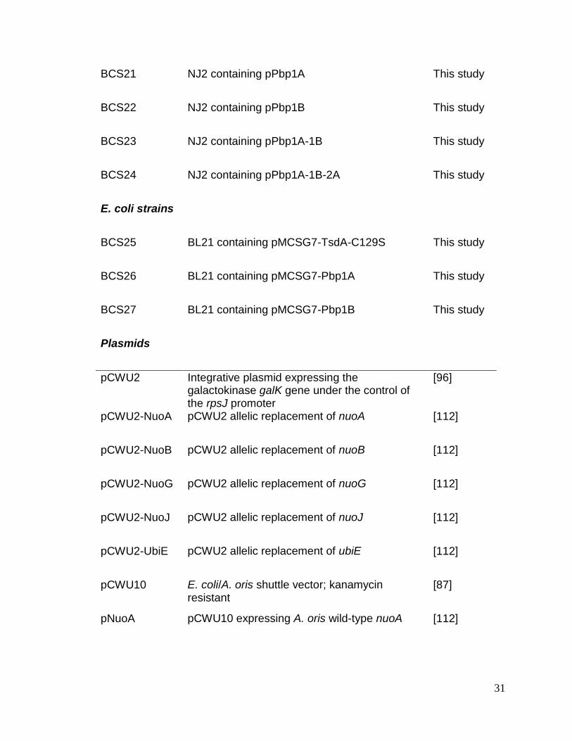
31
BCS21 NJ2 containing pPbp1A This study
BCS22 NJ2 containing pPbp1B This study
BCS23 NJ2 containing pPbp1A-1B This study
BCS24 NJ2 containing pPbp1A-1B-2A This study
E. coli strains
BCS25 BL21 containing pMCSG7-TsdA-C129S This study
BCS26 BL21 containing pMCSG7-Pbp1A This study
BCS27 BL21 containing pMCSG7-Pbp1B This study
Plasmids
pCWU2 Integrative plasmid expressing the galactokinase galK gene under the control of the rpsJ promoter
[96]
pCWU2-NuoA pCWU2 allelic replacement of nuoA [112]
pCWU2-NuoB pCWU2 allelic replacement of nuoB [112]
pCWU2-NuoG pCWU2 allelic replacement of nuoG [112]
pCWU2-NuoJ pCWU2 allelic replacement of nuoJ [112]
pCWU2-UbiE pCWU2 allelic replacement of ubiE [112]
pCWU10 E. coli/A. oris shuttle vector; kanamycin resistant
[87]
pNuoA pCWU10 expressing A. oris wild-type nuoA [112]

32
pUbiE pCWU10 expressing A. oris wild-type ubiE [112]
pJRD215 E. coli/Actinomyces shuttle vector; kanamycin and streptomycin resistant
[116]
pMdbA pJRD215 expressing A. oris wild-type mdbA [55]
pCGL0243 Corynebacterium/E. coli shuttle vector; kanamycin resistant
[89]
pK19MobsacB Corynebacterium integration plasmid; kanamycin resistant
[93]
pMCSG7 Ligation-independent cloning for protein expression; ampicillin resistant
[90]
pMCSG7-TsdA For expression of recombinant C. diphtheriae TsdA
[114]
pMCSG7-TsdA-C129S
For expression of recombinant C. diphtheriae TsdA harboring C129S mutation
This study
pK19mobsacB-SigH
pK19mobsacB allelic replacement of sigH This study
pBsk-GFP GFP expressing plasmid (Addgene plasmid #29459)
[88]
pPtsdA-GFP pCGL0243 expressing GFP from C. diphtheriae wild-type tsdA promoter
This study
pPtsdA-T2G-GFP pCGL0243 expressing GFP from T2G tsdA mutant promoter
This study
pSigH pCGL0243 expressing C. diphtheriae wild-type sigmaH
This study
pMCSG7-Pbp1A
For expression of recombinant C. diphtheriae Pbp1A
This study
pMCSG7-Pbp1B
For expression of recombinant C. diphtheriae Pbp1B
This study
pPbp1A pCGL0243 expressing C. diphtheriae wild-type pbp1A
This study
pPbp1B pCGL0243 expressing C. diphtheriae wild-type pbp1B
This study
pPbp1A-1B pCGL0243 expressing C. diphtheriae wild-type pbp1A and pbp1B
This study

33
pPbp1A-1B-2A pCGL0243 expressing C. diphtheriae wild-type pbp1A, pbp1B and pbp12A
This study

34
Table 2.2. Primers used in this study
Primer Sequence(a) Application
Ana1624-A-KpnI AAAAAGGTACCGTTGAGGAGCATCTCGGGGC
nuoA deletion
Ana1624-B CCCATCCACTAAACTTAAACAGACTGAAGCCGCTCTGACCG
nuoA deletion
Ana1624-C TGTTTAAGTTTAGTGGATGGGGGGGTTCATGCTTCCGGCAC
nuoA deletion
Ana1624-D-HindIII
AAAAAAGCTTCCCAGGCCACCGCCATCGAC
nuoA deletion
Ana1623-A- KpnI AAAAAGGTACCCGTGGGTCAGGCTCATGAGCTGG
nuoB deletion
Ana1623-B CCCATCCACTAAACTTAAACAGTCGAGGCCGCCGCGCTGTC
nuoB deletion
Ana1623-C TGTTTAAGTTTAGTGGATGGGGGCGTTGTGCTTCTTCATGCTCTTCAC
nuoB deletion
Ana1623-D-HindIII
AAAAAAGCTTGCGCAAGCGCCTCATGAAGC
nuoB deletion
Ana1615-A-KpnI AAAAAGGTACCGTTCCTCGGCGGGCAGGCCC
nuoJ deletion
Ana1615-B CCCATCCACTAAACTTAAACAGTCTCCCCATCACCGTCTACCTC
nuoJ deletion
Ana1615-C TGTTTAAGTTTAGTGGATGGGCAGCAGGGTGCTCATCGCAG
nuoJ deletion
Ana1615-D-HindIII
AAAAAAGCTTTGATGCCCCGCTACCATGGTC
nuoJ deletion
Ana1618-A-KpnI AAAAAGGTACCAGTGCGTGGACCAGCCGCCCAGG
nuoG deletion
Ana1618-B CCCATCCACTAAACTTAAACACTCCCAGGTCACCGTGACCCATGCAGCGG
nuoG deletion
Ana1618-C TGTTTAAGTTTAGTGGATGGGCCGCGGGTGCGGAGCTCTTGGTGG
nuoG deletion
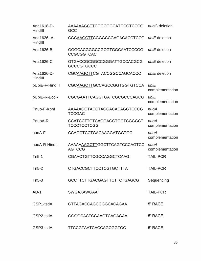
35
Ana1618-D-HindIII
AAAAAAGCTTCGGCGGCATCCGTCCCGGCC
nuoG deletion
Ana1626- A-HindIII
CGCAAGCTTCGGGCCGAGACACCTCCG ubiE deletion
Ana1626-B GGGCACGGGCCGCGTGGCAATCCCGGCCGCGGTCAC
ubiE deletion
Ana1626-C GTGACCGCGGCCGGGATTGCCACGCGGCCCGTGCCC
ubiE deletion
Ana1626-D-HindIII
CGCAAGCTTCGTACCGGCCAGCACCC ubiE deletion
pUbiE-F-HindIII CGCAAGCTTGCCAGCCGGTGGTGTCCA ubiE complementation
pUbiE-R-EcoRI CGCGAATTCAGGTGATCGCGCCCAGCG ubiE complementation
Pnuo-F-KpnI AAAAAGGTACCTAGGACACAGGTCCCGTCCGAC
nuoA complementation
PnuoA-R CCATCCTTGTCAGGAGCTGGTCGGGCTTCCCTCCTCGG
nuoA complementation
nuoA-F CCAGCTCCTGACAAGGATGGTGC nuoA complementation
nuoA-R-HindIII AAAAAAAGCTTGGCTTCAGTCCCAGTCCAGTCCG
nuoA complementation
Tn5-1 CGAACTGTTCGCCAGGCTCAAG TAIL-PCR
Tn5-2 CTGACCGCTTCCTCGTGCTTTA TAIL-PCR
Tn5-3 GCCTTCTTGACGAGTTCTTCTGAGCG Sequencing
AD-1 SWGAXAWGAAb TAIL-PCR
GSP1-tsdA GTTAGACCAGCGGGCACAGAA 5’ RACE
GSP2-tsdA GGGGCACTCGAAGTCAGAGAA 5’ RACE
GSP3-tsdA TTCCGTAATCACCAGCGGTGC 5’ RACE

36
RTPCR-tsdA-F TAGCGGTAAGGCGGGTTCG RT-PCR
RTPCR-tsdA-R GATCTTTCGCGTTACGACGGTG RT-PCR
TsdA-C129S-F AGCGCCCGCTGGTCTAACCAGACCGAG Site-directed mutagenesis
TsdA-C129S-R GAAGGGGCACTCGAAGTCAGAGAATTC Site-directed mutagenesis
PtsdA-HindIII-F AAAAAAAGCTTCGTAGAAAACTCGGTAAGTAAGCC
pPtsdA-GFP reporter
PtsdA-GFP-R CGCTGACTTCTGCATGAAGTACATATGTCTAAAGGTGAAGAACTGTTC
pPtsdA-GFP reporter
GFP-F ATGTCTAAAGGTGAAGAACTGTTC pPtsdA-GFP reporter
GFP-BamHI-R AAAAAGGATCCCTATTTGTAGAGCTCATCCATGCC
pPtsdA-GFP reporter
sigH-A-HindIII AAAAAAAGCTTGAGCCAGCACGTTGGGGA
sigH deletion
sigH-B-R GTCGCTTGTTTTCGTAGCCAC sigH deletion
sigH-C-F GTCGCTTGTTTTCGTAGCCACCCGGCTACATTGCGGGAAAA
sigH deletion
sigH-D-BamHI AAAAAGGATCCCCGTTTGTCGCGATGAAGATC
sigH deletion
PsigH-HindIII-F AAAAAAAGCTTCGCCGCCTTTTTTAGGCT
sigH complementation
PsigH-BamHI-R AAAAAGGATCCGGAAAAGATCCCTTAAGCCAC
sigH complementation
LIC-Pbp1A-F TACTTCCAATCCAATGCAGGCGTGGCCGTCGACCGT
Recombinant Pbp1A
LIC-Pbp1A-R TTATCCACTTCCAATGttaTCCATTGGGTAGTCCCTGTGG
Recombinant Pbp1A
LIC-Pbp1B-F TACTTCCAATCCAATGCACCTAGCGAGCTGGTGACCAAA
Recombinant Pbp1B

37
LIC-Pbp1B-R TTATCCACTTCCAATGttaGTGATACCGCCACGAAGTTGG
Recombinant Pbp1B
pbp1A-A-HindIII-F aaaaaAAGCTTCGCCTGCTCAGCGGTAAC
pbp1A deletion
pbp1A-B-R GCTAAGGACTGCCCGTGG pbp1A deletion
pbp1A-C-F CCACGGGCAGTCCTTAGCGCACGAACAGCCGTGGAT
pbp1A deletion
Pbp1A-D-EcoRI-F aaaaaGAATTCCGACTGGGGGTGCGTTTT
pbp1A deletion
pbp1B-A-HindIII-F aaaaaAAGCTTTCAGGCAATGTGGCGAAC
pbp1B deletion
pbp1B-B-R GCTTCCTTGATCTGCGGT pbp1B deletion
pbp1B-C-F ACCGCAGATCAAGGAAGCCCTGATTCTTACAGGGGGCAC
pbp1B deletion
Pbp1B-D-EcoRI-R
aaaaaGAATTCGCGGCCACAACTTAAACG
pbp1B deletion
Ppbp1A-MluI-R aaaaaACGCGTCTCCCCCTGCTGAGTAT pbp1A complementation
Ppbp1B-1A-overlap-R
TATCCACGGCTGTTCGTG pbp1A-1B complementation
Ppbp1A-MluI-R
aaaaaACGCGTTATCCACGGCTGTTCGTG
pbp1A complementation
Ppbp1A-1B-overlap-F
CACGAACAGCCGTGGATAACCTGCTGAATTCGGTGA
pbp1A-1B complementation
Ppbp1B-MluI-F aaaaaACGCGTACCTGCTGAATTCGGTGA
pbp1B complementation
Ppbp1B-MluI-R aaaaaACGCGTGCCCCCTGTAAGAATCAG
pbp1B complementation
pbp2A-1A-1B-overlap-F
GCCCCCTGTAAGAATCAGATCGAAGAAGCCGTTCGA
pbp1A-1B-2A complementation
Ppbp2a-MluI-R aaaaaACGCGTGGTGGGATGTCCAGGTTT
pbp1A-1B-2A complementation
38
RTPCR-Pbp1A-F ACCCCGACGCGGTGATGG RT-PCR
RTPCR-Pbp1A-R GCTGGTCGGTGCTCAAGCCTT RT-PCR
RTPCR-Pbp1B-F GCTATCTTGGGCCAGCTCACC RT-PCR
RTPCR-Pbp1B-R CCGTAGGCATTGCGGCCG RT-PCR
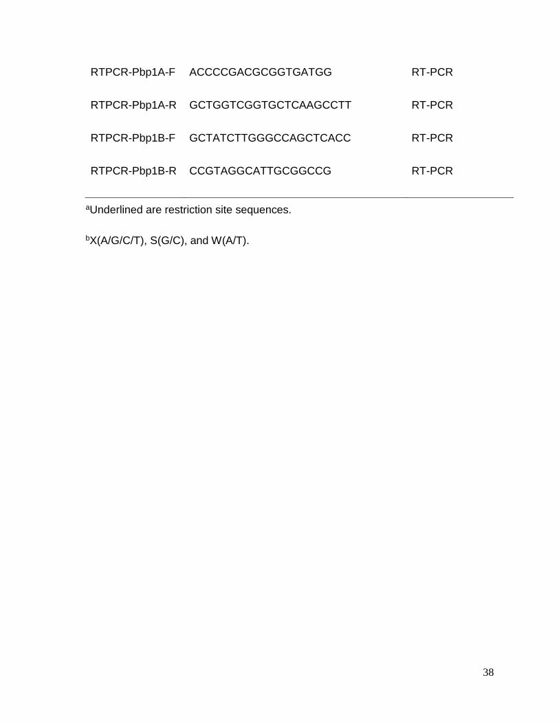
aUnderlined are restriction site sequences.
bX(A/G/C/T), S(G/C), and W(A/T).

39
CHAPTER 3. Electron Transport Chain Is Biochemically Linked to Pilus
Assembly Required for Polymicrobial Interactions and Biofilm Formation in the
Gram- Positive Actinobacterium Actinomyces oris
This chapter is based upon work published in the journal mBio entitled “Electron
Transport Chain Is Biochemically Linked to Pilus Assembly Required for
Polymicrobial Interactions and Biofilm Formation in the Gram- Positive
Actinobacterium Actinomyces oris”. mBio. 2017: 8: e00399-17. Belkys C. Sánchez is
co-first author of this publication (Belkys C. Sanchez, Chungyu Chang, Chenggang
Wu, Bryan Tran, Hung Ton-That) and was responsible for preparing the original
manuscript and conducted the majority of experiments described. Copyright of all
material published in mBio remains with the authors.
For additional information about ASM’s permission policies associated with
commercial reuse of mBio content, see http://mbio.asm.org/site/misc/reprints.xhtml

40
3.1. INTRODUCTION
Found only in Gram-positive bacteria, such as Actinomyces spp.,
Corynebacterium diphtheriae, Bacillus cereus, streptococci and enterococci,
covalently-linked pili, also termed fimbriae, are important virulence determinants [20,
117]. These adhesive pilus structures are assembled and linked to bacterial
peptidoglycan by conserved transpeptidase enzymes collectively named sortase
[118]. Initially reported in C. diphtheriae [19, 20], sortase enzymes that polymerize
pilin subunits into covalently-linked pilus polymers are often called pilus-specific
sortases, or class C sortases [118, 119]. Generally, the housekeeping sortase and
non-polymerizing sortase enzymes anchor pilus polymers to the bacterial cell wall
[120-124] via a process similar to cell wall anchoring of surface proteins by the
archetype SrtA of Staphylococcus aureus [125, 126].
Pili were discovered in many species of Actinomyces in the 1970s [127, 128],
and early pilus characterizations focused on the fimbriae of Actinomyces naeslundii
genospecies 2 [129], which was later renamed Actinomyces oris [130]. In the biofilm-
forming actinobacterium A. oris, two types of fimbriae have been identified. Type 1
fimbriae – consisting of the pilus shaft FimP and the tip pilin FimQ [116] – mediate
bacterial binding to salivary proline-rich proteins on the tooth surface [9]. SrtC1, the
pilus-specific sortase encoded by the type 1 fimbrial gene locus, specifically catalyzes
pilus polymerization of FimP and FimQ [113, 116]. Type 2 fimbriae, with SrtC2 as
their pilus-specific sortase [96, 113], are made of the pilus shaft FimA and the
canonical tip pilin FimB [96].

41
Biofilm formation and Actinomyces interactions with oral streptococci in
biofilm, termed coaggregation [129], are well-documented phenotypes associated
with the type 2 fimbriae since a fimA mutant fails to mediate biofilm formation and
bacterial coaggregation [12, 96]. Nonetheless, it has recently been discovered that a
coaggregation factor named CafA hijacks the sortase SrtC2 machine, forming a
distinct pilus tip with the pilus shaft FimA independent of FimB [111]. It is now clear
that CafA is the major coaggregation factor of A. oris as deletion of cafA results in the
same coaggregation defect as fimA deletion; additionally, a specific antibody against
recombinant CafA or addition of this protein blocks bacterial coaggregation [111].
Thus, the coaggregation defect of the fimA mutant can be attributed to the loss of
CafA pilus assembly on the bacterial cell wall, while the role of FimB in oral
colonization and biofilm formation remains unknown.
Consistent with the above, in a previous small-scale Tn5 transposon screen
intended to identify A. oris mutants that fail to aggregate with Streptococcus oralis,
we found 3 mutants with Tn5 insertions in fimA and srtC2; the fourth mutant was
mapped to vkor, which encodes a bacterial vitamin K epoxide reductase (VKOR) [55].
Importantly, an in-frame, nonpolar vkor deletion mutant was shown to be severely
defective in pilus assembly. Further characterizations established that VKOR is
required for reoxidation of the thiol-disulfide oxidoreductase MdbA [55]. It is
noteworthy that a Mycobacterium tuberculosis homolog of A. oris VKOR was initially
shown to replace DsbB in reoxidation of the disulfide bond forming oxidoreductase
DsbA when expressed in Escherichia coli [131]. Unlike E. coli dsbA, mdbA is an

42
essential gene and a conditional mdbA deletion mutant failed to assemble adhesive
pili [55].
It was shown that MdbA and VKOR together form the thiol-disulfide
oxidoreductase pair that catalyzes oxidative protein folding in A. oris [132]. Thus,
based on available evidence centered on the type 2 fimbriae, a model of pilus
assembly in A. oris has been proposed [55, 111] (Figure 3.1). Synthesized in the
cytoplasm, pilin precursors, such as FimA, FimB, and CafA, are transported across
the cytoplasmic membrane by the Sec translocon in an unfolded state. The thiol-
disulfide oxidoreductase MdbA catalyzes oxidative folding of nascent pilin precursors
before they are embedded into the membrane. Membrane-bound pilin precursors are
polymerized by the pilus-specific sortase SrtC2, resulting in formation of type 2
fimbriae with two distinct tip pilins, FimB and CafA. The resulting polymers are
presumably anchored by the housekeeping sortase SrtA. While it is clear in this model
that the sortase and oxidative folding machines are central elements of pilus
assembly in A. oris, it is unknown if there are additional factors directly or indirectly
involved in the pilus assembly process.
Because A. oris coaggregation and pilus assembly are tightly associated, we
exploited this property in a high-throughput assay to screen more than 6,200 Tn5
transposon mutants for clones that are variably defective in coaggregation with S.
oralis So34, an indicator strain expressing pilus receptor cell-surface polysaccharides
(RPS) [12, 96, 133]. Using a modified-visual coaggregation scoring system, we were
able to identify auxiliary factors contributing to pilus assembly that might have been
missed in the previously reported small-scale screen, which was solely based on

43
Figure 3.1. A model of pilus assembly in A. oris. Presented is a simplified model
of pilus assembly in A. oris that is centered on the type 2 fimbriae (see text for details);
FimA, FimB, and CafA are colored in green, orange, and blue, respectively. The thiol-
disulfide oxidoreductase MdbA catalyzes oxidative folding of nascent protein
precursors as they are translocated into the exoplasm by the Sec machinery.
Reoxidation of MdbA requires the membrane-bound oxidoreductase VKOR. Folded
pilin precursors are polymerized and anchored to the cell wall by the tandem sortase
enzymes. It is not known how electrons generated from reoxidation of MdbA/VKOR
are transferred to (question mark). Dashed arrows denote potential multiple steps;
adapted after [55, 111].

44
positive or negative-coaggregation phenotype scoring [55]. By characterizing a
subset of these coaggregation-defective mutants, we demonstrate here that the
NADH dehydrogenase and menaquinone, components of the electron transport chain
(ETC), are involved in reoxidation of the major disulfide bond-forming machine
MdbA/VKOR in A. oris.
3.2. RESULTS
3.2.1. A Tn5 transposon screen revealed A. oris mutants defective in
polymicrobial interactions
Since pilus assembly and bacterial coaggregation in A. oris are coupled [96,
111], we sought to identify trans-acting factors involved in pilus assembly by
screening a large number of A. oris Tn5 mutants using a previously reported
coaggregation assay in a 96-well plate format [55]. Following a published protocol
[55, 92], a Tn5 transposon library of roughly 6,240 A. oris mutants (> 3-fold genome
coverage) was constructed and screened. Coaggregation efficiency of the Tn5
mutants was scored largely based on the visual scoring system reported by Cisar et
al. [94], whereby the coaggregation-positive phenotype of the parental strain MG1
was designated as 4 and the coaggregation-negative phenotype of type-2 fimbria-
less mutants, i.e. ΔfimA, ΔcafA, and ΔsrtC2, was set as 1; tiny clumps of aggregates
were scored as 2, whereas the larger clumps were considered as 3. We obtained 13
mutants with the coaggregation score (CS) of 1, 7 mutants with the CS of 2 or CS-2,
and 13 mutants with CS-3; of note, the Tn34 mutant exhibiting a coaggregation
phenotype similar to that of MG1 was included for comparison (Figure 3.2). These
coaggregation-defective Tn5 mutants were then subjected to mapping by thermal

45
Figure 3.2. Identification of A. oris coaggregation-defective mutants by Tn5
transposon mutagenesis. Thirty-three A. oris coaggregation-defective Tn5 mutants
identified by a cell-based screen were confirmed by a standard coaggregation assay
[87]. Equal cell numbers of A. oris mutants and S. oralis So34 were mixed together,
and coaggregation was imaged using an AlphaImager. Coaggregation scores
indicate the degrees of coaggregation, with the phenotype of the parental MG1 strain
considered as 4 and that of the fimA, cafA, and srtC2 deletion mutants considered as
1; 2 and 3 represent small and larger clumps of bacterial aggregates. Tn5 target
genes were mapped by TAIL-PCR. Chungyu Chang, PhD and Bryan Tran performed
the Tn5 transposon mutagenesis and coaggregation screen.

46
asymmetric interlaced PCR (TAIL-PCR) and DNA sequencing as previously
described [55].
As expected, 13 coaggregation-negative mutants, i.e. with the CS of 1, were
mapped to genes encoding CafA, FimA and SrtC2, which are known fimbrial factors
essential for A. oris coaggregation with oral streptococci [12, 96, 111]. Intriguingly, 4
out of 7 CS-2 mutants contained Tn5 insertions in nuoA, nuoJ, and nuoG genes; the
remainder were mapped to genes, i.e. ANA_1625, ANA_0218, and ANA_0143, which
code for geranylgeranyl reductase (GGR), a putative metal-binding protein, and β-
glucosidase, respectively. Finally, the CS-3 mutants were mapped to genes encoding
many hypothetical proteins, a carbon starvation protein A (CstA), a two-component
system sensor kinase, an ABC-2 type transporter, and the H, I, M subunits of NADH
dehydrogenase (i.e. NuoH, NuoI, and NuoM) (Figure 3.2 and Table 3.1). Since a large
number of the nuo genes was repeatedly targeted by the Tn5 transposon, they were
further characterized and reported in this study; the remaining candidates will be
investigated in future studies.
3.2.2. Genetic disruption of the A. oris NADH dehydrogenase (complex I)
subunits caused significant defects in CafA-mediated coaggregation and CafA
pilus assembly
The ggr, nuoA, nuoG, nuoH, nuoJ, and nuoM genes revealed from the Tn5
screen above are part of the nuo gene locus in A. oris MG1, (see
http://genome.brop.org/), which are predicted to encode the NADH dehydrogenase
enzyme – often referred to as the NADH:ubiquinone oxidoreductase or respiratory
complex I of the ETC [134] (Figure 3.3A). To confirm that the coaggregation defects

47
Table 3.1. Mapping of A. oris coaggregation-defective Tn5 mutants
Tn5 mutants
Genomic Tn5 Positiona
Target Gene
Predicted Function
1 2,422,225 cafA Coaggregation factor A
2 38,189 fimA Type 2 fimbrial shaft pilin
3 36,541 fimB Type 2 fimbrial tip pilin
4 2,420,317 cafA Coaggregation factor A
5 2,421,799 cafA Coaggregation factor A
6 38,174 fimA Type 2 fimbrial shaft pilin
7 39,115 srtC2 Type 2 pilus-specific sortase
8 39,481 srtC2 Type 2 pilus-specific sortase
9 39,749 srtC2 Type 2 pilus-specific sortase
10 2,421,087 cafA Coaggregation factor A
11 2,421,136 cafA Coaggregation factor A
12 38,721 fimA Type 2 fimbrial tip pilin
13 2,420,135 cafA Coaggregation factor A
14 1,755,107 ana_1625 Geranylgeranyl reductase
15 1,744,569 nuoJ NADH dehydrogenase I, subunit J

48
16 230,679 ana_0218 Zn2+/Mn2+ transport system substrate-binding protein
17 1,754,944 nuoA NADH dehydrogenase I, subunit A
18 162,115 ana_0143 β-D-glucoside glucohydrolase
19 1,747,764 nuoG NADH dehydrogenase I, subunit G
20 1,748,226 nuoG NADH dehydrogenase I, subunit G
21 1,741,097 nuoM NADH dehydrogenase , subunit M
22 2,348,384 cstA Carbon starvation protein A
23 2,863,369 ana_2659 Ni/Fe-hydrogenase III large subunit
24 2,711,713 ana_2514 Histidine kinase (two-component system)
25 2,295,587 ana_2118 Permease component of ABC-type multidrug transport system
26 1,746,526 nuoH NADH dehydrogenase I, subunit H
27 382,186 ana_0363 Conserved hypothetical protein
28 2,566,963 ana_2372 Hypothetical protein with Ser/Arg repeats
29 1,745,086 nuoI NADH dehydrogenase I, subunit I
30 1,739,922 nuoM NADH dehydrogenase I, subunit M
31 2,513,949 ana_2325/ ana_2326b
Ana_2325 (Cys/His-dependent amidohydrolase/peptidase); Ana_2326 (hypothetical protein)
32 1,712,411 ana_1580 Conserved hypothetical protein

49
33 1,080,892 ana_1004 Hypothetical protein
34 2,576,712 ana_2380 Conserved hypothetical protein
aShown is the nucleotide position of the Tn5 transposon mapped in the genome.
bTn5 insertion was found within the intergenic region of ana_2325 and ana_2326.
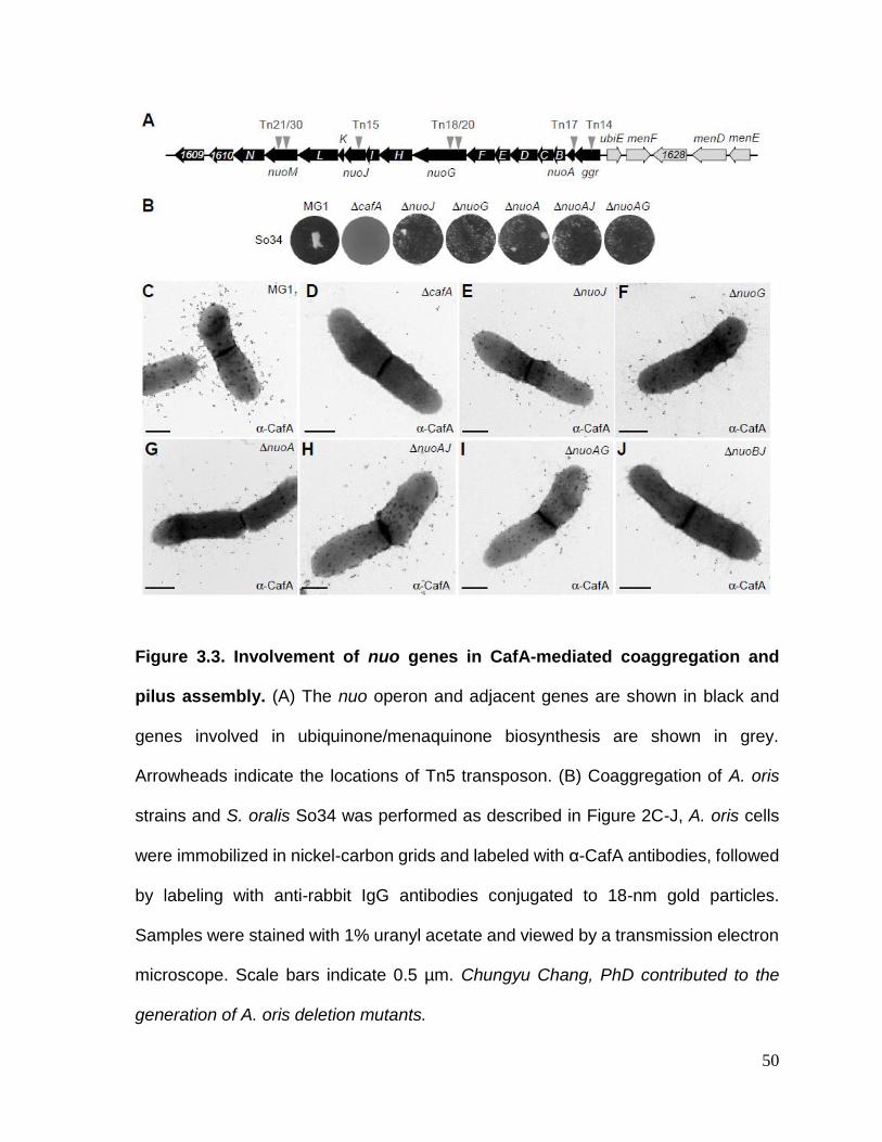
50
Figure 3.3. Involvement of nuo genes in CafA-mediated coaggregation and
pilus assembly. (A) The nuo operon and adjacent genes are shown in black and
genes involved in ubiquinone/menaquinone biosynthesis are shown in grey.
Arrowheads indicate the locations of Tn5 transposon. (B) Coaggregation of A. oris
strains and S. oralis So34 was performed as described in Figure 2C-J, A. oris cells
were immobilized in nickel-carbon grids and labeled with α-CafA antibodies, followed
by labeling with anti-rabbit IgG antibodies conjugated to 18-nm gold particles.
Samples were stained with 1% uranyl acetate and viewed by a transmission electron
microscope. Scale bars indicate 0.5 µm. Chungyu Chang, PhD contributed to the
generation of A. oris deletion mutants.

51
of the Tn5::nuo mutants are not due to polar effects, we generated individual and
combinations of in-frame, nonpolar deletion mutants of nuoJ, nuoG, nuoA. The
generated mutants were confirmed for their inability to aggregate with S. oralis via
CafA-mediated coaggregation using a standard coaggregation assay [111], whereby
A. oris cells were mixed in equal volume with S. oralis So34, and coaggregation was
determined after a few minutes of mixing. As shown in Figure 3.3B, deletion of nuoJ,
nuoG, nuoA, or combination of nuoA and nuoJ or nuoA and nuoG caused significant
coaggregation defects as compared to the wild-type level.
Since CafA pilus assembly is essential for A. oris coaggregation [111], we next
examined whether the observed coaggregation defects are due to pilus assembly
defects of CafA by immunoelectron microscopy (IEM). In this assay, A. oris cells were
stained with antibodies against CafA (α-CafA), followed by staining with gold particles
conjugated with IgG, and the samples were viewed by a transmission electron
microscope (TEM) after staining with 1% uranyl acetate. In the parental strain MG1,
CafA signal was found abundantly on the bacterial surface and at the distal end of
pili; as expected, the CafA signal was absent in the cafA deletion mutant [111].
Importantly, CafA labeling was significantly reduced in the nuoJ, nuoG, nuoA, nuoAJ,
nuoAG, and nuoBJ mutants (Figure 3.3C-J). Since the CafA assembly defects among
these mutants were apparently equal, we chose to focus on the nuoA mutant for
further characterizations (see below) as nuoA is typically the first gene in bacterial
nuo operons [135]. To support that the generated nuoA mutant is non-polar, we
introduced in this mutant a plasmid constitutively expressing nuoA; as shown in
Figure 3.4, ectopic expression of nuoA rescued the assembly defects of both type 1

52
Figure 3.4. Requirement of nuoA for pilus assembly. A. oris cells of indicated
strains were immobilized on carbon-coated n nickel grids and stained with α-FimA
(A-D), α-CafA (G-J), or α-Type1 antibodies (M-P), followed by staining with IgG
conjugated to 18-nm gold particles. Samples were stained with 1% uranyl acetate
prior to be analyzed by electron microscopy. Scale bars indicate 0.5 µm.
DnuoA
a-Type 1
O
DnuoA/pNuoA
a-Type 1
P
N DfimP
a-Type 1
M MG1
a-Type 1
a-CafA
DnuoA/pNuoA
a-CafA
DnuoA
DnuoA/pNuoA
a-FimA
DnuoA
a-FimA
J
C
B
a-FimA
DfimA
A
a-FimA
MG1
I
a-CafA
H DcafA
a-CafA
G MG1
D
Figure S1: Sanchez et al.

53
and type 2 pili of the nuoA mutant. Of note, the defects in pilus assembly and
coaggregation might not be due to the cell growth defect as the growth rate of the
nuoA mutant was not significantly different from that of the parental strain (Figure
3.5). Together, the results confirm the requirement of the NADH dehydrogenase for
optimal bacterial coaggregation and CafA pilus assembly.
3.2.3. The menaquinone C-methyltransferase UbiE is involved in A. oris
coaggregation, biofilm formation, and pilus assembly
In E. coli, the NADH dehydrogenase, encoded by 14 nuo genes (A to N),
generates energy by coupling the transfer of electrons from NADH to ubiquinone with
proton translocation across the membrane [134]. We observed that upstream of the
A. oris nuo locus in the MG1 genome are genes encoding part of the
ubiquinone/menaquinone biosynthesis pathway, with the C-methyltransferase
encoding-gene ubiE adjacent to ggr (Figure 3.3A). In E. coli UbiE catalyzes the
carbon methylation reaction in the biosynthesis of ubiquinone/menaquinone, which
are essential components of the ETC [136]. This information prompted us to examine
whether the ubiquinone/menaquinone biosynthesis pathway is also linked to A. oris
coaggregation and pilus assembly. Since ubiE is not an essential gene in E. coli [136],
we decided to generate an in-frame, nonpolar deletion mutant of A. oris ubiE. The
generated mutant was examined for its ability to aggregate with S. oralis, to mediate
biofilm formation, and to produce pili using published assays [111].
In the coaggregation assay, the ubiE mutant exhibited a severe defect in
bacterial coaggregation with S. oralis when compared to the parental strain MG1, and
this defect was rescued by ectopic expression of UbiE (Figure 3.6A). The same set

54
Figure 3.5. Generation times of the A. oris MG1 and ΔnuoA mutant strains. (A)
Growth of the wild-type MG1, ΔnuoA and ΔnuoA/pNuoA strains was measured by
optical density (OD600). Generation times were calculated as described in materials
and methods. The results are representative of three independent experiments
performed in triplicate. Error bars represent standard deviations, with ns for not
significant.
WT DnuoA DnuoA/pNuoA
GenerationTime
(h/n)
5
4
3
2
1
0
ns
Figure S3: Sanchez et al.

55
Figure 3.6. Requirement of ubiE for bacterial coaggregation, biofilm formation,
and pilus assembly. (A) Coaggregation of A. oris strains and S. oralis So34 was
determined as described in Figure 3.2. (B) Biofilms were obtained by growing the
indicated strains in HIB containing 1% sucrose for 48 h. Harvested biofilms were
subjected to crystal violet staining and optical density measurement at 580 nm using
a microplate reader. The results are shown as representatives of 3 independent
experiments performed in triplicate; ** denotes P < 0.0025, calculated using a One-
way ANOVA (Duncan’s method, non-parametric) with GraphPad Prism. (C-N) A. oris
cells were immobilized in nickel grids and stained with α-FimA (C-F), α-CafA (G-J),
or α-Type1 antibodies (K-N), followed by staining with IgG conjugated to 18-nm gold
particles. Samples were stained with 1% uranyl acetate prior to be analyzed by
electron microscopy. Scale bars indicate 0.5 µm.

56
of strains was then examined in biofilm formation assays, in which A. oris biofilms
were cultivated in Heart Infusion broth (HIB) supplemented with 1% sucrose, followed
by biofilm staining with 1% crystal violet and quantification by OD580. Consistent with
the results above, deletion of ubiE significantly reduced biofilm formation as
compared to the wild-type level, and expression of ubiE in trans restored biofilm
formation to the wild-type (Figure 3.6B).
To examine if the coaggregation and biofilm defects are associated with pilus
assembly, we employed IEM using α-CafA and antibodies against the type 2 fimbrial
shaft FimA (α-FimA). Compared to the MG1 strain, which produced abundant signals
of FimA and CafA, FimA and CafA detection was drastically reduced in the ubiE
mutant; these defects were rescued in the ubiE mutant expressing UbiE from a
plasmid (Figure 3.6C-J and Figure 3.7). Finally, to determine if the role of UbiE also
extends to the type 1 fimbriae, we used antibodies against type 1 fimbriae [137] (α-
Type 1) in IEM experiments. Consistently, the type 1 fimbrial signal was significantly
reduced in the ubiE mutant as compared to the parental MG1 and UbiE
complementing strains (Figure 3.6K-N). Altogether, these results suggest that the
defects of ubiE deletion in bacterial coaggregation and biofilm formation are directly
linked to assembly deficiencies of adhesive fimbriae, which are the major factors
required for the aforementioned processes in A. oris [96, 111].

57
Figure 3.7. Requirement of nuoA and ubiE for surface expression of CafA. (A)
The expression of CafA on the cell surface was determined by whole cell ELISA using
polyclonal α-CafA antibodies. The absorbance measurments at 450nm, as compared
to cafA mutant as background, were determined from three independent experiments
performed in triplicate. Error bars represent standard deviations. *, *** indicate
P<0.05, P<0.001, respectively, which were determined using the unpaired, two-tailed
t-test with GraphPad Prism.
MG1
DcafA
DnuoA
DnuoA/p
Nuo
A
DnuoA/p
Mdb
A
DubiE/p
Ubi
E
DubiE/p
Mdb
A
DubiE
0.8
0.6
0.4
0.2
0.0
Absorbance(450nm) *
***
Figure S2: Sanchez et al.

58
3.2.4. Requirement of the menaquinone C-methyltransferase UbiE and NADH
dehydrogenase subunit NuoA in reoxidation of the major thiol-disulfide
oxidoreductase MdbA
As mentioned above, disulfide bond formation is required for correct folding
of pilin precursors, and this process is catalyzed by the major thiol-disulfide
oxidoreductase MdbA in A. oris [55]. Given UbiE and several subunits of the NADH
dehydrogenase are associated with pilus assembly as presented above, we
hypothesized that the pilus assembly defects in the ubiE and nuo mutants might be
due to aberrant oxidative folding of pilus proteins. Reactivation of MdbA normally
requires the membrane-bound oxidoreductase VKOR, but overexpression of MdbA
in a vkor mutant can compensate the loss of VKOR [55]. Here, we examined if ectopic
expression of MdbA in the absence of UbiE or Nuo subunits would rescue the pilus
assembly defects by introducing a recombinant plasmid that constitutively expresses
MdbA into the ubiE and nuoA mutants. The resulting strains, along with the mutants,
were examined for their ability to assemble pili by IEM with α -FimA. Compared to the
parental MG1 strain, the ubiE mutant produced significantly less FimA (compare
Figure 3.6C and Figure 3.8A), whereas overexpression of MdbA in this mutant
resulted in abundant production of FimA pili (Figure 3.8B). While the assembly defect
of FimA in the nuoA mutant was not as severe as that of the ubiE mutant, MdbA
overexpression in this nuoA mutant also increased pilus production (Figure 3.8C & D
and Figure 3.7). These results suggest that MdbA was not sufficiently reoxidized by
VKOR in the absence of UbiE or NuoA.
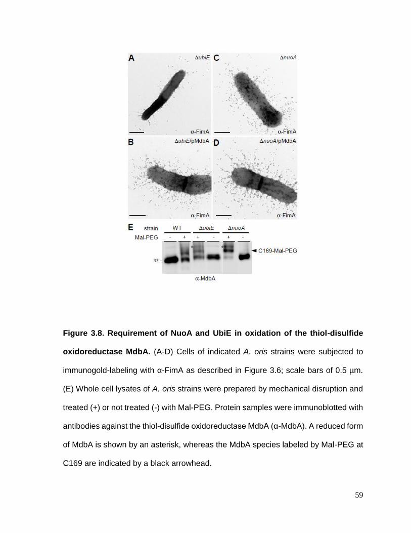
59
Figure 3.8. Requirement of NuoA and UbiE in oxidation of the thiol-disulfide
oxidoreductase MdbA. (A-D) Cells of indicated A. oris strains were subjected to
immunogold-labeling with α-FimA as described in Figure 3.6; scale bars of 0.5 µm.
(E) Whole cell lysates of A. oris strains were prepared by mechanical disruption and
treated (+) or not treated (-) with Mal-PEG. Protein samples were immunoblotted with
antibodies against the thiol-disulfide oxidoreductase MdbA (α-MdbA). A reduced form
of MdbA is shown by an asterisk, whereas the MdbA species labeled by Mal-PEG at
C169 are indicated by a black arrowhead.

60
To confirm this, we examined the oxidation status of MdbA by alkylation with
methoxypolyethylene glycol-maleimide (Mal-PEG) as previously reported [55, 56]. In
this assay, protein samples acid-trapped by TCA precipitation from cell lysates of the
MG1, ΔubiE, and ΔnuoA strains were treated with 10 mM Mal-PEG or mock-treated
prior to immunoblotting with antibodies against MdbA (α-MdbA). As the wild-type
MG1 strain contains MdbA with the catalytic CXXC motif, C139/142, and a non-
catalytic cysteine residue C169 [55], Mal-PEG treatment caused an up-shift in MdbA
mobility that was due to Mal-PEG modification of the free sulfhydryl group of C169 as
previously reported [55] (Figure 3.8E; lanes WT, black arrow). Significantly, Mal-PEG
treatment of the ΔubiE, and ΔnuoA mutants resulted in a higher up-shift as compared
to the MG1 strain in the same condition, consistent with Mal-PEG modification of
additional reduced sulfhydryl groups at the active site of MdbA (Figure 3.8E; the last
4 lanes, asterisks). These results support the conclusion that the catalytic cysteine
residues C139/142 in the CXXC motif are in a reduced form when ubiE and nuoA are
genetically disrupted, very much like the phenotype of MdbA when VKOR is absent
[55].
3.2.5. Exogenous menaquinone rescues the pilus assembly and cell growth
defects of the ubiE mutant
In the majority of Gram-positive bacteria, menaquinone plays a central role in
the ETC, functioning as a conduit to receive electrons from electron donors, e.g.
NADH dehydrogenase, and transfer them to an electron acceptor, e.g. cytochrome c
reductase [138, 139]. As UbiE catalyzes the conversion of dimethyl-menaquinone to
menaquinone [136, 140], the lack of ubiE potentially reduces the quinone pool in the

61
mutant cells, leading to the pleiotropic effects discussed above. To examine this, we
monitored the growth rates of A. oris strains grown in HIB by optical density (OD600)
over time. Indeed, the ubiE mutant displayed a growth defect as compared to the
parental strain, and complementing this strain with the pUbiE plasmid rescued this
growth defect (Figure 3.9A). Importantly, addition of 0.1 mM menaquinone-4 (MK-4)
to the culture medium increased the growth rate to a level comparable to the
complementing strain after 16 h of growth (Fig 3.9A; black and grey triangles).
To further confirm this phenotype, we adapted a streaking assay with
menaquinone [141], whereby the parent MG1 strain and its ubiE isogenic mutant
were streaked as a broad band on heart infusion agar plate, and a 3-µl drop of 50
mM MK-4 was placed on the border of the streak. Cell growth at 37oC was recorded
after 48 h. As shown in Figure 3.9B, the MG1 strain exhibited abundant growth inside
and outside of the MK-4 diffusing zone (dashed lines), whereas the ubiE mutant cells
inside the MK-4 diffusing zone grew significantly better than those found outside.
Finally, to determine whether menaquinone can also rescue pilus assembly
defects, we collected the ubiE mutant cells grown in the presence or absence of MK-
4 for IEM using α-CafA. Indeed, the mutant cells grown in the presence of MK-4
produced abundant CafA signal at the level comparable to the MG1 and the
complementing strains (Figure 3.9C-6F). Altogether, the results support that the
defects of the ΔubiE mutant in pilus assembly, cell growth, and MdbA reoxidation are
mainly due to the reduced level of endogenous menaquinone.
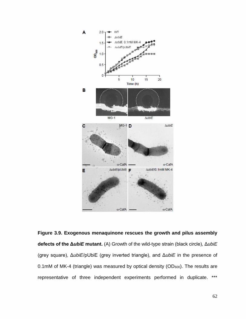
62
Figure 3.9. Exogenous menaquinone rescues the growth and pilus assembly
defects of the ΔubiE mutant. (A) Growth of the wild-type strain (black circle), ΔubiE
(grey square), ΔubiE/pUbiE (grey inverted triangle), and ΔubiE in the presence of
0.1mM of MK-4 (triangle) was measured by optical density (OD600). The results are
representative of three independent experiments performed in duplicate. ***

63
represents P ≤ 0.0001 determined by the paired, two-tailed t test with GraphPad
Prism. (B) A. oris strains were streaked as a broad band on HIA plates. A 3-µl drop
of 50 mM menaquinone-4 (MK-4) was placed on the border of the streaks and growth
the strains at 37oC was recorded after 48 h. Areas of MK-4 diffusion are marked with
dashed lines. (C-F) A. oris cells of WT, ΔubiE, ΔubiE/pUbiE, and ΔubiE grown in the
presence of 0.1 mM MK-4 were subjected to immunogold-labeling with α-CafA as
described in Figure 3.6. Scale bars indicate 0.5 µm.

64
3.3. DISCUSSION
Because pilus assembly and bacterial coaggregation are mutually inclusive in
A. oris, this inherent property was used in our Tn5 transposon screen intended to
identify factors involved in pilus assembly. This screen revealed numerous mutants
that display varying defects in Actinomyces coaggregation with S. oralis. As expected,
roughly one-third of the coaggregation-defective mutants were mapped to genes
encoding the major pilus shaft FimA, the coaggregation factor CafA, and the pilus
sortase machine SrtC2 (Figure 3.2). Mapping of the remainder surprisingly uncovered
the association of coaggregation with the NADH dehydrogenase (complex I) (Table
3.1), whose genes are adjacent to the ubiquinone/menaquinone biosynthesis gene
locus. By genetically and biochemically characterizing two representative in-frame,
nonpolar mutants ΔnuoA and ΔubiE, we have established that the two ETC
components, the NADH dehydrogenase and menaquinone, are linked to pilus
assembly via reoxidation of the major disulfide bond-forming machine MdbA, which
is essential for oxidative folding of pilus proteins in A. oris [55, 132].
By electron microscopy, both ΔnuoA and ΔubiE are shown to be defective in
pilus assembly, although the latter displays the most striking defects (Figures 3.3 and
3.6). Since overexpression of MdbA in the ΔubiE mutant rescues its pilus defects
(Figure 3.8), we argued that in the absence of UbiE the disulfide bond-forming
machine MdbA is not fully reoxidized, given that reoxidation of MdbA is required for
its activity and oxidative folding of A. oris pilus proteins [55]. Indeed, by alkylation with
Mal-PEG we detected a reduced form of MdbA, although oxidized MdbA was also
observed (Figure 3.8E). The latter might be due to inefficient alkylation by Mal-PEG

65
and/or spontaneous oxidation of MdbA during the sampling process. Since
menaquinone, 2-methyl-3-polyprenyl-1,4-naphthoquinone, is the main quinone
utilized in the ETC of Gram-positive actinobacteria [142], the lack of UbiE certainly
reduces the menaquinone pool in the cells as previously demonstrated in the E. coli
ubiE mutant [136]. In fact, by adding MK-4, a menaquinone with 4 isoprene units, to
the ΔubiE cell cultures, we were able to rescue not only the cell growth deficiency but
also the pilus assembly defect of the ΔubiE mutant (Figure 3.9). The results strongly
support that menaquinone is the major quinone source capable of reoxidizing the
disulfide bond machine MdbA/VKOR in A. oris.
In E. coli, DsbA is the primary disulfide bond-forming catalyst, which catalyzes
disulfide bond formation of nascent polypeptides transported to the periplasm by the
Sec apparatus [34, 36, 143]. DsbA becomes reduced after catalysis, and reoxidation
of DsbA requires DsbB [32, 39]. Our results presented here are in line with previous
studies in E. coli that demonstrate the participation of the respiratory ETC in
reoxidation of the DsbA/DsbB system [40, 43]. Kobayashi and colleagues showed
that E. coli mutants lacking hemA – coding for glutamyl-tRNA reductase involved in
protoheme and siroheme biosynthesis – or ubiA-menA, which encode products
involved in menaquinone biosynthesis, are defective in reoxidation of DsbA when
cells are grown in conditions deficient in protoheme or ubiquinone/menaquinone,
respectively [43]. By reconstituting the E. coli disulfide bond-forming machine
DsbA/DsbB with purified components, Bader and colleagues demonstrated that
under anaerobic conditions menaquinone serves as an electron acceptor during
DsbA/DsbB reoxidation; under aerobic growth ubiquinone acts as electron acceptor

66
that reoxidizes DsbB, which in turn reoxidizes DsbA [40]. It is interesting to note that
structural studies of a VKOR homolog of Synechococcus sp. reveal the protein is
complexed with ubiquinone [144]. Thus, it is plausible that a similar mechanism of
MdbA/VKOR reoxidation occurs in A. oris, with menaquinone presumably acting as
an electron acceptor for VKOR during this process; how menaquinone
mechanistically reoxidizes MdbA/VKOR remains to be investigated in future studies.
Of note, the effect of the NADH dehydrogenase in MdbA/VKOR reoxidation,
which has not been reported before with regard to DsbA/DsbB reoxidation, is
somewhat puzzling. In many eukaryotic and prokaryotic systems, Complex I (NADH
quinone:dehydrogenase) serves as an entry point for electrons to enter the
respiratory chain, transferring 2 electrons from NADH to ubiquinone [145]. In E. coli,
the NuoB, NuoD, NuoH, and NuoM subunits form a ubiquinone binding pocket [146].
Thus, it is possible that genetic disruption of the Nuo subunits in A. oris might disturb
electron transfer and/or the quinone pool in the cells. However, the effects seen in
the nuo deletion mutants in pilus assembly are not striking as those observed in the
ubiE mutant, although their defects in coaggregation are obvious (Figure 3.3). Thus,
it is possible that genetic disruption of the Nuo subunits potentially causes pleotropic
effects. It is likely that many other dehydrogenases, such as the malate and succinate
[147], may compensate the loss of the NADH dehydrogenase.
While the association of the NADH dehydrogenase and menaquinone in pilus
assembly is clear, the role of other factors identified from the aforementioned
coaggregation-defective screen with Tn5 mutagenesis is not obvious as many of
these factors are hypothetical proteins (Table 3.1). Except for ANA_0218 and

67
ANA_0143, which are predicted to be a Zn2+/Mn2+ transport system substrate-
binding protein and β-D-glucoside glucohydrolase, respectively, the coaggregation
defects of the remainder is not glaring. Given the nature of the two proteins mentioned
above, it is more likely they may participate in receptor-related binding rather in pilus
assembly. Finally, it is noteworthy that the Tn5 screen in this study is not saturated,
which may explain why an ubiE mutant was not detected in this screen. Future Tn5
screens with multi-fold coverage designed to directly target pilus assembly will be
necessary to reveal any additional novel pilus factors.
68
CHAPTER 4. Regulation of an Alternate Thiol-Disulfide Oxidoreductase in
Corynebacterium diphtheriae
Note: This chapter is derived from work performed by Belkys Sánchez. The X-ray
crystallography study and discussion of TsdA C129S is the work of Jerzy Osipiuk and
Andrzej Joachimiak and was used with permission. The bacterial clone expressing
recombinant TsdA C129S protein was created and supplied by Belkys Sánchez.

69
4.1. INTRODUCTION
Bacteria often express proteins that are structurally and functionally similar but
play different roles. For instance, bacteria and archaea encode variations of thiol-
disulfide oxidoreductase enzymes that catalyze disulfide bond formation and
isomerization in broad or specific substrates that are important for survival in specific
environmental niches [68]. Interestingly, even though these enzymes have distinct
dedicated functions, they can sometimes replace each other in catalyzing folding of
particular substrates [68, 148].
Bacterial pathogens like Corynebacterium diphtheriae are exposed to many
environmental stresses, including those mediated by host immune defense
mechanisms during infection. Iron is an essential nutrient for C. diphtheriae growth
[149], but nutritional immunity exerted by mammalian hosts limits the availability of
iron to pathogens [77]. Consequently, iron regulates gene expression and virulence
in C. diphtheriae [149]. Interestingly, thiol-disulfide oxidoreductase enzymes
expression can also be regulated by iron availability and induced under nutrient
starvation conditions, which are commonly encountered within the host [75, 76].
Another immune defense mechanism is the production of oxygen-derived toxic
compounds like hydrogen peroxide (H2O2) by phagocytic cells to kill engulfed
pathogens [150]. C. diphtheriae has been reported to survive in the acidic and
oxidizing harsh environment inside macrophages [151, 152]. This type of
environmental condition disrupts proper protein folding. Interestingly, some
oxidoreductase enzymes have a role in resistance to oxidative stress exerted by
copper and are important for intramacrophage survival [64, 76, 153]

70
Important bacterial pathogens encode multiple homologs of disulfide bond forming
enzymes which contribute to their pathogenicity [28, 154-156]. In C. diphtheriae, the
main thiol-disulfide oxidoreductase MdbA is required for proper cell division and
virulence in a model of diphtheritic toxemia [56]. Additionally, an alternate disulfide
bond forming enzyme was identified in a ΔmdbA suppressor mutant screen that
selected for three clones (S1, S2, S3) that grew at the non-permissive temperature
similar to the wild-type strain [114].
The C. diphtheriae ΔmdbA suppressor mutants harbor a single T-to-G
nucleotide substitution within the predicted -10 box of the promoter of a putative thiol-
disulfide oxidoreductase, named TsdA (Temperature-sensitive disulfide bond forming
protein A). This mutation caused significant overexpression of TsdA that restored
disulfide bond formation [114]. However, the mechanism by which this DNA change
alters expression of tsdA has not been investigated.
TsdA harbors thiol-disulfide oxidoreductase conserved features, and has been
reported to have isomerase/reductase activity in vitro [157]. Disulfide isomerase
enzymes cannot introduce de novo disulfide bonds into proteins [158].
Overexpression of TsdA in the ΔmdbA strain restores disulfide bond formation in the
shaft pilin subunit SpaA, which contains one disulfide bond [56, 159]. This argues
against the isomerase activity that has been reported for TsdA.
Although overexpression of TsdA rescues the mdbA phenotype, tsdA
deletion does not cause an apparent defect in disulfide bond formation [114]. Since
a mutant lacking tsdA does not present evident phenotypic changes under standard
laboratory growth conditions, we hypothesize that TsdA has a role in resistance to

71
environmental stress. In this chapter we describe in detail the promoter region of tsdA
and investigate the regulation and role of the alternate thiol-disulfide oxidoreductase
TsdA under stress conditions.
4.2. RESULTS
4.2.1. The ΔmdbA compensatory mutation creates a sigma factor A extended
promoter
The T-to-G nucleotide substitution identified in all ΔmdbA compensatory
mutants was predicted to be within the tsdA promoter region. To further characterize
the effects of this change in the ΔmdbA compensatory mutants, we precisely
identified the transcriptional start site (TSS) of tsdA by Rapid Amplification of cDNA
Ends (5’ RACE). Whole-cell RNA samples from wild-type and ΔmdbA compensatory
mutant S1 were used in reverse transcription reactions to amplify cDNA between a
defined internal site within the tsdA mRNA and unknown sequences at the 5'-end.
The resulting cDNA was used as template to generate RACE PCR products, which
were subjected to Sanger sequencing to identify the tsdA transcriptional start site.
Both wild-type and S1 strains showed the same tsdA transcriptional start site (Figure
4.1-A). Based on the identified tsdA transcriptional start site, the upstream DNA
region was analyzed for conserved promoter motifs, and the promoter structure
shown in Figure 4.1 is proposed. This configuration is consistent with a -10 core
hexamer recognized by the housekeeping sigma factor σA, directing initiation of tsdA
transcription at the adenine identified by the 5’ RACE experiment (Figure 4.1-A).
Analysis of -10 and -35 promoter elements suggests low conservation to sigma factor

72
σA consensus motifs (Fig. 4.1-B), which explains low expression of tsdA in the wild-
type strain [114]. The distance between the transcriptional start site and -10 motif was
6bp, and the distance between -35 and the -10 motifs was 16bp, while spacing for
canonical σA-type promoters in C. diphtheriae is 6-9bp and 16-20bp respectively [115,
160, 161]. In agreement with the 5’ RACE data and findings from Wittchen, et al.
[115], promoter activity of this DNA region was confirmed by a transcriptional reporter
experiment as will be discussed in section 4.2.4 (Figure 4.5).
The T-to-G substitution identified in the tsdA promoter region creates an
extended sigma factor σA promoter (Figure 4.1-B), which is characterized by a TG
dinucleotide at position -15, -14 that lowers the thermal energy required to form an
open RNA polymerase initiation complex [115, 160, 162, 163]. This results in
increased basal strength of the promoter and increased levels of transcription. In
agreement, the tsdA T-to-G mutant promoter activity was significantly higher
compared to the wild-type tsdA promoter as discussed in section 4.2.4 (Figure 4.5).
Altogether, the data demonstrate that the T-to-G nucleotide substitution at position -
14 of the tsdA promoter region creates an extended sigma factor σA promoter that
results in increased promoter activity and overexpression of this gene.
4.2.2. Characterization of an alternate thiol-disulfide oxidoreductase in C.
diphtheriae by X-ray crystallization
Overexpression of TsdA rescues the disulfide bond forming defect in C.
diphtheriae cells lacking the main thiol-disulfide oxidoreductase MdbA; this suggested
that TsdA has disulfide bond forming capabilities. To further characterize this putative

73
Figure 4.1. ΔmdbA suppressor mutation results in extended sigma factor σA
promoter motif. (A) Determination of tsdA transcriptional start site by 5’ RACE using
Wild-type and ΔmdbA-S1 strains mRNA. DNA chromatograms from the sequencing
reactions of 5’ RACE PCR products are shown. The homopolymeric tail added to the
3’ end of the cDNA used for the PCR reactions are shown. The nucleotides
complementary to the 5’ ends of mRNA are shown and the direction of the

74
transcription is indicated by arrows. (B) Genetic environment of tsdA. Transcriptional
start site identified by 5’ RACE (arrow) and upstream promoter motifs are depicted.
The T-to-G substitution identified in ΔmdbA suppressor mutants is shown. (B)
Alignment of tsdA and sigma factors σA and σH-dependent promoter consensus
sequences from C. diphtheriae [115], C. glutamicum [160] and M. tuberculosis [164]
are shown. Nucleotides conserved in consensus sequences are in bold, extended
promoter nucleotides are in pink and transcriptional start site in green. Y = C or T;
K=G or T. Core hexamers are underlined.
75
thiol-disulfide oxidoreductase, the C. diphtheriae C129S TsdA protein X-ray crystal
structure was refined to 1.14 Å resolution with R-work and R-free factors equal to
11.4 and 13.3 %, respectively. The structure is a single continuous amino-acid chain
covering 37-282 residues of the protein. Despite using the full-length 289-residue
protein for crystallization, 36 N-terminal and 7 C-terminal residues are not visible in
the structure, most likely due to flexibility of these fragments. The overall structure
(Figure 4.2-A) represents a typical DsbA/MdbA protein family fold [57, 165] which
incorporates a thioredoxin-like domain and an α-helical domain. The thioredoxin-like
domain (residues 54-160 and 230-282) consists of a 5-strand β-sheet in an order of
β1↑- β3↓- β2↓- β4↑- β5↓ and 6 flanking helices (one 310-helix, ƞ1, and 5 α-helices, α2-
4 and α9-10). The conserved catalytic CPFC motif, residues 126-129, forms the
active site together with a conserved cis-Pro loop (residues T248 and P249) (Figure
4.2-B). The MdbA α-helical domain is comprised of 4 α-helices. The N-terminal end
of the protein (residues 37-53 of the structure) is nearly unstructured with a short α1
helix. This fragment is visible due to stabilization by two neighbor protein molecules
in crystals.
The C129S TsdA structure very closely resembles the wild-type protein
structure obtained by our group (not shown) from the same crystallization conditions.
Ser129 mimics exactly the catalytic Cys129 conformation in the wild-type structure
(Figure 4.2-B). The rest of the protein is principally the same in both structures with
RMSD (root-mean-square deviation of superimposed atoms in Å) equal to 0.181 Å
for 237 aligned residues. Also, the wild-type TsdA structure deposited into PDB by
Um et al. (PDB entry: 4PWO) does not differ remarkably from our structures [157].

76

77
Figure 4.2. TsdA is an alternate disulfide bond-forming enzyme in C.
diphtheriae. (A) Overall structure of the C. diphtheriae TsdA generated using Pymol.
Rainbow colors from blue to red indicate the positions of N-terminal and C-terminal
residues. Alpha-helical domains are shown in gray. (B) Structure superposition of
active centers of C. diphtheriae TsdA structures of the C129S mutant (orange and
green) and wild-type proteins (grey). The wild-type structure was shifted after
superposition for better picture clarity. (C-F) Structure alignments of C. diphtheriae
C129S TsdA (shown in green) with wild-type TsdA (C, PDB: 4PWO), B. subtilis BdbD
(D, PBD: 3EU3), A. oris MdbA (E, PDB: 4Z7X) and C. diphtheriae MdbA (F, PDB:
5C00). Crystallization and structural determination of TsdA C129S were performed
by Jerzy Osipiuk and Andrzej Joachimiak and used with permission.

78
The active site configuration is exactly the same as the one in our wild-type protein
structure. The main difference between the structures is the N-terminus positioning
of the TsdA C129S variant which is moved farther away from the wild type protein
main body by 2-4 Å (Figure 4.2C). There are also slight changes in conformation of
66-77 and 245-247 loops. Those differences are unlikely to have any biological
impact and probably result from slightly different crystallization conditions and crystal
packing. RMSD for the C129S TsdA and the 4PWO.pdb structures is equal to 0.760
Å for 234 aligned residues.
The TsdA structure evidently has features characteristic of DsbA family protein
(Figure 4.2). According to DALI server [166], the closest TsdA structural homolog is
BdbD protein from Bacillus subtilis, described as an oxidoreductase containing a
novel metal site [167] (PDB entry: 3EU3). The BdbD calcium binding site is not
present in TsdA. The major difference between structures is the N-terminal part of
the enzymes, which is significantly shorter in the case of BdbD (Figure 4.2-D). The
alignment of those structures has Z score and RMSD equal to 20.8 and 2.0,
respectively, for 186 equivalent residues. The next closest structural homologs are
Silicibacter pomeroyi DSS-3 protein (3GYK PDB entry) and Salmonella enterica
serovar Typhimurium ScsC protein [168] (4GXZ PDB entry) having Z scores equal to
19.2 and 18.3, and RMSD – 2.3 and 2.1, respectively.
4.2.3. Investigation of the regulation of tsdA expression
Even though TsdA is overexpressed in the mdbA compensatory mutants, its
expression in the wild-type strain under standard laboratory growth conditions is very

79
low [114]. In order to identify transcriptional regulators of tsdA, we utilized a
bioinformatic approach to identify DNA motifs involved in regulation of tsdA
expression. We scanned the region upstream of the tsdA transcriptional start site for
motifs with homology to known transcriptional regulator binding sites. One motif with
32% identity to the consensus binding motif of the iron-dependent repressor DtxR
(Diphtheria toxin repressor) [169] was identified (Figure 4.3-A). The DtxR binding site
identified is localized upstream of the -10 core hexamer of the tsdA promoter.
Interestingly, the T-to-G nucleotide change identified in the mdbA compensatory
mutants occurred in proximity to the predicted DtxR binding motif.
Acquisition of iron by bacterial pathogens is an important virulence trait due to the low
iron availability within mammalian hosts [77]. DtxR controls iron uptake, diphtheria
toxin production and virulence in C. diphtheriae [170-172]. To test regulation of tsdA
by DtxR, we determined tsdA expression in a dtxR mutant strain by qRT-PCR and
Western blotting using specific -TsdA antibodies (Figure 4.3-B & C). We found no
evidence of DtxR-dependent regulation of tsdA by these methods. As a
complementary approach, we tested if tsdA expression is induced in low iron
concentrations. To achieve this, C. diphtheriae cell cultures were depleted of iron by
treatment for 2 hours with the iron chelator ethylenediamine-di-(o-
hydroxyphenylacetic) acid (EDDA). Diphtheria toxin isolated from the culture medium
by centrifugation was used as a positive control for iron-dependent protein
expression. Bacterial cells and culture medium were subjected to Western blotting
using α-TsdA and α-Diphtheria toxin, respectively. Depletion of iron did not affect
TsdA protein expression, in contrast to the positive control diphtheria toxin that

80
Figure 4.3. The iron-dependent regulator DtxR is not involved in tsdA
transcriptional regulation. (A) Alignment of the predicted regulatory sequences of
tsdA and tox (encoding Diphtheria toxin), and consensus DtxR binding sequence by
Kunkle and Schmitt, 2003. Red represents conserved nucleotides. TheT-to-G
substitution from a ΔmdbA suppressor mutant is shown. (B) Equal amounts of protein
samples obtained from membrane fractions and culture medium of C. diphtheriae
strains were subjected to immunoblotting with α-TsdA and α-Diphtheria toxin (DT).
Coomassie blue staining of protein samples was used as loading control. (C)
Expression of tsdA was determined by qRT-PCR. Average values of two experiments
performed in triplicate are shown.

81
showed significantly higher levels of expression in cultures from cells lacking DtxR
and containing low iron (Figure 4.3 & 4.4). Altogether, our data agree with the RNA
sequencing studies by Wittchen, et al. [115] suggesting DtxR is not involved in tsdA
transcriptional regulation.
4.2.4. Expression of tsdA is induced under heat stress
The physiological role of TsdA in vivo has not been elucidated. Because
deletion of tsdA does not cause an apparent defect in disulfide bond formation under
normal laboratory growth conditions [114], we hypothesized that TsdA has a role in
resistance to stress conditions encountered within the host. To identify stress
conditions that regulate expression of tsdA, we measured transcription of tsdA using
a transcriptional reporter vector. Promoter-gfp transcriptional reporter vectors were
generated by cloning tsdA wild-type promoter or T-to-G mutant promoter DNA
immediately upstream of a promoter-less gfp reporter gene. To assay promoter
activity, transcriptional fusion vectors were electroporated into C. diphtheriae ΔtsdA,
and cells were exposed to various stress conditions. Promoter activity was
determined by detecting GFP fluorescence using microscopy and microplate
monitoring. As expected, strains expressing GFP from the T-to-G mutant tsdA
promoter showed significantly increased levels of GFP fluorescence in contrast to
strains expressing GFP from the wild-type tsdA promoter (Figure 4.5-A & B). No
difference in GFP expression compared to untreated samples was observed when
cells expressing GFP from the wild-type tsdA promoter were exposed to hemoglobin,
hemin, manganese or H2O2. Additionally, a plate sensitivity assay did not show a

82
Figure 4.4. TsdA protein levels are not affected by iron depletion. (A) Equal
amounts of protein samples obtained from membrane fractions and culture medium
of C. diphtheriae strains were subjected to immunoblotting with α-TsdA and α-
diphtheria toxin (DT). WT-Fe indicates protein samples from wild-type bacterial
cultures depleted of iron by treatment for 2 hours with the iron chelator EDDA. A
nonspecific band at approximately 50 kDa was used as a loading control (*).

83
Figure 4.5. tsdA transcription is induced under heat stress. Activity of the tsdA
promoter was determined using a PtsdA-sfGFP transcriptional reporter by
fluorescence microscopy (A) and measuring relative fluorescence using a microplate
reader (B). Error bars represent standard deviations from biological triplicates. (C)
Expression of tsdA was determined by qRT-PCR. Average values of two experiments
performed in triplicate are shown. Heat+ bacterial cultures were exposed to 40°C for
30 minutes. Scale bar indicates 5m. **p<0.005 and ***p<0.0001 determined by
unpaired two-tailed t test using GraphPad Prism.

84
difference in sensitivity to oxidative stress caused by iron, copper, manganese, DTT
or H2O2 between tsdA and the wild-type strain (data not shown).
Strikingly, increased tsdA promoter activity, indicated by significantly higher
GFP fluorescence detected by microscopy and microplate monitoring (Figure 4.5-A
& B), was observed when cells were exposed to heat stress (40C) for 30 minutes.
Consistent with these results, quantitative RT-PCR studies demonstrated that tsdA
expression was induced nearly 8-fold after heat stress (Figure 4.5-C). These data
strongly suggest that tsdA expression is regulated under heat stress, and that TsdA
could be important for resistance of C. diphtheriae to heat shock.
4.2.5. The sigma factor σH is involved in tsdA transcriptional regulation
We next sought to identify transcription factors that mediate induction of tsdA
under heat stress. Our bioinformatic analysis of the tsdA promoter region showed a
putative alternative sigma factor H binding sequence (Figure 4.1-B) [164].
Alternative sigma factors regulate gene expression, mainly when cells
encounter extra or intracellular stress [173]. In particular, sigma factor H is
responsible for regulating expression of genes that are involved in survival when
exposed to heat stress [160]. To characterize the potential role of sigma factor H in
tsdA expression, we created a non-polar in-frame deletion mutant of sigma factor H
in C. diphtheriae. Quantitative RT-PCR showed that cells lacking sigma factor H had
increased expression of tsdA compared to the wild-type strain. Interestingly, this
phenotype was not rescued in a sigH strain expressing sigma factor H from a
plasmid (Figure 4.6). Overexpression of sigH from a multicopy plasmid caused slow

85
tsdA
Fo
ld c
han
ge
WT sigH sigH/pSigH0
2
4
6
***
Figure 4.6. tsdA expression is induced in cells lacking the alternative sigma
factor σH. Transcription of tsdA was determined by qRT-PCR. Average values of two
experiments performed in triplicate are shown. ***p<0.0001 determined by unpaired
two-tailed t test using GraphPad Prism.

86
bacterial growth (not shown), indicating that H protein stoichiometry is important for
its function. These data suggest that sigma factor H is involved in regulation of tsdA
expression, however further studies are necessary to elucidate its precise role in this
process.
4.3. DISCUSSION
Bacterial gene expression is a tightly regulated process that is essential for
cell adaptation to changing environments. This is especially critical for control of
genes required by bacteria to cause infection. Gene expression can be regulated at
the transcriptional, post-transcriptional or post-translational levels. Transcription is
the first step in the gene expression process, and is controlled by binding of
transcription factors to DNA regulatory regions, which can induce or block its initiation
[174].
Here we report insights into the transcriptional regulation of a novel thiol-
disulfide oxidoreductase named TsdA. The tsdA gene was identified in a screen for
compensatory mutants lacking the MdbA disulfide bond forming machinery. Our X-
ray crystallography analysis demonstrated that this gene codes for an alternate thiol-
disulfide oxidoreductase named TsdA, which is structurally homologous to the main
C. diphtheriae thiol-disulfide oxidoreductase MdbA but seems to have divergent
functions.
Our group has previously demonstrated that wild-type C. diphtheriae
expresses tsdA at very low levels; however, mdbA compensatory mutants
overexpress this gene. We investigated the remarkable effects of the single T-to-G

87
compensatory mutation resulting in an increase in tsdA transcription of over 30-fold.
To initiate this analysis, we performed rapid amplification of cDNA ends and identified
the precise transcriptional start site of tsdA. By scanning for conserved motifs
upstream of the transcriptional start site, we determined the promoter region of this
gene (Figure 4.1-B). Bacterial promoter regions are constituted by distinct motifs,
including the -35 element, the -10 element, and the discriminator region, which are
recognized by sigma factors to initiate transcription, and the UP element recognized
by the -subunits of the RNA polymerase. The sequences of these motifs determine
the activity of a specific promoter [175]. The tsdA promoter has a weakly conserved
sigma factor A binding sequence (Figure 4.1-B). Sigma factor A recruits RNA
polymerase to housekeeping genes expressed during bacterial exponential growth
phase [173]. A weakly conserved sigma factor A promoter is expected to have low
basal levels of expression, as promoters closely resembling consensus recognition
sequences often have higher activities [175]. This is consistent with our
transcriptional reporter analysis showing very low GFP reporter fluorescence when
cells are grown to mid-exponential phase using standard laboratory growth conditions
(Figure 4.5-A & B).
The T-to-G compensatory mutation occurred at position -14 of the tsdA
promoter, resulting in a TG dinucleotide at positions -15 and -14. A TG dinucleotide
at these positions results in an extended sigma A promoter, lowering the thermal
energy required to form an open RNA polymerase initiation complex [115, 160, 162,
163]. Extended sigma A elements are common in non-canonical −10 regions, and
they result in increased basal strength of the promoter and increased transcript levels
88
[161]. Because the wild-type tsdA promoter has low activity (Figure 4.5-A & B), a bias
for selecting a mutation that enhances tsdA promoter strength confers a biological
benefit to cells lacking the MdbA disulfide bond-forming machinery.
Surprisingly, compensatory mutations in genes that could be involved in
regulation of tsdA expression have not been identified. Therefore, we used a
bioinformatic approach to identify transcription factors that affect tsdA expression
regulation. We identified a predicted DtxR binding site in the tsdA promoter region
(Figure 4.3-A). DtxR is a global iron-dependent regulator which controls expression
of an extensive number of genes in C. diphtheriae [115, 176]. DtxR has positive and
negative effects over genes involved in iron homeostasis and virulence [176],
including the potent diphtheria toxin. Experimental approaches did not provide
evidence that tsdA expression is regulated by DtxR or iron-limiting conditions (Figure
4.3-B & C). This is in contrast to the diphtheria toxin positive control (Figure 4.3 &
4.4). Contrary to our bioinformatic analysis predictions, the experimental data indicate
that tsdA is not regulated by DtxR.
Deletion of thiol-disulfide oxidoreductases often results in attenuation of
bacterial pathogenicity [28]. In Actinobacteria, it also results in growth defects [67].
However, a deletion mutant of tsdA did not have any obvious phenotypic changes
when grown under standard laboratory conditions [114]. To get insights into the
physiological roles of this enzyme in vivo, we aimed at identifying the specific
environmental conditions that affect its expression. Using our transcriptional reporter
construct, we tested activity of tsdA promoter upon exposure to stress conditions.
Conditions in which oxidoreductases are required for resistance to stress [52, 64], as
89
well as stress conditions encountered within the host, were tested [76]. We found that
tsdA promoter activity is significantly increased when cells are exposed to heat stress
(40°C); this was demonstrated using transcriptional reporter assays and quantitative
RT-PCR of tsdA. To our knowledge, this is the first report of a thiol-disulfide
oxidoreductase regulated by heat stress.
Development of fever is an important survival mechanism that vertebrates
employ during infection to limit pathogens growth [177]. Elevated temperatures can
affect virulence phenotypes and increase susceptibility to serum-induced bacterial
lysis [178, 179]. Disulfide bonds are likely to confer stability to proteins facing high
temperatures in extremophile bacteria [180, 181]. It is expected that bacterial groups
secreting high levels of proteins predicted to contain disulfide bonds would be
negatively affected by heat shock. About 60% of the secretome of Gram-positive
Actinobacteria like C. diphtheriae are predicted to contain disulfide bonds [49]. The
expression pattern of TsdA suggests a role for this enzyme in resistance to heat
stress, which could be encountered by C. diphtheriae during presentation of fever in
the host. Increased production of TsdA would be especially important to ensure
stability and function of disulfide bond-containing substrates like cell division proteins,
as well as critical virulence factors such as diphtheria toxin and SpaA pilus.
During our analysis of the tsdA promoter region, we identified a DNA binding
motif for alternative sigma factor σH (Figure 4.1-B), which regulates gene expression
of heat shock genes [160]. Since tsdA expression is induced under heat stress, we
aimed at elucidating if this alternative sigma factor is involved in transcriptional
regulation of tsdA. To answer this question, we created a deletion mutant of the sigH

90
gene, encoding alternative sigma factor σH, and determined tsdA expression in this
strain. We observed increased expression of tsdA compared to the wild-type strain in
cells lacking sigH, indicating that the alternative sigma factor σH is involved in tsdA
transcriptional regulation. Bacterial sigma factors typically function as positive
regulators of gene expression. The observation that tsdA expression is increased in
ΔsigH under non-stress conditions was unexpected and suggests indirect regulation
of tsdA by sigma factor σH. Sigma factors guide positioning of RNA polymerase at
promoters and mediate transcription initiation, however the transition from
transcriptional initiation to elongation can be blocked, so that RNA polymerase activity
is induced only under specific environmental changes [175, 182]. It is possible that
deletion of sigH removes blockage of tsdA transcriptional elongation, allowing other
sigma factors to recognize the tsdA promoter region and mediate tsdA expression.
Alternatively, sigma factor σH could regulate a repressor of tsdA expression, so that
in the absence of sigH, this negative regulation is abrogated. Future studies are
warranted to dissect the specific mechanism of tsdA transcriptional regulation.

91
CHAPTER 5. Oxidative Folding is Required for Stability of Penicillin Binding
Proteins in Corynebacterium diphtheriae.

92
5.1. INTRODUCTION
Accurate disulfide bond formation is essential for proper folding, stability,
resistance to protease degradation, and function of exported proteins [27]. Even
though disulfide bonds can form spontaneously, this reaction occurs slowly and with
a high error rate [183]. To overcome this, eukaryotic and prokaryotic cells encode
specialized machineries that efficiently catalyze disulfide bond formation [27]. The
oxidative protein folding machinery in the Gram-positive Actinobacterium C.
diphtheriae is constituted by the thiol-disulfide oxidoreductase MdbA (Monoderm
disulfide bond-forming) [56]. Corynebacterial MdbA is a transmembrane protein, and
harbors a thioredoxin-like fold, an extended α-helical domain, and a CxxC motif in its
active site, which are common features of thiol-disulfide oxidoreductases [56-58].
MdbA is required for growth, adhesive pilus assembly, toxin assembly, and
virulence in C. diphtheriae. This indicates that MdbA has broad substrate specificity
and constitutes the major disulfide bond-forming machinery in C. diphtheriae [56].
Proteomic analysis of Actinobacteria determined that over 60% of their secretome
contains even numbers of cysteines, suggesting a bias for proteins that contain
disulfide bonds [49]. Therefore, it is not surprising that deletion of mdbA causes
pleotropic effects. Unlike Gram-negative bacteria where disulfide bond-forming
pathways are dispensable for growth [67], absence of MdbA causes a significant
temperature sensitive growth defect in C. diphtheriae, suggesting that MdbA
catalyzes folding of growth factors essential for cell division [56]. Similarly, MdbA is
essential for growth in A. oris and C. matruchotii [52, 55].

93
The bacterial cell wall is constituted by a peptidoglycan mesh that maintains cell
shape, and keeps the cell from lysing due to turgor pressures [184, 185]. In
prokaryotes, penicillin binding proteins (PBPs) are involved in cell elongation and
division, constituting the essential machine that builds bacterial peptidoglycan (Figure
5.1) [184, 185]. PBPs are transmembrane proteins that can have transglycosylation
and/or transpeptidation activity, and are responsible for polymerization and cross-
linking of the glycan chains connected by short peptides that comprise the
peptidoglycan mesh [186]. A bioinformatics analysis demonstrated that many
corynebacterial PBPs contain multiple cysteine residues [114]. mdbA deletion in C.
diphtheriae disrupts the rod shape, causes abnormal nascent peptidoglycan
distribution, and increases sensitivity to PBP-targeting β-lactam antibiotics [56]. Even
though single PBPs are not essential for cell viability, as shown in C. glutamicum,
double deletion of pbp genes encoding PBP1A and PBP1B, as well as PBP1A and
PBP2A results in chains of smaller, round cells [187].
In Corynebacteria, insertion of new peptidoglycan into the cell wall occurs at the
poles and at the midpoint of growing cells [188]. Nascent peptidoglycan synthesis
and distribution can be analyzed using a fluorescent antibiotic, fluorescent
vancomycin (Van-Fl), which binds to the D-Ala, D-Ala moiety of unmodified nascent
peptidoglycan, denoting active cell wall biosynthesis [189]. Staining by Van-FL
showed absence of nascent peptidoglycan synthesis at the cell poles, and
mislocalized fluorescence around C. glutamicum pbp mutant cells [187]. These
phenotypes are strikingly similar to the mdbA mutant [56], and support the hypothesis
that PBPs are substrates of MdbA. In agreement, round shape morphology has also

94
Figure 5.1. Localization of Penicillin binding proteins in Corynebacterium.
Depicted are penicillin binding proteins (PBPs) involved in insertion of nascent
peptidoglycan at the cell poles (PBP1A/1B) and mid-cell (FtsI, PBP2A/2B), resulting
in cell elongation and septum formation, respectively. Modified from Letek, et al. 2008
[192].

95
been observed in E. coli and Bacillus subtillis upon alteration of cell wall biosynthesis
[190, 191]. Given that the essentiality of the disulfide bond-forming machinery for
growth in Actinobacteria is not completely understood, an analysis of putative
substrates of MdbA involved in peptidoglycan biosynthesis will reveal novel aspects
of the oxidative folding pathways. In this chapter, I demonstrate that in the absence
of mdbA, stability and function of multiple PBPs is affected. As it has been observed
with other MdbA substrates, protein levels of PBP1A and PBP1B are significantly
lower in the mdbA mutant.
5.2. RESULTS
5.2.1. Analysis of cell division proteins containing multiple cysteine residues
To initiate our analysis of the essentiality of the disulfide bond forming
machinery in C. diphtheriae we employed a bioinformatic approach to identify putative
cell division proteins that could form disulfide bonds in vivo. Since MdbA catalyzes
oxidative folding of substrates as they are exported outside of the cell, I focused my
analysis on proteins that contained signal peptides as determined by the SignalP 4.1
server [193]. I identified a list of Cys-containing putative cell division proteins
predicted to be involved in both cell division and elongation (Table 5.1). Of 11
secreted cell division candidates identified, 7 (64%) contained more than 2 cysteine
residues and 4 (36%) were predicted to form disulfide bonds by Dianna 1.1 and
Cyscon web servers [194].
Noticeably, both class A high molecular weight (HMW) PBPs, PBP1A and
PBP1B, which present transglycosylase and transpeptidase domains, were predicted

96
Table 5.1. C. diphtheriae cell division proteins predicted to be exported
Cell division protein
Predicted activity Cys Residues
Predicted Disulfide bonds
PBP1A Transpeptidase/Transglycosilase
Cell elongation
2 1
PBP1B Transpeptidase/Transglycosilase
Cell elongation
9 4
PBP2A Transpeptidase/Cell division 2 0
PBP2B Transpeptidase/Cell division 4 2
PBP3 (FtsI) Transpeptidase/Cell division 0 0
D-Ala-D-Ala carboxypepidase
D-Ala-D-Ala carboxypepidase 2 0
D-Ala-D-Ala carboxypeptidase
D-Ala-D-Ala carboxypepidase 0 0
FtsK Cell division 12 6
FtsW Cell division 2 0
FtsQ Cell division 1 0
RodA Apical lipid II flipase
Cell elongation
1 0
Sources: C. diphtheriae NCTC13129 genome (See https://www.genome.jp/); [114, 187, 192, 200, 201]. Predicted secreted proteins were identified using the SignalP 4.1 server. Number of predicted disulfide bonds were determined using the Dianna 1.1 and Cyscon web servers.

97
to form at least one disulfide bond. Class A HMW PBPs are proposed to be involved
in apical peptidoglycan insertion and are required for cell elongation [187]. Of the
class B HMW PBPs (PBP2A, PBP2B, PBP3) presenting only a transpeptidase
domain, exclusively PBP2B was predicted to form 2 disulfide bonds. Class B HMW
PBPs are proposed to be involved in septal peptidoglycan insertion and are required
for cell division [187]. It is important to note that both class A and class B HMW PBPS
localize at the poles and midcell in C. glutamicum, with the exception of PBP3 (FtsI)
which is exclusively found at the septum [195]. FtsK, a complex multi-domain
component of the divisome [196], presented the highest number of cysteine residues
and predicted disulfide bonds among identified cell division proteins. FtsK is a DNA
translocase and is involved in assembly of the cell division apparatus [197, 198].
Notably, the FtsK N-terminal domain, which is essential for cell viability in E. coli, is
localized within the membrane and in the reducing environment of the cytoplasm
[199].
5.2.2. PBP1A and PBP1B are not redundant in C. diphtheriae
In C. glutamicum PBP1B and PBP2B are synthetically lethal and a PBP1A/1B
double mutant is phenotypically similar to the C. diphtheriae mdbA mutant [56, 187].
We chose to focus on the class A HMW PBPs, PBP1A and PBP1B for further
characterizations, as they are also predicted to form the maximum and minimum
number of disulfide bonds in C. diphtheriae PBPs, respectively (Table 5.1).
Bioinformatic analysis employing the Dianna 1.1 and Cyscon web servers [194]
predicts that one disulfide bond is formed in PBP1A and 4 disulfide bonds are formed

98
in PBP1B (Table 5.1 and Figure 5.2A). The primary amino acid sequence and
structural modeling of both PBP1A and PBP1B reveals that predicted disulfide bonds
in the transpeptidase domains are conserved and localize near the active site of these
enzymes (Figure 5.2A-C). To examine the role of these proteins in C. diphtheriae, I
generated non-polar, in-frame deletions of pbp1a and pbp1b using a previously
described method [19]. C. diphtheriae mutants lacking pbp1a or pbp1b were unable
to divide correctly, becoming chained and clumped independently of the growth
temperature (Figure 5.3). The pbp1A mutant showed abundant cell clumps, but
partially maintained the rod shape morphology. However, Δpbp1A cells looked wider
compared to the wild-type strain. These phenotypes suggest a role for PBP1A in
insertion of peptidoglycan at the mid-cell and septum formation. This is in contrast to
the proposed role of PBP1A in C. glutamicum cell elongation [187]. The pbp1B mutant
showed chains and round morphology, which suggested a defect in cell elongation.
Since nascent peptidoglycan insertion at the cell poles is responsible for cell
elongation in C. diphtheriae [188], these data suggests that similar to C. glutamicum
PBP1B [187], C. diphtheriae PBP1B localizes to the cell poles and is involved in
peptidoglycan insertion at this site. Deletion of multiple PBPs is required to affect cell
shape and viability in C. glutamicum, E. coli and B. subtillis [187, 202, 203]. Because
C. diphtheriae cells lacking single PBPs present a strong cell division defect, our
findings suggest that these proteins have non-redundant functions. Attempts to
complement the pbp mutants in trans by electroporating a multi-copy plasmid
expressing wild-type PBP1A and PBP1B into the respective deletion strains failed,
despite multiple attempts. Nevertheless, the same plasmids successfully replicated

99
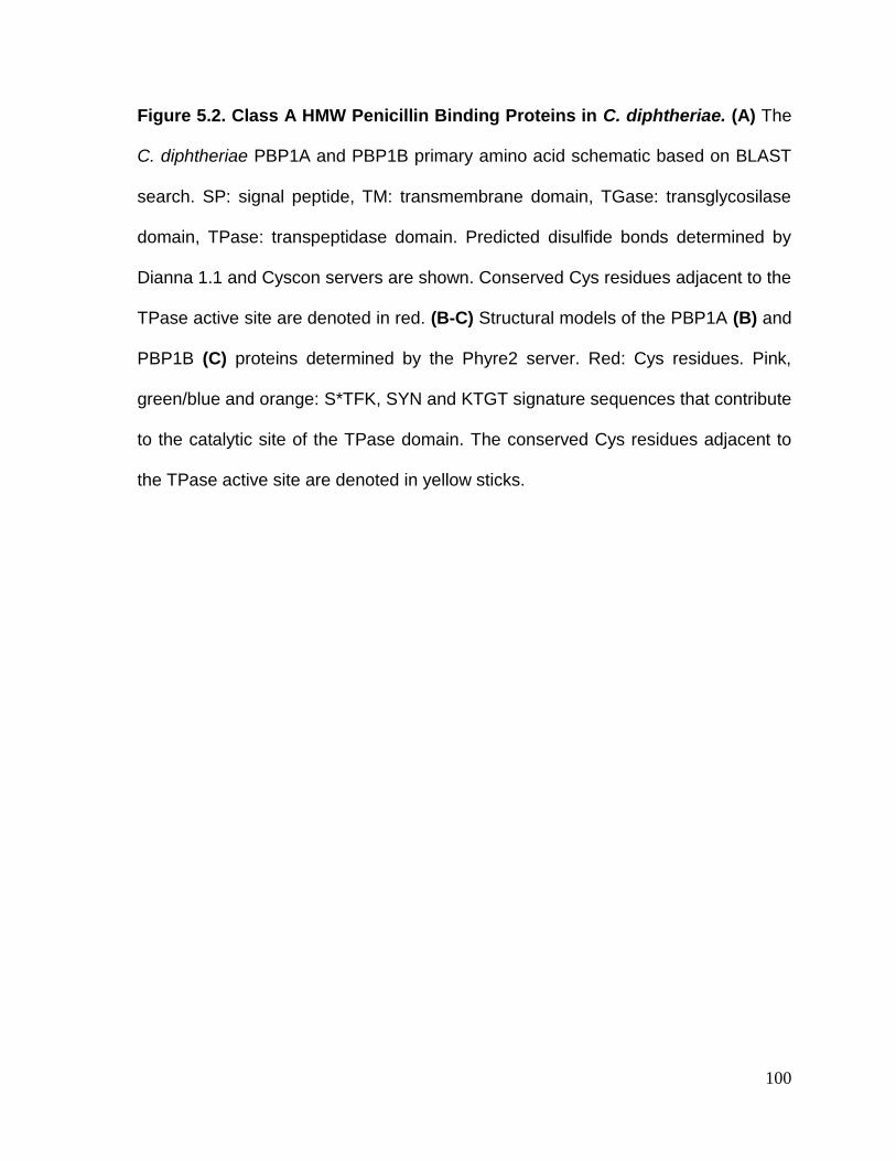
100
Figure 5.2. Class A HMW Penicillin Binding Proteins in C. diphtheriae. (A) The
C. diphtheriae PBP1A and PBP1B primary amino acid schematic based on BLAST
search. SP: signal peptide, TM: transmembrane domain, TGase: transglycosilase
domain, TPase: transpeptidase domain. Predicted disulfide bonds determined by
Dianna 1.1 and Cyscon servers are shown. Conserved Cys residues adjacent to the
TPase active site are denoted in red. (B-C) Structural models of the PBP1A (B) and
PBP1B (C) proteins determined by the Phyre2 server. Red: Cys residues. Pink,
green/blue and orange: S*TFK, SYN and KTGT signature sequences that contribute
to the catalytic site of the TPase domain. The conserved Cys residues adjacent to
the TPase active site are denoted in yellow sticks.

101
Figure 5.3. pbp1A and pbp1B are required for normal cell morphology in C.
diphtheriae. (A-F) Stationary cultures of C. diphtheriae strains grown at 30°C in HIB
were diluted into fresh media and incubated at 30°C or 37°C for 5 hours. Cells were
harvested and immobilized on carbon-coated nickel grids. Samples were stained with
1% uranyl acetate and analyzed by transmission electron microscopy. Scale bars
indicate 0.5 µm.

102
in the mdbA mutant (See section 5.2.3). These results suggest that overexpression
of PBPs from a multi-copy plasmid is lethal to C. diphtheriae.
5.2.3. Overexpression of PBP1A/1B/2B rescues the defects of the mdbA
mutant
Studies in B. subtillis and E. coli showed that some PBPs are substrates of
oxidoreductase enzymes, and disulfide bond formation and breakage is important for
their function [39, 80, 204]. Deletion of mdbA causes a cell division defect and
abnormal nascent peptidoglycan distribution in C. diphtheriae [56]. Furthermore, the
remarkable similarity of the pbp mutant phenotypes (Figure 5.4) to the C. diphtheriae
mdbA mutant grown at the non-permissive temperature supports the proposal that
PBPs are substrates of MdbA. To begin to test this hypothesis, recombinant plasmids
expressing individual or multiple combinations of PBPs predicted to form disulfide
bonds in vivo (PBP1A, PBP1B and PBP2B) were electroporated into the C.
diphtheriae mdbA mutant. The cell morphology of the resulting strains was
examined by transmission electron microscopy. Simultaneous overexpression of all
PBPs predicted to form disulfide bonds in vivo rescued the cell morphology defect of
the mdbA mutant grown at the non-permissive temperature (37C). However,
expression of individual proteins or a combination of PBP1A and PBP1B did not. The
results suggest that in the absence of mdbA, oxidative folding and function of multiple
PBPs predicted to form disulfide bonds is disrupted. Additionally, this data is
consistent with my previous results indicating that PBP1A and PBP1B have non-
redundant functions.

103
Figure 5.4. Ectopic expression of multiple Cys-containing PBPs rescues the
ΔmdbA temperature-sensitive phenotype. (A-L) Stationary cultures of C.
diphtheriae strains grown at 30°C in HIB were diluted into fresh media and incubated
at 30°C or 37°C for 5 hours. Cells were harvested and immobilized on carbon-coated
nickel grids. Samples were stained with 1% uranyl acetate and analyzed by
transmission electron microscopy. Scale bars indicate 0.5 µm.

104
5.2.4. The disulfide bond forming machinery is required to maintain basal PBP
protein levels
Disulfide bonds are important for proper folding and stability of secreted
proteins [27]. Unfolded proteins are more sensitive to proteolysis degradation than
proteins in their native state [205, 206]. Since overexpression of multiple Cys-
containing PBPs rescues the cell division defect of ΔmdbA, I propose that in the
absence of MdbA, PBPs fail to form native disulfide bonds. This likely leads to PBPs
misfolding and subsequent degradation by proteases [27]. If disulfide bond formation
is required for folding and stability of PBPs in vivo, I anticipated that decreased
quantities of PBPs will be detected in mdbA cells compared to the wild-type strain.
I employed Western blot analysis of C. diphtheriae protoplasts using affinity-
purified antibodies specific for PBP1A (α-PBP1A) and PBP1B (α-PBP1B), to assess
stability of PBPs in vivo. pbp1A is predicted to encode a 77.25 kDa transmembrane
protein. Similarly, pbp1B is predicted to encode a 83.46 kDa transmembrane protein.
Immunoblotting with α-PBP1A and α-PBP1B antibodies detected bands that migrated
at the expected molecular masses in the wild-type strain, but that were absent in the
corresponding deletion mutants (Figure 5.5A and B). PBP1A protein levels in the
mdbA mutant were comparable to that of the wild-type strain (Figure 5.5A). However,
a reduction in the PBP1A signal was observed in ΔmdbA cells expressing a
catalytically inactive MdbA protein, where cysteine 91 within the active site was
replaced with alanine (pCxxA). Deletion of mdbA caused a significant decrease in
PBP1B protein levels of cells grown at the permissive temperature (30°C), as
compared to the wild-type strain (Figure 5.5B). PBP1B protein levels were restored

105
by expression of mdbA in trans (pMdbA). However, expression of the catalytically
inactive MdbA variant (pCxxA) failed to restore the wild-type phenotype. Furthermore,
PBP1B protein levels were restored in the ΔmdbA compensatory mutant (ΔmdbA-
S1) that overexpress the thiol-disulfide oxidoreductase TsdA described in chapter 4.
These data suggest that in absence of active MdbA, oxidative folding of PBPs is
disrupted and PBP proteins are unstable. This results in a decreased amount of PBPs
that are successfully folded and embedded in the cell membrane to perform their
function, which agrees with the cell division defect observed in mdbA mutant cells
(See Figure 5.4H).
To ensure that the decreased amount of PBP proteins detected in the mdbA
mutant was due to the oxidative folding defect of this strain, I determined expression
of pbp1A and pbp1B by quantitative RT-PCR. Remarkably, the quantitative RT-PCR
analysis demonstrated that the ΔmdbA strain significantly overexpresses both pbp1A
and pbp1B compared to the wild-type strain (Figure 5.5C and D). Overexpression of
these genes did not result in increased levels of PBP1B protein, but could explain the
wild-type levels of PBP1A protein detected in the mdbA mutant strain. Furthermore,
expression of pbps in the ΔmdbA compensatory mutant was comparable to the wild-
type strain, confirming that restoration of PBP protein levels is likely due to oxidative
folding of PBPs by the alternate oxidoreductase TsdA and not the result of increased
expression of these genes.

106
Figure 5.5. Cells lacking active MdbA present reduced PBP stability. (A-B) Equal
amounts of protein samples from C. diphtheriae strains were subjected to SDS-
PAGE, followed by immunoblotting with α-PBP1A and α-PBP1B. Molecular mass
markers (kDa) are indicated. Coomassie blue staining of protein samples was used
as loading control. A representative image of three experiments is shown. (C-D)
Expression of pbp1A and pbp1B in C. diphtheriae strains was determined by qRT-
PCR analysis. Average values of two experiments performed in triplicate are shown.
*p<0.05 determined by unpaired two-tailed t test using GraphPad Prism. ΔmdbA-S1
indicates ΔmdbA suppressor mutant 1.
107
5.2.5. Growth in synthetic minimal media rescues the mdbA cell division
defect
The temperature sensitive phenotype of the mdbA mutant was puzzling. Since
mdbA is dispensable for growth of C. diphtheriae at 30°C but not at 37°C, it was
postulated that additional oxidoreductase enzymes compensate for the loss of MdbA
at low replication rates. In agreement, the ΔmdbA compensatory mutant S1, which
overexpresses the alternate oxidoreductase TsdA, presented wild-type levels of
PBP1A and PBP1B (See section 5.2.4). To test this hypothesis, I examined cell
morphology in low replication rate conditions by growing cells in synthetic minimal
media. Overnight cultures in rich media (HIB) incubated at 30°C were diluted into
fresh synthetic minimal media or rich media and incubated at 37°C for 5 hours.
Bacterial cultures were then processed for transmission electron microscopy
analysis. Remarkably, the ΔmdbA strain grown in synthetic minimal media at 37°C
maintained a cell morphology similar to the wild-type strain (Figure 5.6A and D). In
addition to forming the characteristic clumps and chains, and losing the rod shape
morphology (See Figure 5.4H); a higher number (29%) of ΔmdbA cells grown in rich
media died when cultures were incubated at 37°C (Figure 5.6C). This is demonstrated
by increased staining with uranyl acetate of dead cells (denoted by arrow heads in
Figure 5.6C). These data suggests that at low replication rates achieved by incubation
in rich media at 30°C (See figure 5.4B) or in synthetic minimal media at 37°C (Figure
5.6D), mdbA mutant cells are capable of folding enough disulfide bond-containing cell
division proteins (for example PBPs) to maintain a cell morphology similar to the wild-
type strain.

108
Figure 5.6. Growth in synthetic minimal media rescues the mdbA cell division
defect. (A-D) Stationary cultures of C. diphtheriae strains grown at 30°C in rich media
were diluted into fresh rich or minimal media and then incubated at 37°C for 5 hours.
Cells were harvested and immobilized on carbon-coated nickel grids. Samples were
stained with 1% uranyl acetate and analyzed by Transmission electron microscopy.
Dead cells show increased staining with uranyl acetate and are denoted by red
arrowheads. The percentage of dead cells per sample is indicated in the upper right
corner of each representative micrograph. Scale bars indicate 0.5 µm.

109
5.3. DISCUSSION
The requirement of the disulfide bond forming machinery for growth in
Actinobacteria has not been previously studied. Results discussed in this chapter
provide insights into the dependence on disulfide bond formation for cell division. The
major oxidoreductase MdbA is essential for cell viability in A. oris and C. matruchotii
[52, 55], but deletion of mdbA results in a temperature-sensitive growth defect in C.
diphtheriae [56]. This makes C. diphtheriae a more suitable model system to study
the requirement of the disulfide bond forming machinery for viability. The cell division
and peptidoglycan distribution defects observed in a ΔmdbA mutant grown at the non-
permissive temperature lead to the logical hypothesis that cell division proteins are
substrates of MdbA. I employed a bioinformatic approach to identify putative MbdA
substrates involved in cell division, by evaluating their predicted location within the
cell, as well as the number of cysteine residues and predicted disulfide bonds. My
bioinformatic analysis identified PBP1A, PBP1B, PBP2B and FtsK as prospective
candidates to be MdbA substrates involved in cell division in vivo. By genetically and
biochemically characterizing two representative putative substrates, PBP1A and
PBP1B, I have established that these two components of the cell wall biosynthesis
machinery require the major disulfide bond-forming machine MdbA for stability and
function.
In B. subtillis, a sporulation-specific HMW class B PBP contains two conserved
cysteine residues near the catalytic site of the transpeptidase domain, that are
important for protein stability and peptidoglycan synthesis during sporulation [82].
These cysteine residues form a disulfide bond that is required for redox regulation of

110
PBP enzymatic activity [80, 81, 207]. Similarly, a disulfide bond forms between
corresponding conserved cysteine residues in the homologous PBPA of M.
tuberculosis [208, 209]. I have shown that both class A PBPs in C. diphtheriae have
a conserved predicted disulfide bond close to the active site of the transpeptidase
domain. The conservation and positioning of these cysteine residues suggest they
might play a role for transpeptidase enzymatic activity. Even though a role for cell
elongation has been proposed for PBP1A and PBP1B homologs in C. glutamicum
[187], my data indicate that PBP functions are not completely conserved between C.
diphtheriae and C. glutamicum. By electron microscopy, both Δpbp1A and Δpbp1b,
present strong but divergent cell division defects (Figure 5.3). Divergent cell
morphology phenotypes observed in Δpbp1A and Δpbp1b could be due to a C-
terminal PASTA (penicillin-binding protein and serine/ threonine kinase associated
domain) domain that is present in PBP1B but absent in PBP1A (Figure 5.2A). PASTA
domains are found at the C-terminus of some PBPs and eukaryotic-like protein
serine/ threonine kinases (PSTKs) in a variety of bacteria [210]. PASTA domains
have been proposed to have a sensory function and to bind -lactam antibiotics and
peptidoglycan [211, 212]. Since recognition of peptidoglycan precursors has been
proposed to determine localization of PASTA kinases involved in cell division [213,
214], it is possible that the presence of the PASTA domain in PBP1B could
differentially affect localization and the role of this protein in cell division with respect
to PBP1A.
I argued that the cell division defect of the mdbA mutant is due to reduced
folding and activity of PBPs. Therefore, I tested if overexpression of PBPs could
111
compensate for the loss of MdbA. I found that simultaneous expression of multiple
PBPs rescued the morphology defect of the mdbA mutant grown at the non-
permissive temperature (37C). Background oxidation of exported proteins can occur
in the presence of oxygen [183]. E. coli mutants of the DsbAB machinery can grow
aerobically, but not anaerobically [36, 63]. This has been attributed in part to
spontaneous oxidative folding of the cell division protein FtsN when cells grow in
presence of oxygen [215]. I believe that a similar mechanism of background oxidative
folding allows viability at 37C of mdbA mutants overexpressing PBPs. Because
background oxidation of exported proteins is less efficient and more error prone than
oxidation catalyzed by a dedicated disulfide bond-forming enzyme [183],
overexpression of PBPs increases the number of PBP molecules that are properly
folded and functional.
As unfolded proteins are more susceptible to protease degradation, I detected
decreased quantities of PBPs in the membranes of cells lacking an active MdbA
(Figure 5.5A & B). This data agrees with a requirement of disulfide bonds for PBP
structural integrity and therefore function, and explains the growth defect observed in
ΔmdbA cells. Interestingly, PBP1A protein levels observed in the mdbA mutant strain
were not as decreased as PBP1B protein levels. This is probably due to the lower
number of cysteine residues in this protein (2 vs. 9 in PBP1B), which makes PBP1A
less susceptible to misfolding in the absence of MdbA, compared to PBP1B.
Additionally, overexpression of pbp1A was much higher than pbp1B in the mdbA
mutant strain, which likely contributes to a higher number of PBP1A molecules that
are correctly folded in this strain (Figure 5.5C and D). In agreement, the mdbA mutant

112
strain expressing a catalytically inactive MdbA (pCxxA), which did not overexpress
pbp1A, presented lower levels of PBP1A protein (Figure 5.5A). Overexpression of
pbps in the mdbA mutant is potentially a compensatory mechanism that C.
diphtheriae employs to ensure that more PBP molecules are correctly folded and to
catalyze cell wall biosynthesis ensuring cell viability when oxidative folding is
disrupted. The relationship between the disulfide bond forming machinery and
mechanisms governing regulation of pbps in C. diphtheriae should be the focus of
further studies.
Finally, I have also demonstrated that oxidative folding of PBPs can be
mediated by the alternate oxidoreductase TsdA (Characterized in chapter 4).
Overexpression of TsdA by the mdbA compensatory mutant S1 restored PBP
protein levels (Figure 5.5A and B). Additionally, when I induced low replicative rates
by growing cells in synthetic minimal media (Figure 5.6), mdbA maintained a
morphology similar to the wild-type strain upon incubation of bacterial cultures at the
non-permissive temperature (37C). Overall, these results indicate that basal levels
of TsdA mediate oxidative folding of sufficient amounts of PBPs to allow viability of
mdbA at low replicative rates, for example when cells are incubated at 30C or grow
in synthetic minimal media.

113
CHAPTER 6. Concluding Remarks and Future Implications

114
Traditionally, disulfide bond forming pathways in prokaryotes were assumed
to be confined to the periplasm of Gram-negative bacteria, due to the oxidative nature
of this compartment. Because Gram-positive bacteria lack an outer membrane and a
conventional periplasmic space, it was believed that they avoid exporting proteins
containing disulfide bonds. However, a very important study by Daniels and
colleagues [49] demonstrated that in contrast to Gram-positive Firmicutes, Gram-
positive Actinobacteria secrete a high number of proteins containing two or more
cysteine residues. In fact, secreted proteins in Actinobacteria have a bias for
containing an even number of cysteine residues, which suggested that they form
disulfide bonds in vivo. Genes encoding oxidoreductase orthologues have also been
found in the genomes of many Actinobacteria [68]. Moreover, it is believed that the
complex cell wall of Gram-positive Actinobacteria, constituted by a thick
peptidoglycan-arabinogalactan and mycolic acid layers, contributes to formation of a
periplasmic-like space in the cell envelope [216, 217], which might in part explain why
Actinobacteria favor secretion of cysteine-containing proteins. Studies in different
bacterial species have demonstrated that although similarities with the classical Dsb
system of E. coli exist, oxidative protein folding pathways are exceptionally diverse
and current knowledge of the E. coli Dsb system cannot be assumed to be a universal
paradigm for disulfide bond formation [50, 67, 68]. Recently, disulfide bond forming
enzymes in Actinobacteria such as Actinomyces, Corynebacterium and
Mycobacterium have been characterized [52, 55, 56, 59, 60, 218, 219]. However,
many aspects of the oxidative protein folding pathways in Actinobacteria remain
poorly understood.

115
The first part of this dissertation (Chapter 3), focused on the characterization
of novel factors involved in the reoxidation of the primary thiol-disulfide
oxidoreductase MdbA in A. oris. Using adhesive pilus assembly as a marker for
correct disulfide bond formation we identified the nuo operon, encoding subunits of
the NADH dehydrogenase, and ubiE, encoding a menaquinone C-methyltransferase
involved in menaquinone biosynthesis, to be required for proper oxidative protein
folding. Analyses of the representative nuoA and ubiE mutants, showed that these
genes are required for display of wild-type levels of the disulfide bond-containing pilin
subunits FimA and CafA, which constitute type 2 pili. The fact that pilus assembly
was rescued by overexpression of MdbA from a plasmid, strongly suggested that this
defect is related to disruption of disulfide bond formation. My results agree with
previous studies from our lab indicating that the disulfide bond forming machinery is
required for pilus assembly, coaggregation and biofilm formation in A. oris [55, 60].
The bacterial electron transport chain provides the oxidizing power to maintain
the E. coli DsbAB in an oxidized state [40, 43]. In E. coli DsbB transfers electrons
from DsbA to ubiquinone or menaquinone [40], therefore maintaining DsbA in an
oxidized active state. DsbB has a quinone binding site, and crystallography studies
determined this site is occupied by quinones in vivo [220]. Our studies indicate that
menaquinone, the main quinone utilized in the electron transport chain of Gram-
positive Actinobacteria [142], is required for reoxidation of MdbA. Deletion of the ubiE
gene, encoding an enzyme catalyzing the last step of menaquinone biosynthesis
[221], disrupts MdbA reoxidation and results in accumulation of the reduced form of
MdbA. Furthermore, addition of exogenous menaquinone-4 to the growth media

116
rescued the pilus assembly and growth phenotypes observed in the ubiE mutant
strain. Similar to the DsbAB system [41], we argue that in A. oris electrons resulting
from reoxidation of MdbA are transferred from VKOR to menaquinone (Figure 6.1).
Therefore, the electron transport chain provides the oxidative power that maintains
the MdbA/VKOR system in an oxidized and active state. Structural studies of A. oris
VKOR would be crucial to elucidate if similar to DsbB, VKOR directly interacts with
quinones. Furthermore, in vitro reconstitution of the A. oris oxidative folding pathway
using menaquinone as electron acceptor would shed light into the electron transfer
process that takes place during reoxidation of MdbA. Monitoring of the menaquinone
reduction status would specifically determine if this process is menaquinone-
dependent [41].
The nuoA locus, encoding a subunit of the NADH dehydrogenase complex I,
was also found necessary for MdbA reoxidation. A role for the NADH dehydrogenase
in disulfide bond formation has not been described before. Because of the activity of
the complex I as NADH:quinone oxidoreductase [134, 222, 223], it is possible that
deletion of nuo genes affects the general electron flow of the electron transport chain,
indirectly affecting MdbA reoxidation. Due to its large size and complexity, and its role
in energy metabolism, dissecting the contributions of the NADH Complex I in MdbA
reoxidation might result challenging. Nonetheless, the fact that a role for the NADH
dehydrogenase complex I in oxidative protein folding has not been described before,
make this a very interesting avenue for future research.

117
Figure 6.1. A model for MdbA/VKOR reoxidation in A. oris. A. oris VKOR
catalyzes reoxidation of MdbA after disulfide bond formation. It is proposed that
VKOR transfer electrons resulting from the reoxidation reaction to menaquinone. The
electron carrier menaquinone can potentially transfer electrons to other electron
acceptors within the electron transport chain (ETC). (see text for details).

118
Overall, part I of this dissertation discovered novel factors involved in disulfide
bond formation in A. oris, and contributes to our understating of the mechanisms
involved in reactivation of the oxidative folding machinery. Because disruption of the
disulfide bond forming machinery results in pathogenicity and growth phenotypes in
Actinobacteria, it seems a promising target for novel antimicrobial therapies. For
example, it would be interesting to investigate the effects of compounds targeting
menaquinone in A. oris. Studies in M. tuberculosis showed that inhibition of the
menaquinone biosynthesis pathway significantly reduces growth of drug-resistant
strains in vitro [138]. Interestingly, a small chemical compound directly interacting with
menaquinone has bactericidal activity and reduces mortality in a murine model of
systemic Staphylococcus aureus infection [224]. Since menaquinone is not found in
mammalian cell membranes [138], antimicrobial targeting of menaquinone potentially
encompasses advantages over other therapies that might have off target effects in
humans.
For the second part of this dissertation (chapter 4 and chapter 5), I focused
on dissecting different aspects of the oxidative folding pathways in C. diphtheriae. In
chapter 4, I characterized the regulation of an alternate oxidoreductase that seems
to be responsible for viability of the mdbA strain at the non-permissive temperature.
In chapter 5, I characterized MdbA substrates involved in cell division in an effort to
elucidate the essentiality of this machinery for bacterial growth.
Previous reports from our lab identified a putative oxidoreductase in C.
diphtheriae named TsdA [56]. Our structural analysis of a TsdA CxxS mutant

119
confirmed that TsdA has DsbA protein family conserved features including a
thioredoxin-like domain and an extended -helical domain, and supports that TsdA is
a thiol-disulfide oxidoreductase enzyme. Regulation of this novel oxidoreductase was
puzzling because it is expressed at low levels in the wild-type strain, and its deletion
did not result in any obvious phenotypes under standard laboratory growth conditions.
Here, I have elucidated that the promoter driving tsdA expression is dependent on
the housekeeping sigma factor A. Because the A binding sequence is weakly
conserved, tsdA is poorly expressed in cells in logarithmic growth phase. I determined
that mdbA compensatory mutants overexpressing tsdA carry a mutation that results
in an extended sigma factor A promoter sequence, which significantly increases the
strength of the promoter. Even though it is possible that the mdbA compensatory
mutation affects regulation of tsdA by other regulatory factors, this is unlikely.
Transcription factors that bind to DNA sequences that overlap with the -10 or adjacent
regions of bacterial promoters often repress gene expression by steric hindrance
caused by blockage of RNA polymerase access to the promoter [175]. On the other
hand, transcription factors that positively regulate gene expression (activators) often
bind to motifs upstream of the promoter or to a site in the promoter adjacent to (or
overlapping with) the –35 element [175]. A TG dinucleotide at positions -14, -15 of
bacterial promoters has been widely described to increase promoter activity [115,
160, 162, 163]. In this sense, it is logical to conclude that the T-to-G mutation located
at position -14 enhances interaction with RNA polymerase sigma factor A, especially
because the wild-type tsdA promoter sequence is suboptimal. Nonetheless, because
our bioinformatic approach identified a putative DtxR binding motif upstream of the

120
-10 tsdA promoter region, we were motivated to investigate if the T-to-G mutation
disrupts DtxR repression resulting in increased tsdA gene expression. Our
experimental approach found no evidence that DtxR is involved in regulation of tsdA
expression.
Interestingly, I found that tsdA transcription was induced when cells were
incubated at 40C. To our knowledge, differential regulation of an oxidoreductase by
temperature has not been reported before. The TsdA expression pattern suggests a
role for this alternate oxidoreductase in resistance to heat stress and potentially
virulence. A possible role for TsdA in C. diphtheriae pathogenesis is intriguing.
Analysis of tsdA expression, as well as comparison of wild-type and tsdA mutant
replication and survival in human phagocytes [225], and animal models of infection
such as Caenorhabditis elegans and guinea pig would provide insights into the role
of TsdA in virulence [98, 225-227]. Sigma factor H is the central regulator that
mediates bacterial response to heat [228, 229]. Induction of tsdA expression by heat
and the identification of a putative sigma factor H promoter upstream of the tsdA
transcriptional start site, prompted us to investigate the role of this alternative sigma
factor in tsdA regulation. Surprisingly, I found that tsdA is upregulated in the absence
of sigH, encoding alternative sigma factor H. Overexpression of tsdA in the sigH
strain suggests that regulation of tsdA by sigma factor H is indirect and it is likely that
additional transcription factors play a role in expression of tsdA. Our PtsdA-gfp
transcriptional reporter vector could be used in a transposon mutant screen to identify
additional genes involved in tsdA regulation.

121
Finally, in chapter 5 I demonstrate that MdbA is required for structural stability
and activity of PBPs, suggesting that PBPs are substrates of MdbA. An in silico
approach identified four exported cell division proteins predicted to form disulfide
bonds in vivo. The characterization of two representative proteins, PBP1A and
PBP1B, showed that they contain conserved cysteine residues in the transpeptidase
domain, and have distinct functions in cell division. Consistent with requirement of
disulfide bonds for protein stability, cells lacking a catalytically active MdbA enzyme
presented decreased amounts of PBPs. Furthermore, overexpression of multiple
cysteine-containing PBPs rescued the morphology defect of ΔmdbA, confirming
PBPs activity is disrupted in absence of mdbA. Studies demonstrating a direct
interaction of PBPs with MdbA would unequivocally demonstrate that MdbA catalyzes
oxidative folding of PBPs. In an effort to test whether MdbA directly interacts with PBP
proteins in vivo, I performed a preliminary pulldown assay. Membrane lysates from
ΔmdbA cells expressing His-tagged versions of MdbA or the MdbA-CxxA mutant,
were incubated with a nickel super flow resin. The obtained eluates were reactive to
antibodies specific for MdbA (α-MdbA) and PBP1B (α-PBP1B), indicating direct
interaction of PBP1B with MdbA in vivo. Furthermore, a high molecular weight band
(~110 kDa), consistent with the expected migration of an MdbA-PBP1B complex, was
detected in the α-PBP1B immunoblot for the MdbA-CxxA mutant sample (Figure 6.2).
I hypothesize that an MdbA-PBP1B intermediate formed via a mixed-disulfide bond
during oxidative folding cannot be resolved in the MdbA-CxxA mutant.
Complementary experiments to confirm these findings, such as elucidating the

122
Figure 6.2. A putative mixed-disulfide MdbA-PBP1B intermediate is detected by
a pull-down assay. Clear lysates obtained from membrane fractions of
ΔmdbA/pMdbA-CxxC-His were incubated with anti-His agarose beads, followed by
immunoblotting with α-His, α-MdbA or α-Pbp1B. MdbA, Pbp1B and MdbA-Pbp1B
complex (black arrows) and molecular weight markers (kDa) are shown.

123
identity of the high molecular weight band by Mass-spectrometry analysis would
provide further evidence that MdbA catalyzes disulfide bond formation in PBPs.
Our finding that cells lacking mdbA overexpress PBPs is intriguing. PBPs are
usually constitutively expressed, however it has been argued that they can be
differentially expressed in response to environmental conditions [162, 230]. In
addition, increased PBP expression in response to stress exerted by cell wall active
antibiotics has been reported in Gram-positive Firmicutes [162, 231, 232]. Since a
relationship has been established between the Cpx-envelope stress response and
oxidative protein folding in E. coli (discussed in chapter 1) [71], it is possible that cell
envelope stress caused by disruption of peptidoglycan synthesis in the ΔmdbA strain
induces PBP expression. Interestingly, I found that cells expressing the catalytically
inactive MdbA-CxxA mutant do not overexpress PBPs. Therefore, potentially multiple
unknown regulatory mechanisms play a role in regulation of PBP expression upon
disruption of oxidative folding.
The studies in this dissertation indicate that the essentiality of the oxidative
folding machinery in C. diphtheriae is related to disruption of disulfide bond formation
in PBPs. Furthermore, our data indicates that survival of ΔmdbA at low replicative
rates is due to basal expression of the alternate oxidoreductase TsdA. However,
important questions regarding the essentiality of the disulfide bond forming machinery
remain. Why does C. diphtheriae favor expression of PBPs containing cysteine
residues? Does this provide a physiological advantage? Is this mechanism conserved
in other bacterial groups? Since Actinobacteria secrete abundance of proteins

124
containing cysteine residues, one could argue that this is likely to be a general
mechanism that provides protein stability in the corynebacterial natural niches, rather
than a specific bias toward cell division proteins. Interestingly, in a survey of the E.
coli secretome I found that of 11 PBPs identified, 3 are predicted to form disulfide
bonds. However, disulfide bridges in these PBPs (PBP1A, PBP1B and PBP4) are
dispensable for function [204, 233, 234]. Therefore, the requirement of disulfide
bonds for PBPs activity does not seem to be universal. On the other hand, a survey
of exported proteins in A. oris identified only 2 cell division proteins containing
cysteine residues. Thus, the essentiality of MdbA for A. oris growth might be related
to disruption of other systems. Whether oxidative folding is required for activity of cell
division factors in other bacterial groups remains to be explored. Nonetheless, the
results presented here are likely to lead to exciting future studies.
Previous reports from our group showed that overexpression of TsdA restored
Diphtheria toxin and SpaA pilin protein levels in cells lacking mdbA [114]. Here I
showed that PBP protein levels are also restored in the mdbA compensatory mutant
overexpressing TsdA. These data suggest that TsdA has a broad substrate specificity
and might provide a compensatory mechanism of oxidative folding under unfavorable
or stress conditions. I propose that under normal growth conditions expression of
MdbA is sufficient to catalyze oxidative folding in C. diphtheriae. However, when
bacterial cells are exposed to stresses encountered within the host, such as elevated
temperatures, expression of the alternate oxidoreductase TsdA is induced by a yet
unknown mechanism. In this situation, increased amounts of TsdA molecules aid in
the oxidative folding of diverse substrates (Figure 6.3). Stimuli like temperature and
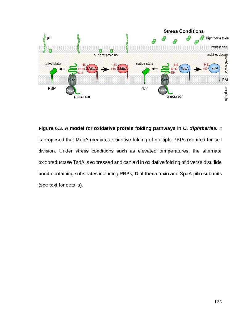
125
Figure 6.3. A model for oxidative protein folding pathways in C. diphtheriae. It
is proposed that MdbA mediates oxidative folding of multiple PBPs required for cell
division. Under stress conditions such as elevated temperatures, the alternate
oxidoreductase TsdA is expressed and can aid in oxidative folding of diverse disulfide
bond-containing substrates including PBPs, Diphtheria toxin and SpaA pilin subunits
(see text for details).

126
iron concentrations indicate to pathogens they are within the host and have deep
effects in bacterial gene regulation [74]. For example, expression of the disulfide
bond-containing Diphtheria toxin is induced within the host [169]. Additionally,
expression of the disulfide-bond containing SpaA-type pilus is highly prevalent among
clinical isolates of C. diphtheriae [98]. Environmental stresses affecting protein
stability together with high expression of disulfide bond-containing proteins, would
exacerbate the necessity for an additional oxidoreductase like TsdA to help maintain
protein homeostasis and ensure survival of C. diphtheriae.
Overall, the studies presented in this dissertation provide insights into diverse
aspects of the oxidative folding pathways in Actinobacteria that have not been
explored before. I showed that components of the electron transport chain are
involved in MdbA reoxidation. Secondly, by characterizing regulation of TsdA by
temperature I have discovered a novel stimulus controlling expression of a bacterial
oxidoreductase. Finally, I determined that this alternate oxidoreductase is involved in
survival of cells lacking mdbA and that the requirement of mdbA for cell division is
related to incorrect folding of PBPs. Due to the impact of thiol-disulfide
oxidoreductases in bacterial physiology, my work provides knowledge applicable not
only to protein folding, but also to bacterial pathogenesis and potentially the discovery
of novel antimicrobial therapies targeting bacterial viability and virulence.
127
REFERENCES
1. Willey JM, Sherwood, L.M., Woolverton, C.J.: Prescott, Harley, and Klein's
microbiology, 7th edn: McGraw-Hill; 2008.
2. Kononen E, Wade WG: Actinomyces and related organisms in human
infections. Clin Microbiol Rev 2015, 28(2):419-442.
3. Mark Welch JL, Rossetti BJ, Rieken CW, Dewhirst FE, Borisy GG:
Biogeography of a human oral microbiome at the micron scale. Proc Natl
Acad Sci U S A 2016, 113(6):E791-800.
4. Zijnge V, van Leeuwen MB, Degener JE, Abbas F, Thurnheer T, Gmur R,
Harmsen HJ: Oral biofilm architecture on natural teeth. PLoS One 2010,
5(2):e9321.
5. Brailsford SR, Tregaskis RB, Leftwich HS, Beighton D: The predominant
Actinomyces spp. isolated from infected dentin of active root caries
lesions. J Dent Res 1999, 78(9):1525-1534.
6. Tanner AC, Mathney JM, Kent RL, Chalmers NI, Hughes CV, Loo CY, Pradhan
N, Kanasi E, Hwang J, Dahlan MA et al: Cultivable anaerobic microbiota of
severe early childhood caries. J Clin Microbiol 2011, 49(4):1464-1474.
7. Bowden GH, Nolette N, Ryding H, Cleghorn BM: The diversity and
distribution of the predominant ribotypes of Actinomyces naeslundii
genospecies 1 and 2 in samples from enamel and from healthy and
carious root surfaces of teeth. J Dent Res 1999, 78(12):1800-1809.

128
8. Ximenez-Fyvie LA, Haffajee AD, Martin L, Tanner A, Macuch P, Socransky
SS: Identification of oral Actinomyces species using DNA probes. Oral
Microbiol Immunol 1999, 14(4):257-265.
9. Gibbons RJ, Hay DI, Cisar JO, Clark WB: Adsorbed salivary proline-rich
protein 1 and statherin: receptors for type 1 fimbriae of Actinomyces
viscosus T14V-J1 on apatitic surfaces. Infect Immun 1988, 56(11):2990-
2993.
10. Kolenbrander PE, Palmer RJ, Jr., Periasamy S, Jakubovics NS: Oral
multispecies biofilm development and the key role of cell-cell distance.
Nat Rev Microbiol 2010, 8(7):471-480.
11. Rickard AH, Gilbert P, High NJ, Kolenbrander PE, Handley PS: Bacterial
coaggregation: an integral process in the development of multi-species
biofilms. Trends Microbiol 2003, 11(2):94-100.
12. Mishra A, Devarajan B, Reardon ME, Dwivedi P, Krishnan V, Cisar JO, Das A,
Narayana SV, Ton-That H: Two autonomous structural modules in the
fimbrial shaft adhesin FimA mediate Actinomyces interactions with
streptococci and host cells during oral biofilm development. Mol
Microbiol 2011, 81(5):1205-1220.
13. Mishra A, Wu C, Yang J, Cisar JO, Das A, Ton-That H: The Actinomyces
oris type 2 fimbrial shaft FimA mediates co-aggregation with oral
streptococci, adherence to red blood cells and biofilm development. Mol
Microbiol 2010.
129
14. Ammann TW, Belibasakis GN, Thurnheer T: Impact of early colonizers on
in vitro subgingival biofilm formation. PLoS One 2013, 8(12):e83090.
15. Petersen PE, Ogawa H: Strengthening the prevention of periodontal
disease: the WHO approach. J Periodontol 2005, 76(12):2187-2193.
16. Brown LJ, Johns BA, Wall TP: The economics of periodontal diseases.
Periodontol 2000 2002, 29:223-234.
17. Hadfield TL, McEvoy P, Polotsky Y, Tzinserling VA, Yakovlev AA: The
pathology of diphtheria. J Infect Dis 2000, 181 Suppl 1:S116-120.
18. Rogers EA, Das A, Ton-That H: Adhesion by pathogenic corynebacteria.
Adv Exp Med Biol, 715:91-103.
19. Ton-That H, Schneewind O: Assembly of pili on the surface of
Corynebacterium diphtheriae. Mol Microbiol 2003, 50(4):1429-1438.
20. Mandlik A, Swierczynski A, Das A, Ton-That H: Pili in Gram-positive
bacteria: assembly, involvement in colonization and biofilm
development. Trends Microbiol 2008, 16(1):33-40.
21. Mandlik A, Swierczynski A, Das A, Ton-That H: Corynebacterium
diphtheriae employs specific minor pilins to target human pharyngeal
epithelial cells. Mol Microbiol 2007, 64(1):111-124.
22. Barksdale L: Corynebacterium diphtheriae and its relatives. Bacteriol Rev
1970, 34(4):378-422.
23. London E: Diphtheria toxin: membrane interaction and membrane
translocation. Biochim Biophys Acta 1992, 1113(1):25-51.

130
24. Naglich JG, Metherall JE, Russell DW, Eidels L: Expression cloning of a
diphtheria toxin receptor: identity with a heparin-binding EGF-like
growth factor precursor. Cell 1992, 69(6):1051-1061.
25. Mitamura T, Higashiyama S, Taniguchi N, Klagsbrun M, Mekada E:
Diphtheria toxin binds to the epidermal growth factor (EGF)-like domain
of human heparin-binding EGF-like growth factor/diphtheria toxin
receptor and inhibits specifically its mitogenic activity. J Biol Chem 1995,
270(3):1015-1019.
26. Sangal V, Hoskisson PA: Evolution, epidemiology and diversity of
Corynebacterium diphtheriae: New perspectives on an old foe. Infect
Genet Evol 2016, 43:364-370.
27. Mamathambika BS, Bardwell JC: Disulfide-linked protein folding
pathways. Annu Rev Cell Dev Biol 2008, 24:211-235.
28. Heras B, Shouldice SR, Totsika M, Scanlon MJ, Schembri MA, Martin JL: DSB
proteins and bacterial pathogenicity. Nat Rev Microbiol 2009, 7(3):215-225.
29. Frand AR, Kaiser CA: The ERO1 gene of yeast is required for oxidation of
protein dithiols in the endoplasmic reticulum. Mol Cell 1998, 1(2):161-170.
30. Anfinsen CB: Principles that govern the folding of protein chains. Science
1973, 181(4096):223-230.
31. Goldberger RF, Epstein CJ, Anfinsen CB: Acceleration of reactivation of
reduced bovine pancreatic ribonuclease by a microsomal system from
rat liver. J Biol Chem 1963, 238:628-635.

131
32. Bardwell JC, Lee JO, Jander G, Martin N, Belin D, Beckwith J: A pathway for
disulfide bond formation in vivo. Proc Natl Acad Sci U S A 1993,
90(3):1038-1042.
33. Peek JA, Taylor RK: Characterization of a periplasmic thiol:disulfide
interchange protein required for the functional maturation of secreted
virulence factors of Vibrio cholerae. Proc Natl Acad Sci U S A 1992,
89(13):6210-6214.
34. Kadokura H, Beckwith J: Detecting folding intermediates of a protein as it
passes through the bacterial translocation channel. Cell 2009,
138(6):1164-1173.
35. Denoncin K, Collet JF: Disulfide bond formation in the bacterial periplasm:
major achievements and challenges ahead. Antioxid Redox Signal 2013,
19(1):63-71.
36. Bardwell JC, McGovern K, Beckwith J: Identification of a protein required
for disulfide bond formation in vivo. Cell 1991, 67(3):581-589.
37. Kadokura H, Tian H, Zander T, Bardwell JC, Beckwith J: Snapshots of DsbA
in action: detection of proteins in the process of oxidative folding.
Science 2004, 303(5657):534-537.
38. Kadokura H, Katzen F, Beckwith J: Protein disulfide bond formation in
prokaryotes. Annu Rev Biochem 2003, 72:111-135.
39. Missiakas D, Georgopoulos C, Raina S: Identification and characterization
of the Escherichia coli gene dsbB, whose product is involved in the

132
formation of disulfide bonds in vivo. Proc Natl Acad Sci U S A 1993,
90(15):7084-7088.
40. Bader M, Muse W, Ballou DP, Gassner C, Bardwell JC: Oxidative protein
folding is driven by the electron transport system. Cell 1999, 98(2):217-
227.
41. Bader MW, Xie T, Yu CA, Bardwell JC: Disulfide bonds are generated by
quinone reduction. J Biol Chem 2000, 275(34):26082-26088.
42. Kobayashi T, Ito K: Respiratory chain strongly oxidizes the CXXC motif of
DsbB in the Escherichia coli disulfide bond formation pathway. EMBO J
1999, 18(5):1192-1198.
43. Kobayashi T, Kishigami S, Sone M, Inokuchi H, Mogi T, Ito K: Respiratory
chain is required to maintain oxidized states of the DsbA-DsbB disulfide
bond formation system in aerobically growing Escherichia coli cells.
Proc Natl Acad Sci U S A 1997, 94(22):11857-11862.
44. Bader M, Muse W, Zander T, Bardwell J: Reconstitution of a protein
disulfide catalytic system. J Biol Chem 1998, 273(17):10302-10307.
45. Takahashi YH, Inaba K, Ito K: Characterization of the menaquinone-
dependent disulfide bond formation pathway of Escherichia coli. J Biol
Chem 2004, 279(45):47057-47065.
46. Hatahet F, Boyd D, Beckwith J: Disulfide bond formation in prokaryotes:
history, diversity and design. Biochim Biophys Acta 2014, 1844(8):1402-
1414.
133
47. Missiakas D, Georgopoulos C, Raina S: The Escherichia coli dsbC (xprA)
gene encodes a periplasmic protein involved in disulfide bond formation.
EMBO J 1994, 13(8):2013-2020.
48. Missiakas D, Schwager F, Raina S: Identification and characterization of a
new disulfide isomerase-like protein (DsbD) in Escherichia coli. EMBO J
1995, 14(14):3415-3424.
49. Daniels R, Mellroth P, Bernsel A, Neiers F, Normark S, von Heijne G,
Henriques-Normark B: Disulfide bond formation and cysteine exclusion in
gram-positive bacteria. J Biol Chem 2010, 285(5):3300-3309.
50. Davey L, Halperin SA, Lee SF: Thiol-Disulfide Exchange in Gram-Positive
Firmicutes. Trends Microbiol 2016, 24(11):902-915.
51. Kouwen TR, van der Goot A, Dorenbos R, Winter T, Antelmann H, Plaisier
MC, Quax WJ, van Dijl JM, Dubois JY: Thiol-disulphide oxidoreductase
modules in the low-GC Gram-positive bacteria. Mol Microbiol 2007,
64(4):984-999.
52. Luong TT, Tirgar R, Reardon-Robinson ME, Joachimiak A, Osipiuk J, Ton-
That H: Structural Basis of a Thiol-Disulfide Oxidoreductase in the
Hedgehog-Forming Actinobacterium Corynebacterium matruchotii. J
Bacteriol 2018, 200(9).
53. Dutton RJ, Wayman A, Wei JR, Rubin EJ, Beckwith J, Boyd D: Inhibition of
bacterial disulfide bond formation by the anticoagulant warfarin. Proc
Natl Acad Sci U S A 2010, 107(1):297-301.
134
54. Sassetti CM, Boyd DH, Rubin EJ: Genes required for mycobacterial growth
defined by high density mutagenesis. Mol Microbiol 2003, 48(1):77-84.
55. Reardon-Robinson ME, Osipiuk J, Chang C, Wu C, Jooya N, Joachimiak A,
Das A, Ton-That H: A Disulfide Bond-forming Machine Is Linked to the
Sortase-mediated Pilus Assembly Pathway in the Gram-positive
Bacterium Actinomyces oris. J Biol Chem 2015, 290(35):21393-21405.
56. Reardon-Robinson ME, Osipiuk J, Jooya N, Chang C, Joachimiak A, Das A,
Ton-That H: A thiol-disulfide oxidoreductase of the Gram-positive
pathogen Corynebacterium diphtheriae is essential for viability, pilus
assembly, toxin production and virulence. Mol Microbiol 2015, 98(6):1037-
1050.
57. Martin JL, Bardwell JC, Kuriyan J: Crystal structure of the DsbA protein
required for disulphide bond formation in vivo. Nature 1993,
365(6445):464-468.
58. Shouldice SR, Heras B, Walden PM, Totsika M, Schembri MA, Martin JL:
Structure and function of DsbA, a key bacterial oxidative folding catalyst.
Antioxid Redox Signal 2011, 14(9):1729-1760.
59. Premkumar L, Heras B, Duprez W, Walden P, Halili M, Kurth F, Fairlie DP,
Martin JL: Rv2969c, essential for optimal growth in Mycobacterium
tuberculosis, is a DsbA-like enzyme that interacts with VKOR-derived
peptides and has atypical features of DsbA-like disulfide oxidases. Acta
Crystallogr D Biol Crystallogr 2013, 69(Pt 10):1981-1994.

135
60. Luong TT, Reardon-Robinson ME, Siegel SD, Ton-That H: Reoxidation of
the Thiol-Disulfide Oxidoreductase MdbA by a Bacterial Vitamin K
Epoxide Reductase in the Biofilm-Forming Actinobacterium
Actinomyces oris. J Bacteriol 2017, 199(10).
61. Vertommen D, Depuydt M, Pan J, Leverrier P, Knoops L, Szikora JP, Messens
J, Bardwell JC, Collet JF: The disulphide isomerase DsbC cooperates with
the oxidase DsbA in a DsbD-independent manner. Mol Microbiol 2008,
67(2):336-349.
62. Bessette PH, Qiu J, Bardwell JC, Swartz JR, Georgiou G: Effect of
sequences of the active-site dipeptides of DsbA and DsbC on in vivo
folding of multidisulfide proteins in Escherichia coli. J Bacteriol 2001,
183(3):980-988.
63. Meehan BM, Landeta C, Boyd D, Beckwith J: The Disulfide Bond Formation
Pathway Is Essential for Anaerobic Growth of Escherichia coli. J Bacteriol
2017, 199(16).
64. Hiniker A, Collet JF, Bardwell JC: Copper stress causes an in vivo
requirement for the Escherichia coli disulfide isomerase DsbC. J Biol
Chem 2005, 280(40):33785-33791.
65. Yu J, Kroll JS: DsbA: a protein-folding catalyst contributing to bacterial
virulence. Microbes Infect 1999, 1(14):1221-1228.
66. Saleh M, Bartual SG, Abdullah MR, Jensch I, Asmat TM, Petruschka L, Pribyl
T, Gellert M, Lillig CH, Antelmann H et al: Molecular architecture of
Streptococcus pneumoniae surface thioredoxin-fold lipoproteins crucial
136
for extracellular oxidative stress resistance and maintenance of
virulence. EMBO Mol Med 2013, 5(12):1852-1870.
67. Reardon-Robinson ME, Ton-That H: Disulfide-Bond-Forming Pathways in
Gram-Positive Bacteria. J Bacteriol 2015, 198(5):746-754.
68. Landeta C, Boyd D, Beckwith J: Disulfide bond formation in prokaryotes.
Nat Microbiol 2018, 3(3):270-280.
69. Choe S, Bennett MJ, Fujii G, Curmi PM, Kantardjieff KA, Collier RJ, Eisenberg
D: The crystal structure of diphtheria toxin. Nature 1992, 357(6375):216-
222.
70. Wilson AC, Hoch JA, Perego M: Two small c-type cytochromes affect
virulence gene expression in Bacillus anthracis. Mol Microbiol 2009,
72(1):109-123.
71. Pogliano J, Lynch AS, Belin D, Lin EC, Beckwith J: Regulation of Escherichia
coli cell envelope proteins involved in protein folding and degradation
by the Cpx two-component system. Genes Dev 1997, 11(9):1169-1182.
72. Hunke S, Keller R, Muller VS: Signal integration by the Cpx-envelope
stress system. FEMS Microbiol Lett 2012, 326(1):12-22.
73. Slamti L, Waldor MK: Genetic analysis of activation of the Vibrio cholerae
Cpx pathway. J Bacteriol 2009, 191(16):5044-5056.
74. Fang FC, Frawley ER, Tapscott T, Vazquez-Torres A: Bacterial Stress
Responses during Host Infection. Cell Host Microbe 2016, 20(2):133-143.
75. Grabowska AD, Wandel MP, Lasica AM, Nesteruk M, Roszczenko P,
Wyszynska A, Godlewska R, Jagusztyn-Krynicka EK: Campylobacter jejuni

137
dsb gene expression is regulated by iron in a Fur-dependent manner and
by a translational coupling mechanism. BMC Microbiol 2011, 11:166.
76. Bringer MA, Rolhion N, Glasser AL, Darfeuille-Michaud A: The
oxidoreductase DsbA plays a key role in the ability of the Crohn's
disease-associated adherent-invasive Escherichia coli strain LF82 to
resist macrophage killing. J Bacteriol 2007, 189(13):4860-4871.
77. Cassat JE, Skaar EP: Iron in infection and immunity. Cell Host Microbe
2013, 13(5):509-519.
78. Andrews SC, Robinson AK, Rodriguez-Quinones F: Bacterial iron
homeostasis. FEMS Microbiol Rev 2003, 27(2-3):215-237.
79. Kpadeh ZZ, Jameson-Lee M, Yeh AJ, Chertihin O, Shumilin IA, Dey R, Day
SR, Hoffman PS: Disulfide bond oxidoreductase DsbA2 of Legionella
pneumophila exhibits protein disulfide isomerase activity. J Bacteriol
2013, 195(8):1825-1833.
80. Liu Y, Carlsson Moller M, Petersen L, Soderberg CA, Hederstedt L: Penicillin-
binding protein SpoVD disulphide is a target for StoA in Bacillus subtilis
forespores. Mol Microbiol 2010, 75(1):46-60.
81. Eichenberger P: The red-ox status of a penicillin-binding protein is an
on/off switch for spore peptidoglycan synthesis in Bacillus subtilis. Mol
Microbiol 2010, 75(1):10-12.
82. Bukowska-Faniband E, Hederstedt L: Transpeptidase activity of penicillin-
binding protein SpoVD in peptidoglycan synthesis conditionally

138
depends on the disulfide reductase StoA. Mol Microbiol 2017, 105(1):98-
114.
83. Eichenberger P, Jensen ST, Conlon EM, van Ooij C, Silvaggi J, Gonzalez-
Pastor JE, Fujita M, Ben-Yehuda S, Stragier P, Liu JS et al: The sigmaE
regulon and the identification of additional sporulation genes in Bacillus
subtilis. J Mol Biol 2003, 327(5):945-972.
84. Imamura D, Kobayashi K, Sekiguchi J, Ogasawara N, Takeuchi M, Sato T:
spoIVH (ykvV), a requisite cortex formation gene, is expressed in both
sporulating compartments of Bacillus subtilis. J Bacteriol 2004,
186(16):5450-5459.
85. Zasada A: Antimicrobial Susceptibility and Treatment. In:
Corynebacterium diphtheriae and Related Toxigenic Species. Edited by
Burkovski A; 2014.
86. Manjelievskaia J, Erck D, Piracha S, Schrager L: Drug-resistant TB: deadly,
costly and in need of a vaccine. Trans R Soc Trop Med Hyg 2016,
110(3):186-191.
87. Siegel SD, Wu C, Ton-That H: A Type I Signal Peptidase Is Required for
Pilus Assembly in the Gram-Positive, Biofilm-Forming Bacterium
Actinomyces oris. J Bacteriol 2016, 198(15):2064-2073.
88. Rohrschneider LR, Custodio JM, Anderson TA, Miller CP, Gu H: The intron
5/6 promoter region of the ship1 gene regulates expression in
stem/progenitor cells of the mouse embryo. Dev Biol 2005, 283(2):503-
521.

139
89. Reyes O, Guyonvarch A, Bonamy C, Salti V, David F, Leblon G: 'Integron'-
bearing vectors: a method suitable for stable chromosomal integration
in highly restrictive corynebacteria. Gene 1991, 107(1):61-68.
90. Stols L, Gu M, Dieckman L, Raffen R, Collart FR, Donnelly MI: A new vector
for high-throughput, ligation-independent cloning encoding a tobacco
etch virus protease cleavage site. Protein Expr Purif 2002, 25(1):8-15.
91. Wu C, Reardon-Robinson ME, Ton-That H: Genetics and Cell Morphology
Analyses of the Actinomyces oris srtA Mutant. Methods Mol Biol 2016,
1440:109-122.
92. Wu C, Huang IH, Chang C, Reardon-Robinson ME, Das A, Ton-That H:
Lethality of sortase depletion in Actinomyces oris caused by excessive
membrane accumulation of a surface glycoprotein. Mol Microbiol 2014,
94(6):1227-1241.
93. Schafer A, Tauch A, Jager W, Kalinowski J, Thierbach G, Puhler A: Small
mobilizable multi-purpose cloning vectors derived from the Escherichia
coli plasmids pK18 and pK19: selection of defined deletions in the
chromosome of Corynebacterium glutamicum. Gene 1994, 145(1):69-73.
94. Cisar JO, Kolenbrander PE, McIntire FC: Specificity of coaggregation
reactions between human oral streptococci and strains of Actinomyces
viscosus or Actinomyces naeslundii. Infect Immun 1979, 24(3):742-752.
95. Chang C, Huang IH, Hendrickx AP, Ton-That H: Visualization of Gram-
positive bacterial pili. Methods Mol Biol, 966:77-95.

140
96. Mishra A, Wu C, Yang J, Cisar JO, Das A, Ton-That H: The Actinomyces
oris type 2 fimbrial shaft FimA mediates co-aggregation with oral
streptococci, adherence to red blood cells and biofilm development. Mol
Microbiol 2010, 77 (4):841–854.
97. Willey JM, Sherwood LM, Woolverton CJ: Prescott, Harley, and Klein's
Microbiology, 7th edn. New York, NY: McGraw-Hill; 2008.
98. Broadway MM, Rogers EA, Chang C, Huang IH, Dwivedi P, Yildirim S, Schmitt
MP, Das A, Ton-That H: Pilus gene pool variation and the virulence of
Corynebacterium diphtheriae clinical isolates during infection of a
nematode. J Bacteriol 2013, 195(16):3774-3783.
99. Schmittgen TD, Livak KJ: Analyzing real-time PCR data by the comparative
C(T) method. Nat Protoc 2008, 3(6):1101-1108.
100. Gorrec F, Palmer CM, Lebon G, Warne T: Pi sampling: a methodical and
flexible approach to initial macromolecular crystallization screening.
Acta Crystallogr D Biol Crystallogr 2011, 67(Pt 5):463-470.
101. Rosenbaum G, Alkire RW, Evans G, Rotella FJ, Lazarski K, Zhang RG, Ginell
SL, Duke N, Naday I, Lazarz J et al: The Structural Biology Center 19ID
undulator beamline: facility specifications and protein crystallographic
results. J Synchrotron Radiat 2006, 13(Pt 1):30-45.
102. Minor W, Cymborowski M, Otwinowski Z, Chruszcz M: HKL-3000: the
integration of data reduction and structure solution--from diffraction
images to an initial model in minutes. Acta Crystallogr D Biol Crystallogr
2006, 62(Pt 8):859-866.

141
103. Vagin A, Teplyakov A: MOLREP: an automated program for molecular
replacement. Journal of Applied Crystallography 1997, 30:1022-1025.
104. Emsley P, Cowtan K: Coot: model-building tools for molecular graphics.
Acta Crystallogr D Biol Crystallogr 2004, 60(Pt 12 Pt 1):2126-2132.
105. Murshudov GN, Vagin AA, Dodson EJ: Refinement of macromolecular
structures by the maximum-likelihood method. Acta Crystallogr D Biol
Crystallogr 1997, 53(Pt 3):240-255.
106. Collaborative Computational Project N: The CCP4 suite: programs for protein
crystallography. Acta Crystallogr D Biol Crystallogr 1994, 50(Pt 5):760-763.
107. Adams PD, Grosse-Kunstleve RW, Hung LW, Ioerger TR, McCoy AJ, Moriarty
NW, Read RJ, Sacchettini JC, Sauter NK, Terwilliger TC: PHENIX: building
new software for automated crystallographic structure determination.
Acta Crystallogr D Biol Crystallogr 2002, 58(Pt 11):1948-1954.
108. Davis IW, Murray LW, Richardson JS, Richardson DC: MOLPROBITY:
structure validation and all-atom contact analysis for nucleic acids and
their complexes. Nucleic Acids Res 2004, 32(Web Server issue):W615-619.
109. Kabsch W, Sander C: Dictionary of protein secondary structure: pattern
recognition of hydrogen-bonded and geometrical features. Biopolymers
1983, 22(12):2577-2637.
110. Gouet P, Robert X, Courcelle E: ESPript/ENDscript: Extracting and
rendering sequence and 3D information from atomic structures of
proteins. Nucleic Acids Res 2003, 31(13):3320-3323.
142
111. Reardon-Robinson ME, Wu C, Mishra A, Chang C, Bier N, Das A, Ton-That
H: Pilus hijacking by a bacterial coaggregation factor critical for oral
biofilm development. Proc Natl Acad Sci U S A 2014, 111(10):3835-3840.
112. Sanchez BC, Chang C, Wu C, Tran B, Ton-That H: Electron Transport Chain
Is Biochemically Linked to Pilus Assembly Required for Polymicrobial
Interactions and Biofilm Formation in the Gram-Positive
Actinobacterium Actinomyces oris. MBio 2017, 8(3).
113. Wu C, Mishra A, Yang J, Cisar JO, Das A, Ton-That H: Dual function of a tip
fimbrillin of Actinomyces in fimbrial assembly and receptor binding. J
Bacteriol 2011, 193(13):3197-3206.
114. Reardon-Robinson ME: Oxidative Protein Folding Pathways in Gram-
Positive Actinobacteria. Houston, Texas: The University of Texas Health
Science Center at Houston and The University of Texas M. D. Anderson
Cancer Center; 2015.
115. Wittchen M, Busche T, Gaspar AH, Lee JH, Ton-That H, Kalinowski J, Tauch
A: Transcriptome sequencing of the human pathogen Corynebacterium
diphtheriae NCTC 13129 provides detailed insights into its
transcriptional landscape and into DtxR-mediated transcriptional
regulation. BMC Genomics 2018, 19(1):82.
116. Mishra A, Das A, Cisar JO, Ton-That H: Sortase-catalyzed assembly of
distinct heteromeric fimbriae in Actinomyces naeslundii. J Bacteriol 2007,
189(8):3156-3165.
143
117. Proft T, Baker EN: Pili in Gram-negative and Gram-positive bacteria -
structure, assembly and their role in disease. Cell Mol Life Sci 2009,
66(4):613-635.
118. Spirig T, Weiner EM, Clubb RT: Sortase enzymes in Gram-positive
bacteria. Mol Microbiol 2011, 82(5):1044-1059.
119. Dramsi S, Trieu-Cuot P, Bierne H: Sorting sortases: a nomenclature
proposal for the various sortases of Gram-positive bacteria. Res Microbiol
2005, 156(3):289-297.
120. Swaminathan A, Mandlik A, Swierczynski A, Gaspar A, Das A, Ton-That H:
Housekeeping sortase facilitates the cell wall anchoring of pilus
polymers in Corynebacterium diphtheriae. Mol Microbiol 2007, 66(4):961-
974.
121. Budzik JM, Oh SY, Schneewind O: Cell wall anchor structure of BcpA pili
in Bacillus anthracis. J Biol Chem 2008, 283(52):36676-36686.
122. Nobbs AH, Rosini R, Rinaudo CD, Maione D, Grandi G, Telford JL: Sortase
A utilizes an ancillary protein anchor for efficient cell wall anchoring of
pili in Streptococcus agalactiae. Infect Immun 2008, 76(8):3550-3560.
123. Kline KA, Kau AL, Chen SL, Lim A, Pinkner JS, Rosch J, Nallapareddy SR,
Murray BE, Henriques-Normark B, Beatty W et al: Mechanism for sortase
localization and the role of sortase localization in efficient pilus
assembly in Enterococcus faecalis. J Bacteriol 2009, 191(10):3237-3247.
124. Sillanpaa J, Chang C, Singh KV, Montealegre MC, Nallapareddy SR, Harvey
BR, Ton-That H, Murray BE: Contribution of Individual Ebp Pilus Subunits

144
of Enterococcus faecalis OG1RF to Pilus Biogenesis, Biofilm Formation
and Urinary Tract Infection. PLoS One 2013, 8(7):e68813.
125. Mazmanian SK, Liu G, Ton-That H, Schneewind O: Staphylococcus aureus
sortase, an enzyme that anchors surface proteins to the cell wall. Science
1999, 285(5428):760-763.
126. Ton-That H, Liu G, Mazmanian SK, Faull KF, Schneewind O: Purification and
characterization of sortase, the transpeptidase that cleaves surface
proteins of Staphylococcus aureus at the LPXTG motif. Proc Natl Acad
Sci U S A 1999, 96(22):12424-12429.
127. Ellen RP, Walker DL, Chan KH: Association of long surface appendages
with adherence-related functions of the gram-positive species
Actinomyces naeslundii. J Bacteriol 1978, 134(3):1171-1175.
128. Cisar JO, Vatter AE: Surface fibrils (fimbriae) of Actinomyces viscosus
T14V. Infect Immun 1979, 24(2):523-531.
129. Yeung MK: Molecular and genetic analyses of Actinomyces spp. Crit Rev
Oral Biol Med 1999, 10(2):120-138.
130. Henssge U, Do T, Radford DR, Gilbert SC, Clark D, Beighton D: Emended
description of Actinomyces naeslundii and descriptions of Actinomyces
oris sp. nov. and Actinomyces johnsonii sp. nov., previously identified
as Actinomyces naeslundii genospecies 1, 2 and WVA 963. Int J Syst Evol
Microbiol 2009, 59(Pt 3):509-516.
131. Wang X, Dutton RJ, Beckwith J, Boyd D: Membrane topology and
mutational analysis of Mycobacterium tuberculosis VKOR, a protein

145
involved in disulfide bond formation and a homologue of human vitamin
K epoxide reductase. Antioxid Redox Signal 2011, 14(8):1413-1420.
132. Reardon-Robinson ME, Ton-That H: Disulfide-Bond-Forming Pathways in
Gram-Positive Bacteria. J Bacteriol 2016, 198(5):746-754.
133. Cisar JO, Sandberg AL, Abeygunawardana C, Reddy GP, Bush CA: Lectin
recognition of host-like saccharide motifs in streptococcal cell wall
polysaccharides. Glycobiology 1995, 5(7):655-662.
134. Friedrich T, Dekovic DK, Burschel S: Assembly of the Escherichia coli
NADH:ubiquinone oxidoreductase (respiratory complex I). Biochim
Biophys Acta 2016, 1857(3):214-223.
135. Virzintiene E, Trane M, Hagerhall C: Revised transmembrane orientation of
the NADH:quinone oxidoreductase subunit NuoA. FEBS Lett 2011,
585(20):3277-3283.
136. Lee PT, Hsu AY, Ha HT, Clarke CF: A C-methyltransferase involved in both
ubiquinone and menaquinone biosynthesis: isolation and identification
of the Escherichia coli ubiE gene. J Bacteriol 1997, 179(5):1748-1754.
137. Clark WB, Wheeler TT, Cisar JO: Specific inhibition of adsorption of
Actinomyces viscosus T14V to saliva-treated hydroxyapatite by antibody
against type 1 fimbriae. Infect Immun 1984, 43(2):497-501.
138. Kurosu M, Begari E: Vitamin K2 in electron transport system: are enzymes
involved in vitamin K2 biosynthesis promising drug targets? Molecules
2010, 15(3):1531-1553.
146
139. Dhiman RK, Mahapatra S, Slayden RA, Boyne ME, Lenaerts A, Hinshaw JC,
Angala SK, Chatterjee D, Biswas K, Narayanasamy P et al: Menaquinone
synthesis is critical for maintaining mycobacterial viability during
exponential growth and recovery from non-replicating persistence. Mol
Microbiol 2009, 72(1):85-97.
140. Koike-Takeshita A, Koyama T, Ogura K: Identification of a novel gene
cluster participating in menaquinone (vitamin K2) biosynthesis. Cloning
and sequence determination of the 2-heptaprenyl-1,4-naphthoquinone
methyltransferase gene of Bacillus stearothermophilus. J Biol Chem
1997, 272(19):12380-12383.
141. Franza T, Delavenne E, Derre-Bobillot A, Juillard V, Boulay M, Demey E, Vinh
J, Lamberet G, Gaudu P: A partial metabolic pathway enables group b
streptococcus to overcome quinone deficiency in a host bacterial
community. Mol Microbiol 2016, 102(1):81-91.
142. Collins MD, Jones D: Distribution of isoprenoid quinone structural types
in bacteria and their taxonomic implication. Microbiol Rev 1981, 45(2):316-
354.
143. Kamitani S, Akiyama Y, Ito K: Identification and characterization of an
Escherichia coli gene required for the formation of correctly folded
alkaline phosphatase, a periplasmic enzyme. EMBO J 1992, 11(1):57-62.
144. Li W, Schulman S, Dutton RJ, Boyd D, Beckwith J, Rapoport TA: Structure of
a bacterial homologue of vitamin K epoxide reductase. Nature 2010,
463(7280):507-512.
147
145. Nakamaru-Ogiso E, Narayanan M, Sakyiama JA: Roles of semiquinone
species in proton pumping mechanism by complex I. J Bioenerg
Biomembr 2014, 46(4):269-277.
146. Gong X, Xie T, Yu L, Hesterberg M, Scheide D, Friedrich T, Yu CA: The
ubiquinone-binding site in NADH:ubiquinone oxidoreductase from
Escherichia coli. J Biol Chem 2003, 278(28):25731-25737.
147. Bott M, Niebisch A: The respiratory chain of Corynebacterium
glutamicum. J Biotechnol 2003, 104(1-3):129-153.
148. Bader MW, Hiniker A, Regeimbal J, Goldstone D, Haebel PW, Riemer J,
Metcalf P, Bardwell JC: Turning a disulfide isomerase into an oxidase:
DsbC mutants that imitate DsbA. EMBO J 2001, 20(7):1555-1562.
149. Schmitt M: Iron Acquisition and Iron-Dependent Gene Expression in
Corynebacterium diphtheriae. In: Corynebacterium diphtheriae and Related
Toxigenic Species. Edited by Burkovski A; 2014.
150. Alberts B JA, Lewis J, et al.: Molecular Biology of the Cell. New York:
Garland Science; 2002.
151. Ott L BA: Toxigenic Corynebacteria: Adhesion, Invasion and Host
Response. In: Corynebacterium diphtheriae and Related Toxigenic Species.
Edited by Burkovski A; 2014.
152. Uribe-Querol E, Rosales C: Control of Phagocytosis by Microbial
Pathogens. Front Immunol 2017, 8:1368.
153. Yoon BY, Kim JS, Um SH, Jo I, Yoo JW, Lee K, Kim YH, Ha NC: Periplasmic
disulfide isomerase DsbC is involved in the reduction of copper binding

148
protein CueP from Salmonella enterica serovar Typhimurium. Biochem
Biophys Res Commun 2014, 446(4):971-976.
154. Totsika M, Heras B, Wurpel DJ, Schembri MA: Characterization of two
homologous disulfide bond systems involved in virulence factor
biogenesis in uropathogenic Escherichia coli CFT073. J Bacteriol 2009,
191(12):3901-3908.
155. Heras B, Totsika M, Jarrott R, Shouldice SR, Guncar G, Achard ME, Wells TJ,
Argente MP, McEwan AG, Schembri MA: Structural and functional
characterization of three DsbA paralogues from Salmonella enterica
serovar typhimurium. J Biol Chem 2010, 285(24):18423-18432.
156. Lafaye C, Iwema T, Carpentier P, Jullian-Binard C, Kroll JS, Collet JF, Serre
L: Biochemical and structural study of the homologues of the thiol-
disulfide oxidoreductase DsbA in Neisseria meningitidis. J Mol Biol 2009,
392(4):952-966.
157. Um SH, Kim JS, Song S, Kim NA, Jeong SH, Ha NC: Crystal Structure of
DsbA from Corynebacterium diphtheriae and Its Functional Implications
for CueP in Gram-Positive Bacteria. Mol Cells 2015, 38(8):715-722.
158. Rietsch A, Belin D, Martin N, Beckwith J: An in vivo pathway for disulfide
bond isomerization in Escherichia coli. Proc Natl Acad Sci U S A 1996,
93(23):13048-13053.
159. Kang HJ, Paterson NG, Gaspar AH, Ton-That H, Baker EN: The
Corynebacterium diphtheriae shaft pilin SpaA is built of tandem Ig-like
149
modules with stabilizing isopeptide and disulfide bonds. Proc Natl Acad
Sci U S A 2009, 106(40):16967-16971.
160. Patek M, Nesvera J: Sigma factors and promoters in Corynebacterium
glutamicum. J Biotechnol 2011, 154(2-3):101-113.
161. Albersmeier A, Pfeifer-Sancar K, Ruckert C, Kalinowski J: Genome-wide
determination of transcription start sites reveals new insights into
promoter structures in the actinomycete Corynebacterium glutamicum.
J Biotechnol 2017, 257:99-109.
162. Haenni M, Moreillon P, Lazarevic V: Promoter and transcription analysis of
penicillin-binding protein genes in Streptococcus gordonii. Antimicrob
Agents Chemother 2007, 51(8):2774-2783.
163. Helmann JD: Compilation and analysis of Bacillus subtilis sigma A-
dependent promoter sequences: evidence for extended contact between
RNA polymerase and upstream promoter DNA. Nucleic Acids Res 1995,
23(13):2351-2360.
164. Raman S, Song T, Puyang X, Bardarov S, Jacobs WR, Jr., Husson RN: The
alternative sigma factor SigH regulates major components of oxidative
and heat stress responses in Mycobacterium tuberculosis. J Bacteriol
2001, 183(20):6119-6125.
165. Shouldice SR, Heras B, Walden PM, Totsika M, Schembri MA, Martin JL:
Structure and function of DsbA, a key bacterial oxidative folding catalyst.
Antioxid Redox Signal, 14(9):1729-1760.

150
166. Holm L, Rosenstrom P: Dali server: conservation mapping in 3D. Nucleic
Acids Res 2010, 38(Web Server issue):W545-549.
167. Crow A, Lewin A, Hecht O, Carlsson Moller M, Moore GR, Hederstedt L, Le
Brun NE: Crystal structure and biophysical properties of Bacillus subtilis
BdbD. An oxidizing thiol:disulfide oxidoreductase containing a novel
metal site. J Biol Chem 2009, 284(35):23719-23733.
168. Shepherd M, Heras B, Achard ME, King GJ, Argente MP, Kurth F, Taylor SL,
Howard MJ, King NP, Schembri MA et al: Structural and functional
characterization of ScsC, a periplasmic thioredoxin-like protein from
Salmonella enterica serovar Typhimurium. Antioxid Redox Signal,
19(13):1494-1506.
169. Boyd J, Oza MN, Murphy JR: Molecular cloning and DNA sequence
analysis of a diphtheria tox iron-dependent regulatory element (dtxR)
from Corynebacterium diphtheriae. Proc Natl Acad Sci U S A 1990,
87(15):5968-5972.
170. Kunkle CA, Schmitt MP: Analysis of the Corynebacterium diphtheriae
DtxR regulon: identification of a putative siderophore synthesis and
transport system that is similar to the Yersinia high-pathogenicity island-
encoded yersiniabactin synthesis and uptake system. J Bacteriol 2003,
185(23):6826-6840.
171. Qian Y, Lee JH, Holmes RK: Identification of a DtxR-regulated operon that
is essential for siderophore-dependent iron uptake in Corynebacterium
diphtheriae. J Bacteriol 2002, 184(17):4846-4856.

151
172. Merchant AT, Spatafora GA: A role for the DtxR family of metalloregulators
in gram-positive pathogenesis. Mol Oral Microbiol 2014, 29(1):1-10.
173. Gruber TM, Gross CA: Multiple sigma subunits and the partitioning of
bacterial transcription space. Annu Rev Microbiol 2003, 57:441-466.
174. Balleza E, Alvarez-Buylla ER, Chaos A, Kauffman S, Shmulevich I, Aldana M:
Critical dynamics in genetic regulatory networks: examples from four
kingdoms. PLoS One 2008, 3(6):e2456.
175. Browning DF, Busby SJ: Local and global regulation of transcription
initiation in bacteria. Nat Rev Microbiol 2016, 14(10):638-650.
176. Brune I, Werner H, Huser AT, Kalinowski J, Puhler A, Tauch A: The DtxR
protein acting as dual transcriptional regulator directs a global
regulatory network involved in iron metabolism of Corynebacterium
glutamicum. BMC Genomics 2006, 7:21.
177. Evans SS, Repasky EA, Fisher DT: Fever and the thermal regulation of
immunity: the immune system feels the heat. Nat Rev Immunol 2015,
15(6):335-349.
178. Elhadad D, McClelland M, Rahav G, Gal-Mor O: Feverlike Temperature is a
Virulence Regulatory Cue Controlling the Motility and Host Cell Entry of
Typhoidal Salmonella. J Infect Dis 2015, 212(1):147-156.
179. Osawa EM, L.H.: Studies relating to the serum resistance of certain Gram-
negative bacteria. J Exp Med 1964(119):41-51.

152
180. Ladenstein R, Ren B: Reconsideration of an early dogma, saying "there is
no evidence for disulfide bonds in proteins from archaea". Extremophiles
2008, 12(1):29-38.
181. Beeby M, O'Connor BD, Ryttersgaard C, Boutz DR, Perry LJ, Yeates TO: The
genomics of disulfide bonding and protein stabilization in thermophiles.
PLoS Biol 2005, 3(9):e309.
182. Reppas NB, Wade JT, Church GM, Struhl K: The transition between
transcriptional initiation and elongation in E. coli is highly variable and
often rate limiting. Mol Cell 2006, 24(5):747-757.
183. Anfinsen CB, Haber E, Sela M, White FH, Jr.: The kinetics of formation of
native ribonuclease during oxidation of the reduced polypeptide chain.
Proc Natl Acad Sci U S A 1961, 47:1309-1314.
184. Typas A, Banzhaf M, Gross CA, Vollmer W: From the regulation of
peptidoglycan synthesis to bacterial growth and morphology. Nat Rev
Microbiol 2012, 10(2):123-136.
185. Egan AJ, Biboy J, van't Veer I, Breukink E, Vollmer W: Activities and
regulation of peptidoglycan synthases. Philos Trans R Soc Lond B Biol Sci
2015, 370(1679).
186. Sauvage E, Kerff F, Terrak M, Ayala JA, Charlier P: The penicillin-binding
proteins: structure and role in peptidoglycan biosynthesis. FEMS
Microbiol Rev 2008, 32(2):234-258.
187. Valbuena N, Letek M, Ordonez E, Ayala J, Daniel RA, Gil JA, Mateos LM:
Characterization of HMW-PBPs from the rod-shaped actinomycete

153
Corynebacterium glutamicum: peptidoglycan synthesis in cells lacking
actin-like cytoskeletal structures. Mol Microbiol 2007, 66(3):643-657.
188. Daniel RA, Errington J: Control of cell morphogenesis in bacteria: two
distinct ways to make a rod-shaped cell. Cell 2003, 113(6):767-776.
189. Tiyanont K, Doan T, Lazarus MB, Fang X, Rudner DZ, Walker S: Imaging
peptidoglycan biosynthesis in Bacillus subtilis with fluorescent
antibiotics. Proc Natl Acad Sci U S A 2006, 103(29):11033-11038.
190. Botta GA, Park JT: Evidence for involvement of penicillin-binding protein
3 in murein synthesis during septation but not during cell elongation. J
Bacteriol 1981, 145(1):333-340.
191. Carballido-Lopez R, Formstone A: Shape determination in Bacillus subtilis.
Curr Opin Microbiol 2007, 10(6):611-616.
192. Letek M, Fiuza M, Ordonez E, Villadangos AF, Ramos A, Mateos LM, Gil JA:
Cell growth and cell division in the rod-shaped actinomycete
Corynebacterium glutamicum. Antonie Van Leeuwenhoek 2008, 94(1):99-
109.
193. Petersen TN, Brunak S, von Heijne G, Nielsen H: SignalP 4.0: discriminating
signal peptides from transmembrane regions. Nat Methods 2011,
8(10):785-786.
194. Yang J, He BJ, Jang R, Zhang Y, Shen HB: Accurate disulfide-bonding
network predictions improve ab initio structure prediction of cysteine-
rich proteins. Bioinformatics 2015, 31(23):3773-3781.

154
195. Valbuena N, Letek M, Ramos A, Ayala J, Nakunst D, Kalinowski J, Mateos
LM, Gil JA: Morphological changes and proteome response of
Corynebacterium glutamicum to a partial depletion of FtsI. Microbiology
2006, 152(Pt 8):2491-2503.
196. Lutkenhaus J, Pichoff S, Du S: Bacterial cytokinesis: From Z ring to
divisome. Cytoskeleton (Hoboken) 2012, 69(10):778-790.
197. Grainge I: FtsK--a bacterial cell division checkpoint? Mol Microbiol 2010,
78(5):1055-1057.
198. Geissler B, Margolin W: Evidence for functional overlap among multiple
bacterial cell division proteins: compensating for the loss of FtsK. Mol
Microbiol 2005, 58(2):596-612.
199. Dorazi R, Dewar SJ: Membrane topology of the N-terminus of the
Escherichia coli FtsK division protein. FEBS Lett 2000, 478(1-2):13-18.
200. Sieger B, Bramkamp M: Interaction sites of DivIVA and RodA from
Corynebacterium glutamicum. Front Microbiol 2014, 5:738.
201. Sieger B, Schubert K, Donovan C, Bramkamp M: The lipid II flippase RodA
determines morphology and growth in Corynebacterium glutamicum.
Mol Microbiol 2013, 90(5):966-982.
202. Goffin C, Ghuysen JM: Multimodular penicillin-binding proteins: an
enigmatic family of orthologs and paralogs. Microbiol Mol Biol Rev 1998,
62(4):1079-1093.
155
203. Scheffers DJ, Jones LJ, Errington J: Several distinct localization patterns
for penicillin-binding proteins in Bacillus subtilis. Mol Microbiol 2004,
51(3):749-764.
204. Mottl H, Nieland P, de Kort G, Wierenga JJ, Keck W: Deletion of an
additional domain located between SXXK and SXN active-site
fingerprints in penicillin-binding protein 4 from Escherichia coli. J
Bacteriol 1992, 174(10):3261-3269.
205. Parsell DA, Sauer RT: The structural stability of a protein is an important
determinant of its proteolytic susceptibility in Escherichia coli. J Biol
Chem 1989, 264(13):7590-7595.
206. Park C, Marqusee S: Pulse proteolysis: a simple method for quantitative
determination of protein stability and ligand binding. Nat Methods 2005,
2(3):207-212.
207. Aad G, Abajyan T, Abbott B, Abdallah J, Abdel Khalek S, Abdinov O, Aben R,
Abi B, Abolins M, Abouzeid OS et al: Search for quantum black hole
production in high-invariant-mass lepton+jet final states using pp
collisions at radicals=8 TeV and the ATLAS detector. Phys Rev Lett,
112(9):091804.
208. Fedarovich A, Nicholas RA, Davies C: Unusual conformation of the SxN
motif in the crystal structure of penicillin-binding protein A from
Mycobacterium tuberculosis. J Mol Biol 2010, 398(1):54-65.

156
209. Fedarovich A, Nicholas RA, Davies C: The role of the beta5-alpha11 loop in
the active-site dynamics of acylated penicillin-binding protein A from
Mycobacterium tuberculosis. J Mol Biol 2012, 418(5):316-330.
210. Pensinger DA, Schaenzer AJ, Sauer JD: Do Shoot the Messenger: PASTA
Kinases as Virulence Determinants and Antibiotic Targets. Trends
Microbiol 2018, 26(1):56-69.
211. Gordon E, Mouz N, Duee E, Dideberg O: The crystal structure of the
penicillin-binding protein 2x from Streptococcus pneumoniae and its
acyl-enzyme form: implication in drug resistance. J Mol Biol 2000,
299(2):477-485.
212. Maestro B, Novakova L, Hesek D, Lee M, Leyva E, Mobashery S, Sanz JM,
Branny P: Recognition of peptidoglycan and beta-lactam antibiotics by
the extracellular domain of the Ser/Thr protein kinase StkP from
Streptococcus pneumoniae. FEBS Lett 2011, 585(2):357-363.
213. Mir M, Asong J, Li X, Cardot J, Boons GJ, Husson RN: The extracytoplasmic
domain of the Mycobacterium tuberculosis Ser/Thr kinase PknB binds
specific muropeptides and is required for PknB localization. PLoS Pathog
2011, 7(7):e1002182.
214. Morlot C, Bayle L, Jacq M, Fleurie A, Tourcier G, Galisson F, Vernet T,
Grangeasse C, Di Guilmi AM: Interaction of Penicillin-Binding Protein 2x
and Ser/Thr protein kinase StkP, two key players in Streptococcus
pneumoniae R6 morphogenesis. Mol Microbiol 2013, 90(1):88-102.

157
215. Meehan BM, Landeta C, Boyd D, Beckwith J: The essential cell division
protein FtsN contains a critical disulfide bond in a non-essential domain.
Mol Microbiol 2017, 103(3):413-422.
216. Burkovski A: Cell envelope of corynebacteria: structure and influence on
pathogenicity. ISRN Microbiol 2013, 2013:935736.
217. Bayan N, Houssin C, Chami M, Leblon G: Mycomembrane and S-layer: two
important structures of Corynebacterium glutamicum cell envelope with
promising biotechnology applications. J Biotechnol 2003, 104(1-3):55-67.
218. Chim N, Riley R, The J, Im S, Segelke B, Lekin T, Yu M, Hung LW, Terwilliger
T, Whitelegge JP et al: An extracellular disulfide bond forming protein
(DsbF) from Mycobacterium tuberculosis: structural, biochemical, and
gene expression analysis. J Mol Biol 2010, 396(5):1211-1226.
219. Goulding CW, Apostol MI, Gleiter S, Parseghian A, Bardwell J, Gennaro M,
Eisenberg D: Gram-positive DsbE proteins function differently from
Gram-negative DsbE homologs. A structure to function analysis of DsbE
from Mycobacterium tuberculosis. J Biol Chem 2004, 279(5):3516-3524.
220. Inaba K, Murakami S, Suzuki M, Nakagawa A, Yamashita E, Okada K, Ito K:
Crystal structure of the DsbB-DsbA complex reveals a mechanism of
disulfide bond generation. Cell 2006, 127(4):789-801.
221. Nowicka B, Kruk J: Occurrence, biosynthesis and function of isoprenoid
quinones. Biochim Biophys Acta 2010, 1797(9):1587-1605.

158
222. Black PA, Warren RM, Louw GE, van Helden PD, Victor TC, Kana BD: Energy
metabolism and drug efflux in Mycobacterium tuberculosis. Antimicrob
Agents Chemother 2014, 58(5):2491-2503.
223. Erhardt H, Steimle S, Muders V, Pohl T, Walter J, Friedrich T: Disruption of
individual nuo-genes leads to the formation of partially assembled
NADH:ubiquinone oxidoreductase (complex I) in Escherichia coli.
Biochim Biophys Acta 2012, 1817(6):863-871.
224. Hamamoto H, Urai M, Ishii K, Yasukawa J, Paudel A, Murai M, Kaji T,
Kuranaga T, Hamase K, Katsu T et al: Lysocin E is a new antibiotic that
targets menaquinone in the bacterial membrane. Nat Chem Biol 2015,
11(2):127-133.
225. Ott L, Hacker E, Kunert T, Karrington I, Etschel P, Lang R, Wiesmann V,
Wittenberg T, Singh A, Varela C et al: Analysis of Corynebacterium
diphtheriae macrophage interaction: Dispensability of corynomycolic
acids for inhibition of phagolysosome maturation and identification of a
new gene involved in synthesis of the corynomycolic acid layer. PLoS
One 2017, 12(7):e0180105.
226. Young RM, Mood GM: Effect of Penicillin on Infection of Guinea Pigs with
Corynebacterium diphtheriae. J Bacteriol 1945, 50(2):205-212.
227. Pappenheimer AM, Jr., Uhr JW, Yoneda M: Delayed hypersensitivity. I.
Induction of hypersensitivity to diphtheria toxin in guinea pigs by
infection with Corynebacterium diphtheriae. J Exp Med 1957, 105(1):1-9.
159
228. Flentie K, Garner AL, Stallings CL: Mycobacterium tuberculosis
Transcription Machinery: Ready To Respond to Host Attacks. J Bacteriol
2016, 198(9):1360-1373.
229. Toyoda K, Inui M: Regulons of global transcription factors in
Corynebacterium glutamicum. Appl Microbiol Biotechnol 2016, 100(1):45-
60.
230. Peters K, Pipo J, Schweizer I, Hakenbeck R, Denapaite D: Promoter
Identification and Transcription Analysis of Penicillin-Binding Protein
Genes in Streptococcus pneumoniae R6. Microb Drug Resist 2016,
22(6):487-498.
231. Cao M, Wang T, Ye R, Helmann JD: Antibiotics that inhibit cell wall
biosynthesis induce expression of the Bacillus subtilis sigma(W) and
sigma(M) regulons. Mol Microbiol 2002, 45(5):1267-1276.
232. Utaida S, Dunman PM, Macapagal D, Murphy E, Projan SJ, Singh VK,
Jayaswal RK, Wilkinson BJ: Genome-wide transcriptional profiling of the
response of Staphylococcus aureus to cell-wall-active antibiotics
reveals a cell-wall-stress stimulon. Microbiology 2003, 149(Pt 10):2719-
2732.
233. Chalut C, Remy MH, Masson JM: Disulfide bridges are not involved in
penicillin-binding protein 1b dimerization in Escherichia coli. J Bacteriol
1999, 181(9):2970-2972.

160
234. Charpentier X, Chalut C, Remy MH, Masson JM: Penicillin-binding proteins
1a and 1b form independent dimers in Escherichia coli. J Bacteriol 2002,
184(13):3749-3752.

161
VITA
Belkys Cecilia Sánchez Martinez was born in Caracas, Venezuela, the
daughter of Belkys Martinez Lopez and Nestor Sánchez Romero. She attended
Universidad Central de Venezuela in Caracas, Venezuela and earned a Bachelor of
Science with honors with a major in Bioanalysis in 2010. For the next two years she
worked as a Clinical Laboratory Scientist at Hospital de Clinicas Caracas and
Policlinica Metropolitana in Caracas, Venezuela. In August of 2012 she joined the
Graduate School of Basic Medical Sciences at New York Medical College in Valhalla,
New York where she obtained a Master of Science degree in Microbiology and
Immunology in 2014. In August 2014, she joined the doctoral program in Microbiology
and Infectious Diseases at The University of Texas MD Anderson Cancer Center
UTHealth Graduate School of Biomedical Sciences.