Liver function tests - DigitalCommons@UNMC
85
University of Nebraska Medical Center University of Nebraska Medical Center DigitalCommons@UNMC DigitalCommons@UNMC MD Theses Special Collections 5-1-1940 Liver function tests Liver function tests Wallace E. Baker University of Nebraska Medical Center This manuscript is historical in nature and may not reflect current medical research and practice. Search PubMed for current research. Follow this and additional works at: https://digitalcommons.unmc.edu/mdtheses Part of the Medical Education Commons Recommended Citation Recommended Citation Baker, Wallace E., "Liver function tests" (1940). MD Theses. 787. https://digitalcommons.unmc.edu/mdtheses/787 This Thesis is brought to you for free and open access by the Special Collections at DigitalCommons@UNMC. It has been accepted for inclusion in MD Theses by an authorized administrator of DigitalCommons@UNMC. For more information, please contact [email protected].
-
Upload
khangminh22 -
Category
Documents
-
view
0 -
download
0
Transcript of Liver function tests - DigitalCommons@UNMC
University of Nebraska Medical Center University of Nebraska Medical Center
DigitalCommons@UNMC DigitalCommons@UNMC
MD Theses Special Collections
5-1-1940
Liver function tests Liver function tests
Wallace E. Baker University of Nebraska Medical Center
This manuscript is historical in nature and may not reflect current medical research and
practice. Search PubMed for current research.
Follow this and additional works at: https://digitalcommons.unmc.edu/mdtheses
Part of the Medical Education Commons
Recommended Citation Recommended Citation Baker, Wallace E., "Liver function tests" (1940). MD Theses. 787. https://digitalcommons.unmc.edu/mdtheses/787
This Thesis is brought to you for free and open access by the Special Collections at DigitalCommons@UNMC. It has been accepted for inclusion in MD Theses by an authorized administrator of DigitalCommons@UNMC. For more information, please contact [email protected].
~· .. )
LIVER FUNCTION TESTS. **********
by
WALLACE E. BAKER. -;(o**
SENIOR THESIS PRESENTED TO THE COLLEGE OF MEDICINE, UNIVERSITY OF NEBRASKA, I940.
•
LIVER FUNCTION TESTS
INTRODUCTION.
As one scans a list of the available writings on
the subject of liver tu.notion tests, one can not help
but be impressed by the yearly increase in the number
of articles that have been published in the last 15
years. One might well conclude from this that here
is a field in which there is an ever growing interest
and where the final chapter must await much further
work before its "Finis" can be written. Indeed, such
i.s the case.
. ' ,_ Disease of the liver in mankind is as old as the
word "Bilious", indeed older. Knowledge of some of its
manifold functions is just as old, but knowledge of
many of these functions is of comparatively.recent date.
It is because of this that attempts at methods of testing
these :functions are of still more recent times.
To cover the entire field of the various tests aimed
at the function of the liver both clinically and experi
mentally in the field of research is bbviously beyond
the scope of this paper. The objective of this thesis
then is to review the clinically applicable tests of the
present times considering the essentials of their history,
physiological basis, method and technique, and compara-
tive value.
Before proceeding with the discussion of the tests
t81078
,...-:-_ ( I x
-2-
proper, it would seem logical to lay some ground work
with regard to the anatomy'and physiology of the liver
which comprehend these functions.
PHYSIOLOGICAL ANATOMICAL DISCUSSION OF LIVER FUNCTIONS.
That the liver combines endocrine as well as ex-·
ocrine glandular activities is a well known fact that
still its manifold function~ are not comprehended.
Maximow and Bloom (1) in introducing their section
on the histological anatomy of the liver make· the follow-
ing statement. "The liver plays an indispensable part in
many processes in the body, particularly those concerned
with its metabolism and with certain digestive processes.
The removal of the liver quickly-leads to the death of the
animal, and it is only within the last few years that methods
have been evolved which permit the study of hepectomized
dogs and rabbits for a day or so. This· procedure has
thrown some light on the funct~ons of the liver, as a
whole, but.it has not helped in correlating the ~tructure
of the organ with its known functions."
It is not that the liver structure is so complex, b.lt
rather that its roles in body economy are so diverse.
Perhaps it would be well to review some of the essen
tials of its anatomical architecture.
The liver in man shows grossly a more or less distinct
mosaic of regular, polygonal small areas. Each of these
areas represents a small architectural unit or lobule.
,.,.....-. __
I . '
-3-
Although the liver is a tive gland, its lobules do not
depend on a duct system; they depend upon the distribution
of blood vessels. Ro.nning through the can~ of the lobule,
in its long ax.is is the central vein, while at the periphery
of the lobule are the branches of the portal vein, the
interlobular bile ducts, branches of the hepatic artery, -
and the lymphatics which form a network about the portal
vein and its branches.
In order to understand that structure of the liver
lobule it is neces·sary to know something of the arrange
ment and inter-relations of its two main vessels, the
hepatic and portal veins. It should be born in mind that
the principal afferent blood vessel of the liver is t~e
portal vein. · It receives only a small part of its blood
supply from the afferent artery 6Bpat1c artery). The
latter supplies the biliary system and the connective tissue
elements·. in the wals of the large veins, and only to a
small extent,_ does it nourish the parenchy.ma of the gland.
The portal vein collects the blood from the digestive
tract from the cardia to the anus and from the spleen,
pancreas, and gallbladder and enters the liver at the
porta together with the hepatic artery. The blood is drained
from the liver by the two or more hepatic veins which in
turn enter the inferior ven cava.
-4- .
Throughout the liver the ramifications of the
hepatic and portal veins everywhere avoid each other so
that the terminal branches of the portal vein and the radicals
of the hepatic vein are so arranged so that they are always
about equal distance apart. Each radicle .of the hepatic
vein (central vein) is surrounded by a layer of liver
tissue of uniform thickness_and this mass constitutes the
hepatic lobule.
Within the lobule the liver cells are arranged more
or less regularly in cords which form columns extending
radically from the central vein to the periphery of the
lobule. Between them are broad irregular, thin walled
vessels which are the sinusoids. These serve to connect
the small branches of the interlobular portal veins with
the intralobular central veins (radicals of the hepatic
veins).
The sinusoids are li~ed by two types of cells. One
is an undifferentiated lining cell. The other is a high-·
ly phagocytic type cell which seem to be anehol'"M in the
blood stream and are the so called Kuppfer cells.
The liver cell cords usually consist of Coburum
of but two adjacent cells between which a thin bile
capillary runs. The latter run through the length of the
liver cell cord and receive short lateral branches which
-5-
extend between the sides of adjoining liver cells. At
the periphery of the lobule certain of the hepatic cords
are directly continuous with the smallest branches of the
interlobular ducts. Here the lumen of the bile cells
give way suddenly to the small compact cells of the
duct. The finer branches of the bile ducts form an
anatomosing network about the portal vein; they increase
in diameter as they proceed toward the porta of the liver.
Thus the liver .cells are in intimate contact with
the thin lining of the sinusoids on one hand and the bile
capillaries on the· other.
The lobules of the liver are separated by the very
thin, translucent strands of connective tissue. It is
of small amount and barely supplies to form a framework
for the interlobular artery, vein, bile ducts, and itmph
a.tics.
Thus we see that the liver as a functioning gland,
essentially depends upon the activity of two types of
cells, the Kuppfer cells and the hepatic cells. The
Kuppfer cells have been shown to be cells of the reticalo
endothelial system (2) present elsewhere in the body as
well as the liver. The hepatic cells remain alone as
differentiated specialized structures. It is here that
the final answer to the question of liver function must
be found.
The hepatic cells are highly ~ifferentiated and of
,,..-----.. (
,,.
-6-
and of characteristic appearance. The cytoplasm of the .
liver cells presents an extremely variable appearance
which reflects the functional state of the cell. By
appropriate methods glycogen, fat, and protein droplets
and inclusions are readily demonstrable. Their actual
content of any one of these constituents show great
variation under normal conditions. Sometimes particularly
during certain periods after digestion, the liver cells
may be almost completely filled with granules which give
the mierochemical reactions fbr glycogen while at other
times these may give way in part to a large number of
fat droplets. The content of the liver cells in protein
inclusions also shows correspondingly great variati_ons.
The relative amount of these substance demonstrable in tbe
liver cells depend primarily on the amounts of carbohydrate,
fat and protein in the diet and on the stage o~ digestion.
Accordingly to one author (Noel 3) in the white rat, the
liver lobule may be divided into three zones depending on
histophysiologic differences in mitochon~ria, (a) zone of
permanent repose, surrounding the central vein. Tb.is zone
probably constitutes a region of reserve~ which becomes
active only when feeding is excessive or when the adjoin
ing pe.renchyma is infused. (b) Zone of permanent function
lies at the periphery of the lobule. Here there is always
evidence of activity. (c) An intermediate zone lies be
tween the two previous ones. This zone is at rest during
(· \
-7-
starvation and begins to function with digestion.
Accordingly, mitochondrial activity procedes from the
periphery to the cent.er of· the lobule during the course
of d1 gestion.
In view of the manifold functions which the liver
cells perform it is quite striking that there should
be such a marked similarity in appearance in all the
liver cells. From the above'.it would appear that all·
of the ·liver cells are equally endowad with the same
functional capacities, but their active participation in
these processes under normal conditions depends on the
location of the cell in the lobule.
It would seem time to answer the question, 8 Wbat
are these functions of the liver,.
Every student of medicine knows that there are two
main divisions of the activities of. the liver cells. One
of these is concerned with the production of bile; the
other has a profound effect on the composition of the
blood passing through the liver.
Ivy (4) cutlines the activity of the liver as follows:
1. the liver stores food materials.·
a. Carbohydrate in the form of glycogen or animal
stareb. In fasting the store lasts from 12-24-
hours.
b. Protein is stored, but probably as liver
protein and not as a special protein.
> 1• -a-
c. Fat. On a well balanced diet, liver fat is
fairly constant. Liver fat is increased
by a fatty diet,'and the liver may become
50% fat. When the glycogen content of liver
decreases, the fat content usually increases.
Thus, on fasting the liver fat increases, if
fat is present in the fat depots. -
d. The liver stores the anti P. A. factor, the sub-
stance important (anti-secondary anemia factor)
for buil~ing hemogloben, and- Vitamins A and D.
It is rich in Vitamin B and G and stores i~on
and copper.
2. The liver manufactures food materials.
Glucose is the sugar of choice of the body cells.
Some levulose and but little galactose can be ox-
idized by body cells.
a. Liver converts glucose, levulose and galactose
to glycogen and then, as needed, the glycogen is
changed to glucose. Galactose is not a good gly-
cogen form.er.
Lactic acid is converted to glycogen in the liver.
The liver maintains blood sugar level.
b. Forms glucose from certain amino acids.
c. May make fat from glucose, protein and glycerol.
d. Synthesizes certain amino acids.
-9-
e. Probably desaturates fatty acids.
f. Makes various organic acids 1 1ilich may be ox
idized or used for synthesis and which result
from deamination of proteins.
g. Makes Vitamin A from carotene.
h. Ketone bodies.
3. Other substances manufactured by the liver.
a. Blood fibrinogen and heparln1 or antlprothrombln.
Red blood cells are formed inembryo.
b. Ketone bodies., glucose retards or prevents for
ma tlon of.
c. Bile salts 1 makes and also destroys.
d. Cholesterol possibly: at least liver is concerned·
in cholesterol metabolism.
e. Urea 1 from ammonia, which arises chiefly from de
amlnatlon of proteins.
f~ Uric acid possibly; liver may des~roy it also.
g. Next to muscle 1 liver is an important source of
body heat.
h. Changes urobilinogen (urobllln is oxidized urobil
inogen) 1 which is produced normally in 1 and absorb•
ed from the intestine, to billrubln.
ii.Forms some bile pigment or billrubln from hemoglobin.
j. Blood,albumin and globulin.
k. Glycuronic acid conjugation products.
-10-
1. Produces histamine in anaphylactic shock.
m. Very probably p~oduces antibodies.
•· Detoxieation.
A. By chemical means:
1. Conjugation of toxic substance with:
2.
B. By
a. sulphuric acid, e. g. indoxyl sulphate,
b. glucuronic acid, e. g. phenol, benzoic
acid, menthol,, etc., form glucuronides,
c. glycine, e. g. benzoic acid plus gj:tcine
yields hippuric acid.
(a) by oxidation
(b) by reduction
(c) methylation
(d) acetylation
excretion in bile:
a. heavy metals such as mercury.
b. ce~tain drugs.
c. bacteria.
C. By storage in liver cells:
a. strychnine
D. By reticul-endothelial activity: -
a. removes bacteria, foreign proteins, dyes,
etc. from blood stream.
5. Blood volume regulation.
a. Serves as a reservoir for red blood cells
and fluid.
r
I I Ii I i
. '
-ll-
b. Tends to prevent blood dilution after drink-
ing water.
c. "Flood chamber" to prevent over distention
of right heart.
d. Some claim that the liver is important in
maintaining the normal ionic equilibrium of
the blood.
6. Excretory substance in b~le:
Bile pigment
Fatty Acids
Bile salts
Alkali
Cholesterol Phospholipins Calcium
Certain dyes, drugs (cinchophen, salicylic acid, mereu:ry.
From the above brief list of the many possible
activities of the liver it is clear that if the liver
possesses a single vital function, it is not yet known.
lf there were one, which is not likely (3) then a single
functional test might be devised. As it is the tests
of function are now directed toward ascertaining the ex
tent of .certain individual activities of the liver.
Not all of theae lend themselves to testing. Let us
then examine further into the physiological aspects of
some of those functions for which tests have been devised
with sometimes more, but more often less success.
BILE PIGMENT METABOLISM:
The oldest known .f\J.nction of the liver is the ex-
cJJetion of bile. Among the many constituents of this
_,------, \
-12-
product is bile pigment or bilirubin. Since the suggestion
of the relationship between hematoiden and bilirubin, much
work has been done to demonstrate that bile pigments are
formed from hemoglobin. The question has arisen as to
whether hemaglobin represents the only scources. Whipple
and Hpoper (5 & 6) agreed that there were other sources.
They demonstrated that diet rich in carbohydrate increased
the amount of bilirubin excreted by the liver in dogs,
and that an Eck-fistula because of resulting harm to the
liver cells caused a decrease. Approximately 10 years
later Rous, Brown, an:J. McMaster (7) and Inlow (8) showed
by collecting bile over a longer period that Lhere was
no average increase in the amcunt of bilirubin on finding
large amounts of carbohydrate. They contend that the Eck
fistula would cause a decrease through the diversion of
blood from the liver and hence a decrease in the amount
of bilirubin brought to the liver for excretion. The
same investigators in an earlier paper (9) concluded that
their data supported the theory that bilirubin has no
sources other than the hemoglobin of destroyed blood.
Rich {10) demonstrated the formation of bilirubin crystals
in cultures of red cells and phagocytes in vitro, as the
red cells were ingested and destroyed.
The next question concerns itself with the relation
of the liver to the actual manufacture of bilo pigments.
-13-
For a long time arrl up to as recently as 1921 (11) it
was assumed by many that the liver cells manufactured
bilirubin.
McNee (2) in 1913 began to establish the importance
of the reticulo-endothelial system throughout the body
in the manufacture of bilirubin. The finar: proof of the
extrahepatic formation of bile pigment was presented by
Mann, Ballman and McGoth (12:13). They reported that
in hepectomized dogs, bile pigment appears in the blood
plasma (normally absent). Furthermore it also appeared
when the abdominal viscera were removed. It was also
showm that the bi le pigment is manufactured by the re
ticulo-endothelial system. Subsequent investigations (4)
lead to the conclusion that the liver was the chief, al-
though not the only site, of formation of bilirubin.
Summarizing the results of this investigation certain
conclusions may be drawn. There is no evidence that the ·
epithelial cells of the liver are concerned in the manu
facture of bilirubin. The evidence that it can be made
extrahepatically is conclusive. The site of this formation
is a matter of much less certainty. lf the reticulo-endo
thelial cells are actually concerned in the manufacture,
then the kupfer cells of the liver, which are present in
-14-
part. The function of the epithelial cells of the liver,
in contrast to that of the Xu.pfer cells, is to excrete
into the bile canaliculi the biliru.bin brought to it by
the blood stream (15).
From the above it may be concluded (16) that an
increase in serum bilirubin is an expression of the in•
creased activity of the reticulo-endothelial cells, an
indication of the decreased excretory ability of the
hepatic cells, or a manifestation of obstruction of the
bile ducts. Therefore a measure Of seIUm bilirubin may
be the basis for testing the liver~function with regard
to the excretion of this substance.
The bilirubin which enters the intestinal tract ' {17) is acted on by bacteria to form urobilinogen; this
is ordinarily returned to the liver and utilized in the
fomation of normal body pigments. In the presence of
hepatic injury this resynthesis may be halted and uro
bilinogen therefore may be excreted in the urine, thus
providing knowledge of liver function. If there is ob-
struction to the bile passages, no bilirubin reaches the
bowel, and consequently no urobilinogen can be formed
which than may indicate liver function by its absence
from the stool and urine.
t:iIE,LlVER'S FUNCTION IN CARBOHYDRATE METABOLISM.
The 'most significant result of experimental hepatectomy
-15-
(18 & 19) perhaps is the decrease in the value for
the blood sugar and the hypoglycemia reaction which
continues because the normal store of glycogen in the
liver is abolished. Glucose administration prolongs
. life up to 24-48 hours ( 4).
In the liver's metabolism of carbohydrates certain
peculiarities with regard to two of the sugars, fructose
and galactose, make them somewhat more valuable in testing
this particular liver activity. According to Cori (20)
Strauss at the beginning of this century i,nt:rJr:>duced
levulose for testing hepatic !'unction, and several years
later Bauer introduced galactose for a similar purpose.
·· Their peculiar adaptability for this is based on the
following experiment.
Cori (21) in his studies of the absorption of
hexos"s and pentoses from the Gastro-intestinal tract
and (20) the rate of glycogen f'ormation in the liver
from the same concludes that although fructose is read
ily converted into glycogen by the liver, galactose is
so converted with difficulty. The extent to which a
particular sugar is converted into glycogen probably
determines the degree of hyperglycemia that results
following its ingestion.
Bodansky (22) found that in the dog galactose
ww.ld produce a rather marked hyperglycemia while levulose
was much less effective in doing so. Which would be ex
pected from the above. It is this property of levulose
-16-
which is made use of as a test for liver :runction be
cause in the presence of hepatic disease it produces a
greater hyperglycemia.
On the other hand from the evidence pres~nted by the
investigations of Foster (23), Mann (19) and Mann and
Boldman (23) we may conclude that levulose can be uti
lized by muscle tissue, at least to some extent. While
practically no galactose is so utilized. Furthermore
Roe and Schwatsmass (24): Shay, Schloss, and Bell (25)
have shown by their work that insulin has practically
no effect on the utilization of galactose while it may
have some effect on that of levulose. However, levulose
has been shown by Bodonshey {22) and others to have a
comparatively high renal threshold so that loss from the
blood by that route is comparatively negligible.
Calactose on the other hand has a consistently low
renal threshold (26) and is not affected by an artificial
ly produced nephritis (20). It has been found (25) & (27)
that a normal person can assimilate a 40 gm. oral do~e
of galactose without the loss of more than 2.5 to 3gm.
of sugar in the urine in the 5 hours immediatly follow
ing the administration. Because galactose is utilized
almost entirely by.the liver (22) if there is a glyco
su.ria above this amount, it is indicative of some failure
of this particular liver :runction.
r ~ ll· If '
__ ,.......___
-17-
CHOLESTE.RAL AND FAT METABOLISM.
Experimental and clinical data (28) in recent
years have focused attention on the important role
of the liver in the cholesterol metabolism. Accord-
ing to the majority of authors there is in the normal
blood plasma of human beings from 140-200 mg. of total
cholesterol per hundred cubic centimeters, depending
upon the method of examination employed, and of this,
50 to 70 per cent is in the form of cholesterol ester
(the combination of one molecule of free cholesterol
with one molecule of a fatty acid). .Among others
Hueck, quoted by lpstein (28) expressed the »elief that
the greatest source of cholesterol in the body is exo
genous, the enterogenous resorption depending on the
amount of cholesterol in the food and on the presence
of fatty acids, bile and pancreatic juice in the intestines.
The main excretlbry :Pa th of the cholesterol is via the
bile. Thannheiser, according to Epstein states that
the li_ver is the regulator not only of the cholesterol
content of the blood, but also of the relation of the
free cholesterol to the cholesterol metabolism. Based
on these estimations of blood cholesterol in the free
and ester form are used to determine this activity of the
liver.
The activity of the liver in relation to fat metabolism
I ;
-18-
is not well known. lt is known (4) that the liver
may make fat from glucose, protein and glycerol.
It is also thought that it probably that it probably
desaturates ·-fatty acids. Some contend ( 29) that it
combines fats with phosphorus for the purpose of trans
portation to other tissues and depots. At least it is
known that the liver functions in some way or ways in
the metabolism of fats. Therefore, the determination
of the amount of phospholiped and total lipids in the
blood has been advocated as giving some !ndication of
the liver's efficiency in this activity.
METABOLISM OF BILE ACIDS.
Here is a substance that is probably produced
exclusively by the liver at least the following avid-
ence would tend toward such a conclusion.
Smyth and Whipple (30) working with Eck-fistula
dogs showed that minute doses of chloroform are capable
of effecting a pronounced decrease in the bile salt
concentration of fistula bile. Soffer (31) quotes
Jenke_and Steinberg showing that although the liver
excretes about 10 grams of bile acids daily the amount
present in the blood did not exceed 0.025 mgrn.per cent.
Gregory and Poscoe (32) could find none in the circula
tion. The late work of Smith and Whipple (33) tends to
I
-19-
prove the primary part of the liver in the manufacture
of these elements. It would seem then that the estima_
tion of this primary liver product should be an excellent
guide to the determim tion of this particulAr activity
and the condition of the liver in general.
PROTEIN METABOLISM.
The effect of total hepatectomy on the DBtabolism of
certain protein derivatives is bf theroretical interest
as Ballman et al (34) have shown. The removal of the liver
is followed by a rapid fall in the concentration of the
urea in the blood, urine, and tissues, which indicate that
thetfier~ation of urea has ceased. According to Saffer (31)
protein catabolism remains the same except for the fact that
the amino acids formed as a result of protein break down are
not deaminized. These compounds accumulate in the b@ood and
tissues and are excreted as such in the urine. The ro ncent
rat!on of amino acids 1n the urine has been utilized as a
test for hepatic .function.
There is another phase of protein metabolism which is
of considerable importance. This has to do with variations
in plasma proteins which have been recognized as dependent
on injury to the hepatic parenchyma. While there is no
direct proof that the liver is the sole site of their llBD.U
faoture, experimental studies furnish some indirect evidence
of their possible hepatic origin. Kerr, et all -(35) demon
strated t:hat poisening -wi tr· phosphorus and carbon tetrachloride
result.ed in a moderate decrease in the value for the serum
proteins, after plasmapheresis ocoured slowly in the presence
-29-
of hepatic injury or Eck-fistula. Recent work (36) indicates
that there is a reserve of protein building material in the
organism, wr,ich is stored at least in part in the liver and
which probably consists of at least 50% of albumin or al
bumin producing substance. The opinion is that there pro
bably is a dynamic equilibrium between tissue and plasma
protein, and the imterial stored in the liver may figure
in this equilibrium. How this is made use of as a test
of .the condition of the liver will be discussed later.
-21-
zoic acid and amino-acetic acid to form hippuric acid
whiah in turn is excreted in the urine. Thus more methods
have been added to the testing armamente.rium.
EXCRETORY FUNCTION OF THE LIVER.
Rowntree et al (41) having previously in 1909 observed
that phenotetrachlorthe.lein 'Nhen injected into the blood
was exreted almost entirely by the liver end appeared in the
bile, developed a method of measuring the liver's efficiency
in this regard by determining the stool content of this dye
a.fter injection. Whipple et al (42) and Rosenthal (43) de
termined that if the liver were injured artificially that
the rate of elimination of the dye was proportionate to the
d,egree of hepatic injury. Rosenthal ( 43) further showed tha. t
the dye eliminatl~n depended on the efficiency of the liver
in this activity. In h ter investigations, Rosenthal (44)
· was able to prove the efficacy of bromsulphalein in tti.is
regard and its superiority over other dyes used because it
is retained almost in toto after injection and because it
disappears quite slowly from the blood s.tream. The question
then remi.flned as to whether the dye was excreted specifically
by the liver. Pratt et al (45) cencluded from their work,
wherein the hepatic circulation was blocked and the partal
vein and infer'ior vena cava was ane.stomosed, tha~ such is the
case because ibhe excretipn of the dye untl.~r such circumstances
is almost ne_gligible. In no:i."mal dogs the dye disappears
rapidly from the blood stream. Rosenthal and Lillie found
; .'
-22-
that splenectomy blockage of the reticulo-endothelial
apparatus with quartz does not alter the rate or degree
of removal of the bromsulphalein from the blood.
Thus it has been ·shown that the liver plays a
definite role in removing some foreign materials, such
as dyes, from the blood stream. That these substances are
excreted in the bile has been proven b,-yond any dobbt. That
the liver cells,. a-sdru~,, are the aole removing agents,is
not so well established. Nevertheless, this particular
phenomena may be used for testing the activity of the liver.
There are other minor tests that purport to measure
liver function wbose physiological basis will be discussed
as they are taken up.
In examining the above p:bysiological activitie_s in- the
light of the anatomical discussion, certain facts stand out.
1. There are only three types of cells different from the
o~dinary run of tissue cells elsewhere in the body. They
are-- (a) Those lining the bile capill~ries (b) Tl:e Kupfer
cells lying along the blood channels (sinusoids) (c) The
liver cells proper (Hepatic cells) which lie between and
form the only thin partition separating these two canals.
2. Further, in all the above discussed activities, the
primary role o~ the liver cell has been shown. One type
of the liver cell must~.enga,ge· _in ·fill~:th••e,· varied activities.
3. The fact that there are so·miuly tests lends toward the
conclusion that no one of them has ~roved as satisfactory
; ,'
-23-
as desired. (Which is indeed the case) • And why not, if
at most, three and in the main only one type of cell is
involved in these activities.
In answering this latter query we nm.st examine liver
physiology a little further. As Ivy (4) has pointed out
in reviewing their own and the work of others it has been
shown that only 2o% of normal liver tissue is necessary
to maintain its normal functions. The capacity of the liver
to regenerate is remarkable. On the removal of 70~ of the
liver in rats and dogs, 90% regeneraT,ion will occur in a
few weeks. Restriction of portal blood supply, obstruction
·of a bile duct or cirrhosis of the liver prevents or retards
r~generation. Regeneration can occur, however, in a liver
in which cirrhosis is progressing due to the administration
of carbon tetrachloride. The regenerated liver cells are,
poison which cuased the necrosis.
The tremendous factor of safety in the liver means
that at least 70 or 8u~ of the hepatic cells must be impaired
to give a positive liver function test.
It has also been fod,nd the. t in liver disease all of
the activities are not reduced in the same proportion or at
the same time. Even in extirpation of the liver there is no
total failure at once, but a grade.d loss. The results follow
somewhat in the following order. (a) Death results in·l2 to
18 hou:es. (b) Hypoglycemia occurs, and glucose administration
·~ l
-24-
prolongs life up to 24-48 hours, but the animal dies with
the bl od laden with glucose. {c) Rise in bilirubin in
the bleed occurs, because it cannot be excreted by the
liver. (c) Urea ralls almost to zero. (e) Amino acids increase
{f) Uric acid increases. The actual cause of death is un-
known •. Is it due to the loss of some sin~le vital functionf
Or, is it due to the loss of the multipal activities of the
liver.
Thus.it is (46) that for twenty years or more c~lnicians,
physiologists and clinical pathologists have attempted to
devise procedures or tests that would indicate the functional
capacity of· the liver, iri order to determine the presence
.and degree of disease of the liver and to obtain information
with regard to prognosis. Physiologists have repeatedly
warned that no one function could be depended on to incidate
the general status of the whole organ, and that the reserve
funct on of the liver was so great that f'unetional abnormal
ities could be expected to appear only when most of it had
been destroyed.
What then is the excuse for liver function tests1 This
question is answered by White et al (47) as follows: Livao
function tests may be used in vario'ils ways. 1. To detect
the presence and amount of liver damage, and to ~ollow the
changes and course in an attack of jaundice. 2. For pro
gnosis during the attack to assist in the decision concEr ning
surgical risk, and to dis cover the amount of residual liver.
damage af~er the attack is over. 3. For differential diag
nosis.
-25-
The objective may be an elaborate biochemical study'
of liver functions in different diseases, or the deveJope
ment of a group of simple tests far routine use. It is
fair to look.critically at these test~ and ask if they
really add anything to the general clinical picture and tell
more about ·prognosis than the patients symptoms and appear
ance. Since no one test is entirely satisfactory, ,it is
better to use several, testing several types of fu""':ctions
such as excretion and some type of metabolism •. In testing
several functions of the liver, one positive test is· almost
as good a. sign of liver damage as if all were positive, but
there is usually a correlation between the amount of injury
to the liver arid the number of different kinds of tests vh ich
show abnormalities.
TESTS OF TEE LIVER FUNCTIONS.
The liver acts as an excretory organ with respect to
bilirubin (li'i just as tlle .kidney does for urea. On this
basis it is possible to ~ttribute increases of serum bilirubin
to three general types of' disturbances which act simply or
in combination (46): 1. Those in which bilirubin is produced
in excess of the capacity of the liver to excrete it. (hemo
lytic jaundice) 2. Those in which the rate of production
is not increased but because of toxic or infectuous injury
to the liver cells and finer bile passages bilirubin accum
ulates in.the blood.stream (hepatogenous jaundice) 3. Those
in which obstruction to the larger bile passages causes a
-27-
reflex of bilirubin into the blood (obstructive jaundice}. \
As Rich (16) has argued, the presence of increased amount
of bilirubin in the blood does not depend on this fact alone
but ~epends in part on an associated disturbance ·in the ex
cretml')! f'unction of the liver cell. The argument can be
carried further and extended to prove that pure forms of
one or the other types of jaundice are probably rare. There
is never gr.oss obstruction of the extrahepatic bile passages
without injury ,to the hepatic parenchyma; and the converse
is also probably true. 3 •. Unfortunately, in those path-
ological conditions such as cirrhosis or new growths of the
liver, where the process is more or less localized in the
sense that not all the parenchymal cells are disturbed, an
increase in serum bilirubin not infrequently fails to occur.
Of 100 instances-of clinically well established cases of liver
disease, which does not include the acute epi~odes mentioned
above, fully one-half showed normal blood bilirubin values.
The explanation u~doubtedly resides in the fact tha.t.because
of the tremendous reserve power of this organ, the excretion
o:f this pigment was adequately cared :for even in the JI' esence
of extensive although not universal parenchymal damage. In
the end "stages of portal cirrhosis or even earlier in the so
called bili~c·y cirrhosis, Where extensive distortion of the
continuity of the smaller bile channels has taken place, bili
rubin begins to accumulate in the bl~od stream and to manifest
I -28-
i ts,elf clinically. Until shortly before this stage is rea<fued,
quantitative estimation of blood bile pigments very often.
fails to reveal any decided increase.
Unt11 the very beginning of the present cm tury, the
presence of an increase of bile pigment in the blood.was
made purely on the clinical observation of jaundiced skin
and sclera. Soffer (31) credits Gilbert and assoclates
with the first elaboration of a method for the estimation
of serum biltrubin. He states, however, that it was not
until Van den Bergh and Snopfer adopted Ehrlichs diazo
reaction for the estimation of blood bilirubin that a
satisfactory and fairly accurate method for this purpose
was evolved. Later ~annhauser and Anderson in 1921 further
increased the sensitivity of the method by salting out pro
teins with a saturated solution of ammonium, sulphate, thus
minimizing the amount of bilirubin absorbed by the proteins
of the serum.
The bas is of the test ( 48) is· Ehrlichs dis covary that
a mixture of sulfanilic acid, hydrochloric aci·d, and sodium
nitrite yielded a reddish violet color when added to solutions
containing bilirubin. The bilirubin combines with the dia
zobenzolsulpbocllloride to form acctophenolozor.ubin.
Bilirubinwhich has passed through the liver cells
(hepatic cells) but because of' obstruction in the bile ducts
is not excreted yields a "direct" reaction. This means that
the reddish color occurs promptly on the addition of the
·diazo reagent to blood sc;rum. Bilirubin which has not
-29-
passed through th~ hepatic cells produces a delayed or "in
direct" reaction. That is, alcohol must be added to the
above mixture before a color reaction talces place. A
thtrd reaction type is known- is known as biphasic • In
this the color reaction appears at once but reaches its
maxLnum intensity sometime later. This dif'ference in pig
ment reaction is in allprobablity due to the medium in
which the pigment is contained. (48) A solution or' pure
bilirubin of the same p. H as that of the blood will yield
a direct reactio~. If it be added to normal plasma the
reaction becomes indirect. Burn (48) attributes this to
the absorption of the pigment by tr~e serum proteins, W1 ich
thus prevents it from reacting with the reagent promptly. -
He further found that when stibstances which lower the sur-
face tension of plasma are introduced before the bilirubin
is added, the reaction will remain "direct". It WO'L'!-ld seem
that such substances are more readily absorbed by the proteins
thus permitting the pigment to remain free in solution.
The patholog~cal significance is as f'oll9ws:(46) Fer prac
ti·cal· purposes it may ·be considered that the bilirubin ord
inarily ifesent in the serum gives the "indirect" reaction 't_"
and may exist in amounts from 0.1 to 2.0 mg. per 100 c.c.
In hemolytic icterus and in the hemolytic anemias the excess
of bilirubin is app-Bientiy "bound" in the blood stream and
is not eliminated by 'the kidney. It gives an indirect re
action and the total quantity seldom exceeds 6-7 mg. per lOOcc.
-30-
of serum. Whether this ind:icates an impairment of liver
function is not entirely clear, although associated visible
hepatic injury of significant degree is rarely demonstr.able.
However, if the value for indirect-reacting bilirubin reaches
a level of more than 4mg % of serum, it may safely be assumed
that the function of the cells of the liver has been at least
functionally impaired.
If the serum shows the presence of a direct reacting
bilirubin, it is practically conclusive pro6f of injury to
the liver and r~pture of the bile capillaries. In other
words, the bilirubin which is taken out of the blood serum
as a result of physiological or mechanical obstruction is
being resorbed through lymphatic channels while a part, at
least, may pass back into the circulation through the poly
gonal cells of the liver.
If the Van den Bergh test is carefully performed as a
ring test instead of a "mixture" test, one may often detect
a direct reaction even though there is only the slightest
increase in the total amount of bilirubin in the serum.
This is an important point which has not been generally rec
ognized. There is no doubt that the presence of direct
reacting bilirupin has some quantitative relation to the
function of the liver; that at a certain point the function
al or pathological changes become so great that the cells of
the liver are forced to return some of the pigment; in this
'
.~.
\
-31-
way high values for indirect reacting bilirubin tend to give
way to the presence of direct reacting bilirubin. This is
typically seen in jaundice due to arsphlenam1ne.
The-biphasic reaction is probably due to the simult
aneous presence of bound and free bilirubin in the blood
serum. (31) Snell (46) thinks it represents conditions in
which there is little direct reacting bilirubin as compared
with the amount of indirect reacting pigment. Pathologically
it would indicate both some bile canaliculi obstruction and
some parenchymal damage. The difference between the so called
"prompt" and delayed biphasic reaction is in all probability
an expression of the degree of obstruction of the smaller
bile channels.
The varilous types of reacting sera and their conversion
from one to another is best seen in a condition such as ar
sphenamine jaundice. Early there is diffuse cloudy swelling
of the hepatic cells. Bile pigment is not' so readily excreted
and as a result some is retained in the blood. Hence, then,
the blood Berum will yield an indirect reaction. As the path
ology increases in severity and extent, there is necrosis of
some of the cells. 'There is disruption of the integrity of
the bile canalic~li. Hence, not only is there interference
with the passage of bile pigment through the cells but there
is blocking of the canaliculi and r'esorption of bile into the
blood from this source. Now the reaction is "biphasic,". The
\
5
-32-
pathology may go on to the point of diffuse necrosis of all
the hepatic cells and blcckin~ of all the intrahepatic b!le
channels. At this stage, the direct reaction will be found.
When healing takes place the reaction will change from
direct back through biphasic to indirect to become.negative
eventually when complete healing has occurred.
, According to McGoth (46) indirect reactions are found
in: pernicious anemia, familial hemolytic jaundice, acute
hemolytic anemia, sickle cell anemia, paroxlsmal hemoglob
inemia, transfusion of the wrong type of blood, pherylhydra
zine poisoning, cardiac decompensation (especially in the
presence of gross pulmonary inf~ction) hemolytic septicemia,
malaria, blackwater fever, lobar pneumonia, and icterus neon-
atorum. It is important to recall that these conditions as
a rule are associated with a typical acholuric jaundice and
that deep jaundice and high values for bilirubin. are Parities
unless the liver is injured.
Direct reacting bilirubin is present in the serum under
the following conditions: various-types of toxic or infect-
uous jaundice chronic parenchymatous disease of the liver;
mechanical obstruction to bile ducts by tumor, stone, cica-
trix, infectuous lesions, or extrinsic pressure, or tumors,
granulomas, cysts and other lesions involving the liver
substance.
The relative depth of jaundice in these various types
of cases may be noted. The highest values {from 30-50 mg. %-}
are found in acute severe hepatogenous forms of jaundice and
in neoplastic biliary obstruction. The lower grades of bili-
J
~\
-33-
rubenemia (from 2 to 10 mg. %) in the presence of a direct
Van den Bergh reaction) are found in subsiding acute so
called intrahepatic jaundice, in very chronic forms of
hepatitis, in the various forms of portal oi-rnhosis and in
syphilitic hepatitis, in the presence of stone in the connnon
bile duct and in infectuous forms of cholecystitis withoa.t
gross biliary obstruction.
The test is of prognostic value when determinations
of daily variations are made (49). Either continued est
imations of plasma bilirubin may be made or the icteric index,
which will be discussed subsequently, may be plotted as a
curve. Falling bilirubin values signify restored potency
of the bile passages or a liver that is undergoing repair.
The one exception is the very chronic type of biliary ob
struction. High or rising values, as· a rule, signify complete
obstruction, or rapidly degenerating hepatic parenchyma or a
combination of the two.
B. .THE ICTERUS INDEX
The technique of this test, according to Foffer (31)
was first described by Meulengracht in 1920. The test is,
for all practical purposes a determination of the degree of
"yellow" intensity of a given serum as compared to a standard
solution ofppotasium dichramate. It was originally thought
that this color was imparted to the sera only by bilirubin.
It was soon found, however, that the color of serum may be
' s
-34-
inf'luenced by substances other than bilirubin. For example
a diet rich in vegetables, increase the color intensity of
the serum due to carotin could be represented by a DIBan
inturus of one •
The technique as originally desciibed (52) constst's::of'
deducting l .O cc of serum with 0 .9 cc of normal sodium chm.oride
solution until the color matches the standar (1:10,000)pot-
assium diohromate) The solutions are compared in a color-
imeter, and the icterus index is calculated according to the
following formula:
Reading of Standard. x dilution = Icterus Index. Reading of Urikriown
The teohniaue has been modified by precipitating the
serum with two volumes of acetone filtering and comparing
this solution with the dicbromate standard.
Normal indices have been established as from 1 to 10
units and more recently by Moue (52) at between 4 and 6.
The icterus index attempts to test the same thing as
.the qualitative Van den Bergh and has the advantage of simp
licity. However, it has been found in many sa- ies of oases
(31) that a discrepancy between the two tests exists not in
frequently in normal instances. In those cases in which the
se~m bilirubin is alx>ve normal, there is no way of pred
icting the icterus.index value. There seems to be no def
inite co~~elation ratior:.}:>etween the icterus index value and
the bilirubin content (53) •
. The test is a simple and may be a valuable procedure
~' f '
.(
-35-
if 1t is born in mind that it gives only a general idea
of the degree of bilirubinemia. Where it is normal, it
is safe to· assume that there is no increase in the bilirubin
content of the blood.
Snell (49} is of the opinion that the icterus index
determinations from day to day and plotted as a curve will
aid by giving some prognostic and diagnostic rlnformation.
In complete biliary obstruction due to neoplasma, a rapid~
ly rising curve is the rule, especially if the gallbladder
has been previously removed or has been rendered nonfunction
ing by. local disease. If the organ is intact, the abrupt rise
is converted into a slow and gradual one. If obstructive
biliary cirrhosis supervenes~ as is frequently the case in
the presence of stricture or stone in the common bile duct,
a low plateau curve is the rule; if the liver is not exten-
sively affected, as is the rule in r.ecent neoplastic obst
ructions, a high plateau curve results; later a gradual fall
may occur.
C. UROBILINOGENURIA
The physiological reasons for this phenomena have been
discussed.
The basis for the methods to determine the presence of
unobilinogen and urobilin are dependent on: (a} fluorescence
in the presence of zinc salts (b} spectroscopic absorption
bands and (c} production of red color by the addition of
Ehrlich.a aldehyde reagent. According to Soffer (31) Schles-
inger introduced t.b.e fluorescence test for urobilin. In
-36-
this, urobilin gives a green fluorescence with a saturated
alcoholic solution of zinc acetate in slightly alkaline
solution. The spectroscopic method for estimating the
combined urobi1:1,nogen and urobilin content of urine was
introduced by Wilbur and Addis (54) •
The method most popular at present is that introduced
by Wallace and Diamond (17). It is·as follows:
The reagent is 2 grams of paradimethylomidobenzoldehyde
in 100 cc. of 20% hydrochloric acid solution.
A series of dilutions of the urine are made, carried
to the point where no further reaction takes place. 'To 1 cc
o:f urtne are added 10, 20, 30, 40, 50 etc. cc. of water. Ten
cc. o:f each dilution are placed in test tubes and l cc. of
the reagent is added. The characteristic pink color may
appear promptly or within 5 minutes and is best seen by look
ing through the mouth of the tube. Warming the tube gently ·
may hasten the appearance of the color.
Care should be taken that the specimen is :fresh
and that the test is performed on a 24 hour speciman as the
amount of this caromogen excreted varies during the course
of the day. Watson (55) has shown that much more oonclusive
information with regard to urobilin and urobilinogen metab
olism can be obtained by quantitative studies of the fecal
and urinary excretion of these substances during definite
test periods; such. studies show the single examlnation of
the urine is of doubtful significance because the amount
-37-
of bilirubin entering the intestine varies from day to day.
The value of urobilinogen determlnations as an index
of liver functions has been and still is a much disputed ·
point. Piersol and Rothman (56) are nuite enthusiastic.
They state in, their conclusions: "Of all the liver function
tests thus .far devised, those of greatest clinical value and
general usefulness are (A) Estimation of urobilinggen;
(B) The determination of serum bilirubin (C) The estimation
of the degree of retention of the dye bromsulphalein.
Urobilinogen is probably tt1e most delicate single
test for liver dysfunction. It is always increased even
when the injury to the parenchyma is slight or when excessive
blood destruction brings about an increase in bile pignent
formation. In our experience, urobilinogen is constantly
inc~eased to a noteworthy degree in portal cirrhosis. Slight
increases in urobilinogen have been noted from time to time in
a lim~_ted number of patients in whom liver disorder :nas suspect
ed but not evident clinically. In many conditions a persist
ent increase of urobilinogen was indicative of a residual
hepB;titis.
Meyer-Betz (57) studied the liver function in 100 cases.
They found the urobilinogen test was positive in 10 while
the bromsulphalein showed abnormal results in 21 patients
and was doubtful in 3. They found that urobilinuria is
founfr only at the beginning and at the end of the course of
catarrhal jaundice. Wallace and Diamond (17) explain this
by reference to the fact that during the height of this dis
ease the parenchymal damage is extensive enongh to distort
-38-
the continuity of the bile canaliculi producing an actual
obstructive lesion so that no bile enters the larger bile
ducts. At this sta,ge the urobilinogen will be entirely
absent t'rom the 1Jrine. When the reparative process be-
gins and the continuity of the bile channels is again est
ablished there will be an outpouring of bile into the gastro
intestinal tract with a marked increase in the urinary ex-
cretion of urobilinogen.
Robertson (58} in 24 cases of clear cut liver disease
found 7 instances in which the urobilinogen was present
in the urine in amounts exceeding a 1:20 dilution.
Sot'fers (31} in a series of 43 definite cases of various
t~es of liver disease found only 9 patients showing abnormal
excretions of the chromogen. Three were well defined cirrhosis
of the liver; 2 were bilia~y cirrhosis; 3 were catarrhal
and arsphenamine jaundice cases; 1 was a case of acute
yellow atrophy. He states that in comparing the respective
merits of the urobilinogen test that it was not rearly as
sensitive an index of liver function as either the brom-
aulphalein or the levulose tolerance test, but did yield as
many positive results as the galactose tolerance teat.
D. THE EXCRETION OF INTRAVENOUSLY INJECTED BILINHJBIN AS A TEST OF LIVER FUNCTION
In view of the normal liver activity in regard to this
substance it is rather interesting that, although the reserve
power of the liver is adPqua.te for handling the amount of
bilirubin which is normally made and brought to it for ex-
oration, when an additional burden is thrown on the excretory
,_'-
-39-
function of the cells by artificially inc!'easing the
amount of' bilirubin in the bL,od, evidence of impaired
excretory ability is often ·revealed.
It WRS on this basis that Ellbott, according to
Soffer & Paulson (59), or~ginally devised this test as
a means of determining the existance of hepatic dysfunction.
The procedure was intorduced into this country by Harrop &
Baron ( 60) ·in 1931. Since then ·Soffer et al ( 59) have mod-
ified the test to increase its practicability. They submit
that here is a substance which measures the excretony. function
of the liver, but in contrast to the dye tests, a. substance
is employed for measuring this particular funct.ion which is
normally manufactured in the body. The excretion of the sub
stance is through the bile. Experimentally, the bilirubi~
when injected is promptly absorbed by the proteins of the
blood serum and hence no excretion in the urine takes pJa ce.
Neither is the injected bilirubin phagocytised by the retie-
ulo-endothelial system except in completely obstrnctive u
jaundice. They warn the user, however, that the use of this
test is limited to those patients who show no elevation of
circulatory blood bilirubin beyond 1 mg %. In the presence
of hyperbilinrubinemia, the retention of in4fected bilinrubin
has no significance, since, the liver cannot adequately handle
t·he amount of bile pigment in the blood prior to performance
of the test.
METHOD:
A total amount of bilirubin equal to 1 mg. per kilogram
of body weight is dissolved in 1 molar solution of sodium
carbonate which has previously been brouglt to the boiling
~·,
~. I
-4u-
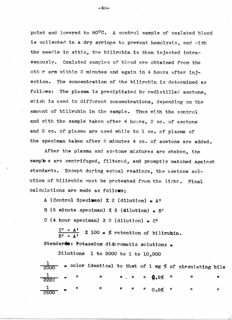
point and lowered to so0 c. A control sample of oxalated blood
is collectea in a dry syringe to prevent hemolysis, and with
the needle in sitic, the bilirubin is then injected intra-..
venously. Oxalated samples of blood are obtained from the
othc-cr arm within 5 minutes and again in 4 hours after inj
ection. The concentration of the bilirubin is determined as
·· follows: The plasma is precipitated by redistillec acetone,
which is used in different concentrations, depending on the
amount of bilirubin in the sample. Thus with the control
and with the sample taken after 4 hours, 2 cc. of acetone
and 2 oo. of plasma are used while to 1 cc. of plasma of
the speciman taken after 5 minutes 4 cc. of acetone are added.
After the plasma and acetone mixtures are shaken, the
samp::S s are centrifuged, filtered, and promptly matched against
standards. Except during actual readings, the acetone· sol-.
ution of b111rubin m\lst be protected from the lirht. Final
calculations are made as follows;
A (Control Speciae.n) X 2 (dilution) •A'
B (5 minute speciman) X 5 (dilution) = B'
c (4 hour speciman) x 2 (dilution) • c• C' - A1
B' - A' X 100 • % retention of bilirubin.
Standarae: Potassium di<h romatic solutions •
Dilutions 1 to 2000 to 1 to 10,000
1 2000 • color identical to that of 1 mg % of circulating bile
1 - " " "· ... ,. n " :l.9% n n " 2222 1 n n " 2soo • n n 0.8% ff " n
-------""'
,,,,----..-..,
-41-
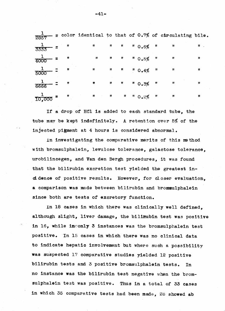
1 = color identical to that of 0.7% of cilreulating bile. 2857 1
= " " " n n 0.6% n " 3333 1 - " n n " " 0.5% " n
4000 -1 " " " " " 0.4% " It
5000
1 - II " " n " 0.3% " " 6666 -1 = II " " " " 0.2% " " ro,ooo
If a drop of HCl is added to each standard tube, the
tube may be kept indefinitely. A retention over 5% of the
injected pigment at 4 hour.a is considered abnormal.
"
" II
II
n
In investigating the comparative merits of this nethod
with bromsulphalein, levulose tolerance, galactose tolerance,
urobilincegen, and Van den Bergh procedures, it was found
that the bilirubin excretion test yielded the greatest in-
atdence of positive results. However, for closer evaluation,
a comparison was made between bilirubin and bro:maulphalein
since both are tests of excretory function.
In 18 cases in which there was clinically well defined,
although slight, liver damage, the bilinubin test was positive
in 16, while inoonly 3 instances was the bromsulphalein test
positive. ·In 15 cases in which there was no clinical data
to indicate hepatic involvement but where such a possibility
was suspected 17 comparative studies yielded 12 positive
bilirubin tests and 3 positive bromsulphalein tests. In
no instance was the bil1rub1n test negative when the brom
sulphalein test was positive. Thus in a total of 33 cases
in which 35 comparative tests had been made, 28 showed ab
-42-
normal retention of injected bilirubin and 6 showed abnormal
retention of injected bromsulphalein. The authors felt
that this served to emphasize the fact the.t the bilirubin
test 1s frequently positive in m1lu in~tancea 0t livEr damage.
H.ence, the bilirubin excretion test is of value where other
tests usually yielded negative results.
Harrop and Baron (60) performed this test on 8 cases
of various types of liver disease together with bromsulph
alein and, levulose tolerance tests. The bilirubin was
positive in all instances, while only one result was ab-
normal with each of the other tests. In 7 cases of chronic an
emia where liver disease was suspected, the excretion of
intravenously injected bilirubin showed the existance of
impa_ired liver function in 6 instances, while both the brom
sulphalein and levulose tolerance tests were entirely negative.
Jonkelson and Gargill (61) employing a somewhat different
method, per.formed the test on 5 instances of hepatic cirrhosis
and on 6 cases of malignancy of the liver. All showed an
~abnormal retention of the pigment.
Snell and McGath (46) point out two draw backs to the
bilirubin "tolerance" test. First and most important from
a clinical aspect is the cost of the material for the test.
Second, the determinations must be extrenely accurate. For
instance, under some conditions, the value for the serum bil
irubin at the end of 4 hours may be only 0.5 mg % greater
than the control value but may represent a retention of 7%.
Even this latter argument, however, tends to shww.the
-43-
the extreme sensitivity of the test.
II. TESTS BASED ON METABOLISM OF FOOD STUFFS. A. TESTS BASED ON CARBOHYDRATE METABOLISM.
As WBS brought out in the physiological discussion the
most significant result of experimental hepatectomy, pa-haps,
is the decrease in the value :fo1'·:~i.the blood sugar and the hyp
oglycemic reaction which continues because the normal store
of glycogen in the liver is abolished. If the capacity of the
liver (46) to maintain the normal amount of blood sugar to be
a nmch less sensitive index of liver function than levulose.
According to McLeon and De Wesselow (62) the test had
its inception in Shirokauer•s abservation that the value for
the blood sugar of normal persons was virtully unaffected by
the oral administration of levulose, whereas it was increased
in the presence of hepatic disease. He administered an oral
dose of 100 gm. Spence and Brett (63) and Tallerman (64)
reduced the dose to 45 grams. Kimball (65) in 480 levuloae
tests 100 of which were done on pa.tients who showed neither
clinical or laboratory evidence of hepatically normal patients
was there an increase1:of. more than 30 mgm per cent and in no
instance was the blood sugar at the end of 2 hours more than
10 mgm % above the fasting level.
METHOD.
A fasting blood sugar is obtained early in the morning
and a dose of 40 or 50 grams of levulose in 250 cc. of water
is administered (40 grams if patient weighs less than 140 lbs.)
-44-
Samples o~ oxalated blood are collected at half hour inter
vals for 2 hours and their sugar values determined.
Cautions to be observed are:
1. Immediate precipitation of the blood.
2. No food or water during test period.
3. Care with regard to the purity of the levulose,
according to Jaliffe (66) and Kimball (66) the
following criteria of abnormality should be useds
1. With fasting blood sugar level of 80 to 100 mgm %, an increase of 30 mgm. % or more.
2. With fasting blood sugar level of 70 to 80 mgm. %
an increase of 35 mgm % or more.
3. With fasting blood sugar level of 60 to 70 mgm. % an increase of 40 mgm. % or more.
4. Where the height of the blood sugar curve exceeds
130 mgm. % regardless of the fasting sugar level
(provided it is not above 115 mgm %) the curve is
considered abnormal. Where the fasting bloo'd sugar
level is more than 130 mgm •. 'Ii, the possibility that
the patient is a diabetic enters and interpretation
of the curve may be misleading.
5. Regardless of the height of the curve the failure
of blood sugar after 2 hours to return to within
15 mgm. % of the fasting level is considered abnormal.
Kimball (65) reports on a series of 142 cases; 81 of
these (57%) yielded abnormal levulose curves. The incidence
~-.
-45-
was higher in patients with severe hepatic damage.
Soffer (31) reporting on ?0 tests done on patients
with definite liver disease found 54 (3%) giving abnonillal
levulose anPves. The greatest incidence of positive results
was ob~~ined in patients with d rrhosis of the liver parti
cularly when associated with ascites. Comparing the results
with those obtained with urobilinogen and bromsulphalein
tests they found that the levulose tolerance tests yielded
a greater incidence of positive results than the urobilino
genuria determinations. While the percentage of positive· re
sults was essentially the same as that yielded by the brom
sulphalein test.
Snell and McGo.th ( 46) concluded that when the test was
performed on experimental animals and all outside influences
were e~~luded, the results were reasonably reliable, but when
the test was performed on patients, many difficulties and erreirB
were encountered.. A low initial value for the fasting blood
sugar, mild diabetic tendencies, and chronic pancreatic dis
ease, all tend to vitiate the results. In genEral the field
of usefuJri.ess or· the test is limlted.
3. GALACTOSE TOLERANCE TEST.
Shaye and Schloss (25) and (27) are responsible for re
viving thts test and bringing it to this count~y. It was
origina~ly introduced by Bauer in 1906.
The test is based on the observation that a normal person
can assimilate a 40 gm. oral dose of galactose without the
loss of more than from 2.5 to 3 gm. of sugar in the urine in
the 5 ho\1.rs immediatly following the administre.tion of the suga:r:
-46-
The test is preceded by a 12 hour .fast and on the morning
of the test the patient is given no breakfast. A fasting
specimen of urine is tested for suge.r. The patient is given
40 grams of galactose dissolved in 250-FiOO cc. of water •.
Hourly voidings of the urine are collected and tested for
sugar. Positive samples a.re added together and the sugar·
dPterm1_ned quanti ta ti vely by the method of Bene di ct. Excre
tion of more than 3.0 grams of .sugar during the 5 hour period
is considered abr;i.orma.l. In employing the test on diabetics
(25) the interferring sugars are fermented out with yeast.
Banks, Sprague and Snell (67) tested 127 cases by this
method. In intra.hepatic jaundice they found 62 .2% to yield
positive results. Of 16 tests on portal and biliary cirr
hosis oniy 18.7% yielded abnormal figures. In 69 instances
of proven obstructive jaundice due to extra.hepatic nepplasma.
and connnon duct stores and without liver damage 33% gave ab
normal results. In 21 cases of liver disease without jaundice
both the gs.lactose and the bromsulphalein tests were performed.
There were 15 positive instances vii t1• the dye wbile only 2
of the gs.lactose tests were abnormal.
Soffer (31) and Snell and McGoth (46) make the following
conclusions, in general, liver diseas~, in the absence of
jaundice, the test is one of the least sensitive. Its great-
. est value lies in the differentiation in pe.tients with jaundic~
between the obstructive and non-obstructive types. Although,
in a certain percentage of obstructive jaundice cases, positive
gs.lactose tests may be obtained (67). In the differentiation
of jaundice the galaotose tolerance test has diftnite advantages
-47-
over the levulose test although the incidence of positive
results is a.bout the same. In the first place, the form.er
test is easier to perform and involves no particular hard
ship on the patient; the galactose test may be done in the
presence of diabetes. This group frequently presents jaun
dice as a cardinal sign of any one of a number of causes.
The fact that _netther tre levulose nor any of the dye tests
are satisfactory in these cases renders the galactose toler
ance determinati,on p srticulerly valuable.
B. TESTS BASED ON CHOLESTEROL METABIDLISM.
The physiological basis for this test has been discussed.
The method of testing is quite complicated in my humble
opinion, and demands considerable laboratory equipment and ex
perience. The rrethod recommended by some- of the latest workers
(68) is Smith and Marbl~s (69) modification of Bloor and Knud
son's (70) method.
Smith and Marble(69) state that they present a new method
for the direct colorimetric determination of free cholerterol
together with modifications of the Bloor and Knudson proced
ures for total and ester cholesterol respectively. They state
further that by means of the three determinations an excellent
check is provided for the accuracy of each, since obviously
the calculated sum of· free and ester should equal! the total.
In brief the procedure is as follows: Plasma lipids
are extracted with alcohol-ether (3:1) as recommended by Bloo~
Total cholesterol is determined on an aliquot o·f filtrate;
Bloor•s procedure is followed closely. Ester and free chol
esterol are determined on a second aliquot, the former by a
l !1
II
I
II I i Ii I'
I I I I-
ti. i
.-48-
,, method similar to that. of Bloor and Knudson except that
saponification is introduced, and the latter by analysis of
the cbolesterol ·digito·nide precipitatfJ;n '·
White et al { 68) consider ·an ester percentage of 60%-
70% as normal. They warn that the blood mu.st not be hemo
lyzed for the cholesterol tests, because the red cells con
tain only free cholesterol which, if liberated by hemolysis,
gives an abnormally high value. According to Epstein {28) ~
in the normal blood plasma of human beings there is from 140
to 2UU mg. of Total Cholesterol per hundred cubic centimeters,
and of this 50 ·to 70% is in the form of cholesterol ester.
The results obtained by Epstein (71) in his latest tests
were as follows:
In obstructive jaundice there is usually a sharp rise
in the values for both the cholesterol and the cholesterol
esters, which may be roughly parallel·: to the elevation of the
value for the serum bilirubin. If biliary obstruction of
long duration, cholangeitis or obstructive biliary cirrhosis
complicates the picture, the value for the cholesterol in the
plasma.may be normal or decreased. In cases in which acute
parenchymatous disease of the 11ver is associated with jaundice
the value for the cholesterol may be decreased or normal and
that for the cholesterol esters may be diminished or these
esters may actually be absent.
Snell and McGoth (46) remark that it has been thought
that the value for cholesterol esters gives scime idea of the
severity of the injury to the liver and of the prognosis,, but
in their experience this has not been entirely substantiated.
·_,~
-49-
In the ordinary types of portal cirrhosis the value for the
cholesterol usually is normal except when acute degeneration
of the liver supervenes.
White et al (47) and (68) in their recent study of 66
cases list the following resultsa
Total cholesterol in cases of obstructive jaundice
showed a nnrked increase in 9U% and showed variations between
253-278 mg. per 100 cc. It was normal ·or only slightly dee-
reased in the other chronic cases, i.e. cirrhosis', and gall-
bladder disease without obstruction of the common duct. In a '
few of the cases of acute hepatitis the values were moderately
low during some period of the illness.
The cholesterol ester percentage (normal 60-70%) was
low i~ about 68% of the cases of acute hepatitis; one half
of whieh returned to normal upon improvement of the patient.
There were two deaths in this group, both of which had a low
ester percentage. In the chronic cirrhosis there was a low
ering of the ester percentage in.45% of the cases. In 5 out of
the 8 deaths in this group, the percentage of ester was normal
or had improved from low to normal. In the gall bladder eases
without known obstruction, one-half were low and about 1/10
remained low, In the tumors all the ester percentages were
normal. In the small group of cases of obstnuctive jaundice,
about 50~ were normal and 50~ were low.
Prognostically, the total cholesterol values in the blood
have given very little information of value. The cholesterol
ester perced; age has definite prognostic value in acute liver
disease. Falling values and a very low figure are, serious
-59-
signs, and a high figure shows a mild disturbance. Prog
ressive improvement from low to normal .figures is seen in
the more severe eases of acute hepatitis which recovered.
The ester percentage has disclosed very little information
of prognostic value in the chronic cases and has been normal
in about 1/2 of the fatal cases.
In the group o.r cirrhosis 1 ess than half showed a
low ester percent.
In the physiological discussion, the determination of
phospholipids and total lipids in the blood was mentiQned
as a method of testing the liver's activity in fat metabolism.
On reviewing. the literature, Snell and McBoth (46), White
et al (47) and (68) and others; it seems that these deter-
mi:mtions show no more than the Total Cholesterol estimations. . ·: . .
The chemistry of the tests is. even more involved (72).
There .fore, it was thought unwise to dwell further on thi a
method of testing liver .function.
C. TESTS BASED OB BILE ACID METABOLISM.
In view of the fact that the liver plays such an important
and primary role in the manufacture of these acids, one might
expect that t:Q.eir quantitative determ1.nation would constitlll.te
a sensitive means for determining liver functi0n.
Soffer ( 31) remarkastr~at the quantitative estina tion of
bile acids as a means of determining the functional status of
the liver has never been widely employed because of the attend-
ing technical difficulties.
In practice trere are other obstacles to the' use of
l.:-··~1' -1·
-51-
this phase as a function test. Soffer (31J quotes Rosen
thal and Zinner to the e.ffect the.t their determinations of
the concentration of bile salts in the bile o.f patients with
advanced atrophic portal cirrhosis showed no deviation from
the established normal. ~his would tend to show that this
function was maintained up to the end stages of liver dis-
ease.
Furthermore, as Foster et al (73) have shown, the
concentration Cbf these substances in bile is markely effected
by diet.
Snell and McGoth (46) suggest that, following operation,
when bile can be obtained from drain~ge tubes.or from-fist
ulas, the study of bile acids may give useful infor.mation.
They feel that low concentrations consistently are of grave
prognosttc import.
D. LIVER FUNCTION TESTS BASED ON PROTEIN MErABOLISM.
As was assumed in the physiological discussion, the
failure of deaminization of amino acids should allow for an
increase in amino acids in the urine, snd sim.e this is a
function of the liver one might expect to find such an in-
crease in disease of the liver. However, as Ballman et al
(34) have pointed out, the amino acids are rapidly absorbed
by the mu:scles where hepatic function is disturbed. The demon
stration o.f an actual increase in amino acids in the blood
or urine in patients with liver disease is quite difficult
except in the presence of extensive hepatic pa:~hology.
~··
-52-
Snell and McGoth (46) however, call attention to the test
proposed by Manchke, a German investigator. In this, an
oral dose of 5U grams of gelatine is administered. The
urine is subsequently examined for amino acids. Normally
2UO mg. of amino acid is found in the 1st four hour2period.
In patients with catarrhal and arsphenamine jaundice and
in patients with cirrhosis of tl:.e liver a considerable in-
crease in the amino acid nitrogen output· occurred in the
urine.
Bollman, Mann and McGoth (?4) found that in the dog
there is. a rise in uric acid content of the tissues snd body
fluids roughly proportional to the a~ount of reduction of
liver tissue, and suggest that this might v1ell be an index
of hepatic function. '
According to Wakeman (45) and others (31), this observa-
tion is not born out clinically.
One is inclined to conclude that as yet, liver tests
based on protein metabolism have not been developed to the
point where they are of much help to the clinician.
In regard to the other phase of protein metabolism
discussed in the physiological section:
• Myers and Keefer (?6) made estimations of plasma
proteins in 16 cases of cirrhosis of the liver and 14 cases
of other forms of disease of the liver. They oonclude from
their work that there is a decrease in plasma proteins in this
disease and that this is a result of the loss of protein in
asci~ic fluid together with defective formation of plasma
-53-
proteins. The decrease was most marked tn the album:i.n fraction
and the albumin globulin ratio was inverted. Similar but less
extensive and less consistent alterations in the plasma pro
tein values were observed in·14 cases of other forms of' dis
ease of the liver. In the less advanced lesions the amount
of albuniin may be only moderately reduced and the amount of
globulin may be normal or increased.
::snell and McGotb. (46) commenting on this observation
state:
In all but a few of a fairly large series of cases
of all types of hapatic disease, the albumin globulin ratio
has been disturbed considerably, but there was not much re
duction in value for the total proteins. The rapidity with
which changes in the amount of total protein and in the· alb
umin-globulin ratio may take place is nath.err striking.
Repeated determim.tions in the same cases indicate that these
changes znay not be without some prognostic 'significance.
It would seem that more work must be cbne in this regard,
but it is well to bear the test in mind as a possibility.
TAKATA-ARA TEST OF LIVER FUNCTION.
It seems logical to mention this teat at this time inas
much as it is thought to be based upon some change in the
serum proteins.
The test was mriginally introduced by Takata and Ara to
differentiate between lobor and bronchopneumonia ('77). They
believed that the test was based on a decreased stability of
-54-
of serum proteins produced essentially by an increase in
the globulin fraction. Since tr• en therP. has been much de
bate as to the basis of the test. Maga.th (78) states "It
becomes clear that the reaction on serum is an emperic test,,
a colloidal phenomenon which has yet not been explained".
In general { 77) the authors are inclined to agree with
the Japanese investigators who thought the reaction ascribable
to changes in the relative amount of albumin and globulin
in the serum with the latter in excess, so that the colloid
was no longer protected. This is a phenomenon dependent on
a reversal or partial reversal of the albumin globulin ratio.
With this latter opinion, Crane (79) and his associates are
in agreement.
The test is a precipitation of mercuric oxide when a
solution of sodium carbonate, merruric chloride and acid
fuchsin is added to an affected sera.
METHOD: In each ·Of 8 small tubes is placed 1.0 cc. of o.9 '•
per cent sodium chloride solution. To the first tube is added
1 cc. of the fluid to be examined. This is mixed and l cc. of
the contents is removed and added to tube 2; then l.O cc. is
transf'enDed from tube 2 to tube 3 etc. One cubic centimeter
of the contents of t"e last tube is removed and discarded. To
each tube 0.25 cc. of 10 % sodium carbonate and 0.3 cc. of the
Takata reagent is added. The latter consists of equal parts
of 0.5% mercuric chloride solution and 0.25% aqueous fuchsin.
Readings are made immediatly after ! hour and 24 hour periods.
r. i
-55-
Positive reHctions a.re indicated by the appearance of a.
definite precipitate in 2 of the first three tubes and in
any number of the following tubes. Negative reactions may
show no precipitate a.t a.11 or only slightly in the last
3 or 4 tubes.
The test is thought (77) to be fairly specific for
cirrhosis, but early in the disease the test is negative.
Actually (78) the test is not specific for either in-
jury to the liver or cirrhosis·, but in the presence of hep
atic disease,. a positive reaction is more likely to indicate
cirrhosis than another disease.
Meyer et al (77) in their discussion of.this test beleives
that it is valuable in fortifying a diagnosis of cirrhosis,
differentiating surgical from medical jaundice, particularly
when jaundice is painless.
It may be stated with some degree of certainty that
extra.heps.tic obstructive processes without much liver damage '
do not result in a positive Ta.le.ta. Ara and therefore aid as
in differentiating an intra from extra hepatic disease unless
a parenchym.atous liver damage is concommitant with extra-hepatic
obstruction.
It must be admitted that the correlation between pos ::lt i ve
reaction and a. reversal of the A/G ratioris high and s im e the
positivity of the test depends upon the elevation of the glob
ulin level, thf;l reaction is lekely to be .. positive in any disease
in which a protein shift occurs. Mmre data needs to be col-
lected on the latter phase (31).
I.
/.,..,--_"•,
-56-
TESTS BASED ON THE DETOXICATION FUNCTION OF THE LIVER.
Familiar examples of this function are ( 46) the con
version of indole to 1ndoxyl sulfuric acid, the conjugation
of oholic acid to form bile salts and the formation of can-
jugate glucuronates. Various clinical tests with such sub
stances as thymal, menthol, camphor, salicylates, phenol, p
cresol, and guaiacol sulfuric acid have been used but have
not been entirely successful. The only survivor of such tests
of the detoxifying function is one based on the ability of the
liver to conjugate benzoie acid and amlno acetic acid to form
hippuric acid.
HIPPURIC Aa! D. ·
It was known that in the dog, hippuric acid is synthes
ized only in the kidney. Subsequent workers tacitly asaimed
that in man the same condition prevailed to a great degree.
Bryan (39) is the only investigator who has simdied the syn
thesis of hippuric acid in human subjects with hepatic invol
vement. This result definitely indicated that liver damage
did influence the condugation of benzoic acid.
Quick (40) in introducing this test makes the f'ollowing
statement a
0 It is quite probable that in man, as in the rabbit,
the synthesis of hippurie acid also takes place in the liver.
Furthermore, the writer has demonstrated that the rate of the
syn.thesis •~ hippuric acid is dependent upon the speed with
which the organism f'urnishes glycine. Irrespective of the 7.
-57-
amount of benzoic acid administened, the quantity of hippurie
acid excreted per hour (in the abserice of exogenous glycine)
is relatively constant, indicatiDg a definite maximum capacity
of the organism to synthesize glycine. Since it is commonly
ancepted th*"? glycin is form~d in the liv$r, it seemed prob
able that certain types of liver damage might produce impair
ment of this synthesis, which should result in a diminished
output of hippuric acid. On this basis, a new test of liver
function was developed.
To exclude a possible factor of delayed el~mination by
the kidney, Kohlstaedt, et al (80) suggested making a simul
taneous study of urea clearance. If this can be done, reduct
ion of synthesis of hippuric acid less than 2 gm. may probably
be regarded as significant.
METHOD.
s·.9 gr~s tiodium benzoate in 30 cc. of water .is administ
ered 1 hour aft·e.p. a breakfast con'at\sting of coffee and toe.st.
The patient is then given l glass of water. Immediatly after
taking the drug the patient voids, and then nollects complete
hourly specimans for 4 hou~s. 2reserve with toluene, and
the hippuric acid is determined in each specimen. In normal
adults, the output of benzo~c acid as hippurie acid is approx
imately 1 gram or more during the second and third hours, and
the total for the 4 hours is from 3 to 5 grams.
CLINICAL MEl'HOD OF DETERMINING THE HIPPURIC ACID.
Each hour a speciman is nee.sured, transferred to a small
/,,,.---.,
-58-
beaken and acidified with concentrated hydrochloric acid
until acid to congo red; 1 cc. of the acid is usually
sufficient. The solution is vigorously stirred until the
precipitation of the hippuric acid is complete, and then is
a~!owed to stand for 1 hour at room temperature. The prec-
ipitate is filtered off on a small Buehner funnel or a filter
paper, washed with a small quantity of cold water, e.nd allowed
to air dry. The hippuric acid thus obtained is either weighed
(to the second decimal place, which is sufficiently accurate)
or titrated with 0.2N. sodium hyroxide, using phanalphthalein
as the indieatbr~. To obtain the total hippuric acid, one mu.st . add to the am:mnt thus obtained the calculated quantity remain
ing in solution; 100 cc. of urine will dissolve 0.33 gm. of
hippurie acid. In case any speciman exceeds 125 ec. it should
be slightly acidified with acetic acid and concentrn.ted on
the water both to about 50 cc. before precipitating the hip
puric ·acid. The results are best expressed in terms of benzoic
acid, one JYIUltiplies by o.68.
Quick (81) has proposed an intravenous method for admin
is.tration of the test dose of benzoic acid. In this, in the
morning after a light breakfast a solution containing 1.77 grams
of sodium benzoate in 8U cc of distilled water is given intra
venously. Five minutes .before the injection, the patient i.s
inatnucted to void before the 'teat and the urine is collected
1 hour after the injection. The Urine is measured and solid
ammonium sulphate added in the proportion of 5 gm. for every
-59-
10 cc. of urine. When the salt is dissolved the urine is
either filtered or centrifuged. Then, proceed with the de
termination of the quantity of hippuric acid as befotre.
This modification is useful in those patients showing
nausea and vomiting with the oral administration.
Quick (40) from his cases feels that here is a test
of a function which is ~irst,~interferred with by hepatic
disease. He found a diminished excretion of hippuric acid
in luetic cirrhosis, catarrhal jaundice and in obstructive
jaundice of moderately long standing. Normal results were
obtained in cholesystitis and in 2 cases of portal cirrhosis.
He concludes that the test offers an.apnroximate quantitative
measure of liver.disease.
He feels that tre test should become useful to the
surgeon in his choice of anesthesia, in his preoperative care
and his preparedness for emergencies. For the internist, 1.t
should prove .equally .valuable since a diminished excretion of
hippuric acid should be a contraindication for arsphenamine
cinchophen and Qther drugs having a potential hepatoxic proper
ties.
Snell and McGoth (46) make the following statement conce~
ing this test. It has been noted that for patients who a.re not
jaundiced, the results closely parallel bromsulphalein, while
in cases of hepatogenous or obstructive "a.undice the retlubtlbon
·in hippuric acid S"..rnthesis corresponded in a general way to the
-60-
degree of hepatice injury noted at operation or necropsy.
BROMSULPHALEIN LIVER FUNCTION TEST.
Bromsulphalein (31) has replaced phenol-tetrachlortha.lein
as a test of liver function since the former substance is nru.ch
less irritating; it dissolves in water with greater ease and
disappears less rapidly from the blood stream. Toxic manif
estations and venous thrombosis, a comparatively frequent
-Occurrence w'.'ten phenoltetrachlorthalein is employed rarely
occurs with the sulfonated dye.
As .first proposed (83~ a dose of 2 mg. of the dye per
kilogram of body weight was injected and specimens were ob
tained at 5 minute, 50 minute, and 1 hour intervals thereafter.
Oleary et al (82) .first showed that the test could be improved
by injection Of 5 mg. of the dye per kilogram of body weight
and Soffer (31) simplified tbe test by omiting the 5 minute
specinum.
METHOD.
Five mgm per kilogram of body weight of bromsulphalein
are injected. Samples of' the blood are collected .from the
other arm at the end of 3u minutes and 1 hour. One sample of
the serum is alkalinized with 3 drops of 10 per cent sodium
hydroxied to bring out any change in color that may be present
because of t>-ie dye. This is compared with a series of ste.nd
ards in a comparometer box. Another sample of serum is placed
in the comparometer box directly behind the standard tube to
give it the general color background of the serum. Normally
20 to 50% of the dye is present in the circulatory blood stream
-61-
e.f'ter five minutes, while all the dye has disappeared f'rom
the blood after 3u minutes. Retentlon of lu% or more at the
end of half an hour is considered definitely abnormal and
therefore indicative of liver dame_ge.
MacDonald (84) proposes a modification to the above
method and makes t-i-,e following objections to the sin~1e
determine. tion:
A. It tells little or nothing if tre test is normal.
B. It gives no information about actual length of time
required for the liver to do a r:iven amount of vo rk,
nor does it tell the efficiency with which it is dom
It is important to know this: two li~ers, both of which
have O~ retention at the time limit, do not necessarily remove
the dye in the same length of time and so cannot be the same
in working capacity. An abnormal liver with its tremendous
reserve can give the same results as .a normal liver, if giv.en
time eno11gh.
He proposes the following remeflies to these objections:
It was thought th0t if the rate at which the liVP.r removes dye
from the blood could be determined there might be produced a
curve typical of the normal liver, and that if a normal could
be established then early and small e.:r"lou.nts of liver changes
could be told.
'.to do this MacDonald estimated the remaining dye in the
blood at 2 minute intervals and plotted graphs. The following
facts were revealed by the graphs:
,r-·.
-62-
A. 18 minutes and not 30 minutes is required for com
plete removal in the normal liver.
B. Livers which remove 100 3 of the dye inside of
30 minutes do not do so by having a eonsistentiy
lower ·reading at each successive estim tion, !l.e.
the liver may not remove any dye at all for 2 1 4, 6,
8 or 10 minutes and yet may remove it all within the
previous normal time limit.
c. As a working unit, a liver represented by the eurve
showing a consistent and continuous disappearance from
the blood is more efficient tran a liver which removes
only 5% in 12 minutes.
Upon further development of this JEthod of testing,
•adDonald found that 5 milligrams of dye per kilcg ram of· body .
weight was such a dose as would put a "maximum load on the liver".·
Further that 5 minute intervals were sufficient.
He concluded from his series of cases, that by repetition
of the test and the use of graphs of 5 minute interval estima
tions:
A. Function tests will show nothing abnormal until that
function fails, but the improved, technique will show
liver changes before the accepted tests of function
are abnormal. By this one can estimate rather acc
urately how much reserve t:t:ere is in store before
function will fail. This is of value surgically,
Also, it is readily seen how this reserve increases
or decreases, hence its value in diagnosis and prog-
~
~' ~I I -63-
nosis, because a· prognosis canno~ be good in the presence
of a curve moving away from the normal, nor can treatment
be considered successful under the same o~rcumstances.
Differences in rate indicate differences in efficiency,
and if changes in liver efficiency either for better. or worse,
can be told, the prognosis can be judged so mu.oh b9tter.
MacDonald states that one venopunoture is sufficient
for taking all the blood samples. Some space has been de-
'1Zl0ted to MacDonald's work be~ause we can find no mention of
any like observations in the literature and because his reason
ing appeals to us as sufficiently logical to warrant some
inve.stigations of his claims.
MoGoth (85) showed that the results, with the single
determination, are extremely satisfactory, since retention
of the dye oooured in 96% of oases in which there was evid
ence of parenchymal hepatic injury, or even moderate mech
anical obstruction of the bile ducts which had not yet pro
duced clinically demonstrable jaundice.
It should be pointed out, however, that this test should
not be used in instances of jaundice where the van den Bergh
reaction is either direct or biphasic. This is because the
abnormal retention of the dye need not be an expression of
the &na.~ility of the liver cells to excrete it, since the
obstlmction of the intra or extra-hepatic bile channels will
cause the ·retention of the dye in the blcod stream. .1.n genera,
the test is contraindicated where the value for the serum bili-
[· r~
-64-
rub1n 1s 5 to 6 mg. %. Soffer (31) nakes the following generalizations concern
ing this tests: In about 55 to 65 % of liver disease this
test will yield definite evidence of impaired hepatic .f'unct1on.
The percentage of positive result is consideratily higher in
cases where the liver damage is wide-spread and extensive as
it is in portal cirrhosi.s. •:J.tll ascites, in biliary cirrhosis,
and in long standing cases of chronic passive conjestion.
When we deal with malignancies of the liver either primary
or metastatic, retention of.the injected dye after one-half'
hour occurs less frequently, while in isolated le·s ions or the
liver, as in hepatic gUmma or cyst, the incidence of positive
results obtained with the dye method is low. Tbis test is
considerably more sensitive as an index· of liver fun·ction.
than either the galactose tolerance test on the determtnation
.of urobilinogen in the urine and yields a somewhat greater
proportion of positive reSU.lts in liver. disease than does the
levulose tolerance test.
Snell and McGoth (46) state th.tin their opinion the
test gives results which indicate the extent of parench.ymatous
damage only roughly, but the determinationaaare sufficiently
accurate to yield valuable information i~ the test is repeated
at intervals.during the course of the disease.
TEE ROSE BENGAL TEST.
Delprat, Epstein and Kerr (87( criginally .introduced
this method of testing the excretory function of the liver.
The.substance used is a dye which is removed from the blood
-65-
by the hepatic cells and its rate of removal is proportional
to the normally of the liver cells.
In view of the comment by Green (86) in which he states
that in a study -Of ,more than 700 cases there is very little
difference in the significance of the various dyes. Phenoltet
rachorphalthalein, bromsulphalein and rose bengal all show
.essentially the same type of changes. Further, and in view of
the fact that the promsulphalein test is by far the simplest
to perform, we will simply give the method for the performance
of this test •
.METHOD. ( 88)
Five to ten cubic centimeters of l to 2% solution of rose
bengal in saline is injected intravenously. Two minutes from
the start of the inject.ion an oxalated sample of blood is col
lected which is desigaated as the "standard.sample• and which
contains the maximum concentration of the injected dye. It is
considered to have 100% of the dye. Exactly 6 minutes later,
that is 8 minutes after the dye has been introduced intravenously,
the •unknow'n" sample is withdrawn. The plasma is separated and
cleared by precipitation of the proteins with 2 volumes of ace~
tone. This is centrifuged and to the plasma thus obtained a drop
of 10% Na OH is added to clear hemolysis. The resultant dye
solutions are read in a calorimeter. Normally 5P% or less of
the injected dye should be present in the circulatory blood after
·8 minutes. In view of the photosensitizing erfect of rose bengal
on tissues, it is advisable to keep the colledted specimens in the
dark until they are to be read. Sim:dllarly, patients should be
warned to avoid bright sunlight for several hours after the per-
formance of the test.
-66-
MISCELLANEOUS TEST ASSOCIATED WITH LIVER INSUFFICIENCY.
A. Measurement of the tendency to bleed.
As Ivy et al (88) have pointed out, the surgical
and medical risks of infections, and other path
ological processes in and about the liver are
seriously augmented by the possibilityof hepatic
insufficiency. One of the most striking surgi
cal features associated with these cases of liver
damages is their tendency to bleed. According
to the results obtained in several clinics, 50
per cent of all post operative deaths in patients
with jaundice or live insufriciency are a result
of hemmrrhage.
Many procedures have been designed to measure the coag
ulation time of the blood accurately but none give a consist
ent picture of the dangers of bleeding in hepatic disease.
Studies on ~ibrinogen, on calcium in its diffusable and non
diffusable forms, and on various other constituents of the
blood have failed to solve the riddle of why a patient who
has hepatic disease is so subject to hemorrhage.
Determination of coagulation time is today a most widely
used procedure. The general conclusions are (88),however,
that prolonged clotting time seems to bear no relation to
clinical bleeding, and it is often normal in severe blood
dyscrasias an«·when bleeding occurs.
On the other hand, Nygaard and Baldes (89) have shown
that the "plasma coagulation index" as measured by their
method gives a satisfactory basis for prediction of the
-67-
~endency to bleed. Repeated "coagelgrams" will often
show the hemorrhagic tendency.
Ivy (88) thinks that a prolonged coagulation time
in jaundice, is not so much an index of bleeding tendency
as liver damage.
Ivy's method of determining bleeding time is the
application of a pressure cuff about the arm at about
40 millimeters of Ug. pressure then determining bleeding, ' time from Duke's puncture on the skin of the forearm near
the .elbow over the pronator muscles.
White et al (68) found that prolonged bleeding time
by this method was commonly associated with poor liver
function •.
The methods that have been mentioned are not as yet
in general use and may· be too involved for practival pur-
poses.
Snell and McGoth (46) state that high valuee for bili
rubin in cases of icterus and high grades of retention of
bromsulphale1n ln cases in which there is no icterus are
danger signals, and furnish information as ·reliable as that
furnished by an1 current method of studying the coagulation
of blood.
QUANTITATIVE ESTIMATION OF SERUM PHOSPHATASE.
Phosphatase {46) which is an enzyme concerned in the
metabolism of bone, is present in mairly' tissues of the body
and is found in high concentr,tions in bone, kidney, intest-
inal mucosa and liver.
Roberts {90) first noted that high value for phospha
tase in the serum were a feature of some cases of obstructive ·
-68-
jaundice, while, in the non-obstructive c7pes, low or normal
values were the rule.
METHOD. It involves a determination of inorganic phosphatic
liberated when the enzyme present ia the blood is allowed
to act on a phosphoric acid ester subst2a~e under standard
conditions. The upper normal value for adults is approx
imately 6 units and th8:t for children is 15 units ..
Rothman et al (91) state that in most cases of proved
obstructive jaundice, the value for the phosphatase was
more than 10 units but that values of 20 or more units
were commonly observed; in cases of non obstructive jaun
dice values of less than 10 units were the rule.
Greene (92) on the other hand, expressed the opinion,
that the infolmll8.tion obtained by determinttions of phosphat
aseiis inaccurate and does not diff'erentiate the type of
jaundice in question.
It is apparent that the .. result of the determination
of phosphatase will have to be w•ight;Jd against the clinical
evidence in cases in which the diagnosis is doubtful.
THE APPEARANCE OF ERYTHROCYTIS IN DESEASE OF THE LIVER.
This may be mentioned, not as a test of liver f'unctinn,
but because studies in morphology of the blood may give
valuable confirmatory evidence of the presence of injury
to the liver. A hypochromic macrocytic anemic ia a ratb.er
uniform accomi;>animent of portal cirrhosis and may be noted
in other types of injury to the liver. (93)
-69-
&tasney and Higgens (94) have demonstrated similar
changes in the experimental animal. They attempt to ex
plain the rapidity of appearance of macrocyt~s in the blood
stream oh the basis of hypoproteinemia and swelling of the
erythrocyt•s because of the altered osmotic pressure of the
plasma.
Soffer (31) states that the macrocytosis was observed
only in cases of long standing and very wide spreaa liver
disease.
SUMMARY AND CONCLUSIONS·.
Obviously all of the tests for investigating the
activity of liver have not been dealt with in this review
of the subject. However, those tests, which have been con-
sidered by mostoof the authorities as clinically applicable,
have at least been mentioned. It is further believed that
the direction which future work in this field will take has
been pointed out. e.g. tests d.1rected at the evaluation of
the liver's activity in protein metabolism.
Certain conclusions as to the comparative value of the
tests remain to be discussed. Of these conclusions there
are some general ones that are quite obvious.
A. There is no one test that will adequately test
all the liver's activities.
B. There is no one test that will show abnormalities
in its reactions in all types of liver injury.
c. Indeed no one test has been shown to be 100%
responsive in all cases of the same type of liver
injury.
\ .
/ D. Some of the tests are obviously more practical
than others.
E. Since no one test is entirely satisfactory, it is
better to use several, testing several types of
functions •.
F. Lastly, but perhaps not so obvious, in testing
several functions of the liver, one positive test
is almost as good a sign of liver damage as if all
were positive, but there is usually a correlation
between the amount of injury to the liver and the
number of different kinds of tests which show ab-
normalities.
In drawing conclusions as to the comparative value
of the tests discussed, I will attempt to sum up the con
clusions of three comprehensive articles on this subject.
The first by L. J. Soffer (31} in 1935. l'he second by
Snell and McGoth (46} in 1938. The third by White and his
associates in November of 1939 and January of this year.
certain general conclusions have been made in all
th~ee of these papers:
There is no correlation between the type of
pathological lesion present in the liver and the function
which is disturbed. Nor is there any difinite index,CUn
ically, as to which function is disturbed first. Concern
ing any one particular liver function test, it is an index
of the degree of liver damage or progression or regression
of the lesion only when performed repeatedly on the same
,,...---.
-71-
on the same patient.
It is utmost importan(rnto recognize when these
tests should not be employed. There is no point in
testing with dyes in the presence of a hyperbilirubinemia
with a direct or biphasic van den Bergh. reaction. Simil
arly the bilirubin excretion test can yieldono informatial
of any value when performed in the presence of a hyper
bilinubinemia however slight. The galactose test on the
other hand may be performed under all circumstances.
Evaluation of the liver function tests will be made
with regard to three of the ways in which they may be used:
A. To detect the presence and amount or liver damage
and to follow the changes and course or the liver
injury.
B. For prognosis.
c. F'or differential diagnosis.
A~ DETECTING PRESENCE AND AMOUNT OF LIVER DAMAGE AND TO FOLLOW CHANGES.
Van den Bergh. reactioni
·As a general test of hepatic function the quantitative
Van de.n Bergh is inadequate; one sees too many instances
of liver disease in which there is no increase in the
amount of circulatory bilirubin.
It is an excellent index of an increase or decrease
in the intensity of jaundice and of tremendous help in de
terminY1g the progress or these patients·. It ~s of value
in determining p~ogress of non-obstructive jaundice. The
i. !
-72-
test is at first indirect, and as the disease progresses
it becomes biphasic and direct. Improvement is heralded
by areversal of the above.
ICTERUS INDEX:
Determinat~ons have to be interpreted with caution.
A normal index definitely excludes hyperbilirupinemia, while
a very high icterus index almost always means an increase
in the bile pigment of the blood. Between these two levels,
it is probably better to resort to the.Van den Bergh and
be sure.
The interpretations of its reaction, with caution,
are the same as the Van den Bergh.
UROBILIMOGEN AND UROBILIN DETERMINATIONS IN URINE:
The test has proved to be of very little value in the
general diagnosis of liver dysfunction.
EXCRETORY TESTS:
Dyes: A high incidence of positive results are found
in cases of cirrhosis particularly when associ·ated with
ascites. By the.plotting of a curve as suggested by Mac
Donald (84) one may be able to· follow the progress of the
injury.
Bilirubin: Soffer states "A greater incidence of positive
results is obtained by this method in demonstrating hepatic
dysfunction in early cases of liver disease than in any other
method." Others are not in complete agreement w.tth this.
CARBOHYDRATE TOLERANCE TEST:
Levulose: Disease of the liver must be widespread and
moderately severe before an abnormal response to levulose
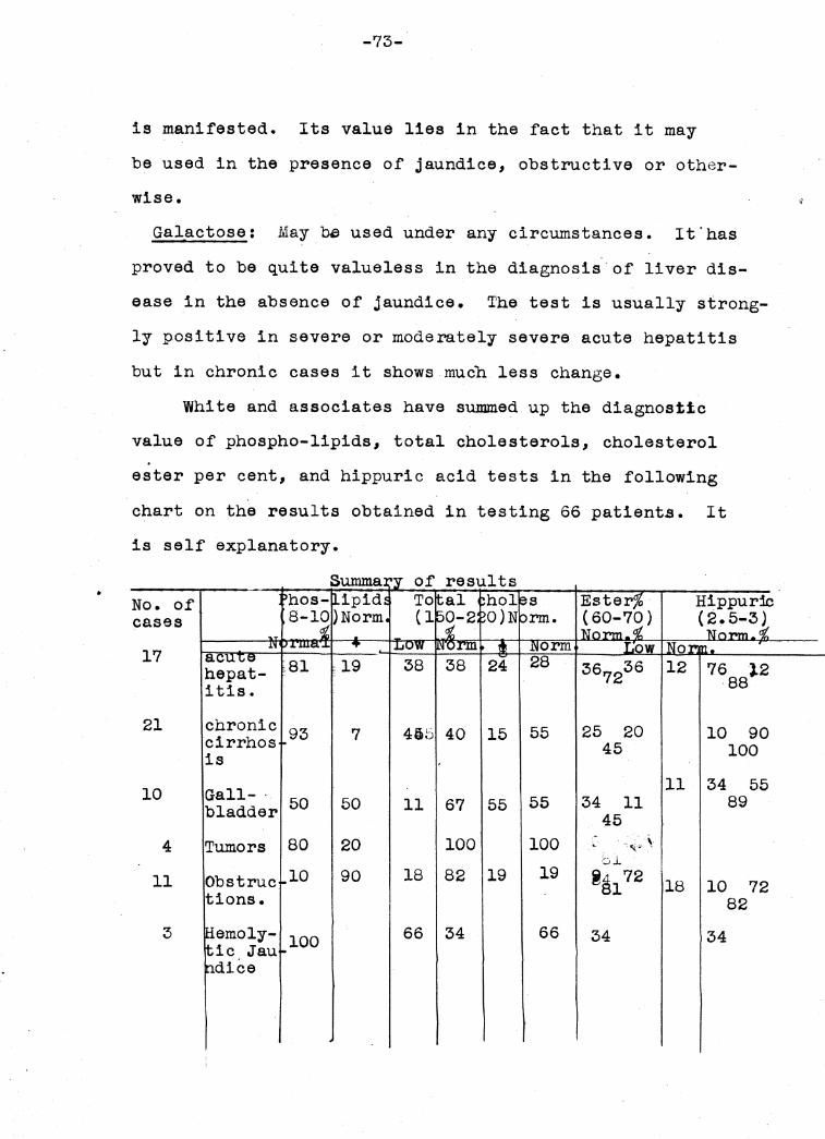
•
.~.
\ ,
-73-
is manifested. Its value lies in the fact that it may
be used in the presence of jaundice, obstructive or other-
wise.
Galactose: May~ used under any circumstances. It.has
proved to be quite valueless in the diagnosis.of liver dis
ease in the absence of jaundice. The test is usually strong
ly positive in severe or moderately severe acute hepatitis
but in chronic cases it shows much less change.
White and associates have summed up the diagnostic
value of phospho-lipids, total cholesterols, cholesterol .
ester per cent, and hippuric acid tests in the following
chart on the results obtained in testing 66 patients. It
is self explanatory •
No. of cases
17
21
10
4
11
chronic cirrhos is
Gall-· bladder
Tumors
Obs true tions.
emoly-ic Jau dice
93 7 495
50 50 ll
80 20
10 90 18
100 66
40 15 55 25 20 10 45
ll 34 67 55 55 34 11
45 100 100 .. -:"(~'
bl. 82 19 19 ~~172 18 10
34 66 34 34
90 100
55 89
72 82
-74-
This should be noted about the hippuric acid ex
retion. It was definitly diminished in a high percentage
of cases~ The results paralleltthe cholesterol ester fig.
urea but the test seemed to be more sensitive in all types
of disease of the liver.
B. FOR PROGNOSIS.
Van den Bergh, reaction:
lDt is of prognostic value in so far as· 1 t shows the
progression of the jaundice.
Bromsulphalein:
Where this test was made after jaundice had disa-
· ppeared, the results of this test closely parallel the
hippuric acid test. In one case of stricture of the com
mon duct, the hippuric acid test seemed slightly less sen
s1 tive.
MacDonald (84) claims that this test as performed
by his method gives invaluable information with regard
to the prognosis of cases of liver dysfunction.
Bilirubin excretion:
Soffer (59) makes practically the same claims as the
above for this test.
Galactose Tolerance:
In assessing the prognosis or in detecting the presence
of liver damage the hippuric acid test waw far more delicate
than the galactose test.
The phospho-lipias and total cholesterol values in
the blood have given very_ little information of prognost~c
value. In one fatal c.ase of portal cirrhosis there was a
i .
.~ )
-75-
progressive change from high to low normal range over·a
period of 16 months, whereas a definitely bad prognosis-·<:.
was indicated by both hippuric acid and bromsulphalein tests.
Cholesterol ester percentage:
It: has definite prognostic value in acute liver dis
eases such as acu1Bhepatitis. Falling values and a very
low figure are serious signs and a high failure shows mild
disturbance.
The ester percentage has disclosed very little inform
ation of prognostic value in the chronic cases and has been
normal in about half of the fatal cases.
In the group of cirrhosis less than half showed a low
ester percentage while all showed a low·'hippuric acid ex
cretion.
The Bippuric Acid Excretion Test: It has been helpf'ul in
prognosis since it invariably gave low results in all severe
and fatal cases. It has proved more sensitive than the
ester ratio in all classes of liver clisease. This test is
valuable.in discovering residual liver dam.age after the
jaundice has disappeared, or in rilling out liver dam.age in
chronic cases. In this respect, it is almost exactly like
the bromsulphalein test.
It is also of assistance in assessing surgical risk
~he post operative course, is generally favorable when the
hippuric acid excretion is between 2.5 to 3.6 or better.
On the other hand, when the hippuric acid excretion is 1 and
li gmasbefpre pieratopm a stpr;iy convalescence may be antici~
-76-
pated.
The Ivy bleeding timei .. '
Results agree with Iv¥' s opinion that a prolonged
bleeding time is associated with poor liver fUnction and
is a serious prognostic sign.
C. FOR DIFFERENTIAL DIAGNOSIS.
Van den Ber~:
Its value in differentiating between obstructive
and non-obstructive jaundice is minimal, as also its value
in differentiating between hemolytic and non hemolytic
jaundice.
Urobilinogen and Urobilinddeterminations in urine:
It is of some help in the differentiation between
obstructive and non obstruct~ve jaundice. The total
absence of urobilinogen in the urine would suggest the
former. Tij.is criteria, however, is to be used with caution.
CarbOJidrate Tolerance;
The tests may have some value in the limited but
important field of distinguishing acute cellular damage
from the early stages of obstruction.
ly important in this respect.
Galactose is chief-'
,fhospho-lipids and total cholesterol:. Are consistently
high in cases of external obstructio~, but may also be
elevated in toxic hepatitis. The latter can usually be
ruled out by a history of exposure to some toxic substance.
Ester percentage:
This was also lound to have little value in ~iffer•n-
tiating obstructive cases.
-77-
Hippuric acid:.
This has no differential value gecause it is con
sistently positive in all types of liver disease.
In the field of differential diagnosis, the avail
able tests are the least satisfactory. Reliance must be
placed on a group picture rather than on the results of
a specific test.
Fer practical purposes, it may be said: A. In types
of disease ·of the liver not associated With jaundice, in
formation gained from the study of retention of bromsul
phalein is as reliable as that.which can be gained in any
o~her way, and under these donditions other tests give
chiefly confirmatory evidence. B. In cases of jaundice,
some information, which is not altogether reliable, as to
the possible hepatogenous or obstructive nature of the
jaundice in any given.case can be had by studies of excre
tion of galactose, the values for the cholesterol and chol
esterol esters in the plasma, and the value 1er serwn phos
phatase. c. The best information as to the functional act
ivity of the liver in cases of jaundice can be gained ~rom
a consideration of the value for the serum bilirubin, its
daily variations, and a knowledge of the anatomic changes
which these pepresent. So f'ar as indirect methods of
measuring liver function in the presence of icterus are
concerned, the hippuric acid gi v·es reasonably accurate re
sults, which should not, however, supplant the impressions
gained from purely clinical study.
BIBLIOGRAPHY
(1 ) Ka.xi.mow, A. A., and Bloom, William: A Text-Book of Histology, Philadelphia, w. B. Saunders Company 1930.
(2 ) :U:cNee : Experiments on hemolytic icterus, J. Path. and Bact. XVIII:325, 1913.
(3 ) Noel: ~uoted trom (1).
(4 ) ITY, A. c.; An outline ot liver !unctions and jaundice, 1. Jlich. State M~. Soc. 37:121-129, 1938.
(5 ) Whipple, G. ·H., and ·Hooper: Bile pigment output influenced by diet, .Amer. J'. Physiol. XL : 349, 1916.
(6) Whipple, G. ·H., and Hooper : ·Hematogenoua and obatructiTe icterus, J. Exper. :U:ed. XVII :593, 1913.
(7 ) Rous, Brown and Kcl(aster : Relation between blood distruction and output ot bile pigment, J. Exper. Med. XXXVIII : 421, 1923.
(8 ) Inlow : The spleen and digestion, Am. J. Med. Sci. CLXVII: 10, 1924.
(9) B•own, KcKaater and Rous : Relation between blood distruction and output of bile pigment, J. Exper. :U:ed. :XXXVI : 733, 1923.
(10) Rich,A. R.: The formation of bile pigment from hemoglobin in tissue cultures, Bull. Johns ·Hopkins ·Hosp. J:XIV : 321, 1923.
(11) Rich, A. R.: Formation ot bile pigment, Physiol. Rev. 5: 190, 1925.
(12) Mann, F. C., Bollman,- and :U:agathi Formation of bile pigment after total removal of the liver, Am.er. J. Physiol. LXIX; 393, 1924.
(13) :U:&ml, F. c.: Studies on the physiology of the liver, Aaer. J. Ked. Sci. CLSIX, 37, 1921.
(14) Mann, F. c., Sheard, c., and Bolman, J.R.: An evaluation of the relative amounts of bilirubin formed in the liver, spleen and bone marrow, Amer. J'. Physiol. ?8;384,1926.
(15) Rich, A. R.: LiTer disease, Bull. Johns Hopkins Hosp. 36: 2~3., 1925.
(16) Rich, A. R.: Pathogenisia of the forms of Jaundice, Bull. Johns nopkins nosp. 47: 338-377, 1930.
(17) Wallace, G. B., and Diamond, J. S.: The significance of urobilinogen in the urne as a test for liTer function, Arch. Int. lled. 35:698, 1925.
(18) llann, F. c., and llagath, T. B.: The effect of the administration of glucose in the condition following total extirpation of the liTer, Arch. Int. Ke6. 30: ·171, 1922.
(20) Cori, c. F.: The fate of sugar in the animal body, J. Biol. Chea. 70: 577, 1926.
(21) Cori, c. F.: The fate of sugar in the animal body, J. Biol. Chem. 66: 691, 1925.
(22) Bodansky, Meyer: Fructose, glucose, and galactose tolerance in dogs, J. Biol. Chem.. 56: 387, 1923.
(23) Kann, F. c., and Bollman, J. L.: The relation of the liver to the utilization of levulose, Amer. J. Physiol. 93: 671, 1930.
(24) Roe, J. H., and Schwartzman, A. s.: Galactose tolerance of normal and diabetic subjects, and the effect of insulin upon galactose metabolism, J. Biol. Chea. (96: 717, 1932.
(25) Shay, h., Schloss, E. K., and Bell, K. A.: The metabolism of galactose, Arch. Int. lled. 47: 391, 1931.
(26) Folin, o., and Berglund, H.: Some new observations and interpretations with reference to transportation, retention, and excretion of carbohydrates, J. Biol. Chem. 51: 213,1922.
(27) Shay, H., Schloss, E. u:., and I. Rodis: II. The galactoae tolerance test in the differential diagnosis of jaundice, Arch. Int. Ked. 47: 650, 1931.
(28)Epstein, E. z.: The cholesterol partition of the blood plasma in parenchymatous diseases of the liTer, Arch. of Int. Yed. 47: 83, 1931.
(30) Smyth, F. s., ~d Whipple, G. H.: Bile Salt metabolism, :r. Biol. Chem. 59: 623, 1924.
(31) Soffer, L. J.: Present day status of liver function tests, Medicine 14: 185-247, 1935.
(32) Gregory, R., and Pascoe, T. A.: The quantitatiTe determination of bile acids by means of a new color reaction and monochromatic light, J. Biol. Chem. 83: 35, 1929.
(#3) Smith, H. P., and Whipple, G. H.: Bile salt metabolism, J. Biol. Chem. 80: 671, 1928.
(34) Bollman, Mann, and Magath: Studies on the physiology of the liver, VII Am. J. Physiol. 69: 371392, 1924.
{~:>) Kerr, Hurwitz, and Whipple: Regeneration o:r olood serum protein, 370-392, 1918.
(36) 'Rollman, R. L., Ma.honey, and 'Whipple: Blood plasma protein given by vein utilized in body metabolism, J. Exper. Med. 59: 269-282, 1934.
(37) Priestley, J. T., Markowitz, J., and Mann, F. C.: Studies on the physiology of the liver, Amer. J. Physiol. 96: 696, 1931.
(38) Lichtman, s. s.: Cinophen oxidation test of the function of the hepatic cells, Arch. Int. Med. 48: 98, 1931.
(39) Bryan, A. w.: The value of sodium benzoate test, and the effect of injury of the liver on hippuric acid synthesis, J. Clin. Invest. 2: 1-33, 1925.
(40) ~uick, A. J.: The synthesis cf hippuric acid: a new test of liver function, Am. J. Med. Sc. 185: 630-635, 1933.
(41) Rowntree, L. G., Jurwi tz, E. 'H., and A. L. Bloomfield: An experimental and clinical study of the value of phenoltetrachlorphthalein as a liver function test, Bull. Johns Hopkins Hosp., XXIV : 327,1913.
(42) Whipple, G. H., Mason, V. R., and T. CJ.Peightal: Tests for hepatic function and disease under experimental conditions, Bull. 1Iohns Hopkins Hosp. XXIV, 207, 1913.
r- .
(43) Rosenthal, S.M.: An improved method for using phenoltetrachlorphthalein as a liver function test. J. Pharm. and Exp Therap. 19: 385, 1922.
(44) Rosenthal, s. M., and White, E. C.: Studies in hepatic function, J. Pharm. and Exp. Therap. 24: 265, 1924.
(45) Pratt, T. w., Vanlandingham, H. W., Talley, E. E., Nelson, J. M., and Johnson, E. o.: Studies of the liver function of dogs. Amer. J. Physiol. 102: 148, 1932.
(46) Snell, A. M., and Magath, T. B.: The use and interpretation of tests for liver function, J. Amer. Med. Assoc. 110: 167 174, 1938.
(47) White, Deutsch, and Maddock: The comparative value of testa in diseases of the liver, II, Amer. ,r. of Dig. Diseasea. VII, 3, 1940.
(48) Barron, E. s. G.: Bilirubinemia, Medicine, 10: 95, 1931.
(49) Snell, s. M.: ·Hepatic parenchymal lesions and hepatic function of patients with obstructive jaundice, Tr. Am. GaatroEnterological Assoc. 1932 196-223.
(50) Stoner, w. c.: Carotinemia, Am. J. Med. Sci. 175, 31,1928.
(51) White, F. D.: Estimation of serum carotin, J. Lab. clin. med. 17: 53, 1931.
(52) Maue, H. P.: Icterus index of blood serum, Surg. Gynec. and Obst. 34: 752, 1922.
(53) Elton, N. W.: Physiology, correlation, and technic of the Van den Bergh reaction, icterus index, and quantitative aerum bilirubin, J. Lab. Clin. Med. 17: 1, 1931.
(54) Wilbur, R. L., and T. Addia: Urobilin: Its clinical significance. Arch. Int. Med. 13:235,1914.
(55) Watson, C.J.1 Studies of Urobilingoen Am. J. Olin. Path. 6:458-473, 1936.
(56) Piersol, G. M., and Rothman, M. K.: Practical value of i;ver function testa, J". A. 1l. A. 91: 1768, 1928.
(57) Jleyer-Betz, F.: Quoted trom (56).
(58) Robert.eon, w. E., Swalm, w. A., and F. w. Konzelmann: Comparative merits of the five most popular tests. J". A. M. A. 99: 2071, 1932.
(59) Sotter L. F. and PaulsonM.: Comparative advantage and further modification of the bilirubin excretion test for hepatic !unctien, Am. J". )led. Sci. CXCII, 535, 1936.
(60) ·Harrop, G. A., and E. s. G. Barron: The excretion of intravenously injected bilirubin as a test of liver funtion, J". Clin. InTea. 9: 577, 1931.
(61) J"ankelson, I. P., and s. R. Gargill: Bilirubin liver function test, New Eng. J. of Med. 204: 547, 1931.
(62) Hae Lean and de Wesselow: The extimation of sugar tolerance. Quart. J. Med. 14: 103-119, 1921.
(63) Spence, J. c., and P. c. Brett: Use of levulose as test of hepatic insufficiency, Lancet 2: 1362, 1921.
(64) Tallerman, K. H.: Levulose test for liver efficiencey and investigation of hepatic condition in pregnanc~. Quart. J". Med. 17: 37, 1923.
(65) Kimball, s.: The levulose tolerance test, Guy's Hospital Reports, 82: 157, 1932.
(66) Jolliffe, N.: The tolerance of normal subjects to levulose, J". Clin. Invex. 9: 363, 1931.
(67) Banks, ·B. :v., Sprague, P. H., and A. M. Snell: Clinical evaluation of the galactose tolerance test, J". A.](. A. 100: 1933.
(68) White, Deutsch, and Maddock: I Value of tests in diseasea of the liver. Am. J. of Dig. Diseases. 6: 603, 1939.
·•
(69) Smith, and Marble: Cholesteral determination. J. of Biol. Chem. 117: 673, 1937.
(70) Bloor, and Knudson: The separate determination of cholesterol and cholesterol esters in small amounts of blood, J. Biol. Chem. 27: 106, 1916.
{71) Epstein, E. z., and Greenspan, E. B.: Clinical significance of the cholesterol partition of the blood plasma. Arch. Int. Med. 58: 860-890, 1936.
(72) Bloor, w. R.: The determination of small amounts of lipid in blood plasma., J. Biol. Chen. 77: 53, 1928.
{73) Foster, M. G., Hooper, c. w., and Whipple, G. H.: The metabolism of bile acids, J. Biol. Chem. 38; 393, 1919.
{74) Bollman, J. L., Mann, F. c., and T. B. Magath: Studies on the physiology of the liver. x. Uric acid following total removal of the liver, Am. J. Physiol. 72: 629, 1925.
(75) Wakeman, A. M., and c. A. Morrell: Chemistry and metabolism in experimental yellow fever in Maeacus rhsus monkeys, Arch. Int. Med. 46: 290, 1930.
(76) Myers, w. K., and Keefer, c. s.: Relation of plasma proteins to ascites and edema in cirrhosis of the liver, Arch. of Int. Med. 55: 349-359, 1935 •.
(77) Meyer, Colob, and Nussbaum: An evaluation of Takata-Ara reaction as a liver function test, Am. J. Dig. Dis. 6: 200, 1939.
{78) Magath, J. B.: The Takata-Ara test of liver function, Proc. Staff Meeting Mayo Clinic. {july 31) 1935.
(79) Crane, M. P.: A modified mercuric chlorid reaction (TakatAra) in cirrhosis and in neoplasms of the 1-i ver, Am. J • Med. Sci. 187: 705, 1934.
(80) Kohlstaed t, K. G., and Helmer, o. M.: A study of the hippu~ic acid excretion as a test of hepatic function, Am. J. Dig. Dis. and Nutrition 3: 459-466, 1936.
(81) ~uick, A. J.: Intravenous modification of the hippuric acid test for liver ~unction, Am. J. Dig. Dis. 6: 716, 1939.
(82) Oleary, P. A., Greene, C. G., and Rowntree, L. G.: Diseases of the Liver: VIII. The various types of syphillis of the liver with reference to tests for hepatic function, Arch. Int. Med. 44: 155-193, 1929.
(83) Rosenthal, s. M., and E. C. White: Clinical application ot the bromsulphalein test for hepatic function, J. Am. Med. Assoc. 84:1112, 1925.
{84) Macdonald,Dean: A practical and clinical test for liver reserve, Surg. Gynec. and Obst. 69: 70-82, 1939.
(85) Magath, T. B.: The takata-ara test of liver function, Amer. J. Dig. Dis. and Nuit>ition. 2: 713-716, 1936.
(86) Green: Discussion rose bengal test for liver function, J. Am. Med. Assn. 85:946, 1925.
(87) Delprat, G. D., Epstein, N. N., and w. J. Kerr: A new liver function test, Arch. Int. Med. 34: 533, 1924.
(88) Ivy, Shapiro, Melnick: The bleeding tendency in jaundice, Surgery, Gynecology, and Obstetrics. 60: 781, 1935.
(89) Nygaard, K. K. 1 and Baldes, E. J.: Interpretation and clinical significance of coagelgr;:,me in obstructive jaundice, Proc. Staff Meet. Mayo clinic, 11:705-709, 1936.
(90) Roberts, w. M.: Variations in the phosphatase activity of the blood in disease, British J. Exper. Path. 11: 90-95, 1930.
(91) Rothman: Blood phophatase as an aid in the differential diagnosis of jaundice, Am. J. M. Sci. 192: 526-535, 193 6.
(92) Greene, c. H.: The phosphatase content of the blood serum in jaundice, J. Clin. Invest. 13: 1079-1087, 1934.
(93) Wintrobe, and Shumacker: The occurrence of macrocytic anaemia in association with disorder of the liver, Bull. Johns Hopkins Hosp~ 52: 387, 1933!
(94) Higgins, G. M. and Stasney, J.: Macrocytic anemia in elRJJel'imental cirrhosis, Proc. Staff Meeting Mayo Climic. 10: 429, 1935.