Field-resolved studies of ultrafast light-matter interaction
Upload
independentCategory
view
0download
0
INSTITUTE OF PHYSICS PUBLISHING PHYSICS IN MEDICINE AND BIOLOGY
Phys. Med. Biol. 49 (2004) 111–119 PII: S0031-9155(04)67041-2
Differential phase optical coherence probe fordepth-resolved detection of photothermal response intissue
Sergey A Telenkov, Digant P Dave, Shriram Sethuraman, Taner Akkinand Thomas E Milner
Department of Biomedical Engineering, University of Texas at Austin, Austin, TX 78712, USA
Received 1 August 2003, in final form 15 October 2003Published 15 December 2003Online at stacks.iop.org/PMB/49/111 (DOI: 10.1088/0031-9155/49/1/008)
AbstractWe describe a differential phase low-coherence interferometric probe fornon-invasive, quantitative imaging of photothermal phenomena in biologicalmaterials. Our detection method utilizes principles of optical coherencetomography with differential phase measurement of interference fringe signals.A dual-channel optical low-coherence probe is used to analyse laser-inducedthermoelastic and thermorefractive effects in tissue with micrometre axialresolution and nanometre sensitivity. We demonstrate an application of thetechnique using tissue phantoms and ex-vivo tissue specimens of rodent dorsalskin.
1. Introduction and theory
Optical coherence tomography (OCT) has recently emerged as a promising diagnostictechnique for imaging of a variety of tissues (Pan et al 1995, Brezinski and Fujimoto 1999).Closely associated techniques include Doppler (Chen et al 1997) and polarization sensitive(de Boer and Milner 2002) OCT that provide additional contrast and may be useful for detectionand characterization of tissue structures such as blood vessels, fibrous layers and burn injuries(de Boer et al 1998). Despite significant success, OCT imaging of tissue refractive indexgradient, Doppler frequency shift due to blood flow or changes in light polarization doesnot provide information directly related to light absorption by specific tissue chromophores.Enhancement of traditional OCT with spectroscopic data can be used to identify tissue analytesat the cell level and improve laser dosimetry in clinical laser procedures. Laser photoacousticshave been frequently utilized to study laser–tissue interaction and to estimate the absorptioncoefficient of tissue from measurements of the transient acoustic response initiated by ananosecond pulsed excitation (Karabutov et al 1996, Oraevsky et al 1997). Although acousticmethods are able to image deep subsurface structures, they rely on mechanical contact and acoupling media between acoustic sensor and specimen to ensure high sensitivity to pressure
0031-9155/04/010111+09$30.00 © 2004 IOP Publishing Ltd Printed in the UK 111
112 S A Telenkov et al
(a)
(b)
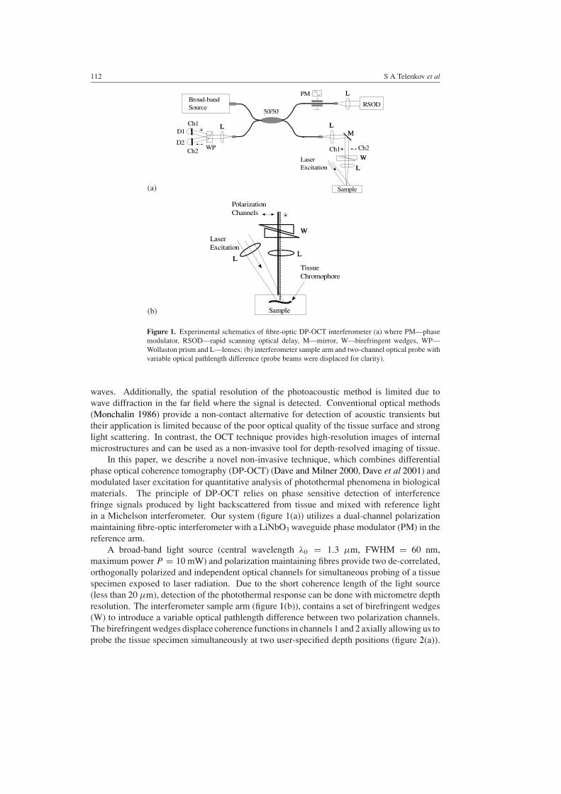
Figure 1. Experimental schematics of fibre-optic DP-OCT interferometer (a) where PM—phasemodulator, RSOD—rapid scanning optical delay, M—mirror, W—birefringent wedges, WP—Wollaston prism and L—lenses; (b) interferometer sample arm and two-channel optical probe withvariable optical pathlength difference (probe beams were displaced for clarity).
waves. Additionally, the spatial resolution of the photoacoustic method is limited due towave diffraction in the far field where the signal is detected. Conventional optical methods(Monchalin 1986) provide a non-contact alternative for detection of acoustic transients buttheir application is limited because of the poor optical quality of the tissue surface and stronglight scattering. In contrast, the OCT technique provides high-resolution images of internalmicrostructures and can be used as a non-invasive tool for depth-resolved imaging of tissue.
In this paper, we describe a novel non-invasive technique, which combines differentialphase optical coherence tomography (DP-OCT) (Dave and Milner 2000, Dave et al 2001) andmodulated laser excitation for quantitative analysis of photothermal phenomena in biologicalmaterials. The principle of DP-OCT relies on phase sensitive detection of interferencefringe signals produced by light backscattered from tissue and mixed with reference lightin a Michelson interferometer. Our system (figure 1(a)) utilizes a dual-channel polarizationmaintaining fibre-optic interferometer with a LiNbO3 waveguide phase modulator (PM) in thereference arm.
A broad-band light source (central wavelength λ0 = 1.3 µm, FWHM = 60 nm,maximum power P = 10 mW) and polarization maintaining fibres provide two de-correlated,orthogonally polarized and independent optical channels for simultaneous probing of a tissuespecimen exposed to laser radiation. Due to the short coherence length of the light source(less than 20 µm), detection of the photothermal response can be done with micrometre depthresolution. The interferometer sample arm (figure 1(b)), contains a set of birefringent wedges(W) to introduce a variable optical pathlength difference between two polarization channels.The birefringent wedges displace coherence functions in channels 1 and 2 axially allowing us toprobe the tissue specimen simultaneously at two user-specified depth positions (figure 2(a)).

Differential phase optical coherence probe 113
(a) (b)
Figure 2. (a) Principle of differential phase detection with the axially separated coherence functionsfor depth-resolved imaging of photothermal response in tissue chromophores; (b) a conventionalOCT image of epoxy phantom with a discrete subsurface chromophore (human hair).
Using a rapid scanning optical delay (RSOD) (Tearney et al 1997), the pathlength in thereference arm of the DP-OCT system was adjusted and held fixed to match the position ofscattering interfaces in the tissue.
The waveguide phase modulator in the interferometer reference arm is used to generateharmonic interference fringe signals at the carrier frequency � = 50–150 kHz in bothchannels. Two optical channels were spatially separated by a Wollaston prism (WP) inthe detection arm and the interference signals were registered by photodetectors D1 and D2.An expression for the interference fringe signals Si recorded by the photodetector in ith opticalchannel (i = 1 or 2) is
Si(t) ∝ 2√
IrIsi |γ (τ)| cos(�t + ϕi(t)) (1)
where Ir is the intensity of light returned from the reference path, Isi is the light intensitybackscattered from a tissue interface in the ith channel, γ is the normalized temporal coherencefunction of the source dependent on the time delay (τ ) between the light in the reference andsample paths, and ϕi(t) is the time-dependent phase which depends on the optical pathlengthof the probing beams. When the laser spot-diameter significantly exceeds the probing depth,temperature increase in tissue [�T (z, t)] can be treated as a one-dimensional function, wherethe z-axis is directed normal to the sample with the origin at the surface. We write the phasedue to light backscattered at depth zi(t) in the ith channel as
ϕi(t) = 2ω
c
∫ zi (t)
0
[n(z) +
(dn
dT
)�T (z, t)
]dz (2)
where ω = 2πc/λ is the central angular frequency of the light source and c is the speed of lightin vacuum. The integrand in equation (2) represents time- and space-dependent tissue refractiveindex n(z, t) = n(z) + (dn/dT )�T (z, t). Since both probe beams propagate along thesame path but are backscattered at different depths, the differential phase �ϕ(t) = ϕ2(t) −ϕ1(t) is
�ϕ(t) = 2ω
c
∫ z2(t)
z1(t)
[n(z) +
(dn
dT
)�T (z, t)
]dz. (3)
In equation (3), we assume that the position of tissue scattering interfaces [z1 and z2] mayvary due to thermoelastic expansion. The equation for �ϕ(t) can be simplified to express
114 S A Telenkov et al
the contribution of thermoelastic and thermorefractive effects. Assuming the refractive indexn(z) = ns for �z = z2 −z1 = const, and taking into account the equation for one-dimensionalstrain in a homogeneous media (Landau and Lifshitz 1959), we get
�ϕ(t) = T + n = 2ωns
c
(1 + σ)
3(1 − σ)
∫ z2
z1
β(T )�T (t, z) dz +2ω
c
∫ z2
z1
(dn
dT
)�T (z, t) dz
(4)
where σ is the Poisson ratio and β is the volumetric thermal expansion coefficient. The first(T ) and second (n) integrals in equation (4) are contributions to the differential phase dueto thermoelastic and thermorefractive effects, respectively.
Analysis of the relative magnitude of thermoelastic and thermorefractive effects intissue on the differential phase was conducted using physical properties of water in thetemperature range 27–87 ◦C (Harvey et al 2000). In water, T and n have opposite signs andthe thermoelastic exceeds the thermorefractive contribution by a factor of 4. The temperaturedependence of �ϕ is closely approximated by a straight line with a slope of 1.69 ×10⁻⁵ rad ◦C−1 per 1 µm of the probe beam axial separation. Equation (4) indicates thata DP-OCT system with a phase sensitivity of 10−3 rad (Akkin et al 2003) and a probe beamaxial separation of 200 µm, is capable of measuring temperature increases in subsurface tissuechromophores with 0.3 ◦C resolution. Because differential phase measurements effectivelycancel common mode phase noise resulting from ambient temperature fluctuations in theinterferometer, air currents, vibrations and other sources, the DP-OCT system is able to detectnanometre level variations of optical pathlength resulting from photothermal stimulation.
2. Experiment
Experimental studies were conducted with a tissue phantom containing a single subsurfacechromophore embedded in an epoxy substrate and an ex-vivo rodent dorsal skin specimen.Both specimens were excited by low frequency (fL < 10 Hz) intensity-modulated laserradiation at λL = 532 nm (Verdi, Coherent, CA). The epoxy phantom was also excited by aflash-lamp pumped pulsed dye laser at λL = 585 nm and pulse duration tL = 0.5 ms (SPTL-1,Candela Corporation, MA) to investigate transient photothermal response. The epoxy phantomwas nearly transparent to the incident laser radiation and absorption was spatially limited tothe cylindrically-shaped chromophore (human hair) positioned approximately 300 µm belowthe surface. A conventional OCT image of the epoxy phantom with subsurface chromophoreis shown in figure 2(b).
In the first experiment, coherence functions of the DP-OCT probe were displaced axiallyto obtain interference fringe signals from the air–phantom interface and the top surface of thehair (see insert in figure 3(a)). An electronic shutter chopped a CW laser beam at frequency2.5 Hz to produce periodic excitation of the test sample (curve 2). A coherent demodulationprocessing algorithm at the signal carrier frequency (10 kHz) was applied to determine thephase in each channel. Our processing algorithm utilizes a quadrature detection techniquewhere signal phase is computed with respect to a harmonic reference signal at the carrierfrequency (Telenkov et al 2002). Integration time (ti) parameter of the coherent demodulationprocessing algorithm can be adjusted to reduce noise outside of the detection band
(∼t−1i
).
The resulting differential phase �ϕ(t) = ϕ2 − ϕ1 is determined by subtracting the phasesof individual channels. A plot of the differential phase (curve 1) computed using ti = 5 ×10⁻⁴ s along with the laser modulation waveform is shown in figure 3(a).
When interference fringe signals in channels 1 and 2 correspond to light backscatteredfrom the top surface of the hair and the air–phantom interface, respectively, the differential

Differential phase optical coherence probe 115
0.0 0.5 1.0 1.5 2.0Time (sec)
-1.0
-0.5
0.0
0.5
1.0
Diff
eren
tial P
hase
(ra
d)
-0.2
0.0
0.2
0.4
0.6
Lase
r In
tens
ity (
arb.
u.)
0.0 0.5 1.0 1.5 2.0 2.5Time (sec)
-0.4
-0.2
0.0
0.2
0.4
Diff
eren
tial P
hase
(ra
d)
-0.2
0.0
0.2
0.4
0.6
Lase
r In
tens
ity (
arb.
u.)
(a)
(b)
Figure 3. (a) Differential phase response of a subsurface chromophore measured with respectto the phantom surface (insert) and (b) response of the chromophore itself probed at the top andthe bottom interfaces. The laser excitation waveform at 2.5 Hz is shown in both measurements(curve 2).
phase undergoes periodic oscillations in-phase with laser excitation. Since light absorptionin the epoxy substrate is negligible, observed variations of differential phase indicate laser-induced thermal expansion of the top surface of the embedded chromophore. The effect ofthermal diffusion remains relatively small at 2.5 Hz because of the poor thermal diffusivityof epoxy (0.12 mm2 s−1) but becomes pronounced at lower modulation frequencies resultingin distortion of the differential phase response. To estimate the temperature increase fromdifferential phase measurements, a three-dimensional strain field must be used to determinethe relative displacement between probe points. However, for superficial chromophoresapproximate estimates can be obtained using the model above. Assuming uniform heating ofthe hair by laser radiation with average power of 80 mW and modulation frequency 2.5 Hz,we estimate that laser-induced temperature increase did not exceed 3 ◦C, which is consistentwith our differential phase measurements.
Taking advantage of the imaging capabilities of OCT, we can measure the photothermalresponse of subsurface chromophores directly. Using the same epoxy phantom, DP-OCTprobe beams were configured to detect interference fringe signals from the top and bottomsurfaces of the hair (insert in figure 3(b)). The recorded differential phase response together

116 S A Telenkov et al
0.000 0.005 0.010 0.015 0.020Time (sec)
-2
-1
0
1
2
3
Diff
eren
tial P
hase
(ra
d)
-1
0
1
2
3
4
Lase
r In
tens
ity (
arb.
u.)
0.000 0.005 0.010 0.015 0.020Time (sec)
-1.0
-0.5
0.0
0.5
1.0
Diff
eren
tial P
hase
(ra
d)
-1.0
-0.5
0.0
0.5
1.0
Lase
r In
tens
ity (
arb.
u.)
Figure 4. (a) Transient differential phase response (1) following a pulsed laser excitation with0.5 ms pulse duration (2); (b) differential phase response at the chromophore position due to pulsedlaser excitation.
with laser excitation is shown in figure 3(b) (curves 1 and 2, respectively). The magnitudeof the differential phase variation in this detection geometry was reduced by a factor of 3and the response appeared inverted with respect to laser excitation. We believe that apparentinversion of the differential phase response is primarily due to the thermoelastic expansion ofthe embedded chromophore detected by channels 1 and 2 as shown in the insert of figure 3(b).
To investigate transient photothermal response, we exposed the epoxy phantom to pulsedlaser radiation with a pulse duration of 0.5 ms. Typical differential phase signal at fringefrequency 150 kHz was computed using ti = 10−4 s and consists of a rapid variation with acharacteristic time equal to the duration of the pulsed laser irradiation followed by a slowerthermal relaxation (figure 4(a), curve 1).
The relaxation time can be determined from differential phase measurements and in thecase of epoxy phantoms was equal to 8.2 ms at the e−1 level, which is consistent with thetheoretically predicted value t0 = r2/4D = 5.2 ms, where r ≈ 50 µm is the chromophoreradius and D = 0.12 mm2 s−1 is the thermal diffusivity of the epoxy substrate. Direct detectionat the chromophore position (figure 4(b)) shows a similar transient response but again withinverted differential phase deviation compared to the probing geometry of figure 4(a). To

Differential phase optical coherence probe 117
0 1 2 3 4Time (sec)
-2
-1
0
1
2
Diff
eren
tial P
hase
(ra
d)
0.0
0.2
0.4
0.6
0.8
Lase
r In
tens
ity (
arb.
u.)
Figure 5. Laser-induced differential phase response in a rodent skin (1) produced by periodicallymodulated laser radiation (2) at wavelength 532 nm and a modulation frequency of 2.5 Hz.
demonstrate an application of the DP-OCT probe for detection of photothermal response intissue, we use ex-vivo specimens of a rodent dorsal skin. In rodent skin, laser radiation (532 nm)is absorbed by both epidermal melanin and dermal blood within an approximately 300 µmthick layer just below the air–stratum corneum interface. The skin specimen was excited byperiodically modulated laser radiation (modulation frequency fL = 2.5 Hz) at a wavelengthof 532 nm, incident power of 100 mW and focused to a 1.5 mm spot on the surface. DP-OCTcoherence functions were displaced axially by L12 = 160 µm for simultaneous detection ofinterference fringes from the air–skin interface and a tissue scattering interface located L12/n
below the surface (n is the skin refractive index). Computed differential phase (�ϕ) over 4 sof recording time and ti = 2 × 10⁻⁴ s (figure 5, curve 1) shows periodic oscillations of opticalpathlength (L12(t)) at laser excitation frequency fL.
Since absorption of laser radiation in skin was not confined to a specific chromophore,the resulting differential phase response may include both thermoelastic and thermorefractiveeffects as indicated in equation (4). Assuming physical properties of hydrated tissue (σ = 1/2,β = 1.23 × 10⁻⁴ ◦C−1, n = 1.38) and the temperature-dependent refractive index of water(Harvey et al 1998) (dn/dT ) ≈ −10−4 ◦C−1, equation (4) gives a temperature increase�T ≈ 18.5 ◦C for differential phase variation 1.5 rad. Our experiments using DP-OCTfor depth-resolved measurements of photothermal response in tissue underscore an importantfeature: because only relative variations of the optical pathlength between two probing beamscontribute to the differential phase signal, DP-OCT is sensitive to localized laser-inducedvariations of optical pathlength, which cannot be achieved with a conventional OCT system(Rao et al 2003).
3. Discussion and conclusion
Although conventional OCT provides spatially resolved measurements of light backscatteredfrom tissue, thermoelastic response of tissue to laser irradiation is not localized and isdue to a composite response of all tissue chromophores. To illustrate, we consider anisolated subsurface cylindrical chromophore in tissue similar to a hair embedded in the tissuephantom employed in the experiments described above. Following absorption of incident laser

118 S A Telenkov et al
radiation, the chromophore expands thermoelastically and induces a strain field throughoutthe entire tissue specimen. If during laser irradiation one probes arbitrary points in thetissue specimen using conventional OCT, frequency shifts and/or fringe amplitude variationsrecorded in a single measurement cannot be used directly to attribute these changes to onespatial location. In comparison to conventional OCT, utility of DP-OCT is apparent: byaxially displacing the probe beams, thermoelastic (β) and thermorefractive (dn/dT ) changesare measured at a user-specified location in tissue corresponding to the region between probebeams. In as much as the DP-OCT probe beams may be displaced axially by an arbitrarydistance, spatial resolution of DP-OCT is approximately two or three source coherence lengths.Application of DP-OCT to record the thermoelastic response in tissue is limited by the DP-OCTphase sensitivity and signal to noise ratio (SNR). Because phase sensitivity of the DP-OCTsystem is 10−3 rad (angstroms in optical pathlength) for measurement times of a millisecond(Akkin et al 2003), we think the principal source of phase variation in DP-OCT measurementof tissues is optical pathlength changes due to either internal movements or temperaturevariations. Further experiments are required to characterize the magnitude of internal phasevariations in various tissues. The range of depths that can be probed using DP-OCT is limitedby the system SNR. In the DP-OCT system used to record the measurements reported here,SNR is limited by cross talk in the polarization maintaining fibre and is approximately 45 dB.The relatively low SNR of the DP-OCT system limits the range of depths that may be probed incomparison to conventional OCT. Although our fibre-based DP-OCT system provides phasesensitivity two- or three-times better than conventional OCT, SNR is sacrificed. Experimentsare underway in our laboratory to improve the SNR of DP-OCT.
In conclusion, we have demonstrated an application of DP-OCT to non-invasive depth-resolved detection of photothermal phenomena in tissue. Subsurface structures can beidentified from interference fringe signals in two optical channels with axially displacedcoherence functions and photothermal response to laser excitation is determined from transientdifferential phase measurements. Because localized temperature increase is caused bysubsurface chromophores at the excitation wavelength, this technique can be used for non-invasive diagnostics of molecular composition of tissue with spatial resolution two- to three-times the source coherence length. Analysis of photothermal response produced by a tunablelaser source can provide spectroscopic information on subsurface tissue microstructures andhas potential to improve dosimetry of clinical laser procedures by monitoring the depth-resolved thermal response of targeted subsurface chromophores.
Acknowledgment
This work was supported by the National Institutes of Health (RR-14069–02).
References
Akkin T, Dave D P, Yong J, Telenkov S A, Raylander III H G and Milner T E 2003 Imaging tissue response toelectrical and photothermal stimulation with nanometer sensitivity Lasers Surg. Med. 33 219–25
Brezinski M E and Fujimoto J G 1999 Optical coherence tomography: high resolution imaging in nontransparenttissue IEEE J. Sel. Top. Quantum Electron. 5 1185–92
Chen Z, Milner T E, Shrinivas S, Wang X, Malekafzali A, van Gemert M J C and Nelson J S 1997 Noninvasiveimaging of in-vivo blood flow using optical Doppler tomography Opt. Lett. 22 1119–21
Dave D P, Akkin T, Milner T E and Raylander III H G 2001 Phase-sensitive frequency-multiplexed optical low-coherence reflectometry Opt. Commun. 193 39–43
Dave D P and Milner T E 2000 Optical low-coherence reflectometer for differential phase measurements Opt. Lett.25 227–9
Differential phase optical coherence probe 119
de Boer J F and Milner T E 2002 Review of polarization sensitive optical coherence tomography and Stokes vectordetermination J. Biomed. Opt. 7 359–71
de Boer J F, Shrinivas S M, Malekafzali A, Chen Z and Nelson J S 1998 Imaging thermally damaged tissue bypolarization sensitive optical coherence tomography Optics Express 3 212–8
Harvey A H, Gallagher J S and Levelt Sengers J M H 1998 Revised formulation for the refractive index of water andsteam as a function of wavelength, temperature and density J. Phys. Chem. Ref. Data 27 761–74
Harvey A H, Peskin A P and Klein S A 2000 NIST/ASME Steam Properties, Database 10. US Dept. of CommerceKarabutov A A, Podymova N B and Letokhov V S 1996 Time-resolved laser optoacoustic tomography of
inhomogeneous media Appl. Phys. B 63 545–63Landau L D and Lifshitz E M 1959 Theory of Elasticity (Oxford: Pergamon)Monchalin J P 1986 Optical detection of ultrasound IEEE Trans. Ultrason. Ferroelectr. Freq. Control 33 485–99Oraevsky A A, Jacques S L and Tittel F K 1997 Measurement of tissue optical properties by time-resolved detection
of laser-induced transient stress Appl. Opt. 36 402–15Pan Y, Birngruber R, Rosperich J and Engelhart R 1995 Low-coherence optical tomography in turbid tissue:
Theoretical analysis Appl. Opt. 34 6564–74Rao K D, Choma M A, Yazdanfar S, Rollins A M and Izatt J A 2003 Molecular contrast in optical coherence
tomography by use of pump-probe technique Opt. Lett. 28 340–2Tearney G J, Bouma B E and Fujimoto J G 1997 High-speed phase- and group-delay scanning with a gratin-based
phase control delay line Opt. Lett. 22 1811–3Telenkov S A, Vargas G, Nelson J S and Milner T E 2002 Coherent thermal wave imaging of subsurface chromophores
in biological materials Phys. Med. Biol. 47 657–71
Copyright © 2022 FDOKUMEN