Differential expression pattern of genes encoding for anti-microbial peptides in the fetal membranes...
13
Differential expression pattern of genes encoding for anti-microbial peptides in the fetal membranes of patients with spontaneous preterm labor and intact membranes and those with preterm prelabor rupture of the membranes OFFER EREZ 1,2 , ROBERTO ROMERO 1,2,3 , ADI L. TARCA 1,4 , TINNAKORN CHAIWORAPONGSA 1,2 , YEON MEE KIM 1 , NANDOR GABOR THAN 1 , EDI VAISBUCH 1,2 , SORIN DRAGHICI 4 , & GERARD TROMP 3 1 Perinatology Research Branch, NICHD, NIH, DHHS, Detroit, Michigan, USA, 2 Department of Obstetrics and Gynecology, Wayne State University School of Medicine, Detroit, Michigan, USA, 3 Center for Molecular Medicine and Genetics, Wayne State University School of Medicine, Detroit, Michigan, USA, and 4 Department of Computer Science, Wayne State University, Detroit, Michigan, USA (Received 2 March 2009; accepted 20 March 2009) Abstract Objective. Increased amniotic fluid concentrations of anti-microbial peptides, components of the innate immune system, have been reported in patients with preterm labor (PTL) with intact membranes and intra-amniotic infection and/or inflammation (IAI), as well as in patients with preterm prelabor rupture of the membranes (PPROM). This study was designed to confirm these results using a targeted approach, detecting DEFA1, DEFB1, GNLY, and S100A9 gene expression in the choriamniotic membranes in pregnancies complicated with PTL and intact membranes or PPROM, with and without histologic chorioamnionitis. Study design. Human fetal membranes were obtained from patients in the following groups: (1) PTL with intact membranes (n ¼ 15); (2) PTL with intact membranes with histologic chorioamnionitis (n ¼ 12); (3) PPROM (n ¼ 17); and (4) PPROM with histologic chorioamnionitis (n ¼ 21). The mRNA expression of a-defensin-1, b-defensin-1, calgranulin B and granulysin in the fetal membranes was determined by qRT-PCR. Results. (1) The expression of a-defensin-1 mRNA in the fetal membranes was higher in patients with PTL and intact membranes with histologic chorioamnionitis, than those without chorioamnionitis (19.4-fold, p 5 0.001); (2) Among patients with histologic chorioamnionitis, patients with PTL and intact membranes had a higher a-defensin-1 mRNA expression than those with PPROM (5.5-fold, p ¼ 0.003); (3) Histologic chorioamnionitis was associated with a higher calgranulin B mRNA expression in the chorioamniotic membranes of patients with both PTL and intact membranes (7.9- fold, p ¼ 0.03) and PPROM (7.6-fold, p 5 0.0001); (4) The expression of calgranulin B mRNA in the fetal membranes was higher in patients with PTL and intact membranes without histologic chorioamnionitis than in those with PPROM without histologic chorioamnionitis (2.7-fold, p ¼ 0.03); (5) There were no differences in the expression of b-defensin-1 and granulysin in the chorioamniotic membranes between the study groups even in the presence of histologic chorioamnioniotis. Conclusions. (1) Among patients with histologic chorioamnionitis, the mRNA expression of a-defensin-1 and calgranulin B in the fetal membranes of patients with PTL and intact membranes as well as that of calgranulin B in the fetal membranes of patients with PPROM is higher than in the membranes of those without histologic chorioamnionitis; (2) histologic chorioamnionitis is associated with differences in the pattern of a-defensin-1 mRNA expression in the fetal membranes in patients with PTL and intact membranes and those with PPROM. Keywords: Human neutrophil peptide, a-defensin, b-defensin, calgranulin, calprotectin, granulysin, preterm prelabour rupture of membranes, spontaneous preterm delivery Correspondence: Roberto Romero, MD, Perinatology Research Branch, NICHD/NIH/DHHS, Wayne State University/Hutzel Women’s Hospital, 3990 John R, Box 4, Detroit, Michigan 48201, USA. Tel: þ1-313-993-2700. Fax: þ1-313-993-2694. E-mail: [email protected] The Journal of Maternal-Fetal and Neonatal Medicine, December 2009; 22(12): 1103–1115 ISSN 1476-7058 print/ISSN 1476-4954 online Ó 2009 Informa UK Ltd. DOI: 10.3109/14767050902994796 J Matern Fetal Neonatal Med Downloaded from informahealthcare.com by 162.248.247.136 on 05/20/14 For personal use only.
Transcript of Differential expression pattern of genes encoding for anti-microbial peptides in the fetal membranes...
Differential expression pattern of genes encoding for anti-microbialpeptides in the fetal membranes of patients with spontaneouspreterm labor and intact membranes and those with pretermprelabor rupture of the membranes
OFFER EREZ1,2, ROBERTO ROMERO1,2,3, ADI L. TARCA1,4,
TINNAKORN CHAIWORAPONGSA1,2, YEON MEE KIM1, NANDOR GABOR THAN1,
EDI VAISBUCH1,2, SORIN DRAGHICI4, & GERARD TROMP3
1Perinatology Research Branch, NICHD, NIH, DHHS, Detroit, Michigan, USA, 2Department of Obstetrics and Gynecology,
Wayne State University School of Medicine, Detroit, Michigan, USA,3Center for Molecular Medicine and Genetics,
Wayne State University School of Medicine, Detroit, Michigan, USA, and 4Department of Computer Science, Wayne State
University, Detroit, Michigan, USA
(Received 2 March 2009; accepted 20 March 2009)
Abstract
Objective. Increased amniotic fluid concentrations of anti-microbial peptides, components of the innate immune system,have been reported in patients with preterm labor (PTL) with intact membranes and intra-amniotic infection and/orinflammation (IAI), as well as in patients with preterm prelabor rupture of the membranes (PPROM). This study wasdesigned to confirm these results using a targeted approach, detecting DEFA1, DEFB1, GNLY, and S100A9 gene expressionin the choriamniotic membranes in pregnancies complicated with PTL and intact membranes or PPROM, with and withouthistologic chorioamnionitis.
Study design. Human fetal membranes were obtained from patients in the following groups: (1) PTL with intactmembranes (n¼ 15); (2) PTL with intact membranes with histologic chorioamnionitis (n¼ 12); (3) PPROM (n¼ 17); and(4) PPROM with histologic chorioamnionitis (n¼ 21). The mRNA expression of a-defensin-1, b-defensin-1, calgranulin Band granulysin in the fetal membranes was determined by qRT-PCR.
Results. (1) The expression of a-defensin-1 mRNA in the fetal membranes was higher in patients with PTL and intactmembranes with histologic chorioamnionitis, than those without chorioamnionitis (19.4-fold, p5 0.001); (2) Amongpatients with histologic chorioamnionitis, patients with PTL and intact membranes had a higher a-defensin-1 mRNAexpression than those with PPROM (5.5-fold, p¼ 0.003); (3) Histologic chorioamnionitis was associated with a highercalgranulin B mRNA expression in the chorioamniotic membranes of patients with both PTL and intact membranes (7.9-fold, p¼ 0.03) and PPROM (7.6-fold, p5 0.0001); (4) The expression of calgranulin B mRNA in the fetal membranes washigher in patients with PTL and intact membranes without histologic chorioamnionitis than in those with PPROM withouthistologic chorioamnionitis (2.7-fold, p¼ 0.03); (5) There were no differences in the expression of b-defensin-1 andgranulysin in the chorioamniotic membranes between the study groups even in the presence of histologic chorioamnioniotis.
Conclusions. (1) Amongpatientswith histologicchorioamnionitis, themRNAexpressionofa-defensin-1 and calgranulin B in thefetal membranes of patients with PTL and intact membranes as well as that of calgranulin B in the fetal membranes of patients withPPROM is higher than in the membranes of those without histologic chorioamnionitis; (2) histologic chorioamnionitis isassociated with differences in the pattern ofa-defensin-1 mRNA expression in the fetal membranes in patients with PTL and intactmembranes and those with PPROM.
Keywords: Human neutrophil peptide, a-defensin, b-defensin, calgranulin, calprotectin, granulysin, preterm prelabour ruptureof membranes, spontaneous preterm delivery
Correspondence: Roberto Romero, MD, Perinatology Research Branch, NICHD/NIH/DHHS, Wayne State University/Hutzel Women’s Hospital, 3990 John
R, Box 4, Detroit, Michigan 48201, USA. Tel: þ1-313-993-2700. Fax: þ1-313-993-2694. E-mail: [email protected]
The Journal of Maternal-Fetal and Neonatal Medicine, December 2009; 22(12): 1103–1115
ISSN 1476-7058 print/ISSN 1476-4954 online � 2009 Informa UK Ltd.
DOI: 10.3109/14767050902994796
J M
ater
n Fe
tal N
eona
tal M
ed D
ownl
oade
d fr
om in
form
ahea
lthca
re.c
om b
y 16
2.24
8.24
7.13
6 on
05/
20/1
4Fo
r pe
rson
al u
se o
nly.

Introduction
The traditional view is that the amniotic cavity in
normal pregnancy is sterile and does not contain
viable bacteria [1] despite the presence of a large
number of microorganisms in the lower genital tract
(vagina and ectocervix). The sterile status of the
amniotic cavity is presumably accomplished by the
participation of the innate immune system, including
the cervical mucous plug [2–5], chorioamniotic
membranes [6–8] and cellular components of the
decidua, amnion, and chorion, including neutro-
phils, macrophages, natural killer (NK) cells, and
trophoblasts [6,9,10].
Natural anti-microbial peptides have been identi-
fied in plants, insects, and vertebrates [11] as part of
the innate limb of the immune system that provides
protection against bacteria, yeast, and viruses [11–
13]. In humans, anti-microbial peptides have been
detected in white blood cells and [14,15] epithelial
cells [11,16–18], as well as in the placenta [9,19],
decidua, fetal membranes [16,20] and amniotic fluid
[1,21]. The latter contains defensins, bactericidal/
permeability-increasing (BPI) protein, and S100B
[22] as well as other proteins, such as lactoferrin and
calprotectin (MRP8/14) [21].
Defensins are anti-microbial peptides classified
into three major groups: alpha (a), beta (b), and theta
(y) [23]. a-defensins have a broad anti-microbial
activity against Gram-negative and Gram-positive
bacteria, fungi, and enveloped viruses [14,23–25].
These anti-microbial peptides interact with the cell
membrane of invading organisms, causing a disrup-
tion of ion-fluxes and eventually leading to cell lysis
[14,23–25]. The group of a-defensins consists of six
distinct peptides, of which a-defensins-1, -2, and -3
share many similarities as their primary structure
differs by only one amino acid [12,26–28]. Bone
marrow precursors of neutrophils synthesize and
store these anti-microbial peptides intracellularly in
azurophil granules [12,28–32]. Thus, a-defensins are
often referred to as human neutrophil peptides
(HNP)-1, -2, and -3 [14,33]. In addition to their
anti-microbial activity, a-defensins are capable of
stimulating a systemic inflammatory response as well
as to chemo-attract T-cells and induce histamine
release from mast cells [34–37]. b-defensins are
mainly effective against Gram-negative bacteria and
yeast, while some have also microbial activity against
Gram-positive bacteria [38–40]. Human b-defensin-
1 has anti-microbial properties against Gram-positive
and Gram-negative bacteria [39–42], as well as
adenovirus [43]. Calgranulin B (MRP14, S100A9)
is an additional anti-microbial peptide that forms
calprotectin (MRP8/14) heterodimer with calgranu-
lin A (MRP8, S100A8) [44]. Calgranulin B can be
detected in neutrophils, monocytes and activated
macrophages, as well as in endothelial and epithelial
cells [44–51]. Calprotectin regulates the adhesion of
myeloid cells to the vascular endothelium and to the
extracellular matrix, controlling the activation of
these effector cells and their direct anti-bacterial
effect by zinc-capturing [44].
Granulysin, a 9 kD protein [52] secreted from
cytolytic granules of cytotoxic T-lymphocytes and
NK cells [53–56], is effective against Gram-positive
and Gram-negative bacteria, as well as fungi [55] and
mycobacteria [55]. Its anti-microbial activity is
mediated through the induction of an increase in
intracellular calcium and the efflux of intracellular
potassium into the pathogen, leading to the activa-
tion of sphingomyelinase and the ceramide pathway,
as well as mitochondrial damage by the activation of
caspases and, consequently, apoptosis [57–59].
Term parturition is associated with both an
inflammatory response and the activation of the
three clinically manifested components of the com-
mon pathway of parturition, including uterine
contraction, cervical dilatation, and decidual/mem-
branes activation [60–62]. Our group demonstrated
that each of these components has a distinct
transcriptome during labor at term [20,63,64].
Moreover, microarray experiments have revealed
that human term labor is characterized by an acute
inflammation gene expression signature in the
extraplacental membranes, which includes the differ-
ential expression of multiple genes encoding for
cytokines and chemokines known to orchestrate
acute inflammatory response [65].
Intrauterine infection and/or inflammation (IAI)
can activate the common pathway of parturition, and
is a major cause of preterm labor (PTL) and delivery
[66–69]. Microbial invasion of the amniotic cavity
(MIAC), spontaneous PTL and preterm prelabor
rupture of the membranes (PPROM) are associated
with increased intra-amniotic concentrations of
a-defensins, BPI, calprotectin, b-defensin-2 [1,21],
and S100B [22]. African-American women with
elevated HNP-1–3 concentrations in vaginal fluid
at mid pregnancy (15–27 weeks of gestation) had an
increased risk for spontaneous preterm birth at 32–
36 weeks (OR 2.4, 95% CI 1.2–4.7) after adjustment
for maternal age, gestational age at enrollment, and
bacterial vaginosis [70]. Calgranulin B (S100A9),
was differentially expressed in the transcriptome of
chorioamniotic membranes of women with preterm
deliveries. Thus, this study was designed to deter-
mine by RT-PCR changes in the chorioamniotic
expression of the mRNA for S100A9 and additional
genes encoding for the following anti-microbial
peptides a-defensin-1 (DEFA1), b-defensin-1
(DEFB1), and granulysin (GNLY), of patients with
PTL with intact membranes and PPROM with and
without histologic chorioamnionitis.
1104 O. Erez et al.
J M
ater
n Fe
tal N
eona
tal M
ed D
ownl
oade
d fr
om in
form
ahea
lthca
re.c
om b
y 16
2.24
8.24
7.13
6 on
05/
20/1
4Fo
r pe
rson
al u
se o
nly.

Materials and methods
Study design and population
The basis for the current study are the results of a
previous microarray study that was completed during
March–April 2001, in which calgranulin B (S100A9)
was differentially expressed in the transcriptome of
chorioamniotic membranes of women with histologic
chorioamnionitis who had either PTL with intact
membranes or PPROM. This confirmatory RT-PCR
cross-sectional study was designed to investigate the
differential expression of the DEFA1, DEFB1, GNLY,
and S100A9 genes in the fetal membranes of patients in
the following groups: (1) PTL with intact membranes
without histologic chorioamnionitis (n¼ 15); (2) PTL
with intact membranes with histologic chorioamnio-
nitis (n¼ 12); (3) PPROM without histologic chor-
ioamnionitis (n¼ 17); and (4) PPROM with histologic
chorioamnionitis (n¼ 21). Patients presenting with
medical complications, multiple pregnancies, and fetal
chromosomal or congenital abnormalities were ex-
cluded. All patients provided written informed con-
sent prior to the collection of samples. The collection
and utilization of samples for research purposes was
approved by the Institutional Review Boards of both
the Eunice Kennedy Shriver National Institute of Child
Health and Human Development (NIH/DHHS) and
Wayne State University. Many of these samples have
been employed to study the biology of PTL and the
inflammation of the fetal membranes.
Definitions
Spontaneous PTL with intact membranes was de-
fined as the presence of regular uterine contractions
that occurred at a frequency of at least 2 in every
10 minutes and associated with cervical changes
which led to spontaneous preterm delivery (537
weeks of gestation) [60,62]. PPROM was defined as
the prelabor rupture of membranes occurring 537
weeks of gestation, diagnosed by speculum examina-
tion of vaginal pooling, nitrazine, and ferning tests
[71]. Amniocentesis was performed at the discretion
of the treating physician. Amniotic fluid was analyzed
for the assessment of the microbial state of the
amniotic cavity. Amniotic fluid was cultured for
aerobic and anaerobic bacteria, as well as for genital
mycoplasmas.
Placental histopathologic examinations
Chorioamniotic membranes containing attached ma-
ternal decidua were obtained from placentas delivered
by spontaneous labor or cesarean section at the Hutzel
Women’s Hospital (Wayne State University, Detroit,
MI). Fetal membranes were fixed in 10% neutral
buffered formalin overnight and embedded in paraffin.
Five micrometer paraffin sections were stained with
hematoxylin and eosin then examined using bright-
field light microscopy. Histopathologic examinations
were performed by pathologists blinded to the clinical
information based on the diagnostic criteria previously
described [72]. Histologic chorioamnionitis was diag-
nosed in the presence of acute inflammation using
previously described criteria [73,74].
Total RNA extraction
The membranes were dissected from the placentas,
rinsed thoroughly with a sterile ice-cold phosphate
buffered saline solution (Sigma Chemical Company,
St Louis, MO), cut into small pieces, placed in RNA
later solution (Ambion, Austin, Texas), and stored at
þ48C for no longer than 2 weeks. Total RNA was
isolated with a modification of the standard guani-
dinium isothiocyanate-cesium chloride method [20].
Briefly, tissues were homogenized with a PRO200
rotor-stator homogenizer (Pro Scientific, Monroe,
CT) in the presence of 4 mol/l guanidinium iso-
thiocyanate, 0.1 mol/l mercaptoethanol, 0.5% sarko-
syl, and 5 mmol/l sodium citrate (pH 7); solid CsCl
was added to the sample (final concentration, 0.25 g/
ml), and the samples were pelleted by ultracentrifu-
gation, according to the protocol. RNA pellets were
resuspended and extracted with chloroform:isoamy-
lalcohol, and the RNA was precipitated with ethanol
and glycogen (Roche Molecular Biochemicals, In-
dianapolis, IN) as a carrier. Before the first use, the
RNA was pelleted and resuspended in water that
contained RNasin (Promega Corp, Madison, WI).
Quantitative real-time reverse transcription-polymerase
chain reaction
Total RNA (2.5 mg) from each sample and a positive
control sample was reverse transcribed using Super-
script II reverse transcriptase, random hexamer
primers, and oligo(dT) primers (Invitrogen Life
Technologies, Rockville, MD). The standard curve
was run with the DEFA1, DEFB1, GNLY, and
S100A9 genes and the 18S ribosomal RNA house-
keeping gene to determine the quantity of cDNA
needed for an approximate cycle threshold (Ct) of
25. Subsequently, cDNA derived from an equivalent
of 75 ng RNA from each sample were run in
triplicate on 96-well plates to obtain technical
replicates for the target and reference assays. A
calibrator sample was run in triplicate in all plates to
account for plate effects. In addition, a negative
control containing no RNA and 12.5 ng of human
genomic DNA were also tested in duplicates.
Samples from the study groups were randomly
allocated on the plates; the DEFA1, DEFB1, GNLY,
S100A9, and 18S rRNA assays were run with the
Anti-microbial peptide gene expression in patients with PTL and PPROM 1105
J M
ater
n Fe
tal N
eona
tal M
ed D
ownl
oade
d fr
om in
form
ahea
lthca
re.c
om b
y 16
2.24
8.24
7.13
6 on
05/
20/1
4Fo
r pe
rson
al u
se o
nly.

same allocation on the parallel plates. The qPCR
reactions were assembled based on the TaqMan
Universal PCR Master Mix protocol (Applied
Biosystems) using the 18S rRNA TaqMan gene
expression assay (Hs99999901_s1; Applied Biosys-
tems, Foster City, CA) for the quantification of the
housekeeping gene and self-designed primers and
probe for the target genes (DEFA1, forward primer:
50-CCCAGAAGTGGTTGTTTCCCT-30; reverse
primer: 50-TTTTCCTTGAGCCTGGATGCT-30;probe: 50-TGGAGCCAAGCTTTCGTCCCATG-
30; DEFB1, forward primer: 50-ATTGCGTCAG
CAGTGGAGG-30; reverse primer: 50-AACAGG
TGCCTTGAATTTTGGT-30; probe: 50-CAATGT
CTCTATTCTGCCTGCCCGATCTT-30; GNLY,
forward primer: 50-AGCAACCTCTGCCGGCT-30;reverse primer: 50-GACAGCAGAGGGAGTCAGG
G-30; probe: 50-CTTCCTCGATCCAGAATCCAC
TCTCCAGTCT-30; S100A9, forward primer: 50-CAGCTGAGCTTCGAGGAGTTC-30; reverse pri-
mer: 50-GCATCTTCTCGTGGGAGGC-30; probe:
50-CAGGTTAGCCTCGCCATCAGCATGA-30).Data were collected by the ABI Prism 7700
Sequence Detection System (Applied Biosystems).
Statistical analysis
Demographic and clinical characteristics of the study
groups were compared using the Pearson’s chi-
square test and the Fisher’s exact test for proportions,
and the Mann–Whitney U test for non-normally
distributed continuous variables using SPSS version
12.0 (SPSS, Chicago, IL). Quantitative RT-PCR
data were analyzed using the R statistical software.
Gene expression levels were profiled in multiple
sample groups (term not in labor, term in labor,
PPROM without histologic chorioamniotis, PPROM
with histologic chorioamnionitis, PTL without histo-
logic chorioamniotis, PTL with histologic chorioam-
niotis) by qRT-PCR experiments, using between 6
and 29 samples per group. The RT reactions were
run on 96-well plates. Samples from the study groups
were randomly allocated on the plates, and only one
target gene and the 18S reference assay were run in
parallel on each given plate. Each reaction was
repeated either two or three times to obtain technical
replicates for both the target assay and the reference
assay. A calibrator patient sample was placed on all
plates to account for eventual plate effects. Briefly,
the d-d method [75,76] was used to generate an
outcome variable, Y, which is a surrogate of the log2
concentration of the target gene in each patient
sample, corrected already for potential plate effects. A
linear model was employed in which Y values were
fitted using the Group variable and the gestational age
as predictors without including the interaction term
between these two variables. The coefficients of the
two predictors in the linear model were estimated
together with their significant p-values.The outcome
variable, Y, included also a positive constant to
render the Y values positive so that larger values
correspond to higher expression. A False Discovery
Rate adjustment [76] of resulting p-values was
performed to account for all parallel tests. For each
pair-wise comparison, the Group effect was consid-
ered significant, if the adjusted p-values were 50.05
and the magnitude of change was at least 2-fold (one
Ct unit difference). For the gestational age effect,
adjusted p-values5 0.05 were considered significant.
Results
Demographic, clinical and histopathological data
Demographic and clinical characteristics of the study
groups are displayed in Table I. The diagnosis of
histologic chorioamnionitis was based on the presence
of maternal and/or fetal inflammatory response in the
placenta and fetal membranes. Among the 12 patients
with PTL intact membranes and histologic chor-
ioamnionitis, maternal inflammatory response was
diagnosed in one case, whereas 11 patients had both a
maternal and a fetal inflammatory response. Among
the 21 patients with PPROM and histologic chor-
ioamnionitis, 7 had a maternal inflammatory re-
sponse, 2 had a fetal inflammatory response, and 12
had both. Amniocentesis was performed in 10
patients with PTL intact membranes and 14 patients
with PPROM. A positive amniotic fluid culture was
detected in 30% (3/10) of patients with PTL intact
membranes and in 46.2% (6/14) of patients with
PPROM (p¼ 0.4). The microorganisms found in
amniotic fluid cultures are presented in Table II.
Within the study groups, there was no correlation
between the chorioamniotic expression of the DEFA1,
DEFB1, GNLY, and S100A9 genes and gestational
age at delivery, in which these samples were collected
(data not shown).
Changes in the fetal membranes mRNA expression of
anti-microbial peptides
a-defensin (Human neutrophil peptide)-1. Among
patients with PTL and intact membranes, histologic
chorioamnionitis was associated with a higher
a-defensin-1mRNA expression in the chorioamniotic
membranes (19.4-fold, p5 0.001). This difference
did not reach statistical significance in those with
PPROM (2.7-fold, p¼ 0.08) (Figure 1). Among
women with histologic chorioamnionitis, patients
with PTL with intact membranes had a higher
amount of a-defensin-1 mRNA expression in the
fetal membranes than those with PPROM (5.5-fold,
p¼ 0.003) (Figure 1).
1106 O. Erez et al.
J M
ater
n Fe
tal N
eona
tal M
ed D
ownl
oade
d fr
om in
form
ahea
lthca
re.c
om b
y 16
2.24
8.24
7.13
6 on
05/
20/1
4Fo
r pe
rson
al u
se o
nly.

b-defensin-1. Among patients with PTL with intact
membranes and those with PPROM, histologic
chorioamnionitis was not associated with a higher
b-defensin-1 mRNA expression in the chorioamnio-
tic membranes (p¼ 0.2 for both comparisons).
Moreover, the expression of b-defensin-1 mRNA in
the fetal membranes did not differ between patients
presenting with PTL with intact membranes and those
presenting with PPROM, regardless of the presence of
histologic chorioamnionitis (without chorioamnioni-
tis: p¼ 0.2; with chorioamnionitis: p¼ 0.2).
Granulysin. There was no correlation between gran-
ulysin mRNA expression level and histologic chor-
ioamnionitis in membranes from either patients with
PTL and intact membranes or from patients with
PPROM (p¼ 0.2 for both comparisons). Moreover,
the expression of granulysin mRNA in the fetal
membranes did not differ between patients presenting
with PTL and intact membranes and those with
Table II. Microorganisms detected in positive amniotic fluid
cultures.
PPROM with
chorioamnionitis
(n¼14)
Preterm labor
intact membranes
with chorioamnionitis
(n¼10)
Ureoplasma ureolyticum 3 1
Mycoplasma hominis 1 2
Gardnerella vaginalis 2 –
Lactobacillus species – 1
Prevotella species 1 –
Candida albicans 1 –
Table I. Demographic and clinical characteristics of the study groups.
PPROM without
chorioamnionitis
(n¼ 17)
PPROM with
chorioamnionitis
(n¼ 21) p-value*
Preterm labor intact
membranes without
chorioamnionitis
(n¼ 15)
Preterm labor intact
membranes with
chorioamnionitis
(n¼12) p-value{
Maternal age (years) 27 [22–31] 27 [22–34] NS 24 [20–28] 21 [18–30] NS
Ethnic origin (%)
African-American 14 (82.4) 19 (90.5) NS 13 (86.7) 10 (83.3) NS
Caucasian 3 (17.6) 2 (9.5) 2 (13.3) 2 (16.7)
Gravidity 3 [1.5–6] 4 [2.5–5.5] NS 3 [1–4] 3 [1.3–3] NS
Parity 2 [0–3.5] 2 [1–4] NS 1 [1–2] 0.5 [0–1] NS
Positive amniotic fluid
culture (%)x0 6 (46.2) 0.051 0 3 (30) NS
Gestational age at diagnosis
(weeks)
31 [30–32.9] 30 [25.6–31.4] NS 25.9 [23.0–31.0] 28.9 [25.5–32] NS
Gestational age at delivery
(weeks)
31.7 [30.1–33.1] 31 [29.1–32.3] NS 28.7 [23.7–32.4] 29.1 [25.6–32] NS
Diagnosis-to-delivery
interval (days)
1 [0–5] 3 [1–10] NS 3 [1–5] 1 [0–6.5] NS
Birth-weight (g) 1530 [1365–1850] 1700 [995–1920] NS 1020 [680–1780] 1045 [670–1683] NS
Female fetus (%) 5 (29.4) 7 (33.3) NS 4 (26.7) 7 (58.3) NS
Values are presented as median [interquartile range] or number (percentage).
PPROM, preterm prelabor rupture of the membranes.
*Comparisons between two groups were performed with the Mann-Whitney test.{The Fisher’s exact test.xPPROM (n¼ 8); PPROM with chorioamnionitis (n¼14); PTL (n¼ 11); PTL with chorioamnionitis (n¼10).
Figure 1. a-defensin 1 mRNA expression in the fetal membranes
of patients with spontaneous preterm labor (PTL) or preterm
prelabor rupture of membranes (PPROM). In the presence of
histologic chorioamnionitis, there was an increased expression
among patients with preterm labor with intact membranes (19.4-
fold, p50.001) or PPROM (2.7-fold, p¼ 0.08). The amount of a-
defensin 1 mRNA was higher in the fetal membranes of patients
presenting with preterm labor with intact membranes and
histologic chorioamnionitis than in patients with PPROM and
histologic chorioamnionitis (5.5-fold, p¼0.003).
Anti-microbial peptide gene expression in patients with PTL and PPROM 1107
J M
ater
n Fe
tal N
eona
tal M
ed D
ownl
oade
d fr
om in
form
ahea
lthca
re.c
om b
y 16
2.24
8.24
7.13
6 on
05/
20/1
4Fo
r pe
rson
al u
se o
nly.
PPROM, irrespective of the presence of histologic
chorioamnionitis (no chorioamnionitis: p¼ 0.2; chor-
ioamnionitis: p¼ 0.2).
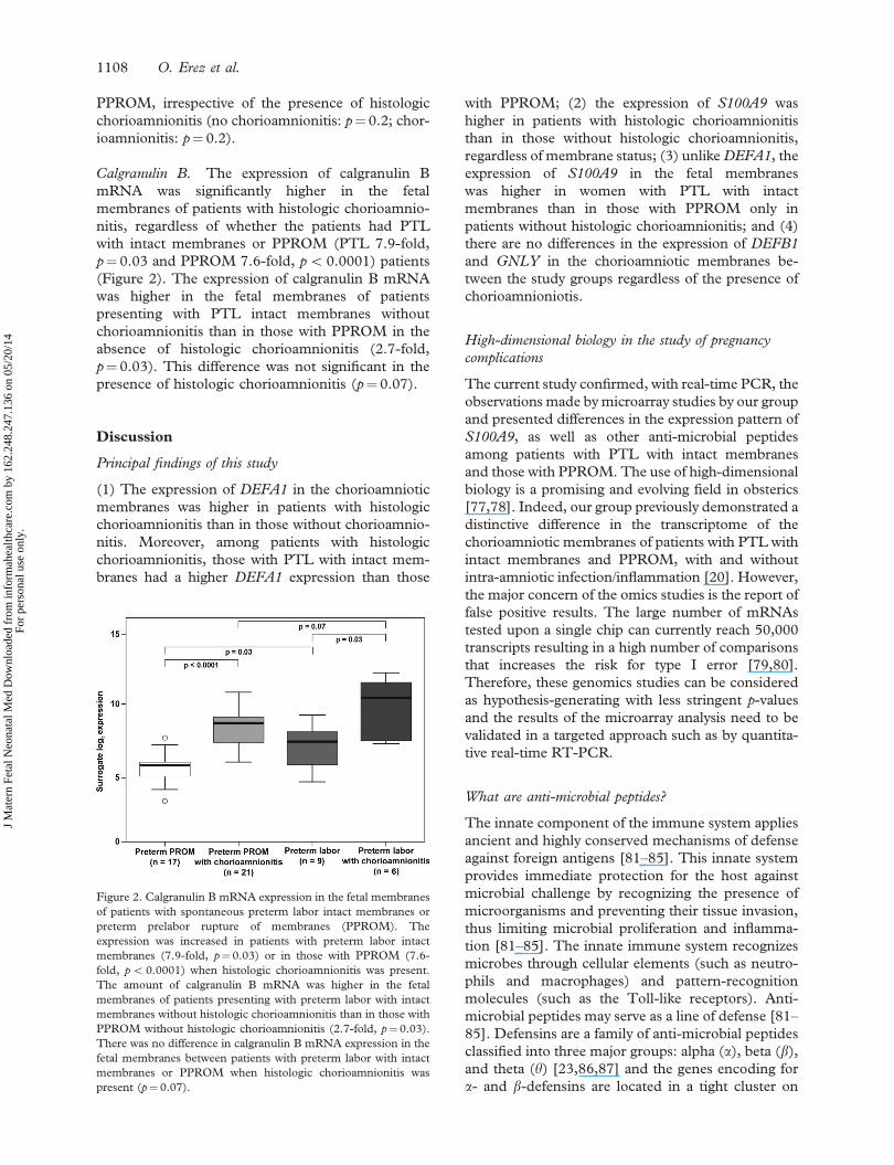
Calgranulin B. The expression of calgranulin B
mRNA was significantly higher in the fetal
membranes of patients with histologic chorioamnio-
nitis, regardless of whether the patients had PTL
with intact membranes or PPROM (PTL 7.9-fold,
p¼ 0.03 and PPROM 7.6-fold, p5 0.0001) patients
(Figure 2). The expression of calgranulin B mRNA
was higher in the fetal membranes of patients
presenting with PTL intact membranes without
chorioamnionitis than in those with PPROM in the
absence of histologic chorioamnionitis (2.7-fold,
p¼ 0.03). This difference was not significant in the
presence of histologic chorioamnionitis (p¼ 0.07).
Discussion
Principal findings of this study
(1) The expression of DEFA1 in the chorioamniotic
membranes was higher in patients with histologic
chorioamnionitis than in those without chorioamnio-
nitis. Moreover, among patients with histologic
chorioamnionitis, those with PTL with intact mem-
branes had a higher DEFA1 expression than those
with PPROM; (2) the expression of S100A9 was
higher in patients with histologic chorioamnionitis
than in those without histologic chorioamnionitis,
regardless of membrane status; (3) unlike DEFA1, the
expression of S100A9 in the fetal membranes
was higher in women with PTL with intact
membranes than in those with PPROM only in
patients without histologic chorioamnionitis; and (4)
there are no differences in the expression of DEFB1
and GNLY in the chorioamniotic membranes be-
tween the study groups regardless of the presence of
chorioamnioniotis.
High-dimensional biology in the study of pregnancy
complications
The current study confirmed, with real-time PCR, the
observations made by microarray studies by our group
and presented differences in the expression pattern of
S100A9, as well as other anti-microbial peptides
among patients with PTL with intact membranes
and those with PPROM. The use of high-dimensional
biology is a promising and evolving field in obsterics
[77,78]. Indeed, our group previously demonstrated a
distinctive difference in the transcriptome of the
chorioamniotic membranes of patients with PTL with
intact membranes and PPROM, with and without
intra-amniotic infection/inflammation [20]. However,
the major concern of the omics studies is the report of
false positive results. The large number of mRNAs
tested upon a single chip can currently reach 50,000
transcripts resulting in a high number of comparisons
that increases the risk for type I error [79,80].
Therefore, these genomics studies can be considered
as hypothesis-generating with less stringent p-values
and the results of the microarray analysis need to be
validated in a targeted approach such as by quantita-
tive real-time RT-PCR.
What are anti-microbial peptides?
The innate component of the immune system applies
ancient and highly conserved mechanisms of defense
against foreign antigens [81–85]. This innate system
provides immediate protection for the host against
microbial challenge by recognizing the presence of
microorganisms and preventing their tissue invasion,
thus limiting microbial proliferation and inflamma-
tion [81–85]. The innate immune system recognizes
microbes through cellular elements (such as neutro-
phils and macrophages) and pattern-recognition
molecules (such as the Toll-like receptors). Anti-
microbial peptides may serve as a line of defense [81–
85]. Defensins are a family of anti-microbial peptides
classified into three major groups: alpha (a), beta (b),
and theta (y) [23,86,87] and the genes encoding for
a- and b-defensins are located in a tight cluster on
Figure 2. Calgranulin B mRNA expression in the fetal membranes
of patients with spontaneous preterm labor intact membranes or
preterm prelabor rupture of membranes (PPROM). The
expression was increased in patients with preterm labor intact
membranes (7.9-fold, p¼0.03) or in those with PPROM (7.6-
fold, p5 0.0001) when histologic chorioamnionitis was present.
The amount of calgranulin B mRNA was higher in the fetal
membranes of patients presenting with preterm labor with intact
membranes without histologic chorioamnionitis than in those with
PPROM without histologic chorioamnionitis (2.7-fold, p¼0.03).
There was no difference in calgranulin B mRNA expression in the
fetal membranes between patients with preterm labor with intact
membranes or PPROM when histologic chorioamnionitis was
present (p¼ 0.07).
1108 O. Erez et al.
J M
ater
n Fe
tal N
eona
tal M
ed D
ownl
oade
d fr
om in
form
ahea
lthca
re.c
om b
y 16
2.24
8.24
7.13
6 on
05/
20/1
4Fo
r pe
rson
al u
se o
nly.

chromosome 8p23 [88]. These peptides have a broad
anti-microbial activity against Gram-negative and
Gram-positive bacteria, fungi, and enveloped viruses
[14,23–25]. The group of a–defensins (HNP) con-
sists of six distinct peptides, of which a-defensins-1,
-2, and -3 share many similarities, as their primary
structure differs by only one amino acid [12,26–28].
Bone marrow precursors of neutrophils synthesized
and stored these anti-microbial peptides intracellu-
larly in azurophil granules [12,28–32]. Because of
their origin, a-defensins are often referred to as
HNP-1, -2 and -3 [14,33]. In addition to their anti-
microbial activity, a-defensins are capable of stimu-
lating a systemic inflammatory response as well as to
chemo-attract T-cells and induce histamine release
from mast cells [34–37]. Human b-defensins are
slightly longer peptides than a-defensins. They are
mainly effective against Gram-negative bacteria and
yeast, though some are also effective against Gram-
positive bacteria [38–40,89]. Human b-defensin-1,
first discovered in 1995 [90], is expressed by the
epithelium of the urinary and respiratory tracts
[39,41,42]. Moreover, its expression is modulated
by inflammation [91–95] and can be induced by
lipopolysaccharide (LPS), heat inactivated Pseudo-
monas aeruginosa, as well as interferon gamma (IFNg)[91–93,95,96]. Human b-defensin-1 displays anti-
microbial activity against Gram-negative bacteria
and fungi, but is relatively less potent against
Gram-positive bacteria [38–40]. In addition to its
anti-microbial properties, human b-defensin-1 can
recruit immature dentritic cells and memory T-cells
[97], thus facilitating foreign antigen presentation
and generation of specific immune response [98–
100]. Moreover, human b-defensin-1 can induce the
production of pro-inflammatory cytokines (e.g.
interleukin (IL)-8, IL-18, and IL-20) [98–100].
Calgranulin B is part of the S100 family of calcium
binding proteins [44]. It is an additional anti-
microbial peptide that composes the calprotectin
heterodimer together with calgranulin A, and can be
detected in neutrophils, monocytes and activated
macrophages, as well as in endothelial and epithelial
cells [44–51]. Calgranulin B has a candidastatic
effect in a zinc-rich medium, an effect abrogated by
calgranulin A [101]. In contrast, a recent report
suggested that calgranulin A is the active component
of calprotectin while calgranulin B seems to regulate
calgranulin A function [102]. Moreover, deletion of
the mouse calgranulin A gene results in an embry-
onically lethal phenotype [103], while mice homo-
zygous for the targeted deletion of S100a9
(S100a97/7) gene protects mice model against
LPS-induced shock [102], but the authors attributed
this effect to the fact that calgranulin A is highly
dependent of calgranulin B and is almost undetect-
able in the plasma of S100a97/7 mice despite
normal mRNA levels of S100a8 [102]. Thus, the
calprotectin heterodimer of calgranulin A and
calgranulin B is regarded as the active form [104].
This heterodimer regulates both the adhesion of
myeloid cells to the endothelium and extracellular
matrix, and the activation of effector cells and their
direct anti-bacterial effect by capturing zinc. Calpro-
tectin concentrations of 50–250 mg/ml can inhibit the
growth of Escherichia coli, Staphylococcus aureus, and
Staphylococcus epidermidis; however, concentrations
as low as 4–32 mg/ml can already inhibit the growth
of Candida albicans [101]. In addition, the expression
of calprotectin assists the cells against the invasion of
Listeria monocytogenes, Salmonella enterica serovar
typhimurium [105], suggesting that this polypeptide
complex may serve as a defense mechanism against
microbial invasion. This is relevant to our study
because the fetal membranes serve as barriers against
invading microorganisms from the maternal decidua
and lower genital tract, and calprotectin may play an
important role in this mechanism.
The fourth anti-microbial peptide explored in this
study is granulysin. This protein is secreted in
cytolytic granules from cytotoxic T-lymphocytes
and NK cells [53–56]. It is a 9 kD protein with four
a-helices connected by a short loop [52]. Recombi-
nant human granulysin is effective against Gram-
positive and Gram-negative bacteria, as well as fungi,
Mycobacterium tuberculosis, Mycobacterium leprae,
Cryptococcus neoformans, and Plasmodium falciparum
[55]. In addition, granulysin induces apoptosis in
varicella infected cells and blocks viral replication
in vitro [55,106–110]. The anti-microbial activity of
the positively charged granulysin is mediated through
its attachment to the negatively charged phospholi-
pids of the invading pathogens and the subsequent
induction of a coupled increase in intracellular
calcium and efflux of intracellular potassium, leading
to the activation of sphingomyelinase, generation of
ceramide, and mitochondrial damage by the activa-
tion of caspases, leading to apoptosis [57–59].
Innate immune and anti-microbial peptides in the
reproductive system in the non-pregnant and pregnant state
Anti-microbial peptides are prevalent in the female
reproductive tract, and their concentrations change in
the different stages of the menstural cycle [111,112].
Indeed, the highest endometrial mRNA expression of
b-defensin-1 is during the mid-secretory phase, of
granulysin in the late secretory phase, and of b-
defensin-2 during menstruation [111,112]. Treat-
ment with hormonal contraceptives was associated
with lower endometrial expression of these anti-
microbial peptides [113], suggesting that the fine-
tuning of the innate immune response in the female
reproductive tract may be under hormonal regulation.
Anti-microbial peptide gene expression in patients with PTL and PPROM 1109
J M
ater
n Fe
tal N
eona
tal M
ed D
ownl
oade
d fr
om in
form
ahea
lthca
re.c
om b
y 16
2.24
8.24
7.13
6 on
05/
20/1
4Fo
r pe
rson
al u
se o
nly.

The need to protect the uppper genital tract from
ascending infection is crucial during pregnancy.
Indeed, multiple mechanisms of defense against
infection are thought to protect pregnancy. Normally,
epithelia represent more than a physical barrier against
microorganisms as most epithelia produce natural
anti-microbial peptides (e.g. defensins, surfactant
proteins) which can kill bacteria by damaging their
cell membrane or facilitating phagocytosis [14,23–25].
However, evidence suggests that bacteria can gain
access to the amniotic cavity by penetrating intact
chorioamniotic membranes [114,115], or by trans-
placental passage in cases of hematogenous dissemi-
nation (bacteremia in the context of periodontal
disease [116–120] or other distant infections [121]).
Thus, the control of microbial proliferation and the
destruction of such microorganisms are required to
maximize the likelihood of a normal pregnancy
outcome. Indeed, anti-microbial peptides are present
in the chorioamniotic membranes, decidua, and
placenta [9,19], and the mRNA expression of human
b-defensin-2 and elafin (an inhibitor of neutrophil
elastase with anti-microbial activity) was shown to be
up-regulated by IL-1b in primary trophoblast cell
culture [19]. Multiple anti-microbial peptides are
also present in the amniotic fluid: lactoferrin [122–
124], lysozyme [124–128], BPI [21], calprotectin
(MRP8/14) [21], LL-37 [125], and a-defensins-1–3
[21,124,125,129]. We have previously reported that
human b-defensin-2 is a physiological constituent of
amniotic fluid, and its amniotic fluid concentration
increases in patients with preterm delivery and
MIAC (with either intact or ruptured membranes),
as well as in those with PTL and intra-amniotic
inflammation [1]. The combination of several anti-
microbial peptides enhances microbial killing. For
example, there is evidence that human b-defensin-2
can act synergistically with LL-37 to kill Group B
Streptococci (GBS) [130]. While LL-37 alone has a
minimal bactericidal concentration (MBC) of 16 mM
and human b-defensin-2 alone has an MBC of 8 mM
against GBS, the combination of these two peptides
effectively reduces their MBC; and at a concentration
of 4 mM each, they kill 100% of GBS [130]. These
studies were conducted in hypotonic media, which
maximizes the anti-microbial action of both anti-
microbial peptides [130]. This observation is relevant
since LL-37 is present in amniotic fluid [125],
Moreover, the minimal inhibitory concentration of
human b-defensin-2 against E. coli, P. aeruginosa, and
E. faecalis is reduced in the presence of lactoferrin or
lysozyme [89]. Collectively, this evidence indicates
that the apparent redundancy in anti-microbial
peptides and proteins in the amniotic fluid is aimed
to maximize anti-microbial activity.
The chorioamniotic membrane is an important
barrier against microbial invasion of the amniotic
fluid and fetal infection. These membranes isolate
the sterile intra-amniotic environment from the
contaminated uterine and extra-uterine environment
[131]. Indeed, increased mRNA expression of hu-
man b-defensins-1–3, secretory leukocyte protease
inhibitor, and elafin (also PI3 or SKALP) were
reported in primary cultures of amnion cells [131].
Moreover, the administration of IL-1b stimulated
the expression of human b-defensins-2 by these cells
[131], supporting the anti-microbial role of these
peptides in the fetal membranes.
The anti-microbial peptide profile of the fetal membranes
of patients with preterm labor and prelabor rupture of the
membranes
The findings of the current study demonstrate a
distinctive pattern of anti-microbial peptide expres-
sion in the fetal membranes of patients with PTL
intact membranes and those with PPROM, espe-
cially in the presence of histologic chorioamnionitis.
Among patients with PTL intact membranes, but not
among those with PPROM, the expression of the
DEFA1 gene was higher in the presence of histologic
chorioamnionitis. Moreover, among patients with
histologic chorioamnionitis, DEFA1 gene expression
was higher in women with PTL intact membranes
than in those with PPROM. Of interest, the
expression of the S100A9 gene was higher in the
presence of histologic chorioamnionitis in both study
groups. Nevertheless, in the absence of histologic
chorioamnionitis, patients with PTL intact mem-
branes had a higher S100A9 mRNA expression than
those with PPROM. These findings also suggest that
the patients with PTL intact membranes have a
different pattern of anti-microbial peptide expression
in the fetal membrane than those with PPROM, and
this pattern changes according to the presence of
histologic chorioamnionitis. Collectively, the results
presented in our study raise several questions. For
example, could the profile of anti-microbial peptide
expression by the chorioamniotic membranes be
associated with an increased risk for PPROM? Also,
is a higher DEFA1 expression in the fetal membranes
during chorioamnionitis or an increased S100A9
gene expression in the abscence of histologic
chorioamnionitis nescessary to maintain the integrity
of the chorioamniotic membranes? Previous studies
reported that intra-amniotic infection was associated
with a significant increase in amniotic fluid concen-
trations of immunoreactive a-defensins-1–3
[21,129], BPI [21], calprotectin [21,132] and
S100B, both in women with PTL with intact
membranes and in women with PPROM [21,22].
Parturition at term was associated with a significant
increase in amniotic fluid concentrations of immu-
noreactive a-defensins-1–3 [21]. Among patients
1110 O. Erez et al.
J M
ater
n Fe
tal N
eona
tal M
ed D
ownl
oade
d fr
om in
form
ahea
lthca
re.c
om b
y 16
2.24
8.24
7.13
6 on
05/
20/1
4Fo
r pe
rson
al u
se o
nly.

with PTL and intact membranes, the elevation of
amniotic fluid concentrations of a-defensins-1–3,
BPI, calprotectin [21,132] and S100B [22] was
associated with intra-amniotic inflammation, histo-
logic chorioamnionitis, and a shorter diagnosis-to-
delivery interval. In addition, proteomic analysis of
amniotic fluid from Rhesus monkeys [133] and
humans [133,134] with IAI identified calgranulin B
[133,134] and a fragment of insulin-like growth
factor binding protein 1 [133] were differentially
expressed. Thus, the present study supports the role
of a-defensin-1 and calgranulin B in the host defense
in PTL with intact membranes and of calgranulin B
in PPROM during IAI through their increased
expression in the fetal membranes and higher intra-
amniotic concentrations.
Further attention has to be given to the role of
a-defensins in the fetal response to infection and
perhaps to the integrity of the fetal membranes.
Evidence in support of this view are: (1) among
patients with IAI, those with PTL intact membranes
had a higher median intra-amniotic a-defensins
concentration than those with PPROM [21]; and
(2) among patients with histologic chorioamnionitis,
those with PTL intact membranes had a higher
mRNA expression of a-defensin 1 in the fetal
membranes than those with PPROM. In contrast to
a-defensin 1, patients with PTL had a higher
calgranulin B mRNA expression in the fetal mem-
branes than those with PPROM, only among women
without histologic chorioamnionitis. These suggest
that the increased calgranulin B expression in the fetal
membranes and its higher intra-amniotic concentra-
tions during infection/inflammation may be asso-
ciated with the inflammatory response of the fetal
membranes and regardless to the clinical presentation
(PTL intact membranes or PPROM). Moreover, in
contrast to the higher concentrations of calprotectin
in the amniotic fluid of patients with PPROM than in
those with PTL intact membranes [21], there was no
difference in calgranulin B mRNA expression in the
fetal membranes of patients with histologic chor-
ioamnionitis, either with PTL intact membranes or
PPROM. Thus, further study is needed in order to
elucidate the implication of the differences in the
expression of calgranulin B mRNA in the fetal
membranes, among patients with PTL intact mem-
branes without histologic chorioamnionitis.
Conclusions
(1) Among patients with histologic chorioamnionitis,
the mRNA expression of a-defensin-1 and calgra-
nulin B in the fetal membranes of patients with PTL
and intact membranes as well as that of calgranulin B
in the fetal membranes of patients with PPROM is
higher than in the membranes of those without
histologic chorioamnionitis; (2) histologic chorioam-
nionitis is associated with differences in the pattern
of a-defensin-1 mRNA expression in the fetal
membranes in patients with PTL and intact mem-
branes and those with PPROM.
Acknowledgements
The authors thank Lark Technologies, Inc. (Hous-
ton, TX) for performing the qRT-PCR experiments.
They wish to acknowledge the invaluable contribu-
tions made to this manuscript by Sandy Field,
Gerardo Rodriguez, and the nursing staff of the
Perinatology Research Branch and the Detroit
Medical Center.
References
1. Soto E, Espinoza J, Nien JK, Kusanovic JP, Erez O, Richani
K, Santolaya-Forgas J, Romero R. Human b-defensin-2: a
natural antimicrobial peptide present in amniotic fluid
participates in the host response to microbial invasion of
the amniotic cavity. J Matern Fetal Neonatal Med 2007;
20:15–22.
2. Romero R, Gomez R, Araneda H, Ramirez M, Cotton DB.
Cervical mucus inhibits microbial growth: a host defense
mechanism to prevent ascending infection in pregnant and
non-pregnant women. Am J Obstet Gynecol 1993;168:312.
3. Eggert-Kruse W, Botz I, Pohl S, Rohr G, Strowitzki T.
Antimicrobial activity of human cervical mucus. Hum
Reprod 2000;15:778–784.
4. Hein M, Helmig RB, Schonheyder HC, Ganz T, Uldbjerg
N. An in vitro study of antibacterial properties of the cervical
mucus plug in pregnancy. Am J Obstet Gynecol 2001;185:
586–592.
5. Hein M, Valore EV, Helmig RB, Uldbjerg N, Ganz T.
Antimicrobial factors in the cervical mucus plug. Am J
Obstet Gynecol 2002;187:137–144.
6. King AE, Paltoo A, Kelly RW, Sallenave JM, Bocking AD,
Challis JR. Expression of natural antimicrobials by human
placenta and fetal membranes. Placenta 2007;28:161–169.
7. Talmi YP, Sigler L, Inge E, Finkelstein Y, Zohar Y.
Antibacterial properties of human amniotic membranes.
Placenta 1991;12:285–288.
8. Kjaergaard N, Hein M, Hyttel L, Helmig RB, Schonheyder
HC, Uldbjerg N, Madsen H. Antibacterial properties of
human amnion and chorion in vitro. Eur J Obstet Gynecol
Reprod Biol 2001;94:224–229.
9. Svinarich DM, Gomez R, Romero R. Detection of human
defensins in the placenta. Am J Reprod Immunol 1997;38:
252–255.
10. Guleria I, Pollard JW. The trophoblast is a component of the
innate immune system during pregnancy. Nat Med 2000;6:
589–593.
11. Zasloff M. Antimicrobial peptides of multicellular organisms.
Nature 2002;415:389–395.
12. Ganz T. Defensins: antimicrobial peptides of innate im-
munity. Nat Rev Immunol 2003;3:710–720.
13. Tosi MF. Innate immune responses to infection. J Allergy
Clin Immunol 2005;116:241–249.
14. Ganz T, Selsted ME, Szklarek D, Harwig SS, Daher K,
Bainton DF, Lehrer RI. Defensins. Natural peptide anti-
biotics of human neutrophils. J Clin Invest 1985;76:1427–
1435.
Anti-microbial peptide gene expression in patients with PTL and PPROM 1111
J M
ater
n Fe
tal N
eona
tal M
ed D
ownl
oade
d fr
om in
form
ahea
lthca
re.c
om b
y 16
2.24
8.24
7.13
6 on
05/
20/1
4Fo
r pe
rson
al u
se o
nly.

15. Weiss J, Elsbach P, Olsson I, Odeberg H. Purification and
characterization of a potent bactericidal and membrane
active protein from the granules of human polymorpho-
nuclear leukocytes. J Biol Chem 1978;253:2664–2672.
16. Zhao C, Wang I, Lehrer RI. Widespread expression of
b-defensin hBD-1 in human secretory glands and epithelial
cells. FEBS Lett 1996;396:319–322.
17. Jones DE, Bevins CL. Paneth cells of the human small
intestine express an antimicrobial peptide gene. J Biol Chem
1992;267:23216–23225.
18. Bals R. Epithelial antimicrobial peptides in host defense
against infection. Respir Res 2000;1:141–150.
19. King AE, Kelly RW, Sallenave JM, Bocking AD, Challis JR.
Innate immune defences in the human uterus during
pregnancy. Placenta 2007;28:1099–1106.
20. Tromp G, Kuivaniemi H, Romero R, Chaiworapongsa T,
Kim YM, Kim MR, Maymon E, Edwin S. Genome-
wide expression profiling of fetal membranes reveals a
deficient expression of proteinase inhibitor 3 in premature
rupture of membranes. Am J Obstet Gynecol 2004;191:
1331–1338.
21. Espinoza J, Chaiworapongsa T, Romero R, Edwin S,
Rathnasabapathy C, Gomez R, Bujold E, Camacho N,
Kim YM, Hassan S, et al. Antimicrobial peptides in amniotic
fluid: defensins, calprotectin and bacterial/permeability-
increasing protein in patients with microbial invasion of the
amniotic cavity, intra-amniotic inflammation, preterm labor
and premature rupture of membranes. J Matern Fetal
Neonatal Med 2003;13:2–21.
22. Friel L, Kuivaniemi H, Gomez R, Goddard K, Nien JK,
Tromp G, Lu Q, Xu Z, Behnke E, Solari M, et al. Genetic
predisposition for preterm PROM: results of a large
candidate-gene association study of mothers and their
offspring. Am J Obstet Gynecol 2005;193:S17.
23. Schneider JJ, Unholzer A, Schaller M, Schafer-Korting M,
Korting HC. Human defensins. J Mol Med 2005;83:587–
595.
24. Lehrer RI, Lichtenstein AK, Ganz T. Defensins: antimicro-
bial and cytotoxic peptides of mammalian cells. Annu Rev
Immunol 1993;11:105–128.
25. Daher KA, Selsted ME, Lehrer RI. Direct inactivation of
viruses by human granulocyte defensins. J Virol 1986;60:
1068–1074.
26. Raj PA, Dentino AR. Current status of defensins and their
role in innate and adaptive immunity. FEMS Microbiol Lett
2002;206:9–18.
27. Klotman ME, Chang TL. Defensins in innate antiviral
immunity. Nat Rev Immunol 2006;6:447–456.
28. Joseph G, Tarnow L, Astrup AS, Hansen TK, Parving HH,
Flyvbjerg A, Frystyk J. Plasma a-defensin is associated with
cardiovascular morbidity and mortality in type 1 diabetic
patients. J Clin Endocrinol Metab 2008;93:1470–1475.
29. Lehrer RI, Barton A, Daher KA, Harwig SS, Ganz T, Selsted
ME. Interaction of human defensins with Escherichia coli.
Mechanism of bactericidal activity. J Clin Invest 1989;84:
553–561.
30. Lindemann RA, Lala A, Miyasaki KT. The in vitro effect of
human polymorphonuclear leukocyte azurophil granule
components on natural killer cell cytotoxicity. Oral Micro-
biol Immunol 1994;9:186–192.
31. Oren A, Taylor JM. The subcellular localization of defensins
and myeloperoxidase in human neutrophils: immunocyto-
chemical evidence for azurophil granule heterogeneity. J Lab
Clin Med 1995;125:340–347.
32. Faurschou M, Sorensen OE, Johnsen AH, Askaa J,
Borregaard N. Defensin-rich granules of human neutrophils:
characterization of secretory properties. Biochim Biophys
Acta 2002;1591:29–35.
33. Selsted ME, Harwig SS, Ganz T, Schilling JW, Lehrer RI.
Primary structures of three human neutrophil defensins.
J Clin Invest 1985;76:1436–1439.
34. Chertov O, Michiel DF, Xu L, Wang JM, Tani K, Murphy
WJ, Longo DL, Taub DD, Oppenheim JJ. Identification of
defensin-1, defensin-2, and CAP37/azurocidin as T-cell
chemoattractant proteins released from interleukin-8-stimu-
lated neutrophils. J Biol Chem 1996;271:2935–2940.
35. Befus AD, Mowat C, Gilchrist M, Hu J, Solomon S,
Bateman A. Neutrophil defensins induce histamine secretion
from mast cells: mechanisms of action. J Immunol
1999;163:947–953.
36. Zhang H, Porro G, Orzech N, Mullen B, Liu M, Slutsky AS.
Neutrophil defensins mediate acute inflammatory response
and lung dysfunction in dose-related fashion. Am J Physiol
Lung Cell Mol Physiol 2001;280:L947–L954.
37. Vaschetto R, Grinstein J, Del SL, Khine AA, Voglis S, Tullis
E, Slutsky AS, Zhang H. Role of human neutrophil peptides
in the initial interaction between lung epithelial cells and
CD4þ lymphocytes. J Leukoc Biol 2007;81:1022–1031.
38. Schulz A, Kluver E, Schulz-Maronde S, Adermann K.
Engineering disulfide bonds of the novel human b-defensins
hBD-27 and hBD-28: differences in disulfide formation and
biological activity among human b-defensins. Biopolymers
2005;80:34–49.
39. Goldman MJ, Anderson GM, Stolzenberg ED, Kari UP,
Zasloff M, Wilson JM. Human b-defensin-1 is a salt-sensitive
antibiotic in lung that is inactivated in cystic fibrosis. Cell
1997;88:553–560.
40. Harder J, Bartels J, Christophers E, Schroder JM. A peptide
antibiotic from human skin. Nature 1997;387:861.
41. Valore EV, Park CH, Quayle AJ, Wiles KR, McCray PB Jr,
Ganz T. Human b-defensin-1: an antimicrobial peptide of
urogenital tissues. J Clin Invest 1998;101:1633–1642.
42. McCray PB Jr, Bentley L. Human airway epithelia express a
b-defensin. Am J Respir Cell Mol Biol 1997;16:343–349.
43. Gropp R, Frye M, Wagner TO, Bargon J. Epithelial
defensins impair adenoviral infection: implication for
adenovirus-mediated gene therapy. Hum Gene Ther 1999;
10:957–964.
44. Striz I, Trebichavsky I. Calprotectin – a pleiotropic molecule
in acute and chronic inflammation. Physiol Res 2004;53:
245–253.
45. Roth J, Goebeler M, Sorg C. S100A8 and S100A9 in
inflammatory diseases. Lancet 2001;357:1041.
46. Pillay SN, Asplin JR, Coe FL. Evidence that calgranulin is
produced by kidney cells and is an inhibitor of calcium
oxalate crystallization. Am J Physiol 1998;275:F255–F261.
47. Helbert MJ, Dauwe SE, De Broe ME. Flow cytometric
immunodissection of the human distal tubule and cortical
collecting duct system. Kidney Int 2001;59:554–564.
48. Doussiere J, Bouzidi F, Vignais PV. The S100A8/A9 protein
as a partner for the cytosolic factors of NADPH oxidase
activation in neutrophils. Eur J Biochem 2002;269:3246–
3255.
49. Brandtzaeg P, Dale I, Fagerhol MK. Distribution of a
formalin-resistant myelomonocytic antigen (L1) in human
tissues. II. Normal and aberrant occurrence in various
epithelia. Am J Clin Pathol 1987;87:700–707.
50. Bhardwaj RS, Zotz C, Zwadlo-Klarwasser G, Roth J,
Goebeler M, Mahnke K, Falk M, Meinardus-Hager G, Sorg
C. The calcium-binding proteins MRP8 and MRP14 form a
membrane-associated heterodimer in a subset of monocytes/
macrophages present in acute but absent in chronic
inflammatory lesions. Eur J Immunol 1992;22:1891–1897.
51. Hessian PA, Edgeworth J, Hogg N. MRP-8 and MRP-14,
two abundant Ca(2þ)-binding proteins of neutrophils and
monocytes. J Leukoc Biol 1993;53:197–204.
1112 O. Erez et al.
J M
ater
n Fe
tal N
eona
tal M
ed D
ownl
oade
d fr
om in
form
ahea
lthca
re.c
om b
y 16
2.24
8.24
7.13
6 on
05/
20/1
4Fo
r pe
rson
al u
se o
nly.

52. Anderson DH, Sawaya MR, Cascio D, Ernst W, Modlin R,
Krensky A, Eisenberg D. Granulysin crystal structure and a
structure-derived lytic mechanism. J Mol Biol 2003;325:
355–365.
53. Krensky AM, Clayberger C. Granulysin: a novel host defense
molecule. Am J Transplant 2005;5:1789–1792.
54. Clayberger C, Krensky AM. Granulysin. Curr Opin Im-
munol 2003;15:560–565.
55. Stenger S, Hanson DA, Teitelbaum R, Dewan P, Niazi KR,
Froelich CJ, Ganz T, Thoma-Uszynski S, Melian A, Bogdan
C, et al. An antimicrobial activity of cytolytic T cells
mediated by granulysin. Science 1998;282:121–125.
56. Pena SV, Krensky AM. Granulysin, a new human cytolytic
granule-associated protein with possible involvement in cell-
mediated cytotoxicity. Semin Immunol 1997;9:117–125.
57. Gamen S, Hanson DA, Kaspar A, Naval J, Krensky AM, Anel
A. Granulysin-induced apoptosis. I. Involvement of at least
two distinct pathways. J Immunol 1998;161:1758–1764.
58. Li Q, Dong C, Deng A, Katsumata M, Nakadai A, Kawada
T, Okada S, Clayberger C, Krensky AM. Hemolysis of
erythrocytes by granulysin-derived peptides but not by
granulysin. Antimicrob Agents Chemother 2005;49:388–
397.
59. Kaspar AA, Okada S, Kumar J, Poulain FR, Drouvalakis
KA, Kelekar A, Hanson DA, Kluck RM, Hitoshi Y, Johnson
DE, et al. A distinct pathway of cell-mediated apoptosis
initiated by granulysin. J Immunol 2001;167:350–356.
60. Romero R, Mazor M, Munoz H, Gomez R, Galasso M,
Sherer DM. The preterm labor syndrome. Ann N Y Acad
Sci 1994;734:414–429.
61. Romero R, Mazor M. Infection and preterm labor. Clin
Obstet Gynecol 1988;31:553–584.
62. Romero R, Espinoza J, Kusanovic J, Gotsch F, Hassan S,
Erez O, Chaiworapongsa T, Mazor M. The preterm
parturition syndrome. BJOG 2006;113(Suppl 3):17–42.
63. Haddad R, Tromp G, Kuivaniemi H, Chaiworapongsa T,
Kim YM, Mazor M, Romero R. Human spontaneous labor
without histologic chorioamnionitis is characterized by an
acute inflammation gene expression signature. Am J Obstet
Gynecol 2006;195:394.e1–394.e24.
64. Hassan SS, Romero R, Haddad R, Hendler I, Khalek N,
Tromp G, Diamond MP, Sorokin Y, Malone J Jr. The
transcriptome of the uterine cervix before and after
spontaneous term parturition. Am J Obstet Gynecol 2006;
195:778–786.
65. Haddad R, Gould BR, Romero R, Tromp G, Farookhi R,
Edwin SS, Kim MR, Zingg HH. Uterine transcriptomes of
bacteria-induced and ovariectomy-induced preterm labor in
mice are characterized by differential expression of arachido-
nate metabolism genes. Am J Obstet Gynecol 2006;195:822–
828.
66. Slattery MM, Morrison JJ. Preterm delivery. Lancet 2002;
360:1489–1497.
67. Romero R, Espinoza J, Mazor M, Chaiworapongsa T. The
preterm parturition syndrome. 2004;First:28–60.
68. Mathews TJ, Menacker F, MacDorman MF. Infant mortal-
ity statistics from the 2002 period: linked birth/infant death
data set. Natl Vital Stat Rep 2004;53:1–29.
69. Tucker J, McGuire W. Epidemiology of preterm birth. BMJ
2004;329:675–678.
70. Xu J, Holzman CB, Arvidson CG, Chung H, Goepfert AR.
Midpregnancy vaginal fluid defensins, bacterial vaginosis, and
risk of preterm delivery. Obstet Gynecol 2008;112:524–531.
71. Chaiworapongsa T, Espinoza J, Yoshimatsu J, Kim YM,
Bujold E, Edwin S, Yoon BH, Romero R. Activation of
coagulation system in preterm labor and preterm premature
rupture of membranes. J Matern Fetal Neonatal Med
2002;11:368–373.
72. Redline RW, Heller D, Keating S, Kingdom J. Placental
diagnostic criteria and clinical correlation – a workshop
report 140. Placenta 2005;26 (Suppl A):S114–S117.
73. Pacora P, Chaiworapongsa T, Maymon E, Kim YM, Gomez
R, Yoon BH, Ghezzi F, Berry SM, Qureshi F, Jacques SM,
et al. Funisitis and chorionic vasculitis: the histological
counterpart of the fetal inflammatory response syndrome.
J Matern Fetal Neonatal Med 2002;11:18–25.
74. Redline RW. Inflammatory responses in the placenta and
umbilical cord. Semin Fetal Neonatal Med 2006;11:296–301.
75. Winer J, Jung CK, Shackel I, Williams PM. Development
and validation of real-time quantitative reverse transcriptase-
polymerase chain reaction for monitoring gene expression in
cardiac myocytes in vitro. Anal Biochem 1999;270:41–49.
76. Livak KJ, Schmittgen TD. Analysis of relative gene expres-
sion data using real-time quantitative PCR and the 2(-Delta
Delta C(T)) method. Methods 2001;25:402–408.
77. Romero R, Tromp G. High-dimensional biology in obste-
trics and gynecology: functional genomics in microarray
studies. Am J Obstet Gynecol 2006;195:360–363.
78. Romero R, Espinoza J, Gotsch F, Kusanovic JP, Friel LA,
Erez O, Mazaki-Tovi S, Than NG, Hassan S, Tromp G. The
use of high-dimensional biology (genomics, transcriptomics,
proteomics, and metabolomics) to understand the preterm
parturition syndrome. BJOG 2006;113 (Suppl 3):118–135.
79. Tarca AL, Romero R, Draghici S. Analysis of microarray
experiments of gene expression profiling. Am J Obstet
Gynecol 2006;195:373–388.
80. Romero R, Tarca AL, Tromp G. Insights into the physiology
of childbirth using transcriptomics. PLoS Med 2006;3:e276.
81. Janeway CA Jr, Travers P, Walport M. Innate immunity.
2001;5th:35–42.
82. Janeway CA Jr, Medzhitov R. Innate immune recognition.
Annu Rev Immunol 2002;20:197–216.
83. Borghesi L, Milcarek C. Innate versus adaptive immunity: a
paradigm past its prime? Cancer Res 2007;67:3989–3993.
84. Medzhitov R. Recognition of microorganisms and activation
of the immune response. Nature 2007;449:819–826.
85. Barton GM. A calculated response: control of inflammation
by the innate immune system. J Clin Invest 2008;118:413–
420.
86. Tang YQ, Yuan J, Osapay G, Osapay K, Tran D, Miller CJ,
Ouellette AJ, Selsted ME. A cyclic antimicrobial peptide
produced in primate leukocytes by the ligation of two
truncated a-defensins. Science 1999;286:498–502.
87. Selsted ME, Tang YQ, Morris WL, McGuire PA, Novotny
MJ, Smith W, Henschen AH, Cullor JS. Purification,
primary structures, and antibacterial activities of b-defensins,
a new family of antimicrobial peptides from bovine neu-
trophils. J Biol Chem 1993;268:6641–6648.
88. Linzmeier R, Ho CH, Hoang BV, Ganz T. A 450-kb contig
of defensin genes on human chromosome 8p23. Gene
1999;233:205–211.
89. Bals R, Wang X, Wu Z, Freeman T, Bafna V, Zasloff M,
Wilson JM. Human b-defensin 2 is a salt-sensitive peptide
antibiotic expressed in human lung. J Clin Invest 1998;
102:874–880.
90. Bensch KW, Raida M, Magert HJ, Schulz-Knappe P,
Forssmann WG. hBD-1: a novel b-defensin from human
plasma. FEBS Lett 1995;368:331–335.
91. Zhu BD, Feng Y, Huang N, Wu Q, Wang BY. Mycobacterium
bovis bacille Calmette-Guerin (BCG) enhances human
b-defensin-1 gene transcription in human pulmonary gland
epithelial cells. Acta Pharmacol Sin 2003;24:907–912.
92. Sorensen OE, Thapa DR, Rosenthal A, Liu L, Roberts AA,
Ganz T. Differential regulation of b-defensin expression in
human skin by microbial stimuli. J Immunol 2005;174:
4870–4879.
Anti-microbial peptide gene expression in patients with PTL and PPROM 1113
J M
ater
n Fe
tal N
eona
tal M
ed D
ownl
oade
d fr
om in
form
ahea
lthca
re.c
om b
y 16
2.24
8.24
7.13
6 on
05/
20/1
4Fo
r pe
rson
al u
se o
nly.

93. Joly S, Organ CC, Johnson GK, McCray PB Jr, Guthmiller
JM. Correlation between b-defensin expression and induc-
tion profiles in gingival keratinocytes. Mol Immunol
2005;42:1073–1084.
94. Feng Z, Jiang B, Chandra J, Ghannoum M, Nelson S,
Weinberg A. Human b-defensins: differential activity against
candidal species and regulation by Candida albicans. J Dent
Res 2005;84:445–450.
95. Duits LA, Ravensbergen B, Rademaker M, Hiemstra PS,
Nibbering PH. Expression of b-defensin 1 and 2 mRNA by
human monocytes, macrophages and dendritic cells. Im-
munology 2002;106:517–525.
96. Fang XM, Shu Q, Chen QX, Book M, Sahl HG, Hoeft A,
Stuber F. Differential expression of a- and b-defensins in
human peripheral blood. Eur J Clin Invest 2003;33:82–87.
97. Yang D, Chertov O, Bykovskaia SN, Chen Q, Buffo MJ,
Shogan J, Anderson M, Schroder JM, Wang JM, Howard OM,
et al. b-defensins: linking innate and adaptive immunity through
dendritic and T cell CCR6. Science 1999;286:525–528.
98. Brogden KA, Heidari M, Sacco RE, Palmquist D, Guthmil-
ler JM, Johnson GK, Jia HP, Tack BF, McCray PB.
Defensin-induced adaptive immunity in mice and its
potential in preventing periodontal disease. Oral Microbiol
Immunol 2003;18:95–99.
99. Yang D, Biragyn A, Hoover DM, Lubkowski J, Oppenheim
JJ. Multiple roles of antimicrobial defensins, cathelicidins,
and eosinophil-derived neurotoxin in host defense. Annu
Rev Immunol 2004;22:181–215.
100. Pazgier M, Hoover DM, Yang D, Lu W, Lubkowski J.
Human b-defensins. Cell Mol Life Sci 2006;63:1294–1313.
101. Murthy AR, Lehrer RI, Harwig SS, Miyasaki KT. In vitro
candidastatic properties of the human neutrophil calprotec-
tin complex. J Immunol 1993;151:6291–6301.
102. Vogl T, Tenbrock K, Ludwig S, Leukert N, Ehrhardt C, van
Zoelen MA, Nacken W, Foell D, van der PT, Sorg C, et al.
Mrp8 and Mrp14 are endogenous activators of Toll-like
receptor 4, promoting lethal, endotoxin-induced shock. Nat
Med 2007;13:1042–1049.
103. Passey RJ, Williams E, Lichanska AM, Wells C, Hu S, Geczy
CL, Little MH, Hume DA. A null mutation in the
inflammation-associated S100 protein S100A8 causes early
resorption of the mouse embryo. J Immunol 1999;163:2209–
2216.
104. Nacken W, Roth J, Sorg C, Kerkhoff C. S100A9/S100A8:
myeloid representatives of the S100 protein family as
prominent players in innate immunity. Microsc Res Tech
2003;60:569–580.
105. Nisapakultorn K, Ross KF, Herzberg MC. Calprotectin
expression inhibits bacterial binding to mucosal epithelial
cells. Infect Immun 2001;69:3692–3696.
106. Mackewicz CE, Ridha S, Levy JA. HIV virions and HIV
replication are unaffected by granulysin. AIDS 2000;14:328–
330.
107. Ochoa MT, Stenger S, Sieling PA, Thoma-Uszynski S,
Sabet S, Cho S, Krensky AM, Rollinghoff M, Nunes SE,
Burdick AE, et al. T-cell release of granulysin contributes to
host defense in leprosy. Nat Med 2001;7:174–179.
108. Farouk SE, Mincheva-Nilsson L, Krensky AM, Dieli F,
Troye-Blomberg M. Gamma delta T cells inhibit in vitro
growth of the asexual blood stages of Plasmodium falciparum
by a granule exocytosis-dependent cytotoxic pathway that
requires granulysin. Eur J Immunol 2004;34:2248–2256.
109. Ma LL, Spurrell JC, Wang JF, Neely GG, Epelman S,
Krensky AM, Mody CH. CD8 T cell-mediated killing of
Cryptococcus neoformans requires granulysin and is dependent
on CD4 T cells and IL-15. J Immunol 2002;169:5787–5795.
110. Hata A, Zerboni L, Sommer M, Kaspar AA, Clayberger C,
Krensky AM, Arvin AM. Granulysin blocks replication of
varicella-zoster virus and triggers apoptosis of infected cells.
Viral Immunol 2001;14:125–133.
111. King AE, Critchley HO, Kelly RW. Innate immune defences
in the human endometrium. Reprod Biol Endocrinol
2003;1:116.
112. King AE, Critchley HO, Sallenave JM, Kelly RW. Elafin in
human endometrium: an antiprotease and antimicrobial
molecule expressed during menstruation. J Clin Endocrinol
Metab 2003;88:4426–4431.
113. Fleming DC, King AE, Williams AR, Critchley HO, Kelly
RW. Hormonal contraception can suppress natural anti-
microbial gene transcription in human endometrium. Fertil
Steril 2003;79:856–863.
114. Evaldson G, Malmborg AS, Nord CE, Ostensson K.
Bacteroides fragilis, Streptococcus intermedius and group B
streptococci in ascending infection of pregnancy. An animal
experimental study. Gynecol Obstet Invest 1983;15:230–
241.
115. Galask RP, Varner MW, Petzold CR, Wilbur SL. Bacterial
attachment to the chorioamniotic membranes. Am J Obstet
Gynecol 1984;148:915–928.
116. Offenbacher S, Katz V, Fertik G, Collins J, Boyd D, Maynor
G, McKaig R, Beck J. Periodontal infection as a possible risk
factor for preterm low birth weight. J Periodontol
1996;67:1103–1113.
117. Jeffcoat MK, Geurs NC, Reddy MS, Cliver SP, Goldenberg
RL, Hauth JC. Periodontal infection and preterm birth:
results of a prospective study. J Am Dent Assoc
2001;132:875–880.
118. Madianos PN, Lieff S, Murtha AP, Boggess KA, Auten RL
Jr, Beck JD, Offenbacher S. Maternal periodontitis and
prematurity, Part II: maternal infection and fetal exposure.
Ann Periodontol 2001;6:175–182.
119. Bearfield C, Davenport ES, Sivapathasundaram V, Allaker
RP. Possible association between amniotic fluid micro-
organism infection and microflora in the mouth. BJOG
2002;109:527–533.
120. Dortbudak O, Eberhardt R, Ulm M, Persson GR. Period-
ontitis, a marker of risk in pregnancy for preterm birth. J Clin
Periodontol 2005;32:45–52.
121. Boggess KA, Madianos PN, Preisser JS, Moise KJ Jr,
Offenbacher S. Chronic maternal and fetal Porphyromonas
gingivalis exposure during pregnancy in rabbits. Am J Obstet
Gynecol 2005;192:554–557.
122. Otsuki K, Yoda A, Saito H, Mitsuhashi Y, Toma Y, Shimizu
Y, Yanaihara T. Amniotic fluid lactoferrin in intrauterine
infection. Placenta 1999;20:175–179.
123. Pacora P, Maymon E, Gervasi MT, Gomez R, Edwin SS,
Yoon BH, Romero R. Lactoferrin in intrauterine infection,
human parturition, and rupture of fetal membranes. Am J
Obstet Gynecol 2000;183:904–910.
124. Akinbi HT, Narendran V, Pass AK, Markart P, Hoath SB.
Host defense proteins in vernix caseosa and amniotic fluid.
Am J Obstet Gynecol 2004;191:2090–2096.
125. Yoshio H, Tollin M, Gudmundsson GH, Lagercrantz H,
Jornvall H, Marchini G, Agerberth B. Antimicrobial poly-
peptides of human vernix caseosa and amniotic fluid:
implications for newborn innate defense. Pediatr Res
2003;53:211–216.
126. Cherry SH, Filler M, Harvey H. Lysozyme content of
amniotic fluid. Am J Obstet Gynecol 1973;116:639–642.
127. Ford LC, DeLange RJ, Lebherz TB. Identification of a
bactericidal factor (B-lysin) in amnionic fluid at 14 and
40 weeks’ gestation. Am J Obstet Gynecol 1977;127:788–
792.
128. Hisanaga S, Umezu T, Shimokawa H, Maesato S. Amniotic
fluid lysozyme content in normal and abnormal pregnancy.
Nippon Sanka Fujinka Gakkai Zasshi 1982;34:541–544.
1114 O. Erez et al.
J M
ater
n Fe
tal N
eona
tal M
ed D
ownl
oade
d fr
om in
form
ahea
lthca
re.c
om b
y 16
2.24
8.24
7.13
6 on
05/
20/1
4Fo
r pe
rson
al u
se o
nly.

129. Heine RP, Wiesenfeld H, Mortimer L, Greig PC. Amniotic
fluid defensins: potential markers of subclinical intrauterine
infection. Clin Infect Dis 1998;27:513–518.
130. Dorschner RA, Lin KH, Murakami M, Gallo RL. Neonatal
skin in mice and humans expresses increased levels of
antimicrobial peptides: innate immunity during development
of the adaptive response. Pediatr Res 2003;53:566–572.
131. Stock SJ, Kelly RW, Riley SC, Calder AA. Natural
antimicrobial production by the amnion. Am J Obstet
Gynecol 2007;196:255–256.
132. Ruetschi U, Rosen A, Karlsson G, Zetterberg H, Rymo L,
Hagberg H, Jacobsson B. Proteomic analysis using protein
chips to detect biomarkers in cervical and amniotic fluid in
women with intra-amniotic inflammation. J Proteome Res
2005;4:2236–2242.
133. Gravett MG, Novy MJ, Rosenfeld RG, Reddy AP, Jacob T,
Turner M, McCormack A, Lapidus JA, Hitti J, Eschenbach
DA, et al. Diagnosis of intra-amniotic infection by proteomic
profiling and identification of novel biomarkers. JAMA
2004;292:462–469.
134. Buhimschi IA, Christner R, Buhimschi CS, Chaiworapongsa
T, Romero R. Proteomic analysis of preterm parturition: a
novel method of identifying the patients at risk of impending
preterm delivery. Am J Obstet Gynecol 2002;187:S55.
Anti-microbial peptide gene expression in patients with PTL and PPROM 1115
J M
ater
n Fe
tal N
eona
tal M
ed D
ownl
oade
d fr
om in
form
ahea
lthca
re.c
om b
y 16
2.24
8.24
7.13
6 on
05/
20/1
4Fo
r pe
rson
al u
se o
nly.