Differential Expression of Markers for Endothelial Cells ... - NCBI
13
American Journal of Pathology, Vol. 138, No. 6, June 1991 Copyright © American Association of Pathologists Differential Expression of Markers for Endothelial Cells, Pericytes, and Basal Lamina in the Microvasculature of Tumors and Granulation Tissue R. 0. Schlingemann,* F. J. R. Rietveld,* F. Kwaspen,* P. C. M. van de Kerkhof,t R. M. W. de Waal,* and D. J. Ruiter* From the Department of Pathology*, Nimegen University Cancer Center, and the Department of Dermatology,J University Hospital Ny/megen, Nymegen, The Netherlands The structure andfunction of the tumor microvascu- lature is of great interestfor cancer biology, diagno- sis, and therapy. The distribution of endothelial cells, pericytes, and basal lamina in tumors is not well documented. In this study, the authors investigated the distribution of markers for these different com- ponents in a series of malignant human tumors and in human granulation tissue, both situations with extensive angiogenesis. Their results show a striking heterogeneity in the expression of markers for peri- cytes and endothelial cells between different tumors, but also within a single tumor lesion. To be able to distinguish between these two adjacent cell types de- cisively, all marker studies were carried out both on the light and the electron microscopical level and compared with staining results in granulation tissue of cutaneous wounds in healthy volunteers and of decubitus lesions. In granulation tissue of decubitus lesions, well-defined zones with increasing levels of maturation can be delineated It was found that an- tibodies recognizing von Willebrand factor often failed to stain the tumor capillaries. Of the pericyte markers; alpha-smooth muscle actin was only lo- cally expressed bypericytes in the tumor vasculature, whereas the high-molecular-weight melanoma- associated antigen, a chondroitin sulfate proteogly- can, stained the microvasculature broadly. Staining of the basal lamina components collagen type IVand laminin was, within the tumor, not restricted to the microvasculature. From their findings the authors conclude that 1) for the visualization of the tumor vasculature, antibodies recognizing endothelial markers, especially monoclonal antibodies PAL-E and BMA 120, are preferable to those recognizing pericytes or basal lamina, 2) within the microvascu- lature of tumors and granulation tissue, a heteroge- neity of expression of endothelial andpericyte mark- ers is observed; 3) during the formation of granula- tion tissue, all three microvascular components can be demonstrated already in the histologically earliest stage, suggesting not only an involvement of endo- thelial cells but also ofpericytes and basal lamina in the initial steps of angiogenesis in wound healing (Am J Pathol 1991, 138:1335-1347) The microvasculature is an important component of the tumor stroma. It mediates transport of nutrients to the tu- mor cells and, therefore, is a crucial element in cancer growth. 1-3 Based on the dependence of tumor growth on neovascularization,1 24 it was suggested that both the process of angiogenesis and the vasculature itself are potential targets for tumor therapy.4- In support of this hypothesis, it was found that the microvasculature in tu- mors differs from that of vessels in normal tissues, both in structure and function.1 37 718 As to structure, it consists of dilated and irregular vessels that lack the strict organi- zation of the vasculature of normal organs." 15,18 Func- tionally the tumor vessels have an increased permeability for macromolecules1119 and a high proliferation rate of the endothelial cells.52021 In tumors, ischemic necrosis often occurs, probably because of insufficient angiogenesis23 or deficient blood supply.11 Functional abnormalities of the tumor vasculature may be accom- panied by phenotypic changes as detected by antibod- Supported in part by grants from the Netherlands Cancer Foundation, the British Cancer Research Campaign, the Maurits and Anna de Kock Foun- dation, the Nijbakker-Morra Foundation, and the Foundation 'Dr. Hendnk Muller Vaderlandsch Fonds.' Accepted for publication January 30, 1991. Address reprint requests to Dirk J. Ruiter, MD, PhD, Department of Pathology, University Hospital Nijmegen, P.O. Box 9101, 6500 HB Ni- jmegen, The Netherlands. 1335
-
Upload
khangminh22 -
Category
Documents
-
view
1 -
download
0
Transcript of Differential Expression of Markers for Endothelial Cells ... - NCBI

American Journal of Pathology, Vol. 138, No. 6, June 1991Copyright © American Association of Pathologists
Differential Expression of Markers forEndothelial Cells, Pericytes, and BasalLamina in the Microvasculature of Tumorsand Granulation Tissue
R. 0. Schlingemann,* F. J. R. Rietveld,*F. Kwaspen,* P. C. M. van de Kerkhof,tR. M. W. de Waal,* and D. J. Ruiter*From the Department ofPathology*, Nimegen UniversityCancer Center, and the Department ofDermatology,JUniversity Hospital Ny/megen, Nymegen, The Netherlands
The structure andfunction of the tumor microvascu-lature is ofgreat interestfor cancer biology, diagno-sis, and therapy. The distribution ofendothelial cells,pericytes, and basal lamina in tumors is not welldocumented. In this study, the authors investigatedthe distribution of markers for these different com-ponents in a series ofmalignant human tumors andin human granulation tissue, both situations withextensive angiogenesis. Their results show a strikingheterogeneity in the expression of markers for peri-cytes and endothelial cells between different tumors,but also within a single tumor lesion. To be able todistinguish between these two adjacent cell types de-cisively, all marker studies were carried out both onthe light and the electron microscopical level andcompared with staining results in granulation tissueof cutaneous wounds in healthy volunteers and ofdecubitus lesions. In granulation tissue ofdecubituslesions, well-defined zones with increasing levels ofmaturation can be delineated It wasfound that an-tibodies recognizing von Willebrand factor oftenfailed to stain the tumor capillaries. Of the pericytemarkers; alpha-smooth muscle actin was only lo-cally expressed bypericytes in the tumor vasculature,whereas the high-molecular-weight melanoma-associated antigen, a chondroitin sulfate proteogly-can, stained the microvasculature broadly. Stainingofthe basal lamina components collagen typeIVandlaminin was, within the tumor, not restricted to themicrovasculature. From their findings the authorsconclude that 1) for the visualization of the tumorvasculature, antibodies recognizing endothelial
markers, especially monoclonal antibodies PAL-Eand BMA 120, are preferable to those recognizingpericytes or basal lamina, 2) within the microvascu-lature of tumors and granulation tissue, a heteroge-neity ofexpression ofendothelial andpericyte mark-ers is observed; 3) during theformation ofgranula-tion tissue, all three microvascular components canbe demonstrated already in the histologically earlieststage, suggesting not only an involvement of endo-thelial cells but also ofpericytes and basal lamina inthe initial steps of angiogenesis in wound healing(AmJ Pathol 1991, 138:1335-1347)
The microvasculature is an important component of thetumor stroma. It mediates transport of nutrients to the tu-mor cells and, therefore, is a crucial element in cancergrowth. 1-3 Based on the dependence of tumor growth onneovascularization,1 24 it was suggested that both theprocess of angiogenesis and the vasculature itself arepotential targets for tumor therapy.4- In support of thishypothesis, it was found that the microvasculature in tu-mors differs from that of vessels in normal tissues, both instructure and function.1 37718 As to structure, it consistsof dilated and irregular vessels that lack the strict organi-zation of the vasculature of normal organs." 15,18 Func-tionally the tumor vessels have an increased permeabilityfor macromolecules1119 and a high proliferation rate ofthe endothelial cells.52021 In tumors, ischemic necrosisoften occurs, probably because of insufficientangiogenesis23 or deficient blood supply.11 Functionalabnormalities of the tumor vasculature may be accom-panied by phenotypic changes as detected by antibod-
Supported in part by grants from the Netherlands Cancer Foundation, theBritish Cancer Research Campaign, the Maurits and Anna de Kock Foun-dation, the Nijbakker-Morra Foundation, and the Foundation 'Dr. HendnkMuller Vaderlandsch Fonds.'
Accepted for publication January 30, 1991.Address reprint requests to Dirk J. Ruiter, MD, PhD, Department of
Pathology, University Hospital Nijmegen, P.O. Box 9101, 6500 HB Ni-jmegen, The Netherlands.
1335
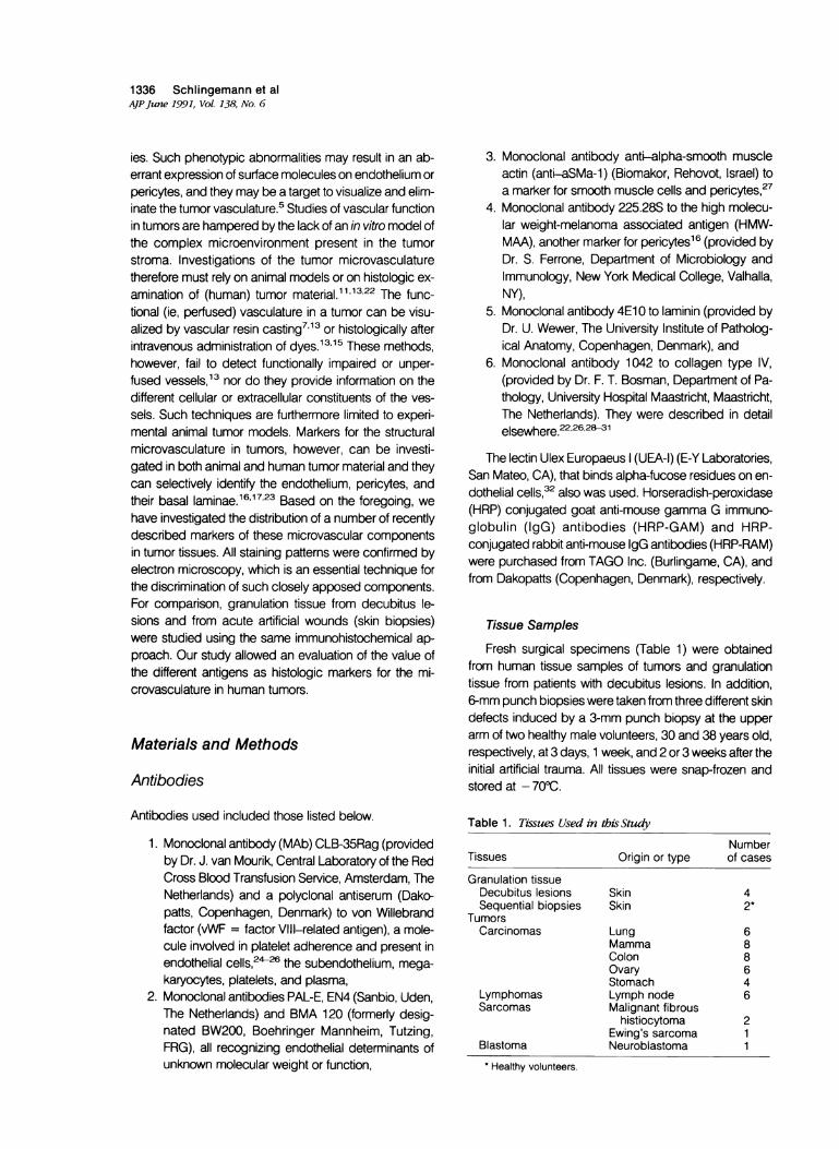
1336 Schlingemann et alAJPJune 1991, Vol. 138, No. 6
ies. Such phenotypic abnormalities may result in an ab-errant expression of surface molecules on endothelium orpericytes, and they may be a target to visualize and elim-inate the tumor vasculature.5 Studies of vascular functionin tumors are hampered by the lack of an in vitro model ofthe complex microenvironment present in the tumorstroma. Investigations of the tumor microvasculaturetherefore must rely on animal models or on histologic ex-
amination of (human) tumor material.11,1322 The func-tional (ie, perfused) vasculature in a tumor can be visu-alized by vascular resin casting71 3 or histologically afterintravenous administration of dyes.13'15 These methods,however, fail to detect functionally impaired or unper-
fused vessels,13 nor do they provide information on thedifferent cellular or extracellular constituents of the ves-
sels. Such techniques are furthermore limited to experi-mental animal tumor models. Markers for the structuralmicrovasculature in tumors, however, can be investi-gated in both animal and human tumor material and theycan selectively identify the endothelium, pericytes, andtheir basal laminae.16'17'23 Based on the foregoing, we
have investigated the distribution of a number of recentlydescribed markers of these microvascular componentsin tumor tissues. All staining patterns were confirmed byelectron microscopy, which is an essential technique forthe discrimination of such closely apposed components.For comparison, granulation tissue from decubitus le-sions and from acute artificial wounds (skin biopsies)were studied using the same immunohistochemical ap-
proach. Our study allowed an evaluation of the value ofthe different antigens as histologic markers for the mi-crovasculature in human tumors.
Materials and Methods
Antibodies
Antibodies used included those listed below.
1. Monoclonal antibody (MAb) CLB-35Rag (providedby Dr. J. van Mourik, Central Laboratory of the RedCross Blood Transfusion Service, Amsterdam, TheNetherlands) and a polyclonal antiserum (Dako-patts, Copenhagen, Denmark) to von Willebrandfactor (vWF = factor VIII-related antigen), a mole-cule involved in platelet adherence and present inendothelial cells,2426 the subendothelium, mega-karyocytes, platelets, and plasma,
2. Monoclonal antibodies PAL-E, EN4 (Sanbio, Uden,The Netherlands) and BMA 120 (formerly desig-nated BW200, Boehringer Mannheim, Tutzing,FRG), all recognizing endothelial determinants ofunknown molecular weight or function,
3. Monoclonal antibody anti-alpha-smooth muscleactin (anti-aSMa-1) (Biomakor, Rehovot, Israel) toa marker for smooth muscle cells and pericytes,27
4. Monoclonal antibody 225.28S to the high molecu-lar weight-melanoma associated antigen (HMW-MM), another marker for pericytes16 (provided byDr. S. Ferrone, Department of Microbiology andImmunology, New York Medical College, Valhalla,NY),
5. Monoclonal antibody 4E1 0 to laminin (provided byDr. U. Wewer, The University Institute of Patholog-ical Anatomy, Copenhagen, Denmark), and
6. Monoclonal antibody 1042 to collagen type IV,(provided by Dr. F. T. Bosman, Department of Pa-thology, University Hospital Maastricht, Maastricht,The Netherlands). They were described in detailelsewhere.22'26'2>1
The lectin Ulex Europaeus (UEA-1) (E-Y Laboratories,San Mateo, CA), that binds alpha-fucose residues on en-dothelial cells,32 also was used. Horseradish-peroxidase(HRP) conjugated goat anti-mouse gamma G immuno-globulin (IgG) antibodies (HRP-GAM) and HRP-conjugated rabbit anti-mouse IgG antibodies (HRP-RAM)were purchased from TAGO Inc. (Burlingame, CA), andfrom Dakopatts (Copenhagen, Denmark), respectively.
Tissue Samples
Fresh surgical specimens (Table 1) were obtainedfrom human tissue samples of tumors and granulationtissue from patients with decubitus lesions. In addition,6-mm punch biopsies were taken from three different skindefects induced by a 3-mm punch biopsy at the upperarm of two healthy male volunteers, 30 and 38 years old,respectively, at 3 days, 1 week, and 2 or 3 weeks after theinitial artificial trauma. All tissues were snap-frozen andstored at - 700C.
Table 1. Tissues Used in this Study
NumberTissues Origin or type of cases
Granulation tissueDecubitus lesions Skin 4Sequential biopsies Skin 2*
TumorsCarcinomas Lung 6
Mamma 8Colon 8Ovary 6Stomach 4
Lymphomas Lymph node 6Sarcomas Malignant fibrous
histiocytoma 2Ewing's sarcoma 1
Blastoma Neuroblastoma 1* Healthy volunteers.

Visualization of Tumor Microvascular Elements 1337AJPJune 1991, Vol. 138, No. 6
Immunoperoxidase Staining
Air-dried 4-,u frozen tissue sections were fixed with ace-tone for 10 minutes and stained by a standard indirectimmunoperoxidase procedure16'33'34 using antisera atappropriate dilutions in phosphate-buffered saline con-taining 1% bovine serum albumin (BSA). Ulex Europaeusconjugated to HRP was used in a direct immunoperox-
idase procedure.34 3-3-Diaminobenzidine (DAB) con-taining 1.0 mmol/l (millimolar) imidazole and 0.01% H202was used as substrate. Sections were counterstainedwith hematoxylin. When indicated, a double-staining pro-cedure by immunoperoxidase and immunofluorescencewas performed according to Lechago et al,35 using com-binations of the MAbs 225.28S and anti-aSMa-1. Thestaining intensity was graded as follows: no staining (-),weak staining (±), moderate staining (+), and markedstaining (+ +).
Immunoelectronmicroscopy
A pre-embedding immunoperoxidase technique wasused to demonstrate the precise cellular localization andthe subcellular distribution of the staining with theMAbs.16'17'23 Tissue specimens with a maximum size of4 x 6 x 2 mm were fixed for 4 hours at room temperaturein freshly prepared 4% paraformaldehyde in Sorensen'sphosphate buffer pH 7.4, and washed overnight in TRIS-buffered saline pH 7.4 (TBS). Fifty-micron vibratome sec-tions were cut, washed in TBS, and incubated overnightat 40C with the MAbs at appropriate dilutions in TBS con-taining 0.5% BSA and 5.0 mmol/l saponin. For the incu-bations and washing steps, a rotary shaker was used.After four 30-minute washings with TBS, the sectionswere incubated with a mixture of HRP-RAM and HRP-GAM at appropriate dilution in TBS containing 20% nor-mal human serum for 90 minutes at room temperature.The control sections were incubated with second anti-bodies alone. After extensive washings in TBS, the sec-tions were preincubated in 0.75 mg/ml DAB in TBS pH5.7 containing 1.0 mmol/l imidazole for 10 minutes. Thesections then were developed for 10 minutes in the samesolution containing in addition 0.01% H202; the reactionwas terminated by rinsing with H20. Subsequently, thesections were postfixed in 1% osmium tetroxide in Paladebuffer for 30 minutes, dehydrated, embedded in epoxyresin (Epon 812), and processed routinely. One-micron-thick sections were cut for light microscopy andstained with toluidine blue. Ultrathin sections were cutand not contrasted. They were examined and photo-graphed on a Philips 300 electron microscope (Eind-hoven, The Netherlands) at 40 kV.
Value of Vascular Markers inImmunohistochemistry
The evaluation of the value of the different vascular mark-ers in visualizing the tumor microvasculature in frozensections was based on the specificity and sensitivity ofthe immunostaining. The specificity was defined as thedegree to which one microvascular cell type or structurewas recognized exclusively. The sensitivity of the endo-thelial markers was defined as the proportion of micro-vascular structures that were recognized by a particularmarker. Because of its high specificity and sensitivity forendothelium, MAb PAL-E was used as golden standardin a semiquantitative evaluation of sensitivity. The sensi-tivity was scored as high (more than 90% of microvascu-lar structures stained), moderate (50% to 90% stained),or low (less than 50% stained).
Results
(Sub)Cellular Distribution of the VascularAntigens in Tumor Stroma
Staining results in tumors are summarized in Table 2. Ashort description of the reaction patterns is given for en-dothelial, pericyte, and basal lamina antigens separately.
Endothelial Antigens
von Willebrand Factor (MAb CLB-Rag 35 andPcAb)
Light Microscopy
Using the MAb CLB-Rag35, marked cytoplasmicstaining in endothelial cells was observed mainly in thelarger vessels of tumor stroma (Figure 1 A). The staining ofcapillary endothelium localized in tumor nests was vari-able and often focal (Figure 1 A, compare with Figure 1 Bstained with PAL-E). Many tumors, especially carcino-mas, often did not show any staining of the capillary en-dothelium. In malignant lymphomas and in lymphocyticinfiltrates associated with carcinomas, however, amarked and uniform staining of the microvessels wasfound (not shown). Nonendothelial cells did not stain withMAb CLB-Rag 35.
The polyclonal anti-von Willebrand antiserum showedstaining of more microvascular structures than the MAb,with a diffuse staining of the complete vascular wall,whereas, in addition, the perivascular stroma often wasdiffusely positive. This stromal staining was not observedin adjacent normal lung and colon tissues (not shown).

1338 Schlingemann et alAJPJune 1991, Vol. 138, No. 6
Table 2. Staining Patterns and Sensitivity of the Vascular Markers in Frozen Sections ofTumors
Antibodies recognizing microvascular constituents
Endothelium Pericytes BL
MAb* PcAb Lectin MAb MAb MAb MAb MAb MAbsvWF vWF UEA-1 EN-4 BMA 120 PAL-E aSMa 225.28 1042/4E10
MicrovesselsCapillary ECLarge-vessel ECPericytesBL
Connective tissueFibroblastsMyofibroblastsInterstitium
Inflammatory infiltrateLymphocytesHistiocytes
TumorTumor cellsBL
+/-t ++ ++ ++ + +1+++ ++
+l-
++ +1+ + +
_ - ~++++ - _
_ _ _ ~~~+_
++ + +
* Monoclonal (MAb) and ployclonal (PcAb) antibodies: see Materials and Methods.t Explanation of score: - = no staining; +/- = low; + = moderate; + + = high sensitivity of staining.BL, basal lamina; EC, endothelial cells.
Electron Microscopy
In the cytoplasm of endothelial cells lining arteriolesand venules, a marked staining of organelles, compatiblewith Weibel-Palade bodies, was found with MAb CLB-Rag35 (not shown). The reaction product also was
present in the subendothelial space. The luminal endo-thelial membrane did not stain.
H Blood Group (Ulex Europaeus I Lectin)Light Microscopy
Ulex Europaeus markedly stained the endothelium ofthe tumor vessels. The tumor cells also were positive in
the majority of the different types of carcinomas (Figure 2)and in a neuroblastoma.
Endothelial Antigen Recognized by MAb EN-4
Light Microscopy
In the tumor bed a marked endothelial staining couldbe observed. The tumor cells of two cases of carcinomaand one lymphoma were also positive. Marked stainingof mononuclear infiltrate cells compatible with histiocyteswas seen in all carcinomas (Figure 3). The alveolar mac-rophages stained, but lymphocytes were negative.
Figure 1. Immunoperoxidase staining with anti-von Willebrandfactor MAb CLB35-Rag (A) and with anti-endothelium MAli PAL-E (B) ofserialffrozen sections ofa carcinoma of the breast. MAb CLB35-Rag only stains a large vessel in the tumor stroma. In contrast, MAib PAL-Estains all microvessels, including capillaries (arrows) (x450).
+1I- ++++
_++
+l-+

Visualization of Tumor Microvascular Elements 1339AJPJune 1991, Vol. 138, No. 6
A\.t.i* :- * * 4'4\ ki3- .*¼' -; <
Figure 2. Immunoperoxidase staining with Ulex Europaeus I lec-tin offrozen section ofa carcinoma ofthe lung UEA-I stains boththe vascular structures (arrow) and tumor acini (asterisks)(x450).
Electron Microscopy
A marked staining of the luminal endothelial mem-
brane and a weak staining of the abluminal membranewas found in all vascular structures (not shown).
Endothelial Antigen Recognized by MAb BMA120
Light Microscopy
The endothelial cells in all tumors tested stained in adiscrete pattern. In one half the carcinoma lesions, a focaltumor cell staining was noted, often at the luminal side ofthe adenocarcinoma cells (Figure 4). Neuroblastoma
Figure 3. Immunoperoxidasestaining with anti-endotelium MAbEN-4 offrozen section of a carcinoma of the breast. MAb EN-4stains the vascular structures (arrows) and inflammatory cells inthe tumor stroma (arrowheads) (x450).
L W ..A,." . '12i:.:;v '%
Figure 4. Immunoperoxidase staining with anti-endotbelium MAbBMA 120 offrozen section of an adenocarcinoma of the lung.MAb BMA 120 stains both the vascular structures (arrows) and theluminal side of tumor acini (asterisks) (X 450).
cells and nontumorous mesothelial cells also stained,whereas other cell types were negative.
Electron Microscopy
Endothelial cells were diffusely positive at their luminalsurface (Figure 5); the abluminal side was negative.
Endothelial Antigen Recognized by MAb PAL-E
Light Microscopy
A discrete endothelial staining of both large and smallvessels was found with a preference for the cell contour;this pattern was seen in all tumors (Figure 1 B).
Electron Microscopy
Plasmalemmal vesicles at the luminal and locally atthe abluminal endothelial surface were markedly positive.The luminal membrane proper stained locally and weakly(not shown).
Pericyte Antigens
Alpha-smooth Muscle Actin (MAb anti-aSMa-1)
Light Microscopy
Staining of the pericytes in the tumor microvasculaturewas variable: locally, vascular structures were positivebut in many tumors large areas were observed withoutany pericyte staining. The presence of unstained peri-cytes in these areas was established by double-staining

1340 Schlingemann et alAJPJune 1991, Vol. 138, No. 6
Figure 5. Electronmicroscopy of immuno-peroxidase staining with anti-endotheliumMAb BMA 120 of a colon carcinoma. MAbBMA 120 stains endothelial cells (E) on theirluminal side (arrowhead). E = endothelium;L = lumen (bar = 0.32 i).
with anti-HMW-MAA MAb 225.28S (Figure 6A, B). In ad-dition to staining of pericytes, the myofibroblasts in thestroma of carcinomas and smooth muscle cells in arterywalls and colon carcinomas also stained with MAb anti-aSMa-1 (not shown).
Electron MicroscopyThe reaction product was observed in the cytoplasm
of pericytes and their processes in capillaries in the tumorstroma (Figure 7).
High-Molecular-weight Melanoma-associatedAntigen (HMW-MAA; MAb 225.28S)Light MicroscopyThe pericytes in the tumor microvessels were mostly
markedly but locally variably stained (Figure 6A, B).
Electron MicroscopyThe reaction product was observed at the cell mem-
brane of pericytes and their processes in the capillaries ofthe tumor stroma (not shown).
Basal Lamina Antigens
Collagen Type IV (MAb 1042)Light MicroscopyThe basal lamina of the vascular, epithelial, and fat
cells stained markedly, as did that of most tumor cells inthe carcinomas. Also the intercellular substance and my-ofibroblasts in the stroma of the carcinomas and sarco-mas stained. In the lymphomas, the interdigitating cellswere positive (not shown). The marked stromal stainingoften hampered a proper identification of the microvas-cular structures.
Electron MicroscopyStaining was observed at the basal lamina of the en-
dothelial cells and surrounding pericytes (Figure 8).
Laminin (MAb 4E10)Light MicroscopyThe basal lamina stained in a pattern similar to that
with the anti-collagen type IV MAb 1042, but the stainingof the tumor stroma was less marked (not shown).
w, ...,"
.. ..4#J-j --t:
/
6BFigure 6. Double-staining with MAb anti-aSMa-1 (A, immunofluorescence) and anti-HM'JW-MAA MAh 225.28S (B, immunoperoxidase) offrozen section ofa seminoma. MAb 225.28S stains thepericytes in the wall ofthe microvessels (arrows) (B). Note that not all these vessels arestained with MAb anti-aSMa-1 (A). (x450).

Visualization of Tumor Microvascular Elements 1341AJPJune 1991, Vol. 138, No. 6
Figure 7. Electronmicroscopy of immuno-peroxidase staining with MAb anti-aSMa-1 ofa colon carcinoma lesion. MAb anti-aSMa-1stains the cytoplasm ofpericytes (arrowhead).E = endothelium; L = lumen; (bar =1.25 i04
Staining Distribution of the VascularAntigens in Granulation Tissue
On the basis of histologic criteria, three zones in the gran-ulation tissue of decubitus lesions with a different degreeof maturation were recognized (Figures 9, 10, 1 1, and 12;Table 3). The superficial zone was covered by a fibrinousexsudate. It contained many elongated capillaries andvascular sprouts embedded in an edematous stroma inwhich scattered polymorphonuclear leukocytes wereseen. In the adjacent intermediate zone, less capillarieswere encountered and in addition, somewhat dilated ve-nules. The stroma was slightly fibrous and containedscattered lymphocytes and histiocytes. Finally the deepzone had much less microvessels. Conversely, severalvenules, arterioles, and some small veins and arterieswere observed. In all three zones, the microvasculaturedemonstrated staining for both endothelial and basallamina markers. In contrast, the pericyte markers showeda zonal distribution of staining (Table 3): in the superficial,'immature' zone, MAb anti-aSMa-1 only sporadicallystained the pericytes, whereas most vessels were posi-tive for HMW-MAA. In the 'mature' zone, however, theexpression of HMW-MAA in microvessels was focal incontrast to an extensive staining for aSMa-1 (Figures 10,11, and 12)
The skin biopsies derived from artificially inducedwounds by punch biopsy in two healthy volunteersshowed a fibrinous and cellular exudate in the woundand a perivascular infiltrate of polymorphonuclear leuko-
Figure 8. Electron microscopy of immuno-peroxidase staining with anti-collagen type IVAMAb 1042 ofa colon carcinoma lesion. AIAb1042 stains basal lamina adjacent to the en-dothelial cell andperticyte. P = pericyte; e =endothelium; L = lumen (bar = 0.68 i).
cytes after 3 days. After 1 week, the wound edge showedmicrovascular sprouts connected with pre-existent der-mal microvessels. In the adjacent dermis, a sparse lym-phohistiocytic perivascular infiltrate was observed. Thebiopsies taken at 2 or 3 weeks showed a cellular fibro-blastic scar tissue in which elongated capillaries and fewinflammatory cells were found.
The immunohistochemical findings are summarized inTable 4 and Figures 13 and 14. Vascular sprouts andcapillaries markedly stained for vWF, PAL-E antigen,aSMa-1, HMW-MAA, collagen type IV, and less for laminin1, 2, and 3 weeks after trauma. The staining pattern of theendothelial and basal lamina antigens was diffuse, that ofthe pericytes more focal and variable in intensity. Pre-existent capillaries and venules in the first biopsy and inthe subsequent biopsies at some distance of the woundarea did not stain for HMW-MAA.
Value of the Reagents Against Markers forVascular Elements in the Tumor Stroma
A comparison of the value of the antibodies against vas-cular markers is presented in Table 5. With the exceptionof vWF, all endothelial markers were found to have a highsensitivity for the tumor endothelium. The specificity, how-ever, was often limited. A cross-reactivity with nonendo-thelial cells, observed in a number of tumors with UEA-Iand Mab EN4, obscured the vascular staining andthereby limited the marker's efficacy. In the lymphomas,

1342 Schlingemann et alAJPJune 1991, Vol. 138, No. 6
Figure 11. Immunoperoxidase staining with anti-collagen type IVMAb 1042 ofafrozen section ofzone I ofgranulation tissue ofadecubitus lesion of the skin. MAb 1042 stains all microvascularstructures with pericytes (arrous) (x350)
9~~~~~~~~~~~
Figure 9. Immunoperoxidase staining with anti-collagen type IVMAb 1042 ofafrozen section ofgranulation tissue ofa decubituslesion ofthe skin. MAb 1042 stains vascular structures throughoutthe three maturation zones (I, II, and III), including microvesselsin the most superficial one (arrows). At this low magnification,vascular sprouts as shoun in Figure 11 are not clearly visible(x35).
'r.X,.. :. .;..
I r)1n
Figure 10. Immunoperoxidase staining with anti-pericyteMAbanti-aSMa-1 ofafrozen section ofzone I ofgranulation tissue ofa decubitus lesion ofthe skin. MAb anti-aSMa-1 stainspericytes inonly one vessel (arrows). Note the absence of staining in mostmicrovessels (asterisks) (X350).
the sinusoid endothelium showed a varying staining withthe tested reagents (not shown). The sensitivity ofaSMa-1 for pericytes was low in the stroma of tumors incontrast to that of HMW-MAA.
Architectural Patterns of Tumor Vasculature
On the basis of their discrete staining, the MAbs PAL-Eand BMA 120 demonstrated the morphologic outlay ofthe tumor microvasculature best. The number of vascularstructures and the distribution of vessels of different sizein different tumors varied widely. Individual tumors, how-ever, tended to demonstrate the same microvascular ar-
Ati z a, 3^.4
>g bl |
s~~~ ?* $ 4'its<<^,4S
122 * _Figure 12. Immunoperoxidase stainingwith anti-HMW-MAAMAb225.28S of a frozen section ofzone I of granulation tissue of adecubitus lesion of the skin. MAb 225.28S stains pericytes in allmicrovascular structures (arrows) (X350).
r .>*

Visualization of Tumor Microvascular Elements 1343AJPJune 1991, Vol 138, No. 6
Table 3. Digstibution ofMicrovascular Components inthe Maturation Zones of Granulation TissuefromDecubitus Lesions
Maturation zones ofgranulation tissue
(Zone I) (Zone II) (Zone ll)Superficial Intermediate Deep
HistologyVessels Sprouts Capillaries Capillaries
Capillaries Venules VenulesArterioles
Interstitium Fibrinous Slight No edemacoating edema
Edema Slight Markedfibrosis fibrosis
Infiltrate Neutrophils Mononuclear Few cellscells
ImmunostainingEndotheliumMAb PAL-E + + + + + +
PericytesAnti-aSMa-1 -/+ + ++Anti-HMW-MAA + + ++ -/+
Basal laminaAnti-collagen
IV ++ ++ ++
Explanation of symbols: -
tive; + +, markedly positive.-/+, occasionally positive; +, posi-
chitecture throughout. These microvascular patternswere apparently associated with the stromal architecture,depending on the direction and degree of differentiationof the tumors.
Discussion
The microvasculature consists of endothelial cells andpericytes91 &-1836 surrounded by basal lamina. There-fore, antibodies to components of any of these constitu-ents may theoretically be used to visualize both the de-gree of vascularization and the structural abnormalities ofvessels in a tumor, and to study vascular invasion bytumor cells.23,2937 We now report on the distribution ofmicrovascular antigens in tumors and present an evalu-ation of their use as markers for the tumor microvascula-ture. Furthermore, we have compared the microvascula-ture of tumor stroma with microvessels in different matu-ration stages of granulation tissue of wound healing indecubitus lesions and in biopsies of artificial skin woundsin two healthy volunteers.
Regarding the anti-endothelial antibodies with respectto their use to recognize vascular markers, the MAbsPAL-E and BMA 120 had the highest value. Monoclonalantibodies BMA 120 and UEA react with paraffinsections,23,28,32 37 and thus seem to be the best choicefor formalin-fixed human tissues. As a marker for tumorvessels, vWF does not seem suitable in view of its lowsensitivity. Of some of the other tested reagents, thespecificity was limited: UEA-l lectin and the MAbs EN4and BMA 120 sometimes reacted with tumor cells of non-endothelial origin. Moreover, the MAb BMA 120 reactedwith mesothelium and MAb EN4 stained infiltrating mono-nuclear cells and alveolar macrophages. Staining for
Table 4. Phenotjpic Changes ofDernal Microvascular Cells During WoundHealing*
Interval after traumaMorphology Pre-existentand markers skint 3 days 1 week 2 and 3 weeks
Conventional Normal architecture Crusta in defect, Organization of fibrin Restoration of epidermis,morphology perivascular by fibroblasts, slight cellular scar tissue, few
infiltrate of PMNs in lymphohistiocytic inflammatory cellsadjacent dermis infiltrate in
perilesional dermisEndotheliumvWF and Marked staining of Remnants of Several vascular Many elongated
PAL-E-ag capillaries and microvessels at sprouts in capillaries and vascularvenules wound edge perilesional dermis sprouts in scar tissue
PericyteaSMa-1 Marked staining of Marked staining of Variable staining of Marked staining of
capillaries and capillaries and vascular sprouts capillaries and vascularvenules venules sprouts in scar tissue
HMW-MAA No staining of No staining of Variable staining of Marked staining ofcapillaries and capillaries and vascular sprouts capillaries and vascularvenules venules sprouts in scar tissue
Basal laminaCollagen IV Marked staining of Marked staining of Marked staining of Marked staining of
capillaries and capillaries and vascular sprouts capillaries and vascularvenules venules sprouts in scar tissue
Laminin Staining of capillaries Staining of capillaries Marked staining of Staining of capillaries andand venules and venules vascular sprouts vascular sprouts in scar
tissue
Frozen sections of sequential skin biopsies from two healthy volunteers.t Taken from upper arm.

1344 Schlingemann et alAJPJune 1991, Vol. 138, No. 6
*w*, s (^! t 's43B *}I
Figure 13. Immunoperoxidase staining with MAb PAL-E (A), anti-HMW-MAA MAb 225.28s (B), and anti-collagen IV MAb 1042 (C) ofadjacentfrozen sections ofa skin biopsy 1 week after arteficial trauma. Note the marked microvascular staining (arrows). E = epidermis.(x450).
these markers has been used as evidence for an endo-thelial origin of tumor cells in histogenetic studies ofneoplasmata.23 Our data suggest that the results of suchstudies must be interpreted with caution. The typicalgranular staining for vWF with MAb CLB-35Rag was notobserved in nonendothelial cells and might still providethe most reliable means of identifying the endothelial or-igin of tumor cells in a histogenetic study. It emerges fromour work, however, that a fully specific and sensitive his-tologic marker for the structural microvasculature in tu-mors, or endothelium in general, is not yet available.
To confirm the cellular localization and to assess thesubcellular distribution of the antigens, a pre-embeddingimmunoperoxidase staining of vibratome sections wasused. On the basis of the specific cytoplasmic stainingwith the MAb CLB-35Rag against vWF and MAb anti-aSMa-1 against pericytes, and of the basal lamina with
I
9
the anti-collagen type IV MAb 1042, this technique ap-pears reliable in determining the differential staining of thedifferent microvascular components. Our results furtherdemonstrate that most of the endothelial membrane de-terminants studied are expressed predominantly at theluminal surface.
The degree of staining observed in the tumor stromawith the MAbs PAL-E, EN4, and BMA 120 was similar toresults obtained in normal adult tissues.23,31 The anti-vWF MAb, however, demonstrated decreased or evenabsent staining in the capillaries of the carcinomas andthe other tumors, indicating that in the tumor stroma amarked quantitative heterogeneity in the degree of vWFexpression among the microvascular endothelial cellsexists. In two previous studies, subsets of tumor vesselsin paraffin sections of follicular carcinoma of thyroid andof gliomas also were found not to stain.-938 In normal
V
Ii
i4A AQMW`< <% s
Figure 14. Immunoperoxidase staining with MAb PAL-E (A), anti-HMW-IAA MAb 225.28s (B), and anti-collagen IV MAb 1042 (C) ofadjacent frozen sections of a skin biopsy 2 weeks after artificial trauma. Note the marked microvascular staining (arrows). The asterisksrepresent the microvascular lumen (A and C, x 450; B, x 700).

Visualization of Tumor Microvascular Elements 1345AVPJune 1991, Vol. 138, No. 6
Table 5. Comparison of Vascular Markers in Visualizing the Tumor Vasculature in Tissue Sections
Marker Specificity Sensitivity Cross-reactions Remarks
Endothelium F PvWF H M M MAb: none PcAb most sensitive, but stromal
PcAb: Stroma reaction obscures vascularstaining
UEA-1 L H H Tumor cells Reaction with tumor cells obscuresvascular staining
EN4-ag L H NA Infiltrate cells, tumor cells Cross-reaction makes vascularstaining less discrete
BMA 120-ag M H H Tumor cells, serosa Useful as marker; staining discretePAL-E-ag H H NA None Useful as marker
PericyteaSMa-1 L L L Myofibroblasts, smooth Variable staining
muscleHMW-MAA M M NA Smooth muscle Less variable staining
Basal laminaCollagen IV L H H BL of tumor, Reaction with perivascular stroma
myofibroblasts obscures vascular stainingsLaminin L H H BL of tumor, stroma Reaction with perivascular stroma
obscures vascular stainingF, frozen sections; P, paraffin sections; h, high; m, moderate; 1, low; ag, antigen; NA, not applicable; BL, basal lamina.
tissues, however, vWF (Mab CLB-35Rag) stains the cap-illary endothelium,26 but this staining is weaker than thatof large-vessel endothelium (own unpublished observa-tions). The biologic implication of these differences is un-clear. The granular staining of endothelial cells probablyis due to the presence of Weibel-Palade (WP) bodies,which have been identified as the storage vesicles forvWF.24 The lesser staining for vWF in the tumor microves-sels might therefore either reflect a decrease in the num-ber of these organelles or indicate that vWF is dischargedby the cells instead of being stored in WP bodies,24'39 assuggested by ultrastructural observations of increasednumbers of WP bodies in tumor microvessels.39
Apart from the decreased expression of vWF in thetumor capillary endothelium, the pericytes in the tumorstroma were found to have decreased or absent expres-sion of aSMa compared with normal adult tissues, whichmay indicate an altered contractile function of the peri-cytes in the tumor microvasculature. The pericytes in thetumor stroma, however, markedly expressed the proteo-glycan HMW-MAA.16 Double-staining of the tumor mi-crovessels by antibodies to HMW-MM and aSMa iden-tified a heterogeneous expression of these antigens inthe pericytes, which may reflect the existence of sub-types of vessels.
The tumor stroma is similar to granulation tissue ofwound healing, as indicated by the presence of fibrin,inflammatory cells, and immature connective tissue andmicrovessels.446 Therefore we compared the mi-crovasculature of the tumor stroma with that of variousmaturation zones in granulation tissue of decubitus le-sions (Table 3). In every zone, including the most super-ficial 'immature' one, all microvessels expressed the en-dothelial and basement membrane antigens. Even the
most superficial microvessels contained pericytes. De-pending on the zone, however, the pericyte markers dis-played different patterns of staining (Table 3). A similarmicrovascular phenotype was found in the edge of ahealing wound and in granulation tissue of wound healingat 1, 2, and 3 weeks after artificial skin trauma. Likewisemicrovessels in the tumor stroma contained pericyteswith heterogeneous expression, but no evident zonal mi-croarchitecture could be established. From the resultsobtained in our studies with granulation tissues it can beinferred, however, that pericytes lacking aSMa positivity,but expressing the HMW-MAA antigen, accompany aproliferative microvascular system, and may be re-garded, therefore, as a sign of active angiogenesis.
Our findings prompted us to reconsider the currentmodel of stepwise angiogenesis as proposed by otherauthors.22247 If we interpreted this model well, vascularsprouts consist of immature vessels, with an incompletebasement membrane,224' and migrating endothelialcells at their tips, but without pericytes.i8'49 The compo-sition of the basement membrane in these sprouts, es-pecially the content of collagen type IV and laminin, isthought to be an important factor in the regulation of en-dothelial migration and proliferation.47' 51 Furthermorethe absence of pericytes is believed to stimulate endo-thelial mitosis.49 In our study, however, we did not findany cross-sections of microvessels without pericytes inthe superficial layer (zone 1) of granulation tissue in de-cubitus lesions, where vascular sprouting should be mostabundant,15 and in uncomplicated wound healing after apunch biopsy in healthy individuals, nor did we observevessels without collagen type IV or laminin in their basallaminas. We are aware of the fact that in transplantedallogeneic dermis the microvasculature has only detect-

1346 Schlingemann et alAJPJune 1991, Vol. 138, No. 6
able laminin but no collagen type IV 4 weeks afterplacement.52 Angiogenesis in cryopreserved cadavericskin transplanted on extensively burned sites, however, inour opinion is not representative for the situation in gran-ulation tissue in wound healing and in tumors. In the tu-mor stroma, we could not determine the exact sites of thevascular sprouting and endothelial mitosis. Still it is as-sumed that both phenomena occur frequently through-out the tumor microvasculature.2,4,20,21 As was the casein granulation tissue, we were unable to demonstrate intumor stroma the immature phenotype (as predicted bythe angiogenesis model), in which pericytes are lacking.Therefore, taken together with ultrastructural observationsby other authors of vascular sprouting in close relationwith pericytes in granulation tissue,46'3 our results sug-gest an early recruitment of pericytes during angiogene-sis. Further research is needed to elucidate these appar-ent contradictory results between the angiogenesis mod-els and those of immunohistochemical and ultrastructuralstudies of human lesions.
Acknowledgments
The authors thank Dr. J. U. Alles, Clinical Research Unit for BloodCoagulation and Thrombosis of the Max Planck-Gesellschaft atthe Justus Liebig-Universitat, Giessen, Federal Republic of Ger-many, for providing the BMA 120 antibody, Georgette Cramer-Knijnenburg for her excellent technical assistance, Maria Arendsfor the preparation of the photomicrographs, and Dr. F. C. S.Ramaekers, Dr. H. Schuurmans Stekhoven, and Dr. R. Wetzelsfor their helpful advice.
References
1. Algire GH, Chalkley HW, Legallais FY, Park HD: Vascularreactions of normal and malignant tissues in vivo: I. Vascularreactions of mice to wounds and to normal and neoplastictransplants. J Natl Cancer Inst 1945, 6:46-73
2. Folkman J: Tumor angiogenesis. Adv Cancer Res 1985,43:175-203
3. Reinhold HS, van den Berg-Blok A: Factors influencing theneovascularization of experimental tumours. Biorheology1984, 21:493-501
4. Folkman F, Cotran R: Relation of vascular proliferation totumor growth. Int Rev Exp Pathol 1976, 16:207-248
5. Denekamp J: Endothelial cell proliferation as a novel ap-proach to targeting tumour therapy. Br J Cancer 1982,45:136-139
6. Schor AM, Schor SL: Tumour angiogenesis. Br J Pathol1983, 141:385-413
7. Grunt TW, Lametschwandtner A, Karrer K, Staindl 0: Theangioarchitecture of the Lewis lung carcinoma in laboratorymice. Scanning Electron Microsc 1986, 11:557-573
8. Hagemeier HH, Vollmer ES, Goerdt 5, Schulze-Osthoff K,
Sorg C: A monoclonal antibody reacting with endothelialcells of budding vessels in tumors and inflammatory tissues,and non-reactive with normal adult tissues. Int J Cancer1986, 38:481-488
9. Ho KL: Ultrastructure of cerebellar capillary hemangioblas-toma: IV. Pericytes and their relationship to endothelial cells.Acta Neuropathol 1985, 67:254-264
10. Hobson B, Denekamp J: Endothelial proliferation in tumoursand normal tissues: Continuous labeling studies. Br J Can-cer 1984, 49:405-413
11. Jain RK: Determinants of tumor blood flow: A review. CancerRes 1985, 48:2641-2658
12. McLone DG: Ultrastructure of the vasculature of central ner-vous system tumors of childhood. Child's Brain 1980,6:242-254
13. Murray JC, Rhandawa V, Denekamp J: The effects of mel-phalan and misonidazole on the vasculature of a murinesarcoma. Br J Cancer 1987, 55:233-238
14. Nawroth P, Handley D, Matsueda G, de Waal R, Gerlach H,Blohm D, Stern DM: Tumor necrosis factor/cachectin-induced intravascular fibrin formation in meth A fibrosarco-mas. J Exp Med 1988,168:637-647
15. Rofstad EK: Growth and vascular structure of human mela-noma xenografts. Cell Tissue Kinet 1984, 17:91-101
16. Schlingemann RO, Rietveld FJR, De Waal RMW, Ferrone S,Ruiter DJ: Expression of the high molecular weight mela-noma-associated antigen by pericytes during angiogenesisin tumors and in healing wounds. Am J Pathol 1990,136:1393-1405
17. Schlingemann RO, Rietveld FJR, de Waal RMW, BradleyNJ, Skene Al, Davies AJS, Greaves, MF, Denekamp J,Ruiter DJ: Leukocyte antigen CD 34 is expressed by a sub-set of cultured endothelial cells and on endothelial abluminalmicroprocesses in the tumor stroma. Lab Invest 1990,62:690-696
18. Warren BA: The vascular morphology of tumors, TumorBlood Circulation. Edited by H.-I. Peterson. Boca Raton, FL,CRC Press, 1979, pp 1-47
19. Heuser LS, Miller FN: Differential macromolecular leakagefrom the vasculature of tumors. Cancer 1986, 57:461-464
20. Cavallo T, Sade R, Folkman J, Cotran RS: Ultrastructuralautoradiographic studies of the early vasoproliferative re-sponse in tumor angiogenesis. Am J Pathol 1973, 70:345-362
21. Tannock IF: Population kinetics of carcinoma cells, capillaryendothelial cells, and fibroblasts in a transplanted mousemammary tumor. Cancer Res 1970, 30:2470-2476
22. Ausprunk DH, Folkman J: Migration and proliferation of en-dothelial cells in preformed and newly formed blood vesselsduring tumor angiogenesis. Microvasc Res 1977, 14:53-65
23. Ruiter DJ, Schlingemann RO, Rietveld FJR, de Waal RWM:Monoclonal antibody defined human endothelial antigensas vascular markers. J Invest Dermatol 1989, 93:25S-32S
24. Reinders JH, de Groot F, van den Berg A, Vervoom R,Sixma J, van Mourik J: The Weibel-Palade body is the stor-age vesicle for von Willebrand factor in endothelial cells.Proceedings of the 4th Intemational Symposium on Biologyof the Vascular Endothelial Cell, 1986
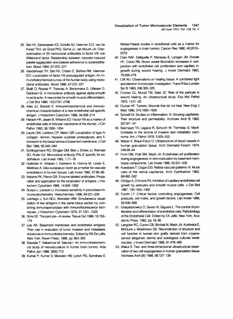
Visualization of Tumor Microvascular Elements 1347AJPJune 1991, Vol. 138, No. 6
25. Stel HV, Sakariassen KS, Scholte BJ, Veerman ECI, van deKwast ThH, de Groot PhG, Sixma JJ, van Mourik JA: Char-acterization of 25 monoclonal antibodies to factor VII1--vonWillebrand factor: Relationship between ristocetin-inducedpatelet aggegration and platelet adherence to subendothe-lium. Blood 1984, 67:222-227
26. VandeKwast TH, Stel HV, Cristen E, Bertina RM, VeermanECI: Localization of factor Vill-procoagulant antigen: An im-munohistochemical survey of the human body using mono-clonal antibodies. Blood 1986, 67:222-227
27. Skalli 0, Ropraz P, Trzeciac A, Benzonana G, Gillesen D,Gabbiani G: A monoclonal antibody against alpha-smoothmuscle actin: A new probe for smooth muscle differentiation.J Cell Biol 1986, 103:2787-2796
28. Alles JU, Bosslet K: Immunohistochemical and immuno-chemical characterization of a new endothelial cell-specificantigen. J Histochem Cytochem 1986, 34:209-214
29. Harach HR, Jasani B, Williams ED: Factor VIII as a marker ofendothelial cells in follicular carcinoma of the thyroid. J ClinPathol 1983, 36:1050-1054
30. Laurie GW, Leblond CP, Martin GR: Localization of type IVcollagen, laminin, heparan sulphate proteoglycan, and fi-bronectin to the basal lamina of basement membrane. J CellBiol 1986, 95:340-344
31. Schlingemann RO, Dingjan GM, Blok J, Emeis JJ, WamaarSO, Ruiter DJ: Monoclonal antibody PAL-E specific for en-dothelium. Lab Invest 1985,1:71-76
32. Holthofer H, Virtanen I, Kariniemi Al, Hormia M, Linder E,Miettinen A: Ulex europaeus lectin as a marker for vascularendothelium in human tissues. Lab Invest 1982, 47:60-66
33. Nakane PK, Pierce GB: Enzyme-labeled antibodies: Prepa-ration and application for the localization of antigens. J His-tochem Cytochem 1966,14:929-1002
34. Scopsi L, Larsson LI: Increased sensitivity in peroxidase im-munocytochemistry. Histochemistry 1986, 84:221-230
35. Lechago J, Sun NCJ, Weinstein WM: Simultaneous visual-ization of two antigens in the same tissue section by com-bining immunoperoxidase with immunofluorescence tech-niques. J Histochem Cytochem 1979, 27:1221-1225
36. Sims DE: The pericyte-A review. Tissue Cell 1986,18:153-174
37. Lee AK: Basement membrane and endothelial antigens:Their role in evaluation of tumor invasion and metastasis.Advances in Immunohistochemistry. Edited by RA De Lellis.New York, Raven Press, 1988, pp 363-393
38. Sawada T, Nakamura M, Sakurai I: An immunohistochemi-cal study of neovasculature in human brain tumors. ActaPathol Jpn 1988, 38(6):713
39. Kumar P, Kumar S, Marsden HB, Lynch PG, Eamshaw E:
Weibel-Palade bodies in endothelial cells as a marker forangiogenesis in brain tumors. Cancer Res 1980, 40:2010-2019
40. Clark RAF, Dellapelle P, Manseau E, Lanigan JM, DvorakHF, Colvin RB: Blood vessel fibron6ctin increases in con-junction with endothelial cell proliferation and capillary in-growth during wound healing. J Invest Dermatol 1982,79:269-276
41. Cliff WJ: Observations on healing tissue: A combined lightand electron microscopic investigation. Trans R Soc LondonSer B 1963, 246:305-325
42. Crocker DJ, Murad TM, Geer JC: Role of the pericyte inwound healing. An ultrastructural study. Exp Mol Pathol1970,13:51-65
43. Dvorak HF: Tumors: Wounds that do not heal. New Engl JMed 1986, 315:1650-1659
44. Schoefl GI: Studies on inflammation: III. Growing capillaries:Their structure and permeability. Virchows Arch B 1963,337:97-141
45. Seemayer TA, Lagace R, Schurch W, Tremblay G: Myofi-broblasts in the stroma of invasive and metastatic carci-noma. Am J Pathol 1979, 3:525-532.
46. Weber K, Braun-Falco 0: Ultrastructure of blood vessels inhuman granulation tissue. Arch Dermatol Forsch 1974,248:29-44
47. Form DM, Pratt BM, Madri JA: Endothelial cell proliferationduring angiogenesis: In vitro modulation by basement mem-brane components. Lab Invest 1986, 55:521-530
48. Kuwabara T, Cogan DG: Retinal vascular pattems: IV. Muralcells of the retinal capillaries. Arch Ophthalmol 1963,69:492-502
49. Orlidge A, D'Amore PA: Inhibition of capillary endothelial cellgrowth by pericytes and smooth muscle cells. J Cell Biol1987,105:1455-1462
50. Furcht LT: Critical factors controlling angiogenesis: Cellproducts, cell matrix, and growth factors. Lab Invest 1986,55:505,50
51. Gospadorowicz D, Savion N, Giguere L: The control of pro-liferation and differentiation of endothelial cells, Pathobiologyof the Endothelial Cell. Edited by EA Jaffe. New York, Aca-demic Press, 1982, pp 19-39
52. Langdon RC, Cuono CB, Birchall N, Madri JA, Kuklinska E,McGuire J, Moellmann GE: Reconstitution of structure andcell function in human skin grafts derived from cryopre-served allogeneic dermis and autologous cultured kerati-nocytes. J Invest Dermatol 1988, 91:478-485
53. Wakui S: Two- and three-dimensional ultrastructural obser-vation of two cell angiogenesis in human granulation tissue.Virchows Arch [B] 1988, 56:127-139