Distinct effects of prefrontal and parietal ... - Princeton University
Upload
independentCategory
view
1download
0
Development/Plasticity/Repair
Differential Balance of Prefrontal Synaptic Activity inSuccessful versus Unsuccessful Cognitive Aging
Cyril Bories,1,2 Zoe Husson,1,2 Matthieu J. Guitton,1,2 and Yves De Koninck1,2
1Mental Health Institute of Quebec, Quebec City, QC, Canada G1J 2G3, and 2Department of Psychiatry and Neuroscience, Faculty of Medicine, LavalUniversity, Quebec City, QC, Canada G1V 0A6
Normal aging is associated with a variable decline in cognitive functions. Among these, executive function, decision-making, and workingmemory are primarily associated with the prefrontal cortex. Although a number of studies have examined the structural substrates ofcognitive decline associated with aging within this cortical area, their functional correlates remain poorly understood. To fill this gap, weaimed to identify functional synaptic substrates of age-associated frontal-dependent deficits in layer 2/3 pyramidal neurons of medialprefrontal cortex of 3-, 9-, and �23-month-old Fischer 344 rats. We combined, in the same animals, novelty recognition and exploratorybehavioral tasks with assessment of structural and functional aspects of prefrontal synaptic properties. We found that subsets of agedanimals displayed stereotyped exploratory behavior or memory deficits. Despite an age-dependent dendritic spine loss, patch-clamprecording of synaptic activity revealed an increase in miniature EPSC frequency restricted to aged animals with preserved exploratorybehavior. In contrast, we found a strong positive relationship between miniature IPSC frequency and the occurrence of both stereotypedexploratory behavior and novelty-related memory deficits. The enhanced miniature inhibitory tone was accompanied by a deficit inactivity-driven inhibition, also suggesting an impaired dynamic range for modulation of inhibition in the aged, cognitively impairedanimals. Together, our data indicate that differential changes in the balance of inhibitory to excitatory synaptic tone may underlie distincttrajectories in the evolution of cognitive performance during aging.
IntroductionWith global aging of the population in Western countries, age-related cognitive disorders are a major problem of public health.Hence, numerous research groups have investigated the processesimplicated in the pathogenesis of those age-related illnesses. One ofthe main objectives of research on brain aging in humans is to isolatebiomarkers or neurobiological mechanisms implicated in the cogni-tive and behavioral degradations associated with senescence(Persson et al., 2006; Raji et al., 2009). During aging, a decline inmultiple domains of cognitive functions, including short- and long-term memory, psychomotor speed, attention, and executive func-tion, can be observed. Moreover, aging is often accompanied by thedevelopment of depression, anxiety, and associated neuropsychiat-ric symptoms (Lyketsos et al., 2002).
During the last decades, the vast majority of animal studies ad-dressing the consequences of aging on brain function focused onaspects related to memory, particularly spatial memory (Rosenzweigand Barnes, 2003). Numerous studies identified AMPA or NMDA
receptor-mediated dysfunction and changes in LTP or LTD proper-ties within several memory-related brain areas, notably the hip-pocampus and associated temporal areas (Burke and Barnes, 2010).Yet, besides these memory deficits, executive function, decision-making, and working memory also decline in an age-dependentmanner (Hedden and Gabrieli, 2004). The prefrontal cortex appearsas a critical brain area to study these age-related cognitive declines(Cummings, 1995; Fuster, 2000, 2001). Although a number of stud-ies have addressed age-dependent structural changes, few relate be-havioral assessments with single-cell morphometric analyses in theaging prefrontal cortex, especially in rodents. In particular, a studyrelating these parameters to functional changes in synaptic activity isstill lacking. Consequently, the fate of the synaptic homeostasis inaging remains primarily unknown. To fill this gap, we adapted be-havioral tests to correlate the animal performances, within differentage groups (3, 6–9, and 23–27 months), with indexes of the struc-tural and functional characteristics of synaptic inputs onto layer II/III prefrontal pyramidal neurons. We provide evidence of afunctional synaptic imbalance toward inhibitory activity as a sub-strate of cognitive impairment in the aged cohort while, in contrast,an increase in excitatory synaptic tone is observed in aged unim-paired animals. The balance between basal inhibitory and excitatorysynaptic tone may thus distinguish successful from unsuccessfulcognitive aging.
Materials and MethodsAnimals. Data presented in this study were obtained from a total of 81male Fischer 344 rats divided into three groups of age: young (3 monthsold; N � 10), adult (7.1 � 1.4 month old; N � 19), and aged (24.2 � 1.6
Received July 9, 2012; revised Nov. 4, 2012; accepted Nov. 24, 2012.Author contributions: C.B., M.J.G., and Y.D.K. designed research; C.B. and Z.H. performed research; C.B. and Z.H.
analyzed data; C.B. and Y.D.K. wrote the paper.This work was supported by National Institute on Aging Grant R01 AG020529 and Canadian Institutes of Health
Research Grant MOP12942 to Y.D.K. C.B. was supported by a studentship from the Centre de Recherche sur leCerveau, le Comportement et la Neuropsychiatrie. M.J.G. and Y.D.K. are, respectively, Chercheur Boursier and Cher-cheur National of the Fonds de la Recherche en Sante du Quebec.
Correspondence should be addressed to Dr. Yves De Koninck, 2601, de la Canardiere, Quebec, QC, Canada G1J 2G3.E-mail: [email protected].
DOI:10.1523/JNEUROSCI.3258-12.2013Copyright © 2013 the authors 0270-6474/13/331344-13$15.00/0
1344 • The Journal of Neuroscience, January 23, 2013 • 33(4):1344 –1356

month old; N � 52). Animals were obtained from the National Instituteon Aging (Bethesda, DC) rodent colony. Animals were maintained on aninverted cycle (light on at 9:00 P.M., light off at 9:00 A.M.) and on adlibitum food and water diet. Animals were allowed to acclimate to theirnew environment for at least 1 month before starting electrophysiologi-cal or behavioral experiments. Animals were removed from the study ifpresenting any evidence of tumors, illness, or general weakness. All pro-cedures were performed according to the guidelines of the CanadianCouncil on Animal Care and were approved by the Animal Care Com-mittee of Laval University.
Behavioral testing. Before testing, animals were transferred from theirhousing facilities to the testing room once per day for 3 d for at least 1 h,to allow them to acclimate to the room. All experiments were conductedunder low-intensity, red-light illumination to minimize visual percep-tions from the rats to minimize differences in visual accuracy between theage groups. To avoid any interference caused by proximity of the exper-imenters, the latter were not present in the room during the testing. Allsessions took place between 2:00 and 5:00 P.M. during the dark phase ofthe light/dark cycle, were recorded with a digital camera, and were storedon a portable personal computer for subsequent analysis. Animals werekilled at least 1 week after the last behavioral test to minimize influence ofthe test itself on the cellular parameters measured.
Modified hole-board task. To quantify motor and directed exploratorybehavior, we modified the classical hole-board task and apparatus (Fileand Wardill, 1975a, b). The modified hole-board (mHB) apparatus usedwas a 50 � 50 � 30 cm arena made of a black Plexiglas floor with a hole(3 cm diameter) in each corner. All sessions were subsequently analyzedwith an automated tracking system (Any-Maze; Stoelting). Rats wereplaced individually in the center of the modified hole-board apparatusand allowed 10 min of free exploration of the novel environment. Eachrat was tested once. The standard measures were distance traveled, max-
imum speed reached by the animal, number of head entries in eachcorner, and average distance from the border (i.e., cumulative sum di-vided by the total duration of the test). The relative exploration for eachcorner was established by dividing the number of head entries by the sumof head entries in the four corners. Finally, the corners were ordered fromthe highest (preferential corner) to the lowest; the preferential corner wasthen chosen as an index of corner exploration (ICE) for each rat, rangingfrom 25% (no preferential corner) to 100% (only one corner explored).
Novel object recognition task. Animals were placed into an adapted testarena for object recognition. The apparatus consisted of a triangular startzone (30 � 30 � 40 cm) separated from a rectangular test zone (40 � 25cm) by a movable barrier.
The novel object recognition task, for two sample objects within thesame environment, was used to study memory after 1 h delays in young,adult, and aged rats. Each session was recorded with a digital camera.First, two identical objects were placed in the chamber as described pre-viously (Bevins and Besheer, 2006). After a 5 min exploration of theobjects, the rat was returned to its home cage for 1 h. To test for objectrecognition, a new copy of the familiar object and a novel object wereplaced in the chamber and the rat was again placed in the chamber for 5min to explore the objects. Object recognition was measured in a singletrial. A novel object recognition (NOR) index was computed as follows:novel object interaction time/total interaction time with both objects.
Slice preparation and electrophysiology. Rats were deeply anesthetizedwith ketamine and xylazine and decapitated. The brain was quickly re-moved (�60 s) and placed in ice-cold solution containing (in mM) 210sucrose, 3.0 KCl, 0.75 CaCl2, 3.0 MgSO4 1.0 NaH2PO4, 26 NaHCO3, and10 glucose, saturated with 95% O2 and 5% CO2. Coronal slices of pre-frontal cortex, including the prelimbic area, were cut at 400 �m and keptin artificial CSF (ACSF) containing (in mM) 124 NaCl, 3.0 KCl, 1.5 CaCl2,1.3 MgSO4 1.0 NaH2PO4, 26 NaHCO3, and 20 glucose, gassed with 95%
Figure 1. Lack of change in synaptic properties during aging in the prefrontal cortex. Representative traces of spontaneous synaptic events (sEPSCs, A; sIPSCs, B) in layer 2/3 pyramidal cellsobtained from young (blue), adult (gray), and aged (black) rats are shown. No significant changes were observed in the mean frequency or mean amplitude of sEPSCs or sIPSCs. The recordingcondition is represented in the inset.
Bories et al. • Aging, Frontal Cortex, and Synaptic Balance J. Neurosci., January 23, 2013 • 33(4):1344 –1356 • 1345

O2-5% CO2 at room temperature. Slices were allowed to recover for atleast 1 h before any recording. A slice was transferred to a submerge-typechamber exposed to ACSF flowing at a rate of 2–3 ml/min. Recordingswere performed between 32°C and 34°C.
Whole-cell patch-clamp recording. Patch pipettes were pulled fromborosilicate glass capillaries (World Precision Instruments) and filledwith an intracellular solution (pH 7.2; 280 –290 mOsm) composed of 100mM Cs gluconate, 5 mM CsCl, 10 mM HEPES, 2 mM MgCl2, 1 mM CaCl2,11 mM BAPTA, 4 mM ATP, 0. mM 4 GTP, and 0.25% Neurobiotin (VectorLaboratories). The junction potential of the pipette was corrected bysubtracting 11.4 mV before recording. A Multiclamp 700B amplifier(Molecular Devices) was used for the recording. The access resistancewas monitored throughout each experiment, and only recordings withstable access and holding current for at least 3 min were used. Meanholding current was not different across age groups (young, �195 � 46pA; adult, �150 � 35 pA; aged, �173 � 27 pA). Access resistance was notcompensated. At the beginning of each recording, three 100-ms-longhyperpolarizing pulses were used to measure the input resistance of theneuron. The extracellular concentration of potassium was raised to 5 mM
to increase the frequency of PSCs. After completion of excitatory andinhibitory spontaneous activity recordings, tetrodotoxin (TTX; 1 mM;Sigma/RBI) was also added to the ACSF to block voltage-gated sodiumchannels and isolate action potential-independent mEPSCs and mIPSCs.All data were acquired and analyzed using pCLAMP (Clampex 9.2 andClampfit 9.2; Molecular Devices). Data were digitized at 10 kHz andfiltered at 1 kHz for subsequent analyses. For EPSC and IPSC analyses, atemplate was created in Clampfit 9.2 (Clements and Bekkers, 1997).
Immunohistochemistry. Freshly fixed 400-�m-thick slices taken from4% PFA were used to inject neurons with Lucifer yellow as describedpreviously (Labrakakis et al., 2009). Briefly, pyramidal neurons in layerII/III of the prelimbic area cortex were impaled with a micropipettecontaining 1–2% Lucifer yellow (Invitrogen) in 0.2 M PB and injected at
0.5–2 nA for approximately 5–10 min to fill the dendritic tree until spineson apical tuft dendrites became clearly visible. For revelation, slices wererinsed in 0.1 M PB three times for 10 min and incubated overnight at 4°Cin a solution of 0.1 M PB, 5% sucrose, and 0.1% Triton. On the secondday, three 10 min washes in 0.1 M PB and 0.1% Triton were performedbefore incubating in a quenching solution (0.1 M PB, 0.1% Triton, 0.3%H2O2) 2 h at room temperature. Slices were washed three times for 10min in 0.1 M PB and 0.1% Triton. After 2 h at room temperature in apreblocking solution (0.1 M PB, 0.1% Triton, 10% NGS), slices wereincubated overnight at 4°C with a rabbit anti-Lucifer yellow primaryantibody (1:10,000; Sigma-Aldrich) in 0.1 M PB, 0.1% Triton, 5% NGS.On the third day, slices were incubated in a goat anti-rabbit biotinylatedsecondary antibody (1:500; Vector Laboratories) for 2 h at room temper-ature, in 0.1 M PB, after three 10 min washes each in 0.1 M PB. Slices werefinally rinsed with three other 10 min washes in 0.1 M PB. The reactionsolution was made from an ABC kit (Vectastain; Vector Laboratories).Sections were washed again three times for 10 min in 0.1 M PB. A 0.025%DAB–nickel solution (Sigma-Aldrich) was filtered and applied to slicesfor 15 min. Then, 0.006% H2O2 was added to the well, and slices wereremoved from the solution a few seconds after and washed several timesin 0.1 M PB to stop the reaction. We thus obtained long-term photostablelabeling of dendritic spines and fine neuronal processes (up to the 13thbranch order).
Morphometric analysis. Three to five neurons (3.73 � 0.79, mean �SD) per animal were reconstructed in 3D using a DM4000 microscope(Leica) equipped with a 40� objective (1.3 NA), a motorized stage, avideo camera system, and Neurolucida morphometry software (MBFBioscience) by an experimenter blind to the behavioral performance.Only well isolated, single-labeled neurons were used for morphometricanalysis, and neurons with blunted apical dendrites were discarded fromthe quantification. The total length of the apical and basal dendrites wascalculated with NeuroExplorer software (MicroBrightField). Sholl anal-
Figure 2. Lack of change in miniature synaptic activity during aging in the prefrontal cortex. Representative traces of miniature synaptic events (mEPSCs, A; mIPSCs, B) recorded in layer 2/3pyramidal cells in the presence of TTX in the same group of rats as in Figure 1, A and B, are shown. No significant changes were observed in the mean frequency or mean amplitude of mEPSCs ormIPSCs ( p � 0.05, Sidak-Holm pairwise multiple comparisons; n � 14 –55 cells from N � 9 –32 rats). The recording condition is represented in the inset.
1346 • J. Neurosci., January 23, 2013 • 33(4):1344 –1356 Bories et al. • Aging, Frontal Cortex, and Synaptic Balance
yses (in 10 �m radial increments) were conducted to estimate changes indendritic tree complexity.
For spine density analysis, three to five neurons (3.67 � 0.72, mean �SD) were imaged per animal. Transmitted z-stacks (z-step, 0.4 �m) wereacquired with an Axioimager microscope (Zeiss), Plan-Apochromat63�/1.40 oil-immersion objective, Axiocam camera (16-bit depth, gray-scale), and Axiovision software (Zeiss), yielding an image with pixel di-mensions of 0.102 � 0.102 � 0.4 �m. Settings for lamp intensity,diaphragm opening size, condenser aperture, and alignment were opti-mized initially to achieve proper Kohler illumination and held constantthroughout the study to ensure that all images were digitized under thesame illumination conditions. To be included in the analyses, segmentsfrom basal dendritic branches had to satisfy the following criteria: (1) beeither parallel to or at acute angles relative to the coronal surface of thesection to allow unambiguous identification of spines; and (2) segmentshad no overlap with other branches that would obscure visualization ofspines, avoid the first and last 10% of dendritic segments, and avoid largefluctuations in the dendritic diameter (Dumitriu et al., 2010, 2011; Blosset al., 2011). For semiautomated spine density analysis, grayscale imageswere first reversed. With this approach, we were able to use the NoNeighbor 2D fluorescence deconvolution algorithm (MetaMorph; Mo-lecular Devices) to remove out-of-focus information (Holmes andO’Connor, 2000). The z-stack images were then imported and analyzedusing NeuronStudio software (Rodriguez et al., 2008). Our primary in-terest was on spine density and the relative change in proportion of spinesubclass. Three-dimensional automated NeuronStudio analysis includedspine density (number of spines per unit of dendritic length of the seg-ment) and spine type (e.g., mushroom, thin, stubby) (Bloss et al., 2011).Each segment was visually inspected to manually add or remove missing/false-positive spines. Spines were classified as thin or mushroom type ifthe ratio of their maximum head diameter-to-maximum neck diameterwas �1.1. Otherwise, spines were classified as “other,” presumablystubby spines. Because stubby spines were classified based on the absenceof a neck and that they can be confounded with a hump on the dendrite(Holmes and O’Connor, 2000; Dumitriu et al., 2010), we decided torestrict our analysis to spines with a clear head, or with a clear increase insignal (i.e., thin and mushroom spines). No further analysis was per-formed on the other spines (presumably stubby spines). Of the spines
that met our criterion, those with a maximum head diameter �0.35 �mand a neck ratio �2.3 were classified as thin spines, and those withmaximum spine-head diameters �0.35 �m were classified as mushroomspines (Bloss et al., 2011). To minimize bias, all analyses were done blindto the behavioral performances of animals. Four to 30 (mean, 21.2) 10-�m-long basal dendritic segments (mean, 10.6 �m) were acquired perneuron. Based on these restrictive criteria, �11,000 spines were analyzed.Dendritic spine data were collapsed across all dendritic distances (i.e.,overall analysis) and first averaged per neuron and then for each animal.
Analysis and statistics. Electrophysiological and morphological datawere analyzed before any behavioral analyses were performed so that theexperimenters were blind to the cognitive status of the animal. Cognitiveindexes were computed automatically, freeing the measurements fromexperimenter biases. Finally, normality of data was tested with a Shapiro-Wilk test. Multiple comparisons were performed with the pairwiseSidak-Holm test. Correlations were tested with the Pearson or Spearmanproduct moment correlation coefficient. Data were considered statisti-cally significant at p � 0.05. Results are given as mean � SEM throughoutthe text, where n is the number of cells and N is the number of animals.
ResultsLack of change in synaptic activity in the different age groupsTo first assess the effect of aging on synaptic activity, we per-formed, in the prelimbic area of the medial prefrontal cortex,whole-cell patch-clamp recordings of layer II/III pyramidal neu-rons from young, adult, and aged rats. Neither mean input resis-tances (young, 94 � 17.7 M�; adult, 102 � 13.3 M�; aged, 151 �33.9 M�) nor access resistance differed between groups (young,18.6 � 0.9 M�; adult, 17.3 � 1.9 M�; aged, 16.3 � 0.65 M�). Toisolate EPSCs, recordings were performed in voltage-clampmode while maintaining the membrane potential at the reversalpotential for GABAA receptor-mediated PSCs (�60 mV), then toisolate IPSCs, membrane potential was maintained at the reversalpotential for ionotropic glutamate receptor-mediated PSCs (0mV). We first recorded action potential-dependent synaptic ac-tivity, and then by adding TTX we isolated action potential-
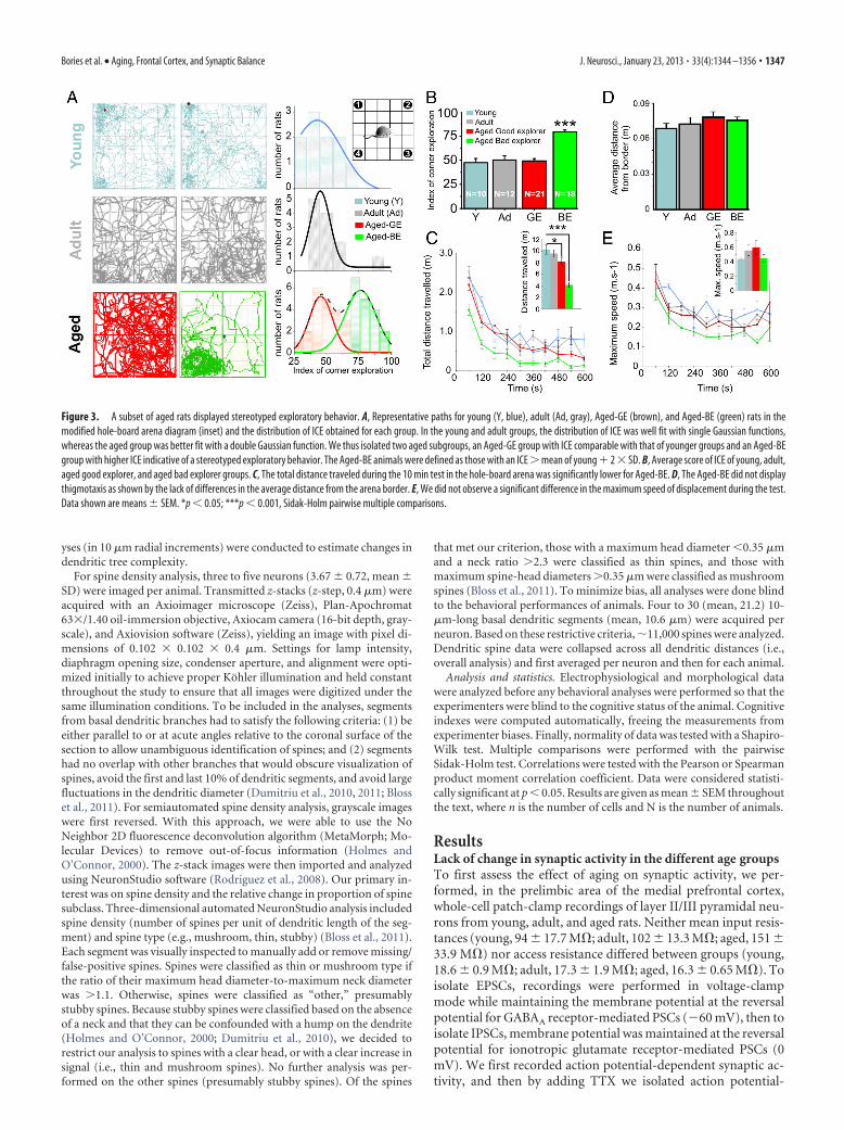
Figure 3. A subset of aged rats displayed stereotyped exploratory behavior. A, Representative paths for young (Y, blue), adult (Ad, gray), Aged-GE (brown), and Aged-BE (green) rats in themodified hole-board arena diagram (inset) and the distribution of ICE obtained for each group. In the young and adult groups, the distribution of ICE was well fit with single Gaussian functions,whereas the aged group was better fit with a double Gaussian function. We thus isolated two aged subgroups, an Aged-GE group with ICE comparable with that of younger groups and an Aged-BEgroup with higher ICE indicative of a stereotyped exploratory behavior. The Aged-BE animals were defined as those with an ICE � mean of young 2 � SD. B, Average score of ICE of young, adult,aged good explorer, and aged bad explorer groups. C, The total distance traveled during the 10 min test in the hole-board arena was significantly lower for Aged-BE. D, The Aged-BE did not displaythigmotaxis as shown by the lack of differences in the average distance from the arena border. E, We did not observe a significant difference in the maximum speed of displacement during the test.Data shown are means � SEM. *p � 0.05; ***p � 0.001, Sidak-Holm pairwise multiple comparisons.
Bories et al. • Aging, Frontal Cortex, and Synaptic Balance J. Neurosci., January 23, 2013 • 33(4):1344 –1356 • 1347

independent spontaneous synapticactivity. As a group, aged rats did not dif-fer from other groups regarding the meanamplitude or frequency of sEPSCs, sIPSCs(Fig. 1A,B), and mEPSCs or mIPSCs (Fig.2A,B). These results demonstrate so farthat, when taken as groups, the differentage cohorts showed a comparable level ofsynaptic activity impinging onto layer II/III pyramidal neurons in the prefrontalcortex.
A subset of aged rat displayedstereotyped exploratory behaviorAs described in several studies, the agingprocess leads to heterogeneous behavioraland cognitive declines in the aged popula-tion with major individual differences, al-lowing segregation of animals into agedimpaired versus unimpaired individuals(Fischer et al., 1987; Gage et al., 1988,1989). To take into account such factors, aseries of exploratory and novelty-basedbehavioral tests were also conducted on asubset of animals before electrophysiolog-ical recordings.
To quantify their locomotor perfor-mance and directed exploratory behavior,animals were tested in a mHB apparatus(see Materials and Methods for details),with a hole inserted in the floor of eachcorner. To minimize stress induced dur-ing experiments, all tests were conductedunder red-light illumination during thedark phase of the light/dark cycle. This was done to reduce theanxiogenic aspect of the procedure, allowing us to study locomo-tor and exploratory behaviors of the animals independently ofemotional factors. Surprisingly, the analysis of animal displace-ments revealed two main types of exploratory behavior in theaged population. Some aged rats showed excessive exploration inone corner, i.e., an increase in the number of head entries, verticalexploration and hole exploration in the corner zone, conse-quently neglecting the other corners, characteristic of a stereo-typed exploratory behavior. However, this behavior was notgeneralized to the entire aged population, as some aged rats ex-plored the new environment in a more equilibrated way such asdid the young and adult groups (Fig. 3A). We created an index ofcorner exploration to quantify the pattern of exploration for eachrat. The relative exploration for each corner was established bydividing the number of head entries by the sum of head entries inthe four corners. For each rat, the corner with the highest relativeexploration index (preferential corner) was chosen and labeled asthe ICE, theoretically ranging from 25% (no preferential corner)to 100% (only one corner explored).
The distributions of the ICE of young and adult groups werewell fitted with a single Gaussian function, whereas the distribu-tion of ICE for the aged group was best fitted with a doubleGaussian function (Fig. 3A). Based on their ICE, two populationsof animals could thus be distinguished in the aged group: (1) anaged group, labeled as the aged good-explorer group (Aged-GE),displayed a directed exploratory behavior similar to that of theyoung and adult groups; and (2) aged animals with a significantlygreater ICE, labeled as the aged bad-explorer group (Aged-BE),
henceforth defined as having an ICE deviating by �2 SD abovethe peak of the distribution observed in the young group. Conse-quently, the Aged-BE group displayed a significant alteration intheir exploratory behavior (Fig. 3B).
The total distance traveled was not significantly different be-tween adults and Aged-GE rats but was significantly lower forAged-BE rats, consistent with the development of stereotypedexploratory behavior, where the animals preferentially explorethe same hole (Fig. 3C). This phenomenon was not explained byan increase in anxiety or thigmotaxis, as demonstrated by the lackof significant difference between the four groups in the averagedistance traveled from the apparatus border (Fig. 3D) (Simon etal., 1994; Hines et al., 2008). It was also not explained by reducedlocomotion performance, as all groups reached similar maxi-mum speed of displacement during the test (Fig. 3E).
A subset of aged rats displayed frontal-relatedmemory deficitsGiven the role of the frontal cortex in both directed exploratorybehavior and novelty-based, nonspatial memory (Apergis-Schoute et al., 2006), animals were assessed using a NOR test at1 h retention delay (test phase). This paradigm is known to bedependent on medial prefrontal cortex integrity (Akirav and Ma-roun, 2006). At the sample phase of the NOR test, no differencewas observed between the young, adult, and aged groups, con-firming that there was no bias in preference for one side of theapparatus. The total time of exploration was also not statisticallydifferent between the three groups at the sample phase (p �0.12), ruling out any differences in motivation. In contrast, aftera 1 h delay, the aged group showed significantly poorer perfor-
Figure 4. Novelty-based memory deficits occur in a subgroup of aged rats. A, Mnemonic performances were assessed by a NORtest. Data are presented as the ratio of time spent exploring the new object at 1 h (NOR index). B, During the training phase (samplephase), the rats explored to the same extent the two identical objects. At 1 h, the aged group explored less the new objectcompared with young rats, confirming a disruption in the memorization process. C, Aged animals explored for a significantlyshorter time the two objects at 1 h but for the same duration during the sample phase. D, E, We isolated as Aged-BL (BL) animalswith a NOR index lower than that of the young group (Y, blue dotted line) by 2 � SD (purple dotted line). F, The aged groupexplored less the objects; however, there was no significant difference between the Aged-GL (GL) and Aged-BL (BL) in total time ofexploration. **p � 0.01; ***p � 0.001, Sidak-Holm pairwise multiple comparisons.
1348 • J. Neurosci., January 23, 2013 • 33(4):1344 –1356 Bories et al. • Aging, Frontal Cortex, and Synaptic Balance

mance than the young and adult groups (Fig. 4 B). We alsofound a decrease in the total exploration time for aged ratsduring the test phase when compared with the young andadult groups (Fig. 4C).
Following similar logic than for themHB analysis, the aged group was dividedinto two subgroups of rats based on theirrespective performance in the NOR task.To this end, the aged group was dividedinto two subgroups: a “good learner”(Aged-GL) subgroup, with a NOR indexcomparable to that in young and adultrats, and a “bad learner” (Aged-BL)group, defined as having a NOR index de-viating by � 2 SD above the mean valueobtained in the young group (Fig. 4D–F).
The lower total exploration time foundin the aged group (Fig. 4C) may reflectpoorer motivation in this age group.Thus, the lower NOR index in this groupmay simply be a consequence of this lowerlevel of motivation. To control for this, wecompared the exploration index in thetwo subgroups of aged animals (Aged-GLand Aged-BL). The total exploration timeat 1 h was not significantly different be-tween the Aged-GL and Aged-BL groups,indicating that the lower NOR index inthe Aged-BL reflects poorer memory per-formance and not decreased motivation(Fig. 4F).
Stratification of the aged groupsunmasks a correlation betweenprefrontal synaptic activity andexploratory behavioral performanceWe next wanted to investigate whetherthese behavioral changes echo changes atthe synaptic level.
Next we analyzed the synaptic activityafter stratification of the aged group intoAged-GE and Aged-BE based on the per-formance of animals tested in the mHBtask (Figs. 5, 6). Whereas no majorchanges were observed regarding actionpotential-dependent synaptic activity(Fig. 5), our analysis revealed a significantincrease in mean mEPSC frequency forthe Aged-GE group (Fig. 6B) when com-pared with the young and Aged-BEgroups. In contrast, the Aged-BE dis-played a drastic increased in mean mIPSCfrequency (Fig. 6C; p � 0.001). The meanamplitude of events was not changed (Fig.6B,C). Plotting the frequency of synap-tic events against the performance in themHB for each rat revealed a significantcorrelation between ICE and mEPSCfrequency within the aged group as wellas between ICE and mIPSC frequencyacross age groups and within the agedgroup (Fig. 6 D, E). From our data, suchchanges seemed to be independent from
changes in neuronal integrative properties; indeed, there wasno correlation between neuronal input resistance and synapticactivity or behavioral performance, neither across age norwithin the aged group (p � 0.05, one-way ANOVA).
Figure 5. Exploratory behavior is not associated with spontaneous synaptic activity. A, Representative traces of sEPSCs andsIPSCs recorded in layer 2/3 prefrontal pyramidal neurons of Aged-GE (red) and Aged-BE (green) rats. B, There were no changes insEPSC frequency in layer 2/3 pyramidal neurons from the Aged-GE group when compared with the young (Y) and Aged-BE groups.There was also no change in mean sEPSC amplitude (N � 6 –10 rats per group). C, No changes in sIPSC frequency in Aged-BE ratscompared with the other groups. There was also no change in mean sIPSC amplitude (N � 6 –9 rats per group). D, To evaluate therelationship between excitatory synaptic activity and behavioral performance, the ICE of each rat was plotted against the sEPSCfrequency. No significant correlation could be established across the four age groups or within the aged group. E, No relationshipbetween the ICE and the sIPSC frequency across the four groups or within the aged group. Data shown are means � SEM;Sidak-Holm pairwise multiple comparisons.
Bories et al. • Aging, Frontal Cortex, and Synaptic Balance J. Neurosci., January 23, 2013 • 33(4):1344 –1356 • 1349

Aged rats with novelty-based memoryimpairments also display an imbalancein prefrontal synaptic activity towardinhibitionIn line with the above results, stratificationof aged animals into good and bad learnersrevealed an inverse correlation betweenmnemonic performance and inhibitorysynaptic activity impinging onto prefrontalpyramidal neurons (Figs. 7, 8). Again, al-though no changes in action potential-dependent synaptic activity were seen (Fig.7), we found that Aged-BL at 1 h displayed adrastic enhancement in GABAA input activ-ity compared with Aged-GL and other agegroups (Fig. 8C). In addition, we found, foreach rat, a significant correlation betweenmIPSC frequency and NOR index at 1 h(Fig. 8E) independent of changes in theirmean amplitude (Fig. 8B,C). On the otherhand, no correlation with mEPSC fre-quency could be established (Fig. 8C,D).These results also suggest an associationbetween the balance of inhibitory-to-excitatory synaptic activity in the prefrontalcortex and novelty-based, nonspatial mem-ory performance in aged animals.
Age-dependent changes in dendriticspine densityChanges in the geometry of dendritic treeand spine density are important correlatesof aging and cognitive deficits (Luebke andRosene, 2003; Dumitriu et al., 2010; Luebkeet al., 2010; Hara et al., 2012). Therefore, wetested whether any modification in the den-dritic tree could underlie changes in the syn-aptic activity and could thus contribute tothe distinct behavioral trajectories occur-ring in the aged group. To achieve this, weperformed single-cell intracellular labelingwith Neurobiotin during patch-clamp re-cording and single-cell Lucifer yellow injec-tion in freshly fixed brain slices (Buhl andLubke, 1989; Wong et al., 2000; Wong et al.,2006; Cordero-Erausquin et al., 2009;Labrakakis et al., 2009). A total of 56 neu-rons, with typical morphological features ofpyramidal neurons, from 15 animals(young, n � 25 cells from 7 rats; aged, n �31 cells from 8 rats; Fig. 9A) were analyzed.The morphometric analyses did not revealsignificant correlations between the lengthof dendrite and the performance in thenovel object recognition task or the explor-atory behavior (Fig. 9B).
We next focused on the evolution ofspine density (Fig. 9C) and its role in age-dependent cognitive impairments. Con-sistent with previous reports (Peters et al.,2008; Dumitriu et al., 2010; Luebke et al.,2010; Bloss et al., 2011; Hara et al., 2012)in nonhuman primate as well as in rodent
Figure 6. Exploratory behavior is associated with specific change in the balance of action potential-independent synaptictransmission in pyramidal neurons of the medial prefrontal cortex. A, Representative traces of mIPSC and mEPSC recorded in layer2/3 prefrontal pyramidal neurons of Aged-GE (red) and Aged-BE (green) rats. B, We observed a significant increase in mEPSCfrequency in layer 2/3 pyramidal neurons from the Aged-GE group when compared with the young (Y) and Aged-BE groups. Therewas no change in mean mEPSC amplitude (N � 4 – 8 rats per group). C, On the other hand, our analysis revealed a significantincrease in mIPSC frequency in Aged-BE rats compared with the other groups. There was no change in mean mIPSC amplitude (N�6 –9 rats per group). D, To evaluate the relationship between excitatory synaptic activity and behavioral performance, the ICE ofeach rat was plotted against the mEPSC frequency. No significant correlation could be established across the four age groups;however, we observed a significant correlation within the aged group ( p � 0.05). E, A linear positive correlation was establishedbetween the ICE and the mIPSC frequency across the four groups (gray text; p � 0.001; r � 0.63) as well as within the aged group(red text and red line; p � 0.05; r � 0.49). Data shown are means � SEM. *p � 0.05; ***p � 0.001, Sidak-Holm pairwisemultiple comparisons.
1350 • J. Neurosci., January 23, 2013 • 33(4):1344 –1356 Bories et al. • Aging, Frontal Cortex, and Synaptic Balance

prefrontal cortex, we found an age-related significant reductionin spine density in layer II/III pyramidal neurons (34% decreasefrom 0.94 � 0.17 spines/�m in the young group to 0.62 � 0.12 inthe aged group, mean � SD; p � 0.001, Student’s t test; Fig. 9D).As reported previously (Dumitriu et al., 2010), this change insynaptic density was mainly driven by a loss of thin spines (40%
decrease from 0.62 � 0.10 in the younggroup to 0.37 � 0.06 in the aged group;p � 0.001, Student’s t test). In contrast,there was no significant age-relatedchange in mushroom spine density(21.8% decrease from 0.32 � 0.07 in theyoung group to 0.25 � 0.06 in the agedgroup; p � 0.09, Student’s t test).
None of the changes in total spine den-sity observed were correlated with the be-havioral performance (neither across allgroups of age nor within the aged group;data not shown). Yet recent reports indicatethat specific depletion of thin spines appearto be a major feature of aging (Dumitriu etal., 2010). We thus evaluated the respectivedensity of thin and mushroom spines foreach cell and collapsed for each rat. A signif-icant correlation was found across all groupsof age between thin spine density and mem-ory performances (Fig. 9E). However, noneof these relationships could be drawn withinthe aged group. There was no relationshipbetween mushroom spine density and cog-nitive performance (Fig. 9F). Together,these results rule out the idea that morpho-logical postsynaptic changes are sufficient toexplain the enhanced excitatory synaptic ac-tivity observed in the Aged-GE group.
Stereotyped exploratory behavior andnovelty-based memory deficitsare correlatedThe above findings implicate an enhance-ment in inhibitory synaptic activity imping-ing onto prefrontal pyramidal neurons as asubstrate of the cognitive deficits observedin a subgroup of aged animals. This infer-ence is consistent with the finding that theNOR index was correlated with the ICEacross all age groups (Fig. 10).
DiscussionThe present study reports three major find-ings. First, besides memory deficits, a subsetof aged rats display stereotyped exploratorybehavior strongly related to the balance insynaptic tone in medial prefrontal cortex.Second, an increase in inhibitory synaptictone is a common substrate of multipleaging-related cognitive declines. Third,most of the correlates of behavioral perfor-mance within the age group seem to be pre-synaptic.
Aged rats with exploratory or memorydeficits displayed an imbalance in back-ground synaptic activity mainly driven byan increase in GABAA receptor-mediated
miniature activity. A similar age-related increase in inhibitorytone has been evidenced in studies of rodent and nonhumanprimate prefrontal cortex (Abdulla et al., 1995; Griffith andMurchison, 1995; Luebke et al., 2004; Dickstein et al., 2007) Nev-ertheless, none of these studies was able to establish a direct cor-
Figure 7. Memory deficits are not correlated with spontaneous synaptic activity. A, Representative traces of sEPSC and sIPSCrecorded in layer II/III prefrontal pyramidal neurons of Aged-GL (red) and Aged-BL (purple) rats. B, No change in sEPSC frequencyor amplitude across age groups (N � 6 –12 rats per group). C, No changes in sIPSC frequency or amplitude was observed across agegroups (N � 6 –11 rats per group). Y, Young; Ad, adult. D and E, No correlation between excitatory or inhibitory synaptic activityand the mnemonic performance at 1 h could be established across the four age groups or within the aged group. Sidak-Holmpairwise multiple comparisons were conducted.
Bories et al. • Aging, Frontal Cortex, and Synaptic Balance J. Neurosci., January 23, 2013 • 33(4):1344 –1356 • 1351

relation between these synaptic changesand the degree of age-related, frontal-dependent cognitive impairment. Suchdiscrepancies may rely on both speciesdifferences between rodent and primates(Luebke et al., 2010; Morrison and Baxter,2012) and some experimental design dif-ferences (e.g., K- vs Cs-based solu-tion), which may affect detection of distalsynaptic events. Besides these studies inthe frontal cortex, recent reports showedthat changes in inhibitory synapse physi-ology or integrity in temporal struc-tures could underlie some age-dependentmemory deficits (Wong et al., 2006; Riss-man et al., 2007; Majdi et al., 2009). Assuch, reducing inhibitory GABAA func-tion by pharmacological intervention hasbeen shown to improve learning ability inaging rodents (Fernandez et al., 2007; Yo-shiike et al., 2008). On the other hand,infusion of GABA within the frontal cor-tex has been shown to be detrimental fornormal learning and memory (Meneses etal., 1993; Kleschevnikov et al., 2004; Fer-nandez et al., 2007), thus reinforcing thecrucial role of this cortical area in severalmemory-dependent tasks.
Interestingly, we observed an increasein AMPA receptor-mediated basal synap-tic activity in aged rats with preserved ex-ploratory behavior. Consistent with thisobservation, several studies reported thatpharmacological activation of AMPA re-ceptor induces cognitive performance en-hancements in frontal-dependent tasks,such as the delayed nonmatch to sampletask, the Y-maze test (Hampson et al.,1998; Bloss et al., 2008), object recogni-tion (Lebrun et al., 2000), or contextualserial discrimination tasks (Beracochea etal., 2007). The increase in glutamatergictone that we observed in successfully agedanimals (i.e., without stereotyped explor-atory behavior) is also in line with a recentreport demonstrating overactivation ofglutamatergic-dependent pathways insuccessful cognitive aging (Menard andQuirion, 2012). Moreover, several studiesin human reported greater activation infrontal cortex related to better cognitiveperformance among older participantscompared with younger individuals, thussupporting the notion of increased brainactivity in successful aging (for review, seeEyler et al., 2011). Nevertheless, nochange in excitatory tone was observed inaged good-learner animals. Such discrep-ancy with the enhanced excitatory activityrecorded in aged good explorers may relate to the behavioraldomain investigated. Indeed, stereotyped behaviors, as quanti-fied in the present study, have remained virtually not investigatedin studies of aging animals (Lalonde and Badescu, 1995; Lalonde
and Strazielle, 2009). In contrast, these behaviors have been in-vestigated extensively in animal models of neuropsychiatric dis-orders (Hines et al., 2008; Kocerha et al., 2009; Chao et al., 2010).Several lines of evidence point to an imbalance in excitatory/
Figure8. Memorydeficitsaresignificantlycorrelatedwithanimbalanceinsynapticactivitytowardinhibition.A,Representativetracesof mIPSC and mEPSC recorded in layer II/III prefrontal pyramidal neurons of Aged-GL (red) and Aged-BL (purple) rats. B, No change in themean excitatory synaptic frequency or amplitude across age groups (N � 4 – 8 rats per group). C, In contrast, a significant increase inmIPSC frequency was observed in the Aged-BL group (N�6 –10 rats per group. Y, Young; Ad, adult. D, No correlation between excitatorysynaptic activity and memory performances. E, A significant correlation between the inhibitory synaptic activity and the mnemonicperformance at 1 h could be established across the four age groups ( p �0.05; r ��0.38) as well as within the aged group ( p �0.05;r ��0.52). Data shown are means � SEM. ***p � 0.001, Sidak-Holm pairwise multiple comparisons.
1352 • J. Neurosci., January 23, 2013 • 33(4):1344 –1356 Bories et al. • Aging, Frontal Cortex, and Synaptic Balance

Figure 9. Age-dependent changes in dendritic spine density. A, Three examples of pyramidal neurons from layer II/III drawn after injection of Lucifer yellow and followed by anti-Lucifer yellowimmunodetection conjugated with DAB-Ni-based reaction. B, No correlation between the dendritic length and behavioral performance. C, Illustration of the methods used to analyze spine densityof layer II/III pyramidal neurons: the grayscale image scale was reversed and deconvolved, and a semiautomated analysis of dendritic spine density was performed, (Figure legend continues.)
Bories et al. • Aging, Frontal Cortex, and Synaptic Balance J. Neurosci., January 23, 2013 • 33(4):1344 –1356 • 1353

inhibitory E/I inputs in prefrontal cortexas a major contributor to psychiatric dis-orders in humans (for review, see Lewis etal., 2012) as well as abnormal stereotypicbehaviors in animal models (Dani et al.,2005; Lise and El-Husseini, 2006; Hines etal., 2008; Chao et al., 2010). Consistentwith this, we found an imbalance in syn-aptic activity similar to that observed inanimal models of neuropsychiatric dis-ease with similar behavioral abnormalitiesto those observed in our study (Lise andEl-Husseini, 2006; Keith and El-Husseini,2008; Sudhof, 2008). Moreover, it hasbeen shown that stereotyped behaviorthat result from drug sensitization canbe blocked by GABAA antagonists or in-duced by GABAA agonists (Karler et al.,1995, 1997), thus reinforcing a link be-tween alterations in GABAA transmis-sion in the frontal cortex andappearance of stereotyped behaviors.Together, our findings substantiate theidea that during aging, differential directions in basal synapticE/I ratio underlie differential cognitive trajectories. As such,whereas an imbalance toward inhibitory tone is detrimental,an imbalance toward excitatory tone may compensate for anage-dependent spine loss and thus preserve optimal explor-atory behavior.
Our morphological analyses combined with electrophysiolog-ical recordings also demonstrate that despite an age-dependentloss in dendritic spines, there was no significant decrease in thefrequency of mEPSCs. On the contrary, we found an increase inmEPSC frequency in cognitively preserved aged rats; thus, a lossof synapses did not necessarily translate into observable func-tional deficits. Indeed, considering that most of the lost spines arethin spines (Dumitriu et al., 2010; Bloss et al., 2011), i.e., likelysilent or weakly contributing to synaptic inputs (Kasai et al.,2003), their loss may not be reflected in significant changes re-corded at the levels of the soma. Yet, our results may also indicatethat presynaptic mechanisms compensate for the loss of postsyn-aptic structure, maintaining synaptic homeostasis (Paradis et al.,2001; Turrigiano and Nelson, 2004).
In contrast to the changes observed in mPSC frequency, nochange in sEPSC or sIPSC frequency was found in slices fromaged rats. Of particular interest, however, is the finding that thefrequency of mIPSC in the aged cognitively impaired groups wascomparable to that of sIPSCs. This indicates a collapse in theaction potential-dependent (or activity-driven) inhibitory tone(i.e., difference between sIPSC and mIPSC frequency) concurrentwith the enhanced basal, action potential-independent inhibitorytone. This results in a loss of dynamic range along which inhibi-tion can be modulated combined with an increase in synapticnoise, specifically in the aged, cognitively impaired groups. Thenet effect may be an impaired capability to adjust network excit-
ability by modulating inhibitory neuron activity, which could bea significant substrate of cognitive impairment. Indeed, thesignal-to-noise ratio is an important feature of information pro-cessing and deficiency in neuromodulation is suspected to resultin unstable cortical representation in aging frontal cortex (Li etal., 2001; Bories et al., 2012). The same phenomenon is not truefor the enhanced frequency of mEPSC observed in the aged goodexplorer group, because in the latter case, a wide dynamic rangeremained: that is, the frequency of sEPSCs was still threefoldlarger than that of mEPSCs. The enhanced excitatory tone maystill be beneficial in contrast to the enhanced inhibitory tone.These observations call for additional modeling studies to ex-plore the role of excitatory versus inhibitory synaptic activity instabilizing cortical network activity in aging.
While a number of studies have examined age-related mem-ory deficits (Rosenzweig and Barnes, 2003; Burke and Barnes,2006; Disterhoft and Oh, 2007), few studies have focused onother cognitive deficits associated with aging, such as psychiatric-like symptoms. Consequently, the neurobiological substrates ofsuch age-related abnormalities have never been clearly identified.We thus focused on this age-related behavioral alteration, giventhat this could represent part of more complex behavioral alter-ations appearing in senile dementia. Our findings of similaritiesbetween abnormal behavior and changes in E/I ratio displayed bya subset of aged rats and those observed in models of psychiatricdisorders thus warrant future investigations into psychiatricsymptoms associated with aging in animals. Our results alsocall for additional studies on presynaptic release of GABAfrom cortical interneuron in cognitively characterized agedanimals to elucidate the biological mechanisms underlying theage-dependent increase in inhibitory tone. Finally, severalstudies have reported an imbalance in synaptic activity in an-imal models of Alzheimer’s disease at an earlier time pointthan those studied here (Palop et al., 2007; Sun et al., 2009;Palop and Mucke, 2010; Verret et al., 2012). It thus remains tobe investigated whether or not the latter phenotype of Alzheimer’sdisease represents an early manifestation of the imbalance in synap-tic activity we observed in our model of dementia associated withnormal aging and whether or not they share a common feature at thesynaptic level.
4
(Figure legend continued.) yielding a separation of thin and mushroom spines. The otherspines, presumably stubby spines, were discarded from analyses. D, Significant age-dependentspine loss. E, Scatter plots of thin spine densities versus exploratory or novel object recognitionperformance. The thin spine density appears to be predictive of behavioral performance acrossall groups of age but not within the aged group. F, No relation between mushroom spinesdensity and behavioral performance. **p � 0.01 Student’s t test.
Figure 10. Exploratory behavior correlates with memory performance and inhibitory synaptic activity. A, We found a significantlinear correlation between the performances of the animals in the NOR test (NOR index) and the modified hole-board arena test(ICE) (red line; p � 0.01; r � �0.38). The black dotted lines represent the cutoff limits between the impaired and unimpairedstatus for each test. The majority of animals displayed optimal behavioral performance (yellow region), whereas a subset of agedanimals was impaired on both tests (gray region) B, Contour map where frequency (in hertz) of mIPSCs is plotted using colorcoding, superimposed on the values plotted in A. Animals with the most severe behavioral impairments also displayed an en-hanced GABAA receptor-mediated synaptic activity (bottom right quadrant), whereas, at the opposite end, animals with a lowerinhibitory synaptic activity displayed the best performance during the behavioral tests (top left quadrant).
1354 • J. Neurosci., January 23, 2013 • 33(4):1344 –1356 Bories et al. • Aging, Frontal Cortex, and Synaptic Balance

ReferencesAbdulla FA, Abu-Bakra MA, Calaminici MR, Stephenson JD, Sinden JD
(1995) Importance of forebrain cholinergic and GABAergic systems tothe age-related deficits in water maze performance of rats. NeurobiolAging 16:41–52. Medline
Akirav I, Maroun M (2006) Ventromedial prefrontal cortex is obligatory forconsolidation and reconsolidation of object recognition memory. CerebCortex 16:1759 –1765. CrossRef Medline
Apergis-Schoute J, Pinto A, Pare D (2006) Ultrastructural organization ofmedial prefrontal inputs to the rhinal cortices. Eur J Neurosci 24:135–144.Medline
Beracochea D, Philippin JN, Meunier S, Morain P, Bernard K (2007) Im-provement of episodic contextual memory by S 18986 in middle-aged mice: comparison with donepezil. Psychopharmacology (Berl) 193:63–73. CrossRef
Bevins RA, Besheer J (2006) Object recognition in rats and mice: a one-trialnon-matching-to-sample learning task to study ‘recognition memory. ’Nat Protoc 1:1306 –1311. CrossRef
Bloss EB, Hunter RG, Waters EM, Munoz C, Bernard K, McEwen BS (2008)Behavioral and biological effects of chronic S18986, a positive AMPAreceptor modulator, during aging. Exp Neurol 210:109 –117. CrossRefMedline
Bloss EB, Janssen WG, Ohm DT, Yuk FJ, Wadsworth S, Saardi KM, McEwenBS, Morrison JH (2011) Evidence for reduced experience-dependentdendritic spine plasticity in the aging prefrontal cortex. J Neurosci 31:7831–7839. CrossRef Medline
Bories C, Guitton MJ, Julien C, Tremblay C, Vandal M, Msaid M, De KoninckY, Calon F (2012) Sex-dependent alterations in social behaviour andcortical synaptic activity coincide at different ages in a model of Alzhei-mer’s disease. PLoS One 7:e46111. CrossRef Medline
Buhl EH, Lubke J (1989) Intracellular Lucifer yellow injection in fixed brainslices combined with retrograde tracing, light and electron microscopy.Neuroscience 28:3–16. Medline
Burke SN, Barnes CA (2006) Neural plasticity in the ageing brain. Nat RevNeurosci 7:30 – 40. CrossRef Medline
Burke SN, Barnes CA (2010) Senescent synapses and hippocampal circuitdynamics. Trends Neurosci 33:153–161. CrossRef Medline
Chao HT, Chen H, Samaco RC, Xue M, Chahrour M, Yoo J, Neul JL, Gong S,Lu HC, Heintz N, Ekker M, Rubenstein JL, Noebels JL, Rosenmund C,Zoghbi HY (2010) Dysfunction in GABA signalling mediates autism-like stereotypies and Rett syndrome phenotypes. Nature 468:263–269.CrossRef Medline
Clements JD, Bekkers JM (1997) Detection of spontaneous synaptic eventswith an optimally scaled template. Biophys J 73:220 –229. CrossRefMedline
Cordero-Erausquin M, Allard S, Dolique T, Bachand K, Ribeiro-da-Silva A,De KoninckY (2009) Dorsal horn neurons presynaptic to lamina I spi-noparabrachial neurons revealed by transynaptic labeling. J Comp Neurol517:601– 615. CrossRef Medline
Cummings JL (1995) Anatomic and behavioral aspects of frontal-subcortical circuits. Ann N Y Acad Sci 769:1–13.
Dani VS, Chang Q, Maffei A, Turrigiano GG, Jaenisch R, Nelson SB (2005)Reduced cortical activity due to a shift in the balance between excitationand inhibition in a mouse model of Rett syndrome. Proc Natl Acad SciU S A 102:12560 –12565. CrossRef Medline
Dickstein DL, Kabaso D, Rocher AB, Luebke JI, Wearne SL, Hof PR (2007)Changes in the structural complexity of the aged brain. Aging Cell 6:275–284. CrossRef Medline
Disterhoft JF, Oh MM (2007) Alterations in intrinsic neuronal excitabilityduring normal aging. Aging Cell 6:327–336. CrossRef Medline
Dumitriu D, Hao J, Hara Y, Kaufmann J, Janssen WG, Lou W, Rapp PR,Morrison JH (2010) Selective changes in thin spine density and mor-phology in monkey prefrontal cortex correlate with aging-related cogni-tive impairment. J Neurosci 30:7507–7515. CrossRef Medline
Dumitriu D, Rodriguez A, Morrison JH (2011) High-throughput, detailed,cell-specific neuroanatomy of dendritic spines using microinjection andconfocal microscopy. Nat Protoc 6:1391–1411. CrossRef Medline
Eyler LT, Sherzai A, Kaup AR, Jeste DV (2011) A review of functional brainimaging correlates of successful cognitive aging. Biol Psychiatry 70:115–122. CrossRef Medline
Fernandez F, Morishita W, Zuniga E, Nguyen J, Blank M, Malenka RC, Gar-
ner CC (2007) Pharmacotherapy for cognitive impairment in a mousemodel of Down syndrome. Nat Neurosci 10:411– 413. CrossRef Medline
File SE, Wardill AG (1975a) The reliability of the hole-board apparatus.Psychopharmacologia 44:47–51. Medline
File SE, Wardill AG (1975b) Validity of head-dipping as a measure of explo-ration in a modified hole-board. Psychopharmacologia 44:53–59.CrossRef Medline
Fischer W, Wictorin K, Bjorklund A, Williams LR, Varon S, Gage FH (1987)Amelioration of cholinergic neuron atrophy and spatial memory impair-ment in aged rats by nerve growth factor. Nature 329:65– 68. CrossRefMedline
Fuster JM (2000) Prefrontal neurons in networks of executive memory.Brain Res Bull 52:331–336. Medline
Fuster JM (2001) The prefrontal cortex–an update: time is of the essence.Neuron 30:319 –333. Medline
Gage FH, Chen KS, Buzsaki G, Armstrong D (1988) Experimental ap-proaches to age-related cognitive impairments. Neurobiol Aging 9:645–655. Medline
Gage FH, Dunnett SB, Bjorklund A (1989) Age-related impairments in spa-tial memory are independent of those in sensorimotor skills. NeurobiolAging 10:347–352. Medline
Griffith WH, Murchison DA (1995) Enhancement of GABA-activatedmembrane currents in aged Fischer 344 rat basal forebrain neurons.J Neurosci 15:2407–2416. Medline
Hampson RE, Rogers G, Lynch G, Deadwyler SA (1998) Facilitative effectsof the ampakine CX516 on short-term memory in rats: enhancement ofdelayed-nonmatch-to-sample performance. J Neurosci 18:2740 –2747.Medline
Hara Y, Rapp PR, Morrison JH (2012) Neuronal and morphologicalbases of cognitive decline in aged rhesus monkeys. Age (Dordr) 34:1051–1073. CrossRef
Hedden T, Gabrieli JD (2004) Insights into the ageing mind: a view fromcognitive neuroscience. Nat Rev Neurosci 5:87–96. CrossRef Medline
Hines RM, Wu L, Hines DJ, Steenland H, Mansour S, Dahlhaus R, SingarajaRR, Cao X, Sammler E, Hormuzdi SG, Zhuo M, El-Husseini A (2008)Synaptic imbalance, stereotypies, and impaired social interactions in micewith altered neuroligin 2 expression. J Neurosci 28:6055– 6067. CrossRef
Holmes TJ, O’Connor NJ (2000) Blind deconvolution of 3D transmittedlight brightfield micrographs. J Microsc 200:114 –127. CrossRef Medline
Karler R, Calder LD, Thai LH, Bedingfield JB (1995) The dopaminergic,glutamatergic, GABAergic bases for the action of amphetamine and co-caine. Brain Res 671:100 –104. CrossRef Medline
Karler R, Bedingfield JB, Thai DK, Calder LD (1997) The role of the frontalcortex in the mouse in behavioral sensitization to amphetamine. BrainRes 757:228 –235. CrossRef Medline
Kasai H, Matsuzaki M, Noguchi J, Yasumatsu N, Nakahara H (2003)Structure-stability-function relationships of dendritic spines. TrendsNeurosci 26:360 –368. CrossRef Medline
Keith D, El-Husseini A (2008) Excitation control: balancing PSD-95 func-tion at the synapse. Front Mol Neurosci 1:4. CrossRef Medline
Kleschevnikov AM, Belichenko PV, Villar AJ, Epstein CJ, Malenka RC, Mo-bley WC (2004) Hippocampal long-term potentiation suppressed by in-creased inhibition in the Ts65Dn mouse, a genetic model of Downsyndrome. J Neurosci 24:8153– 8160. CrossRef Medline
Kocerha J, Faghihi MA, Lopez-Toledano MA, Huang J, Ramsey AJ, CaronMG, Sales N, Willoughby D, Elmen J, Hansen HF, Orum H, Kauppinen S,Kenny PJ, Wahlestedt C (2009) MicroRNA-219 modulates NMDAreceptor-mediated neurobehavioral dysfunction. Proc Natl Acad SciU S A 106:3507–3512. CrossRef Medline
Labrakakis C, Lorenzo L-E, Bories C, Ribeiro-da-Silva A, De Koninck Y(2009) Inhibitory coupling between inhibitory interneurons in the spinalcord dorsal horn. Mol Pain 5:24. CrossRef Medline
Lalonde R, Badescu R (1995) Exploratory drive, frontal lobe function andadipsia in aging. Gerontology 41:134 –144. CrossRef Medline
Lalonde R, Strazielle C (2009) Exploratory activity and motor coordinationin old versus middle-aged C57BL/6J mice. Arch Gerontol Geriatr 49:39 – 42. CrossRef Medline
Lebrun C, PilliereE, Lestage P (2000) Effects of S 18986-1, a novel cognitiveenhancer, on memory performances in an object recognition task in rats.Eur J Pharmacol 401:205–212. CrossRef Medline
Lewis DA, Curley AA, Glausier JR, Volk DW (2012) Cortical parvalbumin
Bories et al. • Aging, Frontal Cortex, and Synaptic Balance J. Neurosci., January 23, 2013 • 33(4):1344 –1356 • 1355

interneurons and cognitive dysfunction in schizophrenia. Trends Neuro-sci 35:57– 67. CrossRef Medline
Li SC, Lindenberger U, Sikstrom S (2001) Aging cognition: from neuro-modulation to representation. Trends Cogn Sci 5:479 – 486. CrossRefMedline
LiseMF, El-Husseini A (2006) The neuroligin and neurexin families: fromstructure to function at the synapse. Cell Mol Life Sci 63:1833–1849.CrossRef Medline
Luebke J, Barbas H, Peters A (2010) Effects of normal aging on prefrontalarea 46 in the rhesus monkey. Brain Res Rev 62:212–232. CrossRefMedline
Luebke JI, Rosene DL (2003) Aging alters dendritic morphology, input re-sistance, and inhibitory signaling in dentate granule cells of the rhesusmonkey. J Comp Neurol 460:573–584. CrossRef Medline
Luebke JI, Chang YM, Moore TL, Rosene DL (2004) Normal aging results indecreased synaptic excitation and increased synaptic inhibition of layer2/3 pyramidal cells in the monkey prefrontal cortex. Neuroscience 125:277–288. CrossRef Medline
Lyketsos CG, Lopez O, Jones B, Fitzpatrick AL, Breitner J, DeKosky S (2002)Prevalence of neuropsychiatric symptoms in dementia and mild cognitiveimpairment: results from the cardiovascular health study. JAMA 288:1475–1483. CrossRef Medline
Majdi M, Ribeiro-da-Silva A, Cuello AC (2009) Variations in excitatory andinhibitory postsynaptic protein content in rat cerebral cortex with respectto aging and cognitive status. Neuroscience 159:896 –907. CrossRefMedline
Menard C, Quirion R (2012) Successful cognitive aging in rats: a role formGluR5 glutamate receptors, homer 1 proteins and downstream signal-ing pathways. PLoS One 7:e28666. CrossRef Medline
Meneses S, Galicia O, Brailowsky S (1993) Chronic infusions of GABA intothe medial prefrontal cortex induce spatial alternation deficits in agedrats. Behav Brain Res 57:1–7. Medline
Morrison JH, Baxter MG (2012) The ageing cortical synapse: hallmarks andimplications for cognitive decline. Nat Rev Neurosci 13:240 –250.CrossRef Medline
Palop JJ, Mucke L (2010) Amyloid-�-induced neuronal dysfunction in Alz-heimer’s disease: from synapses toward neural networks. Nat Neurosci13:812– 818. CrossRef Medline
Palop JJ, Chin J, Roberson ED, Wang J, Thwin MT, Bien-Ly N, Yoo J, Ho KO,Yu G-Q, Kreitzer A, Finkbeiner S, Noebels JL, Mucke L (2007) Aberrantexcitatory neuronal activity and compensatory remodeling of inhibitoryhippocampal circuits in mouse models of Alzheimer’s disease. Neuron55:697–711. CrossRef Medline
Paradis S, Sweeney ST, Davis GW (2001) Homeostatic control of presynap-tic release is triggered by postsynaptic membrane depolarization. Neuron30:737–749. CrossRef Medline
Persson J, Nyberg L, Lind J, Larsson A, Nilsson L-G, Ingvar M, Buckner RL(2006) Structure-function correlates of cognitive decline in aging. CerebCortex 16:907–915. Medline
Peters A, Sethares C, Luebke JI (2008) Synapses are lost during aging in theprimate prefrontal cortex. Neuroscience 152:970 –981. CrossRef Medline
Raji CA, Lopez OL, Kuller LH, Carmichael OT, Becker JT (2009) Age, Alz-heimer disease, and brain structure. Neurology 73:1899 –1905. CrossRefMedline
Rissman RA, De Blas AL, Armstrong DM (2007) GABA(A) receptors inaging and Alzheimer’s disease. J Neurochem 103:1285–1292. CrossRefMedline
Rodriguez A, Ehlenberger DB, Dickstein DL, Hof PR, Wearne SL (2008)Automated three-dimensional detection and shape classification of den-dritic spines from fluorescence microscopy images. PLoS One 3:e1997.CrossRef Medline
Rosenzweig ES, Barnes CA (2003) Impact of aging on hippocampal func-tion: plasticity, network dynamics, and cognition. Prog Neurobiol 69:143–179. CrossRef Medline
Simon P, Dupuis R, Costentin J (1994) Thigmotaxis as an index of anxietyin mice. Influence of dopaminergic transmissions. Behav Brain Res 61:59 – 64. CrossRef Medline
Sudhof TC (2008) Neuroligins and neurexins link synaptic function to cog-nitive disease. Nature 455:903–911. CrossRef Medline
Sun B, Halabisky B, Zhou Y, Palop JJ, Yu G, Mucke L, Gan L (2009) Imbal-ance between GABAergic and glutamatergic transmission impairs adultneurogenesis in an animal model of Alzheimer’s disease. Cell Stem Cell5:624 – 633. CrossRef Medline
Turrigiano GG, Nelson SB (2004) Homeostatic plasticity in the developingnervous system. Nat Rev Neurosci 5:97–107. CrossRef Medline
Verret L, Mann EO, Hang GB, Barth AM, Cobos I, Ho K, Devidze N, MasliahE, Kreitzer AC, Mody I, Mucke L, Palop JJ (2012) Inhibitory interneu-ron deficit links altered network activity and cognitive dysfunction inAlzheimer model. Cell 149:708 –721. CrossRef Medline
Wong TP, Marchese G, Casu MA, Ribeiro-da-Silva A, Cuello AC, De KoninckY (2000) Loss of presynaptic and postsynaptic structures is accompa-nied by compensatory increase in action potential-dependent synapticinput to layer V neocortical pyramidal neurons in aged rats. J Neurosci20:8596 – 8606. Medline
Wong TP, Marchese G, Casu MA, Ribeiro-da-Silva A, Cuello AC, De KoninckY (2006) Imbalance towards inhibition as a substrate of aging-associated cognitive impairment. Neurosci Lett 397:64 – 68. CrossRefMedline
Yoshiike Y, Kimura T, Yamashita S, Furudate H, Mizoroki T, Murayama M,Takashima A (2008) GABA(A) receptor-mediated acceleration ofaging-associated memory decline in APP/PS1 mice and its pharmacolog-ical treatment by picrotoxin. PLoS One 3:e3029. CrossRef Medline
1356 • J. Neurosci., January 23, 2013 • 33(4):1344 –1356 Bories et al. • Aging, Frontal Cortex, and Synaptic Balance
Copyright © 2022 FDOKUMEN