Different Factors Affecting Human ANP Amyloid Aggregation and Their Implications in Congestive Heart...
12
Different Factors Affecting Human ANP Amyloid Aggregation and Their Implications in Congestive Heart Failure Lia Millucci 1 , Eugenio Paccagnini 2 , Lorenzo Ghezzi 1 , Giulia Bernardini 1 , Daniela Braconi 1 , Marcella Laschi 1 , Marco Consumi 3 , Adriano Spreafico 4 , Piero Tanganelli 5 , Pietro Lupetti 2 , Agnese Magnani 3 , Annalisa Santucci 1 * 1 Dipartimento di Biotecnologie, Universita ` degli Studi di Siena, Siena, Italy, 2 Dipartimento di Biologia Evolutiva, Universita ` degli Studi di Siena, Siena, Italy, 3 Dipartimento Farmaco Chimico Tecnologico, Universita ` degli Studi di Siena, Siena, Italy, 4 Dipartimento di Medicina Clinica e Scienze Immunologiche, Universita ` degli Studi di Siena, Siena, Italy, 5 Dipartimento di Patologia Umana e Oncologia, Universita ` degli Studi di Siena, Siena, Italy Abstract Aims: Atrial Natriuretic Peptide (ANP)-containing amyloid is frequently found in the elderly heart. No data exist regarding ANP aggregation process and its link to pathologies. Our aims were: i) to experimentally prove the presumptive association of Congestive Heart Failure (CHF) and Isolated Atrial Amyloidosis (IAA); ii) to characterize ANP aggregation, thereby elucidating IAA implication in the CHF pathogenesis. Methods and Results: A significant prevalence (85%) of IAA was immunohistochemically proven ex vivo in biopsies from CHF patients. We investigated in vitro (using Congo Red, Thioflavin T, SDS-PAGE, transmission electron microscopy, infrared spectroscopy) ANP fibrillogenesis, starting from a-ANP as well as the ability of dimeric b-ANP to promote amyloid formation. Different conditions were adopted, including those reproducing b-ANP prevalence in CHF. Our results defined the uncommon rapidity of a-ANP self-assembly at acidic pH supporting the hypothesis that such aggregates constitute the onset of a fibrillization process subsequently proceeding at physiological pH. Interestingly, CHF-like conditions induced the production of the most stable and time-resistant ANP fibrils suggesting that CHF affected people may be prone to develop IAA. Conclusions: We established a link between IAA and CHF by ex vivo examination and assessed that b-ANP is, in vitro, the seed of ANP fibrils. Our results indicate that b-ANP plays a crucial role in ANP amyloid deposition under physiopathological CHF conditions. Overall, our findings indicate that early IAA-related ANP deposition may occur in CHF and suggest that these latter patients should be monitored for the development of cardiac amyloidosis. Citation: Millucci L, Paccagnini E, Ghezzi L, Bernardini G, Braconi D, et al. (2011) Different Factors Affecting Human ANP Amyloid Aggregation and Their Implications in Congestive Heart Failure. PLoS ONE 6(7): e21870. doi:10.1371/journal.pone.0021870 Editor: Valdur Saks, Universite ´ Joseph Fourier, France Received March 22, 2011; Accepted June 8, 2011; Published July 26, 2011 Copyright: ß 2011 Millucci et al. This is an open-access article distributed under the terms of the Creative Commons Attribution License, which permits unrestricted use, distribution, and reproduction in any medium, provided the original author and source are credited. Funding: This work was supported by Fondazione MPS 2009–2011, TLS Orphan 0108. There are no relationships with industry. The funders had no role in study design, data collection and analysis, decision to publish, or preparation of the manuscript. Competing Interests: The authors have declared that no competing interests exist. * E-mail: [email protected] Introduction IAA is an age-related amyloidosis [1] limited to the atria and ANP is the major subunit of amyloid fibrils [2,3]. ANP is synthesized by atrial cardiomyocytes [4,5] and regulates the hydro-mineral homeostasis under normal and pathological conditions. Although its clinical relevance is unclear, patients with IAA are known to be prone to atrial fibrillation [6,7]. Little is known about the putative role of IAA in the pathogenesis of cardiac arrhythmias in the elderly but a relationship between the incidence and the degree of IAA in cardiac disorders associated to high ANP plasma levels, and the incidence and the degree of IAA has been described, suggesting a prevalence of IAA in CHF [8– 10]. Plasma ANP levels are elevated in CHF in relation to its severity [4,11] as well as in IAA [12,13], due to an increased ANP secretion by the failing heart. Total circulating ANP is composed of three molecular forms [4]: a-ANP comprises 28 aminoacids with an intramolecular disulfide linkage; b-ANP (56 aminoacids), an antiparallel immer of a-ANP and c–ANP (pro-ANP), composed of 126 aminoacids, carring the a-ANP sequence and stored in secretory granules of atrial cardiocytes. The circulating peptide exists also as a high MW form, as we showed [14–17], this latter not taking part in the polymerization process [17]. On the contrary, an ANP oligomeric circulating form can be the actual promoter of amyloid aggregation. Increased ANP concentration in severe CHF (NYHA class III and class IV) is due to the rise of b-ANP [4]. In IAA non-mutated ANP is the aggregating sequence [2,3], but changes in the local environment or alteration of protein concentration are crucial for the onset of amyloidosis [18]. In this work, a significant prevalence of IAA was immunohis- tochemically proven in the atrial appendages of CHF patients PLoS ONE | www.plosone.org 1 July 2011 | Volume 6 | Issue 7 | e21870
-
Upload
independent -
Category
Documents
-
view
1 -
download
0
Transcript of Different Factors Affecting Human ANP Amyloid Aggregation and Their Implications in Congestive Heart...

Different Factors Affecting Human ANP AmyloidAggregation and Their Implications in Congestive HeartFailureLia Millucci1, Eugenio Paccagnini2, Lorenzo Ghezzi1, Giulia Bernardini1, Daniela Braconi1, Marcella
Laschi1, Marco Consumi3, Adriano Spreafico4, Piero Tanganelli5, Pietro Lupetti2, Agnese Magnani3,
Annalisa Santucci1*
1 Dipartimento di Biotecnologie, Universita degli Studi di Siena, Siena, Italy, 2 Dipartimento di Biologia Evolutiva, Universita degli Studi di Siena, Siena, Italy,
3 Dipartimento Farmaco Chimico Tecnologico, Universita degli Studi di Siena, Siena, Italy, 4 Dipartimento di Medicina Clinica e Scienze Immunologiche, Universita degli
Studi di Siena, Siena, Italy, 5 Dipartimento di Patologia Umana e Oncologia, Universita degli Studi di Siena, Siena, Italy
Abstract
Aims: Atrial Natriuretic Peptide (ANP)-containing amyloid is frequently found in the elderly heart. No data exist regardingANP aggregation process and its link to pathologies. Our aims were: i) to experimentally prove the presumptive associationof Congestive Heart Failure (CHF) and Isolated Atrial Amyloidosis (IAA); ii) to characterize ANP aggregation, therebyelucidating IAA implication in the CHF pathogenesis.
Methods and Results: A significant prevalence (85%) of IAA was immunohistochemically proven ex vivo in biopsies fromCHF patients. We investigated in vitro (using Congo Red, Thioflavin T, SDS-PAGE, transmission electron microscopy, infraredspectroscopy) ANP fibrillogenesis, starting from a-ANP as well as the ability of dimeric b-ANP to promote amyloid formation.Different conditions were adopted, including those reproducing b-ANP prevalence in CHF. Our results defined theuncommon rapidity of a-ANP self-assembly at acidic pH supporting the hypothesis that such aggregates constitute theonset of a fibrillization process subsequently proceeding at physiological pH. Interestingly, CHF-like conditions induced theproduction of the most stable and time-resistant ANP fibrils suggesting that CHF affected people may be prone to developIAA.
Conclusions: We established a link between IAA and CHF by ex vivo examination and assessed that b-ANP is, in vitro, theseed of ANP fibrils. Our results indicate that b-ANP plays a crucial role in ANP amyloid deposition under physiopathologicalCHF conditions. Overall, our findings indicate that early IAA-related ANP deposition may occur in CHF and suggest thatthese latter patients should be monitored for the development of cardiac amyloidosis.
Citation: Millucci L, Paccagnini E, Ghezzi L, Bernardini G, Braconi D, et al. (2011) Different Factors Affecting Human ANP Amyloid Aggregation and TheirImplications in Congestive Heart Failure. PLoS ONE 6(7): e21870. doi:10.1371/journal.pone.0021870
Editor: Valdur Saks, Universite Joseph Fourier, France
Received March 22, 2011; Accepted June 8, 2011; Published July 26, 2011
Copyright: � 2011 Millucci et al. This is an open-access article distributed under the terms of the Creative Commons Attribution License, which permitsunrestricted use, distribution, and reproduction in any medium, provided the original author and source are credited.
Funding: This work was supported by Fondazione MPS 2009–2011, TLS Orphan 0108. There are no relationships with industry. The funders had no role in studydesign, data collection and analysis, decision to publish, or preparation of the manuscript.
Competing Interests: The authors have declared that no competing interests exist.
* E-mail: [email protected]
Introduction
IAA is an age-related amyloidosis [1] limited to the atria and
ANP is the major subunit of amyloid fibrils [2,3]. ANP is
synthesized by atrial cardiomyocytes [4,5] and regulates the
hydro-mineral homeostasis under normal and pathological
conditions. Although its clinical relevance is unclear, patients with
IAA are known to be prone to atrial fibrillation [6,7]. Little is
known about the putative role of IAA in the pathogenesis of
cardiac arrhythmias in the elderly but a relationship between the
incidence and the degree of IAA in cardiac disorders associated to
high ANP plasma levels, and the incidence and the degree of IAA
has been described, suggesting a prevalence of IAA in CHF [8–
10]. Plasma ANP levels are elevated in CHF in relation to its
severity [4,11] as well as in IAA [12,13], due to an increased ANP
secretion by the failing heart.
Total circulating ANP is composed of three molecular forms
[4]: a-ANP comprises 28 aminoacids with an intramolecular
disulfide linkage; b-ANP (56 aminoacids), an antiparallel immer of
a-ANP and c–ANP (pro-ANP), composed of 126 aminoacids,
carring the a-ANP sequence and stored in secretory granules of
atrial cardiocytes. The circulating peptide exists also as a high MW
form, as we showed [14–17], this latter not taking part in the
polymerization process [17]. On the contrary, an ANP oligomeric
circulating form can be the actual promoter of amyloid
aggregation. Increased ANP concentration in severe CHF (NYHA
class III and class IV) is due to the rise of b-ANP [4].
In IAA non-mutated ANP is the aggregating sequence [2,3], but
changes in the local environment or alteration of protein
concentration are crucial for the onset of amyloidosis [18].
In this work, a significant prevalence of IAA was immunohis-
tochemically proven in the atrial appendages of CHF patients
PLoS ONE | www.plosone.org 1 July 2011 | Volume 6 | Issue 7 | e21870
showing an unequivocal relationship between these two patholo-
gies. Moreover, we investigated for the first time ANP fibrillogen-
esis, starting from its monomeric form (a-ANP) as well as the
ability of its dimeric form (b-ANP) to promote amyloid fibril
formation. Different conditions were adopted, including those
reproducing b-ANP prevalence in CHF [19]. Our results define
the uncommon rapidity of a-ANP self-assembly at acidic pH
supporting the hypothesis that these early developed aggregated
forms constitute the onset of a fibrillization process subsequently
proceeding at physiological pH. More interestingly, CHF-like
conditions induced the production of the most stable and time-
resistant ANP fibrils supporting the hypothesis that CHF affected
people may be prone to develop IAA.
The study of the conformational preferences of ANP under
different conditions appears to be crucial for shedding light on its
intrinsic structural properties related to amyloid fibril formation.
b-ANP can act as a seed for fibrillization in the heart and we
suggest a role for b-ANP in amyloid deposition, discussing its
pathophysiological significance in CHF and IAA.
Results
CR and anti-ANP immunohistochemistryIn the present stud we investigated the ANP tissue localization
in the atrium of 40 CHF patients by means of CR assay and
immunohistochemical analysis. Myocardial tissues (left atrial
appendages) were obtained from patients with CHF that
underwent surgery in the past 2 years (2009–2010). Biopsies
stored in the tissues archives of ‘‘Patologia Umana e Oncologia’’
Department of the University of Siena were selected on the basis of
pathologies and anonymously analyzed. For this reason in our
Institution a specific written Informed Consent is not required.
The detailed description of the whole study was presented and
approved by local University Hospital Ethics Committee, the
‘‘Comitato Etico Locale dell’Azienda Ospedaliera Universitaria
Senese’’. Patients with diabetes mellitus and/or hypertension other
than CHF were enrolled in the study. Table 1 summarizes
concomitant pathologies, age and sex characterizing each patient
who underwent biopsy. Light microscopy observations of the
sections were made after hematoxylin-eosin staining and mea-
surement of myocyte size (mean diameter 20–40 myocytes/
specimens); the degree of IAA deposition in the atrial wall was
graded subjectively on a scale of 0–3 and the highest grade from
the sites evaluated was taken as representative for each heart.
Paraffin sections were also stained with CR and immunostained
with the antibody to ANP as shown in Figure S11.
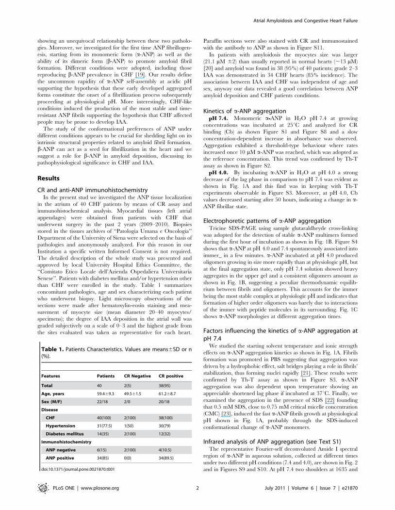
In patients with amyloidosis the myocytes size was larger
(21.1 mM 62) than usually reported in normal hearts (,13 mM)
[20] and amyloid was found in 38 (95%) of 40 patients; grade 2–3
IAA was demonstrated in 34 CHF hearts (85% incidence). The
association between IAA and CHF was independent of age and
sex, anyway our data revealed a good correlation between ANP
amyloid deposition and CHF patients conditions.
Kinetics of a-ANP aggregationpH 7.4. Monomeric a-ANP in H2O pH 7.4 at growing
concentrations was incubated at 25uC and analyzed for CR
binding (Cb) as shown Figure S1 and Figure S8 and a slow
concentration-dependent increase in absorbance was observed.
Aggregation exhibited a threshold-type behaviour where rates
increased once 10 mM a-ANP was reached, which was adopted as
the reference concentration. This trend was confirmed by Th-T
assay as shown in Figure S2.
pH 4.0. By incubating a-ANP in H2O at pH 4.0 a strong
decrease of the lag phase in comparison to pH 7.4 was evident as
shown in Fig. 1A and this find was in keeping with Th-T
experiments observable in Figure S3. Moreover, at pH 4.0, Cb
values decreased starting after 50 hours, indicating a change in a-
ANP fibrillar state.
Electrophoretic patterns of a-ANP aggregationTricine SDS-PAGE using sample glutaraldheyde cross-linking
was adopted for the detection of stable a-ANP multimers formed
during the first hour of incubation as shown in Fig. 1B. Figure S4
shows that a-ANP at pH 4.0 and 7.4 spontaneously associated into
immer_ in a few minutes. a-ANP incubated at pH 4.0 produced
oligomers growing in size more rapidly than at physiologic pH, but
at the final aggregation state, only pH 7.4 solution showed heavy
aggregates in the upper gel and a consistent oligomers amount as
shown in Fig. 1B, suggesting a peculiar thermodynamic equilib-
rium between fibrils and oligomers. This accounts for the immer
being the most stable complex at physiologic pH and indicates that
formation of higher order oligomers was barely due to interactions
of the immer with peptide molecules in its surrounding. Fig. 1C
shows a-ANP morphologies at different aggregation times.
Factors influencing the kinetics of a-ANP aggregation atpH 7.4
We studied the starting solvent temperature and ionic strength
effects on a-ANP aggregation kinetics as shown in Fig. 1A. Fibrils
formation was promoted in PBS suggesting that aggregation was
driven by a hydrophobic effect, salt bridges playing a role in fibrils’
stabilization, thus forming nuclei rapidly [21]. These results were
confirmed by Th-T assay as shown in Figure S3. a-ANP
aggregation was also dependent upon temperature showing an
appreciable shortened lag phase if incubated at 37uC. Finally, we
examined the aggregation in the presence of SDS [22] founding
that 0.5 mM SDS, close to 0.75 mM critical micelle concentration
(CMC) [23], induced the fast a-ANP fibrils growth at physiological
pH shown in Fig. 1A, probably through the SDS-induced
conformational change of a-ANP monomers.
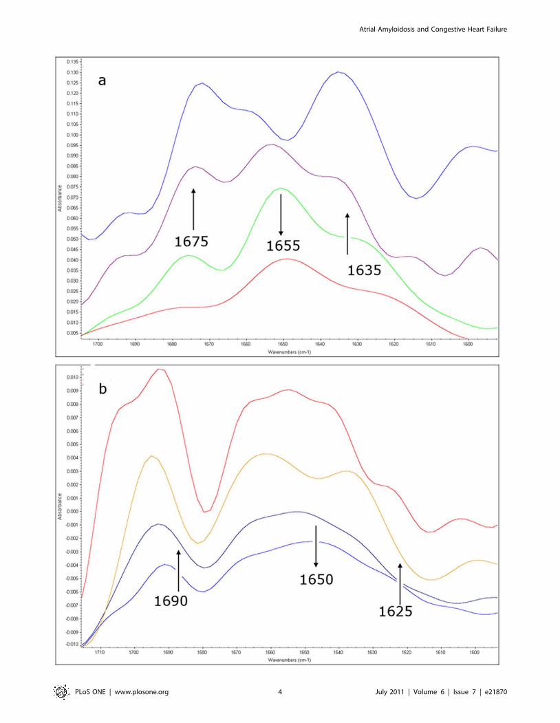
Infrared analysis of ANP aggregation (see Text S1)The representative Fourier-self deconvoluted Amide I spectral
region of a-ANP in aqueous solution, collected at different times
under two different pH conditions (7.4 and 4.0), are shown in Fig. 2
and in Figures S9 and S10. At pH 7.4 two shoulders at 1635 and
Table 1. Patients Characteristics. Values are means6SD or n(%).
Features Patients CR Negative CR positive
Total 40 2(5) 38(95)
Age, years 59.469.3 49.561.5 61.268.7
Sex (M/F) 22/18 2/0 20/18
Disease
CHF 40(100) 2(100) 38(100)
Hypertension 31(77.5) 1(50) 30(79)
Diabetes mellitus 14(35) 2(100) 12(32)
Immunohistochemistry
ANP negative 6(15) 2(100) 4(10.5)
ANP positive 34(85) 0(0) 34(89.5)
doi:10.1371/journal.pone.0021870.t001
Atrial Amyloidosis and Congestive Heart Failure
PLoS ONE | www.plosone.org 2 July 2011 | Volume 6 | Issue 7 | e21870

1675 cm21, attributed to the packing of b strands into b-sheets
and to the subsequent formation of intermolecular b-sheet
aggregates, reached the maximum intensity (then remaining
constant) after 7 days, revealing that the formation of these
aggregates occurs with a very slow kinetics and giving stable
aggregates, whereas the aqueous solution was converted into a gel-
like system.
At pH 4.0 the 1625 and 1685 cm21 contributions of the Amide
I band, attributed to b-sheet structures and intermolecular b-sheet
aggregates, immediately appeared in the spectrum of a-ANP
shown in Fig. 2b and their intensity increases quickly, suggesting a
very rapid aggregation kinetics. The a-ANP IR spectra recorded
from 48 to 168 hours were not any more superimposable
indicating that the aggregated structures are not so stable under
these conditions, according to the microscopic observation.
Moreover, the a-ANP solution after 7 days looks like an aqueous
solution and not as the gel-like system observed at pH 7.4.
a-ANP preformed aggregates seeding abilityWe compared the abilities of a-ANP aggregates preformed at
pH 7.4 and pH 4.0 to act as seeds for initiation of fibril formation
and Fig. 3A illustrates the results of the performed experiments.
Aggregation was measured by Cb and Th-T assays. a-ANP
aggregates preformed at pH 4.0 were able to stimulate fibril
formation to achieve a complete aggregation and to induce a
propagation of amyloid structure at pH 7.4. In contrast,
aggregates preformed at pH 7.4 were unable to accelerate fibril
formation when added to the pH 4.0 solution. Incubation of a-
ANP at pH 4.0 in the presence or absence of seeds resulted in a
rapid high-yield formation of amyloid fibrils as shown in Fig. 3A.
TEM images of a-ANP aggregates formed at physiologic pH from
pH 4.0 seeds showed a typical, well defined amyloid morphology,
analogous to the fibrils prepared at pH 7.4 as shown in Fig. 3C.
These results indicate that, following a pH shift from 4.0 to 7.4, the
acidic pre-aggregates adapted to the physiologic pH. Such
aggregates were also remarkably stable: high MW aggregates
remained after treatment with 90% HFIP, drying and resuspen-
sion in boiling sample buffer, while aggregates formed at ph 4.0
from aggregates preformed at pH 7.4 were monomerized as
shown in Fig. 3B. These findings suggest that: i) the stability of a-
ANP amyloid fibrils is optimal at physiologic pH; ii) the presence
of moderate concentrations (4%) of pH 4.0 pre-formed seeds may
be necessary to partially destabilize a-ANP monomer and make it
amenable to rapidly form stable and resistant aggregates. As
shown in Fig. 3D, such results are supported by the observation
that the extension of a-ANP amyloid fibrils were greatly
dependent on the reaction mixture pH, with an optimum around
4.0–6.0.
CHF a/b-ANP ratio. To simulate a/b-ANP ratio occurring
in vivo in CHF patients (ANP-CHF), experiments were performed
starting aggregation with a mixture of 10 mM ANP immer_ (b)
and monomers (a), ratio 2:1, as shown in Figure S7. The rate of
Cb-monitored aggregation of a-ANP at pH 7.4 was significantly
enhanced by seeding slow aggregating solution with b-ANP as
Figure 1. Aggregation kinetics. A) Aggregation kinetics of a-ANP via Congo red binding method. Data represent the mean of at least threeindependent experiments (n$3). Errors bars represent standard deviations B) Glutaraldehyde Cross-linking, Tris-tricine SDS-PAGE and SDS-PAGEat different times. The formation of a-ANP oligomers and high MW aggregates was monitored. Asterisks indicate dimers, arrows indicate monomersand # indicate high molecular weight aggregates. C) Morphology of a-ANP aggregates at different times and in different conditions. a-ANP wasapplied onto a copper grid and negatively stained with 2 (w/v)% uranyl acetate. Scale bar:100 nm.doi:10.1371/journal.pone.0021870.g001
Atrial Amyloidosis and Congestive Heart Failure
PLoS ONE | www.plosone.org 3 July 2011 | Volume 6 | Issue 7 | e21870
Atrial Amyloidosis and Congestive Heart Failure
PLoS ONE | www.plosone.org 4 July 2011 | Volume 6 | Issue 7 | e21870

shown in Fig. 4A and as confirmed by Th-T experiments
illustrated in Fig. 4B. Interestingly, Th-T and Cb kinetics didn’t
correlate. The maximum value of fluorescence was followed by a
very rapid decrease which was not consistent with aggregation
signals measured by Cb. When analysed by TEM, final aggregates
showed assembled fibrils (Fig. 4B). This finding well correlates with
substantial increase of aggregates’ levels that gave CR red shift and
shoulder peak at 543 nm, even if these structures did not generate
Th-T fluorescence.
Fibril morphologyANP-CHF. In situ generation of ANP-CHF aggregates on
grids at pH 7.4 did not influence morphology (data not shown). At
time 0, oligomers on grid had granular structure, at 45 minutes,
ANP-CHF aggregates exhibited the standard features of amyloid
fibrils showing a substructure composed of parallel alignments of
smaller filaments visible in Fig. 5A. Aggregates at 120 minutes
included thin fibrils with a faintly discernable periodicity, planar
aggregates, approximately 1062.56 nm thick visible in Figure S5.
At 48 hours, fibrillar precipitates formed with widespread
aggregations arranged in lateral arrays of fibres often
superimposed as shown in Fig. 5C. An increase in size of
aggregates was observed and unordered and flat assembly
structures persisted following aggregation to cover fibrils bundles
uniforming the surface area as shown in Fig. 5B. At 48 h fibrils
were entangled, highly superimposed and bundled. Figure S5
shows a sample after 3 weeks of incubation and individual strand
diameters within the bundles ranged in diameter 3–8 nm; after 1
month sample consisted in long, straight, matures fibres while
other kinds of aggregates were completely disappeared as shown in
Fig. 5D. Fibrils had approximately 20 nm diameter and
indeterminate length, with a twist in their longitudinal axis.
Once formed, aggregates were very stable (see below). Together,
these data suggest that an overabundance of immer_,
Figure 3. Seeding. A) Seed-induced fibril formation of a-ANP. Seeds were generated from aged a-ANP (pH 7.4 and pH 4.0, respectively) andadded as percentage weight fractions. Fibril formation was monitored over time by Cb and TEM. B) Treatment in 90% HFIP of ANP aggregatesformed after seeding. High MW aggregates from pH 4.0 seeded solution remained on the top of the gel while aggregates formed by pH 7.4 seedswere monomerized. C) Morphology of samples aged for 720 hours. Morphologies of seeded and unseeded a-ANP at pH 7.4 were still similar inshape. D) pH dependence of a-ANP aggregation. a-ANP was incubated at various pH and after two days the amount of aggregation was quantifiedby Cb.doi:10.1371/journal.pone.0021870.g003
Figure 2. Fourier-self deconvoluted Amide I spectral region of a-ANP in aqueous solution. a: pH 7.4, from the bottom to the top t = 0 h,t = 5 h, t = 30 h, t = 168 h. b: pH = 4.0, from the bottom to the top t = 0 h, t = 1 h, t = 48 h, t = 168 h. Arrows indicate the increase/decrease of therelative band intensities with time.doi:10.1371/journal.pone.0021870.g002
Atrial Amyloidosis and Congestive Heart Failure
PLoS ONE | www.plosone.org 5 July 2011 | Volume 6 | Issue 7 | e21870

corresponding to severe CHF conditions, may enhance ANP
fibrillogenesis promoting amyloid aggregation.
a-ANP at pH 7.4. Light microscopic examination of a-ANP
assemblies revealed that both thin, sheet-like structures and fibrous
aggregates were formed as shown in Figure S5. Ultra-structures
were examined electron microscopically. At starting incubation
time, a-ANP contained neither fibrils nor sheet-like clusters
confirming the monomeric nature of peptide as shown in
Fig. 1C. After 1 hour, a-ANP formed irregular, lumpy,
occasionally branching protofibrils 10–100 nm long visible in
Fig. 5E and after 24 h, 200–400 nm long smooth, straight fibrils
visible in Fig. 5F. At 48 hours, small, irregular protofibrils and
strictly interconnected, fibrillar macroaggregates were present,
while after 6 days, some mature amyloid bent and entangled
strands were observed as shown in Fig. 5G. After 30 days a-ANP
showed precipitates bearing extensive aggregates, arranged in
lateral arrays of fibrils assuming a planar conformation, with
ribbon-like fibres strictly assembled and stratified which peculiar
morphology is well appreciable in Fig. 5H.
a-ANP at pH 4.0. At pH 4.0 a-ANP spontaneously and
rapidly assembled to form amyloid-like fibrils with peculiar
morphologies. Figure S5 C shows that after 90 minutes
incubation ANP formed wide flakes with a diameter of 20–
30 nm. After 4–7 h globular structures were still observed in
concomitance with early fibrillar structures shown in Fig 6C. At
24 hours, the supramolecular spherical structures were formed
and were larger than most previous reports of soluble oligomers,
ranging from a few nm to 1 mm or more in diameter visible in
Fig. 6A. Surprisingly, many of them appeared to be
interconnected with fibrillar material suggesting a direct
association between the two morphological species and a
potential intermediary role for the large peptide assemblies as
shown in Figure S5. Fig. 6D shows that after 48 hours mature,
well-ordered strands 2–3 mm long and 10–40 nm wide were
Figure 4. Time course of ANP-CHF fibril formation. A) Cb and B) Th-T fluorescence. TEM image after 30 days showed amyloid fibril larger incross sectional area and longer in length than fibrils formed in all other conditions tested.doi:10.1371/journal.pone.0021870.g004
Atrial Amyloidosis and Congestive Heart Failure
PLoS ONE | www.plosone.org 6 July 2011 | Volume 6 | Issue 7 | e21870
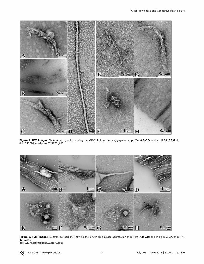
Figure 5. TEM images. Electron micrographs showing the ANP-CHF time course aggregation at pH 7.4 (A,B,C,D) and at pH 7.4 (E,F,G,H).doi:10.1371/journal.pone.0021870.g005
Figure 6. TEM images. Electron micrographs showing the a-ANP time course aggregation at pH 4.0 (A,B,C,D) and in 0.5 mM SDS at pH 7.4(E,F,G,H).doi:10.1371/journal.pone.0021870.g006
Atrial Amyloidosis and Congestive Heart Failure
PLoS ONE | www.plosone.org 7 July 2011 | Volume 6 | Issue 7 | e21870
formed. These fibrils were long, straight and twisted, clearly
indicative of a helical arrangement of protofilaments. We also
observed many twisting ribbon-like structures of different width
and pitch and flat ribbons of parallel protofilaments.
a-ANP in 0.5 mM SDS. The addition of 0.5 mM SDS
promoted the formation of a-ANP fibrils avoiding the lag time as
observed by TEM. At the starting incubation time, reaction
mixture was composed by a sparse number of amorphous clusters
and large spherical structures as shown in Fig. 6E. 2 hours later,
sample exhibited numerous tiny globules and slight, linear fibrous
structures shown in Fig. 6F, evolving in some thin protofibrillar
elements extruding directly from globules, giving rise to
interweaved aggregates. After 6 hours a net change in
morphology was observed, the clusters developing into more
extensive ordered lattice-like structures and showing several
protofibrillar elements orbiting the SDS-micelles. At 30 hours,
globular structures were always present with bundles of strictly
assembled short fibrils, formed through coalescence and
progressive fibrils addition around micellar structures as shown
in Fig. 6G. Fig. 6H shows some amorphous aggregates, in
concomitance with rigid bifurcate fibrillar material the were also
present in the solution (Fig. 6H). This aggregated material was not
stable, since further incubation only produced amorphous,
globular particles without fibrils.
Stability of ANP amyloid aggregatesThe addition of DTT reduced amyloid fibril formation in a
dose-dependent manner as shown in Figure S6 at both 7.4 and 4.0
pH. This effect was common to Cys-containing peptides,
suggesting that the prevention of oxidative dimerization by the
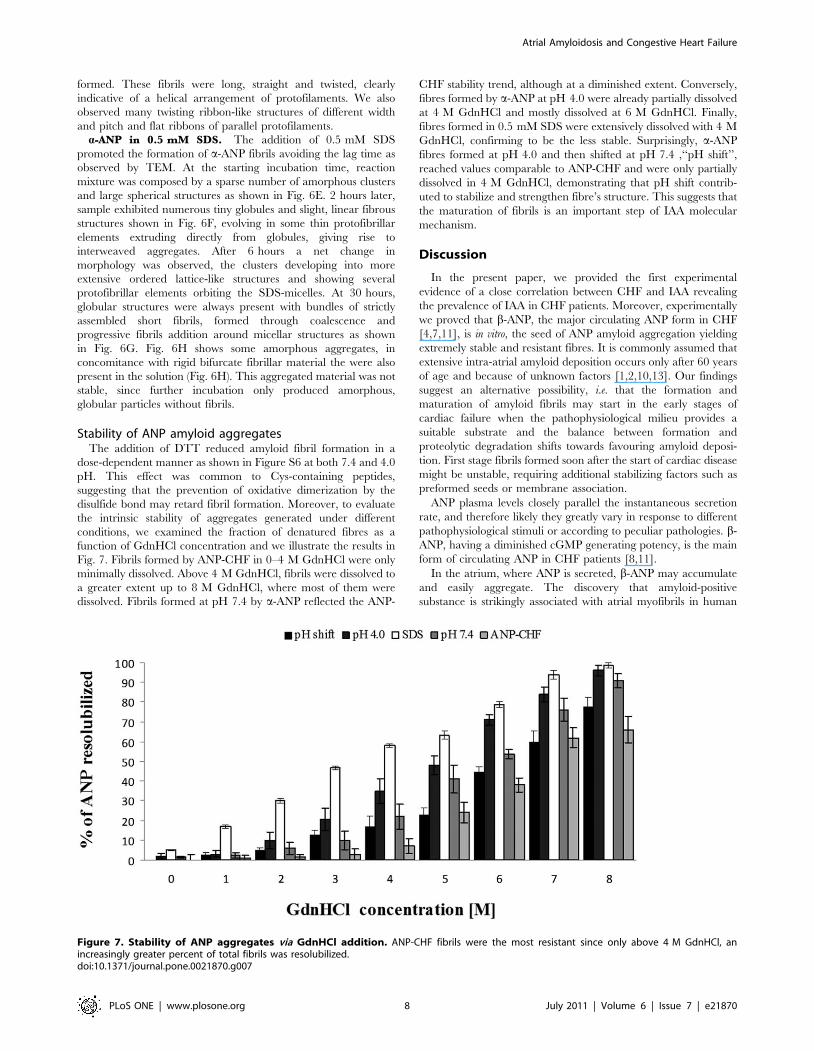
disulfide bond may retard fibril formation. Moreover, to evaluate
the intrinsic stability of aggregates generated under different
conditions, we examined the fraction of denatured fibres as a
function of GdnHCl concentration and we illustrate the results in
Fig. 7. Fibrils formed by ANP-CHF in 0–4 M GdnHCl were only
minimally dissolved. Above 4 M GdnHCl, fibrils were dissolved to
a greater extent up to 8 M GdnHCl, where most of them were
dissolved. Fibrils formed at pH 7.4 by a-ANP reflected the ANP-
CHF stability trend, although at a diminished extent. Conversely,
fibres formed by a-ANP at pH 4.0 were already partially dissolved
at 4 M GdnHCl and mostly dissolved at 6 M GdnHCl. Finally,
fibres formed in 0.5 mM SDS were extensively dissolved with 4 M
GdnHCl, confirming to be the less stable. Surprisingly, a-ANP
fibres formed at pH 4.0 and then shifted at pH 7.4 ,‘‘pH shift’’,
reached values comparable to ANP-CHF and were only partially
dissolved in 4 M GdnHCl, demonstrating that pH shift contrib-
uted to stabilize and strengthen fibre’s structure. This suggests that
the maturation of fibrils is an important step of IAA molecular
mechanism.
Discussion
In the present paper, we provided the first experimental
evidence of a close correlation between CHF and IAA revealing
the prevalence of IAA in CHF patients. Moreover, experimentally
we proved that b-ANP, the major circulating ANP form in CHF
[4,7,11], is in vitro, the seed of ANP amyloid aggregation yielding
extremely stable and resistant fibres. It is commonly assumed that
extensive intra-atrial amyloid deposition occurs only after 60 years
of age and because of unknown factors [1,2,10,13]. Our findings
suggest an alternative possibility, i.e. that the formation and
maturation of amyloid fibrils may start in the early stages of
cardiac failure when the pathophysiological milieu provides a
suitable substrate and the balance between formation and
proteolytic degradation shifts towards favouring amyloid deposi-
tion. First stage fibrils formed soon after the start of cardiac disease
might be unstable, requiring additional stabilizing factors such as
preformed seeds or membrane association.
ANP plasma levels closely parallel the instantaneous secretion
rate, and therefore likely they greatly vary in response to different
pathophysiological stimuli or according to peculiar pathologies. b-
ANP, having a diminished cGMP generating potency, is the main
form of circulating ANP in CHF patients [8,11].
In the atrium, where ANP is secreted, b-ANP may accumulate
and easily aggregate. The discovery that amyloid-positive
substance is strikingly associated with atrial myofibrils in human
Figure 7. Stability of ANP aggregates via GdnHCl addition. ANP-CHF fibrils were the most resistant since only above 4 M GdnHCl, anincreasingly greater percent of total fibrils was resolubilized.doi:10.1371/journal.pone.0021870.g007
Atrial Amyloidosis and Congestive Heart Failure
PLoS ONE | www.plosone.org 8 July 2011 | Volume 6 | Issue 7 | e21870

hearts with dilated or hypertrophic cardiomyopathies [13],
suggests IAA as a potential major pathogenic process in CHF.
Changes in energy production, energy utilization and excitation-
contraction coupling are reported in heart failure [9] and such
changes in cardiac metabolism may be involved in the
augmentation of b-ANP synthesis. Processing of ANP precursor
in the human failing heart differs from that in the normal heart.
Our findings on acidic pH seeds accelerating and stabilizing ANP
fibril formation and on SDS micelles contact enhancing
aggregation, together with the observation of IAA fibrils are also
present inside atrial myocytes [10,13], strongly validate the
importance of initial solution conditions affecting ANP amyloid
aggregation and stability, playing b-ANP the main effective role in
producing the most quickly assembled and strong fibrils. The
amyloid state of endocrine hormones in secretory granules of the
heart atrium contrasts the historical disease association of amyloids
in other organs. On the other hand, ANP may not be very toxic
because the hormone amyloid is stored inside the granules and its
aggregation and secretion may be highly regulated, including by
means of pro-hormone processing. If ANP homeostasis is altered
under stress, age or CHF conditions, hormone aggregation may be
out of control and disease-associated amyloid aggregation may
occur. Whether such aggregation causes disease, or is an indirect
effect of the protein homeostasis altered by disease, remains
undetermined. In fact, pro-hormone maturation seems mostly a
feature of atrial peptide synthesis and enzymes involved in pro-
ANP maturation are produced predominantly in atrial myocytes
[24], while ventricular myocytes don’t contain secretory granules
for peptide storage and maturation. Pro-ANP post-translational
processing may in CHF lead to b-ANP production. In agreement,
CHF patients have increased b-ANP plasma concentrations [4],
and CHF may not be ameliorated by secreted ANP [25].
Although the mechanism of the initial nucleation step in
patients remains unknown, the maturation of fibrils via acidic pH-
seeding is likely to be involved in the development of IAA
amyloidosis. Since the levels of circulating ANP are relatively low
[26], it is unclear where and under which conditions fibril
initiation might occur in vivo. One possibility, analogous to other
amyloid systems, is that fibril formation might be initiated in the
low pH environment of the secretory granules before continuing in
a higher pH environment. This hypothesis is supported by our
findings that optimal pH range for ANP aggregation is 4.0 to 6.0
and that in vitro ANP-aggregates form rapidly upon incubation at
pH 4.0. Our work proved, for the first time, how different
incubation conditions greatly affect not only aggregation kinetics
but also ANP fibril morphology. Although the participation of
additional factors stabilizing the amyloid fibrils is possible, our
findings define that, by acid pH-seeding, ANP amyloid fibrils
adapt to neutral pH conditions and transform into stable matured
fibrils. Acidic pH seeding causes the fibrils to mature and the
amount of fibrils to increase and become stable without additional
factors, finally resulting in a massive amyloid accumulation. These
results are consistent with the hypothesis whereby aggregation may
be initiated intracellularly and end up as a pathological hallmark
in the extracellular space.
The observed acceleration of ANP fibrils formation can be
described by the scheme in Fig. 8 that hypothesizes the existence
of two types of fibrils: i) fibrils formed after maturation in the
cytosol of seeds produced at acid pH, and ii) a second kind
concerning the peculiar CHF conditions, where the b-ANP immer
is the most prevalent circulating form.
Although the information resulting from our in vitro experiments
provide an insight into ANP aggregation process, a comparison of
ANP ex vivo and in vitro fibrils is still required, to directly relate our
findings to in vivo fibrillogenesis and to conclusively account for the
involvement of circulating b-ANP massively produced in CHF in
ANP amyloid deposition. Unfortunately, the retrieval of sufficient
amounts of fresh and correctly collected human CHF endomyo-
cardial bioptic material for TEM analysis is still a hard problem on
which we are currently focusing for further studies.
Our findings suggest that, since IAA may occur at a younger age
than its usual post-mortem diagnosis and early IAA-related ANP
deposition may occur in CHF. Patients with a diagnosed CHF
should be examined histologically, because ANP amyloid
infiltrations may be found also in patients with no clinical
symptoms and young patients having evidence of CHF will almost
always develop IAA. Actuarial study confirms the significant
adverse influence of cardiac involvement in ANP amyloidosis.
Materials and Methods (see Text S1)
Congo Red (CR) staining and anti-ANPimmunohistochemistry
Atrial biopsies from 40 CHF patients listed in Table 1 were
stained with CR for light microscopic evaluation. ANP was
identified as CR–positive amyloid by immunohistochemistry.
Ethics StatementThe whole study was conducted following the approval of the
local University Hospital Ethics Committee, namely ‘‘Comitato
Etico Locale dell’Azienda Ospedaliera Universitaria Senese’’. The
investigation conformed with the principles outlined in the
Declaration of Helsinki.
Myocardial tissues were obtained from biopsies stored in the
tissues archive of ‘‘Patologia Umana e Oncologia Department’’ of
the University of Siena; appropriated samples were selected on the
basis of pathologies and anonymously analyzed. For this reason in
our Institution a written Informed Consent is not required. The
detailed description of the whole study was presented and
approved by the ‘‘Comitato Etico Locale dell’Azienda Ospedaliera
Universitaria Senese’’.
Aggregation kinetic measurementsANP: synthetic ANP (AnaSpec Inc), after treatment with
1,1,1,3,3,3-hexafluoro-2-propanol (HFIP, Sigma), was resus-
pended to the desired concentration in the appropriate buffer.
Aggregation kinetic parameters were obtained by monitoring the
reaction by Congo Red binding (Cb) method [5]. and Thioflavin –
T (Th-T) fluorescence [27,28]. Reactions were initiated by
incubating appropriate concentrations of freshly prepared a-
ANP in the desired buffer.
ANP-CHF preparationANP (1 mg/mL ) was dissolved in H2O pH 7.4 at and
centrifuged at 16,5006 g for 1 h. The supernatant was collected
and filtered with a 20 nm pore filter. Protein concentration was
determined at 280 nm using [29].
ElectrophoresisANP samples incubated for 1 hour were analyzed by
glutaraldehyde cross-linking [30] and analyzed by SDS-PAGE
or Tricine SDS-PAGE.
FTIR measurementsThe spectra were obtained with a Thermo Nicolet 5700 Fourier
Transform Infrared spectrometer operating between 3000 and
900 cm21 and 100 scans at a resolution of 2.0 cm21 were
Atrial Amyloidosis and Congestive Heart Failure
PLoS ONE | www.plosone.org 9 July 2011 | Volume 6 | Issue 7 | e21870

averaged. An ATR (Attenuated total reflectance) cell equipped
with a 45u germanium IRE (internal reflection element) crystal was
used to record the water spectra and ANP solution. The spectra
were taken in a single beam mode at predetermined time intervals
to obtain kinetic information of the peptide aggregation process.
To improve the observability of the overlapping bands, mathe-
matical resolution enhancement was performed by a spectral
deconvolution process.
Cross-seeding experimentsFibrils were sonicated and then incubated at ph 4.0 and pH 7.4
to be used as seeds in cross-seeding experiments.
Disaggregation assayAliquots of mature ANP fibrils suspensions formed under
different conditions were resuspended in guanidine hydrochloride
(GdnHCl) at growing concentrations (0–8 M) and protein
concentration of obtained monomers/oligomers was determined
[31,32].
Transmission Electron MicroscopySamples were suspended in deionized water, diluted 100-fold
and then placed on a glow-discharged 200 mesh carbon coated
copper grid. After to be adsorbed and air dried samples grids were
then stained with 2% uranyl acetate for 45 s. Excess stain was
removed, and the samples were allowed to air-dry and analyzed
utilizing a Philips CM10 TEM operating at 80 kV.
Supporting Information
Figure S1 Aggregation kinetics via Congo Red. Time
course of a-ANP aggregate formation using a-ANP previously
disaggregated by treatment with HFIP (see Methods) and
incubated at different concentrations at pH 7.4. Aggregate
formation was measured by Congo Red binding method. Arrow
indicate the magnification of red box area.
(PPT)
Figure S2 Aggregation kinetics via Th-T. a-ANP amyloid
aggregation monitored by Th-T fluorescence. a-ANP HFIP
treated was incubated at different concentration at pH 7.4. Arrow
indicate the magnification of red box area.
(PPT)
Figure S3 Aggregation kinetics of a-ANP in differentconditions. A) Effect of pH, solvent type and temperature on the
aggregation kinetics of a-ANP measured via Th-T fluorescence. B)
Seed-induced fibril formation of ANP in H2O (pH 7.4) at 25uC.
The seeds or pre-existing fibrils were generated from aged ANP
(pH 7.4 and pH 4.0 respectively) and were added as percentage
Figure 8. Theoretical schema of ANP aggregation.doi:10.1371/journal.pone.0021870.g008
Atrial Amyloidosis and Congestive Heart Failure
PLoS ONE | www.plosone.org 10 July 2011 | Volume 6 | Issue 7 | e21870

weight fractions. Fibril formation was monitored over time by Th-
T fluorescence. C) pH dependence of a-ANP aggregation. a-ANP
at 0.5 mg/ml was incubated at 25uC at various pH values and
after two days the amount of aggregation quantified by Th-T
fluorescence.
(PPT)
Figure S4 Formation of a-ANP oligomers and aggre-gates monitored by Tris-tricine SDS-PAGE. A) a-ANP
solution in H2O at pH 7.4 at different aggregation times; B) a-
ANP solution in H2O at pH 4.0 at different aggregation times; an
appreciable difference in oligomers size and distribution between
the two tested solutions was evident. Arrow indicates monomer,
asterisk indicates dimer and # indicates high molecular aggre-
gates.
(PPT)
Figure S5 Microscope observations. A) CHF-ANP incu-
bated at pH 7.4; Images show branched fibrils indicating that they
are composed of interwined protofibrils and aggregates composed
of fibrils with apparent different morphology. B) TEM micro-
graphs of a-ANP incubated at pH 7.4: images show several fibrils
that lie almost parallel and close to one another. One or more
protofilaments unwinding from a fibril and winding onto an
adjacent one are also observed. On the right, light microscopy of
the a-ANP assembly is shown; CR brightfield (Magnification
20X); C) TEM observations of a-ANP incubated at pH 4.0
showing loose aggregates in which individual fibrils can be seen.
Bundles of fibrils diverging from spherulites are evident.
(PPT)
Figure S6 Effect of DTT addition on a-ANP incubated atpH 7.4 and pH 4.0. DTT is able to dose-dependently reduce
the amount of amyloid fibrils formed under both pH 7.4 and
pH 4.0 conditions.
(PPT)
Figure S7 Electrophoresis. To model the conditions occur-
ring in vivo in the heart of CHF patients, a mixture of ANP dimers
and monomers (ratio 2:1) roughly as in CHF conditions, was
settled up. Figure shows Tricine SDS PAGE of ANP in H2O
pH 7.4. 1) Untreated sample; 2) Treated sample. Quali-quantita-
tive image analysis was performed on silver stained gel by using
Image Quant (Master) software to assess the obtaining of
monomer/dimer 1a-ANP:2b-ANP ratio as shown in the Table.
(PPT)
Figure S8 CR assay. A) Spectral features of CR and
aggregated a-ANP. Absorbance spectra of a suspension of a-
ANP in the absence and the presence of CR and of CR alone; B)
When CR binds to excess fibrillar a-ANP, a change in colour from
orange–red to rose is induced that corresponds to a shift in the
characteristic absorbance spectrum of CR. C) Relationship
between the shoulder peak (543 nm) and time, for 10 mM a-
ANP in 20 mM CR. The original absorbance spectra at 541 nm at
interval of 30 seconds on the aggregation of a-ANP are shown.
The shoulder peaks at 541 nm gradually grew with time,
indicating that CR is kinetically bound to the amyloid aggregates.
(PPT)
Figure S9 Representative infrared spectra of a-ANP inaqueous solution at pH = 7.4. Representative infrared spectra
of a-ANP in aqueous solution (pH = 7.4) collected at different
times: from the bottom to the top t = 0 h, t = 5 h, t = 30 h,
t = 168 h, t = 336 h.
(PPT)
Figure S10 Representative infrared spectra of a-ANP inaqueous solution at pH = 4.0. Representative infrared spectra
of a-ANP in aqueous solution (pH = 4.0) collected at different
times: from the bottom to the top t = 0 h, t = 1 h, t = 10 h,
t = 48 h, t = 168 h.
(PPT)
Figure S11 CR and anti-ANP immunostaining of heartspecimens. Images revealed the co-occurrence of CHF and
IAA. CR-stained paraffin sections from left atrial appendages of
three representative CHF patients showing atrial amyloid under
direct light (A, D, G) and polarized light (B, E, H). C, F, I:Immunostaining of ANP in the atrial myocardium. Original
magnification x 20.
(PPT)
Text S1 Supporting Information. This section presents
detailed supporting materials to the manuscript and itemized,
extensive results pertaining to the Infrared Analysis of ANP
aggregation. Moreover, additional Figures as enhancement to
those presented in the main text are shown.
(DOC)
Author Contributions
Conceived and designed the experiments: LM GB LG A. Santucci PL AM.
Performed the experiments: LM LG A. Spreafico MC DB ML EP.
Analyzed the data: LM LG GB DB EP A. Santucci PT A. Spreafico AM.
Contributed reagents/materials/analysis tools: PT A. Santucci A.
Spreafico AM PL. Wrote the paper: LM GB A. Santucci.
References
1. Kholova I, Niessen HW (2005) Amyloid in the cardiovascular system: a review.
J Clin Pathol 58: 125–133.
2. Kawamura S, Takahashi M, Ishihara T, Uchino F (1995) Incidence and
distribution of isolated atrial amyloid: histologic and immunohistochemical
studies of 100 aging hearts. Pathol Int 45: 335–342.
3. Kaye GC, Butler MG, D’Ardenne AJ, Edmondson SJ, Camm AJ, et al. (1986)
Identification of immunoreactive atrial natriuretic peptide in atrial amyloid.
J Clin Pathol 39: 581–582.
4. Sugawara A, Nakao K, Morii N, Yamada T, Itoh H, et al. (1988) Synthesis of
atrial natriuretic polypeptide in human failing hearts. Evidence for altered
processing of atrial natriuretic polypeptide precursor and augmented synthesis of
beta-human ANP. J Clin Invest 81: 1962–1970.
5. Bensimon M, Chang AI, de Bold ML, Ponce A, Carreras D, et al. (2004)
Participation of G proteins in natriuretic peptide hormone secretion from heart
atria. Endocrinology 145: 5313–5321.
6. Rocken C, Peters B, Juenemann G, Saeger W, Klein HU, et al. (2002) Atrial
amyloidosis: an arrhythmogenic substrate for persistent atrial fibrillation.
Circulation 106: 2091–2097.
7. Wozakowska-Kapłon B, Opolski G, Herman Z, Kosior D (2008) Natriuretic
peptides in patients with atrial fibrillation. Cardiol J 15: 525–529.
8. Looi LM (1993) Isolated atrial amyloidosis: a clinicopathologic study
indicating increased prevalence in chronic heart disease. Hum Pathol 24:
602–607.
9. Abassi Z, Karram T, Ellaham S, Winaver J, Hoffman A (2004) Implications of
the natriuretic peptide system in the pathogenesis of heart failure: diagnostic and
therapeutic importance. Pharmacol Ther 102: 223–241.
10. Pucci A, Wharton J, Arbustini E, Grasso M, Diegoli M, et al. (1991) Atrial
amyloid deposits in the failing human heart display both atrial and brain
natriuretic peptide-like immunoreactivity. J Pathol 165: 235–241.
11. Wei CM, Kao PC, Lin JT, Heublein DM, Schaff HV, et al. (1993) Circulating
beta-atrial natriuretic factor in congestive heart failure in humans. Circulation
88: 1016–1020.
12. Takemura G, Takatsu Y, Doyama K, Itoh H, Saito Y, et al. (1998) Expression of
atrial and brain natriuretic peptides and their genes in hearts of patients with
cardiac amyloidosis. J Am Coll Cardiol 31: 754–765.
13. Steiner I, Hajkova P (2006) Patterns of isolated atrial amyloid: a study of 100
hearts on autopsy. Cardiovasc Pathol 15: 287–290.
14. Torricelli C, Capurro E, Santucci A, Paffetti A, D’Ambrosio C, et al. (2004)
Multiple plasma proteins control atrial natriuretic peptide (ANP) aggregation.
J Mol Endocrinol 33: 335–341.
Atrial Amyloidosis and Congestive Heart Failure
PLoS ONE | www.plosone.org 11 July 2011 | Volume 6 | Issue 7 | e21870

15. Maioli E, Torricelli C, Santucci A, Pacini A (2000) Molecular assembly of
endogenous and synthetic big atrial natriuretic peptide (ANP) and itsamyloidogenic implications. Biochim Biophys Acta 1500: 31–40.
16. Maioli E, Torricelli C, Santucci A, Martelli P, Pacini A (2001) Plasma factors
controlling atrial natriuretic peptide (ANP) aggregation: role of lipoproteins.Biochim Biophys Acta 1536: 123–132.
17. Torricelli C, Capurro E, Santucci A, Paffetti A, D’Ambrosio C, et al. (2004)Small HDL form via apo A-I a complex with atrial natriuretic peptide. Biochem
Biophys Res Commun 315: 16–21.
18. Akimoto K, Miyata A, Kangawa K, Koga Y, Hayakawa K, et al. (1988)Molecular forms of atrial natriuretic peptide in the atrium of patients with
cardiovascular disease. J Clin Endocrinol Metab 67: 93–97.19. Sugawara A, Nakao K, Morii N, Yamada T, Itoh H, et al. (1988) Augmented
synthesis of beta-human atrial natriuretic polypeptide in human failing hearts.Biochem Biophys Res Commun 150: 60–67.
20. van Heerebeek L, Borbely A, Niessen HW, Bronzwaer JG, van der Velden J,
et al. (2006) Myocardial structure and function differ in systolic and diastolicheart failure. Circulation 113: 1966–1973.
21. Cruz L, Urbanc B, Borreguero JM, Lazo ND, Teplow DB, et al. (2005) Solventand mutation effects on the nucleation of amyloid beta-protein folding. Proc
Natl Acad Sci USA 102: 18258–18263.
22. Otzen DE (2010) Amyloid Formation in Surfactants and Alcohols: MembraneMimetics or Structural Switchers? Curr Protein Pept Sci 11: 355–371.
23. Necula M, Chirita CN, Kuret J (2003) Rapid anionic micelle-mediated alpha-synuclein fibrillization in vitro. J. Biol. Chem 278: 46674–46680.
24. Muth E, Driscoll WJ, Smalstig A, Goping G, Mueller GP (2004) Proteomic
analysis of rat atrial secretory granules: a platform for testable hypotheses.
Biochim Biophys Acta 1699: 263–275.
25. Goetze JP, Kastrup J, Rehfeld JF (2003) The paradox of increased natriuretic
hormones in congestive heart failure patients: does the endocrine heart also fail
in heart failure? Eur Heart J 24: 1471–1472.
26. Clerico A, Iervasi G, Berti S, Pilo A, Vitek F, et al. (1995) In vivo measurement
of ANP overall turnover and identification of its main metabolic pathways under
steady state conditions in humans. J Endocrinol Invest 18: 194–204.
27. Klunk WE, Pettegrew JW, Abraham DJ (1989) Quantitative evaluation of congo
red binding to amyloid-like proteins with a beta-pleated sheet conformation.
J Histochem Cytochem 37: 1273–1281.
28. LeVine H 3rd (1999) Quantification of beta-sheet amyloid fibril structures with
thioflavin T. Methods Enzymol 309: 274–284.
29. ProtParam on ExPASy Proteomics Server. Available: http://expasy.org/tools/
protparam.html. Accessed 2011 June 20.
30. Hermann R, Jaenicke R, Rudolph R (1981) Analysis of the reconstitution of
oligomeric enzymes by cross-linking with glutaraldehyde: kinetics of reassocia-
tion of lactic dehydrogenase. Biochemistry 20: 5195–5201.
31. Timasheff SN (1992) Water as ligand: preferential binding and exclusion of
denaturants in protein unfolding. Biochemistry 31: 9857–9864.
32. Bradford MM (1976) A rapid and sensitive method for the quantitation of
microgram quantities of protein utilizing the principle of protein-dye binding.
Anal Biochem 72: 248–254.
Atrial Amyloidosis and Congestive Heart Failure
PLoS ONE | www.plosone.org 12 July 2011 | Volume 6 | Issue 7 | e21870