Diagnostic Accuracy of Clinical Tests for the Different Degrees of Subacromial Impingement Syndrome
10
COPYRIGHT © 2005 BY THE JOURNAL OF BONE AND JOINT SURGERY, INCORPORATED 1446 Diagnostic Accuracy of Clinical Tests for the Different Degrees of Subacromial Impingement Syndrome BY HYUNG BIN PARK, MD, ATSUSHI Y OKOTA, MD, PHD, HARPREET S. GILL, MD, GEORGE EL RASSI, MD, AND EDWARD G. MCFARLAND, MD Investigation performed at the Division of Sports Medicine and Shoulder Surgery, Department of Orthopaedic Surgery, The Johns Hopkins University, Baltimore, Maryland Background: Several tests for making the diagnosis of rotator cuff disease have been described, but their utility for diagnosing bursitis alone, partial-thickness rotator cuff tears, and full-thickness rotator cuff tears has not been stud- ied. The hypothesis of this study was that the degree of severity of rotator cuff disease affects the diagnostic values of the commonly used clinical tests. Methods: Eight physical examination tests (the Neer impingement sign, Hawkins-Kennedy impingement sign, painful arc sign, supraspinatus muscle strength test, Speed test, cross-body adduction test, drop-arm sign, and infraspina- tus muscle strength test) were evaluated to determine their diagnostic values, including likelihood ratios and post- test probabilities, for three degrees of severity in rotator cuff disease: bursitis, partial-thickness rotator cuff tears, and full-thickness rotator cuff tears. A forward stepwise logistic regression analysis was used to determine the best combination of clinical tests for predicting the various grades of impingement syndrome. Results: The sensitivity, specificity, positive predictive value, negative predictive value, and overall accuracy of the eight tests varied considerably. The combination of the Hawkins-Kennedy impingement sign, the painful arc sign, and the infraspinatus muscle test yielded the best post-test probability (95%) for any degree of impingement syndrome. The combination of the painful arc sign, drop-arm sign, and infraspinatus muscle test produced the best post-test probability (91%) for full-thickness rotator cuff tears. Conclusions: The severity of the impingement syndrome affects the diagnostic values of the commonly used clinical tests. The variable accuracy of these tests should be taken into consideration when evaluating patients with symp- toms of rotator cuff disease. Level of Evidence: Diagnostic Level I . See Instructions to Authors for a complete description of levels of evidence. he physical examination for rotator cuff disease re- mains important for the clinical assessment of patients who present with shoulder pain or weakness. Neer 1 de- scribed a physical examination sign that he said was not spe- cific to rotator cuff disease but was helpful in making the diagnosis. He also described an injection test that confirmed the location of the pain as the subacromial space. Other physi- cal examination signs that have been described as aids to diagnosing subacromial impingement include the Hawkins- Kennedy impingement sign 2 , the painful arc sign 3,4 , the supra- spinatus muscle strength test 5,6 , the Speed test 3 , the drop-arm sign 3 , the cross-body adduction test 3,7 , and the infraspinatus muscle strength test 6 . Neer 1 also described three stages of impingement syn- drome, noting that “the symptoms and physical signs in all three stages of impingement are almost identical, including the ‘impingement sign’ . . . , arc of pain, crepitus, and varying weakness.” He stated that weakness was more severe in ad- vanced cases, and he did not distinguish between partial- thickness and full-thickness rotator cuff tears in stage III 1 . The literature contains few reports on the diagnostic values of these clinical tests 3,6,8 (see Appendix). The studies that have been published have used different methodologies, and consequently the results have varied. Only two studies T A commentary is available with the electronic versions of this article, on our web site (www.jbjs.org) and on our quarterly CD-ROM (call our subscription department, at 781-449-9780, to order the CD-ROM).
-
Upload
independent -
Category
Documents
-
view
6 -
download
0
Transcript of Diagnostic Accuracy of Clinical Tests for the Different Degrees of Subacromial Impingement Syndrome

COPYRIGHT © 2005 BY THE JOURNAL OF BONE AND JOINT SURGERY, INCORPORATED
1446
Diagnostic Accuracy of Clinical Tests for the Different
Degrees of Subacromial Impingement SyndromeBY HYUNG BIN PARK, MD, ATSUSHI YOKOTA, MD, PHD, HARPREET S. GILL, MD,
GEORGE EL RASSI, MD, AND EDWARD G. MCFARLAND, MD
Investigation performed at the Division of Sports Medicine and Shoulder Surgery, Department of Orthopaedic Surgery, The Johns Hopkins University, Baltimore, Maryland
Background: Several tests for making the diagnosis of rotator cuff disease have been described, but their utility fordiagnosing bursitis alone, partial-thickness rotator cuff tears, and full-thickness rotator cuff tears has not been stud-ied. The hypothesis of this study was that the degree of severity of rotator cuff disease affects the diagnostic valuesof the commonly used clinical tests.
Methods: Eight physical examination tests (the Neer impingement sign, Hawkins-Kennedy impingement sign, painfularc sign, supraspinatus muscle strength test, Speed test, cross-body adduction test, drop-arm sign, and infraspina-tus muscle strength test) were evaluated to determine their diagnostic values, including likelihood ratios and post-test probabilities, for three degrees of severity in rotator cuff disease: bursitis, partial-thickness rotator cuff tears,and full-thickness rotator cuff tears. A forward stepwise logistic regression analysis was used to determine the bestcombination of clinical tests for predicting the various grades of impingement syndrome.
Results: The sensitivity, specificity, positive predictive value, negative predictive value, and overall accuracy of theeight tests varied considerably. The combination of the Hawkins-Kennedy impingement sign, the painful arc sign, andthe infraspinatus muscle test yielded the best post-test probability (95%) for any degree of impingement syndrome.The combination of the painful arc sign, drop-arm sign, and infraspinatus muscle test produced the best post-testprobability (91%) for full-thickness rotator cuff tears.
Conclusions: The severity of the impingement syndrome affects the diagnostic values of the commonly used clinicaltests. The variable accuracy of these tests should be taken into consideration when evaluating patients with symp-toms of rotator cuff disease.
Level of Evidence: Diagnostic Level I. See Instructions to Authors for a complete description of levels of evidence.
he physical examination for rotator cuff disease re-mains important for the clinical assessment of patientswho present with shoulder pain or weakness. Neer1 de-
scribed a physical examination sign that he said was not spe-cific to rotator cuff disease but was helpful in making thediagnosis. He also described an injection test that confirmedthe location of the pain as the subacromial space. Other physi-cal examination signs that have been described as aids todiagnosing subacromial impingement include the Hawkins-Kennedy impingement sign2, the painful arc sign3,4, the supra-
spinatus muscle strength test5,6, the Speed test3, the drop-armsign3, the cross-body adduction test3,7, and the infraspinatusmuscle strength test6.
Neer1 also described three stages of impingement syn-drome, noting that “the symptoms and physical signs in allthree stages of impingement are almost identical, includingthe ‘impingement sign’ . . . , arc of pain, crepitus, and varyingweakness.” He stated that weakness was more severe in ad-vanced cases, and he did not distinguish between partial-thickness and full-thickness rotator cuff tears in stage III1.
The literature contains few reports on the diagnosticvalues of these clinical tests3,6,8 (see Appendix). The studiesthat have been published have used different methodologies,and consequently the results have varied. Only two studies
T
A commentary is available with the electronic versions of this article,on our web site (www.jbjs.org) and on our quarterly CD-ROM (call oursubscription department, at 781-449-9780, to order the CD-ROM).

1447
TH E JO U R NA L OF BONE & JOINT SURGER Y · JBJS .ORG
VO LU M E 87-A · NU M B E R 7 · JU LY 2005DIAG N OST I C ACC U R A CY OF CLINIC A L TE S T S FOR THE DIFFERENT DE G RE E S OF SU B A C ROM I A L IMPINGEMENT SYN DROME
have addressed the utility of the physical examination find-ings with regard to the different stages of subacromial im-pingement. MacDonald et al.8 studied eighty-five patientsand assessed the diagnostic values of the Neer and the Hawkins-Kennedy impingement signs for subacromial bursitis and forrotator cuff disease. Murrell and Walton9 evaluated 400 pa-tients who were undergoing arthroscopic shoulder surgeryfor a variety of diagnoses. Each of these studies divided thepatients into two groups: those with bursitis and those withrotator cuff tears of any type. Neither study compared the di-agnostic values of these tests for partial-thickness and full-thickness tears.
In a review of the English-language literature, wefound no study that evaluated the physical examinationfindings for the three different degrees of rotator cuff dis-ease: bursitis without a rotator cuff tear, a partial-thicknessrotator cuff tear, and a full-thickness rotator cuff tear. Theutility of the examination tests for these different conditionsis important for those who treat patients with symptoms ofrotator cuff disease. Our hypothesis was that the diagnosticvalue of these eight clinical tests for the differing conditionswould vary. Our goal was to define which tests or combina-tion of tests were the best diagnostic tools for these threeconditions.
Materials and Methodsetween August 1992 and June 2003, 1127 patients whohad shoulder surgery performed by the senior author
(E.G.McF.) provided a thorough history, filled out a question-naire, and underwent a complete physical examination withinthe four weeks before surgery. In addition, plain anteroposte-rior radiographs (internal rotation, external rotation, and ax-illary views) were made for each patient. All patients gaveinformed consent, and their findings and responses were en-tered into a database. The database and study were approvedby our institutional review board. Of the 1127 patients, 214patients who did not undergo arthroscopy were excludedfrom the study. The remaining 913 patients who underwentphysical examination and diagnostic arthroscopy, both per-formed with the patient under general anesthesia, formed ourinitial study group. Surgical data for these study patients wererecorded and added to the database at the time of operativeintervention.
The physical examinations included eight clinical testsfor impingement syndrome. All tests were performed withthe patient standing. The first test was the Neer impinge-ment sign10 (Fig. 1). The scapula was stabilized by the exam-iner, and the arm was forward flexed by the examiner untilthe patient reported pain or until full elevation was reached.A positive test was considered to be pain in the anterior orlateral part of the shoulder, typically in a range of 90° to 140°of flexion.
The second test was the Hawkins-Kennedy impinge-ment sign (Fig. 2), in which the arm was placed in 90° of for-
B
Fig. 1
The Neer impingement sign was performed with the patient standing.
The scapula was stabilized by the examiner, and the arm was forward
flexed until the patient reported pain or full elevation was reached.
Fig. 2
The Hawkins-Kennedy impingement sign was performed with the
patient standing. The arm was elevated to 90° and then was forcibly
rotated medially.
1448
TH E JO U R NA L OF BONE & JOINT SURGER Y · JBJS .ORG
VO LU M E 87-A · NU M B E R 7 · JU LY 2005DIAG N OST I C ACC U R A CY OF CLINIC A L TE S T S FOR THE DIFFERENT DE G RE E S OF SU B A C ROM I A L IMPINGEMENT SYN DROME
ward flexion and then gently rotated into internal rotation.The end point for internal rotation was either when the pa-tient felt pain or when rotation of the scapula was felt or ob-served by the examiner. This test was considered to be positiveif the patient had pain during the maneuver.
The third test was the painful arc sign4 in which the pa-tient was asked to actively elevate the arm in the scapularplane until full elevation was reached and then to bring thearm down in the same arc. The test was considered to be posi-
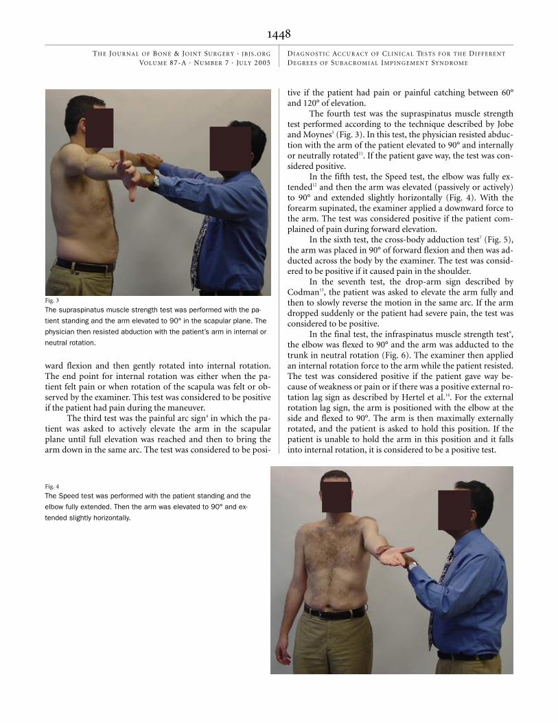
tive if the patient had pain or painful catching between 60°and 120° of elevation.
The fourth test was the supraspinatus muscle strengthtest performed according to the technique described by Jobeand Moynes5 (Fig. 3). In this test, the physician resisted abduc-tion with the arm of the patient elevated to 90° and internallyor neutrally rotated11. If the patient gave way, the test was con-sidered positive.
In the fifth test, the Speed test, the elbow was fully ex-tended12 and then the arm was elevated (passively or actively)to 90° and extended slightly horizontally (Fig. 4). With theforearm supinated, the examiner applied a downward force tothe arm. The test was considered positive if the patient com-plained of pain during forward elevation.
In the sixth test, the cross-body adduction test7 (Fig. 5),the arm was placed in 90° of forward flexion and then was ad-ducted across the body by the examiner. The test was consid-ered to be positive if it caused pain in the shoulder.
In the seventh test, the drop-arm sign described byCodman13, the patient was asked to elevate the arm fully andthen to slowly reverse the motion in the same arc. If the armdropped suddenly or the patient had severe pain, the test wasconsidered to be positive.
In the final test, the infraspinatus muscle strength test6,the elbow was flexed to 90° and the arm was adducted to thetrunk in neutral rotation (Fig. 6). The examiner then appliedan internal rotation force to the arm while the patient resisted.The test was considered positive if the patient gave way be-cause of weakness or pain or if there was a positive external ro-tation lag sign as described by Hertel et al.14. For the externalrotation lag sign, the arm is positioned with the elbow at theside and flexed to 90°. The arm is then maximally externallyrotated, and the patient is asked to hold this position. If thepatient is unable to hold the arm in this position and it fallsinto internal rotation, it is considered to be a positive test.
Fig. 4
The Speed test was performed with the patient standing and the
elbow fully extended. Then the arm was elevated to 90° and ex-
tended slightly horizontally.
Fig. 3
The supraspinatus muscle strength test was performed with the pa-
tient standing and the arm elevated to 90° in the scapular plane. The
physician then resisted abduction with the patient’s arm in internal or
neutral rotation.

1449
TH E JO U R NA L OF BONE & JOINT SURGER Y · JBJS .ORG
VO LU M E 87-A · NU M B E R 7 · JU LY 2005DIAG N OST I C ACC U R A CY OF CLINIC A L TE S T S FOR THE DIFFERENT DE G RE E S OF SU B A C ROM I A L IMPINGEMENT SYN DROME
Of the 913 patients who underwent arthroscopy, 361 wereexcluded for the following reasons: a history of shoulder surgery(126 patients); acromioclavicular arthritis, requiring excision ofthe distal part of the clavicle in conjunction with acromioplastyor rotator cuff repair, combined with impingement syndrome(seventy-three); an incomplete physical examination because oflimited motion or extreme pain (sixty-three); superior labrumanterior-to-posterior lesions, requiring repair, combined withimpingement syndrome (fifty-seven); or instability combinedwith impingement syndrome (forty-two). The remaining 552patients formed our final study group.
To determine the diagnostic values of each of the eightclinical tests, the 552 patients were divided into a nonimpinge-ment or an impingement group. The nonimpingement groupconsisted of 193 patients with normal rotator cuffs and apathologic condition unrelated to impingement syndrome,such as instability (158 patients), isolated degenerative acro-mioclavicular arthritis (twenty-six), a supraspinatus ganglioncyst (five), nonspecific synovitis (three), and a superior la-brum anterior-to-posterior lesion (one). The impingementgroup consisted of 359 patients with a diagnosis of impinge-ment syndrome, regardless of the degree of severity. All pa-tients in this group had a history of pain in the deltoid regionor radiating down the arm and had at least temporary resolu-tion of the pain with a subacromial injection of local anes-thetic (a positive Neer impingement test). These 359 patientswere subdivided into three groups according to the degree ofdisease severity. Group 1 included seventy-two patients whohad a positive impingement test but no rotator cuff disease atthe time of surgery and were judged to have subacromial im-pingement alone. Group 2 included seventy-two patients witha partial rotator cuff tear at the time of surgery, regardless ofdepth; this included patients with bursal-side tears (five) andjoint-side partial tears (sixty-seven). Group 3 included 215 pa-
tients with a full-thickness rotator cuff tear, regardless of size;this included patients with small tears, large tears, massivetears, or multiple tendon tears. Radiographic assessment andthe results of magnetic resonance imaging were not used tomake the final diagnosis and were not included as variables inour study.
Fig. 5
The cross-body adduction test was performed with the patient
standing and the arm elevated to 90°. Then the arm was ad-
ducted across the body.
Fig. 6
The infraspinatus muscle strength test was performed with the pa-
tient standing, the elbow flexed at 90°, and the arm adducted to the
trunk in neutral rotation. The examiner applied an internal rotation
force to the arm while the patient resisted.

1450
TH E JO U R NA L OF BONE & JOINT SURGER Y · JBJS .ORG
VO LU M E 87-A · NU M B E R 7 · JU LY 2005DIAG N OST I C ACC U R A CY OF CLINIC A L TE S T S FOR THE DIFFERENT DE G RE E S OF SU B A C ROM I A L IMPINGEMENT SYN DROME
Statistical analysis was performed with use of StatisticalPackage for the Social Sciences (version 10.0; SPSS, Chicago,Illinois). Sensitivity, specificity, positive predictive value, neg-ative predictive value, and overall accuracy of the eight identi-fied clinical tests were calculated with the two-by-two tablemethod (Table I). The likelihood ratios indicate the values ofvarious tests for increasing the certainty of a diagnosis. In thecase of a single test, a likelihood ratio of >10 is sufficient torule in the target condition15. A forward stepwise logistic re-gression analysis was used to determine the best combinationof clinical tests for predicting the various grades of impinge-ment syndrome. The final diagnosis, as confirmed by arthros-copy, was used as the dependent variable. At each step, each ofthe eight clinical tests was added automatically to the regres-sion model as the explanatory (predictor) variable. The levelof significance for each clinical test was set at 0.05.
Resultshe diagnostic values of the eight clinical tests for overallsubacromial impingement syndrome, regardless of the
degree of rotator cuff disease, are summarized in Table II. The
diagnostic values of the eight clinical tests for the three degreesof severity of the rotator cuff disease are summarized in TableIII. These results show that the physical findings for rotatorcuff disease vary with disease severity.
For patients in the impingement group (that is, thosewith any type of rotator cuff disease), the painful arc sign wasthe most sensitive test (73.5%) and it had the highest negativepredictive value (61.5%) and the highest overall accuracy(76.1%) in detecting impingement (Table II). The infraspina-tus muscle strength test was the most specific (90.1%) and hadthe highest positive predictive value (90.6%) for rotator cuffdisease of any type. The post-test probabilities of the eightclinical tests for any degree of impingement were generallyhigh (>0.70).
The patients in the impingement group were divided intothree subgroups. For patients in Group 1 (that is, those with ro-tator cuff tendinitis or bursitis but no rotator cuff tears), theNeer impingement sign was the most sensitive (85.7%) and ithad the highest positive predictive value (20.9%) and the high-est negative predictive value (95.7%). The cross-body adduc-tion test was the most specific (79.7%) and had the highestoverall accuracy (73.1%). The drop-arm sign had the lowestsensitivity (13.6%).
For the patients in Group 2 (that is, those with partial-thickness rotator cuff tears only), both the Neer and theHawkins-Kennedy impingement signs were highly sensitive(75.4%), but their specificity was only 48% and 44%, respec-tively. The Neer impingement sign had the highest positivepredictive value (18.1%) and the highest negative predictivevalue (92.6%) of all of the tests studied. The cross-body ad-duction test was the most specific (78.5%) and had the highestoverall accuracy (70.8%). The positive predictive values of theeight clinical tests for partial-thickness rotator cuff tears weregenerally very low (<20%), although the negative predictivevalues were generally high (>86%).
For patients in Group 3 (that is, those with full-thicknessrotator cuff tears), a different pattern of examination results wasseen. The painful arc sign was the most sensitive (75.8%) andhad the highest negative predictive value (76.4%), and thedrop-arm sign was the most specific (87.5%). The infraspinatus
T
TABLE I Basic Setup for 2 × 2 Table to Calculate Diagnostic Values*
Test
Disease
Positive Negative
Positive True positive (TP) False positive (FP)
Negative False negative (FN) True negative (TN)
*Diagnostic values were calculated with the following equations:sensitivity = TP/(TP + FN), specificity = TN/(FP + TN), positive pre-dictive value = TP/(TP + FP), negative predictive value = TN/(FN +TN), overall accuracy = (TP + TN)/(TP + FP + FN + TN). The formu-las for likelihood ratio, pretest probability, pretest odds, post-testodds, and post-test probability18,19 are likelihood ratio = sensitiv-ity/(1-specificity), pretest probability = prevalence of disease, pre-test odds = pretest probability/[1-(pretest probability)], post-testodds for the target disorder = pretest odds × likelihood ratio, andpost-test probability = post-test odds/[(post-test odds) + 1].
TABLE II Overall Diagnostic Values of the Eight Clinical Tests for Subacromial Impingement Syndrome Regardless of the Severity of Rotator Cuff Disease ➤
Sensitivity (%) Specificity (%)Positive Predictive
Value (%)Negative Predictive
Value (%)
Neer sign 68.0 68.7 80.4 53.2
Hawkins-Kennedy sign 71.5 66.3 79.7 55.7
Painful arc sign 73.5 81.1 88.2 61.5
Supraspinatus muscle test 44.1 89.5 88.4 46.8
Speed test 38.3 83.3 80.5 42.9
Cross-body adduction test 22.5 82.0 69.3 36.9
Drop-arm test 26.9 88.4 81.0 39.7
Infraspinatus muscle test 41.6 90.1 90.6 45.8

1451
TH E JO U R NA L OF BONE & JOINT SURGER Y · JBJS .ORG
VO LU M E 87-A · NU M B E R 7 · JU LY 2005DIAG N OST I C ACC U R A CY OF CLINIC A L TE S T S FOR THE DIFFERENT DE G RE E S OF SU B A C ROM I A L IMPINGEMENT SYN DROME
muscle strength test and the supraspinatus muscle strength testhad the highest positive predictive values (69.1% and 68.0%, re-spectively) and the highest overall accuracy (70.1% and 70.0%,respectively).
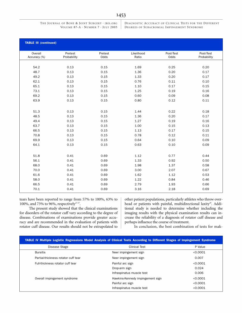
The results of the logistic regression analysis are sum-marized in Table IV. The Neer impingement sign was the onlytest to predict bursitis alone or partial-thickness rotator cufftears. Other clinical tests did not significantly increase theability of the regression analysis to predict bursitis alone orpartial-thickness rotator cuff tears. The painful arc sign, drop-arm sign, and infraspinatus muscle test were retained as vari-ables in the logistic regression analysis to predict full-thicknessrotator cuff tears. The Hawkins-Kennedy impingement sign,the painful arc sign, and the infraspinatus muscle strength testwere retained as variables in the logistic regression analysis topredict overall impingement syndrome. The results of the lo-gistic regression analysis showed that combinations of clinicaltests increased the likelihood ratios and the post-test probabil-ities for overall impingement syndrome and full-thickness ro-tator cuff tears (Table V).
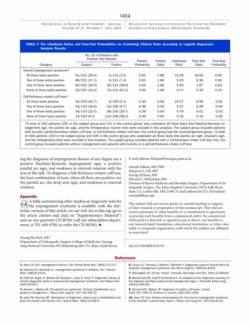
The logistic regression analysis showed that if the Hawkins-Kennedy impingement sign, the painful arc sign, and the in-fraspinatus muscle test were all positive, the likelihood ratio was10.56, and the post-test probability was 0.95 for any degree ofimpingement syndrome. In other words, the likelihood that apatient who tested positive on all three tests would have im-pingement syndrome of some degree was >95%. If these threetests were all negative, the likelihood ratio was 0.17 and thepost-test probability was 0.24, indicating that the likelihood thata patient who had negative findings on all three tests wouldhave impingement syndrome of any degree was <24%.
If the painful arc sign, drop-arm sign, and infraspinatusmuscle test were all positive, the likelihood ratio was 15.57, thepost-test probability was 0.91, and a full-thickness rotator cufftear could be ruled in. If these three tests were all negative, thelikelihood ratio was 0.16, the post-test probability was 0.09,and the probability of a full-thickness rotator cuff tear wouldbe very low.
To analyze the effect of patient age on the results, as sug-gested by Murrell and Walton9, we divided all patients into two
groups according to age: those who were sixty years or older andthose who were younger than sixty years9. For the three degreesof rotator cuff disease, a significant correlation was found onlybetween the age of the patient and the presence of a full-thicknessrotator cuff tear (p < 0.0001). For full-thickness rotator cufftears, if three tests (the painful arc sign, drop-arm sign, and in-fraspinatus muscle test) were positive and the patients weresixty years old or older, the likelihood ratio was 28.00 and thepost-test probability was 0.95. If these three tests were negativeand patients were less than sixty years old, the likelihood ratiowas 0.09 and the post-test probability was 0.06.
Discussionur study compares favorably with other studies in the lit-erature on this subject, although our methodology dif-
fered in several ways. Previous studies have used a positiveresponse to a Neer impingement sign, a positive finding on amagnetic resonance image, or a positive ultrasound evalua-tion as the standard for diagnosing rotator cuff problems3,16.Four previous studies have used surgical findings as inclusioncriteria, but those studies did not have a control group or didnot stratify the patient groups by the degree of disease6,8,9,17.
The study that was most similar in method to ours wasreported by Murrell and Walton9, who prospectively studied400 patients with and without rotator cuff tears. They prospec-tively performed twenty-three different clinical tests on each ofthe 400 patients, all of whom subsequently underwent arthros-copy. The three tests found to be most diagnostic for rotatorcuff disease were weakness in external rotation, weakness in ab-duction, and a positive Neer or Hawkins-Kennedy impinge-ment sign. Murrell and Walton9 found that a patient who testedpositive on all three tests, or who tested positive on any two ofthe three tests and was more than sixty years old, had a 98%likelihood of having a partial or full-thickness rotator cuff tear.
Our finding that most tests for rotator cuff disease havegreater sensitivity than specificity is supported by other studiesin the literature3,8. Many of the physical examination tests stud-ied can be positive in the presence of other shoulder conditions,and the clinician should consider the results of the examinationon the basis of the clinical presentation of the patient.
O
TABLE II (continued)
Overall Accuracy (%)
Pretest Probability
Pretest Odds
Likelihood Ratio
Post-Test Odds
Post-Test Probability
68.3 0.65 1.86 2.17 4.04 0.80
69.7 0.65 1.86 2.12 3.94 0.80
76.1 0.65 1.86 3.89 7.23 0.88
60.2 0.65 1.86 4.20 7.81 0.89
54.4 0.65 1.86 2.29 4.26 0.81
47.8 0.65 1.86 1.25 2.33 0.70
48.6 0.65 1.86 2.32 4.32 0.81
58.7 0.65 1.86 4.20 7.81 0.89

1452
TH E JO U R NA L OF BONE & JOINT SURGER Y · JBJS .ORG
VO LU M E 87-A · NU M B E R 7 · JU LY 2005DIAG N OST I C ACC U R A CY OF CLINIC A L TE S T S FOR THE DIFFERENT DE G RE E S OF SU B A C ROM I A L IMPINGEMENT SYN DROME
Our results could have been influenced by the use of acontrol group that consisted of patients with other conditionsthat required shoulder surgery. It is common in case-controlseries to use as controls other patients undergoing surgery foran unrelated problem, and both the studies by Murrell andWalton9 and MacDonald et al.8 used this approach18,19.
Discrepancies between our study and others in the liter-ature may have resulted from several factors. First, few previ-ous studies have examined the results of these tests in ourthree distinct groups: no rotator cuff tear, partial-thickness ro-tator cuff tear, or full-thickness rotator cuff tear. Second, thedistribution of rotator cuff tears in our study may have dif-fered from that in other studies. Finally, the variability in thedegree of symptoms at the times of examination could haveinfluenced our results.
One limitation of our study is that we did not investigatethe reliability and reproducibility of the physical examinations.In a study by Young et al.20 in England, an orthopaedic consult-ant and senior registrar each examined fifty patients with rota-tor cuff disease. They reported no significant difference betweenthe findings of the two examiners for any of the signs studied,
which included the drop-arm test, the Neer and the Hawkins-Kennedy impingement signs, weakness in abduction and exter-nal rotation, the painful arc sign, the Speed test, the Yergasontest, and the Gerber lift-off test.
Another limitation of our study was that we did not eval-uate every test described for the diagnosis of rotator cuff disease(for example, lag signs14, the lift-off test21, and the hornblowersign22). It is possible that the inclusion of those tests or combi-nations thereof might have influenced the results reported inthe present study. In addition, we did not evaluate the useful-ness of the physical examination for predicting the surgical re-sult. It is possible that the clinical value of these tests could bealtered by including the surgical results in the analysis.
We also did not analyze the use of imaging as part of thedecision-making process. Although plain radiographs, arthro-grams, ultrasound, and magnetic resonance imaging were used,the results were not included in our analysis because the scanswere performed at multiple locations with use of sequences thatwere not standardized or consistent for the entire cohort. A re-view of the literature revealed that the sensitivity, specificity, andaccuracy of ultrasound for making the diagnosis of rotator cuff
TABLE III The Diagnostic Values of the Eight Clinical Tests for Three Different Degrees of Severity of Rotator Cuff Disease in Impingement Syndrome ➤
Clinical Test by Group Sensitivity (%) Specificity (%)Positive Predictive
Value (%)Negative Predictive
Value (%)
Group 1 (bursitis)
Neer sign 85.7 49.2 20.9 95.7
Hawkins-Kennedy sign 75.7 44.5 17.4 92.2
Painful arc sign 70.6 46.9 12.3 93.8
Supraspinatus muscle test 25.0 66.9 8.8 87.4
Speed test 33.3 69.8 14.1 87.6
Cross-body adduction test 25.4 79.7 14.9 88.5
Drop-arm test 13.6 77.3 8.0 86.0
Infraspinatus muscle test 25.0 68.9 9.4 87.7
Group 2 (partial-thickness rotator cuff tear)
Neer sign 75.4 47.5 18.1 92.6
Hawkins-Kennedy sign 75.4 44.4 17.0 92.2
Painful arc sign 67.4 47.0 14.9 91.3
Supraspinatus muscle test 32.1 67.8 11.6 88.4
Speed test 33.3 70.6 16.1 88.8
Cross-body adduction test 16.7 78.5 9.9 86.9
Drop-arm test 14.3 77.5 8.0 86.8
Infraspinatus muscle test 19.4 69.1 10.1 87.7
Group 3 (full-thickness rotator cuff tear)
Neer sign 59.3 47.2 41.3 64.9
Hawkins-Kennedy sign 68.7 48.3 45.2 71.2
Painful arc sign 75.8 61.8 61.0 76.4
Supraspinatus muscle test 52.6 82.4 68.0 71.0
Speed test 39.9 75.3 50.3 66.6
Cross-body adduction test 23.4 80.8 44.6 61.5
Drop-arm test 34.9 87.5 65.0 66.8
Infraspinatus muscle test 50.5 84.0 69.1 70.5
1453
TH E JO U R NA L OF BONE & JOINT SURGER Y · JBJS .ORG
VO LU M E 87-A · NU M B E R 7 · JU LY 2005DIAG N OST I C ACC U R A CY OF CLINIC A L TE S T S FOR THE DIFFERENT DE G RE E S OF SU B A C ROM I A L IMPINGEMENT SYN DROME
tears have been reported to range from 57% to 100%, 63% to100%, and 73% to 96%, respectively23-27.
The present study showed that the clinical examinationsfor disorders of the rotator cuff vary according to the degree ofdisease. Combinations of examinations provide greater accu-racy and are recommended in the evaluation of patients withrotator cuff disease. Our results should not be extrapolated to
other patient populations, particularly athletes who throw over-head or patients with painful, multidirectional laxity28. Addi-tional study is needed to determine whether including theimaging results with the physical examination results can in-crease the reliability of a diagnosis of rotator cuff disease andperhaps influence the course of treatment.
In conclusion, the best combination of tests for mak-
TABLE III (continued)
Overall Accuracy (%)
Pretest Probability
Pretest Odds
Likelihood Ratio
Post-Test Odds
Post-Test Probability
54.2 0.13 0.15 1.69 0.25 0.20
48.7 0.13 0.15 1.36 0.20 0.17
49.2 0.13 0.15 1.33 0.20 0.17
62.1 0.13 0.15 0.76 0.11 0.10
65.1 0.13 0.15 1.10 0.17 0.15
73.1 0.13 0.15 1.25 0.19 0.16
69.2 0.13 0.15 0.60 0.09 0.08
63.9 0.13 0.15 0.80 0.12 0.11
51.3 0.13 0.15 1.44 0.22 0.18
48.5 0.13 0.15 1.36 0.20 0.17
49.4 0.13 0.15 1.27 0.19 0.16
63.7 0.13 0.15 1.00 0.15 0.13
66.5 0.13 0.15 1.13 0.17 0.15
70.8 0.13 0.15 0.78 0.12 0.11
69.9 0.13 0.15 0.64 0.10 0.09
64.1 0.13 0.15 0.63 0.10 0.09
51.8 0.41 0.69 1.12 0.77 0.44
56.1 0.41 0.69 1.33 0.92 0.50
68.0 0.41 0.69 1.98 1.37 0.58
70.0 0.41 0.69 3.00 2.07 0.67
61.6 0.41 0.69 1.62 1.12 0.53
58.0 0.41 0.69 1.22 0.84 0.46
66.5 0.41 0.69 2.79 1.93 0.66
70.1 0.41 0.69 3.16 2.18 0.69
TABLE IV Multiple Logistic Regressions Model Analysis of Clinical Tests According to Different Stages of Impingement Syndrome
Disease Stage Clinical Test P Value
Bursitis Neer impingement sign <0.0001
Partial-thickness rotator cuff tear Neer impingement sign 0.007
Full-thickness rotator cuff tear Painful arc sign <0.0001
Drop-arm sign 0.024
Infraspinatus muscle test 0.006
Overall impingement syndrome Hawkins-Kennedy impingement sign <0.0001
Painful arc sign <0.0001
Infraspinatus muscle test <0.0001
1454
TH E JO U R NA L OF BONE & JOINT SURGER Y · JBJS .ORG
VO LU M E 87-A · NU M B E R 7 · JU LY 2005DIAG N OST I C ACC U R A CY OF CLINIC A L TE S T S FOR THE DIFFERENT DE G RE E S OF SU B A C ROM I A L IMPINGEMENT SYN DROME
ing the diagnosis of impingement disease of any degree are apositive Hawkins-Kennedy impingement sign, a positivepainful arc sign, and weakness in external rotation with thearm at the side. To diagnose a full-thickness rotator cuff tear,the best combination of tests, when all three are positive, arethe painful arc, the drop-arm sign, and weakness in externalrotation.
AppendixA table summarizing other studies on diagnostic tests forthe impingement syndrome is available with the elec-
tronic versions of this article, on our web site at jbjs.org (go tothe article citation and click on “Supplementary Material”)and on our quarterly CD-ROM (call our subscription depart-ment, at 781-449-9780, to order the CD-ROM). �
Hyung Bin Park, MDDepartment of Orthopaedic Surgery, College of Medicine, Gyeong Sang National University, 90 Chilamdong 660-751, Jinju, South Korea.
E-mail address: [email protected]
Atsushi Yokota, MD, PhDHarpreet S. Gill, MDGeorge El Rassi, MDEdward G. McFarland, MDDivision of Sports Medicine and Shoulder Surgery, Department of Or-thopaedic Surgery, The Johns Hopkins University, 10753 Falls Road, Suite 215, Lutherville, MD 21093. E-mail address for E.G. McFarland: [email protected]
The authors did not receive grants or outside funding in support of their research or preparation of this manuscript. They did not receive payments or other benefits or a commitment or agreement to provide such benefits from a commercial entity. No commercial entity paid or directed, or agreed to pay or direct, any benefits to any research fund, foundation, educational institution, or other char-itable or nonprofit organization with which the authors are affiliated or associated.
doi:10.2106/JBJS.D.02335
References
1. Neer CS 2nd. Impingement lesions. Clin Orthop Relat Res. 1983;173:70-7.
2. Hawkins RJ, Kennedy JC. Impingement syndrome in athletes. Am J Sports Med. 1980;8:151-8.
3. Calis M, Akgun K, Birtane M, Karacan I, Calis H, Tuzun F. Diagnostic values of clinical diagnostic tests in subacromial impingement syndrome. Ann Rheum Dis. 2000;59:44-7.
4. Kessel L, Watson M. The painful arc syndrome. Clinical classification as a guide to management. J Bone Joint Surg Br. 1977;59:166-72.
5. Jobe FW, Moynes DR. Delineation of diagnostic criteria and a rehabilitation pro-gram for rotator cuff injuries. Am J Sports Med. 1982;10:336-9.
6. Leroux JL, Thomas E, Bonnel F, Blotman F. Diagnostic value of clinical tests for shoulder impingement syndrome. Rev Rhum Engl Ed. 1995;62:423-8.
7. McLaughlin HL. On the “frozen” shoulder. Bull Hosp Joint Dis. 1951;12:383-93.
8. MacDonald PB, Clark P, Sutherland K. An analysis of the diagnostic accuracy of the Hawkins and Neer subacromial impingement signs. J Shoulder Elbow Surg. 2000;9:299-301.
9. Murrell GAC, Walton JR. Diagnosis of rotator cuff tears. Lancet. 2001;357:769-70. Erratum in: Lancet. 2001;357:1452.
10. Neer CS 2nd. Anterior acromioplasty for the chronic impingement syndrome in the shoulder: a preliminary report. J Bone Joint Surg Am. 1972;54:41-50.
TABLE V The Likelihood Ratios and Post-Test Probabilities for Combining Clinical Tests According to Logistic Regression Analysis Results
Category
No. (%) of Patients with Positive Test Results
Pretest Probability
Pretest Odds
Likelihood Ratio
Post-Test Odds
Post-Test ProbabilitySubject Control
Overall impingement syndrome*
All three tests positive 61/231 (26.4) 3/121 (2.5) 0.65 1.86 10.56 19.64 0.95
Two of three tests positive 86/231 (37.2) 9/121 (7.4) 0.65 1.86 5.03 9.36 0.90
One of three tests positive 60/231 (26.0) 35/121 (28.9) 0.65 1.86 0.90 1.67 0.63
None of three tests positive 24/231 (10.4) 74/121 (61.2) 0.65 1.86 0.17 0.32 0.24
Full-thickness rotator cuff tear†
All three tests positive 50/153 (32.7) 4/195 (2.1) 0.39 0.64 15.57 9.96 0.91
Two of three tests positive 53/153 (34.6) 19/195 (9.7) 0.39 0.64 3.57 2.28 0.69
One of three tests positive 36/153 (23.5) 58/195 (29.7) 0.39 0.64 0.79 0.51 0.33
None of three tests positive 14/153 (9.2) 114/195 (58.5) 0.39 0.64 0.16 0.10 0.09
*A total of 352 patients (231 in the subject group and 121 in the control group) who underwent all three tests (the Hawkins-Kennedy im-pingement sign, the painful arc sign, and the infraspinatus muscle test) were included in this analysis. The subject group included patientswith bursitis, partial-thickness rotator cuff tear, or full-thickness rotator cuff tear; the control group was the nonimpingement group. †A totalof 348 patients (153 in the subject group and 195 in the control group) who underwent all three tests (the painful arc sign, drop-arm sign,and the infraspinatus test) were included in this analysis. The subject group included patients with a full-thickness rotator cuff tear only; thecontrol group included patients without impingement and patients with bursitis or a partial-thickness rotator cuff tear.

1455
TH E JO U R NA L OF BONE & JOINT SURGER Y · JBJS .ORG
VO LU M E 87-A · NU M B E R 7 · JU LY 2005DIAG N OST I C ACC U R A CY OF CLINIC A L TE S T S FOR THE DIFFERENT DE G RE E S OF SU B A C ROM I A L IMPINGEMENT SYN DROME
11. Malanga GA, Jenp YN, Growney ES, An KN. EMG analysis of shoulder posi-tioning in testing and strengthening the supraspinatus. Med Sci Sports Exerc. 1996;28:661-4.
12. Gilcreest EL, Albi P. Unusual lesions of muscles and tendons of the shoulder girdle and upper arm. Surg Gynecol Obstet. 1939;68:903-17.
13. Codman EA. The shoulder. Rupture of the supraspinatus tendon and other lesions in or about the subacromial bursa. Boston: Thomas Todd; 1934. Rupture of the supraspinatus tendon; p 123-77.
14. Hertel R, Ballmer FT, Lambert SM, Gerber C. Lag signs in the diagnosis of rotator cuff rupture. J Shoulder Elbow Surg. 1996;5:307-13.
15. Walton J, Mahajan S, Paxinos A, Marshall J, Bryant C, Shnier R, Quinn R, Murrell GAC. Diagnostic values of tests for acromioclavicular joint pain. J Bone Joint Surg Am. 2004;86:807-12.
16. Naredo E, Aguado P, De Miguel E, Uson J, Mayordomo L, Gijon-Banos J, Martin-Mola E. Painful shoulder: comparison of physical examination and ultra-sonographic findings. Ann Rheum Dis. 2002;61:132-6.
17. Holtby R, Razmjou H. Validity of the supraspinatus test as a single clinical test in diagnosing patients with rotator cuff pathology. J Orthop Sports Phys Ther. 2004;34:194-200.
18. Altman DG. Practical statistics for medical research. London: Chapman and Hall; 1991. Some common problems in medical research; p 396-439.
19. Dawson B, Trapp RG. Basic and clinical biostatistics. 3rd ed. New York: Lange Medical Books/McGraw-Hill; 2001. Reading the medical literature; p 304-33.
20. Young C, Nanda R, Liow RYL, Rangan A. Diagnostic accuracy of clinical signs
in rotator cuff disease. Proceedings of the British Elbow and Shoulder Society [abstract]. J Bone Joint Surg Br. 2003;85 Suppl 1:69.
21. Gerber C, Krushell RJ. Isolated rupture of the tendon of the subscapularis muscle. Clinical features in 16 cases. J Bone Joint Surg Br. 1991;73:389-94.
22. Walch G, Boulahia A, Calderone S, Robinson AHN. The ‘dropping’ and ‘hornblower’s’ signs in evaluation of rotator-cuff tears. J Bone Joint Surg Br. 1998;80:624-8.
23. Teefey SA, Hasan SA, Middleton WD, Patel M, Wright RW, Yamaguchi K. Ultrasonography of the rotator cuff. A comparison of ultrasonographic and ar-throscopic findings in one hundred consecutive cases. J Bone Joint Surg Am. 2000;82:498-504.
24. Brandt TD, Cardone BW, Grant TH, Post M, Weiss CA. Rotator cuff sonogra-phy: a reassessment. Radiology. 1989;173:323-7.
25. Roberts CS, Beck DJ Jr, Heinsen J, Seligson D. Review article: diagnostic ultrasonography: applications in orthopaedic surgery. Clin Orthop Relat Res. 2002;401:248-64.
26. Wallny T, Wagner UA, Prange S, Schmitt O, Reich H. Evaluation of chronic tears of the rotator cuff by ultrasound. A new index. J Bone Joint Surg Br. 1999;81:675-8.
27. Pattee GA, Snyder SJ. Sonographic evaluation of the rotator cuff: correlation with arthroscopy. Arthroscopy. 1988;4:15-20.
28. McFarland EG, Kim TK, Park HB, Neira CA, Gutierrez MI. The effect of varia-tion in definition on the diagnosis of multidirectional instability of the shoulder. J Bone Joint Surg Am. 2003;85:2138-44.