Diagnosing occult tumour cells and their predictive value in sentinel nodes of histologically...
26
Accepted Manuscript Title: Diagnosing occult tumour cells and their predictive value in sentinel nodes of histologically negative patients with colorectal cancer Authors: E.S. van der Zaag, N. Kooij, M.J. van de Vijver, W.A. Bemelman, H.M. Peters, C.J. Buskens PII: S0748-7983(09)00516-2 DOI: 10.1016/j.ejso.2009.11.008 Reference: YEJSO 2919 To appear in: European Journal of Surgical Oncology Received Date: 12 July 2009 Revised Date: 11November2009 Accepted Date: 16 November 2009 Please cite this article as: van der Zaag ES, Kooij N, van de Vijver MJ, Bemelman WA, Peters HM, Buskens CJ. Diagnosing occult tumour cells and their predictive value in sentinel nodes of histologically negative patients with colorectal cancer, European Journal of Surgical Oncology (2009), doi: 10.1016/ j.ejso.2009.11.008 This is a PDF file of an unedited manuscript that has been accepted for publication. As a service to our customers we are providing this early version of the manuscript. The manuscript will undergo copyediting, typesetting, and review of the resulting proof before it is published in its final form. Please note that during the production process errors may be discovered which could affect the content, and all legal disclaimers that apply to the journal pertain.
-
Upload
independent -
Category
Documents
-
view
1 -
download
0
Transcript of Diagnosing occult tumour cells and their predictive value in sentinel nodes of histologically...

Accepted Manuscript
Title: Diagnosing occult tumour cells and their predictive value in sentinel nodes ofhistologically negative patients with colorectal cancer
Authors: E.S. van der Zaag, N. Kooij, M.J. van de Vijver, W.A. Bemelman, H.M.Peters, C.J. Buskens
PII: S0748-7983(09)00516-2
DOI: 10.1016/j.ejso.2009.11.008
Reference: YEJSO 2919
To appear in: European Journal of Surgical Oncology
Received Date: 12 July 2009
Revised Date: 11November2009
Accepted Date: 16 November 2009
Please cite this article as: van der Zaag ES, Kooij N, van de Vijver MJ, Bemelman WA, Peters HM,Buskens CJ. Diagnosing occult tumour cells and their predictive value in sentinel nodes of histologicallynegative patients with colorectal cancer, European Journal of Surgical Oncology (2009), doi: 10.1016/j.ejso.2009.11.008
This is a PDF file of an unedited manuscript that has been accepted for publication. As a service toour customers we are providing this early version of the manuscript. The manuscript will undergocopyediting, typesetting, and review of the resulting proof before it is published in its final form. Pleasenote that during the production process errors may be discovered which could affect the content, and alllegal disclaimers that apply to the journal pertain.

MANUSCRIP
T
ACCEPTED
ARTICLE IN PRESS 1
Diagnosing occult tumour cells and their predictive value in
sentinel nodes of histologically negative patients with colorectal
cancer.
E.S. van der Zaag1, N. Kooij2, M.J. van de Vijver3, W.A. Bemelman4, H.M. Peters2, C.J.
Buskens1.
Departments of Surgery1 and Pathology2, Gelre Ziekenhuizen, Apeldoorn, The Netherlands.
Departments of Pathology3 and Surgery4, Academic Medical Center, Amsterdam, The
Netherlands
Running title: Sentinel node procedure for occult tumour cells
Word count: 2809
Corresponding address:
E.S. van der Zaag
Department of Surgery
Gelre Ziekenhuizen
Albert Schweitzerlaan 31
7334 DZ Apeldoorn
The Netherlands
0031-55-8446016

MANUSCRIP
T
ACCEPTED
ARTICLE IN PRESS 2
ABSRACT
Purpose: Most studies on the sentinel node (SN) procedure in patients with colorectal cancer
include immunohistochemical analysis of the SN only. To evaluate the real diagnostic
accuracy of the SN-procedure with immunohistochemical analysis, the presence of occult
tumour cells in all histologically negative lymph nodes was compared to the presence of these
cells in SNs. Also the reproducibility of diagnosing occult tumour cells (OTC) and the
sensitivity of three different antibodies was assessed.
Methods: Between November 2006 en July 2007, an ex vivo SN procedure was performed in
58 histologically N0 patients with colorectal cancer. All lymph nodes (n=908, mean 15.7)
were step-sectioned and immunohistochemistry was performed using two antibodies against
cytokeratins (Cam5.2, and CK 20) and one antibody against BerEp-4.
Results: OTC were identified in 19 of 58 patients, with micrometastases (0.2-2mm) in 7 and
isolated tumour cells (ITC)(< 0.2mm) in 12 patients. The overall agreement in diagnosing
OTC between two independent pathologists was 86%. A SN was identified in 53 of 58
patients. All micrometastases were found in SNs. In two patients with negative SNs, ITC’s
were demonstrated in non-SNs (sensitivity 88%, and overall accuracy 96%).
Conclusion: Additional immunohistochemical analysis of histologically negative lymph
nodes demonstrates occult tumour cells in 33% of the patients resulting in an upstaging rate of
12%. Occult tumour cells are predominantly found in the SN, therefore SN mapping has the
potential to refine the staging system for patients with colorectal cancer.
Key words: Occult tumour cells, Sentinel lymph node, colorectal cancer

MANUSCRIP
T
ACCEPTED
ARTICLE IN PRESS 3
INTRODUCTION
Sentinel lymph node (SN) mapping is an increasingly popular technique to improve staging
accuracy for patients with colorectal cancer by identifying nodes with the highest likelihood
to harbour metastatic disease. This technique has shown promising results for colon
carcinomas with SN mapping accurately predicting nodal status in 80-100%.[1-24] An
advantage of SN mapping is the potential to use this technique for a more accurate and cost-
effective pathologic assessment of lymph node status. The additional stepwise sectioning
combined with immunohistochemical analysis of the SN has been demonstrated to detect
occult tumour cells (OTC) in up to 40% of histologically negative patients with colorectal
cancer[1-24], possibly identifying a patient group that may benefit from adjuvant systemic
therapy.
However, previously reported results are difficult to interpret because most studies publishing
SN results include immunohistochemical analysis of the SN only. This might result in
flattering negative predicting values and sensitivity rates. Moreover, most studies published
so far have not subdivided the finding of OTC into micrometastases and isolated tumour cells
(ITC) on the basis of their dimensions as recommended by the American Joint Committee on
Cancer (AJCC) staging manual.[25] This possibly results in an overestimation of the number
of upstaged patients and makes the clinical significance of the detected OTC difficult to
evaluate. The widespread use of SN immunohistochemical analysis in patients with breast
cancer has demonstrated that stage migration due to SN mapping harbours the danger of
potential misleading effects and artefacts in prognostication.[26] This highlights the need to
differentiate correctly between clinically relevant (micro)metastases (0.2-2 mm)(pN1mi+) and
immunohistochemically detected isolated tumour cells without prognostic significance
(<0.2mm)(pN0itc+).

MANUSCRIP
T
ACCEPTED
ARTICLE IN PRESS 4
This is particularly so since the main purpose of SN mapping in patients with colorectal
cancer is to establish a more accurate staging of patients at risk of recurrence due to
lymfogenic spread and who could benefit from adjuvant chemotherapy.
For clinical application with therapeutical consequences, an immunohistochemical marker for
OTC has to be both highly sensitive (i.e. detect the majority of tumour cells), and specific (i.e.
absence of false positive staining of cells). Since most studies use different antibodies without
a gold standard and uniform definition of OTC, the antibody of first choice for the detection
of clinically relevant micrometastases in patients with colorectal cancer is still difficult to
determine on the basis of published data.
We performed this study to evaluate the real diagnostic accuracy of upstaging N0 patients
with colorectal cancer by SN-procedure with immunohistochemical analysis. Therefore the
incidence of OTC in SNs was compared to the presence of these cells in all histologically
negative lymph nodes of these patients. Two experienced pathologists independently assessed
the presence of lymph node metastases. The detection of OTC was analysed by three different
antibodies to determine the sensitivity and specificity of these antibodies. In addition, the
presence of OTC was correlated to clinicopathological parameters.

MANUSCRIP
T
ACCEPTED
ARTICLE IN PRESS 5
PATIENTS AND METHODS
Patients
Between November 2006 and July 2007, an ex vivo SN procedure was performed in 100
patients with colorectal carcinoma who were operated on with curative intent. Exclusion
criteria for SN mapping were invasion of other organs (T4 carcinomas), or two simultaneous
colorectal carcinomas. Patients with locally advanced rectal cancer who underwent
neoadjuvant chemoradiotherapy were excluded because of stage migration before surgical
treatment. Of the 100 patients 42 patients were shown to have macrometastases in one or
more lymph nodes. The remaining 58 patients were histologically staged as N0 (Dukes A or
B), and they comprise the population of the current study. All patients with colon cancer
underwent an oncological resection, including the mesocolon of the vascular trunk. Patients
with rectal carcinoma underwent total mesorectal excision (TME) after preoperative short-
course radiotherapy comprising five fractions of five Gray. In addition, the mesenteric lymph
nodes of 6 patients were included who underwent colonic resection for benign disease (e.g.
Crohn’s disease or diverticulosis) as a negative control group.
The study was done in accordance with the guidelines of the local ethics committee.
Sentinel lymph node mapping
In this study, the ex vivo lymph mapping was used since only this technique can also be
performed easily in patients with rectal carcinomas. The ex vivo technique has been
demonstrated to be as accurate as the in vivo technique in identifying SNs[27, 28], and it
facilitates standardised, uniform specimen processing and assessment, recognising that the
principal limitation of this technique is its inability to detect a small percentage (1-10%) of
cases with aberrant lymphatic drainage.[2, 16, 19] After standard resection, 0.5-2 ml
(depending on the volume of the tumour) patent blue V (Guerbet, Gorinchem, The

MANUSCRIP
T
ACCEPTED
ARTICLE IN PRESS 6
Netherlands) was injected around the tumour with the colonic specimen left intact.[29] For
colonic carcinomas, the mesocolon was inspected and the first one to four blue lymph nodes
were identified as SNs and either dissected or marked with a suture. For rectal carcinomas the
specimen was sent to the department of pathology immediately after resection in fresh state.
The identification of blue nodes was performed at the department of pathology immediatly, to
keep the circumferential resection margin intact.
Immunohistochemical staining
The surgical resection specimens were analysed at the pathology department using a
standardised protocol. All lymph nodes (SNs and non-SNs) were collected in separate boxes
and marked according to location, then cut in two with both sides stained with hematoxylin
and eosin and evaluated for tumour involvement. Of the 58 histologically N0 patients, serial
sectioning was performed at 500μm intervals of all lymph nodes from formalin-fixed and
paraffin-embedded archival tissue blocks. Three serial sections at three separate levels were
immunohistochemically stained with three different monoclonal antibodies: the anti-epithelial
cell antibody Ber-EP4 (DAKO, The Netherlands), was combined with two anti-cytokeratin
antibodies: the anti-CK20 antibody, with its expression limited to gastrointestinal epithelial
cells (Euro Diagnostica, Arnhem, The Netherlands), and the anticytokeratin marker Cam5.2
directed against cytokeratin 7 and 8 expressed in all epithelial cells (Becton and Dickinson,
Alphen aan den Rijn, The Netherlands). Appropriate positive and negative controls were
added on each automated run, to confirm the sensitivity and specificity of the antibodies
(sections of colonic carcinoma tissue served as positive controls; negative controls were
obtained by omitting the primary antibody). The staining procedures have been described in
detail previously.[29]

MANUSCRIP
T
ACCEPTED
ARTICLE IN PRESS 7
Microscopic evaluation and definitions
All slides were reviewed by two independent experienced pathologists who were unaware of
the clinical data. The type of involvement (single tumour cells or clusters), topography
(lymph-angio invasion and/or lymph sinuses and/ or parenchyma) and extension of the lesion
was assessed in each case (Figure 1).
Occult tumour cells were classified as either an ITC (diameter < 0.2 mm) or a micrometastasis
(diameter between 0.2 mm and 2 mm). Patients were then restaged according to the AJCC
classification.[25] OTC comprised of micrometastases (0.2-2 mm)(pN1mi+) and ITC
(<0.2mm)(pN0itc+). In case of disagreement, the slides were re-evaluated in a consensus
meeting. False-positive non-neoplastic haematopoeitic cells (e.g. reticular cells and plasma
cells which can also show staining for cytokeratins), were discriminated from ITCs on the
basis of histopathologic features.
Statistical analyses
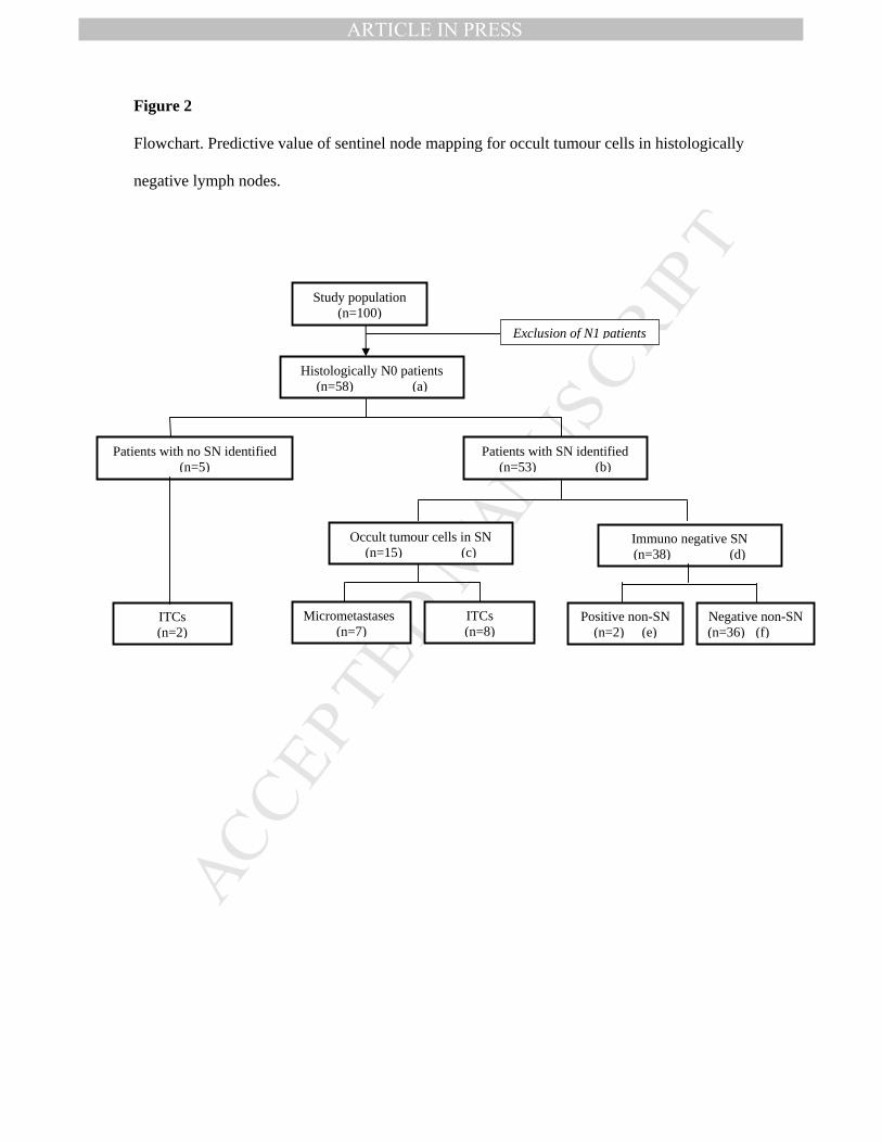
The following definitions were used for calculations (Figure 2). Identification rate is the
number of patients with one or more SN identified (b) divided by the total number of
procedures (a) x 100%. Sensitivity is the number of patients with an immunohistochemically
positive SN (c) divided by the total number of patients with detected OTC (c+e) x 100%.
Negative SNs were considered false negative if one of the other regional lymph nodes (non-
SNs) demonstrated micrometastases or ITCs (e). The accuracy of the immunohistochemical
SN analysis suggests a conformity of the SN status and the regional nodal status, i.e. the total
number of patients with an immunohistochemically positive SN (c) plus the number of
patients with a true-negative SN (f)/ the number of patients with an identified SN (b) x 100%.

MANUSCRIP
T
ACCEPTED
ARTICLE IN PRESS 8
All statistical analyses were performed using the Statistical Software Package version 14.0
(SPSS INC., Chicago, IL, USA). Associations between clinicopathological features and the
presence of micrometastases were analysed using Student's t-test (continuous variables) or
Chi-squared test (categorical variables). P-values of 0.05 or less were considered statistically
significant.

MANUSCRIP
T
ACCEPTED
ARTICLE IN PRESS 9
RESULTS
Restaging patients by detection of occult tumour cells
Three serial sections of all 908 lymph nodes from 58 patients with N0 carcinomas were
examined with three different antibodies (mean 15.7 lymph nodes per patient, range 6-42). In
27 patients none of the lymph nodes had metastases after immunohistochemical staining and
in 8 patients one or more lymph nodes demonstrated false positive staining unanimously
recognised by both pathologists. There was consensus on 6 patients with micrometastases and
9 patients with ITCs; overall agreement 86% (50 of 58 patients). For the remaining 8 patients
there was discrepancy in the diagnosis by the two pathologists. In these 8 patients, one
pathologist diagnosed one micrometastasis and 10 ITCs in 11 lymph nodes, whereas the other
pathologist classified 2 lesions as micrometastases, 6 lesions as ITC and 3 lesions as false
positive staining. It was noted that the discrepancy between micrometastases and ITCs was
mainly due to differences in measuring multiple foci. According to the AJCC 6th edition of
TNM classification of malignant tumours, clusters of tumour cells are considered as one focus
if the nests are separated by a few cells, and when in doubt the lower category should be
selected to prevent upstaging. In a consensus meeting the following was agreed upon: 52
lymph nodes with OTC were found in 19 of the 58 patients (33%); seven patients had
micrometastases (12% pN1mi+) and 12 patients had ITCs (21% pN0itc+)(Table 1).
The presence of OTC (micrometastases and ITCs combined) was not related to any
clinicopathological characteristic (Table 2). Although micrometastases and ITCs were found
more frequently in tumours infiltrating the serosa and in tumours larger than 5cm, this was not
statistically significant. Since micrometastases are thought to represent a different level of
clinical significance, a correlation between pN1(mi+) versus pN0 (itc+/-) and
clinicopathological characteristics was also analysed. The presence of micrometastases was

MANUSCRIP
T
ACCEPTED
ARTICLE IN PRESS 10
also not related to tumour infiltration depth, differentiation grade, and extent of tumour
spread, but a significant relation with lymphangio-invasion could be demonstrated (p<0.001).
Sensitivity and specificity of three different antibodies
No epithelial cells with atypical nuclear features characteristic of carcinoma cells were
identified in lymph nodes from six control patients operated upon for benign disease, although
occasional staining for keratin was observed in stellate cells located in the interfollicular
regions of the nodes consistent with staining of reticulum cells. However, this pattern was
easily distinguishable from the staining of carcinoma cells.
Each micrometastasis was detected with all three antibodies, as were 39 of 45 ITC-positive
lymph nodes. All ITCs were detected with BerEP4. Six OTC were not detected with CK20
and two were missed with Cam5.2. It was noted that the intensity of staining with Cam5.2 and
CK20 was related to differentiation of the primary tumour, and the undetected ITCs were
found in lymph nodes of patients with a dedifferentiated tumour.
In 18% of the patients false-positive staining was detected with BerEP4 (e.g. reticulocytes,
fibroblasts and mastcells) and this antibody yielded the most discordant classifications of
ITCs and false-positive stained cells between the two pathologists. False-positive staining was
less frequently encountered with Cam5.2 (although there was occasional staining of dendritic
cells) and was hardly seen with CK20 immunohistochemical staining. These results would
make Cam5.2 the antibody of first choice since it demonstrated the most consistent staining
with high sensitivity and acceptable specificity.
Diagnostic value of SN mapping
The SN (median two SNs, range 1-4) was found in 53 of the 58 N0 patients (identification
rate 91%). Of the five patients in which the SN could not be identified, two patients
demonstrated OTC in lymph nodes; these cells were all ITC . The remaining 53 patients were
MANUSCRIP
T
ACCEPTED
ARTICLE IN PRESS 11
evaluated for the calculation of the diagnostic value of the SN. Overall, OTC were found in
17 patients; of which 15 were found in the SN and two in a non SN. Resulting in a sensitivity
of 88% (15/17) and a negative predictive value of 94% (34/36)(Figure 2). OTC were found in
18 of the 105 SNs (17%), compared to 34 of 803 non-SNs (4%); this difference was
statistically significant (p<0.001). Because all seven micrometastases were found in the SNs
exclusively, the accuracy of the SN procedure in upstaging patients is 100%.

MANUSCRIP
T
ACCEPTED
ARTICLE IN PRESS 12
DISCUSSION
Value of SN procedure in refining staging of colorectal patients
Our study emphasises the potential value of SN procedure for optimising staging of patients
with colorectal cancer. The examination of 908 lymph nodes by stepwise sections and
immunohistochemical analysis revealed OTC in 33% of histologically N0 patients (12%
micrometastases and 21% ITCs). OTC were predominantly found in SNs with an overall
sensitivity of SN mapping for OTC of 88%.
A large number of studies already addressed the value of SN mapping for colorectal
carcinomas (Table 3).[1-24] In reviewing the literature, most studies demonstrate
identification rates and accuracy rates varying from 80 to 100%, and conclude that this
technique can predict lymph node status with good accuracy rates. However, in these studies
the percentage of histologically N0 patients is often more than 50% which makes the
presented accuracy rates difficult to interpret.[1-7, 9-17, 19-23] This is merely because
accuracy rates up to 90% may be accompanied by sensitivity rates and false negative rates of
50%.[3, 7, 16, 19-21]
For clinical use the SN procedure should be both highly sensitive and specific. Evaluating
upstaging percentages in studies lacking immunohistochemically analysis of non-SNs
harbours the danger of an underestimation of sensitivity rates and the presentation of flattering
negative predictive values. The primary role of SN procedure in patients with colorectal
cancer is to increase the accuracy of staging N0 patients by identifying and analysing those
nodes with the highest likelihood to harbour metastatic disease in a cost-effective manner.
Therefore, it is of great importance that OTC are predominantly found in SNs. We
demonstrate that the incidence of OTC is more than four times higher in SNs compared to
non-SNs. Our results are in agreement with two previous studies that showed SNs to have the
highest probability of harbouring OTC.[23, 24]
MANUSCRIP
T
ACCEPTED
ARTICLE IN PRESS 13
Classification and detection of occult tumour cells
However, our study also emphasises that the classification of OTC remains difficult. Reliable
and reproducible discrimination between micrometastases (pN1) and ITCs (pN0) is an
essential component of adjuvant treatment planning. In analysing discrepant diagnoses for
clinically relevant micrometastases between the two pathologists, we observed that the most
challenging issue was whether multiple tumour clusters should be measured as one. A useful
guideline in the AJCC system is that when controversy exists, the lower category should be
selected. This recommendation was used during the consensus meeting. Another problem was
the distinction between ITCs and false positively staining (non-tumour) cells. Although this
has no impact on treatment recommendations, the inter-observer variation highlights the need
for well defined antibodies.[31] Our data suggest that Cam5.2 would be the antibody of first
choice for the detection of OTC. The anticytokeratin-marker BerEP-4 is more sensitive but
has an almost 20% false positive rate with high inter-observer variation. The use of the highly
specific anti-CK20 with its expression limited to gastrointestinal epithelial cells can not be
recommended due to its loss of expression in dedifferentiated tumours, resulting in low
specificity.
True upstaging rate of SN mapping and predictive value
Comparing the incidence of clinically relevant upstaging by SN mapping with
immunohistochemical analysis to literature is difficult. Before the revised TNM classification
was introduced in 2003 micrometastases were not distinguished from ITCs.[31] Only three
studies discriminated micrometastatic disease from ITCs,[3, 7, 19] with low incidence of
micrometastatic disease varying from 5 to 10%. Other studies reporting higher upstaging rates
all included ITCs in their findings.[5, 10, 12, 13, 23, 24] This variation in definition of
micrometastases makes it difficult to reach well-founded conclusions on the role of OTC and

MANUSCRIP
T
ACCEPTED
ARTICLE IN PRESS 14
could explain the controversial prognostic significance of OTC. So far, only two studies have
analysed the prognostic significance of micrometastatic disease in SNs with a decreased
disease-specific survival in the pN1(mi+) group.[10,11] All other studies commenting on
prognostic significance of OTC did not perform SN mapping.[33] Most studies failed to
demonstrate a relation between the presence of OTC and tumour recurrence or survival.
Messerini et al. demonstrated that the inclusion of ITCs should probably be regarded as a
confounding factor in these studies.[33] Although our study was not designed to comment on
the prognostic significance of OTC, the relation between lymphangio-invasion of tumour cells
and micrometastases is suggestive of clinical relevance. Especially since this relation could
not be demonstrated between lymphangio-invasion and ITCs.
Conclusion
Occult tumour cells are predominantly found in the SN with a true upstaging rate of
12% in patients with pN0 colorectal cancer. For immunohistochemical staining Cam5.2
seems the antibody of choice with an overall agreement of pathologist of 88% in staging
patients. The SN procedure has the potential to refine the staging system for patients with
colorectal cancer. Further survival analyses should be awaited to reveal the real prognostic
relevance of these findings.
MANUSCRIP
T
ACCEPTED
ARTICLE IN PRESS 15
Acknowledgements
The authors greatly acknowledge J. van Benthem and dr E. Weltevreden from the Gelre
Hospital Department of Pathology for excellent technical assistance in staining and reviewing
all immunohistochemically stained slides. Dr. W. Bouma is greatly acknowledged for
supporting this study and critically reading the manuscript.
Conflict of interest statement:
All authors disclose any financial and personal relationships with other people or
organisations that could inappropriately influence our work.
Role of the funding source:
All authors state there is no role of funding sources.

MANUSCRIP
T
ACCEPTED
ARTICLE IN PRESS 16
REFERENCES
1. Matter M, Winckler M, Aellen S, Bouzourene H. Detection of metastatic disease with
sentinel lymph node dissection in colorectal carcinoma patients. Eur J Surg Oncol
2007; 33:1183-90.
2. Kelder W, Braat AE, Karrenbeld A, Grond JA, De Vries JE, Oosterhuis JW, Baas PC,
Plukker JT. The sentinel node procedure in colon carcinoma: a multi-centre study in
The Netherlands. Int J Colorectal Dis 2007; 22:1509-14.
3. Bembenek AE, Rosenberg R, Wagler E, Gretschel S, Sendler A, Siewert JR, Nährig J,
et al. Sentinel lymph node biopsy in colon cancer: a prospective multicenter trial. Ann
Surg 2007; 245:858-63.
4. Stojadinovic A, Nissan A, Protic M, Adair CF, Prus D, Usaj S, Howard RS,
Radovanovic D, et al. Prospective randomized study comparing sentinel lymph node
evaluation with standard pathologic evaluation for the staging of colon carcinoma:
results from the United States Military Cancer Institute Clinical Trials Group Study
G1-01. Ann Surg 2007; 245:864-6.
5. Van Schaik PM, Van der Linden JC, Ernst MF, Gelderman WA, Bosscha K. Ex vivo
sentinel lymph node “mapping” in colorectal cancer. Eur J Surg Oncol 2007;
33 :1177-82.
6. Bianchi PP, Ceriani C, Rottoli M, Torzilli G, Roncalli M, Spinelli A, Montorsi M.
Laparoscopic lymphatic mapping and sentinel lymph node detection in colon cancer:
technical aspects and preliminary results. Surg Endosc 2007; 21:1567-71.
7. Thomas KA, Lechner J, Shen P, Waters GS, Geisinger KR, Levine EA. Use of
sentinel node mapping for cancer of the colon: ‘to map or not to map’. Am Surg 2006;
72:606-11.

MANUSCRIP
T
ACCEPTED
ARTICLE IN PRESS 17
8. Smith J, Hwang H, Wiseman KW, Filipenko D, Phang PT. Ex vivo sentinel lymph
node mapping in colon cancer: improving the accuracy of pathologic staging? Am J
Surg 2006; 191:665-8.
9. Saha S, Seghal R, Patel M, Doan K, Dan A, Bilchik A, Beutler T, Wiese D, Bassily N,
Yee C. A multicenter trial of sentinel lymph node mapping in colorectal cancer:
prognostic implications for nodal staging and recurrence. Am J Surg 2006; 191:305-
10.
10. Codignola C, Zorzi F, Zaniboni A, Mutti S, Rizzi A, Padolecchia E, Morandi GB. Is
there any role for sentinel node mapping in colorectal cancer staging? Personal
experience and review of the literature. Jpn J Clin Oncol 2005; 35:645-50.
11. Braat AE, Oosterhuis JW, Moll FC, de Vries JE, Wiggers T. Sentinel node detection
after preoperative short-course radiotherapy in rectal carcinoma is not reliable. Br J
Surg 2005; 92:1533-8.
12. Cox ED, Kellicut D, Adair C, Marley K, Otchy DP, Peoples GE. Sentinel lymph node
evaluation is technically feasible and may improve staging in colorectal cancer. Curr
Surg 2002; 59:301-6.
13. Smith FM, Coffey JC, Khasri NM, Walsh MF, Parfrey N, Gaffeny E, Stephens R,
Kennedy MJ, Kirwan W, Redmond HP. Sentinel nodes are identifiable in formalin-
fixed specimens after surgeon-performed ex vivo sentinel lymph node mapping in
colorectal cancer. Ann Surg Oncol 2005; 12:504-9.
14. Bell SW, Mourra N, Flejou JF, Parc R, Tiret E. Ex vivo sentinel lymph node mapping
in colorectal cancer. Dis Colon Rectum 2005 ; 48 :74-9.
15. Paramo JC, Summerall J, Poppiti R, Mesko TW. Validation of sentinel node mapping
in patients with colon cancer. Ann Surg Oncol 2002; 9:529-31.

MANUSCRIP
T
ACCEPTED
ARTICLE IN PRESS 18
16. Fitzgerald TL, Khalifa MA, Al Zahrani M, Law CH, Smith AJ. Ex vivo sentinel
lymph node biopsy in colorectal cancer: a feasibility study. J Surg Oncol 2002; 80:27-
32.
17. Bendavid Y, Latulippe JF, Younan RJ, Leclerc YE, Dube S, Heyen F, Morin M,
Girard R, et al. Phase I study on sentinel lymph node mapping in colon cancer: a
preliminary report. J Surg Oncol 2002; 7981-4.
18. Joosten JJ, Strobbe LJ, Wauters CA, Pruszczynski M, Wobbes T, Ruers TJ.
Intraoperative lymphatic mapping and the sentinel node concept in colorectal
carcinoma. Br J Surg 1999; 86:482-6.
19. Bilchik AJ, Hoon DS, Saha S, Turner RR, Wiese D, DiNome M, Koyanagi K, et al.
Prognostic impact of micrometastases in colon cancer: interim results of a prospective
multicenter trial. Ann Surg 2007; 246:568-75.
20. Bertagnolli M, Miedema B, Redston M, et al. Sentinel node staging of resectable
colon cancer: results of a multicenter study. Ann Surg 2004; 240:624-8.
21. Faerden AE, Sjo OH, Andersen SN, Hauglann B, Nazir N, Gravdehaug B, Moberg I,
Svinland A, Nesbakken A, Bakka A. Sentinel node mapping does not improve staging
of lymph node metastasis in colonic cancer. Dis Colon Rectum 2008; 51:891-6.
22. Yagci G, Unlu A, Kurt B, Can MF, Kaymakcioglu N, Cetiner S, Tufan T, Sen D.
Detection of micrometastases and skip metastases with ex vivo sentinel node mapping
in carcinoma of the colon and rectum. Int J Colorectal Dis 2007; 22:167-73.
23. Bembenek A, Schneider U, Gretschel S, Fischer J, Schlag PM. Detection of lymph
node micrometastases and isolated tumor cells I sentinel and nonsentinel lymph nodes
of colon cancer patients. World J Surg 2005; 29:1172-5.

MANUSCRIP
T
ACCEPTED
ARTICLE IN PRESS 19
24. Turner RR, Nora DT, Trocha SD, Bilchik AJ. Frequency and nature of cytokeratin-
positive cells in sentinel and nonsentinel lymph nodes. Arch Pathol Lab Med 2003;
127:673-9.
25. Green FL, Page DL, Fleming ID, et al. AJCC cancer staging manual. 6th ed. New
York, Springer 2002.
26. Singletary SE, Allred C, Ashley P, et al. Revision of the American Joint Committee on
Cancer staging system for breast cancer. J Clin Oncol 2002; 20: 3628-36.
27. Teuch JJ, Pessaux P, Fiore FD, et al. Sentinel node mapping in coloncarcinoma : in-
vivo versus ex-vivo approach. Eur J Surg Oncol 2006 ; 32 :158-161.
28. Wood TF, Saha S, Morton DL, et al. Validation of lymphatic mapping in colorectal
cancer: in vivo, ex vivo, and laparoscopic techniques. Ann Surg Oncol 2001; 8:150-
157.
29. van der Zaag ES, Buskens CJ, Kooij N, Akol H, Peters HM, Bouma WH, Bemelman
WA. Improving staging accuracy in colon and rectal cancer by sentinel lymph node
mapping: a comparative study. Eur J Surg Oncol 2009; Mar 3.
30. Borgen E, Beiske K, Trachsel S, Nesland JM, Kvalheim G, Herstad TK, et al.
Immunocytochemical detection of isolated epithelial cells in bone marrow: non-
specific staining and contribution by plasma cells directly reactive to alkaline
phosphatase. J Pathol 1998;185:427-434.
31. Redston M, Compton CC, Miedema BW, Niedzwiecki D, Dowell JM, Jewell SD,
Fleshman JM, Bem J, Mayer RJ, Bertagnolli MM. Analysis of micrometastatic disease
in sentinel lymph nodes from resectable colon cancer: results of cancer and leukaemia
group B trial 80001. J Clin Oncol 2006; 24:878-883.

MANUSCRIP
T
ACCEPTED
ARTICLE IN PRESS 20
32. De Haas RJ, Wicherts DA, Hobbelink MG, Borel Rinkes IH, Schipper ME, Van der
Zee JA, Van Hillegersberg R. Sentinel lymph node mapping in colon cancer: current
status. Ann Surg Oncol 2007; 14:1070-80.
33. Messerini L, Cianchi F, Cortesini C, Comin CE. Incidence and prognostic significance
of occult tumor cells in lymph nodes from patients with stage IIA colorectal
carcinoma. Hum Pathol 2006; 37:1259-67.

MANUSCRIP
T
ACCEPTED
ARTICLE IN PRESS
Table 1
Classification of immunohistochemical staining results by two independent pathologists of 58
histologically negative patients with colorectal cancer.
Pathologist 1
Micrometastasis Isolated tumour cells
False positive staining
Absent
Micrometastasis 6 1 0 0
Isolated tumour cells 1 9 4 0
Pathologist 2
False positive staining 0 5 8 3
Absent 0 0 2 27
Consensus 7 12 10 29

MANUSCRIP
T
ACCEPTED
ARTICLE IN PRESS
Table 2
The presence of occult tumour cells correlated to tumour characteristics of 58 patients Occult tumour cells
Tumour characteristics
N=58
Micrometastasis (N=7)
ITCs (N=12)
Absent (N=39)
p-value
Depth of invasiona T1 (4) 0 0 4 ns
T2 (18) 2 3 13
T3 (36) 5 9 22
Differentiation grade well (4) 0 0 4 ns
moderate (45) 7 10 28
poor (9) 0 2 7
Extend of tumour spread < 5cm (34) 2 6 26 ns
> 5cm (24) 5 6 13
Lymphangio-invasion absent (51) 3 12 36 0.001
present (7) 4 0 3 aT1: tumour limited to the (sub)mucosa, T2: tumour infiltrates muscularis propria, but not adventitia, T3: tumour infiltrates adventitia.

MANUSCRIP
T
ACCEPTED
ARTICLE IN PRESS
Table 3
Predictive value of blue dye SN mapping in patients with colorectal cancer including
immunohistochemical analyses (comparison of existing literature until may 2008; excluding
duplicate series and princeps studies with updated publications).
Study N No LNs
(mean) N0 (%)
Identification (%)
Accuracy (%)
False neg (%)
Sensitivity (%)
Upstaging (%)
ITCa
Antibody
Immunohistochemical analysis of SN only
Matter1 52 30c 63 92 - 25d 44d 23 N CK11/CEA/Ca19-9
Kelder2 69 11 73 97 96 30d 89d, f 18 N CK7/8/18 Bembenek3 315 20 69 85 86 46 54 5/16g Y MNF16
Stojadinovic4 162 18 65 98 90d 31d 69d, f 11 N AE1/AE3 Cam5.2
Van Schaik5 44 10.5 52 94 100 0 100 30 N LU-5
Bianchi6 22 22c 73 100 95 17 83 13 N CK?
Thomas7 69 16 62 93 78 54 46f 5/-e, g Y CEA/ CK Smith8 17 16 41e 94 100 0 100 20d N MNF116 Saha9 500 15 50d 98 96 10 90 26 N AE1/AE3
Codignola10 56 21 59 100 89e, f 11d 86e, f 38d N Cam5.2
Braat11 91 7.7e 62e 90e 90e 26e 74e, f 12e N CK7/8
Cox12 17 18 82 100 100 0 100f 29 N AE1/AE3
Smith13 40 17 56 98 95e 12 88f 29d N Cam5.2
Bell14 58b 30 57 97 84e 16d 64e 4 N CK
Paramo15 55 12 67e 82 98 3d 93e 11d N Cam5.2
Fitzgerald16 26 15 87e 88 91d 9d 33e 10 N Cam5.2
Bendavid17 20 - 55 90 - - - 25 N AE1/AE3
Joosten18 50 14c 48 70 66e 60 40 13 N LU-5/ CK19
Bilchik19 141 17 73 99 91 39 61e 8/22g Y CK
Bertagnolli20 72 17 66 92 80 54 46 - N AE1/AE3 CEA
Immunohistochemical analysis of all lymph nodes
Faerden21 199 13c 68 93 85 47 53 30 N Cam5.2
Yagci22 47 19 57 98 91e 20 80 15 N AE1/AE3 / CEA
Bembenek23 55 26c 70 85 - 4 96 39 N MNF 116 Turner24 51 14c 55e 100 88e 26e 74e 29 N AE1/AE3
a Discrimination between micrometastasis and isolated tumour cells (N=no, Y=yes) b Specimens c Median d Discrepancy between reported value and calculated value (reported values are mentioned)
e Calculated value f Immunohistochemistry results of SN included in sensitivity g % micrometastases (true upstaging)/ % isolated tumour cells

MANUSCRIP
T
ACCEPTED
ARTICLE IN PRESS
Figure 1
Representative examples of immunohistochemically detected occult tumour cells.
A: micrometastasis > 0.2mm. B: isolated tumour cell, cluster of cells > 0.2mm. C: isolated
tumour cells, cluster of cells < 0.2 mm with characteristic nuclear features. D: false-positively
staining cell
A B
C D
MANUSCRIP
T
ACCEPTED
ARTICLE IN PRESS
Figure 2
Flowchart. Predictive value of sentinel node mapping for occult tumour cells in histologically
negative lymph nodes.
Patients with no SN identified (n=5)
Immuno negative SN (n=38) (d)
Negative non-SN (n=36) (f)
Exclusion of N1 patients
Study population (n=100)
Histologically N0 patients (n=58) (a)
Patients with SN identified (n=53) (b)
Occult tumour cells in SN (n=15) (c)
Micrometastases (n=7)
ITCs (n=8)
Positive non-SN (n=2) (e)
ITCs (n=2)