DHANALAKSHMI SRINIVASAN INSTITUTE OF TECHNOLOGY
33
DHANALAKSHMI SRINIVASAN INSTITUTE OF TECHNOLOGY DEPARTMENT OF BIOMEDICAL ENGINEERING Subject: BM6401 MEDICAL PHYSICS Semester:IV Sem 1. What is a non- ionizing radiation? It refers to any type of electromagnetic radiation that does not carry enough energy per quantum to ionize atoms or molecules—that is, to completely remove an electron from an atom or molecule. 2. Define Electromagnetic (EM) wave. EM waves are the waves that are propagated by simultaneous periodic variations of electric and magnetic field intensity and that include radio waves, infrared, visible light, ultraviolet, X-rays, and gamma rays. 3. What is visual acuity? Visual acuity (VA) is acuteness or clearness of vision. It depends on optical and neural factors, i.e., (i) the sharpness of the retinal focus within the eye, (ii) the intactness and functioning of the retina, and (iii) the sensitivity of the interpretative faculty of the brain. 4. Give any 4 applications of clinical thermography. 1. Assessment of inflammatory conditions such as rheumatoid arthritis. 2. Vascular-disorder studies 3. Metabolic studies. 4. The assessment of pain and trauma. 5. Oncological investigations. 5. What are the natural and artificial piezoelectric materials used in making ultrasound transducers? Natural piezoelectric materials are quarts and Rochelle salts. Artificial piezoelectric material is lead zirconate and titanate (PZT). 6. What is reflection and refraction of US? The reflection of ultrasound energy at a boundary between two tissues occurs because of the differences in the acoustic impedances of the two points. The reflection coefficient describes the fraction of sound intensity incident on a interface that is reflected. The refraction describes the change in direction of the transmitted ultrasound energy at a tissue boundary when the beam is not perpendicular to the boundary.
-
Upload
khangminh22 -
Category
Documents
-
view
0 -
download
0
Transcript of DHANALAKSHMI SRINIVASAN INSTITUTE OF TECHNOLOGY

DHANALAKSHMI SRINIVASAN
INSTITUTE OF TECHNOLOGY
DEPARTMENT OF BIOMEDICAL ENGINEERING
Subject: BM6401 MEDICAL PHYSICS Semester:IV Sem
1. What is a non- ionizing radiation? It refers to any type of electromagnetic radiation that does not carry enough energy per quantum to ionize atoms or molecules—that is, to completely remove an
electron from an atom or molecule.
2. Define Electromagnetic (EM) wave. EM waves are the waves that are propagated by simultaneous periodic
variations of electric and magnetic field intensity and that include radio
waves, infrared, visible light, ultraviolet, X-rays, and gamma rays.
3. What is visual acuity? Visual acuity (VA) is acuteness or clearness of vision. It depends on optical and neural factors, i.e., (i) the sharpness of the retinal focus within the eye, (ii) the
intactness and functioning of the retina, and (iii) the sensitivity of the
interpretative faculty of the brain.
4. Give any 4 applications of clinical thermography. 1. Assessment of inflammatory conditions such as rheumatoid arthritis. 2. Vascular-disorder studies 3. Metabolic studies. 4. The assessment of pain and trauma. 5. Oncological investigations.
5. What are the natural and artificial piezoelectric materials used in
making ultrasound transducers?
Natural piezoelectric materials are quarts and Rochelle salts. Artificial
piezoelectric material is lead zirconate and titanate (PZT).
6. What is reflection and refraction of US? The reflection of ultrasound energy at a boundary between two tissues
occurs because of the differences in the acoustic impedances of the two
points. The reflection coefficient describes the fraction of sound intensity
incident on a interface that is reflected.
The refraction describes the change in direction of the transmitted
ultrasound energy at a tissue boundary when the beam is not perpendicular
to the boundary.

7. Define artifact. Anything artificially made; may be extraneous, irrelevant, or unwanted, such as a substance, str ucture, or piece of data or information. Inradiologic imaging, spurious electronic
signals may appear as an artifact in an image with as much strength as the signals
produced bythe real objects, thereby confusi ng the radiologist and the results of
any examination.
8. Define Doppler shift.
The change in frequency that occurs when high frequency sound waves are reflected
from a moving surface is called doppler ultrasound.

9. Define radioactive decay. Radioactive decay is the spontaneous breakdown of an atomic nucleus resulting in the release of energy and matter from the nucleus. Remember that a radioisotope has
unstable nuclei that does not have enough binding energy to hold the nucleus
together.
10. What is alpha decay? Alpha decay is one process that unstable atoms can use to become more stable. During alpha decay, an atom's nucleus sheds two protons and two neutrons is called an alpha
particle. Since an atom loses two protons during alpha decay, it changes from
one element to another. The atomic number is decreased by 2 and the mass
number is decreased by 4.
11. What is electron capture? Give the equation. During electron capture, an electron in an atom's inner shell is drawn into the
nucleus where it combines with a proton, forming a neutron and a neutrino.
The neutrino is ejected from the atom's nucleus. Electron capture is one
process that unstable atoms can use to become more stable.
12. What is half life? The time taken for the radioactivity of a specified isotope to fall to half
its original value is called half life.
13. What is radionuclide generator? Radionuclide generators are made possible by the occurrence of radioactive
decays where the daughter is also radioactive. Commonly a radioactive decay
proceeds from a radioactive parent to a daughter that is stable. For example
when 32P decays by beta emission the daughter is 32S, which is not
radioactive.
14. Define specific ionization. The number of primary and secondary ion pairs produced per unit length of charged particle’s path is called specific ionization. It increases with the electric charge of the
particle and decreases with the incident particle velocity.
15. Define the term LET. Linear energy transfer is defined as the amount of energy deposited per unit path length. Its unit is eV/cm. The LET of the charged particle is proportional to the square of
the charge and inversely proportional to the particle’s kinetic energy. LET α
Q2/K.E
16. Define the process of Bremsstrahlung radiation. The electrons can undergo inelastic interactions with atomic nuclei in which
the path of electron is deflected by a positively charged nucleus, with loss of
kinetic energy. This energy is instantaneously emitted as ionizing EM

radiations (X-rays).
17. Define annihilation. When a positron comes to rest, it interacts with a negatively charged electron, resulting in annihilation of an electron – positron pair and completely converts their rest
mass to energy in the form of two oppositely directed 0.511 MeV annihilation
photons.
18. What is Compton scattering Compton scattering is the inelastic scattering of a photon by a quasi-free charged particle, usually an

electron. It results in a decrease in energy (increase in wavelength) of the photon
(which may be an Xray or gamma ray photon), called the Compton effect.
19. Define the term Half Value Layer (HVL). HVL can also be expressed in terms of air KERMA rate (AKR), rather than
intensity: the halfvalue layer is the thickness of specified material that
"attenuates the beam of radiation to an extent such that the AKR is reduced to
one-half of its original value.
20. Distinguish between elastic and inelastic scattering. During scattering, if the kinetic energy of the colliding particles is
unchanged it is called elastic scattering. If the kinetic energy is lost during interaction, then it is said to be inelastic scattering.
21. What is KERMA? KERMA is an acronym for "kinetic energy released per unit mass", defined
as the sum of the initial kinetic energies of all the charged particles
liberated by uncharged ionizing radiation (i.e.,
indirectly ionizing radiation such as photons and neutrons) in a sample of matter,
divided by the mass of the sample.
22. What is Bremsstrahlung radiation? Bremsstrahlung radiation is the radiation given off by a charged particle (most often an electron) due to its acceleration caused by an electric field of another charged
particle (most often a proton or an atomic nucleus).
23. What is a Bragg’s curve? The Bragg peak is a pronounced peak on the Bragg curve which plots
the energy loss of ionizing radiation during its travel through matter.
For protons, α-rays, and other ion rays, the peak occurs immediately
before the particles come to rest.
24. What does LD50 mean? An LD50 is a standard measurement of acute toxicity that is stated in milligrams (mg) of pesticide per kilogram (kg) of body weight. An LD50represents the individual
dose required to kill 50 percent of a population of test animals (e.g., rats, fish,
mice, cockroaches).

Part-B
1. Explain the various low and high frequency effects of radiation.
LOW-FREQUENCY EFFECTS: 0.1 Hz–100 kHz
Properties of tissue
Biological tissue contains free charge carriers so that it is meaningful to consider it as an
electrical conductor and to describe it in terms of a conductivity. Bound charges are also
present in tissue so that dielectric properties
also exist and can be expected to give rise to displacement currents when an electric
field is applied. These properties might arise as electronic or nuclear polarization in a
non-polar material, as a relative displacement
of negative and positive ions when these are present or as a result of a molecular electric
dipole moment where there is a distribution of positive and negative charge within a
molecule.
In addition to the above two passive electrical properties, biological tissue contains
mechanisms for the active transport of ions. This is an important mechanismin neural
function and also in membrane absorption processes, such as those which occur in the
gastro-intestinal tract.

Conductivity is the dominant factor when relatively low-frequency (less than
100 kHz) electric fields are applied to tissue.
Frequency-dependent effects The electrical properties of a material can be characterized by an electrical conductivity σ and permittivity ε. If a potentialV is applied between the opposite faces of a unit cube of the
material then a conduction current Ic and displacement current Id will flow,
where
Ic = Vσ Id = dV/dt *εε0

Id is given by -
Id = V 2πf εε0
where f is the frequency of the sinusoidal pote Ic increases only slowly with increasing
frequency and indeed at frequencies up to 100 kHz conductivity is almost constant. Id
increases much more rapidly with increasing frequency and above about 107 Hz the
displacement current exceeds the conduction current. Permittivity decreases with
increasing frequency and there are, in general, three regions where rapid changes take
place. The region around 10 Hz is generally considered to arise from dielectric dispersion
associated with tissue interfaces such as membranes; the region around 1 MHz is
associated with the capacitance
of cell membranes; the region around 1010 Hz represents the dielectric dispersion
associated with polarizability of water molecules in tissue .
Resistivity of various biological tissues
Many tissues contain well-defined long fibres, skeletal muscle being the best
example, so that it might also be expected that conductivity would be different in
the longitudinal and transverse directions.
The values of various biological tissues are given below.

Neural effects
If low-frequency currents are passed between a pair of electrodes placed on the skin
then a current can be found at which sensation occurs. Three fairly distinct types of
sensation occur as frequency increases.
• At very low frequencies (below 0.1 Hz) individual cycles can be discerned and a
‘stinging sensation’ occurs underneath the electrodes. The major effect is thought
to be electrolysis at the electrode/tissue interface where small ulcers can form with
currents as low as 100 μA. The application of low-frequency currents can
certainly cause ion migration and this is the mechanism of iontophoresis. Current
densities within the range 0–10 A m−2 have been used to administer local
anaesthetics through the skin, and also therapeutic drugs for some skin disorders.
• At frequencies above 10 Hz, electrolysis effects appear to be reversible and the
dominant biological effect is that of neural stimulation. If the electrodes are
placed over a large nerve trunk such as the ulnar or median, then the first
sensation arises from the most rapidly conducting sensory fibres. If the amplitude
of the current is increased, then more slowly conducting fibres are stimulated and
motor contractions occur. Stimulation over a nerve trunk arises as a result of
depolarization at a node of Ranvier. The capacitance of a single node is of the
order 10 pF such that a charge of 10−12 C is required to remove the normally
occurring polarization potential of about 0.1 V.
• At frequencies above about 10 kHz the current necessary to cause neural
stimulation is such that heating of the tissue is the more important biological effect.
Displacement currents are usually negligible within the range 10–100 kHz and
therefore the I 2R losses are dominant.

Cardiac stimulation: fibrillation
Electromedical equipment is a possible source of hazard to the patient. In many cases
the patient is directly connected to the equipment so that in cases of a fault electrical
current may flow through the patient. The response of the body to low-frequency
alternating current depends on the frequency and the current density. Low-frequency
current (up to 1 kHz) which includes the main commercial supply frequencies (50 Hz
and 60 Hz) can cause:

• prolonged tetanic contraction of skeletal and respiratory muscles;
• arrest of respiration by interference with the muscles that control breathing;
• heart failure due to ventricular fibrillation (VF).
Indirect cardiac stimulation
Most accidental contact with electrical circuits occurs via the skin surface. The threshold
of current perception is about 1 mA, when a tingling sensation is felt. At 5 mA, sensory
nerves are stimulated. Above 10 mA, it becomes increasingly difficult to let go of the
conductor due to muscle contraction. At high levels the sustained muscle contraction
prevents the victim from releasing their grip. When the surface current reaches about
70– 100 mAthe co-ordinated electrical control of the heart may be affected, causing
ventricular fibrillation (VF).
The fibrillation may continue after the current is removed and will result in death
after a few minutes if it persists.
Larger currents of several amperes may cause respiratory paralysis and burns
due to heating effects. The whole of the myocardium contracts at once
producing cardiac arrest.
Direct cardiac stimulation
Currents of less than 1 mA, although below the level of perception for surface
currents, are very dangerous if they pass internally in the body in the region of the
heart. They can result in ventricular fibrillation and loss of pumping action of the
heart.
Currents can enter the heart via pacemaker leads or via fluid-filled catheters used for
pressure monitoring. The smallest current that can produce VF, when applied
directly to the ventricles, is about 50 μA.

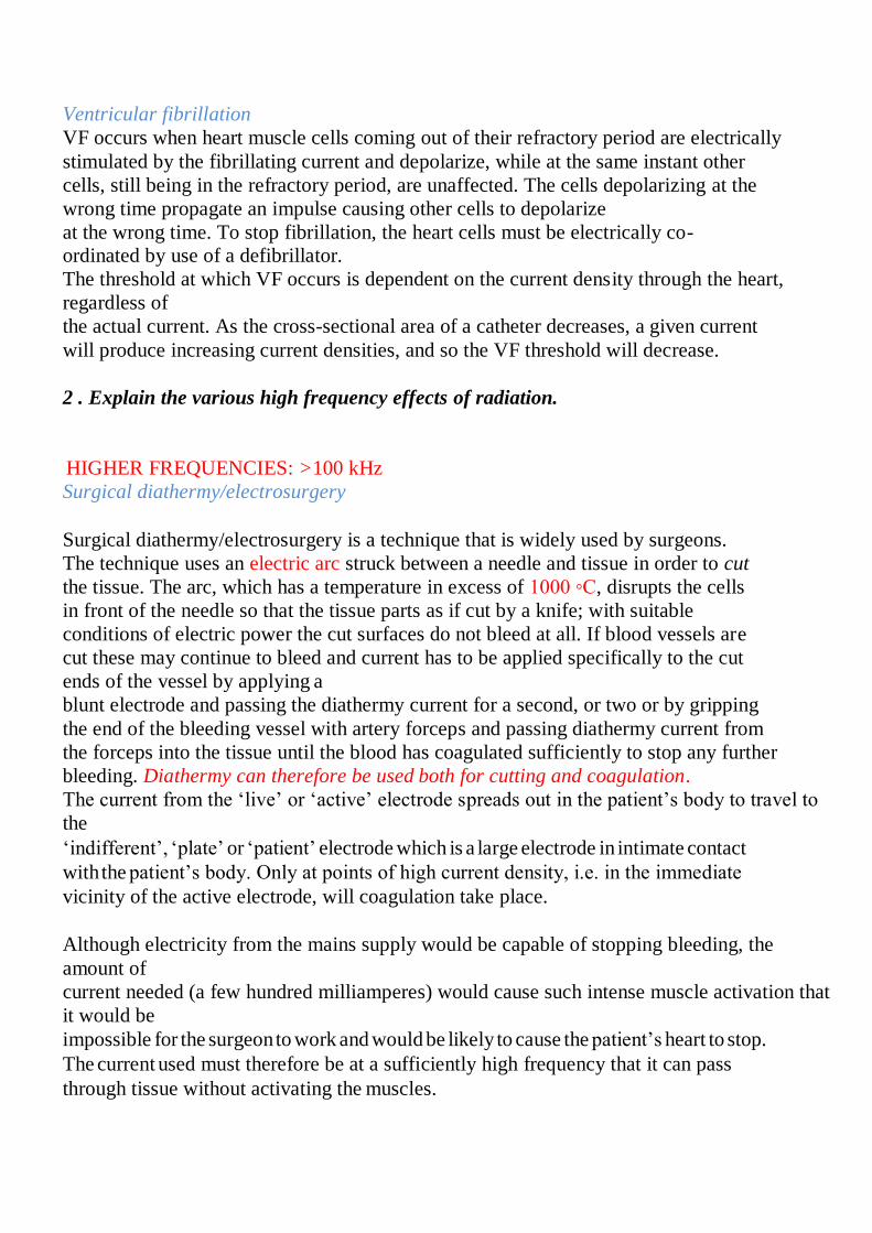
Ventricular fibrillation
VF occurs when heart muscle cells coming out of their refractory period are electrically
stimulated by the fibrillating current and depolarize, while at the same instant other
cells, still being in the refractory period, are unaffected. The cells depolarizing at the
wrong time propagate an impulse causing other cells to depolarize
at the wrong time. To stop fibrillation, the heart cells must be electrically co-
ordinated by use of a defibrillator.
The threshold at which VF occurs is dependent on the current density through the heart,
regardless of
the actual current. As the cross-sectional area of a catheter decreases, a given current
will produce increasing current densities, and so the VF threshold will decrease.
2 . Explain the various high frequency effects of radiation.
HIGHER FREQUENCIES: >100 kHz
Surgical diathermy/electrosurgery
Surgical diathermy/electrosurgery is a technique that is widely used by surgeons.
The technique uses an electric arc struck between a needle and tissue in order to cut
the tissue. The arc, which has a temperature in excess of 1000 ◦C, disrupts the cells
in front of the needle so that the tissue parts as if cut by a knife; with suitable
conditions of electric power the cut surfaces do not bleed at all. If blood vessels are
cut these may continue to bleed and current has to be applied specifically to the cut
ends of the vessel by applying a
blunt electrode and passing the diathermy current for a second, or two or by gripping
the end of the bleeding vessel with artery forceps and passing diathermy current from
the forceps into the tissue until the blood has coagulated sufficiently to stop any further
bleeding. Diathermy can therefore be used both for cutting and coagulation.
The current from the ‘live’ or ‘active’ electrode spreads out in the patient’s body to travel to
the
‘indifferent’, ‘plate’ or ‘patient’ electrode which is a large electrode in intimate contact
with the patient’s body. Only at points of high current density, i.e. in the immediate
vicinity of the active electrode, will coagulation take place.
Although electricity from the mains supply would be capable of stopping bleeding, the
amount of
current needed (a few hundred milliamperes) would cause such intense muscle activation that
it would be
impossible for the surgeon to work and would be likely to cause the patient’s heart to stop.
The current used must therefore be at a sufficiently high frequency that it can pass
through tissue without activating the muscles.

Threshold of sensation as a function of frequency for an electric current applied between 5 mm wide band electrodes encircling the base of two adjacent fingers.
Diathermy equipment
Diathermy machines operate in the radio-frequency (RF) range of the spectrum,
typically 0.4–3 MHz. Diathermy works by heating body tissues to very high
temperatures. The current densities at the active electrode can be 10 A cm−2. The total
power input can be about 200 W. The power density in the vicinity of the cutting edge
can be thousands of W cm−3, falling to a small fraction of a W cm−3 a fewcentimetres
from the cutting edge. The massive temperature rises at the edge (theoretically
thousands of ◦C) cause the tissue fluids to boil in a fraction of a second. The cutting is
a result of rupture of the cells.
An RF current follows the path of least resistance to ground. This would normally be
via the plate (also called dispersive) electrode. However, if the patient is connected to
the ground via the table or any attached leads from monitoring equipment, the current
will flow out through these. The current density will be high at these points of contact,
and will result in surface burns (50 mA cm−2 will cause reddening of the skin;
150 mA cm−2 will cause burns). Even if the operating table is insulated from earth, it
can form a capacitor with the surrounding metal of the operating theatre due to its size,
allowing current to flow. Inductive or capacitive coupling can also be formed between
electrical leads, providing other routes to ground.
Heating effects
If the whole body or even a major part of the body is exposed to an intense

electromagnetic field then the heating produced might be significant. The body
normally maintains a stable deep-body temperature within relatively narrow limits
(37.4±1 ◦C) even though the environmental temperature may fluctuate widely. The
normal minimal metabolic rate for a resting human is about 45Wm−2 (4.5 mWcm−2),
which for an average surface area of 1.8 m2 gives a rate of 81 W for a human body.
Blood perfusion has an important role in maintaining deep-body temperature.

Blood flowing through veins just below the skin plays an important part in controlling
heat transfer. surface temperatures will be affected by vessels carrying blood at a
temperature higher or lower than the surrounding tissue.
.
Exposure to electromagnetic (EM) fields can cause significant effects. We can calculate
what power dissipation this might cause if we make simplifying assumptions. Consider the
cylindrical geometry as shown below
which represents a body which is 30 cm in diameter and 1 m long (L). We will
assume a resistivity (ρ) of 5 m for the tissue. The resistance (R) between the top
and bottom will be given by ρL/A where A is the cross-sectional area. R = 70.7 .
For a field of 1 V m−1 (in the tissue) the current will be 14.1 mA. The power
dissipated is 14.1 mW which is negligible compared to the basal metabolic
rate.
For a field of 1 kV m−1, the current will be 14.1 A and the power 14.1 kW,
which is very significant. The power density is 20 W cm−2 over the input
surface or 200 mW cm−3 over the whole volume.
In the above case we assumed that the quoted field density was the volts per metre
produced in tissue. However, in many cases the field is quoted as volts per metre in air.
There is a large difference between these two cases. A field of 100 V m−1 in air may
only give rise to a field of 10−5 V m−1 in tissue.
3. Explain the various types of interactions of ultrasounds with matter / tissue.
For two tissues, or tissue and water, the fraction reflected is small and most energy continues

through the
interface between the two materials. For the interface between soft tissue and air the fraction
of reflected energy is also high and therefore it is not possible to image through an air
cavity.
The transmitted ultrasound beam will be refracted at an interface, that is the incident angle θi
will
not be the same as the refracted angle θt and the ultrasound beam will deviate from a
straight line. This is analogous to the refraction of light, and the bending of the
ultrasound beam can be found from Snell’s law

where c1 and c2 are the velocities of the ultrasound in the two media. For incident
angles of less than 30◦ the deviation at most interfaces will be less than 2◦. At a
soft-tissue–bone interface, the deviation will be
20◦. For most cases this will lead to only a small degradation of the image, but may give
serious problems for tissue– bone interfaces.
Scattering, absorption and attenuation of
ultrasound Scattering
Specular reflection (analogous to the reflection of light by a mirror) of the ultrasonic
energy will take place when the interface is smooth over an area which is several times
as great as the ultrasound wavelength, λ. When the features of the surface are about the
same size as the wavelength, diffuse reflection will occur. If the scatterers are very
small compared with the wavelength, then Rayleigh scattering will take place, in which
the incident energy is scattered uniformly in all directions. Obviously, with this formof
scattering, very little energy will be reflected back to the transducer. Red blood cells
are about 8–9 μm in diameter and produce Rayleigh scattering.
Absorption
The transmission of mechanical vibrations through a mediumabsorbs energy fromthe
ultrasound beam. This occurs through a variety of mechanisms but essentially arises
because the changes in kinetic and potential energy, caused by the passage of the
pressure wave through the medium, are not always reversible and so some of the
energy is lost as heat. This is especially important at higher frequencies.
Attenuation
Loss of signal produced by reflecting interfaces between the transmitter and the
reflecting object and by scattering and absorption of the signal will reduce the strength
of the detected signal. The divergence of the ultrasound beam will also reduce the
received energy. The received echoes used to form a typical abdominal scan may be 70
dB.Below the level of the transmitted signal, and the signals from moving red blood
cells may be 100–120 dB below the transmitted signal. These effects taken together
represent beam attenuation.
Attenuation follows an exponential law. The beam intensity at depth x is given by

I = I0e−αx
where α, the attenuation coefficient, consists largely of an absorption plus scattering
component. I0 is the unattenuated beam intensity. The attenuation coefficient is a
function of frequency. Although the relationship is complex, α is roughly proportional
to frequency. The decibel attenuation coefficient μ is defined as μ = 4.3α. A pulse of ultrasound contains a range of frequencies and as the pulse propagates through
tissue these
will be differentially attenuated, leading to a net reduction of the average frequency
which will lead to a loss in resolution and a change in the apparent attenuation.
It is necessary to compensate for the increasing attenuation of the signals with
distance. The receiving amplifier compensates for the increase in attenuation of the
echo when it has travelled through a greater
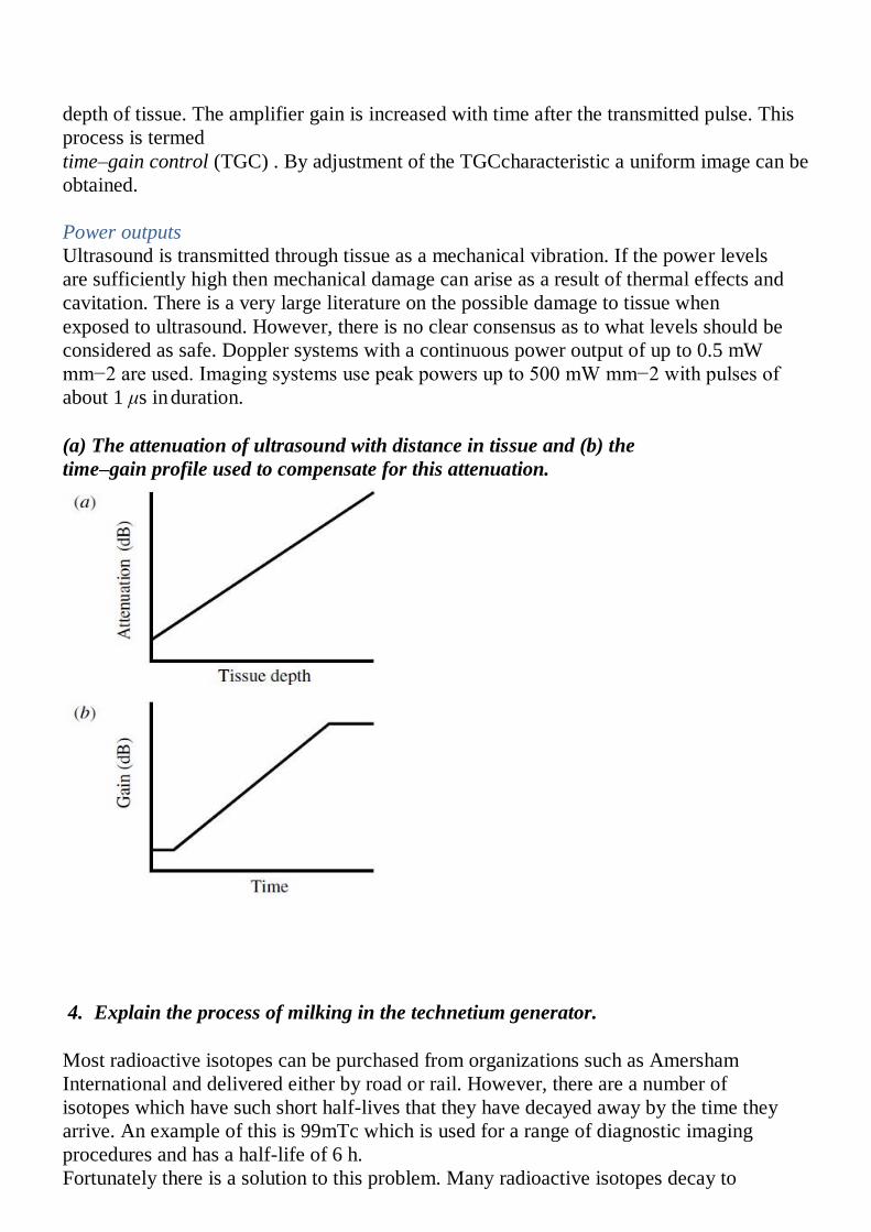
depth of tissue. The amplifier gain is increased with time after the transmitted pulse. This
process is termed
time–gain control (TGC) . By adjustment of the TGCcharacteristic a uniform image can be
obtained.
Power outputs
Ultrasound is transmitted through tissue as a mechanical vibration. If the power levels
are sufficiently high then mechanical damage can arise as a result of thermal effects and
cavitation. There is a very large literature on the possible damage to tissue when
exposed to ultrasound. However, there is no clear consensus as to what levels should be
considered as safe. Doppler systems with a continuous power output of up to 0.5 mW
mm−2 are used. Imaging systems use peak powers up to 500 mW mm−2 with pulses of
about 1 μs in duration.
(a) The attenuation of ultrasound with distance in tissue and (b) the
time–gain profile used to compensate for this attenuation.
4. Explain the process of milking in the technetium generator.
Most radioactive isotopes can be purchased from organizations such as Amersham
International and delivered either by road or rail. However, there are a number of
isotopes which have such short half-lives that they have decayed away by the time they
arrive. An example of this is 99mTc which is used for a range of diagnostic imaging
procedures and has a half-life of 6 h.
Fortunately there is a solution to this problem. Many radioactive isotopes decay to

produce a second radioactive isotope which is called a daughter product. If the mother
isotope has a long half-life then, even though the daughter might have a short half-life,
the daughter product will be produced continuously and can be separated from the
mother when required.
99mTc is produced fromthe mother isotope
99Mo as follows: 99

42Mo= 99m
43 Tc + β-particle
↓
9
9 43Tc + γ.
The process by which the technetiumis obtained from the cow or isotope column
which contains the mother Isotope.
Molybdenum, in the form of ammonium molybdate, is adsorbed onto an alumina column
which is
held in a glass tube. To obtain the daughter product, sterile saline is passed through
the column and the technetium, in the formof sodiumpertechnetate, is eluted (flushed
out) into the lead pot. It takes some time for the concentration of daughter product to
build up once the column has been milked so that the process cannot be repeated for
several hours.
99Mo–99mTc Generator-construction
The 99Mo–99mTc generator is constructed with alumina (Al2O3) loaded in a plastic or
glass column. The 99Mo activity
is adsorbed on alumina in the chemical form MoO4 2− (molybdate) and in various amounts.
The amount of alumina used is about 5–10 g depending on the 99Mo activity. Currently, all
generators use fission-produced 99Mo.
The 99mTc activity is eluted with 0.9% NaCl solution (isotonic saline) and
obtained in the chemical form of Na99mTcO4.
An ideal radionuclide generator should be simple and sturdy for transportation. The
generator eluate should be free of the parent nuclide and the adsorbent material.
Several radionuclide generators are available for ready supply of short-lived radionuclides:
99Mo(66hr)–99mTc(6hr); 113Sn(117 days)– 113mIn(100 min); 68Ge(271 days)–

68Ga(68min); 82Sr(25.6 days)–82Rb(75sec);
81Rb(4.6 hr)–81mKr(13sec).
Of these, the 99Mo–99mTc generator is the workhorse of nuclear pharmacy in nuclear
medicine.
5. Explain the interaction of charged particles with matter.
The energetic charged particles such as a-particles, protons, deuterons, and b-particles
(electrons) interact with the

absorber atoms, while passing through it.The interaction occurs primarily with the orbital
electrons of the atoms and rarely with the nucleus. During the interaction, both ionization
and excitation as well as the breakdown of the molecule may occur. In excitation, the
charged particle transfers all or part of its energy to the orbital electrons, raising
them to higher energy shells. In ionization, the energy transfer may be sufficient to
overcome the binding energy of the orbital electrons, ultimately ejecting them from the
atom.
Specific Ionization
Specific ionization (SI) is the total number of ion pairs produced per unit length of the path of
the incident radiation.
The SI values of a-particles are slightly greater than those of protons and deuterons,
which in turn are larger than those of electrons.
Specific ionization increases with decreasing energy of the charged particle because of
the increased probability of interaction at low energies. Therefore, toward the end of the
travel, the charged particle shows a sharp increase in ionization. This peak ionization is
called Bragg ionization. This phenomenon is predominant for heavy charged particles
and is negligible for electrons.
Linear Energy Transfer
The linear energy transfer (LET) is the amount of energy deposited per unit length of
the path by the radiation. From the preceding, it is clear that
LET = SI × W
The LET is expressed in units of keV/mm and is very useful in concepts of radiation
protection.
Electromagnetic radiations and b-particles interact with matter, losing only little energy with
matter, losing only little energy per interaction and therefore have low LETs. In contrast,
heavy particles (a-particles, neutrons, and protons) lose energy very rapidly, producing many
ionizations in a short distance, and thus have high LETs.
Range
The range (R) of a charged particle in an absorber is the straight-line distance
traversed by the particle in the direction of the particle.The range of a particle
depends on the mass, charge, and kinetic energy
and also on the density of the absorber. The heavier and more highly of the particle
charged particles have shorter ranges than lighter and lower charged particles. The range of
charged particles increases with the energy of
the particle.Thus, a 10-MeV particle will have a longer range than a 1-MeV particle.
The range of the particle depends
on the density of the absorber, in that the denser the absorber, the shorter the range. The
unit of range is given in mg/cm2 of the absorber.

It is seen that the ranges of all identical particles in a given absorber are not exactly the
same but show a spread of 3% to 4% near the end of their path.
Annihilation
When energetic b+-particles pass through an absorber, they lose energy via interaction with
orbital electrons of the atoms of the absorber.When the b+-particle comes to almost rest
after losing all energy it combines with an
orbital electron of the absorber atom and produces two 511-keV annihilation radiations that
are emitted in opposite directions (180°). These annihilation radiations are the basis of
positron emission tomography (PET) in which two photons are detected in coincidence.

Bremsstrahlung
When energetic charged particles, particularly electrons, pass through matter and come
close to the nucleus of the atom, they lose energy as a result of deceleration in the
Coulomb field of atomic nuclei. The loss in energy appears as an x-ray that is called
bremsstrahlung. These bremsstrahlung radiations are commonly used
in radiographic procedures and are are generated by striking a tungsten target with a
highly accelerated electron beam. Bremsstrahlung production increases with the kinetic
energy of the particle and the atomic number (Z)
of the absorber. For example, a 10-MeV electron loses about 50% of its energy by
bremsstrahlung, whereas a
90-MeV electron loses almost 90% of its energy by this process. The bremsstrahlung
production is proportional to Z2 of the absorber atom.
Therefore, bremsstrahlung is unimportant in lighter metals such as air, aluminum, and so
forth, whereas it is very significant in heavy metals such as lead and tungsten. High-energy b−-
particles from radionuclides such as 32P can produce bremsstrahlung in heavy metals such as
lead and tungsten.
Bremsstrahlung is inversely proportional to the mass of the charged particles and
therefore is insignificant for heavy particles, namely a-particles and protons, because
the probability of penetrating close to the nuclei is relatively low due to their heavier
masses.
6. Explain the interaction of gamma radiation and neutron with matter.
Mechanism of Interaction of ᵞ –Radiations
When penetrating ᵞ rays pass through matter, they lose energy by interaction with the orbital
electrons or the nucleus of the absorber atom. The gray photons may lose all of their energy,
or a fraction of it, in a single encounter.
There are three mechanisms by which g-rays interact with absorber atoms during their passage through matter. Photoelectric Effect In the photoelectric effect, the incident g -ray transfers all its energy to an orbital
electron of the absorber atom whereby the electron, called the photoelectron, is ejected
with kinetic energy equal to Eg EB, where Eg and EB are the energy of the g -ray
and the binding energy anof the electron, respectively The photoelectron loses its
energy by ionization and excitation in the absorber. The photoelectric effect occurs
primarily in the low-energy range and decreases sharply with increasing photon energy.
It also increases very rapidly with increasing atomic number Z of the absorber atom.
The photoelectric effect occurs primarily with the K-shell electrons, with about 20%
contribution from the L-shell electrons and even less from higher shells.

Compton Scattering
In Compton scattering, the g-ray photon transfers only a part of its energy to an electron
in the outer shell of the absorber atom, and the electron is ejected. The photon, itself
with reduced energy, is deflected from its original direction This process is called the
Compton scattering. The scattered photon of lower energy may then undergo further
photoelectric or Compton interaction, and the Compton electron may cause ionization or
excitation.
Using the law of conservation of momentum and energy, the scattered photon energy is given
by
where Eg and Esc are the energies in MeV of the initial and scattered photons.The scattered
photon energy varies from a maximum in a collision at 0° (forward) to a minimum at q
180° in a backscattering collision.
If the photon is backscattered, that is, scattered at 180°, then the backscattered photon has the
energy Esc given by the expression (cos 180° 1):
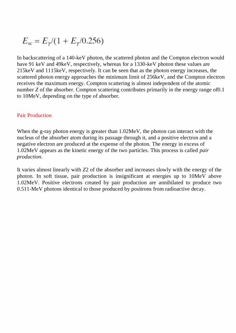
In backscattering of a 140-keV photon, the scattered photon and the Compton electron would
have 91 keV and 49keV, respectively, whereas for a 1330-keV photon these values are
215keV and 1115keV, respectively. It can be seen that as the photon energy increases, the
scattered photon energy approaches the minimum limit of 256keV, and the Compton electron
receives the maximum energy. Compton scattering is almost independent of the atomic
number Z of the absorber. Compton scattering contributes primarily in the energy range of0.1
to 10MeV, depending on the type of absorber.
Pair Production
When the g-ray photon energy is greater than 1.02MeV, the photon can interact with the
nucleus of the absorber atom during its passage through it, and a positive electron and a
negative electron are produced at the expense of the photon. The energy in excess of
1.02MeV appears as the kinetic energy of the two particles. This process is called pair
production.
It varies almost linearly with Z2 of the absorber and increases slowly with the energy of the
photon. In soft tissue, pair production is insignificant at energies up to 10MeV above
1.02MeV. Positive electrons created by pair production are annihilated to produce two
0.511-MeV photons identical to those produced by positrons from radioactive decay.

Relative contributions of the photoelectric effect, Compton scattering, and pair
production as a function of photon energy in absorbers of different atomic numbers.

photoelectric effect is predominant in high Z absorbers at lower energies( 0.1MeV),
whereas the Compton scattering is predominant in inter- mediate Z absorbers at medium
energies (~1MeV). At higher energies ( 10MeV), pair production predominates in all Z
absorbers
. Photodisintegration
When the g-ray photon energy is very high ( 10MeV), the photon may interact with the
nucleus of the absorber atom and transfer sufficient energy to the nucleus such that one or
more nucleons may be emitted.This process is called the photodisintegration reaction, or
photonuclear reaction and produces new nuclides.The (g, n) reactions on targets such as 12C
and 14N have been used to produce 11C and 13N radionuclides but now are rarely used to
produce radionuclides.
Interaction of Neutrons with Matter
Because neutrons are neutral particles, their interactions in the absorber differ from those of
the charged particles. They interact primarily with the nucleus of the absorber atom and very
little with the orbital electrons. The neutrons can interact with the atomic nuclei in three
ways: elastic scattering, inelastic scattering, and neutron capture. If the sum of the kinetic
energies of the neutron and the nucleus before collision is equal to the sum of these quantities
after collision, then the interaction is called elastic. If a part of the initial energy is used for
the excitation of the struck nucleus, the collision is termed inelastic. In neutron capture, a
neutron is captured by the absorber nucleus, and a new excited nuclide is formed. Depending
on the energy deposited, an a-particle, a proton, a neutron, or g-rays can be emitted from the
excited nucleus, and a new product nuclide (usually radioactive) is produced.
7. Explain the stochastic and non-stochastic effects of radiation.
Stochastic and Deterministic Effects
Two categories of radiation effects on biological systems are encountered: stochastic and
deterministic. Stochastic effects are the biological effects that occur randomly, the
probability of which increases with increasing dose without a threshold. Radiation-induced
hereditary effects and cancer incidences are examples of stochastic effects. The assumption
of no threshold is made on the belief that radiation damage to a few cells or a single cell
could theoretically induce the genetic disorder or cancer, and the severity of the disease will
be the same, if it ever occurs. It should be noted that the basic principle of ALARA (as low
as reasonably achievable) in Nuclear Regulatory Commission (NRC) regulations is based on
the assumption of risks linearly proportional to the dose without a threshold.
The deterministic or nonstochastic effects are induced by high radiation doses and the
severity of the damages, rather than their probability of occurrence, increases with the dose.
These effects have a threshold dose below which no damage is evident. Cataracts, skin

erythema, sterility, and fibrosis are examples of deterministic effects induced by high
radiation doses.
Acute Effects of Total Body Irradiation Different tissues of the body respond differently to radiation, due to varying degrees of
radiosensitivity. When an adult subject is irradiated over the entire body, various syndromes
are manifested depending on the dose applied. The effects of radiation are characterized by
the survival time of the species and various stages of acute syndromes following the
totalbody irradiation. These effects are deterministic types and have a threshold dose. The
radiosensitivity of a given species is commonly characterized by the lethal dose, LD50/60,
which is the dose that kills 50% of the species in 60 days. The LD50/60 for humans is 400
to 600 rad
Acute radiation syndromes appear in four stages: prodromal, latent, manifest illness, and
recovery or death.
There are three categories of syndromes in the manifest illness stage depending on the
dose: hemopoietic or bone marrow, gastrointestinal (GI), and cerebrovascular.

Hemopoietic Syndrome
Hemopoietic or bone marrow syndromes appear at a total body dose of 250 to 500 rad (250
to 500 cGy) following irradiation. At this dose, the precursors for RBCs and white blood
cells (WBCs) are greatly affected, so much so that they lose the ability to reproduce. Also,
the number of lymphocytes are greatly depressed, whereby the immune system of the body
is suppressed.
At this dose level, the platelet count is drastically reduced, and therefore bleeding gradually
progresses through various orifices owing to a lack of ability of the blood to coagulate.
Fever, bleeding, and infection result, followed by ultimate death in 10 to 21 days. However,
bone marrow transplantation at the appropriate time may prompt the recovery of the subject.
Whereas at doses 100rad (100 cGy) survival is almost certain, survival is virtually
impossible at doses 500 rad (500 cGy).
Gastrointestinal Syndrome
Gastrointestinal (GI) syndromes are expressed at a total body dose of 500 to 1000 rad (500
to 1000 cGy) and include prodromal syndromes such as nausea, vomiting, and diarrhea of
more severity that appear within hours after exposure.The primary effect of radiation
exposure in this range is that the intestinal crypt cells are destroyed and not replaced, and
consequently the mucosal lining (villi) shrinks and hardens whereby the gut becomes
nonfunctional.
Because of the denudation of the gut, an intestinal ulcer may develop.These GI syndromes
are also accompanied by drastic hemopoietic syndromes including immunosuppression,
loss of white blood cells, and infection.
Cerebrovascular Syndrome
Cerebrovascular syndromes appear in a matter of minutes after radiation exposure at a total
body dose of more than 10,000 rad (10,000 cGy). Because the nerve cells are radioresistant,
such a large dose is required for cerebrovascular
syndromes to appear. The symptoms include severe nausea, vomiting, and burning
sensation of the skin that occurs within minutes of exposure, followed by the malfunction of
the neuron motor pump giving rise to motor incoordination, intermittent stupor, coma, and
ultimately death in two to three days. The cerebrovascular death sequence is so rapid that
there is little time for significant changes to appear in other organs in the body. At this level
of radiation dose, death is a certainty and medical help is of no use.