Development of compression-controlled low-level laser probe system: towards clinical application
13
Development of compression-controlled low-level laser probe system: towards clinical application Changmin Yeo, Department of Biomedical Engineering, Yonsei University, 234 Maeji-Ri, Heungup-Myeon, Wonju-Si, Gangwon-Do 220-710, Korea Taeyoon Son, Department of Biomedical Engineering, Yonsei University, 234 Maeji-Ri, Heungup-Myeon, Wonju-Si, Gangwon-Do 220-710, Korea Junghwan Park, Department of Biomedical Engineering, Yonsei University, 234 Maeji-Ri, Heungup-Myeon, Wonju-Si, Gangwon-Do 220-710, Korea Young-Heum Lee, Department of Biomedical Engineering, Yonsei University, 234 Maeji-Ri, Heungup-Myeon, Wonju-Si, Gangwon-Do 220-710, Korea, Institute of Medical Engineering, Yonsei University, Wonju, Korea Kiwoon Kwon, Department of Mathematics, Dongguk University, Seoul 100-715, Korea J. Stuart Nelson, and Beckman Laser Institute, University of California, Irvine, CA 92612, USA Byungjo Jung Department of Biomedical Engineering, Yonsei University, 234 Maeji-Ri, Heungup-Myeon, Wonju-Si, Gangwon-Do 220-710, Korea. Institute of Medical Engineering, Yonsei University, Wonju, Korea Byungjo Jung: [email protected] Abstract Various physico-chemical tissue optical clearing (TOC) methods have been suggested to maximize photon density in tissue. In order to enhance photon density, a compression-controlled low-level laser probe (CCLLP) system was developed by utilizing the principle of mechanical tissue compression. Negative compression (NC) was applied to the laser probes built in various diameters and simultaneously the laser was irradiated into ex-vivo porcine skin samples. Laser photon density (LPD) was evaluated as a function of NC and probe diameter by analyzing 2D diffusion images of the laser exposures. The CCLLP system resulted in a concentrated laser beam profile, which means enhancement of the LPD. As indicators of LPD, the laser peak intensity increased and the full width at half maximum (FWHM) decreased as a function of NC. The peak intensity at −–30 kPa increased 2.74, 3.22, and 3.64 fold at laser probe diameters of 20, 30, and 40 mm, respectively. In addition, sample temperature was measured with a thermal camera and increased 0.4 K at −30 kPa after 60 s of laser irradiation as a result of enhanced LPD. The CCLLP © Springer-Verlag London Ltd 2010 Correspondence to: Byungjo Jung, [email protected]. NIH Public Access Author Manuscript Lasers Med Sci. Author manuscript; available in PMC 2011 August 24. Published in final edited form as: Lasers Med Sci. 2010 September ; 25(5): 699–704. doi:10.1007/s10103-010-0779-8. NIH-PA Author Manuscript NIH-PA Author Manuscript NIH-PA Author Manuscript
-
Upload
independent -
Category
Documents
-
view
0 -
download
0
Transcript of Development of compression-controlled low-level laser probe system: towards clinical application

Development of compression-controlled low-level laser probesystem: towards clinical application
Changmin Yeo,Department of Biomedical Engineering, Yonsei University, 234 Maeji-Ri, Heungup-Myeon,Wonju-Si, Gangwon-Do 220-710, Korea
Taeyoon Son,Department of Biomedical Engineering, Yonsei University, 234 Maeji-Ri, Heungup-Myeon,Wonju-Si, Gangwon-Do 220-710, Korea
Junghwan Park,Department of Biomedical Engineering, Yonsei University, 234 Maeji-Ri, Heungup-Myeon,Wonju-Si, Gangwon-Do 220-710, Korea
Young-Heum Lee,Department of Biomedical Engineering, Yonsei University, 234 Maeji-Ri, Heungup-Myeon,Wonju-Si, Gangwon-Do 220-710, Korea, Institute of Medical Engineering, Yonsei University,Wonju, Korea
Kiwoon Kwon,Department of Mathematics, Dongguk University, Seoul 100-715, Korea
J. Stuart Nelson, andBeckman Laser Institute, University of California, Irvine, CA 92612, USA
Byungjo JungDepartment of Biomedical Engineering, Yonsei University, 234 Maeji-Ri, Heungup-Myeon,Wonju-Si, Gangwon-Do 220-710, Korea. Institute of Medical Engineering, Yonsei University,Wonju, KoreaByungjo Jung: [email protected]
AbstractVarious physico-chemical tissue optical clearing (TOC) methods have been suggested tomaximize photon density in tissue. In order to enhance photon density, a compression-controlledlow-level laser probe (CCLLP) system was developed by utilizing the principle of mechanicaltissue compression. Negative compression (NC) was applied to the laser probes built in variousdiameters and simultaneously the laser was irradiated into ex-vivo porcine skin samples. Laserphoton density (LPD) was evaluated as a function of NC and probe diameter by analyzing 2Ddiffusion images of the laser exposures. The CCLLP system resulted in a concentrated laser beamprofile, which means enhancement of the LPD. As indicators of LPD, the laser peak intensityincreased and the full width at half maximum (FWHM) decreased as a function of NC. The peakintensity at −–30 kPa increased 2.74, 3.22, and 3.64 fold at laser probe diameters of 20, 30, and 40mm, respectively. In addition, sample temperature was measured with a thermal camera andincreased 0.4 K at −30 kPa after 60 s of laser irradiation as a result of enhanced LPD. The CCLLP
© Springer-Verlag London Ltd 2010Correspondence to: Byungjo Jung, [email protected].
NIH Public AccessAuthor ManuscriptLasers Med Sci. Author manuscript; available in PMC 2011 August 24.
Published in final edited form as:Lasers Med Sci. 2010 September ; 25(5): 699–704. doi:10.1007/s10103-010-0779-8.
NIH
-PA Author Manuscript
NIH
-PA Author Manuscript
NIH
-PA Author Manuscript
system effectively demonstrated enhancement of the LPD in tissue and potentially its clinicalfeasibility.
KeywordsPhoton density; Low-level laser; Compression; Tissue optical clearing
IntroductionTissue optical clearing (TOC) methods have attracted public attention in a short period oftime due to the maturity of light therapy and diagnostics, which are based on light–tissueinteractions [1], such as absorption, reflection, transmission, and scattering [2]. Among all ofthe complex light–tissue interactions, optical scattering limits the light penetration depth intissue and, consequently, photon density. Various chemical (hyperosmotic chemical agent[HCA]), thermal (temperature), and mechanical (compression and stretching) methods havebeen suggested as effective TOC methods for reducing optical scattering. Even though suchmethods have shown some promise, further studies are needed for clinical application.
Chemical methods are based on the mechanism of skin dehydration [3–5], replacement ofinterstitial or intracellular water by HCA, structural modification or dissociation of collagenfibers [6, 7], and refractive index matching [3, 8]. However, further studies are required tounderstand the complex biological interaction mechanisms [9] and to develop better physicalmethods to maximize the HCA diffusion rate through the skin barrier. Laufer et al.demonstrated that the changes in the optical properties of human dermis and sub-dermis canoccur in response to temperature changes [10]. Mechanical compression demonstrated TOCeffect for clinical application [11] and has advantages in low-level laser therapy as a non-invasive modality. Previous studies have evaluated the changes in tissue optical propertiesdue to compression [12, 13]. Shangguan et al. [14] demonstrated that both the absorptionand scattering coefficients increased as a function of compression (0–1.5 kg/cm2) and theirreversibility of tissue optical properties due to the compression. In that study, overalltransmittance increased, and tissue thickness and weight decreased about 72% and 40%,respectively.
Based on the compression effect in tissue, a compression-controlled low-level laser probe(CCLLP) system was developed to enhance the laser photon density (LPD) in soft tissue byquantitatively controlling tissue compression. The laser probes of three different diameterswere built and their potential clinical usefulness was investigated with ex-vivo porcine skinsamples.
Materials and methodsSample preparation
Ex-vivo abdominal porcine skin samples were obtained from a local abattoir and usedbecause their structural and immunohistochemical characteristics were similar to those ofhuman skin [15–17]. The samples were prepared to a size of 70×160 mm2 with an averagethickness of 2.8 mm.
Compression-controlled low-level laser probe systemA CCLLP system was developed to enhance the LPD in tissue. Figure 1a shows a schematicdiagram of the probe, which consisted of a vacuum hole to supply negative compression(NC), a supporter for guiding the optical fiber, and an acrylic probe body to observe skin
Yeo et al. Page 2
Lasers Med Sci. Author manuscript; available in PMC 2011 August 24.
NIH
-PA Author Manuscript
NIH
-PA Author Manuscript
NIH
-PA Author Manuscript

deformation. The CCLLP system was designed to control both NC and laser parameterssimultaneously. In order to evaluate the LPD as a function of probe diameter, three differentprobes were built with diameters of 20, 30, and 40 mm as shown in Fig. 1b.
Experimental setupFigure 2 shows the experimental setup designed to acquire the 2D diffusion images of laserbeam profile (LBP) transmitted by the skin samples. The setup consisted of a compressioncontroller, image acquisition, and diode laser compartments. The compression controllercompartment is composed of a 12-V DC motor pump (KPV36E-12A, KOGE Electronics,Xiamen, China), NC manometer, and DC power supply. The diode laser compartment wascomposed of an 808-nm diode laser (L808P200, Thorlabs, Kansas, USA), which has amaximum output power of 200 mW and is operated in continuous-wave mode, an opticalfiber port (PAF-SMA-11-B, Thorlabs, Kansas, USA), a diode laser mount (LDM21,Thorlabs, Kansas, USA) equipped with aspheric lenses (C570TM-B, A230TM-B, Thorlabs,Kansas, USA), and a multi-mode optical fiber patch cord (FT1.5EMT, Thorlabs, Kansas,USA). For LPD study by NC, the maximum power of the laser was adjusted to 43 mW atthe distal end of optical fiber in order to avoid light saturation. The 2D diffusion imageswere acquired with a CCD camera (cs8620i, Toshiba Teli Corp, Tokyo, Japan) placed on theopposite side of the skin samples. The CCD camera has a field of view of 29.9°(H)×22.6°(V) with a C-mount lens and detector size of 768(H)×494(V) pixels with an 8.4×9.8 μmpixel size. The 251×251 pixel window, including the LBP, was extracted and analyzed witha laboratory-built MATLAB program. The CCD camera in Fig. 2 was replaced with athermal imaging camera (Thermovision A40, FLIR Systems, Boston, MA, USA) in order toevaluate the temperature profile by the LPD. The thermal imaging camera has a spatialresolution of 1.3 mrad and a field of view of 24×18°/0.3 m with a 35-mm lens. Sensingtemperature ranges from −40°C to +500°C with thermal sensitivity of 0.08°C in spectralrange from 7.5 through 13 μm. The output power of the laser was adjusted to 134 mW forthermal effect investigation by LPD.
ProceduresLPD was quantitatively evaluated by analyzing the full width at half maximum (FWHM)and peak intensity of LBP as a function of NC and probe diameter. The NC was appliedfrom 0 through −30 kPa with −5 kPa increment because tissue optical properties can beirreversibly affected by over compression [14]. To verify the fidelity of the experimentresults, five experiments were performed with five different samples.
The sample temperature variation by the LPD was evaluated by analyzing thermal imagestaken every 5 s using the 40-mm probe at −30 kPa. The sample, which has an initialtemperature of 16.42°C, was irradiated by the laser for 60 s in order to minimize artifacts bysample dehydration. Three experiments were performed with three different samples. Analuminum sample holder (Fig. 1) was used to fix the samples during NC. Room temperaturewas maintained at 16.19°C during the experiments.
Mathematical modeling of temperature variation by LPDIn a mathematical model, the temperature in biological tissue is described by the followingbio-heat transfer equation, which is based on the energy exchange between blood vesselsand the surrounding tissue [22, 23]:
(1)
Yeo et al. Page 3
Lasers Med Sci. Author manuscript; available in PMC 2011 August 24.
NIH
-PA Author Manuscript
NIH
-PA Author Manuscript
NIH
-PA Author Manuscript

where T is the tissue temperature depending on time t; ρ is the tissue density; c is the tissuespecific heat; k is the tissue thermal conductivity; Qs is the external heat source; Qp is theheat removal due to perfusion. In this study, ρ, c, and k are constant, Qp is zero, and Qs is
(2)
where C is a point in the tissue right under the probe in our experiment and A is a constantlaser intensity for the time, t. Assuming the following: (1) tissue geometry is a two-dimensional plane; (2) temperature depends only on distance from point C; (3) temperaturegoes to zero as time goes to zero; and (4) the temperature goes to zero at large distancesfrom point C, then the solution of Eq. (1) is given as follows:
(3)
where T0 is the initial temperature and A=134 mW, ρ = 1,000 (kg/m3), c=4,200 (J/kgK), andk=1.8 (W/mK) [24] in mathematical modeling. In a previous study, Cain et al. [24] used athermal conductivity k for water immerged porcine skin in a numerical simulation.However, the k was assumed to be three times larger value in this study because fresh skinsample was considered to be more dehydrated than the previous study.
ResultsFigure 3 shows 3D images of LBPs at 0, −15, and −30 kPa (from left to right in each figure)for (a) 20, (b) 30, and (c) 40 mm probes. Although the images show an anisotropic patternbecause of the complex morphological structure and non-homogeneity of biological tissue,the results illustrates the enhancement of LPD by NC. For all probe diameters, LBPs wereenhanced as a function of NC.
The peak intensity (Fig. 4) was computed as a function of NC and probe diameter. For allprobe diameters, the peak intensity greatly increased at the initial −5 kPa and then, linearlyincreased as a function of the NC with a similar slope. At the maximum −30 kPa, the peakintensities (Fig. 4) increased by a factor of 2.74, 3.22, and 3.64 and the FWHM (Fig. 5)decreased by a factor of 1.35, 1.57, and 3.55 for probe diameters of 20, 30, and 40 mm,respectively. The largest probe diameter resulted in the highest peak intensity for all NCs.Figure 6 shows the sample temperature variation by NC as a function of time. The sampletemperature decreased 0.19 K at 0 kPa but increased 0.4 K at-30 kPa after 60 s of laserirradiation. In addition, Fig. 6 shows the mathematical modeling result based on the analyticsolution in Eq. (3) and experimental results with and without NC.
DiscussionPrevious studies [13, 14] demonstrated that mechanical compression of an elastinbiomaterial resulted in changes in the tissue optical properties due to both tissue thicknessreduction and water content loss. The tissue thickness reduction contributes to a decrease inoptical pathlength, and the water content loss contributes to the spatial decrease betweenelastin layers and therefore, refractive index matching. Most previous mechanicalcompression studies were mainly focused on water and blood movement in tissue, thermalchange in adipose regions, or changes in tissue morphological or mechanical properties [6,
Yeo et al. Page 4
Lasers Med Sci. Author manuscript; available in PMC 2011 August 24.
NIH
-PA Author Manuscript
NIH
-PA Author Manuscript
NIH
-PA Author Manuscript

18–20]. In this study, the mechanical compression method was employed to achieve a TOCeffect and therefore, non-invasively enhance the LPD in soft tissue.
At present, low-level laser probes are simply attached on the skin surface using medical tapeor passively and manually handled by operators. Such manipulations cannot preciselycontrol tissue compression and therefore the penetration depth and LPD. In order to partiallyaddress this limitation, the CCLLP system was developed to control both the NC and laserparameters simultaneously. The self-sustaining capability of the probe was confirmed duringthe application of NC by visually monitoring the airtight of the probe.
The LBP was more concentrated as a function of NC as shown in Fig. 3, demonstrating theefficacy of the probe for enhancement of the LPD in soft tissue. Figure 4 shows thequantitative analysis as a function of NC and probe diameter. The peak intensity waslinearly increased, implying the enhancement of LPD at the region of interest (ROI). Agreater increase in the peak intensity at the initial –5 kPa might be due to abrupt tissuedeformation (e.g., tissue stretching) and therefore, abrupt reduction in epidermal thickness.Hendriks et al. reported a 7% reduction of tissue thickness at 20 kPa and 17% at 35 kPa[21]. The initial changing ratio was greater in larger probe diameters. It might be because thelarger probe results in more tissue deformation (stretching) and therefore, reduction ofoptical pathlength in tissue. The peak intensity linearly increased as a function of NC (−5through −30 kPa) across all probe diameters. As a result, the LPD might be effectivelycontrolled by controlling both the NC and probe diameter, which affect skin deformation. Asanother indicator of LPD, the FWHM decreased as a function of the NC and probe diameter(Fig. 5). The probes with diameters of 20 and 30 mm resulted in a similar decreasing rate ofFWHM without correlation with NC. However, the FWHM of the 40 mm laser probe wasgreatly affected by NC, resulting in a maximum decrease at −30 kPa.
The NC resulted in a temperature rise in the porcine skin samples due to the enhanced LPD.However, sample temperature without NC decreased to room temperature, implying that thetemperature in soft tissue might not be affected only by laser irradiation withoutcompression because the photon energy of the laser might not be efficiently delivered to theROI. Tissue optical properties can be changed by temperature variation [6, 10]. Therefore, atemperature rise due to NC might result in changes in tissue optical properties, which causesadditional enhancement in the LPD by minimizing the refractive index mismatch. Both Figs.4 and 6 imply that the probe might be useful for therapeutic applications. In themathematical modeling, we could confirm a similar pattern between the modeling andexperimental results (Fig. 6) although there is a small difference between them, since manymathematical assumptions were given in the modeling.
The probe can be built in various diameters depending on the intended therapeutic purposeand ROI. In addition, the shape of the optical fiber can be varied by using an optical fiberbundle to manipulate the LBP pattern in tissue and therefore, potentially obtain furtherenhancement of LPD. Other methods such as temperature and hyperosmotic chemicalagents, which also contribute TOC, can be combined into the probe system to furtherenhance LPD.
ConclusionsThis study demonstrated that the CCLLP system can effectively enhance the LPD in softtissues by controlling NC and probe diameter. Its clinical usefulness was potentiallyvalidated with various probe diameters and self-sustaining capability of the probe. In futurestudies, the positive compression applied to tissue by an optical fiber and the variation in
Yeo et al. Page 5
Lasers Med Sci. Author manuscript; available in PMC 2011 August 24.
NIH
-PA Author Manuscript
NIH
-PA Author Manuscript
NIH
-PA Author Manuscript

tissue thickness by NC must be experimentally and numerically studied for betterunderstanding of the CCLLP system.
AcknowledgmentsThis study was supported by a grant from the Korea Healthcare Technology R&D Project, Ministry for Health,Welfare & Family Affairs, Republic of Korea (B090033). JSN was supported by the following grants from theNational Institutes of Health (AR47751 and EB 2495).
References1. Carroll L, Humphreys TR. LASER-tissue interactions. Clin Dermatol. 2006; 24(1):2–7.10.1016/
j.clindermatol.2005.10.019 [PubMed: 16427500]2. Anderson RR, Parrish JA. The optics of human skin. J Invest Dermatol. 1981; 77:13–
19.10.1111/1523-1747.ep12479191 [PubMed: 7252245]3. Wang RK. Signal degradation by multiple scattering in optical coherence tomography of dense
tissue: a Monte Carlo study towards optical clearing of biotissues. Phys Med Biol. 2002; 47 (13):2281–2299.10.1088/0031-9155/47/13/307 [PubMed: 12164587]
4. Xu XQ, Wang RK, Elder JB. Optical clearing effect on gastric tissues immersed with biocompatiblechemical agents investigated by near infrared reflectance spectroscopy. J Phys D Appl Phys. 2003;36:1707–1713.10.1088/0022-3727/36/14/309
5. Xu X, Wang RK. The role of water desorption on optical clearing of biotissues: studied with nearinfrared reflectance spectroscopy. Med Phys. 2003; 30(6):1246–1253.10.1118/1.1576228 [PubMed:12852550]
6. Rylander CG, Milner TE, Baranov SA, Stuart Nelson J. Mechanical tissue optical clearing devices:enhancement of light penetration in ex vivo porcine skin and adipose tissue. Lasers Surg Med.2008; 40(10):688–694.10.1002/lsm.20718 [PubMed: 19065559]
7. Yeh AT, Choi B, Nelson JS, Tromberg BJ. Reversible dissociation of collagen in tissues. J InvestDermatol. 2003; 121:1332–1335.10.1117/1.2166381 [PubMed: 14675178]
8. Vargas G, Chan EK, Barton JK, Rylander HG, Welch AJ. Use of an agent to reduce scattering inskin. Lasers Surg Med. 1999; 24 (2):133–141. [PubMed: 10100651]
9. Rylander CG, Stumpp OF, Milner TE, Kemp NJ, Mendenhall JM, Diller KR, Welch AJ.Dehydration mechanism of optical clearing in tissue. J Biomed Opt. 2006; 11(4):041117.10.1117/1.2343208 [PubMed: 16965145]
10. Laufer J, Simpson R, Kohl M, Essenpreis M, Cope M. Effect of temperature on the opticalproperties of ex vivo human dermis and subdermis. Phys Med Biol. 1998; 43:2479–2489.10.1088/0031-9155/43/9/004 [PubMed: 9755940]
11. Kang H, Son T, Yoon J, Kwon K, Nelson JS, Jung B. Evaluation of laser beam profile in softtissue due to compression, glycerol, and micro-needling. Lasers Surg Med. 2008; 40(8):570–575.10.1002/lsm.20664 [PubMed: 18798289]
12. Vogel A, Dlugos C, Nuffer R, Birngruber R. Optical properties of human sclera, and theirconsequences for transscleral laser applications. Lasers Surg Med. 1991; 11(4):331–340.10.1002/lsm.1900110404 [PubMed: 1895865]
13. Chan EK, Sorg B, Protsenko D, O’Neil M, Motamedi M, Welch AJ. Effects of compression on softtissue optical properties. IEEE J Sel Top Quantum Electron. 1996; 2(4):943–950.10.1109/2944.577320
14. Shangguan, H.; Prahl, SA.; Jacques, SL.; Casperson, LW.; Gregory, KW. Pressure effects on softtissues monitored by changes in tissue optical properties. SPIE Press; San Diego: 1998. p.366-371.
15. Lavker RM, Dong G, Zheng PS, Murphy GF. Hairless micropig skin a novel model for studies ofcutaneous biology. Am J Pathol. 1991; 138(3):687–697. [PubMed: 2000942]
16. Vardaxis NJ, Brans TA, Boon ME, Kreis RW, Marres LM. Confocal laser scanning microscopy ofporcine skin: implications for human wound healing studies. J Anat. 1997; 190(4):601–611.10.1046/j.1469-7580.1997.19040601.x [PubMed: 9183682]
Yeo et al. Page 6
Lasers Med Sci. Author manuscript; available in PMC 2011 August 24.
NIH
-PA Author Manuscript
NIH
-PA Author Manuscript
NIH
-PA Author Manuscript

17. Ross EV, Naseef GS, Mckinlay JR, Barnette DJ, Skrobal M, Grevelink J, Anderson RR.Comparison of carbon dioxide laser, erbium:YAG laser, dermabrasion, and dermatome: a study ofthermal damage, wound contraction, and wound healing in a live pig model: implications for skinresurfacing. J Am Acad Dermatol. 2000; 42(1):92–105. [PubMed: 10607327]
18. Childers M, Franco W, Nelson JS, Aguilar G. Laser surgery of port wine stains using local vacuumpressure: changes in skin morphology and optical properties (Part I). Lasers Surg Med. 2007; 39(2):108–117.10.1002/lsm.20456 [PubMed: 17311268]
19. Pedersen L, Hansen B, Jemec GBE. Mechanical properties of the skin: a comparison between twosuction cup methods. Skin Res Technol. 2003; 9(2):111–115.10.1034/j.1600-0846.2003.00021.x[PubMed: 12709128]
20. Khatyr F, Imberdis C, Varchon D, Lagarde JM, Josse G. Measurement of the mechanicalproperties of the skin using the suction test. Skin Res Technol. 2006; 12(1):24–31.10.1111/j.0909-725X.2006.00126.x [PubMed: 16420535]
21. Hendriks FM, Brokken D, van Eemeren JT, Oomens CW, Baaijens FP, Horsten JB. A numerical-experimental method to characterize the non-linear mechanical behaviour of human skin. Skin ResTechnol. 2003; 9(3):274–283.10.1034/j.1600-0846.2003.00019.x [PubMed: 12877691]
22. Sturesson C, Andersson-Engels S. A mathematical model for predicting the temperaturedistribution in laser-induced hyperthermia. Experimental evaluation and applications. Phys MedBiol. 1995; 40:2037–2052.10.1088/0031-9155/40/12/003 [PubMed: 8719943]
23. Welch, AJ.; Martin, JC.; Gemert, V. Optical-thermal response of laser-irradiated tissue. PlenumPublishing Corporation; New York: 1995.
24. Cain CP, Polhamus GD, Stolarski DJ, Schuster KJ, Stockton KL, Rockwell BA, Bo C, Welch AJ.Porcine skin visible lesion thresholds for near-infrared lasers including modeling at two pulsedurations and spot sizes. J Biomed Opt. 2006; 11(4):041109.10.1117/1.2338815 [PubMed:16965137]
Yeo et al. Page 7
Lasers Med Sci. Author manuscript; available in PMC 2011 August 24.
NIH
-PA Author Manuscript
NIH
-PA Author Manuscript
NIH
-PA Author Manuscript

Fig. 1.a Schematic diagram of compression-controlled low-level laser probe, which consists of anair suction hole, optical fiber supporter, and optical fiber. b The upper image shows theprobe applied on an ex-vivo porcine skin sample, and the bottom images from left to rightshow the probes with the diameters of 20, 30, and 40 mm
Yeo et al. Page 8
Lasers Med Sci. Author manuscript; available in PMC 2011 August 24.
NIH
-PA Author Manuscript
NIH
-PA Author Manuscript
NIH
-PA Author Manuscript

Fig. 2.Experimental setup to measure the laser photon density in soft tissue consisting of acompression controller, image acquisition, and diode laser compartment
Yeo et al. Page 9
Lasers Med Sci. Author manuscript; available in PMC 2011 August 24.
NIH
-PA Author Manuscript
NIH
-PA Author Manuscript
NIH
-PA Author Manuscript
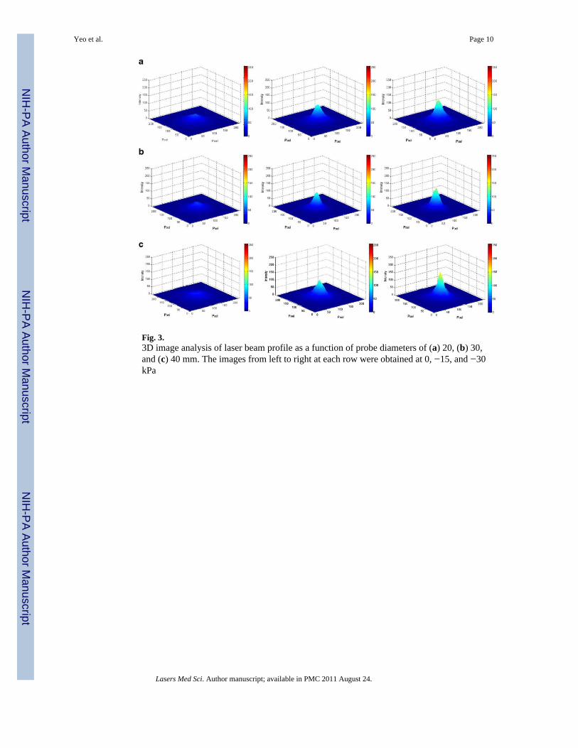
Fig. 3.3D image analysis of laser beam profile as a function of probe diameters of (a) 20, (b) 30,and (c) 40 mm. The images from left to right at each row were obtained at 0, −15, and −30kPa
Yeo et al. Page 10
Lasers Med Sci. Author manuscript; available in PMC 2011 August 24.
NIH
-PA Author Manuscript
NIH
-PA Author Manuscript
NIH
-PA Author Manuscript

Fig. 4.The negative compression effect in laser photon density was quantitatively evaluated byanalyzing the increasing rate of laser peak intensity as a function of the negativecompression at probe diameters of 20, 30, and 40 mm
Yeo et al. Page 11
Lasers Med Sci. Author manuscript; available in PMC 2011 August 24.
NIH
-PA Author Manuscript
NIH
-PA Author Manuscript
NIH
-PA Author Manuscript

Fig. 5.Full width at half maximum (FWHM) decreasing rate of laser beam profile as a function ofprobe diameters at −15 and −30 kPa
Yeo et al. Page 12
Lasers Med Sci. Author manuscript; available in PMC 2011 August 24.
NIH
-PA Author Manuscript
NIH
-PA Author Manuscript
NIH
-PA Author Manuscript
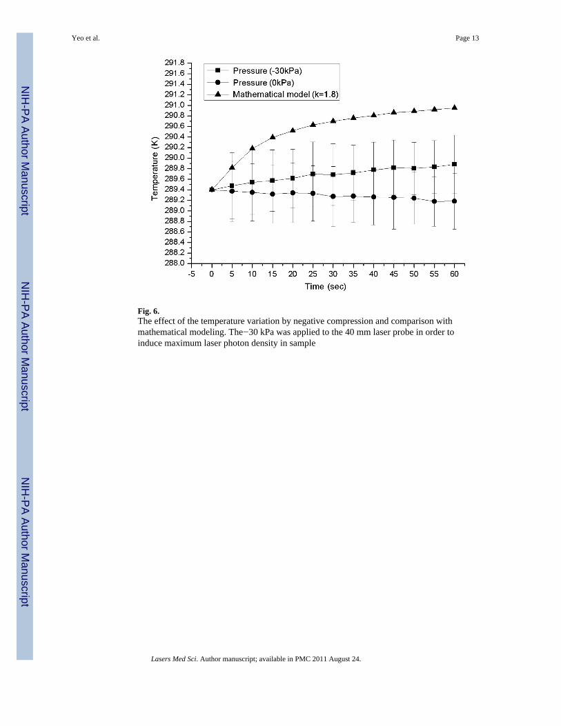
Fig. 6.The effect of the temperature variation by negative compression and comparison withmathematical modeling. The−30 kPa was applied to the 40 mm laser probe in order toinduce maximum laser photon density in sample
Yeo et al. Page 13
Lasers Med Sci. Author manuscript; available in PMC 2011 August 24.
NIH
-PA Author Manuscript
NIH
-PA Author Manuscript
NIH
-PA Author Manuscript