Development of Clinical Referral Score Model for Early ... - MDPI
13
healthcare Article Development of Clinical Referral Score Model for Early Diagnosis of Hirschsprung’s Disease in Suspected Pediatric Patients Jiraporn Khorana 1,2, * , Phawinee Phiromkanchanasak 3 , Jitthiwimon Kumsattra 3 , Suparada Klinoun 3 , Suthasinee Aksorn 3 , Sireekarn Chantakhow 1 , Kanokkan Tepmalai 1 and Jesda Singhavejsakul 1 Citation: Khorana, J.; Phiromkanchanasak, P.; Kumsattra, J.; Klinoun, S.; Aksorn, S.; Chantakhow, S.; Tepmalai, K.; Singhavejsakul, J. Development of Clinical Referral Score Model for Early Diagnosis of Hirschsprung’s Disease in Suspected Pediatric Patients. Healthcare 2021, 9, 678. https://doi.org/10.3390/ healthcare9060678 Academic Editor: Rocío De Andrés Calle Received: 3 May 2021 Accepted: 2 June 2021 Published: 4 June 2021 Publisher’s Note: MDPI stays neutral with regard to jurisdictional claims in published maps and institutional affil- iations. Copyright: © 2021 by the authors. Licensee MDPI, Basel, Switzerland. This article is an open access article distributed under the terms and conditions of the Creative Commons Attribution (CC BY) license (https:// creativecommons.org/licenses/by/ 4.0/). 1 Department of Surgery, Division of Pediatric Surgery, Faculty of Medicine, Chiang Mai University Hospital, Chiangmai 50200, Thailand; [email protected] (S.C.); [email protected] (K.T.); [email protected] (J.S.) 2 Center of Clinical Epidemiology and Clinical Statistic, Faculty of Medicine, Chiang Mai University Hospital, Chiangmai 50200, Thailand 3 Faculty of Medicine, Chiang Mai University Hospital, Chiangmai 50200, Thailand; [email protected] (P.P.); [email protected] (J.K.); [email protected] (S.K.); [email protected] (S.A.) * Correspondence: [email protected] Abstract: The diagnosis of Hirschsprung’s disease (HSCR) relies on history, physical examination, and investigations. Some of investigation modalities could not be done in primary hospital. This study was aimed to develop the clinical score model for diagnosing and early referrals of HSCR, especially in areas where investigations were not available. Overall 483 consecutive suspected HSCR patients who were under 15 years old from January 2006 to December 2020 were included in this study, with 207 (42.86%) patients diagnosed with HSCR and 276 (51.14%) patients in the non-HSCR group. Five clinical parameters were included in the prediction model. The AuROC of clinical parameters, which included having an age younger than one month, male gender, the term infant, history of delayed meconium passage, and history of enterocolitis, was 72%. The prediction score ranged from 0–7, with a score 0–3 meaning a low risk to be HSCR (LHR+ = 0.37). We concluded that patients with suspected HSCR who had clinical score 4–7 had a high probability to be HSCR and, thus, it was suggested that these patients have an early referral for further investigations, which were contrast enema and rectal suction biopsy. In the case of a low probability of HSCR, clinical observation is still warranted. This clinical scoring system can be used as a screening tool to prevent delay diagnosis and complications. Keywords: Hirschsprung’s disease; clinical diagnosis; contrast enema; rectal suction biopsy; pediatric surgery; pediatric emergency 1. Introduction Hirschsprung’s disease (HSCR), or congenital aganglionic megacolon, occurs in ap- proximately 1 in 5000 of all live-born infants. This disease occurrs because of the absence of the ganglion plexus in the large intestine, which causes the large bowel to lose its ability for dilation or peristalsis resulting in intestinal obstruction [1]. Patients with HSCR could develop obstructive symptoms days after birth such as the delayed passage of meconium (failure to pass first meconium within 48 h of life), abdominal distention, constipation, bilious vomiting, failure to thrive, and absence of flatus. Some studies [2,3] define common clinical manifestations as a “classic triad” including delayed passage of meconium, abdominal distention, and vomiting. Physical examination shows signs of malnutrition such as low height and weight. Increased rectal tone and explosive stool after rectal examination are characteristic of HSCR. In some cases, HSCR could have Healthcare 2021, 9, 678. https://doi.org/10.3390/healthcare9060678 https://www.mdpi.com/journal/healthcare
-
Upload
khangminh22 -
Category
Documents
-
view
2 -
download
0
Transcript of Development of Clinical Referral Score Model for Early ... - MDPI
healthcare
Article
Development of Clinical Referral Score Model for EarlyDiagnosis of Hirschsprung’s Disease in SuspectedPediatric Patients
Jiraporn Khorana 1,2,* , Phawinee Phiromkanchanasak 3, Jitthiwimon Kumsattra 3, Suparada Klinoun 3,Suthasinee Aksorn 3, Sireekarn Chantakhow 1, Kanokkan Tepmalai 1 and Jesda Singhavejsakul 1
�����������������
Citation: Khorana, J.;
Phiromkanchanasak, P.; Kumsattra, J.;
Klinoun, S.; Aksorn, S.; Chantakhow,
S.; Tepmalai, K.; Singhavejsakul, J.
Development of Clinical Referral
Score Model for Early Diagnosis of
Hirschsprung’s Disease in Suspected
Pediatric Patients. Healthcare 2021, 9,
678. https://doi.org/10.3390/
healthcare9060678
Academic Editor: Rocío De
Andrés Calle
Received: 3 May 2021
Accepted: 2 June 2021
Published: 4 June 2021
Publisher’s Note: MDPI stays neutral
with regard to jurisdictional claims in
published maps and institutional affil-
iations.
Copyright: © 2021 by the authors.
Licensee MDPI, Basel, Switzerland.
This article is an open access article
distributed under the terms and
conditions of the Creative Commons
Attribution (CC BY) license (https://
creativecommons.org/licenses/by/
4.0/).
1 Department of Surgery, Division of Pediatric Surgery, Faculty of Medicine, Chiang Mai University Hospital,Chiangmai 50200, Thailand; [email protected] (S.C.); [email protected] (K.T.);[email protected] (J.S.)
2 Center of Clinical Epidemiology and Clinical Statistic, Faculty of Medicine, Chiang Mai University Hospital,Chiangmai 50200, Thailand
3 Faculty of Medicine, Chiang Mai University Hospital, Chiangmai 50200, Thailand;[email protected] (P.P.); [email protected] (J.K.); [email protected] (S.K.);[email protected] (S.A.)
* Correspondence: [email protected]
Abstract: The diagnosis of Hirschsprung’s disease (HSCR) relies on history, physical examination,and investigations. Some of investigation modalities could not be done in primary hospital. Thisstudy was aimed to develop the clinical score model for diagnosing and early referrals of HSCR,especially in areas where investigations were not available. Overall 483 consecutive suspected HSCRpatients who were under 15 years old from January 2006 to December 2020 were included in thisstudy, with 207 (42.86%) patients diagnosed with HSCR and 276 (51.14%) patients in the non-HSCRgroup. Five clinical parameters were included in the prediction model. The AuROC of clinicalparameters, which included having an age younger than one month, male gender, the term infant,history of delayed meconium passage, and history of enterocolitis, was 72%. The prediction scoreranged from 0–7, with a score 0–3 meaning a low risk to be HSCR (LHR+ = 0.37). We concludedthat patients with suspected HSCR who had clinical score 4–7 had a high probability to be HSCRand, thus, it was suggested that these patients have an early referral for further investigations, whichwere contrast enema and rectal suction biopsy. In the case of a low probability of HSCR, clinicalobservation is still warranted. This clinical scoring system can be used as a screening tool to preventdelay diagnosis and complications.
Keywords: Hirschsprung’s disease; clinical diagnosis; contrast enema; rectal suction biopsy; pediatricsurgery; pediatric emergency
1. Introduction
Hirschsprung’s disease (HSCR), or congenital aganglionic megacolon, occurs in ap-proximately 1 in 5000 of all live-born infants. This disease occurrs because of the absenceof the ganglion plexus in the large intestine, which causes the large bowel to lose its abilityfor dilation or peristalsis resulting in intestinal obstruction [1].
Patients with HSCR could develop obstructive symptoms days after birth such as thedelayed passage of meconium (failure to pass first meconium within 48 h of life), abdominaldistention, constipation, bilious vomiting, failure to thrive, and absence of flatus. Somestudies [2,3] define common clinical manifestations as a “classic triad” including delayedpassage of meconium, abdominal distention, and vomiting. Physical examination showssigns of malnutrition such as low height and weight. Increased rectal tone and explosivestool after rectal examination are characteristic of HSCR. In some cases, HSCR could have
Healthcare 2021, 9, 678. https://doi.org/10.3390/healthcare9060678 https://www.mdpi.com/journal/healthcare

Healthcare 2021, 9, 678 2 of 13
severe complications such as Hirschsprung-associated enterocolitis, resulting in intestinalperforation and sepsis as a presentation.
The diagnosis of HSCR comprised of clinical presentations, physical examinationtogether with other investigations such as abdominal plain film, contrast enema, anorec-tal manometry, and a rectal biopsy. A contrast enema is commonly used for initiatinginvestigations in HSCR-suspected patients due to its high sensitivity and its availabilityin most healthcare centers. However, there are various sensitivities and specificities ofcontrast enema in other studies; thus, further investigations must be performed to give adiagnosis [4–6]. Anorectal manometry usually needs special instrument to be performed.The sensitivity and specificity are also not as high as a rectal biopsy.
Rectal biopsy is the most specific and representative in diagnosis of HSCR. Two biopsytechniques are commonly performed. The first one is a full-thickness rectal biopsy, whichcould be performed in a surgical setting and is an acceptable reference standard test todiagnose HSCR. The second newer and less invasive technique, rectal suction biopsy (RSB),can be interpreted if there is an adequate specimen. According to the European ReferenceNetwork for rare inherited and congenital digestive disorders (ERNICA) guideline, rectalhistology is required for diagnosis. Both techniques are accurate if adequate submucosaltissue is included [7]. The less invasive procedure, which is rectal suction biopsy, shouldbe considered first. A diagnosis of HSCR is given if the specimen shows an absence ofganglion cells in the hematoxylin and eosin stain or a special immunohistochemistry study,such as acetylcholine esterase or calretinin stain, which shows an aganglionic segment [8].Some studies [3,9] proposed performing a rectal biopsy in patients with signs of abdominalobstruction including the following: delayed passage of meconium, abdominal distention,and vomiting, along with positive findings from other investigations to avoid unnecessaryinvasive procedures. On the other hand, some studies [10,11] recommended performing arectal biopsy in every patient with suspected HSCR despite a high rate of negative results,because many HSCR patients presented with only one symptom and late diagnosis couldincrease the risk of developing life-threatening complications.
However, without the results from a rectal biopsy, it could be difficult to diagnoseHSCR, because some intestinal obstructive conditions manifest similarly to HSCR, suchas meconium plug syndrome, anorectal malformation, malrotation, intestinal atresia, andmeconium ileus associated with cystic fibrosis [2]. In addition, some non-obstructiveconditions, such as allergic proctitis [12,13], lactase deficiency, celiac disease [2,13,14],cow’s milk allergy [2,13], hypothyroidism [2,15], and enterocolitis, could also mimic HSCR.
The other group of conditions, called variants or allied disorders of Hirschsprung’sdisease [16], show a close resemblance to HSCR in terms of clinical presentations andfindings from the investigations, although the ganglion cells are present in the biopsy.The examples are intestinal neuronal dysplasia, intestinal ganglioneuromatosis, isolatedhypoganglionosis, immature ganglia, absence of the argyrophilic plexus, internal analsphincter achalasia, and megacystis microcolon intestinal hypoperistalsis syndrome.
In our study, the diagnosis of HSCR was still challenging in the remote hospitalwith no complete investigation options. There was no specific protocol to refer suspectedHSCR patients for further investigation. Either delayed diagnosis or over-investigationcould occur. The mode of diagnosis was comprised of clinical evaluation, plain abdominalradiography, contrast enema, and rectal biopsy. This study aimed to help the remotehospital to screen the clinical presentation for diagnosing HSCR and for the early referralof the suspected patient to the center that could perform further investigation.
2. Materials and Methods
This study was designed as a retrospective cohort study with approval from the Re-search Ethics Committee, Faculty of Medicine, Chiang Mai University (Study code: SUR-2563-07500) with an exemption from patient informed consent due to the full retrospective study.

Healthcare 2021, 9, 678 3 of 13
2.1. Participants
The data were collected from patients with suspected HSCR in Chiang Mai UniversityHospital that were presented from 2006 to 2020 and recorded in the database. Clinicallysuspected HSCR in this study included abdominal distension, constipation, vomiting, orplain abdominal radiography that showed large bowel dilation with no gas presentedin rectum. The participants included patients with clinically suspected HSCR less than15 years old at the date of consultation. The participants needed to have at least one clinicalpresentation suggesting HSCR and underwent either contrast enema, rectal biopsy, orcurative surgery.
Patients diagnosed with HSCR with a colostomy from the previous hospital lackedinitial clinical presentations of HSCR; thus, these patients were excluded from this study.Patients with an anorectal malformation, which is congenital anatomical anomalies thatalso cause bowel obstruction symptoms, except for anal stenosis, were excluded from thisstudy because HSCR could simply be ruled out by physical examination. Patients lost tofollow-up during the procedure were also excluded due to an unknown final diagnosis.
2.2. Outcome
The participants were diagnosed with HSCR if results from operative specimens,assessed by experienced pathologists, showed the absence of ganglion cells in the distalbowel. The non-Hirschsprung’s disease group (non-HSCR) consisted of participants whoseganglion cells were present or calretinin positive in the rectal biopsy specimen with norecurrence of clinical presentations after six months of follow-up. Some participantswho underwent surgery in the early years of the study and had ganglion cells present inoperative specimens were included in the non-HSCR group because of inadequate evidenceto be HSCR.
2.3. Predictors
Parameters recorded in this study were categorized into the following three groups:general information, clinical presentations, and physical examinations.
The general information group consisted of age, prematurity, gender, and Down’s syn-drome. Participants defined as term had a gestational age more than or equal 37 weeks [17].
Clinical presentations of patients included the history of delayed passage of meconiumat 48 h, abdominal distention, constipation, history of enterocolitis, and vomiting.
The physical examination group consisted of weight and results of per-rectal examina-tions including increased rectal sphincter tone and explosive stool after the examination.Weight was converted to weight-for-age percentile according to the World Health Or-ganization growth standard [18]. Failure to thrive was defined by the patient’s weightbeing less than the 5th weight-for-age percentile [19]. However, the cut-off point of theweight-for-age percentile in the predictive models was determined later on the basis of thepower of prediction.
2.4. Sample Size
The sample size was calculated on the basis of the sensitivity of the statisticallysignificant parameter, which was 81.1% of the parameter age less than three years old,as provided from the previous study [20]. With the significance level (α) of 0.05 and themaximum marginal error (d) of 0.05, the number of approximate sample sizes in this studywas 421.
2.5. Missing Data
The multiple imputation methods were used for the imputation of the missing listedparameters, which were term, history of delayed passage of meconium at 48 h, and historyof enterocolitis (missing at 102, 175, and 300 out of 483 participants, respectively).
Healthcare 2021, 9, 678 4 of 13
2.6. Additional Data
The additional investigations consisted of results from contrast enema, rectal suctionbiopsy, and full-thickness biopsy specimen. As anorectal manometry was only recentlyavailable in the institution, the result of anorectal manometry was not included in this study.Transitional zone [21], jejunization, saw-tooth appearance, reverse rectosigmoid ratio (<1),and delayed evacuation in 24 h were collected as contrast enema signs. The contrast enemaresult was assessed by a radiologist to determine whether the results suggested HSCR. Theoutcome of the rectal suction biopsy was pathologic results with the addition of calretininmarker. In addition, some of the investigations were not performed but the data were alsoincluded in this study. This information was added in this study to compare the result withthe developmental predictive model.
2.7. Statistical Analysis
Statistical analysis was performed with commercial statistical software (STATA 16.0;StataCorp LP, College Station, TX, USA). Categorical descriptive statistics were presentedby count and percentage. Continuous descriptive statistics were presented by the mean andstandard deviation or median and interquartile range (IQR) according to data distribution.Categorical analytic statistics were calculated from Fisher’s exact test. Continuous analyticstatistics were calculated from Student’s t-test or Mann–Whitney U Test. The significancelevel in this study was 0.05.
Multivariable analysis was done by logistic regression reported by diagnostic oddsratio (dOR). Various combinations of significant clinical parameters listed above that had aunivariable p-value < 0.01 were selected in the model and removed by the stepwise methodto retrieve all significant predictive parameters in the developed model.
Total of 5 parameters were used in the reduced model. The assigned score was doneby transforming the regression coefficient of dOR. Total score of 1–7 was derived fromthe model with the cut-off point of 4 to achieve the high true positive rate (sensitivity) topromote active referrals of the suspected patient. The likelihood ratio of positive values fordiagnosing HSCR was presented.
The receiver operating characteristic (ROC) curve, and area under the ROC curve(AuROC) were calculated to derive the discriminative potential of the model. Hosmer–Lemeshow goodness of fit statistics and a calibration plot comparing the agreement of theobserved and expected score values were also presented. The bootstrapping procedurewith 200 replicates was executed for internal validation of the model.
The diagnostic indices of the predictive model, contrast enema, and rectal suctionbiopsy were calculated including sensitivity, specificity, negative predictive value (NPV),and positive predictive value (PPV). The AuROC of the combination of the model with theinvestigation was presented.
3. Results
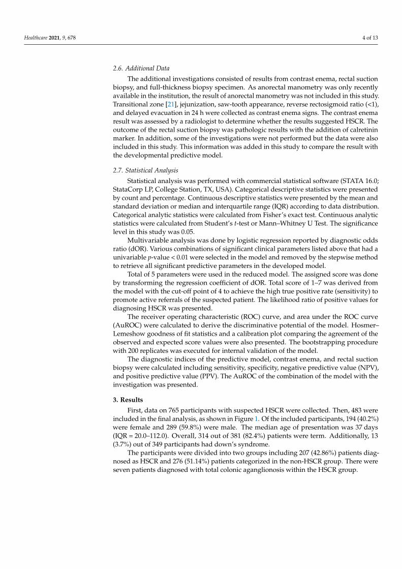
First, data on 765 participants with suspected HSCR were collected. Then, 483 wereincluded in the final analysis, as shown in Figure 1. Of the included participants, 194 (40.2%)were female and 289 (59.8%) were male. The median age of presentation was 37 days(IQR = 20.0–112.0). Overall, 314 out of 381 (82.4%) patients were term. Additionally, 13(3.7%) out of 349 participants had down’s syndrome.
The participants were divided into two groups including 207 (42.86%) patients diag-nosed as HSCR and 276 (51.14%) patients categorized in the non-HSCR group. There wereseven patients diagnosed with total colonic aganglionosis within the HSCR group.
Healthcare 2021, 9, 678 5 of 13
Figure 1. Study flow diagram.
In the non-HSCR group, definitive diagnoses were identified in 110 out of 276 partici-pants. The majority were constipation (63, 57.3%), anal stenosis (9, 8.2%), enterocolitis (5,4.5%), hypothyroidism (2, 1.8%), and other diagnoses (31, 28.2%). Some other diagnosesidentified were meconium plug syndrome, intestinal atresia, lactase deficiency, malrotation,inguinal or umbilical hernia, and overfeeding. The remainder had an uncertain diagnosis.
The general characteristics of the participants were shown in Table 1. The parametersthat showed univariable statistical significance for diagnosing HSCR were age, term,percentile weight for age, male gender, and history of delayed passage of meconium. Thenumber of the participants that provided the univariable data are also presented in Table 1.
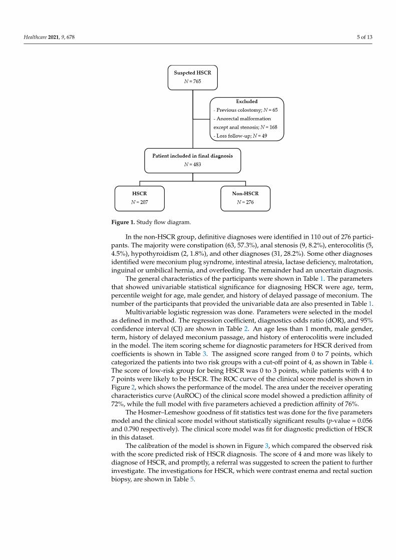
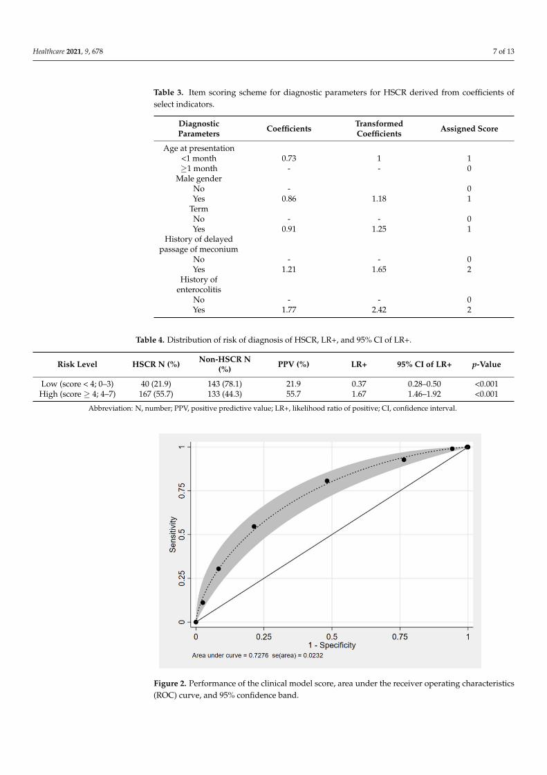
Multivariable logistic regression was done. Parameters were selected in the modelas defined in method. The regression coefficient, diagnostics odds ratio (dOR), and 95%confidence interval (CI) are shown in Table 2. An age less than 1 month, male gender,term, history of delayed meconium passage, and history of enterocolitis were includedin the model. The item scoring scheme for diagnostic parameters for HSCR derived fromcoefficients is shown in Table 3. The assigned score ranged from 0 to 7 points, whichcategorized the patients into two risk groups with a cut-off point of 4, as shown in Table 4.The score of low-risk group for being HSCR was 0 to 3 points, while patients with 4 to7 points were likely to be HSCR. The ROC curve of the clinical score model is shown inFigure 2, which shows the performance of the model. The area under the receiver operatingcharacteristics curve (AuROC) of the clinical score model showed a prediction affinity of72%, while the full model with five parameters achieved a prediction affinity of 76%.
The Hosmer–Lemeshow goodness of fit statistics test was done for the five parametersmodel and the clinical score model without statistically significant results (p-value = 0.056and 0.790 respectively). The clinical score model was fit for diagnostic prediction of HSCRin this dataset.
The calibration of the model is shown in Figure 3, which compared the observed riskwith the score predicted risk of HSCR diagnosis. The score of 4 and more was likely todiagnose of HSCR, and promptly, a referral was suggested to screen the patient to furtherinvestigate. The investigations for HSCR, which were contrast enema and rectal suctionbiopsy, are shown in Table 5.

Healthcare 2021, 9, 678 6 of 13
Table 1. General information, clinical presentations, and physical examination of HSCR and non-HSCR patients in Chiang Mai University Hospital from 2006 to 2020 (all 483 cases).
Characteristics N HSCR (n = 207) Non-HSCR (n = 276) p-Value
General InformationAge (days) a 483 31.0 (10.0, 153.0) 42.0 (23.0, 99.0) 0.005
Age < 1 month 483 105 (50.7%) 175 (63.4%) 0.007Age ≥ 1 month 483
Term 381 147 (88.6%) 167 (77.7%) 0.006Weight for age(Percentile) a 483 16.6 (3.3, 47.4) 31.0 (6.0, 60.6) 0.026
Gender 483Male 150 (72.5%) 139 (50.4%) <0.001
Female 57 (27.5%) 137 (49.6%)Down’s
syndrome 349 9 (5.4%) 4 (2.2%) 0.160
Clinical PresentationsHistory of
delayed passageof meconium
308 61 (43.9%) 39 (23.1%) <0.001
Abdominaldistention 450 191 (97.0%) 238 (94.1%) 0.180
Constipation 397 155 (88.1%) 188 (85.1%) 0.460Bilious vomiting 366 83 (49.4%) 86 (43.4%) 0.290
History ofenterocolitis 183 72 (79%) 61 (66%) 0.068
Physical ExaminationFailure to thrive 483 139 (72.4%) 199 (76.8%) 0.320Explosive stool
after a rectalexamination
383 90 (56.6%) 132 (58.9%) 0.680
Increase rectalsphincter tone
on rectalexamination
406 112 (66.3%) 159 (67.1%) 0.910
Notes: a Median (interquartile range). Abbreviation: N, number.
Table 2. Regression coefficient, diagnostics odds ratio (dOR), and 95% CI of selected diagnosticparameters derived from logistic regression after multiple imputation.
DiagnosticParameters Coefficient dOR 95% CI of dOR p-Value
Age < 1 month 0.73 2.07 1.37–3.13 0.001Male gender 0.86 2.37 1.57–3.60 <0.001
Term 0.91 2.47 1.34–4.58 0.004History of
delayed passageof meconium
1.21 3.36 2.03–5.59 <0.001
History ofenterocolitis 1.77 5.89 3.01–11.55 <0.001
Healthcare 2021, 9, 678 7 of 13
Table 3. Item scoring scheme for diagnostic parameters for HSCR derived from coefficients ofselect indicators.
DiagnosticParameters Coefficients Transformed
Coefficients Assigned Score
Age at presentation<1 month 0.73 1 1≥1 month - - 0
Male genderNo - 0Yes 0.86 1.18 1
TermNo - - 0Yes 0.91 1.25 1
History of delayedpassage of meconium
No - - 0Yes 1.21 1.65 2
History ofenterocolitis
No - - 0Yes 1.77 2.42 2
Table 4. Distribution of risk of diagnosis of HSCR, LR+, and 95% CI of LR+.
Risk Level HSCR N (%) Non-HSCR N(%) PPV (%) LR+ 95% CI of LR+ p-Value
Low (score < 4; 0–3) 40 (21.9) 143 (78.1) 21.9 0.37 0.28–0.50 <0.001High (score ≥ 4; 4–7) 167 (55.7) 133 (44.3) 55.7 1.67 1.46–1.92 <0.001
Abbreviation: N, number; PPV, positive predictive value; LR+, likelihood ratio of positive; CI, confidence interval.
Figure 2. Performance of the clinical model score, area under the receiver operating characteristics(ROC) curve, and 95% confidence band.

Healthcare 2021, 9, 678 8 of 13
Figure 3. Observed risk (circle) vs. score predicted risk (solid line) of HSCR diagnosis (size of circlerepresents frequency of HSCR in each score).
Table 5. Results of investigation of HSCR and non-HSCR patients in Chiang Mai University Hospitalfrom 2006 to 2020.
Characteristics N HSCR (n = 207) Non-HSCR (n = 276) p-Value
InvestigationContrast enema 471
Transitionalzone 156 (75.4%) 62 (22.5%) <0.001
Jejunization 156 (75.4%) 62 (22.5%) 0.089Saw-tooth
appearance 33 (15.9%) 17 (6.2%) <0.001
Reverserectosigmoid
ratio (>1)87 (42.0%) 55 (19.9%) <0.001
Delayedevacuation in
24 h114 (55.1%) 122 (44.2%) 0.021
Contrast enemaresult 471 178 (89.0%) 75 (27.7%) <0.001
Rectal suctionbiopsy 74
Positive for GGor calretininpositive (Not
HSCR)
3 (13%) 49 (98%) <0.001
Negative for GGor calretinin
negative(Diagnose
HSCR)
21 (88%) 1 (2%)
Abbreviation: GG, ganglion cells; HSCR, Hirschsprung’s disease; N, number.
Healthcare 2021, 9, 678 9 of 13
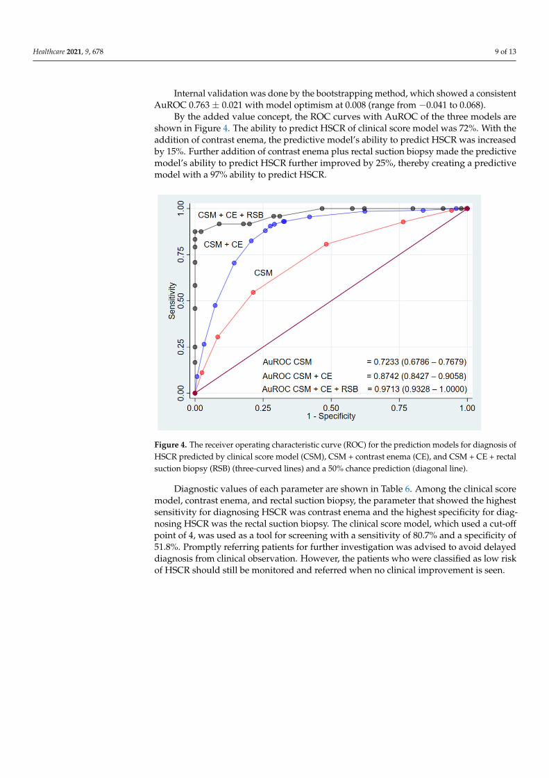
Internal validation was done by the bootstrapping method, which showed a consistentAuROC 0.763 ± 0.021 with model optimism at 0.008 (range from −0.041 to 0.068).
By the added value concept, the ROC curves with AuROC of the three models areshown in Figure 4. The ability to predict HSCR of clinical score model was 72%. With theaddition of contrast enema, the predictive model’s ability to predict HSCR was increasedby 15%. Further addition of contrast enema plus rectal suction biopsy made the predictivemodel’s ability to predict HSCR further improved by 25%, thereby creating a predictivemodel with a 97% ability to predict HSCR.
Figure 4. The receiver operating characteristic curve (ROC) for the prediction models for diagnosis ofHSCR predicted by clinical score model (CSM), CSM + contrast enema (CE), and CSM + CE + rectalsuction biopsy (RSB) (three-curved lines) and a 50% chance prediction (diagonal line).
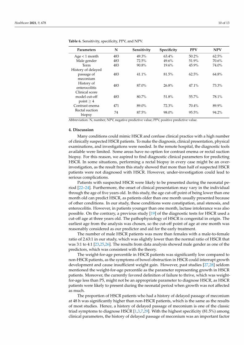
Diagnostic values of each parameter are shown in Table 6. Among the clinical scoremodel, contrast enema, and rectal suction biopsy, the parameter that showed the highestsensitivity for diagnosing HSCR was contrast enema and the highest specificity for diag-nosing HSCR was the rectal suction biopsy. The clinical score model, which used a cut-offpoint of 4, was used as a tool for screening with a sensitivity of 80.7% and a specificity of51.8%. Promptly referring patients for further investigation was advised to avoid delayeddiagnosis from clinical observation. However, the patients who were classified as low riskof HSCR should still be monitored and referred when no clinical improvement is seen.
Healthcare 2021, 9, 678 10 of 13
Table 6. Sensitivity, specificity, PPV, and NPV.
Parameters N Sensitivity Specificity PPV NPV
Age < 1 month 483 49.3% 63.4% 50.2% 62.5%Male gender 483 72.5% 49.6% 51.9% 70.6%
Term 483 90.8% 19.6% 45.9% 74.0%History of delayed
passage ofmeconium
483 41.1% 81.5% 62.5% 64.8%
History ofenterocolitis 483 87.0% 26.8% 47.1% 73.3%
Clinical scoremodel cut-off
point ≥ 4483 80.7% 51.8% 55.7% 78.1%
Contrast enema 471 89.0% 72.3% 70.4% 89.9%Rectal suction
biopsy 74 87.5% 98.0% 95.5% 94.2%
Abbreviation: N, number; NPV, negative predictive value; PPV, positive predictive value.
4. Discussion
Many conditions could mimic HSCR and confuse clinical practice with a high numberof clinically suspected HSCR patients. To make the diagnosis, clinical presentation, physicalexaminations, and investigations were needed. In the remote hospital, the diagnostic toolsavailable were limited. Some areas have no option for contrast enema or rectal suctionbiopsy. For this reason, we aspired to find diagnostic clinical parameters for predictingHSCR. In some situations, performing a rectal biopsy in every case might be an over-investigation, as the result from this study showed that more than half of suspected HSCRpatients were not diagnosed with HSCR. However, under-investigation could lead toserious complications.
Patients with suspected HSCR were likely to be presented during the neonatal pe-riod [22–24]. Furthermore, the onset of clinical presentation may vary in the individualthrough the age of five years old. In this study, the age cut-off point of being lower than onemonth old can predict HSCR, as patients older than one month usually presented becauseof other conditions. In our study, these conditions were constipation, anal stenosis, andenterocolitis. However, in patients younger than one month, lactase intolerance was alsopossible. On the contrary, a previous study [19] of the diagnostic tests for HSCR used acut-off age at three years old. The pathophysiology of HSCR is congenital in origin. Theearliest age from the analysis was chosen, so the cut-off point of age at one month wasreasonably considered as our predictor and aid for the early treatment.
The number of male HSCR patients was more than females with a male-to-femaleratio of 2.63:1 in our study, which was slightly lower than the normal ratio of HSCR thatwas 3:1 to 4:1 [23,25,26]. The results from data analysis showed male gender as one of thepredictors, which was consistent with the theory.
The weight-for-age percentile in HSCR patients was significantly low compared tonon-HSCR patients, as the symptoms of bowel obstruction in HSCR could interrupt growthdevelopment and cause insufficient weight gain. However, past studies [27,28] seldommentioned the weight-for-age percentile as the parameter representing growth in HSCRpatients. Moreover, the currently favored definition of failure to thrive, which was weight-for-age less than P5, might not be an appropriate parameter to diagnose HSCR, as HSCRpatients were likely to present during the neonatal period when growth was not affectedas much.
The proportion of HSCR patients who had a history of delayed passage of meconiumat 48 h was significantly higher than non-HSCR patients, which is the same as the resultsof most studies. Hence, a history of delayed passage of meconium is one of the classictriad symptoms to diagnose HSCR [1,3,7,29]. With the highest specificity (81.5%) amongclinical parameters, the history of delayed passage of meconium was an important factor

Healthcare 2021, 9, 678 11 of 13
to consider for further investigation. However, there were still a few non-HSCR patientswith a history of delayed passage of meconium.
Most of the reports have shown that HSCR patients were likely to occur in term infants.In 2018, the study in California reported the median gestational age at birth was 38 weeksand 6 days [30]. In our study, term infants had very high sensitivity with non-specificparameters for the diagnosis of HSCR.
Enterocolitis was also a presentation and postoperative unfavorable outcome of HSCR.The presence of preoperative enterocolitis was associated with the longer segment andinadequate rectal irrigation [31]. In our study, the presence of enterocolitis was shown asthe highest effect size parameter with a dOR of 2.42. However, there was a limitation ofmissing data. Some of the infants with enterocolitis might not be associated with HSCR.
The results from the contrast enema in this study were consistent with some previousstudies [20,21,29], with a sensitivity of 89.00% (83.82–92.98%) and specificity of 72.32%(66.59–77.57%). All the contrast enema signs included in this study showed statistical sig-nificance in diagnosing HSCR except jejunization, which can be correlated to the infection.The most common sign in HSCR patients was transitional zone (75.4%), which is the finestclue to diagnose HSCR from contrast enema results [6].
The sensitivity and specificity of RSB were 87.50% (67.64–97.34%) and 98% (89.35–99.95%),respectively, which was consistent with other studies [32,33]. Therefore, RSB is the in-vestigation that can be performed without anesthesia and no rectal scarring left whileperforming a definite operation, which could be helpful and cost-effective to the patient.However, the RSB needs the equipment and specialized pathologist to interpret the results.So, the clinical diagnosis is still important.
There was a previous clinical prediction model for diagnosis of HSCR [20]. In 2013,the previous score was combined between the clinical risk factors (delayed passage ofmeconium, age less than 3 years, and male gender) and the investigations that wereanorectal manometry, contrast enema, and rectal biopsy. In our clinical score model, weadded two more factors that were term infant and previous history of enterocolitis. Theage in our study was less than 1 month with the reason stated above. We also obtained asensitivity of 80.7% without the investigations, which were not available in some areas. Thisis the screening score to help in early referrals with a shortened time of clinical observation.
The results from data analysis in this study showed high sensitivity of the contrastenema and high specificity of the rectal suction biopsy. Therefore, contrast enema was moreappropriate to be used in screening. The result from the contrast enema was also helpfulin further surgery to locate the transitional zone. However, due to the high false-positiverate (75/471; 27.7%) and false-negative rate (22/471; 11.0%), subsequently confirmingthe HSCR diagnosis with rectal suction biopsy was needed. In our study, the additionof contrast enema and rectal suction biopsy improved the area under the ROC curve ofclinical parameters only in combination, from 72% to 87% and 97%, respectively. Therefore,the recommendation for the management of patients with suspected HSCR was to screenwith clinical parameters first, followed by contrast enema and rectal suction biopsy.
Because this study was retrospective, there were limitations in controlling factors anddata collection. The data analysis did not include results from the other two frequentlyused investigations, which were abdominal plain film and anorectal manometry. Theabdominal plain film result was not included because nearly all participants showed boweldilatation and absence of air in the rectum as the study domain of this study, as wellas some of the clinical features such as abdominal distension and vomiting. Anorectalmanometry was not included because of recent availability, which also made it difficultto diagnose ultrashort HSCR. A rectal suction biopsy was available in our institute threeyears ago. Before that, diagnosing HSCR mostly depended on clinical presentations andcontrast enema, so some patients had ganglion cells in operative specimens. In the earlyyears, there was also difficulty in differentiating variants of HSCR because of the pathologicunderstanding of the spectrum of disease.
Healthcare 2021, 9, 678 12 of 13
5. Conclusions
Five clinical parameters associated with the diagnosis of HSCR were as follows: ageless than one month, male gender, term infant, had history of delayed passing of meconium48 h after birth, and had history of enterocolitis. Patients with suspected HSCR, who had aclinical score 4–7, had a high probability to be HSCR and were suggested for early referralsfor further investigations, which were contrast enema and rectal suction biopsy. In case ofa low probability of HSCR, clinical observation and follow-up are still warranted until thesymptoms have been resolved. This clinical scoring system can be used as a screening toolto prevent delay diagnosis and complications of HSCR.
Author Contributions: Conceptualization, methodology, formal analysis, writing—review and edit-ing: J.K. (Jiraporn Khorana); data curation, writing—original draft preparation: P.P., J.K. (JitthiwimonKumsattra), S.K. and S.A.; resources: S.C., K.T. and J.S. All authors have read and agreed to thepublished version of the manuscript.
Funding: This research was funded by the Faculty of Medicine, Chiang Mai University Hospital.
Institutional Review Board Statement: The study was conducted according to the guidelines of theDeclaration of Helsinki and approved by the Ethics Committee of the Faculty of Medicine, ChiangMai University Hospital (Study code: SUR-2563-07500, 7 August 2020).
Informed Consent Statement: Patient consent was waived due to the retrospective chart review andno invasion of participants.
Data Availability Statement: The datasets used during the current study are available from thecorresponding author on reasonable request.
Acknowledgments: The authors acknowledge the Faculty of Medicine, Chiang Mai University whosupport the research conduction.
Conflicts of Interest: The authors declare no conflict of interest.
References1. Langer, J.C. Hirschsprung disease. Curr. Opin. Pediatr. 2013, 25, 368–374. [CrossRef]2. Muise, E.D.; Cowles, R.A. Rectal biopsy for Hirschsprung’s disease: A review of techniques, pathology, and complications. World
J. Pediatr. 2016, 12, 135–141. [CrossRef]3. Lewis, N.A.; Levitt, M.A.; Zallen, G.S.; Zafar, M.S.; Iacono, K.L.; Rossman, J.E.; Caty, M.G.; Glick, P.L. Diagnosing Hirschsprung’s
disease: Increasing the odds of a positive rectal biopsy result. J. Pediatr. Surg. 2003, 38, 412–416. [CrossRef] [PubMed]4. Hwang, T.J.; Servaes, S.; Mattei, P.; Anupindi, S.A. Radiologist performance in the interpretation of contrast enemas performed
for Hirschsprung’s disease in children >1 year of age. Clin. Radiol. 2017, 72, 519.e11–519.e19. [CrossRef] [PubMed]5. Rizky, M.; Isa, M.M.; Kamarlis, R.K. Comparison of Barium Enema and frozen section results in the diagnosis of Hirschsprung’s
Disease in a tertiary care hospital at Aceh, Indonesia. Med. J. Malays. 2020, 75, 37–40.6. Chen, X.; Xiaojuan, W.; Zhang, H.; Jiao, C.; Yu, K.; Zhu, T.; Feng, J. Diagnostic value of the preoperatively detected radiological
transition zone in Hirschsprung’s disease. Pediatr. Surg. Int. 2017, 33, 581–586. [CrossRef]7. Kyrklund, K.; Sloots, C.E.J.; de Blaauw, I.; Bjørnland, K.; Rolle, U.; Cavalieri, D.; Francalanci, P.; Fusaro, F.; Lemli, A.; Schwarzer,
N.; et al. ERNICA guidelines for the management of rectosigmoid Hirschsprung’s disease. Orphanet J. Rare Dis. 2020, 15, 164.[CrossRef]
8. Khorana, J.; Chinpakdee, W.; Sukhamwang, N.; Tepmalai, K. Calretinin versus hematoxylin and eosin stain for diagnosis ofhirschsprung’s disease; comparison in ganglionic, transitional, and aganglionic zones. J. Med. Assoc. Thail. 2020, 103, 559–565.[CrossRef]
9. Rahman, N.; Chouhan, J.; Gould, S.; Joseph, V.; Grant, H.; Hitchcock, R.; Johnson, P.; Lakhoo, K. Rectal biopsy for Hirschsprung’sdisease—Are we performing too many? Eur. J. Pediatr. Surg. 2010, 20, 95–97. [CrossRef]
10. Phillips, L.A.F.; Darwish, A.A.; Surana, R. Too Many Biopsies Performed to Rule Out Hirschsprung’s Disease: But It is WorthDoing Them. Eur. J. Pediatr. Surg. 2019, 29, 97–101. [CrossRef]
11. Prato, A.P.; Avanzini, S.; Gentilino, V.; Martucciello, G.; Mattioli, G.; Coccia, C.; Parodi, S.; Bisio, G.; Jasonni, V. Rectal suctionbiopsy in the workup of childhood chronic constipation: Indications and diagnostic value. Pediatr. Surg. Int. 2007, 23, 117–122.[CrossRef]
12. Lee, J.H.; Choe, Y.H.; Lee, S.K.; Seo, J.M.; Kim, J.H.; Suh, Y.L. Allergic proctitis and abdominal distention mimicking Hirschsprung’sdisease in infants. Acta Paediatr. 2007, 96, 1784–1789. [CrossRef]
13. Menchise, A.N.; Condino, A.A.; Levitt, M.A.; Hebra, A.; Wilsey, M.J. Celiac disease and diabetes mellitus diagnosed in a pediatricpatient with Hirschsprung disease. Fetal Pediatr. Pathol. 2013, 31, 7–12. [CrossRef]
Healthcare 2021, 9, 678 13 of 13
14. Arunachalam, P.; Mathai, J. Neonatal segmental enteritis due to cow’s milk allergy. J. Indian Assoc. Pediatr. Surg. 2013, 18, 149–151.[CrossRef]
15. Tahan, S.; Siviero-Miachon, A.A.; de Soares, M.F.F.; Martins-Moura, E.C.S.; Peterlini, F.L.; de Morais, M.B.; Spinola-Castro, A.M.Untreated Congenital Hypothyroidism Mimicking Hirschsprung Disease: A Puzzling Case in a One-Year-Old Child. Case Rep.Pediatr. 2018, 2018, 9209873. [CrossRef]
16. Friedmacher, F.; Puri, P. Classification and diagnostic criteria of variants of Hirschsprung’s disease. Pediatr. Surg. Int. 2013, 29,855–872. [CrossRef]
17. WHO. Preterm Birth; WHO: Genewa, Switzerland, 2018.18. WHO Multicentre Growth Reference Study Group; De Onis, M. WHO Child Growth Standards based on length/height, weight
and age. Acta Paediatr. Suppl. 2006, 450, 76–85. [CrossRef]19. Homan, G.J. Failure to Thrive: A Practical Guide. Am. Fam. Physician 2016, 94, 295–299.20. Wu, X.J.; Zhang, H.Y.; Li, N.; Yan, M.S.; Wei, J.; Yu, D.H.; Feng, J.X. A new diagnostic scoring system to differentiate Hirschsprung’s
disease from Hirschsprung’s disease-allied disorders in patients with suspected intestinal dysganglionosis. Int. J. Colorectal Dis.2013, 28, 689–696. [CrossRef]
21. Peyvasteh, M.; Askarpour, S.; Ostadian, N.; Moghimi, M.R.; Javaherizadeh, H. Diagnostic Accuracy of Barium Enema Findings InHirschsprung’s Disease. Arq. Bras. Cir. Dig. 2016, 29, 155–158. [CrossRef]
22. De Lorijn, F.; Boeckxstaens, G.E.; Benninga, M.A. Symptomatology, pathophysiology, diagnostic work-up, and treatment ofHirschsprung disease in infancy and childhood. Curr. Gastroenterol. Rep. 2007, 9, 245–253. [CrossRef] [PubMed]
23. Bradnock, T.J.; Knight, M.; Kenny, S.; Nair, M.; Walker, G.M. Hirschsprung’s disease in the UK and Ireland: Incidence andanomalies. Arch. Dis. Child. 2017, 102, 722–727. [CrossRef] [PubMed]
24. Juang, D.; Snyder, C.L. Neonatal bowel obstruction. Surg. Clin. N. Am. 2012, 92, 685–711. [CrossRef] [PubMed]25. Karim, A.; Akter, M.; Aziz, T.T.; Hoque, M.; Chowdhury, T.K.; Imam, M.S.; Walid, A.; Kabir, M.; So, M.; Lam, W.Y.; et al.
Epidemiological characteristics of Hirschsprung’s disease (HSCR): Results of a case series of fifty patients from Bangladesh. J.Pediatr. Surg. 2018, 53, 1955–1959. [CrossRef]
26. Chia, S.T.; Chen, S.C.; Lu, C.L.; Sheu, S.M.; Kuo, H.C. Epidemiology of Hirschsprung’s Disease in Taiwanese Children: A 13-yearNationwide Population-based Study. Pediatr. Neonatol. 2016, 57, 201–206. [CrossRef]
27. Bermudez, B.; de Oliveira, C.M.; de Lima Cat, M.N.; Magdalena, N.I.R.; Celli, A. Gastrointestinal disorders in Down syndrome.Am. J. Med. Genet. A 2019, 179, 1426–1431. [CrossRef]
28. Veras, L.V.; Chotai, P.N.; Tumen, A.Z.; Gosain, A. Impaired growth outcomes in children with congenital colorectal diseases. J.Surg. Res. 2018, 229, 102–107. [CrossRef]
29. De Lorijn, F.; Kremer, L.C.; Reitsma, J.B.; Benninga, M.A. Diagnostic tests in Hirschsprung disease: A systematic review. J. Pediatr.Gastroenterol. Nutr. 2006, 42, 496–505. [CrossRef]
30. Anderson, J.E.; Vanover, M.A.; Saadai, P.; Stark, R.A.; Stephenson, J.T.; Hirose, S. Epidemiology of Hirschsprung disease inCalifornia from 1995 to 2013. Pediatr. Surg. Int. 2018, 34, 1299–1303. [CrossRef]
31. Lu, C.; Xie, H.; Li, H.; Geng, Q.; Chen, H.; Mo, X.; Tang, W. Feasibility and efficacy of home rectal irrigation in neonates and earlyinfancy with Hirschsprung disease. Pediatr. Surg. Int. 2019, 35, 1245–1253. [CrossRef]
32. Allen, A.R.; Putnam, A.R.; Presson, A.P.; Allen, C.M.; Barnhart, D.C.; Rollins, M.D. Accuracy of Suction Rectal Biopsy forDiagnosis of Hirschsprung’s Disease in Neonates. Eur. J. Pediatr. Surg. 2019, 29, 425–430. [CrossRef]
33. Tran, V.Q.; Lam, K.T.; Truong, D.Q.; Dang, M.H.; Doan, T.T.; Segers, V.; Butler, M.W.; Robert, A.; Goyens, P.; Steyaert, H. Diagnosticvalue of rectal suction biopsies using calretinin immunohistochemical staining in Hirschsprung’s disease. J. Pediatr. Surg. 2016,51, 2005–2009. [CrossRef]