Clinical characteristics and predictive score of dengue vs ...
25
HAL Id: hal-01845510 https://hal.archives-ouvertes.fr/hal-01845510 Submitted on 22 Oct 2021 HAL is a multi-disciplinary open access archive for the deposit and dissemination of sci- entific research documents, whether they are pub- lished or not. The documents may come from teaching and research institutions in France or abroad, or from public or private research centers. L’archive ouverte pluridisciplinaire HAL, est destinée au dépôt et à la diffusion de documents scientifiques de niveau recherche, publiés ou non, émanant des établissements d’enseignement et de recherche français ou étrangers, des laboratoires publics ou privés. Distributed under a Creative Commons Attribution - NonCommercial| 4.0 International License Clinical characteristics and predictive score of dengue vs. chikungunya virus infections Timothée Bonifay, C Bonnefoy, D Blanchet, M. Dueymes, Mathieu Nacher, Félix Djossou, Loïc Epelboin To cite this version: Timothée Bonifay, C Bonnefoy, D Blanchet, M. Dueymes, Mathieu Nacher, et al.. Clinical char- acteristics and predictive score of dengue vs. chikungunya virus infections. Médecine et Maladies Infectieuses, Elsevier Masson, In press. hal-01845510
-
Upload
khangminh22 -
Category
Documents
-
view
3 -
download
0
Transcript of Clinical characteristics and predictive score of dengue vs ...
HAL Id: hal-01845510https://hal.archives-ouvertes.fr/hal-01845510
Submitted on 22 Oct 2021
HAL is a multi-disciplinary open accessarchive for the deposit and dissemination of sci-entific research documents, whether they are pub-lished or not. The documents may come fromteaching and research institutions in France orabroad, or from public or private research centers.
L’archive ouverte pluridisciplinaire HAL, estdestinée au dépôt et à la diffusion de documentsscientifiques de niveau recherche, publiés ou non,émanant des établissements d’enseignement et derecherche français ou étrangers, des laboratoirespublics ou privés.
Distributed under a Creative Commons Attribution - NonCommercial| 4.0 InternationalLicense
Clinical characteristics and predictive score of denguevs. chikungunya virus infections
Timothée Bonifay, C Bonnefoy, D Blanchet, M. Dueymes, Mathieu Nacher,Félix Djossou, Loïc Epelboin
To cite this version:Timothée Bonifay, C Bonnefoy, D Blanchet, M. Dueymes, Mathieu Nacher, et al.. Clinical char-acteristics and predictive score of dengue vs. chikungunya virus infections. Médecine et MaladiesInfectieuses, Elsevier Masson, In press. �hal-01845510�

Caractéristiques cliniques et score prédictif d’infection par le virus de la dengue versus
chikungunya
Clinical characteristics and predictive score of dengue vs. chikungunya virus infections
Timothée Bonifay*1,2, Guillaume Vesin1, Bastien Bidaud1, Clémence Bonnefoy2,4,
Maryvonne Dueymes5, Mathieu Nacher3,6, Félix Djossou1,6, and Loïc Epelboin1,6
1 Unité de maladie infectieuse et tropicale, Centre Hospitalier Andrée Rosemon, Cayenne F-
97300, Guyane française, France
2 Département de médecine générale, Université des Antilles, France
3 Centre d’Investigation Clinique Antilles Guyane, Inserm CIC 1424, Cayenne F-97300,
Guyane française, France
4 Service d’accueil des urgences, Centre Hospitalier Andrée Rosemon, Cayenne F-97300,
Guyane française, France
5 Laboratoire Hospitalier universitaire d'immunologie, Centre Hospitalier Andrée Rosemon,
Cayenne F-97300, Guyane française, France
6 Equipe EA 3593, Ecosystèmes Amazoniens et Pathologie Tropicale, Université de Guyane,
Cayenne F-97300, Guyane française, France
Keywords: chikungunya, dengue, French Guiana, arboviruses, predictive score
Mots clés : chikungunya, dengue, Guyane française, arbovirus, score prédictif
Corresponding author
Timothée Bonifay, MD,
Unité de maladie infectieuse et tropicale, Centre hospitalier de Cayenne – Av des
flamboyants, Cayenne 97306, Guyane française
© 2018 published by Elsevier. This manuscript is made available under the CC BY NC user licensehttps://creativecommons.org/licenses/by-nc/4.0/
Version of Record: https://www.sciencedirect.com/science/article/pii/S0399077X18300507Manuscript_d8114b65cbfb0858f3be9527c2f31811

Email address: [email protected]
Phone: +594 594 39 50 40 / Fax : +594 594 39 50 41
Alternate author
Loïc Epelboin, MD
Unité de maladie infectieuse et tropicale, Centre hospitalier de Cayenne – Av des
flamboyants, Cayenne 97306, Guyane française
Mail : [email protected]
Phone : +594 594 39 50 40 / Fax : +594 594 39 50 41

1
ABSTRACT
Background. Chikungunya (CHIKV) and dengue viruses (DENV) are two arboviruses with
epidemic potential and similar clinical presentations. The potential life-threatening risk
associated with DENV justifies an immediate biological assessment and medical follow-up
which may be delayed for CHIKV.
Objectives. To compare the clinical variables that would help differentiate patients infected
with CHIKV or DENV, and then to compute a predictive score.
Patients and method. Retrospective case-control study comparing CHIKV-infected patients
diagnosed by RT-PCR in 2014 with patients infected with DENV diagnosed by positive NS1
antigen test in 2013. Children aged <15 years and pregnant women were excluded. Clinical
and biological variables were compared, and a multivariate analysis was performed. A clinical
score was developed using the β coefficients to differentiate the infections.
Results. Over the study period 168 patients infected with CHIKV were compared with 452
patients with DENV. The clinical variables independently associated with CHIKV was joint
and back pain, and those associated with DENV were headache, muscle pain,
nausea/vomiting, diarrhea, and hemorrhagic signs. The clinical score had 98% sensitivity for
DENV and a ROC curve of 0.96.
Conclusion. These two infections have a similar clinical presentation but the use of the
proposed clinical score during the acute phase of the disease would make it possible to
identify cases of DENV during a CHIKV epidemic to suggest adequate patient management.

2
RÉSUMÉ
Objectifs. Les virus du chikungunya (CHIKV) et de la dengue (DENV) sont deux arbovirus à
fort potentiel épidémique avec une présentation clinique similaire. Le risque potentiellement
mortel associé à DENV justifie une évaluation biologique immédiate et un suivi médical, qui
peuvent être retardés en cas d’infection par CHIKV. L'objectif de l'étude était de comparer les
variables cliniques qui permettraient de différencier un patient infecté par le CHIKV ou le
DENV puis de réaliser un score prédictif.
Patients et méthode. Étude cas-témoins rétrospective comparant des patients infectés par
CHIKV diagnostiqués par RT-PCR en 2014 à des patients infectés par DENV diagnostiqués
par antigène NS1 positif en 2013. Les enfants < 15 ans et les femmes enceintes ont été exclus.
Les variables cliniques et biologiques ont été comparées et une analyse multivariée a été
réalisée. Un score clinique a été construit à partir des coefficients β pour différencier les deux
infections.
Résultats. 168 patients infectés par CHIKV ont été comparés à 452 patients atteints par
DENV. Les variables cliniques associées à CHIKV étaient la présence d’arthralgies et
rachialgies, et celles associées au DENV étaient céphalées, myalgies, nausées/vomissements,
diarrhées et signes hémorragiques. Le score clinique avait une sensibilité de 98 % pour le
DENV et une courbe ROC de 0,96.
Conclusions. Ces deux infections ont une présentation clinique similaire mais l'utilisation du
score clinique proposé durant la phase aiguë de la maladie permettrait d'identifier les cas de
DENV lors d'une épidémie de CHIKV pour proposer une prise en charge adéquate.
3
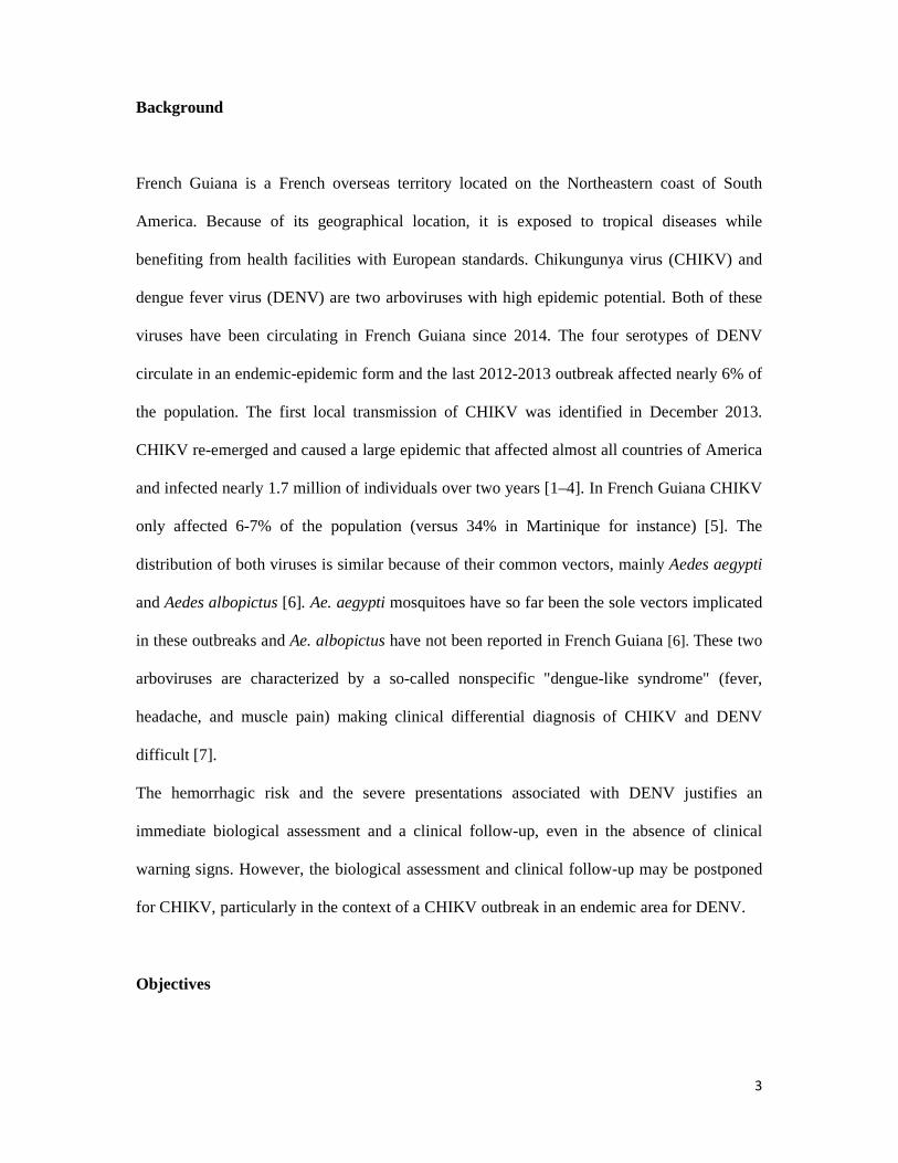
Background
French Guiana is a French overseas territory located on the Northeastern coast of South
America. Because of its geographical location, it is exposed to tropical diseases while
benefiting from health facilities with European standards. Chikungunya virus (CHIKV) and
dengue fever virus (DENV) are two arboviruses with high epidemic potential. Both of these
viruses have been circulating in French Guiana since 2014. The four serotypes of DENV
circulate in an endemic-epidemic form and the last 2012-2013 outbreak affected nearly 6% of
the population. The first local transmission of CHIKV was identified in December 2013.
CHIKV re-emerged and caused a large epidemic that affected almost all countries of America
and infected nearly 1.7 million of individuals over two years [1–4]. In French Guiana CHIKV
only affected 6-7% of the population (versus 34% in Martinique for instance) [5]. The
distribution of both viruses is similar because of their common vectors, mainly Aedes aegypti
and Aedes albopictus [6]. Ae. aegypti mosquitoes have so far been the sole vectors implicated
in these outbreaks and Ae. albopictus have not been reported in French Guiana [6]. These two
arboviruses are characterized by a so-called nonspecific "dengue-like syndrome" (fever,
headache, and muscle pain) making clinical differential diagnosis of CHIKV and DENV
difficult [7].
The hemorrhagic risk and the severe presentations associated with DENV justifies an
immediate biological assessment and a clinical follow-up, even in the absence of clinical
warning signs. However, the biological assessment and clinical follow-up may be postponed
for CHIKV, particularly in the context of a CHIKV outbreak in an endemic area for DENV.
Objectives

4
The main objective of the study was to compare the epidemiological, clinical, and biological
characteristics which would help differentiate patients infected with CHIKV or DENV at the
first medical consultation. The secondary objective was to compute a predictive score to
differentiate DENV from CHIKV infection.
Study design
A monocenter retrospective case-control study was conducted to compare a group of patients
infected with CHIKV with a group infected with DENV. This study was performed in the
hospital of Cayenne, the main city and the main hospital in French Guiana.
Inclusion criteria of participants presenting with chikungunya fever
Retrospectively, all patients diagnosed and/or treated at the hospital for chikungunya from
April to June 2014 (i.e., the first months of the outbreak) constituted our case group. A case of
CHIKV infection was defined as a clinical presentation suggestive of an arbovirus infection
[8] and a microbiological diagnosis of acute CHIKV infection diagnosed by RT-PCR. All
patients underwent medical consultation at the hospital, in the emergency department or
infectious diseases department, with an initial standard infectious biological assessment
including CHIKV RT-PCR, DENV diagnostic test (NS1 antigen and IgM), thin and thick
blood smear for malaria, and standard blood tests at the physician’s discretion. Nucleic acid
was extracted from serum using a MagNA Pure LC total nucleic acid isolation kit and a
MagNA Pure LC2.0 instrument (Roche). Real time RT-PCR amplification was performed
with the RealStar® Chikungunya RT-PCR kit 1.0 (CE-IVD marking) (Altona Diagnostics)
and a thermocycler 7500 Applied Biosystem. Qualitative detection of dengue virus NS1
antigen was performed in serum by enzyme immunoassay with Platelia™ dengue NS1 Ag kit
(Biorad). All biological tests were performed at the hospital laboratory.

5
Inclusion criteria of participants presenting with dengue fever
A retrospective collection was then performed to create a control group of patients presenting
with acute DENV infection. The group was exhaustively made of patients presenting with a
compatible clinical picture of arbovirus infection [8] and a positive NS1 antigen test at the
Cayenne hospital laboratory from March to August 2013 during a DENV outbreak (mainly
DENV2). The NS1 test has been historically used at the hospital and we could not perform the
DENV PCR.
Exclusion criteria
The exclusion criteria were age under 15 years, ongoing pregnancy, and/or incomplete
medical records. Children and pregnant women were monitored in the departments of
pediatrics and gynecology-obstetrics respectively, and thus did not allow for a uniform data
collection.
Data collection and statistical methods
The epidemiological, clinical, and biological data was collected from computerized medical
records: the emergency medical record (DMUnet®), the common medical record (Cora®), and
the biological medical software (SRI®). A clinical variable not reported in the chart was
considered negative. The first day of fever was defined as the first day of the disease.
The clinical and biological characteristics of the groups were compared at the first
consultation using a chi-square test, Student’s t test for the normally distributed quantitative
variables, or the non-parametric Mann-Whitney test for the other continuous variables.
Variables were considered significant with p<0.05.

6
Unconditional multiple logistic regression was used to compute a predictive score with high
sensitivity for identifying probable DENV versus CHIKV infection. The most relevant
clinical variables with a significance level of less than 0.2 were selected and included in the
multivariate model. Nonspecific or inconsistently reported variables in initial medical charts
were considered unreliable and were excluded. A step-by-step backward elimination
procedure was used starting with variables with the highest p value. The predictive score was
computed using the β-coefficients resulting from the multivariate analysis. We used 3,500
bootstrapped replications to obtain robust estimates. We then evaluated the value of our
model by estimating the area under the ROC (receiver operating characteristic) curve and by
calculating the sensitivity and specificity scores of each result to evaluate the performance of
the test. We also computed a goodness of fit test to compare the observed sample distribution.
The data was anonymously collected on Microsoft Access 2010® and analyzed on Stata
12.0®.
Ethical consideration
The present monocenter study included anonymized patient records (the database did not
include names or any variable that could lead to the identification of patients) as authorized by
the French regulatory authorities. The database was declared to the French Data Protection
Authority (French acronym CNIL) (no. TFN1490159N) as per French legal requirements.

7
Results
A total of 620 patients were selected for the study: 168 patients for the CHIKV group and
452 patients for the DENV group (Figure 1). No CHIKV-DENV co-infection was identified
during the CHIKV epidemic nor was co-infection with malaria identified. The male-to-female
sex ratio was equal in both groups at ~1/1. The study population was significantly older in the
CHIKV group and had more comorbid conditions. The time between symptom onset and the
first consultation was shorter in the CHIKV group. At the end of this consultation 8.3% of
CHIKV patients were hospitalized versus 35.2% of patients in the DENV group (p<0.001).
The significant variables associated with CHIKV were joint and back pains and those
associated with DENV were diarrhea, abdominal pain, and neurological signs (Table 1). No
CHIKV patient had severe hemorrhagic presentations, only 1.2% had minor hemorrhage
(epistaxis or gingival hemorrhage) versus 15.9% for the DENV group (OR 0.06 [0.01-0.25]
p<0.001). Of the 14 hospitalized CHIKV patients, four were considered as having an atypical
or severe presentation (two had uncontrolled pain and two had concomitant pancreatitis or
cholecystitis). Nine DENV patients were classified group C (severe dengue): three for severe
plasma leakage, three for severe bleeding (spleen rupture, psoas hematoma, and extensive
purpura), and three for severe organ impairment. DENV patients had a more frequent rash at
the time of their first consultation but the CHIKV group had more frequent pruritus in
particular without rash (OR 7.5 [3.1-20.0] p<0.001). More than 25% of skin presentations
observed in patients of the DENV group occurred after the first consultation versus 4.2% for
CHIKV patients.
CHIKV was significantly associated with CRP >30 mg/l. The variables most often associated
with DENV were thrombocytopenia <150 G/l, neutropenia <1,500/mm3, lymphopenia
8
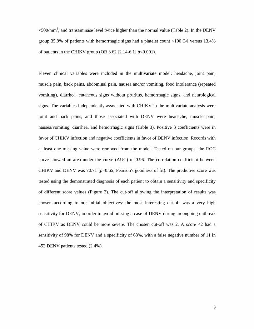
<500/mm3, and transaminase level twice higher than the normal value (Table 2). In the DENV
group 35.9% of patients with hemorrhagic signs had a platelet count <100 G/l versus 13.4%
of patients in the CHIKV group (OR 3.62 [2.14-6.1] p<0.001).
Eleven clinical variables were included in the multivariate model: headache, joint pain,
muscle pain, back pains, abdominal pain, nausea and/or vomiting, food intolerance (repeated
vomiting), diarrhea, cutaneous signs without pruritus, hemorrhagic signs, and neurological
signs. The variables independently associated with CHIKV in the multivariate analysis were
joint and back pains, and those associated with DENV were headache, muscle pain,
nausea/vomiting, diarrhea, and hemorrhagic signs (Table 3). Positive β coefficients were in
favor of CHIKV infection and negative coefficients in favor of DENV infection. Records with
at least one missing value were removed from the model. Tested on our groups, the ROC
curve showed an area under the curve (AUC) of 0.96. The correlation coefficient between
CHIKV and DENV was 70.71 (p=0.65; Pearson's goodness of fit). The predictive score was
tested using the demonstrated diagnosis of each patient to obtain a sensitivity and specificity
of different score values (Figure 2). The cut-off allowing the interpretation of results was
chosen according to our initial objectives: the most interesting cut-off was a very high
sensitivity for DENV, in order to avoid missing a case of DENV during an ongoing outbreak
of CHIKV as DENV could be more severe. The chosen cut-off was 2. A score ≤2 had a
sensitivity of 98% for DENV and a specificity of 63%, with a false negative number of 11 in
452 DENV patients tested (2.4%).
9
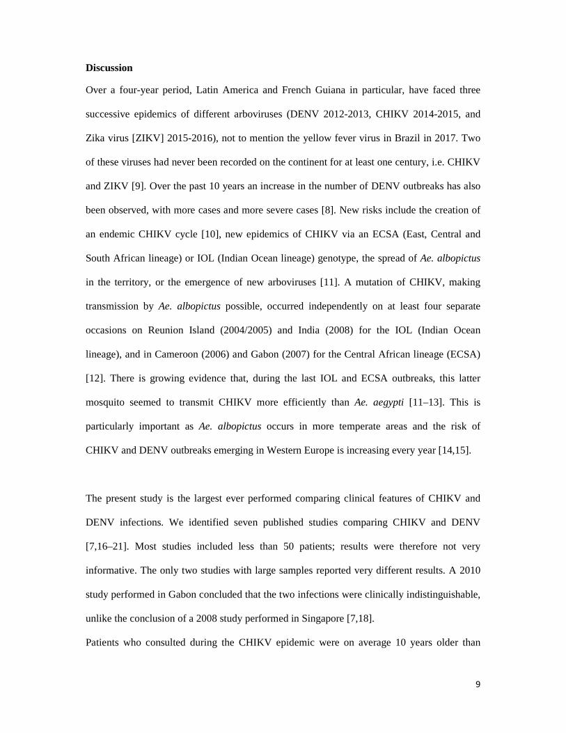
Discussion
Over a four-year period, Latin America and French Guiana in particular, have faced three
successive epidemics of different arboviruses (DENV 2012-2013, CHIKV 2014-2015, and
Zika virus [ZIKV] 2015-2016), not to mention the yellow fever virus in Brazil in 2017. Two
of these viruses had never been recorded on the continent for at least one century, i.e. CHIKV
and ZIKV [9]. Over the past 10 years an increase in the number of DENV outbreaks has also
been observed, with more cases and more severe cases [8]. New risks include the creation of
an endemic CHIKV cycle [10], new epidemics of CHIKV via an ECSA (East, Central and
South African lineage) or IOL (Indian Ocean lineage) genotype, the spread of Ae. albopictus
in the territory, or the emergence of new arboviruses [11]. A mutation of CHIKV, making
transmission by Ae. albopictus possible, occurred independently on at least four separate
occasions on Reunion Island (2004/2005) and India (2008) for the IOL (Indian Ocean
lineage), and in Cameroon (2006) and Gabon (2007) for the Central African lineage (ECSA)
[12]. There is growing evidence that, during the last IOL and ECSA outbreaks, this latter
mosquito seemed to transmit CHIKV more efficiently than Ae. aegypti [11–13]. This is
particularly important as Ae. albopictus occurs in more temperate areas and the risk of
CHIKV and DENV outbreaks emerging in Western Europe is increasing every year [14,15].
The present study is the largest ever performed comparing clinical features of CHIKV and
DENV infections. We identified seven published studies comparing CHIKV and DENV
[7,16–21]. Most studies included less than 50 patients; results were therefore not very
informative. The only two studies with large samples reported very different results. A 2010
study performed in Gabon concluded that the two infections were clinically indistinguishable,
unlike the conclusion of a 2008 study performed in Singapore [7,18].
Patients who consulted during the CHIKV epidemic were on average 10 years older than

10
DENV patients, as already mentioned in other studies [16,18,21]. Dengue fever is endemic in
French Guiana. As immunity increases with age, the virus targets a younger and less immune
population [22]. Several studies reported a lower sensitivity rate with NS1 test in acute
secondary dengue than in acute primary dengue. The NS1 test is therefore more likely to
diagnose acute primary dengue and select a younger population [23]. On the contrary, during
the CHIKV epidemic the population was not previously immunized, and people of the
extreme ages were naturally more affected. Patients consulted earlier during the CHIKV
epidemic than the DENV epidemic because of greater concern, severe symptoms, a source of
epidemic near the hospital, and pressure of health authorities and the media. At the beginning
of the epidemic, CHIKV was assumed to be more serious than DENV in the public
imagination [24]. The vision of saturated hospitals and chronic presentations strongly
encouraged patients to consult earlier, at least at the beginning of the epidemic. It should also
be noted that the outbreak started close to the hospital (unpublished data) and that emergency
departments were the primary access to care for many patients. Its proximity thus made it
easier for patients to consult.
The choice of the cut-off made it possible to obtain a very sensitive clinical score to
differentiate DENV from CHIKV. A score between -9 and +2 was in favor of DENV and
between +3 and +6 in favor of CHIKV. The presence of arthritis was indicative of CHIKV
infection (Table 4). The use of the score requires two prerequisites: an epidemic context with
a circulation of both viruses and patients with dengue-like syndrome. Detection of dengue
cases is essential in endemic areas. Several scores have been published. For instance one has
been built to differentiate dengue from other febrile illnesses [25]. Most clinical symptoms are
based on hemorrhagic signs which are not always observed, e.g. only 26.3% of our DENV
patients presented with clinical symptoms during the consultation. Our score used nonspecific
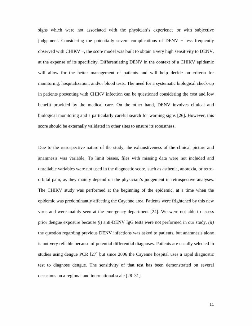
11
signs which were not associated with the physician’s experience or with subjective
judgement. Considering the potentially severe complications of DENV − less frequently
observed with CHIKV −, the score model was built to obtain a very high sensitivity to DENV,
at the expense of its specificity. Differentiating DENV in the context of a CHIKV epidemic
will allow for the better management of patients and will help decide on criteria for
monitoring, hospitalization, and/or blood tests. The need for a systematic biological check-up
in patients presenting with CHIKV infection can be questioned considering the cost and low
benefit provided by the medical care. On the other hand, DENV involves clinical and
biological monitoring and a particularly careful search for warning signs [26]. However, this
score should be externally validated in other sites to ensure its robustness.
Due to the retrospective nature of the study, the exhaustiveness of the clinical picture and
anamnesis was variable. To limit biases, files with missing data were not included and
unreliable variables were not used in the diagnostic score, such as asthenia, anorexia, or retro-
orbital pain, as they mainly depend on the physician’s judgement in retrospective analyses.
The CHIKV study was performed at the beginning of the epidemic, at a time when the
epidemic was predominantly affecting the Cayenne area. Patients were frightened by this new
virus and were mainly seen at the emergency department [24]. We were not able to assess
prior dengue exposure because (i) anti-DENV IgG tests were not performed in our study, (ii)
the question regarding previous DENV infections was asked to patients, but anamnesis alone
is not very reliable because of potential differential diagnoses. Patients are usually selected in
studies using dengue PCR [27] but since 2006 the Cayenne hospital uses a rapid diagnostic
test to diagnose dengue. The sensitivity of that test has been demonstrated on several
occasions on a regional and international scale [28–31].
12
These two infections have a similar clinical presentation but the use of the proposed clinical
score during the acute phase of the disease would make it possible to identify cases of DENV
during a CHIKV outbreak to implement adequate patient management. The study, despite its
limitations, has large samples and provides consistent results. Another outbreak would allow
for an external validation to consolidate the use of the score.

13
Acknowledgments
The authors would like to thank the physicians who supported the patients and helped collect
clinical data and fill in the forms: Philippe Abboud, Gaëlle Walter, Aba Mahamat, Yasmina
Briquet, Rémi Mutricy, and Elodie Rossigneux. They would also like to thank the biologists
who performed the RT-PCR: Denis Blanchet and Magalie Pierre-Demar.
Contribution of authors
T.B. designed the study, performed the literature analysis, performed data acquisition and
analysis, interpreted the data, wrote the first draft of the article and tables.
G.V. performed data acquisition.
B.B. performed data acquisition.
C.B. performed data acquisition and reviewed the article.
M.D. reviewed the article.
F.D. reviewed the article.
M.N. reviewed the article, translated the article, and interpreted the results.
L.E. designed the study project, performed the data analysis, interpreted the results, and
reviewed the article.

14
REFERENCES
1. Kuno G. A Re-Examination of the History of Etiologic Confusion between Dengue and
Chikungunya. PLoS Negl. Trop. Dis. 2015 12;9:e0004101. doi: 10.1371/journal.pntd.0004101
2. Halstead SB. Reappearance of Chikungunya, Formerly Called Dengue, in the Americas. Emerg.
Infect. Dis. 2015;21:557–61. doi: 10.3201/eid2104.141723
3. Geographic Distribution | Chikungunya virus | CDC [Internet]. [cited 2017 16];doi:
https://www.cdc.gov/chikungunya/geo/
4. Weaver SC, Lecuit M. Chikungunya Virus and the Global Spread of a Mosquito-Borne Disease.
N. Engl. J. Med. 2015 26;372:1231–9. doi: 10.1056/NEJMra1406035
5. InVS. Surveillance du chikungunya - Bulletin bimestriel : Période du 13 juin au 4 septembre
2016 - Guyane. ARS; 2016.
6. Vega-Rúa A, Lourenço-de-Oliveira R, Mousson L, Vazeille M, Fuchs S, Yébakima A, et al.
Chikungunya Virus Transmission Potential by Local Aedes Mosquitoes in the Americas and
Europe. PLoS Negl. Trop. Dis. 2015 20;9:e0003780. doi: 10.1371/journal.pntd.0003780
7. Nkoghe D, Kassa Kassa RF, Bisvigou U, Caron M, Grard G, Leroy EM. No clinical or biological
difference between Chikungunya and Dengue Fever during the 2010 Gabonese outbreak.
Infect. Dis. Rep. 2012 3;4:5. doi: 10.4081/idr.2012.e5
8. Simmons CP, Farrar JJ, van Vinh Chau N, Wills B. Dengue. N. Engl. J. Med. 2012;366:1423–1432.
9. Carey DE. Chikungunya and dengue: a case of mistaken identity? J. Hist. Med. Allied Sci.
1971;26:243–262.
10. Lourenço-de-Oliveira R, Failloux A-B. High risk for chikungunya virus to initiate an enzootic
sylvatic cycle in the tropical Americas. PLoS Negl. Trop. Dis. 2017 29;11:e0005698. doi:
10.1371/journal.pntd.0005698
11. Vega-Rúa A, Zouache K, Girod R, Failloux A-B, Lourenço-de-Oliveira R. High Level of Vector
Competence of Aedes aegypti and Aedes albopictus from Ten American Countries as a Crucial
Factor in the Spread of Chikungunya Virus. J. Virol. 2014;88:6294–306. doi: 10.1128/JVI.00370-
14
12. Vazeille M, Zouache K, Vega-Rúa A, Thiberge J-M, Caro V, Yébakima A, et al. Importance of
mosquito “quasispecies” in selecting an epidemic arthropod-borne virus. Sci. Rep. [Internet]
2016 7;6. doi: http://www.ncbi.nlm.nih.gov/pmc/articles/PMC4935986/doi:
10.1038/srep29564
13. Vazeille M, Mousson L, Martin E, Failloux A-B. Orally Co-Infected Aedes albopictus from La
Reunion Island, Indian Ocean, Can Deliver Both Dengue and Chikungunya Infectious Viral
Particles in Their Saliva. PLoS Negl. Trop. Dis. 2010 8;4:e706. doi: 10.1371/journal.pntd.0000706

15
14. Kraemer MU, Sinka ME, Duda KA, Mylne AQ, Shearer FM, Barker CM, et al. The global
distribution of the arbovirus vectors Aedes aegypti and Ae. albopictus. eLife 2015 30;4:e08347.
doi: 10.7554/eLife.08347
15. WHO | Chikungunya – Italy [Internet]. WHO [cited 2017 9];doi:
http://www.who.int/csr/don/15-september-2017-chikungunya-italy/en/
16. Kularatne S a. M, Gihan MC, Weerasinghe SC, Gunasena S. Concurrent outbreaks of
Chikungunya and Dengue fever in Kandy, Sri Lanka, 2006-07: a comparative analysis of clinical
and laboratory features. Postgrad. Med. J. 2009;85:342–6. doi: 10.1136/pgmj.2007.066746
17. Velasco JM, Valderama MT, Lopez MN, Chua D, Latog R, Roque V, et al. Chikungunya Virus
Infections Among Patients with Dengue-Like Illness at a Tertiary Care Hospital in the
Philippines, 2012-2013. Am. J. Trop. Med. Hyg. 2015 9;93:1318–24. doi: 10.4269/ajtmh.15-0332
18. Lee VJ, Chow A, Zheng X, Carrasco LR, Cook AR, Lye DC, et al. Simple Clinical and Laboratory
Predictors of Chikungunya versus Dengue Infections in Adults. PLoS Negl. Trop. Dis. [Internet]
2012 27 [cited 2016 19];6. doi: http://www.ncbi.nlm.nih.gov/pmc/articles/PMC3459852/doi:
10.1371/journal.pntd.0001786
19. Sahadeo N, Mohammed H, Allicock OM, Auguste AJ, Widen SG, Badal K, et al. Molecular
Characterisation of Chikungunya Virus Infections in Trinidad and Comparison of Clinical and
Laboratory Features with Dengue and Other Acute Febrile Cases. PLoS Negl. Trop. Dis.
[Internet] 2015 18 [cited 2016 28];9. doi:
http://www.ncbi.nlm.nih.gov/pmc/articles/PMC4651505/doi: 10.1371/journal.pntd.0004199
20. Hochedez P, Canestri A, Guihot A, Brichler S, Bricaire F, Caumes E. Management of Travelers
with Fever and Exanthema, Notably Dengue and Chikungunya Infections. Am. J. Trop. Med.
Hyg. 2008 5;78:710–3.
21. Mohd Zim MA, Sam I-C, Omar SFS, Chan YF, AbuBakar S, Kamarulzaman A. Chikungunya
infection in Malaysia: Comparison with dengue infection in adults and predictors of persistent
arthralgia. J. Clin. Virol. 2013;56:141–5. doi: 10.1016/j.jcv.2012.10.019
22. Dhar-Chowdhury P, Paul KK, Haque CE, Hossain S, Lindsay LR, Dibernardo A, et al. Dengue
seroprevalence, seroconversion and risk factors in Dhaka, Bangladesh. PLoS Negl. Trop. Dis.
2017 23;11:e0005475. doi: 10.1371/journal.pntd.0005475
23. Kumarasamy V, Chua SK, Hassan Z, Wahab AHA, Chem YK, Mohamad M, et al. Evaluating the
sensitivity of a commercial dengue NS1 antigen-capture ELISA for early diagnosis of acute
dengue virus infection. Singapore Med. J. 2007;48:669–73.
24. Fritzell C, Raude J, Adde A, Dusfour I, Quenel P, Flamand C. Knowledge, Attitude and Practices
of Vector-Borne Disease Prevention during the Emergence of a New Arbovirus: Implications for
the Control of Chikungunya Virus in French Guiana. PLOS Negl Trop Dis 2016 1;10:e0005081.
doi: 10.1371/journal.pntd.0005081
25. Fernández E, Smieja M, Walter SD, Loeb M. A predictive model to differentiate dengue from
other febrile illness. BMC Infect. Dis. [Internet] 2016 [cited 2018 27];16. doi:
http://bmcinfectdis.biomedcentral.com/articles/10.1186/s12879-016-2024-ydoi:
10.1186/s12879-016-2024-y
16
26. Special Programme for Research and Training in Tropical Diseases, World Health Organization,
editors. Dengue: guidelines for diagnosis, treatment, prevention, and control. New ed. Geneva:
TDR : World Health Organization; 2009.
27. Barkham TM, Chung YK, Tang KF, Ooi EE. The performance of RT-PCR compared with a rapid
serological assay for acute dengue fever in a diagnostic laboratory. Trans. R. Soc. Trop. Med.
Hyg. 2006;100:142–8. doi: 10.1016/j.trstmh.2005.05.015
28. Dussart P, Labeau B, Lagathu G, Louis P, Nunes MRT, Rodrigues SG, et al. Evaluation of an
Enzyme Immunoassay for Detection of Dengue Virus NS1 Antigen in Human Serum. Clin.
Vaccine Immunol. 2006;13:1185–9. doi: 10.1128/CVI.00229-06
29. Epelboin L, Boullé C, Ouar-Epelboin S, Hanf M, Dussart P, Djossou F, et al. Discriminating
Malaria from Dengue Fever in Endemic Areas: Clinical and Biological Criteria, Prognostic Score
and Utility of the C-Reactive Protein: A Retrospective Matched-Pair Study in French Guiana.
PLOS Negl Trop Dis 2013 12;7:e2420. doi: 10.1371/journal.pntd.0002420
30. Ahmed NH, Broor S. Comparison of NS1 antigen detection ELISA, real time RT-PCR and virus
isolation for rapid diagnosis of dengue infection in acute phase. J. Vector Borne Dis.
2014;51:194.
31. Anand AM, Sistla S, Dhodapkar R, Hamide A, Biswal N, Srinivasan B. Evaluation of NS1 Antigen
Detection for Early Diagnosis of Dengue in a Tertiary Hospital in Southern India. J. Clin. Diagn.
Res. JCDR 2016;10:DC01–4. doi: 10.7860/JCDR/2016/15758.7562
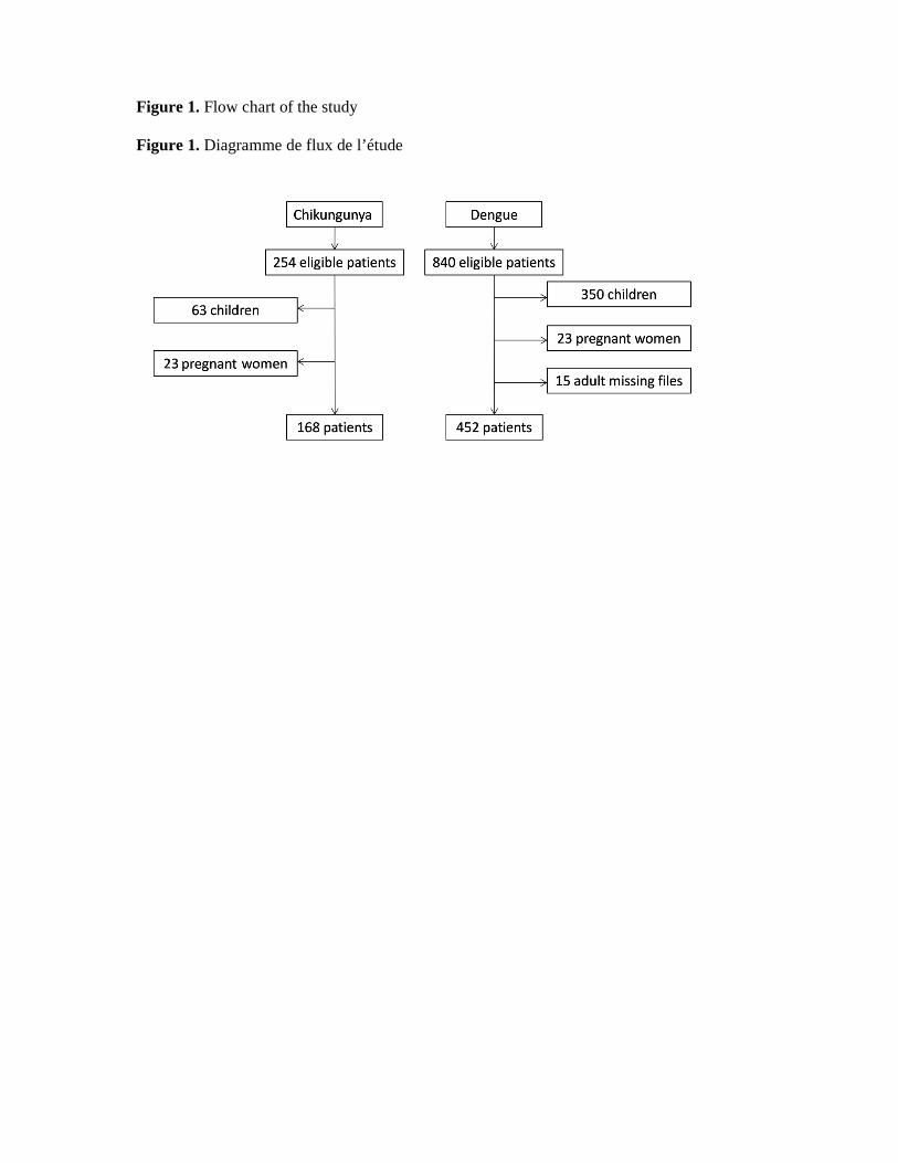
Figure 1. Flow chart of the study
Figure 1. Diagramme de flux de l’étude
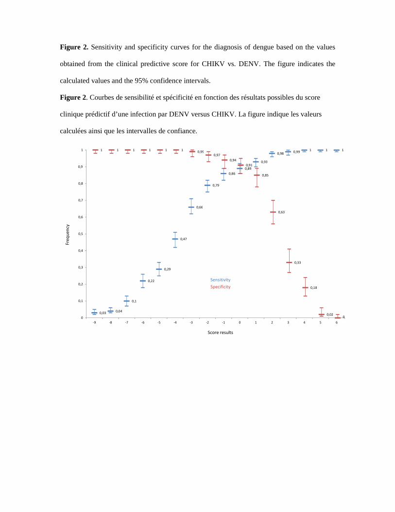
Figure 2. Sensitivity and specificity curves for the diagnosis of dengue based on the values
obtained from the clinical predictive score for CHIKV vs. DENV. The figure indicates the
calculated values and the 95% confidence intervals.
Figure 2. Courbes de sensibilité et spécificité en fonction des résultats possibles du score
clinique prédictif d’une infection par DENV versus CHIKV. La figure indique les valeurs
calculées ainsi que les intervalles de confiance.
0,030,04
0,1
0,22
0,29
0,47
0,66
0,79
0,86 0,89
0,93
0,98 0,991 1 1
0
0,1
0,2
0,3
0,4
0,5
0,6
0,7
0,8
0,9
1
-9 -8 -7 -6 -5 -4 -3 -2 -1 0 1 2 3 4 5 6
1 1 1 1 1 1 0,99
0,97
0,94 0,91
0,85
0,63
0,33
0,18
0,020
Sensitivity
Specificity
Score results
Fre
qu
en
cy

Table 1. Comparison of the demographical and clinical variables in the univariate analysis
between CHIKV and DENV groups
Tableau 1. Comparaison en analyse univariée des variables démographiques et cliniques
entre les groupes CHIKV et DENV
Variables CHIKV
n=168 (%)
DENV
n=452 (%)
OR (95% CI) p
Male sex 77 (45.8) 226 (50.0) 0.85 (0.58 – 1.23) 0.36
Age ≥65 years 24 (14.3) 16 (3.54) 4.54 (2.24 – 9.39) <0.001
Age <18 years 6 (3.5) 72 (15.9) 0.19 (0.07 – 0.46) <0.001
Comorbidity 93 (55.3) 108 (23.9) 3.95 (2.67 – 5.85) <0.001
Hospitalized patients 14 (8.3) 159 (35.2) 0.16 (0.08 – 0.30) <0.001
Consultation before day 3 85 (50.1) 163 (36.1) 1.82 (1.25 – 2.64) 0.01
Fever 118/151 (78.2) 314/444 (70.7) 1.48 (0.94 – 2.37) 0.08
Headache 97 (57.7) 371 (82.1) 0.30 (0.20 – 0.45) <0.001
Retro-orbital pain 24 (14.3) 99 (21.9) 0.59 (0.35 – 0.98) 0.03
Joint pain 156 (92.9) 74 (16.4) 66.4 (34.4 – 136.8) <0.001
Arthritis 16 (9.5) 0 (0.0) - -
Muscle pain 111 (66.1) 366 (80.9) 0.39 (0.46 – 0.70) <0.001
Back pain 80 (47.6) 103 (22.8) 3.08 (2.08-4.55) <0.001
Cough 14 (8.3) 63 (13.9) 0.56 (0.28 – 1.05) 0.06
Abdominal pain 20 (11.9) 154 (34.1) 0.26 (0.15 – 0.44) <0.001
Nausea and/or vomiting 43 (25.6) 253 (60.0) 0.27 (0.18 -0.41) <0.001
Food intolerance* 12 (7.1) 72 (15.9) 0.41 (0.19-0.78) 0.005
Diarrhea 9 (5.4) 108 (23.9) 0.18 (0.08 – 0.37) <0.001
Anorexia 39 (23.2) 189 (41.8) 0.42 (0.27 – 0.64) <0.001
Fatigue 72 (42.9) 236 (52.2) 0.69 (0.47 – 0.99) 0.040
Neurological signs† 4 (2.4) 39 (8.6) 0.26 (0.07 – 0.73) 0.006
*Food intolerance: repeated vomiting
† Neurological signs: collapse, confusion, and encephalitis

Table 2. Comparison of the biological variables in the univariate analysis between CHIKV
and DENV groups
Tableau 2. Comparaison en analyse univariée des variables biologiques entre les groupes
CHIKV et DENV
Variables
CHIKV
no. positive
/ no. tested
(%)
DENV
no. positive
/ no. tested
(%)
OR (95% CI) p
Platelet count <150 G/l 18/153 (11.8) 200/445 (44.9) 0.16 (0.09 -
0.28) <0.001
Neutrophil count <1,500
/mm3 10/152 (6.6) 86/444 (19.4)
0.29 (0.13 -
0.59) <0.001
Lymphocyte count
<500/mm3 20/152 (13.1) 138/440 (31.4)
0.33 (0.19 -
0.56) <0.001
CRP* >30 mg/l 43/148 (29.1) 72/432 (16.7) 2.05 (1.29 –
3.23) 0.001
AST** >2N† 12/109 (11.0) 91/385 (23.6) 0.40 (0.19-0.77) 0.004
ALT ** >2N† 10/126 (7.9) 85/421 (20.2) 0.34 (0.15-0.69) 0.001
Creatinine >115 µmol/l 13/144 (9.0) 23/437 (5.3) 1.78 (0.81-3.80) 0.10
* C-reactive protein, ** aspartate transaminase (AST) and alanine transaminase (ALT) † 2-fold increase as compared with baseline
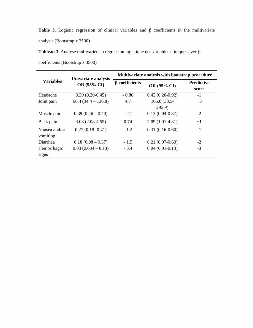
Table 3. Logistic regression of clinical variables and β coefficients in the multivariate
analysis (Bootstrap x 3500)
Tableau 3. Analyse multivariée en régression logistique des variables cliniques avec β
coefficients (Bootstrap x 3500)
Variables Univariate analysis
OR (95% CI)
Multivariate analysis with bootstrap procedure
β coefficients
OR (95% CI) Predictive
score Headache 0.30 (0.20-0.45) - 0.86 0.42 (0.20-0.92) -1 Joint pain 66.4 (34.4 – 136.8) 4.7 106.8 (38.5-
295.9) +5
Muscle pain 0.39 (0.46 – 0.70) - 2.1 0.13 (0.04-0.37) -2
Back pain 3.08 (2.08-4.55) 0.74 2.09 (1.01-4.31) +1
Nausea and/or vomiting
0.27 (0.18 -0.41) - 1.2 0.31 (0.16-0.60) -1
Diarrhea 0.18 (0.08 – 0.37) - 1.5 0.21 (0.07-0.63) -2 Hemorrhagic signs
0.03 (0.004 – 0.13) - 3.4 0.04 (0.01-0.13) -3
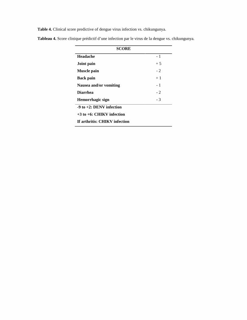
Table 4. Clinical score predictive of dengue virus infection vs. chikungunya.
Tableau 4. Score clinique prédictif d’une infection par le virus de la dengue vs. chikungunya.
SCORE
Headache - 1
Joint pain + 5
Muscle pain - 2
Back pain + 1
Nausea and/or vomiting - 1
Diarrhea - 2
Hemorrhagic sign - 3
-9 to +2: DENV infection
+3 to +6: CHIKV infection
If arthritis: CHIKV infection


![Una Limosna [orch] SCORE](https://static.fdokumen.com/doc/165x107/631e9c1925add517740b2033/una-limosna-orch-score.jpg)