Development and Partial Characterization of Heliothine Cell Lines from Embryonic and Differentiated...
6
In Vitro Cell. Dev. Biol.~nirnal 40:89-94, March and April 2004 2004 Society for In Vitro Biology 1071-2690/04 $18.00+0.00 DEVELOPMENT AND PARTIAL CHARACTERIZATION OF HELIOTHINE CELL LINES FROM EMBRYONIC AND DIFFERENTIATED TISSUES CYNTHIA L. GOODMAN, t AMY A. WANG, = HENDA NABLI/' ARTHUR H. MCINTOSH,JENNIFER L. WITTMEYER, 3 AND JAMES J. GRASELA Biological Control of Insects Research Laboratory, United States Department of Agriculture, Agricultural Research Se~wice, Columbia, Missouri 65203-3535 (C. L. (FT., A. H. M., J. J. G.), Bayer CropScience, Research Triangle Park, NC 27709 (A. A. W..), Department of Entomology, University of Missouri, Missouri 65211 (H. N., J. L. IV..) (Received 14 August 2003; accepted 25 March 2004) SUMMARY The goal of this study was to generate cell lines from a variety of insect tissues that could be useful for developing in vitro assays with tissue-specific properties. In this article, we describe the establishment of new cell cultures from differentiated (primarily neural) and undifferentiated tissues (primarily embryonic) and their initial characterization. Cell lines were established from the following tissues of the budworm, Heliothis virescens, and the bollworm, Helicoverpa zea: larval ventral nerve cords (4 lines), larval midguts (1 line), adult ovaries (1 line), and embuonic tissues (11 lines). Cell lines were primarily characterized by morphological examination and polymerase chain reaction (PCR) (both deoxyribo- nucleic acid amplification fingerprinting arid inter-simple sequence repeats PCR). Key words: neural; midgut; Heliothis virescens; Helicoverpa zea; PCR; Lepidoptera. INTRODUCTION Developing unique and efficacious chemicals for the control of insect pests is one goal of the agrochemical industry-. To accomplish this task, specific screening systems must be established that will identify potential anti-insect target sites from selected tissues. Promising target sites include neurotransmitter or neuromodulator receptors (or both) of insect neurons, calcium channels of insect muscles, and hormone receptors of somatic tissues. Malfunctioning of these sites can lead to inhibition of development or feeding or to the immobilization and mortality of insects. Toward this end, we endeavored to develop insect cell lines from a variety of tissues that could be tested for the presence of selected target sites, enabling their use in in vitro assay systems for the identification of novel anti-insect compounds. Other researchers have shown the feasibility of establishing in- sect cell lines that exhibit properties of differentiated tissues. Ex- amples of cell lines with such properties include LM3(BF), which synthesizes the neurotransmitter acetylcholine (Ui et al., 1994); IPLB-CPB2, which expresses an inositol triphosphate receptor, the ryanodine receptor, and a calcium pump and exhibits spontaneous action potentials (Sheppard and Lynn, 1996); Sf9, which expresses 1To whom correspondence should be addressed at Biological Control of" Insects Research LaboratmT, United States Department of Agriculture, Ag- ricultural Research Service, 1503 South Providence Road, Columbia, Mis- souri 65203-3535. E-maih [email protected] Presently at: High Throughput Cell Physiology, GIaxoSmithKline, RTP, NC 27709. a Presently at: Obstetrics and Gynecology, University of Missouri, Colum- bia, MO 65211. Presently at: Department of Marine, Earth and Atmospheric Sciences, North Carolina State University, Raleigh, NC 27695. an adenylate cyclase-coupled octopamine receptor (Orr et al., 1992); epithelial cell lines from Chironomus tentans, which produce an ecdysone-binding protein (Turberg et al., 1988) and a muscarinic cholinergic receptor (Wegener et al., 1996); and BRL-AG-2 (Dhad- ialla and Tzertzinis, 1997), IPLB-Tex2 (Lynn and Hung, 1981), IPRI-MD-66, IPRI-CF-1, FPMI-CF-70 (Sohi et al., 1995), UMC- OnE (Trisyono et al., 2000), and an ovarian cell line from Galleria mellonella (Zakarian et al., 2002), which express functional 20- hydroxyecdysone receptors. Thus, precedence exists for the estab- lishment of continuous cell lines for use in the development of tests for the assaying of specific physiological functions related to poten- tial anti-insect target sites. We describe here the establishment of cell lines from both dif- ferentiated and undifferentiated tissues, namely nerve cord, midgut, ovarian, and emb13*onic tissues. Cell lines were initiated from un- differentiated tissues to determine whether some could be devel- oped with differentiated properties. All tissues were derived from two important insect pests, Heliothis virescens (Fabricius) and Hel- icoverpa zea (Boddie) (Lepidoptera: Noctuidae), known to cause damage to a number of important crops such as corn and cotton (Fitt, 1989). In addition, newly established cell lines were analyzed by polymerase chain reaction (PCR) to generate cell line-specific fingerprints for identification purposes. MATERIALSAND METHODS Cell culture media. The foUowinginsect cell culture media were used for primary culture initiations: ExCell 401 (JR BioSciences, 5 Lenexa, KS), HyQ- 5 Names are necessary to report factually on available data; however, the USDA neither guarantees nor warrants the standard of the product, and the use of the name by USDA implies no approval of the product to the exclusion of others that may also be suitable. 89
-
Upload
independent -
Category
Documents
-
view
0 -
download
0
Transcript of Development and Partial Characterization of Heliothine Cell Lines from Embryonic and Differentiated...
In Vitro Cell. Dev. Biol.~nirnal 40:89-94, March and April 2004 �9 2004 Society for In Vitro Biology 1071-2690/04 $18.00+0.00
DEVELOPMENT AND PARTIAL CHARACTERIZATION OF HELIOTHINE CELL LINES FROM EMBRYONIC AND DIFFERENTIATED TISSUES
CYNTHIA L. GOODMAN, t AMY A. WANG, = HENDA NABLI/' ARTHUR H. MCINTOSH, JENNIFER L. WITTMEYER, 3 AND JAMES J. GRASELA
Biological Control of Insects Research Laboratory, United States Department of Agriculture, Agricultural Research Se~wice, Columbia, Missouri 65203-3535 (C. L. (FT., A. H. M., J. J. G.), Bayer CropScience, Research Triangle Park, NC 27709 (A. A. W..),
Department of Entomology, University of Missouri, Missouri 65211 (H. N., J. L. IV..)
(Received 14 August 2003; accepted 25 March 2004)
SUMMARY
The goal of this study was to generate cell lines from a variety of insect tissues that could be useful for developing in vitro assays with tissue-specific properties. In this article, we describe the establishment of new cell cultures from differentiated (primarily neural) and undifferentiated tissues (primarily embryonic) and their initial characterization. Cell lines were established from the following tissues of the budworm, Heliothis virescens, and the bollworm, Helicoverpa zea: larval ventral nerve cords (4 lines), larval midguts (1 line), adult ovaries (1 line), and embuonic tissues (11 lines). Cell lines were primarily characterized by morphological examination and polymerase chain reaction (PCR) (both deoxyribo- nucleic acid amplification fingerprinting arid inter-simple sequence repeats PCR).
Key words: neural; midgut; Heliothis virescens; Helicoverpa zea; PCR; Lepidoptera.
INTRODUCTION
Developing unique and efficacious chemicals for the control of insect pests is one goal of the agrochemical industry-. To accomplish this task, specific screening systems must be established that will identify potential anti-insect target sites from selected tissues. Promising target sites include neurotransmitter or neuromodulator receptors (or both) of insect neurons, calcium channels of insect muscles, and hormone receptors of somatic tissues. Malfunctioning of these sites can lead to inhibition of development or feeding or to the immobilization and mortality of insects. Toward this end, we endeavored to develop insect cell lines from a variety of tissues that could be tested for the presence of selected target sites, enabling their use in in vitro assay systems for the identification of novel anti-insect compounds.
Other researchers have shown the feasibility of establishing in- sect cell lines that exhibit properties of differentiated tissues. Ex- amples of cell lines with such properties include LM3(BF), which synthesizes the neurotransmitter acetylcholine (Ui et al., 1994); IPLB-CPB2, which expresses an inositol triphosphate receptor, the ryanodine receptor, and a calcium pump and exhibits spontaneous action potentials (Sheppard and Lynn, 1996); Sf9, which expresses
1To whom correspondence should be addressed at Biological Control of" Insects Research LaboratmT, United States Department of Agriculture, Ag- ricultural Research Service, 1503 South Providence Road, Columbia, Mis- souri 65203-3535. E-maih [email protected]
Presently at: High Throughput Cell Physiology, GIaxoSmithKline, RTP, NC 27709.
a Presently at: Obstetrics and Gynecology, University of Missouri, Colum- bia, MO 65211.
Presently at: Department of Marine, Earth and Atmospheric Sciences, North Carolina State University, Raleigh, NC 27695.
an adenylate cyclase-coupled octopamine receptor (Orr et al., 1992); epithelial cell lines from Chironomus tentans, which produce an ecdysone-binding protein (Turberg et al., 1988) and a muscarinic cholinergic receptor (Wegener et al., 1996); and BRL-AG-2 (Dhad- ialla and Tzertzinis, 1997), IPLB-Tex2 (Lynn and Hung, 1981), IPRI-MD-66, IPRI-CF-1, FPMI-CF-70 (Sohi et al., 1995), UMC- OnE (Trisyono et al., 2000), and an ovarian cell line from Galleria mellonella (Zakarian et al., 2002), which express functional 20- hydroxyecdysone receptors. Thus, precedence exists for the estab- lishment of continuous cell lines for use in the development of tests for the assaying of specific physiological functions related to poten- tial anti-insect target sites.
We describe here the establishment of cell lines from both dif- ferentiated and undifferentiated tissues, namely nerve cord, midgut, ovarian, and emb13*onic tissues. Cell lines were initiated from un- differentiated tissues to determine whether some could be devel- oped with differentiated properties. All tissues were derived from two important insect pests, Heliothis virescens (Fabricius) and Hel- icoverpa zea (Boddie) (Lepidoptera: Noctuidae), known to cause damage to a number of important crops such as corn and cotton (Fitt, 1989). In addition, newly established cell lines were analyzed by polymerase chain reaction (PCR) to generate cell line-specific fingerprints for identification purposes.
MATERIALS AND METHODS
Cell culture media. The foUowing insect cell culture media were used for primary culture initiations: ExCell 401 (JR BioSciences, 5 Lenexa, KS), HyQ-
5 Names are necessary to report factually on available data; however, the USDA neither guarantees nor warrants the standard of the product, and the use of the name by USDA implies no approval of the product to the exclusion of others that may also be suitable.
89
9 0 GOODMAN ET AL.
TABLE 1
NEWLY ESTABLISHED CELL LINES FROM SELECTED TISSUES OF HELIOTHINE SPECIES ~
Establishment Complete Species Stage' Tissue media b designation ~
Heliothis virescens (Hv) Egg Whole embryo HyQ-SfX@ BCIRL/RP-HvE-CLG1 Egg Whole embryo DMEM/F12 BCIRL/RP-HvE-CLG4 Egg Whole embryo HyQ-SFX@ BCIRIdRP-HvE-CLG5 Egg Whole embryo ExCell 401 @ BCIRIdRP-HvE-CLG6 Egg Whole embryo Modified Schneider's BCIRL/RP-HvE-CLG7 Egg Whole embryo DMEM/F12 BCIRIdRP-HvE-HN2 Egg Whole embryo ExCell 401@ BCIRIdRP-HvE-HN3 Egg Whole embryo ExCell 401 @ BCIRL/RP-HvE-HN11 Egg Whole embryo Modified Schneider's BCIRIdRP-HvE-HN12 Egg Whole embryo Shields and Sang BCIRL/RP-HvE-HN14 Egg Whole embryo TC-100 BCIRL/RP-HvE-HN16 Larva Ventral nerve cord ExCell 401 @ BCIRIdRP-HvVNC-WG1 Larva Ventral nerve cord ExCell 401@ BCIRL/RP-HvVNC-WG2 Larva Ventral nerve cord ExCell 401 @ BCIRL/RP-HvVNC-WG3
Helic~verpa zea (Hz) Larva Ventral nerve cord ExCell 401 @ RP-HzVNC-AW1 Larva Midgut ExCell 401 @ RP-HzGUT-AW1 Adult Ovary ExCell 401 @ RP-HzOV-AW2
DMEM/F12, Dulbecco Modified Eagle medium with Ham's F12 Nutrient Mixture. b All media contained 10% fetal bovine serum (FBS), except modified Schneider's medium (which contained 18% FBS and 4 nM insulin).
Only eggs -<24 h old and larvae in their fifth stadium were used. '~ Designations include initials of authors primarily involved in initiations, with "WG" indicating "Wittmeyer-Goodman". The cell line RP-HzVNC-AW1 has
been described previously as RP-HzNU-AW1.
SFX (HyClone, Logan, UT), Modified Schneider's (Schneider's medium, Sig- ma Chemical Co., St. Louis, MO, with 18% fetal bovine serum [FBS] and 4 nM insulin added; R. Goodwin, pers. comm.), Shields and Sang M3 (Sigma), TC-100 (Sigma), and ENCCM (embryonic neuronal cell culture medium; L. M. Hall, pers. comm.). In addition, three mammalian cell culture media were also used in some initiations: DMEM/F12 (Dulbecco Modified Eagle Medium with Ham's F12 Nutrient Mixture, Sigma; used previously by McIntosh et al. [1995] for heliothine cells), ABM (astrocyte basal medium; Clonetics-Cam- brex Bio Sciences, Baltimore, MD), and neurobasal medium (Clonetics-Cam- brex). Media containing 200 U/ml penicillin and 0.2 mg/ml streptomycin (Sigma) were used for the initial cultures, with the concentrations of these antibiotics being reduced to 50 U/ml penicillin and 0.05 mg/ml streptomycin after the first feeding. Heat-inactivated (56 ~ C for 30 rain) FBS (Intergen, Purchase, NY) was also added to many media at the indicated concentrations.
Cell culture initiation and maintenance. Primary cultures were established from larval ventral nerve cords (VNC), larval midgut tissues (GUT), adult ovarian tissues (OV), and embryonic tissues of H. virescens and H. zea (Table 1). Tissue dissections were peffm~ned as described previously (Goodman et al., 2001). To summarize, insects were sterilized by washing in 70% ethanol (containing 1% Triton X-100) and 0.53% sodium hypochlorite (Clorox Co., Oakland, CA) (one to two washes/reagent, 5 min/wash) and rinsing one to two times in Hanks' balanced salt solution (HBSS, containing 200 U/ml pen- icillin, 0.2 mg/ml streptomycin, 0.5 l~g/ml amphotericin B, and 13 mM 3- movpholinopropanesulfonic acid, final pH 6.8-7.0; components purchased from Sigma). After the initial incision, the body cavity was thoroughly rinsed with HBSS to remove hemocytes and other contaminants. Tissues were re- moved, washed in HBSS, and minced using microdissection scissors. After- ward, tissues were transferred into T~25 or T2s cuhure flasks (Costar, Cam- bridge, MA) with either 2.5 or 5 ml medium, respectively. For embryonic cultures, eggs were homogenized in HBSS with a plastic micropestle in a 1.5-ml Eppendorf tube. The homogenate was then centrifuged (500 • g, 10 min) and the resulting pellet was transferred into disposable Tz5 flasks (Good- man et al., 2001). For some cultures, only 2 ml medium (per T2s) was initially added for the first 24 h to encourage attachment, followed by the addition of the final 3 ml. Promising cultures were initially fed every 7 to 10 d (using 50% medium replacement), then once or twice a wk as cell replication in- creased (using total medium replacement). If cells or tissues were in suspen- sion, the medium was centrifuged (500 • g, 10 min) to pellet these materials. The pelleted material was then transferred back into the original flask. All cell lines were maintained at 28 ~ C. The cell line RP-HzVNC-AW1 was
cloned using the feeder layer method with an agarose overlay in combination with ExCell 401 medium (Rice et al., 1989).
Polymerase chain reaction analysis. Two PCR methods were used for char- acterizing the cell lines: deoxyribonucleic acid (DNA) amplification finger- printing (DAF-PCR, McIntosh et al., 1996) using primers to mammalian al- dolase and inter-simple sequence repeats (ISSR-PCR) (Zietkiewicz et al., 1994; Grasela and McIntosh, 2004) using the single 5'-anchored repeat prim- er GCA-CAT-CGA-RTG-(TG)6 (as well as other primers not shown). Deoxy- ribonucleic acid was extracted from cells using an extraction kit (Puregene @, Gentra Systems Inc., Minneapolis, MN).
RESULTS
Cell culture initiations. Cell l ines were successfully generated from a variety of H. vireseens and H. zea tissues, including 4 from
larval VNC (BCIRL/RP-HvVNC-WG1, -WG2, -WG3; RP-HzVNC- AWl), 1 from lmwal GUT (RP-HzGUT-AW1), 1 from OV (RP- HzOV-AW1), and 11 from embryonic tissues (Table 1). (Note:
HvVNC-WG2 was generated by combining cells in suspension from the early primary cultures of HvVNC-WG1 and HvVNC-WG3.) The
newly established lines consisted of cells with varied morphologies, ranging from spherical to elongated (Fig. 1), and included ventral
nerve cord lines that appeared to form networks in culture (Fig. 1A-D).
The medium used for all successful neural cultures was ExCell
401 containing 10% FBS. No other medium generated viable cell cultures over the long term, although some media initially produced
healthy appearing primary cultures that failed to replicate (e.g., DMEM/F12 + 10% FBS, Shields and Sang M3 + 10% FBS, Schneider 's medium with 18% FBS + 4 nM insulin). The ENCCM (the original version o1" a modified version made up with ExCell 401 as the basal medium) did not produce any viable cultures, with
most tissues becoming dark and appearing unhealthy. For the cell l ines initiated from OV and GUT tissues, only ExCell
401 medium was used, which generated healthy cell cultures from
HELIOTHINE CELL LINES 91
FIG. 1. Cells from newly established heliothine cell lines (• (A) Helicoverpa virescens ventral nerve cord cell line, BCIRL/RP- HvVNC-WG1, in ExCe|l 401 + 10% fetal bovine serum (FBS), P45; (B) H. virescens ventral nerve cord cell line, BCIRL/RP-HvVNC- WG2, in ExCell 401 + 10% FBS, P61; (C) H. virescens ventral nerve cord cell line, BCIRL/RP-HvVNC-WG3, in ExCell 401 + 10% FBS, P58, (D) H. zea ventral nerve cord cell line, RP-HzVNC-AW1 Clone 14C4, in ExCell 401 + 10% FBS, P49, (E) H. zea midgut cell |ine, RP-HzGUT-AW1, in ExCell 401 + 10% FBS, P80; (F) H. virescens embryonic cell line, BCIRL-RP-HVE-CLG6, in ExCell 401, P29.
92 GOODMAN ET AL.
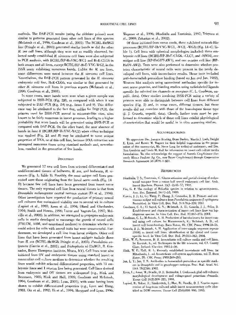
FIG. 2. Polymerase chain reaction (PCR) analysis of selected Helicoverpa virescens and H. zea cell lines using: (A) deoxyribonucleic acid (DNA) amplification fingerprinting-PCR, with aldolase as the primer, (B) inte~simple sequence repeats-PCR, with the single 5'-an- chored repeat GCA-CAT-CGA-RTG-(TG)6 as a primer. Samples are (1) DNA standard (50- 1000 bp), (2) negative control (H20), (3) RP- HzVNC-AW1 clone 14C4 P49, (4) RP-Hz- GUT-AWl P80, (5) BCIRL-HzMG8 P181, (6) RP-HzOV-AW2 P49, (7) BCIRL/RP-HvVNC- WG1 P28, (8) BCIRL/RP-HvVNC-WG2 P39, (9) BCIRL/RP-HvVNC-WG3 P32, and (10) BCIRL/RP-HvE-CLG6 P29.
both tissues (Table 1). These cultures were predominantly spherical in morphology (e,g., Fig. 1E).
For emhryonic cell cultures, the following cuhme media gener- ated the healthiest and fastest developing cultures: ExCell 401 (+ 10% FBS), HyQ-SFX (+ 10% FBS), modified Schneider's (with 18% FBS + 4 nM insulin), DMEM/F12 (+ 10% FBS), Shields and Sang M3 (+ 10% FBS), and TC-100 (+ 10% FBS) (Table 1). Con- versely, culture initiations begun with ENCCM, ABM, and neuro- basal medium did not generate viable embryonic cultures. Obser-
vations on the continuous embryonic cell cultures indicated that they consisted of numerous morphological types, with spherical morphologies being predominant (e.g., Fig. IF).
Polymerase chain reaction analysis. Newly established cell lines were subjected to two different methods of PCR analyses for iden- tification purposes (DAF-PCR and ISSR-PCR), with examples of the results from each method shown in Fig. 2. Helicoverpa zea cell lines RP-HzVNC-AW1 clone 14C4, RP-HzGUT-AW1, and RP-HzOV- AW2 exhibited similar patterns of DNA bands using the two PCR
HELIOTHINE CELL LINES 93
methods. The DAF-PCR results (using the aldolase primer) were similar to patterns generated from other cell lines of this species (McIntosh et al., 1996; Goodman et al., 2001). The BCIRL-HzMG8 line (Pringle et al., 2003) generated similar bands as did the other H. zea cell lines, although they were not as readily observed. Se- lected newly established H. virescens cell lines were also subjected to PCR analysis, with BCIRL/RP-HvVNC-WG1 and HvE-CLG6 in both assays and all lines, except BCIRL/RP-HvE-VNC-WG2, in the ISSR assay exhibiting numerous bands. Unlike the H. zea lines, some differences were noted between the H. virescens cell lines. Nevertheless, the DAF-PCR pattern generated by the H. virescens
embryonic cell line, HvE-CLG6, was similar to that generated by other H. virescens cell lines in previous reports (Mclntosh et al., 1996; Goodman et al., 2001).
In some cases, more bands were seen when a given sample was subjected to ISSR-PCR (Fig. 2B), as compared with when it was subjected to DAF-PCR (Fig. 2A) (e.g., lanes 5 and 9). This differ- ence may be attributed to the fact that, unlike for DAF-PCR, the primers used for ISSR-PCR anneal to microsatellite sites, sites known to be fairly numerous in insect genomes, leading to a higher probability that more bands will be generated using ISSR-PCR as compared with DAF-PCR. On the other hand, the near absence of bands in lane 8 (BCIRL/RP-HvVNC-WG2) when either technique was applied (Fig. 24 and B) may be attributed to some unique properties of DNA, or of this cell line, because DNA extraction was attempted numerous times using standard methods and, neverthe- less, resulted in the generation of few bands.
DISCUSSION
We generated 17 new cell lines from selected differentiated and undifferentiated tissues of bollworm, H. zea, and budworm, H. vi-
rescens (Fig. 1; Table 1). Possibly, the most unique cell lines gen- erated were those originating from the larval nerve cords (Fig. 1A- D) because few cell lines have been generated from insect nerve tissues. The only reported cell line from neural tissues is that from Drosophila melanogaster central nervous system (Ui et al., 1994). Other investigators have reported the production of primary neural cell cultures that maintained viability one to several wk in culture (Lapied et al., 1993; Keen et al., 1994; Olarld and Oberlander, 1994; Smith and Howes, 1996; Lucas and Nagnan-Le, 1997; Mel- ville et al., 2003). In addition, we attempted to propagate embiTonic cells in media developed to encourage the growth of neural cells (ENCCM, ABM, and ueurobasal medium) to determine whether we could select for cells with neural traits but were unsuccessful. Fm~ thermore, we developed a cell line from larval midguts. Other cell lines that have been generated from insect midguts include those from tI. zea (BCIRL-HzMG8; Pringle et al., 2003), Pseudaletia un-
ipuncta (Gareia et al., 2001), and Trichoplusia ni (TnMG1, R. Gra- nados, Boyee Thompson Institute, Ithaca, NY). Cell lines were also initiated from OV and embryonic tissues using standard insect or mammalian cell culture medium to determine whether the resulting lines would exhibit selected differentiated properties, with 11 em- bryonic lines and 1 ovarian line being generated, Cell lines derived from embryonic and OV tissues are widespread (e.g., Hink and Bezanson, 1985; Hink and Hall, 1989; Goodman and MeIntosh, 1994; Goodman et al., 2001; Lynn, 2001), with some having been shown to exhibit differentiated properties (e.g., Lynn and Hung, 1981; Orr et al., 1992; Ui et al,, 1994; Sheppard and Lynn, 1996;
Wegener et al., 1996; Dhadialla and Tzertzinis, 1997; Trisyono et al., 2000; Zakarian et al., 2002).
Of those initiated from nerve cords, three exhibited network-like processes (BCIRL/RP-HvVNC-WG1, -WG2, -WG3) (Fig. 1A-C; Ta- ble 1). Cell lines with spherical morphologies included: three em- bryonic cell lines (BCIRL/RP-HvE-CLG6, -CLG7, and -14N14), one midgut cell line (RP-HzGUT-AW1), and one ovarian cell line (RP- HzOV-AW2). Tests were also performed to determine whether pro- teins characteristic of neural ceils were present in the newly de- veloped cell lines, with inconclusive results. These tests included anti-horseradish peroxidase binding (based on Jan and Jan, 1982), Western blot analysis using nmnoelonal antibodies specific for in- sect nerve proteins, and binding studies using radiolabeled ligands specific for selected ion channels or receptors (C. L. Goodman, un- publ. data). Other studies involving ISSR-PCR using a variety of primers were able to distinguish between cell lines from different species (Fig. 2) and, in some eases, different tissues, but these results did not correlate with those of the homologous tissue type (J. J. Grasela, unpubl, data). Clearly, further tests must be per- formed to determine which of these cell lines exhibit physiological characteristics that would be useful in in vitro screening studies.
ACKNOWLEDGMENTS
We appreciate Drs. Juergen Benting, Dean Bushey, Marcia J. Loeb, Dwight E. Lynn, and Rende M. Wagner for their helpful suggestions in the prepa- ration of this manuscript, Mr. Steve Long for technical assistance, and Drs. Run Goodwin and Linda M. Hall for information on insect cell culture media formulations. We also acknowledge the support of Aventis CropScience (for- merly Rhone Poulenc Ag. Co., now Bayer CropScience) through Cooperative Research Agreement 58-3K95-1-882.
REFERENCES
Dhadialla, T. S.; Tzertzinis, G. Characterization and partial cloning of ecdys- teroid receptor from a cotton boll weevil embryonic cell line. Arch. Insect Biochem. Physiol. 35(1-2):45-57; 1997.
Fitt, G. P. The ecology of Heliothis species in relation to agmecosystems. Ann. Rev. Entomol. 34:17-52; 1989.
Garcia, J. J.; Li, G., Wang, R; Zhong, J.; Granados, R. R. Primary and con- tinuous midgut cell cultures from Pseudaletia unipuncta (Lepidoptera: Noctuidae). In Vitro Cell. Dev. Biol. 37A:353-359; 2001.
Goodman, C. L.; El Sayed, G. N.; Mclntosh, A. H.; Grasela, J. J.; Stiles, B. Establishment and characterization of insect cell lines from ten lep- idopteran species. In Vitro Cell. Dev. Biol. 37:367-373; 2001.
Goodman, C. L.; Mclntosh, A. H. Production of baculoviruses for insect con- trol using cell culture. In: Maramorosch, K.; McIntosh, A. H., ed. Insect cell biotechnology. Boca Raton, FL: CRC Press; 1994:33-56.
Grasela, J. J.; Mclntosh, A. H. Application of inter-simple sequence repeats (ISSR) to insect cell lines: identification at the clonal and tissue- specific level. In Vitro Cell. Dev. Biol. 39:353-363; 2004.
Hink, W. E; Bezanson, D. R. Invertebrate cell culture media and cell lines. In: Kurstak, E., ed. Techniques in the life sciences, vol. C1. County Clare, h'eland: Elsevier; 1985:1-30.
Hink, W. E; Hall, R. L. Recently established invertebrate cell lines. In: Mitsuhashi, J., ed. Invertebrate cell system applications, vol. II. Boca Raton, FL: CRC Press; 1989:269-293.
Jan, L. Y.; Jan, Y. N. Antibodies to horseradish peroxidase as specific mark- ers in Drosophila and in grasshopper embryos. Proc. Natl. Acad. Sci. USA 79:2700; 1982.
Keen, L.; Amar, M.; Beadle, D. J.; Bermudez, I. Cockroach glial cell cultures: morphological development and voltage-gated potassium channels. Tissue Cell 26(2):209-221; 1994.
Lapied, B.; Tribut, E; Sinakevitch, I.; Hue, B.; Beadle, D. J. Neurite regen- eration of long-term cultured adult insect neurosecretoLv cells ideu- tiffed as DUM neurons. Tissue Cell 25(6):893-906; 1993.

94 GOODMAN ET AL.
Lucas, E; Nagnan-Le, M. P. Primary culture of antennal cells of Mamestra bras- sicae: morphology of cell types and evidence for biosynthesis of phero- mone-binding proteins in vitro. Cell Tissue Res. 289(2):375-382; 1997.
Lynn, D. E. Novel techniques to establish new cell lines. In Vitro Cell. Dev. Biol. 37A:319-321; 2001.
Lynn, D. E.; Hung, A_ C. E Development of continuous cell lines from the egg parasitoids Trichogramma confusum and Trichogramma exiguum. Arch. Insect Biochem. Physiol. 18:99-104; 1981.
McIntosh, A. H.; Grasela, J. J.; Goodman, C. L. Replication of Helic~verpa zea polyhedrosis virus in homologous cell lines grown in serum-free media. J. Invertebr. Pathol. 66:121-124; 1995.
McIntosh, A. H.; Grasela, J. J.; Matteri, R. L. Identification of insect cell lines by DNA amplification fingerprinting (DAF). Insect Mol. Biol. 5(3):187-195; 1996.
Melville, J. M; Hoffman, K. L.; Jarrard, H. E.; Weeks, J. C. Cell culture of mechanoreceptor neurons innervating proleg sensory hairs in Man- duca sexta larvae, and co-culture with target motoneurons. Cell Tissue Res. 311:117-130; 2003.
Oland, L. A.; Oberlander, H. Factors that influence the development of cul- tured neurons from the brain of the moth, Manduca sexta. In Vitro Cell. Dev. Biol. 30A(10):709-716; 1994.
Orr, N.; Orr, L. G.; Hollingworth, R. M. The St9 cell line as a model for studying insect octopamine receptors. Insect Biochern. Moh Biol. 22:591-597; 1992.
Pringle, F. M.; Johnson, K. N.; Goodman, C. L.; McIntosh, A. H.; Bell, L. A. Providence virus: a new member of the Tetraveridae that infects cul- tured insect cells. Virology 306:359-370; 2003.
Rice, W. C.; Mclntosh, A. H.; Ignoffo, C. M. Yield and activity of the Heliothis zea nuclear polyhedrosis virus propagated in cloned and uncloned lines of Heliothis cells. In Vitro Cell. Dev. Biol. 25(2):201-204; 1989.
Sheppard, C. A.; Lynn, D. E. lmmunoreactivities for calcium signaling com- ponenls and neural-like properties of a Colorado potato beetle cell line. Arch. Insect Biochem. Physiol. 33(3-4):197-209; 1996.
Smith, P. J. S.; Howes, E. A. Long-term culture of fully differentiated adult insect neurons. J. Neurosci. Methods 69(1):113-122; 1996.
Sohi, S. S.; Palli, S. R.; Cook, B. J.; Retnakaran, A. Forest insect cell lines responsive to 20-hydroxyecdysone and two nonsteroidal ecdysone ag- onists, RI-I-5849 and RH-5992. J. Insect Physiol. 41(6):457-464; 1995.
Trisyono, A.; Goodman, C. L4 Grasela, J. J.; McIntosh, A. H.; Chippendale, G. M. Establishment, characterization and response of an Ostrinia nubilalis cell line to eedysone agonists. In Vitro Cell. Dev. Biol. 36A:400~04; 2000.
Turberg, A.; Spiadler-Barth, M.; Lutz, B.; Lezzi, M.; Spindler, K.-D. Presence of an ecdysteroid-specific binding protein ("receptor") in epithelial tissue cultured cells of Chironomus tentans. J. Insect Physiol. 34(8):797--803; 1988.
Ui, K.; Nishihara, S.; Sakuma, M.; Togashi, S.; Ueda, R.; Miyata, Y.; Miyake, T. Newly established cell lines from Drosophila larval CNS express neural specific characteristics. In Vitro Cell. Dev. Biol. 30A:209-216; 1994.
Wegenez, S.; Spindler-Bswth, M.; Spindler, K.-D. A muscas'inic acetylcholine receptor present in the epithelial cell line from Chironomus tentans. Biol. Chem. 377(12):819-824; 1996.
Zakarian, R. J.; Dunphy, G. B.; Quiot, J. M. Growth of an ovarian cell line of Galleria mellonella and its response to immune-inducing factors. In Vitro Cell. Dev. Biol. 38A(10):572-581; 2002.
Zietkiewicz, E.; Rafalski, A.; Labuda, D. Genome fingerprinting by simple sequence repeats (SSR)-anchored PCR amplification. Genomics 20:176-183; 1994.