Determination of hemispheric emotional valence in individual subjects: A new approach with research...
21
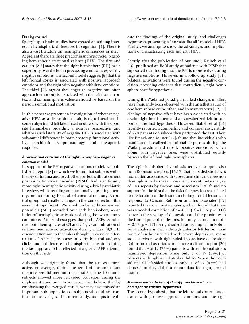
BioMed Central Page 1 of 21 (page number not for citation purposes) Behavioral and Brain Functions Open Access Research Determination of hemispheric emotional valence in individual subjects: A new approach with research and therapeutic implications Fredric Schiffer* 1 , Martin H Teicher 1 , Carl Anderson 1 , Akemi Tomoda 1,2 , Ann Polcari 1 , Carryl P Navalta 1 and Susan L Andersen 1 Address: 1 Department of Psychiatry, Harvard Medical School, and the Developmental Biopsychiatry Research Program, McLean Hospital, 115 Mill Street Belmont, MA 02478 USA and 2 Child Developmental Sociology, Faculty of Medical and Pharmaceutical Sciences, Kumamoto University, Kumamoto, Japan Email: Fredric Schiffer* - [email protected]; Martin H Teicher - [email protected]; Carl Anderson - [email protected]; Akemi Tomoda - [email protected]; Ann Polcari - [email protected]; Carryl P Navalta - [email protected]; Susan L Andersen - [email protected] * Corresponding author Abstract Background: Much has been theorized about the emotional properties of the hemispheres. Our review of the dominant hypotheses put forth by Schore, Joseph, Davidson, and Harmon-Jones on hemispheric emotional valences (HEV) shows that none are supported by robust data. Instead, we propose that individual's hemispheres are organized to have differing HEVs that can be lateralized in either direction. Methods: Probe auditory evoked potentials (AEP) recorded during a neutral and an upsetting memory were used to assess HEV in 28 (20 F) right-handed subjects who were either victims of childhood maltreatment (N = 12) or healthy controls. In a sub-population, we determined HEV by emotional response to lateral visual field stimulation (LVFS), in which vision is limited to one, then the other hemifield. We compare a number of morphometric and functional brain measures between individuals who have right-negative versus left-negative HEV. Results: Using AEPs to determine HEV, we found 62% of controls and 67% of maltreated subjects had right negative HEV. There was a strong interaction between HEV-laterality and gender, which together accounted for 60% of individual variability in total grey matter volume (GMV). HEV- laterality was associated with differences in hippocampal volume, amygdala/hippocampal ratios, and measures of verbal, visual and global memory. HEV-laterality was associated also with different constellations of symptoms comparing maltreated subjects to controls. Emotional response to LVFS provided a convenient and complementary measure of HEV-laterality that correlated significantly with the HEVs determined by AEPs. Conclusion: Our findings suggest that HEV-laterality, like handedness or gender, is an important individual difference with significant implications for brain and behavioral research, and for guiding lateralized treatments such as rTMS. Published: 6 March 2007 Behavioral and Brain Functions 2007, 3:13 doi:10.1186/1744-9081-3-13 Received: 2 January 2007 Accepted: 6 March 2007 This article is available from: http://www.behavioralandbrainfunctions.com/content/3/1/13 © 2007 Schiffer et al; licensee BioMed Central Ltd. This is an Open Access article distributed under the terms of the Creative Commons Attribution License (http://creativecommons.org/licenses/by/2.0 ), which permits unrestricted use, distribution, and reproduction in any medium, provided the original work is properly cited.
-
Upload
independent -
Category
Documents
-
view
1 -
download
0
Transcript of Determination of hemispheric emotional valence in individual subjects: A new approach with research...
BioMed CentralBehavioral and Brain Functions
ss
Open AcceResearchDetermination of hemispheric emotional valence in individual subjects: A new approach with research and therapeutic implicationsFredric Schiffer*1, Martin H Teicher1, Carl Anderson1, Akemi Tomoda1,2, Ann Polcari1, Carryl P Navalta1 and Susan L Andersen1Address: 1Department of Psychiatry, Harvard Medical School, and the Developmental Biopsychiatry Research Program, McLean Hospital, 115 Mill Street Belmont, MA 02478 USA and 2Child Developmental Sociology, Faculty of Medical and Pharmaceutical Sciences, Kumamoto University, Kumamoto, Japan
Email: Fredric Schiffer* - [email protected]; Martin H Teicher - [email protected]; Carl Anderson - [email protected]; Akemi Tomoda - [email protected]; Ann Polcari - [email protected]; Carryl P Navalta - [email protected]; Susan L Andersen - [email protected]
* Corresponding author
AbstractBackground: Much has been theorized about the emotional properties of the hemispheres. Ourreview of the dominant hypotheses put forth by Schore, Joseph, Davidson, and Harmon-Jones onhemispheric emotional valences (HEV) shows that none are supported by robust data. Instead, wepropose that individual's hemispheres are organized to have differing HEVs that can be lateralizedin either direction.
Methods: Probe auditory evoked potentials (AEP) recorded during a neutral and an upsettingmemory were used to assess HEV in 28 (20 F) right-handed subjects who were either victims ofchildhood maltreatment (N = 12) or healthy controls. In a sub-population, we determined HEV byemotional response to lateral visual field stimulation (LVFS), in which vision is limited to one, thenthe other hemifield. We compare a number of morphometric and functional brain measuresbetween individuals who have right-negative versus left-negative HEV.
Results: Using AEPs to determine HEV, we found 62% of controls and 67% of maltreated subjectshad right negative HEV. There was a strong interaction between HEV-laterality and gender, whichtogether accounted for 60% of individual variability in total grey matter volume (GMV). HEV-laterality was associated with differences in hippocampal volume, amygdala/hippocampal ratios, andmeasures of verbal, visual and global memory. HEV-laterality was associated also with differentconstellations of symptoms comparing maltreated subjects to controls. Emotional response toLVFS provided a convenient and complementary measure of HEV-laterality that correlatedsignificantly with the HEVs determined by AEPs.
Conclusion: Our findings suggest that HEV-laterality, like handedness or gender, is an importantindividual difference with significant implications for brain and behavioral research, and for guidinglateralized treatments such as rTMS.
Published: 6 March 2007
Behavioral and Brain Functions 2007, 3:13 doi:10.1186/1744-9081-3-13
Received: 2 January 2007Accepted: 6 March 2007
This article is available from: http://www.behavioralandbrainfunctions.com/content/3/1/13
© 2007 Schiffer et al; licensee BioMed Central Ltd. This is an Open Access article distributed under the terms of the Creative Commons Attribution License (http://creativecommons.org/licenses/by/2.0), which permits unrestricted use, distribution, and reproduction in any medium, provided the original work is properly cited.
Page 1 of 21(page number not for citation purposes)
Behavioral and Brain Functions 2007, 3:13 http://www.behavioralandbrainfunctions.com/content/3/1/13
BackgroundSperry's split-brain studies have created an abiding inter-est in hemispheric differences in cognition [1]. There isalso a vast literature on hemispheric differences in affect.At present there are three predominant hypotheses regard-ing hemispheric emotional valence (HEV). The first andearliest [2-5] states that the right hemisphere (RH) has asuperiority over the left in processing emotions, especiallynegative emotions. The second model suggests [6] that theleft frontal cortex is associated with positive, approachemotions and the right with negative withdraw emotions.The third [7], argues that anger (a negative but oftenapproach emotion) is associated with the left frontal cor-tex, and so hemispheric valence should be based on theperson's emotional motivation.
In this paper we present an investigation of whether neg-ative HEV, as a dispositional trait, is right lateralized insome subjects and left lateralized in others, with the oppo-site hemisphere providing a positive perspective, andwhether such laterality of negative HEV is associated withsubstantial differences in brain anatomy, functional activ-ity, psychiatric symptomatology and therapeuticresponse.
A review and criticism of the right hemisphere negative emotion modelIn support of the RH negative emotions model, we pub-lished a report [8] in which we found that subjects with ahistory of trauma and psychotherapy but without currentposttraumatic stress disorder (PTSD) had significantlymore right hemispheric activity during a brief psychiatricinterview, while recalling an emotionally upsetting mem-ory, but not during the recall of a neutral memory. A con-trol group had smaller changes in the same direction thatwere not significant. We used probe auditory evokedpotentials (AEP) recorded over each hemisphere, as anindex of hemispheric activation, during the two memoryconditions. Prior studies suggest that probe AEPs recordedover both hemispheres at C3 and C4 give an indication ofrelative hemispheric activation during a task [8,9]. Inessence, attention to the task is thought to cause an atten-uation of AEPs in response to 3 Hz bilateral auditoryclicks, and a difference in hemispheric activation duringthe task appears to be reflected in a greater AEP attenua-tion on that side.
Although we originally found that the RH was moreactive, on average, during the recall of the unpleasantmemory, we did mention then that 3 of the 10 traumasubjects showed more left-sided activation during theunpleasant condition. In retrospect, we believe that byemphasizing the averaged results, we may have missed animportant sub-population of subjects who did not con-form to the averages. The current study, attempts to repli-
cate the findings of the original study, and challengeshypotheses presenting a "one size fits all" model of HEV.Further, we attempt to show the advantages and implica-tions of characterizing each subject's HEV.
Shortly after the publication of our study, Rausch et al[10] published an fMRI study of patients with PTSD thatsupported our finding that the RH is more active duringnegative emotions. However, in a follow up study [11],bilateral activations were found during the negative con-dition, providing evidence that contradicts a right hemi-sphere-specific hypothesis.
During the Wada test paradigm marked changes in affecthave frequently been observed with the anesthetization ofone hemisphere or the other, and in many reports [12,13]displays of negative affect have been associated with anawake right hemisphere and an anesthetized left in sup-port of the first hypothesis. However, Stabell et al [14]recently reported a compelling and comprehensive studyof 270 patients on whom they performed the test. They,like Branch and Milner [15], found that individuals whomanifested lateralized emotional responses during theWada procedure had mostly positive emotions, whichalong with negative ones were distributed equallybetween the left and right hemispheres.
The right-hemisphere hypothesis received support alsofrom Robinson's reports [16,17] that left-sided stroke wasmore often associated with subsequent clinical depressionthan right-sided strokes. However, a recent meta-analysisof 143 reports by Carson and associates [18] found nosupport for the idea that the risk of depression was relatedto the location of the lesion, including frontal lesions. Inresponse to Carson, Robinson and his associates [19]reported their own meta-analysis, which found that therewas a pooled correlation of r = -0.59 (R2 = 0.35, p < .001)between the severity of depression and the proximity tothe frontal pole of left lesions, but only a correlation of r= -0.17 (p = .17) for right-sided lesions. Implicit in Robin-son's analysis is that although anterior left lesions maymore often be associated with severe depression, manystoke survivors with right-sided lesions have depression.Robinson and associates' most recent clinical report [20]found that 9 of 12 (75%) patients with left, frontal stokesmanifested depression while only 5 of 17 (29%) ofpatients with right-sided strokes did so. When they con-sidered all left-sided strokes, only 10 of 22 (45%) haddepression; they did not report data for right, frontallesions.
A review and criticism of the approach/avoidance hemispheric valence hypothesisThe second hypothesis, that the left frontal cortex is asso-ciated with positive, approach emotions and the right
Page 2 of 21(page number not for citation purposes)

Behavioral and Brain Functions 2007, 3:13 http://www.behavioralandbrainfunctions.com/content/3/1/13
frontal, with negative, withdraw emotions has long beenadvocated by Davidson and his associates [6,21]. Thishypothesis, which has evolved to incorporate more recentfinding, is still based on earlier EEG studies on right-handed healthy females. In one study [22], an attemptwas made to use a positive emotional film and a negativeone to induce facial expressions reflecting happiness ordisgust. The recorded facial expressions were then com-pared with lateralized frontal alpha EEG activity (LFA). Of37 who entered the study, 11 were eliminated because ofa lack of clear emotional facial expressions and 9 becauseof EEG artifact. During the happy facial expression condi-tion, the right hemisphere showed somewhat more activ-ity than the left. During the disgust facial expression, theRH was again more active, this time by a larger amountaccording to a graph of the mean log-transformed alphapower. Standard deviations were not reported. When LFAwas compared between the whole positive and negativefilms, both of which elicited reports from the subjects ofstrong emotional responses in the expected happy versusdisgust directions, no hemispheric EEG differences werefound.
In a second study [23] baseline LFA among 43 healthy,right-handed women predicted negative affect to a filmintended to be emotionally negative, but did not predictpositive affect. The strength of the prediction was reportedas a squared semipartial correlation of 0.14, p < 0.05,which indicates that it accounted for only 14% of the var-iability. Davidson and colleagues never discuss the possi-bility that their weak correlations may result from a subsetof subjects with reversed laterality. In subsequent workDavidson and his associates have continued to indicatethat an individual's LFA might predict his response toacoustic startle probes [24] and found no relationshipbetween asymmetry at F3/F4 or F7/F8, but did find a rela-tionship between asymmetry and log α2 at FP2-FP1 withan R2 of .17 (p = .02) for negative emotional images (afteroffset but not during picture presentation). Startleresponses to positive pictures did not correlate with alphaasymmetry.
Davidson [6] found no difference in any gross morpho-metric measurement from MRI data between a group ofextreme left- and right-frontally active subjects by LFA.Hagemann et al [25] attempted to replicate Davidson'sreports of an association between LFA and emotionalresponses to affective slides. Using techniques that David-son's group had used, they found no relationship whenthe 1990 procedure [23] was used, but found the oppositeresult (left LFA was associated with negative affect andright LFA was associated with positive affect) when a 1993procedure [26] was employed. Hagemann applied 40 dif-ferent analyses to the raw EEG data and did find that witha novel procedure using a CZ reference and 8 minutes of
eyes-closed EEG recording, he replicated Davidson'shypothesis with an R2 = 0.10. Hagemann cited six failedattempts to replicate this aspect of Davidson's work, anddiscussed serious methodological issues in using alphaEEG. Two PET imaging studies [27,28] did not supportDavidson's hypothesis.
A review and criticism of Harmon-Jones' variation of the approach/avoidance valence hypothesisThe third major hypothesis was put forward by Harmon-Jones [7] who proposed a variation of Davidson'sapproach/avoidance valence hypothesis. He hypothesizedthat negative emotions such as anger can be associatedwith approach, and that the motivational direction wasmore important than the positive/negative dimension. Hesuggested that many researchers associate the right frontalhemisphere with pathology and base treatments such asbiofeedback on that assumption, but he cautioned thatincreased left hemispheric activity may not always be ben-eficial. Unfortunately, Harmon-Jones [29] reported onlyaveraged data and did not include a measure of viability.His main result from 42 healthy female subjects showedthat an anger-inducing condition was associated withgreater left-frontal brain activation than a condition thatdid not induce anger, but his findings suggested that con-siderable inter-subject variability was present. In an earlierstudy [30] on 26 male and female adolescents, he found asignificant correlation between anger scores and LFA withan R2 = .24. While these findings are very important, theyaccount for only 24% of the variability. Similarly, Hewiget al [31] reported a significant correlation between fron-tal asymmetry and anger-out with an R2 value = 0.08.
A review of meta-analyses of imaging studies related to hemispheric emotional valenceMurphy et al [32] performed a meta-analysis of 106 PETand fMRI studies of human emotions and found no evi-dence to support the hypothesis that the left and rightbrains were associated with positive and negative emo-tions respectively. The only positive finding was that"approach" emotions showed maxima in the left hemi-sphere more often than the right (L= 165, R = 134; p < .05), but even with this finding 45% of the individualsshowed greater right hemispheric activity during the"approach" emotions. These authors found no consistentleft versus right differences in frontal cortical activationrelated to either approach versus withdraw emotions orbetween positive and negative emotions.
Phan and associates [33] found evidence for bilateralbrain activations in a meta-analysis of 55 brain imagingstudies during different positive and negative emotions.Eugene et al [34] reviewed the literature and presentedtheir own data to argue that inconsistent results fromfMRI studies of emotion (specifically sadness) seem to be
Page 3 of 21(page number not for citation purposes)

Behavioral and Brain Functions 2007, 3:13 http://www.behavioralandbrainfunctions.com/content/3/1/13
related in large part to inter-subject variability that usuallygoes unreported. They suggest that individual data as wellas group data be reported.
A summary of our interpretation of the literature regarding the prevailing hypotheses concerning hemispheric emotional valenceWe believe that none of the three major established con-cepts on HEV have strong or consistent empirical supportand are contradicted by 1) meta-analyses of imaging stud-ies [32,33]; 2) by a preponderance of Wada reports [13-15,35-37]; and 3) by reports that depression can be asso-ciated with strokes in either hemisphere [18,20]. None-theless, cerebral laterality remains important topsychology and psychopathology. Split-brain studies [1]have definitively demonstrated that each hemisphere iscapable of simultaneous, autonomous mentation that canextend to the psychological properties of each hemispherein a given split-brain patient [38,39]. Bogen [40,41] hassuggested that in intact individuals, each hemispheremight be able to support an autonomous center for men-tation associated with that hemisphere.
Our alternative hypothesis for hemispheric emotional valenceFrom clinical observation consistent with these findings[42,43], we propose an alternative hypothesis for HEVthat one hemisphere may have a more negative disposi-tion and that the other may have one that is more positive,but that the side with the more negative HEV variesamong subjects. ECT, transcranial magnetic stimulation(TMS) [44-46], deep brain stimulation, antidepressantdrugs [47,48], benzodiazepines [49], and neuroleptics[50,51] may exert lateralizing effects, which could benefitsome, but not others, due to these differences in hemi-spheric affective valence. HEV, if shown to be a valid con-cept supported by anatomical, functional andpsychometric data, would likely be a valuable baselinevariable in the evaluation of a wide range of research dataand clinical practices, including psychotherapy [42] andpsychopharmacology [47,48,50,52,53].
Our present study to test our alternative hypothesis on hemispheric emotional valence: that negative emotion can be associated in a given individual, as a trait, with either the left or the right hemisphereIn addition to extensive psychometric evaluations, thesubjects in the present study underwent brain anatomicalMRI's from which hemispheric grey matter volume(GMV) was measured by two different programs, Free-Surfer (FS) and Voxel-Based Morphology (VBM), imple-mented in SPM2 [54]. For female subjects, we used theMRI's to evaluate the total volume (grey and white) ofthree regions of interest: the hippocampus, amygdala, andcorpus callosum. Male and female subjects underwent
also an echo planar imaging-based measurement of waterproton transverse relation times (T2), which appear toestimate steady-state regional cerebral blood volume(rCBV) [55]. We conducted statistical analyses to deter-mine if a history of abuse, gender, and/or HEV, as meas-ured by AEPs, correlated with these any of these MRI-derived measurements.
On a randomly selected subgroup of our subjects, wecompared the AEP results also with lateral visual fieldstimulation (LVFS), a simple technique to assess HEV thatcan be easily performed in the office [56]. LVFS is effectedby the use of glasses that are taped to occlude either theleft (LVF) or right visual field (RVF). LVFS has been dem-onstrated to induce significant alterations in affect in pla-cebo-controlled studies [44,56,57]. According to studiesusing BOLD fMRI [58], theta EEG [57], and ear tempera-ture changes [57], LVFS has induced significant increasesin contralateral brain activity. LVFS has been found to pre-dict the clinical response to a two-week course of left-sided rTMS in severely depressed patients [44]. Ourhypothesis was that the data from the AEPs would corre-late with those from LVFS, and that the agreement of twoindependent measures of HEV as well as correlations withour anatomical, functional, and psychometric measure-ments would lend support to our concept of individualhemispheric emotional valence and its relevance toresearch and practice.
MethodsParticipantsSubjects were recruited via advertisements (i.e., bulletinboard postings, newspaper ads, and subway & bus ads) forhealthy right-handed individuals aged 18–22 years old,interested in participating in "psychiatric research". Sevenhundred and thirty-two adults were initially screened with1) telephone interviews to obtain basic demographicinformation and ascertain whether any exclusion criteriawere met; 2) rating scales to assess current psychiatricsymptomatology; and 3) questionnaires to ascertain fam-ily history of psychopathology and lifetime history ofexposure to traumatic stressors. The primary entry crite-rion was a history of verbal or sexual abuse. Significantverbal abuse was defined as receiving a score of 40 ormore on the Verbal Aggression Questionnaire [59], whichassesses the frequency and degree of swearing, name-call-ing, criticizing, etc. that an individual receives from a fam-ily member. Sexual abuse was defined as 3 or moreepisodes of forced contact sexual abuse before their 18th
year and at least 2 years prior to enrollment. An abusiveepisode was defined as one in which the subject was"forced against her will into contact with the sexual part ofher body or the perpetrator's body". The contact had to beaccompanied by threats of harm to self or others, or feel-ings of fear or terror. Details of the both forms of abuse
Page 4 of 21(page number not for citation purposes)
Behavioral and Brain Functions 2007, 3:13 http://www.behavioralandbrainfunctions.com/content/3/1/13
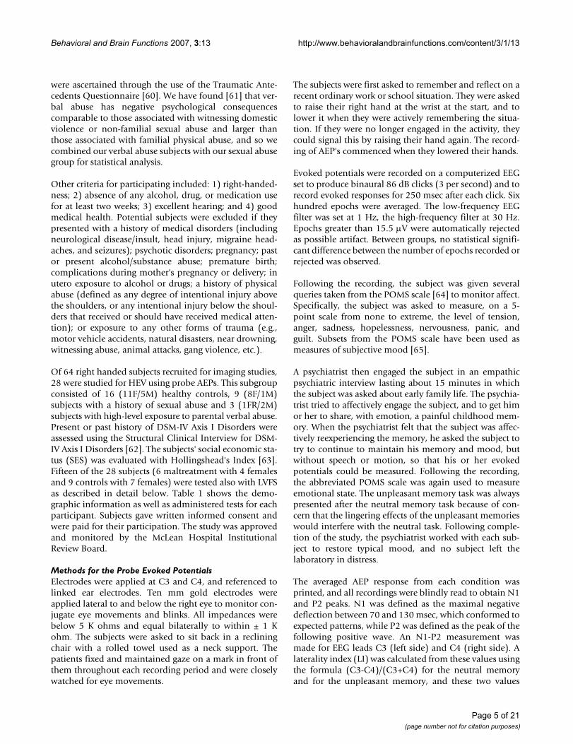
were ascertained through the use of the Traumatic Ante-cedents Questionnaire [60]. We have found [61] that ver-bal abuse has negative psychological consequencescomparable to those associated with witnessing domesticviolence or non-familial sexual abuse and larger thanthose associated with familial physical abuse, and so wecombined our verbal abuse subjects with our sexual abusegroup for statistical analysis.
Other criteria for participating included: 1) right-handed-ness; 2) absence of any alcohol, drug, or medication usefor at least two weeks; 3) excellent hearing; and 4) goodmedical health. Potential subjects were excluded if theypresented with a history of medical disorders (includingneurological disease/insult, head injury, migraine head-aches, and seizures); psychotic disorders; pregnancy; pastor present alcohol/substance abuse; premature birth;complications during mother's pregnancy or delivery; inutero exposure to alcohol or drugs; a history of physicalabuse (defined as any degree of intentional injury abovethe shoulders, or any intentional injury below the shoul-ders that received or should have received medical atten-tion); or exposure to any other forms of trauma (e.g.,motor vehicle accidents, natural disasters, near drowning,witnessing abuse, animal attacks, gang violence, etc.).
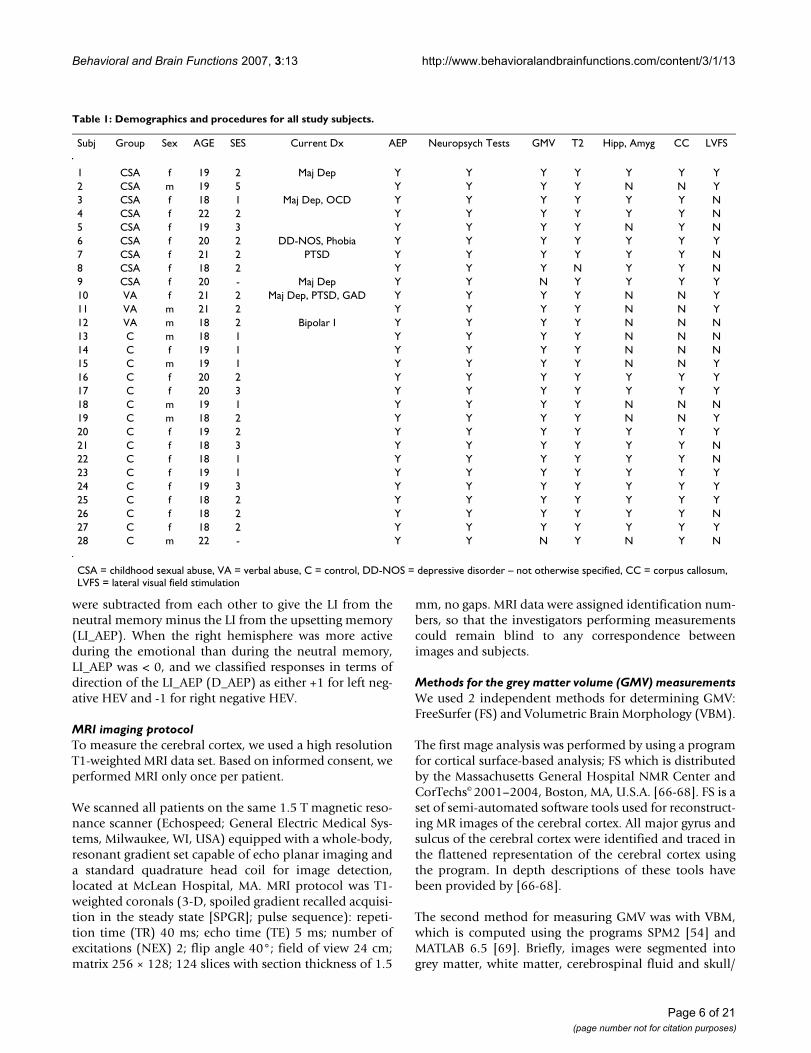
Of 64 right handed subjects recruited for imaging studies,28 were studied for HEV using probe AEPs. This subgroupconsisted of 16 (11F/5M) healthy controls, 9 (8F/1M)subjects with a history of sexual abuse and 3 (1FR/2M)subjects with high-level exposure to parental verbal abuse.Present or past history of DSM-IV Axis I Disorders wereassessed using the Structural Clinical Interview for DSM-IV Axis I Disorders [62]. The subjects' social economic sta-tus (SES) was evaluated with Hollingshead's Index [63].Fifteen of the 28 subjects (6 maltreatment with 4 femalesand 9 controls with 7 females) were tested also with LVFSas described in detail below. Table 1 shows the demo-graphic information as well as administered tests for eachparticipant. Subjects gave written informed consent andwere paid for their participation. The study was approvedand monitored by the McLean Hospital InstitutionalReview Board.
Methods for the Probe Evoked PotentialsElectrodes were applied at C3 and C4, and referenced tolinked ear electrodes. Ten mm gold electrodes wereapplied lateral to and below the right eye to monitor con-jugate eye movements and blinks. All impedances werebelow 5 K ohms and equal bilaterally to within ± 1 Kohm. The subjects were asked to sit back in a recliningchair with a rolled towel used as a neck support. Thepatients fixed and maintained gaze on a mark in front ofthem throughout each recording period and were closelywatched for eye movements.
The subjects were first asked to remember and reflect on arecent ordinary work or school situation. They were askedto raise their right hand at the wrist at the start, and tolower it when they were actively remembering the situa-tion. If they were no longer engaged in the activity, theycould signal this by raising their hand again. The record-ing of AEP's commenced when they lowered their hands.
Evoked potentials were recorded on a computerized EEGset to produce binaural 86 dB clicks (3 per second) and torecord evoked responses for 250 msec after each click. Sixhundred epochs were averaged. The low-frequency EEGfilter was set at 1 Hz, the high-frequency filter at 30 Hz.Epochs greater than 15.5 µV were automatically rejectedas possible artifact. Between groups, no statistical signifi-cant difference between the number of epochs recorded orrejected was observed.
Following the recording, the subject was given severalqueries taken from the POMS scale [64] to monitor affect.Specifically, the subject was asked to measure, on a 5-point scale from none to extreme, the level of tension,anger, sadness, hopelessness, nervousness, panic, andguilt. Subsets from the POMS scale have been used asmeasures of subjective mood [65].
A psychiatrist then engaged the subject in an empathicpsychiatric interview lasting about 15 minutes in whichthe subject was asked about early family life. The psychia-trist tried to affectively engage the subject, and to get himor her to share, with emotion, a painful childhood mem-ory. When the psychiatrist felt that the subject was affec-tively reexperiencing the memory, he asked the subject totry to continue to maintain his memory and mood, butwithout speech or motion, so that his or her evokedpotentials could be measured. Following the recording,the abbreviated POMS scale was again used to measureemotional state. The unpleasant memory task was alwayspresented after the neutral memory task because of con-cern that the lingering effects of the unpleasant memorieswould interfere with the neutral task. Following comple-tion of the study, the psychiatrist worked with each sub-ject to restore typical mood, and no subject left thelaboratory in distress.
The averaged AEP response from each condition wasprinted, and all recordings were blindly read to obtain N1and P2 peaks. N1 was defined as the maximal negativedeflection between 70 and 130 msec, which conformed toexpected patterns, while P2 was defined as the peak of thefollowing positive wave. An N1-P2 measurement wasmade for EEG leads C3 (left side) and C4 (right side). Alaterality index (LI) was calculated from these values usingthe formula (C3-C4)/(C3+C4) for the neutral memoryand for the unpleasant memory, and these two values
Page 5 of 21(page number not for citation purposes)
Behavioral and Brain Functions 2007, 3:13 http://www.behavioralandbrainfunctions.com/content/3/1/13
were subtracted from each other to give the LI from theneutral memory minus the LI from the upsetting memory(LI_AEP). When the right hemisphere was more activeduring the emotional than during the neutral memory,LI_AEP was < 0, and we classified responses in terms ofdirection of the LI_AEP (D_AEP) as either +1 for left neg-ative HEV and -1 for right negative HEV.
MRI imaging protocolTo measure the cerebral cortex, we used a high resolutionT1-weighted MRI data set. Based on informed consent, weperformed MRI only once per patient.
We scanned all patients on the same 1.5 T magnetic reso-nance scanner (Echospeed; General Electric Medical Sys-tems, Milwaukee, WI, USA) equipped with a whole-body,resonant gradient set capable of echo planar imaging anda standard quadrature head coil for image detection,located at McLean Hospital, MA. MRI protocol was T1-weighted coronals (3-D, spoiled gradient recalled acquisi-tion in the steady state [SPGR]; pulse sequence): repeti-tion time (TR) 40 ms; echo time (TE) 5 ms; number ofexcitations (NEX) 2; flip angle 40°; field of view 24 cm;matrix 256 × 128; 124 slices with section thickness of 1.5
mm, no gaps. MRI data were assigned identification num-bers, so that the investigators performing measurementscould remain blind to any correspondence betweenimages and subjects.
Methods for the grey matter volume (GMV) measurementsWe used 2 independent methods for determining GMV:FreeSurfer (FS) and Volumetric Brain Morphology (VBM).
The first mage analysis was performed by using a programfor cortical surface-based analysis; FS which is distributedby the Massachusetts General Hospital NMR Center andCorTechs© 2001–2004, Boston, MA, U.S.A. [66-68]. FS is aset of semi-automated software tools used for reconstruct-ing MR images of the cerebral cortex. All major gyrus andsulcus of the cerebral cortex were identified and traced inthe flattened representation of the cerebral cortex usingthe program. In depth descriptions of these tools havebeen provided by [66-68].
The second method for measuring GMV was with VBM,which is computed using the programs SPM2 [54] andMATLAB 6.5 [69]. Briefly, images were segmented intogrey matter, white matter, cerebrospinal fluid and skull/
Table 1: Demographics and procedures for all study subjects.
Subj Group Sex AGE SES Current Dx AEP Neuropsych Tests GMV T2 Hipp, Amyg CC LVFS
1 CSA f 19 2 Maj Dep Y Y Y Y Y Y Y2 CSA m 19 5 Y Y Y Y N N Y3 CSA f 18 1 Maj Dep, OCD Y Y Y Y Y Y N4 CSA f 22 2 Y Y Y Y Y Y N5 CSA f 19 3 Y Y Y Y N Y N6 CSA f 20 2 DD-NOS, Phobia Y Y Y Y Y Y Y7 CSA f 21 2 PTSD Y Y Y Y Y Y N8 CSA f 18 2 Y Y Y N Y Y N9 CSA f 20 - Maj Dep Y Y N Y Y Y Y10 VA f 21 2 Maj Dep, PTSD, GAD Y Y Y Y N N Y11 VA m 21 2 Y Y Y Y N N Y12 VA m 18 2 Bipolar I Y Y Y Y N N N13 C m 18 1 Y Y Y Y N N N14 C f 19 1 Y Y Y Y N N N15 C m 19 1 Y Y Y Y N N Y16 C f 20 2 Y Y Y Y Y Y Y17 C f 20 3 Y Y Y Y Y Y Y18 C m 19 1 Y Y Y Y N N N19 C m 18 2 Y Y Y Y N N Y20 C f 19 2 Y Y Y Y Y Y Y21 C f 18 3 Y Y Y Y Y Y N22 C f 18 1 Y Y Y Y Y Y N23 C f 19 1 Y Y Y Y Y Y Y24 C f 19 3 Y Y Y Y Y Y Y25 C f 18 2 Y Y Y Y Y Y Y26 C f 18 2 Y Y Y Y Y Y N27 C f 18 2 Y Y Y Y Y Y Y28 C m 22 - Y Y N Y N Y N
CSA = childhood sexual abuse, VA = verbal abuse, C = control, DD-NOS = depressive disorder – not otherwise specified, CC = corpus callosum, LVFS = lateral visual field stimulation
Page 6 of 21(page number not for citation purposes)
Behavioral and Brain Functions 2007, 3:13 http://www.behavioralandbrainfunctions.com/content/3/1/13
scalp compartments, then normalized to standard spaceand re-segmented. The spatially normalized segments ofgrey and white matter were smoothed using a 12-mm full-width half-maximum isotropic Gaussian kernel accordingto the optimized VBM protocol of Good et al [70].
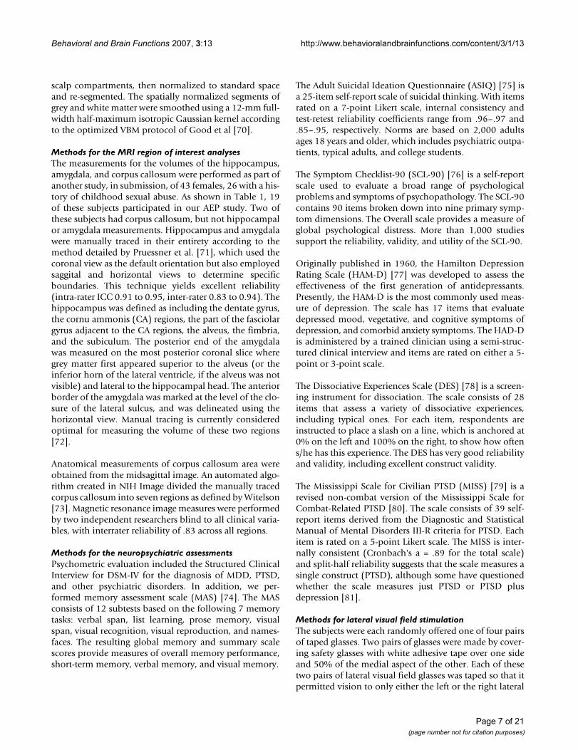
Methods for the MRI region of interest analysesThe measurements for the volumes of the hippocampus,amygdala, and corpus callosum were performed as part ofanother study, in submission, of 43 females, 26 with a his-tory of childhood sexual abuse. As shown in Table 1, 19of these subjects participated in our AEP study. Two ofthese subjects had corpus callosum, but not hippocampalor amygdala measurements. Hippocampus and amygdalawere manually traced in their entirety according to themethod detailed by Pruessner et al. [71], which used thecoronal view as the default orientation but also employedsaggital and horizontal views to determine specificboundaries. This technique yields excellent reliability(intra-rater ICC 0.91 to 0.95, inter-rater 0.83 to 0.94). Thehippocampus was defined as including the dentate gyrus,the cornu ammonis (CA) regions, the part of the fasciolargyrus adjacent to the CA regions, the alveus, the fimbria,and the subiculum. The posterior end of the amygdalawas measured on the most posterior coronal slice wheregrey matter first appeared superior to the alveus (or theinferior horn of the lateral ventricle, if the alveus was notvisible) and lateral to the hippocampal head. The anteriorborder of the amygdala was marked at the level of the clo-sure of the lateral sulcus, and was delineated using thehorizontal view. Manual tracing is currently consideredoptimal for measuring the volume of these two regions[72].
Anatomical measurements of corpus callosum area wereobtained from the midsagittal image. An automated algo-rithm created in NIH Image divided the manually tracedcorpus callosum into seven regions as defined by Witelson[73]. Magnetic resonance image measures were performedby two independent researchers blind to all clinical varia-bles, with interrater reliability of .83 across all regions.
Methods for the neuropsychiatric assessmentsPsychometric evaluation included the Structured ClinicalInterview for DSM-IV for the diagnosis of MDD, PTSD,and other psychiatric disorders. In addition, we per-formed memory assessment scale (MAS) [74]. The MASconsists of 12 subtests based on the following 7 memorytasks: verbal span, list learning, prose memory, visualspan, visual recognition, visual reproduction, and names-faces. The resulting global memory and summary scalescores provide measures of overall memory performance,short-term memory, verbal memory, and visual memory.
The Adult Suicidal Ideation Questionnaire (ASIQ) [75] isa 25-item self-report scale of suicidal thinking. With itemsrated on a 7-point Likert scale, internal consistency andtest-retest reliability coefficients range from .96–.97 and.85–.95, respectively. Norms are based on 2,000 adultsages 18 years and older, which includes psychiatric outpa-tients, typical adults, and college students.
The Symptom Checklist-90 (SCL-90) [76] is a self-reportscale used to evaluate a broad range of psychologicalproblems and symptoms of psychopathology. The SCL-90contains 90 items broken down into nine primary symp-tom dimensions. The Overall scale provides a measure ofglobal psychological distress. More than 1,000 studiessupport the reliability, validity, and utility of the SCL-90.
Originally published in 1960, the Hamilton DepressionRating Scale (HAM-D) [77] was developed to assess theeffectiveness of the first generation of antidepressants.Presently, the HAM-D is the most commonly used meas-ure of depression. The scale has 17 items that evaluatedepressed mood, vegetative, and cognitive symptoms ofdepression, and comorbid anxiety symptoms. The HAD-Dis administered by a trained clinician using a semi-struc-tured clinical interview and items are rated on either a 5-point or 3-point scale.
The Dissociative Experiences Scale (DES) [78] is a screen-ing instrument for dissociation. The scale consists of 28items that assess a variety of dissociative experiences,including typical ones. For each item, respondents areinstructed to place a slash on a line, which is anchored at0% on the left and 100% on the right, to show how oftens/he has this experience. The DES has very good reliabilityand validity, including excellent construct validity.
The Mississippi Scale for Civilian PTSD (MISS) [79] is arevised non-combat version of the Mississippi Scale forCombat-Related PTSD [80]. The scale consists of 39 self-report items derived from the Diagnostic and StatisticalManual of Mental Disorders III-R criteria for PTSD. Eachitem is rated on a 5-point Likert scale. The MISS is inter-nally consistent (Cronbach's a = .89 for the total scale)and split-half reliability suggests that the scale measures asingle construct (PTSD), although some have questionedwhether the scale measures just PTSD or PTSD plusdepression [81].
Methods for lateral visual field stimulationThe subjects were each randomly offered one of four pairsof taped glasses. Two pairs of glasses were made by cover-ing safety glasses with white adhesive tape over one sideand 50% of the medial aspect of the other. Each of thesetwo pairs of lateral visual field glasses was taped so that itpermitted vision to only either the left or the right lateral
Page 7 of 21(page number not for citation purposes)

Behavioral and Brain Functions 2007, 3:13 http://www.behavioralandbrainfunctions.com/content/3/1/13
visual field as the subject was asked to fixate the center ofhis vision on the edge of the tape so that he was lookingout of either the left half of the left eye or the right half ofthe right eye. The two other pairs of glasses were similarsafety goggles, taped completely over one side, but onlyover the bottom one fourth of the other side. These glassesallowed for monocular vision, which has been shown tocause some hemispheric lateralization [82]. The tape onthe bottom of the unoccluded lens gave the monocularglasses a more complex appearance.
After the first pair of glasses was worn for 2 minutes, oneof the experimenters (CA) verbally asked the subject torate his or her present feelings for each of eight affectsfrom an abbreviated POMS scale [64], from none toextreme on a 5 point scale. The eight affects measuredwere: anxiety, tension, anger, sadness, hopelessness,panic, nervousness, and guilt. Following the POMS meas-urements, the first pair of glasses was removed, and thesubject was allowed to rest for 2 minutes. Then the nextpair was placed on the subject. The identical procedurewas then followed for each of the 4 randomly presentedpairs of glasses. We calculated a laterality index from thePOMS scores reported when the subjects looked out of theleft and right visual fields [(LVF-RVF)/(LVF+RVF)] for theexperimental (LI_LVFS) and the monocular glasses [(L-R)/L+R)]. When more negative affect was reported fromthe LVF than the RVF, we assigned a negative right HEVand a left negative HEV when the LI_LVFS was < 0. Wereported this categorization of the LI_LVFS as the direc-tion of LVFS (D_LVFS) and +1 was for a right negativeHEV and -1 for a left negative HEV.
Simply asking a subject to look out of one visual field orthe other, in many subjects evokes a significant change intheir psychological state. In clinical settings, one side isgenerally more neurotic and symptomatic than the other[42,43]. The subjects did nothing other than look out ofone lateral visual field and then the other as describedabove.
With hemifield attention studies, the subject fixates botheyes at a central point and is asked to attend to a task ineither the left or right half of his full visual field, as imagesare presented to both fields of both eyes. Monocular stim-ulation could be expected to activate the contralateral vis-ual cortex since each retina is connected to bothhemispheres with about a 3:2 preference for the contralat-eral hemisphere. With LVFS as used in this study, webelieve the preference for the contralateral hemisphere isgreater than with hemifield attention or with monocularstimulation because the image is presented primarily onlyto the nasal portion of one retina, the segment connectedto the contralateral hemisphere. The taped safety glassesused in this study are not capable of limiting vision to
only the nasal portion of the retina, but they can beexpected to preferentially allow for stimulation of thatsection of the retina, and therefore might be expected togenerate a stronger stimulation of the contralateral hemi-sphere than hemifield attention or monocular stimula-tion. Each subject was instructed to look with half of hiseye, fixating on the edge of the tape, but we did not mon-itor the eye's position, and it is possible that some subjectsmight have been able to move their eyes so that the fixa-tion point was lateral to the tape's edge. To the extent towhich that occurred, the condition would have changedfrom LVFS to monocular vision, and this would have less-ened contralateral hemispheric activation. Since we werecorrelating LVFS to other outcome measures such as probeAEP, GMV, and MAS that are variable and were not knowat the time of LVFS testing, we believe that their correla-tions were not affected by any conscious or unconsciousexperimenter bias or features of the test environment.
Although visual input was presented preferentially to thecontralateral hemisphere, the corpus callosum allows thetransfer of information between the hemispheres. Never-theless, an accumulating body of evidence, as discussed inthe introduction and reviewed in detail elsewhere [42,43],suggests that unilateral sensory or motor stimulation caninfluence cognition and affect. These studies were predi-cated on the hypothesis that unilateral sensory stimula-tion would produce contralateral hemispheric activationthat would, in turn, influence cognition or affect.
To test for the reliability of LVFS, the LVFS procedure wasrepeated between 9 to 12 months following the initialprocedure to see how well the two trials correlated witheach other. For each trial we calculated the differencesbetween the abbreviated POMS scores from the LVF andthe RVF stimulation, and then examined how well thesedifferences correlated from each trial with each other.Only the first LVFS results were used in comparisons withanatomical and functional data.
Statistical methodsAll values are presented with standard deviations (± SD)and reflect means unless otherwise specified. Groups werecompared using paired or unpaired t-tests, UnivariateANCOVA's, or repeated measures ANCOVA's. When weanticipated a result based on prior experimentation, weused a one-tailed test, otherwise, we used a two-tailed test.To test for bivariate correlations we used a Pearson's cor-relation when N was ≥ 25; otherwise, we used a Spear-man's Rho. Linear regression analyses were alsoperformed to ascertain the relationships among variables.A large number of comparisons were made in this study.To set a balance between type I and type II errors, requisitealpha p values was set to 0.01.
Page 8 of 21(page number not for citation purposes)

Behavioral and Brain Functions 2007, 3:13 http://www.behavioralandbrainfunctions.com/content/3/1/13
ResultsProbe auditory evoked potentialsPOMS measures of negative emotion during the memory conditionsWe found that the intensity of negative affect, measuredwith the POMS scale after the unpleasant memory condi-tion (14.3 ± 7.8) was significantly greater than that afterthe neutral memory condition (1.7 ± 2.2) by a 2-sidedpaired t-test (df = 27, t = -8.9, p < 0.001), but this differ-ence was not related to grey matter volumes, measuredMRI sub-cortical regions of interest, T2 relaxation times,or performance on memory tests. The intensity of thechange in negative affect between conditions did not cor-relate directly with any psychological test parameter,although when added to regression models for the differ-ent psychological test parameters, it generally improvedthe models.
Probe Auditory Evoked Potentials during the Memory ConditionsWith the probe AEP, a smaller N1-P2 measurement overeither C3 (left auditory area) or C4 (right auditory area)suggests a greater degree of hemispheric involvement inthe memory task. Thus, a negative laterality index (C3-C4/C3+C4) suggests that the left hemisphere is relativelymore active than the right and a positive laterality indexsuggests that the right hemisphere is relatively moreactive. For the LI_AEP we subtracted the laterality indexobtained during the upsetting memory from thatobtained during the neutral memory. As described indetail on page 17, right negative HEV was associated witha LI_AEP with a negative value.
During the neutral memory, the 12 maltreatment subjectshad a mean AEP laterality index (C3-C4/C3+C4) of -0.120± 0.233 and during the unpleasant memory, 0.045 ±0.211. A paired 2-sided t-test comparing AEP lateralityindices during the neutral and unpleasant conditionsamong the trauma subjects approached significance (p <0.01), (t = 2.50, df = 11, P = 0.029). For the 16 controlsubjects, the mean during the neutral memory was -0.040± 0.218 and during the unpleasant memory was 0.028 ±0.155. The differences between the two memory condi-tions for the control group were not significant (t = 1.008,df = 15, P = 0.33). These results replicated our 1995 find-ings [8].
Of the 28 subjects, 18 (64%) showed a right negativeHEV. Ten subjects (36%), including 6 (38%) from thecontrol group and 4 (33%) from the trauma group had aleft negative HEV.
A grey matter volume (GMV) was calculated for each hem-isphere of each patient using FS and VBM. There was ahigh correlation between the results from the two pro-grams for the LH (N = 26, r = 0.91, p < 0.0001) and theRH (N = 26, r = 0.93, p < 0.0001).
ANCOVA models, controlling for differences in SES,showed that there were associations between GMV, HEV,and gender. As expected, there were significant gender dif-ferences with females showing an 8.4% reduction in lefthemisphere GMV (F = 14.56, df = 1,21, p = 0.001) and8.0% reduction in right hemisphere GMV (F = 10.48, df =1,21, p = 0.004). Overall, eta-squared for effect of genderon total GMV was 0.13 indicating that it accounted for13% of the variance in GMV. Direction of HEV was alsoassociated with main effects that approached significanceon left hemisphere GMV (F= 7.55, df = 1,21, p < 0.02),and right hemisphere GMV (F = 3.30, df = 1,21, p = 0.08).HEV direction accounted for 8% of the variance in totalGMV (p < 0.05). However, there were even more markedinteractions between HEV and gender. Males with rightnegative HEV had a 17.2% increase in left hemisphereGMV relative to males with left negative HEV, whereasright negative HEV females had a 3.3% decrease com-pared to females with left negative HEV (F = 15.79, df =1,21, p = 0.001). Similarly, males with right negative HEVhad a 16.3% increased in right hemisphere GMV andfemales with right negative HEV had a 6.1% decrease incomparison to subjects with the same gender but oppositeHEV (F = 15.87, df = 1,21, p = 0.001). Overall, the inter-action between gender and HEV had an eta-squared of0.39 on total GMV. Together, gender, HEV, and gender ×HEV interaction were able to account for about 60% of theindividual variation in measures of gray matter volume.Figure 1 graphically shows the interaction between genderand D_AEP for total GMV from FS.
The between subjects results of a repeated measuresANOVA comparing the left and right hemispheric GMV'sfrom FS with gender and D_AEP and their interactions asfactors was highly significant for the whole model (F =11.04, df = 3,22, p = 0.0001) as well as for the interactionbetween gender and D_AEP (F = 16.41, df = 1,22, p =0.0005).
Using the GMV data from VBM, performing the samerepeated measure ANOVA comparing LH and RH, wefound a similar between subject interaction between gen-der and D_AEP (F = 23.45, df = 1,22, p = < .0001).
For the 17 females studied, controlling for intracranialvolume, we found significant partial correlation coeffi-cients (df = 14) between LI_AEP and the right (r = .67, p =.004) hippocampal. The left plus right hippocampal vol-umes (r = .60, p = .014) approached significance. For theleft hippocampal volume, the partial correlation coeffi-cient also approached significance (r = .47, p = .064).Since a positive LI_AEP represents a left negative HEV,these correlations indicate that as the HEV becomes moreright negative, the left and right hippocampal volumesbecome smaller.
Page 9 of 21(page number not for citation purposes)

Behavioral and Brain Functions 2007, 3:13 http://www.behavioralandbrainfunctions.com/content/3/1/13
The partial correlation coefficient, again controlling forintracranial volume, between LI_AEP and the right amy-gdala (r = – 0.75, p = .001), and the sum of the left andright amygdala volumes (r = – .75, p = .001) were signifi-cant. This partial correlation coefficient for the left amy-gdala (- 0.54, p = .031) approached significance. Theseresults indicate that as the HEV becomes more right nega-tive, the left and right amygdala both become larger. TheLI_AEP also correlated with the left amygdala volumedivided by the left hippocampal volume (A/H), correctedfor intracranial volumes, (r =- .69, p = .003) as well as withthe right A/H ratio (r = – .87, p = .000) and with the com-bined left and right ratios (r = – .86, p = .000). Figure 2show a scatter plot of the relation between LI_AEP and theleft plus right A/H ratios.
We compared the T2 measurements using LIs calculatedfrom the left and right hemispheres and observed thatthey were different for the left negative and right negative
HEV groups. For the 17 subjects who had a right negativeHEV their mean LI for T2 was 0.089 ± 0.586, and for the10 subjects who had a left negative HEV, the mean LI was-0.366 ± 0.451, which by unpaired t-test (t = 2.11, df = 25,p = 0.045) approached significance. As shown in Table 2,the T2 relaxation times were relatively lower in the lefthemisphere (LH) of the group with left negative HEV. TheHEV groups did not differ significantly in the otherregions of interest that we analyzed.
As shown in Table 3, the visual and global memory sub-scales showed significant differences when the left nega-tive HEV subjects were compared with those from theright negative HEV classification, and the verbal scaleapproached significance. In these 3 comparisons, the rightnegative HEV group performed better. None of the 4 sub-scales of the MAS showed differences when group (abuseor control) or gender was compared among the 28 sub-jects.
Total grey matter volume by gender and hemispheric emotional valenceFigure 1Total grey matter volume by gender and hemispheric emotional valence. This graph shows the total grey matter volume deter-mined by FreeSurfer for male and female subjects with right and left hemispheric emotional valence by probe auditory evoked potentials.
Page 10 of 21(page number not for citation purposes)

Behavioral and Brain Functions 2007, 3:13 http://www.behavioralandbrainfunctions.com/content/3/1/13
As reported above, correlations between LI_AEP and hip-pocampal volumes indicated that as the HEV becomesmore right negative, the hippocampal volumes becomesmaller. Yet, we see here that the right negative HEV grouphad markedly better MAS scores. A model predicting theMAS for global memory using D_AEP (p = .008) covariedby the right hippocampus (p = 0.19) and the intracranialvolumes (p = 0.65), approached significance (F (3,16) =3.50, p = .047).
Table 4 shows the impact of considering D_AEP as anindependent factor in exploring the effect of abuse historyon psychiatric symptom ratings. A simple univariateANOVA with main effects of abuse history and genderreveals only a trend level effect of abuse history on ratingsof suicidaility (F(1,14) = 3.75, p = 0.073). This two factor
plus interaction model also provides only a weak fit(adjusted R2 = 0.051) to the available data. In contrastUnivariate analysis with three main factors (abuse history,gender, D_AEP) and their interactions provides a robustfit (adjusted R2 = 0.755). Taking D_AEP into considera-tion now reveals a marked effect of abuse on ASIQ_Tscores (F(1,10) = 33.23, p < 0.001).
The statistical consequences of considering D_AEP as anindependent variable were most marked for ASIQ scores,but D_AEP also exerted substantial effects on many otherrating scale scores. This relationship is summarized inTable 5. For each administered psychological test, the R2
associated with a regression model with gender and groupand their interactions as the factors was improved whenD_AEP and its interactions were added to the model. The
AEP by left plus right amygdala/hippocampal ratiosFigure 2AEP by left plus right amygdala/hippocampal ratios. A scatter plot of the relation between LI_AEP and the left plus right amy-gdala/hippocampal ratios. (r = -.86, p = .000).
Page 11 of 21(page number not for citation purposes)

Behavioral and Brain Functions 2007, 3:13 http://www.behavioralandbrainfunctions.com/content/3/1/13
R2 values by increased by 22.5% (for the HAM_D) to1,380%, from 0.051 to 0.755 (for the ASIQ_T).
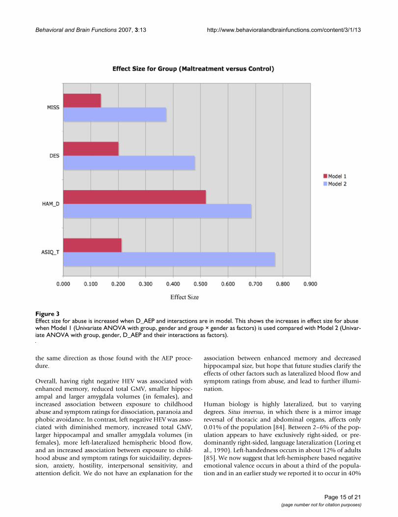
Adding D_AEP not only increased R2 values, but, asshown in figures 3 and 4, it also altered the effect sizes forabuse on these psychological tests.
One possible reason for the added statistical power ofincluding D_AEP in analysis of effects of abuse history onratings is that subjects with right negative HEV mayrespond differently to abuse than subjects with left nega-tive HEV. This appears to be true, at least in a statisticalsense, for many symptom scores. In general, ratings ofdepression (HAM-D, SCL-90 depression, SCL-90 interper-sonal sensitivity, ASIQ_T), anxiety (SCL-90), attentiondeficit (SCL-90), hostility (SCL-90), and trauma history(MISS) were most affected by abuse history in subjectswith left negative HEV. In contrast, ratings of dissociation(DES), paranoid ideation (SCL_90) and phobic anxiety(SCL90) were most affected by abuse in subjects with rightnegative HEV.
Lateral visual field stimulationA positive LI_LVFS suggests a right negative HEV becauseit is based on the negative emotions indicated by the
POMS score when the subject looks out of the LVF (righthemisphere) minus that when he looks out the RVF.LI_AEP, on the other hand is based on the N1-P2 meas-urement from C3 minus that from C4, as described onpage 17. A negative LI_AEP suggests a right negative HEV,and, for all 15 subjects who were given both procedures,comparing the directions of HEV between our 2 methods,AEP and LVFS, we found a highly significant correlation,(1-tailed Spearman's rho = -0.637, p = 0.005) indicatingan agreement between the two methods. Overall, 9 sub-jects had a directional response to the glasses, in 8 ofwhom the direction of response concurred with directionof HEV as determined by AEP.
The HEV indicated by the control, monocular glasses didnot correlate significantly with that from the AEPs (Spear-man Rho = 0.13, p = 0.69). In fact, we found the control,monocular glasses to be not significant in any analyses wecarried out for the LVFS glasses.
To test the reliability of LVFS, we repeated tests of LVFS, atleast 9 months apart, in 14 subjects and found a Spear-man's rho = .93, p = .0001 between the two tests.
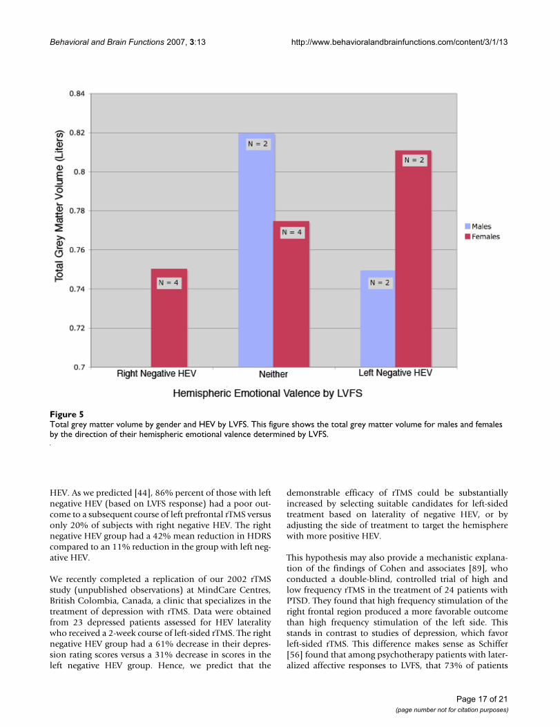
There were no significant bivariate correlations betweenLI_LVFS and hemispheric measures of GMV by FS or VBM.As illustrated in Figure 5, for total GMV by FS, males andfemales have different directions of change with HEV byLVFS and the directions, though not statistically signifi-cant, are similar to those shown in Figure 1 (which illus-trates GMV values categorized by the AEP procedure).
The correlation between LI_LVFS and the total white mat-ter volume, derived from FS, approached significance (2-tailed Spearman's rho = -.54, p = .045).
The total volume for the corpus callosum correlated withthe direction of LI_LVFS (n = 10, 2-tailed Spearman's Rho= -0.81, p < 0.005). The group with right negative HEV byLVFS had smaller total corpus callosi.
Table 3: Comparisons of the results from the Memory Assessment Scale Subscales between those subjects with Left and Right Negative HEV by Probe Auditory Evoked Potentials.
Short-term Verbal Visual Global
Memory Memory Memory Memory
Probe AEPs N
Left Negative HEV 10 109.40 ± 12.28 100.20 ± 11.02 105.40 ± 17.17 103.90 ± 15.60Right Negative HEV 18 108.17 ± 9.41a 113.61 ± 13.87b 120.56 ± 9.33c 120.39 ± 10.81d
a unpaired t-test Left – Right Negative HEV: t = -0.30, df = 26, p = 0.77, nsb unpaired t-test Left – Right Negative HEV: t = 2.62, df = 26, p = 0.014, nsc unpaired t-test Left – Right Negative HEV: t = 3.05, df = 26, p = 0.0052d unpaired t-test Left – Right Negative HEV: t = 3.30, df = 26, p = 0.0028
Table 2: MRI T2 relaxation times (lower values are thought to indicate greater rCBV) for the left and for right hemispheres as well as the laterality indices calculated from these values for subjects who by probe AEP had a left or a right negative hemispheric emotional valence.
Probe AEPs N LH RH LI (L-R/L+R)
Left Negative HEV 10 87.23 ± 7.51 87.56 ± 7.76 -0.366 ± 0.451Right Negative HEV 17 89.88 ± 9.72 89.78 ± 9.54 0.089 ± 0.586*
LH = left hemisphereRH = right hemisphere* unpaired t-test Left – Right Negative HEV:t = 2.11, df = 25, p = 0.045.
Page 12 of 21(page number not for citation purposes)

Behavioral and Brain Functions 2007, 3:13 http://www.behavioralandbrainfunctions.com/content/3/1/13
There were no relationships between the LVFS results andthe volumes for the amygdala, the hippocampus or theirratios.
As shown in Table 6. The Spearman correlations betweenLI_LVFS and the left and right hemisphere T2 relaxationtimes and their combination approached significance.The direction of the correlations show that for both theleft and right hemispheres, as with the Probe AEP proce-dure, those with left negative HEVs had lower T2 relaxa-tion times than those with right negative HEVs.
The results from the LVFS tests did not correlate with theresults from the Memory Assessment Scale or the admin-istered psychological tests.
The statistical results for LVFS came from only 15 subjects.If the probe AEP statistical tests were limited to includeonly the 15 subjects who received the LVFS protocol, the
probe AEP would have had no statistically significantresults. Having an additional 13 subjects in the AEP groupallowed us to observe statistical relationships that wouldhave been missed otherwise.
DiscussionIn the literature there has been a prevailing view that theright hemisphere is associated with the perception andexpression of negative emotions [8,21,83]. Our studies,and review of the literature, suggests that this view pointis overly dogmatic, and that negative valence may be leftlateralized in a substantial number of individuals[44,56,57]. Further, our new data provides evidence thatthe degree and direction of laterality is an important indi-vidual trait, on par with gender, in the degree to which itcan account for individual differences in regional brainsize, functional brain activity, and psychiatric symptoma-tology. Although we have found gender to be an impor-tant factor in regard to HEV, we feel that a future study
Table 4: Effect of hemispheric emotional valence on ratings of suicidaility.
Effect of including direction of hemispheric emotional valence as an independent variable when assessing the effects of abuse and gender on ratings of suicidaility.
ANOVA with main effects and interactions of abuse history and gender
Tests of Between-Subjects EffectsDependent Variable: ASIQ_T
Source Type III Sum Sq df Mean Square F Signif. Partial h2Corrected Model 181.167 3 60.389 1.307 0.311 0.219GROUP 173.361 1 173.36 3.752 0.073 0.211GENDER 2.25 1 2.25 0.049 0.829 0.003GROUP * GENDER 4.694 1 4.694 0.102 0.755 0.007Error 646.833 14 46.202Total 39470 18Corrected Total 828 17
R Squared = .219 (Adjusted R Squared = .051)
ANOVA with main effects and interactions of abuse history, gender and direction of HEV
Tests of Between-Subjects EffectsDependent Variable: ASIQ_T
Source Type III Sum Sq df Mean Square F Signif. Partial h2Corrected Model 708.7 7 101.24 8.486 0.002 0.856GROUP 396.463 1 396.46 33.23 < .001 0.769GENDER 17.473 1 17.473 1.465 0.254 0.128DIR_HEV 70.644 1 70.644 5.922 0.035 0.372GROUP * GENDER 25.796 1 25.796 2.162 0.172 0.178GROUP *DIR_HEV 56.341 1 56.341 4.723 0.055 0.321GENDER*DIR_HEV 133.432 1 133.43 11.18 0.007 0.528GROUP*GENDER*DIR_HEV 257.473 1 257.47 21.58 0.001 0.683Error 119.3 10 11.93Total 39470 18Corrected Total 828 17
R Squared = .856 (Adjusted R Squared = .755)
Page 13 of 21(page number not for citation purposes)
Behavioral and Brain Functions 2007, 3:13 http://www.behavioralandbrainfunctions.com/content/3/1/13
with larger numbers of males and females would be help-ful to more precisely delineate its level of importance. Inthis study we did not evaluate functional imaging duringthe different memory conditions, but we hope to do sucha study in the future.
We found that gender alone accounted for about 13% ofthe variance between individuals in total GMV. In con-trast, direction of HEV, gender, and their interactionaccounted for up to 60% of the variance. Degree anddirection of HEV was also associated with substantial dif-ferences in hippocampal and amygdala volumes. Femaleswith the most right-sided HEV tended to have the smallesthippocampal and largest amygdala volumes. (Volumeswere not measured in males). Degree of HEV in AEPresponse accounted for 36% and 54% of the variance inhippocampal and amygdala measures, respectively.
T2 relaxation time was used as indirect measure of restingrelative cerebral blood volume, with lower levels of T2relaxation times correlating with higher blood volumes[55]. Subjects with left negative HEV had lower T2 relaxa-tion times in their left vs. right hemisphere, suggesting
greater left-sided rCBV, relative to subjects with right neg-ative HEV.
The right negative HEV group performed significantly bet-ter on 3 of 4 memory tests. This difference persisted evenwhen their scores were covaried for intracranial and hip-pocampal volumes. When direction of hemispheric emo-tional valence and its interactions were added toregression models examining the effects of gender andchildhood maltreatment on 15 administered psychologi-cal test parameters, the R2 values for 11 of the 15 modelsimproved, and the effect sizes for the influence of child-hood maltreatment were increased on most of these psy-chological variables.
We presented two methods for determining HEV, probeAEPs and LVFS. They provide highly correlated results,and both appear useful for delineating distinct popula-tions of left vs. right lateralized responders. LVFS, per-formed in only 15 subjects (versus 28 in the AEP group),correlated with the total volume of corpus callosum, andthe right and left hemispheric T2 relaxation times. Thechanges in GMV and T2 relaxation time by LVFS were in
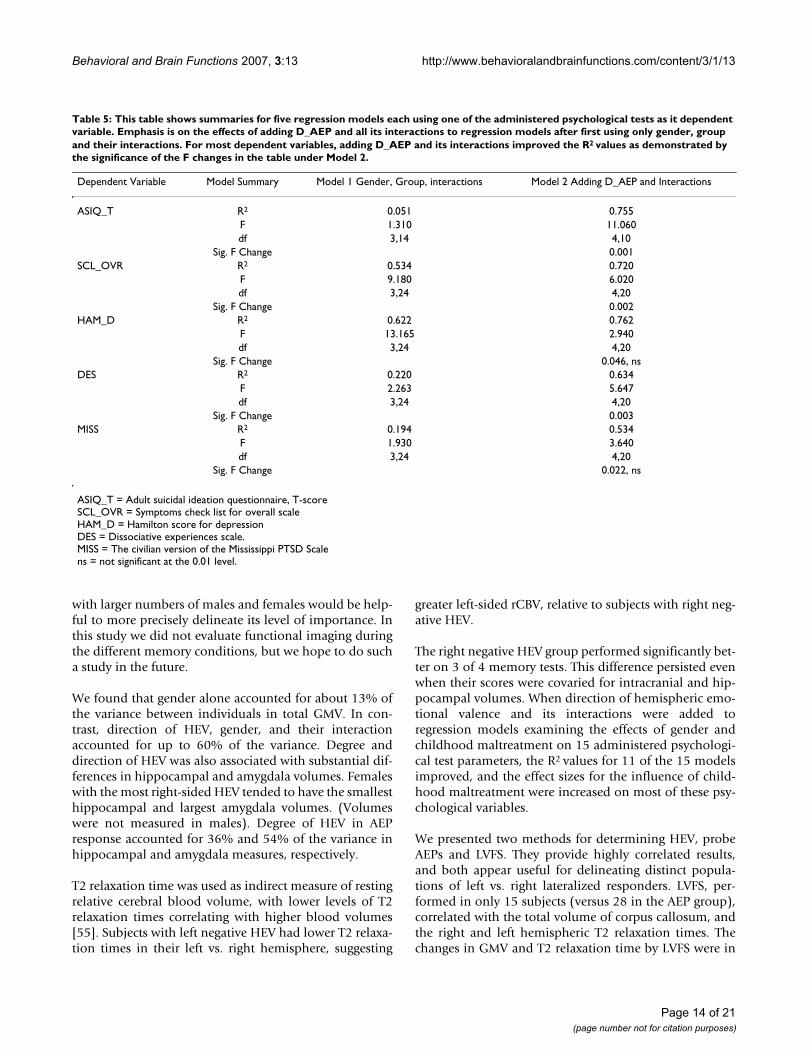
Table 5: This table shows summaries for five regression models each using one of the administered psychological tests as it dependent variable. Emphasis is on the effects of adding D_AEP and all its interactions to regression models after first using only gender, group and their interactions. For most dependent variables, adding D_AEP and its interactions improved the R2 values as demonstrated by the significance of the F changes in the table under Model 2.
Dependent Variable Model Summary Model 1 Gender, Group, interactions Model 2 Adding D_AEP and Interactions
ASIQ_T R2 0.051 0.755F 1.310 11.060df 3,14 4,10
Sig. F Change 0.001SCL_OVR R2 0.534 0.720
F 9.180 6.020df 3,24 4,20
Sig. F Change 0.002HAM_D R2 0.622 0.762
F 13.165 2.940df 3,24 4,20
Sig. F Change 0.046, nsDES R2 0.220 0.634
F 2.263 5.647df 3,24 4,20
Sig. F Change 0.003MISS R2 0.194 0.534
F 1.930 3.640df 3,24 4,20
Sig. F Change 0.022, ns
ASIQ_T = Adult suicidal ideation questionnaire, T-scoreSCL_OVR = Symptoms check list for overall scaleHAM_D = Hamilton score for depressionDES = Dissociative experiences scale.MISS = The civilian version of the Mississippi PTSD Scalens = not significant at the 0.01 level.
Page 14 of 21(page number not for citation purposes)
Behavioral and Brain Functions 2007, 3:13 http://www.behavioralandbrainfunctions.com/content/3/1/13
the same direction as those found with the AEP proce-dure.
Overall, having right negative HEV was associated withenhanced memory, reduced total GMV, smaller hippoc-ampal and larger amygdala volumes (in females), andincreased association between exposure to childhoodabuse and symptom ratings for dissociation, paranoia andphobic avoidance. In contrast, left negative HEV was asso-ciated with diminished memory, increased total GMV,larger hippocampal and smaller amygdala volumes (infemales), more left-lateralized hemispheric blood flow,and an increased association between exposure to child-hood abuse and symptom ratings for suicidaility, depres-sion, anxiety, hostility, interpersonal sensitivity, andattention deficit. We do not have an explanation for the
association between enhanced memory and decreasedhippocampal size, but hope that future studies clarify theeffects of other factors such as lateralized blood flow andsymptom ratings from abuse, and lead to further illumi-nation.
Human biology is highly lateralized, but to varyingdegrees. Situs inversus, in which there is a mirror imagereversal of thoracic and abdominal organs, affects only0.01% of the population [84]. Between 2–6% of the pop-ulation appears to have exclusively right-sided, or pre-dominantly right-sided, language lateralization (Loring etal., 1990). Left-handedness occurs in about 12% of adults[85]. We now suggest that left-hemisphere based negativeemotional valence occurs in about a third of the popula-tion and in an earlier study we reported it to occur in 40%
Effect size for abuse is increased when D_AEP and interactions are in modelFigure 3Effect size for abuse is increased when D_AEP and interactions are in model. This shows the increases in effect size for abuse when Model 1 (Univariate ANOVA with group, gender and group × gender as factors) is used compared with Model 2 (Univar-iate ANOVA with group, gender, D_AEP and their interactions as factors).
Page 15 of 21(page number not for citation purposes)
Behavioral and Brain Functions 2007, 3:13 http://www.behavioralandbrainfunctions.com/content/3/1/13
of patients [44]. This view that negative emotional valenceis lateralized, but to either right or left hemispheres,stands in marked contrast to earlier theories regarding thespecialized role of the right hemispheric in the processingof negative emotions [2-5]. Assuming that negative emo-tional valence is exclusively right lateralized may result inresearch findings in which lateralized difference are eitherdiminished or imperceptible. In contrast, recognizing thatright or left-sided laterality of negative emotional valenceis an important individual difference, may help to resolvediscrepancies in the literature that have emerged in studies
of the neurobiology and treatment of emotional disor-ders.
For example, there is controversy in the literature regard-ing the efficacy of left-sided rapid transcranial magneticstimulation (rTMS) for treatment of refractory depression[86-88]. However we found that outcome of rTMS variesgreatly depending on the whether the patient has right orleft lateralized negative HEV. We predicted that stimulat-ing a hemisphere with a positive HEV would be morelikely to be helpful than stimulating one with a negative
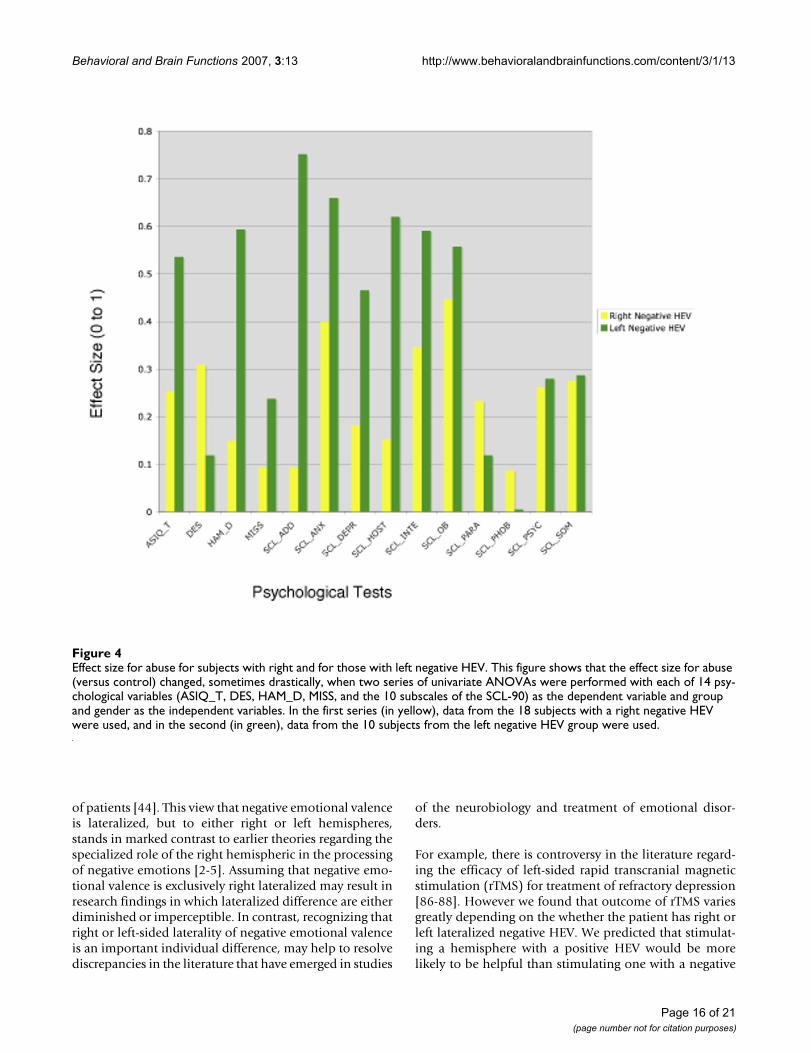
Effect size for abuse for subjects with right and for those with left negative HEVFigure 4Effect size for abuse for subjects with right and for those with left negative HEV. This figure shows that the effect size for abuse (versus control) changed, sometimes drastically, when two series of univariate ANOVAs were performed with each of 14 psy-chological variables (ASIQ_T, DES, HAM_D, MISS, and the 10 subscales of the SCL-90) as the dependent variable and group and gender as the independent variables. In the first series (in yellow), data from the 18 subjects with a right negative HEV were used, and in the second (in green), data from the 10 subjects from the left negative HEV group were used.
Page 16 of 21(page number not for citation purposes)
Behavioral and Brain Functions 2007, 3:13 http://www.behavioralandbrainfunctions.com/content/3/1/13
HEV. As we predicted [44], 86% percent of those with leftnegative HEV (based on LVFS response) had a poor out-come to a subsequent course of left prefrontal rTMS versusonly 20% of subjects with right negative HEV. The rightnegative HEV group had a 42% mean reduction in HDRScompared to an 11% reduction in the group with left neg-ative HEV.
We recently completed a replication of our 2002 rTMSstudy (unpublished observations) at MindCare Centres,British Colombia, Canada, a clinic that specializes in thetreatment of depression with rTMS. Data were obtainedfrom 23 depressed patients assessed for HEV lateralitywho received a 2-week course of left-sided rTMS. The rightnegative HEV group had a 61% decrease in their depres-sion rating scores versus a 31% decrease in scores in theleft negative HEV group. Hence, we predict that the
demonstrable efficacy of rTMS could be substantiallyincreased by selecting suitable candidates for left-sidedtreatment based on laterality of negative HEV, or byadjusting the side of treatment to target the hemispherewith more positive HEV.
This hypothesis may also provide a mechanistic explana-tion of the findings of Cohen and associates [89], whoconducted a double-blind, controlled trial of high andlow frequency rTMS in the treatment of 24 patients withPTSD. They found that high frequency stimulation of theright frontal region produced a more favorable outcomethan high frequency stimulation of the left side. Thisstands in contrast to studies of depression, which favorleft-sided rTMS. This difference makes sense as Schiffer[56] found that among psychotherapy patients with later-alized affective responses to LVFS, that 73% of patients
Total grey matter volume by gender and HEV by LVFSFigure 5Total grey matter volume by gender and HEV by LVFS. This figure shows the total grey matter volume for males and females by the direction of their hemispheric emotional valence determined by LVFS.
Page 17 of 21(page number not for citation purposes)

Behavioral and Brain Functions 2007, 3:13 http://www.behavioralandbrainfunctions.com/content/3/1/13
with major depression (n = 15) had a right negative HEV,whereas 71% of patients with PTSD (n = 14) had a leftnegative HEV. Hence, left-sided rTMS should benefit mostpatients with depression, while right-sided rTMS shouldbenefit most patient with PTSD. Targeting treatment tothe appropriate side for each individual based on LFVSmay further enhance outcomes, though this remains to bedetermined.
These findings may apply to other lateralized treatmentssuch as ECT. There is evidence that psychotropic medica-tions have lateralized effects [49,51], and several authorshave predicted responses to psychotropic medications bymeasurement of asymmetric brain activation by dichoticlistening [47,52], electroencephalogram [50,52], fMRI[53], and PET [48]. LVFS should be explored as a possiblemethod for predicting such outcomes.
These tests for HEV were inspired by observations fromsplit-brain studies that revealed that each hemisphere wascapable of supporting independent mentation [1,38,39].Bogen [40,41] was the first to suggest that these split-brainfindings might relate to intact individuals, and his asser-tion is supported by a number of reports of Wada studiesthat found, not just affect changes, but dramatic personal-ity changes with the anesthetization of one hemisphere[35-37,90]. For example, Masia et al reported 4 patientswho recalled with severe emotional distress a majortrauma such as the decapitation of a friend or an incestu-ous rape when one hemisphere was anesthetized, but notat baseline nor when the other hemisphere was anesthe-tized. The side from which the memory was released var-ied among patients (2 left and 2 right). Ahern et al [35]described two patients with vivid personality changes.One case went from withdrawn and sullen to affable andsocial following anaesthetization of the left hemisphere,and in the other went from pleasant and well adjusted tobelligerent and abusive when his left hemisphere wasanesthetized.
Wittling [91,92] has reported affect, blood pressure, heartrate, and cortisol changes depending on which side anupsetting film was shown to subjects. Placebo controlled
studies have shown that LVFS can induce changes in affect[44,56,57] and Schiffer [42] and Morton [93] havereported not only affect but also cognitive changes withLVFS. Others [94-97] have reported similar changes fol-lowing lateralized auditory stimulation in patient popula-tions. In all of these studies, the side that induced negativeaffects and/or cognitions varied among individuals.Schiffer [42,43] has suggested that LVFS can be a usefuladjunct to psychotherapy.
Considerable evidence indicates that unilateral sensory ormotor stimulation, including LVFS, activates the contral-ateral hemisphere. This evidence includes studies usingtheta EEG [57,98], lateral ear temperature [57], BOLDfMRI [58,99], and PET [100]. The combination of lateral-ized psychological and physiological responses, leadsSchiffer to hypothesize that LVFS might preferentially acti-vate the contralateral hemisphere [101] and produce anassociated mental state that is consistent with that hemi-sphere's emotional valence.
ConclusionOverall, the purpose of this study is to stimulate a dia-logue on hemispheric emotional valence, to indicate thata substantial percent of the population may have negativeemotions preferentially associated with their left hemi-sphere, and to suggest that this is a key individual differ-ence with relevance to researchers in behavioral andneurosciences, as well as to clinicians treating patientswith mood, anxiety and personality disorders.
Competing interestsThe author(s) declare that they have no competing inter-ests.
Authors' contributionsFS made substantial contributions to the conception anddesign of the study, the acquisition, analysis, and interpre-tation of data, and drafting of the manuscript. MT madesubstantial contributions to the conception and design ofthe study, analysis and interpretation of data, and revisionof the manuscript. CA made substantial contributions tothe acquisition of the evoked potential and LVFS data andto the acquisition, analysis, and interpretation of the func-tional MRI data, and to the acquisition of the anatomicalMRI data. CPN made substantial contributions to theacquisition and analysis of the neuropsychological testdata. AP made substantial contributions to the subjectenrollments and diagnostic interviews as well as to theoverall administration of the study. AT made substantialcontributions to the analysis and interpretation of thegrey matter volume data. SA made substantial contribu-tions to the analysis and interpretation of the MRI regionof interest data. All authors read and approved the finalmanuscript.
Table 6: Show the results of 2-tailed Spearman correlations for LI_LVFS and the left and right hemispheric T2 relaxation times as well as for their sums. N = 15.
Spearman Rho Significance
Left Hemisphere 0.52 0.048Right Hemisphere 0.51 0.054Both Hemispheres 0.55 0.034
Page 18 of 21(page number not for citation purposes)

Behavioral and Brain Functions 2007, 3:13 http://www.behavioralandbrainfunctions.com/content/3/1/13
AcknowledgementsThis research was supported, in part, by RO1 grants to MHT from the National Institute of Mental Health (MH-53636, MH-66222) and the National Institute on Drug Abuse (DA-016934, DA-017846).
References1. Sperry RW, Zaidel E, Zaidel D: Self recognition and social aware-
ness in the deconnected minor hemisphere. Neuropsychologia1979, 17:153-166.
2. Galin D: Implications for psychiatry of left and right cerebralspecialization. Arch Gen Psychiatry 1974, 31:572-583.
3. Joseph R: Neuropsychology, Neuropsychiatry, and BehavioralNeurology. New York , Plenum Press; 1990.
4. Schiffer F: Cognitive activity of the right hemisphere: possiblecontributions to psychological function. Harvard Rev Psychiat1996, 4:126-138.
5. Schore AN: Early organization of the nonlinear right brain anddevelopment of a predisposition to psychiatric disorders.Dev Psychopathol 1997, 9(4):595-631.
6. Davidson RJ: Affective style and affective disorders: Perspec-tives from affective neuroscience. Cogn Emot 1998,12(3):307-330.
7. Harmon-Jones E: Early Career Award. Clarifying the emotivefunctions of asymmetrical frontal cortical activity. Psychophys-iology 2003, 40(6):838-848.
8. Schiffer F, Teicher MH, Papanicolaou AC: Evoked potential evi-dence for right brain activity during the recall of traumaticmemories. J Neuropsychiatry Clin Neurosci 1995, 7(2):169-175.
9. Papanicolaou AC, Johnstone J: Probe evoked potentials: theory,method and applications. Int J Neurosci 1984, 24(2):107-131.
10. Rauch RL, Shin LM: Functional neuroimaging studies in post-traumatic stress disorder. Ann N Y Acad Sci 1997, 821:83-98.
11. Shin LM, McNally RJ, Kosslyn SM, Thompson WL, Rauch SL, AlpertNM, Metzger LJ, Lasko NB, Orr SP, Pitman RK: Regional cerebralblood flow during script-driven imagery in childhood sexualabuse-related PTSD: A PET investigation. Am J Psychiatry 1999,156(4):575-584.
12. Lee GP, Loring DW, Meader KJ, Brooks BB: Hemispheric special-ization for emotional expression: a reexamination of resultsfrom intracarotid administration of sodium amobarbital.Brain Cogn 1990, 12(2):267-280.
13. Ahern GL, Herring AM, Tackenberg JN, Schwartz GE, Seeger JF, Lab-iner DM, Weinand ME, KJ. KJO: Affective self-report during theintracarotid sodium amobarbital test. J Clin Exp Neuropsychol1994, 16(3):372-376.
14. Stabell KE, Andresen S, Bakke SJ, Bjornaes H, Borchgrevink HM,Heminghyt E, Roste GK: Emotional responses during unilateralamobarbital anesthesia: differential hemispheric contribu-tions? Acta Neurol Scand 2004, 110(5):313-321.
15. Branch C, Milner B, Rasmussen T: Intracarotid Sodium Amytalfor The Lateraization of Cerebral Speech Dominance;Observations in 123 Patients. J Neurosurg 1964, 21:399-405.
16. Robinson RG, Lipsey JR, Rao K, Price TR: Two-year longitudinalstudy of post-stroke mood disorders: comparison of acute-onset with delayed-onset depression. Am J Psychiatry 1986,143(10):1238-1244.
17. Robinson RG, Starkstein SE, Price TR: Post-stroke depression andlesion location. Stroke 1988, 19(1):125-126.
18. Carson AJ, MacHale S, Allen K, Lawrie SM, Dennis M, House A,Sharpe M: Depression after stroke and lesion location: a sys-tematic review. Lancet 2000, 356(9224):122-126.
19. Narushima K, Kosier JT, Robinson RG: A reappraisal of post-stroke depression, intra- and inter-hemispheric lesion loca-tion using meta-analysis. J Neuropsychiatry Clin Neurosci 2003,15(4):422-430.
20. Morris PL, Robinson RG, Raphael B, Hopwood MJ: Lesion locationand poststroke depression. J Neuropsychiatry Clin Neurosci 1996,8(4):399-403.
21. Davidson RJ: Affective neuroscience and psychophysiology:toward a synthesis. Psychophysiology 2003, 40(5):655-665.
22. Davidson RJ, Ekman P, Saron CD, Senulis JA, Friesen WV:Approach-withdrawal and cerebral asymmetry: emotionalexpression and brain physiology. I. J Pers Soc Psychol 1990,58(2):330-341.
23. Tomarken AJ, Davidson RJ, Henriques JB: Resting frontal brainasymmetry predicts affective responses to films. J Pers Soc Psy-chol 1990, 59(4):791-801.
24. Jackson DC, Mueller CJ, Dolski I, Dalton KM, Nitschke JB, Urry HL,Rosenkranz MA, Ryff CD, Singer BH, Davidson RJ: Now you feel it,now you don't: frontal brain electrical asymmetry and indi-vidual differences in emotion regulation. Psychol Sci 2003,14(6):612-617.
25. Hagemann D, Naumann E, Becker G, Maier S, Bartussek D: Frontalbrain asymmetry and affective style: a conceptual replica-tion. Psychophysiology 1998, 34(4):372-388.
26. Wheeler RE, Davidson RJ, Tomarken AJ: Frontal brain asymme-try and emotional reactivity: a biological substrate of affec-tive style. Psychophysiology 1993, 30(1):82-89.
27. Lane RD, Reiman EM, Ahern GL, Schwartz GE, Davidson RJ: Neuro-anatomical correlates of happiness, sadness, and disgust. AmJ Psychiatry 1997, 154(7):926-933.
28. Reiman EM, Lane RD, Ahern GL, Schwartz GE, Davidson RJ, FristonKJ, Yun LS, Chen K: Neuroanatomical correlates of externallyand internally generated human emotion. Am J Psychiatry 1997,154(7):918-925.
29. Harmon-Jones E, Sigelman J: State anger and prefrontal brainactivity: evidence that insult-related relative left-prefrontalactivation is associated with experienced anger and aggres-sion. J Pers Soc Psychol 2001, 80(5):797-803.
30. Harmon-Jones E, Allen JJ: Anger and frontal brain activity: EEGasymmetry consistent with approach motivation despitenegative affective valence. J Pers Soc Psychol 1998,74(5):1310-1316.
31. Hewig J, Hagemann D, Seifert J, Naumann E, Bartussek D: On theselective relation of frontal cortical asymmetry and anger-out versus anger-control. J Pers Soc Psychol 2004, 87(6):926-939.
32. Murphy FC, Nimmo-Smith I, Lawrence AD: Functional neuroanat-omy of emotions: a meta-analysis. Cogn Affect Behav Neurosci2003, 3(3):207-233.
33. Phan KL, Wager T, Taylor SF, Liberzon I: Functional neuroanat-omy of emotion: a meta-analysis of emotion activation stud-ies in PET and fMRI. Neuroimage 2002, 16(2):331-348.
34. Eugene F, Levesque J, Mensour B, Leroux JM, Beaudoin G, BourgouinP, Beauregard M: The impact of individual differences on theneural circuitry underlying sadness. Neuroimage 2003, 19(2 Pt1):354-364.
35. Ahern GL, Herring AM, Trackenberg J, Seeger JF, Oommen KJ, Lab-iner DM, Weinand ME: The association of multiple personalityand temporolimbic Epilepsy: Intracarotid amobarbital testobservations. Arch Neurol 1993, 50:1020-1025.
36. Kurthen M, Linke DB, Reuter BM, Hufnagel A, Elger CE: Severe neg-ative emotional reactions in intracarotid sodium amytal pro-cedures: further evidence for hemispheric asymmetries?Cortex 1991, 27(2):333-337.
37. Masia SL, Perrine K, Westbrook L, Alper K, Devinsky O: Emotionaloutbursts and post-traumatic stress disorder during intraca-rotid amobarbital procedure. Neurology 2000, 54(8):1691-1693.
38. LeDoux JE, Wilson DH, Gazzaniga MS: A divided mind: observa-tions on the conscious properties of the separated hemi-spheres. Ann Neurol 1977, 2(5):417-421.
39. Schiffer F, Zaidel E, Bogen J, Chasan-Taber S: Different psycholog-ical status in the two hemispheres of two split brain patients.Neuropsychiatry Neuropsychol Behav Neurol 1998, 11:151-156.
40. Bogen JE: The other side of the brain: II. an appositional mind.Bull Los Angeles Neurol Soc 1969, 34:135-162.
41. Bogen JE: Partial hemispheric independence with the neo-commissures intact. Volume Brain Circuits and Functions of the Mind:Essays in Honor of Roger W. Sperry. Edited by: Trevarthen C. Cam-bridge , Cambridge University Press; 1990.
42. Schiffer F: Of Two Minds: The Revolutionary Science of Dual-Brain Psychology. New York , The Free Press; 1998.
43. Schiffer F: Can the Different Cerebral Hemispheres Have Dis-tinct Personalities? Evidence and Its Implications for Theoryand Treatment of PTSD and Other Disorders. J Trauma Disso-ciation 2000, 1(2):83-104.
44. Schiffer F, Stinchfield Z, Pascual-Leone A: Prediction of ClinicalResponse to Transcranial Magnetic Stimulation for Depres-sion by Baseline Lateral Visual Stimulation. NeuropsychiatryNeuropsychol Behav Neurol 2002, 15:18-27.
Page 19 of 21(page number not for citation purposes)
http://www.ncbi.nlm.nih.gov/entrez/query.fcgi?cmd=Retrieve&db=PubMed&dopt=Abstract&list_uids=4421063
http://www.ncbi.nlm.nih.gov/entrez/query.fcgi?cmd=Retrieve&db=PubMed&dopt=Abstract&list_uids=4421063
http://www.ncbi.nlm.nih.gov/entrez/query.fcgi?cmd=Retrieve&db=PubMed&dopt=Abstract&list_uids=9448998
http://www.ncbi.nlm.nih.gov/entrez/query.fcgi?cmd=Retrieve&db=PubMed&dopt=Abstract&list_uids=9448998
http://www.ncbi.nlm.nih.gov/entrez/query.fcgi?cmd=Retrieve&db=PubMed&dopt=Abstract&list_uids=7626959
http://www.ncbi.nlm.nih.gov/entrez/query.fcgi?cmd=Retrieve&db=PubMed&dopt=Abstract&list_uids=7626959
http://www.ncbi.nlm.nih.gov/entrez/query.fcgi?cmd=Retrieve&db=PubMed&dopt=Abstract&list_uids=7626959
http://www.ncbi.nlm.nih.gov/entrez/query.fcgi?cmd=Retrieve&db=PubMed&dopt=Abstract&list_uids=6209236
http://www.ncbi.nlm.nih.gov/entrez/query.fcgi?cmd=Retrieve&db=PubMed&dopt=Abstract&list_uids=6209236
http://www.ncbi.nlm.nih.gov/entrez/query.fcgi?cmd=Retrieve&db=PubMed&dopt=Abstract&list_uids=9238196
http://www.ncbi.nlm.nih.gov/entrez/query.fcgi?cmd=Retrieve&db=PubMed&dopt=Abstract&list_uids=9238196
http://www.ncbi.nlm.nih.gov/entrez/query.fcgi?cmd=Retrieve&db=PubMed&dopt=Abstract&list_uids=2340155
http://www.ncbi.nlm.nih.gov/entrez/query.fcgi?cmd=Retrieve&db=PubMed&dopt=Abstract&list_uids=2340155
http://www.ncbi.nlm.nih.gov/entrez/query.fcgi?cmd=Retrieve&db=PubMed&dopt=Abstract&list_uids=7929704
http://www.ncbi.nlm.nih.gov/entrez/query.fcgi?cmd=Retrieve&db=PubMed&dopt=Abstract&list_uids=7929704
http://www.ncbi.nlm.nih.gov/entrez/query.fcgi?cmd=Retrieve&db=PubMed&dopt=Abstract&list_uids=3766786
http://www.ncbi.nlm.nih.gov/entrez/query.fcgi?cmd=Retrieve&db=PubMed&dopt=Abstract&list_uids=3766786
http://www.ncbi.nlm.nih.gov/entrez/query.fcgi?cmd=Retrieve&db=PubMed&dopt=Abstract&list_uids=3766786
http://www.ncbi.nlm.nih.gov/entrez/query.fcgi?cmd=Retrieve&db=PubMed&dopt=Abstract&list_uids=3336896
http://www.ncbi.nlm.nih.gov/entrez/query.fcgi?cmd=Retrieve&db=PubMed&dopt=Abstract&list_uids=3336896
http://www.ncbi.nlm.nih.gov/entrez/query.fcgi?cmd=Retrieve&db=PubMed&dopt=Abstract&list_uids=9116475
http://www.ncbi.nlm.nih.gov/entrez/query.fcgi?cmd=Retrieve&db=PubMed&dopt=Abstract&list_uids=9116475
http://www.ncbi.nlm.nih.gov/entrez/query.fcgi?cmd=Retrieve&db=PubMed&dopt=Abstract&list_uids=2319445
http://www.ncbi.nlm.nih.gov/entrez/query.fcgi?cmd=Retrieve&db=PubMed&dopt=Abstract&list_uids=2319445
http://www.ncbi.nlm.nih.gov/entrez/query.fcgi?cmd=Retrieve&db=PubMed&dopt=Abstract&list_uids=2319445
http://www.ncbi.nlm.nih.gov/entrez/query.fcgi?cmd=Retrieve&db=PubMed&dopt=Abstract&list_uids=2254854
http://www.ncbi.nlm.nih.gov/entrez/query.fcgi?cmd=Retrieve&db=PubMed&dopt=Abstract&list_uids=2254854
http://www.ncbi.nlm.nih.gov/entrez/query.fcgi?cmd=Retrieve&db=PubMed&dopt=Abstract&list_uids=8416065
http://www.ncbi.nlm.nih.gov/entrez/query.fcgi?cmd=Retrieve&db=PubMed&dopt=Abstract&list_uids=8416065
http://www.ncbi.nlm.nih.gov/entrez/query.fcgi?cmd=Retrieve&db=PubMed&dopt=Abstract&list_uids=8416065
http://www.ncbi.nlm.nih.gov/entrez/query.fcgi?cmd=Retrieve&db=PubMed&dopt=Abstract&list_uids=9210742
http://www.ncbi.nlm.nih.gov/entrez/query.fcgi?cmd=Retrieve&db=PubMed&dopt=Abstract&list_uids=9210742
http://www.ncbi.nlm.nih.gov/entrez/query.fcgi?cmd=Retrieve&db=PubMed&dopt=Abstract&list_uids=9210741
http://www.ncbi.nlm.nih.gov/entrez/query.fcgi?cmd=Retrieve&db=PubMed&dopt=Abstract&list_uids=9210741
http://www.ncbi.nlm.nih.gov/entrez/query.fcgi?cmd=Retrieve&db=PubMed&dopt=Abstract&list_uids=9599445
http://www.ncbi.nlm.nih.gov/entrez/query.fcgi?cmd=Retrieve&db=PubMed&dopt=Abstract&list_uids=9599445
http://www.ncbi.nlm.nih.gov/entrez/query.fcgi?cmd=Retrieve&db=PubMed&dopt=Abstract&list_uids=9599445
http://www.ncbi.nlm.nih.gov/entrez/query.fcgi?cmd=Retrieve&db=PubMed&dopt=Abstract&list_uids=8215959
http://www.ncbi.nlm.nih.gov/entrez/query.fcgi?cmd=Retrieve&db=PubMed&dopt=Abstract&list_uids=8215959
http://www.ncbi.nlm.nih.gov/entrez/query.fcgi?cmd=Retrieve&db=PubMed&dopt=Abstract&list_uids=8215959
http://www.ncbi.nlm.nih.gov/entrez/query.fcgi?cmd=Retrieve&db=PubMed&dopt=Abstract&list_uids=1879162
http://www.ncbi.nlm.nih.gov/entrez/query.fcgi?cmd=Retrieve&db=PubMed&dopt=Abstract&list_uids=1879162
http://www.ncbi.nlm.nih.gov/entrez/query.fcgi?cmd=Retrieve&db=PubMed&dopt=Abstract&list_uids=9742514
http://www.ncbi.nlm.nih.gov/entrez/query.fcgi?cmd=Retrieve&db=PubMed&dopt=Abstract&list_uids=9742514
Behavioral and Brain Functions 2007, 3:13 http://www.behavioralandbrainfunctions.com/content/3/1/13
45. Kimbrell TA, Little JT, Dunn RT, Frye MA, Greenberg BD, Wasser-mann EM, Repella JD, Danielson AL, Willis MW, Benson BE, SpeerAM, Osuch E, George MS, Post RM: Frequency dependence ofantidepressant response to left prefrontal repetitive tran-scranial magnetic stimulation (rTMS) as a function of base-line cerebral glucose metabolism. Biol Psychiatry 1999,46(12):1603-1613.
46. Speer AM, Kimbrell TA, Wassermann EM, J DR, Willis MW, Hersco-vitch P, Post RM: Opposite effects of high and low frequencyrTMS on regional brain activity in depressed patients. Biol Psy-chiatry 2000, 48(12):1133-1141.
47. Bruder GE, Stewart JW, McGrath PJ, Deliyannides D, FM. FMQ:Dichotic listening tests of functional brain asymmetry pre-dict response to fluoxetine in depressed women and men.Neuropsychopharmacology 2004, 29(9):1752-1761.
48. Little JT, Ketter TA, Kimbrell TA, Dunn RT, Benson BE, Willis MW,Luckenbaugh DA, Post RM: Bupropion and venlafaxine respond-ers differ in pretreatment regional cerebral metabolism inunipolar depression. Biol Psychiatry 2005, 57(3):220-228.
49. Ingum J, Bjorklund R: Effects of flunitrazepam on responses tolateralized visual stimuli: evidence for cerebral asymmetryof execution of manual movements to targets in contralat-eral and ipsilateral visual space. Psychopharmacology (Berl) 1994,114(4):551-558.
50. Knott V, Labelle A, Jones B, Mahoney C: EEG hemispheric asym-metry as a predictor and correlate of short-term response toclozapine treatment in schizophrenia. Clin Electroencephalogr2000, 31(3):145-152.
51. Tomer R: Neuroleptic effects on interhemispheric and intra-hemispheric performance of tactile discrimination tasks byschizophrenic patients. Psychiatry Res 1990, 32(3):289-296.
52. Bruder GE, Stewart JW, Tenke CE, McGrath PJ, Leite P, BhattacharyaN, Quitkin FM: Electroencephalographic and perceptual asym-metry differences between responders and nonrespondersto an SSRI antidepressant. Biol Psychiatry 2001, 49(5):416-425.
53. Davidson RJ, Irwin W, Anderle MJ, NH NHK: The neural sub-strates of affective processing in depressed patients treatedwith venlafaxine. Am J Psychiatry 2003, 160(1):64-75.
54. SPM2 [http://www.fil.ion.ucl.ac.uk/spm]. London , Wellcome DeptCogn Neurol
55. Anderson CM, Kaufman MJ, Lowen SB, Rohan M, Renshaw PF,Teicher MH: Brain T2 relaxation times correlate with regionalcerebral blood volume. Magma 2005, 18(1):3-6.
56. Schiffer F: Affect changes observed with right versus left lat-eral visual field stimulation in psychotherapy patients: possi-ble physiological, psychological, and therapeuticimplications. Compr Psychiatry 1997, 38:289-295.
57. Schiffer F, Anderson CM, Teicher MH: EEG, Bilateral Ear Tem-perature, and Affect Changes Induced by Lateral Visual FieldStimulation. Compr Psychiatry 1999, 40:221-225.
58. Schiffer F, Mottaghy FM, Vimal RLP, PF PFR, Cowan R, Pascual-LeoneA, Teicher M, Valente E, Rohan M: Lateral visual field stimulationreveals extrastriate cortical activation in the contralateralhemisphere: an fMRI study. Psychiatry Res 2004, 131(1):1-9.
59. Yudofsky SC, Silver JM, Jackson W, Endicott J, Williams D: TheOvert Aggression Scale for the objective rating of verbal andphysical aggression. Am J Psychiatry 1986, 143(1):35-39.
60. Herman JL, Kolk BA: Traumatic Antecedents Questionnaire.Cambridge, MA , The Cambridge Hospital; 1990.
61. Teicher MH, Samson JA, Polcari A: Sticks, stones, and hurtfulwords: relative effects of various forms of childhood mal-treatment. Am J Psychiatry 2006, 163(6):993-1000.
62. Spitzer RL, Williams JB, Gibbon M, First MB: The Structured Clin-ical Interview for DSM-III-R (SCID). I: History, rationale, anddescription. Arch Gen Psychiatry 1992, 49(8):624-629.
63. Hollingshead AB: Hollingshead two factor index of social posi-tion, occupational categories. In ECDEU assessment manual forpsychopharmacology Edited by: Guy W. Rockville, MD , U.S. NationalInstitute of Health, Psychophamacology Research Branch;1976:516-520.
64. Lorr M, McNair DM, Droppleman LF: Manual: Profile of MoodStates. San Diego, CA , Educational and Industrial Testing Service;1971.
65. McNair DM, Frankenthaler LM, Czerlinsky T, White TW, Sasson S,Fisher S: Simulated public speaking as a model of clinical anx-iety. Psychopharmacology (Berl) 1982, 77(1):7-10.
66. Dale AM, Fischl B, Sereno MI: Cortical surface-based analysis. I.Segmentation and surface reconstruction. Neuroimage 1999,9(179-194):.
67. Fischl B, Liu A, Dale AM: Automated manifold surgery: con-structing geometrically accurate and topologically correctmodels of the human cerebral cortex. IEEE Trans Med Imaging2001, 20(70-80):.
68. Fischl B, Sereno MI, Dale AM: Cortical surface-based analysis. II:Inflation, flattening, and a surface-based coordinate system.Neuroimage 1999, 9(195-207):.
69. MATLAB 6.5. Natick, MA , The Math Works, Inc.. 70. Good CD, Johnsrude IS, Ashburner J, Henson RN, Friston KJ, Frack-
owiak RS: A voxel-based morphometric study of ageing in 465normal adult human brains. Neuroimage 2001, 14:21-36.
71. Pruessner JC, Li LM, Serles W, al : Volumetry of hippocampusand amygdala with high-resolution MRI and three-dimen-sional analysis software: minimizing the discrepanciesbetween laboratories. Cereb Cortex 2000, 10(433-442):.
72. Geuze E, Vermetten E, Bremner JD: MR-based in vivo hippocam-pal volumetrics: 1. Review of methodologies currentlyemployed. Mol Psychiatry 2005, 10:147-159.
73. Witelson SF: Hand and sex differences in the isthmus and genuof the human corpus callosum. A postmortem morphologi-cal study. Brain 1989, 112:799-835.
74. Williams JM: Memory Assessment Scales: Professional Man-ual. Odessa, FL , Psychological Assessment Resources, Inc.; 1991.
75. Reynolds WM: Adult Suicidal Ideation Questionnaire: Profes-sional Manual. Odessa, FL: Psychological AssessmentResources. Odessa, FL , Psychological Assessment Resources; 1991.
76. Derogatis LR, Lipman RS, Covi L: SCL-90: an outpatient psychi-atric rating scale--preliminary report. Psychopharmacol Bull1973, 9(1):13-28.
77. Hamilton M: A rating scale for depression. J Neurol Neurosurg Psy-chiatry 1960, 32(1):56-62.
78. Bernstein EM, Putnam FW: Development, reliability and validityof a dissociation scale. J Nerv Ment Dis 1986, 174:727-735.
79. Vreven DL, Gudanowski DM, King LA, King DW: The civilian ver-sion of the Mississippi PTSD Scale: a psychometric evalua-tion. J Trauma Stress 1995, 8(1):91-109.
80. Keane TM, Caddell JM, Taylor KL: Mississippi Scale for Combat-Related Posttraumatic Stress Disorder: three studies in reli-ability and validity. J Consult Clin Psychol 1988, 56(1):85-90.
81. Norris FH, Perilla JL: The revised Civilian Mississippi Scale forPTSD: reliability, validity, and cross-language stability. JTrauma Stress 1996, 9(2):285-298.
82. Weisz J, Szilagyi N, Lang E, Adam G: The influence of monocularviewing on heart period variability. Int J Psychophysiol 1992,12(1):11-18.
83. Harmon-Jones E: Contributions from research on anger andcognitive dissonance to understanding the motivationalfunctions of asymmetrical frontal brain activity. Biol Psychol2004, 67(1-2):51-76.
84. Wilheim A, Holbert JM: Situs Inversus. eMedicine 2003.85. Peters M, Reimers S, Manning JT: Hand preference for writing
and associations with selected demographic and behavioralvariables in 255,100 subjects: the BBC internet study. BrainCogn 2006, 62(2):177-189.
86. Miniussi C, Bonato C, Bignotti S, AGazzoli, Gennarelli M, PasqualettiP, Tura GB, Ventriglia M, Rossini PM: Repetitive transcranialmagnetic stimulation (rTMS) at high and low frequency: anefficacious therapy for major drug-resistant depression? ClinNeurophysiol 2005, 116:1062-1071.
87. Kozel FA, George MS: Meta-analysis of left prefrontal repetitivetranscranial magnetic stimulation (rTMS) to treat depres-sion. J Psychiatr Pract 2002, 8(5):270-275.
88. Loo CK, Mitchell PB: A review of the efficacy of transcranialmagnetic stimulation (TMS) treatment for depression, andcurrent and future strategies to optimize efficacy. J Affect Dis-ord 2005, 88(3):255-267.
89. Cohen H, Kaplan Z, Kotler M, Kouperman I, Moisa R, Grisaru N:Repetitive transcranial magnetic stimulation of the rightdorsolateral prefrontal cortex in posttraumatic stress disor-der: a double-blind, placebo-controlled study. Am J Psychiatry2004, 161(515-524):.
90. Starkstein SE, Berthier ML, Lylyk PL, Casasco A, Robinson RG, Lei-guarda R: Emotional behavior after a Wada test in a patient
Page 20 of 21(page number not for citation purposes)
http://www.ncbi.nlm.nih.gov/entrez/query.fcgi?cmd=Retrieve&db=PubMed&dopt=Abstract&list_uids=7855216
http://www.ncbi.nlm.nih.gov/entrez/query.fcgi?cmd=Retrieve&db=PubMed&dopt=Abstract&list_uids=7855216
http://www.ncbi.nlm.nih.gov/entrez/query.fcgi?cmd=Retrieve&db=PubMed&dopt=Abstract&list_uids=7855216
http://www.ncbi.nlm.nih.gov/entrez/query.fcgi?cmd=Retrieve&db=PubMed&dopt=Abstract&list_uids=1975103
http://www.ncbi.nlm.nih.gov/entrez/query.fcgi?cmd=Retrieve&db=PubMed&dopt=Abstract&list_uids=1975103
http://www.ncbi.nlm.nih.gov/entrez/query.fcgi?cmd=Retrieve&db=PubMed&dopt=Abstract&list_uids=1975103
http://www.ncbi.nlm.nih.gov/entrez/query.fcgi?cmd=Retrieve&db=PubMed&dopt=Abstract&list_uids=9298322
http://www.ncbi.nlm.nih.gov/entrez/query.fcgi?cmd=Retrieve&db=PubMed&dopt=Abstract&list_uids=9298322
http://www.ncbi.nlm.nih.gov/entrez/query.fcgi?cmd=Retrieve&db=PubMed&dopt=Abstract&list_uids=9298322
http://www.ncbi.nlm.nih.gov/entrez/query.fcgi?cmd=Retrieve&db=PubMed&dopt=Abstract&list_uids=3942284
http://www.ncbi.nlm.nih.gov/entrez/query.fcgi?cmd=Retrieve&db=PubMed&dopt=Abstract&list_uids=3942284
http://www.ncbi.nlm.nih.gov/entrez/query.fcgi?cmd=Retrieve&db=PubMed&dopt=Abstract&list_uids=3942284
http://www.ncbi.nlm.nih.gov/entrez/query.fcgi?cmd=Retrieve&db=PubMed&dopt=Abstract&list_uids=1637252
http://www.ncbi.nlm.nih.gov/entrez/query.fcgi?cmd=Retrieve&db=PubMed&dopt=Abstract&list_uids=1637252
http://www.ncbi.nlm.nih.gov/entrez/query.fcgi?cmd=Retrieve&db=PubMed&dopt=Abstract&list_uids=1637252
http://www.ncbi.nlm.nih.gov/entrez/query.fcgi?cmd=Retrieve&db=PubMed&dopt=Abstract&list_uids=6126900
http://www.ncbi.nlm.nih.gov/entrez/query.fcgi?cmd=Retrieve&db=PubMed&dopt=Abstract&list_uids=6126900
http://www.ncbi.nlm.nih.gov/entrez/query.fcgi?cmd=Retrieve&db=PubMed&dopt=Abstract&list_uids=2731030
http://www.ncbi.nlm.nih.gov/entrez/query.fcgi?cmd=Retrieve&db=PubMed&dopt=Abstract&list_uids=2731030
http://www.ncbi.nlm.nih.gov/entrez/query.fcgi?cmd=Retrieve&db=PubMed&dopt=Abstract&list_uids=2731030
http://www.ncbi.nlm.nih.gov/entrez/query.fcgi?cmd=Retrieve&db=PubMed&dopt=Abstract&list_uids=4682398
http://www.ncbi.nlm.nih.gov/entrez/query.fcgi?cmd=Retrieve&db=PubMed&dopt=Abstract&list_uids=4682398
http://www.ncbi.nlm.nih.gov/entrez/query.fcgi?cmd=Retrieve&db=PubMed&dopt=Abstract&list_uids=3783140
http://www.ncbi.nlm.nih.gov/entrez/query.fcgi?cmd=Retrieve&db=PubMed&dopt=Abstract&list_uids=3783140
http://www.ncbi.nlm.nih.gov/entrez/query.fcgi?cmd=Retrieve&db=PubMed&dopt=Abstract&list_uids=7712062
http://www.ncbi.nlm.nih.gov/entrez/query.fcgi?cmd=Retrieve&db=PubMed&dopt=Abstract&list_uids=7712062
http://www.ncbi.nlm.nih.gov/entrez/query.fcgi?cmd=Retrieve&db=PubMed&dopt=Abstract&list_uids=7712062
http://www.ncbi.nlm.nih.gov/entrez/query.fcgi?cmd=Retrieve&db=PubMed&dopt=Abstract&list_uids=3346454
http://www.ncbi.nlm.nih.gov/entrez/query.fcgi?cmd=Retrieve&db=PubMed&dopt=Abstract&list_uids=3346454
http://www.ncbi.nlm.nih.gov/entrez/query.fcgi?cmd=Retrieve&db=PubMed&dopt=Abstract&list_uids=3346454
http://www.ncbi.nlm.nih.gov/entrez/query.fcgi?cmd=Retrieve&db=PubMed&dopt=Abstract&list_uids=8731548
http://www.ncbi.nlm.nih.gov/entrez/query.fcgi?cmd=Retrieve&db=PubMed&dopt=Abstract&list_uids=8731548
http://www.ncbi.nlm.nih.gov/entrez/query.fcgi?cmd=Retrieve&db=PubMed&dopt=Abstract&list_uids=1740398
Behavioral and Brain Functions 2007, 3:13 http://www.behavioralandbrainfunctions.com/content/3/1/13
Publish with BioMed Central and every scientist can read your work free of charge
"BioMed Central will be the most significant development for disseminating the results of biomedical research in our lifetime."
Sir Paul Nurse, Cancer Research UK
Your research papers will be:
available free of charge to the entire biomedical community
peer reviewed and published immediately upon acceptance
cited in PubMed and archived on PubMed Central
yours — you keep the copyright
Submit your manuscript here:http://www.biomedcentral.com/info/publishing_adv.asp
BioMedcentral
with secondary mania. J Neuropsychiatry Clin Neurosci 1989,1(4):408-412.
91. Wittling W, Pflüger M: Neuroendocrine hemisphere asym-metries: salivary cortisol secretion during lateralized view-ing of emotion-related and neutral films. Brain Cognit 1990,14:243-265.
92. Wittling W, Schweiger E: Neuroendocrine brain asymmetryand physical complaints. Neuropsychologia 1993, 31:591-608.
93. Morton BE: Phased mirror tracing outcomes correlate withseveral hemisphericity measures. Brain Cogn 2003,51(3):294-304.
94. Green P, Flor-Henry P: Origin and treatment of hallucinations.Am J Psychiatry 1988, 145(2):274-275.
95. Green P, Josey F: The use of an earplug to increase speechcomprehension in a subgroup of children with learning disa-bilities: an experimental treatment. Appl Neuropsychol 2002,9(1):13-22.
96. Done DJ, Frith CD, Owens DC: Reducing persistent auditoryhallucinations by wearing an earplug. Br J Clin Psychol 1986, 25(151-152):.
97. Levick SE, Peselow E: Unilateral auditory occlusions and audi-tory hallucinations. Br J Psychiatry 1986, 148:747-748.
98. Levick SE, Lorig T, Welxler BE, Gur RE, Gur RC, Schwartz GE:Asymmetrical visual deprivation: a technique to differen-tially influence lateral hemispheric function. Percept Mot Skills1993, 76:1363-1382.
99. Gandhi SP, Heeger DJ, Boynton GM: Spatial attention affectsbrain activity in human primary visual cortex. Proc Natl AcadSci 1999, 96(6):3314-3319.
100. Greenberg JH, Reivich M, Alavi A, Hand P, Rosenquist A, RintelmannW, Stein A, Tusa R, Dann R, Christman D, Fowler J, MacGregor B,Wolf A: Metabolic mapping of functional activity in humansubjects with the [18F]fluorodeoxyglucose technique. Science1981, 212(4495):678-680.
101. Seigal DJ: The Developing Mind. New York , The Guilford Press;1999.
Page 21 of 21(page number not for citation purposes)
http://www.ncbi.nlm.nih.gov/entrez/query.fcgi?cmd=Retrieve&db=PubMed&dopt=Abstract&list_uids=2521093
http://www.ncbi.nlm.nih.gov/entrez/query.fcgi?cmd=Retrieve&db=PubMed&dopt=Abstract&list_uids=8341416
http://www.ncbi.nlm.nih.gov/entrez/query.fcgi?cmd=Retrieve&db=PubMed&dopt=Abstract&list_uids=8341416
http://www.ncbi.nlm.nih.gov/entrez/query.fcgi?cmd=Retrieve&db=PubMed&dopt=Abstract&list_uids=3341485
http://www.ncbi.nlm.nih.gov/entrez/query.fcgi?cmd=Retrieve&db=PubMed&dopt=Abstract&list_uids=3730653
http://www.ncbi.nlm.nih.gov/entrez/query.fcgi?cmd=Retrieve&db=PubMed&dopt=Abstract&list_uids=3730653
http://www.ncbi.nlm.nih.gov/entrez/query.fcgi?cmd=Retrieve&db=PubMed&dopt=Abstract&list_uids=3779260
http://www.ncbi.nlm.nih.gov/entrez/query.fcgi?cmd=Retrieve&db=PubMed&dopt=Abstract&list_uids=3779260
http://www.ncbi.nlm.nih.gov/entrez/query.fcgi?cmd=Retrieve&db=PubMed&dopt=Abstract&list_uids=8337093
http://www.ncbi.nlm.nih.gov/entrez/query.fcgi?cmd=Retrieve&db=PubMed&dopt=Abstract&list_uids=8337093
http://www.ncbi.nlm.nih.gov/entrez/query.fcgi?cmd=Retrieve&db=PubMed&dopt=Abstract&list_uids=8337093
http://www.ncbi.nlm.nih.gov/entrez/query.fcgi?cmd=Retrieve&db=PubMed&dopt=Abstract&list_uids=6971492