Integrative description of a new Tunisian tardigrade species ...
Upload
khangminh22Category
view
0download
0
Description of a new species Gyraulus from the land thermal spring Khakusy of Lake Baikal 1
Description of a new species Gyraulus (Pulmonata: Planorbidae) from the land thermal spring Khakusy of
Lake Baikal
Tatiana Sitnikova1, Tatiana Peretolchina1
1 Limnological Institute SB RAS, Ulan-Batorskaya Str., 3, Irkutsk, 664033, Russia
Corresponding author: Tatiana Sitnikova ([email protected])
Academic editor: E. Gittenberger | Received 16 January 2018 | Accepted 23 April 2018 | Published 30 May 2018
http://zoobank.org/51238DA0-0010-4BDC-B767-FB0DA78E2996
Citation: Sitnikova T, Peretolchina T (2018) Description of a new species Gyraulus (Pulmonata, Planorbidae) from the land thermal spring Khakusy of Lake Baikal. ZooKeys 762: 1–12. https://doi.org/10.3897/zookeys.762.23661
AbstractA new species of the family Planorbidae is described from the land thermal spring Khakusy, on the north-eastern shore of Lake Baikal. The description of Gyraulus takhteevi sp. n. includes morphological charac-ters and gene sequences (COI of mtDNA) for the species separation from sister taxon Gyraulus acronicus (A. Férussac, 1807) collected from the small Krestovka River in-flowing into the south-western part of the Lake. The new species differs from G. acronicus in small shell size of adults, having smaller number of prostate folds (maximal up to 26 in G. takhteevi n. sp. vs. 40 in G. acronicus), a short preputium (approxi-mately twice shorter than the phallotheca), and an elongated bursa copulatrix. The population of Gyraulus takhteevi sp. n. consists of two co-existent morphs: one of them has a narrow shell spire and the second is characterized by wide spire similar to the shell of G. acronicus. One of the two revealed haplotypes of the new species includes both morphs, while the second consists of snails with wide spired shells.
KeywordsCOI mtDNA, morphology, planorbid gastropods
Introduction
The thermal spring Khakusy is located one kilometer from the shore in the north-eastern part of Lake Baikal and flows into it. The chemical composition of the water is hydrocarbonate-sulphate-sodium with a salinity of 0.3 g/l; the water temperature of
ZooKeys 762: 1–12 (2018)
doi: 10.3897/zookeys.762.23661
http://zookeys.pensoft.net
Copyright Tatiana Sitnikova, Tatiana Peretolchina. This is an open access article distributed under the terms of the Creative Commons Attribution License (CC BY 4.0), which permits unrestricted use, distribution, and reproduction in any medium, provided the original author and source are credited.
RESEARCH ARTICLE
Launched to accelerate biodiversity research
A peer-reviewed open-access journal
Tatiana Sitnikova & Tatiana Peretolchina / ZooKeys 762: 1–12 (2018)2
the mainstream is + 47 °C (Volkov 2010). Bottom ground of this warm stream is sand and pebbles, covered with mats of blue-green algae. Gastropods (limnaeids and planor-bids) live far from the main stream, where the water temperature varies from + 31 °C to 10 °C. Among limnaeids two species, Radix khakusyensis Kruglov & Starobogatov, 1989 and Radix thermobaicalica Kruglov & Starobogatov, 1989, were described, which are now proposed to be considered as ecotypes of Radix auricularia (Linnaeus, 1758) (Aksenova et al. 2017). Together with Radix, small specimens of Gyraulus (Planorbi-dae) were found, which differed from all other species of Gyraulus inhabiting the Baikal region including the thermal springs (Sitnikova and Takhteev 2006). Unique morpho-logical characters of these snails support to their status as a new species.
Materials and methods
Two hundred fourteen specimens of a new species were collected in the thermal spring Khakusy (north-eastern shore of Lake Baikal (55°21'42"N, 109°49'41"E), from peb-bles covered by vegetation, mainly filamentous cyanobacterial mats. The samples were collected on 30 March 1990 (13 specimens) from spring with a water temperature of +31 °C, and 39 specimens from slightly downstream with water temperature at +22 °C by V. Takhteev; 3 July 2003 (6 spec.) by T. Sitnikova; 7 October 2004 (23 specimens) by V. Takhteev; 9 August 2009 (14 specimens, 6 dissected) by T. Sitnikova, 20 March 2003 (61 specimens) by V. Takhteev; 8 June 2015 (53 spec., 8 dissected) by T. Peretol-china, and July 2017 (5 specimens, 1 dissected) by T. Sitnikova.
The 60 adult specimens of the new species were compared with seven individuals of the Gyraulus acronicus (A. Férussac, 1807) collected in a small inflow of the Krestovka River (51°51'44"N, 104°51'11"E) on 13 October 2015 and 2 October 2017 by T. Sit-nikova; 4 of these specimens were dissected. The holotype and paratypes of the newly de-scribed species were deposited in the collection of the Zoological Institute RAS (St. Peters-burg), registration numbers are 522-2015 (1) for the holotype and 522-2015 (2) for three paratypes . An additional 28 paratypes were deposited in the gastropod collection of the Limnological Institute SB RAS (Irkutsk, Russia) under Nos: 901, 902, 1101, and 1102.
Anatomical study and molecular analyses were performed on snails fixed in 80% ethanol that was changed for 70% ethanol after one day. Eight snails were photo-graphed and the shells of 12 individuals were dissected. DNA was extracted from the feet; the teeth of radula were SEM-photographed and the soft tissues were dissected under a light stereomicroscope. Morphological study and descriptive terminology are based on the review of morphological characters of planorbid gastropods (Meier-Brook 1964, 1983; Brown 2001; Glöer 2002). Measurements of the shells were performed using the Image-Pro-Plus program for Windows XP.
Genomic DNA was extracted from muscle tissue using a modified method from Sokolov (2000). Gene fragments of mitochondrial cytochrome c oxidase subunit 1 (CO1) were amplified using primers L1490 (5’ – GGTCAACAAATCATAAAGA-TATTGG – 3’) and H2198 (TAAACTTCAGGGTGACCAAAAAATCA - 3’) (Fol-
Description of a new species Gyraulus from the land thermal spring Khakusy of Lake Baikal 3
mer et al., 1994) and mitochondrial large ribosome subunit (16S) were amplified us-ing primers ARL (5’ – CGCCTGTTTATCAAAAACAT – 3’) and BRH (5’ – CCG-GTCTGAACTCAGATCACGT – 3’) (Palumbi et al., 1996). An average of 1-3 μL of extracted DNA was amplified in a 25 μL of PCR-mix using BioMaster HS-Taq PCR Kit (Biolabmix, Russia) following the manufacturer’s recommendations. Conditions of 30 cycles of amplification for both gene fragments were pre-denaturation at 94 °C for 5 min, followed by denaturation at 94 °C for 40 s, annealing of primers at 50 °C for 40 s, elongation at 72 °C for 60 s, and a final elongation step at 72 °C for 8 min. The reaction products were analyzed in 1% agarose gel. After electrophoresis, visible bands of the expected size were excised and then amplicons were cleaned up according to Maniatis et al. (1982). Sequencing was carried out in an ABI 3130 automated sequencer. The DNA sequences obtained were aligned using default settings by ClustalW (Thompson et al. 2002) and edited using the BioEdit software package (Hall 1999). All sequences were deposited in GenBank under accession numbers (Table 1). Additional sequences of other representatives of Gyraulus retrieved from GenBank are also listed in Table 1.
Mean pairwise, inter-specific p-distances between COI and 16S sequences were calculated using MEGA 6 (Tamura et al. 2013) (Table 3).
Phylogenetic reconstructions for COI mtDNA was inferred following a Bayesian method of phylogenetic inference as implemented by MrBayes v. 3.2.2 (Ronquist and Huelsenbeck 2003). Posterior probabilities of phylogenetic trees were estimated by a 15,000,000 generation Metropolis-coupled Markov chain Monte Carlo simulation (two runs with four chains) under the GTR+I+G model of substitution, determined as a best fit model using jModelTest v.2.1 (Darriba et al. 2012). A 50 % majority-rule consensus tree was constructed following a 25% burn-in of all sampled trees to allow likelihood values to reach stationary equilibrium.
Taxonomy
Family Planorbidae Rafinesque, 1815Genus Gyraulus Charpentier, 1837
Gyraulus takhteevi sp. n.http://zoobank.org/975FA23C-CF59-44E2-91CC-A68285601B06
Gyraulus cf. borealis: Sitnikova and Takhteev 2006: 143 (record from thermal spring Khakusy)
Type locality. Thermal spring Khakusy (East Siberia).Types. Holotype (dry) registration number in ZIN collection (St. Petersburg,
Russia) is 522-2015 (1), 3 paratypes (dry) registration number is 522-2015 (2) with a label: ‘East-northern shore of Lake Baikal, thermal spring Khakusy, pebbles, water temperature 23–25 °C, #0957, col. T. Sitnikova, 09.08.2009’. Collections of the
Tatiana Sitnikova & Tatiana Peretolchina / ZooKeys 762: 1–12 (2018)4
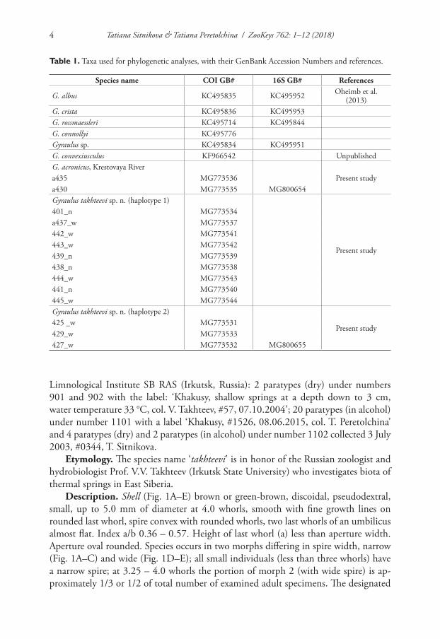
Table 1. Taxa used for phylogenetic analyses, with their GenBank Accession Numbers and references.
Species name COI GB# 16S GB# References
G. albus KC495835 KC495952 Oheimb et al. (2013)
G. crista KC495836 KC495953G. rossmaessleri KC495714 KC495844G. connollyi KC495776Gyraulus sp. KC495834 KC495951G. convexiusculus KF966542 UnpublishedG. acronicus, Krestovaya River
Present studya435 MG773536a430 MG773535 MG800654Gyraulus takhteevi sp. n. (haplotype 1)
Present study
401_n MG773534a437_w MG773537442_w MG773541443_w MG773542439_n MG773539438_n MG773538444_w MG773543441_n MG773540445_w MG773544Gyraulus takhteevi sp. n. (haplotype 2)
Present study425 _w MG773531429_w MG773533427_w MG773532 MG800655
Limnological Institute SB RAS (Irkutsk, Russia): 2 paratypes (dry) under numbers 901 and 902 with the label: ‘Khakusy, shallow springs at a depth down to 3 cm, water temperature 33 °C, col. V. Takhteev, #57, 07.10.2004’; 20 paratypes (in alcohol) under number 1101 with a label ‘Khakusy, #1526, 08.06.2015, col. T. Peretolchina’ and 4 paratypes (dry) and 2 paratypes (in alcohol) under number 1102 collected 3 July 2003, #0344, T. Sitnikova.
Etymology. The species name ‘takhteevi’ is in honor of the Russian zoologist and hydrobiologist Prof. V.V. Takhteev (Irkutsk State University) who investigates biota of thermal springs in East Siberia.
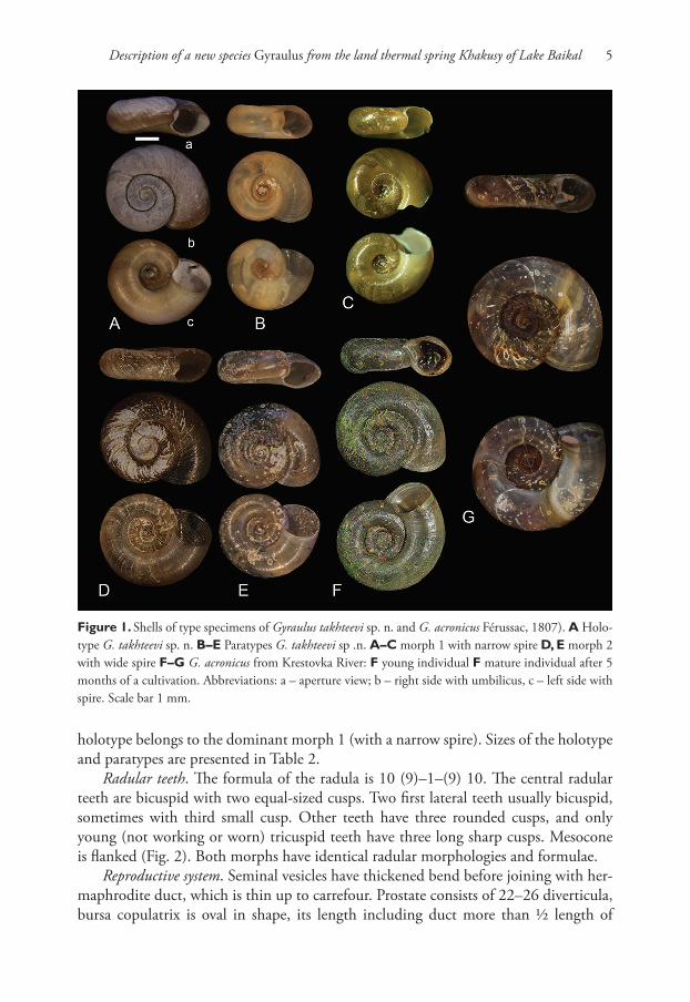
Description. Shell (Fig. 1A–E) brown or green-brown, discoidal, pseudodextral, small, up to 5.0 mm of diameter at 4.0 whorls, smooth with fine growth lines on rounded last whorl, spire convex with rounded whorls, two last whorls of an umbilicus almost flat. Index a/b 0.36 – 0.57. Height of last whorl (a) less than aperture width. Aperture oval rounded. Species occurs in two morphs differing in spire width, narrow (Fig. 1A–C) and wide (Fig. 1D–E); all small individuals (less than three whorls) have a narrow spire; at 3.25 – 4.0 whorls the portion of morph 2 (with wide spire) is ap-proximately 1/3 or 1/2 of total number of examined adult specimens. The designated
Description of a new species Gyraulus from the land thermal spring Khakusy of Lake Baikal 5
Figure 1. Shells of type specimens of Gyraulus takhteevi sp. n. and G. acronicus Férussac, 1807). A Holo-type G. takhteevi sp. n. B–E Paratypes G. takhteevi sp .n. A–C morph 1 with narrow spire D, E morph 2 with wide spire F–G G. acronicus from Krestovka River: F young individual F mature individual after 5 months of a cultivation. Abbreviations: a – aperture view; b – right side with umbilicus, c – left side with spire. Scale bar 1 mm.
holotype belongs to the dominant morph 1 (with a narrow spire). Sizes of the holotype and paratypes are presented in Table 2.
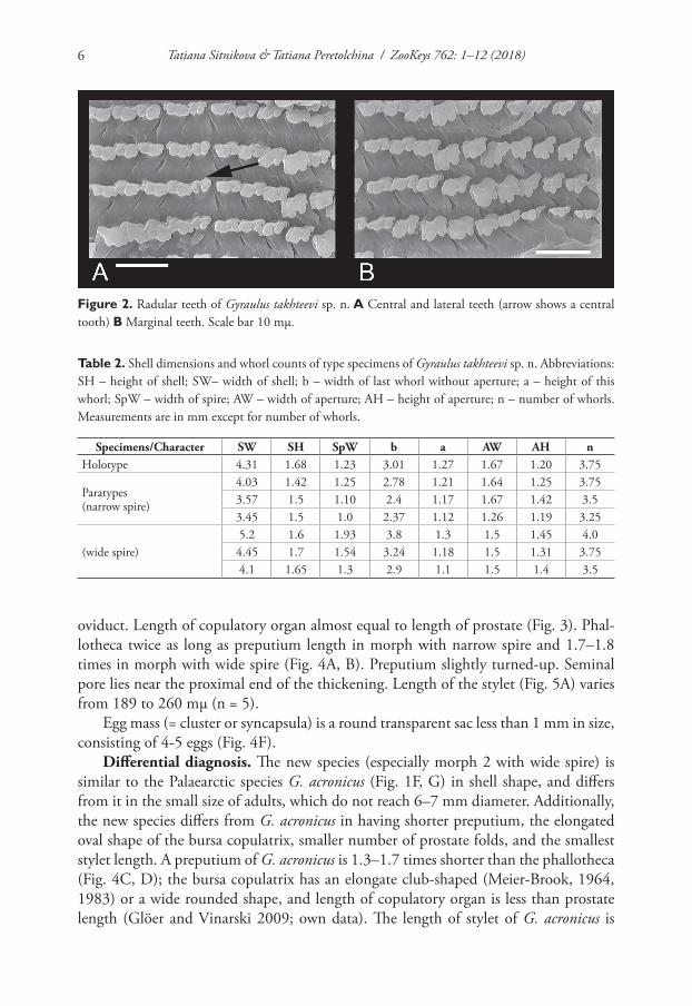
Radular teeth. The formula of the radula is 10 (9)–1–(9) 10. The central radular teeth are bicuspid with two equal-sized cusps. Two first lateral teeth usually bicuspid, sometimes with third small cusp. Other teeth have three rounded cusps, and only young (not working or worn) tricuspid teeth have three long sharp cusps. Mesocone is flanked (Fig. 2). Both morphs have identical radular morphologies and formulae.
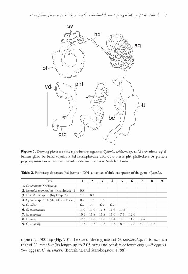
Reproductive system. Seminal vesicles have thickened bend before joining with her-maphrodite duct, which is thin up to carrefour. Prostate consists of 22–26 diverticula, bursa copulatrix is oval in shape, its length including duct more than ½ length of
Tatiana Sitnikova & Tatiana Peretolchina / ZooKeys 762: 1–12 (2018)6
Figure 2. Radular teeth of Gyraulus takhteevi sp. n. A Central and lateral teeth (arrow shows a central tooth) B Marginal teeth. Scale bar 10 mμ.
Table 2. Shell dimensions and whorl counts of type specimens of Gyraulus takhteevi sp. n. Abbreviations: SH – height of shell; SW– width of shell; b – width of last whorl without aperture; a – height of this whorl; SpW – width of spire; AW – width of aperture; AH – height of aperture; n – number of whorls. Measurements are in mm except for number of whorls.
Specimens/Character SW SH SpW b a AW AH nHolotype 4.31 1.68 1.23 3.01 1.27 1.67 1.20 3.75
Paratypes (narrow spire)
4.03 1.42 1.25 2.78 1.21 1.64 1.25 3.753.57 1.5 1.10 2.4 1.17 1.67 1.42 3.53.45 1.5 1.0 2.37 1.12 1.26 1.19 3.25
(wide spire)5.2 1.6 1.93 3.8 1.3 1.5 1.45 4.04.45 1.7 1.54 3.24 1.18 1.5 1.31 3.754.1 1.65 1.3 2.9 1.1 1.5 1.4 3.5
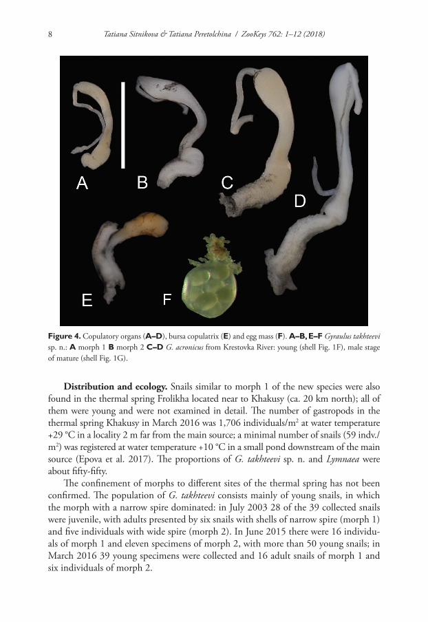
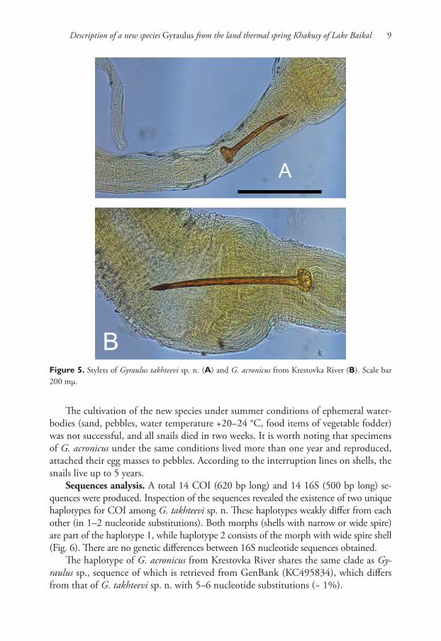
oviduct. Length of copulatory organ almost equal to length of prostate (Fig. 3). Phal-lotheca twice as long as preputium length in morph with narrow spire and 1.7–1.8 times in morph with wide spire (Fig. 4A, B). Preputium slightly turned-up. Seminal pore lies near the proximal end of the thickening. Length of the stylet (Fig. 5A) varies from 189 to 260 mμ (n = 5).
Egg mass (= cluster or syncapsula) is a round transparent sac less than 1 mm in size, consisting of 4-5 eggs (Fig. 4F).
Differential diagnosis. The new species (especially morph 2 with wide spire) is similar to the Palaearctic species G. acronicus (Fig. 1F, G) in shell shape, and differs from it in the small size of adults, which do not reach 6–7 mm diameter. Additionally, the new species differs from G. acronicus in having shorter preputium, the elongated oval shape of the bursa copulatrix, smaller number of prostate folds, and the smallest stylet length. A preputium of G. acronicus is 1.3–1.7 times shorter than the phallotheca (Fig. 4C, D); the bursa copulatrix has an elongate club-shaped (Meier-Brook, 1964, 1983) or a wide rounded shape, and length of copulatory organ is less than prostate length (Glöer and Vinarski 2009; own data). The length of stylet of G. acronicus is
Description of a new species Gyraulus from the land thermal spring Khakusy of Lake Baikal 7
Table 3. Pairwise p-distances (%) between COI sequences of different species of the genus Gyraulus.
Taxa 1 2 3 4 5 6 7 8 91. G. acronicus Krestovaya2. Gyraulus takhteevi sp. n.(haplotype 1) 0.83. G. takhteevi sp. n. (haplotype 2) 1.0 0.24. Gyraulus sp. KC495834 (Lake Baikal) 0.7 1.5 1.35. G. albus 6.9 7.0 6.9 6.96. G. rossmaessleri 11.0 11.0 10.8 10.6 11.37. G. convexius 10.5 10.8 10.8 10.6 7.4 12.68. G. crista 12.3 12.6 12.6 12.4 12.8 11.6 12.49. G. connollyi 11.5 11.5 11.3 11.5 8.8 12.6 9.0 14.7
Figure 3. Drawing pictures of the reproductive organs of Gyraulus takhteevi sp. n. Abbreviations: ag al-bumen gland bc bursa copulatrix hd hermaphrodite duct ot ovotestis pht phallotheca pr prostate prp preputium sv seminal vesicles vd vas deferens u uterus. Scale bar 1 mm.
more than 300 mμ (Fig. 5B). The size of the egg mass of G. takhteevi sp. n. is less than that of G. acronicus (its length up to 2.05 mm) and consists of fewer eggs (4–5 eggs vs. 5–7 eggs in G. acronicus) (Berezkina and Starobogatov, 1988).
Tatiana Sitnikova & Tatiana Peretolchina / ZooKeys 762: 1–12 (2018)8
Distribution and ecology. Snails similar to morph 1 of the new species were also found in the thermal spring Frolikha located near to Khakusy (ca. 20 km north); all of them were young and were not examined in detail. The number of gastropods in the thermal spring Khakusy in March 2016 was 1,706 individuals/m2 at water temperature +29 °C in a locality 2 m far from the main source; a minimal number of snails (59 indv./m2) was registered at water temperature +10 °C in a small pond downstream of the main source (Epova et al. 2017). The proportions of G. takhteevi sp. n. and Lymnaea were about fifty-fifty.
The confinement of morphs to different sites of the thermal spring has not been confirmed. The population of G. takhteevi consists mainly of young snails, in which the morph with a narrow spire dominated: in July 2003 28 of the 39 collected snails were juvenile, with adults presented by six snails with shells of narrow spire (morph 1) and five individuals with wide spire (morph 2). In June 2015 there were 16 individu-als of morph 1 and eleven specimens of morph 2, with more than 50 young snails; in March 2016 39 young specimens were collected and 16 adult snails of morph 1 and six individuals of morph 2.
Figure 4. Copulatory organs (A–D), bursa copulatrix (E) and egg mass (F). A–B, E–F Gyraulus takhteevi sp. n.: A morph 1 B morph 2 C–D G. acronicus from Krestovka River: young (shell Fig. 1F), male stage of mature (shell Fig. 1G).
Description of a new species Gyraulus from the land thermal spring Khakusy of Lake Baikal 9
The cultivation of the new species under summer conditions of ephemeral water-bodies (sand, pebbles, water temperature +20–24 °C, food items of vegetable fodder) was not successful, and all snails died in two weeks. It is worth noting that specimens of G. acronicus under the same conditions lived more than one year and reproduced, attached their egg masses to pebbles. According to the interruption lines on shells, the snails live up to 5 years.
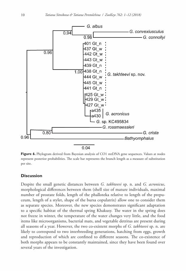
Sequences analysis. A total 14 COI (620 bp long) and 14 16S (500 bp long) se-quences were produced. Inspection of the sequences revealed the existence of two unique haplotypes for COI among G. takhteevi sp. n. These haplotypes weakly differ from each other (in 1–2 nucleotide substitutions). Both morphs (shells with narrow or wide spire) are part of the haplotype 1, while haplotype 2 consists of the morph with wide spire shell (Fig. 6). There are no genetic differences between 16S nucleotide sequences obtained.
The haplotype of G. acronicus from Krestovka River shares the same clade as Gy-raulus sp., sequence of which is retrieved from GenBank (KC495834), which differs from that of G. takhteevi sp. n. with 5–6 nucleotide substitutions (~ 1%).
Figure 5. Stylets of Gyraulus takhteevi sp. n. (A) and G. acronicus from Krestovka River (B). Scale bar 200 mμ.
Tatiana Sitnikova & Tatiana Peretolchina / ZooKeys 762: 1–12 (2018)10
Discussion
Despite the small genetic distances between G. takhteevi sp. n. and G. acronicus, morphological differences between them (shell size of mature individuals, maximal number of prostate folds, length of the phalloteka relative to length of the prepu-ceum, length of a stylet, shape of the bursa copulatrix) allow one to consider them as separate species. Moreover, the new species demonstrates significant adaptation to a specific habitat of the thermal spring Khakusy. The water in the spring does not freeze in winter, the temperature of the water changes very little, and the food items like microorganisms, bacterial mats, and vegetable detritus are present during all seasons of a year. However, the two co-existent morphs of G. takhteevi sp. n. are likely to correspond to two interbreeding generations, hatching from eggs, growth and reproduction of which are confined to different seasons. The co-existence of both morphs appears to be constantly maintained, since they have been found over several years of the investigation.
Figure 6. Phylogram derived from Bayesian analysis of CO1 mtDNA gene sequences. Values at nodes represent posterior probabilities. The scale bar represents the branch length as a measure of substitution per site.
Description of a new species Gyraulus from the land thermal spring Khakusy of Lake Baikal 11
The low level of genetic distances between G. takhteevi sp. n. and G. acronicus from the Krestovka River indicates that a recent divergence of the species happened after the glaciers covering large areas of the northeast coast of Baikal during last glacial period started to melt about 18 kyr BP (Osipov and Khlystov 2010). The adaptation to the thermal conditions of ‘a closed habitat’ formed by island isolation under selective pressures led to an interruption of gene exchange between G. takhteevi sp. n. and G. acronicus, resulting in differences in their morphological and physiological characteris-tics, and ecological preferences.
Acknowledgements
We are grateful to Mr. V.I. Egorov for assistance with the scanning electron microscope equipment (Electron Microscope Centre of Collective Instrumental Centre “Ultra mi-croanalysis” at Limnological Institute SB RAS). We are grateful to the reviewer and Dr E. Soldatenko for comments and advices that have improved the content of the article. The study was partly supported by the governmentally funded projects Nos. 0345-2016-0009 (AAAA-A16-116122110067-8) (morphological investigations), 0345-2016-0004 (AAAA-A16-116122110060-9) (molecular analysis), and Russian Foundation for Basic Research, project No 15-29-02515.
References
Aksenova O, Vinarski M, Bolotov I, Kondakov A, Bespalaya Yu, Tomilova A, Paltser I, Gofarov M (2017) Two Radix spp. (Gastropoda: Lymnaeidae) endemic to thermal springs around Lake Baikal represent ecotypes of the widespread Radix auricularia. Journal of zoological systematics and evolutionary research 55(4): 298–309. https://doi.org/10.1111/jzs.12174
Beriozkina GV, Starobogatov YaI (1988) reproductive ecology and egg clusters of freshwater Pulmonata. Proceedings of the Zoological Institute 174: 1–309. [In Russian]
Brown DS (2001) Freshwater snails of the genus Gyraulus (Planorbidae) in Australia: taxa of the main-land. Molluscan Research 21: 17–107. https://doi.org/10.1080/13235818.2001.10673736
Darriba D, Taboada GL, Doallo R, Posada D (2012) jModelTest 2: more models, new heu-ristics and parallel computing. Nature Methods 9: 772–772. http://dx.doi.org/10.1038/nmeth.2109
Folmer O, Black M, Hoeh W, Lutz R, Vrijenhoek R (1994) DNA Primers for amplification of mitochondrial cytochrome c oxidase subunit I from diverse metazoan invertebrates. Mole-cular Marine Biology and Biotechnology 3: 294–299.
Glöer P (2002) Süßwassergastropoden Nord- und Mitteleuropas. Die Tierwelt Deutschlands 73: 1–327.
Glöer P, Vinarski M (2009) Taxonomic notes on Euro-Siberian freshwater molluscs. 2. Rede-scription of Planorbis (Gyraulus) stroemi Westerlund, 1881 (Mollusca: Gastropoda: Planor-bidae). Journal of Conchology 39(6): 717–725.
Tatiana Sitnikova & Tatiana Peretolchina / ZooKeys 762: 1–12 (2018)12
Hall T (2011) BioEdit: an important software for molecular biology. Green Earth Research Foundation Bulletin of Biosciens 2(1): 60–61.
Epova IO, Takhteev VV, Sitnikova TYa, Khadeeva ER, Lopatovskaya OG (2016) Macrozoo-benthos of some thermal springs of Northern Baikal region in early springtime. Baikal zoological journal 2(19): 54–60 [In Russian].
Maniatis T, Fritsch EF, Sambrook J (1982) Molecular Cloning: A Laboratory Manual. Cold Spring Harbor, NY, 545 pp.
Meier-Brook C (1964) Gyraulus acronicus und G. rossmaessleri, ein anatomischer Vergleich (Pla-norbidae). Archiv für Molluskenkunde 93: 233–242.
Meier-Brook C (1983) Taxonomic studies on Gyraulus (Gastropoda: Planorbidae). Malacologia 24(1/2): 1–113.
Osipov EY, Khlystov OM (2010) Glaciers and meltwater flux to Lake Baikal during the Last Glacial Maximum. Paleogeography, Paleoclimatology, Palaeoecology 294: 4–15. https://doi.org/10.1016/j.palaeo.2010.01.031
Palumbi SR (1996) Nucleic acids II: the polymerase chain reaction. In: Hillis DM, Moritz C, Mable BK (Eds) Molecular Systematics, 2nd ed., Sinauer Associates Inc., Sunderland, Mas-sachusetts, 205–247.
Ronquist F, Huelsenbeck JP (2003) MrBayes 3: Bayesian phylogenetic inference under mixed models. Bioinformatics 19(12): 1572–1574. https://doi.org/10.1093/bioinformatics/btg180
Sitnikova TYa, Takhteev VV (2006) Gastropods of thermal springs of Pribaikalie. Hydrobiol-ogy of waterbodies of Pribaikalie. Proceedings of Biological Soil Institute of Irkutsk State University 6: 137–150. [In Russian]
Sokolov EP (2000) An improved method for DNA isolation from mucopolysaccharide-rich molluscan tissues. Journal of Molluscan Studies 66: 573–575. https://doi.org/10.1093/mollus/66.4.573
Tamura K, Stetecher G, Peterson D, Filipski A, Kumar S (2013) MEGA6: Molecular evolu-tionary genetics analysis, Version 6.0. Molecular Biology and Evolution 30: 2725–2729. https://doi.org/10.1093/molbev/mst197
Thompson JD, Gibson T, Higgins DG (2002) Multiple sequence alignment using ClustalW and ClustalX. Current protocols in bioinformatics, Chapter2: Unit 2.3. https://doi.org/10.1002/0471250953.bi0203s00
Volkov S (2010) Along Baikal. ATS, Moscow, 568 pp.
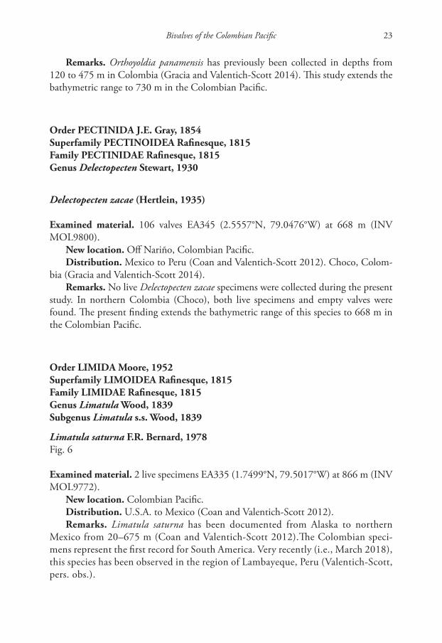
Bivalves of the Colombian Pacific 13
A new species of Malletia (Bivalvia, Malletiidae) and new records of deep-water bivalves
from Pacific Southern Colombia
Nancy Yolimar Suárez-Mozo1,3, Adriana Gracia2, Paul Valentich-Scott4
1 Posgrado en Ciencias del Mar y Limnología, Universidad Nacional Autónoma de México – UNAM, Maza-tlán, Sinaloa, México 2 Programa de Biología, Universidad del Atlántico, Km 7 Antigua Vía Puerto Colombia, Atlántico, Colombia 3 Instituto de Investigaciones Marinas y Costeras – Invemar, Santa Marta, Colombia 4 Department of Invertebrate Zoology, Santa Barbara Museum of Natural History, 2559 Puesta del Sol, Santa Barbara, CA 93105 USA
Corresponding author: Nancy Yolimar Suárez-Mozo ([email protected])
Academic editor: R.C. Willan | Received 16 August 2017 | Accepted 10 April 2018 | Published 30 May 2018
http://zoobank.org/440ED53B-6734-4A55-85AF-9E4CF2ADD89F
Citation: Suárez-Mozo NY, Gracia A, Valentich-Scott P (2018) A new species of Malletia (Bivalvia, Malletiidae) and new records of deep-water bivalves from Pacific Southern Colombia. ZooKeys 762: 13–31. https://doi.org/10.3897/zookeys.762.20335
AbstractIn order to enhance the understanding of Pacific Colombia’s deep-water marine fauna, a benthic research cruise (2012 TUM Offshore 6 and 7) was conducted off the coast of the Department of Nariño, in south-ern Colombia. Biological, oceanographic and sediment samples from the continental shelf and slope were collected at depths between 350 and 941 m. A new species of Malletia obtained on that cruise is described and compared with other species from the eastern Pacific. Sixteen species of bivalve mollusks (belonging to 12 families and 15 genera) were identified. Five of them were the first records for Pacific Colombia (Jupi-teria lobula, Limatula saturna, Lucinoma heroica, Cuspidaria panamensis, and Dallicordia alaskana). Four of them had geographic distributions that now extend to Tumaco at the southern end of Nariño.
KeywordsBivalvia, benthos, Colombia, deep-water, Malletia, Malletiidae, Tumaco
ZooKeys 762: 13–31 (2018)
doi: 10.3897/zookeys.762.20335
http://zookeys.pensoft.net
Copyright Nancy Yolimar Suárez-Mozo et al. This is an open access article distributed under the terms of the Creative Commons Attribution License (CC BY 4.0), which permits unrestricted use, distribution, and reproduction in any medium, provided the original author and source are credited.
RESEARCH ARTICLE
Launched to accelerate biodiversity research
A peer-reviewed open-access journal
Nancy Yolimar Suárez-Mozo et al. / ZooKeys 762: 13–31 (2018)14
Introduction
Throughout the past decade, the search for hydrocarbon and natural gas reserves in Colombia (Pacific and Caribbean coast) has sparked an interest in the country’s remote deep-sea regions. This has resulted in intensified deep-sea baseline studies, primarily along the continental shelves and slopes. Nevertheless, deep-sea studies face logistical and cost limitations, including the availability of research vessels and proper equipment for collecting samples.
Despite the increase in knowledge during recent years, the presently known range of many invertebrates groups inhabiting soft sediments, including mollusks, is still fair-ly fragmentary in remotes parts of the Colombian Pacific. There is a lack of published data on the biology, functional morphology, ecology, development and dispersal mech-anisms for these invertebrates, as well as a lack of baseline faunal inventories. Thus, the true biodiversity of the Pacific Colombian deep-sea must be vastly underestimated.
As a result of recent Colombian expeditions, a rich benthic fauna inhabiting of the deep-seas of Pacific Southern Colombia has been discovered, but few species of mollusks have been described when compared with the mollusks from the coasts of the Colombian Atlantic (e.g., Ardila-Espitia and Diaz 2002, Simone and Gracia 2006, Gracia and Ardila-Espitia 2009).
In the context of faunal inventories, the tropical west coast of America is well doc-umented, with 890 species of bivalves presently recorded (Coan and Valentich-Scott 2012). For northwestern South America to Peru, a basic knowledge of deep-sea bivalve mollusks has been covered by a few recent publications (e.g., Gracia and Valentich-Scott 2014; Paredes et al. 2016). The investigations in Pacific Colombian waters have hitherto focused mainly on the coastal zones (e.g., Cantera et al. 1979, Cosel 1984, Díaz et al. 1997, Cantera 2010, López de Mesa and Cantera 2015) rather than zones farther off-shore (e.g., Hertlein and Strong 1955). Gracia and Valentich-Scott (2014) documented the bivalves off the Department of Choco (Colombian North Pacific) where more than 38 species of bivalves were found, 34 of which were new records for the country.
The current work presents a systematic and annotated list of bivalve species collected in the southern Colombian Pacific region. Each entry includes the species’ geographic and ba-thymetric distribution, plus additional remarks and observations. From the above, several species stand out as being first records for the country. We are also including the description of a new species uncovered in this survey. Our records represent a significant expansion in the knowledge of the Pacific Colombian bivalve fauna, but much more sampling and analysis is needed when one takes into account the large geographic extent of this region.
Materials and methods
Study area
The present study was carried out in the tropical eastern Pacific Ocean (Fig. 1). The study area (TUM Offshore Blocks 6 and 7) covered 7,308 km2 and extended from Sanquianga
Bivalves of the Colombian Pacific 15
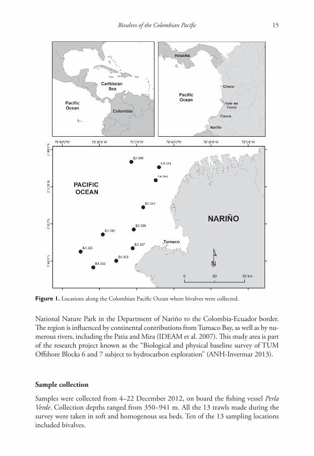
National Nature Park in the Department of Nariño to the Colombia-Ecuador border. The region is influenced by continental contributions from Tumaco Bay, as well as by nu-merous rivers, including the Patia and Mira (IDEAM et al. 2007). This study area is part of the research project known as the “Biological and physical baseline survey of TUM Offshore Blocks 6 and 7 subject to hydrocarbon exploration” (ANH-Invermar 2013).
Sample collection
Samples were collected from 4–22 December 2012, on board the fishing vessel Perla Verde. Collection depths ranged from 350–941 m. All the 13 trawls made during the survey were taken in soft and homogenous sea beds. Ten of the 13 sampling locations included bivalves.
Figure 1. Locations along the Colombian Pacific Ocean where bivalves were collected.
Nancy Yolimar Suárez-Mozo et al. / ZooKeys 762: 13–31 (2018)16
Each sample was collected with a benthic semi-balloon trawl net (9 × 1 m) for 10 minutes at a speed of 3 knots. Because the exact time at which the net opened was un-known, sampling was semi-quantitative. We acknowledge that this sampling technique could have missed small and microscopic species as would be taken by epibenthic sleds, but the equipment needed for this method was not available to us. Collected material was coarsely sorted on deck and later identified to lower levels at the Museo de Historia Natural Marina de Colombia (MHNMC) which is part of Instituto de Investigaciones Marinas y Costeras (INVEMAR). The empty valves were air-dried, while the soft-bodied organisms were preserved in 70% ethanol.
Specimen identification was based upon shell characters. Museum materials, bib-liographic references and bivalve taxonomic experts were consulted to confirm the results (e.g., Dall 1896, 1908; Keen 1971; Coan and Valentich-Scott 2012). The iden-tified material included many complete living organisms as well as empty shells of bivalves. The systematic order of this list corresponds to that proposed by Coan and Valentich-Scott (2012). Specimens from this study, including other mollusks not ana-lyzed in this work (e.g., gastropods, chitons, and cephalopods), now reside at the MH-NMC’s mollusk collection in Santa Marta, Colombia.
Oceanographic data were collected with an Idronaut CTDO marine profiler (yielding data for conductivity, temperature, depth, and oxygen concentration) at sites S333 and S334, both of which contained bivalves (Table 1). Sediment core sets were collected five sites (S331, S333, S334, S341, and S345) with a Gomex II Box corer that had a 32 liter storage capacity. Sediment grain analysis revealed a predominance of silt (Table 1). Grain size classification was conducted according to Folk (1974). All samples were classified as silts; 57% of samples were purely silts, while the remaining 43% also contained sand and gravel fractions (INVEMAR-ANH 2013).
Abbreviations
EA Trawl station; S sediment stationMHNMC (Spanish acronym) Museo de Historia Natural Marina de ColombiaTUM OFF Tumaco-Offshore
Table 1. Size distribution of analyzed sediment samples according to INVEMAR-ANH (2013).
Station Depth (m) % Gravel % Sand % Silt
331 320 0.1 23.2 76.7333 833 0.0 1.0 99.0334 864 0.0 0.7 99.3341 894 0.2 1.1 98.7345 570 0.0 49.7 50.3
Bivalves of the Colombian Pacific 17
Results
A total of 324 bivalve specimens was collected, including 247 empty or disjointed valves and 77 live-collected organisms. The specimens were sorted into 16 species, 15 genera, and 12 families; five species were new observations in the Colombian Pacific. The known geographic range of several species has now been expanded to the Depart-ment of Nariño.
Below is included a listing of the species collected, station data, live-dead status for each specimen, remarks on new verified localities, previously reported distributions for the species, plus general remarks. We have also included an illustration for all newly documented species in Colombia i.e., those other than Nucula iphigenia, Orthoyoldia panamensis and Delectopecten zacae which were previously reported for the Pacific of Colombia by Gracia and Valentich-Scott (2014).
Systematics
Class BIVALVIA Linnaeus, 1758Subclass PROTOBRANCHIA Pelseneer, 1889Order NUCULIDA Dall, 1889Superfamily NUCULOIDEA J.E. Gray, 1824Family NUCULIDAE J.E. Gray, 1824Genus Ennucula Iredale, 1931
Ennucula panamina (Dall, 1908)Fig. 2
Examined material. 1 valve plus 1 live specimen EA 336 (1.9045°N, 79.3030°W) at 612 m (INV MOL9797, INV MOL9796), 1 live specimen EA344 (2.3905°N, 78.8288°W) at 656 m (INV MOL9796), plus 1 live specimen EA 335 (1.7499°N, 79.50177°W) at 866 m (INV MOL9799).
New location. Off Nariño, Colombian Pacific.Distribution. Panama to Peru (Coan and Valentich-Scott 2012).Remarks. New species record for the Colombian Pacific.
Genus Nucula Lamarck, 1799Subgenus Lamellinucula Schenck, 1944
Nucula (Lamellinucula) iphigenia Dall, 1896
Examined material. 1 valve plus 2 live specimens EA331 (2.5078°N, 78.7993°W) at 350 m (INV MOL9794, INV MOL9795).
Nancy Yolimar Suárez-Mozo et al. / ZooKeys 762: 13–31 (2018)18
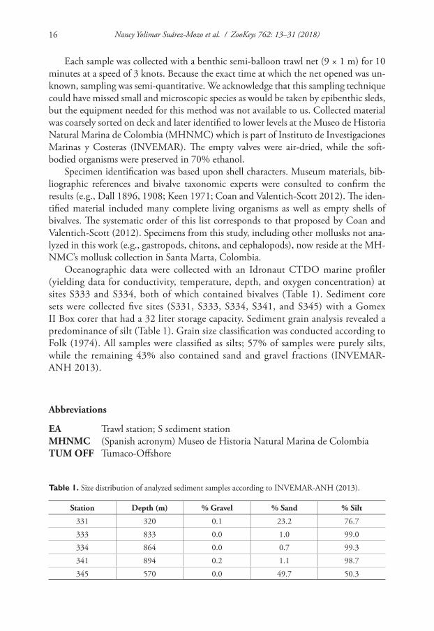
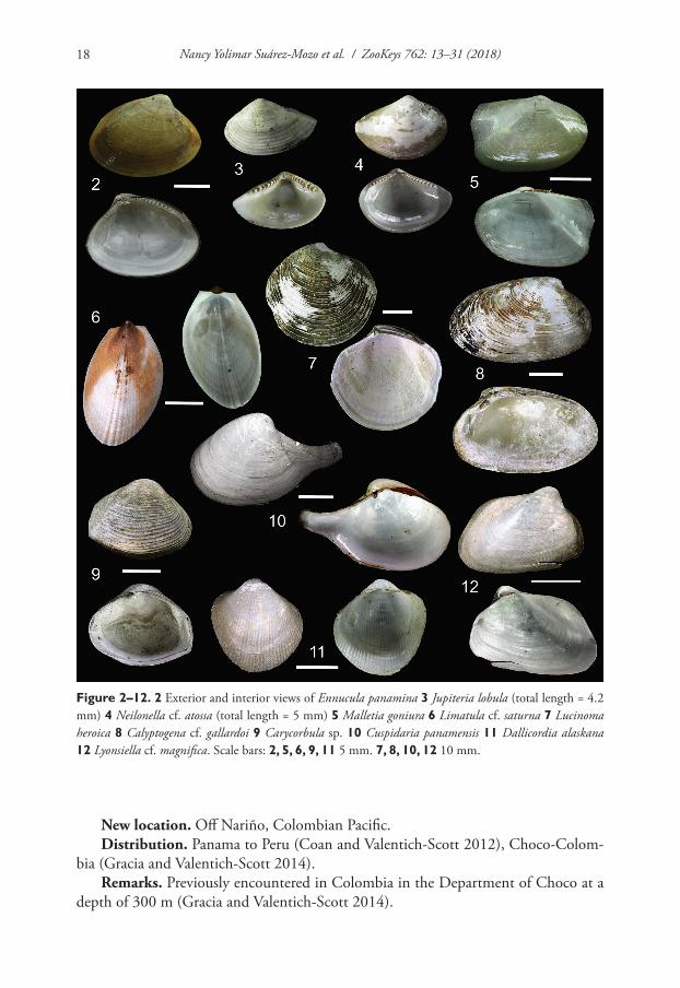
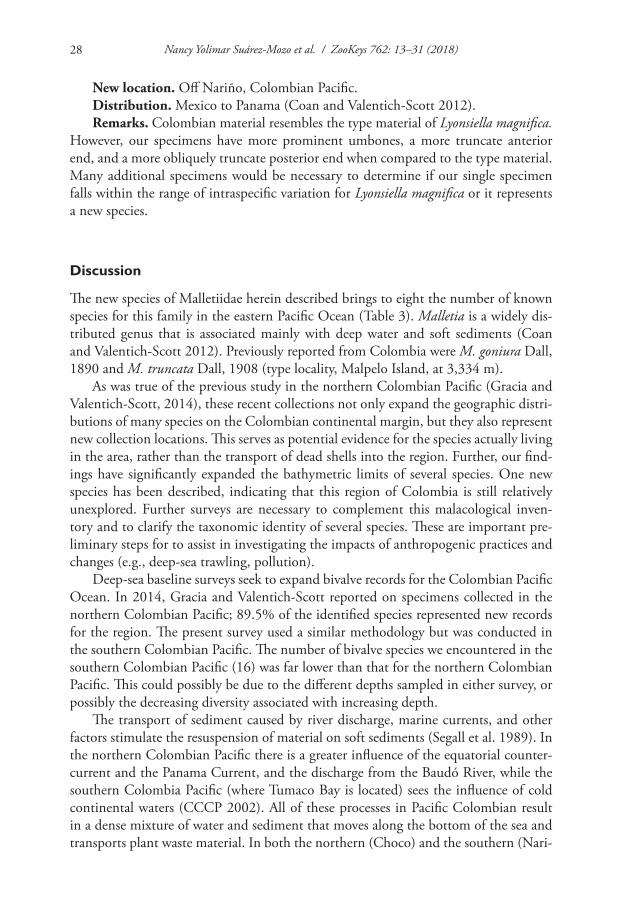
Figure 2–12. 2 Exterior and interior views of Ennucula panamina 3 Jupiteria lobula (total length = 4.2 mm) 4 Neilonella cf. atossa (total length = 5 mm) 5 Malletia goniura 6 Limatula cf. saturna 7 Lucinoma heroica 8 Calyptogena cf. gallardoi 9 Carycorbula sp. 10 Cuspidaria panamensis 11 Dallicordia alaskana 12 Lyonsiella cf. magnifica. Scale bars: 2, 5, 6, 9, 11 5 mm. 7, 8, 10, 12 10 mm.
New location. Off Nariño, Colombian Pacific.Distribution. Panama to Peru (Coan and Valentich-Scott 2012), Choco-Colom-
bia (Gracia and Valentich-Scott 2014).Remarks. Previously encountered in Colombia in the Department of Choco at a
depth of 300 m (Gracia and Valentich-Scott 2014).

Bivalves of the Colombian Pacific 19
Order NUCULANOIDA D.C. Carter & M.R. Campbell, 2000Superfamily NUCULANOIDEA H. & A. Adams, 1858Family NUCULANIDAE H & A. Adams, 1858Genus Jupiteria Bellardi, 1875
Jupiteria lobula (Dall, 1890)Fig. 3
Examined material. 2 valves EA337 (1.7811°N, 79.0351°W) at 530 m (INV MOL9791), 2 valves EA331 (2.5078°N, 78.7993°W) at 350 m (INV MOL9792).
New location. Colombian Pacific.Distribution. Mexico to El Salvador (Coan and Valentich-Scott 2012).Remarks. These records represent a new southern limit for this species. All the
specimens were small (approx. 4 mm), but they were nearly identical to small speci-mens of Jupiteria lobula from Mexico and also the type specimens. The presence of dead valves at different stations and the distance from previous records suggest that this species is living in Colombia.
Family NEILONELLIDAE Schileyko, 1989Genus Neilonella Dall, 1881
Neilonella cf. atossa (Dall, 1908)Fig. 4
Examined Material. 2 valves EA337 (1.7811°N, 79.0351°W) at 530 m (INV MOL9793).New location. Off Nariño, Colombian Pacific.Remarks. The identity of this species cannot be confirmed without a detailed com-
parative examination of additional material. It is potentially a new species.
Family MALLETIIDAE H. & A. Adams, 1858Genus Malletia des Moulins, 1832
Malletia goniura Dall, 1890Fig. 5
Examined material. 8 valves EA341 (2.1484°N, 78.9409°W) at 934 m (INV MOL9774), 7 live specimens EA341 at 934 m (INV MOL9775), 3 valves EA335 (1.7499°N, 79.5017°W) at 855 m (INV MOL9776), 6 live specimens EA335 at 866 m (INV MOL9777), 2 valves EA333 (1.6087°N, 79.3883°W) at 836 m (INV M9778), 4 live specimens EA333 at 836 m (INV MOL9779), 3 valves EA338 (1.9490°N, 79.0257°W) at 941 m (INV MOL9780).
Nancy Yolimar Suárez-Mozo et al. / ZooKeys 762: 13–31 (2018)20
New location. Off Nariño, Colombian Pacific.Distribution. Panama to Peru (Coan and Valentich-Scott 2012).Remarks. These specimens represent the shallowest bathymetric records so far
for Malletia goniura (836–941 m). It has previously been collected in deeper waters (1,500–3,300 m depth) (Coan and Valentich-Scott 2012).
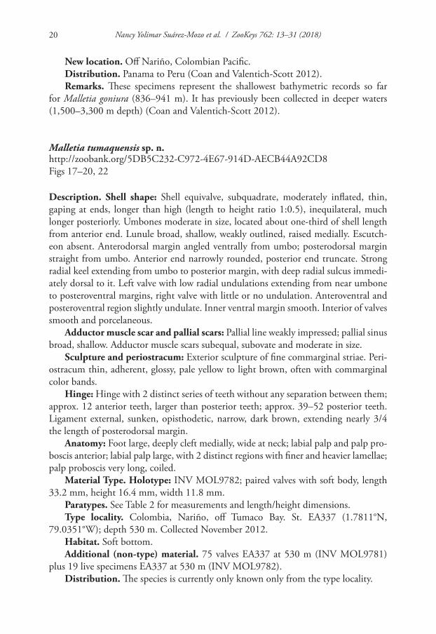
Malletia tumaquensis sp. n.http://zoobank.org/5DB5C232-C972-4E67-914D-AECB44A92CD8Figs 17–20, 22
Description. Shell shape: Shell equivalve, subquadrate, moderately inflated, thin, gaping at ends, longer than high (length to height ratio 1:0.5), inequilateral, much longer posteriorly. Umbones moderate in size, located about one-third of shell length from anterior end. Lunule broad, shallow, weakly outlined, raised medially. Escutch-eon absent. Anterodorsal margin angled ventrally from umbo; posterodorsal margin straight from umbo. Anterior end narrowly rounded, posterior end truncate. Strong radial keel extending from umbo to posterior margin, with deep radial sulcus immedi-ately dorsal to it. Left valve with low radial undulations extending from near umbone to posteroventral margins, right valve with little or no undulation. Anteroventral and posteroventral region slightly undulate. Inner ventral margin smooth. Interior of valves smooth and porcelaneous.
Adductor muscle scar and pallial scars: Pallial line weakly impressed; pallial sinus broad, shallow. Adductor muscle scars subequal, subovate and moderate in size.
Sculpture and periostracum: Exterior sculpture of fine commarginal striae. Peri-ostracum thin, adherent, glossy, pale yellow to light brown, often with commarginal color bands.
Hinge: Hinge with 2 distinct series of teeth without any separation between them; approx. 12 anterior teeth, larger than posterior teeth; approx. 39–52 posterior teeth. Ligament external, sunken, opisthodetic, narrow, dark brown, extending nearly 3/4 the length of posterodorsal margin.
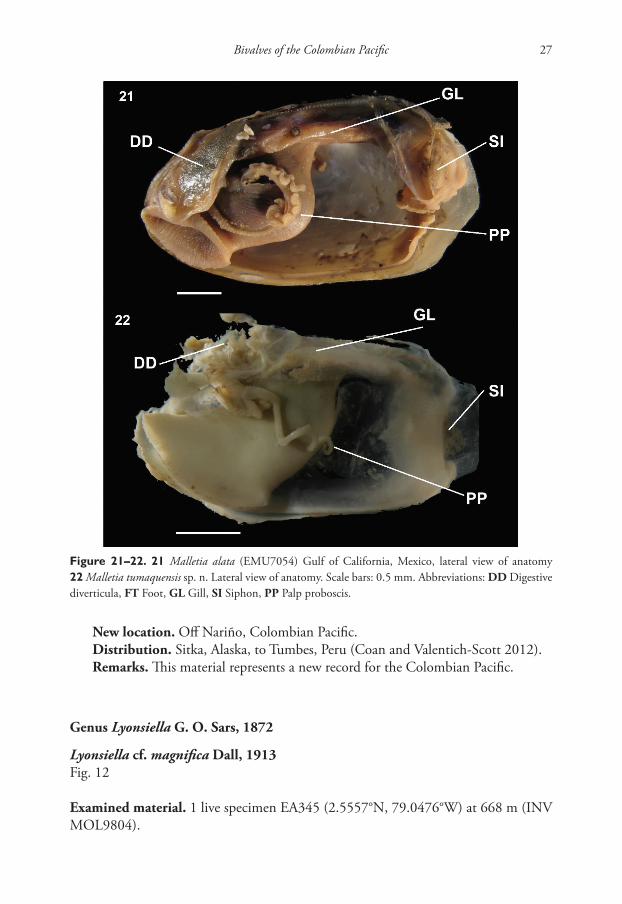
Anatomy: Foot large, deeply cleft medially, wide at neck; labial palp and palp pro-boscis anterior; labial palp large, with 2 distinct regions with finer and heavier lamellae; palp proboscis very long, coiled.
Material Type. Holotype: INV MOL9782; paired valves with soft body, length 33.2 mm, height 16.4 mm, width 11.8 mm.
Paratypes. See Table 2 for measurements and length/height dimensions.Type locality. Colombia, Nariño, off Tumaco Bay. St. EA337 (1.7811°N,
79.0351°W); depth 530 m. Collected November 2012.Habitat. Soft bottom.Additional (non-type) material. 75 valves EA337 at 530 m (INV MOL9781)
plus 19 live specimens EA337 at 530 m (INV MOL9782).Distribution. The species is currently only known only from the type locality.
Bivalves of the Colombian Pacific 21
Etymology. This species is named in honor of the municipality of Tumaco, Nari-ño, where this study was conducted.
Differential diagnosis. Malletia tumaquensis sp. n. is similar in shape to M. alata Bernard, 1989. However, consistent differences exist in conchological features (i.e., M. tumaquensis is more elongate, while M. alata has an alate process) and anatomical characteristics (i.e., very long, thin palp proboscis in M. tumaquensis) makes it a read-ily distinguishable new species. Ecologically, M. tumaquensis has a shallower depth distribution (530 m) than that of M. alata (740 m, Coan and Valentich-Scott 2012). Table 3 summarizes the shell characteristics of all the Malletia species recorded in the eastern Pacific Ocean.
Remarks. Members of the family Malletiidae occur throughout the Pacific and Atlantic Oceans with most records from deep-water (Coan and Valentich-Scott 2012, Kamenev 2015). Malletia tumaquensis is distinguished from the seven other species occurring in tropical west America by its more subquadrate and longer shell. Including our record, this represents the third species of the genus reported for the Colombian Pacific (i.e., M. tumaquensis, M. truncata and M. goniura).
Family YOLDIIDAE Dall, 1908Subfamily YOLDIINAE Dall, 1908Genus Orthoyoldia Verrill & Bush, 1897
Orthoyoldia panamensis (Dall, 1908)
Examined material. 10 valves EA344 (2.3905°N, 78.8288°W) at 656 m (INV MOL9812), 6 live specimens EA344 at 656 m (INV MOL9813), 5 valves EA337 (1.7811°N, 79.035139° W) at 530 m (INV MOL9814), 4 valves EA337 at 530 m (INV MOL9815), 4 live specimens EA332 (1.6677°N, 79.1826°W) at 730 m (INV
Table 2. Measurements of type specimens of Malletia tumaquensis sp. n.
Specimen Length (mm)
Height (mm)
Width (mm)
Length/height
Holotype INV MOL9782 33.2 16.4 11.8 2.0Paratype 1 INV MOL1161 30.5 15.7 11.4 1.9Paratype 1 INV MOL1161 32.7 16.6 11.1 1.9Paratype 1 INV MOL1161 30.4 15.1 10.3 2.0Paratype 2 INV MOL1162 28.7 14.2 9.7 2.0Paratype 2 INV MOL1162 28.4 14.7 9.8 1.9Paratype 2 INV MOL1162 26.5 14.3 9.0 1.8Paratype 3 INV MOL1163 26.1 13.4 9.0 1.9Paratype 3 INV MOL1163 24.9 13.1 9.0 1.9Paratype 3 INV MOL1163 27.2 13.3 9.6 2.0
Nancy Yolimar Suárez-Mozo et al. / ZooKeys 762: 13–31 (2018)22
MOL9816), 2 live specimens EA345 (2.5557°N, 79.0476°W) at 668 m (INV MOL9817).
New location. Off Nariño, Colombian Pacific.Distribution. Mexico to Peru (Coan and Valentich-Scott 2012), Choco-Colom-
bia (Gracia and Valentich-Scott 2014).
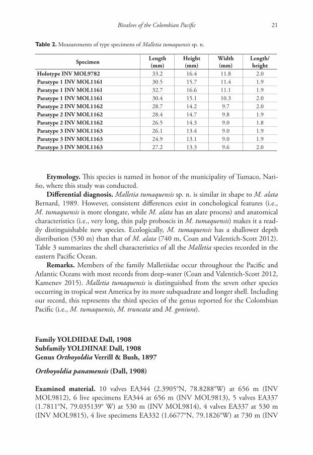
Table 3. Summary of shell characters of Malletia species from the Pacific Ocean (after Coan and Valen-tich-Scott 2012).
Species Shape Type locality
Reported depth
range (m)
Maximum Length (mm)
Posterior end Hinge
Malletia alata F. R. Bernard, 1989
San Diego Trough,
California, USA
1200 30
Straight, forming
alate process
About 11–13 anterior teeth;
about 45 posterior teeth
Malletia arciformis Dall, 1908
Off Acapulco, Guerrero, Mexico
902 11Broadly flared,
rounded
10–13 anterior teeth; 13–17
posterior teeth
Malletia benthima Dall, 1908
Off Acapulco, Guerrero, Mexico
902 10Produced, broadly rounded
12–13 anterior teeth; 13–17
posterior teeth
Malletia faba Dall, 1897
Off Queen Charlotte Islands, British
Columbia, Canada
200–1600 10 Broadly rounded
About 9 anterior teeth;
about 32 posterior teeth
Malletia goniura Dall, 1890
Gulf of Panama, Panama
1500–3300 13
Flaring dorsally, truncate
14–19 anterior teeth; 27–30
posterior teeth
Malletia peruviana Dall, 1908
Off Punta Aguja,
Piura, Peru1900 28 Broadly
rounded
10–11 anterior teeth; 33–36
posterior teeth
Malletia truncata Dall, 1908
Cascadia Plain,
Oregon, USA
2700–4134 30
Flaring, com-
pressed, truncate
About 18 anterior teeth;
about 30 posterior teeth
Malletia tumaquensis sp. n.
Off Tumaco Bay, Nariño, Colombia
530 33 Truncate12 anterior
teeth; 39–52 posterior teeth
Bivalves of the Colombian Pacific 23
Remarks. Orthoyoldia panamensis has previously been collected in depths from 120 to 475 m in Colombia (Gracia and Valentich-Scott 2014). This study extends the bathymetric range to 730 m in the Colombian Pacific.
Order PECTINIDA J.E. Gray, 1854Superfamily PECTINOIDEA Rafinesque, 1815Family PECTINIDAE Rafinesque, 1815Genus Delectopecten Stewart, 1930
Delectopecten zacae (Hertlein, 1935)
Examined material. 106 valves EA345 (2.5557°N, 79.0476°W) at 668 m (INV MOL9800).
New location. Off Nariño, Colombian Pacific.Distribution. Mexico to Peru (Coan and Valentich-Scott 2012). Choco, Colom-
bia (Gracia and Valentich-Scott 2014).Remarks. No live Delectopecten zacae specimens were collected during the present
study. In northern Colombia (Choco), both live specimens and empty valves were found. The present finding extends the bathymetric range of this species to 668 m in the Colombian Pacific.
Order LIMIDA Moore, 1952Superfamily LIMOIDEA Rafinesque, 1815Family LIMIDAE Rafinesque, 1815Genus Limatula Wood, 1839Subgenus Limatula s.s. Wood, 1839
Limatula saturna F.R. Bernard, 1978Fig. 6
Examined material. 2 live specimens EA335 (1.7499°N, 79.5017°W) at 866 m (INV MOL9772).
New location. Colombian Pacific.Distribution. U.S.A. to Mexico (Coan and Valentich-Scott 2012).Remarks. Limatula saturna has been documented from Alaska to northern
Mexico from 20–675 m (Coan and Valentich-Scott 2012).The Colombian speci-mens represent the first record for South America. Very recently (i.e., March 2018), this species has been observed in the region of Lambayeque, Peru (Valentich-Scott, pers. obs.).
Nancy Yolimar Suárez-Mozo et al. / ZooKeys 762: 13–31 (2018)24
Superorder HETEROCONCHIA J.E. Gray, 1854Clade HETERODONTA Neumayr, 1884Order LUCINIDA J.E. Gray, 1854Superfamily LUCINOIDEA Fleming, 1828Family LUCINIDAE Fleming, 1828Genus Lucinoma Dall, 1901
Lucinoma heroica (Dall, 1901)Fig. 7
Examined material. 3 valves EA345 (2.5557°N, 79.0476°W) at 668 m (INV MOL9773).
New location. Colombian Pacific.Distribution. Mexico to Peru (Coan and Valentich-Scott 2012).Remarks. Lucinoma heroica has previously been found in depths greater than
1,838 m (Coan and Valentich-Scott 2012). At 668 m, the Colombian specimens are the shallowest record for the species.
Order VENERIDA J.E. Gray, 1854Superfamily GLOSSOIDEA J.E Gray, 1847Family VESICOMYIDAE Dall & Simpson, 1901Genus Calyptogena Dall, 1891
Calyptogena cf. gallardoi Sellanes & Krylova, 2005Fig. 8
Examined material. 1 valve EA345 (2.5557°N, 79.0476°W) at 668 m (INV MOL9805).
New location. Off Nariño, Colombian Pacific.Distribution. South-central Chile, off Bahía de Concepción (Sellanes and Krylova
2005).Remarks. The single valve collected is insufficient to allow a definitive identifica-
tion to species. The shape and dentition place it closest to Calyptogena gallardoi.
Genus Pliocardia Woodring, 1925
“Pliocardia” cf. donacia (Dall, 1908)
Examined material. 1 valve EA337 (1.7811°N, 79.0351°W) at 530 m (INV MOL9768), 1 valve EA336 (1.9045°N, 79.3030°W) at 612 m (INV MOL9770), 1 live specimen EA344 (2.3905°N, 78.8288°W) at 656 m (INV MOL9771).
Bivalves of the Colombian Pacific 25
New location. Off Nariño, Colombian Pacific.Distribution. Panama (Coan and Valentich-Scott 2012), Choco, Colombia (Gra-
cia and Valentich-Scott 2014).Remarks. Prior to this study, dead shells of Pliocardia donacia were identified in
Pacific Colombia at depths between 272 and 295 m (Gracia and Valentich-Scott 2014). The present collection in southern Colombia yielded one live specimen and two empty valves, suggesting that the species inhabits both the northern and southern Colombian Pacific. Further, the bathymetric limit of the species is extended to 656 m in the Colombian Pacific. Many generic uncertainties exist within the family Vesicomyidae. Thus, we follow Coan and Valentich-Scott (2012) in their tentative placement of P. donacia within the genus Pliocardia.
Order MYOIDA Goldfuss, 1820Suborder MYINA Goldfuss, 1820Superfamily MYOIDEA Lamarck, 1809Family CORBULIDAE Lamarck, 1818Genus Caryocorbula J. Gardner, 1926
Carycorbula sp.Fig. 9
Examined material. 1 valve EA331 (2.5078°N, 78.7993°W) at 350 m (INV MOL9763).
New location. Off Nariño, Colombian Pacific.Remarks. This single valve from station EA331 is similar to several Panamic and
Peru-Chile Province species of Carycorbula, but it is insufficient to allow a definitive identification to species.
Suborder SEPTIBRANCHIA Pelseneer, 1988Superfamily CUSPIDARIOIDEA Dall, 1886Family CUSPIDARIIDAE Dall, 1886Genus Cuspidaria Nardo, 1840
Cuspidaria panamensis Dall, 1908Fig. 10
Examined material. 1 valve EA332 (1.6677°N, 79.1826°W) at 730 m (INV MOL9764), 7 valves EA345 (2.5557°N, 79.0476°W) at 668 m (INV MOL9765), 1 live specimen EA345 at 668 m (INV MOL9766), 4 live specimens A336 (1.9045°N, 79.3030°W) at 612 m (INV MOL9767).
New location. Off Nariño, Colombian Pacific.
Nancy Yolimar Suárez-Mozo et al. / ZooKeys 762: 13–31 (2018)26
Distribution. Panama (Coan & Valentich-Scott 2012).Remarks. Cuspidaria panamensis was previously known only been known from
the type locality in the Gulf of Panama (Coan and Valentich-Scott 2012). Our records extend the distribution over 600 km to the south. Coan and Valentich-Scott (2012) indicate a maximum size of 41 mm for Cuspidaria panamensis. However, our material from station EA345 increases the maximum length to 44.2 mm.
Superfamily VERTICORDIOIDEA Stoliczka, 1870Family VERTICORDIIDAE Stoliczka, 1870Subfamily LYONSIELLINAE Dall, 1895Genus Dallicordia Scarlato & Starobogatov, 1983
Dallicordia alaskana (Dall, 1895)Fig. 11
Examined material. 10 valves EA337 (1.7811°N, 79.0351°W) at 530 m (INV MOL9802), 14 live specimens EA337 at 530 m (INV MOL9803).
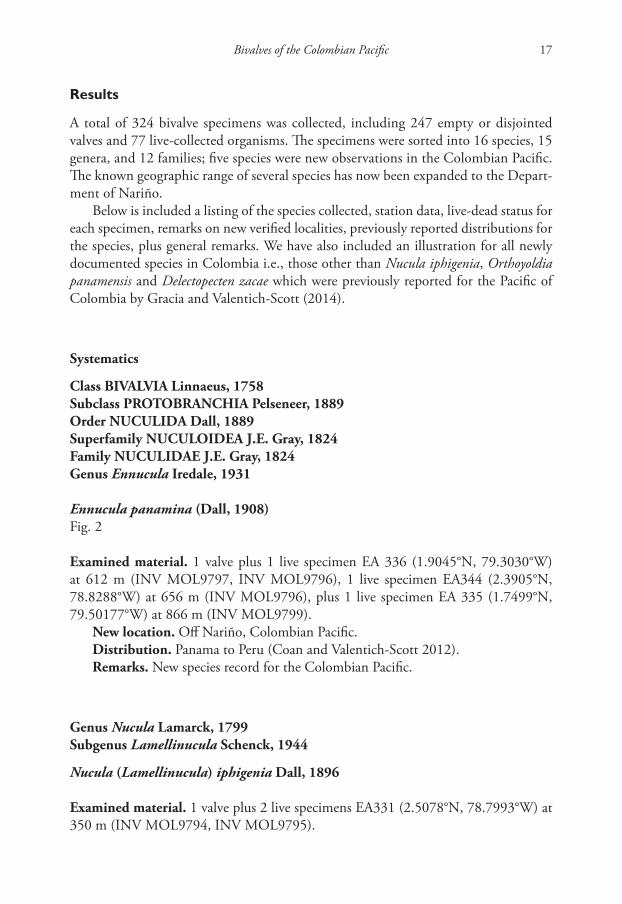
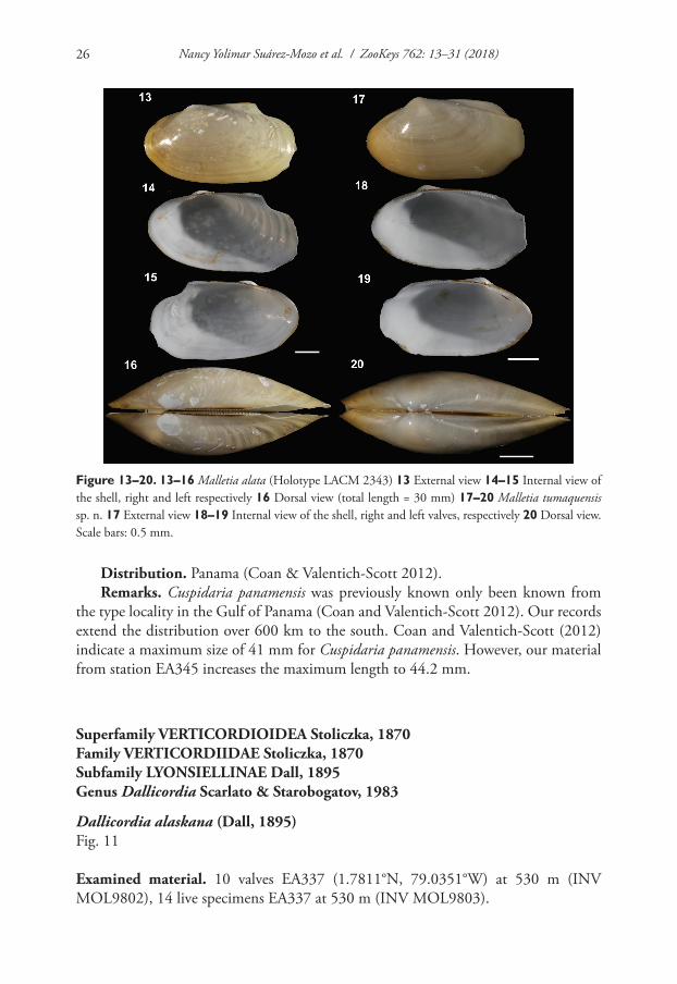
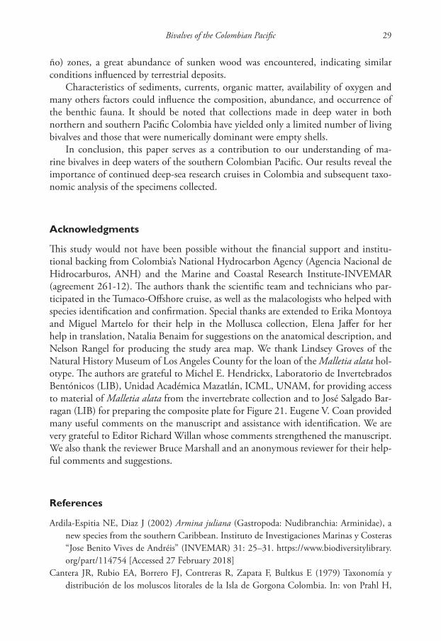
Figure 13–20. 13–16 Malletia alata (Holotype LACM 2343) 13 External view 14–15 Internal view of the shell, right and left respectively 16 Dorsal view (total length = 30 mm) 17–20 Malletia tumaquensis sp. n. 17 External view 18–19 Internal view of the shell, right and left valves, respectively 20 Dorsal view. Scale bars: 0.5 mm.
Bivalves of the Colombian Pacific 27
New location. Off Nariño, Colombian Pacific.Distribution. Sitka, Alaska, to Tumbes, Peru (Coan and Valentich-Scott 2012).Remarks. This material represents a new record for the Colombian Pacific.
Genus Lyonsiella G. O. Sars, 1872
Lyonsiella cf. magnifica Dall, 1913Fig. 12
Examined material. 1 live specimen EA345 (2.5557°N, 79.0476°W) at 668 m (INV MOL9804).
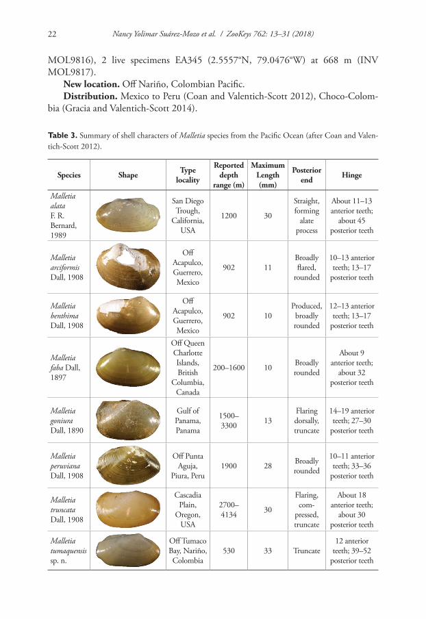
Figure 21–22. 21 Malletia alata (EMU7054) Gulf of California, Mexico, lateral view of anatomy 22 Malletia tumaquensis sp. n. Lateral view of anatomy. Scale bars: 0.5 mm. Abbreviations: DD Digestive diverticula, FT Foot, GL Gill, SI Siphon, PP Palp proboscis.
Nancy Yolimar Suárez-Mozo et al. / ZooKeys 762: 13–31 (2018)28
New location. Off Nariño, Colombian Pacific.Distribution. Mexico to Panama (Coan and Valentich-Scott 2012).Remarks. Colombian material resembles the type material of Lyonsiella magnifica.
However, our specimens have more prominent umbones, a more truncate anterior end, and a more obliquely truncate posterior end when compared to the type material. Many additional specimens would be necessary to determine if our single specimen falls within the range of intraspecific variation for Lyonsiella magnifica or it represents a new species.
Discussion
The new species of Malletiidae herein described brings to eight the number of known species for this family in the eastern Pacific Ocean (Table 3). Malletia is a widely dis-tributed genus that is associated mainly with deep water and soft sediments (Coan and Valentich-Scott 2012). Previously reported from Colombia were M. goniura Dall, 1890 and M. truncata Dall, 1908 (type locality, Malpelo Island, at 3,334 m).
As was true of the previous study in the northern Colombian Pacific (Gracia and Valentich-Scott, 2014), these recent collections not only expand the geographic distri-butions of many species on the Colombian continental margin, but they also represent new collection locations. This serves as potential evidence for the species actually living in the area, rather than the transport of dead shells into the region. Further, our find-ings have significantly expanded the bathymetric limits of several species. One new species has been described, indicating that this region of Colombia is still relatively unexplored. Further surveys are necessary to complement this malacological inven-tory and to clarify the taxonomic identity of several species. These are important pre-liminary steps for to assist in investigating the impacts of anthropogenic practices and changes (e.g., deep-sea trawling, pollution).
Deep-sea baseline surveys seek to expand bivalve records for the Colombian Pacific Ocean. In 2014, Gracia and Valentich-Scott reported on specimens collected in the northern Colombian Pacific; 89.5% of the identified species represented new records for the region. The present survey used a similar methodology but was conducted in the southern Colombian Pacific. The number of bivalve species we encountered in the southern Colombian Pacific (16) was far lower than that for the northern Colombian Pacific. This could possibly be due to the different depths sampled in either survey, or possibly the decreasing diversity associated with increasing depth.
The transport of sediment caused by river discharge, marine currents, and other factors stimulate the resuspension of material on soft sediments (Segall et al. 1989). In the northern Colombian Pacific there is a greater influence of the equatorial counter-current and the Panama Current, and the discharge from the Baudó River, while the southern Colombia Pacific (where Tumaco Bay is located) sees the influence of cold continental waters (CCCP 2002). All of these processes in Pacific Colombian result in a dense mixture of water and sediment that moves along the bottom of the sea and transports plant waste material. In both the northern (Choco) and the southern (Nari-
Bivalves of the Colombian Pacific 29
ño) zones, a great abundance of sunken wood was encountered, indicating similar conditions influenced by terrestrial deposits.
Characteristics of sediments, currents, organic matter, availability of oxygen and many others factors could influence the composition, abundance, and occurrence of the benthic fauna. It should be noted that collections made in deep water in both northern and southern Pacific Colombia have yielded only a limited number of living bivalves and those that were numerically dominant were empty shells.
In conclusion, this paper serves as a contribution to our understanding of ma-rine bivalves in deep waters of the southern Colombian Pacific. Our results reveal the importance of continued deep-sea research cruises in Colombia and subsequent taxo-nomic analysis of the specimens collected.
Acknowledgments
This study would not have been possible without the financial support and institu-tional backing from Colombia’s National Hydrocarbon Agency (Agencia Nacional de Hidrocarburos, ANH) and the Marine and Coastal Research Institute-INVEMAR (agreement 261-12). The authors thank the scientific team and technicians who par-ticipated in the Tumaco-Offshore cruise, as well as the malacologists who helped with species identification and confirmation. Special thanks are extended to Erika Montoya and Miguel Martelo for their help in the Mollusca collection, Elena Jaffer for her help in translation, Natalia Benaim for suggestions on the anatomical description, and Nelson Rangel for producing the study area map. We thank Lindsey Groves of the Natural History Museum of Los Angeles County for the loan of the Malletia alata hol-otype. The authors are grateful to Michel E. Hendrickx, Laboratorio de Invertebrados Bentónicos (LIB), Unidad Académica Mazatlán, ICML, UNAM, for providing access to material of Malletia alata from the invertebrate collection and to José Salgado Bar-ragan (LIB) for preparing the composite plate for Figure 21. Eugene V. Coan provided many useful comments on the manuscript and assistance with identification. We are very grateful to Editor Richard Willan whose comments strengthened the manuscript. We also thank the reviewer Bruce Marshall and an anonymous reviewer for their help-ful comments and suggestions.
References
Ardila-Espitia NE, Diaz J (2002) Armina juliana (Gastropoda: Nudibranchia: Arminidae), a new species from the southern Caribbean. Instituto de Investigaciones Marinas y Costeras “Jose Benito Vives de Andréis” (INVEMAR) 31: 25–31. https://www.biodiversitylibrary.org/part/114754 [Accessed 27 February 2018]
Cantera JR, Rubio EA, Borrero FJ, Contreras R, Zapata F, Bultkus E (1979) Taxonomía y distribución de los moluscos litorales de la Isla de Gorgona Colombia. In: von Prahl H,
Nancy Yolimar Suárez-Mozo et al. / ZooKeys 762: 13–31 (2018)30
Groghl M GF (Eds) Gorgona. Universidad de los Andes. Departamento de Biología, Bo-gota, 141–168.
Cantera JR (2010) Bivalvos perforadores de Madera (Mollusca: Teredinidae, Pholadidae) en la costa Pacífica Colombiana. Revista de la Academia Colombiana de Ciencias 34: 277–288.
CCCP [Centro Control Contaminacion del Pacífico] (2002) Serie Publicaciones Especiales Vol. 2 Compilacion oceanográfica de la Cuenca Pacífica Colombiana. DIMAR. San Andrés de Tumaco, 109 pp.
Coan EV, Valentich-Scott P (2012) Bivalve Seashells of Tropical West America. Marine bivalve mollusks from Baja California to northern Peru. Santa Barbara Museum of Natural His-tory. Santa Barbara, 1258 pp.
Cosel R von (1984) Moluscos marinos de la Isla Gorgona (costa del Pacífico Colombiano). Anales del Instituto de Investigaciones Marinas de Punta de Betín 14: 175–257.
Dall WH (1896) Diagnoses of new species of mollusks from the west coast of the United States. Proceedings of the United States National Museum 18: 7–20. https://doi.org/10.5479/si.00963801.18-1035.21
Díaz JM, Cantera JR, Puyana M (1997) Estado actual del conocimiento en sistemática de mo-luscos marinos recientes de Colombia. Boletín Ecotrópica 33: 15–37.
Gracia A, Ardila-Espitia NE (2009) Striocadulus magdalenensis, a new deep-sea scaphopod (Mol-lusca: Scaphopoda:Gadilidae) from the Colombian Caribbean. Instituto de Investigaciones Marinas y Costeras “Jose Benito Vives de Andréis” (INVEMAR) 38: 143–150. https://www.biodiversitylibrary.org/part/114888 [Accessed 27 February 2018]
Gracia A, Valentich-Scott P (2014) New records of soft bottom bivalves (Mollusca) inhabiting the northern Pacific Ocean of Colombia. Marine Biodiversity Records 7: 1–15. https://doi.org/10.1017/S1755267214000566
Hertlein L, Strong A (1955) Marine mollusks collected during the ‘Askoy’ Expedition to Pan-ama, Colombia, and Ecuador in 1941. Bulletin of the American Museum of Natural His-tory 107: 162–317.
IDEAM, IGAC, IAVH, Invemar, Sinchi IIAP (2007) Ecosistemas continentales, costeros y ma-rinos de Colombia. Instituto de Hidrología Meteorología y Estudios Ambientales- Ideam, Instituto Geográfico Agustin Codazzi, Instituto de Investigación de Recursos Biológicos Alexander Von Humboldt, Instituto de Inv. Bogota, 276 pp.
INVEMAR-ANH (2013) Línea base biológica y física de los bloques TUM Offshore 6 y 7 sujetos a exploración de hidrocarburos. Santa Marta, 258 pp.
Kamenev G (2015) Composition and distribution of bivalves of the abyssal plain adjacent to the Kuril – Kamchatka Trench (Pacific Ocean). Deep Sea Research, Part II: Tropical Stud-ies in Oceanography 111: 188–197. https://doi.org/10.1016/j.dsr2.2014.08.005
Keen AM (1971) Seashells of Tropical West America: Marine Mollusks from Baja California to Peru. Stanford University Press, California, 1064 pp.
López de Mesa LA, Cantera JR (2015) Marine mollusks of Bahía Málaga, Colombia (Tropical Eastern Pacific) Colombia. Check List 11: 1–18. https://doi.org/10.15560/11.1.1497
Okutani T, Fujikura K, Kojima S (1999) Two new hadal bivalves of the family Thyasiridae from the plate convergent area of the Japan Trench. Venus Japanese Journal of Malacology 58: 49–54.
Bivalves of the Colombian Pacific 31
Paredes C, Cardoso F, Santamaría J, Esplana J, Llaja L (2016) Lista anotada de los bivalvos marinos del Perú. Revista Peruana de Biologia 23: 127–150. https://doi.org/10.15381/rpb.v23i2.12397
Segall MP, Kuehl SA, Gipson Jr M (1989) Clay-size minerals as indicators of modern sedi-mentary processes in submarine canyons: application to the Wilmington Canyon System. Marine Geology 90: 175–192. https://doi.org/10.1016/0025-3227(89)90040-6
Sellanes J, Krylova E (2005) A new species of Calyptogena (Bivalvia: Vesicomyidae) from a recently discovered methane seepage area off Concepcion Bay, Chile (~36° S). Journal of the Marine Biological Association of the United Kingdom 85: 969–976. https://doi.org/10.1017/S0025315405011963
Simone LRL, Gracia CA (2006) A new species of Suturoglypta from Colombia (Caenogas-tropoda, Columbellidae). Papéis Avulsos de Zoologia (São Paulo) 46: 133–137. https://doi.org/10.1590/S0031-10492006001200001

Nancy Yolimar Suárez-Mozo et al. / ZooKeys 762: 13–31 (2018)32
Four species of spider genus Cheiracanthium C. L. Koch, 1839 (Araneae, Eutichuridae)... 33
Four species of spider genus Cheiracanthium C. L. Koch, 1839 (Araneae, Eutichuridae)
from Jinggang Mountains, Jiangxi Province, China
Jianshuang Zhang1, Guren Zhang2, Hao Yu2,3
1 School of Life Sciences, Guizhou Normal University, Guiyang, Guizhou, China 2 State Key Laboratory for Biocontrol, Sun Yat-Sen University, Guangzhou, Guangdong, China 3 College of Chemistry and Life Sciences, Integrated Mountain Research Institute, Guizhou Education University, Guiyang, Guizhou, China
Corresponding author: Hao Yu ([email protected])
Academic editor: Shuqiang Li | Received 22 January 2018 | Accepted 6 May 2018 | Published 30 May 2018
http://zoobank.org/35D37CBF-6F91-43CB-9CDA-210B7D0C7CD5
Citation: Zhang J, Zhang G, Yu H (2018) Four species of spider genus Cheiracanthium C. L. Koch, 1839 (Araneae, Eutichuridae) from Jinggang Mountains, Jiangxi Province, China. ZooKeys 762: 33–45. https://doi.org/10.3897/zookeys.762.23786
AbstractFour species of spider genus Cheiracanthium C. L. Koch, 1839 are reported from Jinggang Mountains, Jiangxi Province, China. Two of them are described as new to science: C. auriculatum sp. n. (♀♂) and C. echinulatum sp. n. (♂). Cheiracanthium taiwanicum Chen, Huang, Chen & Wang, 2006 is recorded from Mainland China for the first time. Cheiracanthium zhejiangense Hu & Song, 1982, the most similar species to C. auriculatum sp. n., is a newly recorded species of Jiangxi Province. Detailed descriptions, diagnoses, and photographs of the two new species are given. Cheiracanthium taiwanicum and C. zheji-angense are also illustrated.
KeywordsTaxonomy, morphology, description, new species
ZooKeys 762: 33–45 (2018)
doi: 10.3897/zookeys.762.23786
http://zookeys.pensoft.net
Copyright Jianshuang Zhang et al. This is an open access article distributed under the terms of the Creative Commons Attribution License (CC BY 4.0), which permits unrestricted use, distribution, and reproduction in any medium, provided the original author and source are credited.
RESEARCH ARTICLE
Launched to accelerate biodiversity research
A peer-reviewed open-access journal
Jianshuang Zhang et al. / ZooKeys 762: 33–45 (2018)34
Introduction
Cheiracanthium C. L. Koch, 1839 contains 210 catalogued species and is mainly dis-tributed in the Old World (Marusik and Fomichev 2016; World Spider Catalogue 2018). Although this genus is relatively large and well known, its taxonomy is rather poorly studied. Almost half of its species are known from single sex or juveniles: 36 by males, 60 by females, 2 by juveniles (World Spider Catalogue 2018). Additionally, 16 species were never illustrated and many species were described based on poor illus-trations. So far, the genus has not been the subject of any global or regional revisions (Marusik and Fomichev, 2016).
The Cheiracanthium fauna of China is relatively rare and poorly represented, with only 38 described species (Li and Lin 2016; World Spider Catalogue 2018), of which 14 species are known based on a single sex: for 11, only females are known, and for three, only males are known (World Spider Catalogue 2018). Additionally, illustra-tions of the internal structure of the epigyne are not provided in five species (World Spider Catalogue 2018). Moreover, the diversity of this genus in China is still insuf-ficiently known and several new species have been described in the last few years (Chen and Huang 2012; Barrion et al. 2013; Wang and Zhang 2013).
Field collection in Jinggang Mountains of Jiangxi province, China, was carried out in April 2011. During this field exploration, four Cheiracanthium species were found: C. auriculatum sp. n., C. echinulatum sp. n., C. taiwanicum Chen, Huang, Chen & Wang, 2006 and C. zhejiangense Hu & Song, 1982. Descriptions and pho-tographs of the new species, as well as supplementary micrographs of the known species, are provided.
Materials and methods
Spiders were fixed and preserved in 80% ethanol. Specimens were examined with an Olympus SZX7 stereomicroscope; details were studied with an Olympus BX51 com-pound microscope. Male palps and female epigynes were examined and illustrated after being dissected. Epigynes were cleared in boiling KOH solution to dissolve soft tissues. Photographs were made with a Leica DFC450 digital camera mounted on an Olympus BX51 compound microscope. The digital images were taken and assembled using Helicon Focus 3.10 software package.
All measurements were obtained using an Olympus SZX7 stereomicroscope and given in millimetres. Eye diameters are taken at the widest point. The total body length does not include chelicerae or spinnerets length. Leg lengths are given as total length (femur, patella, tibia, metatarsus, tarsus). The type specimens of the new species are deposited in College of Chemistry and Life Sciences, Guizhou Education University, Guiyang, Guizhou, China.
Four species of spider genus Cheiracanthium C. L. Koch, 1839 (Araneae, Eutichuridae)... 35
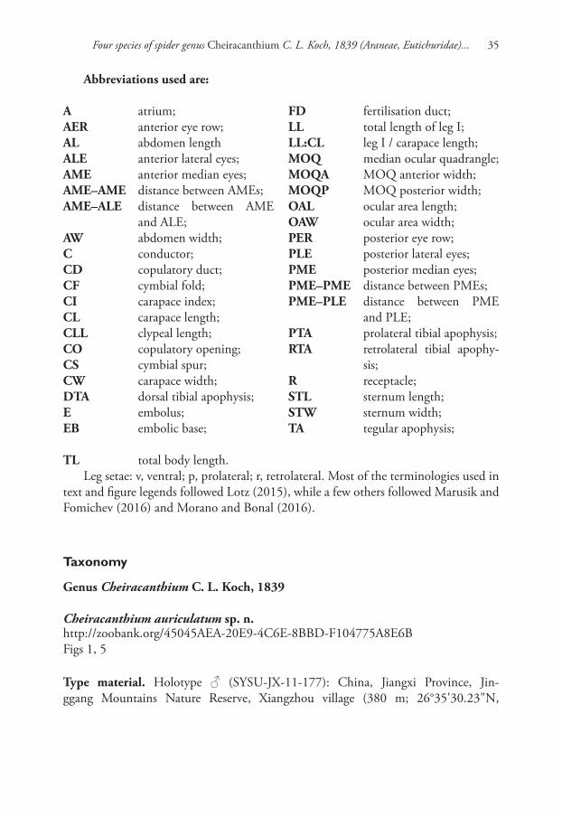
Abbreviations used are:
A atrium;AER anterior eye row;AL abdomen lengthALE anterior lateral eyes;AME anterior median eyes;AME–AME distance between AMEs;AME–ALE distance between AME
and ALE;AW abdomen width;C conductor;CD copulatory duct;CF cymbial fold;CI carapace index;CL carapace length;CLL clypeal length;CO copulatory opening;CS cymbial spur;CW carapace width;DTA dorsal tibial apophysis;E embolus;EB embolic base;
FD fertilisation duct;LL total length of leg I;LL:CL leg I / carapace length;MOQ median ocular quadrangle;MOQA MOQ anterior width;MOQP MOQ posterior width;OAL ocular area length;OAW ocular area width;PER posterior eye row;PLE posterior lateral eyes;PME posterior median eyes;PME–PME distance between PMEs;PME–PLE distance between PME
and PLE;PTA prolateral tibial apophysis;RTA retrolateral tibial apophy-
sis;R receptacle;STL sternum length;STW sternum width;TA tegular apophysis;
TL total body length.Leg setae: v, ventral; p, prolateral; r, retrolateral. Most of the terminologies used in
text and figure legends followed Lotz (2015), while a few others followed Marusik and Fomichev (2016) and Morano and Bonal (2016).
Taxonomy
Genus Cheiracanthium C. L. Koch, 1839
Cheiracanthium auriculatum sp. n.http://zoobank.org/45045AEA-20E9-4C6E-8BBD-F104775A8E6BFigs 1, 5
Type material. Holotype ♂ (SYSU-JX-11-177): China, Jiangxi Province, Jin-ggang Mountains Nature Reserve, Xiangzhou village (380 m; 26°35'30.23"N,
Jianshuang Zhang et al. / ZooKeys 762: 33–45 (2018)36
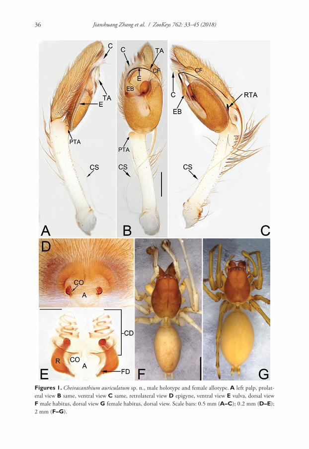
Figures 1. Cheiracanthium auriculatum sp. n., male holotype and female allotype. A left palp, prolat-eral view B same, ventral view C same, retrolateral view D epigyne, ventral view E vulva, dorsal view F male habitus, dorsal view G female habitus, dorsal view. Scale bars: 0.5 mm (A–C); 0.2 mm (D–E); 2 mm (F–G).
Four species of spider genus Cheiracanthium C. L. Koch, 1839 (Araneae, Eutichuridae)... 37
114°15'59.93"E), 26 April 2011, Hao Yu and Zhenyu Jin leg. Paratypes: 1♂ and 3 ♀, same data as holotype.
Etymology. The specific epithet is an adjective and is derived from a Latin word “auriculatus“(ear-like), referring to the tegular apophysis which is like the contour of an ear in ventral view.
Diagnosis. Cheiracanthium auriculatum sp. n. is distinguished from all other Chei-racanthium species, except C. zhejiangense Hu & Song, 1982 (Fig. 4A–E), by having a distally filiform cymbial spur in the male, and by the general shape of the vulva in the female. From C. zhejiangense, the male can be distinguished by the ear contour-shaped tegular apophysis and the uncoiling tip of cymbial spur (vs the falciform tegular apo-physis and the coiled tip of the cymbial spur in C. zhejiangense) (Figs 1A–C; 4A–C), the female can be differentiated by the indistinct atrium and copulatory ducts (vs the distinct atrium and copulatory ducts in C. zhejiangense), the more or less lengthwise receptacles (vs the nearly horizontal receptacles in C. zhejiangense) (Figs 1D–E; 4D–E), and by the different coil number of copulatory ducts (7 coils in C. auriculatum sp. n., instead of 8 coils in C. zhejiangense) (Figs 1E; 4E). In addition, the two species can by separated by their habitus: abdomen without distinct colour pattern in C. auriculatum sp. n. (Fig. 1F–G), but with a median heart-shaped mark which reaches half of the opisthosoma length in C. zhejiangense (Fig. 4F–G).
Description. Male. Total length 8.58–9.15. Holotype (Fig. 1A–C, F): TL 9.15; CL 3.73, CW 2.41, CI (CL/CW) 1.55; AL 4.05, AW 2.42. Carapace (Fig. 1F) brown, uniformly coloured, without distinct pattern. Eye sizes and inter-distances: OAL 0.39, OAW 1.45; AME 0.14, ALE 0.16, PME 0.16, PLE 0.17; AME–AME 0.27, AME–ALE 0.27, PME–PME 0.36, PME–PLE 0.35; MOQA 0.56, MOQP 0.67, CLL 0.10. Cheli-cerae protruding and reddish brown, with 3 teeth on promargin and 3 on retromargin re-spectively. Sternum dark brown, STL 1.69, STW 1.44. Labium and endites brown. Legs yellowish-white, without distinct colour markings. Leg measurements: I 18.84 (4.65, 1.22, 5.10, 5.60, 2.26), II 12.09 (3.30, 1.14, 3.24, 3.23, 1.19), III 8.61 (2.38, 0.86, 1.92, 2.40, 1.05), IV 12.47 (3.72, 0.95, 3.17, 4.00, 1.07); LL:CL 5.03. Leg spines: I 0-0-1p, 2v-1v-1p, 2v1p-1p1v-1v; II 0-0-1p, 1v-2v-1p, 2v1p-1v1p-1v; III 0-0-1p1r, 0-1p1r-0, 2v1p-1p1r-1v2p2r; IV 0-0-1p1r, 1v-1v1p-0, 2v1p-1v1p1r-1v2p2r. Abdomen (Fig. 1F) elongate-oval, dorsally grey, dorsum with indistinct heart-shaped mark and two pairs of not obvious muscle depressions; venter brownish without distinct pattern.
Palp (Fig. 1A–C). Tibia extremely long, about as long as cymbium, with two apo-physes; retrolateral tibial apophysis (RTA) about 20% of tibia length, with a more or less bifurcate apex and hiding behind tegulum; prolateral tibial apophysis (PTA) small and round; cymbial spur (CS) is approximately equal in length to tibia, tapering off into a filiform; cymbial fold (CF) poorly developed, for approximately 4/5 the length of cymbium; tip of cymbium short, about 1/4 of cymbium length. Tegulum oblong, 1.3 times longer than wide; tegular apophysis (TA) long and sinuate, more than 4/5 of tegulum length, filamentous and like an ear’s contour in ventral view; embolus (E) arising at approximately 10 o’clock position, terminating at approximately 11 o’clock position, it’s tip covered by conductor (C); conductor large, falciform.
Jianshuang Zhang et al. / ZooKeys 762: 33–45 (2018)38
Female. Total length 8.66–9.30. Slightly larger in size and lighter in colour. Allo-type (Fig. 1D–E, G) measured: TL 9.30; CL = 3.03, CW = 2.22, CI (CL/CW) = 1.36; AL = 4.95, AW = 2.92. Eye diameters and inter-distances: OAL 0.37, OAW 1.23; AME 0.14, ALE 0.19, PME 0.13, PLE 0.14; AME–AME 0.23, AME–ALE 0.11, PME–PME 0.31, PME–PLE 0.23; MOQA 0.46, MOQP 0.58, CLL 0.24. PMT: RMT = 6:6.STL 1.47, STW 1.23. Leg measurements: I 12.70 (3.30, 1.06, 3.42, 3.39, 1.54), II 8.51 (2.42, 0.86, 2.23, 2.05, 0.96), III 6.42 (1.92, 0.69, 1.35, 1.65, 0.80), IV 9.67 (2.75, 0.89, 2.39, 2.68, 0.96); LL:CL 4.19. Leg spines: I 0-1p-1p, 2v-2v-0, 2v-1p1r-1v; II 0-0-1p, 1v-2v-1p, 2v1p-1p1r-1v; III 0-1p-1p1r, 1v-1p1r-0, 2v1p1r--1p1r-1v2p2r; IV 0-0-1p1r, 1v-1v1p1r-0, 2v1p1r-1v1p1r-1v2p2r.
Epigyne (Fig. 1D–E). Atrium (A) indistinct, without delimited margin, about four times wider than long; receptacles (R) are faintly visible through epigynal plate in ventral view; two copulatory openings (CO) located at lateral borders of atrium; the transparent copulatory ducts (CD) running spirally (length of spira about 1.4 times longer than receptacles), forming 7 entwined loops (including 4 ascending coils and 3 descending coils); receptacle sickle-shaped, separated by three diameters.
Distribution. Presently known only from the type locality, Jinggang Mountains, Jiangxi, China (Fig. 5).
Cheiracanthium echinulatum sp. n.http://zoobank.org/A1935AC9-A0A9-45F2-8BFA-22E0F3172401Figs 2, 5
Type material. Holotype ♂ (SYSU-JX-11-182): China, Jiangxi Province, Jin-ggang Mountains Nature Reserve, Xiangzhou village (380 m; 26°35'30.23"N, 114°15'59.93"E), 26 April 2011, Hao Yu and Zhenyu Jin leg. Paratypes: 1♂, same data as holotype.
Etymology. The species epithet is taken from the Latin adjective echinulatus and refers to the spinule-shaped tegular apophysis.
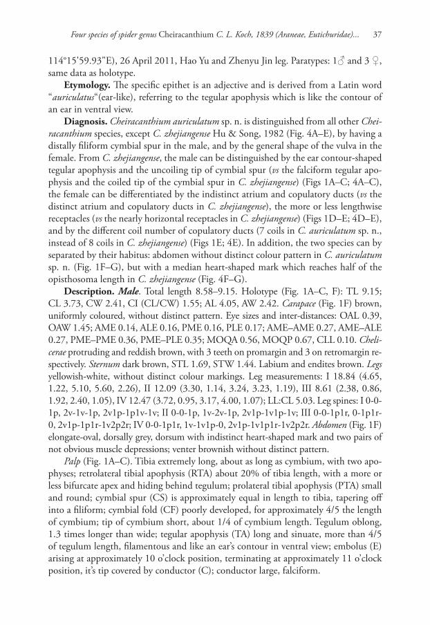
Diagnosis. This new species resembles C. taegense Paik, 1990 (Paik, 1990: 11, f. 39–47; Baba & Yoshitake, 2016: 39, f. 1–4) in having the similar beak-shaped cymbial spur, and stalk-like dorsal tibial apophysis, but can be distinguished by: (1) the embo-lus originated at 3 o’clock position (Fig. 2B–C), vs. originated at 1–2 o’clock position in C. taegense (Paik, 1990: 11, f. 41; Baba & Yoshitake, 2016: 39, f. 3–4); (2) tegular apophysis smaller, straight and acicular (Fig. 2A–C), instead of bigger and slightly curved in C. taegense (Paik, 1990: 11, f. 41, 43; Baba & Yoshitake, 2016: 39, f. 3–4); (3) RTA straight and digitiform (Fig. 2A–C), but with a curved and hook-shaped apex in C. taegense (Baba & Yoshitake, 2016: 39, f. 3–4).
Description. Male. Total length 9.06–9.12. Holotype (Fig. 2): TL 9.06; CL 3.58, CW 2.34, CI (CL/CW) 1.53; AL 4.99, AW 2.27. Carapace (Fig. 2D, F) yellow except reddish ocular area, without distinct colour pattern. Eye sizes and inter-distances: OAL 0.34, OAW 1.26; AME 0.15, ALE 0.15, PME 0.14, PLE 0.13; AME–AME 0.46,
Four species of spider genus Cheiracanthium C. L. Koch, 1839 (Araneae, Eutichuridae)... 39
Figures 2. Cheiracanthium echinulatum sp. n., male holotype. A left palp, prolateral view B same, ventral view C same, retrolateral view D male habitus, dorsal view E same, ventral view F same, lateral view. Scale bars: 0.5 mm (A–C); 2 mm (D–F).
Jianshuang Zhang et al. / ZooKeys 762: 33–45 (2018)40
AME–ALE 0.25, PME–PME 0.54, PME–PLE 0.22; MOQA 0.43, MOQP 0.55, CLL 0.13. Chelicerae light brown and robust, with long and wine-coloured fangs, with 3 teeth on promargin and 3 on retromargin respectively. Sternum (Fig. 2E) yellowish, STL 1.79, STW 1.32. Labium and endites brown. Legs yellowish, without distinct colour markings. Leg measurements: I 23.70 (5.90, 1.05, 7.31, 7.99, 1.46), II 14.92 (3.41, 0.82, 4.14, 5.26, 1.29), III 11.10 (2.53, 1.43, 2.16, 3.94, 1.03), IV 16.42 (4.18, 1.21, 4.13, 5.66, 1.24); LL:CL 6.62. Leg spines: I 0-1p1r-1p1r, 3v-3v-1v1p, 2v-0-1v; II 0-1p1r-1p1r, 3v-2v-1v1p, 2v1p-2v1p-1v; III 0-1p1r-1p1r, 2v1p1r-1p1r-0, 2v1p1r-2v1p1r-2v1p2r; IV 0-1p1r-1p1r, 1v1p1r-1v2r-1v1r, 2v1p1r-2v1p1r-1v1p3r. Abdomen (Fig. 2D–F) lanceolate, dorsally yellowish white, scattered numerous indistinct pig-mented spots; venter yellowish without distinct pattern.
Palp (Fig. 2A–C). Tibia twice shorter than cymbium, with three apophyses; retro-lateral tibial apophysis (RTA) about 50% of tibia length, heavily sclerotised and with a fingerlike apex; prolateral tibial apophysis (PTA) distinctly elevated and relatively short, about 30% of tibia length, coniform in prolateral view and digitiform in ventral view; dorsal tibial apophysis (DTA) thin and stalk-shaped, about as long as RTA; cym-bial spur (CS) beak-shaped, twice shorter than tibia; cymbial fold (CF) strongly devel-oped and well visible in ventral and retrolateral view, for approximately 2/3 the length of cymbium; tip of cymbium long, about 1/3 of cymbium length. Tegulum 1.3 longer than wide, membranous and semitransparent except its margin in ventral view; tegular apophysis (TA) short and thin, spiculate; embolus (E) starts on the retrolateral flank (approximately 3 o’clock of tegulum), surrounds the base and ends at conductor (C) apex, its tip filiform and curved behind conductor; conductor large and membranous.
Female. Unknown.Comments. According to the World Spider Catalogue 2018, a total of 11 Chei-
racanthium species are known from females only in China: C. approximatum O. P.-Cambridge, 1885, C. escaladae Barrion et al., 2013, C. fujianense Gong, 1983, C. hypocyrtum Zhang & Zhu, 1993, C. liuyangense Xie et al., 1996, C. olliforme Zhang & Zhu, 1993, C. potanini Schenkel, 1963, C. rupicola (Thorell, 1897), C. solidum Zhang et al., 1993, C. sphaericum Zhang et al., 1993, C. longtailen Xu, 1993. Among them, C. escaladae is supposedly a Clubiona species based on epigyne morphology, while C. potanini is supposededly doubtful because of the poor original illustrations and description. The other nine can be considered tentatively as valid Cheiracanthium species. However, none of them could be matched with C. echinulatum sp. n. due to their different habitus (abdomen without distinct colour pattern in C. echinulatum sp. n., but with a median heart-shaped mark in C. approximatum, C. fujianense and C. rupicola, with two pairs of muscular depressions in C. hypocyrtum, C. liuyangense and C. sphaericum) and different number of cheliceral teeth (chelicerae with 3 promarginal and 3 retromarginal teeth in C. echinulatum sp. n., but with 3 promarginal and 2 ret-romarginal teeth in C. hypocyrtum, C. olliforme and C. sphaericum, with 3 promarginal and 1 retromarginal teeth in C. liuyangense, with 2 promarginal and 1 retromarginal teeth in C. solidum, with 2 promarginal and 3 retromarginal teeth in C. longtailen).
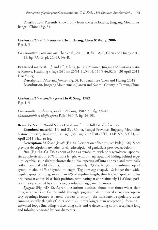
Four species of spider genus Cheiracanthium C. L. Koch, 1839 (Araneae, Eutichuridae)... 41
Distribution. Presently known only from the type locality, Jinggang Mountains, Jiangxi, China (Fig. 5).
Cheiracanthium taiwanicum Chen, Huang, Chen & Wang, 2006Figs 3, 5
Cheiracanthium taiwanicum Chen et al., 2006: 10, fig. 1A–E; Chen and Huang 2012: 25, fig. 7A–G, pl. 2C–D, 3A–B.
Examined material. 1♂ and 1♀, China, Jiangxi Province, Jinggang Mountains Natu-re Reserve, Hexiliong village (680 m; 26°31'51.54"N, 114°8'46.02"E), 30 April 2011, Hao Yu leg.
Description. Male and female (Fig. 3). For details see Chen and Huang (2012).Distribution. Jinggang Mountains in Jiangxi and Nantou County in Taiwan, China.
Cheiracanthium zhejiangense Hu & Song, 1982Figs 4–5
Cheiracanthium zhejiangensis Hu & Song, 1982: 56, fig. 4A–D.Cheiracanthium zhejiangense Paik 1990: 9, fig. 26–38.
Remarks. See the World Spider Catalogue for the full list of references.Examined material. 1♂ and 2♀, China, Jiangxi Province, Jinggang Mountains
Nature Reserve, Xiangzhou village (380 m; 26°35'30.23"N, 114°15'59.93"E), 26 April 2011, Hao Yu leg.
Description. Male and female (Fig. 4). Description of habitus, see Paik (1990). Since previous descriptions are rather brief, redescription of genitalia is provided as below.
Palp (Fig. 4A–C). Tibia about as long as cymbium, with only retrolateral apophy-sis; apophysis about 20% of tibia length, with a sharp apex and hiding behind tegu-lum; cymbial spur slightly shorter than tibia, tapering off into a thread and terminally coiled; cymbial fold distinct, for approximately 2/3 the length of cymbium; tip of cymbium about 1/3 of cymbium length. Tegulum egg-shaped, 1.2 longer than wide; tegular apophysis long, more than 4/5 of tegulim length, thin hook-shaped; embolus originates at about 10 o’clock position, terminating at approximately 11 o’clock posi-tion, it’s tip covered by conductor; conductor large, membranous.
Epigyne (Fig. 4D–E). Apron-like atrium distinct, about four times wider than long; receptacles are faintly visible through epigynal plate in ventral view; two copula-tory openings located at lateral borders of atrium; the transparent copulatory ducts running spirally (length of spira about 2.6 times longer than receptacles), forming 8 entwined loops (including 4 ascending coils and 4 descending coils); receptacle long and tubular, separated by two diameters.
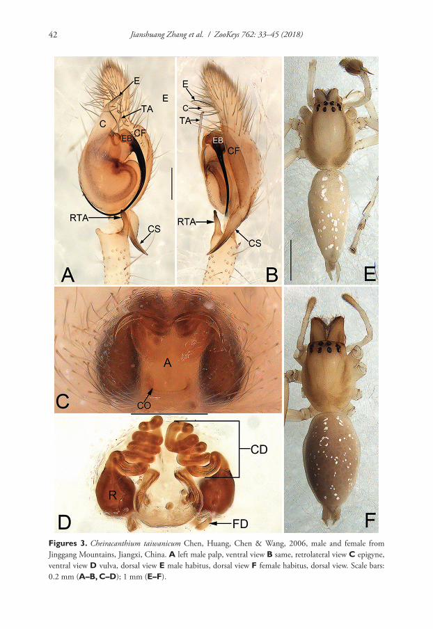
Jianshuang Zhang et al. / ZooKeys 762: 33–45 (2018)42
Figures 3. Cheiracanthium taiwanicum Chen, Huang, Chen & Wang, 2006, male and female from Jinggang Mountains, Jiangxi, China. A left male palp, ventral view B same, retrolateral view C epigyne, ventral view D vulva, dorsal view E male habitus, dorsal view F female habitus, dorsal view. Scale bars: 0.2 mm (A–B, C–D); 1 mm (E–F).
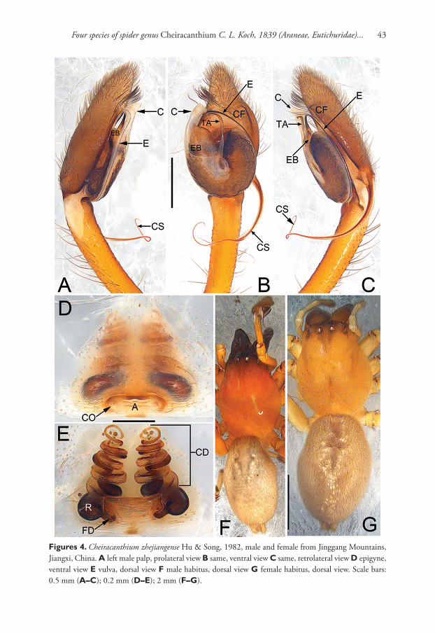
Four species of spider genus Cheiracanthium C. L. Koch, 1839 (Araneae, Eutichuridae)... 43
Figures 4. Cheiracanthium zhejiangense Hu & Song, 1982, male and female from Jinggang Mountains, Jiangxi, China. A left male palp, prolateral view B same, ventral view C same, retrolateral view D epigyne, ventral view E vulva, dorsal view F male habitus, dorsal view G female habitus, dorsal view. Scale bars: 0.5 mm (A–C); 0.2 mm (D–E); 2 mm (F–G).
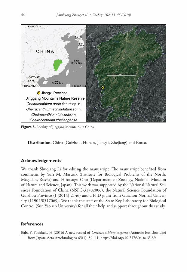
Jianshuang Zhang et al. / ZooKeys 762: 33–45 (2018)44
Distribution. China (Guizhou, Hunan, Jiangxi, Zhejiang) and Korea.
Acknowledgements
We thank Shuqiang Li for editing the manuscript. The manuscript benefited from comments by Yuri M. Marusik (Institute for Biological Problems of the North, Magadan, Russia) and Hirotsugu Ono (Department of Zoology, National Museum of Nature and Science, Japan). This work was supported by the National Natural Sci-ences Foundation of China (NSFC-31702006), the Natural Science Foundation of Guizhou Province (J [2014] 2146) and a PhD grant from Guizhou Normal Univer-sity (11904/0517069). We thank the staff of the State Key Laboratory for Biological Control (Sun Yat-sen University) for all their help and support throughout this study.
References
Baba Y, Yoshitake H (2016) A new record of Cheiracanthium taegense (Araneae: Eutichuridae) from Japan. Acta Arachnologica 65(1): 39–41. https://doi.org/10.2476/asjaa.65.39
Figure 5. Locality of Jinggang Mountains in China.
Four species of spider genus Cheiracanthium C. L. Koch, 1839 (Araneae, Eutichuridae)... 45
Barrion AT, Barrion-Dupo ALA, Catindig JLA, Villareal M, CaiD O, YuanQH, HeonKL (2013) New species of spiders (Araneae) from Hainan Island, China. UPLB Museum Pub-lications in Natural History 3: 1–103.
Dankittipakul P, Beccaloni J (2012) Validation and new synonymies proposed for Cheiracanthi-um species from south and southeast Asia (Araneae, Clubionidae). Zootaxa 3510: 77–86.
Gong JX (1983) Neue und wenig bekannte Clubionidae aus China (Arachnida: Araneae). Verhandlungen des Naturwissenschaftlichen Vereins in Hamburg 26: 61–68.
Hu YJ, Song DX (1982) Notes on some Chinese species of the family Clubionidae. Journal of Hunan Teachers College (Nature Science Edition) 1982(2): 55–62.
Huang WJ, Chen SH (2012) The Spider Fauna of Taiwan. Araneae. Miturgidae, Anyphaeni-dae, Clubionidae. National Taiwan Normal University, Taipei, 130 pp.
Li SQ, Lin YC (2016) Species Catalogue of China. Volume 2. Animals. Invertebrates (I): Arachnida: Araneae. Science Press, Beijing, 549 pp.
Lotz LN (2015) New species of the spider genus Cheiracanthium from continental Africa (Araneae: Eutichuridae). Zootaxa 3973(2): 321–336. https://doi.org/10.11646/zoo-taxa.3973.2.7
Marusik YM, Fomichev AA (2016) A new species of Cheiracanthium (Araneae: Cheiracanthii-dae) from Mongolia. Indian Journal of Arachnology, 5(1-2): 79–83.
Morano E, Bonal R (2016) Cheiracanthium ilicis sp. n. (Araneae, Eutichuridae): a novel spi-der species associated with Holm Oaks (Quercus ilex). ZooKeys 601: 21–39. https://doi.org/10.3897/zookeys.601.8241
Paik KY (1990) Korean spiders of the genus Clubiona (Araneae: Clubionidae) I. Description of eight new species and five unrecorded species from Korea. Korean Arachnology 5(2): 85–129.
Pickard-Cambridge O (1885) Araneida. In: Scientific results of the second Yarkand mission. Calcutta, 1–115.
Schenkel E (1963) Ostasiatische Spinnen aus dem Muséum d’Histoire naturelle de Paris. Mé-moires du Muséum National d’Histoire Naturelle de Paris (A, Zool.) 25: 1–481.
Wang YN, Zhang F (2013) A new spider species of the genus Cheiracanthium (Araneae, Mitur-gidae) from Guangxi, China. Acta Zootaxonomica Sinica 38(1): 59–63.
World Spider Catalogue (2018) World Spider Catalogue. Natural History Museum Bern. http://wsc.nmbe.ch, version 19 [accessed on 14 April 2018]
Xie LP, Yin CM, Yan HM, Kim JP (1996) Two new species of the family Clubionidae from China (Arachnida: Araneae). Korean Arachnology 12(1): 97–101.
Xu YJ (1993) A new species of spider of the genus Chiracanthium from China (Araneae: Clubi-onidae). Acta Arachnologica Sinica 2: 27–28.
Zhang GR, Zhu CD (1993) Two new species of the genus Chiracanthium from China (Araneae: Clubionidae). Sichuan Journal of Zoology 12(4): 5–6.
Zhang GR, Zhu CD, Hu YJ (1993) Three new species of spiders (Arachnida: Araneae) from China. Natural Enemies of Insects 15(3): 106–111.

Jianshuang Zhang et al. / ZooKeys 762: 33–45 (2018)46
Two new species of the purse-web spider genus Atypus Latreille, 1804... 47
Two new species of the purse-web spider genus Atypus Latreille, 1804 from Hainan Island,
China (Araneae, Atypidae)
Fan Li1, Xin Xu1,2, Zengtao Zhang1, Fengxiang Liu1, Hongli Zhang3, Daiqin Li4
1 Centre for Behavioural Ecology & Evolution (CBEE), College of Life Sciences, Hubei University, Wuhan, Hubei, China 2 College of Life Sciences, Hunan Normal University, Changsha, Hunan, China 3 College of Life Sciences, Hebei University, Baoding, Hebei, China 4 Department of Biological Sciences, National University of Singapore, Singapore
Corresponding authors: Xin Xu ([email protected]); Daiqin Li ([email protected])
Academic editor: C. Hamilton | Received 27 December 2017 | Accepted 28 April 2018 | Published 30 May 2018
http://zoobank.org/E6B4B4D2-79B4-4E84-925D-2E5DCE9F377D
Citation: Li F, Xu X, Zhang Z, Liu F, Zhang H, Li D (2018) Two new species of the purse-web spider genus Atypus Latreille, 1804 from Hainan Island, China (Araneae, Atypidae). ZooKeys 762: 47–57. https://doi.org/10.3897/zookeys.762.23282
AbstractTwo species of the purse-web spider genus Atypus Latreille, 1804 collected from Hainan Island, China, are diagnosed and described as new to science based on genital morphology, A. baotingensis sp. n. (♂♀) and A. jianfengensis sp. n. (♀). The DNA barcodes of the two species are also provided for future use.
KeywordsAtypidae, Atypus, DNA barcode, East Asia, Mygalomorphae, taxonomy
Introduction
The purse-web spider family Atypidae is an ancient branch of the infraorder Mygalomor-phae. Atypidae is one of the burrowing mygalomorph families. However, unlike other burrowing mygalomorph spiders, atypids form a tough web with silk from the end of their burrows to the upper ground section, which is expanded and camouflaged as a trap
ZooKeys 762: 47–57 (2018)
doi: 10.3897/zookeys.762.23282
http://zookeys.pensoft.net
Copyright Fan Li et al. This is an open access article distributed under the terms of the Creative Commons Attribution License (CC BY 4.0), which permits unrestricted use, distribution, and reproduction in any medium, provided the original author and source are credited.
RESEARCH ARTICLE
Launched to accelerate biodiversity research
A peer-reviewed open-access journal
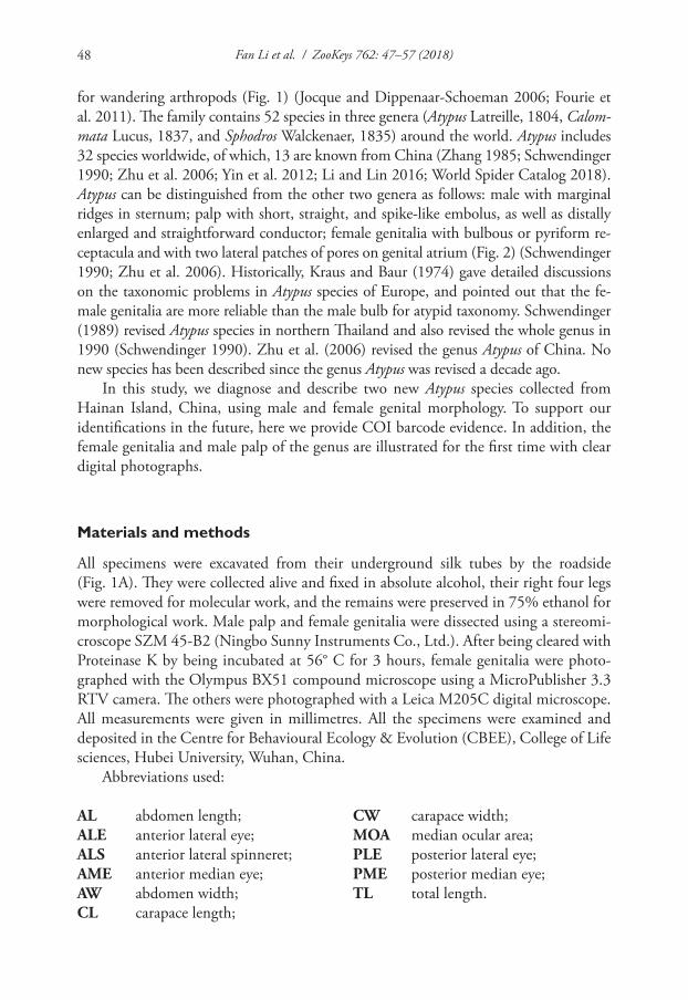
Fan Li et al. / ZooKeys 762: 47–57 (2018)48
for wandering arthropods (Fig. 1) (Jocque and Dippenaar-Schoeman 2006; Fourie et al. 2011). The family contains 52 species in three genera (Atypus Latreille, 1804, Calom-mata Lucus, 1837, and Sphodros Walckenaer, 1835) around the world. Atypus includes 32 species worldwide, of which, 13 are known from China (Zhang 1985; Schwendinger 1990; Zhu et al. 2006; Yin et al. 2012; Li and Lin 2016; World Spider Catalog 2018). Atypus can be distinguished from the other two genera as follows: male with marginal ridges in sternum; palp with short, straight, and spike-like embolus, as well as distally enlarged and straightforward conductor; female genitalia with bulbous or pyriform re-ceptacula and with two lateral patches of pores on genital atrium (Fig. 2) (Schwendinger 1990; Zhu et al. 2006). Historically, Kraus and Baur (1974) gave detailed discussions on the taxonomic problems in Atypus species of Europe, and pointed out that the fe-male genitalia are more reliable than the male bulb for atypid taxonomy. Schwendinger (1989) revised Atypus species in northern Thailand and also revised the whole genus in 1990 (Schwendinger 1990). Zhu et al. (2006) revised the genus Atypus of China. No new species has been described since the genus Atypus was revised a decade ago.
In this study, we diagnose and describe two new Atypus species collected from Hainan Island, China, using male and female genital morphology. To support our identifications in the future, here we provide COI barcode evidence. In addition, the female genitalia and male palp of the genus are illustrated for the first time with clear digital photographs.
Materials and methods
All specimens were excavated from their underground silk tubes by the roadside (Fig. 1A). They were collected alive and fixed in absolute alcohol, their right four legs were removed for molecular work, and the remains were preserved in 75% ethanol for morphological work. Male palp and female genitalia were dissected using a stereomi-croscope SZM 45-B2 (Ningbo Sunny Instruments Co., Ltd.). After being cleared with Proteinase K by being incubated at 56° C for 3 hours, female genitalia were photo-graphed with the Olympus BX51 compound microscope using a MicroPublisher 3.3 RTV camera. The others were photographed with a Leica M205C digital microscope. All measurements were given in millimetres. All the specimens were examined and deposited in the Centre for Behavioural Ecology & Evolution (CBEE), College of Life sciences, Hubei University, Wuhan, China.
Abbreviations used:
AL abdomen length;ALE anterior lateral eye;ALS anterior lateral spinneret;AME anterior median eye;AW abdomen width;CL carapace length;
CW carapace width;MOA median ocular area;PLE posterior lateral eye;PME posterior median eye;TL total length.
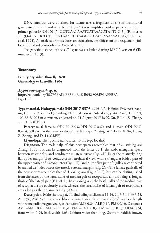
Two new species of the purse-web spider genus Atypus Latreille, 1804... 49
DNA barcodes were obtained for future use: a fragment of the mitochondrial gene cytochrome c oxidase subunit I (COI) was amplified and sequenced using the primer pairs: LCO1490 (5’-GGTCAACAAATCATAAAGATATTGG-3’) (Folmer et al. 1994) and HCO2198 (5’-TAAACTTCAGGGTGACCAAAAAATCA-3’) (Folmer et al. 1994). All molecular procedures on extraction, amplification and sequencing fol-lowed standard protocols (see Xu et al. 2015).
The genetic distance of the COI gene was calculated using MEGA version 6 (Ta-mura et al. 2013).
Taxonomy
Family Atypidae Thorell, 1870Genus Atypus Latreille, 1804
Atypus baotingensis sp. n.http://zoobank.org/B8795BAD-EF8F-4E4E-B032-908E91AFFBFAFigs 1, 2
Type material. Holotype male (HN-2017-037A): CHINA: Hainan Province: Baot-ing County, 2 km to Qixianling National Forest Park along y044 Road, 18.71°N, 109.68°E, 205 m elevation, collected on 21 August 2017 by X. Xu, F. Liu, Z. Zhang, and D. Li (CBEE).
Paratypes. 6 females (HN-2017-032-HN-2017-037) and 1 male (HN-2017-037B), collected at the same locality as the holotype, 21 August 2017 by X. Xu, F. Liu, Z. Zhang, and D. Li (CBEE).
Etymology. The specific name refers to the type locality.Diagnosis. The male palp of this new species resembles that of A. suiningensis
Zhang, 1985, but can be diagnosed from the latter by 1) the wide triangular space between its embolus and conductor in lateral views (Fig. 2H–I); 2) the relatively long flat upper margin of its conductor in retrolateral view, with a triangular folded part of the upper corner of its conductor (Fig. 2H); and 3) the first pair of sigilla are connected by arched wrinkles across the anterior sternal margin (Fig. 2C). The female genitalia of the new species resembles that of A. ledongensis (Fig. 3D–F), but can be distinguished from the latter by the basal stalks of median pair of receptacula almost being as long as those of the lateral pair (Fig. 2J–L). In A. ledongensis, the basal stalks of the median pair of receptacula are obviously short, whereas the basal stalks of lateral pair of receptacula are as long as their diameter (Fig. 3D–F).
Description. Male (holotype). TL (including chelicerae) 11.44. CL 3.34, CW 3.55, AL 4.56, AW 2.78. Carapace black brown. Fovea placed back 2/3 of carapace length with some radiative grooves. Eye diameter: AME 0.24, ALE 0.10, PME 0.10. Distances: AME–AME 0.46, AME–ALE 0.31, PME–PME 0.83, PME–PLE 0.13. MOA 0.34, front width 0.94, back width 1.03. Labium wider than long. Sternum reddish brown,
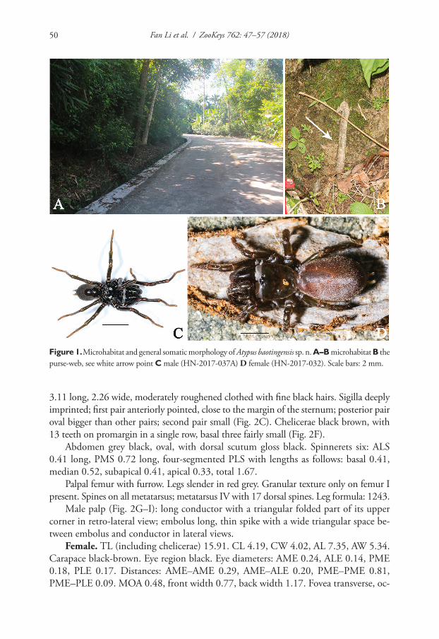
Fan Li et al. / ZooKeys 762: 47–57 (2018)50
3.11 long, 2.26 wide, moderately roughened clothed with fine black hairs. Sigilla deeply imprinted; first pair anteriorly pointed, close to the margin of the sternum; posterior pair oval bigger than other pairs; second pair small (Fig. 2C). Chelicerae black brown, with 13 teeth on promargin in a single row, basal three fairly small (Fig. 2F).
Abdomen grey black, oval, with dorsal scutum gloss black. Spinnerets six: ALS 0.41 long, PMS 0.72 long, four-segmented PLS with lengths as follows: basal 0.41, median 0.52, subapical 0.41, apical 0.33, total 1.67.
Palpal femur with furrow. Legs slender in red grey. Granular texture only on femur I present. Spines on all metatarsus; metatarsus IV with 17 dorsal spines. Leg formula: 1243.
Male palp (Fig. 2G–I): long conductor with a triangular folded part of its upper corner in retro-lateral view; embolus long, thin spike with a wide triangular space be-tween embolus and conductor in lateral views.
Female. TL (including chelicerae) 15.91. CL 4.19, CW 4.02, AL 7.35, AW 5.34. Carapace black-brown. Eye region black. Eye diameters: AME 0.24, ALE 0.14, PME 0.18, PLE 0.17. Distances: AME–AME 0.29, AME–ALE 0.20, PME–PME 0.81, PME–PLE 0.09. MOA 0.48, front width 0.77, back width 1.17. Fovea transverse, oc-
Figure 1. Microhabitat and general somatic morphology of Atypus baotingensis sp. n. A–B microhabitat B the purse-web, see white arrow point C male (HN-2017-037A) D female (HN-2017-032). Scale bars: 2 mm.
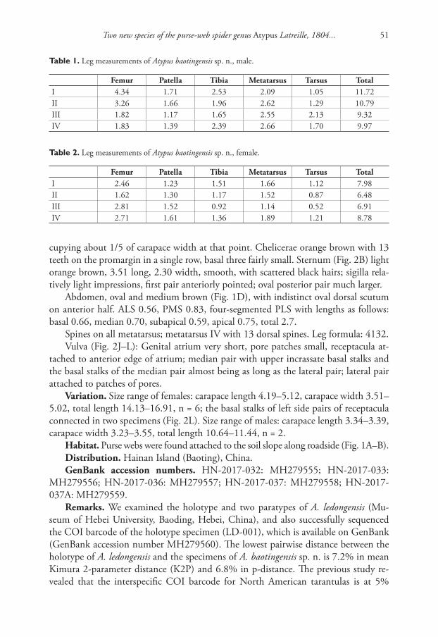
Two new species of the purse-web spider genus Atypus Latreille, 1804... 51
cupying about 1/5 of carapace width at that point. Chelicerae orange brown with 13 teeth on the promargin in a single row, basal three fairly small. Sternum (Fig. 2B) light orange brown, 3.51 long, 2.30 width, smooth, with scattered black hairs; sigilla rela-tively light impressions, first pair anteriorly pointed; oval posterior pair much larger.
Abdomen, oval and medium brown (Fig. 1D), with indistinct oval dorsal scutum on anterior half. ALS 0.56, PMS 0.83, four-segmented PLS with lengths as follows: basal 0.66, median 0.70, subapical 0.59, apical 0.75, total 2.7.
Spines on all metatarsus; metatarsus IV with 13 dorsal spines. Leg formula: 4132.Vulva (Fig. 2J–L): Genital atrium very short, pore patches small, receptacula at-
tached to anterior edge of atrium; median pair with upper incrassate basal stalks and the basal stalks of the median pair almost being as long as the lateral pair; lateral pair attached to patches of pores.
Variation. Size range of females: carapace length 4.19–5.12, carapace width 3.51–5.02, total length 14.13–16.91, n = 6; the basal stalks of left side pairs of receptacula connected in two specimens (Fig. 2L). Size range of males: carapace length 3.34–3.39, carapace width 3.23–3.55, total length 10.64–11.44, n = 2.
Habitat. Purse webs were found attached to the soil slope along roadside (Fig. 1A–B).Distribution. Hainan Island (Baoting), China.GenBank accession numbers. HN-2017-032: MH279555; HN-2017-033:
MH279556; HN-2017-036: MH279557; HN-2017-037: MH279558; HN-2017-037A: MH279559.
Remarks. We examined the holotype and two paratypes of A. ledongensis (Mu-seum of Hebei University, Baoding, Hebei, China), and also successfully sequenced the COI barcode of the holotype specimen (LD-001), which is available on GenBank (GenBank accession number MH279560). The lowest pairwise distance between the holotype of A. ledongensis and the specimens of A. baotingensis sp. n. is 7.2% in mean Kimura 2-parameter distance (K2P) and 6.8% in p-distance. The previous study re-vealed that the interspecific COI barcode for North American tarantulas is at 5%
Table 1. Leg measurements of Atypus baotingensis sp. n., male.
Femur Patella Tibia Metatarsus Tarsus TotalI 4.34 1.71 2.53 2.09 1.05 11.72II 3.26 1.66 1.96 2.62 1.29 10.79III 1.82 1.17 1.65 2.55 2.13 9.32IV 1.83 1.39 2.39 2.66 1.70 9.97
Table 2. Leg measurements of Atypus baotingensis sp. n., female.
Femur Patella Tibia Metatarsus Tarsus TotalI 2.46 1.23 1.51 1.66 1.12 7.98II 1.62 1.30 1.17 1.52 0.87 6.48III 2.81 1.52 0.92 1.14 0.52 6.91IV 2.71 1.61 1.36 1.89 1.21 8.78
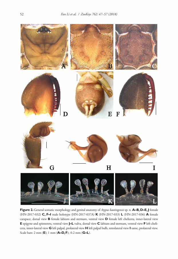
Fan Li et al. / ZooKeys 762: 47–57 (2018)52
Figure 2. General somatic morphology and genital anatomy of Atypus baotingensis sp. n. A–B, D–E, J female (HN-2017-032) C, F–I male holotype (HN-2017-037A) K (HN-2017-033) L (HN-2017-036) A female carapace, dorsal view B female labium and sternum, ventral view D female left chelicera, inner-lateral view E epigyne and spinnerets, ventral view J–L vulva, dorsal view C labium and sternum, ventral view F left cheli-cera, inner-lateral view G left palpal, prolateral view H left palpal bulb, retrolateral view I same, prolateral view. Scale bars: 2 mm (E); 1 mm (A–D, F); 0.2 mm (G–L).
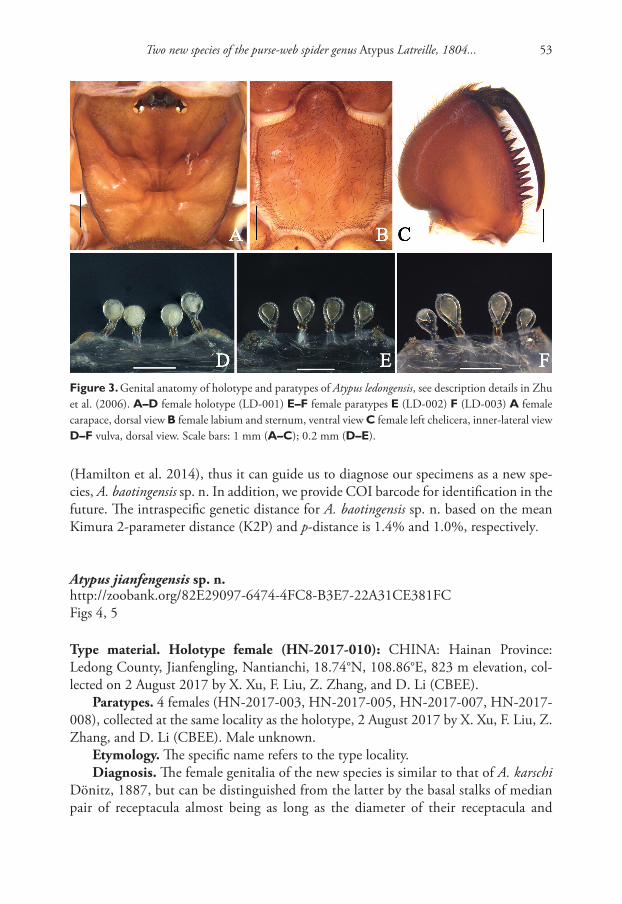
Two new species of the purse-web spider genus Atypus Latreille, 1804... 53
Figure 3. Genital anatomy of holotype and paratypes of Atypus ledongensis, see description details in Zhu et al. (2006). A–D female holotype (LD-001) E–F female paratypes E (LD-002) F (LD-003) A female carapace, dorsal view B female labium and sternum, ventral view C female left chelicera, inner-lateral view D–F vulva, dorsal view. Scale bars: 1 mm (A–C); 0.2 mm (D–E).
(Hamilton et al. 2014), thus it can guide us to diagnose our specimens as a new spe-cies, A. baotingensis sp. n. In addition, we provide COI barcode for identification in the future. The intraspecific genetic distance for A. baotingensis sp. n. based on the mean Kimura 2-parameter distance (K2P) and p-distance is 1.4% and 1.0%, respectively.
Atypus jianfengensis sp. n.http://zoobank.org/82E29097-6474-4FC8-B3E7-22A31CE381FCFigs 4, 5
Type material. Holotype female (HN-2017-010): CHINA: Hainan Province: Ledong County, Jianfengling, Nantianchi, 18.74°N, 108.86°E, 823 m elevation, col-lected on 2 August 2017 by X. Xu, F. Liu, Z. Zhang, and D. Li (CBEE).
Paratypes. 4 females (HN-2017-003, HN-2017-005, HN-2017-007, HN-2017-008), collected at the same locality as the holotype, 2 August 2017 by X. Xu, F. Liu, Z. Zhang, and D. Li (CBEE). Male unknown.
Etymology. The specific name refers to the type locality.Diagnosis. The female genitalia of the new species is similar to that of A. karschi
Dönitz, 1887, but can be distinguished from the latter by the basal stalks of median pair of receptacula almost being as long as the diameter of their receptacula and
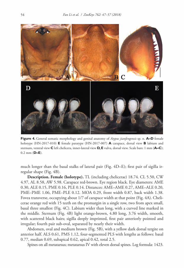
Fan Li et al. / ZooKeys 762: 47–57 (2018)54
Figure 4. General somatic morphology and genital anatomy of Atypus jianfengensis sp. n. A–D female holotype (HN-2017-010) E female paratype (HN-2017-007) A carapace, dorsal view B labium and sternum, ventral view C left chelicera, inner-lateral view D, E vulva, dorsal view. Scale bars: 1 mm (A–C); 0.2 mm (D–E).
much longer than the basal stalks of lateral pair (Fig. 4D–E); first pair of sigilla ir-regular shape (Fig. 4B).
Description. Female (holotype). TL (including chelicerae) 18.74. CL 5.50, CW 4.97, AL 8.58, AW 5.98. Carapace red-brown. Eye region black. Eye diameters: AME 0.30, ALE 0.15, PME 0.16, PLE 0.14. Distances: AME–AME 0.27, AME–ALE 0.20, PME–PME 1.06, PME–PLE 0.12. MOA 0.29, front width 0.87, back width 1.38. Fovea transverse, occupying about 1/7 of carapace width at that point (Fig. 4A). Cheli-cerae orange red with 15 teeth on the promargin in a single row, two from apex small, basal three smallest (Fig. 4C). Labium wider than long, with a curved line marked in the middle. Sternum (Fig. 4B) light orange-brown, 4.80 long, 3.76 width, smooth, with scattered black hairs; sigilla deeply imprinted, first pair anteriorly pointed and irregular; fourth pair sub-oval, separated by nearly their width.
Abdomen, oval and medium brown (Fig. 5B), with a yellow dark dorsal tergite on anterior half. ALS 0.61, PMS 1.12, four-segmented PLS with lengths as follows: basal 0.77, median 0.69, subapical 0.62, apical 0.42, total 2.5.
Spines on all metatarsus; metatarsus IV with eleven dorsal spines. Leg formula: 1423.
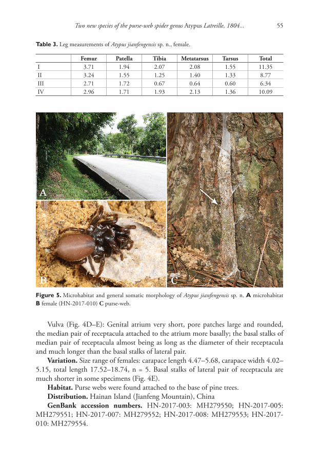
Two new species of the purse-web spider genus Atypus Latreille, 1804... 55
Table 3. Leg measurements of Atypus jianfengensis sp. n., female.
Femur Patella Tibia Metatarsus Tarsus TotalI 3.71 1.94 2.07 2.08 1.55 11.35II 3.24 1.55 1.25 1.40 1.33 8.77III 2.71 1.72 0.67 0.64 0.60 6.34IV 2.96 1.71 1.93 2.13 1.36 10.09
Vulva (Fig. 4D–E): Genital atrium very short, pore patches large and rounded, the median pair of receptacula attached to the atrium more basally; the basal stalks of median pair of receptacula almost being as long as the diameter of their receptacula and much longer than the basal stalks of lateral pair.
Variation. Size range of females: carapace length 4.47–5.68, carapace width 4.02–5.15, total length 17.52–18.74, n = 5. Basal stalks of lateral pair of receptacula are much shorter in some specimens (Fig. 4E).
Habitat. Purse webs were found attached to the base of pine trees.Distribution. Hainan Island (Jianfeng Mountain), ChinaGenBank accession numbers. HN-2017-003: MH279550; HN-2017-005:
MH279551; HN-2017-007: MH279552; HN-2017-008: MH279553; HN-2017-010: MH279554.
Figure 5. Microhabitat and general somatic morphology of Atypus jianfengensis sp. n. A microhabitat B female (HN-2017-010) C purse-web.
Fan Li et al. / ZooKeys 762: 47–57 (2018)56
Remarks. Although A. jianfengensis sp. n. is collected from Ledong County, Hain-an Island, it can be diagnosed from A. ledongensis found at the same area, Jianfeng mountains, by the latter having very short basal stalks of median pair of receptacula. In addition, it can be distinguished from A. baotingensis sp. n. by the latter having upper incrassate basal stalks of the median pair of receptacula. The intraspecific genetic dis-tance for A. jianfengensis sp. n. based on both the mean Kimura 2-parameter distance (K2P) and p-distance is 1.4% and 1.0%, respectively. The molecular data also provide the evidence that A. jianfengensis sp. n. can be distinguished from A. baotingensis sp. n. as well as from A. ledongensis: the interspecific genetic distance based on K2P and p-distance for the two new species are 15.4% and 13.8%, respectively, and between A. jianfengensis sp. n. and A. ledongensis are 17.2% and 15.3%, respectively.
Acknowledgements
We are grateful to Mary Lavelle, Ruairi Somers, Tadhg O`Sullivan and Noreen Mary Ward for their valuable suggestions in an earlier draft of the article. A special thank you goes to Feng Zhang for the loan of valuable spider specimens from the Museum of Hebei University (MHBU). Thanks also to Jian Chen for his help on taxonomy. We thank Chris Hamilton, Akio Tanikawa, and Feng Zhang for their insightful comments on our manuscript. This work was supported by the grants from the National Natural Science Foundation of China (NSFC-31572276 and NSFC-31601850), the Hunan Provincial Natural Science Foundation (2017JJ3202) and the Singapore Ministry of Education AcRF Tier 1 grant (R-154-000-A52-114).
References
Folmer O, Black M, Hoeh W, Lutz R, Vrijenhoek R (1994) DNA primers for amplification of mitochondrial cytochrome c oxidase subunit I from diverse metazoan invertebrates. Molecular Marine Biology and Biotechnology 3: 294–299.
Fourie R, Haddad CR, Jocqué R (2011) A revision of the purse-web spider genus Calom-mata Lucas, 1837 (Araneae, Atypidae) in the Afrotropical Region. ZooKeys 95: 1–28. https://doi.org/10.3897/zookeys.95.745
Hamilton CA, Hendrixson BE, Brewer MS, Bond JE (2014) An evaluation of sampling ef-fects on multiple DNA barcoding methods lead to an integrative approach for delimit-ing species: a case study of the North American tarantula genus Aphonopelma (Araneae, Mygalomorphae, Theraphosidae). Molecular Phylogenetics and Evolution 71: 79–93. https://doi.org/10.1016/j.ympev.2013.11.007
Jocqué R, Dippenaar-Schoeman AS (2006) Spider Families of the World. Royal Museum for Central Africa, Tervuren, 336 pp.
Two new species of the purse-web spider genus Atypus Latreille, 1804... 57
Kraus O, Baur H (1974) Die Atypidae der West-Paläarktis: Systematik, Verbreitung und Bi-ologie (Arach.: Araneae). Abhandlungen und Verhandlungen des Naturwissenschaftlichen Vereins in Hamburg (N.F.) 17: 85–116.
Li SQ, Lin YC (2016) Species Catalogue of China. Volume 2. Animals. Invertebrates (I), Arachnida: Araneae. Science Press, Beijing, 59–60.
Schwendinger PJ (1989) On the genus Atypus (Araneae: Atypidae) in northern Thailand. Bulletin of the British Arachnological Society 8: 89–96.
Schwendinger PJ (1990) A synopsis of the genus Atypus (Araneae, Atypidae). Zoologica Scripta 19: 353–366. https://doi.org/10.1111/j.1463-6409.1990.tb00263.x
Tamura K, Peterson D, Peterson N, Stecher G, Nei M, Kumar S (2013) MEGA6: Molecular evolutionary genetics analysis version 6.0. Molecular Biology and Evolution 28: 2731–2739. https://doi.org/10.1093/molbev/msr121
World Spider Catalog (2018) World spider catalog. Natural History Museum Bern, online at http://wsc.nmbe.ch, version 18.5 https://doi.org/10.24436/2 [Accessed on 30 April 2018]
Xu X, Liu FX, Cheng RC, Chen J, Xu X, Zhang ZS, Ono H, Pham DS, Norma-Rashid Y, Arnedo MA, Kuntner M, Li D (2015) Extant primitively segmented spiders have recently diversified from an ancient lineage. Proceedings of the Royal Society of London B: Biological Sciences 282: 20142486. https://doi.org/10.1098/rspb.2014.2486
Yin CM, Peng XJ, Yan HM, Bao YH, Xu X, Tang G, Zhou QS, Liu P (2012) Fauna Hunan: Araneae in Hunan, China. Hunan Science and Technology Press, Changsha, 1590 pp. http://www.wsc.nmbe.ch/reference/12312
Zhang YJ (1985) Two new species of spiders of the genus Atypus from China (Araneae: Atypi-dae). Acta Zootaxonomica Sinica 10: 140–147. http://www.wsc.nmbe.ch/reference/6172
Zhu MS, Zhang F, Song DX, Qu P (2006) A revision of the genus Atypus in China (Araneae: Atypidae). Zootaxa 1118: 1–42. http://www.wsc.nmbe.ch/reference/12301

Fan Li et al. / ZooKeys 762: 47–57 (2018)58
A new species of Dicranocentrus Schött from Hainan (China) with a key... 59
A new species of Dicranocentrus Schött from Hainan (China) with a key to the Chinese species of the genus
(Collembola, Entomobryidae)
Yuanhao Ren1, Zhaohui Li1, Feng Zhang2
1 Institute of Applied Ecology, Nanjing Xiaozhuang University, Nanjing 211171, P. R. China 2 Department of Entomology, College of Plant Protection, Nanjing Agricultural University, Nanjing 210095, P. R. China
Corresponding author: Feng Zhang ([email protected]; [email protected])
Academic editor: L. Deharveng | Received 27 January 2018 | Accepted 4 May 2018 | Published 30 May 2018
http://zoobank.org/198C08D3-5385-453C-B095-661AE1C67C92
Citation: Ren Y, Li Z, Zhang F (2018) A new species of Dicranocentrus Schött from Hainan (China) with a key to the Chinese species of the genus (Collembola, Entomobryidae). ZooKeys 762: 59–68. https://doi.org/10.3897/zookeys.762.23926
AbstractA new species, Dicranocentrus hainanicus Ren & Zhang, sp. n., is described from Hainan Province, China. Complete tergal chaetotaxy including microchaetae is illustrated and discussed. It is characterized by hav-ing the dental spines arranged in 2–3 rows, two inner teeth on unguis, 5, 2, 2 central macrochaetae on Abd. I–III, two inner S-chaetae on Abd. V displaced anteriorly, and the additional microchaetae associ-ated with the S-chaeta acc.p6 on Th. II–Abd. II. It is most similar to D. chenae Ma, Chen & Soto-Adames but differs from the latter in the number and arrangement of dental spines and the absence of macrochaeta Pa1 on dorsal head. A key to the Chinese species of the genus is provided.
Keywordschaetotaxy, Dicranocentrus hainanicus sp. n., Heteromurinae
Introduction
The genus Dicranocentrus Schött, 1893 is widespread in pantropical regions. It is char-acterized by having the first and the second antennal segments subdivided, the third and the fourth antennal segments annulated, eyes 8+8, prelabral chaetae not bifur-cated, postantennal organ absent, dental lobe without compound spines, dental spines
ZooKeys 762: 59–68 (2018)
doi: 10.3897/zookeys.762.23926
http://zookeys.pensoft.net
Copyright Yuanhao Ren et al. This is an open access article distributed under the terms of the Creative Commons Attribution License (CC BY 4.0), which permits unrestricted use, distribution, and reproduction in any medium, provided the original author and source are credited.
RESEARCH ARTICLE
Launched to accelerate biodiversity research
A peer-reviewed open-access journal
Yuanhao Ren et al. / ZooKeys 762: 59–68 (2018)60
present or absent, and the mucro bidentate with a basal spine (Cipola et al. 2016). Mari-Mutt (1979) made great contribution to its biology, character assessments, and phylogeny, and further divided it into three groups based on dorsal cephalic chaeto-taxy. Posterior macrochaetae on dorsal head are absent in marias-group, and macro-chaeta S2 is present in sundanensis-group but absent in gracilis-group. Dicranocentrus belongs to Heteromurini (Heteromurinae) due to the presence of body scales (Zhang and Deharveng 2015) but its relationships to other genera of Heteromurini are still unclear (Mari-Mutt 1980; Cipola et al. 2016). Dorsal cephalic and tergal macrochae-totaxy, differentiated chaetae on tibiotarsi, tenent hairs, dental spines are the main diagnostic characters widely used by Mari-Mutt and subsequent authors. Soto-Adames and Anderson (2017) attempted to explore the complete idiochaetotaxy (including macrochaetae, microchaetae, partial S-chaetae) for the first time in Dicranocentrus; homology of some elements were uncertain due to the absence of chaetotaxic modifi-cation during postembryonic development. These characteristics are usually unknown in four species from China: D. indicus Bonet, 1930 from Taiwan, D. chenae Ma, Chen & Soto-Adames, 2006 from Guangxi, D. wangi Ma & Chen, 2007 from Guangdong and D. liuae Xu & Zhang, 2014 from Anhui. Here a new species is described from the southernmost province of China, Hainan.
Materials and methods
Specimens were cleared in Nesbitt’s fluid, mounted under a coverslip in Hoyer’s solu-tion, and studied using a ZEISS AXIO Scope.A1 microscope and Axiocam camera. Dorsal body chaetae nomenclature follows Szeptycki (1979), dorsal cephalic chaetae follow Mari-Mutt (1979) and Soto-Adames (2008), inter-ocular chaetae follow Mari-Mutt (1986), tergal S-chaetae follow Zhang and Deharveng (2015), labial palp follows Fjellberg (1999), clypeal chaetae follow Yoshii and Suhardjono (1992) and Zhang et al. (2016), and labial chaetae follow Gisin (1967). The number of macrochaetae is given by half-tergite in the descriptions. Symbols representing chaetal elements used in the figures are as follows: circle, chaeta; cross, bothriotrichum; circle with a slash, pseu-dopore; “v”, scales. All materials are deposited in the collections of the Department of Entomology, College of Plant Protection, Nanjing Agricultural University (NJAU), P. R. China.
Abbreviations
Th. thoracic segment;Abd. abdominal segment;Ant. antennal segment;mac macrochaeta/ae;
mes mesochaeta/ae;mic microchaeta/ae;ms S-microchaeta/ae;sens ordinary tergal S-chaeta/ae.
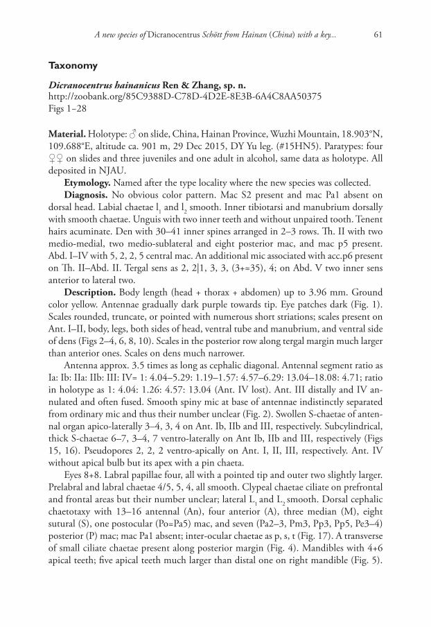
A new species of Dicranocentrus Schött from Hainan (China) with a key... 61
Taxonomy
Dicranocentrus hainanicus Ren & Zhang, sp. n.http://zoobank.org/85C9388D-C78D-4D2E-8E3B-6A4C8AA50375Figs 1−28
Material. Holotype: ♂ on slide, China, Hainan Province, Wuzhi Mountain, 18.903°N, 109.688°E, altitude ca. 901 m, 29 Dec 2015, DY Yu leg. (#15HN5). Paratypes: four ♀♀ on slides and three juveniles and one adult in alcohol, same data as holotype. All deposited in NJAU.
Etymology. Named after the type locality where the new species was collected.Diagnosis. No obvious color pattern. Mac S2 present and mac Pa1 absent on
dorsal head. Labial chaetae l1 and l2 smooth. Inner tibiotarsi and manubrium dorsally with smooth chaetae. Unguis with two inner teeth and without unpaired tooth. Tenent hairs acuminate. Den with 30–41 inner spines arranged in 2–3 rows. Th. II with two medio-medial, two medio-sublateral and eight posterior mac, and mac p5 present. Abd. I–IV with 5, 2, 2, 5 central mac. An additional mic associated with acc.p6 present on Th. II–Abd. II. Tergal sens as 2, 2|1, 3, 3, (3+≈35), 4; on Abd. V two inner sens anterior to lateral two.
Description. Body length (head + thorax + abdomen) up to 3.96 mm. Ground color yellow. Antennae gradually dark purple towards tip. Eye patches dark (Fig. 1). Scales rounded, truncate, or pointed with numerous short striations; scales present on Ant. I–II, body, legs, both sides of head, ventral tube and manubrium, and ventral side of dens (Figs 2–4, 6, 8, 10). Scales in the posterior row along tergal margin much larger than anterior ones. Scales on dens much narrower.
Antenna approx. 3.5 times as long as cephalic diagonal. Antennal segment ratio as Ia: Ib: IIa: IIb: III: IV= 1: 4.04–5.29: 1.19–1.57: 4.57–6.29: 13.04–18.08: 4.71; ratio in holotype as 1: 4.04: 1.26: 4.57: 13.04 (Ant. IV lost). Ant. III distally and IV an-nulated and often fused. Smooth spiny mic at base of antennae indistinctly separated from ordinary mic and thus their number unclear (Fig. 2). Swollen S-chaetae of anten-nal organ apico-laterally 3–4, 3, 4 on Ant. Ib, IIb and III, respectively. Subcylindrical, thick S-chaetae 6–7, 3–4, 7 ventro-laterally on Ant Ib, IIb and III, respectively (Figs 15, 16). Pseudopores 2, 2, 2 ventro-apically on Ant. I, II, III, respectively. Ant. IV without apical bulb but its apex with a pin chaeta.
Eyes 8+8. Labral papillae four, all with a pointed tip and outer two slightly larger. Prelabral and labral chaetae 4/5, 5, 4, all smooth. Clypeal chaetae ciliate on prefrontal and frontal areas but their number unclear; lateral L1 and L2 smooth. Dorsal cephalic chaetotaxy with 13–16 antennal (An), four anterior (A), three median (M), eight sutural (S), one postocular (Po=Pa5) mac, and seven (Pa2–3, Pm3, Pp3, Pp5, Pe3–4) posterior (P) mac; mac Pa1 absent; inter-ocular chaetae as p, s, t (Fig. 17). A transverse of small ciliate chaetae present along posterior margin (Fig. 4). Mandibles with 4+6 apical teeth; five apical teeth much larger than distal one on right mandible (Fig. 5).
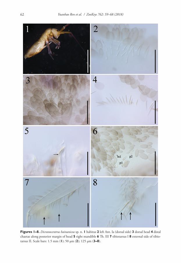
Yuanhao Ren et al. / ZooKeys 762: 59–68 (2018)62
Figures 1–8. Dicranocentrus hainanicus sp. n. 1 habitus 2 left Ant. Ia (dorsal side) 3 dorsal head 4 distal chaetae along posterior margin of head 5 right mandible 6 Th. III 7 tibiotarsus I 8 external side of tibio-tarsus II. Scale bars: 1.5 mm (1); 50 μm (2); 125 μm (3–8).
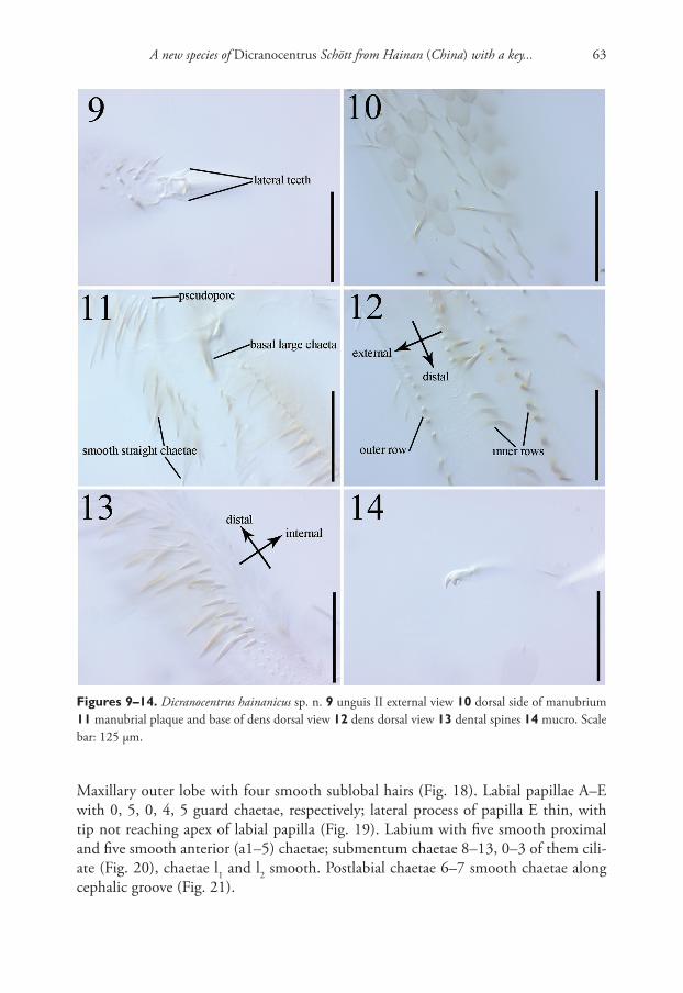
A new species of Dicranocentrus Schött from Hainan (China) with a key... 63
Figures 9–14. Dicranocentrus hainanicus sp. n. 9 unguis II external view 10 dorsal side of manubrium 11 manubrial plaque and base of dens dorsal view 12 dens dorsal view 13 dental spines 14 mucro. Scale bar: 125 μm.
Maxillary outer lobe with four smooth sublobal hairs (Fig. 18). Labial papillae A–E with 0, 5, 0, 4, 5 guard chaetae, respectively; lateral process of papilla E thin, with tip not reaching apex of labial papilla (Fig. 19). Labium with five smooth proximal and five smooth anterior (a1–5) chaetae; submentum chaetae 8–13, 0–3 of them cili-ate (Fig. 20), chaetae l1 and l2 smooth. Postlabial chaetae 6–7 smooth chaetae along cephalic groove (Fig. 21).
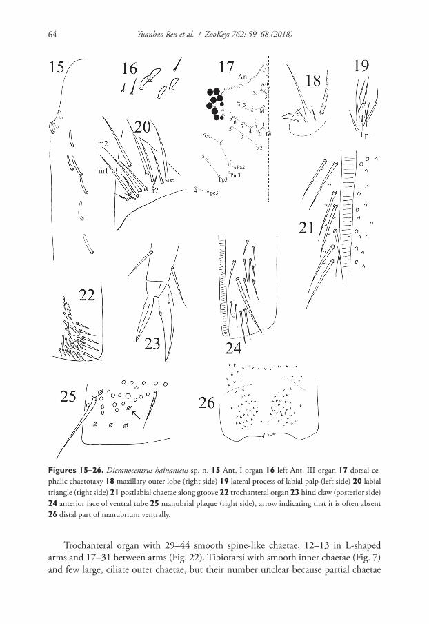
Yuanhao Ren et al. / ZooKeys 762: 59–68 (2018)64
Figures 15–26. Dicranocentrus hainanicus sp. n. 15 Ant. I organ 16 left Ant. III organ 17 dorsal ce-phalic chaetotaxy 18 maxillary outer lobe (right side) 19 lateral process of labial palp (left side) 20 labial triangle (right side) 21 postlabial chaetae along groove 22 trochanteral organ 23 hind claw (posterior side) 24 anterior face of ventral tube 25 manubrial plaque (right side), arrow indicating that it is often absent 26 distal part of manubrium ventrally.
Trochanteral organ with 29–44 smooth spine-like chaetae; 12–13 in L-shaped arms and 17–31 between arms (Fig. 22). Tibiotarsi with smooth inner chaetae (Fig. 7) and few large, ciliate outer chaetae, but their number unclear because partial chaetae
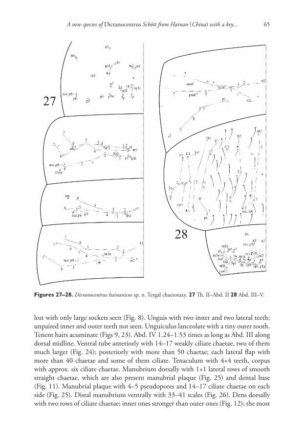
A new species of Dicranocentrus Schött from Hainan (China) with a key... 65
Figures 27–28. Dicranocentrus hainanicus sp. n. Tergal chaetotaxy. 27 Th. II–Abd. II 28 Abd. III–V.
lost with only large sockets seen (Fig. 8). Unguis with two inner and two lateral teeth; unpaired inner and outer teeth not seen. Unguiculus lanceolate with a tiny outer tooth. Tenent hairs acuminate (Figs 9, 23). Abd. IV 1.24–1.53 times as long as Abd. III along dorsal midline. Ventral tube anteriorly with 14–17 weakly ciliate chaetae, two of them much larger (Fig. 24); posteriorly with more than 50 chaetae; each lateral flap with more than 40 chaetae and some of them ciliate. Tenaculum with 4+4 teeth, corpus with approx. six ciliate chaetae. Manubrium dorsally with 1+1 lateral rows of smooth straight chaetae, which are also present manubrial plaque (Fig. 25) and dental base (Fig. 11). Manubrial plaque with 4–5 pseudopores and 14–17 ciliate chaetae on each side (Fig. 25). Distal manubrium ventrally with 33–41 scales (Fig. 26). Dens dorsally with two rows of ciliate chaetae; inner ones stronger than outer ones (Fig. 12); the most
Yuanhao Ren et al. / ZooKeys 762: 59–68 (2018)66
basal chaeta of inner row extremely thicker and longer (Fig. 11). Den internally with 30–41 (33 in holotype) basal spines arranged roughly in 2–3 rows; basal ones more dense than distal ones; outer spines larger than inner ones (Fig. 13). Smooth distal part of dens 4.70–6.25 times as long as mucro; apical tooth slightly longer than subapical one; mucronal spine just reaching the apex of subapical tooth (Fig. 14).
Th. II with two (m1, m2) medio-medial, two (m4, m4i) medio-sublateral and eight (p1, p1i, p1p, p2, p2a, p2p, p2e, p3, p5) posterior mac; m4p, m5, p1a, p4, p6 as mic. Th. III with 15 (a1–6, a4i, a6i, m6, p1–3, p1i, p2a, m6p) mac; a7 and m7 as mes; m1, m4–5 and p4–6 as mic. Abd. I with five (a2–3, m2–4) mac; a1, a5–6, m5–6, p5–6 as mic. Abd. II with two (m3, m3e) mac; a2–3, a5–6, m4–7 and p4–7 as mic (Fig. 27). Abd. III with two (a3, m3) central and two (pm6, p6) lateral mac; a1–2, a6–7, am6, m7, p4–5, and p7 as mic; m4 absent. Abd. IV with five (A5, B5–6, C1, a mac of unclear homology) central and nine (D3, E1, E3–4, F1–3, Ee8, Ee10) lateral mac. Abd. V with 13 (m2–3, m3a, a5, m5, m5a, m5e, p1, p3–5, ap6, p5a) mac (Fig. 28). Abd. VI without smooth chaetae. Tergal ms as 1, 0|1, 0, 1, 0, 0. Sens as 2, 2|1, 3, 3, (3+≈35), 4; Abd. IV with three ordinary sens (as, ps, acc.A6) and approx. 35 elongated sens; inner two sens anterior to lateral two on Abd. V. An additional mic associated with acc.p6 and of unclear homology present on Th. II–Abd. II.
Ecology. In litter of coniferous and broad-leaved mixed forests.Remarks. As a member of sundanensis-group (cephalic mac S2 present), D. hain-
anicus sp. n. is similar to Dicranocentrus chenae Ma, Chen & Soto-Adames in most fea-tures including two inner teeth on unguis and tergal macrochaetae, but differs from it in the absence of mac Pa1 on dorsal head, mac p5 present on Th. II and the abundant (30–41) dental spines arranged in 2–3 rows (single row in D. chenae). The absence of outer tooth on unguis is also interesting but the detail is often lacking or overlooked in descriptions of known species.
Nearly complete dorsal chaetotaxy is again illustrated and compared with Soto-Adames and Anderson (2017). On dorsal head, a mic between the outer two mac of series M is homologous to m3. For the two mac external to S5, the outer mac, having a larger socket than inner one, is supposed to be the primary chaeta S6. On Th. II, the most internal four posterior chaetae labeled as “p1, p2, p2a, p2p” in D. icelosmarias are named here as p1i, p1, p1a, p1p in the light of the presence of the set p2+ (p2, p2a, p2p) between p1 and p3. On Th. III, “p1, p2” in are possibly homologous to p2 and p2a in D. hainanicus sp. n. Mic p5 on Abd. I and mic m4 on Abd. II are absent in D. icelosmarias and Heteromurus nitidus but present in new species. On Abd. III, mic m4 is present in D. icelosmarias and H. nitidus but absent in D. hainanicus sp. n. On Abd. IV homologies of many chaetae are difficult to determine in the absence of sufficient evidence across Entomobryoidea; three ordinary sens are observed as those on Abd. II–III and named as as, acc.A6 and ps, respectively. On Abd. V, the number of sens is identical to those reported in Zhang and Deharveng (2015), therefore the inner two are supposed to as and acc.p3 here although their positions displace more anteriorly compared to D. wangi and D. liuae.
A new species of Dicranocentrus Schött from Hainan (China) with a key... 67
Key to the species of Dicranocentrus from China
1 Abd. I with 5+5 mac (Fig. 27) ....................................................................2– Abd. I with 3+3 mac ...................................................................................32 Mac Pa1 present on dorsal head; Th. II without p5 mac; dens with 15–19
inner spines arranged in a row ......chenae Ma, Chen & Soto-Adames, 2006– Mac Pa1 absent on dorsal head (Fig. 17); Th. II with p5 mac; dens with
30–41 inner spines arranged 2–3 rows .............................. hainanicus sp. n.3 Abd. III with 1+1 central mac ............................... liuae Xu & Zhang, 2014– Abd. III with 2+2 central mac (Fig. 28) ...................................................... 44 Abd. II with 3+3 mac; Abd. III with 3+3 lateral mac ... indicus Bonet, 1930– Abd. II with 2+2 mac (Fig. 27); Abd. III with 2+2 lateral mac (Fig. 28) .......
............................................................................ wangi Ma & Chen, 2007
Acknowledgments
This study was supported by the National Natural Science Foundation of China (31772491) and the Fundamental Research Funds for the Central Universities (KYX201617) granted to Feng Zhang and the Key Subject of Ecology of Jiangsu Prov-ince granted to Yuanhao Ren and Zhaohui Li. Thanks to Mr. Morgan A. McClure, who checked the English text.
References
Bonet F (1930) Sur quelques Collemboles de 1’Inde. Eos 6: 249–273.Cipola NG, Oliveira FG, Morais JW, Bellini BC (2016) The Heteromurini Absolon & Ksene-
mann (Collembola, Entomobryidae): a review of the genera status and diagnoses, keys for species of Alloscopus Börner and Heteromurtrella Mari Mutt and description of a new spe-cies. Zootaxa 4084: 151–186. https://doi.org/10.11646/zootaxa.4084.2.1
Fjellberg A (1999) The labial palp in Collembola. Zoologischer Anzeiger 237: 309–330.Gisin H (1967) Espèces nouvelles et lignées évolutives de Pseudosinella endogés (Collembola).
Memórias e Estudos do Museu Zoológico da Universidade de Coimbra 301: 1–25.Ma Y, Chen JX (2007) A new Dicranocentrus species (Collembola: Entomobryidae) from Chi-
na with a key to all species in the genus from Asia. Zootaxa 1633: 63–68.Ma YT, Chen JX, Soto-Adames FN (2006) A new species of Dicranocentrus from China. En-
tomological News 117: 485–490. https://doi.org/10.3157/0013-872X(2006)117[485:ANSODC]2.0.CO;2
Mari-Mutt JA (1979) A revision of the genus Dicranocentrus Schött (Insecta: Collembola: En-tomobryidae). Agricultural Experiment Station University of Puerto Rico Bulletin 259: 1–79.

Yuanhao Ren et al. / ZooKeys 762: 59–68 (2018)68
Mari-Mutt JA (1980) A classification of the Orchesellinae with a key to tribes, genera and sub-genera (Collembola: Entomobryidae). Annals of the Entomological Society of America 73: 455–459. https://doi.org/10.1093/aesa/73.4.455
Mari-Mutt JA (1986) Puerto rican species of Lepidocyrtus and Pseudosinella (Collembola: Ento-mobryidae). Caribbean Journal of Science 22: 1–48.
Schött H (1893) Beitrage zur kenntnis der insektenfauna von Kamerun. Bihang till Kongliga Svenska Vetenskaps-akademiens Handlingar 19: 5–28.
Soto-Adames FN (2008) Postembryonic development of the dorsal chaetotaxy in Seira dowlingi (Collembola, Entomobryidae); with an analysis of the diagnostic and phylogenetic signifi-cance of primary chaetotaxy in Seira. Zootaxa 1683: 1–31.
Soto-Adames FN, Anderson EW (2017) Two New Species and New Records of Springtails (Col-lembola: Entomobryidae, Paronellidae) from Nevis, Lesser Antilles. Florida Entomologist 100: 32–41. https://doi.org/10.1653/024.100.0107
Szeptycki A (1979) Chaetotaxy of the Entomobryidae and its phylogenetical significance. Mor-pho-systematic studies on Collembola. IV. Polska Akademia Nauk, Zakład Zoologii Sys-tematycznej i Doświadczalnej, Państwowe Wydawnictwo Naukowe, Warszawa, Kraków, Poland, 219 pp.
Xu G-L, Zhang F (2014) A new species of Dicranocentrus (Collembola: Entomobryidae) from China with comments on the systematic position of the genus. Zookeys 417: 1–8. https://doi.org/10.3897/zookeys.417.7373
Yoshii R, Suhardjono YR (1992) Notes on the Collembolan Fauna of Indonesia and its vicini-ties. III: Collembola of Timor Island. Acta Zoologica Asiae Orientalis 2: 75–96.
Zhang F, Deharveng L (2015) Systematic revision of Entomobryidae (Collembola) by integrat-ing molecular and new morphological evidence. Zoologica Scripta 44: 298–311. https://doi.org/10.1111/zsc.12100
Zhang F, Bedos A, Deharveng L (2016) Cave-dwelling Coecobrya from southern China with a survey of clypeal chaetae in Entomobryoidea (Collembola). European Journal of Tax-onomy 226: 1–26. https://doi.org/10.5852/ejt.2016.226
A comparative study of external female genitalia... 69
A comparative study of external female genitalia (including the 8th and 9th abdominal segments) in the
family Megalopodidae and other related families of Chrysomeloidea
Kaiqin Li1,2, Hongbin Liang1
1 Key Laboratory of Zoological Systematics and Evolution, Institute of Zoology, Chinese Academy of Sciences, Beijing 100101, China 2 Kunming Natural History Museum of Zoology, Kunming Institute of Zoology, Chi-nese Academy of Sciences, Kunming 650223, China
Corresponding author: Hongbin Liang ([email protected])
Academic editor: M. Schmitt | Received 9 November 2017 | Accepted 5 April 2018 | Published 30 May 2018
http://zoobank.org/8A70A177-55C9-47C1-9374-AE2DF926ADE0
Citation: Li K, Liang H (2018) A comparative study of external female genitalia (including the 8th and 9th abdominal segments) in the family Megalopodidae and other related families of Chrysomeloidea. ZooKeys 762: 69–104. https://doi.org/10.3897/zookeys.762.22163
AbstractThe external female genitalia of 29 species belonging to three genera of Megalopodidae and 80 species belonging to 61 genera of another four families in Chrysomeloidea were studied. The external female genitalia within the superfamily Chrysomeloidea can be divided into a cerambycid type and a chrysomelid type. The comparative study of external female genitalia shows Megalopodidae is more closely related to the family Cerambycidae than to the family Chrysomelidae s.l. Among five subfamilies of Cerambycidae we studied, the subfamily Lamiinae is most closely allied to Megalopodidae. An evolutionary path is pro-posed for the spiculum gastrale in Chrysomeloidea: the characteristic state of the spiculum gastrale with-out a joint is primary, and that with a joint is secondary. The family Orsodacnidae has probably evolved in isolation from the early chrysomelids, due to their shared external female genitalia (cerambycid type). In the family Chrysomelidae, the structure of external female genitalia and ovipositing behavior show that the subfamily Synetinae is closer to the Camptosomata than the subfamily Eumolpinae. In general, the shape of the terminal ovipositor is palp-like in the Chrysomeloidea. Terminal ovipositors are generally palp-shaped in Chrysomeloidea except for those that are lamellate in the genus Callispa and the subfamily Cassidinae who produce egg-sheaths.
ZooKeys 762: 69–104 (2018)
doi: 10.3897/zookeys.762.22163
http://zookeys.pensoft.net
Copyright Kaiqin Li, Hongbin Liang. This is an open access article distributed under the terms of the Creative Commons Attribution License (CC BY 4.0), which permits unrestricted use, distribution, and reproduction in any medium, provided the original author and source are credited.
RESEARCH ARTICLE
Launched to accelerate biodiversity research
A peer-reviewed open-access journal
Kaiqin Li & Hongbin Liang / ZooKeys 762: 69–104 (2018)70
KeywordsChrysomeloidea, female genitalia, Megalopodidae, morphology, 8th abdominal segment
Introduction
Adults of the family Megalopodidae commonly feed on the juices in stems and leaf tissues of plants in the families of Rosaceae, Oleaceae, Salicaceae, and Cel-astraceae, or consume the pollen of the Araucaria species (Tan et al. 1980, Reid 1989, Kuschel and May 1990, Li et al. 2013). They are distributed in Neotropi-cal, Afrotropical, Palaearctic, Oriental, Nearctic and Australian regions (Tan et al. 1980). In China the Megalopodidae is represented by two subfamilies Megalopo-dinae and Zeugophorinae (Yu and Liang 2002, Medvedev 2010, Silfverberg 2010). The subfamily Megalopodinae includes two genera (Temnaspis Lacordaire, 1845 and Poecilomorpha Hope, 1840). The subfamily Zeugophorinae includes only a single genus (Zeugophora Kunze, 1818) which is divided into two subgenera, Zeugophora Kunze, 1818 and Pedrillia Westwood, 1864 (Monrós 1959, Gressitt and Kimoto 1961, Chen and Pu 1962, Kimoto and Gressitt 1979, Medvedev 1985, 1997, Reid 1989, 1992, 1995, Schöller 2009, Warchałowski 2010). There are about 450 species of Megalopodidae in the world with 31 species of Megalopodinae and 24 species of Zeugophorinae in China (Gressitt and Kimoto 1961, Chen and Pu 1962, Chen 1974, Kimoto and Gressitt 1979, Tan et al. 1980, Medvedev and Sprecher-Ueber-sax 1997, Medvedev 2002, 2010, Yu and Liang 2002, Medvedev 2010, Silfverberg 2010, Lawrence and Ślipiński 2013, Li et al. 2013). Latreille (1802) established the subfamily Megalopodinae. Chapuis (1874) constructed the early taxonomic system for the Chrysomelidae. The subfamily Megalopodinae was included in the Camptosomes for some time until Chen (1940) transferred this subfamily from the Camptosomes to the Eupodes, which was regarded as the most primitive group. The subfamily Megalopodinae was then included in the family Donaciidae. This includ-ed the subfamilies Sagrinae, Donaciinae, Orsodacninae, Megascelinae, and Mega-lopodinae. The subfamily Zeugophorinae was established by Böving and Craighead (1931) and was included in the family Orsodacnidae based on characteristics of larvae. Following research on the external male genitalia within the subfamily Zeu-gophorinae, Chûjô (1952) pointed out that subfamily Zeugophorinae was more closely related to the Megalopodinae than other taxa in the Chrysomeloidea. Chen (1964, 1973) proposed that both subfamilies Megalopodinae and Zeugophorinae belonged to the criocerid line. The criocerid line also included the six following subfamilies: Sagrinae, Donaciinae, Criocerinae, Bruchidae, Orsodacninae and Syn-etinae. Crowson (1981) included the subfamily Zeugophorinae as a member of Megalopodidae. Therefore the Megalopodidae now included two subfamilies the Megalopodinae and the Zeugophorinae. Kuschel and May (1990) added a new subfamily, the Palophaginae. Some researchers continued to regard the Megalopodi-
A comparative study of external female genitalia... 71
dae (subfamilies Megalopodinae, Zeugophorinae, Palophaginae) as an independent family or subfamily (Lawrence and Britton 1991, Suzuki 1994). In addition, Reid (1995) also supported the Megalopodidae as an independent family and gradu-ally more researchers accepted this treatment (Hunt et al. 2007, Gómez-Zurita et al. 2007, 2008; Marvaldi et al. 2009, Löbl and Smetana 2010, McKenna et al. 2015). However, the phylogenetic relationships were not well resolved among the Chrysomeloidea (Reid 1995, 2000, Hunt et al. 2007, Gómez-Zurita et al. 2007, 2008, Marvaldi et al. 2009, McKenna et al. 2015). Chen (1985) examined sev-eral characteristics including head orientation, male genitalia, larval feeding habits, thorax, and wing-venation, and proposed that the Megalopodidae was related to the family of Cerambycidae. Suzuki (1988, 1994) considered that the Megalopo-didae probably descended from lamiid-type ancestors after studying their internal reproductive systems and hind wing venation. Schmitt (1992) also considered the Megalopodidae to be closer to the Cerambycidae than to the Chrysomelidae. Reid (1995) revealed Megalopodidae as a monophyletic group, with Orsodacnidae and Chrysomelidae as its sister groups, but the evidence supporting a close relationship between Megalopodidae and Cerambycidae was insufficient. In recent years, mo-lecular evidence has shown that the Megalopodidae is closest to the Cerambycidae or Oxypeltidae (Hunt et al. 2007, Gómez-Zurita et al. 2007, 2008, Marvaldi et al. 2009, McKenna et al. 2015).
Male and female genitalia are among the most important identification charac-teristics in the Megalopodidae and receive much attention from taxonomists. Chûjô (1952, 1953) studied the morphology of external male genitalia. Following his exten-sive analysis of the external male genitalia of the Megalopodinae and Zeugophorinae, Chûjô found similarities between the two subfamilies. Subsequently, taxonomists be-gan to describe the male genitalia of the Megalopodidae when publishing descriptions of new species and when they constructed or analyzed molecular phylogenetic rela-tionships within the Chrysomeloidea (Reid 1989, Kuschel and May 1990, Mann and Crowson 1996, Verma 1996, Medvedev 1997).
Research on external female genitalia in the Megalopodidae is rare. There were only a few descriptions of male or female genitalia for this family when new species were described or revisions were made (Reid 1989, 1992, Li et al. 2013, Rodríguez-Mirón and Zaragoza-Caballero 2017). Suzuki (1994) reported comprehensively on the internal reproductive system of Chrysomeloidea s.l., including the Megalopodinae and Zeugophorinae. Kasap and Crowson (1985) described briefly the 8th abdominal segment of the external female genitalia of Sphondylia afra (Megalopodinae) and Zeu-gophora fulvicollis (Zeugophorinae). Reid (1989, 1992) described the structure of the ovipositor and spermathecae of the Zeugophorinae when he published new species. Recently Rodríguez-Mirón et al. (2017) studied the spermathecae morphology of the Megalopodidae. In the present study, we dissected and describe the 8th abdominal seg-ments and the external genitalia in females, then compared them with those of other related families in the Chrysomeloidea.
Kaiqin Li & Hongbin Liang / ZooKeys 762: 69–104 (2018)72
Material and methods
Preparation of specimens
All measurements were made with the aid of an ocular micrometer in the Nikon SMZ1500 stereoscopic microscope. Dry specimens were soaked in boiled water for 1–2 hours. For larger specimens the lateral margin of the abdomen was opened at its apex and the geni-talia were pulled out of the abdomen with fine forceps. For smaller specimens the whole abdomen was removed from the body. The genitalia and/or whole abdomen were soaked in a warm solution of 10% KOH for 10–20 minutes as a pretreatment. The treatment time depended upon the degree of sclerotization found in different species. After pretreat-ment these organs were washed in water several times. Genitalia were then detached and some were dyed with Chlorazol Black E to stain membranous tissue a blue color. They were then transferred to glycerin for observation, photography, and preservation.
Photographs of female genitalia were taken using a Nikon SMZ-1500 stereoscop-ic dissecting microscope fitted with a Cannon 450D digital camera. Photographs of habitus were captured by a Cannon Macro 100 mm lens fitted to the Cannon 450D camera. For each final image, several photographs were taken at different focal planes, combined with Helicon Focus software to obtain one synthesized photograph, and finally edited with Adobe Photoshop software.
Terminology
Morphological terminology for the female genitalia of the Megalopodidae follows Snodgrass (1935), Chûjô (1952, 1953), Kasap and Crowson (1985) and Lawrence et al. (2010). In females of the Chrysomeloidea, the genitalia segment is located on the 9th segment of the abdomen. The ovipositor consists of the proctiger, paraproct, median plate, valvifer, coxite, and stylus (Kasap and Crowson, 1985). In contrast, the ovipositor of the Cerambycidae consists of the proctiger and vaginal palpi with baculi. The dorsal side of the baculi of the vaginal palpi is called the dorsal baculi. The ventral side of the baculi includes the paraproct baculi, valvifer baculi, and coxite baculi (Saito 1989). The ovipositor of the Megalopodidae includes two long vaginal palpi, which have one or two pair of baculi. Therefore, the ovipositor consists of the paraproct, valvifer, coxite, and stylus without a proctiger or a median plate.
Outside the ovipositor of Nupserha bicolor Thomson (Cerambycidae) there is a membranous bag-like capsule known as the capsular sheath (Dutt 1958). Saito (1989) suggested that this structure was the inter-segmental membrane found between the ovipositor and the 8th segment. The ovipositor of Megalopodidae is surrounded by a pocket-like membrane. Kuschel and May (1990) named this membrane as a genital pocket. The base of the pocket is connected with the apical margin of the 8th segment and the apex of the pocket is connected with the base of the ovipositor. The surface of the genital pocket is thickened by many sclerotized rings (Figs 9, 10).
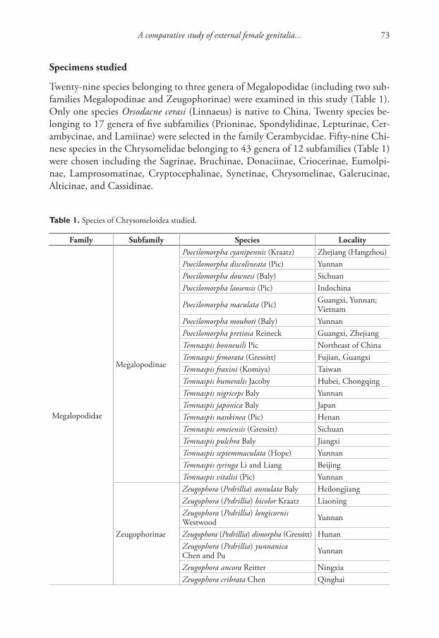
A comparative study of external female genitalia... 73
Specimens studied
Twenty-nine species belonging to three genera of Megalopodidae (including two sub-families Megalopodinae and Zeugophorinae) were examined in this study (Table 1). Only one species Orsodacne cerasi (Linnaeus) is native to China. Twenty species be-longing to 17 genera of five subfamilies (Prioninae, Spondylidinae, Lepturinae, Cer-ambycinae, and Lamiinae) were selected in the family Cerambycidae. Fifty-nine Chi-nese species in the Chrysomelidae belonging to 43 genera of 12 subfamilies (Table 1) were chosen including the Sagrinae, Bruchinae, Donaciinae, Criocerinae, Eumolpi-nae, Lamprosomatinae, Cryptocephalinae, Synetinae, Chrysomelinae, Galerucinae, Alticinae, and Cassidinae.
Table 1. Species of Chrysomeloidea studied.
Family Subfamily Species Locality
Megalopodidae
Megalopodinae
Poecilomorpha cyanipennis (Kraatz) Zhejiang (Hangzhou)Poecilomorpha discolineata (Pic) YunnanPoecilomorpha downesi (Baly) SichuanPoecilomorpha laosensis (Pic) Indochina
Poecilomorpha maculata (Pic) Guangxi, Yunnan; Vietnam
Poecilomorpha mouhoti (Baly) YunnanPoecilomorpha pretiosa Reineck Guangxi, ZhejiangTemnaspis bonneuili Pic Northeast of China Temnaspis femorata (Gressitt) Fujian, GuangxiTemnaspis fraxini (Komiya) TaiwanTemnaspis humeralis Jacoby Hubei, ChongqingTemnaspis nigriceps Baly YunnanTemnaspis japonica Baly JapanTemnaspis nankinea (Pic) HenanTemnaspis omeiensis (Gressitt) SichuanTemnaspis pulchra Baly JiangxiTemnaspis septemmaculata (Hope) YunnanTemnaspis syringa Li and Liang BeijingTemnaspis vitalisi (Pic) Yunnan
Zeugophorinae
Zeugophora (Pedrillia) annulata Baly HeilongjiangZeugophora (Pedrillia) bicolor Kraatz LiaoningZeugophora (Pedrillia) longicornis Westwood Yunnan
Zeugophora (Pedrillia) dimorpha (Gressitt) HunanZeugophora (Pedrillia) yunnanica Chen and Pu Yunnan
Zeugophora ancora Reitter NingxiaZeugophora cribrata Chen Qinghai
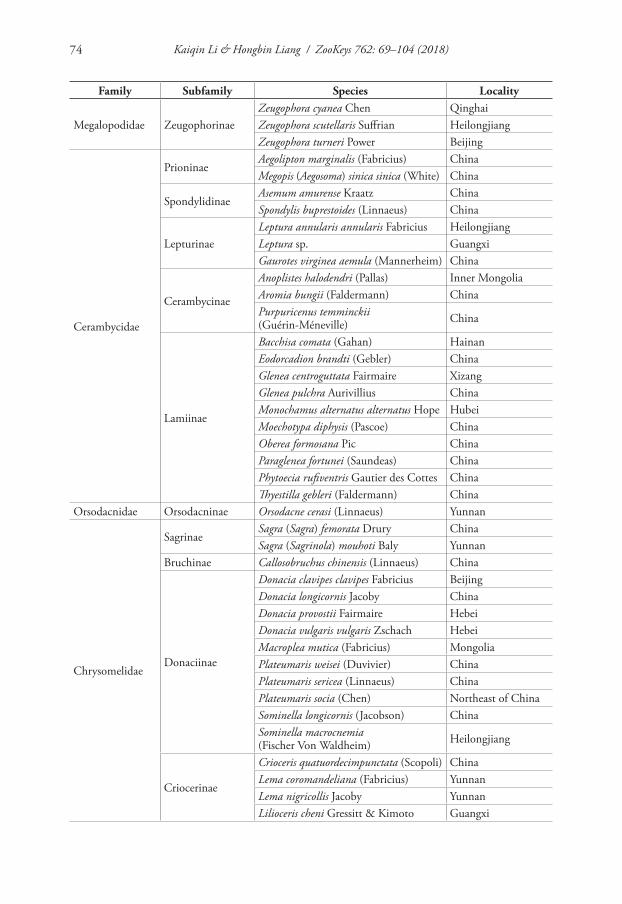
Kaiqin Li & Hongbin Liang / ZooKeys 762: 69–104 (2018)74
Family Subfamily Species Locality
Megalopodidae ZeugophorinaeZeugophora cyanea Chen QinghaiZeugophora scutellaris Suffrian Heilongjiang Zeugophora turneri Power Beijing
Cerambycidae
PrioninaeAegolipton marginalis (Fabricius) ChinaMegopis (Aegosoma) sinica sinica (White) China
SpondylidinaeAsemum amurense Kraatz ChinaSpondylis buprestoides (Linnaeus) China
LepturinaeLeptura annularis annularis Fabricius HeilongjiangLeptura sp. GuangxiGaurotes virginea aemula (Mannerheim) China
Cerambycinae
Anoplistes halodendri (Pallas) Inner MongoliaAromia bungii (Faldermann) ChinaPurpuricenus temminckii (Guérin-Méneville) China
Lamiinae
Bacchisa comata (Gahan) HainanEodorcadion brandti (Gebler) ChinaGlenea centroguttata Fairmaire XizangGlenea pulchra Aurivillius ChinaMonochamus alternatus alternatus Hope HubeiMoechotypa diphysis (Pascoe) ChinaOberea formosana Pic ChinaParaglenea fortunei (Saundeas) ChinaPhytoecia rufiventris Gautier des Cottes ChinaThyestilla gebleri (Faldermann) China
Orsodacnidae Orsodacninae Orsodacne cerasi (Linnaeus) Yunnan
Chrysomelidae
SagrinaeSagra (Sagra) femorata Drury ChinaSagra (Sagrinola) mouhoti Baly Yunnan
Bruchinae Callosobruchus chinensis (Linnaeus) China
Donaciinae
Donacia clavipes clavipes Fabricius BeijingDonacia longicornis Jacoby ChinaDonacia provostii Fairmaire HebeiDonacia vulgaris vulgaris Zschach HebeiMacroplea mutica (Fabricius) MongoliaPlateumaris weisei (Duvivier) ChinaPlateumaris sericea (Linnaeus) ChinaPlateumaris socia (Chen) Northeast of ChinaSominella longicornis (Jacobson) ChinaSominella macrocnemia (Fischer Von Waldheim) Heilongjiang
Criocerinae
Crioceris quatuordecimpunctata (Scopoli) ChinaLema coromandeliana (Fabricius) YunnanLema nigricollis Jacoby YunnanLilioceris cheni Gressitt & Kimoto Guangxi
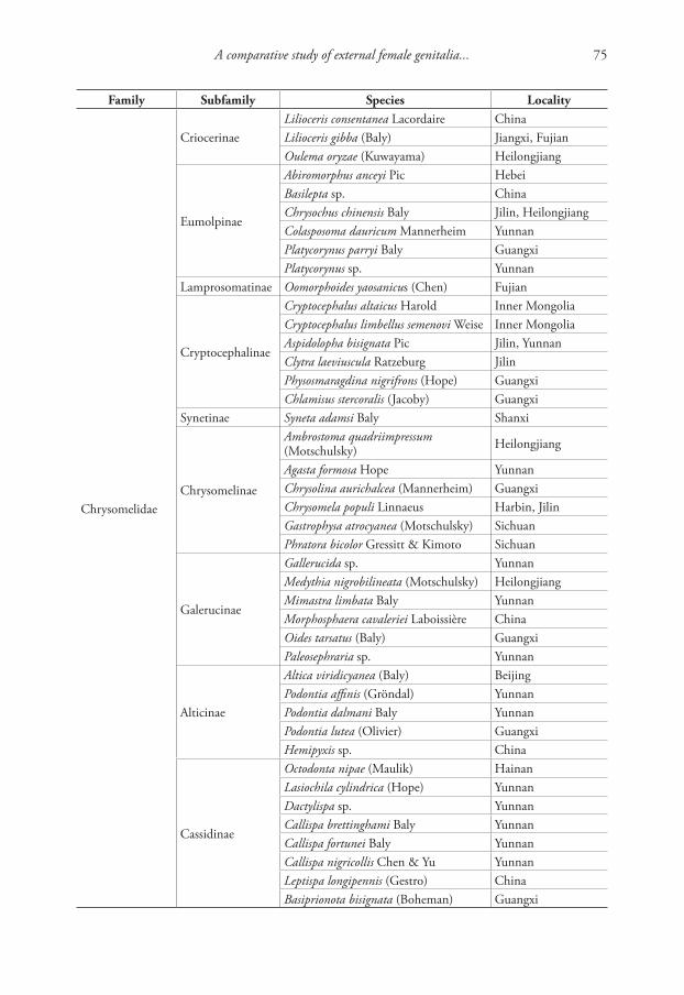
A comparative study of external female genitalia... 75
Family Subfamily Species Locality
Chrysomelidae
CriocerinaeLilioceris consentanea Lacordaire ChinaLilioceris gibba (Baly) Jiangxi, FujianOulema oryzae (Kuwayama) Heilongjiang
Eumolpinae
Abiromorphus anceyi Pic HebeiBasilepta sp. ChinaChrysochus chinensis Baly Jilin, HeilongjiangColasposoma dauricum Mannerheim YunnanPlatycorynus parryi Baly GuangxiPlatycorynus sp. Yunnan
Lamprosomatinae Oomorphoides yaosanicus (Chen) Fujian
Cryptocephalinae
Cryptocephalus altaicus Harold Inner MongoliaCryptocephalus limbellus semenovi Weise Inner MongoliaAspidolopha bisignata Pic Jilin, YunnanClytra laeviuscula Ratzeburg JilinPhysosmaragdina nigrifrons (Hope) GuangxiChlamisus stercoralis (Jacoby) Guangxi
Synetinae Syneta adamsi Baly Shanxi
Chrysomelinae
Ambrostoma quadriimpressum (Motschulsky) Heilongjiang
Agasta formosa Hope YunnanChrysolina aurichalcea (Mannerheim) GuangxiChrysomela populi Linnaeus Harbin, JilinGastrophysa atrocyanea (Motschulsky) SichuanPhratora bicolor Gressitt & Kimoto Sichuan
Galerucinae
Gallerucida sp. YunnanMedythia nigrobilineata (Motschulsky) HeilongjiangMimastra limbata Baly YunnanMorphosphaera cavaleriei Laboissière ChinaOides tarsatus (Baly) GuangxiPaleosephraria sp. Yunnan
Alticinae
Altica viridicyanea (Baly) BeijingPodontia affinis (Gröndal) YunnanPodontia dalmani Baly YunnanPodontia lutea (Olivier) GuangxiHemipyxis sp. China
Cassidinae
Octodonta nipae (Maulik) HainanLasiochila cylindrica (Hope) YunnanDactylispa sp. YunnanCallispa brettinghami Baly YunnanCallispa fortunei Baly YunnanCallispa nigricollis Chen & Yu YunnanLeptispa longipennis (Gestro) ChinaBasiprionota bisignata (Boheman) Guangxi
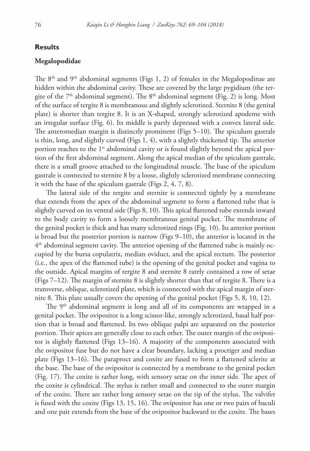
Kaiqin Li & Hongbin Liang / ZooKeys 762: 69–104 (2018)76
Results
Megalopodidae
The 8th and 9th abdominal segments (Figs 1, 2) of females in the Megalopodinae are hidden within the abdominal cavity. These are covered by the large pygidium (the ter-gite of the 7th abdominal segment). The 8th abdominal segment (Fig. 2) is long. Most of the surface of tergite 8 is membranous and slightly sclerotized. Sternite 8 (the genital plate) is shorter than tergite 8. It is an X-shaped, strongly sclerotized apodeme with an irregular surface (Fig. 6). Its middle is partly depressed with a convex lateral side. The anteromedian margin is distinctly prominent (Figs 5–10). The spiculum gastrale is thin, long, and slightly curved (Figs 1, 4), with a slightly thickened tip. The anterior portion reaches to the 1st abdominal cavity or is found slightly beyond the apical por-tion of the first abdominal segment. Along the apical median of the spiculum gastrale, there is a small groove attached to the longitudinal muscle. The base of the spiculum gastrale is connected to sternite 8 by a loose, slightly sclerotized membrane connecting it with the base of the spiculum gastrale (Figs 2, 4, 7, 8).
The lateral side of the tergite and sternite is connected tightly by a membrane that extends from the apex of the abdominal segment to form a flattened tube that is slightly curved on its ventral side (Figs 8, 10). This apical flattened tube extends inward to the body cavity to form a loosely membranous genital pocket. The membrane of the genital pocket is thick and has many sclerotized rings (Fig. 10). Its anterior portion is broad but the posterior portion is narrow (Figs 9–10), the anterior is located in the 4th abdominal segment cavity. The anterior opening of the flattened tube is mainly oc-cupied by the bursa copulatrix, median oviduct, and the apical rectum. The posterior (i.e., the apex of the flattened tube) is the opening of the genital pocket and vagina to the outside. Apical margins of tergite 8 and sternite 8 rarely contained a row of setae (Figs 7–12). The margin of sternite 8 is slightly shorter than that of tergite 8. There is a transverse, oblique, sclerotized plate, which is connected with the apical margin of ster-nite 8. This plate usually covers the opening of the genital pocket (Figs 5, 8, 10, 12).
The 9th abdominal segment is long and all of its components are wrapped in a genital pocket. The ovipositor is a long scissor-like, strongly sclerotized, basal half por-tion that is broad and flattened. Its two oblique palpi are separated on the posterior portion. Their apices are generally close to each other. The outer margin of the oviposi-tor is slightly flattened (Figs 13–16). A majority of the components associated with the ovipositor fuse but do not have a clear boundary, lacking a proctiger and median plate (Figs 13–16). The paraproct and coxite are fused to form a flattened sclerite at the base. The base of the ovipositor is connected by a membrane to the genital pocket (Fig. 17). The coxite is rather long, with sensory setae on the inner side. The apex of the coxite is cylindrical. The stylus is rather small and connected to the outer margin of the coxite. There are rather long sensory setae on the tip of the stylus. The valvifer is fused with the coxite (Figs 13, 15, 16). The ovipositor has one or two pairs of baculi and one pair extends from the base of the ovipositor backward to the coxite. The bases
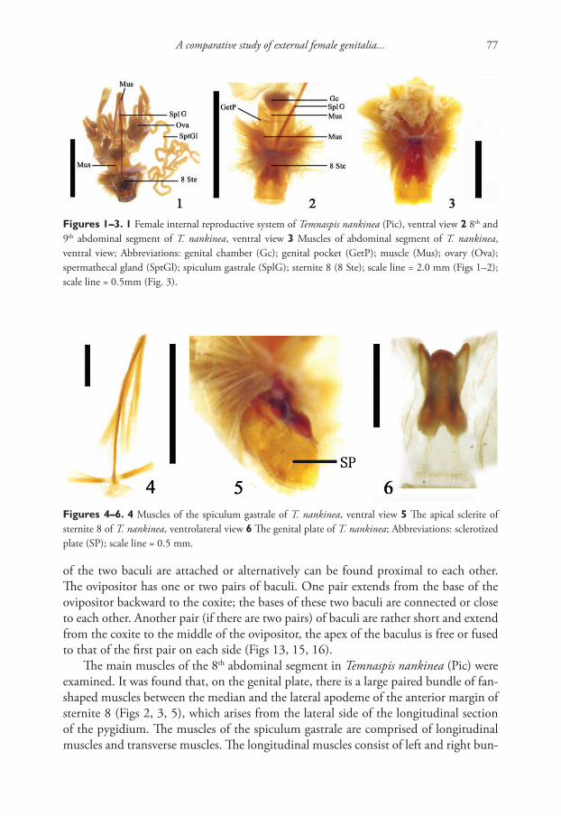
A comparative study of external female genitalia... 77
of the two baculi are attached or alternatively can be found proximal to each other. The ovipositor has one or two pairs of baculi. One pair extends from the base of the ovipositor backward to the coxite; the bases of these two baculi are connected or close to each other. Another pair (if there are two pairs) of baculi are rather short and extend from the coxite to the middle of the ovipositor, the apex of the baculus is free or fused to that of the first pair on each side (Figs 13, 15, 16).
The main muscles of the 8th abdominal segment in Temnaspis nankinea (Pic) were examined. It was found that, on the genital plate, there is a large paired bundle of fan-shaped muscles between the median and the lateral apodeme of the anterior margin of sternite 8 (Figs 2, 3, 5), which arises from the lateral side of the longitudinal section of the pygidium. The muscles of the spiculum gastrale are comprised of longitudinal muscles and transverse muscles. The longitudinal muscles consist of left and right bun-
Figures 1–3. 1 Female internal reproductive system of Temnaspis nankinea (Pic), ventral view 2 8th and 9th abdominal segment of T. nankinea, ventral view 3 Muscles of abdominal segment of T. nankinea, ventral view; Abbreviations: genital chamber (Gc); genital pocket (GetP); muscle (Mus); ovary (Ova); spermathecal gland (SptGl); spiculum gastrale (SplG); sternite 8 (8 Ste); scale line = 2.0 mm (Figs 1–2); scale line = 0.5mm (Fig. 3).
Figures 4–6. 4 Muscles of the spiculum gastrale of T. nankinea, ventral view 5 The apical sclerite of sternite 8 of T. nankinea, ventrolateral view 6 The genital plate of T. nankinea; Abbreviations: sclerotized plate (SP); scale line = 0.5 mm.
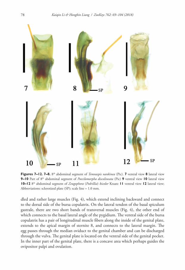
Kaiqin Li & Hongbin Liang / ZooKeys 762: 69–104 (2018)78
dled and rather large muscles (Fig. 4), which extend inclining backward and connect to the dorsal side of the bursa copulatrix. On the lateral tendon of the basal spiculum gastrale, there are two short bands of transversal muscles (Fig. 4), the other end of which connects to the basal lateral angle of the pygidium. The ventral side of the bursa copulatrix has a pair of longitudinal muscle fibers along the inside of the genital plate, extends to the apical margin of sternite 8, and connects to the lateral margin. The egg passes through the median oviduct to the genital chamber and can be discharged through the vulva. The genital plate is located on the ventral side of the genital pocket. In the inner part of the genital plate, there is a concave area which perhaps guides the ovipositor palpi and ovulation.
Figures 7–12. 7–8. 8th abdominal segment of Temnaspis nankinea (Pic). 7 ventral view 8 lateral view 9–10 Part of 8th abdominal segment of Poecilomorpha discolineata (Pic) 9 ventral view 10 lateral view 10–12 8th abdominal segment of Zeugophora (Pedrillia) bicolor Kraatz 11 ventral view 12 lateral view; Abbreviations: sclerotized plate (SP); scale line = 1.0 mm.
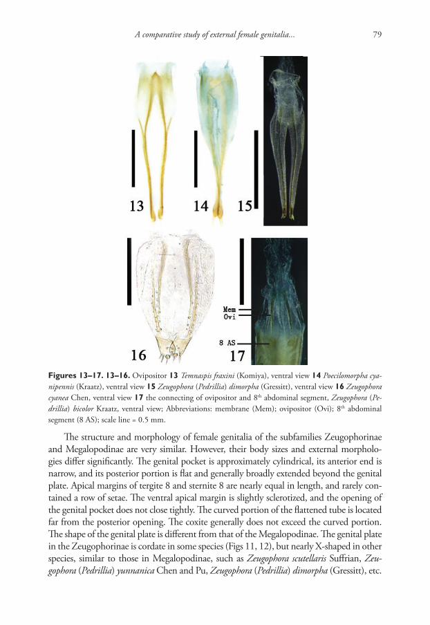
A comparative study of external female genitalia... 79
The structure and morphology of female genitalia of the subfamilies Zeugophorinae and Megalopodinae are very similar. However, their body sizes and external morpholo-gies differ significantly. The genital pocket is approximately cylindrical, its anterior end is narrow, and its posterior portion is flat and generally broadly extended beyond the genital plate. Apical margins of tergite 8 and sternite 8 are nearly equal in length, and rarely con-tained a row of setae. The ventral apical margin is slightly sclerotized, and the opening of the genital pocket does not close tightly. The curved portion of the flattened tube is located far from the posterior opening. The coxite generally does not exceed the curved portion. The shape of the genital plate is different from that of the Megalopodinae. The genital plate in the Zeugophorinae is cordate in some species (Figs 11, 12), but nearly X-shaped in other species, similar to those in Megalopodinae, such as Zeugophora scutellaris Suffrian, Zeu-gophora (Pedrillia) yunnanica Chen and Pu, Zeugophora (Pedrillia) dimorpha (Gressitt), etc.
Figures 13–17. 13–16. Ovipositor 13 Temnaspis fraxini (Komiya), ventral view 14 Poecilomorpha cya-nipennis (Kraatz), ventral view 15 Zeugophora (Pedrillia) dimorpha (Gressitt), ventral view 16 Zeugophora cyanea Chen, ventral view 17 the connecting of ovipositor and 8th abdominal segment, Zeugophora (Pe-drillia) bicolor Kraatz, ventral view; Abbreviations: membrane (Mem); ovipositor (Ovi); 8th abdominal segment (8 AS); scale line = 0.5 mm.
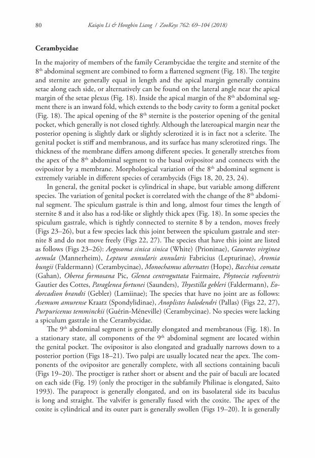
Kaiqin Li & Hongbin Liang / ZooKeys 762: 69–104 (2018)80
Cerambycidae
In the majority of members of the family Cerambycidae the tergite and sternite of the 8th abdominal segment are combined to form a flattened segment (Fig. 18). The tergite and sternite are generally equal in length and the apical margin generally contains setae along each side, or alternatively can be found on the lateral angle near the apical margin of the setae plexus (Fig. 18). Inside the apical margin of the 8th abdominal seg-ment there is an inward fold, which extends to the body cavity to form a genital pocket (Fig. 18). The apical opening of the 8th sternite is the posterior opening of the genital pocket, which generally is not closed tightly. Although the lateroapical margin near the posterior opening is slightly dark or slightly sclerotized it is in fact not a sclerite. The genital pocket is stiff and membranous, and its surface has many sclerotized rings. The thickness of the membrane differs among different species. It generally stretches from the apex of the 8th abdominal segment to the basal ovipositor and connects with the ovipositor by a membrane. Morphological variation of the 8th abdominal segment is extremely variable in different species of cerambycids (Figs 18, 20, 23, 24).
In general, the genital pocket is cylindrical in shape, but variable among different species. The variation of genital pocket is correlated with the change of the 8th abdomi-nal segment. The spiculum gastrale is thin and long, almost four times the length of sternite 8 and it also has a rod-like or slightly thick apex (Fig. 18). In some species the spiculum gastrale, which is tightly connected to sternite 8 by a tendon, moves freely (Figs 23–26), but a few species lack this joint between the spiculum gastrale and ster-nite 8 and do not move freely (Figs 22, 27). The species that have this joint are listed as follows (Figs 23–26): Aegosoma sinica sinica (White) (Prioninae), Gaurotes virginea aemula (Mannerheim), Leptura annularis annularis Fabricius (Lepturinae), Aromia bungii (Faldermann) (Cerambycinae), Monochamus alternates (Hope), Bacchisa comata (Gahan), Oberea formosana Pic, Glenea centroguttata Fairmaire, Phytoecia rufiventris Gautier des Cottes, Paraglenea fortunei (Saunders), Thyestilla gebleri (Faldermann), Eo-dorcadion brandti (Gebler) (Lamiinae); The species that have no joint are as follows: Asemum amurense Kraatz (Spondylidinae), Anoplistes halodendri (Pallas) (Figs 22, 27), Purpuricenus temminckii (Guérin-Méneville) (Cerambycinae). No species were lacking a spiculum gastrale in the Cerambycidae.
The 9th abdominal segment is generally elongated and membranous (Fig. 18). In a stationary state, all components of the 9th abdominal segment are located within the genital pocket. The ovipositor is also elongated and gradually narrows down to a posterior portion (Figs 18–21). Two palpi are usually located near the apex. The com-ponents of the ovipositor are generally complete, with all sections containing baculi (Figs 19–20). The proctiger is rather short or absent and the pair of baculi are located on each side (Fig. 19) (only the proctiger in the subfamily Philinae is elongated, Saito 1993). The paraproct is generally elongated, and on its basolateral side its baculus is long and straight. The valvifer is generally fused with the coxite. The apex of the coxite is cylindrical and its outer part is generally swollen (Figs 19–20). It is generally
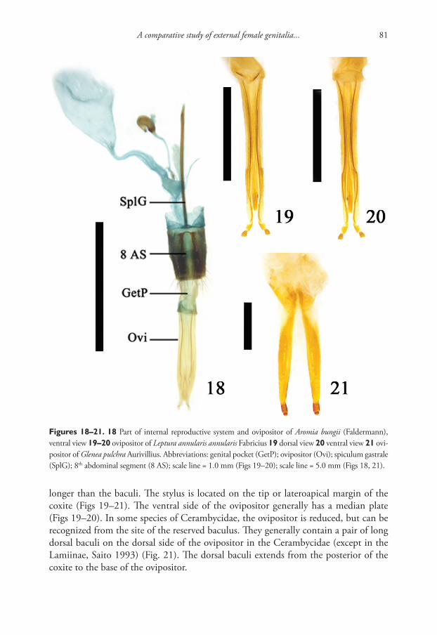
A comparative study of external female genitalia... 81
longer than the baculi. The stylus is located on the tip or lateroapical margin of the coxite (Figs 19–21). The ventral side of the ovipositor generally has a median plate (Figs 19–20). In some species of Cerambycidae, the ovipositor is reduced, but can be recognized from the site of the reserved baculus. They generally contain a pair of long dorsal baculi on the dorsal side of the ovipositor in the Cerambycidae (except in the Lamiinae, Saito 1993) (Fig. 21). The dorsal baculi extends from the posterior of the coxite to the base of the ovipositor.
Figures 18–21. 18 Part of internal reproductive system and ovipositor of Aromia bungii (Faldermann), ventral view 19–20 ovipositor of Leptura annularis annularis Fabricius 19 dorsal view 20 ventral view 21 ovi-positor of Glenea pulchra Aurivillius. Abbreviations: genital pocket (GetP); ovipositor (Ovi); spiculum gastrale (SplG); 8th abdominal segment (8 AS); scale line = 1.0 mm (Figs 19–20); scale line = 5.0 mm (Figs 18, 21).
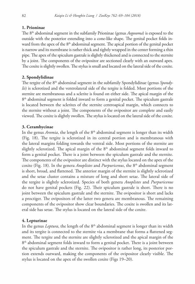
Kaiqin Li & Hongbin Liang / ZooKeys 762: 69–104 (2018)82
1. PrioninaeThe 8th abdominal segment in the subfamily Prioninae (genus Aegosoma) is exposed to the outside with the posterior extending into a cone-like shape. The genital pocket folds in-ward from the apex of the 8th abdominal segment. The apical portion of the genital pocket is narrow and its membrane is rather thick and tightly wrapped in the center forming a thin pipe. The apex of the spiculum gastrale is slightly thickened and is connected to the sternite by a joint. The components of the ovipositor are sectioned clearly with an outward apex. The coxite is slightly swollen. The stylus is small and located on the lateral side of the coxite.
2. SpondylidinaeThe tergite of the 8th abdominal segment in the subfamily Spondylidinae (genus Spondy-lis) is sclerotized and the ventrolateral side of the tergite is folded. Most portions of the sternite are membranous and a sclerite is found on either side. The apical margin of the 8th abdominal segment is folded inward to form a genital pocket. The spiculum gastrale is located between the sclerites of the sternite centroapical margin, which connects to the sternite without a joint. The components of the ovipositor are complete and easily viewed. The coxite is slightly swollen. The stylus is located on the lateral side of the coxite.
3. CerambycinaeIn the genus Aromia, the length of the 8th abdominal segment is longer than its width (Fig. 18). The tergite is sclerotized in its central portion and is membranous with the lateral margins folding towards the ventral side. Most portions of the sternite are slightly sclerotized. The apical margin of the 8th abdominal segment folds inward to form a genital pocket. There is a joint between the spiculum gastrale and the sternite. The components of the ovipositor are distinct with the stylus located on the apex of the coxite (Fig. 18). In the genera Anoplistes and Purpuricenus, the 8th abdominal segment is short, broad, and flattened. The anterior margin of the sternite is slightly sclerotized and the setae cluster contains a mixture of long and short setae. The lateral side of the tergite is slightly sclerotized. Species of both genera Anoplistes and Purpuricenus do not have genital pockets (Fig. 22). Their spiculum gastrale is short. There is no joint between the spiculum gastrale and the sternite. The ovipositor is short and lacks a proctiger. The ovipositors of the latter two genera are membranous. The remaining components of the ovipositor show clear boundaries. The coxite is swollen and its lat-eral side has setae. The stylus is located on the lateral side of the coxite.
4. LepturinaeIn the genus Leptura, the length of the 8th abdominal segment is longer than its width and its tergite is connected to the sternite via a membrane that forms a flattened seg-ment. The tergite and the sternite are slightly sclerotized and the apical margin of the 8th abdominal segment folds inward to form a genital pocket. There is a joint between the spiculum gastrale and the sternite. The ovipositor is rather long, its posterior por-tion extends outward, making the components of the ovipositor clearly visible. The stylus is located on the apex of the swollen coxite (Figs 19–20).
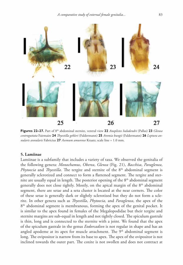
A comparative study of external female genitalia... 83
5. LamiinaeLamiinae is a subfamily that includes a variety of taxa. We observed the genitalia of the following genera: Monochamus, Oberea, Glenea (Fig. 21), Bacchisa, Paraglenea, Phytoecia and Thyestilla. The tergite and sternite of the 8th abdominal segment is generally sclerotized and connect to form a flattened segment. The tergite and ster-nite are usually equal in length. The posterior opening of the 8th abdominal segment generally does not close tightly. Mostly, on the apical margin of the 8th abdominal segment, there are setae and a seta cluster is located at the near corners. The color of these setae is generally dark or slightly sclerotized but they do not form a scle-rite. In other genera such as Thyestilla, Phytoecia, and Paraglenea, the apex of the 8th abdominal segment is membranous, forming the apex of the genital pocket. It is similar to the apex found in females of the Megalopodidae but their tergite and sternite margins are sub-equal in length and not tightly closed. The spiculum gastrale is thin, long and is connected to the sternite with a joint. We found that the apex of the spiculum gastrale in the genus Eodorcadion is not regular in shape and has an angled apodeme at its apex for muscle attachment. The 9th abdominal segment is long. The ovipositor is narrow from its base to apex. The apex of the ovipositor is not inclined towards the outer part. The coxite is not swollen and does not contract at
Figures 22–27. Part of 8th abdominal sternite, ventral view 22 Anoplistes halodendri (Pallas) 23 Glenea centroguttata Fairmaire 24 Thyestilla gebleri (Faldermann) 25 Aromia bungii (Faldermann) 26 Leptura an-nularis annularis Fabricius 27 Asemum amurense Kraatz; scale line = 1.0 mm.
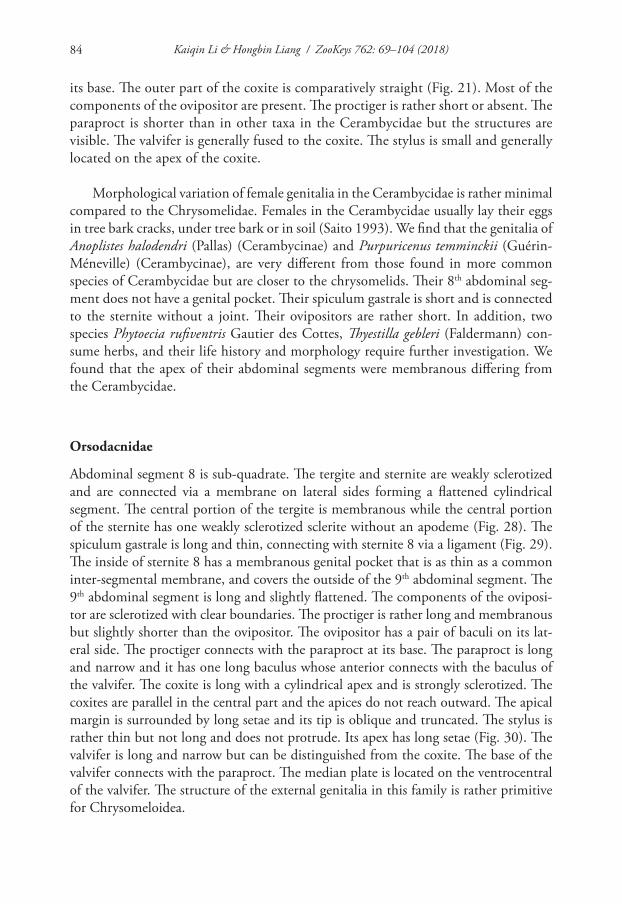
Kaiqin Li & Hongbin Liang / ZooKeys 762: 69–104 (2018)84
its base. The outer part of the coxite is comparatively straight (Fig. 21). Most of the components of the ovipositor are present. The proctiger is rather short or absent. The paraproct is shorter than in other taxa in the Cerambycidae but the structures are visible. The valvifer is generally fused to the coxite. The stylus is small and generally located on the apex of the coxite.
Morphological variation of female genitalia in the Cerambycidae is rather minimal compared to the Chrysomelidae. Females in the Cerambycidae usually lay their eggs in tree bark cracks, under tree bark or in soil (Saito 1993). We find that the genitalia of Anoplistes halodendri (Pallas) (Cerambycinae) and Purpuricenus temminckii (Guérin-Méneville) (Cerambycinae), are very different from those found in more common species of Cerambycidae but are closer to the chrysomelids. Their 8th abdominal seg-ment does not have a genital pocket. Their spiculum gastrale is short and is connected to the sternite without a joint. Their ovipositors are rather short. In addition, two species Phytoecia rufiventris Gautier des Cottes, Thyestilla gebleri (Faldermann) con-sume herbs, and their life history and morphology require further investigation. We found that the apex of their abdominal segments were membranous differing from the Cerambycidae.
Orsodacnidae
Abdominal segment 8 is sub-quadrate. The tergite and sternite are weakly sclerotized and are connected via a membrane on lateral sides forming a flattened cylindrical segment. The central portion of the tergite is membranous while the central portion of the sternite has one weakly sclerotized sclerite without an apodeme (Fig. 28). The spiculum gastrale is long and thin, connecting with sternite 8 via a ligament (Fig. 29). The inside of sternite 8 has a membranous genital pocket that is as thin as a common inter-segmental membrane, and covers the outside of the 9th abdominal segment. The 9th abdominal segment is long and slightly flattened. The components of the oviposi-tor are sclerotized with clear boundaries. The proctiger is rather long and membranous but slightly shorter than the ovipositor. The ovipositor has a pair of baculi on its lat-eral side. The proctiger connects with the paraproct at its base. The paraproct is long and narrow and it has one long baculus whose anterior connects with the baculus of the valvifer. The coxite is long with a cylindrical apex and is strongly sclerotized. The coxites are parallel in the central part and the apices do not reach outward. The apical margin is surrounded by long setae and its tip is oblique and truncated. The stylus is rather thin but not long and does not protrude. Its apex has long setae (Fig. 30). The valvifer is long and narrow but can be distinguished from the coxite. The base of the valvifer connects with the paraproct. The median plate is located on the ventrocentral of the valvifer. The structure of the external genitalia in this family is rather primitive for Chrysomeloidea.
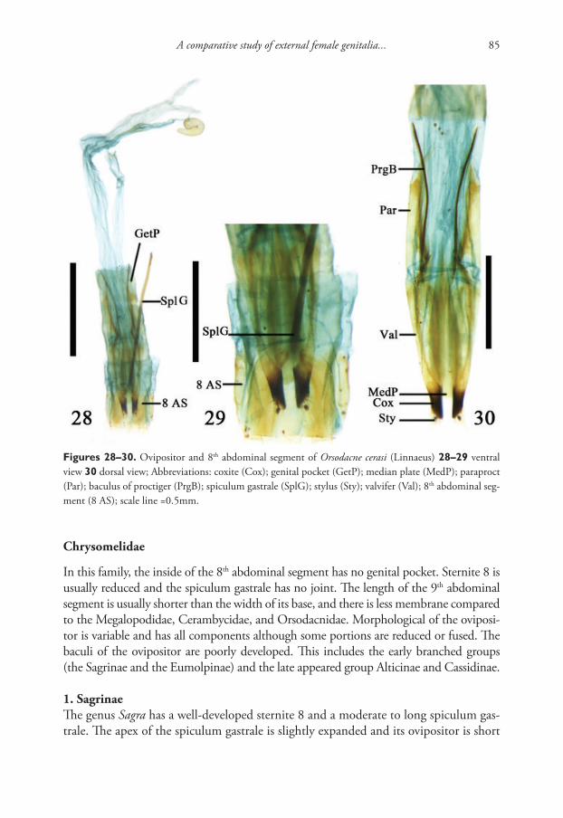
A comparative study of external female genitalia... 85
Chrysomelidae
In this family, the inside of the 8th abdominal segment has no genital pocket. Sternite 8 is usually reduced and the spiculum gastrale has no joint. The length of the 9th abdominal segment is usually shorter than the width of its base, and there is less membrane compared to the Megalopodidae, Cerambycidae, and Orsodacnidae. Morphological of the oviposi-tor is variable and has all components although some portions are reduced or fused. The baculi of the ovipositor are poorly developed. This includes the early branched groups (the Sagrinae and the Eumolpinae) and the late appeared group Alticinae and Cassidinae.
1. SagrinaeThe genus Sagra has a well-developed sternite 8 and a moderate to long spiculum gas-trale. The apex of the spiculum gastrale is slightly expanded and its ovipositor is short
Figures 28–30. Ovipositor and 8th abdominal segment of Orsodacne cerasi (Linnaeus) 28–29 ventral view 30 dorsal view; Abbreviations: coxite (Cox); genital pocket (GetP); median plate (MedP); paraproct (Par); baculus of proctiger (PrgB); spiculum gastrale (SplG); stylus (Sty); valvifer (Val); 8th abdominal seg-ment (8 AS); scale line =0.5mm.
Kaiqin Li & Hongbin Liang / ZooKeys 762: 69–104 (2018)86
and thick. The proctiger is membranous with a sclerite on each side of the proctiger. The apex of the proctiger inserts into tergite 8. The paraproct is strongly sclerotized and thick, while its apex is connected to the valvifer, coxite, and stylus. There is a median plate between the valvifer on its ventral side. This is representative of primitive types in the Chrysomelidae (Figs 31–32).
2. BruchinaeThe 8th abdominal segment, associated with members of the genus Callosobruchus, is quadrate. The ventral side of the tergite is close to the proctiger. The central part of the tergite is membranous and its lateral side is sclerotized. The sternite is mem-branous. The tergite and sternite are connected to each other on the lateral side. The spiculum gastrale is short, about 1.5 times the length of the sternite, and lacks a broad apex. In Callosobruchus chinensis (Linnaeus) the spiculum gastrale extends for-ward turning backward at its base. The length of the 9th abdominal segment is almost equal to the length of the 8th abdominal segment. The proctiger is approximately triangular and membranous with sclerotized lateral parts. The base of the proctiger is connected to the paraproct. The ovipositor is small. The valvifer, coxite, and stylus lack clear boundaries.
3. CriocerinaeThis subfamily is similar to the Sagrinae but the spiculum gastrale is relatively short (Figs 33–34). The components of the ovipositor show clear boundaries but the valvifer generally fuses with the coxite or the paraproct. In some cases the stylus may be either indistinct (genus Oulema) or distinct according to species (e.g. Lilioceris and Lema). The components of the ovipositor have clear boundaries in the genus Mecoprosopus although their valvifer is fused with the paraproct.
4. DonaciinaeDonaciinae is an aquatic subfamily. The 8th and 9th abdominal segments are slightly sclerotized. The spiculum gastrale of the 8th abdominal segment is rather long. The 9th abdominal segment is significantly long and membranous. The proctiger is usually long, triangular and its basolateral side is connected to the paraproct. The valvifer usu-ally fuses with the coxite. The stylus is generally indistinct. The area of sensory setae is located on the surface of the tip of the coxite and may be interpreted as a stylus (Figs 35, 38, 39). It is only in the genus Macroplea that the ovipositor has all its components with clear boundaries. The female of Donacia provostii Fairmaire inserts her ovipositor in a small hole on the dorsal surface of a leaf and lays eggs on the leaf ’s ventral side. The inter-segmental membrane between the 8th and 9th abdominal segments is very long. In the genus Plateumaris the 8th abdominal segment is sclerotized and specialized form-ing a flattened sheath (Figs 35–36) with the ovipositor in this sheath. Members of this genus have no proctiger. We found that the inter-segmental membrane between the 8th and 9th abdominal segments in Plateumaris socia (Chen) was rather long.
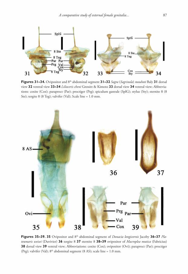
A comparative study of external female genitalia... 87
Figures 31–34. Ovipositor and 8th abdominal segment 31–32 Sagra (Sagrinola) mouhoti Baly 31 dorsal view 32 ventral view 33–34 Lilioceris cheni Gressitt & Kimoto 33 dorsal view 34 ventral view; Abbrevia-tions: coxite (Cox); paraproct (Par); proctiger (Prg); spiculum gastrale (SplG); stylus (Sty); sternite 8 (8 Ste); tergite 8 (8 Teg); valvifer (Val); Scale line = 1.0 mm.
Figures 35–39. 35 Ovipositor and 8th abdominal segment of Donacia longicornis Jacoby 36–37 Pla-teumaris weisei (Duvivier) 36 tergite 8 37 sternite 8 38–39 ovipositor of Macroplea mutica (Fabricius) 38 dorsal view 39 ventral view; Abbreviations: coxite (Cox); ovipositor (Ovi); paraproct (Par); proctiger (Prg); valvifer (Val); 8th abdominal segment (8 AS); scale line = 1.0 mm.
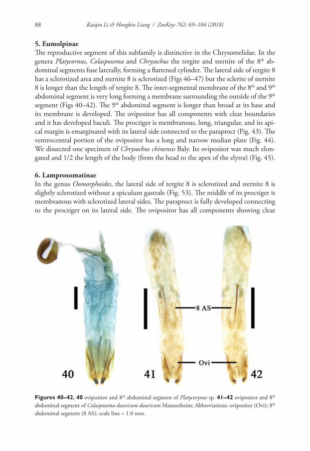
Kaiqin Li & Hongbin Liang / ZooKeys 762: 69–104 (2018)88
5. EumolpinaeThe reproductive segment of this subfamily is distinctive in the Chrysomelidae. In the genera Platycornus, Colasposoma and Chrysochus the tergite and sternite of the 8th ab-dominal segments fuse laterally, forming a flattened cylinder. The lateral side of tergite 8 has a sclerotized area and sternite 8 is sclerotized (Figs 46–47) but the sclerite of sternite 8 is longer than the length of tergite 8. The inter-segmental membrane of the 8th and 9th abdominal segment is very long forming a membrane surrounding the outside of the 9th segment (Figs 40–42). The 9th abdominal segment is longer than broad at its base and its membrane is developed. The ovipositor has all components with clear boundaries and it has developed baculi. The proctiger is membranous, long, triangular, and its api-cal margin is emarginated with its lateral side connected to the paraproct (Fig. 43). The ventrocentral portion of the ovipositor has a long and narrow median plate (Fig. 44). We dissected one specimen of Chrysochus chinensis Baly. Its ovipositor was much elon-gated and 1/2 the length of the body (from the head to the apex of the elytra) (Fig. 45).
6. LamprosomatinaeIn the genus Oomorphoides, the lateral side of tergite 8 is sclerotized and sternite 8 is slightly sclerotized without a spiculum gastrale (Fig. 53). The middle of its proctiger is membranous with sclerotized lateral sides. The paraproct is fully developed connecting to the proctiger on its lateral side. The ovipositor has all components showing clear
Figures 40–42. 40 ovipositor and 8th abdominal segment of Platycorynus sp. 41–42 ovipositor and 8th abdominal segment of Colasposoma dauricum dauricum Mannerheim; Abbreviations: ovipositor (Ovi); 8th abdominal segment (8 AS); scale line = 1.0 mm.
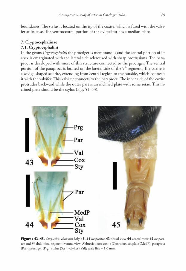
A comparative study of external female genitalia... 89
boundaries. The stylus is located on the tip of the coxite, which is fused with the valvi-fer at its base. The ventrocentral portion of the ovipositor has a median plate.
7. Cryptocephalinae7.1. CryptocephaliniIn the genus Cryptocephalus the proctiger is membranous and the central portion of its apex is emarginated with the lateral side sclerotized with sharp protrusions. The para-proct is developed with most of this structure connected to the proctiger. The ventral portion of the paraproct is located on the lateral side of the 9th segment. The coxite is a wedge-shaped sclerite, extending from central region to the outside, which connects it with the valvifer. This valvifer connects to the paraproct. The inner side of the coxite protrudes backward while the outer part is an inclined plate with some setae. This in-clined plate should be the stylus (Figs 51–53).
Figures 43–45. Chrysochus chinensis Baly 43–44 ovipositor 43 dorsal view 44 ventral view 45 oviposi-tor and 8th abdominal segment, ventral view; Abbreviations: coxite (Cox); median plate (MedP); paraproct (Par); proctiger (Prg); stylus (Sty); valvifer (Val); scale line = 1.0 mm.
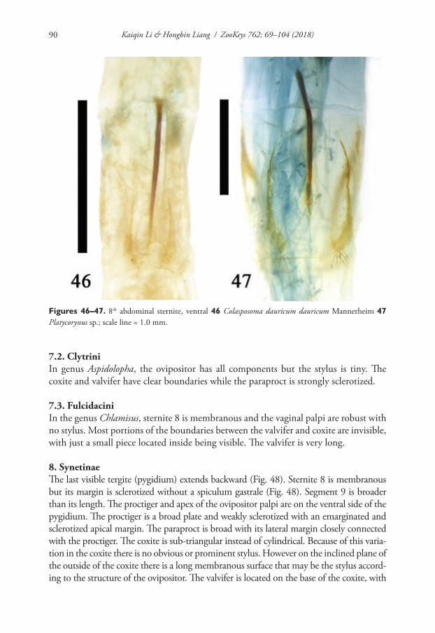
Kaiqin Li & Hongbin Liang / ZooKeys 762: 69–104 (2018)90
7.2. ClytriniIn genus Aspidolopha, the ovipositor has all components but the stylus is tiny. The coxite and valvifer have clear boundaries while the paraproct is strongly sclerotized.
7.3. FulcidaciniIn the genus Chlamisus, sternite 8 is membranous and the vaginal palpi are robust with no stylus. Most portions of the boundaries between the valvifer and coxite are invisible, with just a small piece located inside being visible. The valvifer is very long.
8. SynetinaeThe last visible tergite (pygidium) extends backward (Fig. 48). Sternite 8 is membranous but its margin is sclerotized without a spiculum gastrale (Fig. 48). Segment 9 is broader than its length. The proctiger and apex of the ovipositor palpi are on the ventral side of the pygidium. The proctiger is a broad plate and weakly sclerotized with an emarginated and sclerotized apical margin. The paraproct is broad with its lateral margin closely connected with the proctiger. The coxite is sub-triangular instead of cylindrical. Because of this varia-tion in the coxite there is no obvious or prominent stylus. However on the inclined plane of the outside of the coxite there is a long membranous surface that may be the stylus accord-ing to the structure of the ovipositor. The valvifer is located on the base of the coxite, with
Figures 46–47. 8th abdominal sternite, ventral 46 Colasposoma dauricum dauricum Mannerheim 47 Platycorynus sp.; scale line = 1.0 mm.
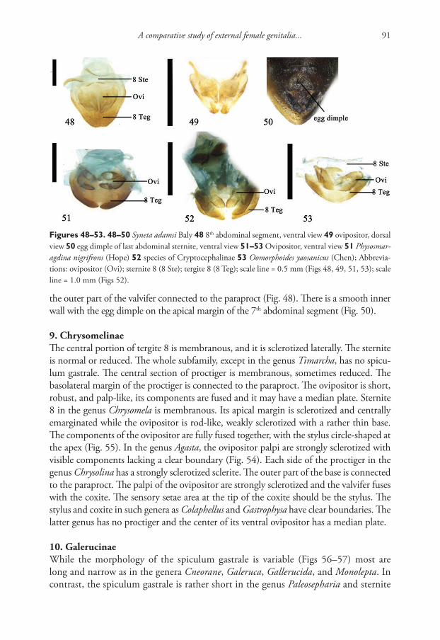
A comparative study of external female genitalia... 91
the outer part of the valvifer connected to the paraproct (Fig. 48). There is a smooth inner wall with the egg dimple on the apical margin of the 7th abdominal segment (Fig. 50).
9. ChrysomelinaeThe central portion of tergite 8 is membranous, and it is sclerotized laterally. The sternite is normal or reduced. The whole subfamily, except in the genus Timarcha, has no spicu-lum gastrale. The central section of proctiger is membranous, sometimes reduced. The basolateral margin of the proctiger is connected to the paraproct. The ovipositor is short, robust, and palp-like, its components are fused and it may have a median plate. Sternite 8 in the genus Chrysomela is membranous. Its apical margin is sclerotized and centrally emarginated while the ovipositor is rod-like, weakly sclerotized with a rather thin base. The components of the ovipositor are fully fused together, with the stylus circle-shaped at the apex (Fig. 55). In the genus Agasta, the ovipositor palpi are strongly sclerotized with visible components lacking a clear boundary (Fig. 54). Each side of the proctiger in the genus Chrysolina has a strongly sclerotized sclerite. The outer part of the base is connected to the paraproct. The palpi of the ovipositor are strongly sclerotized and the valvifer fuses with the coxite. The sensory setae area at the tip of the coxite should be the stylus. The stylus and coxite in such genera as Colaphellus and Gastrophysa have clear boundaries. The latter genus has no proctiger and the center of its ventral ovipositor has a median plate.
10. GalerucinaeWhile the morphology of the spiculum gastrale is variable (Figs 56–57) most are long and narrow as in the genera Cneorane, Galeruca, Gallerucida, and Monolepta. In contrast, the spiculum gastrale is rather short in the genus Paleosepharia and sternite
Figures 48–53. 48–50 Syneta adamsi Baly 48 8th abdominal segment, ventral view 49 ovipositor, dorsal view 50 egg dimple of last abdominal sternite, ventral view 51–53 Ovipositor, ventral view 51 Physosmar-agdina nigrifrons (Hope) 52 species of Cryptocephalinae 53 Oomorphoides yaosanicus (Chen); Abbrevia-tions: ovipositor (Ovi); sternite 8 (8 Ste); tergite 8 (8 Teg); scale line = 0.5 mm (Figs 48, 49, 51, 53); scale line = 1.0 mm (Figs 52).
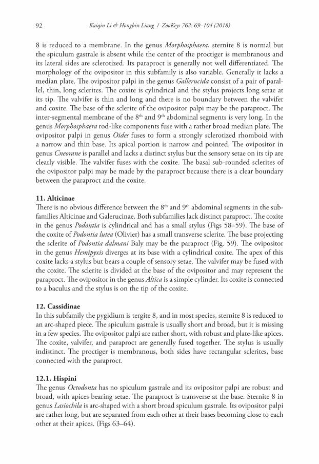
Kaiqin Li & Hongbin Liang / ZooKeys 762: 69–104 (2018)92
8 is reduced to a membrane. In the genus Morphosphaera, sternite 8 is normal but the spiculum gastrale is absent while the center of the proctiger is membranous and its lateral sides are sclerotized. Its paraproct is generally not well differentiated. The morphology of the ovipositor in this subfamily is also variable. Generally it lacks a median plate. The ovipositor palpi in the genus Gallerucida consist of a pair of paral-lel, thin, long sclerites. The coxite is cylindrical and the stylus projects long setae at its tip. The valvifer is thin and long and there is no boundary between the valvifer and coxite. The base of the sclerite of the ovipositor palpi may be the paraproct. The inter-segmental membrane of the 8th and 9th abdominal segments is very long. In the genus Morphosphaera rod-like components fuse with a rather broad median plate. The ovipositor palpi in genus Oides fuses to form a strongly sclerotized rhomboid with a narrow and thin base. Its apical portion is narrow and pointed. The ovipositor in genus Cneorane is parallel and lacks a distinct stylus but the sensory setae on its tip are clearly visible. The valvifer fuses with the coxite. The basal sub-rounded sclerites of the ovipositor palpi may be made by the paraproct because there is a clear boundary between the paraproct and the coxite.
11. AlticinaeThere is no obvious difference between the 8th and 9th abdominal segments in the sub-families Alticinae and Galerucinae. Both subfamilies lack distinct paraproct. The coxite in the genus Podontia is cylindrical and has a small stylus (Figs 58–59). The base of the coxite of Podontia lutea (Olivier) has a small transverse sclerite. The base projecting the sclerite of Podontia dalmani Baly may be the paraproct (Fig. 59). The ovipositor in the genus Hemipyxis diverges at its base with a cylindrical coxite. The apex of this coxite lacks a stylus but bears a couple of sensory setae. The valvifer may be fused with the coxite. The sclerite is divided at the base of the ovipositor and may represent the paraproct. The ovipositor in the genus Altica is a simple cylinder. Its coxite is connected to a baculus and the stylus is on the tip of the coxite.
12. CassidinaeIn this subfamily the pygidium is tergite 8, and in most species, sternite 8 is reduced to an arc-shaped piece. The spiculum gastrale is usually short and broad, but it is missing in a few species. The ovipositor palpi are rather short, with robust and plate-like apices. The coxite, valvifer, and paraproct are generally fused together. The stylus is usually indistinct. The proctiger is membranous, both sides have rectangular sclerites, base connected with the paraproct.
12.1. HispiniThe genus Octodonta has no spiculum gastrale and its ovipositor palpi are robust and broad, with apices bearing setae. The paraproct is transverse at the base. Sternite 8 in genus Lasiochila is arc-shaped with a short broad spiculum gastrale. Its ovipositor palpi are rather long, but are separated from each other at their bases becoming close to each other at their apices. (Figs 63–64).

A comparative study of external female genitalia... 93
Figures 54–59. Ovipositor and 8th abdominal segment 54 Agasta formosa Hope 55 Chrysomela populi Linnaeus 56 Paleosephraria 57 Mimastra limbata Baly 58 Podontia affinis (Gröndal) 59 Podontia dalmani Baly; scale line = 1.0 mm.
Figures 60–65. 60 8th abdominal sternite of Callispa brettinghami Baly 61 8th abdominal sternite of Callispa nigricollis Chen & Yu 62 ovipositor of Callispa nigricollis Chen & Yu 63 8th abdominal sternite of Lasiochila cylindrica (Hope) 64 ovipositor of Lasiochila cylindrica (Hope) 65 ovipositor of Basiprionota bisignata (Boheman); scale line = 0.5 mm (Figs 63–65); scale line = 1.0 mm (Figs 60–62).
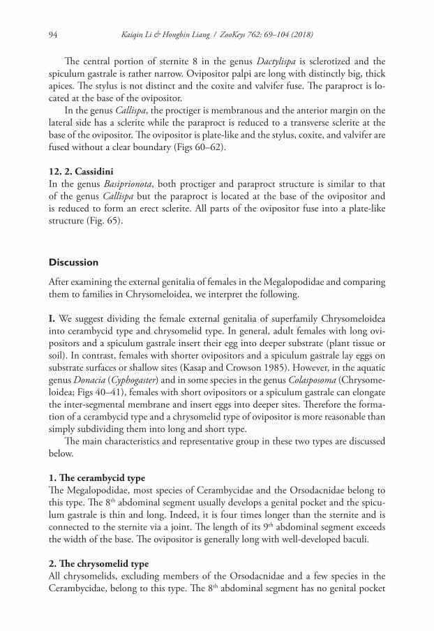
Kaiqin Li & Hongbin Liang / ZooKeys 762: 69–104 (2018)94
The central portion of sternite 8 in the genus Dactylispa is sclerotized and the spiculum gastrale is rather narrow. Ovipositor palpi are long with distinctly big, thick apices. The stylus is not distinct and the coxite and valvifer fuse. The paraproct is lo-cated at the base of the ovipositor.
In the genus Callispa, the proctiger is membranous and the anterior margin on the lateral side has a sclerite while the paraproct is reduced to a transverse sclerite at the base of the ovipositor. The ovipositor is plate-like and the stylus, coxite, and valvifer are fused without a clear boundary (Figs 60–62).
12. 2. CassidiniIn the genus Basiprionota, both proctiger and paraproct structure is similar to that of the genus Callispa but the paraproct is located at the base of the ovipositor and is reduced to form an erect sclerite. All parts of the ovipositor fuse into a plate-like structure (Fig. 65).
Discussion
After examining the external genitalia of females in the Megalopodidae and comparing them to families in Chrysomeloidea, we interpret the following.
I. We suggest dividing the female external genitalia of superfamily Chrysomeloidea into cerambycid type and chrysomelid type. In general, adult females with long ovi-positors and a spiculum gastrale insert their egg into deeper substrate (plant tissue or soil). In contrast, females with shorter ovipositors and a spiculum gastrale lay eggs on substrate surfaces or shallow sites (Kasap and Crowson 1985). However, in the aquatic genus Donacia (Cyphogaster) and in some species in the genus Colasposoma (Chrysome-loidea; Figs 40–41), females with short ovipositors or a spiculum gastrale can elongate the inter-segmental membrane and insert eggs into deeper sites. Therefore the forma-tion of a cerambycid type and a chrysomelid type of ovipositor is more reasonable than simply subdividing them into long and short type.
The main characteristics and representative group in these two types are discussed below.
1. The cerambycid typeThe Megalopodidae, most species of Cerambycidae and the Orsodacnidae belong to this type. The 8th abdominal segment usually develops a genital pocket and the spicu-lum gastrale is thin and long. Indeed, it is four times longer than the sternite and is connected to the sternite via a joint. The length of its 9th abdominal segment exceeds the width of the base. The ovipositor is generally long with well-developed baculi.
2. The chrysomelid typeAll chrysomelids, excluding members of the Orsodacnidae and a few species in the Cerambycidae, belong to this type. The 8th abdominal segment has no genital pocket
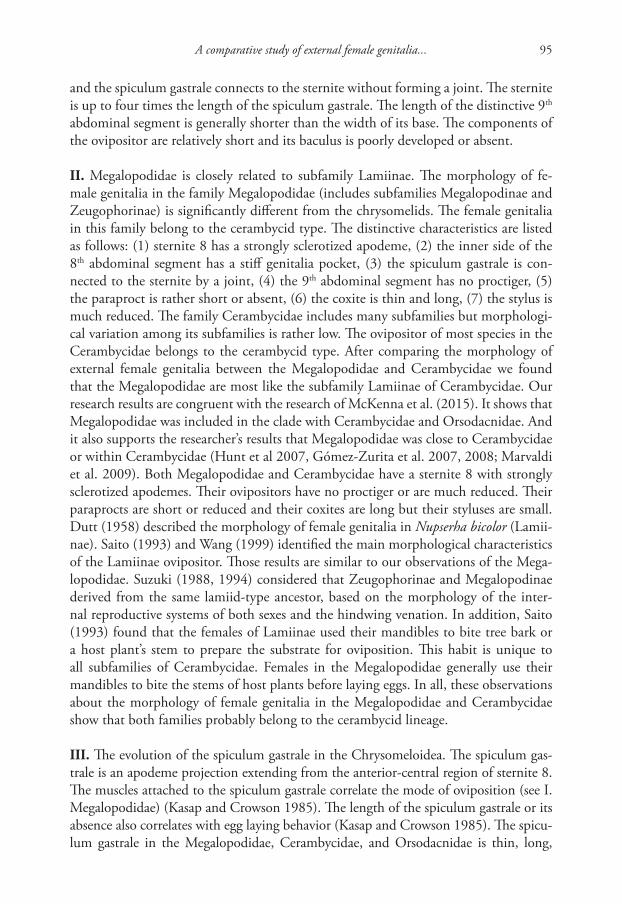
A comparative study of external female genitalia... 95
and the spiculum gastrale connects to the sternite without forming a joint. The sternite is up to four times the length of the spiculum gastrale. The length of the distinctive 9th abdominal segment is generally shorter than the width of its base. The components of the ovipositor are relatively short and its baculus is poorly developed or absent.
II. Megalopodidae is closely related to subfamily Lamiinae. The morphology of fe-male genitalia in the family Megalopodidae (includes subfamilies Megalopodinae and Zeugophorinae) is significantly different from the chrysomelids. The female genitalia in this family belong to the cerambycid type. The distinctive characteristics are listed as follows: (1) sternite 8 has a strongly sclerotized apodeme, (2) the inner side of the 8th abdominal segment has a stiff genitalia pocket, (3) the spiculum gastrale is con-nected to the sternite by a joint, (4) the 9th abdominal segment has no proctiger, (5) the paraproct is rather short or absent, (6) the coxite is thin and long, (7) the stylus is much reduced. The family Cerambycidae includes many subfamilies but morphologi-cal variation among its subfamilies is rather low. The ovipositor of most species in the Cerambycidae belongs to the cerambycid type. After comparing the morphology of external female genitalia between the Megalopodidae and Cerambycidae we found that the Megalopodidae are most like the subfamily Lamiinae of Cerambycidae. Our research results are congruent with the research of McKenna et al. (2015). It shows that Megalopodidae was included in the clade with Cerambycidae and Orsodacnidae. And it also supports the researcher’s results that Megalopodidae was close to Cerambycidae or within Cerambycidae (Hunt et al 2007, Gómez-Zurita et al. 2007, 2008; Marvaldi et al. 2009). Both Megalopodidae and Cerambycidae have a sternite 8 with strongly sclerotized apodemes. Their ovipositors have no proctiger or are much reduced. Their paraprocts are short or reduced and their coxites are long but their styluses are small. Dutt (1958) described the morphology of female genitalia in Nupserha bicolor (Lamii-nae). Saito (1993) and Wang (1999) identified the main morphological characteristics of the Lamiinae ovipositor. Those results are similar to our observations of the Mega-lopodidae. Suzuki (1988, 1994) considered that Zeugophorinae and Megalopodinae derived from the same lamiid-type ancestor, based on the morphology of the inter-nal reproductive systems of both sexes and the hindwing venation. In addition, Saito (1993) found that the females of Lamiinae used their mandibles to bite tree bark or a host plant’s stem to prepare the substrate for oviposition. This habit is unique to all subfamilies of Cerambycidae. Females in the Megalopodidae generally use their mandibles to bite the stems of host plants before laying eggs. In all, these observations about the morphology of female genitalia in the Megalopodidae and Cerambycidae show that both families probably belong to the cerambycid lineage.
III. The evolution of the spiculum gastrale in the Chrysomeloidea. The spiculum gas-trale is an apodeme projection extending from the anterior-central region of sternite 8. The muscles attached to the spiculum gastrale correlate the mode of oviposition (see I. Megalopodidae) (Kasap and Crowson 1985). The length of the spiculum gastrale or its absence also correlates with egg laying behavior (Kasap and Crowson 1985). The spicu-lum gastrale in the Megalopodidae, Cerambycidae, and Orsodacnidae is thin, long,
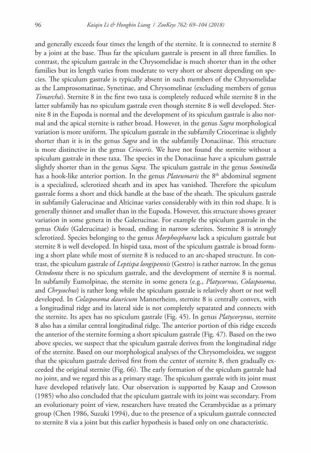
Kaiqin Li & Hongbin Liang / ZooKeys 762: 69–104 (2018)96
and generally exceeds four times the length of the sternite. It is connected to sternite 8 by a joint at the base. Thus far the spiculum gastrale is present in all three families. In contrast, the spiculum gastrale in the Chrysomelidae is much shorter than in the other families but its length varies from moderate to very short or absent depending on spe-cies. The spiculum gastrale is typically absent in such members of the Chrysomelidae as the Lamprosomatinae, Synetinae, and Chrysomelinae (excluding members of genus Timarcha). Sternite 8 in the first two taxa is completely reduced while sternite 8 in the latter subfamily has no spiculum gastrale even though sternite 8 is well developed. Ster-nite 8 in the Eupoda is normal and the development of its spiculum gastrale is also nor-mal and the apical sternite is rather broad. However, in the genus Sagra morphological variation is more uniform. The spiculum gastrale in the subfamily Criocerinae is slightly shorter than it is in the genus Sagra and in the subfamily Donaciinae. This structure is more distinctive in the genus Crioceris. We have not found the sternite without a spiculum gastrale in these taxa. The species in the Donaciinae have a spiculum gastrale slightly shorter than in the genus Sagra. The spiculum gastrale in the genus Sominella has a hook-like anterior portion. In the genus Plateumaris the 8th abdominal segment is a specialized, sclerotized sheath and its apex has vanished. Therefore the spiculum gastrale forms a short and thick handle at the base of the sheath. The spiculum gastrale in subfamily Galerucinae and Alticinae varies considerably with its thin rod shape. It is generally thinner and smaller than in the Eupoda. However, this structure shows greater variation in some genera in the Galerucinae. For example the spiculum gastrale in the genus Oides (Galerucinae) is broad, ending in narrow sclerites. Sternite 8 is strongly sclerotized. Species belonging to the genus Morphosphaera lack a spiculum gastrale but sternite 8 is well developed. In hispid taxa, most of the spiculum gastrale is broad form-ing a short plate while most of sternite 8 is reduced to an arc-shaped structure. In con-trast, the spiculum gastrale of Leptispa longipennis (Gestro) is rather narrow. In the genus Octodonta there is no spiculum gastrale, and the development of sternite 8 is normal. In subfamily Eumolpinae, the sternite in some genera (e.g., Platycornus, Colasposoma, and Chrysochus) is rather long while the spiculum gastrale is relatively short or not well developed. In Colasposoma dauricum Mannerheim, sternite 8 is centrally convex, with a longitudinal ridge and its lateral side is not completely separated and connects with the sternite. Its apex has no spiculum gastrale (Fig. 45). In genus Platycorynus, sternite 8 also has a similar central longitudinal ridge. The anterior portion of this ridge exceeds the anterior of the sternite forming a short spiculum gastrale (Fig. 47). Based on the two above species, we suspect that the spiculum gastrale derives from the longitudinal ridge of the sternite. Based on our morphological analyses of the Chrysomeloidea, we suggest that the spiculum gastrale derived first from the center of sternite 8, then gradually ex-ceeded the original sternite (Fig. 66). The early formation of the spiculum gastrale had no joint, and we regard this as a primary stage. The spiculum gastrale with its joint must have developed relatively late. Our observation is supported by Kasap and Crowson (1985) who also concluded that the spiculum gastrale with its joint was secondary. From an evolutionary point of view, researchers have treated the Cerambycidae as a primary group (Chen 1986, Suzuki 1994), due to the presence of a spiculum gastrale connected to sternite 8 via a joint but this earlier hypothesis is based only on one characteristic.
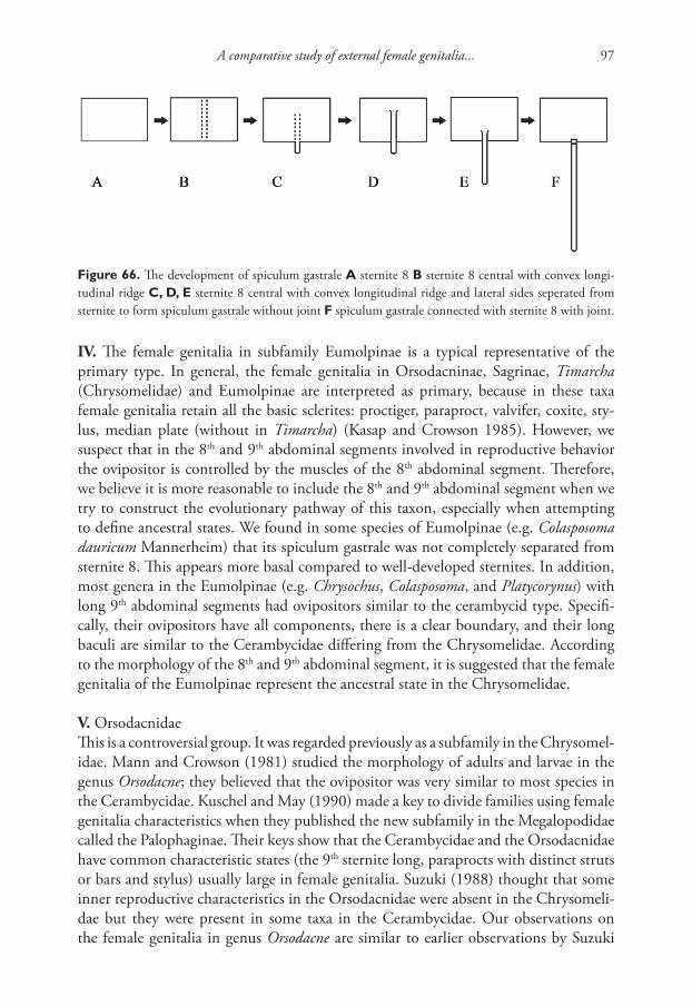
A comparative study of external female genitalia... 97
IV. The female genitalia in subfamily Eumolpinae is a typical representative of the primary type. In general, the female genitalia in Orsodacninae, Sagrinae, Timarcha (Chrysomelidae) and Eumolpinae are interpreted as primary, because in these taxa female genitalia retain all the basic sclerites: proctiger, paraproct, valvifer, coxite, sty-lus, median plate (without in Timarcha) (Kasap and Crowson 1985). However, we suspect that in the 8th and 9th abdominal segments involved in reproductive behavior the ovipositor is controlled by the muscles of the 8th abdominal segment. Therefore, we believe it is more reasonable to include the 8th and 9th abdominal segment when we try to construct the evolutionary pathway of this taxon, especially when attempting to define ancestral states. We found in some species of Eumolpinae (e.g. Colasposoma dauricum Mannerheim) that its spiculum gastrale was not completely separated from sternite 8. This appears more basal compared to well-developed sternites. In addition, most genera in the Eumolpinae (e.g. Chrysochus, Colasposoma, and Platycorynus) with long 9th abdominal segments had ovipositors similar to the cerambycid type. Specifi-cally, their ovipositors have all components, there is a clear boundary, and their long baculi are similar to the Cerambycidae differing from the Chrysomelidae. According to the morphology of the 8th and 9th abdominal segment, it is suggested that the female genitalia of the Eumolpinae represent the ancestral state in the Chrysomelidae.
V. OrsodacnidaeThis is a controversial group. It was regarded previously as a subfamily in the Chrysomel-idae. Mann and Crowson (1981) studied the morphology of adults and larvae in the genus Orsodacne; they believed that the ovipositor was very similar to most species in the Cerambycidae. Kuschel and May (1990) made a key to divide families using female genitalia characteristics when they published the new subfamily in the Megalopodidae called the Palophaginae. Their keys show that the Cerambycidae and the Orsodacnidae have common characteristic states (the 9th sternite long, paraprocts with distinct struts or bars and stylus) usually large in female genitalia. Suzuki (1988) thought that some inner reproductive characteristics in the Orsodacnidae were absent in the Chrysomeli-dae but they were present in some taxa in the Cerambycidae. Our observations on the female genitalia in genus Orsodacne are similar to earlier observations by Suzuki
Figure 66. The development of spiculum gastrale A sternite 8 B sternite 8 central with convex longi-tudinal ridge C, D, E sternite 8 central with convex longitudinal ridge and lateral sides seperated from sternite to form spiculum gastrale without joint F spiculum gastrale connected with sternite 8 with joint.
Kaiqin Li & Hongbin Liang / ZooKeys 762: 69–104 (2018)98
(1988). We found that some structures associated with the ovipositor belong to the cerambycid type (including the Megalopodidae and Cerambycidae) and they differ distinctly from the chrysomelid type. The structure of the cerambycid type suggests it represents the ancestral structure. Although we found that the morphology of the reproductive segments in the genus Orsodacne differed from those in the Megalopodi-dae and Cerambycidae, for example sternite 8 has no strongly sclerotized genital plate, the genital pocket is an inter-segmental and long cylindrical membranous membrane, the posterior opening of the 8th abdominal segment has no sclerite plate, and the 9th abdominal segment has a rather long proctiger. These differences are not sufficient to indicate that Orsodacne is more closely allied to the chrysomelids.
Crowson (1955) thought that some morphological characteristics of the larvae of Orsodacne were similar to the Criocerinae, Donaciinae, Sagra, Clytra, and Eumolpinae. Cox (1981) also suggested that the Orsodacninae was closer to the Zeugophorinae and Donaciinae based on larva characteristics. There is no doubt that the Orsodacninae are closer to the Galerucinae. In our study, we found that the 9th abdominal segment of Orsodacne is very similar to the same structure in some genera (Colasposoma, Platycory-nus and Chrysochus) placed in the Eumolpinae. They share the same following features: a long 9th abdominal segment, the ovipositor visibly retains all structures and has de-veloped baculi. However, the 8th abdominal segment in these three genera differs from that in the genus Orsodacne. Based on our comparison of the external female genitalia in the Chrysomeloidea and observations by other researchers on the genus Orsodacne, we suggest that Orsodacne separated from the cerambycid lineage at an earlier period. It moved from bored stems of plants to leaf-eating in the early evolution of the an-cestor of Chrysomeloidea, but it keeps some plesiomorphy. It was in the clade that became the earliest independent chrysomelid lineage. Chrysomelidae (include other chrysomelids) was in the clade that evolved later than Orsodacne. We agree that the Orsodacnidae was an independent family but a sister family of the Chrysomelidae and it was equal in status to the Megalopodidae and Cerambycidae (Kuschel and May 1990, Reid 1995). Specifically the Chrysomeloidea now includes four families: the Megalopodidae, Cerambycidae, Orsodacnidae, and Chrysomelidae.
VI. SynetinaeWith a reduced sternite 8, the oviposition behavior in the Synetinae is very similar to the Camptosomata including the subfamily Cryptocephalinae (Cryptocephalini, Clytrini and Chlamisus). All of them place a protective layer outside their eggs. They hold each egg by their hind tarsi and keep turning in the egg dimple of the 7th ab-dominal segment when the egg is expelled from their bodies (Jiang 1983, Yu 1996). Their digestive tracts discharge evenly to cover each egg forming a protective layer and then the egg is kicked out of their bodies (Yu 1996, Jiang 1983). The protective layer is formed by the Kotpresse (German language, meaning fecal press), which is a special structure on the rectum (Lécaillon 1899). The Kotpresse was reported in the Camptosomata (Erber 1968) and Synetinae (Reid 1995). We also found some vertical arrangements of a special Kotpresse on the rectum of Syneta adamsi Baly.
A comparative study of external female genitalia... 99
The female genitalia of the Synetinae are different from the more common Chrysomelidae. The tergite 8 (pygidium) was exposed and backward, so one could see the exposed apices of the ovipositor from its ventral side. The coxite is triangular lacking a distinct stylus. But on the inclined plane outside the coxite there is a small protrusion that should be the stylus because the valvifer connects with the coxite and paraproct at the base. This structure is similar to the membrane of the ovipositor in the Camptosomata (especially in some species in the genera Cryptocephalus and Clytra). Both the Synetinae and Camptosomata have a well-developed proctiger and a broad, sclerotized paraproct. Based on similar oviposition structure and behaviors, we speculate that the Synetinae has the closest relationship to the Camptosomata in the Chrysomelidae. However, we cannot exclude the possibility of parallel or convergent evolution of oviposition behavior in both taxa. Schmitt (1996) suggested that the Camptosomata, Synetinae, and some species in the Eumolpinae belonged to a monophyletic group because their Kotpresses were homologous structures. Our observation supports Schmitt (1996), but we found that the ovipositor of the Eumolpinae is ancestral and differs from the ovipositor in the Camptosomata. We have not observed the egg-laying habits of the genus Chlamisus.
VII. Based on comprehensive observations of the female genitalia of the Chrysome-loidea, it was found that the apices of the ovipositor are usually palp-like and backward except in the subfamily Cassidinae and in the genus Callispa of Hispini, which has a wide, plate-like ovipositor. Muir and Sharp (1904) found that the plate-like ovipositor of Aspidomorpha puncticosta Boheman was the key structure required to form the dia-phragm between the eggs in the egg sac. Chen et al. (1986) suggested that the trait of laying eggs in an egg sheath in the genus Callispa and subfamily Cassidinae was further evidence of their close relationship. Our studies show that the plate-like ovipositor is found only in species that make a sheath to protect an egg. In addition, we found that the ovipositor of the genus Leptispa belongs to the same tribe as the genus Callispa and it is palp-like, not plate-like. We also found that the adult and larvae in the genus Leptispa live in a leaf roll (Chen 1986). The adult lays its egg in a leaf roll but does not make a sheath. Therefore we suggest that the morphology of the ovipositor in these genera show a close relationship even though they differ in life-history. Based on this evidence, we conclude that the genus Callispa branched early and the genus Leptispa appeared later.
Conclusions
Female genitalia are diverse, complex, and very important for classification of the Chrysomeloidea. With a comparative analysis of the female genitalia we can reach a well-resolved classification system of this family. Egg-laying behavior and oviposition media cannot to be ignored when we explore the evolution of this huge taxon, but we still know very little about the reproductive biology of individual species. This research is particularly limited in Chinese lineages and future research is desperately needed to com-pare all the members of this lineage to resolve the systematics of the Chrysomeloidea.
Kaiqin Li & Hongbin Liang / ZooKeys 762: 69–104 (2018)100
Acknowledgments
The authors thank Professor Peiyu Yu (Institute of Zoology, Chinese Academy of Sci-ences, China) for her valuable help in our Megalopodidae study; Dr. Meiying Lin (Institute of Zoology, Chinese Academy of Sciences, China) for providing some speci-mens of Cerambycidae and identifying them; Professor Peter Bernhardt (Department of Biology, Saint Louis University, USA) and Mr. Michael Fizdale (Hampshire College, Massachusetts, USA) and Dr. Zongxin Ren (Kunming Institute of Botany, Chinese Academy of Sciences, China) for correcting English for an earlier draft. This work is supported by grants from the National Natural Science Foundation of China (Grant No. 31572235) and the National Special Fund on Basic Research of Science and Tech-nology (2014FY110100).
References
Böving AG, Craighead FC (1931) An illustrated synopsis of the principal larval forms of the order Coleoptera. Entomologica Americana 11: 1–351.
Chamorro-Lacayo ML, Konstantinov AS, Moseyko AG (2006) Comparative morphology of the female genitalia and some abdominal structures of Neotropical Cryptocephalini (Co-leoptera: Chrysomelidae: Cryptocephalinae). Coleopterists Bulletin 60(2): 113–134.
Chapuis F (1874) Famille des phytophages. In: Lacordaire T, Chapuis F (Eds) Histoire na-turelle des insects, genera des Coléoptères, Volume 10. Paris, 455 pp.
Chen SX (1940) Attempt at a new classification of the leaf beetles. Sinensia11(5–6): 451–481.Chen SX (1964) Evolution and classification of the chrysomelid beetles. Acta Entomologica
Sinica 13(4): 469–483.Chen SX (1973) The classification of leaf beetles. Acta Entomologica Sinica 16(1): 47–56.Chen SX (1974) New Chrysomelid Beetles from West China. Acta Entomologica Sinica 17(1):
43–45, 47–48.Chen SX (1985) Phylogeny and classification of the Chrysomeloidea. Entomograph 3: 465–475.Chen SX (1986) Fauna Sinica insect Coleoptera Hispidae. Science Press, Beijing, 653 pp. [15
pls.; In Chinese]Chen SX, Pu FJ (1962) Notes on Chinese Megalopodinae. Acta Entomologica Sinica 11 (Sup-
pl.): 114–118.Chûjô MA (1952) Taxonomic Study on the Chrysomelidae (Insecta-Coleoptera) from For-
mosa. Part IV Subfamily Zeugopodinae. The Technical Bulletin of the Kagawa Agricultural College 3(3): 166–183.
Chûjô MA (1953) A taxonomic study on the Chrysomelidae with special reference to the fauna of Formosa. The Technical Bulletin of the Kagawa Agricultural College 5(2): 121–136.
Crowson RA (1967) The natural classification of the families of Coleoptera. E. W. Classey Ltd., Middlesex, England, 214 pp.
Crowson RA (1981) The Biology of the Coleoptera. Academic Press, London, 694–698.
A comparative study of external female genitalia... 101
Du C (1986) Study on taxonomy of the genus Obera Mulsant using the comparative anatomy of both sex genitalia. Journal of Southwest Agricultural University (3): 76–83. [In Chinese with English abstract].
Dutt N (1958) Observations on the genital system and alimentary canal of a lamiid beetle, Nupserha bicolor. Quarterly Journal of Microscopical Science 99(part 3): 393–399.
Erber D (1968) Bau, Funktion and Bildung der Kotpresse mitteleuropaeischer Clytrinen und Cryptocephalinen (Coleoptera, Chrysomelidae). Zeitschrift für Morphologie der Tiere 62: 245–306. https://doi.org/10.1007/BF00401486
Gómez-Zurita J, Hunt T, Kopliku F, Vogler AP (2007) Recalibrated tree of leaf beetles (Chrysomelidae) indicates independent diversification of angiosperms and their insect her-bivores. PLoS ONE 2(4): 1–8. https://doi.org/10.1371/journal.pone.0000360
Gómez-Zurita J, Hunt T, Vogler AP (2008) Multilocus ribosomal RNA phylogeny of the leaf beetles (Chrysomelidae). Cladistics 24: 34–50. https://doi.org/10.1111/j.1096-0031.2007.00167.x
Gressitt JL, Kimoto S (1961) The Chrysomelidae (Coleopt.) of China and Korea, Part 1. Pacific Insects Monograph 1A: 1–299.
Hubweber L, Schmitt M (2009) Differences in genitalia structure and function between sub-families of longhorn beetles (Coleoptera: Cerambycidae). Genetica 138: 37–43. https://doi.org/10.1007/s10709-009-9403-x
Hunt T, Bergsten J, Levkanicova Z, Papadopoulou A et al. (2007) A comprehensive phylogeny of beetles reveals the evolutionary origins of a superradiation. Science 318: 1913–1916. https://doi.org/10.1126/science.1146954
Jiang SQ (1980) The living habits and raising method of Chrysochus chinensis Baly. Entomologi-cal knowledge 5: 214–215. [In Chinese]
Kasap H, Crowson RA (1985) The studies on the ovipositors and 8th abdominal segments of species of Bruchidae and Chrysomelidae (Coleoptera). Türkiye Bitkiler kor. Dergisi 9(3): 131–145.
Kimoto S, Gressitt J (1979) Chrysomelidae (Coleoptera) of Thailand, Cambodia, Laos and Vietnam. 1. Sagrinae, Donaciinae, Zeugphorinae, Megalopodinae and Criocerinae. Pacific Insects 20(2–3): 191–256.
Konstantinov AS (1998) On the structure and function of the female genitalia in flea bee-tles (Coleoptera: Chrysomelidae: Alticinae). Proceedings of the Entomological Society of Washington 100: 353–360.
Kuschel G, May BM (1990) Palophaginae, a new subfamily for leaf-beetles, feeding as adult and larva on Araucarian pollen in Australia (Coleoptera: Megalopodidae). Invertebrate Taxonomy 3: 697–719. https://doi.org/10.1071/IT9890697
Latreille PA (1802) Histoire Naturelle, générale et particuliere des Crustacés et des Insectes. Ouverage faisant suite a l’Histoire Naturelle générale et particuliere, composée par Leclerc de Buffon, et rédigé par C. S. Sonnini, membre de plusieurs Sociétés savantes. Familles naturelles des Genres. Tome troisieme. Paris, xii + 13 + 468 pp.
Lawrence JF, Britton EB (1991) Coleoptera – The Insects of Australia (2nd edn), II. University Press, Melbourne, 543–683.
Kaiqin Li & Hongbin Liang / ZooKeys 762: 69–104 (2018)102
Lawrence JF, Beutel RG, Leschen RA, Slipinski A (2010) Glossary of Morphological Terms. In: Leschen RA, Beutel RG, Lawrence JF (Eds) Handbook of Zoology, Anthropoda: In-secta, Coleoptera, Beetles, Volume 2: Morphology and Systematics (Elateroidea, Bostrichi-formia, Cucujiformia partim). Walter de Gruyter, Berlin, New York, 9–20.
Lawrence JF, Ślipiński A (2013) Megalopodidae. In: Lawrence JF, Ślipiński A (Eds) Australian Beetles, Volume I, Morphology, Classification and Keys. CSIRO Publishing, Collingwood, 387–389.
Lécaillon A (1899) Sur les envelopes ovularies de quelques Chrysomélides. Archives d'anatomie microscopique 2: 89–117.
Li KQ, Liang ZL, Liang HB (2013) Two new species of the genus Temnaspis Lacordaire, 1845, (Coleoptera: Chrysomeloidea: Megalopodidae) from China and Myanmar, with notes on the biology of the genus. Zootaxa 3737(4): 379–398. https://doi.org/10.11646/zootaxa.3737.4.3
Löbl I, Smetana A (2010) Catalogue of Palaearctic Coleoptera, Volume 6, Chrysomeloidea. Stenstrup: Apollo Books, Stenstrup, 334–336.
Mann JS, Crowson RA (1981) The systematic positions of Orsodacne Latr. and Syn-eta Lac. (Coleoptera Chrysomelidae), in relation to characters of larvae, internal anatomy and tarsal vestiture. Journal of Natural History 15: 727–749. https://doi.org/10.1080/00222938100770531
Mann JS, Crowson, RA (1996) Internal Sac structure and Phylogeny of Chrysomelidae. In: Jolivet P, Cox ML (Eds) Chrysomelidae Biology, vol. 1, Classification, Phylogeny and Ge-netics. Amsterdam (The Netherlands): Academic Publishing, 291–316.
Marvaldi AE, Duckett CN, Kier KM, Gillespie JJ (2009) Structural alignment of 18S and 28rDNA sequences provides insights into phylogeny of Phytophaga (Coleoptera: Curcu-lionoidea and Chrysomeloidea). Zoologica Scripta 38: 63–77. https://doi.org/10.1111/j.1463-6409.2008.00360.x
McKenna DD, Wild AL, Kanda K et al. (2015) The beetle tree of life reveals that Coleoptera survived end-Permian mass extinction to diversify during the Cretaceous terrestrial revolu-tion. Systematic Entomology 40: 835–880. https://doi.org/10.1111/syen.12132
Medvedev LN (1985) Zeugophorinae and Megalopodinae (Coleoptera, Chrysomelidae) fauna of Vietnam. Insects of Vietnam: 59–63.
Medvedev LN (1997) To the knowledge of Zeugophorinae (Chrysomelidae) of the Old World. Russian Entomological Journal 6(3–4): 65–69.
Medvedev LN (2002) Jacoby’s types of Chrysomelidae (Coleoptera) from Burma in the Museo Civico di Storia Naturale “Giacomo Doria”, Genova. Part 3. Annali del Museo Civico di Storia Naturale “Giacomo Doria” 94: 249–264.
Medvedev LN (2010) Subfamily Megalopodinae. In: Löbl I, Smetana A (Eds) Catalogue of Palaearctic Coleoptera, Volume 6, Chrysomeloidea. Apollo Books, Stenstrup, 336–337.
Medvedev LN, Sprecher-Uebersax E (1997) Chrysomelidae of Nepal and neighbouring re-gions. (Coleoptera: Chrysomelidae). Coleoptera 1: 203–247.
Monrós F (1959) Notas sobre Chrysomelidae. Acta Zoologica Lilloana 17: 1–24.Muir F, Sharp D (1904) On the egg-cases and early stages of some Cassididae. Transactions of the
Entomological Society of London 1904: 1–24. https://doi.org/10.1111/j.1365-2311.1904.tb02735.x
A comparative study of external female genitalia... 103
Reid CAM (1989) The Australian species of the tribe Zeugophorini (Coleoptera: Chrysomeli-dae: Megalopodinae). General and Applied Entomology 21: 39–48.
Reid CAM (1992) A new species of Zeugophora Kunze from Java (Coleoptera: Chrysomelidae: Megalopodinae). Treubia 30: 403–408.
Reid CAM (1995) A cladistic analysis of subfamilial relationships in the Chrysomelidae senus lato (Chrysomeloidea). In: Pakaluk J, Slipinski SA (Eds) Biology, phylogeny, and classifi-cation of Coleoptera papers celebrating the 80th Brithday of Roy A. Crowson. Warzawa: Muzeum i Instytut Zoologii PAN, 559–631.
Reid CAM (2000) Spilopyrinae Chapuis: a new subfamily in the Chrysomelidae and its systematic placement (Coleoptera). Invertebrate Taxonomy 14: 837–862. https://doi.org/10.1071/IT00042
Rodríguez-Mirón GM, Zaragoza-Caballero S (2017) Revisión taxonómica del género Mastoste-thus (Coleoptera: Megalopodidae) en México y descripción de dos especies nuevas. Revista Mexicana de Biodiversidad 88: 312–334. https://doi.org/10.1016/j.rmb.2017.03.027
Rodríguez-Mirón GM, Zaragoza-Caballero S, López-Pérez S (2017) Comparative morphol-ogy of the spermatheca in Megalopodidae (Coleoptera, Chrysomeloidea). ZooKeys 720: 47–64. https://doi.org/10.3897/zookeys.720.14088
Saito A (1989) Female reproductive organs of Lepturinae cerambycid beetles from Japan, with reference to their taxonomic significance (Part 1). Elytra (Tokyo) 17(1): 65–86.
Saito A (1993) Female reproductive organs of cerambycid beetles from Japan and the neigh-bouring areas. Elytra, Tokyo 21(2): 199–216.
Schöller M (2009) First records of Zeugophorinae from New Caledonia with description of two new species (Coleoptera: Megalopodidae, Zeugophorinae). Entomologische Zeitschrift Stuttgart 119(5): 195–198.
Schmitt M (1992) Stridulatory devices of Leaf Beetles (Chrysomelidae) and other Coleoptera. In: Zunino M, Bells X, Blas M, (Eds) Advances in Coleopterology, 263–279.
Schmitt M (1996) The phylogenetic system of the Chryssomelidae-history of ideas and present state of knowledge. In: Jolivet P, Cox ML (Eds) Chrysomelidae Biology, vol. 1, Classifica-tion, Phylogeny and Genetics. Academic Publishing, Amsterdam, 57–96.
Schwarz EA (1894) Note on the ovipositor of some species of Donacia. Proceeding of the En-tomological Society of Washington 3: 24–26.
Silfverberg H (2010) Subfamily Zeugophorinae. In: Löbl I, Smetana A (Eds) Catalogue of Palaearctic Coleoptera, Volume 6, Chrysomeloidea. Apollo Books, Stenstrupm, 334–335.
Snodgrass RE (1935) Principles of Insect Morphology. McGraw-Hill Book Company, New York, 667 pp.
Suzuki K (1988) Comparative morphology of the internal reproductive system of the Chrysomelidae (Coleoptera). In: Jolivet P, Petitpierre E, Hsiao TH (Eds) Biology of Chrysomelidae. Kluwer Academic Publishers, Dordrecht, 317–355. https://doi.org/10.1007/978-94-009-3105-3_19
Suzuki K (1994) Comparative morphology of the hindwing venation of the Chrysomelidae (Coleoptera), In: Jolivet P, Cox M L, Petitpierre E (Eds) Novel Aspects of the Biology of Chrysomelidae, Series Entomologica, Vol. 50. Kluwer Academic Publishers, Dordrecht, 337–354. https://doi.org/10.1007/978-94-011-1781-4_28
Tan JJ, Yu PY, Li HX, Wang SY, Jiang SQ (1980) Economic Insect Fauna of China, Coleoptera: Chrysomeloidea (I).Science Press, Beijing, 213 pp. [In Chinese]
Kaiqin Li & Hongbin Liang / ZooKeys 762: 69–104 (2018)104
Tian LC, Chen L (2009) Comparative studies on the female genitalia of eight species (subspe-cies) of the genus Aphrodisium Thomson (Coleoptera, Cerambycidae, Cerambycinae). Acta Zootaxonomica Sinica 34(4): 823–829.
Verma KK (1996) Inter-subfamily relations among Chrysomelidae (Coleoptera) as suggested by organization of the male genital system. In: Jolivet P, Cox ML (Eds) Chrysomelidae Biology, vol. 1, Classification, Phylogeny and Genetics. Academic Publishing, Amsterdam, 317–351.
Wang WK (1999) Taxonomic study on the female reproductive organs of the higher taxa of the Cerambycoidea (Coleoptera). Journal of Southwest Agricultural University 21(5): 452–455. [In Chinese with English abstract]
Warchałowski A (2010) The Palaearctic Chrysomelidae: identification keys, Volume 1. War-szawa: Natura optima dux Foundation, 629 pp.
Yu PY, Liang HB (2002) A check-list of the Chinese Megalopodinae (Coleoptera: Chrysomeli-dae). Oriental Insects 36: 117–128. https://doi.org/10.1080/00305316.2002.10417329
Plectranthias ahiahiata, a new species of perchlet from a mesophotic ecosystem... 105
Plectranthias ahiahiata, a new species of perchlet from a mesophotic ecosystem at Rapa Nui (Easter Island)
(Teleostei, Serranidae, Anthiadinae)
Bart Shepherd1, Tyler Phelps1, Hudson T. Pinheiro1, Alejandro Pérez-Matus2, Luiz A. Rocha1
1 California Academy of Sciences, San Francisco, CA 94118, USA 2 Subtidal Ecology Laboratory, Estación Costera de Investigaciones Marinas, Facultad de Ciencias Biologicas, Pontificia Universidad Católica de Chile, Santiago, Chile
Corresponding author: Bart Shepherd ([email protected])
Academic editor: D. Morgan | Received 22 February 2018 | Accepted 13 April 2018 | Published 31 May 2018
http://zoobank.org/4232E4D4-0528-47A1-8993-13F3C603EAA0
Citation: Shepherd B, Phelps T, Pinheiro HT, Pérez-Matus A, Rocha LA (2018) Plectranthias ahiahiata, a new species of perchlet from a mesophotic ecosystem at Rapa Nui (Easter Island) (Teleostei, Serranidae, Anthiadinae). ZooKeys 762: 105–116. https://doi.org/10.3897/zookeys.762.24618
AbstractA new species of the perchlet genus Plectranthias is herein described from a single specimen found at Rapa Nui (Easter Island) in the South Pacific. Plectranthias ahiahiata sp. n. was collected at a depth of 83 m in a mesophotic coral ecosystem at Rapa Nui. The main difference between Plectranthias ahiahiata and other members of the genus is higher fin-ray counts (X, 18 dorsal; 18 pectoral) and its distinctive coloration. Compared to the three other known eastern South Pacific species, P. ahiahiata has more dorsal-fin rays, more pectoral-fin rays, fewer tubed lateral-line scales, fewer gill rakers, a longer head relative to SL, a very short first dorsal spine relative to SL, and a short third anal spine relative to SL. Plectranthias ahiahiata is distinguished from western Pacific species, by having more dorsal- and pectoral-fin rays. The closest rela-tive based on genetic divergence (with 12.3% uncorrected divergence in the mitochondrial COI gene) is Plectranthias winniensis, a widely distributed species, suggesting important links between Rapa Nui and western Pacific islands. This new species adds to the high endemism of the Rapa Nui ichthyofauna, and is further evidence of the importance of mesophotic reefs as unique communities.
KeywordsEndemism, ichthyology, reef fish, South Pacific, taxonomy
ZooKeys 762: 105–116 (2018)
doi: 10.3897/zookeys.762.24618
http://zookeys.pensoft.net
Copyright Bart Shepherd et al. This is an open access article distributed under the terms of the Creative Commons Attribution License (CC BY 4.0), which permits unrestricted use, distribution, and reproduction in any medium, provided the original author and source are credited.
RESEARCH ARTICLE
Launched to accelerate biodiversity research
A peer-reviewed open-access journal
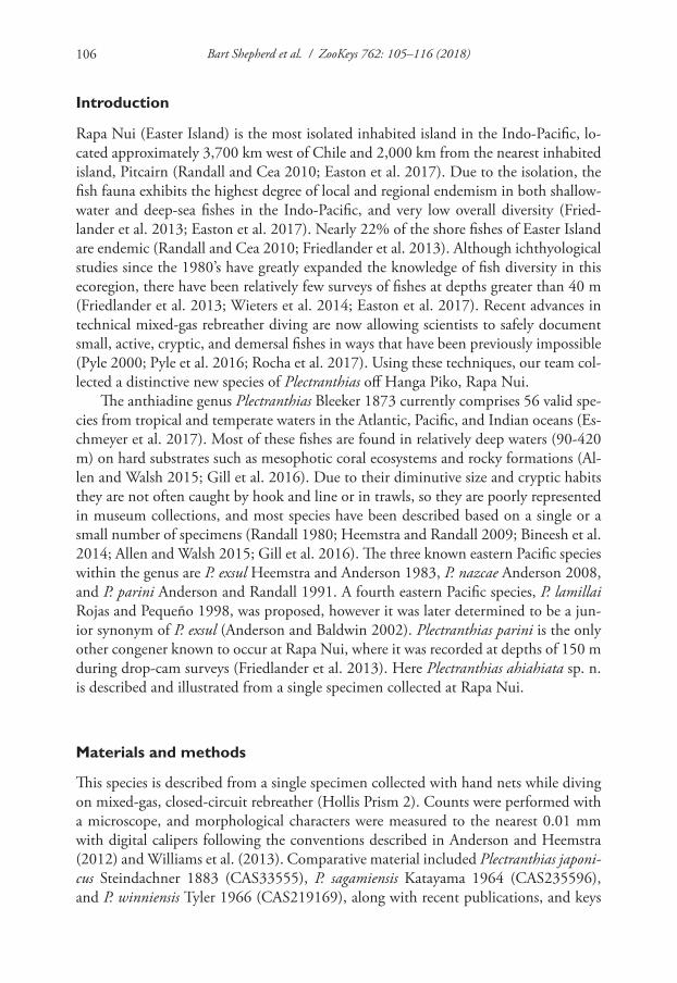
Bart Shepherd et al. / ZooKeys 762: 105–116 (2018)106
Introduction
Rapa Nui (Easter Island) is the most isolated inhabited island in the Indo-Pacific, lo-cated approximately 3,700 km west of Chile and 2,000 km from the nearest inhabited island, Pitcairn (Randall and Cea 2010; Easton et al. 2017). Due to the isolation, the fish fauna exhibits the highest degree of local and regional endemism in both shallow-water and deep-sea fishes in the Indo-Pacific, and very low overall diversity (Fried-lander et al. 2013; Easton et al. 2017). Nearly 22% of the shore fishes of Easter Island are endemic (Randall and Cea 2010; Friedlander et al. 2013). Although ichthyological studies since the 1980’s have greatly expanded the knowledge of fish diversity in this ecoregion, there have been relatively few surveys of fishes at depths greater than 40 m (Friedlander et al. 2013; Wieters et al. 2014; Easton et al. 2017). Recent advances in technical mixed-gas rebreather diving are now allowing scientists to safely document small, active, cryptic, and demersal fishes in ways that have been previously impossible (Pyle 2000; Pyle et al. 2016; Rocha et al. 2017). Using these techniques, our team col-lected a distinctive new species of Plectranthias off Hanga Piko, Rapa Nui.
The anthiadine genus Plectranthias Bleeker 1873 currently comprises 56 valid spe-cies from tropical and temperate waters in the Atlantic, Pacific, and Indian oceans (Es-chmeyer et al. 2017). Most of these fishes are found in relatively deep waters (90-420 m) on hard substrates such as mesophotic coral ecosystems and rocky formations (Al-len and Walsh 2015; Gill et al. 2016). Due to their diminutive size and cryptic habits they are not often caught by hook and line or in trawls, so they are poorly represented in museum collections, and most species have been described based on a single or a small number of specimens (Randall 1980; Heemstra and Randall 2009; Bineesh et al. 2014; Allen and Walsh 2015; Gill et al. 2016). The three known eastern Pacific species within the genus are P. exsul Heemstra and Anderson 1983, P. nazcae Anderson 2008, and P. parini Anderson and Randall 1991. A fourth eastern Pacific species, P. lamillai Rojas and Pequeño 1998, was proposed, however it was later determined to be a jun-ior synonym of P. exsul (Anderson and Baldwin 2002). Plectranthias parini is the only other congener known to occur at Rapa Nui, where it was recorded at depths of 150 m during drop-cam surveys (Friedlander et al. 2013). Here Plectranthias ahiahiata sp. n. is described and illustrated from a single specimen collected at Rapa Nui.
Materials and methods
This species is described from a single specimen collected with hand nets while diving on mixed-gas, closed-circuit rebreather (Hollis Prism 2). Counts were performed with a microscope, and morphological characters were measured to the nearest 0.01 mm with digital calipers following the conventions described in Anderson and Heemstra (2012) and Williams et al. (2013). Comparative material included Plectranthias japoni-cus Steindachner 1883 (CAS33555), P. sagamiensis Katayama 1964 (CAS235596), and P. winniensis Tyler 1966 (CAS219169), along with recent publications, and keys
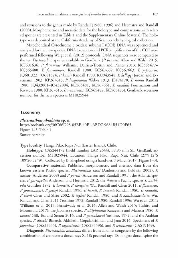
Plectranthias ahiahiata, a new species of perchlet from a mesophotic ecosystem... 107
and revisions to the genus made by Randall (1980, 1996) and Heemstra and Randall (2008). Morphometric and meristic data for the holotype and comparisons with relat-ed species are presented in Table 1 and the Supplementary Online Material. The holo-type was deposited at the California Academy of Sciences ichthyological collection.
Mitochondrial Cytochrome c oxidase subunit I (COI) DNA was sequenced and analyzed for the new species. DNA extraction and PCR amplification of the COI were performed following Weigt et al. (2012) protocols. DNA sequences were compared to the ten Plectranthias species available in GenBank (P. bennetti Allen and Walsh 2015: KT601636; P. flammeus Williams, Delrieu-Trottin and Planes 2013: KC565477–KC565480; P. fourmanoiri Randall 1980: KC567662, KC567663; P. japonicus: JQ681323, JQ681324; P. kamii Randall 1980: KU943548; P. kelloggi Jordan and Ev-ermann 1903: KP267643; P. longimanus Weber 1913: JF494178; P. nanus Randall 1980: JQ432001–JQ432004, KC565481, KC567661; P. randalli Fourmanoir and Rivaton 1980: KP267613; P. winniensis: KC565482, KC565483). GenBank accession number for the new species is MH025944.
Taxonomy
Plectranthias ahiahiata sp. n.http://zoobank.org/36C66D98-05BE-40F1-ABD7-9684B51D0E65Figure 1–3, Table 1Sunset perchlet
Type locality. Hanga Piko, Rapa Nui (Easter Island), Chile.Holotype. CAS244172 (Field number LAR 2644). 39.95 mm SL, GenBank ac-
cession number MH025944. Location: Hanga Piko, Rapa Nui, Chile (27°9'12"S 109°26'52"W). Collected by B. Shepherd using a hand-net, 7 March 2017 (Figure 1–3).
Comparative material. Published morphometric and meristic data from the known eastern Pacific species, Plectranthias exsul (Anderson and Baldwin 2002), P. nazcae (Anderson 2008) and P. parini (Anderson and Randall 1991); the Atlantic spe-cies P. garrupellus Anderson and Heemstra 2012; the Western Pacific species P. anthi-odes Gunther 1872, P. bennetti, P. elongatus Wu, Randall and Chen 2011, P. flammeus, P. fourmanoiri, P. jothyi Randall 1996, P. kamii, P. inermis Randall 1980, P. randalli, P. sheni Chen and Shao 2002, P. taylori Randall 1980, and P. xanthomaculatus Wu, Randall and Chen 2011 (Yoshino 1972; Randall 1980; Randall 1996; Wu et al. 2011; Williams et al. 2013; Peristiwady et al. 2014; Allen and Walsh 2015; Tashiro and Motomura 2017); the Japanese species, P altipinnatus Katayama and Masuda 1980, P. takasei Gill, Tea and Senou 2016, and P. yamakawai Yoshino, 1972; and the Arabian species, P. alcocki Bineesh, Akhilesh, Gopalakrishnan and Jena 2014. Specimens of P. japonicus (CAS33555), P. sagamiensis (CAS235596), and P. winniensis (CAS19169).
Diagnosis. Plectranthias ahiahiata differs from all of its congeners by the following combination of characters: dorsal rays X, 18; pectoral rays 18; longest dorsal spine the
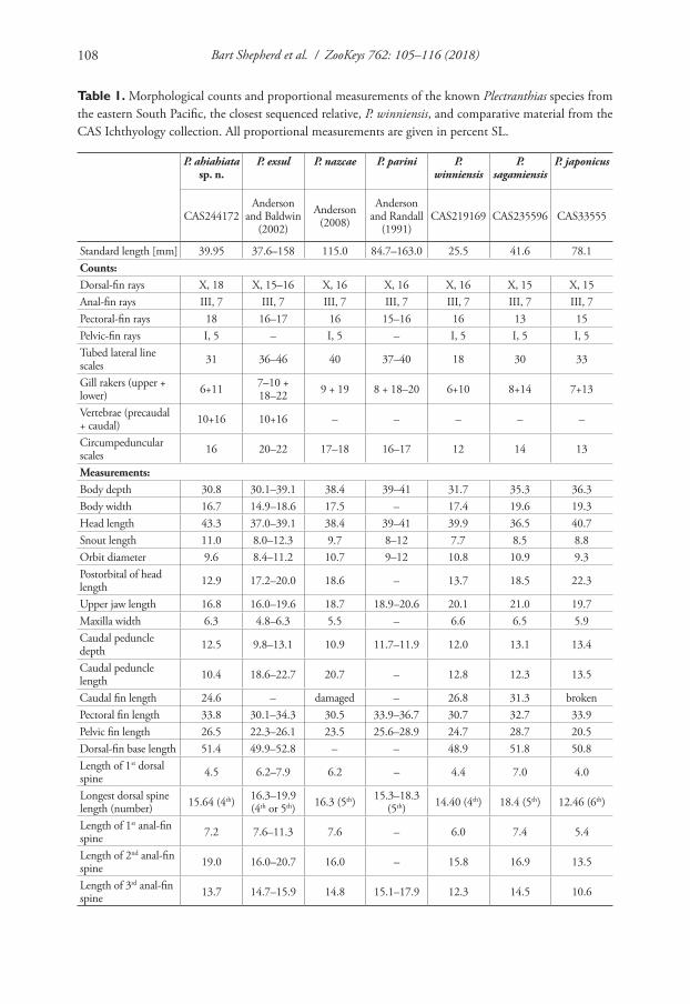
Bart Shepherd et al. / ZooKeys 762: 105–116 (2018)108
Table 1. Morphological counts and proportional measurements of the known Plectranthias species from the eastern South Pacific, the closest sequenced relative, P. winniensis, and comparative material from the CAS Ichthyology collection. All proportional measurements are given in percent SL.
P. ahiahiata sp. n.
P. exsul P. nazcae P. parini P. winniensis
P. sagamiensis
P. japonicus
CAS244172Anderson
and Baldwin (2002)
Anderson (2008)
Anderson and Randall
(1991)CAS219169 CAS235596 CAS33555
Standard length [mm] 39.95 37.6–158 115.0 84.7–163.0 25.5 41.6 78.1Counts:Dorsal-fin rays X, 18 X, 15–16 X, 16 X, 16 X, 16 X, 15 X, 15Anal-fin rays III, 7 III, 7 III, 7 III, 7 III, 7 III, 7 III, 7Pectoral-fin rays 18 16–17 16 15–16 16 13 15Pelvic-fin rays I, 5 – I, 5 – I, 5 I, 5 I, 5Tubed lateral line scales 31 36–46 40 37–40 18 30 33
Gill rakers (upper + lower) 6+11 7–10 +
18–22 9 + 19 8 + 18–20 6+10 8+14 7+13
Vertebrae (precaudal + caudal) 10+16 10+16 – – – – –
Circumpeduncular scales 16 20–22 17–18 16–17 12 14 13
Measurements:Body depth 30.8 30.1–39.1 38.4 39–41 31.7 35.3 36.3Body width 16.7 14.9–18.6 17.5 – 17.4 19.6 19.3Head length 43.3 37.0–39.1 38.4 39–41 39.9 36.5 40.7Snout length 11.0 8.0–12.3 9.7 8–12 7.7 8.5 8.8Orbit diameter 9.6 8.4–11.2 10.7 9–12 10.8 10.9 9.3Postorbital of head length 12.9 17.2–20.0 18.6 – 13.7 18.5 22.3
Upper jaw length 16.8 16.0–19.6 18.7 18.9–20.6 20.1 21.0 19.7Maxilla width 6.3 4.8–6.3 5.5 – 6.6 6.5 5.9Caudal peduncle depth 12.5 9.8–13.1 10.9 11.7–11.9 12.0 13.1 13.4
Caudal peduncle length 10.4 18.6–22.7 20.7 – 12.8 12.3 13.5
Caudal fin length 24.6 – damaged – 26.8 31.3 brokenPectoral fin length 33.8 30.1–34.3 30.5 33.9–36.7 30.7 32.7 33.9Pelvic fin length 26.5 22.3–26.1 23.5 25.6–28.9 24.7 28.7 20.5Dorsal-fin base length 51.4 49.9–52.8 – – 48.9 51.8 50.8Length of 1st dorsal spine 4.5 6.2–7.9 6.2 – 4.4 7.0 4.0
Longest dorsal spine length (number) 15.64 (4th) 16.3–19.9
(4th or 5th) 16.3 (5th) 15.3–18.3 (5th) 14.40 (4th) 18.4 (5th) 12.46 (6th)
Length of 1st anal-fin spine 7.2 7.6–11.3 7.6 – 6.0 7.4 5.4
Length of 2nd anal-fin spine 19.0 16.0–20.7 16.0 – 15.8 16.9 13.5
Length of 3rd anal-fin spine 13.7 14.7–15.9 14.8 15.1–17.9 12.3 14.5 10.6
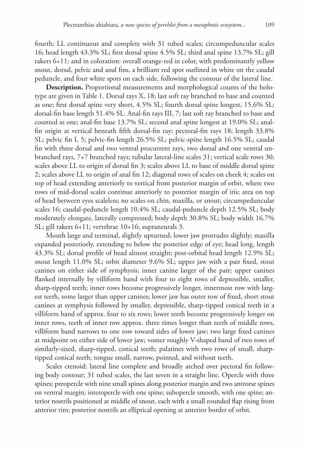
Plectranthias ahiahiata, a new species of perchlet from a mesophotic ecosystem... 109
fourth; LL continuous and complete with 31 tubed scales; circumpeduncular scales 16; head length 43.3% SL; first dorsal spine 4.5% SL; third anal spine 13.7% SL; gill rakers 6+11; and in coloration: overall orange-red in color, with predominantly yellow snout, dorsal, pelvic and anal fins, a brilliant red spot outlined in white on the caudal peduncle, and four white spots on each side, following the contour of the lateral line.
Description. Proportional measurements and morphological counts of the holo-type are given in Table 1. Dorsal rays X, 18; last soft ray branched to base and counted as one; first dorsal spine very short, 4.5% SL; fourth dorsal spine longest, 15.6% SL; dorsal-fin base length 51.4% SL. Anal-fin rays III, 7; last soft ray branched to base and counted as one; anal-fin base 13.7% SL; second anal spine longest at 19.0% SL; anal-fin origin at vertical beneath fifth dorsal-fin ray; pectoral-fin rays 18; length 33.8% SL; pelvic fin I, 5; pelvic-fin length 26.5% SL; pelvic-spine length 16.5% SL; caudal fin with three dorsal and two ventral procurrent rays, two dorsal and one ventral un-branched rays, 7+7 branched rays; tubular lateral-line scales 31; vertical scale rows 30; scales above LL to origin of dorsal fin 3; scales above LL to base of middle dorsal spine 2; scales above LL to origin of anal fin 12; diagonal rows of scales on cheek 4; scales on top of head extending anteriorly to vertical from posterior margin of orbit, where two rows of mid-dorsal scales continue anteriorly to posterior margin of iris; area on top of head between eyes scaleless; no scales on chin, maxilla, or snout; circumpeduncular scales 16; caudal-peduncle length 10.4% SL; caudal-peduncle depth 12.5% SL; body moderately elongate, laterally compressed; body depth 30.8% SL; body width 16.7% SL; gill rakers 6+11; vertebrae 10+16; supraneurals 3.
Mouth large and terminal, slightly upturned; lower jaw protrudes slightly; maxilla expanded posteriorly, extending to below the posterior edge of eye; head long, length 43.3% SL; dorsal profile of head almost straight; post-orbital head length 12.9% SL; snout length 11.0% SL; orbit diameter 9.6% SL; upper jaw with a pair fixed, stout canines on either side of symphysis; inner canine larger of the pair; upper canines flanked internally by villiform band with four to eight rows of depressible, smaller, sharp-tipped teeth; inner rows become progressively longer, innermost row with larg-est teeth, some larger than upper canines; lower jaw has outer row of fixed, short stout canines at symphysis followed by smaller, depressible, sharp-tipped conical teeth in a villiform band of approx. four to six rows; lower teeth become progressively longer on inner rows, teeth of inner row approx. three times longer than teeth of middle rows, villiform band narrows to one row toward sides of lower jaw; two large fixed canines at midpoint on either side of lower jaw; vomer roughly V-shaped band of two rows of similarly-sized, sharp-tipped, conical teeth; palatines with two rows of small, sharp-tipped conical teeth; tongue small, narrow, pointed, and without teeth.
Scales ctenoid; lateral line complete and broadly arched over pectoral fin follow-ing body contour; 31 tubed scales, the last seven in a straight line. Opercle with three spines; preopercle with nine small spines along posterior margin and two antrorse spines on ventral margin; interopercle with one spine; subopercle smooth, with one spine; an-terior nostrils positioned at middle of snout, each with a small rounded flap rising from anterior rim; posterior nostrils an elliptical opening at anterior border of orbit.
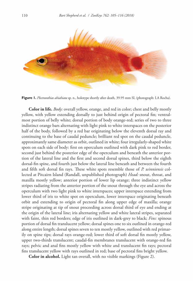
Bart Shepherd et al. / ZooKeys 762: 105–116 (2018)110
Figure 1. Plectranthias ahiahiata sp. n., holotype shortly after death, 39.95 mm SL (photograph: LA Rocha).
Color in life. Body: overall yellow, orange, and red in color; chest and belly mostly yellow, with yellow extending dorsally to just behind origin of pectoral fin; ventral-most portion of belly white; dorsal portion of body orange-red; series of two to three indistinct orange bars alternating with light pink to white interspaces on the posterior half of the body, followed by a red bar originating below the eleventh dorsal ray and continuing to the base of caudal peduncle; brilliant red spot on the caudal peduncle, approximately same diameter as orbit, outlined in white; four irregularly-shaped white spots on each side of body: first on operculum outlined with dark pink to red border, second just behind the posterior edge of the operculum and beneath the anterior por-tion of the lateral line and the first and second dorsal spines, third below the eighth dorsal-fin spine, and fourth just below the lateral line beneath and between the fourth and fifth soft dorsal fin rays. These white spots resemble those of P. winniensis col-lected at Pitcairn Island (Randall, unpublished photograph) Head: snout, throat, and maxilla mostly yellow; anterior portion of lower lip orange; three indistinct yellow stripes radiating from the anterior portion of the snout through the eye and across the operculum with two light pink to white interspaces; upper interspace extending from lower third of iris to white spot on operculum, lower interspace originating beneath orbit and extending to origin of pectoral fin along upper edge of maxilla; orange stripe originating at tip of snout proceeding across dorsal third of eye and ending at the origin of the lateral line; iris alternating yellow and white lateral stripes, separated with faint, thin red borders; edge of iris outlined in dark-grey to black; Fins: spinous portion of dorsal fin translucent yellow; dorsal spines one to six outlined in orange-red along entire length; dorsal spines seven to ten mostly yellow, outlined with red primar-ily on spine tips; dorsal rays orange-red; lower third of soft dorsal fin mostly yellow, upper two-thirds translucent; caudal-fin membranes translucent with orange-red fin rays; pelvic and anal fins mostly yellow with white and translucent fin rays; pectoral fins translucent yellow with rays outlined in red; base of pectoral fins bright yellow.
Color in alcohol. Light tan overall, with no visible markings (Figure 2).
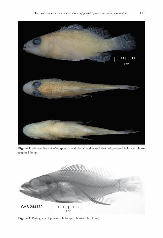
Plectranthias ahiahiata, a new species of perchlet from a mesophotic ecosystem... 111
Figure 2. Plectranthias ahiahiata sp. n., lateral, dorsal, and ventral views of preserved holotype (photo-graphs: J Fong).
Figure 3. Radiograph of preserved holotype (photograph: J Fong).
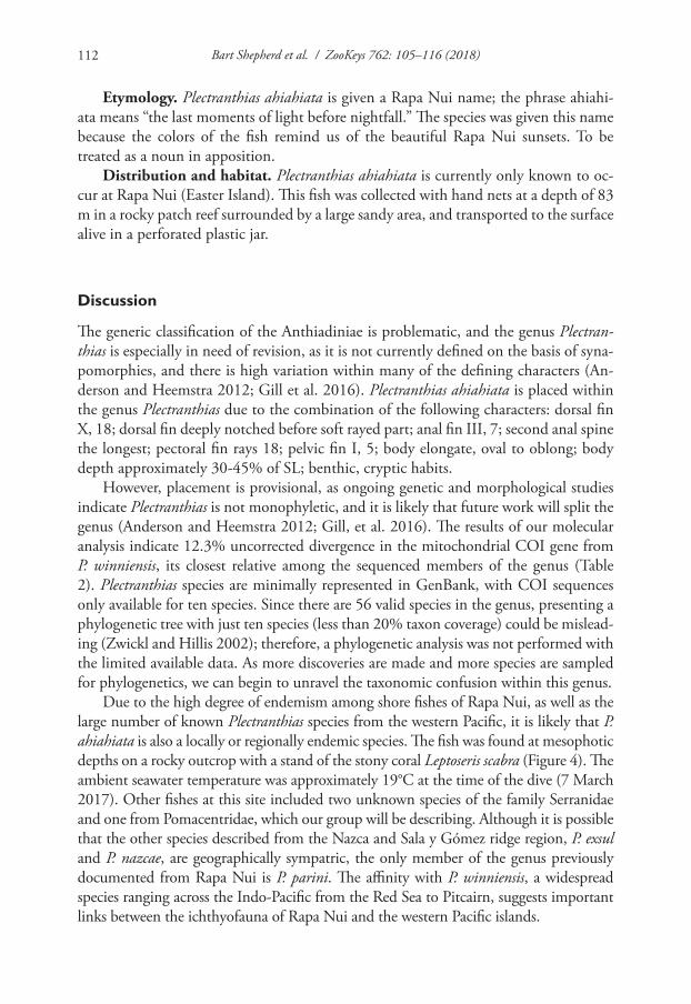
Bart Shepherd et al. / ZooKeys 762: 105–116 (2018)112
Etymology. Plectranthias ahiahiata is given a Rapa Nui name; the phrase ahiahi-ata means “the last moments of light before nightfall.” The species was given this name because the colors of the fish remind us of the beautiful Rapa Nui sunsets. To be treated as a noun in apposition.
Distribution and habitat. Plectranthias ahiahiata is currently only known to oc-cur at Rapa Nui (Easter Island). This fish was collected with hand nets at a depth of 83 m in a rocky patch reef surrounded by a large sandy area, and transported to the surface alive in a perforated plastic jar.
Discussion
The generic classification of the Anthiadiniae is problematic, and the genus Plectran-thias is especially in need of revision, as it is not currently defined on the basis of syna-pomorphies, and there is high variation within many of the defining characters (An-derson and Heemstra 2012; Gill et al. 2016). Plectranthias ahiahiata is placed within the genus Plectranthias due to the combination of the following characters: dorsal fin X, 18; dorsal fin deeply notched before soft rayed part; anal fin III, 7; second anal spine the longest; pectoral fin rays 18; pelvic fin I, 5; body elongate, oval to oblong; body depth approximately 30-45% of SL; benthic, cryptic habits.
However, placement is provisional, as ongoing genetic and morphological studies indicate Plectranthias is not monophyletic, and it is likely that future work will split the genus (Anderson and Heemstra 2012; Gill, et al. 2016). The results of our molecular analysis indicate 12.3% uncorrected divergence in the mitochondrial COI gene from P. winniensis, its closest relative among the sequenced members of the genus (Table 2). Plectranthias species are minimally represented in GenBank, with COI sequences only available for ten species. Since there are 56 valid species in the genus, presenting a phylogenetic tree with just ten species (less than 20% taxon coverage) could be mislead-ing (Zwickl and Hillis 2002); therefore, a phylogenetic analysis was not performed with the limited available data. As more discoveries are made and more species are sampled for phylogenetics, we can begin to unravel the taxonomic confusion within this genus.
Due to the high degree of endemism among shore fishes of Rapa Nui, as well as the large number of known Plectranthias species from the western Pacific, it is likely that P. ahiahiata is also a locally or regionally endemic species. The fish was found at mesophotic depths on a rocky outcrop with a stand of the stony coral Leptoseris scabra (Figure 4). The ambient seawater temperature was approximately 19°C at the time of the dive (7 March 2017). Other fishes at this site included two unknown species of the family Serranidae and one from Pomacentridae, which our group will be describing. Although it is possible that the other species described from the Nazca and Sala y Gómez ridge region, P. exsul and P. nazcae, are geographically sympatric, the only member of the genus previously documented from Rapa Nui is P. parini. The affinity with P. winniensis, a widespread species ranging across the Indo-Pacific from the Red Sea to Pitcairn, suggests important links between the ichthyofauna of Rapa Nui and the western Pacific islands.
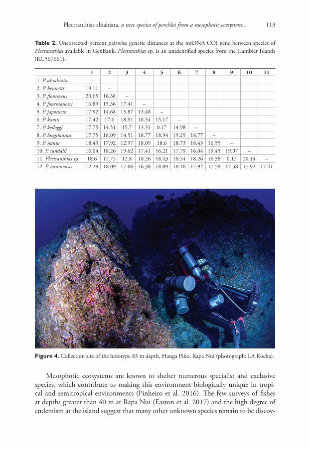
Plectranthias ahiahiata, a new species of perchlet from a mesophotic ecosystem... 113
Table 2. Uncorrected percent pairwise genetic distances at the mtDNA COI gene between species of Plectranthias available in GenBank. Plectranthias sp. is an unidentified species from the Gambier Islands (KC567661).
1 2 3 4 5 6 7 8 9 10 111. P. ahiahiata –2. P. bennetti 19.11 –3. P. flammeus 20.65 16.38 –4. P. fourmanoiri 16.89 15.36 17.41 –5. P. japonicus 17.92 14.68 15.87 13.48 –6. P. kamii 17.42 17.6 18.91 18.54 15.17 –7. P. kelloggi 17.75 14.51 15.7 13.31 0.17 14.98 –8. P. longimanus 17.75 18.09 14.51 18.77 18.94 19.29 18.77 –9. P. nanus 18.43 17.92 12.97 18.09 18.6 18.73 18.43 16.55 –10. P. randalli 16.04 18.26 19.62 17.41 16.21 17.79 16.04 19.45 19.97 –11. Plectranthias sp. 18.6 17.75 12.8 18.26 18.43 18.54 18.26 16.38 0.17 20.14 –12. P. winniensis 12.29 18.09 17.06 16.38 18.09 18.16 17.92 17.58 17.58 17.92 17.41
Figure 4. Collection site of the holotype 83 m depth, Hanga Piko, Rapa Nui (photograph: LA Rocha).
Mesophotic ecosystems are known to shelter numerous specialist and exclusive species, which contribute to making this environment biologically unique in tropi-cal and semitropical environments (Pinheiro et al. 2016). The few surveys of fishes at depths greater than 40 m at Rapa Nui (Easton et al. 2017) and the high degree of endemism at the island suggest that many other unknown species remain to be discov-
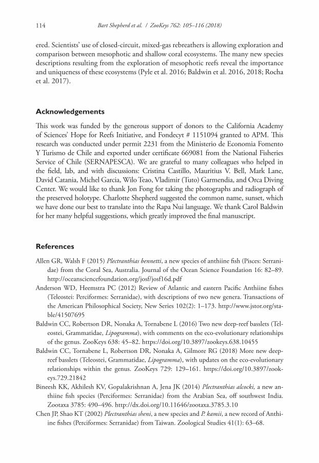
Bart Shepherd et al. / ZooKeys 762: 105–116 (2018)114
ered. Scientists’ use of closed-circuit, mixed-gas rebreathers is allowing exploration and comparison between mesophotic and shallow coral ecosystems. The many new species descriptions resulting from the exploration of mesophotic reefs reveal the importance and uniqueness of these ecosystems (Pyle et al. 2016; Baldwin et al. 2016, 2018; Rocha et al. 2017).
Acknowledgements
This work was funded by the generous support of donors to the California Academy of Sciences’ Hope for Reefs Initiative, and Fondecyt # 1151094 granted to APM. This research was conducted under permit 2231 from the Ministerio de Economia Fomento Y Turismo de Chile and exported under certificate 669081 from the National Fisheries Service of Chile (SERNAPESCA). We are grateful to many colleagues who helped in the field, lab, and with discussions: Cristina Castillo, Mauritius V. Bell, Mark Lane, David Catania, Michel Garcia, Wilo Teao, Vladimir (Tuto) Garmendia, and Orca Diving Center. We would like to thank Jon Fong for taking the photographs and radiograph of the preserved holotype. Charlotte Shepherd suggested the common name, sunset, which we have done our best to translate into the Rapa Nui language. We thank Carol Baldwin for her many helpful suggestions, which greatly improved the final manuscript.
References
Allen GR, Walsh F (2015) Plectranthias bennetti, a new species of anthiine fish (Pisces: Serrani-dae) from the Coral Sea, Australia. Journal of the Ocean Science Foundation 16: 82–89. http://oceansciencefoundation.org/josf/josf16d.pdf
Anderson WD, Heemstra PC (2012) Review of Atlantic and eastern Pacific Anthiine fishes (Teleostei: Perciformes: Serranidae), with descriptions of two new genera. Transactions of the American Philosophical Society, New Series 102(2): 1–173. http://www.jstor.org/sta-ble/41507695
Baldwin CC, Robertson DR, Nonaka A, Tornabene L (2016) Two new deep-reef basslets (Tel-eostei, Grammatidae, Lipogramma), with comments on the eco-evolutionary relationships of the genus. ZooKeys 638: 45–82. https://doi.org/10.3897/zookeys.638.10455
Baldwin CC, Tornabene L, Robertson DR, Nonaka A, Gilmore RG (2018) More new deep-reef basslets (Teleostei, Grammatidae, Lipogramma), with updates on the eco-evolutionary relationships within the genus. ZooKeys 729: 129–161. https://doi.org/10.3897/zook-eys.729.21842
Bineesh KK, Akhilesh KV, Gopalakrishnan A, Jena JK (2014) Plectranthias alcocki, a new an-thiine fish species (Perciformes: Serranidae) from the Arabian Sea, off southwest India. Zootaxa 3785: 490–496. http://dx.doi.org/10.11646/zootaxa.3785.3.10
Chen JP, Shao KT (2002) Plectranthias sheni, a new species and P. kamii, a new record of Anthi-ine fishes (Perciformes: Serranidae) from Taiwan. Zoological Studies 41(1): 63–68.
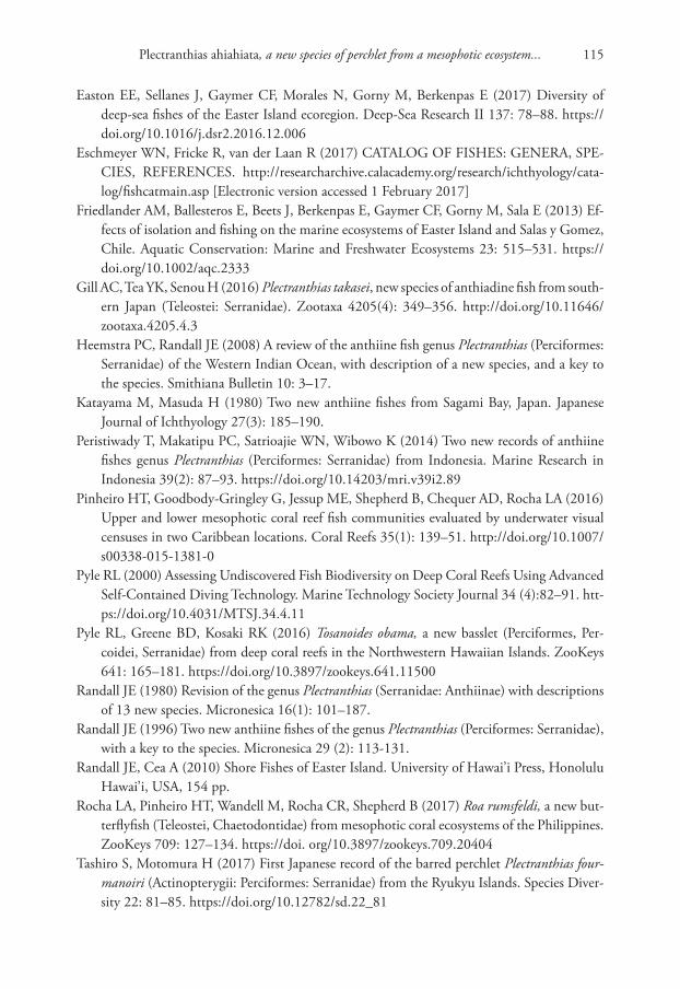
Plectranthias ahiahiata, a new species of perchlet from a mesophotic ecosystem... 115
Easton EE, Sellanes J, Gaymer CF, Morales N, Gorny M, Berkenpas E (2017) Diversity of deep-sea fishes of the Easter Island ecoregion. Deep-Sea Research II 137: 78–88. https://doi.org/10.1016/j.dsr2.2016.12.006
Eschmeyer WN, Fricke R, van der Laan R (2017) CATALOG OF FISHES: GENERA, SPE-CIES, REFERENCES. http://researcharchive.calacademy.org/research/ichthyology/cata-log/fishcatmain.asp [Electronic version accessed 1 February 2017]
Friedlander AM, Ballesteros E, Beets J, Berkenpas E, Gaymer CF, Gorny M, Sala E (2013) Ef-fects of isolation and fishing on the marine ecosystems of Easter Island and Salas y Gomez, Chile. Aquatic Conservation: Marine and Freshwater Ecosystems 23: 515–531. https://doi.org/10.1002/aqc.2333
Gill AC, Tea YK, Senou H (2016) Plectranthias takasei, new species of anthiadine fish from south-ern Japan (Teleostei: Serranidae). Zootaxa 4205(4): 349–356. http://doi.org/10.11646/zootaxa.4205.4.3
Heemstra PC, Randall JE (2008) A review of the anthiine fish genus Plectranthias (Perciformes: Serranidae) of the Western Indian Ocean, with description of a new species, and a key to the species. Smithiana Bulletin 10: 3–17.
Katayama M, Masuda H (1980) Two new anthiine fishes from Sagami Bay, Japan. Japanese Journal of Ichthyology 27(3): 185–190.
Peristiwady T, Makatipu PC, Satrioajie WN, Wibowo K (2014) Two new records of anthiine fishes genus Plectranthias (Perciformes: Serranidae) from Indonesia. Marine Research in Indonesia 39(2): 87–93. https://doi.org/10.14203/mri.v39i2.89
Pinheiro HT, Goodbody-Gringley G, Jessup ME, Shepherd B, Chequer AD, Rocha LA (2016) Upper and lower mesophotic coral reef fish communities evaluated by underwater visual censuses in two Caribbean locations. Coral Reefs 35(1): 139–51. http://doi.org/10.1007/s00338-015-1381-0
Pyle RL (2000) Assessing Undiscovered Fish Biodiversity on Deep Coral Reefs Using Advanced Self-Contained Diving Technology. Marine Technology Society Journal 34 (4):82–91. htt-ps://doi.org/10.4031/MTSJ.34.4.11
Pyle RL, Greene BD, Kosaki RK (2016) Tosanoides obama, a new basslet (Perciformes, Per-coidei, Serranidae) from deep coral reefs in the Northwestern Hawaiian Islands. ZooKeys 641: 165–181. https://doi.org/10.3897/zookeys.641.11500
Randall JE (1980) Revision of the genus Plectranthias (Serranidae: Anthiinae) with descriptions of 13 new species. Micronesica 16(1): 101–187.
Randall JE (1996) Two new anthiine fishes of the genus Plectranthias (Perciformes: Serranidae), with a key to the species. Micronesica 29 (2): 113-131.
Randall JE, Cea A (2010) Shore Fishes of Easter Island. University of Hawai’i Press, Honolulu Hawai’i, USA, 154 pp.
Rocha LA, Pinheiro HT, Wandell M, Rocha CR, Shepherd B (2017) Roa rumsfeldi, a new but-terflyfish (Teleostei, Chaetodontidae) from mesophotic coral ecosystems of the Philippines. ZooKeys 709: 127–134. https://doi. org/10.3897/zookeys.709.20404
Tashiro S, Motomura H (2017) First Japanese record of the barred perchlet Plectranthias four-manoiri (Actinopterygii: Perciformes: Serranidae) from the Ryukyu Islands. Species Diver-sity 22: 81–85. https://doi.org/10.12782/sd.22_81
Bart Shepherd et al. / ZooKeys 762: 105–116 (2018)116
Weigt LA, Baldwin CC, Driskell A, Smith DG, Ormos A, Reyier EA (2012) Using DNA Bar-coding to Assess Caribbean Reef Fish Biodiversity: Expanding Taxonomic and Geographic Coverage. PLoS ONE 7: e41059. https://doi.org/10.1371/journal.pone.0041059
Wieters EA, Medrano A, Perez-Matus A (2014) Functional community structure of shallow hard bottom communities at Easter Island (Rapa Nui). Latin American Journal of Aquatic Research 42(4): 827–844. https://doi.org/10.3856/vol42-issue4-fulltext-10
Wu KY, Randall JE, Chen JP (2011) Two new species of anthiine fishes of the genus Plectran-thias (Perciformes: Serranidae) from Taiwan. Zoological Studies 50(2): 247-253. http://zoolstud.sinica.edu.tw/Journals/50.2/247.pdf
Yoshino T (1972) Plectranthias yamakawai, a new anthiine fish from Ryukyu Islands, with a revision of the genus Plectranthias. Japanese Journal of Ichthyology 19(2): 49–56.
Zwickl DJ, Hillis DM (2002) Increased taxon sampling greatly reduces phylogenetic error. Systematic Biology 51(4): 588-598. https://doi.org/10.1080/10635150290102339
Cis pallidus Mellié, 1849: redescription, new synonym, geographic distribution... 117
Cis pallidus Mellié, 1849: redescription, new synonym, geographic distribution, and host fungi records
Paula V. Borlini1,2, Cristiano Lopes-Andrade2, Lucimar S. Araujo3
1 Programa de Pós-Graduação em Ecologia, Departamento de Biologia Geral, Universidade Federal de Viçosa, Campus Viçosa, Viçosa – MG, 36570-900, Brazil 2 Laboratório de Sistemática e Biologia de Coleoptera, De-partamento de Biologia Animal, Universidade Federal de Viçosa, Campus Viçosa, Viçosa – MG, 36570-900, Brazil 3 Instituto de Ciências Biológicas e da Saúde, Universidade Federal de Viçosa, Campus Rio Paranaíba, MG 230, Km 8, Caixa Postal 22, Rio Paranaíba – MG, 38810-000, Brazil
Corresponding author: Paula V. Borlini ([email protected])
Academic editor: A. Smith | Received 5 January 2018 | Accepted 14 May 2018 | Published 31 May 2018
http://zoobank.org/14726F94-B559-4F8F-9651-5C004FC63FF8
Citation: Borlini PV, Lopes-Andrade C, Araujo LS (2018) Cis pallidus Mellié, 1849: redescription, new synonym, geographic distribution, and host fungi records. ZooKeys 762: 117–129. https://doi.org/10.3897/zookeys.762.23433
AbstractCis pallidus Mellié, 1849 is redescribed based on specimens from Northeast, Southeast, and South Brazil, and from Argentina. A lectotype is designated for Cis semipallidus Pic, 1916, and the species is syno-nymized with C. pallidus. The first host fungi records and a distribution map for the species are provided.
KeywordsCiinae, Ciini, minute tree-fungus beetles, Neotropical
Introduction
Cis Latreille is the most diverse genus of Ciidae with approximately 400 described spe-cies and a worldwide distribution (Oliveira et al. 2013, Lawrence 2016). The genus contains more than half of all described Ciidae species (Lawrence 1971, Oliveira et al. 2013, Lawrence 2016). The Neotropical species of Cis (biogeographic regions sensu Morrone 2015) are represented by nearly 70 described species and at least half of them are organized into artificial species-groups, such as the comptus, creberrimus, melliei, pallidus, taurus, tricornis, and vitulus species-groups.
ZooKeys 762: 117–129 (2018)
doi: 10.3897/zookeys.762.23433
http://zookeys.pensoft.net
Copyright Paula V. Borlini et al. This is an open access article distributed under the terms of the Creative Commons Attribution License (CC BY 4.0), which permits unrestricted use, distribution, and reproduction in any medium, provided the original author and source are credited.
RESEARCH ARTICLE
Launched to accelerate biodiversity research
A peer-reviewed open-access journal
Paula V. Borlini et al. / ZooKeys 762: 117–129 (2018)118
The pallidus-group comprises C. corticinus Gorham, 1883 from Totonicapán, Gua-temala, C. pallidus Mellié, 1849 from the state of Bahia, Northeast Brazil, C. semipal-lidus Pic, 1916 from Buenos Aires, Argentina, and C. tetracentrum Gorham, 1886, which occurs from southern California and Arizona to southern Mexico (Mellié 1849, Lawrence 1971, Gorham 1883, 1886,). These four species share an elongate body with single and uniform elytral punctation, dorsal vestiture of short to long bristles, slightly tumid prosternum, and males with a very small sex patch on the first abdominal ven-trite or none at all (Lawrence 1971; pers. obs.). These species are morphologically closely related to species in the vitulus-group, which are similar but usually have a comparatively less elongate body (Lawrence 1971; pers. obs.).
The aim of this paper is to redescribe C. pallidus, propose a new synonym, and provide new host fungi and geographic distribution records.
Materials and methods
The examined specimens are listed in the section on “type material” and “additional material” below. A total of 12 males from eight localities were dissected, as follows (number of specimens between parentheses): Buenos Aires (1; lectotype of Cis semipal-lidus) and Famaillá (1), in Argentina; Rio de Janeiro (1), São João del-Rei (1), Viçosa (5), Palotina (1), Nova Teutônia (1) and Urubici (1), in Brazil. Abdominal ventrites shown in Fig. 29 and genitalia in Figs 34–35 were extracted from the same male. Geni-talia shown in Figs 30–31 were extracted from the male shown in Fig. 16; the same applies for Figs 43–44 and Fig. 20.
Museum abbreviations are as follows:
ANIC Australian National Insect Collection, CSIRO Entomology (Canberra, Aus-tralian Capital Territory, Australia)
CELC Coleção Entomológica do Laboratório de Sistemática e Biologia de Coleop-tera da Universidade Federal de Viçosa (Viçosa, MG, Brazil)
DZUP Coleção Entomológica Pe. Jesus Santiago Moure, Universidade Federal do Paraná (Curitiba, PR, Brazil)
FMNH Field Museum of Natural History (Chicago, Illinois, USA)MACN Museo Argentino de Ciencias Naturales “Bernardino Rivadavia” (Buenos
Aires, Argentina)MCNZ Fundação Zoobotânica do Rio Grande do Sul (Porto Alegre, RS, Brazil)MNHN Muséum National d’Histoire Naturelle (Paris, France)MNRJ Museu Nacional do Rio de Janeiro (Rio de Janeiro, RJ, Brazil)
Terms for external morphology and male terminalia of ciids follow Lopes-Andrade and Lawrence (2005, 2011), Lawrence et al. (2011), and Lawrence (2016), but see also Oliveira et al. (2013) for an explanation on the use of “tegmen”. The following abbreviations are used for measurements (in mm) and ratios:
Cis pallidus Mellié, 1849: redescription, new synonym, geographic distribution... 119
BW (basal width of scutellar shield),CL (length of antennal club measured from base of the eighth to apex of the tenth
antennomere),EL (elytral length along the midline),EW (greatest width of both elytra),FL (length of antennal funicle measured from base of the third to apex of the
seventh antennomere),GD (greatest depth of body measured in lateral view),GW (greatest diameter of eye),PL (pronotal length along midline),PW (greatest pronotal width),SL (length of scutellar shield),TL (total length counted as EL+PL, i.e. excluding head).
The GD/EW and TL/EW ratios indicate the degree of body convexity and elonga-tion, respectively.
A total of 21 males and 20 females were measured, with representative specimens from all examined localities. Measurements of antennomeres, GW, and BW provided in the description are the mean measurements of three males from three localities (Viçosa, Atílio Vivacqua, and Nova Teutônia); in these cases, standard deviations are not provided because they were 0.01 or less.
For scanning electron microscopy (SEM), specimens were dehydrated in a series of alcohol and acetone solutions, critical point dried (CPD 020, Balzers, Liechtenstein), mounted on aluminum stubs and sputter coated with gold (sputter module SCA 010, Balzers). Samples were then examined under a SEM (LEO VP 1430, Zeiss). Transcrip-tion of labels, dissection, photography under optical equipment, and measurement of specimens followed the methods provided by Araujo and Lopes-Andrade (2016). Names of host fungi extracted from labels were updated consulting the online database of Index Fungorum (http://www.indexfungorum.org) and are summarized in the sec-tion “Host fungi”. The criteria provided in Orledge and Reynolds (2005) are followed for determining breeding records. The distribution map (Fig. 45) was generated using the on-line SimpleMappr tool (Shorthouse 2010).
Taxonomy
Variation in pronotal and elytral color occurred between specimens from the same locality or even from the same basidiome. Also noticeable was variation in the length of dorsal bristles, mainly those on the pronotum (compare Figs 1, 9, 12, 15–21). The comparatively darker elytra and longer pronotal bristles of specimens of the type series of C. semipallidus were observed in several named specimens of C. pallidus.
There was little variation in size and morphology of tegmen and penis between dissected specimens from eight localities (Figs 6–7, 30–44). Length of tegmen varied
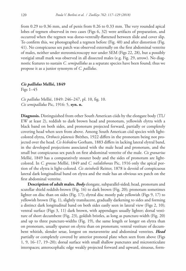
Paula V. Borlini et al. / ZooKeys 762: 117–129 (2018)120
from 0.29 to 0.36 mm, and of penis from 0.26 to 0.33 mm. The very rounded apical lobes of tegmen observed in two cases (Figs 6, 32) were artifacts of preparation, and occurred when the tegmen was dorso-ventrally-flattened between slide and cover slip. To confirm this, we photographed a tegmen before (Fig. 40) and after distortion (Fig. 41). No conspicuous sex patch was observed externally on the first abdominal ventrite of males, neither under stereomicroscopy nor under SEM (Figs 22, 28), but a possibly vestigial small mark was observed in all dissected males (e.g. Fig. 29, arrow). No diag-nostic features to sustain C. semipallidus as a separate species have been found; thus we propose it as a junior synonym of C. pallidus.
Cis pallidus Mellié, 1849Figs 1–45
Cis pallidus Mellié, 1849: 246–247, pl. 10, fig. 10.Cis semipallidus Pic, 1916: 5, syn. n.
Diagnosis. Distinguished from other South American ciids by the elongate body (TL/EW at least 2), reddish to dark brown head and pronotum, yellowish elytra with a black band on both sides, and pronotum projected forward, partially or completely covering head when seen from above. Among South American ciid species with light-colored elytra, Orthocis platensis Brèthes, 1922 differs in the pronotum being not pro-jected over the head. Cis bisbidens Gorham, 1883 differs in lacking lateral elytral band, in the developed projections associated with the male head and pronotum, and the small but conspicuous sex patch on first abdominal ventrite of the male. Cis granarius Mellié, 1849 has a comparatively stouter body and the sides of pronotum are light-colored. In C. grossus Mellié, 1849 and C. validithorax Pic, 1916 only the apical por-tion of the elytra is light-colored. Cis steinheili Reitter, 1878 is devoid of conspicuous lateral dark longitudinal band on elytra and the male has an obvious sex patch on the first abdominal ventrite.
Description of adult males. Body elongate, subparallel-sided; head, pronotum and scutellar shield reddish-brown (Fig. 16) to dark brown (Fig. 20); pronotum sometimes lighter on disc than on sides (Fig. 17); elytral disc mostly pale yellowish (Figs 9, 17) to yellowish brown (Fig. 1), slightly translucent, gradually darkening to sides and forming a distinct dark longitudinal band on both sides easily seen in lateral view (Figs 2, 10); ventral surface (Figs 3, 11) dark brown, with appendages usually lighter; dorsal vesti-ture of short decumbent (Fig. 23), goldish bristles, as long as puncture-width (Fig. 20) and up to three puncture-widths (Fig. 19), the same length or longer on elytra than on pronotum, usually sparser on elytra than on pronotum; ventral vestiture of decum-bent whitish, slender setae, longest on metaventrite and abdominal ventrites. Head partially or completely covered by anterior pronotal plate when seen from above (Figs 1, 9, 16–17, 19–20); dorsal surface with small shallow punctures and microreticulate interspaces; anterocephalic edge weakly projected forward and upward, sinuous, form-
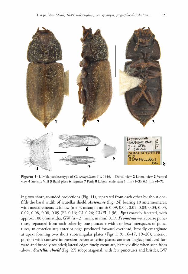
Cis pallidus Mellié, 1849: redescription, new synonym, geographic distribution... 121
Figures 1–8. Male paralectotype of Cis semipallidus Pic, 1916. 1 Dorsal view 2 Lateral view 3 Ventral view 4 Sternite VIII 5 Basal piece 6 Tegmen 7 Penis 8 Labels. Scale bars: 1 mm (1–3); 0.1 mm (4–7).
ing two short, rounded projections (Fig. 11), separated from each other by about one-fifth the basal width of scutellar shield. Antennae (Fig. 24) bearing 10 antennomeres, with measurements as follow (n = 3, mean; in mm): 0.09, 0.05, 0.05, 0.03, 0.03, 0.03, 0.02, 0.08, 0.08, 0.09 (FL 0.16; CL 0.26; CL/FL 1.56). Eyes coarsely facetted, with approx. 100 ommatidia; GW (n = 3, mean; in mm) 0.17. Pronotum with coarse punc-tures, separated from each other by one puncture-width or less; interspaces of punc-tures, microreticulate; anterior edge produced forward overhead, broadly emarginate at apex, forming two short subtriangular plates (Figs 1, 9, 16–17, 19–20); anterior portion with concave impression before anterior plates; anterior angles produced for-ward and broadly rounded; lateral edges finely crenulate, barely visible when seen from above. Scutellar shield (Fig. 27) subpentagonal, with few punctures and bristles; BW
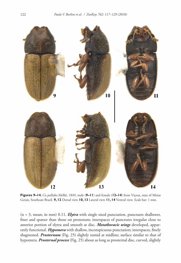
Paula V. Borlini et al. / ZooKeys 762: 117–129 (2018)122
Figures 9–14. Cis pallidus Mellié, 1849, male (9–11) and female (12–14) from Viçosa, state of Minas Gerais, Southeast Brazil. 9, 12 Dorsal view 10, 13 Lateral view 11, 14 Ventral view. Scale bar: 1 mm.
(n = 3, mean; in mm) 0.11. Elytra with single sized punctation, punctures shallower, finer and sparser than those on pronotum; interspaces of punctures irregular close to anterior portion of elytra and smooth at disc. Metathoracic wings developed, appar-ently functional. Hypomera with shallow, inconspicuous punctation; interspaces, finely shagreened. Prosternum (Fig. 25) slightly tumid at midline; surface similar to that of hypomera. Prosternal process (Fig. 25) about as long as prosternal disc, curved, slightly
Cis pallidus Mellié, 1849: redescription, new synonym, geographic distribution... 123
Figures 15–20. Cis pallidus Mellié, 1849, dorsal view from various localities, shown in the same scale. 15 Female from the state of Bahia, compared with the holotype deposited in the MNHN 16 Male from Corcovado, state of Rio de Janeiro 17 Male from Marechal Cândido Rondon, state of Paraná 18 Female from the province of Tucuman, Argentina 19 Male from Nova Teutônia, state of Santa Catarina 20 Male from Urubici, state of Santa Catarina. Scale bar: 1 mm.
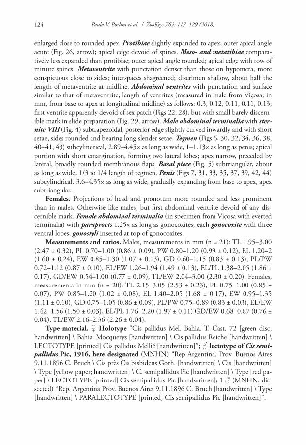
Paula V. Borlini et al. / ZooKeys 762: 117–129 (2018)124
enlarged close to rounded apex. Protibiae slightly expanded to apex; outer apical angle acute (Fig. 26, arrow); apical edge devoid of spines. Meso- and metatibiae compara-tively less expanded than protibiae; outer apical angle rounded; apical edge with row of minute spines. Metaventrite with punctation denser than those on hypomera, more conspicuous close to sides; interspaces shagreened; discrimen shallow, about half the length of metaventrite at midline. Abdominal ventrites with punctation and surface similar to that of metaventrite; length of ventrites (measured in male from Viçosa; in mm, from base to apex at longitudinal midline) as follows: 0.3, 0.12, 0.11, 0.11, 0.13; first ventrite apparently devoid of sex patch (Figs 22, 28), but with small barely discern-ible mark in slide preparation (Fig. 29, arrow). Male abdominal terminalia with ster-nite VIII (Fig. 4) subtrapezoidal, posterior edge slightly curved inwardly and with short setae, sides rounded and bearing long slender setae. Tegmen (Figs 6, 30, 32, 34, 36, 38, 40–41, 43) subcylindrical, 2.89–4.45× as long as wide, 1–1.13× as long as penis; apical portion with short emargination, forming two lateral lobes; apex narrow, preceded by lateral, broadly rounded membranous flaps. Basal piece (Fig. 5) subtriangular, about as long as wide, 1/3 to 1/4 length of tegmen. Penis (Figs 7, 31, 33, 35, 37, 39, 42, 44) subcylindrical, 3.6–4.35× as long as wide, gradually expanding from base to apex, apex subtriangular.
Females. Projections of head and pronotum more rounded and less prominent than in males. Otherwise like males, but first abdominal ventrite devoid of any dis-cernible mark. Female abdominal terminalia (in specimen from Viçosa with everted terminalia) with paraprocts 1.25× as long as gonocoxites; each gonocoxite with three ventral lobes; gonostyli inserted at top of gonocoxites.
Measurements and ratios. Males, measurements in mm (n = 21): TL 1.95–3.00 (2.47 ± 0.32), PL 0.70–1.00 (0.86 ± 0.09), PW 0.80–1.20 (0.99 ± 0.12), EL 1.20–2 (1.60 ± 0.24), EW 0.85–1.30 (1.07 ± 0.13), GD 0.60–1.15 (0.83 ± 0.13), PL/PW 0.72–1.12 (0.87 ± 0.10), EL/EW 1.26–1.94 (1.49 ± 0.13), EL/PL 1.38–2.05 (1.86 ± 0.17), GD/EW 0.54–1.00 (0.77 ± 0.09), TL/EW 2.04–3.00 (2.30 ± 0.20). Females, measurements in mm (n = 20): TL 2.15–3.05 (2.53 ± 0.23), PL 0.75–1.00 (0.85 ± 0.07), PW 0.85–1.20 (1.02 ± 0.08), EL 1.40–2.05 (1.68 ± 0.17), EW 0.95–1.35 (1.11 ± 0.10), GD 0.75–1.05 (0.86 ± 0.09), PL/PW 0.75–0.89 (0.83 ± 0.03), EL/EW 1.42–1.56 (1.50 ± 0.03), EL/PL 1.76–2.20 (1.97 ± 0.11) GD/EW 0.68–0.87 (0.76 ± 0.04), TL/EW 2.16–2.36 (2.26 ± 0.04).
Type material. ♀ Holotype “Cis pallidus Mel. Bahia. T. Cast. 72 [green disc, handwritten] \ Bahia. Mocquerys [handwritten] \ Cis pallidus Reiche [handwritten] \ LECTOTYPE [printed] Cis pallidus Mellié [handwritten]”; ♂ lectotype of Cis semi-pallidus Pic, 1916, here designated (MNHN) “Rep Argentina. Prov. Buenos Aires 9.11.1896 C. Bruch \ Cis près Cis bisbidens Gorh. [handwritten] \ Cis [handwritten] \ Type [yellow paper; handwritten] \ C. semipallidus Pic [handwritten] \ Type [red pa-per] \ LECTOTYPE [printed] Cis semipallidus Pic [handwritten]; 1 ♂ (MNHN, dis-sected) “Rep. Argentina Prov. Buenos Aires 9.11.1896 C. Bruch [handwritten] \ Type [handwritten] \ PARALECTOTYPE [printed] Cis semipallidus Pic [handwritten]”.
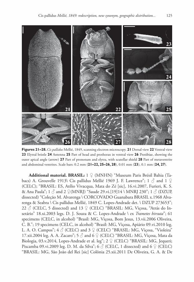
Cis pallidus Mellié, 1849: redescription, new synonym, geographic distribution... 125
Figures 21–28. Cis pallidus Mellié, 1849, scanning electron microscopy. 21 Dorsal view 22 Ventral view 23 Elytral bristle 24 Antenna 25 Part of head and prothorax in ventral view 26 Protibiae, showing the outer apical angle (arrow) 27 Part of pronotum and elytra, with scutellar shield 28 Part of metaventrite and abdominal ventrites. Scale bars: 0.2 mm (21–22, 25–26, 28), 0.01 mm (23), 0.1 mm (24, 27).
Additional material. BRASIL: 1 ♀ (MNHN) “Museum Paris Brésil Bahia (Ta-bacs) A. Grouvelle 1913\ Cis pallidus Mellié 1969 J. F. Lawrence”; 1 ♂ and 1 ♀ (CELC); “BRASIL: ES, Atílio Vivacqua, Mata do Zé [sic], 16.vi.2007, Furieri, K. S. & Ana Paula”; 1 ♂ and 2 ♀(MNRJ) “Saude 29.vi.[19]14 \ MNRJ 230”; 1 ♂ (DZUP, dissected) “Coleção M. Alvarenga \ CORCOVADO Guanabara BRASIL x.1968 Alva-renga & Seabra \ Cis pallidus Mellié, 1849 C. Lopes-Andrade det. \ DZUP 273653”; 22 ♂ (CELC, 5 dissected) and 13 ♀ (CELC) “BRASIL: MG, Viçosa, “Atrás do In-setário” 18.xi.2003 legs. D. J. Souza & C. Lopes-Andrade \ ex Trametes hirsuta”; 61 specimens (CELC, in alcohol) “Brasil: MG, Viçosa, Bom Jesus, 13.vii.2006 Oliveira, C. B.”; 19 specimens (CELC, in alcohol) “Brasil: MG, Viçosa, Apiário 09.vi.2010 leg. L. A. O. Campos”; 4 ♂ (CELC) and 3 ♀ (CELC) “BRASIL: MG, Viçosa, “Violeira” 17.xii.2004 leg. A. A. Zacaro”; 5 ♂ and 6 ♀ (CELC) “BRASIL: MG, Viçosa, Mata da Biologia, 03.v.2014, Lopes-Andrade et al. leg”; 2 ♀ (CELC) “BRASIL: MG, Jequeri; Piscamba 09.vi.2009 leg. D. M. da Silva”; 6 ♂ (CELC, 1 dissected) and 6 ♀ (CELC) “BRASIL: MG, São João del Rei [sic] Colônia 25.xii.2011 De Oliveira, G. A. & De
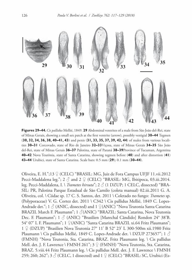
Paula V. Borlini et al. / ZooKeys 762: 117–129 (2018)126
Figures 29–44. Cis pallidus Mellié, 1849. 29 Abdominal ventrites of a male from São João del-Rei, state of Minas Gerais, showing a small sex patch at the first ventrite (arrow), possibly vestigial 30–44 Tegmen (30, 32, 34, 36, 38, 40–41, 43) and penis (31, 33, 35, 37, 39, 42, 44) of males from various locali-ties 30–31 Corcovado, state of Rio de Janeiro 32–33Viçosa, state of Minas Gerais 34–35 São João del-Rei, state of Minas Gerais 36–37 Palotina, state of Paraná 38–39Province of Tucuman, Argentina 40–42 Nova Teutônia, state of Santa Catarina, showing tegmen before (40) and after distortion (41) 43–44 Urubici, state of Santa Catarina. Scale bars: 0.5 mm (29); 0.1 mm (30–44).
Oliveira, E. H.”;13 ♀ (CELC) “BRASIL: MG, Juiz de Fora Campus UFJF 11.vii.2012 Pecci-Maddalena leg.”; 2 ♂ and 2 ♀ (CELC) “BRASIL: MG, Ibitipoca, 03.iii.2014. leg. Pecci-Maddalena, I. \ Trametes hirsuta”; 2 ♂ (1 DZUP; 1 CELC, dissected) “BRA-SIL: PR, Palotina Parque Estadual de São Camilo (coleta manual) 02.iii.2011 G. A. Oliveira, col. \ Ciidae sp. 17 C. S. Santos, det. 2011 \ Coletado no fungo: Trametes sp. (Polyporaceae) V. G. Cortez det. 2011 \ C342 \ Cis pallidus Mellié, 1849 C. Lopes-Andrade det.”; 1 ♂ (ANIC, dissected) and 1 ♀(ANIC) “Nova Teutonia Santa Catarina BRAZIL March F. Plaumann”; 1 ♂(ANIC) “BRAZIL: Santa Catarina, Nova Teutonia Dec. F. Plaumann”; 1 ♂ (ANIC) “Brasilien [Marechal Cândido] Rondon 24° 38’B. 54° 07’ L F. Plaumann”; 1 ♀(ANIC) “Santa Catarina BRAZIL xi.64 Fritz Plaumann”; 1 ♀ (DZUP) “Brasilien Nova Teutonia 27° 11’ B 52° 23’ L 300-500m xii.1980 Fritz Plaumann \ Cis pallidus Mellié, 1849 C. Lopes-Andrade det. \ DZUP 273657”; 1 ♂ (FMNH) “Nova Teutonia, Sta. Catarina, BRAZ. Fritz Plaumann leg. \ Cis pallidus Mell. det. J. F. Lawrence \ FMNH 261”; 3 ♀ (FMNH) “Nova Teutonia, Sta. Catarina, BRAZ. 5.viii.44 Fritz Plaumann leg. \ Cis pallidus Mell. det. J. F. Lawrence \ FMNH 259; 260; 262”; 3 ♂ (CELC, 1 dissected) and 1 ♀ (CELC) “BRASIL: SC, Urubici (Es-
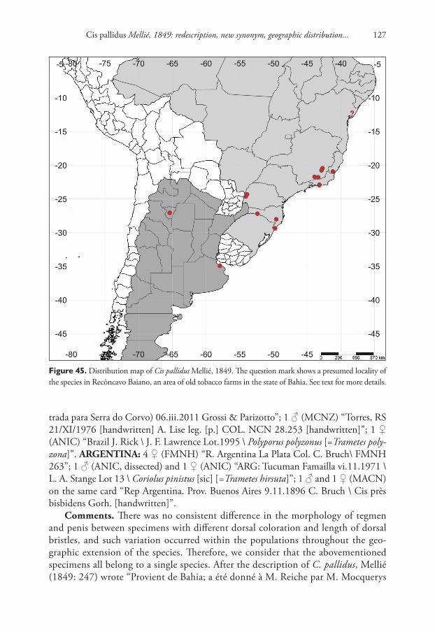
Cis pallidus Mellié, 1849: redescription, new synonym, geographic distribution... 127
Figure 45. Distribution map of Cis pallidus Mellié, 1849. The question mark shows a presumed locality of the species in Recôncavo Baiano, an area of old tobacco farms in the state of Bahia. See text for more details.
trada para Serra do Corvo) 06.iii.2011 Grossi & Parizotto”; 1 ♂ (MCNZ) “Torres, RS 21/XI/1976 [handwritten] A. Lise leg. [p.] COL. NCN 28.253 [handwritten]”; 1 ♀ (ANIC) “Brazil J. Rick \ J. F. Lawrence Lot.1995 \ Polyporus polyzonus [=Trametes poly-zona]”. ARGENTINA: 4 ♀ (FMNH) “R. Argentina La Plata Col. C. Bruch\ FMNH 263”; 1 ♂ (ANIC, dissected) and 1 ♀ (ANIC) “ARG: Tucuman Famailla vi.11.1971 \ L. A. Stange Lot 13 \ Coriolus pinistus [sic] [=Trametes hirsuta]”; 1 ♂ and 1 ♀ (MACN) on the same card “Rep Argentina. Prov. Buenos Aires 9.11.1896 C. Bruch \ Cis près bisbidens Gorh. [handwritten]”.
Comments. There was no consistent difference in the morphology of tegmen and penis between specimens with different dorsal coloration and length of dorsal bristles, and such variation occurred within the populations throughout the geo-graphic extension of the species. Therefore, we consider that the abovementioned specimens all belong to a single species. After the description of C. pallidus, Mellié (1849: 247) wrote “Provient de Bahia; a été donné à M. Reiche par M. Mocquerys
Paula V. Borlini et al. / ZooKeys 762: 117–129 (2018)128
de Rouen”, a statement in the singular, suggesting that he had only one specimen at the time of the description. It is important to note that Mellié, in the same work, clearly used plural in case he had examined two or more specimens. Therefore, we consider the single type specimen located in the MNHN as the holotype, even though it has a lectotype label. Two specimens from MACN, pinned at the same card, have the same locality label of the type series of C. semipallidus, but these do not have any indication whether they were examined or not by Maurice Pic. Thus we think they do not belong to the type series but they were possibly collected to-gether. The specimen from the MNHN labeled “(…) Bahia (Tabacs) A. Grouvelle (…)” is possibly from Recôncavo Baiano (Fig. 45, question mark), a name for the geographic area around Bay of All Saints, the biggest bay at the northeastern coast of Brazil. The collector Antoine Grouvelle was the director of the “Manufactures nationales des Tabacs” (national manufacturers of tobacco) and used to catch small insects in the tobacco leaves exported to France. These insects, mostly Coleoptera, were probably retained during their flight by the more or less abundant pubescence and viscosity which covers the tobacco leaves; it is also possible that some of them had been attracted by the rainwater which remains in the axils of the leaves, and that others came from the washing water in the country of production (Régimbart 1895). In the 19th century, most of the tobacco production from Bahia came from Recôncavo Baiano; therefore, we consider that it is plausible that this specimen of C. pallidus has come from this area.
Host fungi. Trametes hirsuta Lloyde (Polyporaceae), one record and two breeding records; Trametes polyzona Pers. (Polyporaceae), one record; and Trametes sp. (Polypo-raceae), one record.
Acknowledgements
We would like to especially thank John F. Lawrence for sending a large number of named specimens from the C. pallidus and C. vitulus species-groups for comparison; and Vivian E. Sandoval-Gòmez for examining historical and type material related to C. pallidus in MACN and MNHN. We are grateful to everyone who collected and sent us specimens (see section on Additional material for their names), and for museum cura-tors for allowing examination and loan of specimens. We are also grateful to Glenda M. Orledge and Daniel K. Young for their corrections and suggestions to the manuscript, and Aaron D. Smith for his assistance during the revision process. P.V.B. thanks the Programa de Pós-Graduação em Ecologia (Universidade Federal de Viçosa, Brazil) for the academic support. C.L.A. thanks Núcleo de Microscopia e Microanálise (NMM) of Universidade Federal de Viçosa for SEM facilities in 2004. Financial support was provided by Fundação de Amparo à Pesquisa do Estado de Minas Gerais (FAPEMIG; Edital 01/2016 – Demanda Universal, APQ-02675-16; Edital FORTIS; master grant to P.V.B.), Conselho Nacional de Desenvolvimento Científico e Tecnológico (CNPq; research grant to C.L.A. n° 307116/2015-8).
Cis pallidus Mellié, 1849: redescription, new synonym, geographic distribution... 129
References
Araujo LS, Lopes-Andrade C (2016) A new species of Falsocis (Coleoptera: Ciidae) from the Atlantic Forest biome with new geographic records and an updated identification key for the species of the genus. Zoologia 33: 1–6. https://doi.org/10.1590/S1984-4689zo-ol-20150173
Gorham HS (1883) Cleridae, Ptinidae, Bostrychidae, Cioidae. In: Godman FD, Salvin O (Eds) Biologia Centrali-Americana. Insecta. Coleoptera. Vol. 3, Part 2. Porter, London, 169–224. [pl. 10, figs. 23–27]
Gorham HS (1886) Supplement to Malacodermata. In: Godman FD, Salvin O (Eds) Biologia Centrali-Americana. Insecta. Coleoptera. Vol. 3, Part 2. Porter, London, 313–360.
Lawrence JF (1971) Revision of the North American Ciidae (Coleoptera). Bulletin of the Mu-seum of Comparative Zoology 142: 419–522.
Lawrence JF (2016) The Australian Ciidae (Coleoptera: Tenebrionoidea): A Preliminary Revi-sion. Zootaxa 4198: 1–208. https://doi.org/10.11646/zootaxa.4198.1.1
Lawrence JF, Slipinski A, Seago AE, Thayer MK, Newton AF, Marvaldi AE (2011) Phylogeny of the Coleoptera based on morphological characters of adults and larvae. Annales Zoo-logici 61: 1–217. https://doi.org/10.3161/000345411X576725
Lopes-Andrade C, Lawrence JF (2005) Phellinocis, a new genus of Neotropical Ciidae (Coleop-tera: Tenebrionoidea). Zootaxa 1034: 43–60.
Lopes-Andrade C, Lawrence JF (2011) Synopsis of Falsocis Pic (Coleoptera, Ciidae), new spe-cies, new records and an identification key. ZooKeys 145: 59–78. https://doi.org/10.3897/zookeys.145.1895
Mellié J (1849) Monographie de l’ancien genre Cis des auteurs. Annales de la Société Ento-mologique de France 6(2): 205–274, 313–396.
Morrone JJ (2015) Biogeographical regionalization of the world: a reappraisal. Australian Sys-tematic Botany 28: 81–90. https://doi.org/10.1071/SB14042
Oliveira EH, Lopes-Andrade C, Lawrence JF (2013) Review of the Neotropical Ciidae (In-secta: Coleoptera) in the Cis taurus species-group. Arthropod Systematics & Phylogeny 71: 181–210.
Orledge GM, Reynolds SE (2005) Fungivore host-use groups from cluster analysis: patterns of utilisation of fungal fruiting bodies by ciid beetles. Ecological Entomology 30: 620–641. https://doi.org/10.1111/j.0307-6946.2005.00727.x
Pic M (1916) Diagnoses génériques et spécifiques. Mélanges exotico-entomologiques 18: 1–20.Régimbart M (1895) Dytiscides trouvés dans les tabacs par les soins de M. Antoine Grouvelle.
Annales de la Société entomologique de France 64: 321–348.Shorthouse DP (2010) SimpleMappr, an online tool to produce publication-quality point
maps. http://www.simplemappr.net

Paula V. Borlini et al. / ZooKeys 762: 117–129 (2018)130
Anuran species composition of Cancão Municipal Natural Park... 131
Anuran species composition of Cancão Municipal Natural Park, Municipality of Serra do Navio, Amapá state, Brazil
Yuri Breno Silva e Silva1, Carlos Eduardo Costa-Campos1
1 Universidade Federal do Amapá, Departamento de Ciências Biológicas e da Saúde, Laboratorio de Herpeto-logia, Rod. Juscelino Kubitschek, km 02, Jardim Marco Zero, CEP 68.903-419, Macapá, AP, Brasil
Corresponding author: Carlos Eduardo Costa-Campos (email address)
Academic editor: A. Crottini | Received 29 November 2017 | Accepted 28 February 2018 | Published 31 May 2018
http://zoobank.org/65F2D1E9-2925-425B-94DA-FF5ECDCEC3EA
Citation: Silva e Silva YB, Costa-Campos CE (2018) Anuran species composition of Cancão Municipal Natural Park, Municipality of Serra do Navio, Amapá state, Brazil. ZooKeys 762: 131–148. https://doi.org/10.3897/zookeys.762.22634
AbstractIn this study, the first survey of anuran species in the Cancão Municipal Natural Park is presented, a pro-tected area of approximately 370 hectares of Amazonian forest located in the northwest center region of the state of Amapá, Brazil. The work was performed during the dry and rainy season, through active visual and auditory survey, totaling 216 man hours of sampling effort. Forty-nine species of anuran amphibians were recorded in the Cancão Municipal Natural Park, including three new records: Hyalinobatrachium iaspidiense, Pristimantis cf. ockendeni, and Scinax garbei. Three species, Hyalinobatrachium iaspidiense, Ameerega pulchripecta, and Anomaloglossus baeobatrachus, are listed as Data Deficient and one is listed as Vulnerable (Atelopus hoogmoedi) according red lists of IUCN. The rarefaction curve cumulative species did not reach an asymptote, indicating that site has potential for species that have not yet been recorded. Nine species were represented by only one individual and were considered rare in the studied environments, eight species were defined as common, and the 32 remaining species were classified as having intermediary abundance. Our data indicated that Cancão Municipal Natural Park contains a considerable portion of the anurans species richness of Amapá state, turn the area into a place of great importance for the conser-vation of the anurans of the Eastern Amazon.
KeywordsAmazonia, conservation, eastern Amazon, species list
ZooKeys 762: 131–148 (2018)
doi: 10.3897/zookeys.762.22634
http://zookeys.pensoft.net
Copyright Y.B. Silva e Silva, C.E. Costa-Campos. This is an open access article distributed under the terms of the Creative Commons Attribution License (CC BY 4.0), which permits unrestricted use, distribution, and reproduction in any medium, provided the original author and source are credited.
CHECKLIST
Launched to accelerate biodiversity research
A peer-reviewed open-access journal
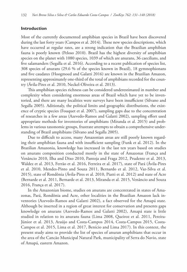
Yuri Breno Silva e Silva & Carlos Eduardo Costa-Campos / ZooKeys 762: 131–148 (2018)132
Introduction
Most of the currently documented amphibian species in Brazil have been discovered during the last forty years (Campos et al. 2014). These new species descriptions, which have occurred at regular rates, are a strong indication that the Brazilian amphibian fauna is poorly known (Peloso 2010). Brazil has the highest diversity of amphibian species on the planet with 1080 species, 1039 of which are anurans, 36 caecilians, and five salamanders (Segalla et al. 2016). According to a recent publication of species list, 308 species of anurans (29.6 % of the species known in Brazil), 18 gymnophionans and five caudates (Hoogmoed and Galatti 2016) are known in the Brazilian Amazon, representing approximately one-third of the total of amphibians recorded for the coun-try (Ávila-Pires et al. 2010, Neckel-Oliveira et al. 2013).
This amphibian species richness can be considered underestimated in number and complexity when considering enormous areas of Brazil which have yet to be inven-toried, and there are many localities were surveys have been insufficient (Silvano and Segalla 2005). Aditionaly, the political limits and geographic distributions, the exist-ence of cryptic species (Fouquet et al. 2007), sampling gaps due to the concentration of researches in a few areas (Azevedo-Ramos and Galatti 2002), sampling effort used appropriate methods for inventories of amphibians (Miranda et al. 2015) and prob-lems in various taxonomic groups, frustrate attempts to obtain a comprehensive under-standing of Brazil amphibians (Silvano and Segalla 2005).
Due to difficult to access, many Amazonian areas are still poorly known regard-ing their amphibian fauna and with insufficient sampling (Funk et al. 2012). In the Brazilian Amazonia, knowledge has increased in the last ten years based on studies on anurans composition conducted mostly in the state of Amazonas (França and Venâncio 2010, Ilha and Dixo 2010, Pantoja and Fraga 2012, Prudente et al. 2013, Waldez et al. 2013, Ferrão et al. 2016, Ferreira et al. 2017), state of Pará (Ávila-Pires et al. 2010, Mendes-Pinto and Souza 2011, Bernardo et al. 2012, Vaz-Silva et al. 2015), state of Rondônia (Ávila-Pires et al. 2010, Piatti et al. 2012) and state of Acre (Bernarde et al. 2011, Bernarde et al. 2013, Miranda et al. 2015, Venâncio and Souza 2016, França et al. 2017).
In the Amazonian biome, studies on anurans are concentrated in states of Ama-zonas, Pará, Rondônia and Acre, other localities in the Brazilian Amazon lack in-ventories (Azevedo-Ramos and Galatti 2002), a fact observed for the Amapá state. Although be inserted in a region of great interest for conservation and presents gaps knowledge on anurans (Azevedo-Ramos and Galatti 2002), Amapá state is little studied in relation to its anurans fauna (Lima 2008, Queiroz et al. 2011, Pereira-Júnior et al. 2013, Araújo and Costa-Campos 2014, Costa-Campos 2015, Costa-Campos et al. 2015, Lima et al. 2017, Benício and Lima 2017). In this context, the present study aims to provide the list of species of anuran amphibians that occur in the area of the Cancão Municipal Natural Park, municipality of Serra do Navio, state of Amapá, eastern Amazon.
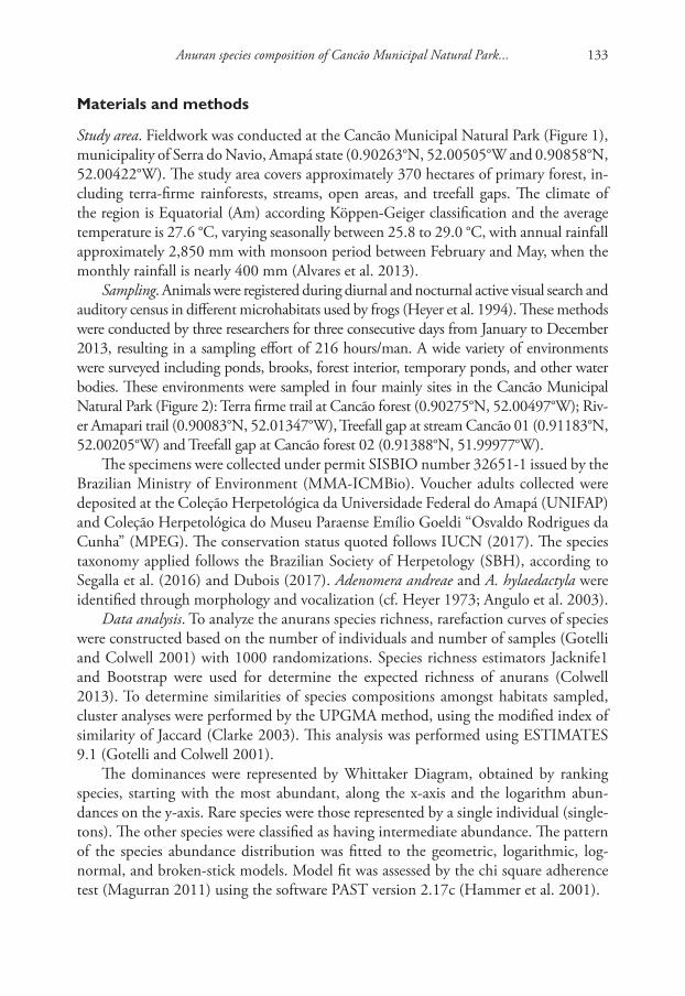
Anuran species composition of Cancão Municipal Natural Park... 133
Materials and methods
Study area. Fieldwork was conducted at the Cancão Municipal Natural Park (Figure 1), municipality of Serra do Navio, Amapá state (0.90263°N, 52.00505°W and 0.90858°N, 52.00422°W). The study area covers approximately 370 hectares of primary forest, in-cluding terra-firme rainforests, streams, open areas, and treefall gaps. The climate of the region is Equatorial (Am) according Köppen-Geiger classification and the average temperature is 27.6 °C, varying seasonally between 25.8 to 29.0 °C, with annual rainfall approximately 2,850 mm with monsoon period between February and May, when the monthly rainfall is nearly 400 mm (Alvares et al. 2013).
Sampling. Animals were registered during diurnal and nocturnal active visual search and auditory census in different microhabitats used by frogs (Heyer et al. 1994). These methods were conducted by three researchers for three consecutive days from January to December 2013, resulting in a sampling effort of 216 hours/man. A wide variety of environments were surveyed including ponds, brooks, forest interior, temporary ponds, and other water bodies. These environments were sampled in four mainly sites in the Cancão Municipal Natural Park (Figure 2): Terra firme trail at Cancão forest (0.90275°N, 52.00497°W); Riv-er Amapari trail (0.90083°N, 52.01347°W), Treefall gap at stream Cancão 01 (0.91183°N, 52.00205°W) and Treefall gap at Cancão forest 02 (0.91388°N, 51.99977°W).
The specimens were collected under permit SISBIO number 32651-1 issued by the Brazilian Ministry of Environment (MMA-ICMBio). Voucher adults collected were deposited at the Coleção Herpetológica da Universidade Federal do Amapá (UNIFAP) and Coleção Herpetológica do Museu Paraense Emílio Goeldi “Osvaldo Rodrigues da Cunha” (MPEG). The conservation status quoted follows IUCN (2017). The species taxonomy applied follows the Brazilian Society of Herpetology (SBH), according to Segalla et al. (2016) and Dubois (2017). Adenomera andreae and A. hylaedactyla were identified through morphology and vocalization (cf. Heyer 1973; Angulo et al. 2003).
Data analysis. To analyze the anurans species richness, rarefaction curves of species were constructed based on the number of individuals and number of samples (Gotelli and Colwell 2001) with 1000 randomizations. Species richness estimators Jacknife1 and Bootstrap were used for determine the expected richness of anurans (Colwell 2013). To determine similarities of species compositions amongst habitats sampled, cluster analyses were performed by the UPGMA method, using the modified index of similarity of Jaccard (Clarke 2003). This analysis was performed using ESTIMATES 9.1 (Gotelli and Colwell 2001).
The dominances were represented by Whittaker Diagram, obtained by ranking species, starting with the most abundant, along the x-axis and the logarithm abun-dances on the y-axis. Rare species were those represented by a single individual (single-tons). The other species were classified as having intermediate abundance. The pattern of the species abundance distribution was fitted to the geometric, logarithmic, log-normal, and broken-stick models. Model fit was assessed by the chi square adherence test (Magurran 2011) using the software PAST version 2.17c (Hammer et al. 2001).
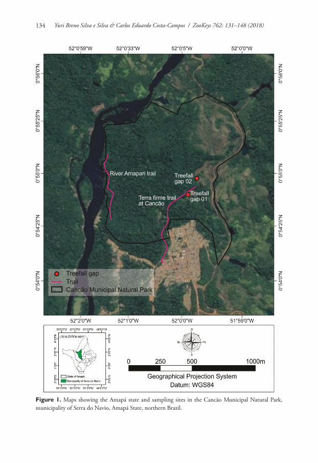
Yuri Breno Silva e Silva & Carlos Eduardo Costa-Campos / ZooKeys 762: 131–148 (2018)134
Figure 1. Maps showing the Amapá state and sampling sites in the Cancão Municipal Natural Park, municipality of Serra do Navio, Amapá State, northern Brazil.
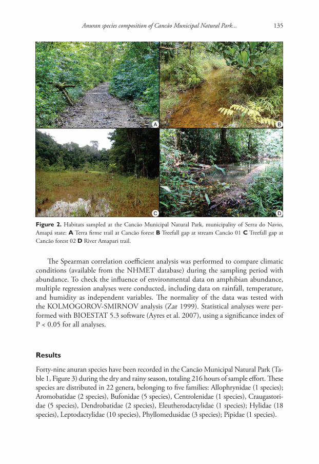
Anuran species composition of Cancão Municipal Natural Park... 135
Figure 2. Habitats sampled at the Cancão Municipal Natural Park, municipality of Serra do Navio, Amapá state: A Terra firme trail at Cancão forest B Treefall gap at stream Cancão 01 C Treefall gap at Cancão forest 02 D River Amapari trail.
The Spearman correlation coefficient analysis was performed to compare climatic conditions (avail able from the NHMET database) during the sampling period with abundance. To check the influence of environmental data on amphibian abundance, multiple re gression analyses were conducted, including data on rain fall, temperature, and humidity as independent variables. The normality of the data was tested with the KOLMOGOROV-SMIRNOV analysis (Zar 1999). Statistical analyses were per-formed with BIOESTAT 5.3 software (Ayres et al. 2007), using a significance in dex of P < 0.05 for all analyses.
Results
Forty-nine anuran species have been recorded in the Cancão Municipal Natural Park (Ta-ble 1, Figure 3) during the dry and rainy season, totaling 216 hours of sample effort. These species are distributed in 22 genera, belonging to five families: Allophrynidae (1 species); Aromobatidae (2 species), Bufonidae (5 species), Centrolenidae (1 species), Craugastori-dae (5 species), Dendrobatidae (2 species), Eleutherodactylidae (1 species); Hylidae (18 species), Leptodactylidae (10 species), Phyllomedusidae (3 species); Pipidae (1 species).
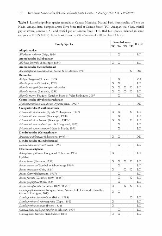
Yuri Breno Silva e Silva & Carlos Eduardo Costa-Campos / ZooKeys 762: 131–148 (2018)136
Table 1. List of amphibian species recorded at Cancão Municipal Natural Park, municipality of Serra do Navio, Amapá State. Sampled areas: Terra firme trail at Cancão forest (TC), Amapari trail (TA), treefall gap at stream Cancão (TS), and treefall gap at Cancão forest (TF). Red List species included in some category of IUCN (2017): LC – Least Concern; VU – Vulnerable; DD – Data Deficient.
Family/SpeciesSampled areas
IUCNTC TA TS TF
AllophrynidaeAllophryne ruthveni Gaige, 1926 X LCAromobatidae (Allobatinae)Allobates femoralis (Boulenger, 1884) X X LCAromobatidae (Aromobatinae)Anomaloglossus baeobatrachus (Boistel & de Massari, 1999) X DDBufonidaeAtelopus hoogmoedi Lescure, 1974 X VURhaebo guttatus (Schneider, 1799) X X LCRhinella margaritifera complex of species X X X X LCRhinella marina (Linnaeus, 1758) X X X X LCRhinella martyi Fouquet, Gaucher, Blanc & Vélez-Rodriguez, 2007 X LCCentrolenidae (Hyalinobatrachinae)Hyalinobatrachium iaspidiense (Ayarzaqüena, 1992) * X DDCraugastoridae (Ceuthomantinae)Pristimantis chiastonotus (Lynch & Hoogmoed, 1977) X X X LCPristimantis marmoratus (Boulenger, 1900) X LCPristimantis cf. ockendeni (Boulenger, 1912) * X X X LCPristimantis zeuctotylus (Lynch & Hoogmoed, 1977) X LCPristimantis zimmermanae (Heyer & Hardy, 1991) X LCDendrobatidae (Colostethinae)Ameerega pulchripecta (Silverstone, 1976) ** X X DDDendrobatidae (Dendrobatinae)Dendrobates tinctorius (Cuvier, 1797) X LCEleutherodactylidaeAdelophryne gutturosa Hoogmoed & Lescure, 1984 X LCHylidaeBoana boans (Linnaeus, 1758) X X X X LCBoana calcarata (Troschel in Schomburgk 1848) X LCBoana cinerascens (Spix, 1824) X X X LCBoana dentei (Bokermann, 1967) ** X LCBoana fasciata (Günther, 1859 “1858”) X X LCBoana geographica (Spix, 1824) X LCBoana multifasciata (Günther, 1859 “1858”) X X X LCDendropsophus counani Fouquet, Souza, Nunes, Kok, Curcio, de Carvalho, Grant & Rodrigues, 2015 X X –
Dendropsophus leucophyllatus (Beireis, 1783) X LCDendropsophus cf. microcephalus (Cope, 1886) X LCDendropsophus minutus (Peters, 1872) X LCOsteocephalus oophagus Jungfer & Schiesari, 1995 X LCOsteocephalus taurinus Steindachner, 1862 X X LC
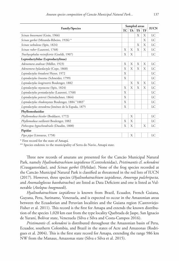
Anuran species composition of Cancão Municipal Natural Park... 137
Family/SpeciesSampled areas
IUCNTC TA TS TF
Scinax boesemani (Goin, 1966) X X LCScinax garbei (Miranda-Ribeiro, 1926) * X LCScinax nebulosus (Spix, 1824) X X LCScinax ruber (Laurenti, 1768) X X X X LCTrachycephalus resinifictrix (Goeldi, 1907) X X LCLeptodactylidae (Leptodactylinae)Adenomera andreae (Müller, 1923) X X X X LCAdenomera hylaedactyla (Cope, 1868) X X X X LCLeptodactylus knudseni Heyer, 1972 X LCLeptodactylus lineatus (Schneider, 1799) X LCLeptodactylus longirostris Boulenger, 1882 X X X LCLeptodactylus mystaceus (Spix, 1824) X X X X LCLeptodactylus pentadactylus (Laurenti, 1768) X LCLeptodactylus petersii (Steindachner, 1864) X LCLeptodactylus rhodomystax Boulenger, 1884 “1883” X LCLeptodactylus stenodema Jiménez de la Espada, 1875 X LCPhyllomedusidaePhyllomedusa bicolor (Boddaert, 1772) X LCPhyllomedusa vaillantii Boulenger, 1882 X X LCPithecopus hypochondrialis (Daudin, 1800) X X X LCPipidaePipa pipa (Linnaeus, 1758) X LC
* First record for the state of Amapá.** Species endemic to the municipality of Serra do Navio, Amapá state.
Three new records of anurans are presented for the Cancão Municipal Natural Park, namely Hyalinobatrachium iaspidiense (Centrolenidae), Pristimantis cf. ockendeni (Craugastoridae), and Scinax garbei (Hylidae). None of the frog species recorded at the Cancão Municipal Natural Park is classified as threatened in the red lists of IUCN (2017). However, three species (Hyalinobatrachium iaspidiense, Ameerega pulchripecta, and Anomaloglossus baeobatrachus) are listed as Data Deficient and one is listed as Vul-nerable (Atelopus hoogmoedi).
Hyalinobatrachium iaspidiense is known from Brazil, Ecuador, French Guiana, Guyana, Peru, Suriname, Venezuela, and is expected to occur in the Amazonian areas between the Ecuadorian and Peruvian localities and the Guiana region (Castroviejo-Fisher et al. 2011). This record is the first for Amapa and extends the known distribu-tion of the species 1,020 km east from the type locality Quebrada de Jaspe, San Ignacio de Yuraní, Bolívar state, Venezuela (Silva e Silva and Costa-Campos 2016).
Pristimantis cf. ockendeni is distributed throughout the Amazonian basin of Peru, Ecuador, southern Colombia, and Brazil in the states of Acre and Amazonas (Rodri-gues et al. 2004). This is the first state record for Amapa, extending the range 986 km NW from the Manaus, Amazonas state (Silva e Silva et al. 2015).
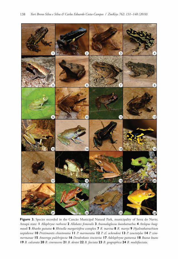
Yuri Breno Silva e Silva & Carlos Eduardo Costa-Campos / ZooKeys 762: 131–148 (2018)138
Figure 3. Species recorded in the Cancão Municipal Natural Park, municipality of Serra do Navio, Amapá state: 1 Allophryne ruthveni 2 Allobates femoralis 3 Anomaloglossus baeobatrachus 4 Atelopus hoog-moedi 5 Rhaebo guttatus 6 Rhinella margaritifera complex 7 R. marina 8 R. martyi 9 Hyalinobatrachium iaspidiense 10 Pristimantis chiastonotus 11 P. marmoratus 12 P. cf. ockendeni 13 P. zeuctotylus 14 P. zim-mermanae 15 Ameerega pulchripecta 16 Dendrobates tinctorius 17 Adelophryne gutturosa 18 Boana boans 19 B. calcarata 20 B. cinerascens 21 B. dentei 22 B. fasciata 23 B. geographica 24 B. multifasciata.

Anuran species composition of Cancão Municipal Natural Park... 139
Figure 3. Continued. 25 Dendropsophus counani 26 D. leucophyllatus 27 Dendropsophus cf. microcephalus 28 D. minutus 29 Osteocephalus oophagus 30 O. taurinus 31 Scinax boesemani 32 S. garbei 33 S. nebulo-sus 34 S. ruber 35 Adenomera andreae 36 A. hylaedactyla 37 Leptodactylus knudseni 38 L. lineatus 39 L. longirostris 40 L. mystaceus 41 L. pentadactylus 42 L. petersii 43 L. rhodomystax 44 L. stenodema 45 Phyl-lomedusa bicolor 46 P. vaillantii 47 Pithecopus hypochondrialis, and 48 Pipa pipa.
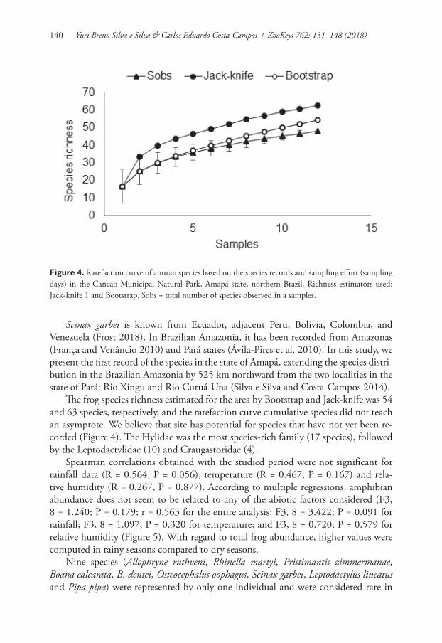
Yuri Breno Silva e Silva & Carlos Eduardo Costa-Campos / ZooKeys 762: 131–148 (2018)140
Scinax garbei is known from Ecuador, adjacent Peru, Bolivia, Colombia, and Venezuela (Frost 2018). In Brazilian Amazonia, it has been recorded from Amazonas (França and Venâncio 2010) and Pará states (Ávila-Pires et al. 2010). In this study, we present the first record of the species in the state of Amapá, extending the species distri-bution in the Brazilian Amazonia by 525 km northward from the two localities in the state of Pará: Rio Xingu and Rio Curuá-Una (Silva e Silva and Costa-Campos 2014).
The frog species richness estimated for the area by Bootstrap and Jack-knife was 54 and 63 species, respectively, and the rarefaction curve cumulative species did not reach an asymptote. We believe that site has potential for species that have not yet been re-corded (Figure 4). The Hylidae was the most species-rich family (17 species), followed by the Leptodactylidae (10) and Craugastoridae (4).
Spearman correlations obtained with the studied period were not sig nificant for rainfall data (R = 0.564, P = 0.056), temperature (R = 0.467, P = 0.167) and rela-tive humid ity (R = 0.267, P = 0.877). Accord ing to multiple regressions, amphibian abundance does not seem to be related to any of the abiotic factors considered (F3, 8 = 1.240; P = 0.179; r = 0.563 for the entire analysis; F3, 8 = 3.422; P = 0.091 for rainfall; F3, 8 = 1.097; P = 0.320 for temperature; and F3, 8 = 0.720; P = 0.579 for relative humid ity (Figure 5). With regard to total frog abundance, higher values were computed in rainy seasons compared to dry seasons.
Nine species (Allophryne ruthveni, Rhinella martyi, Pristimantis zimmermanae, Boana calcarata, B. dentei, Osteocephalus oophagus, Scinax garbei, Leptodactylus lineatus and Pipa pipa) were represented by only one individual and were considered rare in
Figure 4. Rarefaction curve of anuran species based on the species records and sampling effort (sampling days) in the Cancão Municipal Natural Park, Amapá state, northern Brazil. Richness estimators used: Jack-knife 1 and Bootstrap. Sobs = total number of species observed in a samples.
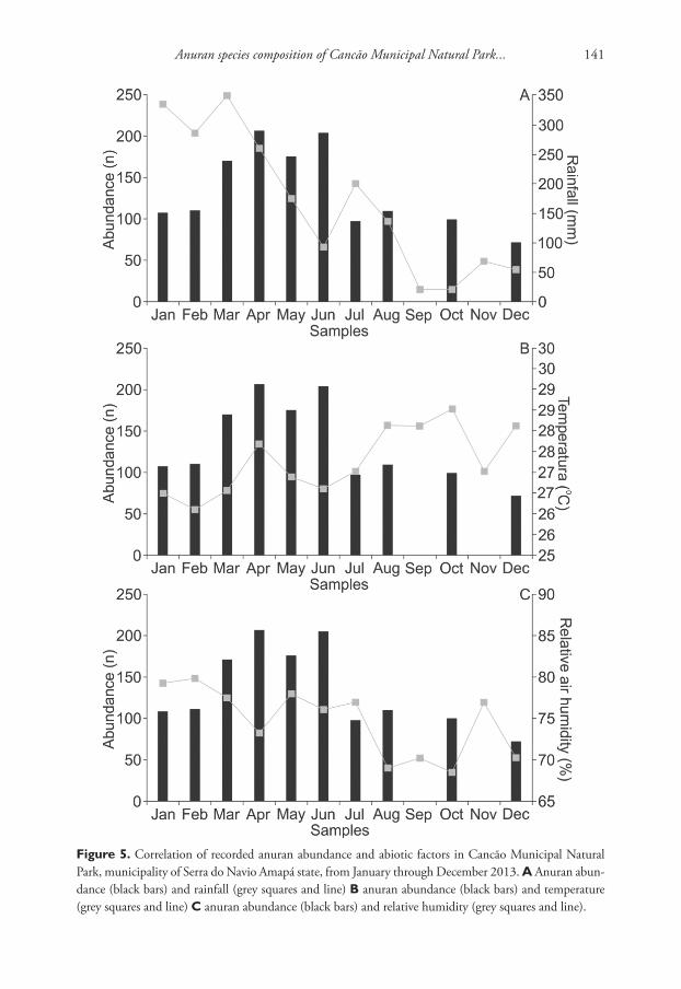
Anuran species composition of Cancão Municipal Natural Park... 141
Figure 5. Correlation of recorded anuran abundance and abiotic factors in Cancão Municipal Natural Park, municipality of Serra do Navio Amapá state, from January through December 2013. A Anuran abun-dance (black bars) and rainfall (grey squares and line) B anuran abundance (black bars) and temperature (grey squares and line) C anuran abun dance (black bars) and relative humidity (grey squares and line).
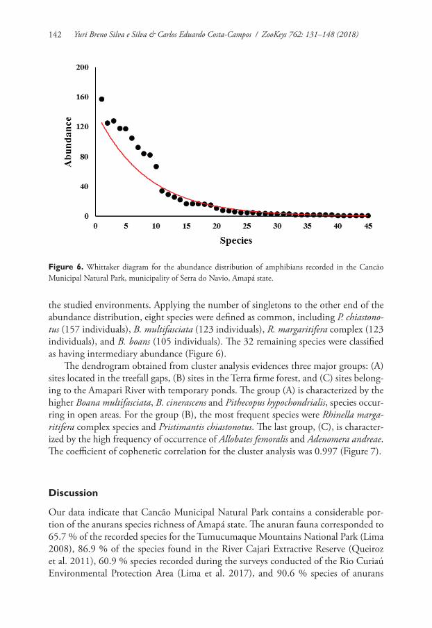
Yuri Breno Silva e Silva & Carlos Eduardo Costa-Campos / ZooKeys 762: 131–148 (2018)142
the studied environments. Applying the number of singletons to the other end of the abundance distribution, eight species were defined as common, including P. chiastono-tus (157 individuals), B. multifasciata (123 individuals), R. margaritifera complex (123 individuals), and B. boans (105 individuals). The 32 remaining species were classified as having intermediary abundance (Figure 6).
The dendrogram obtained from cluster analysis evidences three major groups: (A) sites located in the treefall gaps, (B) sites in the Terra firme forest, and (C) sites belong-ing to the Amapari River with temporary ponds. The group (A) is characterized by the higher Boana multifasciata, B. cinerascens and Pithecopus hypochondrialis, species occur-ring in open areas. For the group (B), the most frequent species were Rhinella marga-ritifera complex species and Pristimantis chiastonotus. The last group, (C), is character-ized by the high frequency of occurrence of Allobates femoralis and Adenomera andreae. The coefficient of cophenetic correlation for the cluster analysis was 0.997 (Figure 7).
Discussion
Our data indicate that Cancão Municipal Natural Park contains a considerable por-tion of the anurans species richness of Amapá state. The anuran fauna corresponded to 65.7 % of the recorded species for the Tumucumaque Mountains National Park (Lima 2008), 86.9 % of the species found in the River Cajari Extractive Reserve (Queiroz et al. 2011), 60.9 % species recorded during the surveys conducted of the Rio Curiaú Environmental Protection Area (Lima et al. 2017), and 90.6 % species of anurans
Figure 6. Whittaker diagram for the abundance distribution of amphibians recorded in the Cancão Municipal Natural Park, municipality of Serra do Navio, Amapá state.
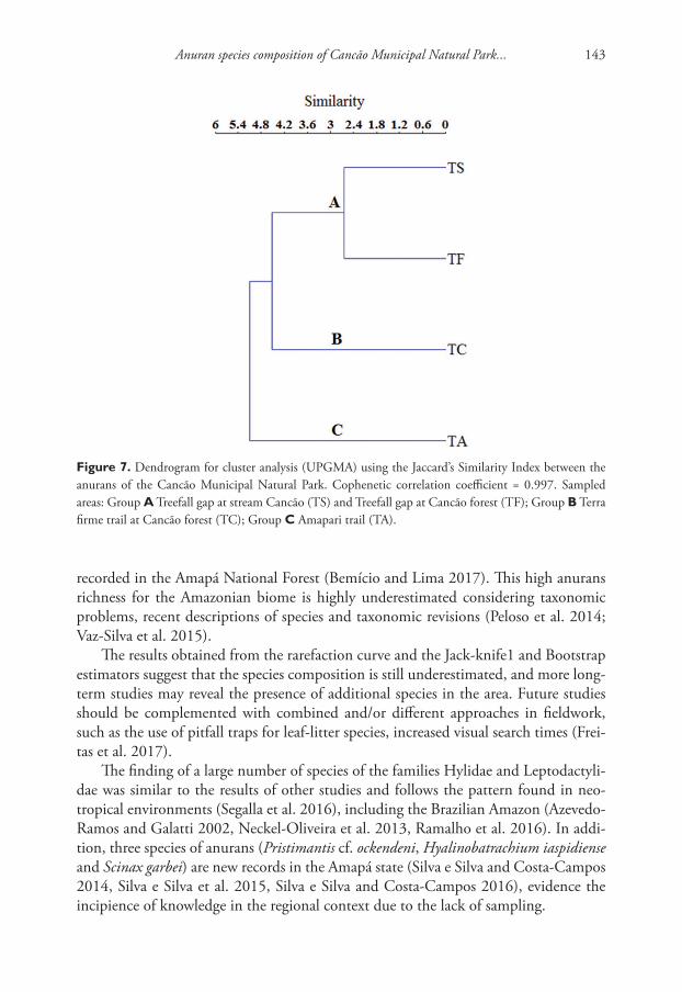
Anuran species composition of Cancão Municipal Natural Park... 143
recorded in the Amapá National Forest (Bemício and Lima 2017). This high anurans richness for the Amazonian biome is highly underestimated considering taxonomic problems, recent descriptions of species and taxonomic revisions (Peloso et al. 2014; Vaz-Silva et al. 2015).
The results obtained from the rarefaction curve and the Jack-knife1 and Bootstrap estimators suggest that the species composition is still underestimated, and more long-term studies may reveal the presence of additional species in the area. Future studies should be complemented with combined and/or different approaches in fieldwork, such as the use of pitfall traps for leaf-litter species, increased visual search times (Frei-tas et al. 2017).
The finding of a large number of species of the families Hylidae and Leptodactyli-dae was similar to the results of other studies and follows the pattern found in neo-tropical environments (Segalla et al. 2016), including the Brazilian Amazon (Azevedo-Ramos and Galatti 2002, Neckel-Oliveira et al. 2013, Ramalho et al. 2016). In addi-tion, three species of anurans (Pristimantis cf. ockendeni, Hyalinobatrachium iaspidiense and Scinax garbei) are new records in the Amapá state (Silva e Silva and Costa-Campos 2014, Silva e Silva et al. 2015, Silva e Silva and Costa-Campos 2016), evidence the incipience of knowledge in the regional context due to the lack of sampling.
Figure 7. Dendrogram for cluster analysis (UPGMA) using the Jaccard’s Similarity Index between the anurans of the Cancão Municipal Natural Park. Cophenetic correlation coefficient = 0.997. Sampled areas: Group A Treefall gap at stream Cancão (TS) and Treefall gap at Cancão forest (TF); Group B Terra firme trail at Cancão forest (TC); Group C Amapari trail (TA).
Yuri Breno Silva e Silva & Carlos Eduardo Costa-Campos / ZooKeys 762: 131–148 (2018)144
The record of Atelopus hoogmoedi and Ameerega pulchripecta in the area studied is relatively important. Atelopus hoogmoedi is a terrestrial and diurnal species, and is most commonly found at small streams in primary forest (Ouboter and Jairam 2012). The species occurs in the Amazonian lowlands of Colombia, Ecuador, and eastern Peru, to Amazonas, Pará, Amapá (Brazil), and the Guianas (Frost 2018). Ameerega pulchripecta was hard to find, and it has been heard only during less than an hour around dawn and again around twilight (Costa-Campos et al. 2016). Additionally, its distribution appears restricted to Serra do Navio, in the state of Amapá, northeastern Brazilian Amazon. These species are classified as vulnerable and data deficient by the IUCN due to their areas of occurrence, status and little known ecological requirements (IUCN 2017).
The cluster analysis of the anuran assemblages generated three groups. The group A and B showed a more differentiated assemblage. Group C are located on the right bank of the Amapari River, and presented high values of abundance and richness. The results can be attributed to the similar characteristics between the sites. The main hy-potheses proposed to explain barrier formation separating populations and causing the differentiation of species in Amazonia during the course of geological history are based on different factors (Haffer 2008). According the river hypothesis, rivers may play a major role in creating and maintaining high levels of spatial separation of populations (Vaz-Silva et al. 2015).
Conclusions
The results of the present study thus provide new data on geographic distribution of species showed three new records of the Brazilian Amazonian and important insights into the diversity of amphibians in the northern Brazil. The high amphibian richness recorded in this study for the eastern Amazon, combined with the presence of popula-tions of Data Deficient or Vulnerable species, contributes to the knowledge on species, reinforcing the importance of the Cancão Municipal Natural Park for the conservation of anurans species.
Acknowledgements
We thank Jiří Moravec and the anonymous reviewers for their constructive comments, which helped us to improve the manuscript. We are also grateful to Albertina Lima, Antoine Fouquet, Marinus Hoogmoed, Tiago de Carvalho and Ulisses Caramaschi for collaborating in the identification of some species. We thank all those who helped on the fieldwork Bruna Lobo, Daniel Valentim, Hélio Pamphylio, Lígia Carvalho, Ma-yara Melo and Raimundo Baía. We thank Universidade Federal do Amapá (UNIFAP) and Christopher Jaster (PARNA Montanhas do Tumucumaque) for logistical support during the fieldwork and Instituto Chico Mendes de Conservação da Biodiversidade – ICMBio for collecting permit (# 32651-1).
Anuran species composition of Cancão Municipal Natural Park... 145
References
Alvares CA, Stape JL, Sentelhas PC, Gonçalves JLM, Sparovek G (2013) Köppen’s climate classification map for Brazil. Meteorologische Zeitschrift 22(6): 711–728. https://doi.org/10.1127/0941-2948/2013/0507
Angulo A, Cocroft RB, Reichle S (2003) Species identity in the genus Adenomera (Anura: Leptodactylidae) in southeastern Peru. Herpetologica 59(4): 490–504. https://doi.org/10.1655/20-104
Araújo AS, Costa-Campos CE (2014) Anurans of the Reserva Biológica do Parazinho, Mu-nicipality of Macapá, state of Amapá, eastern Amazon. Check List 10(6): 1414–1419. doi: http://dx.doi.org/10.15560/10.6.1414
Ávila-Pires TCS, Hoogmoed MS, Rocha WA (2010) Notes on the Vertebrates of northern Pará, Brazil: a forgotten part of the Guianan Region, I. Herpetofauna. Boletim do Museu Paraense Emílio Goeldi, Ciências Naturais 5: 13–112.
Ayres M, Ayres Jr. M, Ayres DL, Santos AA (2007) Bioestat 5.3. Aplicações estatísticas nas áreas das ciências biomédicas. Ong Mamiraua, Belém, PA.
Azevedo-Ramos C, Galatti U (2002) Patterns of Amphibian Diversity in Brazilian Amazo-nia: Conservation Implications. Biological Conservation 103(1): 103–111. https://doi.org/10.1016/S0006-3207(01)00129-X
Benício RA, Lima JD (2017) Anurans of Amapá National Forest, Eastern Amazonia, Brazil. Herpetology Notes 10: 627–633.
Bernarde PS, Albuquerque S, Miranda DB, Turci LCB (2013) Herpetofauna da floresta do baixo rio Moa em Cruzeiro do Sul, Acre – Brasil. Biota Neotropica 13(1): 220–244. htt-ps://doi.org/10.1590/S1676-06032013000100023
Bernarde PS, Machado RA, Turci LCB (2011) Herpetofauna da área do Igarapé Esperança na Reserva Extrativista Riozinho da Liberdade, Acre – Brasil. Biota Neotropica 11(3): 117–144. https://doi.org/10.1590/S1676-06032011000300010
Bernardo PH, Guerra-Fuentes RA, Matiazzi W, Zaher H (2012) Checklist of Amphibians and Reptiles of Reserva Biológica do Tapirapé, Pará, Brazil. Check List 8(5): 839–846. https://doi.org/10.15560/8.5.839
Campos FS, Brito D, Solé M (2014) Diversity patterns, research trends and mismatches of the investigative efforts to amphibian conservation in Brazil. Anais da Academia Brasileira de Ciências 86(4): 1873–1886. http://dx.doi.org/10.1590/0001-3765201420140170
Castroviejo-Fisher S, Vilà C, Ayarzagüena J, Blanc M, Ernst R (2011) Species diversity of Hyalinobatrachium glassfrogs (Amphibia: Centrolenidae) from the Guiana Shield, with the description of two new species. Zootaxa 3132: 1–55.
Clarke KR (1993) Non-parametric multivariate analysis of changes in community structure. Austral Ecology 18: 117–143. https://doi.org/10.1111/j.1442-9993.1993.tb00438.x
Colwell RK (2013) EstimateS: Statistical estimation of species richness and shared species from samples. Version 9. http://purl.oclc.org/estimates [accessed 12 March 2017]
Costa-Campos CE (2015) Ecologia de comunidade e comportamento reprodutivo de anfíbios anuros em Savana Amazônica. Tese de Doutorado, Natal, Rio Grande do Norte: Universi-dade Federal do Rio Grande do Norte.
Yuri Breno Silva e Silva & Carlos Eduardo Costa-Campos / ZooKeys 762: 131–148 (2018)146
Costa-Campos CE, Lima JD, Reis JRF (2015) Riqueza e composição de répteis Squamata (lagartos e anfisbenas) da Área de Proteção Ambiental da Fazendinha, Amapá, Brasil. Biota Amazônia 5(2): 84–90. https://doi.org/10.18561/2179-5746/biotaamazonia.v5n2p84-90
Costa-Campos CE. Lima AP, Amézquita A (2016) The advertisement call of Ameerega pulchrip-ecta (Silverstone, 1976) (Anura, Dendrobatidae). Zootaxa 4136(2): 387–389. http://doi.org/10.11646/zootaxa.4136.2.9
Dubois A (2017) The nomenclatural status of Hysaplesia, Hylaplesia, Dendrobates and related nomina (Amphibia, Anura), with general comments on zoological nomenclature and its governance, as well as on taxonomic databases and websites. Bionomina 11: 1–48. https://doi.org/10.11646/bionomina.11.1.1
Ferrão M, Colatreli O, de Fraga R, Kaefer IL, Moravec J, Lima AP (2016) High Species Rich-ness of Scinax Treefrogs (Hylidae) in a Threatened Amazonian Landscape Revealed by an Integrative Approach. PLoS ONE 11 (11): e0165679. https://doi.org/10.1371/journal.pone.0165679
Ferreira GC, Sturaro MJ, Peloso PLV (2017) Amphibians and reptiles from Floresta Nacional de Pau-Rosa, Amazonas, Brazil: an important protected area at the heart of Amazonia. Acta Amazônia 47(3): 259–268. http://dx.doi.org/10.1590/1809-4392201602982
Fouquet A, Vences M, Salducci MD, Meyer A, Marty C, Blanc M, Gilles A (2007) Revealing cryptic diversity using molecular phylogenetics and phylogeography in frogs of the Scinax ruber and Rhinella margaritifera species groups. Molecular Phylogenetics and Evolution 43: 567–582. https://doi.org/10.1016/j.ympev.2006.12.006
França FGR, Venâncio NM (2010) Reptiles and amphibians of a poorly known region in southwest Amazonia. Biotemas 23(3): 71–84. http://dx.doi.org/10.5007/2175-7925.2010v23n3p71
França DPF, Freita MA, Ramalho WP, Bernarde OS (2017) Local diversity and influence of sea-sonality on amphibians and reptiles assemblages in the Reserva Extrativista Chico Mendes, Acre, Brazil. Iheringia, Série Zoologia 107: e2017023. http://dx.doi.org/10.1590/1678-4766e2017023
Freitas MA, Vieira RS, Entiauspe-Neto OM, Oliveira e Sousa S, Farias T, Sousa AG, Moura GJB (2017) Herpetofauna of the Northwest Amazon forest in the state of Maranhão, Bra-zil, with remarks on the Gurupi Biological Reserve. ZooKeys 643: 141–155. https://doi.org/10.3897/zookeys.643.8215
Frost DR (2018) Amphibian Species of the World: an Online Reference. Version 6.0. http://research.amnh.org/herpetology/amphibia/index.html [accessed on 15 January 2018]
Funk WC, Caminer M, Ron SR (2012) High level of cryptic species diversity uncovered in Amazonian frogs. Proceedings of the Royal Society B: Biological Sciences 279: 1806–1814. https://doi.org/10.1098/rspb.2011.1653
Gotelli NJ, Colwell RK (2001) Quantifying biodiversity: procedures and pitfalls in the meas-urement and comparison of species richness. Ecology Letters 4(4): 379–391. https://doi.org/10.1046/j.1461-0248.2001.00230.x
Haffer J (2008) Hypotheses to explain the origin of species in Amazonia. Brazilian Journal of Biology 68(4): 917–47. https://doi.org/10.1590/S1519-69842008000500003
Anuran species composition of Cancão Municipal Natural Park... 147
Hammer O, Harper DAT, Ryan PD (2001) PAST: Paleontological Statistics Software Pack-age for Education and Data Analysis. Palaeontologia Electronica 4(1): 9. http://palaeo-electronica.org/2001_1/past/issue1_01.htm
Heyer WR (1973) Systematics of the marmoratus group of the frog genus Leptodactylus (Am-phibia, Leptodactylidae). Contributions in Science, Natural History Museum, Los Angeles County 251: 1–50.
Heyer WR, Donnelly MA, McDiarmid RW, Hayek LAC, Foster MS (1994) Measuring and monitoring biological diversity: standard methods for amphibians. Smithsonian Institu-tion Press, Washington, 384 pp.
Hoogmoed MS, Galatti U (2016) Censo da Biodiversidade da Amazônia Brasileira. Grupo: Anura. http://www.museu-goeldi.br/censo [accessed on 25 March 2016]
Ilha P, Dixo M (2010) Anurans and Lizards, Rio Preto da Eva, Amazonas, Brazil. Check List 6(1): 17–21. https://doi.org/10.15560/6.1.017
IUCN (2017) IUCN Red List of Threatened Species. Version 2015.1. http://www.iucnredlist.org [accessed on 25 February 2017]
Lima JD (2008) A herpetofauna do Parque Nacional Montanhas do Tumucumaque, Amapá, Brasil, Expedições I a V. In: Bernard E (Ed.) Inventários Biológicos Rápidos no Parque Nacional Montanhas do Tumucumaque, Amapá, Brasil. RAP Bulletin of Biological Assess-ment 48. Conservation International, Arlington, 38–50.
Lima JRF, Lima JDL, Lima SD, Lima-Silva RB, Andrade GV (2017) Amphibians found in the Amazonian Savanna of the Rio Curiaú Environmental Protection Area in Amapá, Brazil. Biota Neotropica 17(2): e20160252. http://dx.doi.org/10.1590/1676-0611-BN-2016-0252
Magurran AE (2011) Medindo a diversidade biológica. Editora UFPR, Curitiba, 261 pp.Mendes-Pinto TJ, Souza SM (2011) Preliminary assessment of amphibians and reptiles from
Floresta Nacional do Trairão, with a new snake record for the Pará state, Brazilian Amazon. Salamandra 47(4): 199–206.
Miranda DB, Albuquerque S, Turci LCB, Bernarde PS (2015) Richness, breeding environ-ments and calling activity of the anurofauna of the lower moa river forest, state of Acre, Brazil. Zoologia 32(2): 93–108. http://dx.doi.org/10.1590/S1984-46702015000200001
Neckel-Oliveira S, Galatti U, Faveri SB (2013) Ecological correlates in Brazilian Amazoni-an anurans: implications for conservation. Amphibia-Reptilia 34: 217–232. https://doi.org/10.1163/15685381-00002890
Ouboter PE, Jairam R (2012) Amphibians of Suriname. Brill Academic Publisher, 388 pp.Pantoja DL, Fraga R (2012) Herpetofauna of the Reserva Extrativista do Rio Gregório, Juruá
Basin, southwest Amazonia, Brazil. Check List 8(3): 360–374.Peloso PLV (2010) A safe place for amphibians? A cautionary tale on the taxonomy and con-
servation of frogs, caecilians, and salamanders in the Brazilian Amazonia. Zoologia 27(5): 667–673. https://doi.org/10.1590/S1984-46702010000500001
Peloso PLV, Sturaro MJ, Forlani MC, Gaucher P, Motta AP, Wheeler WC (2014) Phylog-eny, Taxonomic Revision, and Character Evolution of the Genera Chiasmocleis and Syncope (Anura, Microhylidae) in Amazonia, with Descriptions of Three New Species.
Yuri Breno Silva e Silva & Carlos Eduardo Costa-Campos / ZooKeys 762: 131–148 (2018)148
Bulletin of the American Museum of Natural History 386(1): 1–112. http://dx.doi.org/10.1206/834.1
Pereira-Júnior AP, Costa-Campos CE, Araújo AS (2013) Composição e diversidade de an-fíbios anuros do campus da Universidade Federal do Amapá. Biota Amazônia 3(1): 13–21. http://dx.doi.org/10.18561/2179-5746/biotaamazonia.v3n1p13-21
Piatti L, Amaro PMO, Araújo JFJ, Sanches VQA, Bernarde PS (2012) Anurans of a disturbed area in Jarú, Rondônia, Brazil. Check List 8(1): 083–087.
Prudente ALC, Sturaro MJ, Travassos AEM, Maschio GF, Santos-Costa MC (2013) Anurans of the Urucu Petrol Basin, municipality of Coari, State of Amazonas, northern Brazil. Check List 9(3): 601–606.
Queiroz SS, Silva AR, Reis FM, Lima JD, Lima JRF (2011) Anfíbios de uma área de castanhal da Reserva Extrativista do Rio Cajari, Amapá. Biota Amazônia 1(1): 1–18. https://doi.org/10.18561/2179-5746/biotaamazonia.v1n1p1-18
Ramalho WP, Andrade MS, Matos LRA, Vieira LJS (2016) Amphibians of varzea environments and floating meadows of the oxbow lakes of the Middle Purus River, Amazonas, Brazil. Biota Neotropica 16(1): e20150093. https://doi.org/10.1590/1676-0611-BN-2015-0093
Rodríguez L, Martinez JL, Coloma LA, Ron S, Azevedo-Ramos C, Castro F, Rueda JV, Cis-neros-Heredia D, Hoogmoed M, Gascon C (2004) Pristimantis ockendeni. The IUCN Red List of Threatened Species 2004. e.T56803A11534464. http://dx.doi.org/10.2305/IUCN.UK.2004.RLTS.T56803A11534464.en [accessed on 23 February 2018]
Segalla MV, Caramaschi U, Cruz CAG, Garcia PCA, Grant T, Haddad CFB, Langone P (2016) Brazilian amphibians – List of species. http://www.sbherpetologia.org.br [accessed on 15 July 2016]
Silva e Silva YB, Costa-Campos CE (2014) Scinax garbei (Miranda-Ribeiro, 1926) (Amphibia: Anura: Hylidae): Distribution extension for Brazilian Amazonia and first record in the state of Amapá. Check List 10(2): 448–449. http://www.checklist.org.br/getpdf?NGD089-13
Silva e Silva YB, Costa-Campos CE (2016) Hyalinobatrachium iaspidiense (Ayarzaguena, 1992) (Anura: Centrolenidae): first record in Amapá state, Brazil and geographic distribution map. Check List 12(2): 1849, doi: http://dx.doi.org/10.15560/12.2.1849
Silva e Silva YB, Costa-Campos CE, Valentim DSS, Melo-Furtado MF (2015) Pristimantis ockendeni (Carabaya Robber Frog). Brazil, Amapá. Herpetological Review 46(1): 58.
Silvano DL, Segalla MV (2005) Conservação de anfíbios no Brasil. Megadiversidade: Desafios e oportunidades para a conservação da biodiversidade no Brasil 1(1): 79–86.
Vaz-Silva W, Oliveira RM, Gonzaga AFN, Pinto KC, Poli FC, Bilce TM, Penhacek M, Wronski L, Martins JX, Junqueira TG, Cesca LCC, Guimarães VY, Pinheiro RD (2015) Contribu-tions to the knowledge of amphibians and reptiles from Volta Grande do Xingu, northern Brazil. Brazilian Journal of Biology 75(3): S205–S218. https://doi.org/10.1590/1519-6984.00814BM
Venâncio NM, Souza MB (2016) Anfíbios do Parque Ambiental Chico Mendes, Rio Branco – Acre, Brasil. Biotemas 29(1): 85–95. https://doi.org/10.5007/2175-7925.2016v29n1p85
Waldez F, Menin M, Vogt RC (2013) Diversidade de anfíbios e répteis Squamata na região do baixo rio Purus, Amazônia Central, Brasil. Biota Neotropica 13(1): 300–316.
Zar JH (1999) Biostatistical analysis. Prentice Hall, New Jersey, 663 pp.
Copyright © 2022 FDOKUMEN