Dendritic cell modification as a route to inhibiting corneal graft rejection by the indirect pathway...
13
734 Adnan Khan et al. Eur. J. Immunol. 2013. 43: 734–746 DOI: 10.1002/eji.201242914 Dendritic cell modification as a route to inhibiting corneal graft rejection by the indirect pathway of allorecognition Adnan Khan 1 , Hongmei Fu 1 , Lee Aun Tan 1 , Jennifer E. Harper 1 , Sven C. Beutelspacher 1 , Daniel F. P. Larkin 2 , Giovanna Lombardi 3 , Myra O. McClure 4 and Andrew J. T. George 1 1 Section of Molecular Immunology, Department of Medicine, Imperial College London, Hammersmith Hospital, London, United Kingdom 2 Moorfields Eye Hospital, London, United Kingdom 3 Medical Research Council Centre for Transplantation, King’s College London, Guy’s Hospital, London, United Kingdom 4 Jefferiss Research Trust Laboratories, Wright-Fleming Institute, Department of Medicine, Imperial College London, St. Mary’s Hospital, London, United Kingdom Dendritic cell (DC) modification is a potential strategy to induce clinical transplantation tolerance. We compared two DC modification strategies to inhibit allogeneic T-cell prolif- eration. In the first strategy, murine DCs were transduced with a lentiviral vector express- ing CTLA4-KDEL, a fusion protein that prevents surface CD80/86 expression by retaining the co-stimulatory molecules within the ER. In the second approach, DCs were trans- duced to express the tryptophan-catabolising enzyme IDO. CTLA4-KDEL-expressing DCs induced anergy in alloreactive T cells and generated both CD4 + CD25 + and CD4 + CD25 − Treg cells (with direct and indirect donor allospecificity and capacity for linked suppres- sion) both in vitro and in vivo. In contrast, T-cell unresponsiveness induced by IDO + DCs lacked donor specificity. In the absence of any immunosuppressive treatment, i.v. administration of CTLA4-KDEL-expressing DCs resulted in long-term survival of corneal allografts only when the DCs were capable of indirect presentation of alloantigen. This study demonstrates the therapeutic potential of CTLA4-KDEL-expressing DCs in tolerance induction. Keywords: Corneal transplantation Dendritic cells Gene therapy Tolerance induction Transplantation tolerance See accompanying Commentary by Benichou and Tocco Additional supporting information may be found in the online version of this article at the publisher’s web-site Introduction Dendritic cells (DCs) not only initiate allogeneic rejection of grafts, by either the direct or indirect pathway, but can also con- Correspondence: Prof. Andrew J. T. George e-mail: [email protected] tribute to tolerance induction [1]. DCs treated with a range of pharmacological inhibitors to prevent maturation and/or activa- tion have been used to induce tolerance to alloantigens [1, 2]. The tolerogenic potential of DCs can be enhanced by genetic mod- ification, including transfection/transduction with genes encod- ing immunomodulatory proteins or molecules that prevent DC activation [3, 4]. C 2013 WILEY-VCH Verlag GmbH & Co. KGaA, Weinheim www.eji-journal.eu
-
Upload
independent -
Category
Documents
-
view
0 -
download
0
Transcript of Dendritic cell modification as a route to inhibiting corneal graft rejection by the indirect pathway...
734 Adnan Khan et al. Eur. J. Immunol. 2013. 43: 734–746DOI: 10.1002/eji.201242914
Dendritic cell modification as a route to inhibitingcorneal graft rejection by the indirect pathway ofallorecognitionAdnan Khan1, Hongmei Fu1, Lee Aun Tan1, Jennifer E. Harper1,Sven C. Beutelspacher1, Daniel F. P. Larkin2, Giovanna Lombardi3,Myra O. McClure4 and Andrew J. T. George1
1 Section of Molecular Immunology, Department of Medicine, Imperial College London,Hammersmith Hospital, London, United Kingdom
2 Moorfields Eye Hospital, London, United Kingdom3 Medical Research Council Centre for Transplantation, King’s College London, Guy’s Hospital,
London, United Kingdom4 Jefferiss Research Trust Laboratories, Wright-Fleming Institute, Department of Medicine,
Imperial College London, St. Mary’s Hospital, London, United Kingdom
Dendritic cell (DC) modification is a potential strategy to induce clinical transplantationtolerance. We compared two DC modification strategies to inhibit allogeneic T-cell prolif-eration. In the first strategy, murine DCs were transduced with a lentiviral vector express-ing CTLA4-KDEL, a fusion protein that prevents surface CD80/86 expression by retainingthe co-stimulatory molecules within the ER. In the second approach, DCs were trans-duced to express the tryptophan-catabolising enzyme IDO. CTLA4-KDEL-expressing DCsinduced anergy in alloreactive T cells and generated both CD4+CD25+ and CD4+CD25−
Treg cells (with direct and indirect donor allospecificity and capacity for linked suppres-sion) both in vitro and in vivo. In contrast, T-cell unresponsiveness induced by IDO+
DCs lacked donor specificity. In the absence of any immunosuppressive treatment, i.v.administration of CTLA4-KDEL-expressing DCs resulted in long-term survival of cornealallografts only when the DCs were capable of indirect presentation of alloantigen. Thisstudy demonstrates the therapeutic potential of CTLA4-KDEL-expressing DCs in toleranceinduction.
Keywords: Corneal transplantation � Dendritic cells � Gene therapy � Tolerance induction �
Transplantation tolerance
See accompanying Commentary by Benichou and Tocco
� Additional supporting information may be found in the online version of this article at thepublisher’s web-site
Introduction
Dendritic cells (DCs) not only initiate allogeneic rejection ofgrafts, by either the direct or indirect pathway, but can also con-
Correspondence: Prof. Andrew J. T. Georgee-mail: [email protected]
tribute to tolerance induction [1]. DCs treated with a range ofpharmacological inhibitors to prevent maturation and/or activa-tion have been used to induce tolerance to alloantigens [1, 2].The tolerogenic potential of DCs can be enhanced by genetic mod-ification, including transfection/transduction with genes encod-ing immunomodulatory proteins or molecules that prevent DCactivation [3,4].
C© 2013 WILEY-VCH Verlag GmbH & Co. KGaA, Weinheim www.eji-journal.eu
Eur. J. Immunol. 2013. 43: 734–746 Immunomodulation 735
We have developed two strategies for creating tolerogenic DCs.The first is to inhibit the expression of CD80/86 using a fusion pro-tein, CTLA4-KDEL. The KDEL peptide retains/retrieves proteins tothe ER [5]. CTLA4-KDEL is therefore confined to the ER, whereit binds CD80/86, preventing their passage to the cell surface.We have shown, using human cells, that inhibition of CD80/86expression with CTLA4-KDEL results in DCs that can induce bothanergy and regulatory activity in allospecific T cells [6].
The second strategy for creating tolerogenic DCs is to expressIDO. IDO catabolises tryptophan, resulting in the production ofkynurenines [7,8]. Both the depletion of tryptophan and the pro-duction of kynurenines inhibit T-cell responses [9,10].
The aim of this study is to determine the ability of DCs, express-ing either CTLA4-KDEL or IDO, to prevent corneal graft rejec-tion. Cornea is the most commonly transplanted tissue [11], andwhile it is considered an immune-privileged tissue expressing var-ious immunomodulatory enzymes such as IDO [12] and arginase[13], there is significant immunological rejection of corneal graftswith a 5-year graft survival of approximately 75% [14]. As thecornea lacks resident DCs, rejection of the cornea occurs predom-inantly by the indirect pathway of allorecognition [15–18]. In thisstudy, we demonstrate long-term survival of corneal allograftsafter administering CTLA4-KDEL-expressing DCs targeted to theindirect pathway, and demonstrate the superiority of these cellsover IDO-expressing DCs in preventing allograft rejection.
Results
Phenotype of transduced DCs
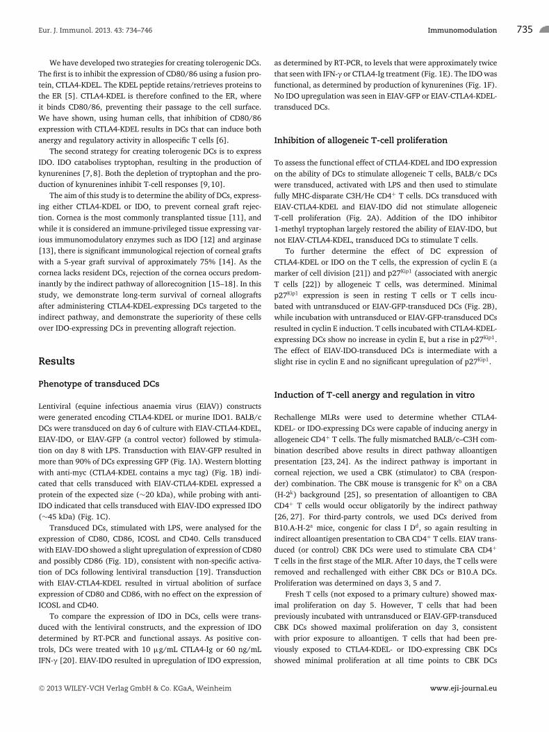
Lentiviral (equine infectious anaemia virus (EIAV)) constructswere generated encoding CTLA4-KDEL or murine IDO1. BALB/cDCs were transduced on day 6 of culture with EIAV-CTLA4-KDEL,EIAV-IDO, or EIAV-GFP (a control vector) followed by stimula-tion on day 8 with LPS. Transduction with EIAV-GFP resulted inmore than 90% of DCs expressing GFP (Fig. 1A). Western blottingwith anti-myc (CTLA4-KDEL contains a myc tag) (Fig. 1B) indi-cated that cells transduced with EIAV-CTLA4-KDEL expressed aprotein of the expected size (∼20 kDa), while probing with anti-IDO indicated that cells transduced with EIAV-IDO expressed IDO(∼45 kDa) (Fig. 1C).
Transduced DCs, stimulated with LPS, were analysed for theexpression of CD80, CD86, ICOSL and CD40. Cells transducedwith EIAV-IDO showed a slight upregulation of expression of CD80and possibly CD86 (Fig. 1D), consistent with non-specific activa-tion of DCs following lentiviral transduction [19]. Transductionwith EIAV-CTLA4-KDEL resulted in virtual abolition of surfaceexpression of CD80 and CD86, with no effect on the expression ofICOSL and CD40.
To compare the expression of IDO in DCs, cells were trans-duced with the lentiviral constructs, and the expression of IDOdetermined by RT-PCR and functional assays. As positive con-trols, DCs were treated with 10 μg/mL CTLA4-Ig or 60 ng/mLIFN-γ [20]. EIAV-IDO resulted in upregulation of IDO expression,
as determined by RT-PCR, to levels that were approximately twicethat seen with IFN-γ or CTLA4-Ig treatment (Fig. 1E). The IDO wasfunctional, as determined by production of kynurenines (Fig. 1F).No IDO upregulation was seen in EIAV-GFP or EIAV-CTLA4-KDEL-transduced DCs.
Inhibition of allogeneic T-cell proliferation
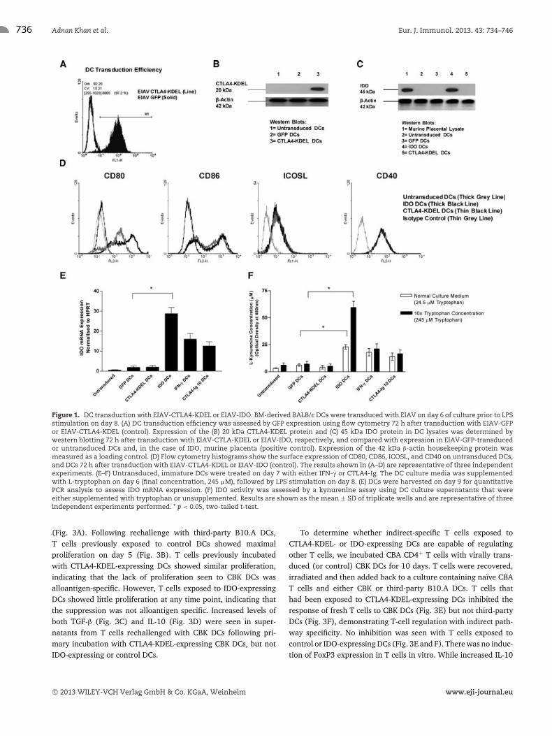
To assess the functional effect of CTLA4-KDEL and IDO expressionon the ability of DCs to stimulate allogeneic T cells, BALB/c DCswere transduced, activated with LPS and then used to stimulatefully MHC-disparate C3H/He CD4+ T cells. DCs transduced withEIAV-CTLA4-KDEL and EIAV-IDO did not stimulate allogeneicT-cell proliferation (Fig. 2A). Addition of the IDO inhibitor1-methyl tryptophan largely restored the ability of EIAV-IDO, butnot EIAV-CTLA4-KDEL, transduced DCs to stimulate T cells.
To further determine the effect of DC expression ofCTLA4-KDEL or IDO on the T cells, the expression of cyclin E (amarker of cell division [21]) and p27Kip1 (associated with anergicT cells [22]) by allogeneic T cells, was determined. Minimalp27Kip1 expression is seen in resting T cells or T cells incu-bated with untransduced or EIAV-GFP-transduced DCs (Fig. 2B),while incubation with untransduced or EIAV-GFP-transduced DCsresulted in cyclin E induction. T cells incubated with CTLA4-KDEL-expressing DCs show no increase in cyclin E, but a rise in p27Kip1.The effect of EIAV-IDO-transduced DCs is intermediate with aslight rise in cyclin E and no significant upregulation of p27Kip1.
Induction of T-cell anergy and regulation in vitro
Rechallenge MLRs were used to determine whether CTLA4-KDEL- or IDO-expressing DCs were capable of inducing anergy inallogeneic CD4+ T cells. The fully mismatched BALB/c–C3H com-bination described above results in direct pathway alloantigenpresentation [23, 24]. As the indirect pathway is important incorneal rejection, we used a CBK (stimulator) to CBA (respon-der) combination. The CBK mouse is transgenic for Kb on a CBA(H-2k) background [25], so presentation of alloantigen to CBACD4+ T cells would occur obligatorily by the indirect pathway[26, 27]. For third-party controls, we used DCs derived fromB10.A-H-2a mice, congenic for class I Dd, so again resulting inindirect alloantigen presentation to CBA CD4+ T cells. EIAV trans-duced (or control) CBK DCs were used to stimulate CBA CD4+
T cells in the first stage of the MLR. After 10 days, the T cells wereremoved and rechallenged with either CBK DCs or B10.A DCs.Proliferation was determined on days 3, 5 and 7.
Fresh T cells (not exposed to a primary culture) showed max-imal proliferation on day 5. However, T cells that had beenpreviously incubated with untransduced or EIAV-GFP-transducedCBK DCs showed maximal proliferation on day 3, consistentwith prior exposure to alloantigen. T cells that had been pre-viously exposed to CTLA4-KDEL- or IDO-expressing CBK DCsshowed minimal proliferation at all time points to CBK DCs
C© 2013 WILEY-VCH Verlag GmbH & Co. KGaA, Weinheim www.eji-journal.eu
736 Adnan Khan et al. Eur. J. Immunol. 2013. 43: 734–746
Figure 1. DC transduction with EIAV-CTLA4-KDEL or EIAV-IDO. BM-derived BALB/c DCs were transduced with EIAV on day 6 of culture prior to LPSstimulation on day 8. (A) DC transduction efficiency was assessed by GFP expression using flow cytometry 72 h after transduction with EIAV-GFPor EIAV-CTLA4-KDEL (control). Expression of the (B) 20 kDa CTLA4-KDEL protein and (C) 45 kDa IDO protein in DC lysates was determined bywestern blotting 72 h after transduction with EIAV-CTLA-KDEL or EIAV-IDO, respectively, and compared with expression in EIAV-GFP-transducedor untransduced DCs and, in the case of IDO, murine placenta (positive control). Expression of the 42 kDa β-actin housekeeping protein wasmeasured as a loading control. (D) Flow cytometry histograms show the surface expression of CD80, CD86, ICOSL, and CD40 on untransduced DCs,and DCs 72 h after transduction with EIAV-CTLA4-KDEL or EIAV-IDO (control). The results shown in (A–D) are representative of three independentexperiments. (E–F) Untransduced, immature DCs were treated on day 7 with either IFN-γ or CTLA4-Ig. The DC culture media was supplementedwith L-tryptophan on day 6 (final concentration, 245 μM), followed by LPS stimulation on day 8. (E) DCs were harvested on day 9 for quantitativePCR analysis to assess IDO mRNA expression. (F) IDO activity was assessed by a kynurenine assay using DC culture supernatants that wereeither supplemented with tryptophan or unsupplemented. Results are shown as the mean ± SD of triplicate wells and are representative of threeindependent experiments performed. * p < 0.05, two-tailed t-test.
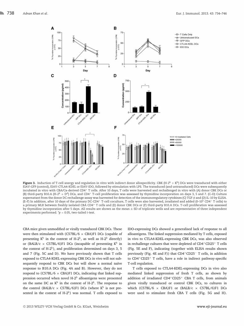
(Fig. 3A). Following rechallenge with third-party B10.A DCs,T cells previously exposed to control DCs showed maximalproliferation on day 5 (Fig. 3B). T cells previously incubatedwith CTLA4-KDEL-expressing DCs showed similar proliferation,indicating that the lack of proliferation seen to CBK DCs wasalloantigen-specific. However, T cells exposed to IDO-expressingDCs showed little proliferation at any time point, indicating thatthe suppression was not alloantigen specific. Increased levels ofboth TGF-β (Fig. 3C) and IL-10 (Fig. 3D) were seen in super-natants from T cells rechallenged with CBK DCs following pri-mary incubation with CTLA4-KDEL-expressing CBK DCs, but notIDO-expressing or control DCs.
To determine whether indirect-specific T cells exposed toCTLA4-KDEL- or IDO-expressing DCs are capable of regulatingother T cells, we incubated CBA CD4+ T cells with virally trans-duced (or control) CBK DCs for 10 days. T cells were recovered,irradiated and then added back to a culture containing naıve CBAT cells and either CBK or third-party B10.A DCs. T cells thathad been exposed to CTLA4-KDEL-expressing DCs inhibited theresponse of fresh T cells to CBK DCs (Fig. 3E) but not third-partyDCs (Fig. 3F), demonstrating T-cell regulation with indirect path-way specificity. No inhibition was seen with T cells exposed tocontrol or IDO-expressing DCs (Fig. 3E and F). There was no induc-tion of FoxP3 expression in T cells in vitro. While increased IL-10
C© 2013 WILEY-VCH Verlag GmbH & Co. KGaA, Weinheim www.eji-journal.eu
Eur. J. Immunol. 2013. 43: 734–746 Immunomodulation 737
Figure 2. Inhibition of allogeneic T-cell proliferation after DC expression of CTLA4-KDEL or IDO. BALB/c (H-2d) DCs were transduced on day 6 ofculture with EIAV-GFP (control), EIAV-CTLA4-KDEL or EIAV-IDO and stimulated with LPS on day 8 of culture. All DC populations were cultured onday 9 with fully MHC-disparate, spleen-derived C3H/He CD4+ T cells. Where indicated, 250 μM 1-methyl-D,L-tryptophan (1-MT), in combinationwith excess tryptophan, was added to the medium at the start of coculture. (A) Increasing numbers of both EIAV transduced and untransducedDC populations (0–105) were co-cultured with C3H/He-derived CD4+ T cells. Proliferation of CD4+ T cells was detected by thymidine incorporationon day 5 of the MLR, and the results are shown as the mean ± SD of triplicate wells and are representative of three independent experiments.*p < 0.05, two-tailed t-test. (B) Expression of the 52 kDa cyclin E protein and p27Kip1 protein (and the 42 kDa β-actin protein), in the lysates of CD4+
T cells incubated with either the transduced or untransduced DCs described was determined by western blotting on day 4 of co-culture. Datashown are representative of three independent experiments performed.
and TGF-β was seen in supernatant from the cultures previouslyexposed to CTLA4-KDEL-expressing CBK DCs, no such increasewas seen in suppressor assays containing third-party DCs (data notshown).
Alloantigen-specific induction of anergy was also observedusing a direct pathway (multiple mismatch) strain combinationwith CTLA4-KDEL-expressing BALB/c DCs and C3H CD4+ T cells,and associated with secretion of TGF-β and IL-10, while expressionof IDO by BALB/c DCs resulted in inhibition of both specificand third-party alloresponses (Supporting Information Fig. 1A–D).C3H T cells that had been exposed to CTLA4-KDEL-expressingBALB/c DCs also inhibited the response of fresh T cells to BALB/cbut not third-party DCs (Supporting Information Fig. 1E and F).Only a moderate, but significant, inhibition was seen with T cellsexposed to IDO-expressing DCs.
Induction of T-cell anergy in vivo
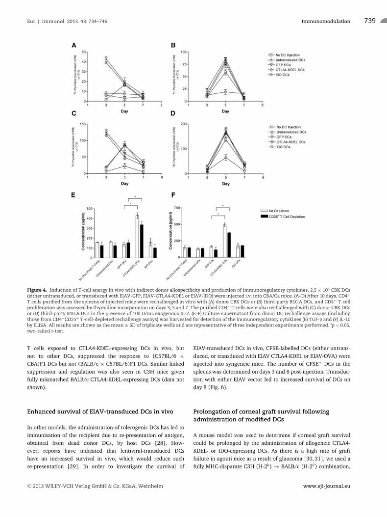
To determine that if modified DCs can induce anergy with indi-rect donor allospecificity in vivo, CD4+ T cells, purified fromthe spleens of CBA mice 10 days after CBK DC administration,were challenged in vitro with CBK or third-party B10.A DCs.T-cell proliferation was determined on days 3, 5 and 7. CD4+
T cells from mice given control DCs showed a rapid responseto CBK DCs, while T cells from naıve animals showed a max-imal response on day 5 (Fig. 4A). Mice given DCs expressingeither CTLA4-KDEL or IDO showed minimal T-cell proliferation.The response of T cells to third-party DCs was normal in allcases, except following injection of IDO-expressing DCs in which areduced response was seen (Fig. 4B). Addition of IL-2 (100 U/mL)to the cultures restored the ability of T cells from mice givenCTLA4-KDEL-expressing DCs, but not IDO-expressing DCs, torespond to CBK DCs (Fig. 4C and D). Similar results were obtainedfollowing the injection of modified BALB/c DCs into C3H mice
(Supporting Information Fig. 2A–D). These data indicate thatCTLA4-KDEL-expressing DCs can induce anergy in alloantigen-specific T cells in vivo, but IDO-expressing DCs induce generalisedunresponsiveness. The alloantigen-specific unresponsiveness inT cells exposed to CTLA4-KDEL-modified DCs is associated withincreased concentrations of IL-10 and TGF-β on day 7 of the cul-ture for both the CBK-CBA combination (Fig. 4E and F), and theBALB/c-C3H combination (Supporting Information Fig. 2E and F).This was seen both with CD4+ T cells and CD4+ T cells depletedof CD25 cells.
CTLA4-KDEL-expressing DCs can induce Treg cellsin vivo
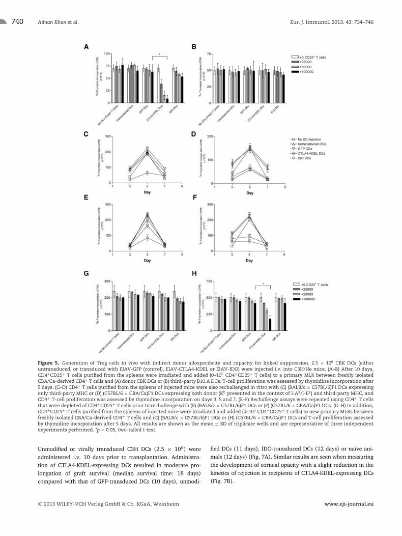
To determine if administration of CTLA4-KDEL- or IDO-expressingDCs can induce Treg cell activity in vivo, 2.5 × 10⁶ CBK DCs(virally transduced or control) were injected i.v. into CBK mice.After 10 days, CD4+CD25+ splenocytes were purified, irradiatedand then added to a fresh culture of CBA CD4+ T cells and CBK DCsand the proliferation assessed on day 5. Addition of T cells frommice given CTLA4-KDEL-expressing DCs, but not from mice givenIDO-expressing- or control DCs, inhibited allogeneic proliferationof fresh CBA T cells (Fig. 5). No inhibition was observed whenB10.A DCs were used as stimulators (Fig. 5B). Similar inductionof alloantigen-specific Treg cells was seen following the adminis-tration of CTLA4-KDEL-expressing BALB/c DCs to C3H animals,with no evidence of Treg cells in mice given IDO-expressing DCs(Supporting Information Fig. 3A and B). As shown in Support-ing Information Fig. 4, there was no increase in FoxP3 expres-sion (∼6%) in C3H animals given untransduced, GFP- or IDO-transduced BALB/c DCs, but FoxP3 expression increased (∼20%)in mice given CTLA4-KDEL-expressing DCs.
To determine if the indirect pathway-specific regulatory cellscan mediate linked suppression, CD4+ T cells were isolated from
C© 2013 WILEY-VCH Verlag GmbH & Co. KGaA, Weinheim www.eji-journal.eu
738 Adnan Khan et al. Eur. J. Immunol. 2013. 43: 734–746
Figure 3. Induction of T-cell anergy and regulation in vitro with indirect donor allospecificity. CBK (H-2k + Kb) DCs were transduced with eitherEIAV-GFP (control), EIAV-CTLA4-KDEL or EIAV-IDO, followed by stimulation with LPS. The transduced (and untransduced) DCs were subsequentlyincubated in vitro with CBA/Ca-derived CD4+ T cells. After 10 days, T cells were harvested and rechallenged in vitro with (A) donor CBK DCs or(B) third-party B10.A (H-2k + Dd) DCs, and CD4+ T-cell proliferation was assessed by thymidine incorporation on days 3, 5 and 7. (C–D) Culturesupernatant from the donor DC rechallenge assay was harvested for detection of the immunoregulatory cytokines (C) TGF-β and (D) IL-10 by ELISA.(E–F) In addition, after 10 days of the primary DC-CD4+ T-cell coculture, T cells were also harvested, irradiated and added (0–105 CD4+ T cells) toa primary MLR between freshly isolated CBA CD4+ T cells and (E) donor CBK DCs or (F) third-party B10.A DCs. T-cell proliferation was assessedby thymidine incorporation after 5 days. All results are shown as the mean ± SD of triplicate wells and are representative of three independentexperiments performed. *p < 0.05, two-tailed t-test.
CBA mice given unmodified or virally transduced CBK DCs. Thesewere then stimulated with (C57BL/6 × CBA)F1 DCs (capable ofpresenting Kb in the context of H-2k, as well as H-2b directly)or (BALB/c × C57BL/6)F1 DCs (incapable of presenting Kb inthe context of H-2k), and proliferation determined on days 3, 5and 7 (Fig. 5C and D). We have previously shown that T cellsexposed to CTLA4-KDEL-expressing CBK DCs in vivo will not sub-sequently respond to CBK DCs but will show a normal naıveresponse to B10.A DCs (Fig. 4A and B). However, they do notrespond to (C57BL/6 × CBA)F1 DCs, indicating that linked sup-pression occurred when novel H-2b alloantigens were presentedon the same DC as Kb in the context of H-2k. The response tothe control (BALB/c × C57BL/6)F1 DCs (where Kb is not pre-sented in the context of H-2k) was normal. T cells exposed to
IDO-expressing DCs showed a generalised lack of response to allalloantigens. The linked suppression mediated by T cells, exposedin vivo to CTLA4-KDEL-expressing CBK DCs, was also observedin rechallenge cultures that were depleted of CD4+CD25+ T cells(Fig. 5E and F), indicating (together with ELISA results shownpreviously (Fig. 4E and F)) that CD4+CD25− T cells, in additionto CD4+CD25+ T cells, have a role in indirect pathway-specificT-cell regulation.
T cells exposed to CTLA4-KDEL-expressing DCs in vivo alsomediated linked suppression of fresh T cells, as shown byaddition of irradiated CD4+CD25+ CBA T cells, from animalsgiven virally transduced or control CBK DCs, to cultures inwhich (C57BL/6 × CBA)F1 or (BALB/c × C57BL/6)F1 DCswere used to stimulate fresh CBA T cells (Fig. 5G and H).
C© 2013 WILEY-VCH Verlag GmbH & Co. KGaA, Weinheim www.eji-journal.eu
Eur. J. Immunol. 2013. 43: 734–746 Immunomodulation 739
Figure 4. Induction of T-cell anergy in vivo with indirect donor allospecificity and production of immunoregulatory cytokines. 2.5 × 10⁶ CBK DCs(either untransduced, or transduced with EIAV-GFP, EIAV-CTLA4-KDEL or EIAV-IDO) were injected i.v. into CBA/Ca mice. (A–D) After 10 days, CD4+
T cells purified from the spleens of injected mice were rechallenged in vitro with (A) donor CBK DCs or (B) third-party B10.A DCs, and CD4+ T-cellproliferation was assessed by thymidine incorporation on days 3, 5 and 7. The purified CD4+ T cells were also rechallenged with (C) donor CBK DCsor (D) third-party B10.A DCs in the presence of 100 U/mL exogenous IL-2. (E–F) Culture supernatant from donor DC rechallenge assays (includingthose from CD4+CD25+ T-cell-depleted rechallenge assays) was harvested for detection of the immunoregulatory cytokines (E) TGF-β and (F) IL-10by ELISA. All results are shown as the mean ± SD of triplicate wells and are representative of three independent experiments performed. *p < 0.05,two-tailed t-test.
T cells exposed to CTLA4-KDEL-expressing DCs in vivo, butnot to other DCs, suppressed the response to (C57BL/6 ×CBA)F1 DCs but not (BALB/c × C57BL/6)F1 DCs. Similar linkedsuppression and regulation was also seen in C3H mice givenfully mismatched BALB/c CTLA4-KDEL-expressing DCs (data notshown).
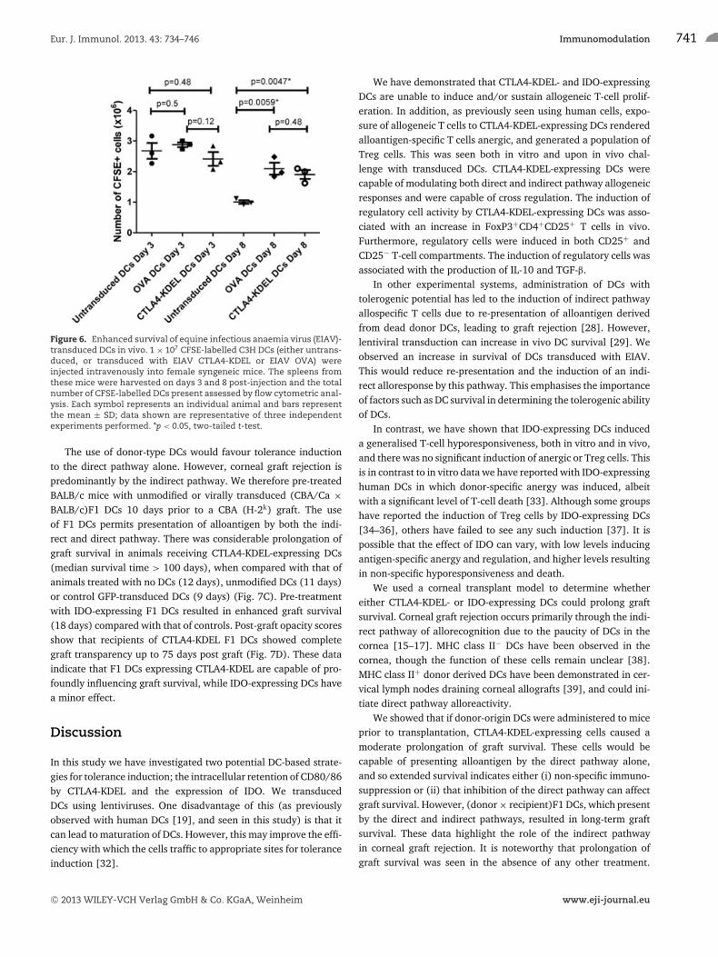
Enhanced survival of EIAV-transduced DCs in vivo
In other models, the administration of tolerogenic DCs has led toimmunisation of the recipient due to re-presentation of antigen,obtained from dead donor DCs, by host DCs [28]. How-ever, reports have indicated that lentiviral-transduced DCshave an increased survival in vivo, which would reduce suchre-presentation [29]. In order to investigate the survival of
EIAV-transduced DCs in vivo, CFSE-labelled DCs (either untrans-duced, or transduced with EIAV CTLA4-KDEL or EIAV-OVA) wereinjected into syngeneic mice. The number of CFSE+ DCs in thespleens was determined on days 3 and 8 post-injection. Transduc-tion with either EIAV vector led to increased survival of DCs onday 8 (Fig. 6).
Prolongation of corneal graft survival followingadministration of modified DCs
A mouse model was used to determine if corneal graft survivalcould be prolonged by the administration of allogeneic CTLA4-KDEL- or IDO-expressing DCs. As there is a high rate of graftfailure in agouti mice as a result of glaucoma [30,31], we used afully MHC-disparate C3H (H-2k) → BALB/c (H-2d) combination.
C© 2013 WILEY-VCH Verlag GmbH & Co. KGaA, Weinheim www.eji-journal.eu
740 Adnan Khan et al. Eur. J. Immunol. 2013. 43: 734–746
Figure 5. Generation of Treg cells in vivo with indirect donor allospecificity and capacity for linked suppression. 2.5 × 10⁶ CBK DCs (eitheruntransduced, or transduced with EIAV-GFP (control), EIAV-CTLA4-KDEL or EIAV-IDO) were injected i.v. into C3H/He mice. (A–B) After 10 days,CD4+CD25+ T cells purified from the spleens were irradiated and added (0–105 CD4+CD25+ T cells) to a primary MLR between freshly isolatedCBA/Ca-derived CD4+ T cells and (A) donor CBK DCs or (B) third-party B10.A DCs. T-cell proliferation was assessed by thymidine incorporation after5 days. (C–D) CD4+ T cells purified from the spleens of injected mice were also rechallenged in vitro with (C) (BALB/c × C57BL/6)F1 DCs expressingonly third-party MHC or (D) (C57BL/6 × CBA/Ca)F1 DCs expressing both donor (Kb presented in the context of I-Ak/I-Ek) and third-party MHC, andCD4+ T-cell proliferation was assessed by thymidine incorporation on days 3, 5 and 7. (E–F) Rechallenge assays were repeated using CD4+ T cellsthat were depleted of CD4+CD25+ T cells prior to rechallenge with (E) (BALB/c × C57BL/6)F1 DCs or (F) (C57BL/6 × CBA/Ca)F1 DCs. (G–H) In addition,CD4+CD25+ T cells purified from the spleens of injected mice were irradiated and added (0–105 CD4+CD25+ T cells) to new primary MLRs betweenfreshly isolated CBA/Ca-derived CD4+ T cells and (G) (BALB/c × C57BL/6)F1 DCs or (H) (C57BL/6 × CBA/Ca)F1 DCs and T-cell proliferation assessedby thymidine incorporation after 5 days. All results are shown as the mean ± SD of triplicate wells and are representative of three independentexperiments performed. *p < 0.05, two-tailed t-test.
Unmodified or virally transduced C3H DCs (2.5 × 10⁶) wereadministered i.v. 10 days prior to transplantation. Administra-tion of CTLA4-KDEL-expressing DCs resulted in moderate pro-longation of graft survival (median survival time: 18 days)compared with that of GFP-transduced DCs (10 days), unmodi-
fied DCs (11 days), IDO-transduced DCs (12 days) or naive ani-mals (12 days) (Fig. 7A). Similar results are seen when measuringthe development of corneal opacity with a slight reduction in thekinetics of rejection in recipients of CTLA4-KDEL-expressing DCs(Fig. 7B).
C© 2013 WILEY-VCH Verlag GmbH & Co. KGaA, Weinheim www.eji-journal.eu
Eur. J. Immunol. 2013. 43: 734–746 Immunomodulation 741
Figure 6. Enhanced survival of equine infectious anaemia virus (EIAV)-transduced DCs in vivo. 1 × 10⁷ CFSE-labelled C3H DCs (either untrans-duced, or transduced with EIAV CTLA4-KDEL or EIAV OVA) wereinjected intravenously into female syngeneic mice. The spleens fromthese mice were harvested on days 3 and 8 post-injection and the totalnumber of CFSE-labelled DCs present assessed by flow cytometric anal-ysis. Each symbol represents an individual animal and bars representthe mean ± SD; data shown are representative of three independentexperiments performed. *p < 0.05, two-tailed t-test.
The use of donor-type DCs would favour tolerance inductionto the direct pathway alone. However, corneal graft rejection ispredominantly by the indirect pathway. We therefore pre-treatedBALB/c mice with unmodified or virally transduced (CBA/Ca ×BALB/c)F1 DCs 10 days prior to a CBA (H-2k) graft. The useof F1 DCs permits presentation of alloantigen by both the indi-rect and direct pathway. There was considerable prolongation ofgraft survival in animals receiving CTLA4-KDEL-expressing DCs(median survival time > 100 days), when compared with that ofanimals treated with no DCs (12 days), unmodified DCs (11 days)or control GFP-transduced DCs (9 days) (Fig. 7C). Pre-treatmentwith IDO-expressing F1 DCs resulted in enhanced graft survival(18 days) compared with that of controls. Post-graft opacity scoresshow that recipients of CTLA4-KDEL F1 DCs showed completegraft transparency up to 75 days post graft (Fig. 7D). These dataindicate that F1 DCs expressing CTLA4-KDEL are capable of pro-foundly influencing graft survival, while IDO-expressing DCs havea minor effect.
Discussion
In this study we have investigated two potential DC-based strate-gies for tolerance induction; the intracellular retention of CD80/86by CTLA4-KDEL and the expression of IDO. We transducedDCs using lentiviruses. One disadvantage of this (as previouslyobserved with human DCs [19], and seen in this study) is that itcan lead to maturation of DCs. However, this may improve the effi-ciency with which the cells traffic to appropriate sites for toleranceinduction [32].
We have demonstrated that CTLA4-KDEL- and IDO-expressingDCs are unable to induce and/or sustain allogeneic T-cell prolif-eration. In addition, as previously seen using human cells, expo-sure of allogeneic T cells to CTLA4-KDEL-expressing DCs renderedalloantigen-specific T cells anergic, and generated a population ofTreg cells. This was seen both in vitro and upon in vivo chal-lenge with transduced DCs. CTLA4-KDEL-expressing DCs werecapable of modulating both direct and indirect pathway allogeneicresponses and were capable of cross regulation. The induction ofregulatory cell activity by CTLA4-KDEL-expressing DCs was asso-ciated with an increase in FoxP3+CD4+CD25+ T cells in vivo.Furthermore, regulatory cells were induced in both CD25+ andCD25− T-cell compartments. The induction of regulatory cells wasassociated with the production of IL-10 and TGF-β.
In other experimental systems, administration of DCs withtolerogenic potential has led to the induction of indirect pathwayallospecific T cells due to re-presentation of alloantigen derivedfrom dead donor DCs, leading to graft rejection [28]. However,lentiviral transduction can increase in vivo DC survival [29]. Weobserved an increase in survival of DCs transduced with EIAV.This would reduce re-presentation and the induction of an indi-rect alloresponse by this pathway. This emphasises the importanceof factors such as DC survival in determining the tolerogenic abilityof DCs.
In contrast, we have shown that IDO-expressing DCs induceda generalised T-cell hyporesponsiveness, both in vitro and in vivo,and there was no significant induction of anergic or Treg cells. Thisis in contrast to in vitro data we have reported with IDO-expressinghuman DCs in which donor-specific anergy was induced, albeitwith a significant level of T-cell death [33]. Although some groupshave reported the induction of Treg cells by IDO-expressing DCs[34–36], others have failed to see any such induction [37]. It ispossible that the effect of IDO can vary, with low levels inducingantigen-specific anergy and regulation, and higher levels resultingin non-specific hyporesponsiveness and death.
We used a corneal transplant model to determine whethereither CTLA4-KDEL- or IDO-expressing DCs could prolong graftsurvival. Corneal graft rejection occurs primarily through the indi-rect pathway of allorecognition due to the paucity of DCs in thecornea [15–17]. MHC class II− DCs have been observed in thecornea, though the function of these cells remain unclear [38].MHC class II+ donor derived DCs have been demonstrated in cer-vical lymph nodes draining corneal allografts [39], and could ini-tiate direct pathway alloreactivity.
We showed that if donor-origin DCs were administered to miceprior to transplantation, CTLA4-KDEL-expressing cells caused amoderate prolongation of graft survival. These cells would becapable of presenting alloantigen by the direct pathway alone,and so extended survival indicates either (i) non-specific immuno-suppression or (ii) that inhibition of the direct pathway can affectgraft survival. However, (donor × recipient)F1 DCs, which presentby the direct and indirect pathways, resulted in long-term graftsurvival. These data highlight the role of the indirect pathwayin corneal graft rejection. It is noteworthy that prolongation ofgraft survival was seen in the absence of any other treatment.
C© 2013 WILEY-VCH Verlag GmbH & Co. KGaA, Weinheim www.eji-journal.eu

742 Adnan Khan et al. Eur. J. Immunol. 2013. 43: 734–746
Figure 7. Prolongation of corneal graft survival following administration of modified DCs. BALB/c mice were either untreated (n = 6) or given 2.5× 10⁶ C3H/He DCs intravenously. The DCs were either untransduced (n = 5), or transduced 72 h earlier ex vivo with equine infectious anaemiavirus (EIAV)-GFP (control, n = 7), EIAV-IDO (n = 7) or EIAV-CTLA4-KDEL (n = 8). Ten days later, the BALB/c mice received a complete MHC-disparateC3H/He corneal graft or a syngeneic BALB/c graft (n = 6). (A) Data were plotted using the Kaplan–Meyer method and differences in graft survivalwere analysed using a log-rank test. (B) Corneal graft opacity scores were plotted and statistical differences between CTLA4-KDEL- and GFP(control)-expressing DCs calculated using the Mann–Whitney U test. * p < 0.008. (C) Using the same protocol, BALB/c mice were either untreated(n = 4) or given 2.5 × 10⁶ (CBA × BALB/c)F1 DCs intravenously. The DCs were either untransduced (n = 5), or transduced 72 h earlier ex vivo withEIAV-GFP (n = 5), EIAV-IDO (n = 5) or EIAV-CTLA4-KDEL (n = 5). 10 days later, BALB/c mice received a complete MHC-disparate CBA/Ca cornealgraft or a syngeneic BALB/c graft (n = 6). Data were analysed as above. (D) Corneal graft opacity scores were plotted and statistical differencesbetween CTLA4-KDEL- or IDO-expressing DCs and GFP-expressing DCs calculated using the Mann–Whitney U Test. *p < 0.008. Data shown arerepresentative of three independent experiments performed.
Previously, when dexamethasone-treated F1 DCs were injectedinto rats to induce indirect allospecific tolerance, the admin-istration of a single dose of CTLA4-Ig was necessary to pre-vent sensitisation caused by representation of donor DC-derivedalloantigens [2].
In contrast to CTLA4-KDEL, IDO-expressing DCs were lesseffective, with only a moderate increase in survival using F1 DCs.Previously, we have shown that IDO expression in the graft itselfcan prolong corneal graft survival [11]. However, in such a setting,IDO operates to block the effector cell response, rather than inhibitalloreactive T-cell activation. It may be that IDO has more potentialto protect tissues from damage than to prevent T-cell activation.We have also shown, however, that topical and systemic admin-istration of kynurenines suppresses CD4+ T-cell proliferation andprolongs corneal allograft survival [40].
In these experiments we have used F1 DCs to present alloanti-gen by the indirect pathway, which is not clinically applicable.There are several alternatives that might be feasible that includepulsing recipient DCs with alloantigen-derived protein or peptide.However, in order to achieve long-term presentation by the indi-rect pathway, we favour further genetic modification of the DCs toexpress donor-type alloantigen. This approach has the advantageover administration of CTLA4-Ig in being both alloantigen specificand not causing upregulation of IDO in DCs.
Whilst these present studies are restricted to corneal grafts,the use of CTLA4-KDEL DCs could be important in other settings.Given the strength of the direct pathway after transplantationof vascularised organs, the most plausible strategy for inducingtolerance in the clinical setting is a dual approach in which thefrequency of direct pathway alloreactive T cells is reduced bydeletion and/or anergy, and tolerance of the residual direct andthe indirect pathway then induced by a regulatory mechanism[41]. Therefore, a combination of short-term immunosuppres-sion during the acute phase of rejection post-transplantation andthe administration of tolerogenic CTLA4-KDEL-transduced recip-ient DCs, presenting antigen by the indirect pathway, prior tothe chronic phase of allograft rejection may prove to be a clini-cally applicable strategy to achieve donor-specific transplantationtolerance.
Materials and methods
Mice
CBK mice [25] were bred in-house. All other mice were pur-chased from Harlan Olac (Bicester, UK). Animals were treated in
C© 2013 WILEY-VCH Verlag GmbH & Co. KGaA, Weinheim www.eji-journal.eu
Eur. J. Immunol. 2013. 43: 734–746 Immunomodulation 743
accordance with UK regulations and the ARVO Statement for theUse of Animals in Ophthalmic and Vision Research.
Generation of lentiviral constructs
The extracellular domain of murine CTLA4 was amplified fromcDNA of activated T cells, inserted into the pCMV/myc/ER vec-tor (Invitrogen, Paisley, UK) and then subcloned into the EIAVplasmid pSMART2G [42] (Oxford Biomedica Co., Oxford, UK)replacing the GFP gene, resulting in pSMART-CTLA4-KDEL. ThepSMART-IDO construct encoding murine IDO1 has been previ-ously described [12]. The li-OVA gene, encoding the murineinvariant chain (li) in which the CLIP has been replaced by theOVA peptide (OVA328–339), was generated by excision from thepSL8-lipOVA vector (donated by Dr. Stephen Thirdborough,Southampton University) and subcloned into the EIAV plasmid.pSMART2G, encoding GFP, was used as a reporter construct andcontrol. Vesicular stomatitis virus G (VSV-G)-pseudotyped EIAVlentiviruses were produced using three-plasmid cotransfection of293T cells followed by ultracentrifugation as described [19,43].
Murine BM-derived DC cultures
DCs were generated from murine BM as described [44, 45] andcultured in RPMI 1640 medium (Invitrogen) supplemented with10% heat-inactivated foetal calf serum (PAA, Pasching, Austria),100 units/mL penicillin, 100 μg/mL streptomycin, 2 mML-glutamine (Cambrex Biosciences, Wokingham, UK) and 50 μMβ-mercaptoethanol (Invitrogen) (complete medium) and super-natant from a GM-CSF-producing cell line [46]. Where indi-cated, cells were treated with LPS, murine IFN-γ (PeproTech EC,London, UK) or mouse CTLA4-Ig (R&D Systems, Abingdon, UK).DCs were transduced with EIAV lentiviral vectors on day 6 ofculture at MOI 300 for 72 h.
Flow cytometry
Flow cytometry was carried out as previously described [19,47,48]using the following antibodies: ICOSL (GL1) (Insight Biotechnol-ogy, Wembley, UK); CD40 (3/23) (Serotec, Kidlington, UK), CD80(RMMP-1) and CD86 (RMMP-2) (Caltag, Buckingham, UK). Puri-fied Rat IgG2a (54447) antibody was used as an isotype control.Goat anti-rat IgG-PE (BD Biosciences, Oxford, UK) was used asa secondary antibody. FoxP3-allophycocyanin (FJK-16s) and ratIgG2a-allophycocyanin (eBioscience, USA) were used for intracel-lular detection of FoxP3.
Immunoblotting
Cells (1–2 × 10⁶) were resuspended in 130 μL lysis buffer (1%NP-40, 150 mM NaCl, 5 mM MgCl2, and 10 mM Hepes buffer)supplemented with Protease Inhibitor Cocktail (Sigma-Aldrich,
Poole, UK) and incubated on ice for 30 min followed by cen-trifugation at 4000 × g for 5 min. The lysate was separated bySDS-PAGE under reducing conditions and transferred to a nitro-cellulose membrane. Membranes were probed using antibod-ies specific for IDO (rabbit polyclonal IgG, Cosmo Bio Co. Ltd,Japan); c-myc epitope tag (4A6) and cyclin E (rabbit polyclonalIgG) (Upstate-Millipore, Watford, UK); p27Kip1 (G173–524, BDBiosciences); β-actin (AC-15, Sigma-Aldrich). The following sec-ondary antibodies were used: goat anti-rabbit IgG HRP, rab-bit anti-mouse IgG HRP and rabbit anti-goat IgG HRP (Dako-Millipore). Proteins were visualised using the ECL
TMplus Western
Blotting detection system (Amersham Biosciences, Little Chalfont,UK).
T-cell purification
Splenocytes were obtained by passing a spleen through70 μm cell strainers into cold-complete medium with 10 units/mLDNAse-Pulmozyme/Dornase Alfa (Roche Applied Sciences, Lewes,UK). Following lysis of erythrocytes, splenocytes were incu-bated on a horizontal roller at 4◦C with rat antibodies spe-cific for CD45R/B220 (RA3-3A1), CD8 (53.6.7), H2-Ek,d/Ab,d
(M5/114.15.2), and CD16/32 (2.4G2 all ATCC Manassas, VA,USA), washed twice in RPMI 1640 medium and incubated withsheep anti-rat IgG Dynabeads R© (Dynal Biotech, Bromborough,UK) on a horizontal roller at 4◦C before magnetic separation toobtain CD4+ T cells. Where indicated, CD25+ T cells were puri-fied or depleted from CD4+ T-cell populations using the MACS R©
CD25 MicroBead Kit (Miltenyi Biotec GmbH, Bergisch Gladbach,Germany).
T-cell proliferation assays
CD4+ T cells were cultured for 5 days with allogeneic BM-derivedDCs in complete medium. Unless otherwise stated, 25 × 104 CD4+
T cells to 5 × 10⁴ DCs were used. Cells were pulsed on day4 with 3H-thymidine (1 μCi/well) (Amersham Biosciences) andharvested 17 h later. Two-stage MLRs (rechallenge assays) wereperformed using modifications of established protocols [6, 49].To investigate the induction of anergy and linked suppression invitro, DC populations were incubated with allogeneic CD4+ T cells(2 × 10⁵ DCs:2 × 10⁶ T cells) for 10 days. The T cells were har-vested, washed twice in PBS and rechallenged with WT DCs (1 ×10⁴ DCs:2 × 10⁴ T cells). To investigate the induction of anergyand linked suppression in vivo, 2.5 × 10⁶ DCs were injected i.v. viathe tail vein. Ten days later, CD4+ splenocytes were purified andrechallenged with DCs as described for the assays to investigateanergy induction and linked suppression in vitro. Where indicated100 U/mL exogenous IL-2 (Roche) was added to cultures. Prolif-eration in rechallenge experiments was determined by thymidineincorporation on days 3, 5 and 7. Cytokines were detected inculture supernatants using mouse IL-10 and TGF-β1 ELISA Kits(eBioscience). To determine functional Treg-cell activity, CD4+
T cells were harvested on day 10 of primary MLRs. Alternatively,
C© 2013 WILEY-VCH Verlag GmbH & Co. KGaA, Weinheim www.eji-journal.eu
744 Adnan Khan et al. Eur. J. Immunol. 2013. 43: 734–746
CD4+CD25+ T cells were purified from the spleens and lymphnodes of mice following injection with DCs 10 days previously.The cells were irradiated (50 Gy) and added (0–100 000 cells) to‘fresh’ MLRs, consisting of WT DCs and allogeneic CD4+ T cells.
CFSE labelling
BM-derived C3H DCs, either untransduced or transduced withEIAV-CTLA4-KDEL or EIAV-OVA, were labelled with CFSE usingthe Vybrant R© CFDA SE Cell Tracer Kit (Molecular Probes-Invitrogen, Paisley, UK). Female C3H mice were injected with1 × 107 CFSE-labelled syngeneic DCs. Spleens were harvestedon days 3 and 8 post-injection. CFSE-labelled DCs present in thespleen were detected using a FACScalibur flow cytometre. Fiftythousand events were collected per mouse and the percentageof FL1-positive cells used to calculate the total number of CFSE-labelled DCs present per spleen.
Quantitative PCR
Messenger RNA was prepared using TRIzolTM (Invitrogen) andreverse transcribed using M-MLV RT (Promega), according tothe manufacturers’ instructions. PCR was performed using aLightCycler (Roche Molecular Biochemicals, Hertfordshire, UK)and SYBR R© Green Taq ReadyMixTM (Sigma-Aldrich) accord-ing to the manufacturers’ instructions. The programme was95◦C for 3 min followed by 40 cycles of (95◦C for 5 s, 56◦Cfor 10 s, and 72◦C for 13 s) followed by quantification at81◦C. The IDO primers were 5′-TGGCAAACTGGAAGAAAAAG-3′
(forward) and 5′-AATGCTTTCAGGTCTTGACG-3′ (reverse).Hypoxanthine phosphoribosyl transferase primers were3′-GTAATGATCCAGTCAACGGGGGAC-5′ (forward) and 3′-CCAGCAAGCTTGCAACCTTAACCA-5′ (reverse) [11].
L-kynurenine assay
L-kynurenine was measured as described [12,50,51].
Orthotopic corneal transplantation and criteriafor graft rejection
Murine corneal transplantation was performed in the right eye asdescribed [52,53]. The eyes were examined every 2 days followingsuture removal on day 7. The grading of corneal opacity and theonset of graft rejection were graded as described by an examinermasked to the treatment group [53].
Acknowledgments: This work was funded by The Roche OrganTransplantation Research Foundation (ROTRF) (A.K.). Additional
support was from the Wellcome Trust (H.F.) and British HeartFoundation (L.T.). The authors would like to thank OxfordBiomedica, United Kingdom, for providing the lentiviral vectorsand Dr. Yakup Tanriver for provision of the CBK mice.
Conflict of interest: The authors declare no financial or commer-cial conflict of interest.
References
1 Morelli, A. E. and Thomson, A. W., Tolerogenic dendritic cells and the
quest for transplant tolerance. Nat. Rev. Immunol. 2007. 7: 610–621.
2 Mirenda, V., Berton, I., Read, J., Cook, T., Smith, J., Dorling, A. and Lech-
ler, R. I., Modified dendritic cells coexpressing self and allogeneic major
histocompatability complex molecules: an efficient way to induce indi-
rect pathway regulation. J. Am. Soc. Nephrol. 2004. 15: 987–997.
3 Andreakos, E., Smith, C., Monaco, C., Brennan, F. M., Foxwell, B. M. and
Feldmann, M., Ikappa B kinase 2 but not NF-kappa B-inducing kinase is
essential for effective DC antigen presentation in the allogeneic mixed
lymphocyte reaction. Blood 2003. 101: 983–991.
4 Yoshimura, S., Bondeson, J., Brennan, F. M., Foxwell, B. M. and Feld-
mann, M., Antigen presentation by murine dendritic cells is nuclear
factor-kappa B dependent both in vitro and in vivo. Scand. J. Immunol.
2003. 58: 165–172.
5 Munro, S. and Pelham, H. R., A C-terminal signal prevents secretion of
luminal ER proteins. Cell 1987. 48: 899–907.
6 Tan, P. H., Yates, J. B., Xue, S. A., Chan, C., Jordan, W. J., Harper, J. E.,
Watson, M. P. et al., Creation of tolerogenic human dendritic cells via
intracellular CTLA4: a novel strategy with potential in clinical immuno-
suppression. Blood 2005. 106: 2936–2943.
7 Stone, T. W. and Darlington, L. G., Endogenous kynurenines as targets for
drug discovery and development. Nat. Rev. Drug Discov. 2002. 1: 609–620.
8 Moffett, J. R. and Namboodiri, M. A., Tryptophan and the immune
response. Immunol. Cell Biol. 2003. 81: 247–265.
9 Frumento, G., Rotondo, R., Tonetti, M., Damonte, G., Benatti, U. and Fer-
rara, G. B., Tryptophan-derived catabolites are responsible for inhibition
of T and natural killer cell proliferation induced by indoleamine 2,3-
dioxygenase. J. Exp. Med. 2002. 196: 459–468.
10 Terness, P., Bauer, T. M., Rose, L., Dufter, C., Watzlik, A., Simon, H.
and Opelz, G., Inhibition of allogeneic T cell proliferation by indoleamine
2,3-dioxygenase-expressing dendritic cells: mediation of suppression by
tryptophan metabolites. J. Exp. Med. 2002. 196: 447–457.
11 George, A. J. and Larkin, D. F., Corneal transplantation: the forgotten
graft. Am. J. Transplant 2004. 4: 678–685.
12 Beutelspacher, S. C., Pillai, R., Watson, M. P., Tan, P. H., Tsang, J.,
McClure, M. O., George, A. J. et al., Function of indoleamine 2,3-
dioxygenase in corneal allograft rejection and prolongation of allograft
survival by over-expression. Eur. J. Immunol. 2006. 36: 690–700.
13 Fu, H., Khan, A., Coe, D., Zaher, S., Chai, J. G., Kropf, P., Muller, I. et al.,
Arginine depletion as a mechanism for the immune privilege of corneal
allografts. Eur. J. Immunol. 2011. 41: 2997–3005.
14 Williams, K. A., Lowe, M., Bartlett, C., Kelly, T. L. and Coster, D. J., Risk
factors for human corneal graft failure within the Australian corneal graft
registry. Transplantation 2008. 86: 1720–1724.
15 Illigens, B. M., Yamada, A., Fedoseyeva, E. V., Anosova, N., Boisgerault,
F., Valujskikh, A., Heeger, P. S. et al., The relative contribution of direct
C© 2013 WILEY-VCH Verlag GmbH & Co. KGaA, Weinheim www.eji-journal.eu

Eur. J. Immunol. 2013. 43: 734–746 Immunomodulation 745
and indirect antigen recognition pathways to the alloresponse and graft
rejection depends upon the nature of the transplant. Hum. Immunol. 2002.
63: 912–925.
16 Sano, Y., Streilein, J. W. and Ksander, B. R., Detection of minor
alloantigen-specific cytotoxic T cells after rejection of murine orthotopic
corneal allografts: evidence that graft antigens are recognized exclusively
via the “indirect pathway.” Transplantation 1999. 68: 963–970.
17 Boisgerault, F., Liu, Y., Anosova, N., Dana, R. and Benichou, G., Differen-
tial roles of direct and indirect allorecognition pathways in the rejection
of skin and corneal transplants. Transplantation 2009. 87: 16–23.
18 Fu, H., Larkin, D. F. and George, A. J., Immune modulation in corneal
transplantation. Transplant Rev (Orlando) 2008. 22: 105–115.
19 Tan, P. H., Beutelspacher, S. C., Xue, S. A., Wang, Y. H., Mitchell, P.,
McAlister, J. C., Larkin, D. F. et al., Modulation of human dendritic-cell
function following transduction with viral vectors: implications for gene
therapy. Blood 2005. 105: 3824–3832.
20 Mellor, A. L., Chandler, P., Baban, B., Hansen, A. M., Marshall, B.,
Pihkala, J., Waldmann, H. et al., Specific subsets of murine dendritic cells
acquire potent T cell regulatory functions following CTLA4-mediated
induction of indoleamine 2,3 dioxygenase. Int. Immunol. 2004. 16:
1391–1401.
21 Sherr, C. J. and Roberts, J. M., Inhibitors of mammalian G1 cyclin-
dependent kinases. Genes Dev. 1995. 9: 1149–1163.
22 Rowell, E. A., Walsh, M. C. and Wells, A. D., Opposing roles for the
cyclin-dependent kinase inhibitor p27kip1 in the control of CD4+ T cell
proliferation and effector function. J. Immunol. 2005. 174: 3359–3368.
23 Sherman, L. A. and Chattopadhyay, S., The molecular basis of allorecog-
nition. Annu. Rev. Immunol. 1993. 11: 385–402.
24 Warrens, A. N., Lombardi, G. and Lechler, R. I., Presentation and recog-
nition of major and minor histocompatibility antigens. Transpl. Immunol.
1994. 2: 103–107.
25 Yeoman, H. and Mellor, A. L., Tolerance and MHC restriction in trans-
genic mice expressing a MHC class I gene in erythroid cells. Int. Immunol.
1992. 4: 59–65.
26 Tarazona, R., Sponaas, A. M., Mavria, G., Zhou, M., Schulz, R., Tomlinson,
P., Antoniou, J. et al., Effects of different antigenic microenvironments on
the course of CD8+ T cell responses in vivo. Int. Immunol. 1996. 8: 351–358.
27 Ensminger, S. M., Spriewald, B. M., Witzke, O., Pajaro, O. E., Yacoub,
M. H., Morris, P. J., Rose, M. L. et al., Indirect allorecognition can play an
important role in the development of transplant arteriosclerosis. Trans-
plantation 2002. 73: 279–286.
28 van Kooten, C., Lombardi, G., Gelderman, K. A., Sagoo, P., Buckland, M.,
Lechler, R. and Cuturi, M. C., Dendritic cells as a tool to induce trans-
plantation tolerance: obstacles and opportunities. Transplantation 2011.
91: 2–7.
29 Arce, F., Rowe, H. M., Chain, B., Lopes, L. and Collins, M. K., Lentivi-
ral vectors transduce proliferating dendritic cell precursors leading to
persistent antigen presentation and immunization. Mol. Ther. 2009. 17:
1643–1650.
30 Puk, O., Dalke, C., Favor, J., de Angelis, M. H. and Graw, J., Variations
of eye size parameters among different strains of mice. Mamm. Genome.
2006. 17: 851–857.
31 John, S. W., Hagaman, J. R., MacTaggart, T. E., Peng, L. and Smithes, O.,
Intraocular pressure in inbred mouse strains. Invest. Ophthalmol. Vis. Sci.
1997. 38: 249–253.
32 De Vries, I. J., Krooshoop, D. J., Scharenborg, N. M., Lesterhuis, W. J.,
Diepstra, J. H., Van Muijen, G. N., Strijk, S. P. et al., Effective migration
of antigen-pulsed dendritic cells to lymph nodes in melanoma patients
is determined by their maturation state. Cancer Res. 2003. 63: 12–17.
33 Tan, P. H., Beutelspacher, S. C., Wang, Y. H., McClure, M. O., Ritter, M. A.,
Lombardi, G. and George, A. J., Immunolipoplexes: an efficient, nonviral
alternative for transfection of human dendritic cells with potential for
clinical vaccination. Mol. Ther. 2005. 11: 790–800.
34 Fallarino, F., Grohmann, U., You, S., McGrath, B. C., Cavener, D. R., Vacca,
C., Orabona, C. et al., The combined effects of tryptophan starvation
and tryptophan catabolites downregulate T cell receptor zeta-chain and
induce a regulatory phenotype in naive T cells. J. Immunol. 2006. 176:
6752–6761.
35 Fallarino, F., Grohmann, U., You, S., McGrath, B. C., Cavener, D. R., Vacca,
C., Orabona, C. et al., Tryptophan catabolism generates autoimmune-
preventive Treg cells. Transpl. Immunol. 2006. 17: 58–60.
36 Hill, M., Tanguy-Royer, S., Royer, P., Chauveau, C., Asghar, K., Tesson,
L., Lavainne, F. et al., IDO expands human CD4+CD25-high Treg cells
by promoting maturation of LPS-treated dendritic cells. Eur. J. Immunol.
2007. 37: 3054–3062.
37 Mellor, A. L. and Munn, D. H., Tryptophan catabolism and T-cell tol-
erance: immunosuppression by starvation? Immunol. Today 1999. 20:
469–473.
38 Hamrah, P., Zhang, Q., Liu, Y. and Dana, M. R., Novel characteriza-
tion of MHC class II-negative population of resident corneal Langer-
hans cell-type dendritic cells. Invest. Ophthalmol. Vis. Sci. 2002. 43:
639–646.
39 Liu, Y., Hamrah, P., Zhang, Q., Taylor, A. W. and Dana, M. R., Drain-
ing lymph nodes of corneal transplant hosts exhibit evidence for
donor major histocompatibility complex (MHC) class II-positive dendritic
cells derived from MHC class II-negative grafts. J. Exp. Med. 2002. 195:
259–268.
40 Zaher, S. S., Germain, C., Fu, H., Larkin, D. F. and George, A. J.,
3-Hydroxykynurenine suppresses CD4+ T-cell proliferation, induces
T-regulatory-cell development, and prolongs corneal allograft survival.
Invest. Ophthalmol. Vis. Sci. 2011. 52: 2640–2648.
41 Lechler, R. I., Garden, O. A. and Turka, L. A., The complementary roles of
deletion and regulation in transplantation tolerance. Nat. Rev. Immunol.
2003. 3: 147–158.
42 Bienemann, A. S., Martin-Rendon, E., Cosgrave, A. S., Glover, C. P., Wong,
L. F., Kingsman, S. M., Mitrophanous, K. A. et al., Long-term replacement
of a mutated nonfunctional CNS gene: reversal of hypothalamic diabetes
insipidus using an EIAV-based lentiviral vector expressing arginine vaso-
pressin. Mol. Ther. 2003. 7: 588–596.
43 Beutelspacher, S. C., Ardjomand, N., Tan, P. H., Patton, G. S., Larkin,
D. F., George, A. J. and McClure, M. O., Comparison of HIV-1 and EIAV-
based lentiviral vectors in corneal transduction. Exp. Eye Res. 2005. 80:
787–794.
44 Inaba, K., Inaba, M., Romani, N., Aya, H., Deguchi, M., Ikehara,
S., Muramatsu, S. et al., Generation of large numbers of dendritic
cells from mouse bone marrow cultures supplemented with granu-
locyte/macrophage colony-stimulating factor. J. Exp. Med. 1992. 176:
1693–1702.
45 Lutz, M. B., Kukutsch, N., Ogilvie, A. L., Rossner, S., Koch, F., Romani,
N. and Schuler, G., An advanced culture method for generating large
quantities of highly pure dendritic cells from mouse bone marrow.
J. Immunol. Methods 1999. 223: 77–92.
46 Zal, T., Volkmann, A. and Stockinger, B., Mechanisms of toler-
ance induction in major histocompatibility complex class II-restricted
T cells specific for a blood-borne self-antigen. J. Exp. Med. 1994. 180:
2089–2099.
47 Tan, P. H., Manunta, M., Ardjomand, N., Xue, S. A., Larkin, D. F., Haskard,
D. O., Taylor, K. M. et al., Antibody targeted gene transfer to endothelium.
J. Gene Med. 2003. 5: 311–323.
C© 2013 WILEY-VCH Verlag GmbH & Co. KGaA, Weinheim www.eji-journal.eu
746 Adnan Khan et al. Eur. J. Immunol. 2013. 43: 734–746
48 Tan, P. H., Sagoo, P., Chan, C., Yates, J. B., Campbell, J., Beutelspacher,
S. C., Foxwell, B. M. et al., Inhibition of NF-kappa B and oxidative path-
ways in human dendritic cells by antioxidative vitamins generates Treg
cells. J. Immunol. 2005. 174: 7633–7644.
49 Marelli-Berg, F. M., Frasca, L., Imami, N., Lombardi, G. and Lechler,
R. I., Lack of T cell proliferation without induction of nonresponsive-
ness after antigen presentation by endothelial cells. Transplantation 1999.
68: 280–287.
50 Takikawa, O., Kuroiwa, T., Yamazaki, F. and Kido, R., Mechanism
of interferon-gamma action. Characterization of indoleamine 2,3-
dioxygenase in cultured human cells induced by interferon-gamma and
evaluation of the enzyme-mediated tryptophan degradation in its anti-
cellular activity. J. Biol. Chem. 1988. 263: 2041–2048.
51 Feng, G. S. and Taylor, M. W., Interferon gamma-resistant mutants are
defective in the induction of indoleamine 2,3-dioxygenase. Proc. Natl.
Acad. Sci. USA 1989. 86: 7144–7148.
52 Zhang, E. P., Schrunder, S. and Hoffmann, F., Orthotopic corneal
transplantation in the mouse–a new surgical technique with mini-
mal endothelial cell loss. Graefes Arch. Clin. Exp. Ophthalmol. 1996. 234:
714–719.
53 Ardjomand, N., McAlister, J. C., Rogers, N. J., Tan, P. H., George, A. J. and
Larkin, D. F., Modulation of costimulation by CD28 and CD154 alters the
kinetics and cellular characteristics of corneal allograft rejection. Invest.
Ophthalmol. Vis. Sci. 2003. 44: 3899–3905.
Abbreviations: EIAV: equine infectious anaemia virus · HPRT: hypoxan-
thine phosphoribosyl transferase · MST: median survival time · 1-MT:
1-methyl tryptophan · VSV-G: vesicular stomatitis virus G
Full correspondence: Prof. Andrew J. T. George, Section of MolecularImmunology, Department of Medicine, Imperial College London,Hammersmith Hospital, Du Cane Road, London W12 0NN,United KingdomFax: +44-(0)20-8383-2788e-mail: [email protected]
See accompanying Commentary:http://dx.doi.org/10.1002/eji.201343361
Received: 13/8/2012Revised: 30/10/2012Accepted: 28/11/2012Accepted article online: 4/12/2012
C© 2013 WILEY-VCH Verlag GmbH & Co. KGaA, Weinheim www.eji-journal.eu