
dcis-book-newman.pdf - Breast Cancer Surgery Melbourne, VIC
171
-
Upload
khangminh22 -
Category
Documents
-
view
1 -
download
0
Transcript of dcis-book-newman.pdf - Breast Cancer Surgery Melbourne, VIC


Ductal Carcinoma In Situ and Microinvasive/Borderline Breast Cancer

Lisa A. NewmanJessica M. BensenhaverEditors
Ductal Carcinoma In Situ and Microinvasive/Borderline Breast Cancer
2123

ISBN 978-1-4939-2034-1 ISBN 978-1-4939-2035-8 (eBook)DOI 10.1007/978-1-4939-2035-8Springer New York Heidelberg Dordrecht London
Library of Congress Control Number: 2014950151
© Springer Science+Business Media New York 2015This work is subject to copyright. All rights are reserved by the Publisher, whether the whole or part of the material is concerned, specifically the rights of translation, reprinting, reuse of illustrations, recitation, broadcasting, reproduction on microfilms or in any other physical way, and transmission or information storage and retrieval, electronic adaptation, computer software, or by similar or dissimilar methodology now known or hereafter developed. Exempted from this legal reservation are brief excerpts in connection with reviews or scholarly analysis or material supplied specifically for the purpose of being entered and executed on a computer system, for exclusive use by the purchaser of the work. Duplication of this publication or parts thereof is permitted only under the provisions of the Copyright Law of the Publisher’s location, in its cur-rent version, and permission for use must always be obtained from Springer. Permissions for use may be obtained through RightsLink at the Copyright Clearance Center. Violations are liable to prosecution under the respective Copyright Law.The use of general descriptive names, registered names, trademarks, service marks, etc. in this publication does not imply, even in the absence of a specific statement, that such names are ex-empt from the relevant protective laws and regulations and therefore free for general use.While the advice and information in this book are believed to be true and accurate at the date of publication, neither the authors nor the editors nor the publisher can accept any legal responsibil-ity for any errors or omissions that may be made. The publisher makes no warranty, express or implied, with respect to the material contained herein.
Printed on acid-free paper
Springer is part of Springer Science+Business Media (www.springer.com)
EditorsLisa A. NewmanComprehensive Cancer CenterUniversity of Michigan Health SystemAnn ArborMichiganUSA
Jessica M. BensenhaverComprehensive Cancer CenterUniversity of Michigan Health SystemAnn ArborMichiganUSA

v
Preface
Any physician with a clinical practice spanning between the twentieth and the twenty-first centuries has witnessed the dramatic increases that have occurred in the volume of newly diagnosed cases of ductal carcinoma in situ, with/without associated microinvasion. Many controversial debates have been sparked in accordance with these rising incidence rates, related to over-diagnosis and its necessary companion overtreatment; variation in opinions over pathology findings; and extent of appropriate local therapy as well as systemic therapy.
This book has convened some of the greatest minds in oncology to address these various questions. As with many topics in medicine, this book cannot provide definitive answers, but our distinguished authors have distilled and summarized the existing data. We are confident that trainees, physicians and survivor advocates alike will find this book to be a valuable resource in under-standing the broad spectrum of options in managing this particular aspect of the most common malignancy afflicting women throughout the world.
Lisa A. Newman, MD, MPH, FACS Jessica M. Bensenhaver, MD

vii
Acknowledgements
We thank our fantastic roster of chapter authors as well as the energetic and brilliant Springer editorial staff. Above all of course, we thank our patients, without whose courage and commitment to clinical research none of our advancing breast cancer knowledge would be possible.
Lisa A. Newman, MD, MPH, FACS Jessica M. Bensenhaver, MD

ix
Contents
1 Epidemiology of Ductal Carcinoma In Situ ................................... 1Prathima Kanumuri and Anees B. Chagpar
2 Role of Screening Mammography in Early Detection/Outcome of Breast Cancer ............................................................................... 13Renee W. Pinsky and Mark A. Helvie
3 Imaging DCIS: Digital/Film-Screening Mammography, Tomosynthesis, MRI, Ultrasonography ......................................... 27Annette Ingram Joe and Stephanie K. Patterson
4 Pathology of Ductal Carcinoma In Situ: Features and Diagnostic Challenges ..................................................................... 39Julie M. Jorns and Celina G. Kleer
5 Molecular Markers in DCIS ........................................................... 51Theresa L. Schwartz and Chelsea Horwood
6 History of Ductal Carcinoma In Situ Management Based Upon Data from Prospective, Randomized Clinical Trials..................... 57Lisa A. Newman and Jessica M. Bensenhaver
7 Extent and Role of Margin Control for DCIS Managed by Breast-Conserving Surgery ............................................................. 67Melissa Pilewskie and Monica Morrow
8 Ductal Carcinoma In Situ Treated with Breast-Conserving Surgery Alone ................................................................................... 85Jessica M. Bensenhaver
9 DCIS Managed with BCS: Whole-Breast XRT vs. Partial Breast XRT ............................................................................... 91Chirag Shah and Frank A. Vicini

x Contents
10 Anti-HER2/neu Therapy in DCIS .................................................. 99 Amelia Tower, Ruta D. Rao, Kalliopi P. Siziopikou, Melody A. Cobleigh and Thomas B. Julian
11 Role of Genetic Profiling and Recurrence Scores in Treatment Planning for DCIS ......................................................... 109Julie A. Margenthaler and Amy E. Cyr
12 DCIS and Sentinel Lymph Node Biopsy ........................................ 117Alfred J. Colfry, Alastair M. Thompson and Henry Mark Kuerer
13 Role of Postmastectomy Radiation for DCIS ................................ 125Corey W. Speers and Reshma Jagsi
14 Monitoring and Surveillance Following DCIS Treatment ........... 139Jennifer L. Zakhireh and E. Shelley Hwang
15 DCIS and Hereditary Susceptibility for Breast Cancer ............... 147Patrick G. Pilie, Kara J. Milliron and Sofia D. Merajver
16 Introduction, Evolution, and Application of the Van Nuys Prognostic Index in DCIS ................................................................ 155Aeisha K. S. Rivers
17 Disparities in DCIS Detection and Outcomes Related to Race/Ethnicity ............................................................................. 161Erin A. Strong, Azadeh Stark and Lisa A. Newman
Index ....................................................................................................... 167

xi
Contributors
Jessica M. Bensenhaver Comprehensive Cancer Center, University of Michigan Health System, Ann Arbor, MI, USA
University of Michigan Health System, Comprehensive Cancer Center, Ann Arbor, MI, USA
Anees B. Chagpar Department of Surgery, Smilow Cancer Hospital at Yale New Haven, Yale New Haven Hospital, New Haven, CT, USA
Melody A. Cobleigh Section of Medical Oncology, Department of Internal Medicine, Rush University Medical Center, Chicago, IL, USA
Alfred J. Colfry Department of Surgical Oncology, The University of Texas MD Anderson Cancer Center, Houston, TX, USA
Amy E. Cyr Department of Surgery, Washington University School of Med-icine, St. Louis, MO, USA
Mark A. Helvie Department of Radiology, University of Michigan Health System, Ann Arbor, MI, USA
Chelsea Horwood Saint Louis University School of Medicine, St. Louis, MO, USA
E. Shelley Hwang Department of Surgery, Duke University Medical Center, Durham, NC, USA
Reshma Jagsi Department of Radiation Oncology, University of Michigan Health System, Ann Arbor, MI, USA
Annette Ingram Joe Department of Radiology, University of Michigan Health System, Ann Arbor, MI, USA
Julie M. Jorns Department of Pathology, University of Michigan Health System, Ann Arbor, MI, USA
xii
Thomas B. Julian Division of Breast Surgical Oncology, Allegheny General Hospital, Pittsburgh, PA, USA
WPAHS Breast Surgical Oncology, Allegheny Health Network, Pittsburgh, USA
Temple University School of Medicine, Philadelphia, USA
Medical Affairs, National Surgical Adjuvant Breast and Bowel Project (NSABP), Pittsburgh, USA
Prathima Kanumuri Department of Surgery, Yale University School of Medicine, Yale New Haven Hospital, New Haven, CT, USA
Celina G. Kleer Department of Pathology, Comprehensive Cancer Center, University of Michigan Health System, Ann Arbor, MI, USA
Henry Mark Kuerer Department of Surgical Oncology, The University of Texas MD Anderson Cancer Center, Houston, TX, USA
Julie A. Margenthaler Department of Surgery, Washington University School of Medicine, St. Louis, MO, USA
Sofia D. Merajver Department of Internal Medicine and Epidemiology, University of Michigan Medical School and School of Public Health, Univer-sity of Michigan Health System, Comprehensive Cancer Center, Ann Arbor, MI, USA
Kara J. Milliron Department of Hematology Oncology, University of Mich-igan Health System, Comprehensive Cancer Center, Ann Arbor, MI, USA
Monica Morrow Breast Service, Department of Surgery, Memorial Sloan Kettering Cancer Center, New York, NY, USA
Lisa A. Newman Comprehensive Cancer Center, University of Michigan Health System, Ann Arbor, MI, USA
Breast Care Center, Comprehensive Cancer Center, University of Michigan, Ann Arbor, MI, USA
Stephanie K. Patterson Department of Radiology, University of Michigan Health System, Ann Arbor, MI, USA
Melissa Pilewskie Breast Service, Department of Surgery, Memorial Sloan Kettering Cancer Center, New York, NY, USA
Patrick G. Pilie Department of Internal Medicine, University of Michigan Health System, Taubman Center, Ann Arbor, MI, USA
Renee W. Pinsky University of Michigan Health System, Ann Arbor, MI, USA
Ruta D. Rao Section of Medical Oncology, Rush University Medical Center, Chicago, IL, USA
Aeisha K. S. Rivers Division of Surgical Oncology, Department of Surgery, UT Southwestern Medical Center, Dallas, TX, USA
Contributors

xiiiContributors
Theresa L Schwartz Department of Surgery, Saint Louis University School of Medicine, St. Louis, MO, USA
Chirag Shah Department of Radiation Oncology, Summa Health System/Northeast Ohio Medical University, Akron, OH, USA
Kalliopi P. Siziopikou Breast Pathology Section, Department of Pathology, Northwestern University Feinberg School of Medicine, Chicago, IL, USA
Robert H. Lurie Comprehensive Cancer Center, Chicago, USA
Corey W. Speers Department of Radiation Oncology, University of Michi-gan Health System, Ann Arbor, MI, USA
Azadeh Stark Department of Pathology, Henry Ford Health System, Detroit, MI, USA
Erin A. Strong University of Michigan Health System, Ann Arbor, MI, USA
Alastair M. Thompson Department of Surgical Oncology, The University of Texas MD Anderson Cancer Center, Houston, TX, USA
Amelia Tower Division of Breast Surgical Oncology, Allegheny General Hospital, Pittsburgh, PA, USA
Frank A. Vicini Department of Radiation Oncology, Michigan Healthcare Professionals/21st Century Oncology, Farmington Hills, MI, USA
Jennifer L. Zakhireh Atlanta, GA, USA

1
Epidemiology of Ductal Carcinoma In Situ
Prathima Kanumuri and Anees B. Chagpar
L. A. Newman, J. M. Bensenhaver (eds.), Ductal Carcinoma In Situ and Microinvasive/Borderline BreastCancer, DOI 10.1007/978-1-4939-2035-8_1, © Springer Science+Business Media New York 2015
A. B. Chagpar ()Department of Surgery, Smilow Cancer Hospital at Yale New Haven, Yale New Haven Hospital, 20 York Street, North Pavilion, First Floor, 06510 New Haven, CT 06510, USA e-mail: [email protected]
P. KanumuriDepartment of Surgery, Yale University School of Medicine, Yale New Haven Hospital, 20 York Street, North Pavilion, First Floor, New Haven, CT 06510, USAe-mail: [email protected]
Definition
In 1932, Broders defined ductal carcinoma in situ (DCIS) as “a condition in which malignant epi-thelial cells and their progeny are found in or near positions occupied by their ancestors before the ancestors underwent malignant transformation, and they have not migrated beyond the basement membrane” [1]. The World Health Organization (WHO) refined this definition in 2012, noting that DCIS was “a neoplastic proliferation confined to the mammary ductal-lobular system and charac-terized by increased epithelial proliferation, subtle to marked cytologic atypia, and an inherent but not necessarily obligate tendency for progression to invasive breast carcinoma” [2]. DCIS progresses to invasive disease in only 20–50 % of the cases; although, it is not possible to accurately predict which cases will progress and which will not [3]. In more than seven autopsy series of women, the median prevalence of previously undetected DCIS was 8.9 % (0 –14.7 %), suggesting that not
all cases of DCIS will progress over a woman’s lifetime, and may be otherwise indolent [4].
Incidence
Historically, DCIS accounted for less than 2–5 % of breast cancers. With the adoption of mammog-raphy as a breast cancer screening (BCS) tool, the incidence of DCIS increased dramatically. DCIS currently accounts for about 20–25 % of all newly diagnosed breast cancer cases in the USA [5] and 18–33 % of mammographically detected cases in various large screening mammography studies [6]. The American Cancer Society (ACS) estimates that 62,570 new cases of in situ breast cancers will be diagnosed in 2014 [7].
The in situ disease incidence rates in the USA rose rapidly in the 1980s and 1990s largely because of widespread use of screening mam-mography and have since continued to rise steadily. The Surveillance, Epidemiology and End Results (SEER) Cancer Statistics Review (1975–2011) shows an overall incidence of in situ disease from 5.8 per 100,000 in 1975 to 35.5 per 100,000 in 2011. The increase has been ob-served in women both under and over 50 years of age; however, the increase is more pronounced in women over 50 years of age (Fig. 1.1) [8]. A com-parison of the trends in incidence between DCIS and invasive cancer is shown in Fig. 1.2. These trends seem to parallel the increase in screening mammography noted in the National Health In-terview Survey (NHIS) from 1987 to 2000 [9].
1

2 P. Kanumuri and A. B. Chagpar
International Comparisons
As breast-screening programs have been imple-mented in many countries around the world, the rates of DCIS have similarly increased. In a study of women aged 50–69 years from 15 screen-ing programs across 12 International Cancer Screening Network (ICSN) countries between
2004 and 2008, the overall incidence of DCIS averaged 16 % (0.82 per 1000 examinations) with incidence being the highest in the USA (24 %; 95 % confidence interval (CI): 22–25 %) and the lowest in Finland (9 %; 95 % CI: 8–10 %; Table 1.1 [10]. Sorum et al., using data from the Norway Cancer Registry and the Norwegian Breast Cancer Screening Program (NBCSP),
Fig. 1.1 Age-adjusted Surveillance, Epidemiology, and End Results ( SEER) incidence rates by age at di-agnosis/death; breast (in situ), female, all races, female 1975–2011 (SEER 9; Cancer sites include invasive cases only unless otherwise noted. Rates are per 100,000 and are age-adjusted to the 2000 US Std Population (19 age
groups—Census P25—1130). Regression lines are cal-culated using the Joinpoint Regression Program Version 4.1.0, April 2014, National Cancer Institute. Incidence source: SEER 9 areas (San Francisco. Connecticut, De-troit, Hawaii, Iowa, New Mexico, Seattle, Utah, and At-lanta))

31 Epidemiology of Ductal Carcinoma In Situ
found that the incidence of DCIS increased from 4 per 100,000 women-years before implementa-tion of the screening program (1993–1994) to 11 per 100,000 women-years after implementation (2006–2007). The proportion of cases in whom DCIS was found was higher among screen-de-tected cases than the non-screen-detected cases (18 % vs. 5.5 %), after NBCSP was fully imple-mented [11].
Sociodemographic Factors
Race and EthnicityNon-Hispanic white women tend to have the highest incidence of DCIS in the US popula-tion, followed by black and Asian women, while Hispanic women have the lowest incidence (Fig. 1.3) [12]. This may be reflective of the screening behaviors of each of these racial and ethnic groups [13]. These national results have been echoed in other studies as well. Innos et al., for example, evaluated the trends in racial
and ethnic differences in the incidence on DCIS in California in women ≥ 40 years, from 1988 to 1999. They observed an average annual age-adjusted incidence of DCIS of 45.3 per 100,000 in white women, 35.0 in black women, 30.9 in Asian-Pacific Islander women, and 21.8 in His-panic women. Interestingly, while they found a steady increase in the incidence of DCIS in all racial/ethnic groups over the study period, Asian-Pacific Islander women were found to have experienced the steepest increase of a 9.1 % estimated annual percentage change, particular-ly in the age group 50–64 years in whom it was 12 % [14].
GenderWhile DCIS is primarily considered a disease of women, it can also occur in men. Anderson et al., in a review of in situ breast cancers reported in the SEER database from 1973 to 2001, found in situ carcinomas comprised 9.4 % of all male (280 of 2984) and 11.9 % of all female breast carcinomas (53,928 of 454,405). In situ rates
Fig. 1.2 Age-adjusted Sur-veillance, Epidemiology, and End Results ( SEER) incidence rates by cancer site, all ages, all races, female 1975–2011 (SEER 9; Cancer sites include invasive cases only unless otherwise noted. Rates are per 100,000 and are age-adjusted to the 2000 US Std Population (19 age groups—Census P25—1130). Regression lines are calculated using the Join-point Regression Program Version 4.1.0, April 2014, National Cancer Institute.)

4 P. Kanumuri and A. B. Chagpar
rose 123 % for men and 555 % for women over this time period, perhaps due to screening mam-mography which would make it more detectable in women. Men also seem to be slightly older than women at diagnosis, with a median age of 62 years versus 58 years for their female coun-terparts [15].
Other Sociodemographic FactorsIn addition to race/ethnicity and gender, other sociodemographic factors have also been linked to higher rates of DCIS. For example, there seems to be a higher incidence of DCIS in urban areas compared to rural areas, and the incidence of DCIS seems to be positively correlated, increas-ing socioeconomic status [16, 17]. It is plausible that these trends may be due to increasing use of mammography.
Clinicopathologic FeaturesIn the broadest classification scheme, DCIS can be grouped into “comedo” and “noncomedo” types; the latter comprising various other sub-types including cribiform, solid, micropapillary, and papillary [18]. While the value of such clas-
sification schemes can be debated, particularly in the current era of genomic and molecular medicine, there have been a number of studies evaluating trends in the aggressiveness of DCIS cases diagnosed over time using these broad definitions. For example, Li et al. evaluated the trends in the incidence of comedo and noncom-edo DCIS from 1980 to 2001, using the SEER database. Rates of noncomedo DCIS increased 6.1-fold (95 % CI: 5.7–6.5) over this time frame, while rates of comedo DCIS increased 15.7 times (95 % CI: 13.5–18.4) [19]. Pandya et al. evalu-ated the clinical and pathological characteristics of 204 DCIS patients treated before (1969–1985) and after (1986–1990) increased use of screening mammography. Between the two time periods, the incidence of comedo DCIS increased from 14 to 36 %, and grade 3 DCIS cases increased from 24 to 33 % [20].
Risk Factors
DCIS and invasive breast cancer have similar risk factors, suggesting a common etiology for both diseases [5].
Table 1.1 Total number of tests, DCIS cases, and age-standardized DCIS detection rates per 1000 women aged 50–69 years from 15 screening programs across 12 International Cancer Screening Network ( ICSN) countries: 2004–2008 [10]Country/Region Total tests DCIS cases ( n) DCIS cases
percent (%)DCIS cases per 1000 tests
DCIS per 1000 subsequent tests
Czech Republic 699,726 359 10 0.51 –Denmark/Copenhagen
47,249 73 19 1.55 1.38
Denmark Fyn 97,176 63 10 0.64 0.62Finland 862,908 361 9 0.45 0.44Ireland 331,854 393 19 1.21 1.01Italy 1,453,292 1066 15 0.72 –Japan 106,898 72 23 0.66 0.62Luxembourg 45,586 48 16 1.06 1.06Netherlands 718,202 576 16 0.80 0.76Norway 963,424 899 18 0.93 0.86Spain/Barcelona 184,748 90 15 0.49 0.41Spain/Navarra 131,948 95 18 0.71 0.68Spain/Valencia 739,829 422 14 0.57 0.55Switzerland 176,318 190 18 1.07 0.83USA 616,892 617 24 1.00 0.98DCIS ductal carcinoma in situ

5
Hereditary Factors
A first-degree family history of breast cancer is associated with an increased risk of DCIS, and this risk increases with the number of affected family members, particularly if there is a fam-ily history of breast cancer being diagnosed at a young age. In an analysis of the Million Women Study in the UK, Reeves et al. found no difference in the increased risk that a first-degree family his-tory imparted for DCIS or invasive breast cancer (RR = 1.56 and 1.60, respectively) [21]. Reinier et al. also noted that the effect of family history also did not significantly vary in increasing the risk of premenopausal versus postmenopausal
DCIS (RR = 1.9; 95 % CI: 1.2–2.8, and RR = 1.4; 95 % CI: 1.0–2.0 for pre- and postmenopausal women, respectively) [22]. Other studies have reported risk estimates ranging from 1.48 to 2.67 [23–25].
Claus et al. evaluated the BRCA 1 and BRCA 2 mutation status in 369 DCIS patients. They found, three patients (0.8 %) had mutations in BRCA 1 and nine (2.45 %) patients had muta-tions in BRCA 2, similar to rates found in pa-tients with invasive breast cancer [26]. Similar to invasive cancers, mutation carriers also tend to present with DCIS at an earlier age than nonmu-tation carriers [27].
Fig. 1.3 Comparison of age-adjusted Surveillance, Epidemiology, and End Results ( SEER) incidence rates over time by race/ethnicity (SEER 9)
1 Epidemiology of Ductal Carcinoma In Situ

6 P. Kanumuri and A. B. Chagpar
Reproductive and Hormonal Factors
In general, the reproductive factors that put women at risk of invasive breast cancer have a similar effect in increasing their risk of in situ disease. Several studies have found that the risk conferred by early menarche, parity, later age at first full-term pregnancy, and later menopause on the development of invasive cancer was the same as that for DCIS [21, 28, 29]. Interest-ingly, Reinier et al. found that nulliparity was more strongly associated with the development of DCIS than with invasive cancer [22]. While Meeske et al. found that parity reduced the risk of developing DCIS, they found no correlation be-tween age at first full-term pregnancy and risk of DCIS [30]. Kabat et al., in evaluating data in the Women’s Health Initiative, found that only older age at menopause was associated with increased risk of developing DCIS; age at menarche, parity, and months of breast-feeding were not signifi-cant predictors [31]. Meeske et al., on the other hand, found that long duration of breast-feeding ( > 24 months) was associated with an increased risk of DCIS (odds ratio (OR) = 2.00; 95 % CI: 1.11–3.60) [30].
Oral contraceptive use has not been shown to increase the risk for developing DCIS [32, 33]. Studies by Longnecker et al. and Reeves et al. have shown an increased risk for DCIS with the use of hormone replacement therapy (OR = 1.60; 95 % CI: 1.00–2.58, and OR = 1.51; 95 % CI: 1.39–1.63, respectively) [21, 28]. Other studies, however, have not found a correlation between hormone replacement therapy and DCIS [29, 34].
Breast Density; Benign Breast Disease and Breast BiopsiesPatients with heterogeneously dense breasts and extremely dense breasts are at a higher risk for developing DCIS and invasive breast cancer. Gill et al. reported that women with a high breast den-sity (≥ 50 %) were nearly three times as likely to develop DCIS compared to women with a low breast density (< 10 %) [35]. Two studies found an increasing risk of DCIS with increasing breast density; however, this risk was greater in pre-menopausal women [22, 36].
Personal history of benign biopsied breast dis-ease is associated with an increased risk of DCIS. Trentham-Dietz et al. found that a personal his-tory of benign biopsied breast disease was asso-ciated with twofold increased risk for developing DCIS (OR = 2.19; 95 % CI: 1.62–2.95). Interest-ingly, Weiss et al. found that the increased risk conferred by a previous benign breast biopsy was much greater for DCIS (adjusted RR = 1.99; 95 % CI: 1.2–3.0) than for local (adjusted RR = 1.23, 95 % CI: 0.9–1.7) and advanced disease (adjusted RR = 1.28; 95 % CI: 0.9–1.9).
Body Mass IndexData regarding the impact of BMI on risk of DCIS are varied. While a number of studies have shown no association between DCIS and increasing body mass index (BMI) [22, 25, 34, 37], Kerlikowske et al. found that women with a BMI ≥ 35 had a significantly increased risk of DCIS compared to those with a normal BMI (OR = 1.46; 95 % CI: 1.14–1.87) [38]. Other studies, however, have found that, particularly in young women, there is a significant decrease in the risk of DCIS as BMI increases [28, 37]. While Longnecker et al. found this to be true in premenopausal patients (multi-variate adjusted OR = 0.92; 95 % CI: 0.86–0.99), the same did not hold for the postmenopausal population (multivariate adjusted OR = 1.02; 95 % CI: 0.99–1.06) [28]. Others, however, who have similarly evaluated the pre- and postmeno-pausal populations separately, found no increased risk in either group [22].
Behavioral Risk Factors
Alcohol and TobaccoA study by Trentham-Dietz et al. showed that alcohol consumption was associated with an in-creased risk for in situ disease (DCIS and lobular carcinoma in situ (LCIS), n = 291 patients). The OR among women who drank at least 183 g/week or two drinks/day was 2.34 (95 % CI, 1.32–4.16) compared to those who denied any alcohol intake [25]. However, a number of other studies have found no association between alcohol intake and risk of DCIS [23, 39, 40].
7
Similarly, there have been conflicting data regarding the impact of cigarette smoking on the risk of developing DCIS. One study found an in-verse association between current smoking and risk of DCIS among women undergoing BCS [41]. Other studies, however, did not find an as-sociation of cigarette smoking with the risk of DCIS in postmenopausal women [42].
Exercise and Physical ActivityWhile a number of studies have found no association between physical activity and risk of DCIS [43, 44], one study found that, in women without a family history of breast cancer, an average exercise activity 0> 4 h/week was as-sociated with a lower odds of DCIS than inac-tive women (OR = 0.53; 95 % CI: 0.34–0.82). This relationship, however, was not seen in women with a family history (OR = 2.29; 95 % CI: 0.62–8.22) [45].
Trends in Clinical Presentation and Detection
Prior to the introduction of national BCS pro-grams, the incidence of DCIS was very low and nearly all diagnosed cases were symptomatic [1, 46]. Currently, DCIS accounts for 20–25 % of all newly diagnosed breast cancers and only 13–14 % of these cancers are symptomatic [47, 48]; the majority are screen detected. Barnes et al., in a series of 375 patients presenting with DCIS, noted that 82 % were screen detected. Symptomatic DCIS was more likely to be as-sociated with an invasive component. Of those with pure DCIS presented symptomatically, the most common presentations were a breast mass (55 %), Pagets disease (13 %), nipple discharge (2.6 %), and a breast asymmetry (2.6 %) [48].
Since the introduction of screening mam-mography, the detection rates of DCIS have in-creased exponentially with the majority of cases being asymptomatic. Development of new, breast radiological techniques and improvement in physician expertise over the years, have further refined our ability to detect various benign and malignant breast pathology.
Screen film mammography (SFM) has been largely replaced by digital mammography (DM) over the past 15 years. Skaane, in a review of ten studies comparing SFM and DM, found that de-tection rates of DCIS were significantly higher with DM in three of the studies in comparison to SFM [49].
Breslin et al. analyzed data from MarketScan Commercial Claims and Encounters Research Database for the years 2005 through 2008 and found a significant increase in MRI use for both DCIS and invasive breast cancer in women ≤ 65 years of age. In 2005, 22.8 % of patients under-went an MRI and by 2008, this proportion in-creased to 52.9 % [50]. Prior to 2000, breast MRI was considered a relatively poor imaging tool for DCIS. Three specific shifts in breast MRI oc-curred, which changed this assessment: (1) a shift from high temporal to high spatial imaging, re-vealing specific morphological features on MRI suspicious for DCIS; (2) increased use of MRI as a screening tool for high-risk patients, allowing more accurate comparisons of mammography versus MRI; and (3) improved understanding of features of non-mass-like malignant lesions, dis-tinct from benign background parenchymal en-hancement patterns [51].
Menell et al. compared the ability of MRI and mammography to detect DCIS in a study of 39 sites of pure DCIS in 33 breasts of 32 women. Of 33 breasts involved, DCIS was discovered by MRI alone in 21 (64 %), by both MRI and mam-mography in 8 (24 %), and by mammography alone in 1 (3 %); in 3 breasts (9 %), DCIS was found at mastectomy without findings on mam-mography or MRI. MRI had significantly higher sensitivity than mammography for DCIS detec-tion (29/33 = 88 % vs. 9/33 = 27 %, p < 0.001). Breast density was not found to impact these re-sults [52]. Similarly, Kuhl et al. evaluated 7319 women who received both MRI and mammog-raphy for diagnostic assessment and screening. A total of 193 women were diagnosed with DCIS, 167 underwent both imaging tests preoperatively. A total of 93 (56 %) cases of DCIS were diag-nosed by mammography and 153 (92 %) by MRI ( p < 0.0001). Of the 89 cases of high-grade DCIS, 43 (48 %) were missed by mammography but
1 Epidemiology of Ductal Carcinoma In Situ

8 P. Kanumuri and A. B. Chagpar
were detected by MRI; MRI detected 87 (98 %) cases of high-grade DCIS [53].
While MRI may be successful in detecting DCIS, its utility in screening is limited and it is cost prohibitive. Further, there are limited data that MRI results in improved long-term out-comes. In a recent study by Pilewskie et al., MRI resulted in additional biopsies in 38 % of patients with DCIS (vs. 7 % in patients who did not have an MRI), yet the rate at which these biopsies yielded a cancer diagnosis was not significantly different between those who had an MRI and those who did not (26 % vs. 33 %, respectively, p = 0.73). In addition, MRI tended to overestimate the size of the DCIS, with 43.9 % of DCIS lesions being overestimated by at least 1 cm in the MRI group [54]. A number of studies have noted that MRI results in an increased rate of mastectomy; [55, 56] and in those who underwent breast-con-serving surgery, use of perioperative MRI has not been shown to be associated with a lower locore-gional or contralateral recurrence rate [57].
Recently, there has also been increasing inter-est in the use of 3D digital breast tomosynthesis (DBT) as a screening tool. One study showed that more low and intermediate-grade in situ cancers were detected by 3D DBT screening and more high-grade in situ cancers were detected in regular 2D DM; however, there was no sig-nificant difference in cancer detection rates for in situ cancer based on modality used [58].
Trends in Treatment for DCIS
The National Surgical Adjuvant Breast and Bowel Project (NSABP) B-17 trial paved the way for breast conservation to become an accepted mode of treatment for patients with DCIS. Eight hundred and eighteen patients with DCIS were randomized to lumpectomy versus lumpectomy followed by radiation. At 5 years, the ipsilateral recurrence rates were 16.4 % and 7 %, respec-tively, with 50 % of recurrences being invasive [59]. There has been a trend for increased use of BCS for DCIS. One study noted an increase in BCS from 31 % in 1983 to 49 % in 1986, [60] and another noted a decrease in mastectomy rates
from 43 % in 1992 to 28 % in 1999 [61]. Tuttle et al. identified 51,030 women with unilateral DCIS treated with surgery from 1998 through 2005 in the SEER database; 69.9 % of these patients underwent BCS and 26.1 % underwent unilateral mastectomy. Among patients who un-derwent mastectomy, 13.5 % also underwent contralateral prophylactic mastectomy (CPM). The rate of BCS increased from 66.9 % in 1998 to 71.5 % in 2005, whereas the rate of unilateral mastectomy decreased from 30.9 to 23.3 % over this same time period. Of note, the CPM rate in patients who underwent a mastectomy increased by 18.8 % from 6.4 % in 1998 to 18.4 % in 2005 [62]. As reconstructive techniques have im-proved, skin-sparing mastectomy rates for DCIS have increased over the years and nipple-sparing mastectomy (NSM) is increasingly being consid-ered as an option for patients with DCIS, as long as there is no clinical or pathological involve-ment of the nipple and no evidence of bloody nipple discharge [63].
Given that DCIS should not have the ability to involve regional lymphatics, the current Ameri-can Society of Clinical Oncology guidelines do not recommend routine sentinel lymph node bi-opsy (SLNB) for BCS in DCIS patients, except in the setting of mastectomy. Other possible ex-ceptions may include cases where breast imaging or physical examination show an obvious mass characteristic of invasive cancer or a large area of calcifications (≥ 5 cm) where the probability of finding invasive cancer on the resection speci-men is high [64].
For patients undergoing BCS for DCIS, radia-tion therapy has become an essential component of BCS based on the data from the NSABP B1-17 trial. In 1995, Silverstein and colleagues created the Van Nuys Prognostic index in an attempt to identify a group of patients with DCIS in whom radiation could be avoided [65]. Similarly, in 2009, the Eastern Cooperative Oncology Group (ECOG) published the results of a prospective randomized trial, where they found that patients with δ 2.5 cm of low- to intermediate-grade DCIS that had an excision with a 3-mm margin had a low rate of 5-year local recurrence at 6.1 % and radiation could be avoided [66]. The widespread
9
acceptance of this remains uncertain. Particularly with the advent of accelerated partial breast irra-diation (APBI), there has been an increasing use of this modality for DCIS from 1.6 % in 2003 to 11.9 % in 2008 [67].
In addition to surgery and radiation, hormonal therapy has shown to be beneficial in decreasing local recurrence rates in patients with DCIS and is currently the standard of care for estrogen and progesterone receptor-positive DCIS. The NSABP B-24 trial was the first to show fewer breast cancer events at 5 years in patients treated with tamoxifen versus placebo (8.2 vs. 13.4 %, p = 0.0009) [68]. Currently, the results from trial evaluating the role of aromatase inhibitors (AIs) in the treatment of DCIS (NSABP B-35) are pending. Interestingly, Zujewski et al., in evaluating trends in hormonal therapy for treatment of DCIS using the SEER da-tabase, found a decrease in the use of tamoxifen from 36 % in 2000 to 21 % in 2005. However, in 2005, AIs were used in 4 % of patients despite the lack of clinical trial evidence [69].
Outcomes in Patients with DCIS
The 5-year survival for in situ disease is close to 100 % [70]. Ernster et al. reported the likelihood of breast cancer death at 5 years among women > 40 years of age with DCIS from 1978 to 1983 to be 1.5 %; this reduced to 0.7 % for women diag-nosed between 1984 and 1989, likely reflecting improvements in detection and treatment [71].
Conclusion
DCIS is an increasingly common clinical entity that is frequently found asymptomatically on screening imaging. While some may argue that many of these cases are simply “overdiagnosis,” i.e., would never have become manifest in the patient’s lifetime to cause harm, it remains im-possible to parse out which DCIS would lead to invasive disease and which has an indolent course not warranting aggressive treatment. Nonethe-less, the appropriate treatment of this disease has resulted in a near-perfect 5-year survival rate. Further study is warranted to more accurately
identify factors that differentiate DCIS in terms of its potential to progress to invasive disease, and to more accurately (and perhaps noninva-sively) reverse this cascade.
References
1. Westbrook KC, Gallager HS. Intraductal carcinoma of the breast. A comparative study. Am J Surg. 1975;130(6):667–70.
2. Ross DS, Wen YH, Brogi E. Ductal carcinoma in situ: morphology-based knowledge and molecular advances. Adv Anat Pathol. 2013;20(4):205–16.
3. Cowell CF, Weigelt B, Sakr RA, Ng CK, Hicks J, King TA, et al. Progression from ductal carcinoma in situ to invasive breast cancer: revisited. Mol Oncol. 2013;7(5):859–69.
4. Welch HG, Black WC. Using autopsy series to esti-mate the disease “reservoir” for ductal carcinoma in situ of the breast: how much more breast cancer can we find? Ann Intern Med. 1997;127(11):1023–8.
5. Kerlikowske K. Epidemiology of ductal car-cinoma in situ. J Natl Cancer Inst Monogr. 2010;2010(41):139–41.
6. Ernster VL, Ballard-Barbash R, Barlow WE, Zheng Y, Weaver DL, Cutter G, et al. Detection of ductal carcinoma in situ in women undergo-ing screening mammography. J Natl Cancer Inst. 2002;94(20):1546–54.
7. Siegel R, Ma J, Zou Z, Jemal A. Cancer statistics, 2014. CA: Cancer J Clin. 2014;64(1):9–29.
8. Howlader NNA, Krapcho M, Garshell J, Miller D, Altekruse SF, Kosary CL, Yu M, Ruhl J, Tatalovich Z, Mariotto A, Lewis DR, Chen HS, Feuer EJ, Cro-nin KA. SEER cancer statistics review, 1975–2011, National Cancer Institute 2014. http://seer.cancer.gov/csr/1975_2011/.
9. Breen N, Gentleman JF, Schiller JS. Update on mammography trends: comparisons of rates in 2000, 2005, and 2008. Cancer. 2011;117(10):2209–18.
10. Lynge E, Ponti A, James T, Majek O, von Euler-Chelpin M, Anttila A, et al. Variation in detection of ductal carcinoma in situ during screening mam-mography: a survey within the international cancer screening network. Eur J Cancer. 2014;50(1):185–92.
11. Sorum R, Hofvind S, Skaane P, Haldorsen T. Trends in incidence of ductal carcinoma in situ: the effect of a population-based screening programme. Breast. 2010;19(6):499–505.
12. SEER. Age adjusted incidence rates. http://seer.can-cer.gov/faststats/selections.php?#Output.
13. Chagpar AB, Polk HC Jr, McMasters KM. Racial trends in mammography rates: a population-based study. Surgery. 2008;144(3):467–72.
14. Innos K, Horn-Ross PL. Recent trends and racial/ethnic differences in the incidence and treatment of ductal carcinoma in situ of the breast in California women. Cancer. 2003;97(4):1099–106.
1 Epidemiology of Ductal Carcinoma In Situ
10 P. Kanumuri and A. B. Chagpar
15. Anderson WF, Devesa SS. In situ male breast carcinoma in the surveillance, epidemiology, and end results database of the national cancer institute. Cancer. 2005;104(8):1733–41.
16. Schootman M, Kinman E, Farria D. Rural-urban differences in ductal carcinoma in situ as a proxy for mammography use over time. J Rural Health. 2003;19(4):470–6.
17. Kricker A, Goumas C, Armstrong B. Ductal car-cinoma in situ of the breast, a population-based study of epidemiology and pathology. Br J Cancer. 2004;90(7):1382–5.
18. Allred DC. Ductal carcinoma in situ: terminology, classification, and natural history. J Natl Cancer Inst Monogr. 2010;2010(41):134–8.
19. Li CI, Daling JR, Malone KE. Age-specific inci-dence rates of in situ breast carcinomas by histologic type, 1980 to 2001. Cancer Epidemiol Biomarkers Prev. 2005;14(4):1008–11.
20. Pandya S, Mackarem G, Lee AKC. Ductal carcinoma in situ: the impact of screening on clini-cal presentation and pathologic features. Breast J. 1998;4(3):146–51.
21. Reeves GK, Pirie K, Green J, Bull D, Beral V, Mil-lion Women Study C. Comparison of the effects of genetic and environmental risk factors on in situ and invasive ductal breast cancer. Int J Cancer. 2012;131(4):930–7.
22. Reinier KS, Vacek PM, Geller BM. Risk factors for breast carcinoma in situ versus invasive breast cancer in a prospective study of pre- and post-menopausal women. Breast Cancer Res Treat. 2007;103(3):343–8.
23. Claus EB, Stowe M, Carter D. Breast carcinoma in situ: risk factors and screening patterns. J Natl Cancer Inst. 2001;93(23):1811–7.
24. Kerlikowske K, Barclay J, Grady D, Sickles EA, Ernster V. Comparison of risk factors for ductal carcinoma in situ and invasive breast cancer. J Natl Cancer Inst. 1997;89(1):76–82.
25. Trentham-Dietz A, Newcomb PA, Storer BE, Remington PL. Risk factors for carcinoma in situ of the breast. Cancer Epidemiol Biomarkers Prev. 2000;9(7):697–703.
26. Claus EB, Petruzella S, Matloff E, Carter D. Preva-lence of BRCA1 and BRCA2 mutations in women diagnosed with ductal carcinoma in situ. JAMA. 2005;293(8):964–9.
27. Hwang ES, McLennan JL, Moore DH, Crawford BB, Esserman LJ, Ziegler JL. Ductal carcinoma in situ in BRCA mutation carriers. J Clin Oncol. 2007;25(6):642–7.
28. Longnecker MP, Bernstein L, Paganini-Hill A, Enger SM, Ross RK. Risk factors for in situ breast cancer. Cancer Epidemiol Biomarkers Prev. 1996;5(12):961–5.
29. Phillips LS, Millikan RC, Schroeder JC, Barnholtz-Sloan JS, Levine BJ. Reproductive and hormonal risk factors for ductal carcinoma in situ of the breast. Cancer Epidemiol Biomarkers Prev. 2009;18(5):1507–14.
30. Meeske K, Press M, Patel A, Bernstein L. Impact of reproductive factors and lactation on breast carci-noma in situ risk. Int J Cancer. 2004;110(1):102–9.
31. Kabat GC, Kim MY, Woods NF, Habel LA, Messina CR, Wactawski-Wende J, et al. Reproductive and menstrual factors and risk of ductal carcinoma in situ of the breast in a cohort of postmenopausal women. Cancer Causes Control. 2011;22(10):1415–24.
32. Claus EB, Stowe M, Carter D. Oral contraceptives and the risk of ductal breast carcinoma in situ. Breast Cancer Res Treat. 2003;81(2):129–36.
33. Nichols HB, Trentham-Dietz A, Egan KM, Titus-Ernstoff L, Hampton JM, Newcomb PA. Oral contraceptive use and risk of breast carci-noma in situ. Cancer Epidemiol Biomarkers Prev. 2007;16(11):2262–8.
34. Gapstur SM, Morrow M, Sellers TA. Hormone replacement therapy and risk of breast cancer with a favorable histology: results of the Iowa Women’s Health Study. JAMA. 1999;281(22):2091–7.
35. Gill JK, Maskarinec G, Pagano I, Kolonel LN. The association of mammographic density with duc-tal carcinoma in situ of the breast: the Multiethnic Cohort. Breast Cancer Res. 2006;8(3):R30.
36. MacKenzie TA, Titus-Ernstoff L, Vacek PM, Geller B, Weiss JE, Goodrich ME, et al. Breast density in relation to risk of ductal carcinoma in situ of the breast in women undergoing screening mammogra-phy. Cancer Causes Control. 2007;18(9):939–45.
37. Weiss HA, Brinton LA, Brogan D, Coates RJ, Gam-mon MD, Malone KE, et al. Epidemiology of in situ and invasive breast cancer in women aged under 45. Br J Cancer. 1996;73(10):1298–305.
38. Kerlikowske K, Walker R, Miglioretti DL, Desai A, Ballard-Barbash R, Buist DS. Obesity, mammogra-phy use and accuracy, and advanced breast cancer risk. J Natl Cancer Inst. 2008;100(23):1724–33.
39. Kabat GC, Kim M, Shikany JM, Rodgers AK, Wac-tawski-Wende J, Lane D, et al. Alcohol consumption and risk of ductal carcinoma in situ of the breast in a cohort of postmenopausal women. Cancer Epide-miol Biomarkers Prev. 2010;19(8):2066–72.
40. Terry MB, Zhang FF, Kabat G, Britton JA, Teitelbaum SL, Neugut AI, et al. Lifetime alco-hol intake and breast cancer risk. Ann Epidemiol. 2006;16(3):230–40.
41. Trentham-Dietz A, Nichols HB, Egan KM, Titus-Ernstoff L, Hampton JM, Newcomb PA. Cigarette smoking and risk of breast carcinoma in situ. Epide-miology. 2007;18(5):629–38.
42. Kabat GC, Kim M, Kakani C, Tindle H, Wactawski-Wende J, Ockene JK, et al. Cigarette smoking in relation to risk of ductal carcinoma in situ of the breast in a cohort of postmenopausal women. Am J Epidemiol. 2010;172(5):591–9.
43. Kabat GC, Kim M, Wactawski-Wende J, Lane D, Adams-Campbell LL, Gaudet M, et al. Recreational physical activity, anthropometric factors, and risk of ductal carcinoma in situ of the breast in a cohort of postmenopausal women. Cancer Causes Control. 2010;21(12):2173–81.

11
44. Sprague BL, Trentham-Dietz A, Newcomb PA, Titus-Ernstoff L, Hampton JM, Egan KM. Lifetime recreational and occupational physical activity and risk of in situ and invasive breast cancer. Cancer Epidemiol Biomarkers Prev. 2007;16(2):236–43.
45. Patel AV, Press MF, Meeske K, Calle EE, Bernstein L. Lifetime recreational exercise activ-ity and risk of breast carcinoma in situ. Cancer. 2003;98(10):2161–9.
46. Ashikari R, Hajdu SI, Robbins GF. Intraductal carcinoma of the breast. (1960–1969). Cancer. 1971;28(5):1182–7.
47. Rauch GM, Kuerer HM, Scoggins ME, Fox PS, Benveniste AP, Park YM, et al. Clinicopathologic, mammographic, and sonographic features in 1187 patients with pure ductal carcinoma in situ of the breast by estrogen receptor status. Breast Cancer Res Treat. 2013;139(3):639–47.
48. Barnes NL, Dimopoulos N, Williams KE, Howe M, Bundred NJ. The frequency of presentation and clinico-pathological characteristics of symptomatic versus screen detected ductal carcinoma in situ of the breast. Eur J Surg Oncol. 2014;40(3):249–54.
49. Skaane P. Studies comparing screen-film mammog-raphy and full-field digital mammography in breast cancer screening: updated review. Acta Radiol. 2009;50(1):3–14.
50. Breslin TM, Banerjee M, Gust C, Birkmeyer NJ. Trends in advanced imaging use for wom-en undergoing breast cancer surgery. Cancer. 2013;119(6):1251–6.
51. Lehman CD. Magnetic resonance imaging in the evaluation of ductal carcinoma in situ. J Natl Cancer Inst Monogr. 2010;2010(41):150–1.
52. Menell JH, Morris EA, Dershaw DD, Abramson AF, Brogi E, Liberman L. Determination of the presence and extent of pure ductal carcinoma in situ by mam-mography and magnetic resonance imaging. Breast J. 2005;11(6):382–90.
53. Kuhl CK, Schrading S, Bieling HB, Wardelmann E, Leutner CC, Koenig R, et al. MRI for diagnosis of pure ductal carcinoma in situ: a prospective observa-tional study. Lancet. 2007;370(9586):485–92.
54. Pilewskie M, Kennedy C, Shappell C, Helenowski I, Scholtens D, Hansen N, et al. Effect of MRI on the management of ductal carcinoma in situ of the breast. Ann Surg Oncol. 2013;20(5):1522–9.
55. Itakura K, Lessing J, Sakata T, Heinzerling A, Vriens E, Wisner D, et al. The impact of preoperative mag-netic resonance imaging on surgical treatment and outcomes for ductal carcinoma in situ. Clin Breast Cancer. 2011;11(1):33–8.
56. Kropcho LC, Steen ST, Chung AP, Sim MS, Kirsch DL, Giuliano AE. Preoperative breast MRI in the surgical treatment of ductal carcinoma in situ. Breast J. 2012;18(2):151–6.
57. Pilewskie M, Olcese C, Eaton A, Patil S, Morris E, Morrow M, et al. Perioperative breast MRI is not associated with lower locoregional recurrence rates in DCIS patients treated with or without radiation. Ann Surg Oncol. 2014;21(5):1552–60.
58. Greenberg JS, Javitt MC, Katzen J, Michael S, Holland AE. Clinical performance metrics of 3D digital breast tomosynthesis compared with 2D digital mammography for breast cancer screen-ing in community practice. AJR Am J Roentgenol. 2014:1–7.
59. Fisher B, Costantino J, Redmond C, Fisher E, Margolese R, Dimitrov N, et al. Lumpectomy com-pared with lumpectomy and radiation therapy for the treatment of intraductal breast cancer. N Engl J Med. 1993;328(22):1581–6.
60. Coleman EA, Kessler LG, Wun LM, Feuer EJ. Trends in the surgical treatment of ductal carcinoma in situ of the breast. Am J Surg. 1992;164(1):74–6.
61. Baxter NN, Virnig BA, Durham SB, Tuttle TM. Trends in the treatment of ductal carcinoma in situ of the breast. J Natl Cancer Inst. 2004;96(6):443–8.
62. Tuttle TM, Jarosek S, Habermann EB, Arrington A, Abraham A, Morris TJ, et al. Increasing rates of contralateral prophylactic mastectomy among patients with ductal carcinoma in situ. J Clin Oncol. 2009;27(9):1362–7.
63. Coopey SB, Tang R, Lei L, Freer PE, Kansal K, Colwell AS, et al. Increasing eligibility for nipple-sparing mastectomy. Ann Surg Oncol. 2013;20(10):3218–22.
64. Lyman GH, Temin S, Edge SB, Newman LA, Turner RR, Weaver DL, et al. Sentinel lymph node biopsy for patients with early-stage breast cancer: American society of clinical oncology clinical practice guide-line update. J Clin Oncol. 2014;32(13):1365–83.
65. Silverstein MJ, Poller DN, Waisman JR, Colburn WJ, Barth A, Gierson ED, et al. Prognostic classi-fication of breast ductal carcinoma-in-situ. Lancet. 1995;345(8958):1154–7.
66. Hughes LL, Wang M, Page DL, Gray R, Solin LJ, Davidson NE, et al. Local excision alone without irradiation for ductal carcinoma in situ of the breast: a trial of the eastern cooperative oncology group. J Clin Oncol. 2009;27(32):5319–24.
67. Yao K, Czechura T, Liederbach E, Winchester DJ, Pesce C, Shaikh A, et al. Utilization of accelerated partial breast irradiation for ductal carcinoma in situ, 2003-2011: report from the national cancer database. Ann Surg Oncol. 2014.(Epub ahead of print).
68. Fisher B, Dignam J, Wolmark N, Wickerham DL, Fisher ER, Mamounas E, et al. Tamoxifen in treat-ment of intraductal breast cancer: national surgical adjuvant breast and bowel project B-24 randomised controlled trial. Lancet. 1999;353(9169):1993–2000.
69. Zujewski JA, Harlan LC, Morrell DM, Stevens JL. Ductal carcinoma in situ: trends in treatment over time in the US. Breast Cancer Res Treat. 2011;127(1):251–7.
70. http://seer.cancer.gov/faststats/.71. Ernster VL, Barclay J, Kerlikowske K, Wilkie
H, Ballard-Barbash R. Mortality among wom-en with ductal carcinoma in situ of the breast in the population-based surveillance, epidemiol-ogy and end results program. Arch Intern Med. 2000;160(7):953–8.
1 Epidemiology of Ductal Carcinoma In Situ

13
Role of Screening Mammography in Early Detection/Outcome of Breast Cancer
Renee W. Pinsky and Mark A. Helvie
L. A. Newman, J. M. Bensenhaver (eds.), Ductal Carcinoma In Situ and Microinvasive/Borderline Breast Cancer, DOI 10.1007/978-1-4939-2035-8_2, © Springer Science+Business Media New York 2015
R. W. Pinsky (M.D.) ()Department of Radiology, University of Michigan Health System, 1500 East Medical Center Drive, Med Inn Building, Ann Arbor, MI 48109, USAe-mail: [email protected]
M. A. Helvie (M.D.)Department of Radiology, University of Michigan Health System, Taubman Center, 1500 East Medical Center Drive, Ann Arbor, MI 48109, USAe-mail: [email protected]
Mammography as a Screening Test
Mammography is the gold standard of breast can-cer screening for both invasive cancer and ductal carcinoma in situ (DCIS). A screening test must fulfill multiple criteria to be considered viable. The diagnosis in question must have significant consequences and there must be sufficient preva-lence in the screened population with detectible disease before it is clinically appreciated. The test should have high sensitivity and specificity with limited overdiagnosis. It should cause little morbidity, be widely available and relatively in-expensive [1]. Mammography, which came into widespread use when introduced in dedicated film screen format in 1969, has undergone exten-sive scrutiny as a screening test for breast cancer and controversy still exists. Clearly, breast can-cer has considerable consequences with approxi-mately 233,000 women diagnosed with invasive cancer and 40,000 women dying each year of the disease in the USA [2]. The sensitivity of screen-ing has been reported as high as 85 %. Mammog-
raphy is the only screening imaging modality proven in randomized controlled trials (RCT) to decrease breast cancer mortality.
The sensitivity of mammography, or the abili-ty of mammography to detect breast cancer when it is truly present, was reported by the National Cancer Institute Breast Cancer Surveillance Con-sortium in 2009 in a study of 1.8 million mam-mogram examinations to be 73.4–88.4 % from age 40 to 79, respectively, from the years 2004 to 2008. Sensitivity can be affected by many fac-tors including quality control of the production of the mammogram study, the skill of the inter-preter, the ability of the patient to cooperate, and breast density. Sensitivity improves with age, which may be largely related to breast density. The specificity of mammography is the ability of the study to confirm the absence of cancer. The specificity of mammography in the same popu-lation was reported as 87.7–93.5 % for patients from 40 to 79 years, respectively [3].
Inherent in screening studies are the concepts of lead time and sojourn time. Lead time is the length of time between when the breast cancer is detected by screening and when the diagnosis would have been made clinically without screen-ing. Lead time bias is a lengthening of the time from diagnosis to the end point, most typically mortality, only by moving the time of diagnosis earlier and not changing timing of mortality. It af-fects research that addresses timing of events that occur after diagnosis such as metastases, disease-free survival, and overall survival [4].
2

14 R. W. Pinsky and M. A. Helvie
Sojourn time is the length of the preclinical phase, which appears to vary by patient age [5]. The sojourn time is important for determining the best interval for mammographic screening to optimize the number of cancers detected and minimize the number of interval cancers that would become clinically apparent before the next screening study. Younger women typically have a shorter sojourn time and older women, a longer sojourn time. This was reported by Tabar et al. in the Swedish Two County Trial, who reported an interval cancer rate twice as high in the 40–49-year-old age group compared to the 50–69-year-old age group in a 2-year inter-val screening program. The 2-year interval for screening was too long for timely detection of interval cancers in the younger age group [6, 7].
This chapter will focus on mammography, al-though modern breast imaging employs supple-mental screening with magnetic resonance imag-ing (MRI) and ultrasound for selected high-risk populations.
History of Mammographic Screening for Breast Cancer: RCTs
The eight RCTs that form the bedrock of the his-tory of screening mammography are a diverse group of studies which were performed in the late 1960s through the 1990s designed to deter-mine the efficacy of mammographic screening in reducing breast cancer mortality. Long-term follow-up is now available to 29 years. The RCTs have been reviewed extensively elsewhere [6, 8]. These RCT had variations in their screening pro-tocols with regard to number of views obtained, the number of screening rounds performed, their frequency, as well as their years of follow-up. Im-portantly, the study results were quantified by in-vitation, not as treated. Typically, the study groups were invited to participate in a mammographic screening trial and the controls received usual care in their communities. As expected, many in-vited women elected not to participate or partici-pated less than recommended (noncompliance). These studies also allowed non-invited women to undergo screening mammography (contamina-
tion). In a review of RCTs, Demissie estimated compliance with screening in the invited group to range from 50 to 80 % and contamination among the non-invited group to be 20 to 30 % [9]. Each of the RCT studies had a variable age range which included ranges from 39 to 64 or 74. Due to the variability in the study designs, their results are viewed in a pooled fashion. Taken together, mul-tiple meta-analyses of these studies demonstrate a significant reduction in breast cancer mortality for women invited to screening compared to non-invited controls. The US preventive services task force (USPSTF) funded meta-analysis demon-strated a 15 % reduction in breast cancer mortal-ity in women aged 39–49 in favor of screening, a 14 % reduction in women aged 50–59, and a 32 % decrease in women 60–69 years old [10]. Demis-sie estimated that controlling for noncompliance and crossover in the RCTs would lower the rela-tive risk to 0.52 for women aged 40–74. Highly significant and related information derived from these trials was the incidence of node-positive disease. As the screening process reduced the rate of node-positive disease, the breast cancer mor-tality, similarly declined (Table 2.1).
The Canadian National Breast Screening Tri-als, 1 and 2, were the only RCTs in which the relative risk of node-positive disease was slightly higher in the screened than in the unscreened population and the related relative risk of breast cancer mortality was close to 1.
These early RCT trials also revealed age-specific phenomenon including a shorter sojourn time for younger women and the resultant need for more frequent screening of women in their 40s compared to women over 50. These RCT re-sults were not without controversy, however. In 2001, in the Cochrane Collaboration review, the RCT results were each challenged as flawed, put-ting into question the benefit of screening mam-mography. Issues that were raised included the misclassification of deaths due to breast cancer or other causes and methodological flaws. In a re-view of the Cochrane database [11], no reduction in mortality related to mammographic screening was shown. This has subsequently been refuted by USPSTF and UK reviews confirming the sig-nificant benefit of screening mammography.

152 Role of Screening Mammography in Early Detection/Outcome of Breast Cancer
RCT Long-Term Follow-Up
The 29-year follow-up of the Swedish Two Coun-ty Trial published by Tabar, 2012, was an exten-sion of the original RCT trial. Among 133,065 women aged 40–74 years, there was a highly sig-nificant reduction in breast cancer mortality with a relative risk of 0.69 (95 % confidence interval 0.56–0.84; P < 0.0001) for women invited to screening (relative risk = 0.73 for data determined by Swedish overview committee consensus data regarding cause of death; Fig. 2.1).
Importantly, it demonstrated that most pre-vented breast cancer deaths occurred after 10 years of follow-up emphasizing that a long fol-low-up period is necessary to confirm a reduc-tion in cause specific mortality. This study dem-onstrated that the long-term benefit of screening
persisted over 29 years proving the benefit was not due to lead time bias [7]. A 25-year follow-up of the Canadian national breast screening study (CNBSS) by Miller, 2014, reported findings sim-ilar to there original study in that mammography in women aged 40–59 did not reduce mortality from breast cancer [12].
Modern Era Observational Studies
It is unlikely that studies on the scale of the early RCTs will be repeated in the current day due to the extremely large size of those trials. There have been, however, multiple observational stud-ies evaluating women who were screened and controls, not screened with mammography [13]. Nickson et al. (2012) performed a meta-analysis of ten breast cancer observational screening stud-
Table 2.1 Results for breast cancer mortality and for incidence of node-positive disease in the eight-randomized stud-ies. (Source: Reprinted with permission from Ref. [8])RCT RR mortality RR node positiveHIP 0.78 0.85Malmö 0.78 0.83Two County 0.68 0.73Edinburgh 0.78 0.81Stockholm 0.90 0.82NBSS1 0.97 1.20NBSS2 1.02 1.09Gothenburg 0.79 0.80RCT randomized control trial, RR relative risk, Health Insurance Plan of New York (HIP)
Fig. 2.1 Graph shows cumulative mortality from breast cancer according to study group as determined with local end point com-mittee data. ASP active study population, PSP passive study popula-tion (usual care), RR relative risk. (Reprinted by permission from the Radiological Society of North America from Ref. [48])

16 R. W. Pinsky and M. A. Helvie
ies in the modern era between the years of 1989 and 2012 in Europe and Australia. Some studies included women in their 40s. The data from the meta-analysis demonstrated odds ratios (OR) of 0.35–0.65 with a pooled OR of 0.51 for breast cancer mortality for women who underwent screening (Fig. 2.2). These results are similar to estimates of Demisse when the RCT results were adjusted for noncompliance and contamination.
A different 2012 review of published results in European, population-based, mammographic screening programs reported 38 and 48 % re-ductions in breast cancer deaths for women who actually were screened with mammography in incidence-based mortality studies and case-con-trolled studies, respectively [14]. Their interpre-tation of the published data was that for 1000 women screened biennially for 20 years, seven to nine women’s lives are saved. This estimates a number needed to screen (NNS) of 111–143.
Recently, two additional observational studies have demonstrated a marked decrease in breast
cancer mortality with population-based screen-ing. An Icelandic population-based service mam-mography screening study by Sigurdsson et al. (2013) analyzed Icelandic population-based ser-vice mammography screening of women aged 40–69, who were invited for screening at 2-year intervals. They reported a 41 % decrease in the mortality rate for all age groups in the screened group compared to the non-screened group. The screen-detected cancers were smaller, had lower tumor grade and fewer associated axillary metas-tases [15].
In a large multiregional case reference study in the Netherlands including 1233 cases and 2090 referent controls, Paap et al. found a 58 % reduction in breast cancer mortality in screened versus non-screened women aged 50–75 in the five regional screening organizations in the Neth-erlands which includes more than half of the tar-get screening population. This result adjusted for self-selection bias of those agreeing to participate [16].
Fig. 2.2 Meta-analysis of ten case-controlled studies that have estimated the mortality benefit of screening for breast cancer. Boxes show the estimate for each study and
horizontal lines show the confidence interval for each study estimate (Reprinted by permission from Ref. [13])

172 Role of Screening Mammography in Early Detection/Outcome of Breast Cancer
The benefit of screening mammography in decreasing late-stage disease was recently pub-lished by Foca et al. This was a temporal corre-lation study performed in Italy [17] evaluating the incidence of late-stage cancers (pT2–pT4) occurring in the first 8 years after a large mam-mographic screening program was started in 700 municipalities including 692,824 women aged 55–74 years and comparing it to the 3 years prior to initiation of screening. They demonstrated a significant decrease in the incidence of late-stage breast cancer from the third to the eighth year of screening with an incidence rate ratio of 0.71 (95 % confidence interval 0.64–0.79) at 8 years. The incidence of late-stage disease can be used as a surrogate indicator for breast cancer mortality reduction.
Helvie et al., in a deeper investigation of the effect of screening on the reduction of late-stage breast cancer, looked at the late-stage cancer in-cidence after adjusting for temporal incidence trends. Using estimates of temporal trends of increasing breast cancer incidence from the pre-mammography era (1977–1979) to the mammo-graphic screening period (2007–2009) from 0.8 to 2.3 %, an average percentage change of 1.3 % correlated to a decrease in late-stage breast can-cer incidence of 37 % and a reciprocal increase in early-stage disease [18].
Computer Models of the Relative Merit of Screening and Adjuvant Therapy
In 2005, the results of the National Cancer In-stitute (NCI) sponsored Cancer Intervention and Surveillance Modeling Network breast cancer working group study (CISNET) were published by Berry et al. “to provide estimates of the contri-butions of screening mammography and adjuvant treatments to the reduction in the rate of death from breast cancer among US women from 1975 to 2000” [19]. These seven computerized mod-els created varying computer assessments using common sources of data. They reported a 28 to 65 % (median 46 %) reduction in rate of death from breast cancer attributed to mammographic
screening and the remaining benefit to adjuvant chemotherapy in women aged 40 and older. The diversity of this result was felt to be related to the variable assumptions of the computer mod-els. Prior to 1995, fewer than half of the eligi-ble women had been screened. Even at this low screening rate, a significant mortality reduction was attributed to screening mammography.
Another analysis from the Netherlands, where mammographic screening use was 80 %, using computer simulation models evaluated the relative effects of adjuvant chemotherapy and screening mammography. They demonstrated a mortality reduction of 34 % in women aged 55–74 who were participants in the Netherlands nation-al biannual screening program, which began in 1990. They showed that 80 % of this reduction in mortality was related to mammographic screen-ing with approximately 20 % related to the use of adjuvant chemotherapy [20].
Controversies in Screening
There is general agreement that screening mam-mography saves lives with some variability be-tween the age groups studied. Numerous US and overseas professional and public organi-zations have accepted and endorsed mammo-graphic breast cancer screening above the age of 50. There are however areas of controversy in mammographic screening. The fundamental issues relate to the qualitative value comparison between lives saved or life years gained and the value of harms to women who would never de-velop breast cancer. Because these opinions are subjective, there will always be some differences of opinion. When economic and political con-straints are added to the mix, the controversy can become intense. Typically, the controversies are mostly centered on the age to start screening and at what interval.
US Preventive Services Task Force
In 2009, USPSTF issued new guidelines for mam-mography screening. This organization, funded
18 R. W. Pinsky and M. A. Helvie
by the US government, but viewed historically as a quasi-independent, took on new heightened sig-nificance due to the impending Affordable Care Act (ACA) in 2009. Under the ACA, the USPSTF level A and B recommendations were to be fund-ed by insurers but not level C or D. In 2009, the USPSTF recommended against routine screening for women aged 40–49 years (level C), and after public outcry added the phrasing: “The decision to start regular, biennial screening mammography before the age of 50 should be an individual one and take patient context into account including the patient’s values regarding specific benefits and harms,” but the recommendation remained a level C. The guidelines also recommended a change from annual to biennial screening mammography for women aged 50–74 years. They claimed in-sufficient evidence to make statements regarding screening above age 75, use of MRI, and digital mammography. A strong emphasis was placed on the harms related to screening mammography, which will be discussed in detail later. This rep-resented an important departure from the 2002 USPSTF recommendations for screening mam-mography beginning at age 40 every 1–2 years. There was a rapid and passionate response to the 2009 guidelines from many branches of the med-ical community. The basis for the disagreement was the USPSTF’s own data showing significant benefit for screening women age 39–74 and a higher mortality reduction for annual rather than biennial screening yet the USPSTF judged the harms to outweigh the benefits. After congressio-nal hearings, the 2009 USPSTF breast screening guidelines were rescinded from the ACA. Health and Human Services (HHS) currently uses the 2002 guidelines to comply with the ACA.
USPSTF recommendations were partially based upon an assessment of the number needed to invite (NNI) to a screening study to prevent one breast cancer death in the RCTs. A different but related metric is the NNS which is the number of women who need to actually undergo screening to prevent one breast cancer death. In the USP-STF analysis, the NNI of 1904 for women in their 40s was unacceptably high compared to women in their 50s (1339 women). Using CISNET mod-eling, Hedrick et al. reported that the NNS was much smaller with only 746 women aged 40–49
and overall 84 women, from aged 40–84, need-ing to be screened to prevent one death [21].
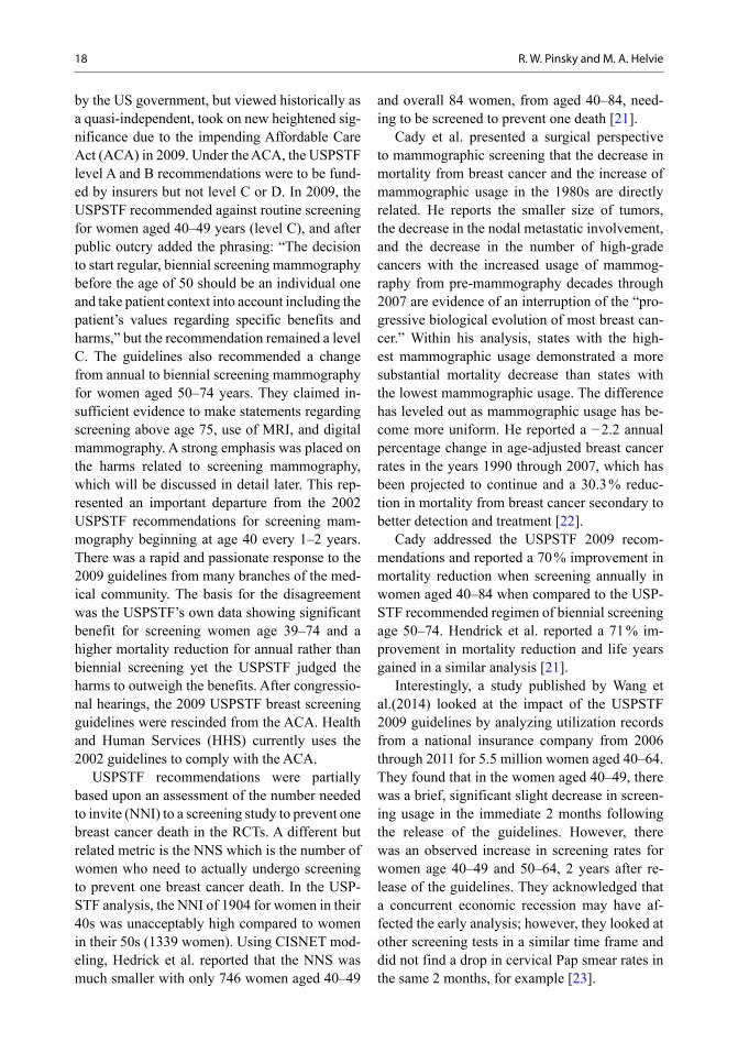
Cady et al. presented a surgical perspective to mammographic screening that the decrease in mortality from breast cancer and the increase of mammographic usage in the 1980s are directly related. He reports the smaller size of tumors, the decrease in the nodal metastatic involvement, and the decrease in the number of high-grade cancers with the increased usage of mammog-raphy from pre-mammography decades through 2007 are evidence of an interruption of the “pro-gressive biological evolution of most breast can-cer.” Within his analysis, states with the high-est mammographic usage demonstrated a more substantial mortality decrease than states with the lowest mammographic usage. The difference has leveled out as mammographic usage has be-come more uniform. He reported a − 2.2 annual percentage change in age-adjusted breast cancer rates in the years 1990 through 2007, which has been projected to continue and a 30.3 % reduc-tion in mortality from breast cancer secondary to better detection and treatment [22].
Cady addressed the USPSTF 2009 recom-mendations and reported a 70 % improvement in mortality reduction when screening annually in women aged 40–84 when compared to the USP-STF recommended regimen of biennial screening age 50–74. Hendrick et al. reported a 71 % im-provement in mortality reduction and life years gained in a similar analysis [21].
Interestingly, a study published by Wang et al.(2014) looked at the impact of the USPSTF 2009 guidelines by analyzing utilization records from a national insurance company from 2006 through 2011 for 5.5 million women aged 40–64. They found that in the women aged 40–49, there was a brief, significant slight decrease in screen-ing usage in the immediate 2 months following the release of the guidelines. However, there was an observed increase in screening rates for women age 40–49 and 50–64, 2 years after re-lease of the guidelines. They acknowledged that a concurrent economic recession may have af-fected the early analysis; however, they looked at other screening tests in a similar time frame and did not find a drop in cervical Pap smear rates in the same 2 months, for example [23].

192 Role of Screening Mammography in Early Detection/Outcome of Breast Cancer
In a registry-based study of trends in breast cancer screening before and after the 2009 guidelines, Sprague et al. [24] looked at 150,000 women in Vermont aged 40 and over for their trends in mammography utilization. Their study demonstrated that after years of increasing screening mammographic utilization, there was a significant decline in screening with the 2009 USPSTF guidelines. The percentage of women aged 40 years and older screened in the past 1 year decreased from 45 % in 2009 to 41 % in 2011 ( P < 0.01) and for those screened in the past 2 years decreased from 59.6 % in 2009 to 54.9 % in 2011 ( P < 0.01). The decline was most promi-nent in the 40–49-year-age group. An observed decline was similarly stated in another study of the effect of the USPSTF guidelines by Sharpe et al., who demonstrated a 4.3 % drop in screening mammography utilization in the US Medicare population in the first year after the recommen-dations were issued. There had previously been a 1 % annual increase in mammographic uti-lization between 2005 and 2009. No change in mammographic utilization for women over the age of 40 after the USPSTF guidelines was re-ported in a different study looking at surveys of 27,829 women over the age of 40 in 2005, 2008, and 2011, before and after the guidelines were published [25].
The controversy surrounding the USPSTF guidelines is further detailed in a commentary by Martin et al. addressing the special circumstances of African-American women who are at greater risk of developing breast cancer in their 40s and at greater risk of dying from breast cancer than other women at all ages. The concern was raised that the advisement against routine screening in a woman’s 40s would be particularly burdensome to African-American women and a revision to the guidelines in this regard was proposed [26].
Mammography Screening of Women in Their 40s
Screening of women in their 40s remains contro-versial. The UK AGE trial was established in 1991 to specifically address the efficacy of screening
mammography in women in their 40s. A total of 60,921 women were randomized with one third of the group receiving an invitation to screening and two thirds receiving usual care. After a mean follow-up period of 10.7 years, there was a 17 % reduction in breast cancer deaths for the group invited to screening. When controlled for non-compliance of those invited to screening, a 24 % reduction in mortality was observed (relative risk = 0.76, 95 % confidence interval 0.51–1.01). The concern for overdiagnosis in this younger age group was shown to be very low by Gun-soy et al. using the AGE trial data demonstrat-ing their main analysis estimate of overdiagnosis was 0.7 % of screen-detected cancers for women in their 40s [27]. Combining the results of the entire group of RCT studies for women in their 40s demonstrated a relative risk for breast cancer mortality for women randomly assigned to mam-mography of 0.85 (95 % confidence interval 0.75 to 0.96), which was statistically significant, cor-responding to a 15 % reduction in breast cancer mortality, favoring screening [10].
Several of the observational trials previously described included women in their 40s and with-in this group each demonstrated improvement in survival (Nickson, Sigurdsson, etc.). A failure analysis of the breast cancer deaths by mam-mographic screening history looked at 10-year cohorts of women over 40 treated at the same Harvard health care system from 1990 through 2007. For women in their 40s, the death rate from breast cancer was reported as 11.4 %. Fifty per-cent of breast cancer deaths occurred in women under age 50. In women between the age of 40 and 49, the study demonstrated that 70.8 % of the breast cancer deaths occurred in the 20 % of women who were unscreened [28].
2012 CISNET models for annual digital screening mammography showed an additional 1.7 lives saved and 51 life years gained per 1000 screened when the age of onset of screening was lowered from 50 to 40 [29].
A study performed by Plecha et al. in 2014 reviewed screened and non-screened women in their 40s to determine whether there was a dif-ference with respect to the recommendations, stage at cancer diagnosis, and identification of

20 R. W. Pinsky and M. A. Helvie
high-risk lesions. They found that screened pa-tients with cancer were significantly more likely to receive a diagnosis at a lower stage (stage I in 49 % of the screened group vs. 23 % of the non-screened group, P = 0.001), to have nega-tive axillary lymph nodes (69 % in the screened group vs. 48 % in the non-screened group of DCIS and invasive cancer patients, P = 0.005) and to have smaller tumors (69 % < 2 cm in the screened group vs. 37 % in the non-screened group, P < 0.001), while non-screened patients were statistically more likely to undergo chemo-therapy (44 % for the screened patients vs. 66 % for the non-screened patients, P = 0.042). Women who had high-risk lesions diagnosed in their 40s had the potential benefit of chemoprevention or screening with MRI [30].
Illness in women in their 40s has been report-ed to have a greater economic impact than dis-ease in older women. For women under age 55, breast cancer resulted in the greatest productivity loss when using models relying on earnings as a measure of productivity and including costs of caregiving and household work when compared to other cancers [31].
Screening Interval
The optimal mammographic screening inter-val has been controversial since the initiation of mammography. At the root of the issue is the value of incremental lives saved versus the cost and perceived harms of additional screen-ing. The early RCT studies had variable screen-ing intervals from 12 to 28 months. Modern era studies typically address annual versus biennial screening, while studies out of the UK compare annual versus triennial screening which is their standard. Computerized simulation models of mammographic screening have been used to as-sess optimal screening intervals. Using CISNET computer modeling, Mandelblatt evaluated the reduction in deaths from breast cancer and life years gained using different screening strategies based on screening interval and age to start and
age to end. This study concluded that biennial screening is most efficient from age 50 through 79. If life years gained is the emphasis, start-ing at age 40 is preferred. It is important to note that “efficiency” represents a balance of benefits and screening harms, and they report that this is contrary to many current practices in the USA. Screening annually age 40–69 was shown to save about 30–35 % more lives than biennial screen-ing (8.3 vs. 6.1 lives/1000 screened) [32]. Cady et al. in reviewing CISNET models demonstrates that yearly screening is more effective than the biennial screening recommended by the USPSTF with a 70 % proportional increase in mortality reduction and in life years gained by screening annually between age 40 and 84 compared with the biennial screening recommendation of the USPSTF between age 50 and 74. Michaelson et al. [33] in a different computerized simulation looked at screening intervals and various aspects of tumor biology, and concluded that screening annually could result in as much as a 51 % reduc-tion in distant metastatic disease compared to a 22 % reduction at a 2-year interval. Currently in the USA, the optimal screening interval remains controversial; however, it is agreed that screen-ing on a regular basis should occur. Many large medical societies including the American Cancer Society, The American College of Obstetrics and Gynecology, American College of Radiology, National Cancer Consortium Network and others advocate annual screening. The NCI, HHS, the FDA, and the AMA recommend screening every 1–2 years, while others including the USPSTF and the American College of Physicians recom-mending biennial screening.
Mammograms not performed can have a sig-nificant effect on patient outcome. Onitilo et al. looked at patient characteristics associated with missed mammograms and the association of missed mammograms with breast cancer stage at diagnosis. They concluded that missing a mam-mogram before breast cancer diagnosis increases the chance of cancer being diagnosed at a later stage [34].
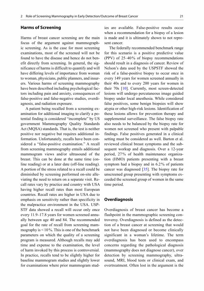
212 Role of Screening Mammography in Early Detection/Outcome of Breast Cancer
Harms of Screening
Harms of breast cancer screening are the main focus of the argument against mammograph-ic screening. As is the case for most screening examinations, most of the screened will not be found to have the disease and hence do not ben-efit directly from screening. In general, the sig-nificance of harms is difficult to quantify and will have differing levels of importance from woman to woman, physicians, public planners, and insur-ers. Various harms of screening mammography have been described including psychological fac-tors including pain and anxiety, consequences of false-positive and false-negative studies, overdi-agnosis, and radiation exposure.
A patient being recalled from a screening ex-amination for additional imaging to clarify a po-tential finding is considered “incomplete” by US government Mammography Quality Standards Act (MQSA) standards. That is, the test is neither positive nor negative but requires additional in-formation. Unfortunately, recalls have been con-sidered a “false-positive examination.” A recall from screening mammography entails additional mammogram views and/or ultrasound of the breast. This can be done at the same time (on-line reading) or at a later date (off-line reading). A portion of the stress related to a recall could be diminished by screening performed on-site alle-viating the need to return on a separate visit. Re-call rates vary by practice and country with USA having higher recall rates than most European countries. Recall rates are higher in USA due to emphasis on sensitivity rather than specificity in the malpractice environment in the USA. USP-STF data showed a recall will occur only once every 11.9–17.8 years for women screened annu-ally between age 40 and 84. The recommended goal for the rate of recall from screening mam-mography is < 10 %. This is one of the benchmark parameters on which the quality of a screening program is measured. Although recalls may add time and expense to the examination, the level of harm invoked by this process is controversial. In practice, recalls tend to be slightly higher for baseline mammogram studies and slightly lower for examinations where prior mammogram stud-
ies are available. False-positive results occur when a recommendation for a biopsy of a lesion is made and it is ultimately shown to not repre-sent cancer.
The federally recommended benchmark range for this scenario is a positive predictive value (PPV) of 25–40 % of biopsy recommendations should result in a diagnosis of cancer. Review of Nelson’s data used by the USPSTF showed the risk of a false-positive biopsy to occur once in every 149 years for women screened annually in their 40s and to every 200 years for women in their 70s [10]. Currently, most screen-detected lesions will undergo percutaneous image guided biopsy under local anesthesia. While considered false positives, some benign biopsies will show atypia or other high-risk lesions. Identification of these lesions allows for prevention therapy and supplemental surveillance. The false biopsy rate also needs to be balanced by the biopsy rate for women not screened who present with palpable findings. False positives generated in a clinical setting must be considered as well. Barton et al. reviewed clinical breast symptoms and the sub-sequent workup and diagnosis. Over a 12-year period, 27 % of health maintenance organiza-tion (HMO) patients presenting with a breast symptom had a biopsy and in 6.2 % of patients cancer was diagnosed [35]. The biopsy rate for unscreened group presenting with symptoms ex-ceeded the screened group of women in the same time period.
Overdiagnosis
Overdiagnosis of breast cancer has become a flashpoint in the mammographic screening con-troversy. Overdiagnosis is defined as the detec-tion of a breast cancer at screening that would not have been diagnosed or become clinically significant in a woman’s lifetime. The term overdiagnosis has been used to encompass concerns regarding the pathological diagnosis (mammography does not diagnose cancer), over detection by screening mammography, ultra-sound, MRI, blood tests or clinical exam, and overtreatment. Often lost in the argument is the

22 R. W. Pinsky and M. A. Helvie
goal of the optimal level of diagnosis. This level of diagnosis would occur when the maximum number of lives saved is matched to the minimal number of diagnoses. Clearly, most would agree that underdiagnosis would occur if only those cancers are diagnosed in metastatic stage where cure is not possible. Prior to mammographic screening, many women who were diagnosed did not die of breast cancer yet the survivors were not considered overdiagnosed. Estimat-ing overdiagnosis is difficult, complex, and subjective. Estimates must consider risk levels of the screened group, lead time, and underly-ing temporal incidence trends. Multiple stud-ies and reviews have addressed this issue with recent estimates of overdiagnosis ranging from 1 to 31 % [36]. Puliti et al. reviewed the Euro-pean literature on mammographic overdiagno-sis in 2012 including 13 primary studies with a characterization of how the papers adjusted for breast cancer risk and lead time, which are is-sues which affect expected incidence and timing of diagnosis, respectively. When categorizing the studies based on those that had appropriate controls for lead time and breast cancer risk, a clear delineation was apparent demonstrating a 1–10 % estimate of overdiagnosis in those with appropriate controls versus higher estimates of overdiagnosis in those without controls [36]. Similar estimates of overdiagnosis were de-scribed in the meta-analysis used in the 2009 USPSTF concluding that the studies were too heterogeneous to combine statistically however most demonstrated an overdiagnosis rate in the range of 1–10 % [10].
Gunsoy et al. (2012) included overdiagno-sis in his analysis of women aged 40–49 in the UK from the AGE trial. They reported that a high proportion of screen-detected DCIS was nonprogressive; however, a majority of those would have presented clinically in the absence of screening. They reported their main analysis estimate of overdiagnosis was 0.7 % of screen-detected cancers. Their finding of a short sojourn time of less than 1 year for women in their 40s for both invasive and progressive DCIS led to an additional recommendation of annual screening for women in this age range [27].
Radiation Risk
The risk of radiation-induced breast cancer from mammographic screening has been addressed in several studies [37–39]. For women within the recommended screening ages of 40 or above, the risk is considered very small. There exists back-ground exposure for all women due to ambient sources of radiation. In the USA, this is typically 3–4 mSv per year. Some areas of the USA receive much higher ambient dose without known impact on breast cancer incidence. The typical dose for a two-view mammogram is 0.4 mSv or stated in a different metric, the average glandular dose of a screening mammogram with two views of each breast is 3 mGy. A seminal 50-year follow-up study of the incidence of breast cancer among atomic bomb survivors showed that radiation ex-posure prior to the age of 40 resulted in higher rates of excess breast cancer. However, no in-creased risk was observed for women older than age 40 at time of exposure at a dose of 1000 mSv [37]. The radio sensitivity of the breast at younger ages, which declines with age, is the cause similar to that seen in young women treated with thorac-ic radiation for Hodgkin lymphoma. In looking at the effect of mammographic screening bienni-ally from age 40 to 74, de Gelder et al. reported that 1302 lives would be saved per 100,000, but radiation-induced effect was predicted to induce 3.7 potential breast cancer deaths [38].
Screening and Breast Density
Mammographic breast density refers to the rela-tive amount of fibrous and glandular tissue rela-tive to fat which projects as white or shades of gray, respectively, on the mammogram. The as-sessment of mammographic breast density is predominantly subjective with the 2013 Breast Imaging Reporting and Data System (BIRADS) defining a four-category ranking system of breast density which approximates quartiles of den-sity. The mammographic density categories are almost entirely fatty, scattered areas of fibro-

232 Role of Screening Mammography in Early Detection/Outcome of Breast Cancer
glandular density, heterogeneously dense and extremely dense [41]. Mammographic breast densities in the heterogeneously dense or ex-tremely dense categories are considered “dense breasts” (Fig. 2.3).
Computerized methods of assessing breast density are now available but are currently in limited clinical use. Mammographic breast den-sity has a dual effect on breast cancer screening. When a mammogram demonstrates dense tissue, which would be considered greater than 50 % dense tissue relative to non-dense tissue, there is a known masking effect. This masking effect makes detection of breast cancer more difficult as a cancer could be hidden by the dense white tissue. Having dense breasts also places a woman at greater risk for breast cancer although this is somewhat controversial. This risk is reported to be up to a relative risk = 2.1 for women with the highest breast density representing approximate-ly 10 % of the screening population of women over age 40 compared to average density [40].
Improvements in Screening
Greater than 90 % of mammographic imag-ing performed today is performed using digi-tal technique. Pisano et al. (2005) in the Digi-tal Mammography Imaging Screening Trial (DMIST) included 42,760 asymptomatic women at 33 breast imaging sites demonstrating that screening with digital was more accurate than film screen mammography in women under the age of 50, women with dense breasts and pre- or perimenopausal women [41].
The standardization of breast imaging report-ing has been an effective way to improve com-munication of imaging results. The BIRADS (2013) includes standards for breast density re-porting, description of imaging findings, and im-aging conclusions and recommendations.
Fig. 2.3 Mammographic examples of Breast Imaging Reporting and Data System (BIRADS) breast density cat-egories from left to right: almost entirely fatty, scattered
areas of fibroglandular density, heterogeneously dense, and extremely dense

24 R. W. Pinsky and M. A. Helvie
Screening and DCIS
Increasing usage of mammography for breast cancer screening has resulted in a significant in-crease in the number of DCIS cases diagnosed [42]. A fivefold increase in the rate of DCIS has been reported in the USA in the past 25 years [43]. In a review of Surveillance and Epide-miological End Results Study (SEER) data, the incidence of DCIS increased 7.2-fold (95 % confidence interval 6.8–7.7) from 1980 to 2001, most pronounced for women ≥ 50 years of age [44]. All sources relate this dramatic increase in diagnosis of DCIS to the increase in screening utilization for breast cancer detection. Prior to mammographic screening, DCIS was diagnosed as an incidental finding or when it was palpable, which is rare. The mammographic imaging char-acteristics of DCIS are most commonly calcifica-tions and far less commonly a mass, distortion, or focal asymmetry. The fact that DCIS is rarely palpable results in an ideal situation for preclini-cal diagnosis with screening mammography.
It has been suggested that much of screen-detected DCIS is overdiagnosed [45]. DCIS is considered by many to be a precursor of invasive cancer although a non-obligatory one [42]. It has been proposed that a significant portion of the contribution of mammography to the reduction in breast cancer mortality is the detection and sub-sequent treatment of DCIS which has removed precursor lesions [46]. Prognosis for women di-agnosed with DCIS is excellent with a known but low risk of recurrence as DCIS or invasive cancer of approximately 1 % per year. In a study evaluating women who chose breast conserva-tion therapy with radiation treatment for DCIS, surveillance mammography detected 97 % of the recurrences. The recurrences were less than 1 cm in size in 91 % and all were of low stage, zero or one. Following mastectomy, the recurrence rate after DCIS is 1–4 % and is typically invasive, presenting with a palpable chest wall mass [47]. The challenge ahead is to determine prospec-tively which cases of DCIS will lead to invasive cancer requiring aggressive treatment and which can be managed with less intervention.
References
1. Obuchowski NA, Graham RJ, Baker ME, Powell KA. Ten criteria for effective screening: their application to multislice CT screening for pulmo-nary and colorectal cancers. Am J Roentgenol. 2001;176(6):1357–62.
2. ACS. American Cancer Society. 2014 [01/29/2014]. http://www.cancer.org/. Accessed 22 Sep 2014.
3. NCI. Breast Cancer Surveillance Consortium (BCSC). 2014 [cited 02/07/2014]. http://breastscreening.cancer.gov/. Accessed 30 June 2014.
4. Smith RA, D’Orsi C, Newell MS. Screening for breast cancer. In: Harris JR, Lippman ME, Morrow M, Osborne CK, editors. Diseases of the breast, sec-tion III, breast imaging and image-guided biopsy techniques. 4th ed. Philadelphia: Lippincott Williams & Wilkins; 2010. pp. 87–115.
5. Cox B, Sneyd MJ. Bias in breast cancer research in the screening era. Breast. 2013;22(6):1041–5.
6. Harris JR, Lippman ME, Morrow M, Osborne CK. Diseases of the Breast. Wolters Kluwer/Lippincott, Williams & Wilkins. 2010; Ch. 11, p. 89–94.
7. Tabar L, Faberberg G, Day NE, Holmberg L. What is the optimum interval between mammographic screening examinations? An analysis based on the latest results of the Swedish two-county breast cancer screening trial. Br J Cancer. 1987;55(5):547–51.
8. Smith RA, Duffy SW, Gabe R, Tabar L, Yen AM, Chen TH. The randomized trials of breast cancer screening: what have we learned? Radiol Clin North Am. 2004;42(5):793–806, v.
9. Demissie K, Mills OF, Rhoads GG. Empirical com-parison of the results of randomized controlled trials and case-control studies in evaluating the effective-ness of screening mammography. J Clin Epidemiol. 1998;51(2):81–91.
10. Nelson HD, Tyne K, Naik A, Bougatsos C, Chan BK, Humphrey L, et al. Screening for breast cancer: an update for the U.S. Preventive Services Task Force. Ann Intern Med. 2009;151(10):727–37, W237–42.
11. Olsen O, Gotzsche PC. Cochrane review on screen-ing for breast cancer with mammography. Lancet. 2001;358(9290):1340–2.
12. Miller AB, Wall C, Baines CJ, Sun P, To T, Narod SA Twenty five year follow-up for breast cancer inci-dence and mortality of the Canadian national breast screening study: randomised screening trial. BMJ. 2014;348:g366.
13. Nickson C, Mason K, English DR, Kavanagh AM. Mammographic screening and breast cancer mortal-ity: a case-control study and meta-analysis. Cancer Epidemiol Biomarkers Prev. 2012;21(9):1479–88.
14. Paci E, Group EW. Summary of the evidence of breast cancer service screening outcomes in Europe and first estimate of the benefit and harm balance sheet. J Med Screen. 2012;19(Suppl 1):5–13.
252 Role of Screening Mammography in Early Detection/Outcome of Breast Cancer
15. Sigurdsson K, Olafsdottir EJ. Population-based service mammography screening: the Icelan-dic experience. Breast Cancer (Dove Med Press). 2013;5:17–25.
16. Paap E, Verbeek AL, Botterweck AA, van Doorne-Nagtegaal HJ, Imhof-Tas M, de Koning HJ, et al. Breast cancer screening halves the risk of breast cancer death: a case-referent study. Breast. 2014;23(4):439–44.
17. Foca F, Mancini S, Bucchi L, Puliti D, Zappa M, Naldoni C, et al. Decreasing incidence of late-stage breast cancer after the introduction of orga-nized mammography screening in Italy. Cancer. 2013;119(11):2022–8.
18. Helvie MA, Chang JT, Hendrick RE, Banerjee M Reduction in late-stage breast cancer incidence in the mammography era: implications for overdiagnosis of invasive cancer. Cancer. 2014;120:2649–56.
19. Berry DA, Cronin KA, Plevritis SK, Fryback DG, Clarke L, Zelen M, et al. Effect of screening and adjuvant therapy on mortality from breast cancer. N Engl J Med. 2005;353(17):1784–92.
20. Vervoort MM, Draisma G, Fracheboud J, van de Poll-Franse LV, de Koning HJ. Trends in the usage of adjuvant systemic therapy for breast cancer in the Netherlands and its effect on mortality. Br J Cancer. 2004;91(2):242–7.
21. Hendrick RE, Helvie MA. United States preventive services task force screening mammography rec-ommendations: science ignored. Am J Roentgenol. 2011;196(2):W112–6.
22. Cady B, Michaelson JS, Chung MA. The “tipping point” for breast cancer mortality decline has resulted from size reductions due to mammographic screen-ing. Ann Surg Oncol. 2011;18(4):903–6.
23. Wang AT, Fan J, Van Houten HK, Tilburt JC, Stout NK, Montori VM, et al. Impact of the 2009 US pre-ventive services task force guidelines on screening mammography rates on women in their 40s. PloS ONE. 2014;9(3):e91399.
24. Sprague BL, Bolton KC, Mace JL, Herschorn SD, James TA, Vacek PM, et al. Registry-based study of trends in breast cancer screening mammogra-phy before and after the 2009 U.S. preventive ser-vices task force recommendations. Radiology. 2014;270(2):354–61.
25. Pace LE, He Y, Keating NL. Trends in mammog-raphy screening rates after publication of the 2009 US preventive services task force recommendations. Cancer. 2013;119(14):2518–23.
26. Martin N, Wingfield J. USPSTF screening recom-mendations for breast cancer: the potential impact on the African American community. J Health Care Poor Underserved. 2012;23(2 Suppl):91–7.
27. Gunsoy NB, Garcia-Closas M, Moss SM. Modelling the overdiagnosis of breast cancer due to mammogra-phy screening in women aged 40 to 49 in the United Kingdom. Breast Cancer Res. 2012;14(6):R152.
28. Webb ML, Cady B, Michaelson JS, Bush DM, Calvillo KZ, Kopans DB, et al. A failure analysis
of invasive breast cancer: Most deaths from disease occur in women not regularly screened. Cancer. 2014; Sept 15;120(18):2839–2846..
29. van Ravesteyn NT,M, Buist DS, et al. Tipping the balance of benefits and harms to favor screening mammography starting at age 40 years: a com-parative modeling study of risk. Ann Intern Med. 2012;156(9):609–17.
30. Plecha D, Salem N, Kremer M, Pham R, Downs-Holmes C, Sattar A, et al. Neglecting to screen women between 40 and 49 years old with mammog-raphy: what is the impact on treatment morbidity and potential risk reduction? AJR Am J Roentgenol. 2014;202(2):282–8.
31. Bradley CJ, Neumark D, Shickle LM, Farrell N. Dif-ferences in breast cancer diagnosis and treatment: experiences of insured and uninsured women in a safety-net setting. Inquiry. 2008;45(3):323–39.
32. Mandelblatt JS, Cronin KA, Bailey S, Berry DA, de Koning HJ, Draisma G, et al. Effects of mammog-raphy screening under different screening schedules: model estimates of potential benefits and harms. Ann Intern Med. 2009;151(10):738–47.
33. Michaelson JS, Halpern E, Kopans DB. Breast cancer: computer simulation method for estimat-ing optimal intervals for screening. Radiology. 1999;212(2):551–60.
34. Onitilo AA, Engel JM, Liang H, Stankowski RV, Miskowiak DA, Broton M, et al. Mammogra-phy utilization: patient characteristics and breast cancer stage at diagnosis. AJR Am J Roentgenol. 2013;201(5):1057–63.
35. Barton MB, Elmore JG, Fletcher SW. Breast symp-toms among women enrolled in a health maintenance organization: frequency, evaluation, and outcome. Ann Intern Med. 1999;130(8):651–7.
36. Puliti D, Duffy SW, Miccinesi G, de Koning H, Lynge E, Zappa M, et al. Overdiagnosis in mammographic screening for breast cancer in Europe: a literature review. J Med Screen. 2012;19(Suppl 1):42–56.
37. Land CE, Tokunaga M, Koyama K, Soda M, Pres-ton DL, Nishimori I, et al. Incidence of female breast cancer among atomic bomb survivors, Hiroshima and Nagasaki, 1950–1990. Radiat Res. 2003;160(6):707–17.
38. de Gelder R, Draisma G, Heijnsdijk EA, de Koning HJ. Population-based mammography screen-ing below age 50: balancing radiation-induced vs prevented breast cancer deaths. Br J Cancer. 2011;104(7):1214–20.
39. Mainiero MB, Lourenco A, Mahoney MC, Newell MS, Bailey L, Barke LD, et al. ACR appropriateness criteria breast cancer screening. J Am Coll Radiol. 2013;10(1):11–4.
40. Sickles EA. The use of breast imaging to screen women at high risk for cancer. Radiol Clin North Am. 2010;48(5):859–78.
41. Pisano ED, Gatsonis C, Hendrick E, Yaffe M, Baum JK, Acharyya S, et al. Diagnostic performance of digital versus film mammography for breast-cancer screening. N Engl J Med. 2005;353(17):1773–83.

26 R. W. Pinsky and M. A. Helvie
42. Lynge E, Ponti A, James T, Majek O, von Euler-Chelpin M, Anttila A, et al. Variation in detection of ductal carcinoma in situ during screening mam-mography: a survey within the international cancer screening network. Eur J Cancer. 2014;50(1):185–92.
43. Virnig BA, Tuttle TM, Shamliyan T, Kane RL. Ductal carcinoma in situ of the breast: a systematic review of incidence, treatment, and outcomes. J Natl Cancer Inst. 2010;102(3):170–8.
44. Li CI, Daling JR, Malone KE. Age-specific incidence rates of in situ breast carcinomas by histologic type, 1980 to 2001. Cancer Epidemiol Biomarkers Prev. 2005;14(4):1008–11.
45. Wells CJ, O’Donoghue C, Ojeda-Fournier H, Retal-lack HE, Esserman LJ. Evolving paradigm for imag-ing, diagnosis, and management of DCIS. J Am Coll Radiol JACR. 2013;10(12):918–23.
46. Cady B, Chung MA. Preventing invasive breast cancer: another benefit from mammographic screen-ing. Cancer. 2011;117(14):3064–8.
47. Pinsky RW, Rebner M, Pierce LJ, Ben-David MA, Vicini F, Hunt KA, et al. Recurrent cancer after breast-conserving surgery with radiation therapy for ductal carcinoma in situ: mammographic features, method of detection, and stage of recurrence. Am J Roentgenol. 2007;189(1):140–4.
48. Tabár L, Vitak B, Hsiu-Hsi Chen T, Yen A, Cohen A, Tot T, Chiu S, Chen S, Fann J, Rosell J, Fohlin H, Smith R, Duffy S. Swedish two-county trial: impact of mammographic screening on breast cancer mortal-ity during 3 decades. Radiology 2011 260:3, 658–663.

27
Imaging DCIS: Digital/Film-Screening Mammography, Tomosynthesis, MRI, Ultrasonography
Annette Ingram Joe and Stephanie K. Patterson
L. A. Newman, J. M. Bensenhaver (eds.), Ductal Carcinoma In Situ and Microinvasive/Borderline Breast Cancer, DOI 10.1007/978-1-4939-2035-8_3, © Springer Science+Business Media New York 2015
A. I. Joe ()Department of Radiology, University of Michigan Health System, Taubman Center, 1500 East Medical Center Drive, Ann Arbor, MI 48109, USAe-mail: [email protected]
S. K. PattersonDepartment of Radiology, University of Michigan Health System, Med Inn Building, 1500 East Medical Center Drive, Ann Arbor, MI 48109, USAe-mail: [email protected]
Mammography
Mammography, the primary breast-cancer-screening tool, is the most important method of detecting ductal carcinoma in situ (DCIS). Be-fore screening mammography, DCIS comprised only 5 % of breast cancers; however, it now ac-counts for 25 % of breast cancers diagnosed in the USA. The sensitivity of mammography for detecting DCIS is between 87 and 95 % [1, 2]. The DCIS lesions that are mammographically occult are usually of low grade. The higher-grade DCIS will more likely be mammographically ap-parent [1].
Although DCIS can present clinically with a palpable mass, nipple discharge, or skin changes, it is usually detected mammographically. Micro-calcifications are the most common mammo-graphic finding, although DCIS may manifest as a mass or architectural distortion. Ninety percent of DCIS lesions are associated with calcifica-tions, and 80 % manifest as calcifications alone with no other mammographic finding [3, 4]. The
calcifications in DCIS are the result of necrosis of the cells or calcified secretions.
The morphology and distribution of calcifi-cations seen on mammograms are evaluated to determine their suspicion. Magnification views are indicated to evaluate any detected calcifica-tions that are not typically benign. Magnifica-tion allows better visualization of calcifications because of the increase in contrast to noise ratio and improved resolution. Electronically zoomed images should not be used as an alternative to geometrically magnified views in the evaluation of microcalcifications [5, 6]. The magnified im-ages should be performed in the craniocaudal and lateral projections. Benign milk of calcium will have a typical layered appearance and can be dif-ferentiated from more suspicious calcifications. Calcifications that are clearly vascular, dermal, or milk of calcium are benign and do not require further imaging. Larger round calcifications, scattered punctate or amorphous calcifications, rim calcifications which are often seen in fat necrosis, popcorn calcifications associated with degenerating fibroadenomas, and large rod-like secretory calcifications associated with ductal ec-tasia are also typically benign (Figs. 3.1 and 3.2). When benign calcifications, such as milk of cal-cium, are detected, care should be taken that no suspicious calcifications in the area are over-looked. Calcifications in DCIS can be found in close proximity to typically benign calcifications.
Tiny round punctate calcifications (< 0.5 mm) in a group (five or more calcifications per cubic centimeter) can be considered probably benign if
3

28 A. I. Joe and S. K. Patterson
no prior mammograms are available to confirm stability and if there is no know ipsilateral breast cancer. If punctate calcifications are in a linear or segmental distribution, they are suspicious and biopsy is indicated [7].
The morphology of the calcifications related to DCIS is usually fine linear or fine linear branch-ing, fine pleomorphic, amorphous, or coarse het-erogeneous. Fine linear and fine linear branching calcifications have the highest risk of malignancy with a positive predictive value (PPV) of 70 % (Fig. 3.3). The PPV of fine pleomorphic, amor-phous, and coarse heterogeneous is 29, 21, and 13 %, respectively (Table 3.1) [8–12]. The fine
linear and fine linear branching calcifications seen in DCIS are usually more irregular, thinner, and have a more discontinuous pattern than the benign, large, rod-like calcifications. Unlike the suspicious linear morphology, secretory calcifi-cations may have lucent centers if the calcifica-tion is in the wall of a duct.
The distribution of calcifications in a segmen-tal or linear distribution has the highest risk of malignancy, followed by grouped and regional (Table 3.2) [9, 11, 12]. Calcifications located in a line suggest malignant deposits in a duct and cal-cifications in a segmental distribution suggest de-posits in ducts and their branches. Calcifications associated with DCIS can involve a large portion of the breast. However, diffuse, randomly distrib-uted, punctate, and amorphous calcifications are usually benign and are usually bilateral.
Most mammographic calcifications are be-nign; however, those which are malignant are usually associated with DCIS. Stomper and colleagues reviewed malignant mammographic calcifications without an associated parenchymal lesion and found pure DCIS in 65 %, DCIS with a focus of invasion in 32 %, and invasive carci-noma alone in 4 % of the cases [13].
When DCIS presents as a parenchymal le-sion, with or without associated calcifications, it can have the appearance of invasive cancer. The
Fig. 3.3 High-nuclear-grade DCIS with comedonecrosis and microcalcifications in a 61-year-old woman presented with a palpable invasive lobular carcinoma in the upper outer quadrant. Fine linear branching calcifications in a segmental distribution are located in the lower inner quad-rant, remote from the palpable finding
Fig. 3.2 Milk of calcium is associated with fibrocystic changes. a Cranial caudal view demonstrates amorphous-appearing calcifications. b The calcifications have a cur-vilinear appearance on the lateral view because they layer in the dependent portion of the microcysts
Fig. 3.1 Secretory calcifications present as large rod-like calcifications and are associated with ductal ectasia

293 Imaging DCIS: Digital/Film-Screening Mammography, Tomosynthesis, MRI, Ultrasonography
atypical mammographic features seen in 20 % of DCIS lesions include circumscribed masses, spiculated and irregular masses, focal asymme-tries, and architectural distortion. The masses may be single or multiple.
If suspicious calcifications are associated with an asymmetry or mass, there is a greater prob-ability that the calcifications are related to inva-sive carcinoma or DCIS with invasive carcinoma rather than DCIS alone (Fig. 3.4). Farshid et al.’s study demonstrated that when DCIS formed pa-renchymal lesions without radiographically vis-ible calcifications, it was more frequently low grade and with calcifications more frequently high grade. Periductal fibrosis and chronic in-flammation resulted in the parenchymal finding. The majority of the discrete masses had a papil-lary component (Fig. 3.5) [4].
Many studies have attempted to correlate the pathologic findings or grade with the mammo-graphic findings of DCIS; however, a significant overlap has been found. The presence or absence of necrosis is the pathologic feature that has the most correlation with the mammographic ap-pearance of DCIS. Calcifications associated with necrosis are more likely to be in a segmental or linear distribution than calcifications in DCIS without necrosis [14]. Barreau et al. found a re-lationship between the extent of the calcification seen mammographically and the grade of DCIS, with grade 3 being more extensive.
The Digital Mammographic Imaging Screen-ing Trial (DMIST) proved digital mammography is as efficacious as film screen mammography [15]. Digital mammography has been shown to be of better and more consistent quality compared
Table 3.1 Likelihood of malignancy as a function of BI-RADS® descriptors of calcification morphologya [8]. (2013 BI-RADS® Mammography section, Table 2. Reprinted with permission of the American College of Radiology (ACR). No other representation of this material is authorized without expressed, written permission from the ACR. Refer to the ACR website at www.acr.org/Quality-Safety/Resources/BIRADS for the most current and complete version of the BI-RADS® Atlas)Morphology descriptor
Liberman et al. [9]
Berg et al. [10] Burnside et al. [11]
Bent et al. [12] Total
Amorphous 9/35 (26) 30/150 (20) 4/30 (13) 10/51 (20) 53/266 (21)Coarse heterogeneous
N/Ab N/Sc 1/14 (7) 2/10 (20) 3/24 (13)
Fine pleomorphic N/Ab N/Sc 10/34 (29) 14/50 (28) 24/84 (29)Fine linear or fine linear branching
26/32 (81) N/Sc 10/19 (53) 16/23 (70) 52/74 (70)
N/A not applicable, N/S not specifieda Data are presented as cancer cases/all cases biopsied, with percentage of cancer cases in parenthesesb This study, published in 1998, reported 98 cancers among 241 cases of pleomorphic calcifications (41 %). The fourth edition of BI-RADS® (published later, in 2003) subdivided the pleomorphic descriptor into course heterogeneous and fine pleomorphic descriptorsc This study involved only amorphous calcifications
Table 3.2 Likelihood of malignancy as a function of BI-RADS® descriptors of calcification distributiona [8]. (2013 BI-RADS® Mammography Section, Table 3. Reprinted with permission of the American College of Radiology (ACR). No other representation of this material is authorized without expressed, written permission from the ACR. Refer to the ACR website at www.acr.org/Quality-Safety/Resources/BIRADS for the most current and complete version of the BI-RADS® Atlas)Distribution descriptor Liberman et al. [9] Burnside et al. [11] Bent et al. [12] TotalDiffuse 0/1 (0) 0/1 (0) 0/0 (0) 0/2 (0)Regional 6/13 (46) 0/1 (0) 0/9 (0) 6/23 (26)Grouped 93/254 (37) 14/76 (18) 19/81 (23) 126/411 (31)Linear 13/19 (68) 8/11 (73) 14/28 (50) 35/58 (60)Segmental 17/23 (74) 3/8 (38) 9/16 (56) 29/47 (62)a Data are presented as cancer cases/all cases biopsied, with percentage of cancer cases in parentheses
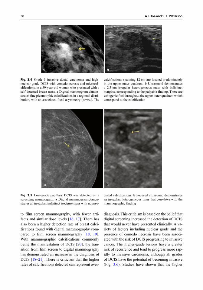
30 A. I. Joe and S. K. PattersonA. I. Joe and S. K. Patterson
to film screen mammography, with fewer arti-facts and similar dose levels [16, 17]. There has also been a higher detection rate of breast calci-fications found with digital mammography com-pared to film screen mammography [18, 19]. With mammographic calcifications commonly being the manifestation of DCIS [20], the tran-sition from film screen to digital mammography has demonstrated an increase in the diagnosis of DCIS [18–21]. There is criticism that the higher rates of calcifications detected can represent over-
diagnosis. This criticism is based on the belief that digital screening increased the detection of DCIS that would never have presented clinically. A va-riety of factors including nuclear grade and the presence of comedo necrosis have been associ-ated with the risk of DCIS progressing to invasive cancer. The higher-grade lesions have a greater risk of recurrence and tend to progress more rap-idly to invasive carcinoma, although all grades of DCIS have the potential of becoming invasive (Fig. 3.6). Studies have shown that the higher
Fig. 3.5 Low-grade papillary DCIS was detected on a screening mammogram. a Digital mammogram demon-strates an irregular, indistinct isodense mass with no asso-
ciated calcifications. b Focused ultrasound demonstrates an irregular, heterogeneous mass that correlates with the mammographic finding
Fig. 3.4 Grade 3 invasive ductal carcinoma and high-nuclear-grade DCIS with comedonecrosis and microcal-cifications, in a 39-year-old woman who presented with a self-detected breast mass. a Digital mammogram demon-strates fine pleomorphic calcifications in a regional distri-bution, with an associated focal asymmetry ( arrow). The
calcifications spanning 12 cm are located predominately in the upper outer quadrant. b Ultrasound demonstrates a 2.5-cm irregular heterogeneous mass with indistinct margins, corresponding to the palpable finding. There are echogenic foci throughout the upper outer quadrant which correspond to the calcification

313 Imaging DCIS: Digital/Film-Screening Mammography, Tomosynthesis, MRI, Ultrasonography
DCIS detection rates are related to higher detec-tion rates of intermediate and high-grade DCIS lesions, not low grade. In the study by Weigel and colleagues, of the 1074 screen detected cancers, 17.2 % were low grade, 37.3 % were intermediate grade, and 40.3 % were high grade [22].
All suspicious calcifications on the mam-mogram should be identified because the extent
of the visible DCIS can affect whether breast conservation therapy (BCT) or mastectomy is recommended. It is crucial to obtain prior mam-mograms for comparison whenever possible, to avoid recommending removal of stable, benign calcifications. Many calcifications that would be considered suspicious if new would have a be-nign assessment if stability could be confirmed.
Fig. 3.6 Invasive ductal carcinoma, modified Bloom-Richardson grade 3, with high-nuclear-grade DCIS in a patient who did not follow up with the recommended biopsy. Calcifications are present in the invasive carci-noma and the DCIS. a 15 mm group of new pleomorphic calcifications were detected on screening mammogram. The patient did not come for the recommended biopsy. Fourteen months later, the patient presented with a pal-pable mass in the region of the previously demonstrated
calcifications. b Mammogram demonstrates extensive new pleomorphic calcifications in a regional distribution spanning 8 cm. c US demonstrates a 7-mm round, hy-poechoic mass with microlobulated margins. The mass contains echogenic foci consistent with calcifications. d Axillary US reveals an abnormal lymph node with compression of the fatty hilum. A US-guided fine-needle aspiration biopsy of the lymph node was positive for ad-enocarcinoma

32 A. I. Joe and S. K. Patterson
When DCIS is related to Paget’s disease, the mammogram may demonstrate subareolar or dif-fuse calcifications, a mass or masses, as well as skin thickening or nipple retraction. If nipple or subareolar findings are present on a mammo-gram, the nipple should be inspected clinically for signs of the disease. When clinical findings raise the possibility of Paget’s disease, a mam-mogram should be obtained to look for an un-derlying malignancy, although the mammogram may be normal [23].
Digital Breast Tomosynthesis
Digital breast tomosynthesis (DBT) is a tech-nique that uses conventional X-rays and a digital detector to obtain multiple low-dose mammo-graphic exposures. As the X-ray tube moves in an arc over the breast, the images are obtained and then reconstructed to form thin cross-sectional images. A 1-mm slice of tissue is usually in focus with the tissue above and below the slice out of focus. This technique can eliminate the overlap-ping tissue that can obscure cancers and that can cause unnecessary recalls [24]. The FDA requires DBT to be combined with digital mammography (DM) for clinical use. Adding DM makes it pos-sible to evaluate any asymmetry of the breasts and to compare prior mammograms.
Rafferty et al. showed an increase in sensitivi-ty for invasive cancer and in situ cancers with the addition of DBT to DM; however, the increase was much smaller for in situ cancers. The ma-jority of DCIS present as calcifications only, and no significant benefit was achieved by adding tomosynthesis for calcification-only lesions [25]. This is consistent with Skaane et al.’s study re-sults that showed no benefit in detecting DCIS by adding DBT [26]. Spangler et al. compared de-tection and characterization of calcifications with DM and DBT. DM seemed to be slightly more sensitive for detecting calcifications than DBT; however, the difference was not significant. The detection of malignant and benign calcifications was slightly better with DM [27].
Studies have shown that DPT can decrease false-positive call back rates by minimizing the
effect of overlapping tissues [26, 28, 29]. When two-view DBT was added to DM, a reduction in the false-positive recall rate was achieved with-out a negative impact on cancer recall rate [25]. The decrease in the recall rate with DBT demon-strated by Haas et al. varied by the type of lesion, and there was no change in the recall rate of cal-cifications and stellate lesions [30].
The distribution of findings, especially cal-cifications, is also better seen on DM than DBT according to Rafferty et al. [25]. Anderson et al. reported that the distribution of calcifications was as well seen on DBT as DM; however, the morphologic details of the calcifications were not as well visualized (Fig. 3.7) [31]. Since DCIS usually presents as calcifications on mammogra-phy, visualizing the morphology and distribution of the calcifications is important for its detection.
Ultrasound
Ultrasound (US) plays a major role in breast cancer detection. For many years, it has been used to characterize masses and asymmetries detected on mammography and to evaluate clinical areas of concern. “Second-look” US is performed after an abnormality is detected by magnetic resonance (MR) imaging to determine if the lesion can be targeted for US-guided bi-opsy. The use of US in breast cancer screen-ing has increased since it has been shown to detect some cancers that are missed by screen-ing mammography. In the American College of
Fig. 3.7 a High–nuclear-grade DCIS with comedonecro-sis and microcalcifications presented as fine pleomorphic calcifications in a segmental distribution demonstrated on digital mammogram. b The calcifications are also dem-onstrated on digital breast tomosynthesis; however, the morphology of the calcifications is not as well visualized

333 Imaging DCIS: Digital/Film-Screening Mammography, Tomosynthesis, MRI, Ultrasonography
Radiology Imaging Network (ACRIN) 6666 trial, 2637 high-risk women with dense breasts were screened with mammography and ultra-sound. The mammographic sensitivity was 50 % and the sensitivity of mammography plus ultra-sound was 77.5 %. The diagnosis of DCIS in-creased significantly with the boost in screening mammography; however, screening US is not as beneficial in detecting DCIS. In the ACRIN trial, six women were diagnosed with DCIS and only one was detected by US [32].
The ultrasound findings of DCIS are nonspe-cific and often subtle. Although most DCIS is detected by calcifications on mammography, the most common US finding is a mass (Fig. 3.8). Sixty-four DCIS mass lesions diagnosed by US-guided core biopsy were reviewed by Wang et al. The ultrasounds demonstrated a variety of fea-tures, most commonly a hypoechoic, irregular, or oval mass with noncircumscribed margins, paral-lel orientation, and no posterior acoustic change. Hypervascularity was demonstrated in 69 % of these masses [33]. A study by Lee et al. showed that micropapillary DCIS, a subtype that is asso-ciated with more extensive disease at diagnosis, had similar US features except hypervascularity was only seen in 39 % of the lesions.
Although US is less sensitive than mammog-raphy for the detection of calcifications, malig-nant calcifications are more often visualized by US than those located in benign tissue [33].
Calcifications may be demonstrated by US as echogenic foci within a mass or duct; however, the echogenic foci may be apparent without any other visible abnormality. Microcalcifications with associated ductal changes were reported by Park et al. to be the most common US finding in high-grade DCIS [34]. The morphology of calci-fications cannot be easily assessed by ultrasound. However, calcifications in DCIS do not typically cause posterior acoustic shadowing, which is often seen with benign dystrophic calcifications and degenerating fibroadenomas. The capability to detect calcifications and characterize masses has become better with the improvement in US technology and the use of higher frequency transducers. Good spatial resolution and good contrast make it possible to see calcifications
by US. Therefore, harmonic imaging and spatial compounding may make calcifications more vis-ible [35].
When malignant calcifications without an as-sociated mass or asymmetry are detected at mam-mography, they are most likely related to DCIS; however, an invasive carcinoma may be present. A mass associated with an invasive component may be apparent on US but obscured by dense fibroglandular tissue on the mammogram. Since the management may be different for DCIS alone versus invasive carcinoma with or without DCIS, it is important to target any finding that is sugges-tive of invasion at the time of the initial biopsy.
MRI
Dynamic contrast-enhanced magnetic resonance imaging (MRI) is the most sensitive method for detecting DCIS. In the past, breast MRI was felt to have a low sensitivity and specificity for DCIS [36]; however, with the use of high spatial resolu-tion techniques, the sensitivity of MR for DCIS is 92 %. This is significantly higher than the 56 % sensitivity of mammography [37, 38]. Although some calcified DCIS lesions may be occult on MRI, MRI is more sensitive than mammography
Fig. 3.8 Low-nuclear-grade DCIS in a 45-year-old woman who detected a palpable breast mass. a Digital mammo-gram demonstrates extremely dense breast tissue with no focal abnormalities. b Ultra-sound of the palpable finding demonstrates a 1-cm hypoechoic irregular mass with ductal extension

34 A. I. Joe and S. K. Patterson
in detecting all grades of DCIS. The sensitiv-ity is even greater for detecting high-grade and intermediate-grade DCIS than low-grade DCIS [38, 39].
In addition to the overall higher sensitivity, the advantages of MRI include the higher sen-sitivity in dense breasts, better assessment of multicentricity, and better estimation of the size of the DCIS [38–40]. Since the extent of disease is more accurately assessed by MRI than mam-mography, MRI may be helpful in presurgical planning. However, no reduction in the reexci-sion rate with preoperative MRI was shown in the Comparative Effectiveness of MR Imaging in Breast Cancer (COMICE) trial [41].
The main disadvantage of MRI is the high false-positive rate which can lead to unnecessary additional work-up, delay of definitive treatment of known cancer, and possibly more extensive treatment than is necessary. Another disadvan-tage is that intravenous contrast is needed in
order to detect DCIS. DCIS is not usually visible on noncontrast MR images because it is masked by the normal breast tissue [42].
The most common morphologic appear-ance of DCIS on MRI is nonmass enhancement which has been reported in 60–80 % of the cases (Fig. 3.9). DCIS may also appear as an enhancing mass (14–41 %) or as a focus (1–12 %) [43–45]. No significant relationship has been shown be-tween morphologic features and nuclear grade [42].
Nonmass enhancement DCIS can have sev-eral different internal enhancement patterns, the most common is a clumped pattern (41–64 %). Other possible internal enhancement patterns are heterogeneous, clustered ring enhancement, ho-mogeneous, and stippled [3, 44, 46]. The most common distribution of nonmass enhancement DCIS is segmental (14–77 %). Other possible patterns of distribution include linear, focal, re-gional, and diffuse (Fig. 3.10) [43, 45].
When DCIS is demonstrated as a mass on MRI, the shape is usually irregular and the margins ir-regular or spiculated, circumscribed margins are uncommon. The most common enhancement pat-tern is heterogeneous followed by homogeneous.
Fig. 3.10 Intermediate-nuclear-grade ductal DCIS in a 60-year-old woman with a history of a previous lumpec-tomy for high-nuclear-grade ductal DCIS. Axial post-contrast fat-sat T-1-weighted MR image demonstrates ho-mogeneous nonmass enhancement in a linear distribution ( arrow)
Fig. 3.9 High-nuclear-grade DCIS with comedonecrosis and microcalcifications detected in a 62-year-old woman with high risk for malignancy. Axial post-contrast fat-sat T-1-weighted MR image demonstrates clumped, nonmass enhancement in a segmental distribution. The digital mammogram was negative

353 Imaging DCIS: Digital/Film-Screening Mammography, Tomosynthesis, MRI, Ultrasonography
Although the rim enhancement pattern is often seen in invasive breast carcinoma, it is the least common pattern in DCIS [43, 45].
A focus, or a small < 5 mm region of enhance-ment, is the least common morphology of DCIS on MRI. Although an uncommon presentation, Rosen et al. reported that 62.5 % of the DCIS that manifested as a focus were high grade, 37.5 % were intermediate grade, and 0 % were low grade (Fig. 3.11) [44].
DCIS lesions can have various kinetic patterns on MRI. The kinetic curve shows the enhance-
ment characteristics over time following the ad-ministration of intravenous contrast material. The curve has two phases, the initial phase and the delayed phase. The initial phase is described as rapid, medium, or slow and refers to the rate of contrast uptake by an enhancing lesion. The de-layed phase is described as persistent, plateau, or washout (Fig. 3.12) [47]. The most common ini-tial phase in DCIS is rapid uptake, which has been reported in 49–68 % of cases. The most common delayed phase reported in DCIS is a plateau curve (20–52 %) followed by washout (28–44 %) and
Fig. 3.11 High-nu-clear-grade DCIS in a high-risk patient. a Axial post-contrast fat-sat T-1-weighted MR image demon-strates a new focus of enhancement in lateral breast. b Comparison from 1 year prior was negative
Fig. 3.12 2013 BI-RADS® Breast MRI Section [8]. (2013 BI-RADS® Breast MRI Section. BI-RADS Breast imaging reporting and data system. Reprinted with permission of the American College of Radiology (ACR). No other represen-
tation of this material is authorized without expressed, writ-ten permission from the ACR. Refer to the ACR website at www.acr.org/Quality-Safety/Resources/BIRADS for the most current and complete version of the BI-RADS® Atlas)

36 A. I. Joe and S. K. Patterson
persistent enhancement (20–30 %). In a study by Facius et al., only half of DCIS lesions demon-strated rapid initial enhancement followed by a plateau or washout, that is typical for malignancy [48]. Since DCIS can have a benign-appearing kinetic curve, image interpretation should be based primarily on the morphology of the lesion.
When a suspicious lesion is detected on MRI, an attempt is usually made to locate the lesion by ultrasound for biopsy. If no ultrasound correlate is detected, an MRI-guided biopsy should be per-formed.
References
1. Yang WT, Tse GM. Sonographic, mammographic, and histopathologic correlation of symptomatic ductal carcinoma in situ. AJR Am J Roentgenol. 2004;182:101–10.
2. Wright B, Shumak R. Part II. Medical imaging of ductal carcinoma in situ. Curr Probl Cancer. 2000;24:112–24.
3. Yamada T, Mori N, Watanabe M, Kimijima I, Oku-moto T, Seiji K, et al. Radiologic-pathologic cor-relation of ductal carcinoma in situ. Radiographics. 2010;30:1183–98.
4. Farshid G, Downey P, Gill PG. Atypical presen-tations of screen-detected DCIS Implications for pre-operative assessment and surgical intervention. Breast. 2007;16:161–71.
5. Koutalonis M, Delis H, Pascoal A, Spyrou G, Cos-taridou L, Panayiotakis G. Can electronic zoom replace magnification in mammography? A compara-tive Monte Carlo study. Br J Radiol. 2010;83:569–77.
6. Kim MJ, Kim EK, Kwak JY, Son EJ, Youk JH, Choi SH, et al. Characterization of microcalcification: can digital monitor zooming replace magnification mam-mography in full-field digital mammography? Eur Radiol. 2009;19:310–7.
7. Leung JW, Sickles EA. The probably benign assess-ment. Radiol Clin North Am. 2007;45:773–89.
8. American College of Radiology (ACR). ACR BI-RADS® Mammography. In: Sickles EA, D’Orsi CJ, Bassett LW, Appleton CM, Berg WA, Burnside ES et al. editors. ACR BI-RADS® Atlas, breast imaging reporting and data system. 5th ed. Reston: American College of Radiology; 2013.
9. Liberman L, Abramson AF, Squires FB, Glassman JR, Morris EA, Dershaw DD. The breast imaging reporting and data system: positive predictive value of mammographic features and final assessment cat-egories. AJR Am J Roentgenol. 1998;171:35–40.
10. Berg WA, Arnoldus CL, Teferra E, Bhargavan M. Biopsy of amorphous breast calcifications: patho-
logic outcome and yield at stereotactic biopsy. Radi-ology. 2001;221:495–503.
11. Burnside ES, Ochsner JE, Fowler KJ, Fine JP, Salkowski LR, Rubin DL, et al. Use of microcalcifi-cation descriptors in BI-RADS 4th edition to stratify risk of malignancy. Radiology. 2007;242:388–95.
12. Bent CK, Bassett LW, D’Orsi CJ, Sayre JW. The pos-itive predictive value of BI-RADS microcalcification descriptors and final assessment categories. AJR Am J Roentgenol. 2010;194:1378–83.
13. Stomper PC, Geradts J, Edge SB, Levine EG. Mam-mographic predictors of the presence and size of invasive carcinomas associated with malignant microcalcification lesions without a mass. AJR Am J Roentgenol. 2003;181:1679–84.
14. Evans A, Pinder S, Wilson R, Sibbering M, Poller D, Elston C, et al. Ductal carcinoma in situ of the breast: correlation between mammographic and pathologic findings. AJR Am J Roentgenol. 1994;162:1307–11.
15. Pisano ED, Gatsonis C, Hendrick E, Yaffe M, Baum JK, Acharyya S, et al. Diagnostic performance of digital versus film mammography for breast-cancer screening. N Engl J Med. 2005;353:1773–83.
16. Obenauer S, Luftner-Nagel S, von Heyden D, Munzel U, Baum F, Grabbe E. Screen film vs full-field digital mammography: image quality, detect-ability and characterization of lesions. Eur Radiol. 2002;12:1697–702.
17. Berns EA, Hendrick RE, Cutter GR. Performance comparison of full-field digital mammography to screen-film mammography in clinical practice. Med Phys. 2002;29:830–4.
18. Vigeland E, Klaasen H, Klingen TA, Hofvind S, Skaane P. Full-field digital mammography compared to screen film mammography in the prevalent round of a population-based screening programme: the vestfold county study. Eur Radiol. 2008;18:183–91.
19. Del Turco MR, Mantellini P, Ciatto S, Bonardi R, Martinelli F, Lazzari B, et al. Full-field digital versus screen-film mammography: comparative accuracy in concurrent screening cohorts. AJR Am J Roentgenol. 2007;189:860–6.
20. Weigel S, Decker T, Korsching E, Hungermann D, Bocker W, Heindel W. Calcifications in digital mammographic screening: improvement of early detection of invasive breast cancers? Radiology. 2010;255:738–45.
21. Bluekens AM, Holland R, Karssemeijer N, Broeders MJ, den Heeten GJ. Comparison of digital screening mammography and screen-film mammography in the early detection of clinically relevant cancers: a multi-center study. Radiology. 2012;265:707–14.
22. Weigel S, Heindel W, Heidinger O, Berkemeyer S, Hense HW. Digital mammography screening: asso-ciation between detection rate and nuclear grade of ductal carcinoma in situ. Radiology. 2014;271:38–44.
23. Burke ET, Braeuning MP, McLelland R, Pisano ED, Cooper LL. Paget disease of the breast: a pictorial essay. Radiographics. 1998;18:1459–64.

373 Imaging DCIS: Digital/Film-Screening Mammography, Tomosynthesis, MRI, Ultrasonography
24. Niklason LT, Christian BT, Niklason LE, Kopans DB, Castleberry DE, Opsahl-Ong BH, et al. Digi-tal tomosynthesis in breast imaging. Radiology. 1997;205:399–406.
25. Rafferty EA, Park JM, Philpotts LE, Poplack SP, Sumkin JH, Halpern EF, et al. Diagnostic accuracy and recall rates for digital mammography and digital mammography combined with one-view and two-view tomosynthesis: results of an enriched reader study. AJR Am J Roentgenol. 2014;202:273–81.
26. Skaane P, Bandos AI, Gullien R, Eben EB, Ekseth U, Haakenaasen U, et al. Comparison of digital mam-mography alone and digital mammography plus tomosynthesis in a population-based screening pro-gram. Radiology. 2013;267:47–56.
27. Spangler ML, Zuley ML, Sumkin JH, Abrams G, Ganott MA, Hakim C, et al. Detection and classifica-tion of calcifications on digital breast tomosynthesis and 2D digital mammography: a comparison. AJR Am J Roentgenol. 2011;196:320–4.
28. Rafferty EA, Park JM, Philpotts LE, Poplack SP, Sumkin JH, Halpern EF, et al. Assessing radiologist performance using combined digital mammogra-phy and breast tomosynthesis compared with digital mammography alone: results of a multicenter, multi-reader trial. Radiology. 2013;266:104–13.
29. Gur D, Abrams GS, Chough DM, Ganott MA, Hakim CM, Perrin RL, et al. Digital breast tomosynthesis: observer performance study. AJR Am J Roentgenol. 2009;193:586–91.
30. Haas BM, Kalra V, Geisel J, Raghu M, Durand M, Philpotts LE. Comparison of tomosynthesis plus digital mammography and digital mammogra-phy alone for breast cancer screening. Radiology. 2013;269:694–700.
31. Andersson I, Ikeda DM, Zackrisson S, Ruschin M, Svahn T, Timberg P, et al. Breast tomosynthesis and digital mammography: a comparison of breast cancer visibility and BIRADS classification in a population of cancers with subtle mammographic findings. Eur Radiol. 2008;18:2817–25.
32. Berg WA, Blume JD, Cormack JB, Mendelson EB, Lehrer D, Bohm-Velez M, et al. Combined screening with ultrasound and mammography vs mammogra-phy alone in women at elevated risk of breast cancer. JAMA. 2008;299:2151–63.
33. Wang LC, Sullivan M, Du H, Feldman MI, Mendel-son EB. US appearance of ductal carcinoma in situ. Radiographics. 2013;33:213–28.
34. Park JS, Park YM, Kim EK, Kim SJ, Han SS, Lee SJ, et al. Sonographic findings of high-grade and non-high-grade ductal carcinoma in situ of the breast. J Ultrasound Med. 2010;29:1687–97.
35. Soo MS, Baker JA, Rosen EL. Sonographic detection and sonographically guided biopsy of breast microcal-cifications. AJR Am J Roentgenol. 2003;180:941–8.
36. Bazzocchi M, Zuiani C, Panizza P, Del Frate C, Soldano F, Isola M, et al. Contrast-enhanced breast MRI in patients with suspicious microcalcifications
on mammography: results of a multicenter trial. AJR Am J Roentgenol. 2006;186:1723–32.
37. Lehman CD. Magnetic resonance imaging in the evaluation of ductal carcinoma in situ. J Natl Cancer Inst Monogr. 2010;2010:150–1.
38. Kuhl CK, Schrading S, Bieling HB, Wardelmann E, Leutner CC, Koenig R, et al. MRI for diagnosis of pure ductal carcinoma in situ: a prospective observa-tional study. Lancet. 2007;370:485–92.
39. Schouten vanderVAP, Boetes C, Bult P, Wobbes T. The value of magnetic resonance imaging in diagno-sis and size assessment of in situ and small invasive breast carcinoma. Am J Surg. 2006;192:172–8.
40. Santamaria G, Velasco M, Farrus B, Zanon G, Fer-nandez PL. Preoperative MRI of pure intraductal breast carcinoma-a valuable adjunct to mammogra-phy in assessing cancer extent. Breast. 2008;17:186–94.
41. Turnbull L, Brown S, Harvey I, Olivier C, Drew P, Napp V, et al. Comparative effectiveness of MRI in breast cancer (COMICE) trial: a randomised con-trolled trial. Lancet. 2010;375:563–71.
42. Greenwood HI, Heller SL, Kim S, Sigmund EE, Shaylor SD, Moy L. Ductal carcinoma in situ of the breasts: review of MR imaging features. Radiograph-ics. 2013;33:1569–88.
43. Jansen SA, Newstead GM, Abe H, Shimauchi A, Schmidt RA, Karczmar GS. Pure ductal carcinoma in situ: kinetic and morphologic MR characteris-tics compared with mammographic appearance and nuclear grade. Radiology. 2007;245:684–91.
44. Rosen EL, Smith-Foley SA, DeMartini WB, Eby PR, Peacock S, Lehman CD. BI-RADS MRI enhance-ment characteristics of ductal carcinoma in situ. Breast J. 2007;13:545–50.
45. Chan S, Chen JH, Agrawal G, Lin M, Mehta RS, Carpenter PM, et al. Characterization of pure ductal carcinoma in situ on dynamic contrast-enhanced MR imaging: do nonhigh grade and high grade show different imaging features? J Oncol. 2010 (Epub ahead of print). doi:10.1155/2010/431341.
46. Tozaki M, Igarashi T, Fukuda K. Breast MRI using the VIBE sequence: clustered ring enhancement in the differential diagnosis of lesions showing non-masslike enhancement. AJR Am J Roentgenol. 2006;187:313–21.
47. Edwards SD, Lipson JA, Ikeda DM, Lee JM. Updates and revisions to the BI-RADS magnetic resonance imaging lexicon. Magn Reson Imaging Clin N Am. 2013;21:483–93.
48. Facius M, Renz DM, Neubauer H, Bottcher J, Gajda M, Camara O, et al. Characteristics of ductal carci-noma in situ in magnetic resonance imaging. Clin Imaging. 2007;31:394–400.

39
Pathology of Ductal Carcinoma In Situ: Features and Diagnostic Challenges
Julie M. Jorns and Celina G. Kleer
L. A. Newman, J. M. Bensenhaver (eds.), Ductal Carcinoma In Situ and Microinvasive/Borderline Breast Cancer, DOI 10.1007/978-1-4939-2035-8_4, © Springer Science+Business Media New York 2015
J. M. Jorns ()Department of Pathology, University of Michigan Health System, 2G332 University Hospital, 1500 E. Medical Center Drive, Ann Arbor, MI 48109, USAe-mail: [email protected]
C. G. KleerDepartment of Pathology, Comprehensive Cancer Center, University of Michigan Health System, 1500 East Medi-cal Center Drive, Ann Arbor, MI 48109, USAe-mail: [email protected]
Definition and General Features
Ductal carcinoma in situ (DCIS) is a neoplastic proliferation of breast glandular epithelium that has not penetrated the basement membrane and is thus confined to the mammary ductal-lobular units. In the current era of routine mammograph-ic screening, the diagnosis of DCIS has increased considerably and due to improved screening, most often presents as mammographically de-tected microcalcifications [1, 2]. However, up to 30 % may have other radiographic abnormalities including architectural distortion or density [3]. DCIS may uncommonly present clinically as a palpable mass, nipple discharge, or Paget’s dis-ease of the nipple.
DCIS is regarded as a non-obligate precursor of invasive carcinoma. The ability of DCIS to evolve into invasive carcinoma is supported by similarities in morphology and hormone receptor profiles. Molecular alterations identified in low and high-grade DCIS also have significant over-lap with those in low and high-grade invasive
carcinomas, respectively, further supporting that both low and high-grade DCIS lesions can be di-rect precursors, with distinct, separate pathways leading to invasive carcinoma [4–9]. However, the natural progression of DCIS is unclear and it is thought that many DCIS lesions may have limited potential to evolve into invasive carci-noma, especially those with low-grade cytologic features and small size.
Estimating progression of untreated DCIS is difficult because current standards of care re-sult in most patients with any DCIS subtype, grade, and size undergoing excision to negative margins, usually with the addition of radiation therapy if undergoing lumpectomy and with the addition of hormonally targeted therapies if in-dicated [10]. Autopsy series support the indo-lent nature of some DCIS lesions, with DCIS identified in 6–18 % of patients who died of other causes [11–13]. Other studies that offer insight into the natural progression of DCIS include patients who were initially given a benign diagnosis, but on later review, were re-classified as DCIS. These studies are limited by low number of cases, variable follow-up, and propensity for low-grade lesions due to increased likelihood of being misinterpreted as benign. The rate of development of ipsilateral invasive breast cancer ranged from 14 to 73 % [14–21]; however, the largest series of this type by Eusebi et al. [21] showed that progression to invasive carcinoma was seen in just 11 of 80 (14 %) patients.
4
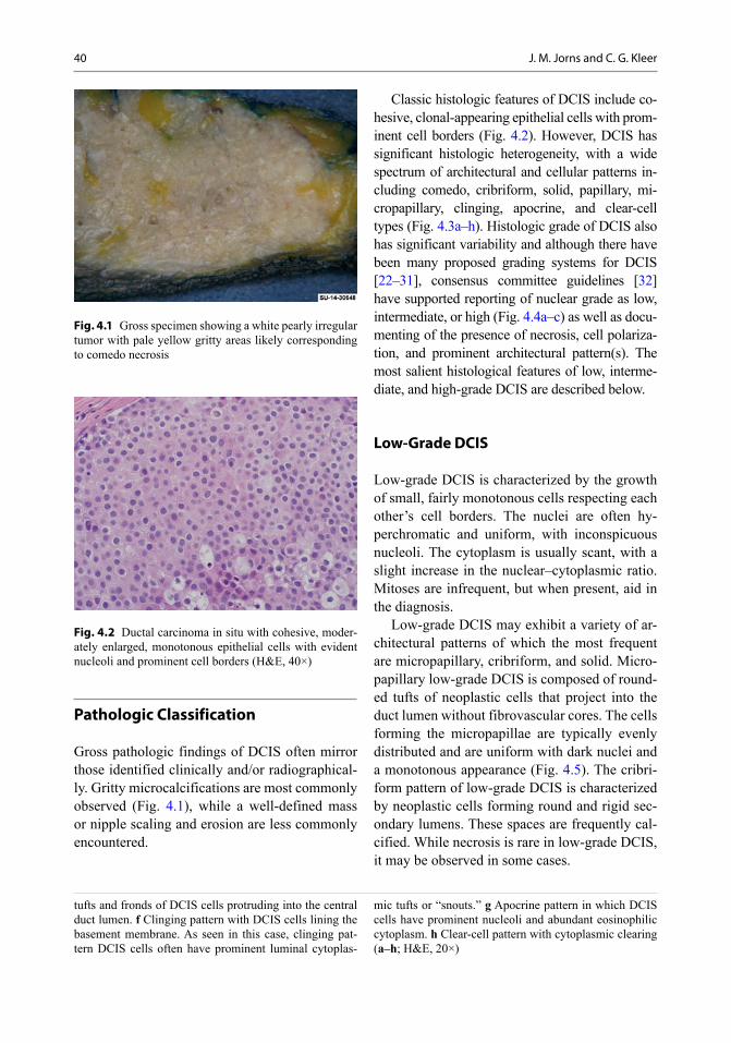
40 J. M. Jorns and C. G. Kleer
Pathologic Classification
Gross pathologic findings of DCIS often mirror those identified clinically and/or radiographical-ly. Gritty microcalcifications are most commonly observed (Fig. 4.1), while a well-defined mass or nipple scaling and erosion are less commonly encountered.
Classic histologic features of DCIS include co-hesive, clonal-appearing epithelial cells with prom-inent cell borders (Fig. 4.2). However, DCIS has significant histologic heterogeneity, with a wide spectrum of architectural and cellular patterns in-cluding comedo, cribriform, solid, papillary, mi-cropapillary, clinging, apocrine, and clear-cell types (Fig. 4.3a–h). Histologic grade of DCIS also has significant variability and although there have been many proposed grading systems for DCIS [22–31], consensus committee guidelines [32] have supported reporting of nuclear grade as low, intermediate, or high (Fig. 4.4a–c) as well as docu-menting of the presence of necrosis, cell polariza-tion, and prominent architectural pattern(s). The most salient histological features of low, interme-diate, and high-grade DCIS are described below.
Low-Grade DCIS
Low-grade DCIS is characterized by the growth of small, fairly monotonous cells respecting each other’s cell borders. The nuclei are often hy-perchromatic and uniform, with inconspicuous nucleoli. The cytoplasm is usually scant, with a slight increase in the nuclear–cytoplasmic ratio. Mitoses are infrequent, but when present, aid in the diagnosis.
Low-grade DCIS may exhibit a variety of ar-chitectural patterns of which the most frequent are micropapillary, cribriform, and solid. Micro-papillary low-grade DCIS is composed of round-ed tufts of neoplastic cells that project into the duct lumen without fibrovascular cores. The cells forming the micropapillae are typically evenly distributed and are uniform with dark nuclei and a monotonous appearance (Fig. 4.5). The cribri-form pattern of low-grade DCIS is characterized by neoplastic cells forming round and rigid sec-ondary lumens. These spaces are frequently cal-cified. While necrosis is rare in low-grade DCIS, it may be observed in some cases.
tufts and fronds of DCIS cells protruding into the central duct lumen. f Clinging pattern with DCIS cells lining the basement membrane. As seen in this case, clinging pat-tern DCIS cells often have prominent luminal cytoplas-
mic tufts or “snouts.” g Apocrine pattern in which DCIS cells have prominent nucleoli and abundant eosinophilic cytoplasm. h Clear-cell pattern with cytoplasmic clearing (a–h; H&E, 20×)
Fig. 4.2 Ductal carcinoma in situ with cohesive, moder-ately enlarged, monotonous epithelial cells with evident nucleoli and prominent cell borders (H&E, 40×)
Fig. 4.1 Gross specimen showing a white pearly irregular tumor with pale yellow gritty areas likely corresponding to comedo necrosis
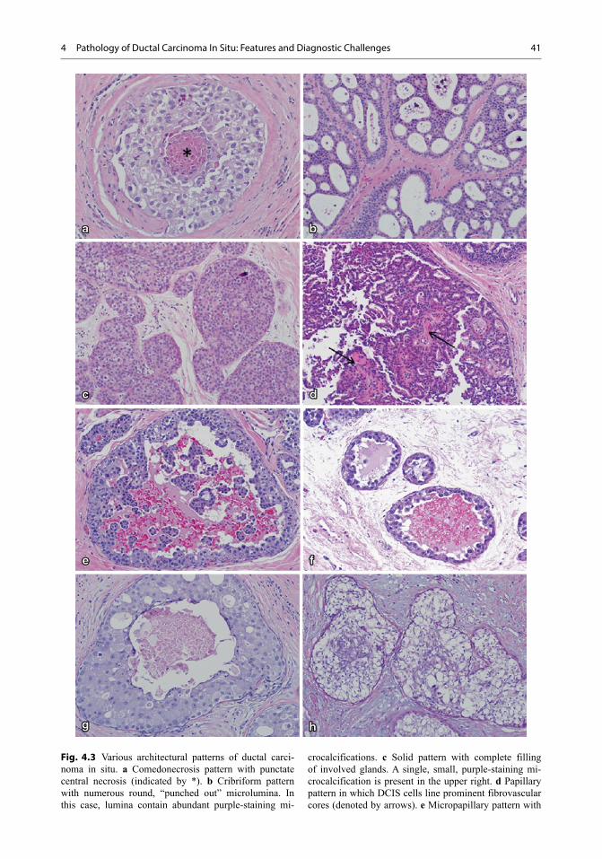
414 Pathology of Ductal Carcinoma In Situ: Features and Diagnostic Challenges
Fig. 4.3 Various architectural patterns of ductal carci-noma in situ. a Comedonecrosis pattern with punctate central necrosis (indicated by *). b Cribriform pattern with numerous round, “punched out” microlumina. In this case, lumina contain abundant purple-staining mi-
crocalcifications. c Solid pattern with complete filling of involved glands. A single, small, purple-staining mi-crocalcification is present in the upper right. d Papillary pattern in which DCIS cells line prominent fibrovascular cores (denoted by arrows). e Micropapillary pattern with

42 J. M. Jorns and C. G. Kleer
Micropapillary low-grade DCIS is frequently multicentric, and most of these patients accord-ingly have extensive microcalcifications on im-aging. Not surprisingly, despite the low-grade features, micropapillary DCIS is nevertheless a risk factor for recurrence in patients treated with lumpectomy. The ducts with tumor are often admixed with benign or atypical ducts. Hence, diagnosis can be a challenge, especially in core biopsies.
Stringent criteria need to be used to diagnose micropapillary low-grade DCIS. These include: (a) micropapillae are club like and involve the entire duct circumference, extend at least one third of the duct diameter, and show a tendency to detach, (b) the nuclei are not overlapping, and (c) at least three adjacent ducts must be fully in-volved.
Intermediate Grade DCIS
While the architectural patterns of intermedi-ate grade DCIS are similar to low-grade lesions, intermediate-grade DCIS is characterized by slightly increased cellular pleomorphism, more frequent prominent nucleoli, and coarser chro-matin than low-grade DCIS (Fig. 4.2). Mitoses and necrosis are also more frequently encoun-tered in intermediate-grade DCIS compared to low-grade lesions.
High-Grade DCIS
High-grade DCIS is defined by marked nuclear enlargement and pleomorphism. The nuclei have coarse chromatin and prominent nucleoli. There
Fig. 4.5 Low-grade ductal carcinoma in situ with mi-cropapillary pattern. The neoplastic cells are monotonous and form rounded micropapillae lacking fibrovascular cores that project into the ductal lumens (H&E, 40×)
Fig. 4.4 Spectrum of nuclear grade in ductal carcinoma in situ (DCIS). a Low nuclear grade DCIS with small, uni-form, monotonous nuclei. b Intermediate nuclear grade DCIS with moderate enlargement and pleomorphism.
c High nuclear grade DCIS with marked enlargement and pleomorphism. Comedonecrosis (denoted by *) and nu-merous mitotic figures (denoted by arrow) are often present in high-grade DCIS as seen in this case (a–c; H&E, 40×)

434 Pathology of Ductal Carcinoma In Situ: Features and Diagnostic Challenges
is loss of cell polarity and mitoses are frequent and often atypical. The comedo-type necrosis architectural pattern is very common, but solid, cribriform, micropapillary, and clinging patterns are also often seen. Central comedonecrosis is often accompanied by calcifications, which may be large and amorphous (Fig. 4.6). High-grade DCIS is also frequently associated with periduc-tal chronic inflammation (Fig. 4.7) and angio-genesis, and a desmoplastic or sclerotic stromal response, with both concentric and distorting pat-terns (Fig. 4.8a–c).
High-grade DCIS may sometimes mimic mi-croinvasive carcinoma, especially when DCIS extends into small glands of lobules that are dis-torted by marked sclerosis or secondarily involve sclerosing adenosis, radial scars, or complex sclerosing lesions. In such cases, deeper sec-tions and immunohistochemical (IHC) stains that highlight myoepithelium, such as p63, calponin, muscle-specific or smooth muscle actin, among others, may be helpful in assessing the presence of microinvasion. The use of myoepithelial stains is particularly helpful if DCIS extends into scle-rosing lesions such as sclerosing adenosis, which is rich in myoepithelial cells (Fig. 4.9a, b). How-ever, staining for myoepithelial markers may not always aid in the differentiation between DCIS and microinvasive carcinoma, as staining may be patchy and difficult to interpret or the small focus may be mostly or completely lost in deeper im-munostained slides. In such cases, the suspicion for a focus of microinvasion should be stated in the pathology report.
Biomarkers and Molecular Pathology
Consonant with the histological heterogeneity, DCIS exhibits significant variability in biomark-er profiles and genetic aberrations. Low nucle-ar grade DCIS almost universally has diffuse, strong expression of estrogen receptor (ER) and progesterone receptor (PR) and lacks HER-2/neu protein overexpression or gene amplification. In contrast, high nuclear grade DCIS can be ER/PR positive or negative and is HER-2/neu positive in 60–80 % of cases (Fig. 4.10a–h) [33–36]. Unlike low-grade DCIS, high-grade DCIS also has high proliferative rates and is often p53 positive by IHC and mutational analyses [29, 37]. Molecular studies have shown that low-grade DCIS is char-acterized by chromosomal losses at 16q and 17p and gains at 1q, whereas high-grade DCIS shows more numerous and variable alterations, includ-ing losses at 11q, 14q, 8p, and 13q and gains at 17q, 8q, and 5p [38, 39].
Morphologic, immunophenotypic, and mo-lecular evidences support that DCIS represents a
Fig. 4.6 High-grade ductal carcinoma in situ with com-edonecrosis and large, amorphous calcifications (H&E, 10×); inset shows large, pleomorphic cells with readily identifiable mitotic forms (denoted by arrow; H&E, 40×)
Fig. 4.7 High-grade ductal carcinoma in situ with can-cerization of lobules and marked periglandular chronic inflammation (H&E, 20×)

44 J. M. Jorns and C. G. Kleer
heterogeneous group of diseases that differ in bi-ologic potential, ranging from little-to-no to very high risk of progression to invasive carcinoma. Due to its diversity and in attempt to avoid the anxiety-producing word “carcinoma” in its name, some support renaming DCIS as ductal intraepi-thelial neoplasia (DIN) as proposed by Tavassoli
et al. [30], which is similar nomenclature to that used to describe dysplasias of the genital tract and perineum. Further study is necessary to de-termine whether DCIS should be further subclas-sified and/or redefined based on grade, hormone receptor status or other remarkable histological features.
Differential Diagnosis of DCIS Relevant to Clinical Practice
Atypical ductal hyperplasia. The main differen-tial diagnosis of low-grade DCIS is with atypical ductal hyperplasia (ADH). ADH is character-ized by one or two moderately distended ducts, filled by cells that are evenly spaced, having uniform nuclei, and some regular secondary lu-mens (Fig. 4.11). Importantly, ADH lacks pleo-morphism, individual cell necrosis, frequent mi-toses, or prominence of nucleoli, which charac-terize DCIS. The presence of cells that overlap and stream, features more in keeping with benign usual ductal hyperplasia, mixed with atypical cells is a helpful feature to distinguish ADH from DCIS.
It is common to observe a spectrum of dis-ease in the same breast that ranges from ADH to DCIS. Thus, borderline cases may be seen at either the low end or high ends of the spec-trum. At times, the distinction between ADH and low-grade DCIS is one of the most challenging diagnostic difficulties in breast pathology. Most experts would assign these challenging cases to the less severe diagnostic category. Interestingly, most ADH is detected in the vicinity of—or with-in—a sclerosing lesion, a papilloma and—espe-cially—within or close to a worrisome columnar altered lobule. ADH is often near the targeted calcifications in a biopsy.
Pleomorphic lobular carcinoma in situ. Pleo-morphic lobular carcinoma in situ (pLCIS) has morphologic features of both classic LCIS and high-grade DCIS. Like classic LCIS, the cells are discohesive and have prominent plasmacytoid morphology. However, the cells of pLCIS are also enlarged and have marked nuclear pleomor-phism and readily identifiable mitotic activity as
Fig. 4.8 Variable sclerosis associated with high-grade ductal carcinoma in situ, ranging from a concentric, b mixed concentric and distorting, and c markedly distort-ing (a–c; H&E, 20×)

454 Pathology of Ductal Carcinoma In Situ: Features and Diagnostic Challenges
seen in high-grade DCIS. pLCIS may also have comedo-type necrosis, a common architectural pattern of high-grade DCIS. pLCIS has been further divided into non-apocrine and apocrine types, the latter of which has apocrine features including prominent nucleoli and abundant eo-sinophilic cytoplasm (Fig. 4.12).
pLCIS demonstrates loss of E-cadherin ex-pression and chromosomal losses at 16q and 17p and gains of 1q, which are hallmark features of classic invasive and in situ lobular carcinomas [40, 41]. However, compared to classic LCIS which is invariably ER/PR positive and HER-2/neu negative, pLCIS has lower levels and is less frequently ER positive and may be HER-2/neu positive in approximately 25 % of cases [42].
pLCIS also has been shown to have additional genetic aberrations including deletion of 8p and 13q and gains of 8q, which were also identified in matched pleomorphic invasive lobular carci-noma (pILC) [40, 41, 43]. However, Chen et al. [44] found that apocrine pLCIS had greater num-bers and diversity of genetic abnormalities than non-apocrine pLCIS, which had a similar profile to classic LCIS. Specifically, they identified am-plification at 17p11.2–17q12 and 11.q.13.3, gain of 16p and losses of 3q, 11q, 13q, and 17p in the apocrine pLCIS group.
Classic LCIS is generally regarded as a mark-er of bilateral future cancer risk. In contrast, pLCIS has higher-grade morphology, an often unfavorable biomarker profile and greater extent
Fig. 4.10 a Low nuclear grade ductal carcinoma in situ ( DCIS; H&E, 10×) that is b estrogen receptor ( ER) positive, c progesterone receptor (PR) positive, and d HER-2/neu (1+) negative for overexpression by immunohistochemical ( IHC)
staining (b, d; IHC, 10×). e High nuclear grade DCIS (H&E, 10×) that is f ER negative (with positive staining in benign glands in the lower left-hand corner), g PR negative, and h HER-2/neu (3+) positive for overexpression (f–h; IHC, 10×)
Fig. 4.9 a Apocrine ductal carcinoma in situ involving sclerosing adenosis (H&E, 10×) and b corresponding immunohistochemical (IHC) stain for muscle-specific
actin which highlights myoepithelium throughout, confirming in situ disease only (IHC, 10×)
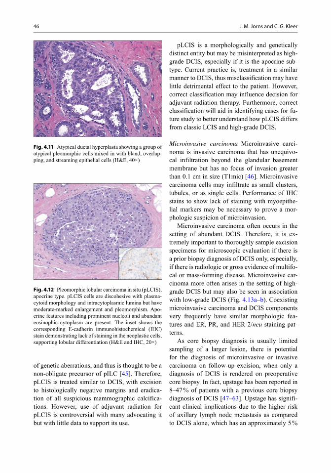
46 J. M. Jorns and C. G. Kleer
of genetic aberrations, and thus is thought to be a non-obligate precursor of pILC [45]. Therefore, pLCIS is treated similar to DCIS, with excision to histologically negative margins and eradica-tion of all suspicious mammographic calcifica-tions. However, use of adjuvant radiation for pLCIS is controversial with many advocating it but with little data to support its use.
pLCIS is a morphologically and genetically distinct entity but may be misinterpreted as high-grade DCIS, especially if it is the apocrine sub-type. Current practice is, treatment in a similar manner to DCIS, thus misclassification may have little detrimental effect to the patient. However, correct classification may influence decision for adjuvant radiation therapy. Furthermore, correct classification will aid in identifying cases for fu-ture study to better understand how pLCIS differs from classic LCIS and high-grade DCIS.
Microinvasive carcinoma Microinvasive carci-noma is invasive carcinoma that has unequivo-cal infiltration beyond the glandular basement membrane but has no focus of invasion greater than 0.1 cm in size (T1mic) [46]. Microinvasive carcinoma cells may infiltrate as small clusters, tubules, or as single cells. Performance of IHC stains to show lack of staining with myoepithe-lial markers may be necessary to prove a mor-phologic suspicion of microinvasion.
Microinvasive carcinoma often occurs in the setting of abundant DCIS. Therefore, it is ex-tremely important to thoroughly sample excision specimens for microscopic evaluation if there is a prior biopsy diagnosis of DCIS only, especially, if there is radiologic or gross evidence of multifo-cal or mass-forming disease. Microinvasive car-cinoma more often arises in the setting of high-grade DCIS but may also be seen in association with low-grade DCIS (Fig. 4.13a–b). Coexisting microinvasive carcinoma and DCIS components very frequently have similar morphologic fea-tures and ER, PR, and HER-2/neu staining pat-terns.
As core biopsy diagnosis is usually limited sampling of a larger lesion, there is potential for the diagnosis of microinvasive or invasive carcinoma on follow-up excision, when only a diagnosis of DCIS is rendered on preoperative core biopsy. In fact, upstage has been reported in 8–47 % of patients with a previous core biopsy diagnosis of DCIS [47–63]. Upstage has signifi-cant clinical implications due to the higher risk of axillary lymph node metastasis as compared to DCIS alone, which has an approximately 5 %
Fig. 4.12 Pleomorphic lobular carcinoma in situ (pLCIS), apocrine type. pLCIS cells are discohesive with plasma-cytoid morphology and intracytoplasmic lumina but have moderate-marked enlargement and pleomorphism. Apo-crine features including prominent nucleoli and abundant eosinophic cytoplasm are present. The inset shows the corresponding E-cadherin immunohistochemical (IHC) stain demonstrating lack of staining in the neoplastic cells, supporting lobular differentiation (H&E and IHC, 20×)
Fig. 4.11 Atypical ductal hyperplasia showing a group of atypical pleomorphic cells mixed in with bland, overlap-ping, and streaming epithelial cells (H&E, 40×)
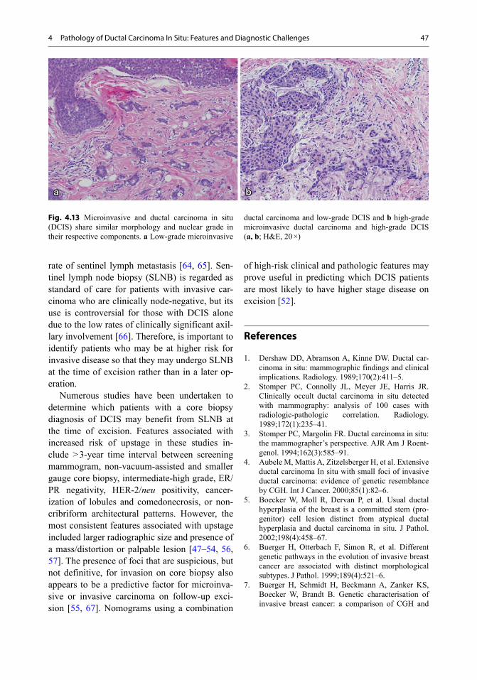
474 Pathology of Ductal Carcinoma In Situ: Features and Diagnostic Challenges
rate of sentinel lymph metastasis [64, 65]. Sen-tinel lymph node biopsy (SLNB) is regarded as standard of care for patients with invasive car-cinoma who are clinically node-negative, but its use is controversial for those with DCIS alone due to the low rates of clinically significant axil-lary involvement [66]. Therefore, is important to identify patients who may be at higher risk for invasive disease so that they may undergo SLNB at the time of excision rather than in a later op-eration.
Numerous studies have been undertaken to determine which patients with a core biopsy diagnosis of DCIS may benefit from SLNB at the time of excision. Features associated with increased risk of upstage in these studies in-clude > 3-year time interval between screening mammogram, non-vacuum-assisted and smaller gauge core biopsy, intermediate-high grade, ER/PR negativity, HER-2/neu positivity, cancer-ization of lobules and comedonecrosis, or non-cribriform architectural patterns. However, the most consistent features associated with upstage included larger radiographic size and presence of a mass/distortion or palpable lesion [47–54, 56, 57]. The presence of foci that are suspicious, but not definitive, for invasion on core biopsy also appears to be a predictive factor for microinva-sive or invasive carcinoma on follow-up exci-sion [55, 67]. Nomograms using a combination
of high-risk clinical and pathologic features may prove useful in predicting which DCIS patients are most likely to have higher stage disease on excision [52].
References
1. Dershaw DD, Abramson A, Kinne DW. Ductal car-cinoma in situ: mammographic findings and clinical implications. Radiology. 1989;170(2):411–5.
2. Stomper PC, Connolly JL, Meyer JE, Harris JR. Clinically occult ductal carcinoma in situ detected with mammography: analysis of 100 cases with radiologic-pathologic correlation. Radiology. 1989;172(1):235–41.
3. Stomper PC, Margolin FR. Ductal carcinoma in situ: the mammographer’s perspective. AJR Am J Roent-genol. 1994;162(3):585–91.
4. Aubele M, Mattis A, Zitzelsberger H, et al. Extensive ductal carcinoma In situ with small foci of invasive ductal carcinoma: evidence of genetic resemblance by CGH. Int J Cancer. 2000;85(1):82–6.
5. Boecker W, Moll R, Dervan P, et al. Usual ductal hyperplasia of the breast is a committed stem (pro-genitor) cell lesion distinct from atypical ductal hyperplasia and ductal carcinoma in situ. J Pathol. 2002;198(4):458–67.
6. Buerger H, Otterbach F, Simon R, et al. Different genetic pathways in the evolution of invasive breast cancer are associated with distinct morphological subtypes. J Pathol. 1999;189(4):521–6.
7. Buerger H, Schmidt H, Beckmann A, Zanker KS, Boecker W, Brandt B. Genetic characterisation of invasive breast cancer: a comparison of CGH and
Fig. 4.13 Microinvasive and ductal carcinoma in situ (DCIS) share similar morphology and nuclear grade in their respective components. a Low-grade microinvasive
ductal carcinoma and low-grade DCIS and b high-grade microinvasive ductal carcinoma and high-grade DCIS (a, b; H&E, 20 ×)
48 J. M. Jorns and C. G. Kleer
PCR based multiplex microsatellite analysis. J Clin Pathol. 2001;54(11):836–40.
8. Farabegoli F, Champeme MH, Bieche I, et al. Genetic pathways in the evolution of breast ductal carcinoma in situ. J Pathol. 2002;196(3):280–6.
9. Waldman FM, DeVries S, Chew KL, Moore DH 2nd, Kerlikowske K, Ljung BM. Chromosomal alterations in ductal carcinomas in situ and their in situ recur-rences. J Natl Cancer Inst. 2000;92(4):313–20.
10. Morrow M, Strom EA, Bassett LW, et al. Standard for the management of ductal carcinoma in situ of the breast (DCIS). CA Cancer J Clin. 2002;52(5):256–76.
11. Alpers CE, Wellings SR. The prevalence of carci-noma in situ in normal and cancer-associated breasts. Hum Pathol. 1985;16(8):796–807.
12. Bhathal PS, Brown RW, Lesueur GC, Russell IS. Frequency of benign and malignant breast lesions in 207 consecutive autopsies in Australian women. Br J Cancer. 1985;51(2):271–8.
13. Nielsen M. Autopsy studies of the occurrence of can-cerous, atypical and benign epithelial lesions in the female breast. APMIS Suppl. 1989;10:1–56.
14. Farrow JH. Current concepts in the detection and treatment of the earliest of the early breast cancers. Cancer. 1970;25(2):468–77.
15. Haagensen CD, Lane N, Lattes R. Neoplastic prolif-eration of the epithelium of the mammary lobules: adenosis, lobular neoplasia, and small cell carci-noma. Surg Clin North Am. 1972;52(2):497–524.
16. Kraus FT, Neubecker RD. The differential diag-nosis of papillary tumors of the breast. Cancer. 1962;15:444–55.
17. Millis RR, Thynne GS. In situ intraduct carcinoma of the breast: a long term follow-up study. Br J Surg. 1975;62(12):957–62.
18. Page DL, Dupont WD, Rogers LW, Jensen RA, Schuyler PA. Continued local recurrence of car-cinoma 15–25 years after a diagnosis of low grade ductal carcinoma in situ of the breast treated only by biopsy. Cancer. 1995;76(7):1197–200.
19. Page DL, Dupont WD, Rogers LW, Landenberger M. Intraductal carcinoma of the breast: follow-up after biopsy only. Cancer. 1982;49(4):751–8.
20. Rosen PP, Braun DW Jr, Kinne DE. The clinical sig-nificance of pre-invasive breast carcinoma. Cancer. 1980;46(4 Suppl):919–25.
21. Eusebi V, Feudale E, Foschini MP, et al. Long-term follow-up of in situ carcinoma of the breast. Semin Diagn Pathol. 1994;11(3):223–35.
22. Holland R, Peterse JL, Millis RR, et al. Ductal car-cinoma in situ: a proposal for a new classification. Semin Diagn Pathol. 1994;11(3):167–80.
23. Scott MA, Lagios MD, Axelsson K, Rogers LW, Anderson TJ, Page DL. Ductal carcinoma in situ of the breast: reproducibility of histological subtype analysis. Hum Pathol. 1997;28(8):967–73.
24. Silverstein MJ, Poller DN, Waisman JR, et al. Prog-nostic classification of breast ductal carcinoma-in-situ. The Lancet. 1995;345(8958):1154–7.
25. Bellamy CO, McDonald C, Salter DM, Chetty U, Anderson TJ. Noninvasive ductal carcinoma of the
breast: the relevance of histologic categorization. Hum Pathol. 1993;24(1):16–23.
26. Lagios MD. Duct carcinoma in situ. Pathology and treatment. Surg Clin North Am. 1990;70(4):853–71.
27. Lennington WJ, Jensen RA, Dalton LW, Page DL. Ductal carcinoma in situ of the breast. Heterogeneity of individual lesions. Cancer. 1994;73(1):118–24.
28. Ottesen GL, Graversen HP, Blichert-Toft M, Zedeler K, Andersen JA. Ductal carcinoma in situ of the female breast. Short-term results of a prospective nationwide study. The Danish Breast Cancer Cooper-ative Group. Am J Surg Pathol. 1992;16(12):1183–96.
29. Poller DN, Silverstein MJ, Galea M, et al. Ideas in pathology. Ductal carcinoma in situ of the breast: a proposal for a new simplified histological clas-sification association between cellular prolifera-tion and c-erbB-2 protein expression. Mod Pathol. 1994;7(2):257–62.
30. Tavassoli FA. Ductal carcinoma in situ: introduction of the concept of ductal intraepithelial neoplasia. Mod Pathol. 1998;11(2):140–54.
31. Warnberg F, Casalini P, Nordgren H, Bergkvist L, Holmberg L, Menard S. Ductal carcinoma in situ of the breast: a new phenotype classification system and its relation to prognosis. Breast Cancer Res Treat. 2002;73(3):215–21.
32. Consensus conference on the classification of ductal carcinoma in situ. Hum Pathol. 1997;28(11):1221–5.
33. Allred DC, Clark GM, Molina R, et al. Overexpres-sion of HER-2/neu and its relationship with other prognostic factors change during the progression of in situ to invasive breast cancer. Hum Pathol. 1992;23(9):974–9.
34. Gusterson BA, Machin LG, Gullick WJ, et al. c-erbB-2 expression in benign and malignant breast disease. Br J Cancer. 1988;58(4):453–7.
35. Gusterson BA, Machin LG, Gullick WJ, et al. Immunohistochemical distribution of c-erbB-2 in infiltrating and in situ breast cancer. Int J Cancer. 1988;42(6):842–5.
36. Poller DN, Roberts EC, Bell JA, Elston CW, Blamey RW, Ellis IO. p53 protein expression in mammary ductal carcinoma in situ: relationship to immuno-histochemical expression of estrogen receptor and c-erbB-2 protein. Hum Pathol. 1993;24(5):463–8.
37. Done SJ, Arneson NC, Ozcelik H, Redston M, Andru-lis IL. p53 mutations in mammary ductal carcinoma in situ but not in epithelial hyperplasias. Cancer Res. 1998;58(4):785–9.
38. Simpson PT, Reis-Filho JS, Gale T, Lakhani SR. Molecular evolution of breast cancer. J Pathol. 2005;205(2):248–54.
39. Reis-Filho JS, Lakhani SR. The diagnosis and man-agement of pre-invasive breast disease: genetic alter-ations in pre-invasive lesions. Breast Cancer Res. 2003;5(6):313–9.
40. Reis-Filho JS, Simpson PT, Jones C, et al. Pleomor-phic lobular carcinoma of the breast: role of compre-hensive molecular pathology in characterization of an entity. J Pathol. 2005;207(1):1–13.
494 Pathology of Ductal Carcinoma In Situ: Features and Diagnostic Challenges
41. Simpson PT, Reis-Filho JS, Lambros MB, et al. Molecular profiling pleomorphic lobular carcino-mas of the breast: evidence for a common molecular genetic pathway with classic lobular carcinomas. J Pathol. 2008;215(3):231–44.
42. Chivukula M, Haynik DM, Brufsky A, Carter G, Dabbs DJ. Pleomorphic lobular carcinoma in situ (PLCIS) on breast core needle biopsies: clinical significance and immunoprofile. Am J Surg Pathol. 2008;32(11):1721–6.
43. Vargas AC, Lakhani SR, Simpson PT. Pleomorphic lobular carcinoma of the breast: molecular pathology and clinical impact. Future Oncol. 2009;5(2):233–43.
44. Chen YY, Hwang ES, Roy R, et al. Genetic and phenotypic characteristics of pleomorphic lobular carcinoma in situ of the breast. Am J Surg Pathol. 2009;33(11):1683–94.
45. Lopez-Garcia MA, Geyer FC, Lacroix-Triki M, Mar-chio C, Reis-Filho JS. Breast cancer precursors revis-ited: molecular features and progression pathways. Histopathology. 2010;57(2):171–92.
46. Edge SB. ed. AJCC Cancer Staging Manual. Seventh ed. Springer: New York. 2010.
47. Dillon MF, McDermott EW, Quinn CM, O’Doherty A, O’Higgins N, Hill AD. Predictors of invasive dis-ease in breast cancer when core biopsy demonstrates DCIS only. J Surg Oncol. 2006;93(7):559–63.
48. Han JS, Molberg KH, Sarode V. Predictors of invasion and axillary lymph node metastasis in patients with a core biopsy diagnosis of ductal carcinoma in situ: an analysis of 255 cases. Breast J. 2011;17(3):223–9.
49. Houssami N, Ambrogetti D, Marinovich ML, et al. Accuracy of a preoperative model for predicting invasive breast cancer in women with ductal carci-noma-in-situ on vacuum-assisted core needle biopsy. Ann Surg Oncol. 2011;18(5):1364–71.
50. Huo L, Sneige N, Hunt KK, Albarracin CT, Lopez A, Resetkova E. Predictors of invasion in patients with core-needle biopsy-diagnosed ductal carcinoma in situ and recommendations for a selective approach to sentinel lymph node biopsy in ductal carcinoma in situ. Cancer. 2006;107(8):1760–8.
51. Kurniawan ED, Rose A, Mou A, et al. Risk fac-tors for invasive breast cancer when core needle biopsy shows ductal carcinoma in situ. Arch Surg. 2010;145(11):1098–104.
52. Lee SK, Yang JH, Woo SY, Lee JE, Nam SJ. Nomo-gram for predicting invasion in patients with a pre-operative diagnosis of ductal carcinoma in situ of the breast. Br J Surg. 2013;100(13):1756–63.
53. Liao N, Zhang GC, Liu YH, et al. HER2-positive status is an independent predictor for coexisting inva-sion of ductal carcinoma in situ of the breast present-ing extensive DCIS component. Pathol Res Pract. 2011;207(1):1–7.
54. Miyake T, Shimazu K, Ohashi H, et al. Indication for sentinel lymph node biopsy for breast cancer when core biopsy shows ductal carcinoma in situ. Am J Surg. 2011;202(1):59–65.
55. Park HS, Park S, Cho J, Park JM, Kim SI, Park BW. Risk predictors of underestimation and the need for sentinel node biopsy in patients diagnosed with ductal carcinoma in situ by preoperative needle biopsy. J Surg Oncol. 2013;107(4):388–92.
56. Renshaw AA. Predicting invasion in the excision specimen from breast core needle biopsy specimens with only ductal carcinoma in situ. Arch Pathol Lab Med. 2002;126(1):39–41.
57. Yen TW, Hunt KK, Ross MI, et al. Predictors of invasive breast cancer in patients with an initial diagnosis of ductal carcinoma in situ: a guide to selective use of sentinel lymph node biopsy in man-agement of ductal carcinoma in situ. J Am Coll Surg. 2005;200(4):516–26.
58. Chan MY, Lim S. Predictors of invasive breast cancer in ductal carcinoma in situ initially diagnosed by core biopsy. Asian J Surg. 2010;33(2):76–82.
59. Diepstraten SC, van de Ven SM, Pijnappel RM, et al. Development and evaluation of a prediction model for underestimated invasive breast cancer in women with ductal carcinoma in situ at stereotactic large core needle biopsy. PLoS ONE. 2013;8(10):e77826.
60. Guillot E, Vaysse C, Goetgeluck J, et al. Extensive pure ductal carcinoma in situ of the breast: identifica-tion of predictors of associated infiltrating carcinoma and lymph node metastasis before immediate recon-structive surgery. Breast. 2014;23(2):97–103.
61. Rutstein LA, Johnson RR, Poller WR, et al. Predic-tors of residual invasive disease after core needle biopsy diagnosis of ductal carcinoma in situ. Breast J. 2007;13(3):251–7.
62. Sakr R, Antoine M, Barranger E, et al. Value of sen-tinel lymph node biopsy in breast ductal carcinoma in situ upstaged to invasive carcinoma. Breast J. 2008;14(1):55–60.
63. Schulz S, Sinn P, Golatta M, et al. Prediction of under-estimated invasiveness in patients with ductal carci-noma in situ of the breast on percutaneous biopsy as rationale for recommending concurrent sentinel lymph node biopsy. Breast. 2013;22(4):537–42.
64. Dauway EL, Giuliano R, Pendas S, et al. Lymphatic Mapping: A Technique Providing Accurate Staging for Breast Cancer. Breast Cancer. 1999;6(2):145–54.
65. Wilkie C, White L, Dupont E, Cantor A, Cox CE. An update of sentinel lymph node mapping in patients with ductal carcinoma in situ. Am J Surg. 2005;190(4):563–6.
66. Lyman GH, Giuliano AE, Somerfield MR, et al. American Society of Clinical Oncology guide-line recommendations for sentinel lymph node biopsy in early-stage breast cancer. J Clin Oncol. 2005;23(30):7703–20.
67. Walters L, Pang JC, Zhao L, Jorns JM Ductal carci-noma in situ with distorting sclerosis on core biopsy may be predictive of upstage on excision. Mod Pathol. 2014;27(Supp. 2):87A.

51
Molecular Markers in DCIS
Theresa L. Schwartz and Chelsea Horwood
L. A. Newman, J. M. Bensenhaver (eds.), Ductal Carcinoma In Situ and Microinvasive/Borderline Breast Cancer, DOI 10.1007/978-1-4939-2035-8_5, © Springer Science+Business Media New York 2015
T. L. Schwartz ()Department of Surgery, Saint Louis University School of Medicine, 3635 Vista Avenue, DT—3rd Floor, 63110 St. Louis, MO, USAe-mail: [email protected]
C. HorwoodSaint Louis University School of Medicine, 3635 Vista Avenue, DT—3rd Floor, 63110 St. Louis, MO, USAe-mail: [email protected]
Introduction
Ductal carcinoma in situ (DCIS) includes a het-erogeneous group of noninvasive breast malig-nancies with a variable potential for evolution. It is characterized by a proliferation of malignant epithelial cells that are confined to the duct with-out invasion through the basement membrane into the surrounding stroma [1]. When it was ini-tially recognized as a distinct clinical entity dur-ing the first half of the twentieth century, DCIS accounted for only 1–2 % of newly diagnosed breast cancers. Since it generally presented as a large palpable mass, mastectomy was accepted as the standard of care and was routinely cura-tive [2]. Due to improvements in both quality and consistency of mammographic screening, the in-cidence of DCIS has continued to increase and it now accounts for approximately 20 % of all breast cancer diagnoses [3]. DCIS is also able to be detected at an earlier stage with overall small-er tumor burden. These advances have led to the widespread use of breast conservation therapy for women with DCIS who are appropriate can-
didates. Early studies conducted following the institution of breast-conserving surgery for DCIS showed a local recurrence rate of approximately 25 % in women who did not undergo adjuvant radiation therapy with half of these recurrences presenting as invasive disease [4–8]. Although the use of adjuvant radiation and endocrine ther-apy have substantially decreased the local recur-rence rate in these women, our understanding of the pathophysiology of DCIS and the factors involved in its progression to invasive disease is still unclear. Developing a mechanism that can better classify DCIS subtypes that are more like-ly to progress to invasive disease would greatly enhance individualization of management strate-gies by assisting in both reducing overtreatment of less concerning lesions and identifying those patients who should be treated more aggres-sively. The study of the biologic features and mo-lecular markers of DCIS that could offer accurate prognostic information and ultimately provide treatment guidelines is of paramount importance.
Natural History
The natural history of breast cancer involves progression of normal breast tissue to invasive carcinoma due to accumulation of somatic muta-tions involving key growth, differentiation, and cell communication pathways. The initiation of breast cancer is due to transforming events in a single cell with point mutations, copy number alterations, epigenetic modifications, and general
5

52 T. L. Schwartz and C. Horwood
genome instability being frequently associated with disease progression [9, 10]. DCIS is thought to be the immediate precursor of invasive disease based on molecular and pathological studies [11, 12]. Information on the specific functional events that drive progression of DCIS to invasive car-cinoma is limited. Numerical grading systems have been developed to reflect tumor differentia-tion and growth by assigning points for specific cellular features of the tumor. Although no ac-cepted standard method of grading is available for DCIS, there are three overall grades used that describe the tumor cells according to the degree to which they resemble normal breast cells. The grades commonly used are well differentiated (grade 1), moderately differentiated (grade 2), and poorly differentiated (grade 3) DCIS.
Progressive growth of DCIS results from al-terations in normal growth-regulating mecha-nisms [13]. A molecular biological marker, or biomarker, is an objective measurement of a normal biological response. Biomarkers most commonly associated with breast cancer include the hormones estrogen and progesterone and their respective receptors, the oncogene erbB2 (HER2/neu), the tumor suppressor proteins p16 and p53, and the tumor proliferation marker Ki-67. Although numerous histopathological charac-teristics have been identified as predictors of re-currence of DCIS, including lesion size, nuclear grade, architectural pattern, margin status, and presence of comedonecrosis, finding molecular markers to further target the malignancy in ques-tion and guide risk stratification at the time of di-agnosis is paramount [14]. Understanding these biomarkers and their predictive value for both treatment response and potential disease recur-rence could help individualize treatment plans to optimize patient outcomes.
Estrogen and Progesterone
Estrogen and progesterone are steroid hormones that stimulate the development and maintenance of female breast tissue. Estrogen exposure has been established as a risk factor for future breast cancer development. It has been hypothesized
that estrogen acts as a stimulatory hormone, thereby increasing the frequency of mitotic activ-ity within the breast with malignant phenotypes developing due to errors in cell division [15]. The estrogen and progesterone receptors (ER and PR) are commonly present on breast tumors with ap-proximately 70 % of all DCIS being ER positive [16]. ER positivity is more often seen in well-dif-ferentiated and moderately differentiated DCIS, whereas it is reported to be present in only 25 % of poorly differentiated lesions [17–19].
Expression of ER, and to a less common ex-tent PR, is routinely assessed on pathological specimens of DCIS in order to predict clinical response to hormonal therapy, such as selective estrogen receptor modulators (SERMs). SERMs, namely Tamoxifen and Raloxifene, are competi-tive inhibitors of estrogen binding to the estro-gen receptor. The National Surgical Adjuvant Breast and Bowel Project (NSABP) B-24 study evaluated the use of Tamoxifen compared to placebo in 1804 women with DCIS undergoing breast-conserving surgery followed by radiation therapy. After 5 years of treatment, women in the Tamoxifen arm had fewer breast cancer events (8.2 vs. 13.4 %, p = 0.0009) than did those on pla-cebo [19, 20]. In a Cochrane review from 2012 including both NSABP B-24 and the UK/ANZ Trial 2011, Staley et al. showed that Tamoxifen after breast-conserving surgery for DCIS reduced recurrence of both ipsilateral DCIS (hazard ratio {HR} = 0.75) and contralateral DCIS (relative risk {RR} = 0.50) as well as ipsilateral invasive disease (HR = 0.79) and contralateral invasive disease (RR = 0.57). Despite this reduction in recurrence seen with Tamoxifen use, this review did not detect a difference in all-cause mortality (RR = 1.11) [21].
In order to investigate whether or not ER sta-tus plays a role in breast cancer recurrence, Ker-likowske et al. evaluated biomarker expression and risk of local recurrence among 329 patients who underwent lumpectomy alone for DCIS be-tween 1983 and 1994. With a median follow-up time of 8 years, their univariate analysis suggest-ed that ER-negative status was associated with DCIS recurrence (RR = 1.7) [22]. In a study from Vienna published in 2004, Roka et al. analyzed

535 Molecular Markers in DCIS
132 patients who were treated by breast con-servation between 1978 and 2001. Rates of in-breast tumor recurrence were significantly higher in ER-negative disease compared to ER-positive disease (12.2 vs. 3.7 %, p < 0.04) [23]. Newer studies will need to be performed in our current era of more homogeneous treatment strategies and pathological evaluation of DCIS to further elucidate the prognostic value of ER status.
Her2/neu Gene (erbB2 Oncogene)
Her2/neu amplification status is routinely as-sessed in invasive breast cancer as a predictor of responsiveness to both standard chemotherapy and targeted monoclonal antibody therapy. In addition to the steroid hormone receptors, the Her2/neu gene is one of the most thoroughly in-vestigated biomarkers in the study of DCIS. It is a member of the epidermal growth factor receptor (EGFR) family and Her2/neu has the strongest tyrosine kinase activity of all EGFRs. Activation of Her2/neu leads to an increased rate of cell sur-vival, cell proliferation, and tumorigenesis [24].
Her2 overexpression has been shown to be as-sociated with increased cell motility and has been localized to portions of the cell membrane active-ly engaged in cell migration [25]. It is thought that this increase in cell motility could increase the extent of DCIS within the breast. DePot-ter et al. demonstrated a statistically significant relationship between Her2 overexpression and extent of DCIS. This group showed that 67 % of Her2-negative DCIS spanned less than 1 cm, whereas only 17 % of Her2-positive DCIS were this small [25]. Her2/neu overexpression has also been consistently associated with higher grade DCIS [26] and is amplified more frequently in DCIS than in invasive disease [27–29]. Wei et al. reported a 64.3 % rate of Her2 overexpression in pure DCIS, compared to only 42.5 % in invasive disease [29]. This emphasizes the importance of determining the prognostic capabilities of Her2 status in women with DCIS.
Multiple studies have investigated the local recurrence rates in Her2-positive DCIS. Holmes et al. analyzed 141 patients who underwent
lumpectomy alone for DCIS between 1983 and 2002. With a median follow-up of 10 years, 60 recurrences were noted (42.6 %). Through mul-tivariate analysis, Her2 positivity was the only pathological characteristic that was statistically significantly associated with disease recurrence ( p = 0.28) [30]. Kerlikowske et al. also showed on univariate analysis that Her2 positivity was associated with DCIS ipsilateral recurrence (HR = 2.0) [22].
Although the role of Her2 status in women with DCIS is not clearly defined, the outcomes of the ongoing NSABP B-43 trial will shed some light on its relevance. This trial is comparing ip-silateral breast cancer recurrences in women with Her2-positive DCIS being treated with lumpec-tomy followed by radiation therapy and Trastu-zumab to women treated with lumpectomy and radiation therapy alone. Results of this trial will further delineate the amount of clinical impor-tance that should be placed on Her2 status when determining appropriate treatment strategies in women with DCIS.
p53 Gene
The p53 gene is a tumor suppressor gene that plays an important role in the regulation of cell proliferation by controlling the progression of cells from the G1 phase to the S phase [31]. Loss of p53 function eliminates the G1 check-point which interferes with DNA repair and leads to replication of a damaged template during S phase. The result is increased proliferation and genomic instability as well as accumulation of genetic defects that contribute to progression of malignancies [13]. Mutations in p53 are seen in many human cancers, including breast. Hieken et al. demonstrated that p53 expression is more commonly associated with high tumor grade and the presence of comedo histology and necrosis in DCIS [32].
Local recurrence has also been suggested to be higher in patients with p53 mutation. In an analysis of 103 patients with pure DCIS, eight patients developed an ipsilateral recurrence, with five of these patients having strong p53
54 T. L. Schwartz and C. Horwood
expression (63 %). In those patients who did not recur, only 24 % had p53 expression ( p = 0.03) [32]. Although there is no defined therapeutic role for p53 determination in DCIS, it may have predictive implications to better identify those patients at a potentially higher rate of recurrence who may benefit from more aggressive therapy.
p16
Like p53, p16 is a tumor suppressor protein that regulates the cell cycle. It normally acts as a cyclin-dependent kinase (CDK) inhibitor by inactivating CDK 4/6 and preventing the phos-phorylation of retinoblastoma. Inactivation of p16 causes unregulated persistent retinoblastoma phosphorylation, resulting in loss of control of cell cycle arrest [33]. In a study of 70 patients, Gauthier et al. found that 26 % of DCIS lesions harbored high p16 staining, however this was not associated with nuclear grade or hormone re-ceptor status. Additionally, they did not find that high p16 expression stratified a woman’s risk for a future breast cancer event (HR = 1.1) [34]. In contrast, Kerlikowske et al. demonstrated an in-creased risk of subsequent invasive breast cancer development in women with p16-positive DCIS (HR = 2.3). The risk of DCIS recurrence was not increased with p16 positivity alone in this study, however, when both p16 and Ki-67 were posi-tive, the risk of subsequent DCIS events was el-evated (HR = 3.2) [22]. The role of p16 in acting as a prognostic indicator by predicting DCIS re-currence needs to be further investigated.
Tumor Proliferative Index (Ki-67)
Ki-67 is a nonhistone nuclear protein that is closely linked to proliferating cells and is mainly expressed during mitosis [33]. It is commonly used to assess the proliferation rate of breast cancers and assist in predicting response to sys-temic therapy. Ringberg et al. found that Ki-67 was strongly associated with high-grade DCIS and comedo histology. This group also showed a shorter disease-free interval in patients with an
elevated Ki-67 [35]. Kerlikowske et al. demon-strated that Ki-67 expression was associated with a higher risk of DCIS recurrence (HR = 2.3) and a slight increase in risk of invasive disease was seen (HR = 1.7) [22]. Although there is currently no therapeutic decision making associated with Ki-67 in DCIS, the prognostic information that can be obtained from lesions with a high prolif-erative index may help to identify those patients that might benefit from closer surveillance or more aggressive treatment strategies.
Conclusion
The incidence of DCIS has continued to increase due to improvements in screening techniques and imaging quality. It has been shown that most invasive breast cancers are actually generated from in situ disease. Understanding the genetic alterations that prompt the development of DCIS as well as the potential biomarkers that could be used for prognostic information are of the utmost importance. This will allow us to better predict overall outcomes as well as response to treatment so that management strategies can be more indi-vidualized and tumor focused. The ability to po-tentially risk stratify patients so that both under-treatment and overtreatment are avoided would also be beneficial. Further, population-based studies focused on these biomarkers will need to be performed in order to develop more targeted therapies and possibly create practice manage-ment guidelines.
References
1. Lagios M. Heterogeneity of ductal carcinoma in situ of the breast. J Cell Biochem. 1993;53:49–52.
2. Morrow M, Harris JR. Ductal carcinoma in situ and microinvasive carcinoma. In: Harris JR, Lippman ME, Morrow M, Osborne CK, editors. Diseases of the breast. 3rd ed. Philadelphia: Lippincott Williams and Wilkins; 2004. p. 521–37.
3. Han K, Nofech-Mozes S, Narod S, et al. Expression of HER2neu in ductal carcinoma in situ is associated with local recurrence. Clin Oncol. 2012;24:183–9.
4. Baird RM, Worth A, Hislop G. Recurrence after lumpectomy for comedo-type intraductal carcinoma of the breast. Am J Surg. 1990;159:479–81.

555 Molecular Markers in DCIS
5. Fisher ER, Leeming R, Anderson S, et al. Conserva-tive management of intraductal carcinoma (DCIS) of the breast. J Surg Oncol. 1991;47:139–47.
6. Silverstein MJ, Cohlan BF, Gierson ED, et al. Duct carcinoma in situ: 227 cases without microinvasion. Eur J Cancer. 1992;28:630–4.
7. Fisher ER, Sass R, Fisher B, et al. Pathologic find-ings from the National Surgical Adjuvant Breast Project (Protocol 6) I. intraductal carcinoma (DCIS). Cancer. 1986;57:197–208.
8. Fisher B, Costantino J, Redmond C, et al. Lumpec-tomy compared with lumpectomy and radiation ther-apy for the treatment of intraductal breast cancer. N Engl J Med. 1993;328:1581–6.
9. Polyak K. Breast cancer: origins and evolution. J Clin Invest. 2007;117:3155–63.
10. Bartlett J, Nofech-Moses S, Rakovitch E. Ductal car-cinoma in situ of the breast: can biomarkers improve current management? Clin Chem. 2014;60:60–7.
11. Burstein HJ, Polyak K, Wong JS, et al. Ductal carcinoma in situ of the breast. N Engl J Med. 2004;350:1430–41.
12. Simpson PT, Reis-Filho JS, Gale T, et al. Molecular evolution of breast cancer. J Pathol. 2005;205:248–54.
13. Allred DC. Biologic characteristics of ductal car-cinoma in situ. In: Silverstein MJ, Recht A, Lagios MD, editors. Ductal carcinoma in situ of the breast. 2nd ed. Philadelphia: Lippincott Williams and Wilkins; 2002. p. 37–48.
14. Lari SA, Kuerer HM. Biological markers in DCIS and risk of breast recurrence: a systematic review. J Cancer. 2011;2:232–61.
15. Henderson BE, Ross R, Bernstein L. Estrogens as a cause of human cancer: the Richard and Hindau Rosenthal Foundation Award Lecture. Cancer Res. 1988;48:246–53.
16. Hanley K, Wang J, Bourne P, et al. Lack of expres-sion of androgen receptor may play a critical role in transformation from in situ to invasive basal subtype of high-grade ductal carcinoma of the breast. Hum Pathol. 2008;39:386–92.
17. LebrechtA,BuchmannJ,HeflerL,etal.Histologi-cal category and expression of hormone receptors in ductal carcinoma in situ of the breast. Anticancer Res. 2002;22:1909–11.
18. Meijnen P, Peterse JL, Antonini N, et al. Immunohis-tochemical categorization of ductal carcinoma in situ of the breast. Br J Cancer. 2008;98:137–42.
19. Fisher B, Dignam J, Wolmark N, et al. Tamoxi-fen in treatment of intraductal breast cancer: National Surgical Adjuvant Breast and Bowel Project B-24 randomised controlled trial. Lancet. 1999;353(9169):1993–2000.
20. Allred C, Anderson SJ, Paik S, et al. Adjuvant tamox-ifen reduces subsequent breast cancer in women with estrogen receptor-positive ductal carcinoma in situ: a study based on NSABP Protocol B-24. J Clin Oncol. 2012;30(12):1268–73.
21. Staley H, McCallum I, Bruce J. Postoperative tamoxifen for ductal carcinoma in situ. Cochrane Database Syst Rev. 2012;(10). Art No: CD007847. doi:10.1002/14651858.CD007847.pub2.
22. Kerlikowske K, Molinaro AM, Gauthier ML, et al. Biomarker expression and risk of subsequent tumors after initial ductal carcinoma in situ diagnosis. J Natl Cancer Inst. 2010;102:627–37.
23. Roka S, Rudas M, Taucher S, et al. High nuclear gradeandnegativeestrogenreceptoraresignificantrisk factors for recurrence in DCIS. Eur J Surg Oncol. 2004;30:243–7.
24. Jones K, Buzdar A. Evolving novel anti-HER2 strat-egies. Lancet Oncol. 2009;10:1179–87.
25. DePotter CR, DeCorte V, Beyaert R, et al. A clinio-pathological and biological approach to the function of the c-erbB-2 protein in breast cancer. In Interna-tionalSymposiumontheclinicalandscientificrele-vance of Her2/neu/erbB2. Springer; 2002. (in press).
26. Moreno A, Lloveras B, Figueras A, et al. Ductal carcinoma in situ of the breast: correlation between histologic classification andbiologicmarkers.ModPathol. 1997;10:1088–92.
27. Somerville J, Clarke L, Biggart J. c-erbB-2 overex-pression and histological type of in situ and invasive breast carcinoma. J Clin Pathol. 1992;45:16–20.
28. Allred C, Clark GM, Molina R, et al. Overexpression of HER-2/neu and its relationship with other prog-nostic factors change during the progression of in situ to invasive breast cancer. Hum Pathol. 1992;23:974–9.
29. Wei Z, Gao EL, Zhou YL, et al. Different distribu-tion of breast ductal carcinoma in situ, ductal carci-noma in situ with microinvasion and invasion breast cancer. World J Surg Oncol. 2012;10:262.
30. Holmes P, Lloyd J, Chervoneva I, et al. Prognostic markers and long-term outcomes in ductal carci-noma in situ of the breast treated with excision alone. Cancer. 2011 (Epub ahead of print).
31. Rajan P, Scott DJ, Perry RH, et al. p53 protein expression in ductal carcinoma in situ (DCIS) of the breast. Breast Cancer Res Treat. 1997;42:283–90.
32. Hieken T, Farolan M, D’Alessandro S, Velasco M. Predicting the biologic behavior of ductal carcinoma in situ: an analysis of molecular markers. Surgery. 2001;130(4):593–600.
33. Sugianto J, Sarode V, Peng Y. Ki-67 expression is increased in p16-expressing triple-negative breast carcinoma and correlates with p16 only in p53-nega-tive tumors. Hum Pathol. 2014;45:802–9.
34. Gauthier, Berman HK, Miller C, et al. Abrogated responsetocellularstressidentifiedDCISassociatedwithsubsequenttumoreventsanddefinesbasal-likebreast tumors. Cancer Cell. 2007;12:479–91.
35. Ringberg A, Anagnostaki L, Anderson H, et al. Cell biological factors in ductal carcinoma in situ (DCIS) of the breast-relationship to ipsilateral local recur-rence and histopathological characteristics. Eur J Cancer. 2001;37(12):1514–22.

57
History of Ductal Carcinoma In Situ Management Based Upon Data from Prospective, Randomized Clinical Trials
Lisa A. Newman and Jessica M. Bensenhaver
L. A. Newman, J. M. Bensenhaver (eds.), Ductal Carcinoma In Situ and Microinvasive/Borderline Breast Cancer, DOI 10.1007/978-1-4939-2035-8_6, © Springer Science+Business Media New York 2015
L. A. Newman () · J. M. BensenhaverComprehensive Cancer Center, University of Michigan Health System, 1500 East Medical Center Drive, Ann Arbor, MI 48167, USAe-mail: [email protected]
J. M. Bensenhavere-mail: [email protected]
Prior to the widespread adoption of mammogra-phy screening programs, ductal carcinoma in situ (DCIS) was an extremely uncommon pattern of breast cancer presentation. In this prescreening era, DCIS was detected within palpable breast lumps, and the very high majority of these cases included an invasive cancer component. Pure DCIS therefore accounted for very few newly diagnosed breast cancer cases. Between 1983 and 1993, population-based incidence rates of DCIS rose by 557 %, and currently we see more than 40,000 new cases of DCIS each year in the USA [1–3]. This dramatic rise in mammographically detected preinvasive disease has generated con-cerns regarding overdiagnosis and its companion problem, overtreatment [4, 5], since the natural history of untreated DCIS has never been rigor-ously studied in any prospective clinical trial.
As discussed by Khan and Newman [3], data from autopsy studies suggest that some DCIS le-sions may be latent, clinically occult conditions. These series have revealed DCIS present in the breasts of 6–18 % of women dying from unre-lated diseases [6–9]. Autopsy studies, however, cannot shed light on the age of these DCIS lesions
nor on the length of time before they would have become symptomatic had the affected woman survived.
Even more enlightening are at least four stud-ies providing long-term follow-up information on breast cancer incidence among women with “untreated” DCIS, also discussed by Khan and Newman [3] and summarized in Table 6.1 [3, 10–14]. These datasets were compiled by retrospec-tive reviews of breast biopsies that were initially interpreted as being benign, but that were found to contain foci of DCIS on subsequent reevalua-tion. They demonstrate that with up to 30 years follow-up, invasive cancer is detected in approxi-mately two thirds of cases. These findings indi-cate that a substantial fraction of untreated DCIS lesions progress to infiltrating disease, but not necessarily all. Also, these findings are probably only relevant for low-grade DCIS pathology that is easily overlooked and misinterpreted as rep-resenting completely benign fibrocystic changes. Lastly, some of the DCIS in these biopsies might have been resected in their entirety, and therefore “cured” surgically. Nonetheless, these data sug-gest that at least some DCIS lesions can be man-aged conservatively with a prolonged control of both locoregional and distant disease.
Identifying which cases of DCIS can be safely managed with a minimized-treatment ap-proach has been the challenge addressed by sev-eral prospective, randomized clinical trials that have been reviewed in this chapter. This chap-ter focuses on reviewing the major cooperative group-designed studies that have been conducted
6

58 L. A. Newman and J. M. Bensenhaver
internationally in evaluating surgery and adjuvant therapy for DCIS. Since the preinvasive nature of DCIS defines its primary management require-ments as focusing on local therapy to the breast alone, most studies have involved comparisons of lumpectomy with versus without radiation therapy. Other studies have looked at the value of endocrine therapy as well, because of the anti-proliferative effects of these treatments and also because of the fact that approximately three quar-ters of DCIS lesions are hormone receptor posi-tive [15]. Furthermore, it has been consistently demonstrated that half of the local recurrences following breast-conserving treatment of DCIS are detected as invasive cancers, and studies of endocrine therapy have also sought to determine whether these systemic agents might also have an impact on either survival or risk of invasive recurrence. This chapter also reviews the limited information available on primary prevention of DCIS from the perspectives of the international chemoprevention studies as well as from data on incidence of contralateral breast cancer in women receiving adjuvant endocrine therapy for hormone receptor-positive invasive breast can-cers. Of note, most data on endocrine agents for either prevention or treatment of DCIS have fo-cused on selective estrogen receptor modulation. An additional goal of this chapter is to review the growing body of literature on aromatase inhibi-tion and DCIS, since some of these data are avail-able from chemoprevention studies as well as adjuvant therapy trials. Finally, we review trends
in treatment for DCIS as reflected by the surveil-lance, epidemiology, and end results (SEER) pro-gram.
Clinical Trials of DCIS Treatment
In the premammography era, cases of DCIS and invasive breast cancer were largely detected as similar patterns by clinical presentation as palpa-ble lumps or bloody nipple discharge. They were also, therefore, treated with comparable surgi-cal approaches, including mastectomy prior to the clinical trials of breast-conservation surgery. The national surgical adjuvant breast project (NSABP) B-06 trial was the major trial designed to compare mastectomy with breast-conserving surgery and conducted in the USA. This trial ran-domized more than 1800 women with operable breast cancers measuring up to 4 cm in size to one of three different treatment arms: lumpecto-my alone, lumpectomy plus whole breast radia-tion, and mastectomy. All patients underwent a staging axillary lymph node dissection as well, as this pathologic regional nodal information was necessary for determining adjuvant systemic therapy needs and lymphatic mapping/sentinel lymph node biopsy had not yet become avail-able as an axillary staging procedure during the 1970s–1980s when this trial was accruing and monitoring participants. A 20-year follow-up of these patients has confirmed the overall survival equivalence for all three treatment arms [16]. Although the B-06 study was designed to study
Table 6.1 Retrospective studies of the natural history of untreated ductal carcinoma in situ, from Khan and Newman [3, 10–14]Study Initial biopsy period # cases F/U (years) # subsequent invasive
cancers (%)Betsill et al. [11] 1940–1950 10 21.6 6 (60 %)Rosen et al. [10](expansion of Betsill study)
1940–1950 15 18 10 (66 %)
Page et al. [12] 1950–1968 25 15 7 (28 %)Page et al. [13](expansion of Page study)
1950–1968 28 30 9 (32 %)
Sanders et al. [14](follow-up and expansion of Page study)
1950–1968 28 46 11 (39 %)
Needs a permission note from Springer

596 History of Ductal Carcinoma In Situ Management Based Upon Data…
breast conservation among women with invasive breast cancer, it remains the only prospective, randomized clinical trial providing head-to-head comparisons of mastectomy versus breast con-servation in cases of DCIS. Centralized pathol-ogy review subsequently revealed that 78 trial participants actually had DCIS rather than inva-sive disease, and the outcomes for these patients (who were evenly balanced between the three randomization arms) have been reported [17]. These data are shown in Table 6.2 [17]. Overall survival was equal at 96 % and 83 months follow-up. Local recurrences were reduced from 42.8 to 7.4 % among the lumpectomy patients receiving radiation therapy, and there were no local recur-rences among the mastectomy cases. As shown by many other studies of breast conservation for DCIS, approximately half of all recurrences were invasive, demonstrating the importance of local control as a factor in minimizing the likelihood of the patient ever having to face the more extensive surgical and systemic therapy treatment needs of invasive breast cancer.
Other prospective, randomized clinical trials have since specifically addressed breast-con-serving surgery in DCIS patients, but with ad-juvant radiation and/or tamoxifen serving as the comparison arms. Results from these trials are summarized in Table 6.3 [3, 18–25].
The NSABP B-17 trial was specifically de-signed to evaluate breast conservation in women with DCIS, but mastectomy was not one of the randomization arms. Having accepted the safety of breast-conservation therapy in terms of overall survival for DCIS patients based upon retrospec-tive data, the B-17 trial was initiated in 1985 and sought to evaluate the value of lumpectomy with versus without adjuvant, 50-Gy whole-breast radiation [22, 23, 26, 27]. Overall survival at 8 years was similarly high for the 818 patients randomized to lumpectomy alone versus lumpec-tomy/radiation (94 % vs. 95 %, respectively; rela-tive risk 1.07; 95 %, confidence interval 0.82–1.39; p = 0.084) [22]. Adjuvant radiation therapy reduced the incidence of noninvasive breast local recurrences from 13.4 to 8.2 % ( p = 0.007) and re-duced the incidence of invasive recurrences from 13.4 to 3.9 % ( p < 0.0001). At 12 years follow-up ipsilateral breast tumor recurrence was reduced by 57 % in the radiation arm (rate ratio 0.43) [28]. Pathologic findings suggested that the extent of comedo necrosis was associated with risk of local recurrence (and therefore predicted for ben-efit from radiation therapy) [23]. A tumor-free inked margin surface was mandated by central pathology review also evaluated outcomes ac-cording to the extent and/or uncertainty of margin
Table 6.2 Subset analysis of National Surgical Adjuvant Breast Project (NSABP) trial B-06, demonstrating outcomes for patients with ductal carcinoma in situ (DCIS) that were inadvertantly randomized in this trial that was designed to evaluate the safety of breast-conserving surgery compared with mastectomy in patients with invasive breast cancers up to 4 cm in size. This subset analysis represents the only direct comparison of mastectomy versus lumpectomy in cases of DCIS within the context of a prospective, randomized clinical trial [17]Study NSABP B-06 [17]Study goal and eligibility requirements
Designed to evaluate the safety of breast conservation invasive breast cancers; primary tumor up to 4 cmInked lumpectomy margin tumor-free
Average follow-up 83 monthsRandomization armsa Lumpectomy alone Lumpectomy + whole-
breast radiationMastectomy
No. of patients 21 27 28No. of local recurrences (%) 9 (42.8 %) 2 (7.4 %) 0 (0 %)No. of invasive local recur-rences (%)
5/9 (45 %) 1/2 (50 %) NA
Overall survival (all causes) 96 % 96 % 96 %Risk factors for local recurrence
Lack of XRT following lumpectomyComedonecrosis
XRT radiation therapya All patients underwent staging axillary lymph node dissection

60 L. A. Newman and J. M. Bensenhaver
Tabl
e 6.
3 Pr
ospe
ctiv
e, ra
ndom
ized
clin
ical
tria
ls o
f bre
ast-c
onse
rvin
g tre
atm
ent w
ith v
ersu
s with
out b
reas
t rad
iatio
n, a
nd w
ith v
ersu
s with
out e
ndoc
rine
ther
apy
for d
ucta
l car
ci-
nom
a in
situ
. (M
odifi
ed fr
om N
ewm
an [3
, 18–
25] w
ith p
erm
issi
on o
f Oxf
ord
Uni
vers
ity P
ress
)St
udy
EORT
C [1
9–21
]N
SAB
P B
-17
[22,
23]
NSA
BP
B-2
4 [2
4]U
K/A
Z [2
5]Tr
ial g
oals
an
d el
igib
ility
re
quire
men
ts
Des
igne
d to
eva
luat
e lu
mpe
ctom
y w
ith v
ersu
s w
ithou
t bre
ast X
RT
Des
igne
d to
eva
luat
e lu
mpe
ctom
y w
ith v
ersu
s w
ithou
t bre
ast X
RT
Des
igne
d to
eva
luat
e th
e ad
ded
bene
fit o
f tam
oxife
n as
adj
uvan
t th
erap
y fo
r DC
IS p
atie
nts t
reat
ed
with
lum
pect
omy
and
brea
st X
RT
Des
igne
d to
ass
ess t
he e
ffect
iven
ess o
f adj
uvan
t tam
oxife
n an
d/or
XRT
afte
r lum
pect
omy
Mam
mog
raph
ical
ly
dete
cted
DC
IS ≤
5 cm
DC
IS d
etec
ted
by m
amm
o-gr
am o
r phy
sica
l exa
mD
CIS
det
ecte
d by
mam
mog
ram
or
phys
ical
exa
mD
CIS
resp
ecta
ble
by lu
mpe
ctom
y
No
mar
gin
spec
ifica
tion
Inke
d m
argi
n tu
mor
-fre
eIn
ked
mar
gin
tum
or-f
ree
Inke
d m
argi
n tu
mor
-fre
eA
ccru
al y
ears
1986
–199
619
85–1
990
1991
–199
419
90–1
998
Aver
age
follo
w-
up (m
onth
s)12
690
7453
Ran
dom
izat
ion
arm
sLu
mp
Lum
p + X
RTLu
mp
Lum
p + X
RTLu
mp +
XRT
Lum
p + X
RT +
Tam
Lum
pLu
mp +
Tam
Lum
p + X
RTLu
mp +
Tam
+ X
RT
# Pa
tient
s50
350
740
341
190
290
254
456
726
731
6#
Loca
l rec
ur-
renc
es (%
)13
2 (2
6.2 %
)75
(14.
8 %)
104
(25.
8 %)
47 (1
1.4 %
)87
(9.6
%)
63 (7
.0 %
)11
9 (2
2 %)
101
(18 %
)22
(8 %
)21
(6 %
)
# In
vasi
ve lo
cal
recu
rren
ces (
%)
66/1
32
(50 %
)40
/75
(53 %
)53
/104
(5
1 %)
17/4
7 (3
6 %)
40/8
7 (4
6 %)
23/6
3 (3
7 %)
39/1
19
(33 %
)43
/101
(4
3 %)
12/2
2 (5
5 %)
14/2
1 (6
7 %)
Ove
rall
surv
ival
(a
ll ca
uses
)95
%95
%97
%96
%97
%97
%N
RN
RN
RN
R
Ris
k fa
ctor
s for
lo
cal r
ecur
renc
eLa
ck o
f XR T
Age
≤ 40
yea
rsSy
mpt
omat
ic D
CIS
Invo
lved
mar
gins
Non
-wel
l-diff
eren
tiate
d D
CIS
Solid
/crib
rifor
m/c
omed
o pa
ttern
s
Lack
of X
RTC
alci
ficat
ions
on
mam
mog
ram
Lack
of t
amox
ifen
Age
< 50
yea
rsIn
volv
ed m
argi
nsC
omed
onec
rosi
sSy
mpt
omat
ic D
CIS
Lack
of X
RT
Nee
ds a
per
mis
sion
not
e fr
om S
prin
ger
Lum
p lu
mpe
ctom
y, X
RT ra
diat
ion
ther
apy,
NR
not r
epor
ted,
tam
tam
oxife
n, D
CIS
duc
tal c
arci
nom
a in
situ

616 History of Ductal Carcinoma In Situ Management Based Upon Data…
control. Interestingly, while margin negativity correlated with local recurrence in the 5-year follow-up analysis, it was no longer significant at 8 years; the investigators, nonetheless, remained advocates of margin-free resections for DCIS lumpectomies [23]. Approximately 80 % of cases were associated with microcalcifications [22]; however, no specific data are available on post-lumpectomy mammography findings (which can be a confounding factor related to margin status, and therefore can potentially impact local recur-rence risk).
The European Organization for Research and Treatment of Cancer (EORTC) Randomized Phase III Trial 10853 involved 1010 women with DCIS [19–21], and these patients received either lumpectomy alone or lumpectomy followed by 50-Gy whole-breast radiation. Margin status was defined as negative if reported as having at least a 1-mm margin or through the performance of a re-excision lumpectomy. As with the NSABP B-17 trial, post-lumpectomy imaging was not mandat-ed and so local recurrence cannot be assessed on the basis of margin control in conjunction with confirmed adequacy of resecting mammographic microcalcifications. Ten-year overall survival rates were 95 % for both randomization arms; but at median 10.5 years follow-up, radiation therapy was associated with a significant reduction in 10-year local recurrence-free survival (85 % vs. 74 %; p < 0.0001; hazard ratio = 0.053) [20]. Mul-tivariate analysis revealed that young age (≤ 40 years); intermediate or poorly differentiated his-tology; cribriform or solid growth pattern; and questionable margins were all associated with an increased risk for local recurrence [20].
The next-generation NSABP treatment trial for DCIS (NSABP B-24) was initiated in 1991. This trial accepted lumpectomy and breast radia-tion as the standard of care for resectable DCIS and randomized 902 women to this control arm versus 902 women in the experimental arm re-ceiving lumpectomy, radiation, and 5 years ad-juvant tamoxifen therapy [24, 29, 30]. The con-trol patients received a placebo for 5 years as well. With median follow-up of 74 months, the 5-year overall survival rates were 97 % for each study arm. Tamoxifen reduced the ipsilateral
breast tumor recurrence rate at 5 years by 30 % (87 events in the placebo arm vs. 63 events in the tamoxifen; rate ratio 0.70; p = 0.04). Inter-estingly, tamoxifen was particularly beneficial in reducing the incidence of ipsilateral invasive cancers. The 5-year risk of ipsilateral invasive tumors in the placebo versus the tamoxifen arms were 4.2 and 2.1 %, respectively, ( p = 0.03); and for the ipsilateral noninvasive events, these five-year risks were 5.1 and 3.9 % ( p = 0.43). The rela-tive effect on invasive compared to noninvasive events in the contralateral breast were reversed. The 5-year rates of invasive contralateral tumors for the placebo versus tamoxifen arms were 2.3 and 1.8 % ( p = 0.22); and for noninvasive events 1.1 and 0.2 % ( p = 0.02) [24]. Hormone receptor expression was not an eligibility prerequisite for B-24, but a subsequent pathology subset analysis revealed that benefit from tamoxifen therapy was limited to patients whose DCIS was positive for estrogen receptor expression [15, 31].
Long-term outcomes for the B-17 and B-24 trials were reported together in 2011, including median follow-up of 207 months for B-17 and 163 months for B-24. Overall survival rates re-mained similar for all randomization arms re-gardless of whether patients received radiation or tamoxifen. For the B-17 trial, cumulative all-cause mortality through 15 years was 15.8 % for the lumpectomy alone and 17.1 % for lumpecto-my plus radiation; for the B-24 trial, these rates were 17.1 % for the placebo arm and 14.4 % in the tamoxifen arm [30].
Radiation therapy and tamoxifen were also studied together by the DCIS working group of the UK, Australia, and New Zealand for the UK Coordinating Committee on Cancer Research, and this 2 × 2 factorial design (four-arm) study is commonly called the UK Study. A total of 1701 patients with a margin-negative lumpectomy for screen-detected DCIS were enrolled, and of these participants, 912 opted to be randomized to ei-ther receive radiation or tamoxifen, or to receive neither, or to receive both; 782 voluntarily chose whether or not they wanted to receive tamoxifen or radiation therapy, and they were then random-ized to either receive or not receive the alter-native therapy. Ultimately, this randomization

62 L. A. Newman and J. M. Bensenhaver
design yielded 544 patients that had lumpectomy alone, 567 that had lumpectomy plus tamoxifen, 267 that had lumpectomy plus radiation, and 316 that had lumpectomy plus both radiation and tamoxifen. Overall survival rates were not specif-ically reported, but a total of 45 deaths occurred at median follow-up of 52.6 months; in 23 of these deaths, breast cancer was either the cause of death or was present at death. The investiga-tors commented that the small number of deaths precluded any meaningful analysis by cause/treatment. Another breast event (invasive cancer or DCIS) occurred in 22 % of the patients treated by lumpectomy alone, 18 % of lumpectomy pa-tients receiving tamoxifen, 8 % of lumpectomy patients receiving radiation, and 6 % of lumpec-tomy patients receiving both tamoxifen as well as radiation. The UK trial therefore did not detect a substantial added benefit of tamoxifen among lumpectomy patients receiving radiation.
The International Breast Cancer Intervention Study (IBIS) II [32–34] evaluates the aromatase inhibitor, anastrozole, in the management of post-menopausal women with DCIS and as chemo-prevention in a second cohort of high-risk post-menopausal women. The outcomes for the DCIS patients are pending, and the chemoprevention results relative to DCIS are discussed below. This DCIS treatment component of this trial random-izes patients that have undergone lumpectomy and radiation therapy to anastrozole, tamoxifen, or placebo for 5 years.
Similar to the IBIS II trial, the NSABP B-35 trial will also evaluate aromatase inhibition for postmenopausal DCIS. Final results are not yet available, but participants will be breast-conserv-ing surgery patients (lumpectomy plus radiation) with hormone receptor-positive DCIS, and they will be randomized to anastrozole plus placebo or tamoxifen plus placebo for 5 years [35].
Clinical Trials of Chemoprevention and Outcomes Related to DCIS
While there have been no prospective, random-ized clinical trials that were specifically designed to evaluate effectiveness of chemopreventing
DCIS, we can evaluate these strategies in two ways: first, by looking at the incidence of DCIS in women participating in the completed chemo-prevention trials; and second, by looking at the incidence of contralateral DCIS among women participating in adjuvant endocrine therapy tri-als for hormone receptor-positive invasive breast cancer.
As summarized in Table 6.4 [33, 36–38], the major internationally conducted chemopreven-tion trials have consistently shown that incidence of DCIS is reduced in high-risk women receiving a variety of agents. Placebo-controlled studies of tamoxifen and aromatase inhibitors have all re-ported fewer DCIS events in the women random-ized to receive chemoprevention; however, more of the breast cancer events in these trials were invasive tumors. The Study of Tamoxifen and Raloxifene (STAR) trial compared two selective estrogen receptor modulators and demonstrated similar rates of DCIS risk reduction for these two agents. Studies of adjuvant endocrine therapy for hormone receptor-positive invasive breast cancer have generally demonstrated a reduction in the incidence of contralateral breast cancer associ-ated with treatment, but few details are available regarding incidence of contralateral DCIS versus invasive lesions. The data available, however, have suggested a benefit in reducing the risk of DCIS and most recently these studies have provided the rationale for evaluating aromatase inhibitors for management of established DCIS [39, 40].
Changes in Management of DCIS Over Time
Practice patterns for managing DCIS have evolved substantially over the past several de-cades. These patterns have been influenced by the increasing volumes of mammography screen-detected DCIS, data from clinical trials, and in-ferred benefits of novel approaches. Zujewski et al. [2] utilized the population-based SEER Pro-gram to report on treatments delivered to DCIS patients in the USA for the time periods 1991, 1995, 2000, and 2005. Mastectomy use was

636 History of Ductal Carcinoma In Situ Management Based Upon Data…
highest (45.6 %) and breast-conserving surgery was lowest (lumpectomy with radiation, 25.7 %; and lumpectomy without radiation, 27.3 %) in the earliest time frame of 1991. Following the 1993 publication and widespread dissemination of the NSABP B-17 trial results, breast-conserving sur-gery rose and mastectomy rates declined. The 2005 sample revealed 24.4 % undergoing mastec-tomy, 46.1 % undergoing lumpectomy with radia-tion, and 28.1 % undergoing lumpectomy without radiation. Use of tamoxifen also increased over time, correlating with the 1999 publication of the NSABP B-24 trial results. In 1991 and 1995, tamoxifen was used in 9.0 and 6.4 % of cases, re-spectively. By comparison, tamoxifen was used in 35.3 and 20.6 % of the cases sampled in 2000 and 2005, respectively. Interestingly, aromatase inhibitors were used in 3.7 % of the samples eval-uated in 2005, suggesting that many clinicians
are expecting favorable results from the NSABP B-35 and IBIS-II trials, although these data are not yet available. Other single-institution studies have documented evidence of clinician and pa-tient resistance to endocrine therapy with tamoxi-fen, largely because of concerns regarding side effects [41, 42].
References
1. Ernster VL, Barclay J, Kerlikowske K, Grady D, Henderson C. Incidence of and treatment for ductal carcinoma in situ of the breast. JAMA. 1996;275:913–8.
2. Zujewski JA, Harlan LC, Morrell DM, Stevens JL. Ductal carcinoma in situ: trends in treatment over time in the US. Breast Cancer Res Treat. 2011;127:251–7.
3. Khan A, Newman LA. Diagnosis and management of ductal carcinoma in situ. Curr Treat Options Oncol. 2004;5:131–44.
Table 6.4 Prospective, randomized chemoprevention trials and risk of ductal carcinoma in situ (DCIS) events [33, 36–38]
Invasive cancer events DCIS eventsInternational Breast Inter-vention Study I [36]96-month follow-up
TamoxifenN = 3579
4.34/1000 woman-years 0.60/1000 woman-years
PlaceboN = 3575
5.88/1000 woman-years 0.94/1000 woman-yearsRelative risk tamoxifen versus placebo 0.74 (0.58–0.94)
Relative risk tamoxifen versus placebo 0.63 (0.32–1.20)
Breast Cancer Prevention Triala [37]7-year follow-up
TamoxifenN = 6597
145 events42.5/1000 women
60 events10.2/1000 women
PlaceboN = 6610
250 events24.8/1000 women
93 events15.8/1000 women
Risk ratio tamoxifen versus placebo 0.57 (0.46–0.70)
Risk ratio tamoxifen versus placebo 0.63 (0.45–0.89)
Study of Tamoxifen and Raloxifen [38]81-month follow-up
TamoxifenN = 9736
247 events 70 events
RaloxifeneN = 9754
310 events 86 eventsRelative risk raloxifene versus tamoxifen 1.24 (1.05–1.47)
Relative risk raloxifene versus tamoxifen 1.22 (0.88–1.69)
NCIC Clinical Trials Group Mammary Prevention.3 trial35-month follow-up
ExemestaneN = 2285
11 events(0.19 % annual incidence)
nine events(0.16 % annual incidence)
PlaceboN = 2275
32 events(0.55 % annual incidence)
14 events(0.24 % annual incidence)
Rate ratio exemestane versus placebo 0.35 (0.18–0.55)
Rate ratio exemestane versus placebo 0.65 (0.28–1.51)
International Breast Inter-vention Study II [33]5.0-year follow-up
AnastrozoleN = 1920
32 events (2 %) Six events ( < 1 %)
PlaceboN = 1944
64 events (3 %) 20 events (1 %)Rate ratio 0.50 (0.32–0.76) Rate ratio 0.30 (0.12–0.74)
NCIC National Cancer Institute of Canadaa Noninvasive cancer events (ductal carcinoma in situ plus lobular carcinoma in situ) reported together

64 L. A. Newman and J. M. Bensenhaver
4. Bleyer A, Welch HG. Effect of three decades of screening mammography on breast-cancer inci-dence. N Engl J Med. 2012;367:1998–2005.
5. Bleyer A, Welch HG. Effect of screening mammog-raphy on breast cancer incidence. N Engl J Med. 2013;368:679.
6. Nielsen M, Jensen J, Andersen J. Precancer-ous and cancerous breast lesions during lifetime and at autopsy. A study of 83 women. Cancer. 1984;54:612–5.
7. Nielsen M, Thomsen JL, Primdahl S, Dyreborg U, Andersen JA. Breast cancer and atypia among young and middle-aged women: a study of 110 medicole-gal autopsies. Br J Cancer. 1987;56:814–9.
8. Alpers CE, Wellings SR. The prevalence of car-cinoma in situ in normal and cancer-associated breasts. Hum Pathol. 1985;16:796–807.
9. Bhathal PS, Brown RW, Lesueur GC, Russell IS. Frequency of benign and malignant breast lesions in 207 consecutive autopsies in Australian women. Br J Cancer. 1985;51:271–8.
10. Rosen PP, Braun DW Jr., Kinne DE. The clinical sig-nificance of pre-invasive breast carcinoma. Cancer. 1980;46:919–25.
11. Betsill WL Jr., Rosen PP, Lieberman PH, Rob-bins GF. Intraductal carcinoma. Long-term fol-low-up after treatment by biopsy alone. JAMA. 1978;239:1863–7.
12. Page DL, Dupont WD, Rogers LW, Landenberger M. Intraductal carcinoma of the breast: follow-up after biopsy only. Cancer. 1982;49:751–8.
13. Page DL, Dupont WD, Rogers LW, Jensen RA, Schuyler PA. Continued local recurrence of carci-noma 15–25 years after a diagnosis of low grade ductal carcinoma in situ of the breast treated only by biopsy. Cancer. 1995;76:1197–200.
14. Sanders ME, Schuyler PA, Dupont WD, Page DL. The natural history of low-grade ductal carcinoma in situ of the breast in women treated by biopsy only revealed over 30 years of long-term follow-up. Can-cer. 2005;103:2481–4.
15. Allred D, Bryant J, Land S, et al. Estrogen receptor expression as a predictive marker of the effective-ness of tamoxifen in the treatment of DCIS: findings from NSABP Protocol B-24. 25th Annual San Anto-nio Breast Cancer Symposium, Abstract No 30. San Antonio, Texas; 2002.
16. Fisher B, Anderson S, Bryant J, et al. Twenty-year follow-up of a randomized trial comparing total mastectomy, lumpectomy, and lumpectomy plus irradiation for the treatment of invasive breast can-cer. N Engl J Med. 2002;347:1233–41.
17. Fisher ER, Leeming R, Anderson S, Redmond C, Fisher B. Conservative management of intra-ductal carcinoma (DCIS) of the breast. Col-laborating NSABP investigators. J Surg Oncol. 1991;47:139–47.
18. Newman LA. Local control of ductal carcinoma in situ based on tumor and patient characteristics: the
surgeon’s perspective. J Natl Cancer Inst Monogr. 2010;2010:152–7.
19. Julien JP, Bijker N, Fentiman IS, et al. Radiotherapy in breast-conserving treatment for ductal carcinoma in situ: first results of the EORTC randomised phase III trial 10853. EORTC Breast Cancer Cooperative Group and EORTC Radiotherapy Group. Lancet. 2000;355:528–33.
20. Bijker N, Meijnen P, Peterse JL, et al. Breast-conserving treatment with or without radiotherapy in ductal carcinoma-in-situ: 10-year results of euro-pean organisation for research and treatment of cancer randomized phase III trial 10853–a study by the EORTC Breast Cancer Cooperative Group and EORTC Radiotherapy Group. J Clin Oncol. 2006;24:3381–7.
21. Bijker N, Peterse JL, Duchateau L, et al. Risk fac-tors for recurrence and metastasis after breast-conserving therapy for ductal carcinoma-in-situ: analysis of European Organization for Research and Treatment of Cancer Trial 10853. J Clin Oncol. 2001;19:2263–71.
22. Fisher B, Dignam J, Wolmark N, et al. Lumpectomy and radiation therapy for the treatment of intraductal breast cancer: findings from National Surgical Adju-vant Breast and Bowel Project B-17. J Clin Oncol. 1998;16:441–52.
23. Fisher ER, Dignam J, Tan-Chiu E, et al. Patho-logic findings from the National Surgical Adju-vant Breast Project (NSABP) eight-year update of Protocol B-17: intraductal carcinoma. Cancer. 1999;86:429–38.
24. Fisher B, Dignam J, Wolmark N, et al. Tamoxi-fen in treatment of intraductal breast cancer: National Surgical Adjuvant Breast and Bowel Project B-24 randomised controlled trial. Lancet. 1999;353:1993–2000.
25. Houghton J, George WD, Cuzick J, Duggan C, Fen-timan IS, Spittle M. Radiotherapy and tamoxifen in women with completely excised ductal carci-noma in situ of the breast in the UK, Australia, and New Zealand: randomised controlled trial. Lancet. 2003;362:95–102.
26. Fisher ER, Costantino J, Fisher B, et al. Pathologic findings from the National Surgical Adjuvant Breast Project (NSABP) Protocol B-17. Five-year observa-tions concerning lobular carcinoma in situ. Cancer. 1996;78:1403–16.
27. Fisher ER, Costantino J, Fisher B, Palekar AS, Redmond C, Mamounas E. Pathologic findings from the National Surgical Adjuvant Breast Proj-ect (NSABP) Protocol B-17. Intraductal carcinoma (ductal carcinoma in situ). The National Surgical Adjuvant Breast and Bowel Project Collaborating Investigators. Cancer. 1995;75:1310–9.
28. Fisher B, Land S, Mamounas E, Dignam J, Fisher ER, Wolmark N. Prevention of invasive breast cancer in women with ductal carcinoma in situ: an update of the National Surgical Adjuvant Breast and Bowel Project experience. Semin Oncol. 2001;28:400–18.

656 History of Ductal Carcinoma In Situ Management Based Upon Data…
29. Fisher ER, L, and SR, Saad RS, et al. Pathologic variables predictive of breast events in patients with ductal carcinoma in situ. Am J Clin Pathol. 2007;128:86–91.
30. Wapnir IL, Dignam JJ, Fisher B, et al. Long-term outcomes of invasive ipsilateral breast tumor recur-rences after lumpectomy in NSABP B-17 and B-24 randomized clinical trials for DCIS. J Natl Cancer Inst. 2011;103:478–88.
31. Allred DC, Anderson SJ, Paik S, et al. Adjuvant tamoxifen reduces subsequent breast cancer in women with estrogen receptor-positive ductal car-cinoma in situ: a study based on NSABP protocol B-24. J Clin Oncol. 2012;30:1268–73.
32. Cuzick J. IBIS II: a breast cancer prevention trial in postmenopausal women using the aromatase inhibitor anastrozole. Expert Rev Anticancer Ther. 2008;8:1377–85.
33. Cuzick J, Sestak I, Forbes JF, et al. Anastrozole for prevention of breast cancer in high-risk postmeno-pausal women (IBIS-II): an international, double-blind, randomised placebo-controlled trial. Lancet. 2014;383:1041–8.
34. Juraskova I, Butow P, Bonner C, et al. Improv-ing decision making about clinical trial participa-tion—a randomised controlled trial of a decision aid for women considering participation in the IBIS-II breast cancer prevention trial. Br J Cancer. 2014;111:1–7.
35. Dixon JM, Jane Macaskill E. Endocrine therapy in DCIS: how do we proceed? Breast J. 2012;18:295–8.
36. Cuzick J, Forbes JF, Sestak I, et al. Long-term results of tamoxifen prophylaxis for breast cancer–96-month follow-up of the randomized IBIS-I trial. J Natl Cancer Inst. 2007;99:272–82.
37. Fisher B, Costantino J, Wickerham D, Cecchini R, Cronin W, Robidoux A. Tamoxifen for the preven-tion of breast cancer: current status of the National Surgical Adjuvant Breast and Bowel Project P-1 study. J Natl Cancer Inst. 2005;97:1652–62.
38. Vogel VG, Costantino JP, Wickerham DL, et al. Update of the National Surgical Adjuvant Breast and Bowel Project Study of tamoxifen and raloxifene (STAR) P-2 trial: preventing breast cancer. Cancer Prev Res (Phila). 2010;3:696–706.
39. Dixon JM, Faratian D, White S, et al. DCIS and aromatase inhibitors. J Steroid Biochem Mol Biol. 2007;106:173–9.
40. Chlebowski R. Lifestyle change including dietary fat reduction and breast cancer outcome. J Nutr. 2007;137:233S–5S.
41. Yen TW, Hunt KK, Mirza NQ, et al. Physician recommendations regarding tamoxifen and patient utilization of tamoxifen after surgery for ductal car-cinoma in situ. Cancer. 2004;100:942–9.
42. Hird RB, Chang A, Cimmino V, et al. Impact of estrogen receptor expression and other clinicopatho-logic features on tamoxifen use in ductal carcinoma in situ. Cancer. 2006;106:2113–8.

67
Extent and Role of Margin Control for DCIS Managed by Breast-Conserving Surgery
Melissa Pilewskie and Monica Morrow
L. A. Newman, J. M. Bensenhaver (eds.), Ductal Carcinoma In Situ and Microinvasive/Borderline Breast Cancer, DOI 10.1007/978-1-4939-2035-8_7, © Springer Science+Business Media New York 2015
M. Morrow () · M. PilewskieBreast Service, Department of Surgery, Memorial Sloan Kettering Cancer Center, 300 East 66th Street, New York, NY 10065, USAe-mail: [email protected]
M. Pilewskiee-mail: [email protected]
Introduction
There is perhaps no topic in the field of breast cancer surgery more contentious than margin sta-tus. While this is true for both invasive and in situ carcinoma, ductal carcinoma in situ (DCIS) poses specific challenges in determining the optimal margin for women treated with breast-conserving surgery (BCS), given the common treatment both with and without radiation therapy (RT) and the lesion’s unique growth pattern. Here, we review the current controversies in margin status for DCIS, the anatomy of DCIS which may impact margin assessment, and attempts to study the optimal margin required to minimize local recur-rence (LR) for DCIS treated with BCS both with and without RT. When considering the optimal margin width, it is important to remember that a negative margin, defined as no ink on tumor, does not guarantee the absence of residual tumor in the breast, but in clinical trials of BCS and RT for the management of DCIS, negative margins defined as no ink on tumor are associated with a low risk of LR and breast cancer-specific mortality.
Background
DCIS accounts for approximately 20–30 % of all breast cancer diagnoses in the modern era of mammographic screening. While DCIS was his-torically treated with total mastectomy with cure rates of 98–99 % [1], this has been considered by many as overtreatment for a premalignant le-sion that is usually asymptomatic and may never evolve into an invasive carcinoma. Following the acceptance of breast-conserving therapy for invasive carcinoma, BCS, either with or without RT, has been used increasingly for the manage-ment of DCIS. Surveillance, Epidemiology, and End Results (SEER) data have shown a decrease in the receipt of mastectomy for the treatment of DCIS over time and, conversely, a substantial in-crease in the use of BCS. In 1991, approximately half (53 %) of all women with DCIS reported to SEER were treated with BCS, either with or with-out RT, and by 2005 that number had increased to 74 % [2]. While the overall breast cancer-specific mortality is low for patients with DCIS treated with BCS, and similar to that seen with mastec-tomy [3], the rates of LR differ between the two surgical procedures.
It is important to examine factors related to the risk of LR following BCS to aid in appro-priate surgical decision making because approxi-mately half of all LRs are invasive at the time of detection [4] and because women who have an invasive LR have an increased risk of breast cancer-specific mortality [5]. Among all patients with DCIS treated with BCS in the National
7

68 M. Pilewskie and M. Morrow
Surgical Adjuvant Breast and Bowel Project (NSABP) B-17 and B-24 trials ( n = 2612), the 15-year cumulative incidence of an invasive LR ranged from 9–10 % in women treated with RT with or without tamoxifen to 19 % in women treated with lumpectomy alone. While the over-all 15-year cumulative incidence of breast can-cer death was low—2.3 % for those treated with lumpectomy, RT, and tamoxifen, and 4.7 % for excision alone—the development of an invasive LR was associated with an increased risk of mor-tality (hazard ratio (HR) of death 1.75; 95 % con-fidence interval (CI) 1.45–2.96; p < 0.001) [5].
A number of factors are associated with in-creased rates of LR in DCIS, including young age, symptomatic presentation, presence of ne-crosis, poor tumor differentiation [6–10], and, perhaps most important and modifiable, margin status and the use of RT. Studies examining the impact of margin status have shown that positive margins are definitely associated with increased rates of LR compared to negative margins [11, 12], with two large meta-analyses reporting a 55–64 % reduction in the risk of LR with a nega-tive versus positive margin for patients with DCIS treated with BCS and RT (odds ratio, OR, 0.45; 95 % CI 0.36–0.57; p < 0.001 [11]; OR 0.36, 95 % CI 0.27–0.47, p < 0.0001 [12]). The NSABP B-24 trial randomized women with DCIS treated with BCS and RT to treatment with adjuvant tamoxifen or placebo. Positive margins and residual mammographic microcalcifications were allowed in this study. Placebo-treated pa-tients with positive margins had the highest rates of LR at 30.89 per 1000 women compared to 16.05 per 1000 women with negative margins. The increased LR risk with positive margins was not overcome by the use of tamoxifen; rates of LR remained higher among women treated with tamoxifen with positive compared to negative margins (LR rates per 1000 women with positive and negative margins: 17.37 and 12.45, respec-tively) [13].
While there is agreement that positive margins are inadequate, the optimal negative margin width appropriate in a lumpectomy specimen for DCIS
remains controversial. Surveys show significant variation in surgeon and radiation oncologist attitudes regarding acceptable negative margin widths in DCIS. A survey of radiation oncologists reported significant differences in the recommen-dation for re-excision based on margin width for patients with DCIS, with approximately 60 % of respondents recommending re-excision “al-ways” or “sometimes” for grade 3 DCIS excised to a negative margin of at least 2 mm and 9 % of respondents recommending re-excision when tumor cells were 10 mm or more from the inked edge [14]. Similarly, three additional surveys re-ported significant heterogeneity among surgeons in response to the question of what constitutes an acceptable margin for DCIS treated with BCS, ranging from no ink on tumor to > 1 cm [15–17]. When given the clinical scenario of a 60-year-old female with a 1.4-cm estrogen-receptor-positive DCIS with planned RT, 42 % of surgeons pre-ferred a margin of > 1–2 mm, while 15 % pre-ferred a negative margin distance of > 1 cm. For the same patient treated with surgery alone, 61 % preferred a margin of > 1 cm [16]. In the report by Azu et al., surgeons devoting at least 50 % of their practice to breast surgery were much more like-ly to favor a wider negative margin for patients with DCIS being treated with excision alone compared to surgeons whose practice was < 15 % breast surgery (OR 2.72, 95 % CI 1.24–5.95). Clinical breast volume was not associated with margin preference for women with DCIS treated with BCS and RT [16]. Conversely, a survey of surgeons in the UK reported that higher-volume breast surgeons accepted smaller negative mar-gin widths when treating DCIS than those with lower clinical volume [17]. The inconsistency of physician reporting on acceptable margins for DCIS is reflected in the differing margin recom-mendations endorsed by various national and international consensus groups (Table 7.1). The desire to obtain wider negative margins must be balanced with the negative impact of large exci-sions on cosmetic outcome [18] since the ability to preserve a cosmetically acceptable breast is a major reason to pursue BCS.

697 Extent and Role of Margin Control for DCIS Managed by Breast-Conserving Surgery
DCIS Growth Pattern
In attempting to better define the optimal nega-tive margin for DCIS treated with BCS, one must consider the anatomic growth pattern of the le-sion. Studies comparing the radiologic distribu-tion of calcifications to the pathologic evaluation of DCIS have provided insight into the typical growth patterns of subsets of DCIS lesions and impact margin interpretation. Holland and Hen-dricks [24] examined 119 mastectomy specimens containing DCIS and found that only one speci-men had true multicentric disease (defined as two tumor foci separated by at least 4 cm of unin-volved breast tissue), but that segments of DCIS within one quadrant could be extensive, with 46 % of lesions measuring larger than 3 cm. Fa-verly et al. [25] examined the three-dimensional growth of DCIS by injecting the ducts of 60 mas-tectomy specimens. They observed two distinct growth patterns with equal frequency: a continu-ous growth pattern with uninterrupted intraductal carcinoma spread through the ductal system and a discontinuous or multifocal growth pattern with skip lesions of normal ductal segments dispersed throughout the lesion. Differentiation of DCIS was associated with specific growth patterns, with 90 % of poorly differentiated DCIS lesions showing continuous growth and 70 % of well-dif-ferentiated DCIS having a multifocal growth pat-tern. Of all specimens examined, 82 % had skip lesions measuring between 0 and 5 mm, while
only a small percentage (8 %) had skip lesions of > 10 mm. These studies suggest that DCIS is very rarely multicentric, but within a breast quadrant, there may be continuous growth or discontinuous growth with skip lesions. Theoretically, margin assessment would be more reliable in the contin-uous lesions associated with poorly differentiated DCIS. For discontinuous lesions, a margin may lie within a skip lesion of normal breast tissue, and a small negative margin may be associated with a substantial residual tumor burden.
Studies evaluating risk factors for residual disease following lumpectomy for DCIS support these anatomic concepts. Neuschatz et al. [26] looked at BCS margin status as a predictor of re-sidual DCIS at the time of re-excision. Positive margins were significantly associated with the presence of residual DCIS on re-excision, and the rate of residual disease varied by extent of mar-gin involvement, with the following percentages of patients having residual disease at re-excision: 85 % of extensively positive margins (greater than or equal to eight involved sections or greater than four low-power fields, LPFs), 68 % of mod-erately positive margins (five to seven involved sections or two to four LPFs), 46 % of minimally positive margins (two to four involved sections at one geographic edge of the specimen or one LPF), and 30 % of focally positive margins (one single microscopic focus in one section). The negative margin distance was also associated with the likelihood of finding residual DCIS at
Table 7.1 Consensus guidelines for margin width for DCIS treated with BCS [19–23]Consensus group Definition of negative margin Additional recommendationsThe American Society of Breast Surgeons [19]
No ink on tumor No further surgery if margins > 1 mm, consider re-excision on a case-by-case basis for negative margins < 1 mm
NCCN [20] > 1 mm Margins < 1 mm at the chest wall or skin do not mandate re-excision
NICE [21] 2 mmNew Zealand Guidelines Group [22] 2 mm Several factors should impact re-excision in
close margins (< 2 mm) including age, size, grade, presence of comedo necrosis, margin location (acceptable smaller margins for posterior/anterior margins), and extent of disease near the margin
ESMO [23] 2 mmDCIS ductal carcinoma in situ, BCS breast-conserving surgery, NCCN National Comprehensive Cancer Network, NICE National Institute for Health and Clinical Excellence, ESMO, European Society for Medical Oncology
70 M. Pilewskie and M. Morrow
reexcision (41 % of margins 0–1 mm, 31 % of margins > 1–2 mm, and 0 % of margins > 2 mm, p < 0.0001). While there were a small number of patients with margins > 2 mm ( n = 10), none of these patients had residual disease at the time of re-excision, supporting the idea that most skip lesions in DCIS span a small distance.
Randomized Controlled Trials of Breast-Conserving Therapy in DCIS
Between the late 1980s and 1990s, four random-ized controlled trials studied the benefit of RT in addition to BCS for women with DCIS. How-ever, these studies were not designed to evaluate the association of margin status and LR. Three of the four studies required negative margins with no specification of a negative margin dis-tance beyond no ink on tumor [27–29], while the SweDCIS [30] trial did not require negative margins, and 11 and 9 % of enrolled patients had positive and unknown margin statuses, respec-tively. Additional details regarding margin status are available from the NSABP B-17 and Euro-pean Organization for Research and Treatment of Cancer (EORTC) 10853 trials. The definition of a negative margin in NSABP B-17 was no ink on transected tumor; on central pathology review, 18 % of patients included in the study were found to have involved or uncertain margins [31]. Simi-larly, central pathology review of the EORTC 10853 trial found that only 21 % of patients had “free” margins when a negative margin was de-fined as a distance of > 1 mm from ink to DCIS or a negative re-excision [10]. In spite of this, the overall incidence of LR at 8 years of follow-up was only 22 % in the NSABP B-17 trial, and 23 % at 15 years of follow-up in the EORTC study, in-dicating that high rates of local control can be ob-tained with minimal negative margins.
The Early Breast Cancer Trialists’ Collabora-tive Group reviewed patient-level data on 3729 women from these four trials comparing treat-ment of DCIS with BCS either with or without RT. The addition of RT was associated with a 10-year absolute risk reduction in any LR (either invasive or in situ) of 15 % (13 % with RT versus
28 % without RT, 2 p < 0.00001). A statistically significant benefit of RT was seen in every co-hort examined, including patients with unifocal or multifocal disease and those undergoing local excision or sector resection. Of 3355 women with data available on margin status, the 10-year rates of LR were higher for women with positive mar-gins treated with or without RT (no RT: involved margins 44 %, negative margins 26 %; RT: in-volved margins 24 %, negative margins 12 %) [4]. Positive margins clearly increased the risk of LR, and women with positive margins who re-ceived RT had LR rates similar to women with negative margins treated with excision alone.
Impact of RT on Optimal Margin Width
The optimal negative margin width may differ based on whether or not RT is part of the treatment plan. In the setting of BCS followed by adjuvant RT, the goal of surgery is to remove the bulk of the carcinoma and leave, at most, a subclinical volume of microscopic disease within the breast that is likely to be controlled by RT. For women treated with excision alone, the goal of surgery is to remove all of the DCIS from the breast as most LR after treatment of DCIS is thought to arise from residual disease [32]. A prospective ran-domized trial assessing LR in relation to margin width in DCIS has not been conducted in patients treated with or without RT, but retrospective studies have addressed this question.
Margin Width in Patients Undergoing Lumpectomy Alone
A substantial number of women with DCIS re-ported to the SEER database are treated with excision alone. The choice of treatment by mas-tectomy, BCS plus RT, or excision alone is sig-nificantly associated with patient age, histologic grade, and tumor size [33]. In a study of patients undergoing treatment for DCIS between 1996 and 2001, patients were stratified into high, mod-erate, and low-risk groups based on cumulative points assigned for age (> 60, 40–60, < 40 years),

717 Extent and Role of Margin Control for DCIS Managed by Breast-Conserving Surgery
grade (low or intermediate without comedo ne-crosis, low or intermediate with comedo necro-sis, high grade), and DCIS size (< 16, 16–40, > 40 mm). In this population-based sample, 17 % of high-risk patients were treated with excision alone compared to 31 % of moderate-risk patients and 44 % of low-risk patients ( p < 0.001) [33]. Although treatment with excision alone is rela-tively common, the appropriate patient cohort for this approach and the necessary margin width are controversial.
Perhaps the most well-known study attempt-ing to define adequate margin width in DCIS treated with and without RT was a retrospec-tive review by Silverstein et al., examining 469 women treated between 1972 and 1998. Adju-vant RT was given to 213, and 256 were treated with surgery alone. Allocation of RT was not ran-domized and was routine prior to 1989. Follow-ing that time period, physician and patient prefer-ences guided treatment decisions. Patients were retrospectively divided into margin width co-horts measuring < 1 mm ( n = 112), 1 to < 10 mm ( n = 224), or ≥ 10 mm ( n = 133), and rates of LR were compared. Women in the no-RT group had significantly smaller tumors (9 mm vs. 13 mm, p = 0.04). At a mean follow-up of 81 months, there were three LRs in the ≥ 10 mm cohort (2 %), with no reduction in the rate of LR with the addition of RT ( p = 0.92). For margin widths 1 to < 10 mm, there was a nonsignificant trend toward improved LR with the addition of RT compared to excision alone (relative risk of LR 1.49, p = 0.24), while for margins < 1 mm, there was a significant benefit with the addition of RT compared to surgery alone (RR 2.54, p = 0.01; Table 7.2) [34].
From this study, the authors concluded that if a 1-cm margin was obtained, RT did not provide additional benefit in reducing the risk of LR. This
study was updated to include 272 cases of DCIS treated with BCS, including 212 patients treated with BCS alone and 60 patients receiving adju-vant RT. All patients had a final margin of 10 mm or greater, and the median follow-up was 53 months. Tumors treated with excision alone were significantly smaller than those treated with BCS and RT ( p = 0.02), but the probability of LR at 12 years after excision alone was 14 % compared to 3 % for excision plus RT [35] in spite of the 1-cm margins. Other studies have failed to iden-tify a 1-cm margin as obviating the need for RT in patients with DCIS. Wong et al. [36] reported a prospective study of women with low- or in-termediate-grade DCIS, measuring < 2.5 cm with final surgical margins ≥ 1 cm, who were treated with excision alone, without RT or tamoxifen. The median age at diagnosis was 51 years. All patients received a post-procedure mammogram to exclude the presence of residual calcifications. From 1995 to 2002, 158 women were enrolled on the trial, but the study closed prematurely due to the number of LR events. The estimated cumula-tive incidence of LR for all patients at 5 years was 9.8 %, and 15.6 % by 10 years.
To further evaluate the impact of margin width on LR in patients treated with excision alone, Wehner et al. retrospectively applied the National Comprehensive Care Network (NCCN) treatment guideline recommendations to a group of low-risk DCIS patients deemed eligible for treatment with excision alone to assess rates of LR. From a single-institution database, 205 pa-tients were identified as age 50 years or older with DCIS ≤ 2 cm, margins ≥ 2 mm, nuclear grade 1 or 2, and treatment with excision alone. Median patient age was 59 years, median DCIS size was 8 mm, and 119 patients (58 %) had a margin width of 10 mm or more. While the 6- and 12-year probabilities of LR were low at 6.6 and
Table 7.2 Rates of LR by margin width as reported by Silverstein et al. [34]Margin width (mm) 8-year LR with RT
( n = 256) (%)8-year LR without RT ( n = 213) (%)
p-value
< 1 30 58 0.011–10 12 20 0.24> 10 4 3 0.92LR local recurrence, RT radiation therapy
72 M. Pilewskie and M. Morrow
7.8 %, respectively, eight of the nine observed LRs occurred in patients with a margin width of 10 mm or greater [37].
The impact of margin width was reported as a secondary outcome in the Eastern Cooperative Oncology Group (ECOG) 5194 trial, a prospec-tive study of LR rates following excision alone for patients with DCIS > 3 mm in size. Inclusion criteria were low- to intermediate-grade DCIS measuring ≤ 2.5 cm or high-grade DCIS mea-suring ≤ 1 cm, with a negative margin width of at least 3 mm. In this study, complete specimen embedding and sequential sectioning for margin evaluation were performed, and a post-procedure mammogram with no residual calcifications was required. Between 1997 and 2002, 670 eligible women enrolled: 565 with low- or intermediate-grade lesions and 105 with high-grade DCIS. While the minimum margin width for inclusion was 3 mm, 49 and 53 % of the low–intermedi-ate- and high-grade groups, respectively, had negative margin widths > 10 mm. With a median follow-up of 6.2 years, the 5- and 7-year rates of LR for the low–intermediate-grade group were 6.1 and 10.5 %, respectively. With a median fol-low-up of 6.7 years, the 5- and 7-year rates of LR for the high-grade group were 15.3 and 18 %, respectively [38]. Interestingly, when margins of < 10 and ≥ 10 mm were compared within the two groups, there was no significant difference in LR rates at 5 years in either the low–intermediate-grade cohort (5.6 and 6.7 %, respectively) or in the high-grade cohort (14.8 and 15.9 %, respec-tively) [38]. The failure of this multi-institution-al, prospective study, and the study of Wong et al., to validate the importance of a 1-cm margin in maintaining local control without RT raises significant concerns about the continued use of this standard in clinical practice.
To compare these results to those obtained in a similar cohort of women treated with RT, Mot-wani et al. [39] retrospectively reviewed 263 pa-tients who met ECOG Study 5194 eligibility cri-teria and were treated with BCS and RT between 1980 and 2009. Median patient age was slightly younger at 55 years, and median lesion size was slightly larger at 8 mm. All patients had a mini-mum negative margin width of 3 mm; however,
additional information on margin status is not available. At a median follow-up of 6.9 years, the 5- and 7-year rates of LR for the low–intermedi-ate-grade group were 1.5 and 4.4 %, respectively. The LR rates at 5 and 7 years for the high-grade cohort were 2.0 and 2.0 %, respectively. In this study, regardless of grade, a minimum margin width of 3 mm resulted in a low rate of LR when RT was given.
Studies of DCIS treated with excision alone have differing minimum negative margin re-quirements and show significant heterogeneity in LR rates (Table 7.3). Attempts to validate the importance of a 1-cm margin in DCIS patients treated without RT have been unsuccessful, and it remains unclear what the optimal margin width is for women treated with excision alone. It is likely that there is not a “one-size-fits-all margin” for this cohort as rates of LR vary based on factors such as age, tumor size, and grade, which are not controlled for in the majority of retrospective studies that have addressed this question.
Lumpectomy + Radiation Therapy
While it is not clear what margin width minimiz-es LR in patients with DCIS treated with excision alone, it makes intuitive sense, given the anato-my of DCIS and the goal of surgical resection, that a smaller negative margin may be adequate for patients treated with BCS and RT. Solin et al. reported 15-year outcomes from a collaborative, multi-institutional database of women with DCIS treated with BCS and RT from ten institutions between 1967 and 1985. Margins were catego-rized into positive, close (≤ 2 mm), or negative (> 2 mm). On univariate analysis, there was a nonsignificant trend toward increased LR with positive/close or unknown margins compared to negative margins (10-year LR rates: 10 % for negative margins, 17 % positive/close margins, 20 % unknown margins, p = 0.16). While these LR rates are higher than more recent reports, this study represents a more historic cohort of DCIS patients, being imaged with older techniques and with only 42 % of patients presenting with mam-mographic abnormalities alone [40]. An updated

737 Extent and Role of Margin Control for DCIS Managed by Breast-Conserving Surgery
analysis of a more contemporary patient popula-tion from this multi-institutional DCIS database reported on 422 women with mammographi-cally detected DCIS treated with definitive BCS and RT. Again, margin status was evaluated as a predictor of LR; however, it is noted that par-ticipating institutions used varying definitions of close margins (ranging from < 1 to < 3 mm), with the majority defining close margins as < 2 mm. Fifty-three percent of patients had negative mar-gins, 11 % close margins, 9 % positive margins, and 27 % unknown. Final margin status was sig-nificantly associated with LR, with 10-year LR rates of 24 % for positive margins, 7 % for close margins, 9 % for negative margins, and 12 % for unknown margins ( p = 0.03). Overall, close and negative margins had similar local failure rates, but when stratified by age groups, younger women (< 39 years of age) with close margins had higher rates of LR (67 %) than young women with negative margins (11 %). On multivariate analysis, of all margin groups, only a positive margin was independently associated with an
increased risk of LR ( p = 0.023) [41]. Table 7.4 summarizes studies evaluating the risk of LR by margin status following BCS for DCIS.
A meta-analysis by Dunne et al. [12] evalu-ated margin status as a risk factor for LR, includ-ing 4660 women with DCIS treated with BCS and RT with a median follow-up of 85.2 months. Studies included used heterogeneous definitions of negative and close margins, and there were variations in patient age and the use of boost dose of radiotherapy. When comparing nega-tive to positive margins, negative margins were associated with a significant reduction in LR (OR 0.36, 95 % CI 0.27–0.47, p < 0.0001). When negative margins were compared to close mar-gins (defined by studies as anywhere from < 1 to < 5 mm) or unknown margin status, again, nega-tive margins were significantly associated with reduced rates of LR (negative compared to close margin OR 0.59, 95 % CI 0.42–0.83, p < 0.001; negative compared to unknown margin OR 0.56, 95 % CI 0.36–0.87, p < 0.01). Close margins sig-nificantly reduced the rate of LR compared to
Table 7.3 LR rates for DCIS treated with excision alone by margin status [34–38]Author Minimum margin
required/margin cohorts (mm)
Additional inclu-sion criteria
Patients with margins ≥ 10 mm
Median patient age (years)
Median tumor size (mm)
Number of years for reported LR (years)
LR rates (%)
Silverstein [34]
< 11–10> 10
– 93/256 (36 %)
NA 19 8 9
8 5820 3
Macdon-ald [35]
10 – 272/272 (100 %)
a b 12 14
Hughes [38]
< 10c
> 10Low–intermedi-ate grade, tumor size ≤ 2.5 cm
274/565 (48 %)
60 6 5 6 7
< 10c
> 10High grade, tumor size < 1 cm
56/105 (53 %)
59 5 7 1516
Wehner [37]
2 Tumor size ≤ 2 cm, age ≥ 50 years, non-high grade
119/205 (58 %)
59 8 12 8
Wong [36] 10 Low–intermedi-ate grade, tumor size ≤ 2.5 cm
143/143 (100 %)
51 8d 10 16
LR local recurrence, DCIS ductal carcinoma in situ, NA not applicablea Patient age reported as: 4 %, < 40 years; 74 %, 40–60 years; 22 %, > 60 yearsb Tumor size reported as: 72 %, ≥ 15 mm; 24 %, 15–40 mm; 4 %, > 40 mmc Minimum margin required in study: 3 mmd Mammographic size of DCIS

74 M. Pilewskie and M. Morrow
positive margins (OR 0.43, 95 % CI 0.24–0.77, p < 0.01). However, when actual rates of LR were compared for different margin distances, small absolute differences were noted between groups. Rates of LR by margin distance were 9 % with no ink on tumor, 10 % with 1-mm margin, 6 % with a 2-mm margin, and 4 % with ≥ 5-mm mar-gin (Table 7.5). Margins of 5 mm or greater were found to have lower rates of LR compared to margins of 1 mm or less (no ink on tumor: OR 2.56, 95 % CI 1.1–7.3, p < 0.05; 1 mm: OR 2.89, 95 % CI 1.3–8.1, p < 0.05), but no significant difference was seen between the 2- and 5-mm
margin groups (2 mm compared to 5 mm: OR 1.51, 95 % CI 0.51–5.0, p > 0.05).
Wang et al. [11] performed a network meta-analysis of margin status in DCIS, including 17 studies evaluating BCT plus RT (4466 patients) and 15 studies examining BCS without RT (3098 patients), with median follow-up ranging from 43 to 132 months. Regardless of receipt of RT, patients with negative margins were less likely to experience an LR than patients with positive margins (BCT + RT: OR 0.45, 95 % CI 0.36–0.57, p < 0.001; BCT alone: OR 0.34, 95 % CI 0.25–0.46, p < 0.001). Specific margin distances
Table 7.4 Relative risk of LR by margin status following breast-conserving surgery for DCIS [5, 42–49]Reference Margin groups (mm) N HR for LR p value Additional treatmentsBolanda [43] < 1
1–9≥ 10
7212936
212.4
1
< 0.001 24 RT60 tamoxifen15 both RT/tamoxifen
Pindera [46] < 1≥ 1Uncertain
196838182
1.5011.67
0.02 435 RT
MacDonald [45] Positive< 11–1.92–2.93–5.96–9.9≥ 10
325320823922
197
10.610.580.210.350.200.07
< 0.001 No RT or tamoxifen
Rudloffb [47] ≤ 2> 2
3601501
1.731
0.002 870 RT370 tamoxifen
Vargasb [48] ≤ 2> 2
34198
3.651
0.007 313 RT
Cutulib [44] Positive/uncertainNegative
2370
1.641
No RT
Positive/uncertainNegative
98361
1.391
0.016 All RT
Waib [49] PositiveCloseNegativeUncertain
2816
33620
4.11.314.2
< 0.0010.637
< 0.001
No RT or tamoxifen
Bijkerb [42] Positive or≤ 1> 1
163578
1.841
0.0005 Patients randomized to RT or no RT
Wapnir [5] Positive/uncertainNegative
224676
2.61 (invasive)1.65 (DCIS)1
< 0.0010.05
All RTc
RT radiation therapy, HR hazard ratio, LR local recurrence, DCIS ductal carcinoma in situa Univariate analysisb Multivariate analysisc Patients from NSABP B-24 trial treated with RT alone

757 Extent and Role of Margin Control for DCIS Managed by Breast-Conserving Surgery
were compared to positive margins (reference group) to evaluate the margin threshold asso-ciated with the lowest rates of LR. Margins of 10 mm had the lowest OR of LR (OR 0.17, 95 % CI 0.12–0.24) compared to 0-mm margins (OR 0.45, 95 % CI 0.38–0.53), 2-mm margins (OR 0.38, 95 % CI 0.28–0.51), and 5-mm margins (OR 0.55, 95 % CI 0.15–1.30) when adjusted for RT and length of follow-up. Table 7.5 provides LR by margin threshold and treatment group. Cautious interpretation of these results suggest-ing the need for 10-mm margins in all patients with DCIS is needed, both because of the clinical implications and because of possible confound-ing factors which could bias these results [50]. This meta-analysis included only five studies that evaluated margin widths of 10 mm or more and does not control for a number of potential con-founding factors that impact LR and final margin status, including year of treatment [51], patient age [4], use of tamoxifen [5], and RT technique [52]. Additionally, the absolute differences in LR based on margin width were quite small. An in-crease in negative margin width from ≤ 2 mm to > 2 mm reduced LR by 1.7 %, while an increase from a 2-mm margin to > 1 cm reduced LR by 5 %. As previously noted, in a prospective trial of excision alone for DCIS with strict inclusion cri-teria and standard margin assessment, there was no difference in LR between margins < 10 mm and those ≥ 10 mm, even among a population of patients treated with no RT [38].
Other Factors Related to Margin Evaluation
Use of Post-Procedure Mammogram
Knowing that DCIS may be multifocal with skip areas and that many DCIS lesions present as ex-tensive mammographic calcifications, research-ers have evaluated the impact of post-procedure mammography on margin interpretation. Waddell et al. [53] reported 67 patients with DCIS treat-ed with BCS who underwent a post-procedure mammogram. Residual microcalcifications were identified in 24 % of the total population, includ-ing 17 % (8/46) of patients with a negative mar-gin and 38 % (8/21) of patients with a positive or unknown margin. Eighty-eight percent ( n = 14) of patients with residual calcifications underwent additional surgery (two patients had benign-appearing calcifications), and DCIS was present in 9 of 14 of specimens, including three patients with initial negative lumpectomy margins. Simi-larly, Kestin et al. reported 177 cases of DCIS treated with BCS, of which 21 % (37/177) un-derwent post-procedure mammogram. Seven pa-tients were found to have residual calcifications, and residual DCIS was identified in 86 % (6/7) [54]. A retrospective review of 281 women with DCIS treated with BCS and RT between 1984 and 2010 from a single institution reported that 51 % of this population underwent post-proce-dure mammogram. Women who underwent post-procedure mammogram were more likely to have presented with microcalcifications, to have high-grade DCIS, and to have close/positive margins. Of the 144 women imaged with post-procedure mammogram, 24 % had residual suspicious
Reference N Margin widthNo ink on tumor/0 mm
1 mm 2 mm 5 mm 10 mm
Dunne [12]RT
2514 9 % 10 % 6 % 4 %a –
Wang [11]RT
2908 10 % – 9 % 11 % 4 %
Wang [11]No RT
1856 20 % – 17 % 20 % 9 %
RT radiation therapya ≥ 5 mm cohort
Table 7.5 Rates of LR by margin width from meta-analyses evaluating the effect of margin status on local control in DCIS treated with breast-conserving surgery [11, 12]

76 M. Pilewskie and M. Morrow
calcifications. All patients with residual calcifi-cations underwent re-excision, and 56 % (19/34) had residual malignancy, including six patients with negative margins on initial pathology [55]. Post-procedure mammogram appears beneficial in surgical planning for patients with DCIS who present with extensive calcifications, regardless of the initial lumpectomy margin status, for when residual calcifications were identified in these studies; 56–86 % of patients were found to have residual disease.
Volume of Disease
In addition to the actual negative margin dis-tance, groups have reported on the association of volume of disease in the specimen or near the margin and rates of positive margins and LR. In a retrospective study, Rudloff et al. [47] found that the volume of DCIS near the margin was sig-nificantly associated with LR, for women treat-ed both with and without RT. Volume of DCIS near the margin was defined as having 0, 1, or ≥ 2 involved ducts near the closest margin. After adjusting for other factors, higher volume of dis-ease near the margin was associated with higher risk of LR in the no-RT group (HR 3.37, 95 % CI 1.57–7.24, p = 0.002) and an improved ben-efit with the addition of RT (HR 0.14, p = 0.004). Other single-institution retrospective series have also reported significant associations between volume or size of DCIS in a lumpectomy speci-men and rates of positive margins and residual disease at re-excision [56–59]. Thus, the extent of disease near the margin surface is another fac-tor to consider in evaluating the adequacy of a resection.
Margin Definition and Evaluation
When comparing studies evaluating margin sta-tus, it is important to remember that definitions of the terms positive, close, and negative margins vary, as do methods of specimen processing, and the ability to reproducibly identify differences in margin width of 1 or 2 mm, which are used
to separate an adequate from an inadequate re-section, is uncertain. Fisher [60] points out the inherent difficulty in comparing outcomes by margin status among different studies by exam-ining the margin definitions used in the NSABP trials compared to results reported in the multi-institutional series of Solin et al. [40], noting that margin language was often ambiguous, leaving room for subjective interpretation. Similar mar-gin definition ambiguity is likely a real issue in studies evaluating margin status.
In addition to negative and close margin definitions, pathologic margin assessment tech-nique can vary greatly and influence the reported margin status, as described in a study looking at margin reporting for 91 consecutive breast exci-sions obtained from 50 different hospitals and re-viewed in a single pathology department. Among all reports, only 18 % described the technique for margin submission (shaved or perpendicular margins) and 58 % used ink for margin assess-ment. Thirty percent of specimens were submit-ted in total, 69% were submitted in representa-tive sections, and an unknown amount of tissue was submitted in 1% [61]. A survey sent to the lead pathologist at all breast units in England, with a response from 117 units, identified mul-tiple different techniques for margin labeling of breast specimens, including 11 different suturing techniques and 14 different clipping techniques, as well as variation in surgeon inking and tech-niques of specimen fixation. Twenty-six respon-dents (22 %) had no standardized method of mar-gin labeling at their institution, and nearly half (48 %) were interested in a national standard for margin marking [62]. Margin labeling impacts accuracy of orientation, although this is clearly an imperfect science. A study assessing margin orientation agreement between surgeon and pa-thologists revealed a 31 % rate of discordance. While the presence of skin or muscle on the specimen did not impact rates of discordance, the specimen size did, with disagreement in margin labeling in 78 % of specimens < 20 cm [3] in size compared to 20 % in larger specimens ( p < 0.001) [63].
Significant heterogeneity also exists in the technique of margin acquisition. The
777 Extent and Role of Margin Control for DCIS Managed by Breast-Conserving Surgery
perpendicular margin approach consists of ori-enting the specimen, inking the margins different colors, and serially sectioning the tissue perpen-dicular to the long axis to allow accurate mea-surement of the distance between the tumor and specimen edge. Disadvantages of this technique include imprecise specimen orientation, running and mixing of different colors of ink, and the in-ability to examine the entirety of the specimen margins, all of which result in the possibility of sampling error. It has been estimated that in order to visualize the complete surface of a spherical specimen, more than 3000 sections would be re-quired using this method [64]. Shaved margins (also called “en face” or “tangential” margins) use a technique that inks the entire specimen one color while maintaining specimen orienta-tion, preventing the mixing of different ink col-ors. Margins of 2–3 mm are then “shaved” off parallel to the outer surface of the specimen. In this technique, a margin is reported as positive if there is tumor anywhere within the margin speci-men, meaning that tumor cells may be present 2–3 mm from the inked edge and still be reported as a “positive” margin. This technique allows evaluation of a large surface area with fewer sec-tions, but at a cost of significantly higher rates of reported margin positivity. Wright et al. re-ported positive margin rates at Memorial Sloan Kettering Cancer Center (MSKCC) when the Department of Pathology switched from the per-pendicular margin technique to shaved margin assessment. While surgical technique remained the same, the rate of positive margins increased from 16 to 49 % with the switch from perpen-dicular to shaved margin assessment ( p < 0.001) [65]. Lastly, the cavity shave technique involves the surgeon obtaining separate margin specimens from the cavity after removal of the lumpecto-my specimen. The lumpectomy specimen is not oriented or inked, while each separate margin specimen is marked to designate the final margin surface, which is then inked for pathologic evalu-ation. The margin specimens are then submitted either entirely or in representative blocks. This technique allows for accurate margin orienta-tion and margin width measurement, and has re-sulted in decreased rates of positive margins and
re-excision [66–71] at the expense of requiring evaluation of a larger number of blocks and slides and possibly excision of larger volumes of breast tissue [66]. A study comparing the three methods of margin assessment (perpendicular n = 140, tan-gential n = 124, cavity shave n = 291) over time at MSKCC found the highest rates of positive mar-gins in the tangential method (49 %) followed by the perpendicular method (15 %), with the cavity shave method having the lowest rate of positivity (11 %; p < 0.0001). While the overall total volume of tissue excised to achieve negative margins was similar between the groups (55 ml for the perpen-dicular method, 64 ml for the tangential method, and 62 ml for the cavity shave method; p = 0.24), when controlling for tumor factors and surgeon, the perpendicular method was associated with smaller volumes of excision ( p = 0.002). There was also variability by surgeon for each method of margin assessment in regard to the volume of excision and rates of positive margins [72].
Tissue handling and compression for specimen X-ray impact the shape of a specimen and the re-sulting distance between the tumor and the speci-men edge at the time of margin evaluation—this is particularly relevant to DCIS due to the large number of cases diagnosed by mammographic screening. Graham et al. measured the volume and height of 100 breast biopsy specimens in the operative room and again in pathology. By the time a specimen was measured by the patholo-gist, the volume had decreased by 30 % and the height by 46 %. There was a significant decrease in specimen size with the use of compression during specimen radiograph (54 % decrease with compression compared to 41 % decrease without; p = 0.003) [73]. Thus, anterior and posterior mar-gin width measurements are relatively meaning-less in cases compressed for specimen radiogra-phy, and other margin widths may be artificially increased by compression as the specimen is flat-tened. Finally, the likelihood of finding disease at the margin or in proximity to it is influenced by the extent of sampling. DCIS may appear to be 2 mm from the inked margin surface in one sec-tion and, in an additional section from the same tissue block, may be much closer to the inked surface (Fig. 7.1). The College of American Pa-

78 M. Pilewskie and M. Morrow
thology (CAP) guidelines for reporting of breast specimens make no mention of any standardized extent of sampling.
Margins for DCIS Compared to Invasive Carcinoma
A recent evidence-based consensus on margins in invasive cancer treated with whole breast ir-radiation endorsed by the Society of Surgical Oncology, the American Society of Therapeutic Radiation Oncology, the American Society of Clinical Oncology, and the American Society of Breast Surgeons concluded that no ink on tumor is an acceptable negative lumpectomy margin for invasive carcinoma [74]. There are a number of pathologic and treatment factors that differ between invasive carcinoma and DCIS, which
means that this guideline cannot be directly ex-trapolated to patients with DCIS. As previously described, DCIS may grow in a diffuse pattern with skip lesions, which could result in a signifi-cant residual tumor burden in the breast with a margin distance of no tumor on ink. Secondly, adjuvant systemic therapy greatly impacts local control, and rates of LR for invasive carcinoma have been decreasing substantially over time [75, 76]. Although some of the improvement in local control can be attributed to routine patho-logic margin evaluation and improvements in mammography technique, the increased utiliza-tion of systemic therapy for small node-negative breast cancers is a major factor in the improved rates of local control [77–79]. Additionally, im-proved survival outcomes in invasive disease due to the use of aromatase inhibitor therapy in hormone-receptor-positive, postmenopausal
Fig. 7.1 Margin assessment. Margin of 2 mm on initial diagnostic section (on left). A deeper section from the same block, taken for research purposes, shows tumor 1 mm from the margin (on right). (Photomicrographs courtesy of Stuart Schnitt, MD)
797 Extent and Role of Margin Control for DCIS Managed by Breast-Conserving Surgery
breast cancer patients, and trastuzumab in HER-2neu overexpressing cancers, have been accom-panied by further improvements in local control over those seen with tamoxifen or chemotherapy alone, respectively [80, 81]. Because DCIS is a nonmetastatic process, a minority of patients are treated with endocrine therapy for the reduction of LR and contralateral breast cancer incidence. In a population-based review of patients with DCIS treated between 1991 and 2005, 21 % were treated with adjuvant tamoxifen, and only 1.8 % received an aromatase inhibitor [2]. For the ma-jority of women, surgery alone or surgery plus RT constitutes the entirety of their treatment for DCIS. For all of these reasons, margin guidelines for invasive cancer cannot be extrapolated to DCIS, and a meta-analysis of the existing litera-ture, adjusting for other factors known to influ-ence local control in DCIS, is planned for 2015, to be followed by a multidisciplinary, evidence-based consensus on margin width specific to pa-tients with DCIS.
Conclusion
It should be evident from the preceding discus-sion that margin width is one of a number of factors to consider in assessing the adequacy of resection in BCS for DCIS. Positive mar-gins, defined as tumor-filled ducts transected at ink or touching ink, are associated with higher rates of LR in virtually all studies, and additional surgery is warranted since 50 % of these recur-rences are invasive carcinoma. Clear evidence of an optimal negative margin width greater than no ink on tumor for patients treated with or without RT is lacking at this time. Considering what is known about DCIS growth patterns and the available studies assessing rates of LR with specific margin distances, a negative margin of 2 mm results in a low rate of LR in the major-ity of women. Factors impacting upon the appro-priateness of larger or smaller margins include the extent of DCIS in proximity to the margin, the grade of the DCIS, the presence of residual calcifications on mammogram, and patient age. Additionally, which margin is approached by
DCIS is important in determining the need for re-excision. The anterior and posterior margins are anatomically limited, and if the excision has been carried to the level of the pectoralis major fascia or the subcutaneous fat with no residual breast tissue, re-excision is not warranted for a “close” margin, while the same amount of DCIS approaching a margin within the breast might warrant re-excision. However, given the un-certainties discussed regarding the accuracy of margin measurement, patients should not be sub-jected to mastectomy or potentially deforming re-excisions for the purpose of obtaining an ar-bitrary margin width beyond tumor not touching ink. While RT has been shown to decrease rates of LR in all subsets of women with DCIS, there are some patients who obtain minimal benefit from RT and opt to forgo this therapy. Tradition-ally, these patients have been excised to widely negative margins with the goal of removing all microscopic disease in order to minimize the risk of LR. However, studies such as the ECOG trial [38] suggest that grade may be a more important determinant of LR after excision alone than mar-gin width and cast some doubt upon the benefit of very large (1 cm) margins. As in patients re-ceiving RT, determining the appropriate nega-tive margin distance for an individual patient is complex and needs to take into account not only the actual margin width but also the volume of disease near the margin, the presence of calcifi-cations on postexcision mammogram, a patient’s age, the plan for adjuvant RT, the patient’s breast size and cosmetic outcome following initial BCS, and the planned use of endocrine therapy.
References
1. Virnig BA, Shamliyan T, Tuttle TM, Kane RL, Wilt TJ. Introduction. In: Diagnosis and Management of Ductal Carcinoma in Situ (DCIS). (Evidence Reports/Tech-nology Assessments, No. 185.) Rockville: Agency for Healthcare Research and Quality (US). 2009. http://www.ncbi.nlm.nih.gov/books/NBK32580/. Accessed 20 June 2014.
2. Zujewski JA, Harlan LC, Morrell DM, Stevens JL. Ductal carcinoma in situ: trends in treatment over time in the US. Breast Cancer Res Treat. 2011;127(1):251–7.

80 M. Pilewskie and M. Morrow
3. Sprague BL, McLaughlin V, Hampton JM, New-comb PA, Trentham-Dietz A. Disease-free survival by treatment after a DCIS diagnosis in a popula-tion-based cohort study. Breast Cancer Res Treat. 2013;141(1):145–54.
4. Correa C, McGale P, Taylor C, Wang Y, Clarke M, Davies C, et al. Early Breast Cancer Trialists’ Collab-orative, Group. Overview of the randomized trials of radiotherapy in ductal carcinoma in situ of the breast. J Natl Cancer Inst Monogr. 2010;2010(41):162–77.
5. Wapnir IL, Dignam JJ, Fisher B, Mamounas EP, Anderson SJ, Julian TB, et al. Long-term outcomes of invasive ipsilateral breast tumor recurrences after lumpectomy in NSABP B-17 and B-24 random-ized clinical trials for DCIS. J Natl Cancer Inst. 2011;103(6):478–88.
6. Vicini FA, Shaitelman S, Wilkinson JB, Shah C, Ye H, Kestin LL, et al. Long-term impact of young age at diagnosis on treatment outcome and patterns of failure in patients with ductal carcinoma in situ treated with breast-conserving therapy. Breast J. 2013;19(4):365–73.
7. Wilkinson JB, Vicini FA, Shah C, Shaitelman S, Jawad MS, Ye H, et al. Twenty-year outcomes after breast-conserving surgery and definitive radiothera-py for mammographically detected ductal carcinoma in situ. Ann Surg Oncol. 2012;19(12):3785–91.
8. Rodrigues N, Carter D, Dillon D, Parisot N, Choi DH, Haffty BG. Correlation of clinical and patho-logic features with outcome in patients with ductal carcinoma in situ of the breast treated with breast-conserving surgery and radiotherapy. Int J Radiat Oncol Biol Phys. 2002;54(5):1331–5.
9. Eusebi V, Feudale E, Foschini MP, Micheli A, Conti A, Riva C, et al. Long-term follow-up of in situ carcinoma of the breast. Semin Diagn Pathol. 1994;11(3):223–35.
10. Donker M, Litiere S, Werutsky G, Julien JP, Fenti-man IS, Agresti R, et al. Breast-conserving treatment with or without radiotherapy in ductal carcinoma In Situ: 15-year recurrence rates and outcome after a re-currence, from the EORTC 10853 randomized phase III trial. J Clin Oncol. 2013;31(32):4054–9.
11. Wang SY, Chu H, Shamliyan T, Jalal H, Kuntz KM, Kane RL, et al. Network meta-analysis of margin threshold for women with ductal carcinoma in situ. J Natl Cancer Inst. 2012;104(7):507–16.
12. Dunne C, Burke JP, Morrow M, Kell MR. Effect of margin status on local recurrence after breast conser-vation and radiation therapy for ductal carcinoma in situ. J Clin Oncol. 2009;27(10):1615–20.
13. Fisher B, Dignam J, Wolmark N, Wickerham DL, Fisher ER, Mamounas E, et al. Tamoxifen in treat-ment of intraductal breast cancer: National Surgical Adjuvant Breast and Bowel Project B-24 randomised controlled trial. Lancet. 1999;353(9169):1993–2000.
14. Taghian A, Mohiuddin M, Jagsi R, Goldberg S, Ceil-ley E, Powell S. Current perceptions regarding sur-gical margin status after breast-conserving therapy: results of a survey. Ann Surg. 2005;241(4):629–39.
15. Blair SL, Thompson K, Rococco J, Malcarne V, Beitsch PD, Ollila DW. Attaining negative margins in breast-conservation operations: is there a con-sensus among breast surgeons? J Am Coll Surg. 2009;209(5):608–13.
16. Azu M, Abrahamse P, Katz SJ, Jagsi R, Morrow M. What is an adequate margin for breast-conserving surgery? Surgeon attitudes and correlates. Ann Surg Oncol. 2010;17(2):558–63.
17. Hassani A, Griffith C, Harvey J. Size does matter: high volume breast surgeons accept smaller exci-sion margins for wide local excision-a national sur-vey of the surgical management of wide local exci-sion margins in UK breast cancer patients. Breast. 2013;22(5):718–22.
18. de la Rochefordiere A, Abner AL, Silver B, Vicini F, Recht A, Harris JR. Are cosmetic results following conservative surgery and radiation therapy for early breast cancer dependent on technique? Int J Radiat Oncol Biol Phys. 1992;23(5):925–31.
19. American Society of Breast Surgeons. The American Society of Breast Surgeons Position Statement on Breast Cancer Lumpectomy Margins. https://www.breastsurgeons.org/statements/PDF_Statements/Lumpectomy_Margins.pdf. Accessed 26 June 2014.
20. National Comprehensive Cancer Network (NCCN). NCCN clinical practice guidelines, Breast Cancer. Version 3.2014. http://www.nccn.org. Accessed 20 June 2014.
21. National Institute for Heath and Clinical Excellence. Early and locally advanced breast cancer: diagnosis and treatment. NICE clinical guideline 80. 2009. http://www.nice.org.uk/nicemedia/pdf/CG80NICE-Guideline.pdf. Accessed 20 June 2014.
22. New Zealand Guidelines Group (NZGG). Ductal car-cinoma in situ. In New Zealand Guidelines Group. Management of early breast cancer. Wellington: New Zealand Guidelines Group (NZGG); 2009. p. 133–41.
23. Aebi S, Davidson T, Gruber G, Castiglione M, Group EGW. Primary breast cancer: ESMO clinical practice guidelines for diagnosis, treatment and follow-up. Ann Oncol. 2010;21(Suppl 5):v9–14.
24. Holland R, Veling SH, Mravunac M, Hendriks JH. Histologic multifocality of Tis, T1-2 breast carcino-mas: implications for clinical trials of breast-con-serving surgery. Cancer. 1985;56(5):979–90.
25. Faverly DR, Burgers L, Bult P, Holland R. Three dimensional imaging of mammary ductal carcinoma in situ: clinical implications. Semin Diagn Pathol. 1994;11(3):193–8.
26. Neuschatz AC, DiPetrillo T, Steinhoff M, Safaii H, Yunes M, Landa M, et al. The value of breast lumpectomy margin assessment as a predictor of re-sidual tumor burden in ductal carcinoma in situ of the breast. Cancer. 2002;94(7):1917–24.
27. Fisher B, Dignam J, Wolmark N, Mamounas E, Costantino J, Poller W, et al. Lumpectomy and ra-diation therapy for the treatment of intraductal breast cancer: findings from National Surgical Ad-juvant Breast and Bowel Project B-17. J Clin Oncol. 1998;16(2):441–52.

817 Extent and Role of Margin Control for DCIS Managed by Breast-Conserving Surgery
28. Houghton J, George WD, Cuzick J, Duggan C, Fenti-man IS, Spittle M, et al. Radiotherapy and tamoxi-fen in women with completely excised ductal carci-noma in situ of the breast in the UK, Australia, and New Zealand: randomised controlled trial. Lancet. 2003;362(9378):95–102.
29. Julien JP, Bijker N, Fentiman IS, Peterse JL, Delle-donne V, Rouanet P, et al. Radiotherapy in breast-conserving treatment for ductal carcinoma in situ: first results of the EORTC randomised phase III trial 10853. EORTC Breast Cancer Cooperative Group and EORTC Radiotherapy Group. Lancet. 2000;355(9203):528–33.
30. Emdin SO, Granstrand B, Ringberg A, Sandelin K, Arnesson LG, Nordgren H, et al. SweDCIS: radio-therapy after sector resection for ductal carcinoma in situ of the breast. Results of a randomised trial in a population offered mammography screening. Acta Oncol. 2006;45(5):536–43.
31. Fisher ER, Dignam J, Tan-Chiu E, Costantino J, Fisher B, Paik S, et al. Pathologic findings from the National Surgical Adjuvant Breast Project (NSABP) eight-year update of Protocol B-17: intraductal carci-noma. Cancer. 1999;86(3):429–38.
32. Fisher ER, Sass R, Fisher B, Gregorio R, Brown R, Wickerham L. Pathologic findings from the National Surgical Adjuvant Breast Project (protocol 6). II. Re-lation of local breast recurrence to multicentricity. Cancer. 1986;57(9):1717–24.
33. Smith GL, Smith BD, Haffty BG. Rationalization and regionalization of treatment for ductal carcinoma in situ of the breast. Int J Radiat Oncol Biol Phys. 2006;65(5):1397–403.
34. Silverstein MJ, Lagios MD, Groshen S, Waisman JR, Lewinsky BS, Martino S, et al. The influence of mar-gin width on local control of ductal carcinoma in situ of the breast. N Engl J Med. 1999;340(19):1455–61.
35. Macdonald HR, Silverstein MJ, Lee LA, Ye W, Sanghavi P, Holmes DR, et al. Margin width as the sole determinant of local recurrence after breast con-servation in patients with ductal carcinoma in situ of the breast. Am J Surg. 2006;192(4):420–2.
36. Wong JS, Chen YH, Gadd MA, Gelman R, Lester SC, Schnitt SJ, et al. Eight-year update of a prospec-tive study of wide excision alone for small low- or intermediate-grade ductal carcinoma in situ (DCIS). Breast Cancer Res Treat. 2014;143(2):343–50.
37. Wehner P, Lagios MD, Silverstein MJ. DCIS treated with excision alone using the National Comprehen-sive Cancer Network (NCCN) guidelines. Ann Surg Oncol. 2013;20(10):3175–9.
38. Hughes LL, Wang M, Page DL, Gray R, Solin LJ, Davidson NE, et al. Local excision alone without ir-radiation for ductal carcinoma in situ of the breast: a trial of the Eastern Cooperative Oncology Group. J Clin Oncol. 2009;27(32):5319–24.
39. Motwani SB, Goyal S, Moran MS, Chhabra A, Haffty BG. Ductal carcinoma in situ treated with breast-con-serving surgery and radiotherapy: a comparison with ECOG study 5194. Cancer. 2011;117(6):1156–62.
40. Solin LJ, Kurtz J, Fourquet A, Amalric R, Recht A, Bornstein BA, et al. Fifteen-year results of breast-conserving surgery and definitive breast irradiation for the treatment of ductal carcinoma in situ of the breast. J Clin Oncol. 1996;14(3):754–63.
41. Solin LJ, Fourquet A, Vicini FA, Haffty B, Taylor M, McCormick B, et al. Mammographically detected ductal carcinoma in situ of the breast treated with breast-conserving surgery and definitive breast ir-radiation: long-term outcome and prognostic signifi-cance of patient age and margin status. Int J Radiat Oncol Biol Phys. 2001;50(4):991–1002.
42. Bijker N, Meijnen P, Peterse JL, Bogaerts J, Van Hoo-rebeeck I, Julien JP, et al. Breast-conserving treatment with or without radiotherapy in ductal carcinoma-in-situ: ten-year results of European Organisation for Research and Treatment of Cancer randomized phase III trial 10853-a study by the EORTC Breast Cancer Cooperative Group and EORTC Radiotherapy Group. J Clin Oncol. 2006;24(21):3381–7.
43. Boland GP, Chan KC, Knox WF, Roberts SA, Bun-dred NJ. Value of the Van Nuys prognostic index in prediction of recurrence of ductal carcinoma in situ after breast-conserving surgery. Br J Surg. 2003;90(4):426–32.
44. Cutuli B, Cohen-Solal-le Nir C, de Lafontan B, Mi-gnotte H, Fichet V, Fay R, et al. Breast-conserving therapy for ductal carcinoma in situ of the breast: the French Cancer Centers’ experience. Int J Radiat On-col Biol Phys. 2002;53(4):868–79.
45. MacDonald HR, Silverstein MJ, Mabry H, Moor-thy B, Ye W, Epstein MS, et al. Local control in ductal carcinoma in situ treated by excision alone: incremental benefit of larger margins. Am J Surg. 2005;190(4):521–5.
46. Pinder SE, Duggan C, Ellis IO, Cuzick J, Forbes JF, Bishop H, et al. A new pathological system for grad-ing DCIS with improved prediction of local recur-rence: results from the UKCCCR/ANZ DCIS trial. Br J Cancer. 2010;103(1):94–100.
47. Rudloff U, Brogi E, Reiner AS, Goldberg JI, Brock-way JP, Wynveen CA, et al. The influence of mar-gin width and volume of disease near margin on benefit of radiation therapy for women with DCIS treated with breast-conserving therapy. Ann Surg. 2010;251(4):583–91.
48. Vargas C, Kestin L, Go N, Krauss D, Chen P, Gold-stein N, et al. Factors associated with local recur-rence and cause-specific survival in patients with ductal carcinoma in situ of the breast treated with breast-conserving therapy or mastectomy. Int J Ra-diat Oncol Biol Phys. 2005;63(5):1514–21.
49. Wai ES, Lesperance ML, Alexander CS, Truong PT, Moccia P, Culp M, et al. Predictors of local recurrence in a population-based cohort of women with ductal carcinoma in situ treated with breast conserving sur-gery alone. Ann Surg Oncol. 2011;18(1):119–24.
50. Morrow M, Katz SJ. Margins in ductal carcinoma in situ: is bigger really better? J Natl Cancer Inst. 2012;104(7):494–5.

82 M. Pilewskie and M. Morrow
51. Rudloff U, Jacks LM, Goldberg JI, Wynveen CA, Brogi E, Patil S, et al. Nomogram for predicting the risk of local recurrence after breast-conserving surgery for ductal carcinoma in situ. J Clin Oncol. 2010;28(23):3762–9.
52. Omlin A, Amichetti M, Azria D, Cole BF, Fourneret P, Poortmans P, et al. Boost radiotherapy in young women with ductal carcinoma in situ: a multicen-tre, retrospective study of the Rare Cancer Network. Lancet Oncol. 2006;7(8):652–6.
53. Waddell BE, Stomper PC, DeFazio JL, Hurd TC, Edge SB. Postexcision mammography is indicated after resection of ductal carcinoma-in-situ of the breast. Ann Surg Oncol. 2000;7(9):665–8.
54. Kestin LL, Goldstein NS, Martinez AA, Rebner M, Balasubramaniam M, Frazier RC, et al. Mammo-graphically detected ductal carcinoma in situ treated with conservative surgery with or without radiation therapy: patterns of failure and 10-year results. Ann Surg. 2000;231(2):235–45.
55. Whaley JT, Lester-Coll NH, Morrissey SM, Milby AB, Hwang WT, Prosnitz RG. Use of postexci-sion preirradiation mammography in patients with ductal carcinoma in situ of the breast treated with breast-conserving therapy. Pract Radiat Oncol. 2013;3(3):e107–12.
56. Sigal-Zafrani B, Lewis JS, Clough KB, Vincent-Sa-lomon A, Fourquet A, Meunier M, et al. Histologi-cal margin assessment for breast ductal carcinoma in situ: precision and implications. Mod Pathol. 2004;17(1):81–8.
57. Cheng L, Al-Kaisi NK, Gordon NH, Liu AY, Ge-brail F, Shenk RR. Relationship between the size and margin status of ductal carcinoma in situ of the breast and residual disease. J Natl Cancer Inst. 1997;89(18):1356–60.
58. Wei S, Kragel CP, Zhang K, Hameed O. Factors associated with residual disease after initial breast-conserving surgery for ductal carcinoma in situ. Hum Pathol. 2012;43(7):986–93.
59. Melstrom LG, Melstrom KA, Wang EC, Pilewskie M, Winchester DJ. Ductal carcinoma in situ: size and resection volume predict margin status. Am J Clin Oncol. 2010;33(5):438–42.
60. Fisher ER. Pathobiological considerations relating to the treatment of intraductal carcinoma (ductal carcinoma in situ) of the breast. CA Cancer J Clin. 1997;47(1):52–64.
61. Apple SK. Variability in gross and microscopic pa-thology reporting in excisional biopsies of breast cancer tissue. Breast J. 2006;12(2):145–9.
62. Volleamere AJ, Kirwan CC. National survey of breast cancer specimen orientation marking systems. Eur J Surg Oncol. 2013;39(3):255–9.
63. Molina MA, Snell S, Franceschi D, Jorda M, Gomez C, Moffat FL, et al. Breast specimen orientation. Ann Surg Oncol. 2009;16(2):285–8.
64. Carter D. Margins of “lumpectomy” for breast can-cer. Hum Pathol. 1986;17(4):330–2.
65. Wright MJ, Park J, Fey JV, Park A, O’Neill A, Tan LK, et al. Perpendicular inked versus tangential shaved margins in breast-conserving surgery: does the meth-od matter? J Am Coll Surg. 2007;204(4):541–9.
66. Huston TL, Pigalarga R, Osborne MP, Tousimis E. The influence of additional surgical margins on the total specimen volume excised and the reoperative rate after breast-conserving surgery. Am J Surg. 2006;192(4):509–12.
67. Jacobson AF, Asad J, Boolbol SK, Osborne MP, Boachie-Adjei K, Feldman SM. Do additional shaved margins at the time of lumpectomy eliminate the need for re-excision? Am J Surg. 2008;196(4):556–8.
68. Kobbermann A, Unzeitig A, Xie XJ, Yan J, Euhus D, Peng Y, et al. Impact of routine cavity shave margins on breast cancer re-excision rates. Ann Surg Oncol. 2011;18(5):1349–55.
69. Marudanayagam R, Singhal R, Tanchel B, O’Connor B, Balasubramanian B, Paterson I. Effect of cavity shaving on reoperation rate following breast-con-serving surgery. Breast J. 2008;14(6):570–3.
70. Rizzo M, Iyengar R, Gabram SG, Park J, Birdsong G, Chandler KL, et al. The effects of additional tu-mor cavity sampling at the time of breast-conserving surgery on final margin status, volume of resec-tion, and pathologist workload. Ann Surg Oncol. 2010;17(1):228–34.
71. Tengher-Barna I, Hequet D, Reboul-Marty J, Frassati-Biaggi A, Seince N, Rodrigues-Faure A, et al. Prevalence and predictive factors for the de-tection of carcinoma in cavity margin performed at the time of breast lumpectomy. Mod Pathol. 2009;22(2):299–305.
72. Moo TA, Choi L, Culpepper C, Olcese C, Heerdt A, Sclafani L, et al. Impact of margin assessment meth-od on positive margin rate and total volume excised. Ann Surg Oncol. 2014;21(1):86–92.
73. Graham RA, Homer MJ, Katz J, Rothschild J, Safaii H, Supran S. The pancake phenomenon contributes to the inaccuracy of margin assessment in patients with breast cancer. Am J Surg. 2002;184(2):89–93.
74. Moran MS, Schnitt SJ, Giuliano AE, Harris JR, Khan SA, Horton J, et al. Society of surgical oncology-American society for radiation oncol-ogy consensus guideline on margins for breast-conserving surgery with whole-breast irradiation in stages I and II invasive breast cancer. J Clin Oncol. 2014;32(14):1507–15.
75. Pass H, Vicini FA, Kestin LL, Goldstein NS, Decker D, Pettinga J, et al. Changes in management tech-niques and patterns of disease recurrence over time in patients with breast carcinoma treated with breast-conserving therapy at a single institution. Cancer. 2004;101(4):713–20.
76. Ernst MF, Voogd AC, Coebergh JW, Poortmans PM, Roukema JA. Using loco-regional recurrence as an indicator of the quality of breast cancer treatment. Eur J Cancer. 2004;40(4):487–93.

837 Extent and Role of Margin Control for DCIS Managed by Breast-Conserving Surgery
77. Fisher B, Dignam J, Bryant J, DeCillis A, Wicker-ham DL, Wolmark N, et al. Five versus more than five years of tamoxifen therapy for breast cancer patients with negative lymph nodes and estro-gen receptor-positive tumors. J Natl Cancer Inst. 1996;88(21):1529–42.
78. Fisher B, Jeong JH, Anderson S, Wolmark N. Treatment of axillary lymph node-negative, estro-gen receptor-negative breast cancer: updated find-ings from National Surgical Adjuvant Breast and Bowel Project clinical trials. J Natl Cancer Inst. 2004;96(24):1823–31.
79. Mannino M, Yarnold JR. Local relapse rates are falling after breast conserving surgery and
systemic therapy for early breast cancer: can radio-therapy ever be safely withheld? Radiother Oncol. 2009;90(1):14–22.
80. Dahabreh IJ, Linardou H, Siannis F, Fountzilas G, Murray S. Trastuzumab in the adjuvant treatment of early-stage breast cancer: a systematic review and meta-analysis of randomized controlled trials. On-cologist. 2008;13(6):620–30.
81. Dowsett M, Cuzick J, Ingle J, Coates A, Forbes J, Bliss J, et al. Meta-analysis of breast cancer out-comes in adjuvant trials of aromatase inhibitors ver-sus tamoxifen. J Clin Oncol. 2010;28(3):509–18.

85
Ductal Carcinoma In Situ Treated with Breast-Conserving Surgery Alone
Jessica M. Bensenhaver
L. A. Newman, J. M. Bensenhaver (eds.), Ductal Carcinoma In Situ and Microinvasive/Borderline Breast Cancer, DOI 10.1007/978-1-4939-2035-8_8, © Springer Science+Business Media New York 2015
J. M. Bensenhaver ()University of Michigan Health System, Comprehensive Cancer Center, 1500 East Medical Center Drive, Ann Arbor, MI 48109, USAe-mail: [email protected]
Background
With the adoption of screening practices, the past three decades have seen an increased incidence of early stage breast cancer and a shift in the clinical presentation of breast cancer from large palpable tumors seen in the 1970s to pre-clinical mammo-graphic detection today. This has also resulted in the evolution of the surgical management of early stage breast cancer. By the early 1990s, with re-lease of the National Surgical Adjuvant Breast and Bowel Project (NSABP) B-17 trial that spe-cifically looked at excision versus excision plus radiotherapy (RT), breast-conserving therapy (BCT) joined mastectomy as a standard of care breast cancer surgery management option. [1].
Data from high-quality prospective random-ized trials and from population-based retrospec-tive studies support that RT following breast-conserving surgery (BCS) lowers the relative risk of local recurrence (LR) of both invasive and in situ disease, similarly across all DCIS subgroups (those perceived as low, moderate, and high risk) [2]. As discussed in other chapters, there are four major prospective randomized trials including NSABP B-17, the European Organization for the Research and Treatment of Cancer (EORTC) 10853 trial, the Swedish Breast Cancer Group
(SweDCIS), and UK Coordinating Committee on Cancer Research (UKCCCR); which togeth-er—along with their follow-up reports—have es-tablished the role of RT after BCS to reduce the risk of LR consistently across a wide spectrum of clinical and pathologic baseline characteristics [1, 3–9]. Comparing excision alone to excision plus RT, NSABP B-17 revealed a decrease in ip-silateral breast tumor recurrence (IBTR) risk from 16.4 to 7 % at 5 years, 32 to 16 % at 12 years, and 35 to 19.8 % at 15 years [1, 5, 9]. With the addition of RT to BCS, EORTC 10853 reported decrease from 26 to 15 % at 10 years, SweDCIS revealed 22 to 7 % at 5 years, UKCCCR reported 14 to 6 % of IBTR at 4.5 years [7, 10, 11]. A meta-analysis published in 2007 of these trials concluded the RT lowers the relative risk of both invasive and in situ IBTR by 60 % (40–67 % of invasive recurrence, 47–69 % for in situ) [12]. Furthermore, a 2010 meta-analysis by the Early Breast Cancer Trialists’ Collaborative Group reported a 15.2 % absolute risk reduction of IBTR with the use of adjuvant RT [13]. Lastly, two population-based prospective studies further supported these findings. Warren et al. looked at a population-based sample from 1991 to 1992 and found that these prospective trial findings do extend to the community setting [2, 14]. A second by Smith et al. examined a SEER cohort of patients both with and without high-risk features from 1992 to 1999 and noted a 5-year event risk benefit from RT in both groups [15].
Clearly, the benefit of RT for decreasing IBTR is well established; however, none of the four prospective studies showed an overall survival
8

86 J. M. Bensenhaver
benefit, and there was no decrease in metastatic recurrence. This finding was supported by the 2007 meta-analysis and the 2010 meta-analysis which reported a breast cancer-specific sur-vival (BCSS) rate of approximately 95 % at 10 years [12, 13]. These facts posed the question of whether RT should be a standard component of treatment. Are there patients for whom omission of RT is reasonable? This chapter aims at: (1) re-viewing the studies done in attempt to validate BCS alone and (2) to disclose the current status of BCS alone as an option.
Concern for Overtreatment
The subject of overtreatment of DCIS with RT has been a heavily debated topic over the past 20 years, with many retrospective analyses arguing that there are certain low-risk subsets of DCIS pa-tients that may appropriately be treated with BCS alone [16–23]. Proponents of BCS alone argue that with no survival benefit, omission of RT will help spare these particular patients the morbidity of RT without sacrificing any meaningful clinical outcome [2]. Furthermore, there is also an argu-ment of absolute versus relative risk reduction in these certain perceived low-risk groups (low grade, small size, advanced age) [21]. Taking all of this into account, the attempt to guide utiliza-tion of RT based on individual patient character-istics sounds reasonable; however, there are no data to support this practice as reproduction of the retrospective studies’ findings in prospective trials has been a challenge and has ultimately re-sulted in conflicting evidence [2].
Endorsing Omission of Radiation
Early Retrospective Studies
The practice of BCS alone for DCIS treatment can be traced back as far as the early 1970s to La-gios et al. who, to date, have some of the lowest published LR rates reported for excision alone [16, 17, 24]. Their criteria for excision alone
were strict: low-grade disease, not greater than 25 mm, discovered mammographically, and ex-cised with at least 1 mm margins. Their findings revealed a 12 % IBTR rate at 5 years and 16 % at 10 years, with no breast cancer-related deaths and no systemic recurrences [25].
Following the 1993 NSABP B-17 publication, many attempts were made to identify low-risk patients with DCIS and to validate the safety of BCS alone as treatment for these patients. In 1996 (as thoroughly discussed in Chap. 18), Silverstein et al. introduced the Van Nuys Prognostic Index (VNPI), a retrospective, single-institution series, with the purpose of guiding treatment recommen-dations from a prognostic score acquired from a combination of variables (tumor size, grade, mar-gin width, and the presence or absence of com-edonecrosis) [26]. In the original cohort of 330 cases, a VNPI of 3 or 4 was considered low risk. The 8-year recurrence-free survival rate for this subgroup was 97 % compared to 77 % for VNPI score of 5–7 and 20 % for 8 or 9. Silverstein et al. followed this up in 1999 by looking at 93 low-risk patients treated with BCS alone, with a mar-gin width of at least 1 centimeters, and reported a 8-year IBTR risk of 3% [23]. It is worth noting, although still less than 15 %, at 12-year follow-up and with expansion of the cohort to 212 patients, the IBTR risk significantly increased to 13.9 % (10.5 % DCIS, 3.4 % invasive) [2, 22]. The VNPI was expanded in 2003 to the University of South-ern California/Van Nuys Prognostic Index (USC/VNPI) by adding age to the variables [20]. At its publication, the study reported that by using the USC/VNPI to identify low-risk patients, BCS could be safely omitted with a 12-year IBTR probability of 6 % or less. Then, in 2011, when genomic grade index was incorporated, the results showed improved prognostic value, especially for predicting early relapse [27].
These encouraging results, however, are all based off of single-institution, retrospective data. As discussed further in this chapter, a handful of prospective randomized trials and a population-based cohort study have been performed in at-tempt to further validate this; unfortunately the data has not been conclusive. These studies are discussed below.

878 Ductal Carcinoma In Situ Treated with Breast-Conserving Surgery Alone
Prospective Trials and Population-Based Study
Wong et al. and researchers at the Dana-Farber Cancer Institute opened a prospective phase II clinical trial in 1995 looking at women with low or intermediate nuclear grade DCIS, not great-er than 25 mm in size, with margins of at least 10 mm or negative re-excisions, treated with BCS alone (no RT, no endocrine therapy) [28]. During the trial, at a 3.6-year median follow-up, the 5-year LR rate was found to be around 12 %. Because NSABP B-17 revealed that the recurrence rate could double between years 5 and 12 (16.4 vs. 7 % at 5 years to 32 vs. 16 % at 12 years), there was concern that the long-term recurrence risk may approach 24 %; therefore, the trial was closed prematurely in 2002 [1, 29]. The initial results were published in 2006 with an 8-year follow-up in 2013 that revealed a 10-year estimated cumulative incidence of LR of 15.6 % (11-year median follow-up) [30]. This led to the authors’ conclusion that even patients with favor-able DCIS maintain a substantial and ongoing LR risk when treated by BCS alone.
The Eastern Cooperative Oncology Group (ECOG) ran a phase II prospective study from 1997 to 2002 that looked at two groups of pa-tients, those with low- or intermediate-grade DCIS less than 25 mm and those with high-grade DCIS less than 1 cm [31]. All participants had negative post-lumpectomy mammograms and at least 3 mm margins. Starting in 2000, tamoxifen was allowed but not randomized. A 5-year IBTR risk in the low-to-intermediate-grade group (me-dian follow-up of 6.2 years) was reported as 6.1 % (95 % CI, 4.1–8.2 %) and in the high-grade group (median follow-up of 6.7 years) of 15.3 % (95 % CI, 8.2–22.5 %). In reference to the NSABP B-17 data suggesting a potential doubling of the recur-rence risk between 5 and 12 years, in the low-to-intermediate-grade group, the risk should still remain less than 15 % [2]. The authors concluded low-to-intermediate-grade DCIS cohort had an ac-ceptably low rate of 5-year IBTR; however, with high-grade lesions, the higher rate of 5-year IBTR suggests excision alone is inadequate treatment.
The Radiation Therapy and Oncology Group (RTOG) opened RTOG 98–04 in 1998, a pro-spective study to compare BCS alone to BCS with RT. Trial candidates had mammographically detected low- or intermediate-grade lesions, with a minimum of 3 mm margins (tamoxifen was left to the discretion of the treating physician). Pa-tients were randomized to BC plus RT or to BCS alone. The median follow-up was 6.46 years. The trial unfortunately closed prematurely in 2006 due to poor accrual; however, the initial results of those enrolled were reported in 2011 [32]. Local failure at 5 years was 0.4 % for women treated with radiation versus 3.2 % of women who were observed. Size, grade, margin status, and age had no impact on local failure.
In summary, the prospective randomized studies looking at low-risk subgroups by Wong et al., ECOG, and RTOG, reported the 5-year IBTR risk as 12, 6.1, and 3.2 %, respectively, in low-risk DCIS patients [28, 31, 32]. The 10-year IBTR risk in the Wong et al. group was 15.6 % [30]. It is again worth noting that tamoxifen was allowed but not randomized in the ECOG trial. In the RTOG trial, the use of tamoxifen was left up to the discretion of the treating physician.
More justification for RT omission was at-tempted by a retrospective study of a population-based cohort treated by excision alone from 1983 to 1994 in the San Francisco Bay Area [33]. At a 6.4-year median follow-up, there was a 20 % IBTR risk; however, patients with low-grade DCIS and a margin of at least 1 cm revealed a 9 % IBTR risk. Multivariate analysis revealed grade and margin status as factors associated with a higher risk of IBTR. High-grade lesions were five times more likely to recur and margins less than 1 cm were three times more likely to recur. This 9 % IBTR risk at 6.4 years suggests the prospective trial findings to extend to the community; however, it is worth mentioning again that this is early follow-up and long-term efficacy needs to be established as both prospec-tive and retrospective trials discussed previously showed significant increase in incidence at lon-ger follow-up.

88 J. M. Bensenhaver
NCCN Recommendations: 2008 Guidelines Change
Prior to 2008, the National Comprehensive Can-cer Network (NCCN) guidelines only accepted BCS alone as an option for a small subset of women with DCIS that measured less than 5 mm, was unicentric, and low grade. Outside of this strict subgroup, the guidelines recommend-ed either mastectomy or BCS with radiation. In 2008, after reviewing all the available literature, noting no evidence of survival advantage with RT, and noting uncertainty over selecting the ap-propriate group to omit radiation, the guidelines panel elected to change the recommendations to include BCS alone as an option essentially for all women with DCIS as the prior distinction of less than 5 mm only in those who had low-grade tu-mors was not supported by any specific data [34]. They noted that RT approximately reduces recur-rence rates by at least 50 % (half of which are invasive) but appreciated that a number of fac-tors determine LR including size, grade, margin status, and age. Therefore, the panel recommen-dations changed, commenting that it was reason-able to treat with excision alone if the patient and physician view the individual risks as low, thus putting the responsibility back on the physician to have an appropriate discussion with the patient in making RT recommendations.
In appreciation of this change, a single-insti-tution, retrospective analysis of a prospective da-tabase was performed for patients who met the NCCN guidelines for DCIS treatment by exci-sion alone [35]. All patients were at least 50 years old, had pure DCIS not greater than 20 mm, mar-gins were at least 2 mm, and the DCIS was ei-ther grade 1 or 2. No patients had adjuvant hor-monal, systemic, or radiation therapy. This study found these patients with low-risk characteristic to have a 7.8 % 12-year recurrence rate. NSABP B-17 reported an overall 32 % recurrence rate for excision-alone treatment (however, it is worth noting that none of the prospective studies of DCIS evaluating excision versus excision with radiation noted lesion size or margin status). The authors concluded that the NCCN guidelines can
be used to safely select patients with a low risk of LR.
Review of the DCIS Treatment Controversies
An expert review of the controversies of radiation in DCIS treatment by Smith et al. was published in 2008 which reviewed many of the papers dis-cussed in this chapter. The authors comment that it is unclear whether certain low-risk patients may be safely treated with BCS alone, further noting that there is no consensus in the literature of what constitutes safe. Similar to the NCCN recommendations, the authors recommended counseling of patients with small, low-grade le-sions excised with widely negative margins, in-cluding that the 5-year risk of recurrence ranges from approximately 7 to 12 % and also that the long-term recurrence risk may be as high as 24 % [2]. The authors suggest clearly relaying to the patient: (1) RT will lower their relative risk of IBTR (both invasive and in situ) by approximate-ly 60 %; (2) RT may or may not confer a small survival benefit; (3) tamoxifen cannot be consid-ered a substitute for RT.
Ultimately, there are no solid data to support the practice of BCS alone as prospective tri-als have been a challenge and have resulted in conflicting evidence. Thus, if a patient opts for BCS alone, a post-excision mammogram to con-sider complete removal is recommended and an increased surveillance plan should be consid-ered. The patient needs to understand that the recurrence risk includes both invasive and in situ disease for which aggressive salvage therapy is often needed and this increased risk of invasive disease also predisposes them to metastatic dis-ease [2].
Summary
The NSABP B-17 showed a 5-year IBTR risk of 16.4 % with excision alone. Of the prospective studies aimed at identifying low-risk subgroups

898 Ductal Carcinoma In Situ Treated with Breast-Conserving Surgery Alone
(mainly low-grade, small lesions) and validating the use of BCS alone for these patients, the Dana-Farber trial showed a 12 % IBTR risk at 5 years and an 8-year follow-up (median of 11 years) showed an IBTR rate of 15.6 % [28, 30]. The ECOG trial showed an IBTR risk at 5 years of 6.1 % and the RTOG trial showed a 5-year IBTR risk of 3 % [31, 32]. The Dana-Farber trial, how-ever, was closed prematurely due to the number of LRs meeting predefined stopping criteria. The RTOG study closed prematurely due to accrual difficulties. The ECOG trial did meet comple-tion; however, it was noted that the incidence of IBTR did increase significantly (6.1 % at 5 years to 10.5 % at 7 years) which continued to fuel concern for BCS alone being acceptable for long term.
Despite multiple single-institution series sug-gesting BCS alone as a reasonable treatment for low-risk DCIS with a low IBTR rate, these data have not been prospectively validated. Further-more, four large, randomized, prospective trials support the use of RT after BCS with clear evi-dence of decreased IBTR (relative risk reduction of approximately 60 %) for both in situ and inva-sive disease consistently across a variety of clini-cal and pathologic characteristics [2, 12]. Thus, it is still widely accepted that RT after BCS is rec-ommended even for low-risk patients as research to date has not provided sufficient evidence to advocate RT omission [36]. Hopefully, further clinical research will shed light on these enduring adjuvant RT questions. In the meantime, as dis-cussed above, omission of RT is an option only after vigorous physician-to-patient counseling and absolute patient understanding of potential increased risks.
References
1. Fisher B, et al. Lumpectomy compared with lumpec-tomy and radiation therapy for the treatment of intra-ductal breast cancer. N Engl J Med. 1993;328:1581–6.
2. Smith B, Smith G, Bucholz T. Controversies over the role of radiation therapy for ductal carcinoma in situ. Expert Rev Anticancer Ther. 2008;8(3):433–41.
3. Cuzick J, et al. Effect of tamoxifen and radiotherapy in women with locally excised ductal carcinoma in situ: long-term results from the UK/ANZ DCIS trial. Lan-cet Oncol. 2011;12(1):21–9.
4. Holmberg L, et al. Absolute risk reductions for local recurrence after postoperative radiotherapy after sector resection for ductal carcinoma in situ of the breast. J Clin Oncol. 2008;26(8):1247–52.
5. Wapnir IL, et al. Long-term outcomes of invasive ipsilateral breast tumor recurrences after lumpectomy in NSABP B-17 and B-24 randomized clinical trials for DCIS. J Natl Cancer Inst. 2011;103(6):478–88.
6. Donker M, et al. Breast-conserving treatment with or without radiotherapy in ductal carcinoma In Situ: 15-year recurrence rates and outcome after a recur-rence, from the EORTC 10853 randomized phase III trial. J Clin Oncol. 2013;31(32):4054–9.
7. Group EBCC, et al. Breast-conserving treatment with or without radiotherapy in ductal carcinoma-in-situ: ten-year results of European Organisation for Research and Treatment of Cancer randomized phase III trial 10853-a study by the EORTC Breast Cancer Cooperative Group and EORTC Radiother-apy Group. J Clin Oncol. 2006;24(21):3381–7.
8. Julien J-P, et al. Radiotherapy in breast-conserving treatment for ductal carcinoma in situ: first results of the EORTC randomised phase III trial 10853. Lancet. 2000;355(9203):528–33.
9. Fisher E, et al. Pathologic findings from the National Surgical Adjuvant Breast Project (NSABP) eight-year update of Protocol B-17: intraductal carcinom. Cancer. 1999;86(3):429–38.
10. George, WD, et al. Radiotherapy and tamoxifen in women with completely excised ductal carci-noma in situ of the breast in the UK, Australia, and New Zealand: randomised controlled trial. Lancet. 2003;362(9378):95–102.
11. Emdin SO, et al. SweDCIS: radiotherapy after sector resection for ductal carcinoma in situ of the breast. Results of a randomised trial in a popula-tion offered mammography screening. Acta Oncol. 2006;45(5):536–43.
12. Viani GA, et al. Breast-conserving surgery with or without radiotherapy in women with ductal carci-noma in situ: a meta-analysis of randomized trials. Radiat Oncol. 2007;2:28.
13. Correa C, et al. Overview of the randomized trials of radiotherapy in ductal carcinoma in situ of the breast. J Natl Cancer Inst Monogr. 2010;2010(41):162–77.
14. Warren JL, et al. The frequency of ipsilateral second tumors after breast-conserving surgery for DCIS: a population based analysis. Cancer. 2005;104(9):1840–8.
15. Smith BD, et al. Effectiveness of radiation therapy in older women with ductal carcinoma in situ. J Natl Cancer Inst. 2006;98(18):1302–10.
16. Lagios M, et al. Mammographically detected duct car-cinoma in situ: frequency of local recurrence follow-ing tylectomy and prognostic effect of nuclear grade on local recurrence. Cancer. 1989;63(4):618–24.
17. Lagios M, et al. Duct carcinoma in situ relationship of extent of noninvasive disease to the frequency of occult invasion, multicentricity, lymph node metas-tases, and short-term treatment failures. Cancer. 1982;50(7):1309–14.

90 J. M. Bensenhaver
18. Schwartz G. Role of excision and surveillance alone in subclinical DCIS of the breast. Oncology. 1994;8(2):21–6.
19. Schwartz G, et al. Nonpalpable in situ ductal carci-noma of the breast. Predictors of multicentricity and microinvasion and implications for treatment. Arch Surg. 1989;124(1):29–32.
20. Silverstein M. The University of Southern Califor-nia/Van Nuys prognostic index for ductal carcinoma in situ of the breast. Am J Surg. 2003;186(4):337–43.
21. Silverstein M. An argument against routine use of radiotherapy for ductal carcinoma in situ. Oncology. 2003;17(11):1511–46.
22. Macdonald HR, et al. Margin width as the sole deter-minant of local recurrence after breast conservation in patients with ductal carcinoma in situ of the breast. Am J Surg. 2006;192(4):420–2.
23. Silverstein M, et al. The influence of margin width on local control of ductal carcinoma in situ of the breast. N Engl J Med. 1999;340(19):1455–61.
24. Lagios M. Ductal carcinoma in situ: pathology and treatment. Surg Clin North Am. 1990;70(4):853–71.
25. Lagios M. The management of ductal carcinoma in situ: controversies in diagnosis, biology and treat-ment. Breast J. 1995;1(2):68–78.
26. Silverstein M, et al. A prognostic index for ductal carcinoma is situ of the breast. Cancer. 1996;77(11):2267–74.
27. Altintas S, et al. Fine tuning of the Van Nuys prog-nostic index (VNPI) 2003 by integrating the genomic grade index (GGI): new tools for ductal carcinoma in situ (DCIS). Breast J. 2011;17(4):343–51.
28. Wong JS, et al. Prospective study of wide excision alone for ductal carcinoma in situ of the breast. J Clin Oncol. 2006;24(7):1031–6.
29. Fisher B, et al. Prevention of invasive breast cancer in women with ductal carcinoma in situ: an update of the National Surgical Adjuvant Breast and Bowel Project experience. Semin Oncol. 2001;28(4):400–18.
30. Wong JS, et al. Eight-year update of a prospec-tive study of wide excision alone for small low- or intermediate-grade ductal carcinoma in situ (DCIS). Breast Cancer Res Treat. 2014;143(2):343–50.
31. Hughes LL, et al. Local excision alone without irra-diation for ductal carcinoma in situ of the breast: a trial of the Eastern Cooperative Oncology Group. J Clin Oncol. 2009;27(32):5319–24.
32. Lee RJ, et al. Ductal carcinoma in situ of the breast. Int J Surg Oncol. 2012;2012:1–12.
33. Kerlikowske K. Characteristics associated with recurrence among women with ductal carcinoma in situ treated by lumpectomy. CancerSpectrum Knowl Environ. 2003;95(22):1692–702.
34. Carlson R, et al. NCCN clinical practice guidelines in oncology: breast cancer. www.NCCN.org. Accessed 15 July 2014.
35. Wehner P, Lagios MD, Silverstein MJ. DCIS treated with excision alone using the National Comprehen-sive Cancer Network (NCCN) guidelines. Ann Surg Oncol. 2013;20(10):3175–9.
36. Masson S, Bahl A. The management of ductal carci-noma in situ: current controversies and future direc-tions. Clin Oncol (R Coll Radiol). 2013;25(5):275–82.

91
DCIS Managed with BCS: Whole-Breast XRT vs. Partial Breast XRT
Chirag Shah and Frank A. Vicini
L. A. Newman, J. M. Bensenhaver (eds.), Ductal Carcinoma In Situ and Microinvasive/Borderline BreastCancer, DOI 10.1007/978-1-4939-2035-8_9, © Springer Science+Business Media New York 2015
F. A. Vicini ()Department of Radiation Oncology, Michigan Healthcare Professionals/21st Century Oncology, 28595 Orchard Lake Road, Farmington Hills, MI 48334, USA e-mail: [email protected]
C. ShahDepartment of Radiation Oncology, Summa Health System/Northeast Ohio Medical University, 161N. Forge, Suite G90, Akron, OH 44304, USA e-mail: [email protected]
Introduction
Despite a lack of randomized trials, breast- conserving therapy (BCT) represents a standard of care in the management of ductal carcinoma in situ (DCIS). This is based primarily upon data extrapolated from randomized trials that included invasive carcinomas. Further, with the publications of several randomized trials examining the omission of radiation following breast conserving therapy in patients with DCIS, the importance of adjuvant radiotherapy after excision for women desiring breast conservation was confirmed [1–4]. Subsequent updates of these trials and meta-analyses have continued to demonstrate an approximately 50 % reduction in the rates of ipsilateral breast tumor recurrence (IBTR) with the addition of radiotherapy [5–7]. Importantly, these studies have failed to identify a consistent and reproducible subset of patients who do not benefit from radiotherapy with respect to local control. Recent prospective trials of “low-risk” DCIS patients have also demonstrated higher local recurrence rates without radiotherapy
following breast conserving surgery despite favorable clinical and pathologic criteria [8, 9]. For example, the Radiation Therapy Oncology Group (RTOG) 98–04 trial of low-risk DCIS patients recently found a significantly lower rate of local recurrence with adjuvant radiotherapy compared with excision alone [10].
All of the above studies that employed radiation therapy utilized whole-breast irradia-tion (WBI) as the standard radiotherapy technique following breast-conserving surgery (BCS). WBI typically consists of 5 weeks of treatment to the entire breast with an optional boost to the tumor bed (five to ten additional daily fractions). The lengthy duration of WBI represents one key rea-son why radiotherapy following BCS remains underutilized with 20–50 % of women failing to receive it [11, 12]. Further, with improved long-term survival of breast cancer patients, concerns regarding long-term toxicities associated with WBI have emerged. These toxicities include poor cosmesis, cardiac toxicity, lymphedema, and shoulder dysfunction, with clinicians and pa-tients looking for new techniques to reduce such toxicities and improve quality of life following radiotherapy [13, 14].
Accelerated partial breast irradiation (APBI) represents a new technique to administer adjuvant radiotherapy. APBI is delivered in 1 week or less, reducing concerns regarding treatment duration particularly for those patients with significant distances to radiation therapy centers or other medical comorbidities [11, 12, 15]. With more women able to receive BCT due to the shorter
9

92 C. Shah and F. A. Vicini
treatment duration, fewer women would opt for mastectomy, further improving quality-of-life outcomes [16]. Also, because APBI treats only the area around the lumpectomy cavity, dose to critical structures including the heart and axilla can be reduced. This offers the potential for re-duced toxicity profiles and, therefore, improved quality of life [17, 18].
APBI can be performed using a variety of techniques including multi-catheter interstitial brachytherapy, applicator-based brachytherapy, and external beam techniques offering women in-vasive and noninvasive options [19]. At this time, there is a growing body of literature supporting the clinical efficacy of APBI and it has emerged as an excellent alternative to WBI in appropriate-ly selected women with DCIS. This chapter will review data supporting WBI following BCS and compare and contrast this to the emerging data for APBI in patients with DCIS.
Outcomes with Whole Breast Irradiation
WBI represents the standard radiotherapy technique for patients with DCIS pursuing BCT and, as such, there is a plethora of quality out-comes data with long-term follow-up. Four randomized trials performed in the 1980s and 1990s compared BCS with or without adjuvant radiotherapy in women with DCIS. The Nation-al Surgical Adjuvant Breast and Bowel Project (NSABP) B-17 trial randomized 818 women with negative surgical margins following BCS to adjuvant WBI (50 Gy) or no further treat-ment. With 15-year follow-up, the addition of radiotherapy reduced the risk of local recurrence (35.0 vs. 19.8 %) with similar reductions for in-vasive (19.6 vs. 10.7 %) and noninvasive (15.4 vs. 9.0 %) recurrences [5]. These findings were confirmed by the EORTC 10853 trial, which randomized 1010 women to either WBI (50 Gy) or no further treatment following BCS. Fifteen-year outcomes demonstrated reduced local recur-rences by 48 % (31 vs. 18 %) with radiotherapy [6]. Similarly, a Swedish randomized trial found the addition of radiotherapy reduced local recur-
rences (27 vs. 12 %) following BCS, with women benefiting regardless of age [4].
It should be noted that these three randomized trials did not include tamoxifen and therefore the benefit of radiotherapy in women with DCIS receiving tamoxifen was unaddressed. However, the United Kingdom Coordinating Committee on Cancer Research (UKCCR) randomized trial used a four-arm approach to evaluate the role of radiotherapy and tamoxifen and any potential synergy between the two treatments. Consistent with the trials above that did not include tamoxi-fen, radiotherapy reduced the rates of invasive (hazard ratio 0.32) and noninvasive (hazard ratio 0.38) ipsilateral recurrences at 12 years. No syn-ergy between radiation and tamoxifen was noted [3]. The results of these four trials have been confirmed by a meta-analysis that demonstrated a reduction in local recurrence with radiation following BCS. Further, even among “low-risk” patients with DCIS (small, low-grade, negative margins), the addition of radiotherapy signifi-cantly reduced the rate of local recurrences (30 vs. 12 %) [7].
More recent outcomes for patients with DCIS treated with WBI come from retrospective and single-institution studies. Solin et al. published a multi-institutional series of over 1000 patients treated with WBI, finding a local recurrence rate of 10 % at 9 years and 19 % at 15 years [20]. These findings are consistent with other series with long-term follow-up. A large series from William Beaumont Hospital (WBH) found the 10-, 15-, and 20-year rates of local recurrence to be 12.2, 13.7, and 17.5 %, respectively, with a median follow-up of 19.3 years [21, 22]. Data on patients treated with WBI can also be gleaned from the NSABP B-24 phase III trial, which in-cluded WBI in both arms of the trial (randomiza-tion was to receive tamoxifen or placebo). The 15-year rates of local recurrence were 8.5 and 10 % in this trial [5]. Though follow-up is short at this time, RTOG 98–04 was a randomized trial of “low-risk” (< 2.5 cm, margins > 3 mm, grade I–II or grade III with necrosis in <1/3 of the ducts) DCIS patients comparing adjuvant WBI to BCS alone. Though this trial was underpowered and closed due to poor accrual, a total of 636 women

939 DCIS Managed with BCS: Whole-Breast XRT vs. Partial Breast XRT
were randomized. The 5-year local recurrence rate was 0.4 % with WBI [10]. Table 9.1 presents series-evaluating outcomes in women with DCIS treated with WBI [3–6, 10, 20, 23–26].
Outcomes with Accelerated Partial Breast Irradiation
While there remain few studies directly compar-ing outcomes between WBI and APBI, there are several reports documenting the safety and effi-cacy of APBI in women with DCIS. The largest of such studies is a pooled analysis of the American Society of Breast Surgeons MammoSite Registry Trial (ASBrS) and data from WBH. This study included 300 patients with DCIS treated between 1993 and 2010. All patients on the registry trial ( n = 192) were treated with single-lumen balloon brachytherapy and those at WBH ( n = 108) were treated with interstitial brachytherapy balloon brachytherapy, or external beam APBI. With a median follow-up of 57 months, the 5-year rate of IBTR was 2.6 % with a cause-specific survival of 99.5 %. When comparing patients with DCIS (currently listed as cautionary in the ASTRO guidelines) treated with APBI to patients with invasive cancers, who were classified as suit-able per ASTRO guidelines, no difference in the
rates of IBTR was noted (2.6 vs. 2.4 %) [27]. The results of this study are consistent with previous publications from the ASBrS Registry Trial as well as WBH for their DCIS cohorts [28, 29].
Recently, a multi-institutional analysis of 200 women with DCIS treated with multi-catheter interstitial brachytherapy was presented; with a median follow-up of 60 months, the 5-year ac-tuarial risk of IBTR was 4 % with a 100 % CSS [30]. Further, on univariate analysis, no asso-ciation between IBTR and patient age, grade, margin status, and receptor status was identified [30]. Similarly, a retrospective review of 126 DCIS patients from the Georgia Breast Center treated with balloon APBI found a 2-year IBTR rate of 0.81 % with no factors associated with IBTR. When evaluating the subset of patients with longer follow-up ( n = 50, median follow-up 40 months), the 3-year actuarial rate of IBTR was 2.15 % [31]. A study from Washington Uni-versity, with a median follow-up of 69 months, found the IBTR rate to be 2.6 % with 40 DCIS patients. A summary of studies evaluating out-comes in women with DCIS treated with APBI is presented in Table 9.2 [27–37].
Toxicity profiles have been well documented for each APBI technique. The multi-catheter interstitial brachytherapy technique represents the APBI technique with the longest follow-up
Table 9.1 Local recurrence rates for patients with ductal carcinoma in situ treated with whole-breast irradiationYear published Patients Years treated Follow-up (months) Local recurrence rate (%)
RandomizedNSABP B-17 2011 411 1985–1990 206 19.8 (15 years)EORTC 10853 2013 507 1986–1996 128 18 (15 years)Swedish DCIS 2008 526 1987–1999 102 12 (10 years)UKCCR 2011 583 1990–1998 151 7.1 (12 years)NSABP B-24 2011 1804 1991–1994 162 8.5–10 (15 years)RTOG 9804 2012 287 1999–2006 84 0.4 (5 years)Non-randomizedJapan 2000 336 1962–1995 180 10 (15 years)Multi-institutional 2005 1003 1973–1995 102 19 (15 years)Multi-institutional 2007 798 1989–2003 59 9 (RFS at 5 years)Canada 2013 1895 1994–2003 120 12.7 (10 years)Multi-institutional 2012 609 N/A 62 4.2–7.2 (5 years)NSABP National Surgical Adjuvant Breast and Bowel Project, EORTC European Organization for Research and Treatment of Cancer, DCIS Ductal carcinoma in situ, UKCCR United Kingdom Coordinating Committee on Cancer Research, RTOG Radiation Therapy Oncology Group, RFS relapse-free and surviving

94 C. Shah and F. A. Vicini
available with respect to toxicity. The Hungarian Phase III Trial, with 10-year follow-up, found that interstitial APBI had high rates of excel-lent/good cosmesis (81 %) and low rates of fat necrosis (11 %) [38]. Further, prospective data from Hungary demonstrated a 2 % rate of grade 3 fibrosis with no grade 3 telangiectasias noted and a 2 % rate of fat necrosis with interstitial brachy-therapy at 12 years. Overall, excellent/good cos-mesis was reported in 78 % of cases [39]. RTOG 95–17 trial was a phase I/II prospective trial of 98 patients treated with either low dose rate or high dose rate interstitial brachytherapy. With 5-year follow-up, the rate of grade 3 skin toxic-ity was 13 %, with 45 and 15 % of patients hav-ing telangiectasias and fat necrosis, respectively. Overall, patient satisfaction was 75 %, with 66 % of patients having excellent/good cosmesis [40]. Over the last decade, balloon- and applicator-based APBI has supplanted multi-catheter APBI as the primary APBI brachytherapy modality. The largest toxicity profile with this technique comes from the ASBrS Registry Trial. The final toxicity analysis from this prospective study demonstrated a 91 % rate of excellent/good cos-mesis at 7 years with 13, 3, 10, and 13 % rates of symptomatic seroma, fat necrosis, infection, and telangiectasias, respectively [41]. With the implementation of newer balloon and applica-tor techniques including multi-lumen and strut devices, one would expect toxicity rates to con-tinue to decline.
External beam APBI was initially developed using three-dimensional conformal radiotherapy (3D-CRT). However, concerns regarding toxicity and cosmesis based on data from single-institu-tion series and RTOG 03–19 have emerged re-garding this technique [42–44]. Initial outcomes from the randomized trial of accelerated partial breast irradiation (RAPID) randomized trial found that 3D-CRT APBI was associated with higher rates of adverse cosmesis with no differ-ences in grade 3 toxicity rates compared with WBI [45]. Over the past few years, increasing use of intensity modulated radiation therapy (IMRT) to deliver external beam-based APBI has oc-curred with data supporting low rates of toxicity, though further study is required; it is possible that
the switch from 3D-CRT to IMRT may alleviate some of these toxicity and cosmesis concerns [46, 47].
Whole Breast Irradiation Versus Accelerated Partial Breast Irradiation
To date, limited data exist directly comparing clinical outcomes, toxicity, and quality of life between women with DCIS treated with WBI versus APBI. When evaluating radiotherapy treatment options for patients with DCIS, data have often been extrapolated from studies focus-ing on invasive cancers. For example, there are no large randomized trials comparing mastec-tomy with BCT in DCIS, yet findings from ran-domized trials with invasive cancers are utilized to justify the use of BCT.
When examining clinical outcomes between WBI and APBI, there are several studies avail-able. A randomized trial comparing WBI and APBI with long-term follow-up comes from the National Institute of Oncology in Hungary. Two hundred and fifty eight women with invasive breast cancers were randomized to WBI (50 Gy) or partial breast irradiation delivered with inter-stitial catheters (seven fractions of 5.2 Gy, n = 88) or electron beam (50 Gy, n = 40). With a median follow-up of 10 years, no difference in the rate of local recurrence was noted (5.9 % PBI vs. 5.1 % WBI, p = 0.77) between techniques and no differences in disease-free survival, cause-specific survival, or overall survival were noted [38]. These findings are validated by a matched pair analysis from WBH; the study evaluated 199 women treated with WBI with 199 women treated with multi-catheter APBI. Patients were matched based on tumor size, age, nodal status, ER status, and hormonal therapy utilization. With 12 years follow-up, no difference in the rates of local recurrence (3.8 vs. 5.01 %, p = 0.40), dis-ease-free survival, or cause-specific survival was noted between WBI and APBI with a reduced rate of distant metastases noted in the APBI group (10.1 vs. 4.5 %, p = 0.05) [48]. A similar study from Washington University, which did in-clude DCIS patients ( n = 18 WBI, n = 40 APBI),

959 DCIS Managed with BCS: Whole-Breast XRT vs. Partial Breast XRT
compared 94 patients treated with WBI with 202 treated with APBI. With a 5-year follow-up, no difference in local control was noted [32]. These findings are consistent with a meta-analysis eval-uating local recurrence and survival in patients treated with APBI compared with WBI. Ye et al. evaluated four studies with a total of 919 patients included; at 5 years, no difference in local control was noted between APBI and WBI, with no dif-ferences in disease-free survival, cause-specific survival, or overall survival noted [49]. Also, comparable rates of local control can be noted by looking at Tables 9.1 and 9.2. In the NSABP B-17 trial, the 5-year IBTR rate was 2.9 % with a 6 % rate in the UKCCR trial with patients receiv-ing WBI [50, 51]. Similarly, the 5-year rate of ipsilateral events was between 6 and 9.5 % in the NSABP B-24 trial [52]. These are comparable to the 5-year outcomes presented in Table 9.2 with APBI.
With regard to toxicity, in light of radiation techniques being the same regardless of histol-ogy, data can be extrapolated with greater ease. Data from the Hungarian phase III trial demon-strated improved cosmesis (81 vs. 63 %) with APBI compared to WBI [38]. Recently, a chronic toxicity analysis comparing outcomes for pa-tients treated with WBI using IMRT ( n = 489) and patients treated with brachytherapy-based APBI ( n = 545) was conducted. When evaluating rates of grade 2 or greater toxicity, no difference
in hypopigmentation, breast edema, breast pain, induration/fibrosis, or volume reduction was noted. APBI was associated with higher rates of hyperpigmentation and telangiectasias when compared to WBI. When evaluating grade 3 or greater toxicity, no difference was noted between techniques. Importantly, the data show that toxic-ity rates, with the transition from single-lumen to multi-lumen brachytherapy applicators, declined over time, offering patients the possibility of re-duced toxicities moving forward [53].
With regard to external beam techniques, an Italian randomized trial of 259 patients compared APBI (30 Gy/5 fractions) delivered with IMRT and WBI (50 Gy/25 fractions). The study found that acute toxicities were reduced with APBI with respect to grade 1 (5 vs. 22 %) and grade 2 toxici-ties (0.8 vs. 19 %) [47]. However, interim anal-ysis of the RAPID trial, as noted above, found that 3D-CRT APBI was associated with higher rates of adverse cosmesis as evaluated by trained nurses, physicians, and patients when compared with WBI [45].
When evaluating toxicity with applicator-based APBI, review of toxicity profiles between APBI and WBI was provided in the final toxic-ity analysis of the ASBrS MammoSite Registry Trial; while no direct comparison was made, the review demonstrates comparable to im-proved toxicity rates and improved cosmesis with APBI [41]. These findings are consistent
Table 9.2 Local recurrence rates for patients with ductal carcinoma in situ treated with accelerated partial breast irradiation
Year published Patients Years treated Follow-up (months)
Local recurrence rate (%)
Pooled ASBrS-WBH 2013 300 1993–2010 56.6 2.6Multi-institutional 2014 200 1997–2013 60 4ASBrS registry 2013 194 2002–2004 63 4.1Georgia Breast Center 2010 126 2003–2009 24 2.4MammoSite phase II 2006 100 2003–2006 9.5 2William Beaumont Hospital 2012 99 2002–2010 36 1.4NorthShore University Health System 2012 68 2002–2009 49 4.4Bryn Mawr 2011 46 2004–2009 36 0University of Minnesota 2013 41 2003–2009 63 9.8Washington University 2012 40 2002–2007 69 2.6University of Wisconsin 2011 32 2001–2006 60 0ASBrS American Society of Breast Surgeons, WBH William Beaumont Hospital

96 C. Shah and F. A. Vicini
with a meta-analysis that found that APBI was associated with improved rates of excellent/good cosmesis compared with WBI [48]. With respect to quality of life, a study comparing 30 patients treated with APBI ( n = 15) or WBI ( n = 15) found that those undergoing APBI had less fatigue with increasing trajectory of quality of life for APBI patients when compared with a downward tra-jectory for WBI patients, a finding confirmed by meta-analysis [49, 54].
Conclusion
Radiation therapy remains a vital component of treatment for women pursuing BCT for DCIS. Over the last decade, alternatives to the standard radiotherapy technique (WBI) have emerged. APBI represents a technique that shortens treat-ment duration and offers the ability to improve toxicity profiles and quality of life compared with WBI. Data continue to emerge regarding the long-term safety and efficacy of APBI with cur-rent data demonstrating comparable clinical out-comes and toxicity profiles compared with WBI. These findings support the continued utilization of APBI in appropriately selected women with DCIS.
References
1. Fisher B, Land S, Mamounas E, et al. Prevention of invasive breast cancer in women with ductal carcinoma in situ: an update of the National Surgical Adjuvant Breast and Bowel Project experience. Semin Oncol. 2001;28:400–18.
2. Bijker N, Meijnen P, Peterese JL, et al. Breast-conserv-ing treatment with or without radiotherapy in ductal carcinoma-in-situ: ten-year results of European Organ-isation for Research and Treatment of Cancer random-ized phase III trial 10853—a study by the EORTC Breast Cancer Cooperative Group and EORTC Radio-therapy Group. J Clin Oncol. 2006;24:3381–7.
3. Cuzick J, Sestak I, Pinder SE, et al. Effect of tamoxifen and radiotherapy in women with locally excised ductal carcinoma in situ: long-term results from the UK/ANZ DCIS trial. Lancet Oncol. 2011;12:21–9.
4. Holmberg L, Garmo H, Granstrand B, et al. Absolute risk reduction for local recurrence after postoperative radiotherapy after sector resection for ductal carcinoma in situ of the breast. J Clin Oncol. 2008;26:1247–52.
5. Wapnir IL, Dignam JJ, Fisher B, et al. Long-term outcomes of invasive ipsilateral breast tumor recur-rences after lumpectomy in NSABP B-17 and B-24 randomized clinical trials for DCIS. J Natl Cancer Inst. 2011;103:478–88.
6. Donker M, Litier S, Werutsky G, et al. Breast-con-serving treatment with or without radiotherapy in ductal carcinoma in-situ: 15-Year recurrence rates and outcome after a recurrence, from the EORTC 10853 randomized phase III trial. J Clin Oncol. 2013;31:4054–9.
7. Early breast cancer trialists collaborative group. Overview of the randomized trials in ductal carci-noma in situ of the breast. J Natl Cancer Inst Monogr. 2010;2010:162–77.
8. Hughes LL, Wang M, Page DL, et al. Local exci-sion alone without irradiation for ductal carcinoma in situ of the breast: a trial of the Eastern Cooperative Oncology Group. J Clin Oncol. 2009;27:5319–24.
9. Wong JS, Chen YH, Gadd MA, et al. Eight year update of a prospective study of wide excision alone for small low- or intermediate-grade ductal carci-noma in situ of the breast. Breast Cancer Res Treat. 2014;143:343–50.
10. McCormick B. RTOG 9804: a prospective random-ized trial for “good risk” ductal carcinoma in situ (DCIS), comparing radiation (RT) to observation (OBS). J Clin Oncol. 2012;30(s):1004.
11. Greenberg CC, Lipsitz SR, Hughes ME, et al. Institutional variation in the surgical treatment of breast cancer: a study of the NCCN. Ann Surg. 2011;254:339–45.
12. Shroen AT, Brenin DR, Kelly MD, et al. Impact of patient distance to radiation therapy on mastectomy use in early-stage breast cancer. J Clin Oncol. 2005;23:7074–80.
13. Darby SC, Ewertz M, McGale P, et al. Risk of isch-emic heart disease in women after radiotherapy for breast cancer. N Engl J Med. 2013;368:987–98.
14. Shah C, Vicini FA. Breast cancer-related arm lymph-edema: incidence rates, diagnostic techniques, optimal management and risk reduction strategies. Int J Radiat Oncol Biol Phys. 2011;81:907–14.
15. Beitsch PD, Shaitelman SF, Vicini FA. Accelerated partial breast irradiation. J Surg Oncol. 2011;103:-362–8.
16. Lee MC, Bhati RS, von Rottenthaler EE, et al. Therapy choices and quality of life in young breast cancer survivors: a short-term follow-up. Am J Surg. 2013;206:625–31.
17. Shah C, Badiyan S, Berry S, et al. Cardiac dose sparing and avoidance techniques in breast cancer radiotherapy. Radiother Oncol. 2014 (Epub ahead of print).
18. Kirby AM, Evans PM, Donovan EM, et al. Prone versus supine positioning for whole and partial-breast radiotherapy: a comparison of non-target tissue dosimetry. Radiother Oncol. 2010;96:178–84.
19. Swanson TA, Vicini FA. Overview of acceler-ated partial breast irradiation. Curr Oncol Rep. 2008;10:54–60.

979 DCIS Managed with BCS: Whole-Breast XRT vs. Partial Breast XRT
20. Solin LJ, Fourquet A, Vicini FA, et al. Long-term outcome after breast-conservation treatment with radiation for mammographically detected ductal carci-noma in situ of the breast. Cancer. 2005;103:1137–46.
21. Shaitelman SF, Wilkinson JB, Kestin LL, et al. Long-term outcome in patients with ductal carcinoma treat-ed with breast-conserving therapy: implications for optimal follow-up strategies. Int J Radiat Oncol Biol Phys. 2012;83:e305–12.
22. Wilkinson JB, Vicini FA, Shah C, et al. Twenty-year outcomes after breast-conserving surgery and definitive radiotherapy for mammographically detected ductal carcinoma in situ. Ann Surg Oncol. 2012;19:3785–91.
23. Akashi-Tanaka S, Fukutomi T, Nanasawa T, et al. Treatment of noninvasive carcinoma: fifteen-year re-sults at the National Cancer Center Hospital in Tokyo. Breast Cancer. 2000;7:341–4.
24. Schouten van der Valden AP, van Vugt R, Van Dijck JA, et al. Local recurrences after different treatment strategies for ductal carcinoma in situ of the breast: a population-based study in the East Netherlands. Int J Radiat Oncol Biol Phys. 2007;69:703–10.
25. Rakovitch E, Nofech-Mozes S, Narod SA, et al. Can we select individuals with low risk ductal carcinoma in situ (DCIS)? A population-based outcomes analysis. Breast Cancer Res Treat. 2013;138:581–90.
26. Nelson C, Bai H, Neboori H, et al. Multi-institutional experience of ductal carcinoma in situ in black vs. white patients treated with breast-conserving surgery and whole breast radiotherapy. Int J Radiat Oncol Biol Phys. 2012;84:e279–83.
27. Vicini F. Should ductal carcinoma-in-situ (DCIS) be removed from the ASTRO consensus panel cautionary group for off-protocol use of accelerated partial breast irradiation (APBI)? A pooled analysis of outcomes for 300 patients with DCIS treated with APBI. Ann Surg Oncol. 2013;20:1275–81.
28. Shah C, Badiyan S, Ben Wilkinson J, et al. Treatment efficacy with accelerated partial breast irradiation (APBI): final analysis of the American Society of Breast Surgeons MammoSite breast brachytherapy registry trial. Ann Surg Oncol. 2013;20:3279–85.
29. Shah C, McGee M, Wilkinson JB, et al. Clinical out-comes using accelerated partial breast irradiation in patients with ductal carcinoma in situ. Clin Breast Cancer. 2012;12:259–63.
30. Kuske R, Kamrava M, Chen PY, et al. Interstitial multi-catheter brachytherapy for select DCIS with 5 year follow-up: a multi-institutional study PROMIS: pooled Registry of Multicatheter Interstitial Sites.
31. Israel PZ, Vicini F, Robbins AB, et al. Ductal carcinoma in situ of the breast treated with accelerated partial breast irradiation using balloon-based brachy-therapy. Ann Surg Oncol. 2010;17:2940–4.
32. Ferraro DJ, Garsa AA, DeWees TA, et al. Comparison of accelerated partial breast irradiation via multicath-eter interstitial brachytherapy versus whole breast radiation. Radiat Oncol. 2012;7:53.
33. Benitez PR, Streeter O, Vicini F, et al. Prelimi-nary results and evaluation of MammoSite balloon brachytherapy for partial breast irradiation in pure ductal carcinoma in situ: a phase II clinical study. Am J Surg. 2006;192:427.
34. Stull TS, Goodwin M, Gracely EJ, et al. A single-institution review of accelerated partial breast irradiation in patients considered “cautionary” by the American Society for Radiation Oncology. Ann Surg Oncol. 2011 (Epub ahead of print).
35. Shaikh AY, LaCombe MA, Du H, et al. Accelerated partial breast irradiation using once-daily fraction-ation: analysis of 213 cases with four years median follow-up. Radiat Oncol. 2012;7:17.
36. Abbott AM, Portschy PR, Lee C, et al. Prospective multicenter trial evaluating balloon-catheter partial-breast irradiation for ductal carcinoma in situ. Int J Radiat Oncol Biol Phys. 2013;87:494–8.
37. McHaffie DR, Patel RR, Adkison JB, Das RK, Geye HM, Cannon GM. Outcomes after accelerated partial breast irradiation in patients with ASTRO consensus statement cautionary features. Int J Radiat Oncol Biol Phys. 2011;81:46–51.
38. Polgar C, Fodor J, Major T, et al. Breast-conserv-ing therapy with partial or whole breast irradiation: ten-year results of the Budapest randomized trial. Ra-diother Oncol. 2013;108:197–202.
39. Polgar C, Major T, Fodor J, et al. Acceler-ated partial-breast irradiation using high-dose-rate interstitial brachytherapy: 12-year update of a prospective clinical study. Radiother Oncol. 2010;94:274–9.
40. Rabinovitch R, Winter K, Kuske R, et al. RTOG 95-17, a phase II trial to evaluate brachytherapy as the sole method of radiation therapy for stage I and II breast carcinoma- year-5 toxicity and cosmesis. Brachytherapy. 2014;13:17–22.
41. Shah C, Khwaja S, Badiyan S, et al. Brachytherapy-based partial breast irradiation is associated with low rates of complications and excellent cosmesis. Brachytherapy. 2013;12:278–84.
42. Liss AL, Ben-David MA, Jagsi R, et al. Decline of cosmetic outcomes following accelerated partial breast irradiation using intensity modulated radiation therapy: results of a single-institution prospec-tive clinical trial. Int J Radiat Oncol Biol Phys. 2014;89:96–102.
43. Hepel JT, Tokita M, MacAusland SG, et al. Toxic-ity of three-dimensional conformal radiotherapy for accelerated partial breast irradiation. Int J Radiat On-col Biol Phys. 2009;75:1290–6.
44. Chafe S, Moughan J, McCormick B, et al. Late toxicity and patient self-assessment of breast appearance/sat-isfaction on RTOG 0319: a phase 2 trial of 3-dimen-sional conformal radiation therapy-accelerated partial breast irradiation following lumpectomy for stages I and II breast cancer. Int J Radiat Oncol Biol Phys. 2013;86:854–9.
98 C. Shah and F. A. Vicini
45. Olivotto IA, Whelan TJ, Parpia S, et al. Inter-im cosmetic and toxicity results from RAPID: a randomized trial of accelerated partial breast irra-diation using three-dimensional conformal external beam radiation therapy. J Clin Oncol. 2013;31:4038.
46. Lei RY, Leonard CE, Howell KT, et al. Four-year clinical update from a prospective trial of accelerated partial breast intensity-modulated radiotherapy (AP-BIMRT). Breast Cancer Res Treat. 2013;140:119–33.
47. Livi L, Buonamici FB, Simontacchi G, et al. Accelerated partial breast irradiation with IMRT: new technical approach and interim analysis of acute toxicity in a phase III randomized clinical trial. Int J Radiat Oncol Biol Phys. 2010;77:509–15.
48. Shah C, Antonucci JB, Wilkinson JB, et al. Twelve-year clinical outcomes and patterns of failure with ac-celerated partial breast irradiation versus whole-breast irradiation: results of a matched-pair analysis. Radio-ther Oncol. 2011;100:210–4.
49. Ye X, Bao S, Guo L, et al. Accelerated partial breast irradiation for breast cancer: a meta-analysis. Transl Oncol. 2013;6:619–27.
50. Fisher B, Constantino J, Redmond C, et al. Lumpectomy compared with lumpectomy and radiation therapy for
the treatment of intraductal breast cancer. N Engl J Med. 1993;328:1581–6.
51. Houghton J, George WD, Cuzick J, et al. Radiotherapy and tamoxifen in women with completely excised ductal carcinoma in situ of the breast in the UK, Aus-tralia, and New Zealand: randomised controlled trial. Lancet. 2003;362:95–102.
52. Fisher B, Dignam J, Wolmark N, et al. Tamoxi-fen in treatment of intraductal breast cancer: National Surgical Adjuvant Breast and Bowel Project B-24 randomised controlled trial. Lancet. 1999;353:1993–2000.
53. Wobb JL, Shah C, Wallace M, et al. Comparison of chronic toxicities between brachytherapy-based accelerated partial breast irradiation and whole breast intensity modulated radiotherapy. Poster session pre-sented at the American Society of Radiation Oncol-ogy, Sept 2014; San Francisco, California.
54. Albuquerque K, Tell D, Lobo P, et al. Impact of partial versus whole breast radiation therapy on fatigue, per-ceived stress, and quality of life and natural kill cell activity in women with breast cancer. BMC Cancer. 2012;12:251.

99
Anti-HER2/neu Therapy in DCIS
Amelia Tower, Ruta D. Rao, Kalliopi P. Siziopikou, Melody A. Cobleigh and Thomas B. Julian
L. A. Newman, J. M. Bensenhaver (eds.), Ductal Carcinoma In Situ and Microinvasive/Borderline Breast Cancer, DOI 10.1007/978-1-4939-2035-8_10, © Springer Science+Business Media New York 2015
T. B. Julian () · A. TowerDivision of Breast Surgical Oncology, Allegheny General Hospital, 320 East North Avenue, Pittsburgh, PA 15212, USA
T. B. JulianWPAHS Breast Surgical Oncology, Allegheny Health Network, Pittsburgh, USAe-mail: [email protected]
Temple University School of Medicine, Philadelphia, USA
Medical Affairs, National Surgical Adjuvant Breast and Bowel Project (NSABP), Pittsburgh, USA
A. Towere-mail: [email protected]
R. D. RaoSection of Medical Oncology, Rush University Medical Center, 1725 West Harrison Street, Suite 809, Chicago, IL 60612, USA e-mail: [email protected]
K. P. SiziopikouBreast Pathology Section,Department of Pathology, Northwestern University Feinberg School of Medicine, 251 East Huron Street, Feinberg 7, Chicago, IL 60611, USA e-mail: [email protected]
Robert H. Lurie Comprehensive Cancer Center, Chicago, USA
Background/History of HER2/neu
In 1985, three different laboratories simultane-ously identified a new gene designated human epidermal growth factor receptor (HER)2, c-erbB2, or epidermal growth factor receptor (EGFR )2. In the years following, the pivotal ob-
servation that the HER2 protein is overexpressed in a notable percentage of breast cancers, due to gene amplification, led to a series of functional studies and culminated in the hypothesis that blockade of the signaling pathway would inhibit breast cancer cell growth. These studies resulted in the development of trastuzumab (Herceptin®), a new, targeted therapy for breast cancer [1].
Today, almost three decades later, anti-HER2-targeted therapy for breast cancer has become a cornerstone for the treatment of HER2-positive disease, with unprecedented success achieved through the use of trastuzumab. When given early in the course of disease, this drug has been shown to have a major impact on patient survival [2]. HER2 is amplified in 15–20 % of invasive breast cancers, and in these cases, the encoded protein is present in abnormally high levels in the malignant cells. Women with breast cancers that overexpress HER2 have an aggressive form of the disease and considerably shortened disease-free survival and overall survival [3]. Some studies have reported higher rates of HER2 overexpression in pure non-invasive ductal carcinoma in situ (DCIS; 56 %), with levels decreasing when invasive cancer is as-sociated with DCIS (22 %) and dropping further in pure invasive cancer (11 %) [4, 5]. In addition,
M. A. CobleighSection of Medical Oncology, Department of Internal Medicine, Rush University Medical Center, 1725 West Harrison Street, Suite 809, Chicago, IL 60612, USA e-mail: [email protected]
10

100 A. Tower et al.
HER2 plays critical roles in the progression of breast cancer tumorigenesis and metastasis [6].
Many modalities of management of DCIS have mirrored those of invasive breast cancer, and because trastuzumab has proven to be a success-ful therapeutic agent for the treatment of HER2-positive invasive breast cancers, attention has re-cently been focused on this drug’s utility in DCIS. This initiative has recently been championed by the National Surgical Adjuvant Breast and Bowel Project (NSABP) in its B-43 clinical trial [7]. In addition, significant focus has been dedicated to investigating the biologic and immunologic ef-fects of trastuzumab in DCIS. As demonstrated by Kuerer et al. in 2010, one intravenous dose of trastuzumab induced immune response in pa-tients with DCIS in the neoadjuvant setting [8].
The work is just beginning on the use of trastuzumab in DCIS, and the data to date are in their infancy but interesting. Anti-HER2 therapy is a cornerstone of targeted therapy for invasive breast cancer and is a “poster child” for person-alized cancer care [1]. In the future, it may also take on that role in the treatment of DCIS.
Pathology of DCIS, with Emphasis on HER2-Positive Lesions
As with invasive breast carcinoma, DCIS encompasses a heterogeneous group of lesions with unique biologic characteristics, a propensity for progression to invasive carcinoma, and vari-able responses to treatment. The histologic pa-rameters of clinical significance in DCIS include architectural subtypes, nuclear grade, presence or absence of microcalcifications and/or necro-sis, estimate of the size/extent of the lesion, and margin status [9–11].
The architectural subtypes of DCIS are clas-sically divided into noncomedo and comedo subtypes; noncomedo subtypes are further subdi-vided into cribriform, micropapillary, solid, and papillary subtypes, whereas the comedo subtype is defined by high-grade cells, prominent central necrosis, and associated pleomorphic microcal-cifications [10–12]. This dichotomous classifica-tion of DCIS lesions correlates with a number of
differences in many important tumor markers as well: In addition to the high-grade cytology of the cells of the comedo subtype, comedo DCIS lesions are also often negative for estrogen receptor (ER) expression, show frequent amplification of the HER2 gene, are frequently aneuploid, and have mutations in the suppressor gene p53 and high proliferative rates [11–20]. Noncomedo subtypes are composed of cells with low-grade cytology, are very frequently positive for ER and negative for HER2 amplification, are negative for p53 muta-tions, are not aneuploid, and have low prolifera-tion rates [11–20]. Furthermore, angiogenesis and foci of microinvasion are common around comedo DCIS lesions, whereas noncomedo lesions show low levels of angiogenesis and infrequent microin-vasive foci [11, 21–23]. Finally, whereas patholog-ic correlation of the extent of mammographically detected calcifications is very good for comedo lesions, noncomedo lesions often extend beyond the area of mammographically detected calcifica-tions [11, 24, 25]. This dichotomous behavior also pertains to rates of local recurrence after breast-conserving surgery and radiotherapy, with comedo DCIS showing higher rates of local recurrence than do noncomedo DCIS lesions [11, 23–26].
Reporting of DCIS nuclear grade is a step for-ward in further subcategorizing DCIS lesions [11, 27–29]. Of the many classification systems that exist, the European system that takes into account the degree of atypia of the nuclei correlates best with clinical outcomes [28, 29]. In this system, the nuclear grade is defined as low (grade 1), interme-diate (grade 2), or high (grade 3). This informa-tion is now one of the necessary components of a breast pathology report for DCIS, as emphasized in the 2009 College of American Pathologists-American Society for Clinical Oncology (CAP-ASCO) protocol for reporting of DCIS lesions [9].
Luminal A, luminal B, basal-like, and HER2 subtypes of DCIS along the lines that exist for invasive breast cancers were recently reported by Siziopikou, Livasy, and Bryan [11, 30, 31]. These researchers noted that 61 % of the DCIS lesions they examined belonged in the luminal A group (ER-positive/HER2-negative), 9 % in the luminal B group (ER-positive/HER2-positive), 8 % in the basal-like group (ER-negative/HER2-

10110 Anti-HER2/neu Therapy in DCIS
negative/EGFR-positive/CK5/6-positive), and 16 % in the HER2 group (ER-negative/HER2-positive); 6 % were unclassified [11, 30]. The discovery of these molecular signature subtypes in DCIS further establishes them as precursors of corresponding subtypes of invasive breast carci-nomas. HER2 is well known to be expressed in DCIS; its expression is inversely related to ER status. Considerable variability in HER2 expres-sion is reported in DCIS lesions, ranging from 28 to 65 % [32–47]. In most of these studies, HER2 expression was measured by immunohistochem-istry (IHC). However, the numbers of DCIS pa-tients in these studies were limited, ranging from 37 to 255. We recently reported the incidence of HER2 overexpression in an international DCIS study. The cohort included approximately 2500 DCIS patients [48]. In that patient population, the percentage of HER2-positive DCIS cases was much lower (34.9 %) than reported previ-ously among these patients who were candidates for breast preservation. In that study, high-grade DCIS ranged from 81 to 84 % in the two treat-ment arms. This lower percentage of HER2-pos-itive DCIS cases is much better correlated with the percentage of HER2-positive invasive breast carcinomas, reported to be between 15 and 30 % [3, 37]. Thus, our results seem to support the recent progression model of breast cancer sug-gesting that high-grade DCIS lesions give rise to high-grade invasive carcinomas, and low-grade DCIS lesions give rise to low-grade invasive car-cinomas, a concept supported by recent genetic studies [49]. Of interest, HER2 overexpression is also seen in a small percentage of ER-posi-tive DCIS cases. In a Yale-New Haven Hospital study, 19 % of ER-positive DCIS lesions also over-expressed HER2 [32]. Collins and Schnitt recently calculated that 20 % of newly diagnosed breast cancer cases each year in the USA are DCIS cases (a total of approximately 45,000), and approximately 80 % of those DCIS cases are ER positive (resulting in 36,000 such cases). If 10 to 20 % of these ER-positive DCIS cases also overexpress HER2, then between 3600 and 7200 DCIS cases/year in the USA will be of the ER-positive/HER2-positive phenotype [37, 48]. Knowledge of the status of HER2 overexpression
may have potential clinical implications about the use of tamoxifen in this subset of DCIS patients, especially in light of experimental data and data on invasive breast cancer that suggest the simul-taneous presence of HER2 overexpression in ER-positive DCIS lesions may interfere with the ben-eficial effects of tamoxifen in these lesions [49–54]. An understanding of the complex interplay of the molecular pathways that drive the natural history, progression, and response to treatment of DCIS lesions may result in innovative preventive and therapeutic strategies for DCIS.
Trastuzumab
In 1987, the HER2/neu oncogene was described as amplified in 30 % of a sample of 189 primary breast tumors [55]. This study showed the correla-tion of HER2/neu gene amplification with short-ened relapse-free and overall survival. Building on this discovery, a murine monoclonal antibody targeted against HER2/neu was developed [56]. Laboratory trials found that this antibody was effective against the HER2/neu protein, and the humanization of this antibody in 1990 led to its use in clinical trials.
The mechanism by which trastuzumab exerts its therapeutic action remains incompletely understood. Proposed mechanisms include inhi-bition of HER2 extracellular domain proteolysis, disruption of downstream signaling pathways, G1 cell-cycle arrest, inhibition of DNA repair, suppression of angiogenesis, and potentiation of immune response.
In 1995, based on encouraging phase I and II trial results, a pivotal phase III randomized trial of chemotherapy alone or with trastuzumab for patients with HER2/neu overexpressing, previ-ously untreated, metastatic breast cancer was car-ried out [3]. This trial showed an improvement in median time to disease progression from 4.6 to 7.4 months ( p < 0.001) and an improvement in overall survival from 20.3 to 25.1 months ( p = 0.046) with the addition of trastuzumab to chemotherapy. These encouraging results were achieved despite the fact that two thirds of pa-tients in the control group were allowed to re-

102 A. Tower et al.
ceive trastuzumab (with or without chemother-apy) after disease progression on chemotherapy alone. This trial, along with a large phase II sup-porting trial (Cobleigh et al. [57]), led to the ap-proval of trastuzumab by the US Food and Drug Administration in 1998.
The subsequent use of trastuzumab in the adjuvant setting was based on the results of four large, randomized phase III trials evaluating more than 13,000 women with HER2-overexpressing, early-stage breast cancer [58–61]. The combined analysis of NSABP trial B-31 and North Cen-tral Cancer Treatment Group (NCCTG) N9831 showed that the addition of trastuzumab to an anthracycline-and-taxane-based chemotherapy regimen improved both disease-free survival (hazard ratio (HR) 0.48, p < 0.001) and overall survival (HR 0.65, p < 0.001) with a median of 3.9 years of follow-up [58, 59]. Adjuvant trastu-zumab was approved for early-stage breast can-cer in 2006.
The Herceptin Adjuvant (HERA) trial [62] randomly assigned women to observation or 1 or 2 years of trastuzumab after the completion of standard adjuvant or neoadjuvant chemotherapy. The addition of 1 year of trastuzumab improved disease-free survival (HR 0.76, p > 0.0001) and overall survival (HR 0.76, p = 0.0005) versus observation, but there were no further improve-ments seen with 2 years of therapy [60]. The Breast Cancer International Research Group (BCIRG) 006 trial [61] confirmed the benefit of trastuzumab when added to anthracycline and taxane-containing chemotherapy regimens (disease-free survival HR 0.64, p < 0.001) as well as to a nonanthracycline regimen of docetaxel, carboplatin, and trastuzumab (TCH; disease-free survival HR 0.75, p = 0.04) [61].
Trastuzumab is currently indicated for the treatment of HER2-overexpressing metastatic breast cancer in combination with paclitaxel for first-line treatment and as a single agent for the treatment of patients who have undergone one or more chemotherapy regimens for metastatic dis-ease. Trastuzumab is indicated for the adjuvant treatment of HER2-overexpressing node-positive or node-negative (ER/progesterone receptor [PR] negative or with one high-risk feature, which in-
cludes tumor size > 2 cm, age < 35 years, or tumor grade 2 or 3) breast cancer as part of a treatment regimen consisting of doxorubicin, cyclophos-phamide, and either paclitaxel or docetaxel, with docetaxel and carboplatin, or as a single agent following multi-modality anthracycline-based therapy [62].
Risks of Trastuzumab
A reported in metastatic and adjuvant therapy tri-als [61], trastuzumab is associated with a number of adverse reactions. The incidence of conges-tive heart failure was increased from 1.3 to 3.2 % when this agent was added to anthracycline-based chemotherapy and was noted to be 0.4 % for patients who received the nonanthracycline-based TCH regimen, although there was no TC control group for comparison. Infusion reactions may include fever, chills, nausea, vomiting, pain (in some cases at tumor sites), headache, dizzi-ness, dyspnea, hypotension, rash, and asthenia. Pulmonary toxicity and exacerbation of chemo-therapy-induced neutropenia have been noted. Trastuzumab is classified as pregnancy category D, as it has been linked with fetal harm, including oligohydramnios.
Interaction of Trastuzumab with Radiotherapy
Radiotherapy of patients with breast cancer plays an essential role in local control of the disease and has been shown to reduce recurrence rates by up to 50 % in several studies [63, 64].
Few studies have explored the relationship between HER2 overexpression and radiosen-sitivity of breast cancer cells [65]. Preclinical studies have shown that trastuzumab boosts the effectiveness of radiation in xenograft mod-els and in cell lines, without harming irradiated HER2-normal cells [66, 67].
Trastuzumab is used concurrently with radio-therapy in breast cancer patients. The administra-tion of trastuzumab during radiotherapy appears to be safe with regard to cardiac morbidity and

10310 Anti-HER2/neu Therapy in DCIS
mortality, with a relatively modest follow-up du-ration of less than 5 years. A study by Alanyali et al. [68] showed interactions between radiother-apy and trastuzumab in the HER2-positive breast cancer cell line MDA-MB-453, which were treat-ed with an increased dose of trastuzumab and radiotherapy. Cell viability at 24 and 48 h was statistically significantly decreased ( p = 0.0012) compared to single exposures (trastuzumab or radiotherapy), thus indicating that trastuzumab sensitizes HER2-positive breast cancer cells to radiotherapy [68].
Sensitization of human cancer cells to ra-diotherapy by targeting EGFR family tyrosine kinases is being recognized as a promising novel approach for enhancing the therapeutic effect of radiotherapy [69]. Several studies have reported that treatment of human cancer cells that over-express the EGFR with either EGFR-blocking monoclonal antibodies (such as cetuximab, also known as Erbitux® or C225) or small-molecule EGFR inhibitors (such as gifitinib, also known as Iressa™ or ZD1839) markedly sensitized the cells to the cytotoxic effect of ionizing radiation both in vitro and in vivo [70–76]. Before evi-dence of the interaction of trastuzumab with ra-diotherapy in breast cancer cells, much of this was seen in human head and neck cancer cells. Important preclinical studies with encouraging results prompted clinical trial researchers to in-vestigate the potential enhancement of the thera-peutic effects of radiotherapy combined with Erbitux® or Iressa™ treatment in head and neck cancer patients, 90 % of whom have overexpres-sion of the EGF receptor [77]. It has also been reported that trastuzumab sensitizes the cells of head and neck cancer to ionizing radiation [78].
Liang et al. [67] examined the potential role of HER2 in breast cancer radioresistance. They explored whether trastuzumab may sensitize breast cancer cells to ionizing radiation and what may be the major affected downstream pathway responsible for the potential radiosensitization by trastuzumab. That study used a panel of six breast cancer cell lines expressing different lev-els of HER2 (BT474, SKBR3, MDA453, MCF7, ZR75B, and MDA468). The investigators found that trastuzumab inhibits breast cancer cell pro-
liferation but does not induce apoptosis when used alone; trastuzumab also enhanced radiation-induced apoptosis of the cells in a HER2 level-dependent manner. Liang’s study demonstrated that trastuzumab markedly sensitized the induc-tion of apoptosis by ionizing radiation in cell lines with high levels of HER2, but not in cell lines with low levels of HER2 [7, 67]. Research-ers concluded that trastuzumab downregulated the levels of HER2 and reduced phosphorylation levels of specific cells and increased the sensitiv-ity of these cells to radiotherapy [67].
Wattenberg [79] studied radiation’s ability to upregulate monoclonal antibody (mAb) therapy targets. That study used radiation to sensitize tumor cells to antibody-dependent, cell-mediated cytotoxicity (ADCC). Focused on HER2 targeted by trastuzumab, their results showed significant upregulation of HER2 following radiation in 3 of 3 breast cancer cell lines, one of which was triple negative, as well as in residential stem-cell populations. HER2 upregulation was sustained following radiation exposure and was largely de-pendent on intracellular reactive oxygen species. Improved ADCC and sensitization to the antip-roliferative effects of trastuzumab demonstrated the functional significance of radiation-induced HER2 upregulation. That study showed that sin-gle-dose radiation enhances mAb therapy. These findings highlight a mechanism for combining radiation with immunotherapy and expand the patient population that can be treated with tar-geted therapy.
Given the accumulated body of evidence, it is reasonable to ask whether trastuzumab admin-istered during radiotherapy will improve the re-sults of lumpectomy and radiotherapy in women with HER2-positive DCIS. The NSABP B-43 study focuses on this hypothesis, with the hope of better understanding the biology of breast can-cer, including its prevention, and of extending the benefits of breast-conserving surgery for women with DCIS [7]. B-43 is examining the potential efficacy and role of postoperative trastuzumab for DCIS in a phase III randomly assigned trial for patients with DCIS treated with breast con-servation surgery. Patients are being randomly assigned to whole-breast irradiation with or with-
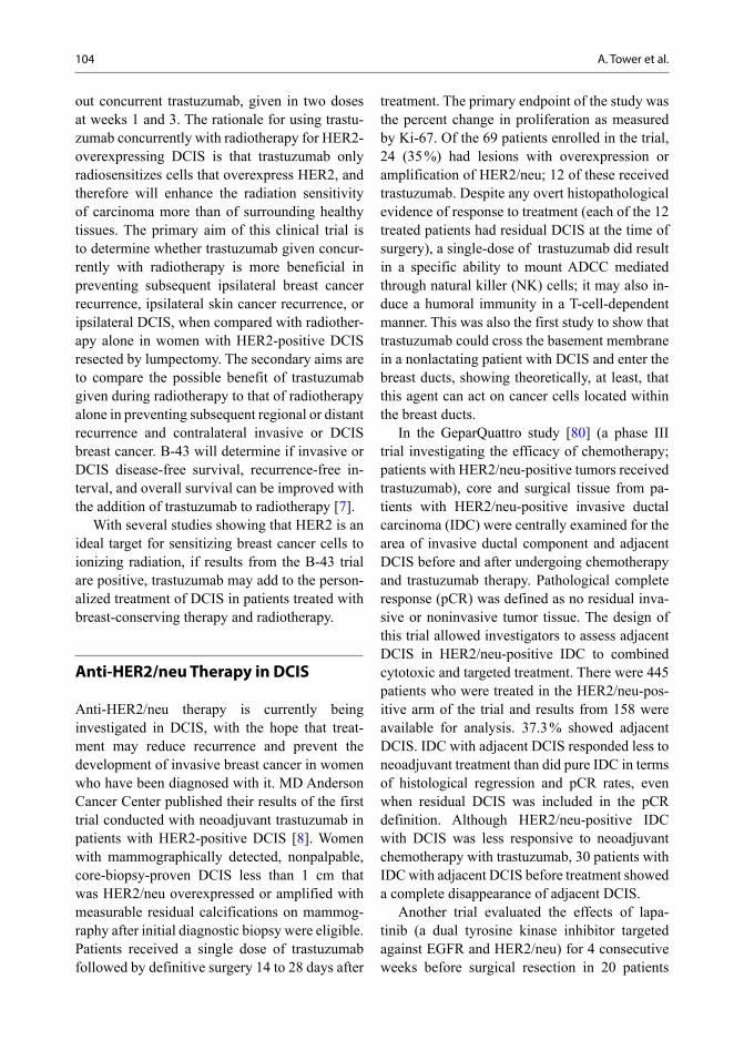
104 A. Tower et al.
out concurrent trastuzumab, given in two doses at weeks 1 and 3. The rationale for using trastu-zumab concurrently with radiotherapy for HER2-overexpressing DCIS is that trastuzumab only radiosensitizes cells that overexpress HER2, and therefore will enhance the radiation sensitivity of carcinoma more than of surrounding healthy tissues. The primary aim of this clinical trial is to determine whether trastuzumab given concur-rently with radiotherapy is more beneficial in preventing subsequent ipsilateral breast cancer recurrence, ipsilateral skin cancer recurrence, or ipsilateral DCIS, when compared with radiother-apy alone in women with HER2-positive DCIS resected by lumpectomy. The secondary aims are to compare the possible benefit of trastuzumab given during radiotherapy to that of radiotherapy alone in preventing subsequent regional or distant recurrence and contralateral invasive or DCIS breast cancer. B-43 will determine if invasive or DCIS disease-free survival, recurrence-free in-terval, and overall survival can be improved with the addition of trastuzumab to radiotherapy [7].
With several studies showing that HER2 is an ideal target for sensitizing breast cancer cells to ionizing radiation, if results from the B-43 trial are positive, trastuzumab may add to the person-alized treatment of DCIS in patients treated with breast-conserving therapy and radiotherapy.
Anti-HER2/neu Therapy in DCIS
Anti-HER2/neu therapy is currently being investigated in DCIS, with the hope that treat-ment may reduce recurrence and prevent the development of invasive breast cancer in women who have been diagnosed with it. MD Anderson Cancer Center published their results of the first trial conducted with neoadjuvant trastuzumab in patients with HER2-positive DCIS [8]. Women with mammographically detected, nonpalpable, core-biopsy-proven DCIS less than 1 cm that was HER2/neu overexpressed or amplified with measurable residual calcifications on mammog-raphy after initial diagnostic biopsy were eligible. Patients received a single dose of trastuzumab followed by definitive surgery 14 to 28 days after
treatment. The primary endpoint of the study was the percent change in proliferation as measured by Ki-67. Of the 69 patients enrolled in the trial, 24 (35 %) had lesions with overexpression or amplification of HER2/neu; 12 of these received trastuzumab. Despite any overt histopathological evidence of response to treatment (each of the 12 treated patients had residual DCIS at the time of surgery), a single-dose of trastuzumab did result in a specific ability to mount ADCC mediated through natural killer (NK) cells; it may also in-duce a humoral immunity in a T-cell-dependent manner. This was also the first study to show that trastuzumab could cross the basement membrane in a nonlactating patient with DCIS and enter the breast ducts, showing theoretically, at least, that this agent can act on cancer cells located within the breast ducts.
In the GeparQuattro study [80] (a phase III trial investigating the efficacy of chemotherapy; patients with HER2/neu-positive tumors received trastuzumab), core and surgical tissue from pa-tients with HER2/neu-positive invasive ductal carcinoma (IDC) were centrally examined for the area of invasive ductal component and adjacent DCIS before and after undergoing chemotherapy and trastuzumab therapy. Pathological complete response (pCR) was defined as no residual inva-sive or noninvasive tumor tissue. The design of this trial allowed investigators to assess adjacent DCIS in HER2/neu-positive IDC to combined cytotoxic and targeted treatment. There were 445 patients who were treated in the HER2/neu-pos-itive arm of the trial and results from 158 were available for analysis. 37.3 % showed adjacent DCIS. IDC with adjacent DCIS responded less to neoadjuvant treatment than did pure IDC in terms of histological regression and pCR rates, even when residual DCIS was included in the pCR definition. Although HER2/neu-positive IDC with DCIS was less responsive to neoadjuvant chemotherapy with trastuzumab, 30 patients with IDC with adjacent DCIS before treatment showed a complete disappearance of adjacent DCIS.
Another trial evaluated the effects of lapa-tinib (a dual tyrosine kinase inhibitor targeted against EGFR and HER2/neu) for 4 consecutive weeks before surgical resection in 20 patients
10510 Anti-HER2/neu Therapy in DCIS
with HER2/neu-positive DCIS [81]. Lapatinib showed significant antitumor effects through the RAS/mitogen-activated protein kinase (MAPK) signaling pathway by decreasing cytoplasm pERK1 in 11 patients and decreasing MRI signal intensity and tumor size in 7 patients.
There are two ongoing clinical trials of trastu-zumab in DCIS. NSABP B-43 is a multi-institu-tion, prospective, randomized phase III trial tar-geting high-risk, HER2/neu-positive DCIS [7]. Its primary aims are to determine whether trastuzum-ab given concurrently with radiotherapy is more beneficial in preventing subsequent ipsilateral breast cancer recurrence, ipsilateral skin cancer recurrence, or ipsilateral DCIS than is radiothera-py alone. A major secondary aim is to evaluate the effect of two doses of trastuzumab on contralat-eral breast cancer, given the improved immunity seen after a single dose of trastuzumab. Patients with DCIS resected by lumpectomy with histo-logically free margins whose tumors test positive for HER2/neu by central testing are randomly assigned to receive either trastuzumab plus radi-ography or radiotherapy alone. The trastuzumab is given in two doses: 8 mg/kg with the start of whole breast radiotherapy, followed by 6 mg/kg 3 weeks later. Patients with ER-positive and/or PR-positive DCIS are to receive a minimum of 5 years of hormone therapy. The rationale for using trastuzumab concurrently with radiotherapy is that trastuzumab only radiosensitizes cells that overexpress HER2, and therefore will enhance the radiation sensitivity of carcinoma more than sur-rounding healthy tissues. As of August 31, 2014, a total of 1,907 patients have been accrued (95.4 % of accrual goal). In this ongoing trial, there have been no grade 4 or 5 toxicity or trastuzumab-re-lated safety signals observed. This trial opened in November 2008 with an expected enrollment of 2000, which should be reached by end of 2014.
The second ongoing clinical trial is a neoadju-vant trial of lapatinib for the treatment of DCIS [82]. This randomized phase I/II trial’s primary outcome measures are reduction in the percentage of Ki67-positive cells and incidence of adverse events, and its secondary outcome measures are incidence of DCIS remaining at resection as well as biomarker analysis of proliferation markers.
Patients with DCIS that is HER2/neu-positive or is EGFR-positive are randomly assigned to receive either 2–6 weeks of lapatinib or placebo. This study began in January 2008 and is expected to complete enrollment in September 2014 with an expected accrual of 60 patients.
Conclusion
Treatment of DCIS is becoming more personal-ized, as it is for invasive breast cancer. Targeted molecular therapies have improved outcomes for patients with invasive breast cancer. Early work has demonstrated synergy between HER2-target-ed therapy and radiotherapy. Anti-HER2 therapy may also induce an immune response. Ongoing clinical trials ask whether HER2-targeted therapy will improve patient outcomes in DCIS.
References
1. Davidson NE. Fifteen years of anti-HER2 therapy. Oncology (Williston Park). 2013;27(3):151.
2. Ross JS. Saving lives with accurate HER2 testing. Am J Clin Pathol. 2010;134(2):183–4.
3. Slamon DJ, Leyland-Jones B, Shak S, et al. Use of che-motherapy plus a monoclonal antibody against HER2 for metastatic breast cancer that overexpresses HER2. N Engl J Med. 2001;344(11):783–92.
4. Allred DC, Clark GM, Tandon AK, et al. HER2 in node-negative breast cancer: prognostic significance of overexpression influenced by the presence of in situ carcinoma. J Clin Oncol. 1992;10(4):566–605.
5. Allred DC, Clark GM, Molina R, et al. Overexpres-sion of HER2 and its relationship with other prognostic factors change during the progression of in situ to inva-sive breast cancer. Hum Pathol. 1992;23(9):974–9.
6. Eccles SA. The role of c-erbB-2/HER2/neu in breast cancer progression and metastasis. J Mammary Gland Biol Neoplasia. 2001;6(4):393–406.
7. NSABP B-43 protocol. ClinicalTrials identifier: NCT00769379. https://clinicaltrials.gov/ct2/show/NCT00769379?term=NSABP+B-43&rank=1. Accessed on 23 Sept 2014.
8. Kuerer HM, Buzdar AU, Mittendorf EA, et al. Biologic and immunologic effects of preoperative trastuzumab for ductal carcinoma in situ of the breast. Cancer. 2011;117(1):39–47.
9. Lester SC, Bose S, Chen Y, et al. Protocol for the examination of specimens from patients with ductal carcinoma in situ of the breast. Arch Pathol Lab Med. 2009;133(1):15–25.

106 A. Tower et al.
10. Fitzgibbons PL, Henson DE, Hutter RV. Benign breast changes and the risk for subsequent breast cancer: an update of the 1985 consensus statement. Cancer Committee of the College of American Pathologists. Arch Pathol Lab Med. 1998;122(12):1053–5.
11. Siziopikou KP. Ductal carcinoma in situ of the breast: current concepts and future directions. Arch Pathol Lab Med. 2013;137(4):462–6.
12. Consensus conference on the classification of duc-tal carcinoma in situ. The Consensus Conference Committee. Cancer. 1997;80(9):1798–802.
13. Bur ME, Zimarowski MJ, Schnitt SJ, et al. Estrogen receptor immunohistochemistry in carcinoma in situ of the breast. Cancer. 1992;69(5):1174–81.
14. Bobrow LG, Happerfield LC, Bult P, et al. The classification of ductal carcinoma in situ and its asso-ciation with biological markers. Semin Diagn Pathol. 1994;11(3):199–207.
15. Bose S, Lesser ML, Norton L, et al. Immunopheno-type of intraductal carcinoma. Arch Pathol Lab Med. 1996;120(1):81–5.
16. Rudas M, Neumayer R, Gnant MF, et al. p53 protein expression, cell proliferation and steroid hormone re-ceptors in ductal and lobular in situ carcinomas of the breast. Eur J Cancer. 1997;33(1):39–44.
17. Mack L, Doig G, O’Malley FP. Relationship of a new histological categorization of ductal carcinoma in situ of the breast with size and the immunohistochemical expression of p53, c-erbB-2, bcl-2 and ki-67. Hum Pathol. 1997;28(8):974–9.
18. van de Vijver MJ, Peterse, Mooi WJ, et al. Neu-protein overexpression in breast cancer. Association with comedo-type ductal carcinoma in situ and limited prognostic value in stage II breast cancer. N Engl J Med. 1988;319(19):1239–45.
19. Bartkova J, Barnes DM, Millis RR, et al. Immuno-histochemical demonstration of c-erbB-2 protein in mammary ductal carcinoma in situ. Hum Pathol. 1990;21(11):1164–7.
20. Poller DN, Roberts EC, Bell JA, et al. p53 protein expression in mammary ductal carcinoma in situ: relationship to immunohistochemical expression of estrogen receptor and c-erbB-2 protein. Hum Pathol. 1993;24(5):463–8.
21. Guidi AJ, Fischer L, Harris JR, et al. Microvessel density and distribution in ductal carcinoma in situ of the breast. J Natl Cancer Inst. 1994;86(8):614–9.
22. Engels K, Fox SB, Whitehouse RM, et al. Distinct angiogenic patterns are associated with high grade in situ ductal carcinomas of the breast. J Pathol. 1997;181(2):207–12.
23. Lagios MD, Westdahl PR, Margolin FR, et al. Duct car-cinoma in situ: relationship of extent of non-invasive disease to the frequency of occult invasion, multicen-tricity, lymph node metastases and short-term treat-ment failures. Cancer. 1982;50(7):1309–14.
24. Holland R, Hendriks JH. Microcalcifica-tions associated with ductal carcinoma in situ: mammographic-pathologic correlation. Semin Diagn Pathol. 1994;11(3):181–92.
25. Faverly DRG, Burgers L, Bult P, et al. Three dimensional imaging of mammary ductal carcinoma in situ: clinical implications. Semin Diagn Pathol. 1994;11(3):193–8.
26. Solin LJ, Kurtz J, Fourquet A, et al. Fifteen year results of breast-conserving surgery and definitive breast ir-radiation for the treatment of ductal carcinoma in situ of the breast. J Clin Oncol. 1996;14(3):754–63.
27. Scott MA, Lagios MD, Axelsson K, et al. Ductal carcinoma in situ of the breast: reproducibil-ity of histologic subtype analysis. Hum Pathol. 1997;28(8):967–73.
28. Holland R, Peterse JL, Millis RR, et al. Ductal carcinoma in situ: a proposal for a new classification. Semin Diagn Pathol. 1994;11(3):167–80.
29. Sloane JP, Amendoeira I, Apostolikas N, et al. Consistency achieved by 23 European pathologists in categorizing ductal carcinoma in situ of the breast using five classifications. European Commission Working Group on Breast Screening Pathology. Hum Pathol. 1998;29(10):1056–62.
30. Livasy CA, Perou CM, Karaca G, et al. Identifica-tion of basal-like subtype of breast ductal carcinoma in situ. Hum Pathol. 2007;38(2):197–204.
31. Bryan BB, Schnitt SJ, Collins LC. Ductal carcinoma in situ with basal-like phenotype: a possible precur-sor to invasive basal-like breast cancer. Mod Pathol. 2006;19(5):617–21.
32. Claus EB, Chu P, Howe CL, et al. Pathobiologic find-ings in DCIS of the breast: morphologic features, angiogenesis, HER2/neu and hormone receptors. Exp Mol Pathol. 2001;70(3):303–16.
33. Siziopikou KP, Khan S. Correlation of HER2 gene amplification with expression of the apoptosis-sup-pressing genes bcl-2 and bcl-x-L in ductal carcinoma in situ of the breast. Appl Immunohistochem Mol Morphol. 2005;13(1):14–8.
34. Lebeau A, Unholzer A, Amann G, et al. EGFR, HER2/neu, cyclin D1, p21 and p53 in correlation to cell proliferation and steroid hormone receptor status in ductal carcinoma in situ of the breast. Breast Can-cer Res Treat. 2003;79(2):187–98.
35. Cornfield DB, Palazzo JP, Schwartz GF, et al. The prognostic significance of multiple morphologic features and biologic markers in ductal carcinoma in situ of the breast: a study of a large cohort of patients treated with surgery alone. Cancer. 2004;100(11):2317–27.
36. Barnes NL, Khavari S, Boland GP, et al. Absence of HER4 expression predicts recurrence of ductal carcinoma in situ of the breast. Clin Cancer Res. 2005;11(6):2163–8.
37. Collins LC, Schnitt SJ. HER2 protein overexpression in estrogen receptor-positive ductal carcinoma in situ of the breast: frequency and implications for tamoxi-fen therapy. Mod Pathol. 2005;18(5):615–20.
38. Provenzano E, Hopper JL, Giles GG, et al. Biological markers that predict recurrence in duc-tal carcinoma in situ of the breast. Eur J Cancer. 2003;39(5):622–30.

10710 Anti-HER2/neu Therapy in DCIS
39. Rodrigues NA, Dillon D, Carter D, et al. Differences in the pathologic and molecular features of intra-ductal breast carcinoma between younger and older women. Cancer. 2003;97(6):1393–403.
40. Perin T, Canzonieri V, Massarut S, et al. Immunohis-tochemical evaluation of multiple biological markers in ductal carcinoma in situ of the breast. Eur J Cancer. 1996;32A(7):1148–55.
41. Ringberg A, Anagnostaki L, Anderson H, et al. Cell biological factors in ductal carcinoma in situ (DCIS) of the breast-relationship to ipsilateral local recur-rence and histopathological characteristics. Eur J Cancer. 2001;37(12):1514–22.
42. Bijker N, Peterse JL, Duchateau L, et al. Histologi-cal type and marker expression of the primary tu-mour compared with its local recurrence after breast-conserving therapy for ductal carcinoma in situ. Br J Cancer. 2001;84(4):539–44.
43. Ramachandra S, Machin L, Ashley S, et al. Immu-nohistochemical distribution of c-erbB-2 in in situ breast carcinoma—a detailed morphological analy-sis. J Pathol. 1990;161(1):7–14.
44. Schimmelpenning H, Eriksson ET, Pallis L, et al. Immunohistochemical c-erbB-2 protooncogene expression and nuclear DNA content in human mam-mary carcinoma in situ. Am J Clin Pathol. 1992;97(5 Suppl 1):S48–52.
45. Bobrow LG, Happerfield LC, Gregory WM, et al. Ductal carcinoma in situ: assessment of necrosis and nuclear morphology and their association with bio-logical markers. J Pathol. 1995;176(4):333–41.
46. Leal CB, Schmitt FC, Bento MJ, et al. Ductal carcinoma in situ of the breast. Histologic categoriza-tion and its relationship to ploidy and immunohisto-chemical expression of hormone receptors, p53, and c-erbB-2 protein. Cancer. 1995;75(8):2123–31.
47. Somerville JE, Clarke LA, Biggart JD. c-erbB-2 over-expression and histological type of in situ and invasive breast carcinoma. J Clin Pathol. 1992;45(1):16–20.
48. Siziopikou KP, Anderson SJ, Cobleigh MA, et al. Pre-liminary results of centralized HER2 testing in ductal carcinoma in situ of the breast (DCIS): NSABP B-43. Breast Cancer Res Treat. 2013;142(2):415–21.
49. Lopez-Garcia MA, Geyer FC, Lacroix-Triki M, et al. Breast cancer precursors revisited: molecular features and progression pathways. Histopathology. 2010;57(2):171–92.
50. Ellis MJ, Coop A, Singh B, et al. Letrozole is more effective neoadjuvant endocrine therapy than tamoxi-fen for ErbB1 and/or ErbB2-positive, estrogen re-ceptor positive primary breast cancer: evidence from a phase III randomized trial. J Clin Oncol. 2001;19(18):3808–16.
51. Dowsett M. Overexpression of HER2 as a resistance mechanism to hormonal therapy for breast cancer. Endocr Relat Cancer. 2001;8(3):191–5.
52. Benz CC, Scott GK, Sarup JC, et al. Estrogen- dependent, tamoxifen-resistant tumorigenic growth of MCF-7 cells transfected with HER2/neu. Breast Cancer Res Treat. 1992;24(2):85–95.
53. Nicholson RI, Hutcheson IR, Harper ME, et al. Modulation of epidermal growth factor receptor in endocrine-resistant, estrogen-receptor-positive breast cancer. Ann N Y Acad Sci. 2002;963:104–15.
54. Osborne CK, Shou J, Massarweh S, et al. Crosstalk between estrogen receptor and growth factor receptor pathways as a cause for endocrine therapy resis-tance in breast cancer. Clin Cancer Res. 2005;11(2 pt 2):865s–70s.
55. Slamon, DJ, Clark GM, Wong SG, et al. Human breast cancer: correlation of relapse and survival with amplification of the HER-2/neu oncogene. Science. 1987;235(4785):177–82.
56. Bazell R. HER2: the making of Herceptin, a revolutionary treatment for breast cancer. New York: Random House; 1998.
57. Cobleigh MA, Vogel CL, Tripathy D, et al. Multina-tional study of the efficacy and safety of humanized anti-HER2 monoclonal antibody in women who have HER2-overexpressing metastatic breast cancer that has progressed after chemotherapy for metastatic dis-ease. J Clin Oncol. 1999;17(9):2639–48.
58. Romond EH, Perez EA, Bryant J, et al. Trastuzumab plus adjuvant chemotherapy for oper-able HER2-positive breast cancer. N Engl J Med. 2005;353(16):1673–84.
59. Perez EA, Romond EH, Vera J, et al. Four-year follow-up of trastuzumab plus adjuvant chemother-apy for operable human epidermal growth factor re-ceptor 2-positive breast cancer: joint analysis of data from NCCTG N9831 and NSABP B-31. J Clin On-col. 2011;29(25):3366–73.
60. Goldhirsch A, Gelber RD, Piccart-Gebhart MJ, et al. 2 years versus 1 year of adjuvant trastuzum-ab for HER2-positive breast cancer (HERA): an open-label, randomised controlled trial. Lancet. 2013;382(9897):1021–28.
61. Slamon D, Eiermann W, Robert N, et al. Adjuvant trastuzumab in HER2-positive breast cancer. N Engl J Med. 2011;365(14):1273–83.
62. Herceptin Prescribing Information. http://www.gene.com/download/pdf/herceptin_prescribing.pdf. Accessed on 23 Sept 2014.
63. Early Breast Cancer Trialists’ Collaborative Group (EBCTCG), Darby S, McGale P, et al. Effect of radiotherapy after breast-conserving surgery on 10-year recurrence and 15-year breast cancer death: meta-analysis of individual patient data for 10,801 women in 17 randomised trials. Lancet. 2011;378(9804):1707–16.
64. Early Breast Cancer Trialists’ Collaborative Group (EBCTCG), Correa C, McGale P, et al. Overview of the randomized trials of radiotherapy in ductal carcinoma in situ of the breast. J Natl Cancer Inst Monogr. 2010;2010(41):162–77.
65. Yu D, Hung MC. Overexpression of ErbB2 in cancer and ErbB2-targeting strategies. Oncogene. 2000;19(53):6115–21.

108 A. Tower et al.
66. Rodrigues L, Mansi J, Griffiths J. Radiation enhanc-es the antitumor effect of herceptin on c-neu/HER2 mammary carcinomas in transgenic oncomice. Proc Am Assoc Cancer Res. 2003;44:30, Abstract 132.
67. Liang K, Lu Y, Jin W, et al. Sensitization of breast cancer cells to radiation by trastuzumab. Mol Cancer Ther. 2003;2(11):1113–20.
68. Alanyali SD, Bozkurt E, Alanyali H, et al. Radiosen-sitization of HER2-positive breast cancer cell lines with trastuzumab. 2013 ASCO Annual Meeting. J Clin Oncol. 2013;31(15s):abstr e11501.
69. Sartor CI. Epidermal growth factor family receptors and inhibitors: radiation response modulators. Semin Radiat Oncol. 2003;13(1):22–30.
70. Milas L, Mason K, Hunter N, et al. In vivo enhance-ment of tumor radioresponse by C225 antiepidermal growth factor receptor antibody. Clin Cancer Res. 2000;6(2):701–8.
71. Nasu S, Ang KK, Fan Z, et al. C225 antiepidermal growth factor receptor antibody enhances tumor radiocurability. Int J Radiat Oncol Biol Phys. 2001;51(2):474–7.
72. Huang SM, Bock JM, Harari PM. Epidermal growth factor receptor blockade with C225 modulates proliferation, apoptosis, and radiosensitivity in squa-mous cell carcinomas of the head and neck. Cancer Res. 1999;59(8):1935–40.
73. Huang SM, Harari PM. Modulation of radiation response after epidermal growth factor receptor blockade in squamous cell carcinomas: inhibition of damage repair, cell cycle kinetics, and tumor angio-genesis. Clin Cancer Res. 2000;6(6):2166–74.
74. Huang SM, Li J, Armstrong EA. Modulation of radiation response and tumor-induced angiogenesis after epidermal growth factor receptor inhibition by ZD1839 (Iressa). Cancer Res. 2002;62(15):4300–6.
75. Huang SM, Li J, Harari PM. Molecular inhibi-tion of angiogenesis and metastatic potential in human squamous cell carcinomas after epidermal growth factor receptor blockade. Mol Cancer Ther. 2002;1(7):507–14.
76. Liang K, Ang KK, Milas L, et al. The epidermal growth factor receptor mediates radioresistance. Int J Radiat Oncol Biol Phys. 2003;57(1):246–54.
77. Harari PM, Huang SM. Epidermal growth factor receptor modulation of radiation response: preclini-cal and clinical development. Semin Radiat Oncol. 2002;12(3 Suppl 2):21–6.
78. Uno M, Otsuki T, Kurebayashi J, et al. Anti-HER2-antibody enhances irradiation-induced growth inhibition in head and neck carcinoma. Int J Cancer. 2001;94(4):474–9.
79. Wattenberg MM, Kwilas AR, Gameiro SR, et al. Ex-panding the use of monoclonal antibody therapy of cancer by using ionising radiation to upregulate anti-body targets. Br J Cancer. 2014;110(6):1472–80.
80. von Minckwitz G, Darb-Esfahani S, Loibl S, et al. Responsiveness of adjacent ductal carcinoma in situ and changes in HER2 status after neoadjuvant chemotherapy/trastuzumab treatment in early breast cancer—results from the GeparQuattro study (GBG 40). Breast Cancer Res Treat. 2012;132(3):863–70.
81. Estevez LG, Suarez A, Calvo I, et al. Molecular effects of lapatinib in HER2 positive ductal carci-noma in situ (DCIS). Cancer Res. 2012;72(24_suppl 3):SABCS P5-18-15.
82. ClinicalTrials.gov Identifier: NCT00555152, PI Powell Brown, MD.
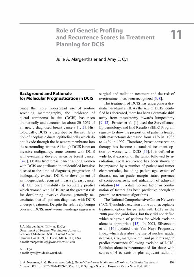
109
Role of Genetic Profiling and Recurrence Scores in Treatment Planning for DCIS
Julie A. Margenthaler and Amy E. Cyr
L. A. Newman, J. M. Bensenhaver (eds.), Ductal Carcinoma In Situ and Microinvasive/Borderline BreastCancer, DOI 10.1007/978-1-4939-2035-8_11, © Springer Science+Business Media New York 2015
J. A. Margenthaler () · A. E. CyrDepartment of Surgery, Washington University School of Medicine, 660 S. Euclid Avenue, Campus Box 8109, St. Louis, MO 63110, USA e-mail: [email protected]
A. E. Cyre-mail: [email protected]
Background and Rationale for Molecular Prognostication in DCIS
Since the more widespread use of routine screening mammography, the incidence of ductal carcinoma in situ (DCIS) has risen dramatically and accounts for about 20–30 % of all newly diagnosed breast cancers [1, 2]. His-tologically, DCIS is described by the prolifera-tion of neoplastic ductal epithelial cells which do not invade through the basement membrane into the surrounding stroma. Although DCIS is not an invasive malignancy, some women with DCIS will eventually develop invasive breast cancer [3–7]. Deaths from breast cancer among women with DCIS are attributed to unidentified invasive disease at the time of diagnosis, progression of inadequately excised DCIS, or development of an independent, recurrent invasive breast cancer [3]. Our current inability to accurately predict which women with DCIS are at the greatest risk for developing invasive disease generally ne-cessitates that all patients diagnosed with DCIS undergo treatment. Despite the relatively benign course of DCIS, most women undergo aggressive
surgical and radiation treatment and the risk of overtreatment has been recognized [3, 8].
The treatment of DCIS has undergone a dra-matic paradigm shift. As the size of DCIS identi-fied has decreased, there has been a dramatic shift away from mastectomy towards lumpectomy [9–12]. Ernster et al. [1] used the Surveillance, Epidemiology, and End Results (SEER) Program registry to show the proportion of patients treated with mastectomy decreased from 71 % in 1983 to 44 % in 1992. Therefore, breast-conservation therapy has become a standard treatment op-tion for women with DCIS [13]. It is defined as wide local excision of the tumor followed by ir-radiation. Local recurrence has been shown to be impacted by a number of patient and tumor characteristics, including patient age, extent of disease, nuclear grade, margin status, presence of comedonecrosis, and utilization of adjuvant radiation [14]. To date, no one factor or combi-nation of factors has been predictive enough to generalize treatment algorithms.
The National Comprehensive Cancer Network (NCCN) included excision alone as an acceptable treatment option for patients with DCIS in the 2008 practice guidelines, but they did not define which subgroup of patients for which excision alone is appropriate [15]. In 2003, Silverstein et al. [16] updated their Van Nuys Prognostic Index which describes the use of nuclear grade, necrosis, size, margin width, and patient’s age to predict recurrence following excision of DCIS. Excision alone is recommended for those with scores of 4–6; excision plus adjuvant radiation
11

J. A. Margenthaler and A. E. Cyr110
therapy is recommended for those with scores of 7–9; and mastectomy is recommended for those with scores of 10–12 [16]. This translated to a less than 20 % local recurrence rate at 12 years when these criteria were followed [16]. Howev-er, the primary limitation for the Van Nuys Prog-nostic Index is the lack of an ability to account for the wide heterogeneity of DCIS.
The clinical dilemma regarding the care of women with DCIS is apparent. The major-ity of women diagnosed with DCIS undergo breast-conserving therapy. The number of women subsequently affected by the decision regarding adjuvant radiation therapy is staggering. As a re-sult, the potential impact of a reliable, accurate molecular tool with the ability to differentiate those who will benefit from adjuvant radiation from those who are so low risk for future inva-sive disease that adjuvant radiation has no real role is invaluable.
DCIS Score Design and Validation
Molecular and biologic markers that provide prognostic and predictive information hold the most promise for tailoring therapy on an indi-vidual level. Expression of p16, COX-2, and Ki-67, which are indicative of an abrogated response to cellular stress, has been shown to delineate which DCIS lesions are more likely to confer a high risk for recurrence [17]. The 21-gene recur-rence score for estrogen receptor (ER)-positive invasive breast cancer was used as a benchmark for the development of a 12-gene subset that has been used to develop and validate a DCIS Score that divides patients into low risk, intermediate risk, and high risk for a 10-year local in-breast recurrence [18]. The DCIS Score was prospec-tively evaluated using archived tumor samples from the Eastern Cooperative Oncology Group (ECOG) E5194 study [19]. This prospective, nonrandomized study investigated the risk of local recurrence in 670 patients with DCIS fol-lowing wide local excision alone. Patients were required to have negative margin widths of ≥ 3 mm and were divided into two treatment arms consisting of grade 1 or 2, size ≤ 2.5 cm or grade
3, size ≤ 1.0 cm. Radiation was not allowed, but approximately 30 % of patients did receive op-tional adjuvant tamoxifen. The 5-year risk of an ipsilateral breast event was 6.1 % in the low/intermediate-grade group ( n = 565) and 15.3 % in the high-grade group ( n = 105), and the 7-year risk of an ipsilateral breast event was 10.5 % in the low/intermediate-grade group ( n = 565) and 18.0 % in the high-grade group ( n = 105). For the low/intermediate-grade group, there were 49 total ipsilateral breast events, including 26 with inva-sive cancer and 23 with DCIS. For the high-grade group, there were 17 total ipsilateral events, in-cluding 6 with invasive cancer and 11 with DCIS.
The inconsistent use of tamoxifen by patients with DCIS did require development of a modi-fied algorithm before the clinical validation study could be conducted. This was accomplished using data from the National Surgical Adjuvant Breast and Bowel Project (NSABP) B-14 study and a case-control study [20, 21]. As a result, seven genes that were predictive of recurrence and five reference genes were chosen (Table 11.1). The risk categories include low risk (DCIS Score < 39), intermediate risk (DCIS Score 39–54), and high risk (DCIS Score 55–100).
The E5194 trial was initiated in 1997 and was chosen as the independent study for DCIS Score validation. Of the 670 patients enrolled in the trial, 327 (49 %) had tissue that was suf-ficient for analysis. This trial was designed to determine which clinical and pathologic features, if any, could predict a subset of patients at low risk for local failure without the use of adjuvant radiation [19]. Of the 327 patients who had suffi-cient tissue for DCIS Score validation, the tumor size was smaller than the 343 patients who were not included. There were no other differences between the two groups with respect to patient and tumor characteristics. The majority of pa-tients in the E5194 validation study had a low-risk DCIS Score ( n = 230). However, there were 53 patients who had intermediate-risk scores and 44 patients who had high-risk scores. This was somewhat surprising given that patients were enrolled into E5194 on the basis of “perceived” low-risk clinicopathologic features. As a result of this validation study, the DCIS Score was made

11 Role of Genetic Profiling and Recurrence Scores in Treatment Planning for DCIS 111
available for commercial use for women with DCIS who were treated by wide local excision, with or without tamoxifen.
DCIS Score and Ipsilateral Breast Events
The relationship between DCIS Score risk group and 10-year risk of an ipsilateral breast event, whether in situ or invasive, was highly statisti-cally significant ( P = 0.006 for any ipsilateral breast event and P = 0.003 for an invasive ipsi-lateral breast event) [18]. This was true with or without the adjustment for tamoxifen use. For the 230 patients with a low-risk DCIS Score, the 10-year risk of any ipsilateral breast event was 10.6 % (95 % CI 6.9–16.2 %) and 3.7 % (95 % CI 1.8–7.7 %) for an invasive breast event. For the 53 patients with an intermediate-risk DCIS Score, the 10-year risk of any ipsilateral breast event was 26.7 % (95 % CI 16.2–41.9 %) and 12.3 % (95 % CI 5.1–27.8 %) for an invasive breast event. For the 44 patients with a high-risk DCIS Score, the
10-year risk of any ipsilateral breast event was 25.9 % (14.8–43.1 %) and 19.2 % (95 % CI 9.5–36.4 %) for an invasive breast event. Multivari-able models of risk for ipsilateral breast events, both excluding and including the DCIS Score, were also performed. When the DCIS Score was excluded, tumor size and menopausal status were the only factors significantly associated with risk for an ipsilateral breast event (HR 1.54, P = 0.006 and HR 0.49, P = 0.02, respectively). However, when the DCIS Score was included in the model, the 12-gene score was statistically significant (HR 2.37, P = 0.02), in addition to tumor size and menopausal status. Tumor grade and the pres-ence of comedonecrosis were not associated with a risk for ipsilateral breast events.
DCIS Score and Adjuvant Radiation Therapy Use
There is currently no known subset of patients with DCIS who do not benefit from adjuvant radiation for risk reduction following breast-conserving
Table 11.1 Seven cancer-related genes and five reference genes included in the Oncotype DX DCIS Score [18, 22–29]Proliferation genesKi-67 Higher Ki-67 is seen in poorly differentiated DCIS; an increase in Ki-67 is associ-
ated with invasion [22, 23]STYKI5 Loss of overexpression is associated with progression from DCIS to invasive breast
cancer [24]Survivin Inhibits apoptosis; its expression is associated with DCIS recurrence [25]Cyclin B1 Overexpression is related to DCIS grade, Ki-67, and HER-2 overexpression [26]MYLBL2 More frequently seen in Luminal B versus Luminal A breast cancers; it is associated
with resistance to tamoxifen [27]Hormone receptor genesProgesterone receptor (PR) Predicts distant recurrence and survival in patients treated and not treated with
tamoxifen; its presence is associated with estrogen receptor positivity and lower-grade DCIS [18, 28, 29]
Other genesGSTM1 Predicts distant recurrence and survival in patients treated and not treated with
tamoxifen [18]References genesACTB –GAPDH –GUSB –RPLPO –TFRC –DCIS ductal carcinoma in situ

J. A. Margenthaler and A. E. Cyr112
therapy. This is true regardless of patient’s age, tumor size, tumor grade, and margin status [30]. In the Early Breast Cancer Trialists’ Collab-orative Group analysis, the risk of an ipsilateral breast event was decreased from 18.1 to 7.6 % at 5 years and 28.1–12.9 % at 10 years in patients with ≤ 2 cm of DCIS who underwent adjuvant radiation [30]. Despite this, survival is equiva-lent for excision alone and excision followed by adjuvant radiation therapy. Radiation therapy is not without risk. There are short-term side effects and potential long-term consequences, some of which can be devastating. In addition, the therapy is quite costly and access to care can be difficult for many patients. Are these risks and costs justi-fied for all patients with DCIS?
The ability to identify a subset of patients who could safely omit radiation is desirable. Thus, patients who have undergone wide local exci-sion for DCIS who have a low-risk score could reasonably elect to omit radiation therapy, while patients in the intermediate or high-risk groups should consider adjuvant radiation therapy and tamoxifen following wide local excision.
Limitations of the DCIS Score
Despite these promising findings, the DCIS Score has not yet been universally adopted for making treatment decisions. There are several important limitations. First, the E5194 study used to validate the DCIS Score included a narrow subset of patients with DCIS. Specifically, the study included patients already considered to be at low risk for recurrence, and included patients with small volumes of DCIS and with margins of at least 3 mm. Therefore, these results cannot be applied to women with larger volumes of DCIS or with closer surgical margins. In addition, only 30 % of the patients in the E5194 study received adjuvant tamoxifen therapy, and the absolute number of tamoxifen-treated patients included in the validation study was only 96 [18]. Because tamoxifen decreases the risk of both local recur-rence and contralateral breast cancer events in patients with ER-positive DCIS [6, 31], use of tamoxifen would likely mean fewer recurrences
for patients with all DCIS Scores. The ER gene was specifically excluded from the DCIS Score given that it specifically predicts benefit with tamoxifen therapy. For patients with ER-negative DCIS, the DCIS Score has not been adequately studied; only nine patients with ER-negative DCIS were included in the validation study (less than 3 % of the total cohort). Additional study is needed to verify these validation results across broader populations of patients with DCIS.
Although the DCIS Score is promising for stratifying patients by risk of in-breast recur-rence, it does not supplant other patient and tumor characteristics such as tumor size and menopaus-al status, which remained significant predictors of recurrence with and without inclusion of the DCIS Score on multivariate analysis, P < 0.02 for each [18]. The DCIS Score is therefore a helpful addition to, but not replacement for, the use of already available clinicopathologic features used to make treatment decisions.
It is important to note that the DCIS Score does not equate with recommendations for or against post-lumpectomy radiation for patients with DCIS. Rather, it provides a guide regarding expected local recurrence, of either in situ or in-vasive disease, when radiation therapy is omitted. Established studies demonstrating the reduced risk of in-breast recurrence due to the addition of radiation [30] should be included in the treatment discussion with patients. It also does not stratify recurrence risk into risk of only in situ or of inva-sive disease, the latter of which is associated with potential mortality and need for more aggressive treatment, such as lymph node evaluation, ex-tended radiation fields, chemotherapy, and bio-logic therapy. Such information would be particu-larly informative for guiding treatment decisions. In addition, the DCIS Score does not predict re-sponse to post-lumpectomy radiation. Indeed, a trial evaluating the utilization of the DCIS Score on outcomes would be of great interest.
Although the DCIS Score is associated with in-breast recurrence, the test cannot explain the behavior of individual DCIS cases: why does some DCIS remain indolent, and why do other cases progress to invasive disease? With this in-formation, overtreatment may truly be prevented.

11 Role of Genetic Profiling and Recurrence Scores in Treatment Planning for DCIS 113
Beyond Risk of Local Recurrence: Which DCIS Has the Potential for Invasion?
There is an increasing amount of knowledge of the genetic features of DCIS, but there is still not a uniformly accepted understanding of how DCIS and invasive breast cancer are related. DCIS is associated with many invasive breast cancers and tends to have similar grade, nuclear mor-phology, and molecular subtype as the invasive component [32–35]. In addition, low-grade DCIS is genetically similar to low-grade invasive can-cers with both frequently showing loss of the 16q and gain of the 1q chromosomal regions, and poorly differentiated DCIS and high-grade in-vasive cancers both demonstrate more frequent amplification of 17q12 [36, 37]. Despite these findings, not all DCIS lesions left unresected de-velop into invasive malignancies [38, 39].
Many investigators have analyzed synchro-nous ipsilateral DCIS and invasive breast cancer samples from the same patient in order to identify genetic similarities and differences between the in situ and invasive components. Many of these studies have focused on specific genes such as HER-2, MYC, and CCND1 [40–43], and there appears to be significant variation both between patients and within both in situ and invasive com-ponents in individual patients [40–46]. Because of this heterogeneity in these individual genes, it has proven difficult to identify universal genetic markers of progression to invasive disease [47].
Beyond Predictive and Prognostic Information
The capability to profile entire tumor genomes relatively quickly and affordably will continue to improve understanding of the relationship between DCIS and invasive disease [22, 40]. Instead of providing information only on individ-ual genes and gene expression, next-generation sequencing or massively parallel sequencing of-fers the capability to delineate the genomics of a tumor with an unprecedented amount of detail. While this information may improve understand-ing of the development and behavior of DCIS
and invasive cancer, the ultimate goal is to use this information to provide targeted therapies to prevent DCIS from ever evolving into an inva-sive cancer [23, 47, 48].
References
1. Ernster VL, Barclay J, Kerlikowske K, et al. Inci-dence of and treatment for ductal carcinoma in situ of the breast. J Am Med Assoc. 1996;275:913–8.
2. Schwartz GF, Solin LJ, Olivotto IA, Ernster VL, Pressman PI, Consensus Conference Committee. Consensus conference on the treatment of in situ ductal carcinoma of the breast, April 22–25, 1999. Cancer. 2000;88:946–54.
3. Ernster VL, Barclay J, Kerlikowske K, et al. Mortality among women with ductal carcinoma in situ of the breast in the population-based surveillance, epide-miology and end results program. Arch Intern Med. 2000;160:953–8.
4. Fisher B, Bryant J, Wolmark N, et al. Effect of preoperative chemotherapy on the outcome of women with operable breast cancer. J Clin Oncol. 1998;16:2672–85.
5. Fisher B, Costantino JP, Wickerham DL, et al. Tamox-ifen for prevention of breast cancer: report of the Na-tional Surgical Adjuvant Breast and Bowel Project P-1 Study. J Natl Cancer Inst. 1998;90:1371–88.
6. Fisher B, Dignam J, Wolmark N, et al. Tamoxi-fen in treatment of intraductal breast cancer: National Surgical Adjuvant Breast and Bowel Project B-24 randomised controlled trial. Lancet. 1999;353:1993–2000.
7. Julien JP, Bijker N, Fentiman IS, et al. Radiotherapy in breast-conserving treatment for ductal carcinoma in situ: first results of the EORTC randomised phase III trial 10853. EORTC Breast Cancer Cooperative Group and EORTC Radiotherapy Group. Lancet. 2000;355:528–33.
8. Fisher ES, Welch HG. Avoiding the unintended consequences of growth in medical care: how might more be worse? J Am Med Assoc. 1999;281:446–53.
9. Winchester DJ, Menck HR, Winchester DP. The National Cancer Data Base report on the results of a large nonrandomized comparison of breast preservation and modified radical mastectomy. Can-cer. 1997;80:162–7.
10. Winchester DJ, Menck HR, Winchester DP. National treatment trends for ductal carcinoma in situ of the breast. Arch Surg. 1997;132:660–5.
11. Winchester DP, Menck HR, Osteen RT, Kraybill W. Treatment trends for ductal carcinoma in situ of the breast. Ann Surg Oncol. 1995;2:207–13.
12. Winchester DP, Osteen RT, Menck HR. The National Cancer Data Base report on breast carcinoma char-acteristics and outcome in relation to age. Cancer. 1996;78:1838–43.

J. A. Margenthaler and A. E. Cyr114
13. Fisher B, Anderson S, Bryant J, et al. Twenty-year follow-up of a randomized trial comparing total mastectomy, lumpectomy, and lumpectomy plus irradiation for the treatment of invasive breast cancer. N Engl J Med. 2002;347:1233–41.
14. Benson JR, Wishart GC. Predictors of recurrence for ductal carcinoma in situ after breast-conserving surgery. Lancet Oncol. 2013;14:e348–57.
15. Carlson RW, Allred DC, Anderson BO, et al. NCCN clinical practice guidelines in oncology: breast cancer. Fort Washington: National Comprehensive Cancer Network. 2008. http://www.nccnorg. Accessed 23 Aug 2013.
16. Silverstein MJ. The University of Southern Califor-nia/Van Nuys prognostic index for ductal carcinoma in situ of the breast. Am J Surg. 2003;186:337–43.
17. Gauthier ML, Berman HK, Miller C, et al. Abrogated response to cellular stress identifies DCIS associated with subsequent tumor events and defines basal-like breast tumours. Cancer Cell. 2007;12:479–91.
18. Solin L, Gray R, Baehner F, et al. A multigene expression assay to predict local recurrence risk for ductal carcinoma in situ of the breast. J Natl Cancer Inst. 2013;105:701–10.
19. Hughes LL, Wang M, Page DL, et al. Local exci-sion alone without irradiation for ductal carcinoma in situ of the breast: a trial of the Eastern Cooperative Oncology Group. J Clin Oncol. 2009;27:5319–24.
20. Mam as E, Tang G, Fisher B, et al. Association between the 21-gene recurrence score assay and risk of locoregional recurrence in node-negative, estro-gen receptor-positive breast cancer: results from the NSABP B-14 and NSABP B-20. J Clin Oncol. 2010;28:1677–83.
21. Habel LA, Shak S, Jacobs MK, et al. A population-based study of tumor gene expression and risk of breast cancer death among lymph node-negative patients. Breast Cancer Res. 2006;8:R25.
22. Kaur H, Mao S, Shah S, et al. Next-generation sequencing: a powerful tool for the discovery of molecular markers in breast ductal carcinoma in situ. Expert Rev Mol Diagn. 2013;13:151–65.
23. Sarode VR, Han JS, Morris DH, Peng Y, Rao R. A comparative analysis of biomarker expression and molecular subtypes of pure ductal carcinoma in situ and invasive breast carcinoma by image analysis: relationship of the subtypes with histologic grade, Ki67, p53 overexpression, and DNA ploidy. Int J Breast Cancer. 2011;2011:217060.
24. Hoque A, Carter J, Xia W, et al. Loss of aurora A/STK15/BTAK overexpression correlates with transition of in situ to invasive ductal carcinoma of the breast. Cancer Epidemiol Biomark Prev. 2003;12:1518–22.
25. Barnes N, Haywood P, Flint P, Knox WF, Bundred NJ. Survivin expression in in situ and invasive breast cancer relates to COX-2 expression and DCIS recur-rence. Br J Cancer. 2006;94:253–8.
26. Bostrom P, Soderstrom M, Palokangas T, et al. Analysis of cyclins A, B1, D1 and E in breast cancer
in relation to tumour grade and other prognostic factors. BMC Res Notes. 2009;2:140.
27. Cheang M, Chia SK, Voduc D, et al. Ki67 index, HER2 status, and prognosis of patients with luminal B breast cancer. J Natl Cancer Inst. 2009;101:736–50.
28. Claus EB, Chu P, Howe CL, et al. Pathobiologic find-ings in DCIS of the breast: morphologic features, an-giogenesis, HER-2/neu and hormone receptors. Exp Mol Path. 2001;70:303–16.
29. Barnes NL, Boland GP, Davenport A, Knox WF, Bundred NJ. Relationship between hormone receptor status and tumour size, grade and comedo necrosis in ductal carcinoma in situ. Br J Surg. 2005;92:429–34.
30. Early Breast Cancer Trialists’ Collaborative Group (EBCTCG). Overview of the randomized trials of ra-diotherapy in ductal carcinoma in situ of the breast. J Natl Cancer Inst Monogr. 2010;2010:162–77.
31. Allred DC, Anderson SJ, Paik S, et al. Adjuvant tamoxifen reduces subsequent breast cancer in wom-en with estrogen receptor-positive ductal carcinoma in situ: a study based on NSABP Protocol B-24. J Clin Oncol. 2012;30:1268–73.
32. Fisher ER, Gregorio RM, Fisher B, Redmond C, Vel-lios F, Sommers SC. The pathology of invasive breast cancer: a syllabus derived from the findings of the National Surgical Adjuvant Breast Project (protocol no. 4). Cancer. 1975;36:1–85.
33. Steinman S, Wang J, Bourne P, Yang Q, Tang P. Expression of cytokeratin markers, Er-alpha, PR, HER-2/neu, and EGFR in pure ductal carcinoma in situ (DCIS) and DCIS with co-existing invasive duc-tal carcinoma (IDC) of the breast. Ann Clin Lab Sci. 2007;37:127–34.
34. Giardina C, Serio G, Lepore G, et al. Pure ductal carci-noma in situ and in situ component of ductal invasive carcinoma of the breast: a preliminary morphometric study. J Exp Clin Cancer Res. 2003;22:279–88.
35. Ottesen GL. Carcinoma in situ of the female breast: a clinic-pathological, immunohistological, and DNA ploidy study. APMIS Suppl. 2003;108:1–67.
36. Buerger H, Otterbach F, Simon R, et al. Comparative genomic hybridization of ductal carcinoma in situ of the breast: evidence of multiple genetic pathways. J Pathol. 1999;187:396–402.
37. Buerger H, Otterbach F, Simon R, et al. Different genetic pathways in the evolution of invasive breast cancer are associated with distinct morphologic subtypes. J Pathol. 1999;189:521–6.
38. Page DL, Dupont WD, Rogers LW, Jensen RA, Schuyler PA. Continued local recurrence of carcinoma 15–25 years after a diagnosis of low grade ductal carcinoma in situ of the breast treated only by biopsy. Cancer. 1995;76:1197–200.
39. Sanders ME, Schuyler PA, Dupont WD, Page DL. The natural history of low-grade ductal carcinoma in situ of the breast in women treated by biopsy only re-vealed over 30 years of long-term follow-up. Cancer. 2005;103:2481–4.
40. Jang MH, Kim EJ, Choi Y, et al. FGFR1 is amplified during the progression of in situ to invasive breast carcinoma. Breast Cancer Res. 2012;14:R115.

11 Role of Genetic Profiling and Recurrence Scores in Treatment Planning for DCIS 115
41. Burkhardt L, Grob TJ, Hermann I, et al. Gene amplification in ductal carcinoma in situ of the breast. Breast Cancer Res Treat. 2010;123:757–65.
42. Robanus-Maandag EC, Bosch CA, Kristel PM, et al. Association of C-MYC amplification with progression from the in situ to the invasive stage in C-MYC-amplified breast carcinomas. J Pathol. 2003;201:75–82.
43. Park K, Han S, Kim JH, Kim J, Shin E. HER2 status in pure ductal carcinoma in situ and in the intraductal and invasive components of invasive ductal carcinoma determined by fluorescence in situ hybrid-ization and immunohistochemistry. Histopathology. 2006;48:702–7.
44. Hernandez L, Wilkerson PM, Lambros MB, et al. Genomic and mutational profiling of ductal carci-nomas in situ and matched adjacent invasive breast cancers reveals intra-tumour genetic heterogeneity and clonal selection. J Pathol. 2012;227:42–52.
45. Heselmeyer-Haddad K, Berroa Garcia LY, Bradley A, et al. Single-cell genetic analysis of ductal carcinoma in situ and invasive breast cancer reveals enormous tumor heterogeneity yet conserved genomic imbal-ances and gain of MYC during progression. Am J Pathol. 2012;181:1807–22.
46. Vincent-Salomon A, Lucchesi C, Gruel N, et al. In-tegrated genomic and transcriptomic analysis of ductal carcinoma of the breast. Clin Cancer Res. 2008;14:1956–65.
47. Cowell CF, Weigelt B, Sakr R, et al. Progression from ductal carcinoma in situ to invasive breast cancer: re-visited. Mol Oncol. 2013;7:859–69.
48. Mwenifumbo JC, Marra MA. Cancer genome-sequencing study design. Nat Rev Genet. 2013;14:321–32.
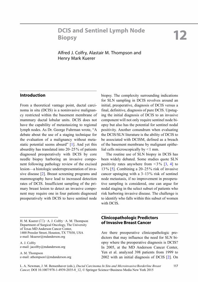
117
DCIS and Sentinel Lymph Node Biopsy
Alfred J. Colfry, Alastair M. Thompson and Henry Mark Kuerer
L. A. Newman, J. M. Bensenhaver (eds.), Ductal Carcinoma In Situ and Microinvasive/Borderline Breast Cancer, DOI 10.1007/978-1-4939-2035-8_12, © Springer Science+Business Media New York 2015
H. M. Kuerer () · A. J. Colfry · A. M. ThompsonDepartment of Surgical Oncology, The University of Texas MD Anderson Cancer Center, 1400 Pressler Street, Houston, TX 77030, USA e-mail: [email protected]
A. J. Colfrye-mail: [email protected]
A. M. Thompsone-mail: [email protected]
Introduction
From a theoretical vantage point, ductal carci-noma in situ (DCIS) is a noninvasive malignan-cy restricted within the basement membrane of mammary ductal lobular units. DCIS does not have the capability of metastasizing to regional lymph nodes. As Dr. George Fuhrman wrote, “A debate about the use of a staging technique for the evaluation of a malignancy without meta-static potential seems absurd” [1]. And yet this absurdity has translated into 20–25 % of patients diagnosed preoperatively with DCIS by core needle biopsy harboring an invasive compo-nent following pathology review of the excised lesion—a histologic underrepresentation of inva-sive disease [2]. Breast screening programs and mammography have lead to increased detection rates of DCIS. Insufficient sampling of the pri-mary breast lesion to detect an invasive compo-nent may require one in four patients diagnosed preoperatively with DCIS to have sentinel node
biopsy. The complexity surrounding indications for SLN sampling in DCIS revolves around an initial, preoperative, diagnosis of DCIS versus a final, definitive, diagnosis of pure DCIS. Upstag-ing the initial diagnosis of DCIS to an invasive component will not only require sentinel node bi-opsy but also has the potential for sentinel nodal positivity. Another conundrum when evaluating the DCIS/SLN literature is the ability of DCIS to be associated with DCISM, defined as a breach of the basement membrane by malignant epithe-lial cells microscopically by < 1 mm.
The routine use of SLN biopsy in DCIS has been widely debated. Some studies quote SLN positivity rates anywhere from < 3 % [3, 4] to 13 % [5]. Combining a 20–25 % risk of invasive cancer upstaging with a 3–13 % risk of sentinel node metastasis, if no improvement in preopera-tive sampling is considered, one can argue for nodal staging in the select subset of patients who risk harboring invasive disease. The challenge is to identify who falls within this subset of women with DCIS.
Clinicopathologic Predictors of Invasive Breast Cancer
Are there preoperative clinicopathologic pre-dictors that may influence the need for SLN bi-opsy where the preoperative diagnosis is DCIS? In 2005, at the MD Anderson Cancer Center, Yen et al. analyzed 398 patients from 1999 to 2002 with an initial diagnosis of DCIS [2]. On
12

118 A. J. Colfry et al.
final pathologic review, 66 patients (17 %) had invasive disease and a further 14 (3 %) had DCIS with microinvasion. Of these 80 patients, 58 (72 %) underwent SLN biopsy at the time of their initial operation sparing the patient a second future operation. Multivariate analysis revealed four independent predictors of invasive cancer: age < 55, diagnosis by core needle biopsy, large mammographic DCIS size > 5 cm, and high-grade DCIS. Only a palpable tumor was predic-tive of a positive sentinel node.
A few years later in 2008, again at the MD Anderson Cancer Center, Yi et al. expanded their experience from 1994 to 2006 to include analysis of 624 patients with a preoperative diagnosis of DCIS or DCISM [6]. Among 624 patients, 149 (23.9 %) were upstaged to invasive cancer on final pathologic assessment. Multivariate analysis revealed two independent predictors of invasive breast cancer: DCIS size > 5 cm and pre-operative diagnosis by core needle biopsy.
Several other studies have examined preoperative clinicopathologic predictors of inva-sive cancer. On univariate analysis, Guillot et al. identified palpable tumor, opacity on mammog-raphy, and preoperative high-grade DCIS as risk factors for invasion [7]. Lee et al. described the following associations with upstaging: use of core needle biopsy, DCIS > 15 mm, noncribiform sub-type of DCIS, intermediate- or high-nuclear-grade DCIS, and lack of hormone receptor [8].
Clinicopathologic Predictors of Increased Risk of Nodal Positivity
Others have found extensive disease requiring mastectomy and the presence of necrosis to be associated with an increased risk of nodal posi-tivity [9]; one study of 854 patients with pure DCIS found age, clinical presentation, and tumor size important prognostic factors for axillary nodal positivity [10].
The previous two studies described from the MD Anderson Cancer Center divided clini-copathological predictors of sentinel node positivity into two useful groups—predictors of positive SLN with an initial diagnosis of DCIS/DCISM and predictors of positive SLN with a
final diagnosis of DCIS/DCISM [2, 6]. Yi et al. [6] evaluated 624 women with an initial preop-erative diagnosis of DCIS or DCISM. Sentinel metastases were identified in 6.4 % of 624 pa-tients; half-harbored micrometastases (defined as a nodal tumor deposit < 2 mm in size) and half-harbored macrometastases (2 mm in size). The SLN was the only involved node in 92.5 % of patients. Multivariate analysis revealed DCIS size and final histologic type of invasive cancer as risk factors for sentinel node positivity. Yen et al. [2] revealed presence of a palpable tumor to be the only independent preoperative predictor for SLN positivity in DCIS [2].
Again, from 624 patients with a preoperative diagnosis of DCIS or DCISM, after pathologic assessment, 475 had a final diagnosis of DCIS or DCISM [6]. In this final diagnostic group, SLN metastases were confirmed in just nine patients (1.9 %), all but one with micrometastatic dis-ease. Multivariate analysis revealed DCIS size > 5 cm as the only independent predictor of SLN positivity for patients with an initial preoperative diagnosis and patients with a final diagnosis of DCIS/DCISM.
SLN Biopsy in DCIS: Who Needs One?
“The principal justification for SLN biopsy in DICS is diagnostic uncertainty,” writes Dr. Hiram Cody [11]. The clinician must ask the question whether or not he/she believes there is a risk of having missed invasive foci of cancer pre-operatively, through the sampling by core needle biopsy, which will be evident on final histology [10]. Intra et al. [10] propose this occurs for the following three scenarios:1. When breast conservation surgery results in
positive margins or residual microcalcifica-tions on postoperative follow-up mammogram
2. Large solid tumors3. Inability to sample diffuse or multicentric
microcalcifications on core needle/vacuum assisted biopsy [10]
In patients undergoing mastectomy for DCIS, there has been a documented 28–48 % risk of upstaging to invasive cancer [2]. As a result of the significant risk of upstaging, together with

11912 DCIS and Sentinel Lymph Node Biopsy
an inability to perform SLN biopsy post mastec-tomy, SLN biopsy may be considered advisable for patients undergoing mastectomy for DCIS. Certainly, this dogma was upheld by the recent guidelines published by the American Society of Clinical Oncology (ASCO) [12]. One needs to consider further the implications of SLN metastasis in patients with pure DCIS/DCISM, the consequences of identifying micrometastatic disease and isolated tumor cells (ITCs) in the SLN, and the utility of immunohistochemical (IHC) staining in SLN evaluation.
Consequences of SLN Positivity in Pure DCIS
For a final diagnosis of pure DCIS, SLN posi-tivity rates ranged from 0 to 18 % [2–4, 9, 10, 13–22] (Table 12.1).
When these data were combined in two me-ta-analyses, Ansari et al. reported an overall 3.7 % nodal positivity rate while van Deurzen et al. similarly reported the nodal positivity as 4 %; both were for patients with a definitive postoperative diagnosis of pure DCIS [23, 24]. The implications of SLN positivity in pure DCIS require further discussion.
From 1996 to 2006, Intra et al. evaluated 854 patients with pure DCIS who underwent SLN biopsy at the European Institute of Oncology [10]. DCIS with microinvasion was excluded. SLN metastases were discovered in 12 out of 854 patients (1.4 %)—a total of 5 patients with macro-metastases, 7 patients with micrometastases, and 4 patients with ITCs (pN0i +). Of the 12 patients with SLN positivity, 11 underwent axillary node dissection but none had additional nodal disease. Other studies [2–5, 19, 25] similarly demon-strate lone SLN positivity after formal axillary lymph node dissection. This questions the value of axillary dissection for pure DCIS with SLN positivity particularly in women with microme-tastases or ITCs.
Mabry et al. analyzed 564 patients with pure DCIS who underwent either axillary lymph node dissection or SLN biopsy [19]. Only 2 of 564 patients had positive nodal disease, both in the axillary dissection group. These patients were upstaged, underwent mastectomy followed by systemic treatment, and survived beyond 10 years without local or distant recurrence. Six patients in the axillary dissection group had local invasive recurrences and died from metastatic disease; all six had no evidence of nodal disease at the time of axillary dissection. In addition, of the 171 patients who underwent SLN biopsy,
Table 12.1 Analysis of retrospective studies evaluating sentinel lymph node (SLN) biopsy in patients with a final diagnosis of pure ductal carcinoma in situ (DCIS) [2–4, 9, 10, 13–22]Author Number of patients with DCIS in whom
SLN biopsy was performedNumber (%) of patients with positive SLN
Klauber De More [3] 72 5 (7 %)Wilkie [4] 559 27 (5 %)Moore [9] 470 43 (10 %)Veronesi [13] 508 9 (2 %)Zavagno [14] 102 1 (1 %)Kelly [15] 41 1 (2 %)Farkas [16] 46 0 (0 %)Mittendorf [17] 34 6 (18 %)Yen [2] 99 3 (3 %)Cserni [18] 36 4 (11 %)Mabry [19] 171 10 (6 %)Katz [20] 110 8 (7 %)Sakr [21] 39 4 (10 %)Fraile [22] 92 1 (1 %)Intra [10] 854 12 (1 %)

120 A. J. Colfry et al.
ITCs alone were detected immunohistochemical-ly in 10 patients but only 2 of the171 patients had local recurrence and none developed regional or distant recurrence. Consequently, the authors concluded that lymph node status did not predict poor outcome in patients with DCIS. However, lymph node status may predict which primary tumors have the potential to be upstaged. Treat-ment of DCIS will not influence long-term dis-ease-specific survival which already approaches 100 %; the focus should remain on eliminating invasive local recurrences. Perhaps, this is what a positive SLN means in the setting of DCIS—the patient has a higher risk of having a missed focus of primary invasion, and thus these patients have the potential to be upstaged and systemic therapy should be considered based on biologic subtype of the invasive component.
Even with invasive local recurrences after DCIS excision, outcomes remain encouraging. Lee et al. analyzed 1236 patients previously treated for pure DCIS in order to examine local invasive recurrence, distant recurrence, and breast cancer-specific mortality [26]. There were 150 local recurrences (87 DCIS and 63 invasive). The overall 12-year breast cancer-specific mor-tality after mastectomy versus breast-conserving surgery was 0.8 and 1.0 %, respectively. Even in the 63 patients with invasive local recurrence, the 12-year probabilities for metastasis and breast cancer-specific death were 15 and 12 %, respectively. Thus, even for the worst prognos-tic group of DCIS patients (those with local in-vasive recurrences), a favorable prognosis with minimal evidence of distant disease prevails and is unlikely to prove fatal. Thus, for DCIS, local control should be the ultimate goal; not improv-ing survival which already exceeds 98 %.
Consequences of SLN Positivity in DCISM
When a microinvasive component to DCIS is considered, SLN positivity may increase by up to 17 % [5]. Even though DCIS with microinva-sion (DCISM) is a rare pathologic entity (< 1 %), it can be intimately associated with a diagnosis
of DCIS [27]. A recent meta-analysis by Gojon et al. evaluated 756 patients in 18 different stud-ies to determine the role of SLN biopsy in patients with DCISM [28]. The authors reported cumu-lative SLN positivity rates for DCISM of 3.2 % for macrometastasis, 4.0 % for micrometastasis, and 2.9 % for ITCs. Several retrospective studies have evaluated SLN positivity rates categorized to either macrometastatic disease or micrometa-static/isolated tumor cell foci (Table 12.2).
The typical diagnosis of DCISM occurs dur-ing postoperative pathologic analysis. While the appearance of microinvasion could represent ar-tefactual disruption during processing, patients with DCISM may be recommended a second operation for SLN biopsy [43]. However, Gojon et al.’s meta-analysis challenges this axiom, and there is support to consider that DCISM parallels pure DCIS. Parikh et al. analyzed 393 patients with DCIS/DCISM; of the 393 patients, 72 were diagnosed with DCISM [44]. Axillary evaluation yielded nodal positivity in 1 of 42 patients with DCISM (2.3%) and 0 of 58 patients with DCIS. In addition, comparing DCIS to DCISM, the au-thors demonstrated 10-year breast relapse-free survival of 89.0 versus 90.7 % ( p = 0.36), distant relapse-free survival of 98.5 versus 97.9 % ( p = 0.78), and overall survival of 93.2 versus 95.7 % (p = 0.95), respectively. Given the analogous re-lationship between DCIS and DCISM compared with long-term outcomes, patients with DCISM should only be considered for sentinel node bi-opsy where there is large-size DCIS, palpable tumor, or diagnostic uncertainty.
Consequences of Micrometastatic Disease and ITCs in SLN
Murphy et al. evaluated 322 patients with DCIS or DCISM on final pathology that underwent sentinel node biopsy [45]; of the 322 patients (9.0 %), 29 had positive sentinel node biop-sies—18 (5.6 %) identified by IHC alone and 11 (3.4 %) by hematoxylin and eosin (H&E) stain-ing. Of the 29 patients, 25 patients had ITCs, 3 patients had micrometastases, and 1 patient had a macrometastasis. Yet, considering recurrence

12112 DCIS and Sentinel Lymph Node Biopsy
rates for all 322 patients at 47.9 months, only 1 of 13 local recurrences was SLN positive. This ar-gues against any prognostic significance in terms of local recurrence or survival for micrometastat-ic/isolated tumor cell sentinel node metastases in DCIS/DCISM.
Moore et al. considered SLN biopsy on 470 patients with high-risk DCIS at three different institutions [9]. Patients were considered high risk if they met the following criteria: palpable or mammographic mass, extensive disease re-quiring mastectomy, pathology suspicious of invasion, or high-nuclear-grade pathology. Of the 470 patients, 43 (9 %) had SLN metasta-ses—3 (7 %) had macrometastases, 4 (9 %) had micrometastases, and 36 (84 %) had ITCs. Of the 25 patients who underwent axillary lymph node dissection, only one was found to have addi-tional positive nodes. No local recurrences were detected, and only one patient with ITCs in the SLN went on to develop distant metastases at 27 months. However, the authors argue that of the 43 high-risk DCIS patients who were SLN positive, nine patients were upstaged to stage I or stage II and thus went on to receive appropriate systemic therapy. They conclude “that the prin-cipal benefit of SLN biopsy to the patient with a
definitive diagnosis of DCIS is to identify those with occult invasion.” Finding occult disease in this subset of patients with high-risk DCIS al-lowed for appropriate staging and adjuvant treat-ment. Other authors have described this theory of “occult invasion” [2, 46] in DCIS. In an appropri-ate, high-risk DCIS patient, SLN biopsy serves as a sensitive screening test for areas of missed microinvasion on final histopathology.
Implications of Immunohistochemi-cal Cytokeratin Staining in Thorough Lymph Node Evaluation
The significance and clinical relevance of immu-nohistochemically detected metastatic deposits in SLN remains controversial suggesting, “Our ability to recognize metastatic disease may now exceed our understanding of its clinical relevance [2].” Lara et al. reviewed 102 patients diagnosed with DCIS from 1972 to1999 who underwent extirpation of their primary tumor followed by a negative axillary lymph node dissection. Axil-lary specimens were reanalyzed, resectioned, and underwent immunohistochemical evaluation; a total of 13 patients had micrometastatic disease
Table 12.2 Analysis of retrospective studies evaluating sentinel lymph node (SLN) positivity rates in ductal carci-noma in situ with microinvastion (DCISM) [3, 18, 29–42]Author Number of patients with
DCISM% of positive SLN: macromets
% of positive SLN: micromets/ITCs
Gray [29] 77 1 (1.3 %) 5 (6.5 %)Sakr [30] 36 2 (5.5 %) 1 (2.7 %)Intra [31] 41 2 (4.8 %) 2 (4.8 %)Katz [20] 21 1 (4.7 %) 1 (4.7 %)Ross [32] 9 0 0Takacs [33] 8 0 0Ko [34] 293 4 (1.4 %) 18 (6.1 %)Zavagno [35] 43 3 (6.9 %) 1 (2.3 %)Zavotsky [36] 14 1 (7.1 %) 1 (7.1 %)Pimiento [37] 87 4 (4.5 %) 5 (5.7 %)Cserni [38] 31 0 2 (6.5 %)Meretoja [39] 34 1 (2.9 %) 6 (17.6 %)Klauber-DeMore [3] 31 1 (3.2 %) 2 (6.4 %)Lyons [40] 112 3 (2.6 %) 11 (9.8 %)Le Bouedec [41] 41 1 (2.4 %) 3 (7.3 %)Cserni [18] 20 0 1 (5.0 %)Guth [42] 44 3 (6.8 %) 2 (4.5 %)
122 A. J. Colfry et al.
identified. After 10–28 years of follow-up, 87 patients (85 %) had no evidence of disease and 25 patients (15 %) had perished unrelated to breast cancer mortality. The recurrence rate was 12 %; however, none of the patients who suffered a re-currence had evidence of micrometastatic disease on immunohistochemical reevaluation [47].
Further analysis of the clinical relevance of immunohistochemically detected apparent meta-static lymph node disease centers on the theory of passive transport of benign epithelial cells to the axilla after percutaneous biopsy. Bleiweiss et al. reviewed 25 cases of cytokeratin (CK)-positive SLN [48] in which the epithelial cells in these lymph nodes had histologic and IHC characteristics dissimilar to the patient’s respec-tive breast carcinoma. Cytologic features of epi-thelial cells in SLN were benign. In 22 women, the SLN IHC-detected cells matched those of corresponding intraductal papillomas that were involved by or were separate from DCIS in the original core/open surgical biopsies; for six car-cinomas that stained positive for HER2, IHC of sentinel node CK-positive cells did not stain for HER2 in the node. Finally, 13 carcinomas that stained strongly and uniformly positive for estro-gen receptor (ER) were negative for ER staining in the sentinel node CK-positive cells. Thus, the IHC-detected SLN metastases in DCIS may sim-ply represent false positive results which could have the adverse potential to result in overtreat-ment of patients.
The MD Anderson Cancer Center retrospec-tively evaluated 1321 patients with a final diag-nosis of DCIS from 1993 to 2008 [49]. Of these patients, 472 underwent SLN biopsy, and 33 patients were found to have a positive SLN. All 33 patients had either micrometastatic disease or ITCs in their SLN; there was no macrometastatic disease detected. Seven patients experienced a change in management—chemotherapy, axil-lary lymph node dissection, or both. There were only two local recurrences in the SLN positive group but no regional nodal recurrences. Positive SLN had a higher incidence in patients who had an excisional biopsy or more than three total in-terventions, supporting the theory of benign me-chanical transportation of cells into the SLN with
an unknown biologic significance of these cells. Thus, there remains the potential to overtreat pa-tients with micrometastases/ITCs discovered in the SLN.
Conclusion
The evidence presented here can be summarized by the 2014 ASCO updated recommendations (to previous 2005 guidelines) on the use of sentinel node biopsy for patients with early-stage breast cancer [12]. Based on the type of evidence, evi-dence quality, and strength of recommendation, SLN biopsy is recommended when mastectomy is performed for DCIS. However, this was an “informal consensus” with benefits outweigh-ing risks of “insufficient” evidence and of weak strength in recommendation. The ASCO panel recommended SLN biopsy for DCIS treated with breast-conserving surgery only for: minimally in-vasive breast (core needle) biopsy combined with physical examination/imaging concerning for a mass lesion highly suspicious of invasive cancer or for minimally invasive breast biopsy com-bined with an area of DCIS on imaging > 5 cm. These ASCO guidelines serve as a rubric for evi-dence presented throughout this chapter.
With minimally invasive preoperative core biopsy techniques, 20 % of preoperatively diag-nosed DCIS will be upstaged to invasive cancer on final pathology. A palpable tumor mass and large mammographic DCIS size > 5 cm are the most consistent predictors of underlying inva-sive breast cancer and for SLN positivity. Meta-analyses have reported SLN positivity rates for pure DCIS between 3 and 4 %. However, lymph node status has failed to predict poor outcomes in patients with pure DCIS. DCISM parallels DCIS, and a hasty reaction toward sentinel node biopsy should be thwarted; patients should be individualized as recommended in the ASCO DCIS guidelines. The importance of micro-metastatic deposits/ ITCs in the SLN should be approached cautiously as it may generate false positive results and lead to overtreatment. Over-all, in consideration of all the evidence available, local control should be the goal in the treatment of DCIS.

12312 DCIS and Sentinel Lymph Node Biopsy
Conflict of Interest
The authors indicate no potential conflict of in-terest.
References
1. Fuhrman GM. Pro: SLNB in DCIS. Ann Surg Oncol. 2007;14(3):1005–6.
2. Yen TW, Hunt KK, Ross MI, et al. Predictors of invasive breast cancer in patients with an ini-tial diagnosis of ductal carcinoma in situ: a guide to selective use of sentinel lymph node biopsy in management of ductal carcinoma in situ. J Am Coll Surg. 2005;200(4):516–26.
3. Klauber-DeMore N, Tan LK, Liberman L, et al. Sen-tinel lymph node biopsy: is it indicated in patients with high-risk ductal carcinoma-in-situ and ductal carcinoma-in-situ with microinvasion? Ann Surg Oncol. 2000;7(9):636–42.
4. Wilkie C, White L, Dupont E, et al. An update of sentinel lymph node mapping in patients with ductal carcinoma in situ. Am J Surg. 2005;190(4):563–6.
5. Cox CE, Nguyen K, Gray RJ, et al. Importance of lymphatic mapping in ductal carcinoma in situ (DCIS): why map DCIS? Am Surg. 2001;67(6): 513–9.
6. Yi M, Krishnamurthy S, Kuerer HM, et al. Role of primary tumor characteristics in predicting positive sentinel lymph nodes in patients with ductal carci-noma in situ or microinvasive breast cancer. Am J Surg. 2008;196(1):81–7.
7. Guillot E, Vaysse C, Goetgeluck J, et al. Extensive pure ductal carcinoma in situ of the breast: identifi-cation of predictors of associated infiltrating carci-noma and lymph node metastasis before immediate reconstructive surgery. Breast. 2014;23(2):97–103.
8. Lee SK, Yang JH, Woo SY, Lee JE, Nam SJ. Nomo-gram for predicting invasion in patients with a preoperative diagnosis of ductal carcinoma in situ of the breast. Br J Surg. 2013;100(13):1756–63.
9. Moore KH, Sweeney KJ, Wilson ME, et al. Out-comes for women with ductal carcinoma-in-situ and a positive sentinel node: a multi-institutional audit. Ann Surg Oncol. 2007;14(10):2911–7.
10. Intra M, Rotmensz N, Veronesi P, et al. Sentinel node biopsy is not a standard procedure in ductal carcinoma in situ of the breast: the experience of the European institute of oncology on 854 patients in 10 years. Ann Surg. 2008;247(2):315–9.
11. Cody HS 3rd. Sentinel lymph node biopsy for DCIS: are we approaching consensus? Ann Surg Oncol. 2007;14(8):2179–81.
12. Lyman GH, Temin S, Edge SB, et al. Sentinel lymph node biopsy for patients with early-stage breast cancer: american society of clinical oncol-ogy clinical practice guideline update. J Clin Oncol. 2014;32(13):1365–83.
13. Veronesi P, Intra M, Vento AR, et al. Sentinel lymph node biopsy for localized ductal carcinoma in situ. Breast 2005;14:520–2.
14. Zavagno G, Carcoforo P, Marconato R, et al. Role of axillary sentinel lymph node biopsy in patients with pure ductal carcinoma in situ of the breast. BMC Cancer. 2005;5:28.
15. Kelly TA, Kim JA, Patrick R, et al. Axillary lymph node metastases in patients with a final di-agnosis of ductal carcinoma in situ. Am J Surg. 2003;186:368–70.
16. Farkas EA, Stolier AJ, Teng SC, et al. An argument against routine sentinel node mapping for DCIS. Am Surg. 2004:70:876–80.
17. Mittendorf ME, Arciero CA, Gutchell V, et al. Core biopsy diagnosis of ductal carcinoma in situ: an indication for sentinel lymph node biopsy. Curr Surg. 2005:62:253–7.
18. Cserni G, Boross G, Maraz R, et al. Sentinel lymph node biopsy for in situ carcinoma of the breast. Experience at the Bacs-Kiskun County Hospital and review of the literature. Magy Seb. 2006;59:164–72.
19. Mabry H, Giuliano AE, Silverstein MJ. What is the value of axillary dissection or sentinel node biopsy in patients with ductal carcinoma in situ. Am J Surg. 2006;192:455–7.
20. Katz A, Gage I, Evans S, et al. Sentinel lymph node positivity of patients with ductal carcinoma in situ or microinvasive breast cancer. Am J Surg. 2006;191:761–6.
21. Sakr R, Barranger E, Antoine M, et al. Ductal carci-noma in situ: value of sentinel lymph node biopsy. J Surg Oncol. 2006:94:426–30.
22. Fraile M, Gurben JM, Rull M, et al. Is it possible to refine the indication for sentinel node biopsy in high-risk ductal carcinoma in situ. Nucl Med Commun. 2006:94:380–4.
23. Ansari B, Ogston SA, Purdie CA, et al. Meta-anal-ysis of sentinel node biopsy in ductal carcinoma in situ of the breast. Br J Surg. 2008;95(5):547–54.
24. van Deurzen CH, Hobbelink MG, van Hillegers-berg R, et al. Is there an indication for sentinel node biopsy in patients with ductal carcinoma in situ of the breast? A review. Eur J Cancer. 2007;43(6): 993–1001.
25. Pendas S, Dauway E, Giuliano R, Ku N, Cox CE, Reintgen DS. Sentinel node biopsy in duc-tal carcinoma in situ patients. Ann Surg Oncol. 2000;7(1):15–20.
26. Lee LA, Siverstein MJ, Chung CT, et al. Breast cancer-specific mortality after invasive local recur-rence in patients with ductal carcinoma in situ of the breast. Am J Surg. 2006;192(4):416–9.
27. International (Ludwig) Breast Cancer Study Group. Prognostic importance of occult axillary lymph node micrometastases from breast cancers. Lancet. 1990;335(8705):1565–8.
28. Gojon H, Fawunmi D, Valachis A. Sentinel lymph node biopsy in patients with microinvasive breast cancer: a systematic review and meta-analysis. Eur J Surg Oncol. 2014;40(1):5–11.
124 A. J. Colfry et al.
29. Gray RJ, Mulheron B, Pockaj BA, et al. The optimal management of the axillae of patients with mircoin-vasive breast cancer in the sentinel lymph node era. Am J Surg. 2007;194:845–8.
30. Sakr R, Antoine M, Barranger E, et al. Value of sentinel lymph node biopsy in breast ductal carci-noma in situ upstaged to invasive carcinoma. Breast J. 2008;14:55–60.
31. Intra M, Zurrida S, Maffini F, et al. Sentinel lymph node metastasis in microinvasive breast cancer. Ann Surg Oncol. 2003;10:1160–5.
32. Ross DS, Hoda SA. Microinvasive (T1mic) lobular carcinoma of the breast: clinicopathologic profile of 16 cases. Am J Surg Pathol. 2011;35:75–6.
33. Takacs T, Paszt A, Szentpali K, et al. Importance of sentinel lymph node biopsy in surgical ther-apy of in situ breast cancer. Pathol Oncol Res. 2009;15:329–33.
34. Ko BS, Lim WS, Kim HJ, et al. Risk factor for axillary lymph node metastases in microinvasive breast cancer. Ann Surg Oncol. 2012;19:212–6.
35. Zavagno G, Belardinelli V, Marconato R, et al. Sentinel lymph node metastasis from mammary ductal carcinoma in situ with microinvasion. Breast. 2007;16:146–51.
36. Zavotsky J, Hansen N, Brennan MB, et al. Lymph node metastasis from ductal carcinoma in situ with microinvasion. Cancer. 1999;85:2439–43.
37. Pimiento JM, Lee MC, Esposito NN, et al. Role of axillary staging in women diagnosed with ductal carcinoma in situ with microinvasion. J Oncol Pract. 2011;7:309–13.
38. Cserni G, Bianchi S, Vezzosi V, et al. Sentinel lymph node biopsy in staging small (up to 15 mm) breast carcinomas. Results from a European multi-institu-tional study. Pathol Oncol Res. 2007;13:5–14.
39. Meretoja TJ, Heikkila PS, Salmenkivi K, et al. Outcome of patients with ductal carcinoma in situ and sentinel node biopsy. Ann Surg Oncol. 2012;19:2345–51.
40. Lyons 3rd JM, Stempel M, Van Zee KJ, et al. Axillary node staging for microinvasive breast cancer: is it justified? Ann Surg Oncol. 2012;19:3416–21.
41. Le Bouedec G, de Lapasse C, Mishellany F, et al. Ductal carcinoma in situ of the breast with microinvasion. Role of sentinel lymph node biopsy. Gynecol Obstet Fertil. 2007;35:317–22.
42. Guth AA, Mercado C, Roses DF, et al. Microinvasive breast cancer and the role of sentinel node biopsy: an institutional experience and review of the literature. Breast J. 2008;14:335–9.
43. Lyman GH, Giuliano AE, Somerfield MR, et al. American society of clinical oncology guideline recommendations for sentinel lymph node biopsy in early-stage breast cancer. J Clin Oncol. 2005;23(30):7703–20.
44. Parikh RR, Haffty BG, Lannin D, et al. Ductal carcinoma in situ with microinvasion: prognostic implications, long-term outcomes, and role of axillary evaluation. Int J Radiat Oncol Biol Phys. 2012;82(1):7–13.
45. Murphy CD, Jones JL, Javid SH, et al. Do sentinel node micrometastases predict recurrence risk in ductal carcinoma in situ and ductal carcinoma in situ with microinvasion? Am J Surg. 2008;196(4):566–8.
46. Cody 3rd HS, Van Zee KJ. Point: sentinel lymph node biopsy is indicated for patients with DCIS. J Natl Compr Canc Netw. 2003;1(2):199–206.
47. Lara JF, Young SM, Velilla RE, et al. The relevance of occult axillary micrometastasis in ductal carcino-ma in situ: a clinicopathologic study with long-term follow-up. Cancer. 2003;98(10):2015–13.
48. Bleiweiss IJ, Nagi CS, Jaffer S. Axillary sentinel lymph nodes can be falsely positive due to iatrogen-ic displacement and transport of benign epithelial cells in patients with breast carcinoma. J Clin Oncol. 2006;24(13):2013–8.
49. Francis AM, Grimes LM, Yi M, et al. Utility of sentinel lymph node dissection (SLND) in ductal carcinoma in situ (DCIS). Abstract Society of Sur-gical Oncology 66th annual cancer symposium. National Harbor, Maryland, 2013.
125
Role of Postmastectomy Radiation for DCIS
Corey W. Speers and Reshma Jagsi
L. A. Newman, J. M. Bensenhaver (eds.), Ductal Carcinoma In Situ and Microinvasive/Borderline Breast Cancer, DOI 10.1007/978-1-4939-2035-8_13, © Springer Science+Business Media New York 2015
R. Jagsi () · C. W. SpeersDepartment of Radiation Oncology, University of Michi-gan Health System, 1500 East Medical Center Drive, Ann Arbor, MI 48109, USAe-mail: [email protected]
Background
As discussed in other chapters in this book, radia-tion therapy in the adjuvant setting plays an im-portant role in the management of patients with ductal carcinoma in situ (DCIS). Multiple previ-ous studies have established the role of postop-erative radiotherapy at reducing the risk of local recurrence after breast-conserving surgery [1–3]. There exist, however, limited data for the role of postmastectomy radiation in women with DCIS, and much of the rationale for postmastectomy radiation in the few DCIS patients who receive this treatment is extrapolated from experiences in women with invasive disease treated surgically with mastectomy.
Even within the context of invasive disease, however, the indications for postmastectomy radiation remain somewhat controversial. The rationale for radiation treatment in this setting, as in the adjuvant treatment of invasive breast cancer after lumpectomy, is to not only mitigate the potential of disease recurrence from disease reservoirs within the chest wall and regional lymphatics but also to eliminate any microscopic disease that might serve as the nidus for distant metastasis. For patients with DCIS, in whom adjuvant treatment after lumpectomy is primar-
ily pursued for local control, the rationale for postmastectomy radiotherapy is similarly more restricted with local control as the primary aim. In both settings, to ensure net benefit from treat-ment, one must begin by identifying which pa-tients have significant risk of harboring residual microscopic disease that might be eradicated by radiotherapy. The adequate and appropriate iden-tification of patients who are at significant risk of harboring such microscopic disease, however, remains an area of controversy.
In this chapter, we begin by reviewing the role of postmastectomy radiation for invasive breast cancer, including the randomized trials that have established its role in the management of node-positive patients and the retrospective studies that have sought to identify subgroups of node-negative patients who also might benefit. The chapter describes the studies that identified surgi-cal margin status as a risk factor for locoregional recurrence in node-negative patients with inva-sive cancer. It then proceeds to discuss in detail the few retrospective studies that have explored risk factors for local recurrence after mastectomy for DCIS. Because many of these studies have considered margin status as a risk factor, it re-flects on the challenges of margin assessment be-fore reflecting briefly on the limited data regard-ing outcomes in patients with DCIS treated with postmastectomy radiotherapy. Finally, treatment techniques and expected toxicities in this setting, again extrapolating from the much larger experi-ence in the invasive cancer setting, are summa-rized. Ultimately, the chapter concludes that the
13
126 C. W. Speers and R. Jagsi
decision regarding postmastectomy radiotherapy in patients with DCIS is challenging, given the limited data available, but that through extrapola-tion from existing studies, physicians may guide patients to make appropriate choices that reflect their personal values and preferences in this set-ting.
The Role of Postmastectomy Radiation for Invasive Breast Cancer
Before discussing the more limited role of post-mastectomy radiation therapy for DCIS, it is im-portant to examine the rationale and experience in the more common setting of invasive disease. Initial randomized trials evaluating the efficacy of postmastectomy radiation in women with in-vasive breast cancer were limited by increased toxicity and late complications associated with more primitive radiation techniques. Those early studies clearly showed an improvement in lo-coregional control with the administration of postoperative radiotherapy, but this did not trans-late into an overall survival benefit given the higher rates of noncancer-related mortality likely associated with the side effects of radiation treat-ment techniques that, at the time, did not spare the dose to surrounding critical structures includ-ing the heart and lungs [4–6]. This, coupled with the lack of effective systemic treatments at the time, meant that effective locoregional control of disease was not translated into an overall survival benefit [1, 7–12]. Despite the limitations to the early postmastectomy radiation trials in women with invasive breast cancer, subsequent trials in a more modern era that incorporated increasingly sophisticated radiation treatment techniques and effective systemic therapies have shown both a locoregional disease control benefit as well as a survival benefit for appropriately selected pa-tients [13, 14]. These trials, which included pri-marily lymph-node-positive patients as well as locally advanced, lymph-node-negative patients, provide the evidence upon which current recom-mendations and guidelines are based [1, 2, 15].
One such study, from investigators in Den-mark, evaluated the role of postmastectomy ra-
diation in premenopausal women with invasive breast cancer and clearly demonstrated a substan-tial reduction on locoregional recurrences (from 32 to 9 % at 10 years) and an overall survival benefit at 10 years of 10 % (from 45 % with no radiation to 54 % with radiation, P-value < 0.001) [16]. Perhaps just as importantly, analysis of the patients and tumor characteristics in this study identified risk factors associated with local recur-rence and overall survival that remain founda-tional to the current indications for postmastec-tomy radiation in the invasive disease settings. In this study, multivariate analysis identified tumor size, number of involved lymph nodes, grade, age, and the use of radiation therapy as all being significantly associated with, and independent predictors of, outcome in these patients [16]. Subsequent analysis of the data failed to show a difference in survival in patients with left-sided versus right-sided disease and there was no ex-cess risk of ischemic heart disease or death in irradiated versus nonirradiated patients [17]. Thus, in an era of more sophisticated radiation treatment techniques, the abrogation of a survival benefit seen in previous trials was lost once radi-ation treatment techniques were adapted to limit heart and lung toxicity.
Another trial from the Danish group, this time in postmenopausal women, also not only dem-onstrated a locoregional disease control benefit, with local recurrences at 10 years reduced from 35 % without radiation to 8 % with radiation, but also confirmed an absolute overall survival ben-efit at 10 years of 9 % (from 36 to 45 %, P-value 0.03) [18]. A similarly designed Canadian study in postmenopausal women also showed a 20-year survival advantage with the administration of postmastectomy radiotherapy [19]. Perhaps most compellingly, the Early Breast Cancer Trialists’ Collaborative Group (EBCTCG) published the definitive meta-analyses that demonstrated the benefit of local control with adjuvant radiation therapy after mastectomy [3]. In this meta-anal-ysis in more than 8000 women who underwent mastectomy and axillary clearance for lymph-node-positive disease, the 5-year local recurrence risk was reduced by the addition of adjuvant ra-diation from 22.8 to 5.8 % with an associated
12713 Role of Postmastectomy Radiation for DCIS
reduction in 15-year breast cancer mortality risk of 5.4 % (from 60.1 to 54.7 %). Addition-ally, there was a 4.4 % absolute overall survival benefit at 15 years (from 59.8 to 64.2 %) which indicates that despite the technical limitations of the radiation treatment techniques at the time (and the associated cardiac and lung toxicity), postmastectomy radiotherapy for patients with lymph-node-positive disease provided a signifi-cant survival benefit. While the 2005 publication of the EBCTCG meta-analysis suggested a local control benefit for women with node-negative disease, subsequent updated analyses have not demonstrated a clear improvement of any end point, including local control, for women with node-negative breast cancer treated with mastec-tomy on these trials if restricted to the subgroup of patients who had complete axillary dissection [3, 13].
These studies have established postmastec-tomy radiation as the standard of care for patients with invasive breast cancer and at least four posi-tive lymph nodes. Given advances in systemic therapy and low rates of locoregional recurrence even without radiotherapy in selected patients in retrospective modern series, the role of post-mastectomy radiation for patients with 1–3 posi-tive lymph nodes is more controversial [20–22]. These studies also suggested that with more modern radiation techniques, postmastectomy radiotherapy can improve locoregional control without any adverse impact on survival. These findings led to increased interest in identifying additional patients, including those with node-negative invasive disease or DCIS, who may also benefit from postmastectomy radiation therapy under certain circumstances.
Patients with Node-Negative Invasive Cancer at Risk for Locoregional Recurrence
In addition to the postmastectomy patients with lymph-node-positive disease who clearly benefit from radiation in the adjuvant setting, there re-main additional groups of women with invasive disease in whom postmastectomy radiation may
be considered. The strongest evidence supporting the use of postmastectomy radiotherapy in node-negative invasive cancer patients has come from the Danish trials, which included patients with T3 and T4 node-negative disease. In those pa-tients, locoregional recurrence was reduced from 17 to 3 % in premenopausal women and from 23 to 6 % in postmenopausal women [22]. Ad-ditionally, there was a survival advantage (from 70 to 82 %) in the premenopausal women [22] with the addition of postmastectomy radiation in node-negative invasive breast cancer with large primary tumors. However, this impact was not observed in the Oxford EBCTCG meta-analysis of the impact of postmastectomy radiotherapy in node-negative patients who received mastectomy and axillary dissection [13]. Moreover, retrospec-tive analyses of patients with T3N0 disease have suggested that the risk of locoregional recurrence may be relatively low, and additional selection criteria may be necessary in order to identify a population of node-negative patients who really do have sufficient risk of locoregional recurrence to merit postmastectomy radiotherapy [23, 24].
Several retrospective studies have sought to further explore locoregional recurrence risks in patients with node-negative disease who un-dergo mastectomy. Such studies have identified possible additional risk factors for locoregional recurrence in lymph-node-negative patients who underwent mastectomy that may have implica-tions in the setting of DCIS. These include young patient age, large tumor size, high nuclear grade, triple negative subtype, and close or positive surgical margins [25–28]. Of greatest relevance when considering patients with DCIS may be those studies which identified close or positive surgical margins as a possible indication for post-mastectomy radiotherapy in node-negative inva-sive cancer [23].
Surgical margins in particular have received much recent attention. Initial indications regard-ing the effect of margin status on postmastectomy recurrence in women with invasive breast cancer comes from data from British Columbia. In this study, there was a higher likelihood of recur-rence in early-stage patients treated with mastec-tomy with positive surgical margins that was age
128 C. W. Speers and R. Jagsi
dependent (all recurrences were in women less than 50 years old) [25]. Additional data from in-vestigators at Harvard suggested that close mar-gins, T2 or larger tumors, premenopausal status, and lymphovascular invasion (LVI) put patients at higher risk for locoregional recurrences, and these patients may benefit from postmastectomy radiation [28]. These older studies, however, in-cluded patients treated before the advent of more sophisticated approaches to systemic therapy that may themselves reduce locoregional recur-rence risk. In a more recent series from Boston, risks in invasive cancer patients with positive margins were higher than in those with close or negative margins, but considerably lower than in older studies; locoregional recurrence rates at 5 years increased from 1.9 % for women with negative margins to 6 % in women with margin-positive disease [29]. Women with close margins (< 2 mm) had a 1.5 % risk of locoregional recur-rence at 5 years [29].
Rates of Local Recurrence After Mastectomy for DCIS
There exist no prospectively designed, random-ized controlled studies evaluating postmastec-tomy radiation in women with DCIS. Thus, de-cisions in this setting must rely upon the much weaker rationale afforded by extrapolation from other sources of evidence. First, as discussed in detail elsewhere in this book, there is robust evidence from randomized trials that radiation therapy can reduce the risk of local recurrence of DCIS after breast-conserving surgery (with a relative risk reduction of approximately 50 %), without any observed impact on survival. Be-cause the relative risk reductions afforded by radiotherapy after breast-conserving surgery and after mastectomy are similar for patients with invasive disease, one might extrapolate to infer that radiotherapy roughly halves the risk of local recurrence in DCIS patients regardless of surgery type. Some patients with DCIS, albeit few, do recur locally after mastectomy. If we could iden-tify a subgroup of patients at sufficiently high risk of such recurrence, it might be reasonable
to consider postmastectomy radiotherapy in that group.
Retrospective, single institutional studies pro-vide some insight into the types of patients and disease characteristics associated with higher rates of local recurrence among patients with DCIS treated with mastectomy. One of the earli-est of these studies evaluated women treated with mastectomy for DCIS between 1994 and 2002 [30]. In this series of patients from the South-ern California Permanente group, a total of 574 women were identified as having undergone mas-tectomy for pure DCIS. Of these women, a total of 84 patients (18 %) were identified as having resections with close (< 10 mm) or positive mar-gins after mastectomy, with 80 of the 84 women having not received postmastectomy radiation. This 80-patient cohort was then further analyzed to evaluate the risk and patterns of recurrence as well as the impact of local therapy on survival. Of the 80 samples, 47 (59%) had high-grade DCIS, 45/80 (56 %) samples had evidence of comedonecrosis, and 30/80 (38 %) samples had multifocal disease. The median follow-up time of patients in the study was 61 months and the ma-jority (51/80) of patients were younger than 60 years of age. The overall rate of local recurrence in these patients was 7.5 %, with a 16 % (5/31) local recurrence rate in those patients whose re-section margins were ≤ 2 mm. In the 49 patients with a margin > 2 mm, the recurrence rate was only 2 % (1/49). The majority of patients (5/6) with local recurrence had high-grade disease and/or comedonecrosis. The authors concluded that patients with surgical margins ≤ 2 mm have a greater-than-expected risk of local recurrence, and that this, coupled with other unfavorable fea-tures (like high-grade disease, comedonecrosis, or young age), may be indications for postmas-tectomy radiation [30].
A subsequent study published a few years later from investigators at the University of Cali-fornia San Francisco identified 155 patients with pure DCIS who were surgically managed with mastectomy [31]. Of those 155 patients, 55 were found to have close surgical margins (< 5 mm) and 4 patients had truly positive surgical mar-gins. With a median follow-up of 8 years, only

12913 Role of Postmastectomy Radiation for DCIS
one local recurrence (2 %) was noted in these 59 patients and it occurred in a patient with < 5 mm close margins and grade 3 disease after skin-sparing mastectomy. Contrary to the conclusions drawn by the Southern California Permanente group, these authors concluded that the risk of a chest wall recurrence was low enough not to war-rant postmastectomy radiation therapy. Given the very few patients with margin-positive disease, they could not make any recommendations for patients in this population [31].
A more recent series from a group at Harvard evaluated 142 consecutive patients who under-went mastectomy without adjuvant radiation for pure DCIS between 1998 and 2005. Of these 142 patients, 23 patients (16 %) had close margins (≤ 2 mm) and 21 patients (15 %) had frankly posi-tive margins [32]. With a median follow-up time of 7.6 years (range 0.6–13.0 years), there were only two (1.2 %) chest wall recurrences. The rate of recurrence for patients with close margins was 4.3 % (1/23 patients), 4.8 % (1/21) in patients with positive margins, and 0 % (0/98) in patients with negative surgical margins. Like previous investigators, the authors concluded that mastec-tomy for pure DCIS results in a very low rate of local recurrences and that even in the setting of close or positive margins, postmastectomy radia-tion is not warranted [32].
Additional data evaluating the rates of local recurrence after mastectomy for patients with DCIS come from investigators at Beth Israel Medical Center in New York [33]. In this ret-rospective study of a prospective database from 1997–2007, 207 patients with DCIS who under-went mastectomy were identified. With a median follow-up of 55 months (< 5 years), the 10-year relapse-free survival rate was 97 %. The majority of the patients were more than 45 years old with a mixture of ethnic backgrounds and an equal distribution of grade II and III disease. Final mar-gins were negative in 88.6 % of patients but were close (< 1 mm) in 9 % and positive in 2.4 %. In this cohort, there were only two recurrences in the 207 patients (0.9 %) and they were both in pa-tients with < 1 mm final surgical margins. Given the extremely low rate of local recurrence, there was a statistical inability to identify factors as-
sociated with local recurrence, though it is inter-esting that both recurrences were in patients with close, yet not positive, margins. The authors con-clude that the use of postmastectomy radiation is unnecessary for patients with DCIS treated with mastectomy as the rates of local recurrence are diminishingly small.
Finally, investigators in British Columbia examined a large, population-based cohort of women to determine the risk factors associated with local recurrence after mastectomy [34]. They identified 637 patients with pure DCIS treated with mastectomy between 1990 and 1999 and with a median follow-up time of 12.0 years, the 10-year local recurrence rate was 1 % with breast-cancer-specific survival of 98.0 %. The majority of the patients in this cohort had high-grade dis-ease (grade III, 42.5 %) with 87.1 % of patients having negative margins, 4.9 % with positive margins, and 5.5 % with close (< 2 mm) margins. In this population-based cohort they identified 12 local recurrences in the chest wall, with 11/12 re-currences being invasive disease, not DCIS. All 12 patients were successfully salvaged after re-currence, and the only factor found to be associ-ated with increased rates of local recurrence was very young age (< 40 years, locoregional (LRR) 7.5 % vs. 1.5 %, P = 0.003). As with the previous groups, the authors conclude that mastectomy provides excellent locoregional control for DCIS and that the routine use of postmastectomy radia-tion therapy, even in young patients (< 40 years old), is not justified [34].
Some have questioned whether the technique and degree of mastectomy influences the rate of local recurrence. In a series evaluating the rate of local recurrence after skin-sparing mas-tectomy, 223 patients were identified as having received skin-sparing mastectomy with imme-diate reconstruction without adjuvant radiation for pure DCIS [35]. With a mean follow-up of 6.9 years (range 0.4–10.3 years), the total re-currence rate was 5.1 % (11 patients) with local recurrences comprising the majority of the re-currences (3.3 % with 7/223 patients developing local-only recurrence). The authors did find that the rate of local recurrence was 10.5 % (2/19) in patients with close surgical margins of ≤ 1 mm.

130 C. W. Speers and R. Jagsi
Age, size > 4 cm, tumor necrosis, and type of bi-opsy were not significantly associated with risk of local recurrence, but high tumor grade was significantly associated with likelihood of recur-rence ( p = 0.02). These authors concluded that in patients with close surgical margins after skin-sparing mastectomy, re-excision or adjuvant ra-diation should be performed.
Thus, the literature to assess the risk of local recurrence in DCIS after mastectomy is rela-tively limited, with authors from different institu-tions documenting differences in experience and drawing conflicting conclusions. Differences in individual surgeons’ approaches may be particu-larly important in this setting but are highly dif-ficult to evaluate.
Challenges of Assessing Margin Status
Given that much of the previously mentioned data consider the possible role of surgical margin status as a risk factor for local recurrence of DCIS after mastectomy, it is important to reflect on the additional challenges that arise with regard to the accuracy and reproducibility of margin assess-ment. Recent consensus guidelines published by the joint Society of Surgical Oncology-American Society for Radiation Oncology consensus panel have nicely summarized the challenges associ-ated with accurately defining margin status [36]. While the consensus guidelines deal with appro-priate margins governing the need for re-excision for invasive breast cancer, the challenges regard-ing margin assessment apply equally to patients with DCIS. One such challenge involves the processing of specimens after resection. Upon removal of the breast tissue, there is flattening of the specimen either from extrinsic compres-sion of the sample or because of a lack of support from the surrounding tissue ex vivo. This leads to artificial narrowing of the margin and may false-ly categorize as specimen having close or posi-tive margins [37]. Further confounding of margin status occurs as superficially applied ink (either during surgery or during postsurgical sample pathologic processing) penetrates deeper into tissue, again artificially narrowing the margin.
Methods of margin assessment may itself influ-ence the rates of margin positivity, as data sug-gest that shaved margin assessment results in the categorization of many positive margins that would have been called negative by the inking method, thus leading to increased rates of re-ex-cision and mastectomy [38]. Furthermore, margin status assessment is by necessity a highly selec-tive process as true cell-by-cell assessment of the margin is impractical. Thus, random sampling of margin status is routinely performed but often only examines a tiny fraction of the total mar-gin and thus may miss areas of close or positive margins [39]. Finally, margin status assessment is performed on fixed, inked, sectioned samples in two dimensions and clearly is unable to appreci-ate three-dimensional architecture. Thus, margin status on any given slide in two dimensions may not accurately reflect true margin status in three-dimensional space a few millimeters superficial or deep to the sectioned tissue. It is therefore important to consider these inherent limitations when examining the data regarding margin status postmastectomy for women with DCIS.
Outcomes After Radiation for DCIS Postmastectomy
As mentioned previously, there are no prospec-tive, randomized data assessing the efficacy of postmastectomy radiation in women with DCIS. The limited data that do exist suggest that post-mastectomy radiation is effective in controlling local disease with exceptionally low rates of local recurrence after postmastectomy radiation, even in women with positive surgical margins. The largest published series comes from inves-tigators at the University of Pennsylvania who tracked the outcomes of 287 women treated with postmastectomy radiation between 1978 and 1992 [40]. Of these 287 women, 1 % (three pa-tients) had DCIS and underwent mastectomy be-cause of diffuse microcalcifications on screening mammography. All three women subsequently received postmastectomy radiation because of positive surgical margins after mastectomy. All patients were clinically node negative, and one
13113 Role of Postmastectomy Radiation for DCIS
of the women did undergo axillary lymph node dissection with 0 out of 10 lymph nodes positive. All women received between 42.75 and 50 Gy in 1.8–2.25 Gy daily fractions delivered to the chest wall (one with 1-cm bolus applied every other day) utilizing tangential fields without regional nodal irradiation. None of the patients received adjuvant endocrine or systemic chemotherapy. With a median follow-up time of 7.4 years (7.1–19.4 years), all patients were alive and disease-free with no evidence of local or distant recur-rence. There were no contralateral breast events and the authors reported no significant long-term side effects. However, given the small numbers of patients with DCIS included, it is difficult to draw conclusions from these findings.
Treatment Indications/Current Consensus
Given the limited data regarding the role of post-mastectomy radiation for DCIS, treatment guide-lines have been developed based on institutional experience and have been borrowed from expe-rience and principles gleaned from trials in the invasive cancer space. Indeed, data from multiple randomized trials involving thousands of patients are unequivocal in demonstrating that adjuvant radiation significantly decreases the rates of local recurrence after lumpectomy or mastectomy for invasive breast cancer and also significantly de-creases local recurrence after lumpectomy for DCIS. More limited data also demonstrate excel-lent local control rates when adjuvant radiation is utilized in the postmastectomy setting, even for women with positive margins. The important un-answered question, however, is whether patients with close or positive surgical margins after mas-tectomy for DCIS are at sufficiently high risk to justify the use of the adjuvant postmastectomy radiation. As noted above, different retrospective series have generated widely varying estimates of risk in patients with DCIS who have close or positive margins after mastectomy.
As was discussed previously, using margin status as the sole criteria for postmastectomy ra-diation decision may be fraught with challenges
and is itself not without controversy [41]. Given these extreme limitations to the existing evidence, neither the National Comprehensive Cancer Net-work (NCCN) nor any other professional organi-zation has officially endorsed a recommendation regarding the utilization of adjuvant radiation postmastectomy for DCIS. Based on extrapola-tions and limited existing data, many practitio-ners do consider postmastectomy radiotherapy in certain patients felt to be at particularly high risk, especially those with positive margins, and perhaps also in certain cases with close margins, young age, and high-grade DCIS. Ultimately, in the absence of high-quality data, providers must consider local institutional experience, along with patient preferences, to deliver individual-ized care in the few situations where risk may be substantial after mastectomy alone.
Treatment Delivery
In the large Danish and Canadian trials that re-vealed the survival benefit of postmastectomy radiation in women with lymph-node-positive, invasive breast cancer, the targets of radiation therapy included the chest wall and regional nodal areas, including the supraclavicular, axil-lary, and internal mammary regions. As DCIS represents preinvasive disease, the risk for lymph node metastases is miniscule and therefore there is no indication for regional nodal irradiation after mastectomy. Similarly, because the inter-nal mammary nodes are not at risk for metastatic spread of DCIS, they are not included in the treat-ment planning volume. The omission of regional nodal and internal mammary node radiation de-creases the dose to cardiac and pulmonary struc-tures and thus aids in limiting potential long-term complications.
The chest wall is typically treated with tan-gent beams of photons generated by a mega-voltage linear accelerator. Because the regional lymph nodes are not considered to be at risk in DCIS, there is no need for additional fields to treat the supraclavicular, axillary, and/or internal mammary nodal regions, which can be particu-larly complicated to treat in patients who have

132 C. W. Speers and R. Jagsi
undergone breast reconstruction. In women treat-ed with radiation after mastectomy for DCIS, the mastectomy scar can be treated with an addition-al boost dose using en face electrons if the indi-cation for treatment is a close or positive margin that can be localized.
Three-dimensional planning techniques are growing in popularity because they allow for the individualization of treatment plans and detailed assessment of the coverage of important targets as well as requisite shielding of critical normal tissues. Care should be taken to avoid cardiac exposure, particularly in patients with DCIS, as the cardiotoxic effects of radiotherapy can eas-ily result in an unfavorable shift in the balance between potential benefits and risks.
A common dose and fractionation schedule employed in the USA for postmastectomy treat-ment of DCIS involves 50 Gy to the chest wall, with a possible subsequent scar boost to 60 Gy. While many of the hypofractionation (fewer frac-tions of radiation with higher dose per fraction and lower total dose) trials from the UK did in-clude some postmastectomy patients, practitio-ners in the USA have generally embraced stan-dard fractionation in the postmastectomy setting, even when regional nodes are not to be treated, because of concerns about late effects. This is particularly true in women who wish to pursue breast reconstruction.
Follow-up and Side Effects
Given the paucity of data regarding the role of postmastectomy radiation in patients with DCIS, it is not surprising that limited data exist on the long-term side effects of treatment in this patient population. Furthermore, given the relatively benign disease course of in situ disease, careful consideration should be given to both the acute and long-term side effects of radiation in these patients. It is not, however, unreasonable to ex-trapolate from the postmastectomy radiation lit-erature for invasive disease to evaluate the risks and potential for developing acute and late side effects in women treated with postmastectomy radiation.
Acute toxicities of radiation are commonly defined as those effects that are evident during the course of, or shortly after (within weeks) the completion of, a typical course of 5-week radia-tion. The most common acute side effects en-countered within this time frame include general-ized fatigue and skin reactions, which range in severity from erythema to moist desquamation. The severity of these side effects depends on a number of factors that include treatment tech-nique, modality and energy of radiation used, duration, and patient factors. Late skin reactions occur in the months and years following radiation treatment and include telangiectasias, fibrosis, and dermal thickening. Such late effects, which currently cannot be prevented, may compromise cosmetic outcomes after reconstructive surgery as discussed later in the chapter.
Late complications of radiation therapy also include other effects on normal tissues that are within the irradiated field and include lung and cardiac toxicities, lymphedema, and secondary malignancies that may manifest in the months and years after radiation treatment. One of the most severe complications that may develop after postmastectomy radiation is radiation pneumo-nitis, with peak incidence 6–12 weeks after the completion of radiation treatment. Total volume of lung irradiated, mean lung dose, and use of concurrent chemotherapy are significant factors impacting the likelihood of developing radia-tion pneumonitis, and current three-dimensional planning is utilized to minimize the likelihood of exceeding normal lung tissue dose constraints. Studies have suggested that this complication is uncommon even when a larger volume of lung is incidentally irradiated when attempting to cover the regional lymph nodes; chest wall-only treat-ment, as would be expected in the rare cases of DCIS receiving postmastectomy radiotherapy, would not be expected to cause pneumonitis with much frequency, particularly among patients without indications for chemotherapy. Clinically, radiation pneumonitis manifests with nonproduc-tive cough, dyspnea (often only with exertion), low-grade fever, and pleuritic chest pain. As these symptoms mimic those of infectious pro-cesses of the lung, it is important to consider the
13313 Role of Postmastectomy Radiation for DCIS
radiographic findings and temporal relationship of the symptoms to the completion of radiation therapy as treatment for pneumonitis is mark-edly different from that of pulmonary infections. The mainstay of radiation pneumonitis treat-ment involves an initial 2-week course of high-dose corticosteroids with a tapering regimen that is dependent upon the resolution of symptoms. Clinical suspicion for radiation pneumonitis should remain high in women who have recently completed (within 4–12 weeks) postmastectomy radiation.
As was mentioned previously, numerous stud-ies have attempted to assess the impact of radia-tion therapy on the risk of cardiovascular disease and death in women treated with postmastec-tomy radiation. Early radiation techniques from the 1960s to the 1980s delivered high doses of radiation to the heart, significantly impacting the risk of cardiotoxicity and death [42]. Subsequent single-institution studies have suggested that there may be an increase in the relative risk of ischemic cardiac events after radiation therapy for left-sided breast cancer, although the abso-lute magnitude of this increased risk appears to be low [43]. In more recent years, population-based studies have suggested that the impact of postmastectomy radiation on cardiac risk is much lower, though the risks may be synergistically increased in women who have hypertension or continue smoking [44–46]. Despite advances in radiation treatment planning and more recent at-tention placed on protecting the heart from doses of radiation, demonstrable impacts on cardiac perfusion have been noted in more recent series [47]. Though the true clinical impact of these perfusion defects remains to be defined, recent literature suggests that there may be no threshold dose below which effects of radiation can be ig-nored [48]. In the context of postmastectomy ra-diation for DCIS, where no randomized evidence exists to clearly demonstrate which patients are likely to benefit significantly, careful consider-ation of the long-term cardiac risks should be made prior to recommending radiation treatment, especially for patients with left-sided disease. In situations where a woman with DCIS is felt to have substantial risk of local recurrence in the absence of radiotherapy and she elects to receive
postmastectomy radiation to reduce that risk, treatment planning that includes consideration of sophisticated technology and respiratory gating in cases where cardiac anatomy is unfavorable may be helpful to ensure that cardiac dose and the attendant risks are minimized.
Lymphedema, which may occur in the context of invasive disease, is not a typical toxicity asso-ciated with postmastectomy radiation in patients with DCIS. Most women with DCIS do not un-dergo axillary lymph node dissection. Moreover, while it is true that women with invasive breast cancer treated with postmastectomy radiation have non-negligible rates of lymphedema, espe-cially after axillary dissection, this occurs almost exclusively in the setting of regional nodal irra-diation with radiation fields that extend into the high axilla and infraclavicular regions. As there is no indication for regional nodal irradiation in women with DCIS treated with mastectomy, the risk of developing lymphedema from radiation therapy is minimal. Similarly, while there are case reports of brachial plexopathy after post-mastectomy radiation, it is exceedingly uncom-mon when women are treated using the standard dose and fractionation schemes currently em-ployed; again, this is a toxicity associated with the treatment of the regional nodes and would not be expected in the chest wall-only treatment of DCIS. Finally, costochondritis and rib fractures may occur as a late side effect of postmastectomy radiation, but most series report no more than a 1 % risk of this occurring.
Perhaps the most concerning long-term toxic-ity in this setting is the effect of radiation ther-apy on the irradiated chest wall and its impact on surgical reconstruction. Recent data suggest that a growing percentage of women are pursu-ing breast reconstruction after mastectomy and consideration must be given to the potential impact of radiotherapy on the outcomes of that reconstruction. As previously discussed, post-mastectomy radiation is seldom expected ex ante for women with DCIS treated with mastectomy; therefore, such consideration may not have been given prior to surgery. Thus, women may well present for consideration of postmastectomy ra-diation having already undergone reconstruction
134 C. W. Speers and R. Jagsi
or with an expander in place, and an understand-ing of the potential side effects and complica-tions of chest wall radiation on reconstructive outcomes is helpful.
The potential side effects on the treated chest wall and skin and its relation to reconstruction outcomes are poorly understood. As was men-tioned previously, acute effects of radiation are typically transient in nature (lasting days to sev-eral weeks) and are consistent with a general inflammatory response, resulting in erythema, edema, and occasionally skin desquamation (either dry or moist). The more concerning late effects often include telangiectasias, skin discol-oration, vascular compromise, and soft tissue fi-brosis. These late effects, when severe enough, may complicate future breast reconstruction and may require repeated conservative and surgical attempts at correction.
The majority of the data detailing the type and frequency of such complications come from large retrospective, single-institution series. Pa-tients have multiple options when considering reconstructions, and each is associated with a different set of risks after radiation treatment. Patients undergoing autologous reconstruction face potentially increased risks of fat necrosis, fi-brosis, atrophy, and flap contracture in the setting of radiotherapy [49–51]. Patients who choose implant placement may be at increased risk for capsular contracture, infection, pain, skin necro-sis, fibrosis, and impaired wound healing [49, 52–57]. These studies are limited by factors that include the lack of different reconstruction tech-niques to be used as comparison or lack or mean-ingful covariate analysis (i.e. diabetes, body mass index (BMI), type of implant used) and should therefore be interpreted with caution, but they nevertheless underscore the potential risks and complications associated with postmastectomy, postreconstruction radiotherapy.
A final rare yet severe long-term toxicity of postmastectomy radiation therapy is radiation-induced secondary malignancy, which has an estimated excess lifetime risk of < 1 % [58–61]. These malignancies, when they occur, are found within the previously irradiated portals and mani-fest as contralateral breast cancers, esophageal or
lung carcinomas, and sarcomas. Data from the EBCTCG suggest that this risk is less than 1 %, and other series have reported the incidence of radiation-induced sarcomas to be two to three cases per 1000 patients at 10 years [62]. There appear to be differences in the temporal distribu-tion of radiation-induced secondary malignan-cies as well, with the peak incidence of second-ary leukemia (primarily myeloid) occurring at 5–7 years post treatment, solid tumors occurring 10 years after treatment completion, and angio-sarcomas occurring within 5–8 years [63, 64]. Despite the rarity of developing such secondary malignancies, careful consideration to the risk of these and other previously discussed side ef-fects should be given when considering the use of postmastectomy radiation in patients with DCIS, where scant data exist to support its benefit.
Finally, we reiterate that these data were col-lected from women with invasive disease treated with postmastectomy radiation and extrapolated to women with DCIS. Caution should be exer-cised when doing so as these data may not ac-curately reflect the risk of acute and late side ef-fects in women with DCIS, who generally have differences in treatment fields and overall prog-nosis. The majority of the invasive disease data on side effects after radiation relied on radiation treatment fields that included regional nodal irra-diation which is not indicated for DCIS postmas-tectomy. The only data that specifically evaluate late side effects in women with DCIS treated with radiation postmastectomy are from Beatty and colleagues. In a small cohort of 16 women, none developed lymphedema, though 2/16 (2 %) demonstrated decreased shoulder range of mo-tion and chronic mild pain. Additionally, in two women with immediate breast reconstruction there was failure of the reconstruction. Neither of two patients with delayed reconstruction had reconstruction failure [65].
Conclusion
In summary, the only high-quality data pertaining to the use of postmastectomy radiotherapy have come from randomized trials in patients with in-

13513 Role of Postmastectomy Radiation for DCIS
vasive cancer; no such data exist to guide deci-sion making in patients with DCIS. The primary lesson of the trials in invasive cancer is that ra-diotherapy can safely be administered after mas-tectomy and can reduce the risk of breast cancer recurrence, with a proportional risk reduction that is similar to that observed when radiother-apy is administered after breast-conserving sur-gery. Although the role of radiation therapy after breast-conserving surgery for DCIS is addressed in another chapter, if we extrapolate benefit from the invasive disease setting, we might expect that radiation would provide a 50 % relative reduc-tion in the risk of local recurrence after mastec-tomy for DCIS, just as it does after lumpectomy for DCIS. Given that we observe a 50 % relative risk reduction in local recurrence of DCIS after lumpectomy, one might expect that radiotherapy delivered after mastectomy would offer a similar risk reduction. The difficulty lies in identifying patients at sufficient risk of local recurrence to merit postmastectomy radiation for DCIS. Sev-eral institutions have reported their experience of extremely low rates of local failure in patients treated with mastectomy alone, even with close margins. However, certain other series have re-ported higher rates, particularly in patients with other adverse prognostic factors, and it is rea-sonable to discuss the option of postmastectomy radiation in patients who have close or positive margins and other risk factors for local recur-rence. Ultimately, the decision of whether or not to receive postmastectomy radiation in this set-ting is an extremely personal one, which must weigh the personal preferences of the patient herself, the attendant risks (which may depend on anatomy as well as whether reconstruction is planned), and the physician’s best estimate of risk given the surgery performed and the clinic–pathologic and biologic profile of the DCIS. In future years, we hope that more accurate genom-ic predictors of local recurrence for breast cancer, including DCIS, may help us to identify the few patients with DCIS who do experience local fail-ures after mastectomy, so that we can target treat-ment appropriately to only those patients with substantial potential benefit.
References
1. Effects of radiotherapy and surgery in early breast cancer. An overview of the randomized trials. Early breast cancer trialists’ collaborative group. N Engl J Med. 1995;333(22):1444–55.
2. Favourable and unfavourable effects on long-term survival of radiotherapy for early breast cancer. An overview of the randomised trials. Early breast cancer trialists’ collaborative group. Lancet. 2000;355(9217):1757–70.
3. Clarke M, Collins R, Darby S, et al. Effects of radio-therapy and of differences in the extent of surgery for early breast cancer on local recurrence and 15-year survival: an overview of the randomised trials. Lancet. 2005;366(9503):2087–106.
4. Easson E. Postoperative radiotherapy in breast cancer. In: Forrest AP, Kunkler PB, editors. Evolu-tion of postoperative radiotherapy. Edinburgh: E & S Livingstone; 1968. pp. 118–27.
5. Host H, Brennhovd IO. The effect of postoperative radiotherapy in breast cancer. Int J Radiat Oncol Biol Phys. 1977;2:1061–7.
6. Tapley N, Spanos WJ, Fletcher GH, et al. Results in patient with breast cancer treated by radical mastec-tomy and post-operative irradiation with no adjuvant chemotherapy. Cancer. 1983;49:1316–9.
7. Cuzick J, Stewart H, Rutqvist L, et al. Cause-specific mortality in long-term survivors of breast cancer who participated in trials of radiotherapy. J Clin Oncol. 1994;12(3):447–53.
8. Cuzick J, Stewart H, Peto R, et al. Overview of ran-domized trials comparing radical mastectomy with-out radiotherapy against simple mastectomy with radiotherapy in breast cancer. Cancer Treat Rep. 1987;71(1):7–14.
9. Cooper MR, Rhyne AL, Muss HB, et al. A random-ized comparative trial of chemotherapy and irra-diation therapy for stage II breast cancer. Cancer. 1981;47(12):2833–9.
10. Ahmann DL, O’Fallon JR, Scanlon PW, et al. A pre-liminary assessment of factors associated with recur-rent disease in a surgical adjuvant clinical trial for patients with breast cancer with special emphasis on the aggressiveness of therapy. Am J Clin Oncol. 1982;5(4):371–81.
11. Buzdar AU, Blumenschein GR, Smith TL, et al. Adjuvant chemotherapy with fluorouracil, doxorubi-cin, and cyclophosphamide, with or without Bacil-lus Calmette-Guerin and with or without irradiation in operable breast cancer. A prospective randomized trial. Cancer. 1984;53(3):384–9.
12. Griem KL, Henderson IC, Gelman R, et al. The 5-year results of a randomized trial of adjuvant radia-tion therapy after chemotherapy in breast cancer patients treated with mastectomy. J Clin Oncol. 1987;5(10):1546–55.
13. EBCTCG. Effect of radiotherapy after mastectomy and axillary surgery on 10-year recurrence and

136 C. W. Speers and R. Jagsi
20-year breast cancer mortality: meta-analysis of individual patient data for 8135 women in 22 ran-domised trials. Lancet. 2014;383(9935):2127–35.
14. Effects of radiotherapy and of differences in the extent of surgery for early breast cancer on local recurrence and 15-year survival: an overview of the randomised trials. Lancet. 2005;366(9503):2087–106.
15. Nielsen HM, Overgaard M, Grau C, et al. Loco-regional recurrence after mastectomy in high-risk breast cancer-risk and prognosis. An analysis of patients from the DBCG 82 b & c randomization trials. Radiother Oncol. 2006;79(2):147–55.
16. Overgaard M, Hansen PS, Overgaard J, et al. Post-operative radiotherapy in high-risk premenopausal women with breast cancer who receive adjuvant che-motherapy. Danish Breast Cancer Cooperative Group 82b trial. N Engl J Med. 1997;337(14):949–55.
17. Højris I, Overgaard M, Christensen JJ, Overgaard J. Morbidity and mortality of ischaemic heart disease in high-risk breast-cancer patients after adjuvant postmastectomy systemic treatment with or with-out radiotherapy: analysis of DBCG 82b and 82c randomised trials. Radiotherapy Committee of the Danish Breast Cancer Cooperative Group. Lancet. 1999;354(9188):1425–30.
18. Overgaard M, Jensen MB, Overgaard J, et al. Post-operative radiotherapy in high-risk postmenopausal breast-cancer patients given adjuvant tamoxifen: Danish Breast Cancer Cooperative Group DBCG 82c randomised trial. Lancet. 1999;353(9165):1641–8.
19. Ragaz J, Olivotto IA, Spinelli JJ, et al. Locoregional radiation therapy in patients with high-risk breast cancer receiving adjuvant chemotherapy: 20-year results of the British Columbia randomized trial. J Natl Cancer Inst. 2005;97(2):116–26.
20. Recht A, Edge SB, Solin LJ, et al. Postmastec-tomy radiotherapy: clinical practice guidelines of the American Society of Clinical Oncology. J Clin Oncol. 2001;19(5):1539–69.
21. Harris JR, Halpin-Murphy P, McNeese M, et al. Con-sensus statement on postmastectomy radiation ther-apy. Int J Radiat Oncol Biol Phys. 1999;44(5):989–90.
22. McBride A, Allen P, Woodward W, et al. Locore-gional recurrence risk for patients with T1,2 breast cancer with 1–3 positive lymph nodes treated with mastectomy and systemic treatment. Int J Radiat Oncol Biol Phys. 2014;89(2):392–8.
23. Taghian AG, Jeong JH, Mamounas EP, et al. Low locoregional recurrence rate among node-negative breast cancer patients with tumors 5 cm or larger treated by mastectomy, with or without adjuvant systemic therapy and without radiotherapy: results from five national surgical adjuvant breast and bowel project randomized clinical trials. J Clin Oncol. 2006;24(24):3927–32.
24. Floyd SR, Buchholz TA, Haffty BG, et al. Low local recurrence rate without postmastectomy radiation in node-negative breast cancer patients with tumors 5 cm and larger. Int J Radiat Oncol Biol Phys. 2006;66(2):358–64.
25. Truong PT, Lesperance M, Culhaci A, et al. Patient subsets with T1-T2, node-negative breast cancer at high locoregional recurrence risk after mastectomy. Int J Radiat Oncol Biol Phys. 2005;62(1):175–82.
26. Wallgren A, Bonetti M, Gelber RD, et al. Risk fac-tors for locoregional recurrence among breast cancer patients: results from International Breast Cancer Study Group trials I through VII. J Clin Oncol. 2003;21(7):1205–13.
27. Abdulkarim BS, Cuartero J, Hanson J, et al. Increased risk of locoregional recurrence for women with T1-2N0 triple-negative breast cancer treated with modified radical mastectomy without adjuvant radiation therapy compared with breast-conserving therapy. J Clin Oncol. 2011;29(21):2852–8.
28. Jagsi R, Raad RA, Goldberg S, et al. Locoregional recurrence rates and prognostic factors for failure in node-negative patients treated with mastectomy: implications for postmastectomy radiation. Int J Radiat Oncol Biol Phys. 2005;62(4):1035–9.
29. Childs SK, Chen YH, Duggan MM, et al. Surgical margins and the risk of local-regional recurrence after mastectomy without radiation therapy. Int J Radiat Oncol Biol Phys. 2012;84(5):1133–8.
30. Rashtian A, Iganej S, Amy Liu IL, et al. Close or positive margins after mastectomy for DCIS: pattern of relapse and potential indications for radiotherapy. Int J Radiat Oncol Biol Phys. 2008;72(4):1016–20.
31. Chan LW, Rabban J, Hwang ES, et al. Is radiation indicated in patients with ductal carcinoma in situ and close or positive mastectomy margins? Int J Radiat Oncol Biol Phys. 2011;80(1):25–30.
32. Childs SK, Chen YH, Duggan MM, et al. Impact of margin status on local recurrence after mastectomy for ductal carcinoma in situ. Int J Radiat Oncol Biol Phys. 2013;85(4):948–52.
33. Chadha M, Portenoy J, Boolbol SK, et al. Is there a role for postmastectomy radiation therapy in ductal carcinoma in situ? Int J Surg Oncol. 2012;2012:5.
34. Owen D, Tyldesley S, Alexander C, et al. Outcomes in patients treated with mastectomy for ductal carcinoma in situ. Int J Radiat Oncol Biol Phys. 2013;85(3):e129–34.
35. Carlson GW, Page A, Johnson E, et al. Local recur-rence of ductal carcinoma in situ after skin-sparing mastectomy. J Am Coll Surg. 2007;204(5):1074–8 (discussion 1078–80).
36. Moran MS, Schnitt SJ, Giuliano AE, et al. Society of Surgical Oncology-American Society for Radiation Oncology consensus guideline on margins for breast-conserving surgery with whole-breast irradiation in stages I and II invasive breast cancer. Int J Radiat Oncol Biol Phys. 2014;88(3):553–64.
37. Graham RA, Homer MJ, Katz J, et al. The pancake phenomenon contributes to the inaccuracy of margin assessment in patients with breast cancer. Am J Surg. 2002;184(2):89–93.
38. Guidi AJ, Connolly JL, Harris JR, et al. The rela-tionship between shaved margin and inked margin status in breast excision specimens. Cancer. 1997;79(8):1568–73.

13713 Role of Postmastectomy Radiation for DCIS
39. Carter D. Margins of “lumpectomy” for breast cancer. Hum Pathol. 1986;17(4):330–2.
40. Metz JM, Solin LJ. Long-term outcome after post-mastectomy radiation therapy for the treatment of ductal carcinoma in situ of the breast. Am J Clin Oncol. 1999;22(3):215–7.
41. Moran MS, Schnitt SJ, Giuliano AE, et al. Society of Surgical Oncology-American Society for Radiation Oncology consensus guideline on margins for breast-conserving surgery with whole-breast irradiation in stages I and II invasive breast cancer. J Clin Oncol. 2014;32(14):1507–1515
42. Paszat LF, Mackillop WJ, Groome PA, et al. Mor-tality from myocardial infarction after adjuvant radiotherapy for breast cancer in the surveillance, epidemiology, and end-results cancer registries. J Clin Oncol. 1998;16(8):2625–31.
43. Jagsi R, Griffith KA, Koelling T, et al. Rates of myo-cardial infarction and coronary artery disease and risk factors in patients treated with radiation therapy for early-stage breast cancer. Cancer. 2007;109(4):650–7.
44. Giordano SH, Kuo YF, Freeman JL, et al. Risk of cardiac death after adjuvant radiotherapy for breast cancer. J Natl Cancer Inst. 2005;97(6):419–24.
45. Harris EE, Correa C, Hwang WT, et al. Late cardiac mortality and morbidity in early-stage breast cancer patients after breast-conservation treatment. J Clin Oncol. 2006;24(25):4100–6.
46. Hooning MJ, Botma WT, Aleman BM, et al. Long-term risk of cardiovascular disease in 10-year survivors of breast cancer. J Natl Cancer Inst. 2007;99(5):365–75.
47. Marks LB, Yu X, Prosnitz RG, et al. The incidence and functional consequences of RT-associated car-diac perfusion defects. Int J Radiat Oncol Biol Phys. 2005;63(1):214–23.
48. Darby SC, Ewertz M, McGale P, et al. Risk of isch-emic heart disease in women after radiotherapy for breast cancer. N Engl J Med. 2013;368(11):987–98.
49. Kronowitz SJ. Current status of autologous tissue-based breast reconstruction in patients receiving postmastectomy radiation therapy. Plast Reconstr Surg. 2012;130(2):282–92.
50. Williams JK, Carlson GW, Bostwick J 3rd, et al. The effects of radiation treatment after TRAM flap breast reconstruction. Plast Reconstr Surg. 1997;100(5):1153–60.
51. Rogers NE, Allen RJ Radiation effects on breast reconstruction with the deep inferior epigastric per-forator flap. Plast Reconstr Surg. 2002;109(6):1919–24 (discussion 1925–6).
52. Whitfield GA, Horan G, Irwin MS, et al. Incidence of severe capsular contracture following implant-based immediate breast reconstruction with or without
postoperative chest wall radiotherapy using 40 Gray in 15 fractions. Radiother Oncol. 2009;90(1):141–7.
53. Jhaveri JD, Rush SC, Kostroff K, et al. Clinical out-comes of postmastectomy radiation therapy after immediate breast reconstruction. Int J Radiat Oncol Biol Phys. 2008;72(3):859–65.
54. Krueger EA, Wilkins EG, Strawderman M, et al. Complications and patient satisfaction following expander/implant breast reconstruction with and without radiotherapy. Int J Radiat Oncol Biol Phys. 2001;49(3):713–21.
55. Contant CM, van Geel AN, van der Holt B, et al. Morbidity of immediate breast reconstruction (IBR) after mastectomy by a subpectorally placed silicone prosthesis: the adverse effect of radiotherapy. Eur J Surg Oncol. 2000;26(4):344–50.
56. Tallet AV, Salem N, Moutardier V, et al. Radiother-apy and immediate two-stage breast reconstruction with a tissue expander and implant: complications and esthetic results. Int J Radiat Oncol Biol Phys. 2003;57(1):136–42.
57. Nahabedian MY, Tsangaris T, Momen B, et al. Infec-tious complications following breast reconstruction with expanders and implants. Plast Reconstr Surg. 2003;112(2):467–76.
58. Schaapveld M, Visser O, Louwman MJ, et al. Risk of new primary nonbreast cancers after breast cancer treatment: a Dutch population-based study. J Clin Oncol. 2008;26(8):1239–46.
59. Galper S, Gelman R, Recht A, et al. Second non-breast malignancies after conservative surgery and radiation therapy for early-stage breast cancer. Int J Radiat Oncol Biol Phys. 2002;52(2):406–14.
60. Brown LM, Chen BE, Pfeiffer RM, et al. Risk of second non-hematological malignancies among 376,825 breast cancer survivors. Breast Cancer Res Treat. 2007;106(3):439–51.
61. Berrington de Gonzalez AC, Gilbert E, et al. Second solid cancers after radiotherapy for breast cancer in SEER cancer registries. Br J Cancer. 2010;102(1):220–6.
62. Kirova YM, Gambotti L, De Rycke Y, et al. Risk of second malignancies after adjuvant radiotherapy for breast cancer: a large-scale, single-institution review. Int J Radiat Oncol Biol Phys. 2007;68(2):359–63.
63. Roychoudhuri R, Evans H, Robinson D, et al. Radi-ation-induced malignancies following radiotherapy for breast cancer. Br J Cancer. 2004;91(5):868–72.
64. Morton LM, Gilbert ES, Hall P, et al. Risk of treat-ment-related esophageal cancer among breast cancer survivors. Ann Oncol. 2012;23(12):3081–91.
65. Eulau SM, Beatty JD The role of adjuvant radio-therapy after mastectomy in ductal carcinoma in situ, breast. 49th Annual Meeting of the American Society for Therapeutic Radiology and Oncology; 2007.

139
Monitoring and Surveillance Following DCIS Treatment
Jennifer L. Zakhireh and E. Shelley Hwang
L. A. Newman, J. M. Bensenhaver (eds.), Ductal Carcinoma In Situ and Microinvasive/Borderline Breast Cancer, DOI 10.1007/978-1-4939-2035-8_14, © Springer Science+Business Media New York 2015
E. S. Hwang ()Department of Surgery, Duke University Medical Center, DUMC 3513, 465 Seeley Mudd Bldg., Durham, NC 27710, USAe-mail: [email protected]
J. L. ZakhirehAtlanta, GA 30327, USAe-mail: [email protected]
Consideration of Surgical Management
The mainstay of surgical management for ductal carcinoma in situ (DCIS) has historically been equivalent to the treatment of early-stage inva-sive breast cancer with 97 % of patients undergo-ing surgical excision [5]. Breast-conserving sur-gery (BCS) with radiation therapy or a mastec-tomy with or without reconstruction are the most commonly used surgical options. Clinical trials and population-based studies have demonstrated local recurrence rates after mastectomy for DCIS to be 1–2 % with long-term follow-up compared to 10–15 % following BCS with radiation [6–9]. Despite the higher local recurrence rates with BCS, no data have demonstrated a clinically meaningful reduction in mortality for women undergoing mastectomy over breast-conserving options. It is important to consider the differ-ent common locoregional treatment options for DCIS in the context of how they influence the location of a recurrence and posttreatment sur-veillance.
Mastectomy Without Reconstruction
Although mastectomy rates for DCIS have steadily declined [5], mastectomy is still a widely used option for surgical excision of DCIS. The most likely site of a local recurrence after mas-tectomy is along the perimeter of the mastectomy site, which includes the anterior margin along the preserved skin envelope and the posterior mar-gin along the chest wall. For patients that have had a mastectomy without reconstruction, a local recurrence should be appreciable on physical exam of the chest wall. Furthermore, surveillance mammography is not technically feasible for the affected side because there is not enough remain-ing tissue to obtain a mammogram. For these women, surveillance clinical examination is the best method for detecting a local recurrence.
Mastectomy with Reconstruction
Advances in mastectomy and reconstruction techniques allow women to achieve a normal-appearing breast after mastectomy. It is impor-tant to appreciate the different reconstruction techniques as they pertain to recurrence site pos-sibilities. For women with implant-based recon-struction, the implant is placed beneath the pec-toralis major muscle. The posterior and anterior margins are effectively fused together, keeping the site of potential local recurrence anterior to the reconstruction. Much like women who have
14

140 J. L. Zakhireh and E. S. Hwang
undergone a mastectomy without reconstruction, there should be minimal tissue to image, and thus early recurrences are best appreciated by routine physical examination of the chest wall.
In contrast to implant-based reconstruction, a transferred autologous tissue reconstruction is placed anterior to the pectoralis major muscle, which places the posterior margin of the mastec-tomy site deep to the reconstruction. In one study, which included both women with DCIS and in-vasive breast cancer, the site of loco-regional re-currence (LRR) after mastectomy and transverse rectus abdominis myocutaneous (TRAM) flap re-construction was evenly divided between the skin flap and along the chest wall [10]. The anterior margin and preserved skin envelope are still ame-nable to surveillance with physical examination, but half of the local recurrences are anticipated to be along the posterior margin and chest wall and may not be appreciable by physical examina-tion alone. Surveillance imaging could add value for these women in the detection of nonpalpable chest wall recurrences after mastectomy and au-tologous reconstruction for DCIS [11]. Further-more, a mammogram is technically feasible in these women and may theoretically be as effec-tive at detecting a recurrence in a TRAM flap as in a native breast tissue due to its fatty composi-tion. Lee et al. used a decision analysis model to calculate a nonpalpable cancer detection rate of 1.9 % needed to justify mammographic surveil-lance of TRAM reconstructions [12]. The non-palpable chest wall recurrence risk after DCIS can be theoretically estimated to be less than 1 % (or half of the overall recurrence risk of 1–2 %). This would not meet the model’s threshold and would be less effective than screening asymptom-atic women in their 40s for primary breast cancer. To date, there is insufficient evidence to support routine surveillance imaging in women who have undergone a mastectomy and reconstruction [13]. Even in circumstances where there are close or positive margins, the risk of recurrence does not meet this threshold for routine chest wall imag-ing for surveillance [14, 15]. Prospective cohort studies are needed to evaluate surveillance mam-mography in autologous tissue reconstructions for patients with a nonpalpable local recurrence risk approaching 2 %.
Current data do not support routine surveil-lance imaging for women who have undergone a mastectomy with or without reconstruction for DCIS.
Breast Conserving Surgery
BCS is the most commonly used method of sur-gical excision for DCIS (> 60 %) [5]. Prospective randomized trials have demonstrated that ad-juvant radiation therapy and tamoxifen reduces ipsilateral in-breast tumor recurrences [6, 7, 16]. Based on the results of national surgical adjuvant breast project (NSABP) B-17 and NSABP B-24, about half of the in-breast recurrences after treat-ment for DCIS are DCIS again and half are inva-sive breast cancers [16]. Most recurrences treated with BCS with or without adjuvant therapy occur in the immediate vicinity of the primary surgi-cal site [17]. A recurrence in the surgical site may be difficult to appreciate on clinical breast ex-amination with postoperative changes and local scarring, especially considering that a recurrence detected early may not be palpable in the best of circumstances.
Women with a history of DCIS also have in-creased risk of developing a new primary breast cancer. Among early-stage breast cancers includ-ing DCIS (stage 0–2), the locoregional relapse rate remains constant at 1–1.5 % per year for at least 10 years [18]. This may be due to a decline in true recurrence rates over time and an increase in new primary cancers over time creating this steady risk state [19]. In contrast, if DCIS is considered with high-risk precursor lesions, it is possible that DCIS has an analogous risk to that of atypical ductal hyperplasia for developing an invasive cancer [20], which has at least a fourfold risk for developing a subsequent breast cancer [21, 22]. Thus, surveillance strategies for women that have undergone BCS for DCIS must take into account the risk of recurrence, likely type and location of recurrence, as well as the risk for a new primary.
Recommendations for SurveillanceAs indicated previously, surveillance imaging is not recommended for women who have had
14114 Monitoring and Surveillance Following DCIS Treatment
a mastectomy for DCIS. With or without recon-struction, these women should undergo a routine clinical examination of the chest wall and/or the reconstructed breast. Given the risk for recur-rent or new primary breast cancer after BCS for DCIS, it would be reasonable to apply lessons learned from screening the general population and surveillance after invasive disease.
Like screening in the general population, post-treatment surveillance must achieve the well-es-tablished principles set forth by the World Health Organization (WHO): The disease must be prev-alent and treatable; the test must be sensitive, in-expensive, and well tolerated; and early detection must change the patient’s outcome or treatment [23]. Screening mammography meets these cri-teria and is the only modality proven to detect early-stage disease and reduce overall mortal-ity [24–31]. Moreover, the majority of DCIS is detected on mammography [32]. These qualities have maintained mammography as the predomi-nant imaging modality for surveillance in women treated with BCS after DCIS.
Much like the treatment for DCIS, the post-treatment surveillance of DCIS has paralleled that of invasive breast cancer. Because there are cur-rently no published surveillance guidelines spe-cific for DCIS, clinicians rely on the surveillance guidelines published for all breast cancer patients treated with curative intent. Several organiza-tions, including The American Society for Clini-cal Oncology [33], the National Comprehensive Cancer Network (www.nccn.org), and the Steer-ing Committee on Clinical Practice Guidelines for the Care and Treatment of Breast Cancer for Health Canada [34] have generated guidelines based on available data and expert panel consen-sus. These guidelines provide the foundation for which clinicians can build individualized surveil-lance strategies. The following discussion will evaluate the varying modalities used for detecting a local recurrence in women treated with BCS.
Clinical Breast Examination and Mammography
Clinical breast examination and mammography are complementary methods used most common-
ly in posttreatment surveillance to identify treat-able local recurrences or evidence of metastatic disease. The qualities that have made mammog-raphy successful in screening have also made it successful in surveillance. This is supported by prospective trials demonstrating that both clinical breast examination and mammography are the most effective methods of detection and main-tenance of survival [35, 36]. Furthermore, mam-mography detects more recurrences earlier [18, 37] and continues to be the primary imaging tool for surveillance after BCS (Fig. 14.1).
Although the guideline consensus recom-mends annual mammographic surveillance, one retrospective review has demonstrated a ben-efit from semi-annual ipsilateral surveillance for women that have undergone successful BCS [38]. These researchers found that semi-annual surveillance with mammography detected recur-rences at an earlier stage when compared to annu-al surveillance, which may translate to a survival benefit. This study has not changed the current guidelines, but it does suggest that further studies and prospective evaluation are needed to deter-mine the most effective surveillance frequency.
Current data and consensus panels support routine clinical examination and posttreatment an-nual mammographic surveillance starting 1 year after the initial mammogram and at least 6 months after the completion of radiation treatment.
MRI
Magnetic resonance imaging (MRI) is an increas-ingly used imaging tool in the evaluation and man-agement of breast cancer. The potential clinical applications of MRI have expanded beyond the concrete evidence that supports its use. Although survival data are not available, annual screening MRI has been accepted for women with known or suspected breast cancer susceptibility gene (BRCA) mutations and women that have an equiv-alent lifetime risk > 20 % [39, 40]. Overall, MRI is not recommended for routine posttreatment surveillance for breast cancer [33]. However, it would be reasonable to continue MRI surveillance for women who meet the criteria for annual MRI screening and have undergone BCS for DCIS.

142 J. L. Zakhireh and E. S. Hwang
MRI is the most sensitive imaging modality for detecting all tumor types (88–100 %) [41–48]. Specifically with respect to DCIS, MRI has been shown to be more accurate than mammography in detection and determining the extent of disease, especially high-grade lesions [47, 48]. MRI has shown promise in detection of occult invasion in DCIS and measuring tumor response to neoadju-vant hormonal therapy [49, 50]. Prospective stud-
ies evaluating neoadjuvant hormonal therapy for women with DCIS in which serial MRI is used to monitor the response are ongoing. Although there is much promise for the potential uses of MRI in patients with DCIS, MRI is expensive and has a moderate specificity (37–70 %) [51–55] generat-ing false positives and unnecessary biopsies. Due to these valid concerns, MRI has not been able to surpass mammography’s ability when balanced with cost-efficacy. Survival data is not available for MRI in the detection of breast cancer.
Current data do not support the routine use of MRI after treatment for DCIS. Surveillance MRI should be considered if a patient has a lifetime risk > 20 % of breast cancer.
Positron Emission Tomography/Computed Tomography
Distant metastasis and death after treatment for DCIS are not common; but when it occurs, it is usually simultaneous with an invasive breast cancer recurrence [56]. Based on a systematic review, researchers concluded that the use of positron emission tomography/computed tomog-raphy (PET/CT) would not be cost-effective in every breast cancer survivor suspected of having a recurrence [57]. Women treated for DCIS have a much lower likelihood of a distant recurrence than women with a history of invasive disease and would have even less justification for rou-tine PET/CT as part of a surveillance program. If a study were needed to evaluate symptoms suspected to be related to a recurrence, PET/CT demonstrated an advantage over PET alone for the diagnosis of recurrent breast cancer and is currently considered a useful adjunct when com-bined with conventional imaging [58].
Current data do not support the routine use of PET/CT in surveillance after treatment for DCIS.
Additional Surveillance Studies
Randomized trial and meta-analysis evaluation compared clinical visits and mammography with more intensive follow-up including bone scans, liver ultrasonography, chest radiographs,
Fig. 14.1 Baseline and follow-up mammograms in a 51-year-old woman with initial diagnosis of DCIS, treat-ed with breast-conserving surgery and adjuvant radiation. Left breast mammogram at initial diagnosis and at recur-rence. a Cranio-caudal (CC). b CC magnification view showing cluster of pleomorphic microcalcifications in the left medial breast at diagnosis. c CC. d CC magnification view left breast 10 months following radiation, demon-strating new faint linear microcalcifications in the same quadrant as the index DCIS. Core biopsy demonstrated recurrent DCIS

14314 Monitoring and Surveillance Following DCIS Treatment
and laboratory testing among women treated for breast cancer [36, 59, 60]. These studies found no significant disease-free or overall survival advantage using the intensive surveillance regi-men. Furthermore, the intensive regimen did not improve the quality of life [59]. Thus, there is no role for such intensive surveillance regimens in early-stage breast cancer, and even less justifica-tion for patients with DCIS.
Blood tests, bone scans, chest radiographs, and liver ultrasounds are not recommended for routine surveillance after DCIS in asymptomatic patients with no clinical evidence of disease.
Future Directions and Summary
Active Surveillance for Invasive Disease
Since there is limited ability to predict which women with DCIS will (or will not) progress to invasive disease in the absence of treatment, sur-gical excision presently remains part of the rec-ommended treatment algorithm. However, there is increased interest in an “active surveillance” option for well-informed women who choose to decline surgery. It is important to follow these high-risk women with close radiographic sur-veillance because DCIS is often nonpalpable. The goal of such surveillance would be aimed towards early detection of invasive progression, rather than detection of DCIS itself [61]. Al-though the end points and effectiveness of sur-
veillance in this setting require further study, one option would be to follow these patients similar to known BRCA-mutation carriers, with alternat-ing annual bilateral mammography and annual bilateral breast MRI, since the latter has been shown to have higher sensitivity for invasive cancer than mammography alone [50]. Future re-search will determine which patients are the best candidates for such an approach, weighing the benefits of treatment against the uncertainty of active surveillance.
Summary
DCIS is a frequently encountered diagnosis among a screened population of women, and its detection may increase with more prevalent use of breast MRI as well as other advanced breast imaging technologies. Surveillance guidelines for women treated with invasive cancer are gen-erally applied for DCIS, although the compara-tively low risk of invasive recurrence and negli-gible likelihood of metastatic disease makes the implications of screening different from those for invasive cancer. Current guidelines call for the routine use of mammography and physical exam-ination after treatment for DCIS, and a summary of recommendations are outlined in Table 14.1, with a different surveillance strategy according to initial treatment for DCIS. For both index and contralateral breasts that remain at risk, annual screening is recommended beyond 5 years from
Table 14.1 Surveillance guidelines for patients after treatment for DCISPatient Clinical breast examination ImagingMastectomy without reconstruction
Every 3–6 months for 3 years, every 6–12 months for 2 years, then annually
No surveillance imaging required for index breast
Mastectomy with implant-based reconstruction
Every 3–6 months for 3 years, every 6–12 months for 2 years, then annually
No surveillance imaging required for index breast
Mastectomy with autologous tissue reconstruction
Every 3–6 months for 3 years, every 6–12 months for 2 years, then annually
No surveillance imaging required for index breast
Partial mastectomy with or without radiation
Every 3–6 months for 3 years, every 6–12 months for 2 years, then annually
Annual mammography starting 1 year after initial mammogram and at least 6 months after completion of radiation therapy
No surgical excision Every 3–6 months for 3 years, every 6–12 months for 2 years, then annually
Baseline MRI at diagnosis, then alter-nating breast MRI and mammography every 6 months
MRI magnetic resonance imaging

144 J. L. Zakhireh and E. S. Hwang
diagnosis. Additional studies will help establish the upper age limit for surveillance as well as the optimal incorporation of emerging technologies in surveillance following DCIS treatment.
References
1. Brinton LA, Sherman ME, Carreon JD, et al. Recent trends in breast cancer among younger women in the United States. J Natl Cancer Inst. 2008;100:1643–8.
2. DeSantis C, Ma J, Bryan L, et al. Breast cancer statis-tics, 2013. CA Cancer J Clin. 2014;64:52–62.
3. Ernster VL, Ballard-Barbash R, Barlow WE, et al. Detection of ductal carcinoma in situ in women undergoing screening mammography. J Natl Cancer Inst. 2002;94:1546–54.
4. Ernster VL, Barclay J, Kerlikowske K, et al. Mor-tality among women with ductal carcinoma in situ of the breast in the population-based surveillance, epidemiology and end results program. Arch Intern Med. 2000;160:953–8.
5. Baxter NN, Virnig BA, Durham SB, et al. Trends in the treatment of ductal carcinoma in situ of the breast. J Natl Cancer Inst. 2004;96:443–8.
6. Fisher B, Dignam J, Wolmark N, et al. Lumpectomy and radiation therapy for the treatment of intraductal breast cancer: findings from national surgical adju-vant breast and bowel project B-17. J Clin Oncol. 1998;16:441–52.
7. Fisher B, Dignam J, Wolmark N, et al. Tamoxifen in treatment of intraductal breast cancer: national surgical adjuvant breast and bowel project B-24 randomised controlled trial. Lancet. 1999;353:1993–2000.
8. Lee LA, Silverstein MJ, Chung CT, et al. Breast cancer-specific mortality after invasive local recur-rence in patients with ductal carcinoma-in-situ of the breast. Am J Surg. 2006;192:416–9.
9. Schouten van der Velden AP, van Vugt R, Van Dijck JA, et al. Local recurrences after different treatment strategies for ductal carcinoma in situ of the breast: a population-based study in the East Netherlands. Int J Radiat Oncol Biol Phys. 2007;69:703–10.
10. Howard MA, Polo K, Pusic AL, et al. Breast cancer local recurrence after mastectomy and TRAM flap reconstruction: incidence and treatment options. Plast Reconstr Surg. 2006;117:1381–6.
11. Zakhireh J, Fowble B, Esserman LJ. Application of screening principles to the reconstructed breast. J Clin Oncol. 2010;28:173–80.
12. Lee JM, Georgian-Smith D, Gazelle GS, et al. Detect-ing nonpalpable recurrent breast cancer: the role of routine mammographic screening of transverse rectus abdominis myocutaneous flap reconstructions. Radiology. 2008;248:398–405.
13. Barnsley GP, Grunfeld E, Coyle D, et al. Surveillance mammography following the treatment of primary breast cancer with breast reconstruction: a systematic review. Plast Reconstr Surg. 2007;120:1125–32.
14. Chan LW, Rabban J, Hwang ES, et al. Is radiation indicated in patients with ductal carcinoma in situ and close or positive mastectomy margins? Int J Radiat Oncol Biol Phys. 2011;80:25–30.
15. Fitzsullivan E, Lari SA, Smith B, et al. Incidence and consequence of close margins in patients with ductal carcinoma-in situ treated with mastectomy: is further therapy warranted? Ann Surg Oncol. 2013;20:4103–12.
16. Wapnir IL, Dignam JJ, Fisher B, et al. Long-term outcomes of invasive ipsilateral breast tumor recur-rences after lumpectomy in NSABP B-17 and B-24 randomized clinical trials for DCIS. J Natl Cancer Inst. 2011;103:478–88.
17. Fisher B, Costantino J, Redmond C, et al. Lumpec-tomy compared with lumpectomy and radiation ther-apy for the treatment of intraductal breast cancer. N Engl J Med. 1993;328:1581–6.
18. Montgomery DA, Krupa K, Jack WJ, et al. Chang-ing pattern of the detection of locoregional relapse in breast cancer: the Edinburgh experience. Br J Cancer. 2007;96:1802–7.
19. Freedman GM, Anderson PR, Hanlon AL, et al. Pat-tern of local recurrence after conservative surgery and whole-breast irradiation. Int J Radiat Oncol Biol Phys. 2005;61:1328–36.
20. Esserman L, Sepucha K, Ozanne E, et al. Applying the neoadjuvant paradigm to ductal carcinoma in situ. Ann Surg Oncol. 2004;11:28–36S.
21. Degnim AC, Visscher DW, Berman HK, et al. Strati-fication of breast cancer risk in women with atypia: a Mayo cohort study. J Clin Oncol. 2007;25:2671–7.
22. Hartmann LC, Sellers TA, Frost MH, et al. Benign breast disease and the risk of breast cancer. N Engl J Med. 2005;353:229–37.
23. Andermann A, Blancquaert I, Beauchamp S, et al. Revisiting Wilson and Jungner in the genomic age: a review of screening criteria over the past 40 years. Bull World Health Organ. 2008;86:317–9.
24. Alexander FE, Anderson TJ, Brown HK, et al. 14 years of follow-up from the Edinburgh ran-domised trial of breast-cancer screening. Lancet. 1999;353:1903–8.
25. Andersson I, Aspegren K, Janzon L, et al. Mammo-graphic screening and mortality from breast cancer: the Malmo mammographic screening trial. BMJ. 1988;297:943–8.
26. Bjurstam N, Bjorneld L, Warwick J, et al. The Gothen-burg breast screening trial. Cancer. 2003;97:2387–96.
27. Nystrom L, Andersson I, Bjurstam N, et al. Long-term effects of mammography screening: updated overview of the Swedish randomised trials. Lancet. 2002;359:909–19.

14514 Monitoring and Surveillance Following DCIS Treatment
28. Nystrom L, Rutqvist LE, Wall S, et al. Breast cancer screening with mammography: overview of Swedish randomised trials. Lancet. 1993;341:973–8.
29. Rijnsburger AJ, van Oortmarssen GJ, Boer R, et al. Mammography benefit in the Canadian national breast screening study-2: a model evaluation. Int J Cancer. 2004;110:756–62.
30. Shapiro S. Periodic screening for breast cancer: the HIP randomized controlled trial. Health insurance plan. J Natl Cancer Inst Monogr. 1997;22:27–30.
31. Tabar L, Fagerberg CJ, Gad A, et al. Reduction in mortality from breast cancer after mass screen-ing with mammography: randomised trial from the breast cancer screening working group of the Swed-ish National Board of Health and Welfare. Lancet. 1985;1:829–32.
32. Barnes NL, Dimopoulos N, Williams KE, et al. The frequency of presentation and clinico-pathologi-cal characteristics of symptomatic versus screen detected ductal carcinoma in situ of the breast. Eur J Surg Oncol. 2014;40:249–54.
33. Khatcheressian JL, Hurley P, Bantug E, et al. Breast cancer follow-up and management after primary treatment: American Society of Clinical Oncology clinical practice guideline update. J Clin Oncol. 2013;31:961–5.
34. Grunfeld E, Dhesy-Thind S, Levine M, et al. Clini-cal practice guidelines for the care and treatment of breast cancer: follow-up after treatment for breast cancer (summary of the 2005 update). CMAJ. 2005;172:1319–20.
35. Palli D, Russo A, Saieva C, et al. Intensive vs clinical follow-up after treatment of primary breast cancer: 10-year update of a randomized trial. National Research Council project on breast cancer follow-up. JAMA. 1999;281:1586.
36. Rosselli Del Turco M, Palli D, Cariddi A, et al. Inten-sive diagnostic follow-up after treatment of primary breast cancer; a randomized trial. National Research Council project on breast cancer follow-up. JAMA. 1994;271:1593–7.
37. Orel SG, Troupin RH, Patterson EA, et al. Breast cancer recurrence after lumpectomy and irradia-tion: role of mammography in detection. Radiology. 1992;183:201–6.
38. Arasu VA, Joe BN, Lvoff NM, et al. Benefit of semiannual ipsilateral mammographic surveillance following breast conservation therapy. Radiology. 2012;264:371–7.
39. Morrow M, Waters J, Morris E. MRI for breast cancer screening, diagnosis, and treatment. Lancet. 2011;378:1804–11.
40. Zakhireh J, Gomez R, Esserman L. Converting evi-dence to practice: a guide for the clinical application of MRI for the screening and management of breast cancer. Eur J Cancer. 2008;44:2742–52.
41. Berg WA, Gutierrez L, NessAiver MS, et al. Diagnos-tic accuracy of mammography, clinical examination, US, and MR imaging in preoperative assessment of breast cancer. Radiology. 2004;233:830–49.
42. Heywang SH, Wolf A, Pruss E, et al. MR imaging of the breast with Gd-DTPA: use and limitations. Radi-ology. 1989;171:95–103.
43. Kaiser WA, Zeitler E. MR imaging of the breast: fast imaging sequences with and without Gd-DTPA. Pre-liminary observations. Radiology. 1989;170:681–6.
44. Pierce WB, Harms SE, Flamig DP, et al. Three-dimensional gadolinium-enhanced MR imaging of the breast: pulse sequence with fat suppression and magnetization transfer contrast. Work in progress. Radiology. 1991;181:757–63.
45. Revel D, Brasch RC, Paajanen H, et al. Gd-DTPA contrast enhancement and tissue differentiation in MR imaging of experimental breast carcinoma. Radi-ology. 1986;158:319–23.
46. Stack JP, Redmond OM, Codd MB, et al. Breast dis-ease: tissue characterization with Gd-DTPA enhance-ment profiles. Radiology. 1990;174:491–4.
47. Kim do Y, Moon WK, Cho N, et al. MRI of the breast for the detection and assessment of the size of ductal carcinoma in situ. Korean J Radiol. 2007;8:32–9.
48. Kuhl CK, Schrading S, Bieling HB, et al. MRI for diagnosis of pure ductal carcinoma in situ: a prospec-tive observational study. Lancet. 2007;370:485–92.
49. Hwang ES, Esserman L. Neoadjuvant hormonal ther-apy for ductal carcinoma in situ: trial design and pre-liminary results. Ann Surg Oncol. 2004;11:37–43S.
50. Wisner DJ, Hwang ES, Chang CB, et al. Features of occult invasion in biopsy-proven DCIS at breast MRI. Breast J. 2013;19:650–8.
51. Bhattacharyya M, Ryan D, Carpenter R, et al. Using MRI to plan breast-conserving surgery following neoadjuvant chemotherapy for early breast cancer. Br J Cancer. 2008;98:289–93.
52. Harms SE, Flamig DP, Hesley KL, et al. MR imaging of the breast with rotating delivery of excitation off resonance: clinical experience with pathologic cor-relation. Radiology. 1993;187:493–501.
53. Hoogerbrugge N, Kamm YJ, Bult P, et al. The impact of a false-positive MRI on the choice for mastectomy in BRCA mutation carriers is limited. Ann Oncol. 2008;19:655–9.
54. Hrung JM, Sonnad SS, Schwartz JS, et al. Accuracy of MR imaging in the work-up of suspicious breast lesions: a diagnostic meta-analysis. Acad Radiol. 1999;6:387–97.
55. Peters NH, Borel Rinkes IH, Zuithoff NP, et al. Meta-analysis of MR imaging in the diagnosis of breast lesions. Radiology. 2008;246:116–24.
56. Bijker N, Peterse JL, Duchateau L, et al. Risk factors for recurrence and metastasis after breast-conserving therapy for ductal carcinoma-in-situ: analysis of European Organization for Research and Treatment of Cancer trial 10853. J Clin Oncol. 2001;19:2263–71.
57. Auguste P, Barton P, Hyde C, et al. An economic eval-uation of positron emission tomography (PET) and positron emission tomography/computed tomography (PET/CT) for the diagnosis of breast cancer recur-rence. Health Technol Assess. 2011;15:iii–iv, 1–54.

146 J. L. Zakhireh and E. S. Hwang
58. Pennant M, Takwoingi Y, Pennant L, et al. A system-atic review of positron emission tomography (PET) and positron emission tomography/computed tomog-raphy (PET/CT) for the diagnosis of breast cancer recurrence. Health Technol Assess. 2010;14:1–103.
59. Impact of follow-up testing on survival and health-related quality of life in breast cancer patients. A multicenter randomized controlled trial. The GIVIO investigators. JAMA. 1994;271:1587–92.
60. Rojas MP, Telaro E, Russo A, et al. Follow-up strategies for women treated for early breast cancer. Cochrane Database Syst Rev. 2005 Jan;25;(1):CD001768.
61. Meyerson AF, Lessing JN, Itakura K, et al. Out-come of long term active surveillance for estrogen receptor-positive ductal carcinoma in situ. Breast. 2011;20:529–33.

DCIS and Hereditary Susceptibility for Breast Cancer
Patrick G. Pilie, Kara J. Milliron and Sofia D. Merajver
L. A. Newman, J. M. Bensenhaver (eds.), Ductal Carcinoma In Situ and Microinvasive/Borderline Breast Cancer, DOI 10.1007/978-1-4939-2035-8_15, © Springer Science+Business Media New York 2015
S. D. Merajver ()Department of Internal Medicine and Epidemiology, University of Michigan Medical School and School of Public Health, University of Michigan Health System, Comprehensive Cancer Center, 1500 East Medical Center Drive, Ann Arbor, MI 48109, USAe-mail: [email protected]
P. G. PilieDepartment of Internal Medicine, University of Michi-gan Health System, Taubman Center, 1500 East Medical Center Drive, Ann Arbor, MI 48109, USAe-mail: [email protected]
K. J. MillironDepartment of Hematology Oncology, University of Michigan Health System, Comprehensive Cancer Center, 300 North Ingalls, Ann Arbor, MI 48109, USAe-mail: [email protected]
Introduction
Cancers arise from a diverse accumulation of mutations affecting cellular pathways that con-trol growth, cell death, differentiations, and in-teractions with the environment. Many of these functions are carried out or modulated by key tumor suppressor genes, mismatch repair genes, and oncogenes. Mutations may occur in tissues, termed somatic, and/or in germline DNA. Germ-line mutations may be passed on to subsequent generations leading to cancer family syndromes, namely families in which carriers of deleteri-ous mutations exhibit increased susceptibility to cancer development. Approximately 10 % of all cancers, including breast cancer (invasive and ductal carcinoma in situ, DCIS), are attributable
to inherited cancer-susceptibility gene mutations [1, 2]. Inherited susceptibility to cancer is asso-ciated with early-onset disease (varies by can-cer site), multiple effected generations, and rare tumor types and/or multiple primary malignan-cies. Well-known examples of hereditary cancer syndromes include Lynch (hereditary nonpol-yposis colorectal cancer syndrome or HNPCC), PTEN hamartomatous tumor syndrome (PHTS or Cowden), Li-Fraumeni, and hereditary breast and ovarian cancer (HBOC) syndromes due to mutations in mismatch repair genes, PTEN, p53, and BRCA1/2, respectively [3]. Identifying indi-viduals with deleterious germline mutations in these and other cancer susceptibility genes is pri-marily based on family and personal cancer his-tory and is conducive to the prevention and early detection of cancer in high-risk populations. In addition, specific ethnic backgrounds (Ashkenazi Jewish (AJ), Dutch, and Finnish) are known to be at increased risk to harbor mutations in genes associated with inherited susceptibility to cancer [4, 5].
Approximately 5–10 % of breast cancers are attributable to inherited cancer-susceptibility genes. Although there is significant overlap, fa-milial breast cancer may differ from hereditary breast cancer in that familial breast cancer does not as often display early-age onset or the same inheritance patterns as hereditary cancers due to single germline variants [3]. It is important in all hereditary cancers to determine and validate an algorithm for genetic testing. For example, if HBOC is suspected in an individual, there are
147
15

148 P. G. Pilie et al.
various predictive models that aid in determin-ing likelihood that an individual is a BRCA1/2 carrier including BRCAPRO [6], UPENN [7], and BOADICEA [8]. However, only a fraction of individuals with familial breast cancer harbor germline mutations. Importantly, there are popu-lations that are enriched for BRCA1/2 germline variants, including persons of AJ descent, among which approximately 1 in 40 individuals harbors a BRCA mutation. The estimated prevalence of BRCA1 and BRCA2 mutations across all women diagnosed with invasive breast cancer (IBC) is 0.4–2.6 % and 1.4–1.5 %, respectively, with per-centages increasing in women diagnosed at ear-lier ages (< 45 years old) [9].
DCIS Within the Spectrum of Breast Diseases Predisposing to IBC and Family History
DCIS is the most common form of non-IBC. There is no age group in which DCIS is more common than IBC. In studies comparing in situ breast disease versus controls and IBC, it has been shown that a family history of IBC leads to a two- to threefold increase in risk of in situ dis-ease when compared to controls. The increased risk conferred by a family history of breast can-cer is similar to in situ disease and invasive dis-ease [10]. A large population study comparing women with DCIS ( n = 875) and LCIS ( n = 123) to age-matched controls ( n = 999) found that women with in-situ disease were significantly more likely than their age-matched controls to report a family history of breast cancer in a first-degree relative. In addition, women with a first-degree relative with breast cancer onset prior to the age of 49 were at even higher risk than those with onset after age 49 (2.1-fold increase vs. 1.5) [11]. However, family history of breast cancer in a first- or second-degree relative did not confer an increased risk of DCIS in a study focused on Han Chinese population but did confer increased risk of IBC. The increased risk of DCIS due to family history may be population specific; how-ever, this study included only 123 patients with
DCIS and used benign breast cases as a control for comparison [12].
Early studies have debated whether DCIS should in fact be included as part of HBOC syn-drome, as BRCA1/2 mutation carriers showed a paucity of DCIS compared to sporadic controls. For example, a study published in 1997 compar-ing 447 familial breast cancer cases (including 196 BRCA mutation carriers) to 547 age-matched controls found significantly less DCIS surround-ing the invasive tumor in BRCA1 carriers when compared to sporadic controls, 41 and 56 %, respectively; however, BRCA2 showed a simi-lar amount of invasive tumor-associated DCIS as sporadic cases [13]. This and other similar early studies led to the hypothesis that BRCA1-associated IBCs may in fact have a shortened preinvasive phase or skip this phase altogether [14, 15]. One subsequent study investigated the prevalence of premalignant breast lesions in 67 women who underwent prophylactic bilateral mastectomy due to high hereditary risk (66 % of which were known BRCA mutation carriers) and found that more than half of these women had premalignant lesions in at least one resected breast, with 15 % being DCIS. Women over the age of 40 from this hereditary group were at sta-tistically significantly increased risk for develop-ing a premalignant lesion compared to younger women [16].
It was pivotal to define certain groups of women with DCIS who would be appropriate for BRCA testing, as it greatly impacts surveillance and prophylaxis. A population-based case-con-trol study evaluating BRCA mutation prevalence among 369 DCIS cases unselected for age, fam-ily history, or ethnicity found that 0.8 and 2.4 % of women carried disease-associated BRCA1 and BRCA2 mutations, respectively. These carriers were significantly more likely to have a first-de-gree relative with breast cancer [9]. Many tools for carrier-status prediction do not incorporate DCIS. A recent study by Mazzola et al. assessed the absolute risk for DCIS among BRCA muta-tion carriers and found a sixfold increased life-time risk of DCIS in deleterious mutation carriers when compared to noncarriers [17]. Another study of 118 women with DCIS who were referred for

14915 DCIS and Hereditary Susceptibility for Breast Cancer
genetic counseling and underwent BRCA1/2 test-ing found that 27 % (32/118) of these women had a BRCA mutation, with 10 % BRCA1 mutation carriers and 17 % BRCA2 carriers. In addition, they found that in these high-risk women with DCIS, a family history of ovarian cancer and/or a BRCAPRO score ≥ 10 % conferred higher rates of BRCA mutations [18]. These studies and oth-ers led to the current recommendation that DCIS should be included as part of the risk assessment for HBOC syndrome, and that patients with a concerning family history should be screened for mutations regardless of diagnosis of IBC versus DCIS. However, in current BRCAPRO model, DCIS is still not weighted as heavily as invasive disease in risk assessment.
It has been shown that the prevalence of DCIS is roughly equivalent among women who carry deleterious BRCA mutations (37 %) versus women who are BRCA-mutation negative, but who have a high familial risk of breast cancer (34 %). In this study, the women with BRCA mu-tations overall had an earlier age of onset of both DCIS and IBC. Interestingly, there were differ-ences seen even among BRCA1 versus BRCA2 carriers with BRCA1 carriers being statistically more likely to have high-grade DCIS when com-pared to controls. However, this study comprised a relatively small number of DCIS-only cases in mutation carriers [19].
A follow-up study assessed three groups, a prevalent series of women with DCIS of AJ de-scent (retrospective), an incident series of women with DCIS of AJ descent (prospective, pre-op), and a clinic-based series of women with DCIS referred for hereditary cancer risk assessment. These cases of pure DCIS were compared to IBC-matched controls. They found that among
the women of AJ descent, the control cases with IBC were significantly more likely to harbor BRCA mutations when compared to DCIS cases; however, similar mutation frequency was found among those women who presented for heredi-tary cancer risk assessment based on family his-tory and early age of onset (12.7 % carried muta-tions in DCIS versus 14 % in IBC). The risk of harboring a mutation among the DCIS group was higher if the woman had a family history of ovar-ian cancer or early-onset breast cancer [14]. This highlights the importance of screening for DCIS in mutation carriers, as this might better identify cancers while still in the in situ phase. Addition-ally, the fact that a lower portion of mutations carriers had DCIS when compared to IBC might suggest that some BRCA mutation carriers have a shortened preinvasive period when compared to noncarriers (Table 15.1).
Characteristics of DCIS in the Context of Cancer Family Syndromes
Similar to its invasive counterpart, DCIS is a heterogeneous disease across individuals, with distinct molecular pathology and phenotypic outcomes based on mutation carrier status. For example, DCIS associated with invasive disease in BRCA1 carriers tends to show a more basal-like phenotype with low expression of estrogen receptor (ER), progesterone receptor (PR), and Her2/neu (Her2) while positive for cytokeratin and epidermal growth factor receptor (EGFR). BRCA2 carriers however tend to display a more luminal-type pattern, frequently staining posi-tive for ER/PR and negative for cytokeratin and EGFR. Not surprisingly, DCIS in BRCA1 carriers
Table 15.1 DCIS statistics in BRCA mutation carriersVariable BRCA1 BRCA2Surrounding invasive tumor < Sporadic controls = Sporadic controlsPresence in prophylactic mastectomy specimens
Increased in women more than 40 years of age
Increased in women over 40 years of age
Prevalence in unselected patients 0.8 % 2.4 %Prevalence in patients presenting for genetic counseling
10 % 17 %
Grade High Similar to sporadic controlsAbsolute risk for mutation carriers Sixfold Sixfold

150 P. G. Pilie et al.
is more likely to be highly proliferative. In IBC, these molecular subtypes greatly impact the phenotypic outcomes in patients, and this study suggests that these crucial drivers of cancer are already determined in the preinvasive stage [20].
There also exist some similarities between DCIS in BRCA1 and BRCA2 carriers with re-gard to expression of certain hypoxia markers. Hypoxia-inducible factor-1α (HIF-1α) expres-sion was detected in 63 % of BRCA1 ( n = 32) and 62 % of BRCA2 ( n = 16) as compared to 34 % of non-BRCA mutation-related ( n = 77) DCIS cases ( p = 0.005). Similar overexpression of CAIX and Glut-1 was seen in the BRCA-related cases. The expression of these hypoxia-related proteins in BRCA mutation-associated DCIS was similar to the expression in the matched invasive cancer in 60 % or more of cases [21]. These unique bio-chemical and molecular characteristics of DCIS in BRCA carriers have important implications for biomarkers of early detection and targeted treatment (Table 15.2).
DCIS lesions in BRCA1 mutation carriers were:• Mostly basal type• Low ER/PR/Her2• Frequently expressing CK5/6, CK14, and
EGFR• Grade 3, with high proliferation
DCIS lesions in BRCA2 mutation carriers were:• Mostly luminal type• Frequent expressions of ER and PR• Infrequent CK5/6, CK14, and EGFR expres-
sion• Grade 3, with low proliferationThere exists a variety of other germline muta-tions associated with hereditary breast cancer syndrome including ATM, PALB2, NBN, BRIP1, MRE11, RAD51C, RAD50, STK11, CDH1, TP53, CHEK2, and PTEN. While their associa-tions with IBC are relatively well defined, there has not been any large study to date focusing strictly on DCIS in persons harboring variants in these genes (Fig. 15.1). A smaller study of 43 malignant breast specimens from 39 women with known TP53 mutations included 32 inva-sive ductal carcinomas and 11 DCIS cases. The women in this study had an earlier average age of onset of DCIS (34 years), and their DCIS was noted to be of high nuclear grade, with 55 %
Table 15.2 Immunohistochemical profile of DCIS in BRCA1/2 mutation carriers ( n = 28)Characteristic BRCA1 BRCA2ER negative 17 1ER positive 8 8PR negative 32 5PR positive 4 4Her2 negative 25 6Her2 positive 0 3
Fig. 15.1 Inherited germline mutations in breast cancer
15115 DCIS and Hereditary Susceptibility for Breast Cancer
being ER/PR + and 73 % Her2(+) [22]. Further studies of DCIS in individuals who carry these rarer germline mutations in non-BRCA genes should be pursued as this hereditary population is likely to benefit most from earlier detection and targeted treatment of their malignant lesions.
It is important to note that since it has been only a few months that we have been more com-monly testing for germline mutations in these non-BRCA genes, we cannot, at this point in time, speculate as to what percentage of the total burden of DCIS present in cancer families is at-tributable to any one of these genes. We need to entertain the possibility that, when more individ-uals are tested, we may find that these mutations are in aggregate responsible for a very significant number of cases of DCIS, perhaps in numbers comparable to the BRCA genes, but possibly at lower risk in each individual person.
Early Detection by Circulating Epithelial Cells and Future Research
Recent studies have highlighted the importance of circulating tumor cells (CTC) as an indepen-dent prognostic indicator of progression-free sur-vival and breast cancer-related death. CTCs are found in ~ 70 % of metastatic breast cancer cases, and the presence of CTCs prior to neoadjuvant chemotherapy is associated with a significant risk for breast cancer recurrence. A study of 602 patients undergoing breast surgery showed that having CTCs in peripheral blood prior to cura-tive surgery was associated with an increased risk of breast cancer-related death. Interestingly, they found similar percentage of women with DCIS had CTCs as compared to invasive disease (19 %), though those women with DCIS were excluded from further follow-up analysis or risk outcomes [23]. This study raises an interesting thought that CTCs may set the milieu for metas-tasis even as early as the preinvasive stage.
A vital clinical question that remains largely unanswered is how to better determine which cases of DCIS will recur and which will go on to develop invasive disease. Molecular subtyping
analysis has identified differences at the protein level of DCIS specimens from individuals who subsequently go on to develop invasive cancer versus those who have recurrence of DCIS alone. Interestingly, in this study, a family history of breast cancer was not associated with disease re-currence or progression to invasive disease [24]. Clinical prediction tools have been proposed to better predict DCIS recurrence. For example, Memorial Sloan Kettering Cancer Center’s Breast Cancer Normogram includes a clinical calculator to determine risk of DCIS recurrence. This calculator is based on ten clinicopathologic variables based on age, family history, surgery, radiation therapy, endocrine therapy, and pathol-ogy; however, family history was not shown to be a statistically significant predictor of recur-rence [25].
Previous studies have shown epigenetic and protein changes in mammary cells obtained by random periareolar fine needle aspiration (RPFNA) from asymptomatic women at high risk for developing breast cancer even prior to the development of DCIS. A study of promoter methylation in key tumor suppressor genes that included 40 unaffected premenopausal women who underwent BRCA1/2 testing based on strong family history of breast cancer showed that women with BRCA1/2 mutations had a low frequency of CpG island promoter methylation events in key tumor suppressor genes ( RARB, ESR1, INK4a/ARF, BRCA1, PRA, PRB, RASS-F1A, HIN-1, and CRBP-1) whereas women with-out a mutation but still at high risk based on fam-ily history showed a high frequency of promoter methylation events in this same gene panel [26]. In a small pilot study of 26 similarly high-risk as-ymptomatic women (27 % of which had a strong family history of breast cancer), the majority of RPFNA samples with atypia (based on Masood cytology) showed high expression of key cell survival proteins when compared to non-atypical cells [27]. These studies and others shed light on the molecular underpinnings of cancer initiation and progression from premalignant to preinva-sive in situ stages.

152 P. G. Pilie et al.
Risk Management and Therapy for DCIS in Cancer Families
The decision to undergo prophylactic mastec-tomy in women with a hereditary predisposition to breast cancer is fraught with emotional, social, moral, and ethical issues. The ability to give a woman a personalized breast cancer risk assess-ment and to better determine timing of risk-reduc-ing surgeries is crucial. Traditional mammogra-phy has been the mainstay of screening for breast cancer in recent history; however, false-negative rates of mammography in hereditary breast can-cer populations are not inconsequential. Interval cancer rates in hereditary breast cancer popula-tions are as high as 55 % with screening mam-mography alone [28]. Magnetic resonance imag-ing (MRI) has a high sensitivity for detection of breast cancer and does not have the cumulative radiation side effect of mammography. In early studies, MRI showed a higher false-negative rate with limited sensitivity for DCIS. However, MRI imaging interpretation for DCIS has improved, and thus MRI is now recommended yearly for BRCA, TP53, and PTEN mutation carriers and for non-BRCA carriers with ≥ 20 % lifetime risk by a breast cancer risk assessment model that incorpo-rates family history. The EVA trial compared and contrasted various breast cancer detection modal-ities (mammography, ultrasound, clinical breast exam, and MRI) via a prospective, multicenter observational study of 687 women at high risk for developing breast cancer over a 3-year pe-riod. Twenty-seven women were diagnosed with breast cancer during this time (including 11 cases of DCIS) with 14 of these cancers diagnosed only by MRI (52 %). Thus, cancer yield achieved by MRI alone was significantly higher with MRI’s sensitivity of 93 % and positive predictive value at 48 % compared to sensitivity of 33 % and posi-tive predictive value of 39 % for mammography alone [28]. This study supports that MRI screen-ing in hereditary/familial breast cancer popula-tions can actually shift detection towards DCIS and away from invasive disease.
Chemoprevention using selective estrogen re-ceptor modulators (SERMs) or aromatase inhibi-tors (AIs) has shown significant risk reductions
in IBC and pre-IBC in high-risk populations. However, chemoprevention is not a widely used practice, and chemoprevention with tamoxifen in BRCA1 and BRCA2 mutation carriers has been somewhat controversial. King et al. initially re-ported on tamoxifen’s role as a chemoprevention agent using data from the National Surgical Ad-juvant Breast and Bowel Project (NSABP-P1) Breast Cancer Prevention Trial comparing tamox-ifen versus placebo with end point of breast can-cer risk reduction. They compared the BRCA1/2 status of women who developed breast cancer during their participation in the study, assuming that the equal randomization to tamoxifen versus placebo also held for BRCA1/2 gene mutation status. DNA samples from 288 of the 315 women who were diagnosed with IBC during the preven-tion trial were included in this study. Eight of the women had BRCA1 mutations (five of whom were in tamoxifen arm, three in placebo), and 11 had mutations in BRCA2 (three of whom were in tamoxifen arm, eight in placebo). Breast cancer risk reduction ratios were calculated to be 1.67 (0.41–8.00) for BRCA1 carriers and 0.38 (0.06–1.56) for BRCA2 carriers. Thus, it was concluded based on this very preliminary and small dataset that tamoxifen did not have a significant impact on reducing breast cancer risk in BRCA mutation carriers [29]. However, the sample size was too small to draw any major conclusions affecting this important issue.
A subsequent case-control study evaluated tamoxifen’s role as a chemoprevention agent in 1036 women with IBC, including 285 with bilat-eral breast cancer and 751 with unilateral disease, who were also known as BRCA mutation carri-ers. They found the multivariate odds ratio for BRCA1 mutation carriers was 0.50 (0.30–0.85) and for BRCA2 mutation carriers was 0.42 (0.17–1.02). This translated into an approximate 50 % risk reduction for BRCA1 mutation carriers and an approximate 58 % risk reduction for BRCA2 mutation carriers [30]. A recent follow-up study found that short-term use of tamoxifen for che-moprevention in BRCA1 and BRCA2 mutation carriers is likely as effective as a conventional 5-year course treatment [31]. This stresses again the importance of identifying those women at

15315 DCIS and Hereditary Susceptibility for Breast Cancer
risk for hereditary or familial breast cancer so that prevention strategies can be tailored accord-ingly.
Hereditary DCIS, like its invasive counter-part, can behave quite differently depending on the individual patient. Both genetic and envi-ronmental factors alter the mammary milieu in these at-risk women, and the ability to detect these early changes at the preinvasive or even premalignant phase is crucial. Once not even in-cluded as part of a hereditary breast cancer risk assessment, DCIS has been shown to be more prevalent and have a higher risk of progression in these predisposed populations. It is not clear that all invasive cancers stem from a DCIS precursor in hereditary populations, and breast cancer may even skip a preinvasive stage, altogether in some individuals. Further studies that better define the molecular changes of DCIS in HBOC popula-tions are needed. Not all DCIS behaves similarly, and high-risk women would be the group to most benefit from earlier prevention, detection, and targeted treatments.
Acknowledgments This work was supported in part by the Breast Cancer Research Foundation (SDM), the Avon Foundation (SDM), the Debbie Strange-Browne Inflam-matory Breast Cancer Foundation, and the Tempting Tables Organization (Muskegon, MI).
References
1. Claus EB, Risch N, Thompson WD. Genetic analysis of breast cancer in the cancer and steroid hormone study. Am J Hum Genet. 1991;48(2):232–42.
2. Newman B, Austin MA, Lee M, King MC. Inheri-tance of human breast cancer: evidence for autoso-mal dominant transmission in high-risk families. Proc Natl Acad Sci U S A. 1988;85(9):3044–8.
3. Foulkes WD. Inherited susceptibility to common cancers. N Engl J Med. 2008;359(20):2143–53.
4. Struewing JP. BRCA1 in special populations. Breast Dis. 1998;10(1–2):71–5.
5. Wang F, Fang Q, Ge Z, Yu N, Xu S, Fan X. Com-mon BRCA1 and BRCA2 mutations in breast cancer families: a meta-analysis from systematic review. Mol Biol Rep. 2012;39(3):2109–18.
6. Berry DA, Iversen ES Jr, Gudbjartsson DF, et al. BRCAPRO validation, sensitivity of genetic test-ing of BRCA1/BRCA2, and prevalence of other breast cancer susceptibility genes. J Clin Oncol. 2002;20(11):2701–12.
7. Lindor NM, Johnson KJ, Harvey H, et al. Predicting BRCA1 and BRCA2 gene mutation carriers: com-parison of PENN II model to previous study. Fam Cancer. 2010;9(4):495–502.
8. Antoniou AC, Hardy R, Walker L, et al. Predict-ing the likelihood of carrying a BRCA1 or BRCA2 mutation: validation of BOADICEA, BRCAPRO, IBIS, myriad and the Manchester scoring system using data from UK genetics clinics. J Med Genet. 2008;45(7):425–31.
9. Claus EB, Petruzella S, Matloff E, Carter D. Preva-lence of BRCA1 and BRCA2 mutations in women diagnosed with ductal carcinoma in situ. JAMA. 2005;293(8):964–9.
10. Trentham-Dietz A, Newcomb PA, Storer BE, Rem-ington PL. Risk factors for carcinoma in situ of the breast. Cancer Epidemiol Biomarkers Prev. 2000;9(7):697–703.
11. Claus EB, Stowe M, Carter D. Family history of breast and ovarian cancer and the risk of breast carci-noma in situ. Breast Cancer Res Treat. 2003;78(1):7–15.
12. Zhou W, Pan H, Liang M, et al. Family history and risk of ductal carcinoma in situ and triple nega-tive breast cancer in a Han Chinese population: a case-control study. World J Surg Oncol. 2013. doi:10.11:248-7819-11-248.
13. Pathology of familial breast cancer: differ-ences between breast cancers in carriers of BRCA1 or BRCA2 mutations and sporadic cases. Breast cancer linkage consortium. Lancet. 1997;349(9064):1505–10.
14. Smith KL, Adank M, Kauff N, et al. BRCA mutations in women with ductal carcinoma in situ. Clin Cancer Res. 2007;13(14):4306–10.
15. Jacquemler J, Eisinger F, Guinebretiere JM, Stoppa-Lyonnet D, Sobol H. Intraductal compo-nent and BRCA1-associated breast cancer. Lancet. 1996;348(9034):1098.
16. Hoogerbrugge N, Bult P, de Widt-Levert LM, et al. High prevalence of premalignant lesions in prophy-lactically removed breasts from women at hereditary risk for breast cancer. J Clin Oncol. 2003;21(1):41–5.
17. Mazzola E, Cheng SC, Parmigiani G. The penetrance of ductal carcinoma in situ among BRCA1 and BRCA2 mutation carriers. Breast Cancer Res Treat. 2013;137(1):315–8.
18. Bayraktar S, Elsayegh N, Gutierrez Barrera AM, et al. Predictive factors for BRCA1/BRCA2 muta-tions in women with ductal carcinoma in situ. Cancer. 2012;118(6):1515–22.
19. Hwang ES, McLennan JL, Moore DH, Crawford BB, Esserman LJ, Ziegler JL. Ductal carcinoma in situ in BRCA mutation carriers. J Clin Oncol. 2007;25(6):642–7.
20. van der Groep PD, van Diest PJ, Menko FH, Bart J, de Vries EG, van der Wall E. Molecular profile of ductal carcinoma in situ of the breast in BRCA1 and BRCA2 germline mutation carriers. J Clin Pathol. 2009;62(10):926–30.

154 P. G. Pilie et al.
21. van der Groep PD, Smolders YH, et al. HIF-1alpha overexpression in ductal carcinoma in situ of the breast in BRCA1 and BRCA2 mutation carriers. PLoS ONE. 2013;8(2):e56055.
22. Masciari S, Dillon DA, Rath M, et al. Breast cancer phenotype in women with TP53 germline mutations: a li-fraumeni syndrome consortium effort. Breast Cancer Res Treat. 2012;133(3):1125–30.
23. Franken B, de Groot MR, Mastboom WJ, et al. Cir-culating tumor cells, disease recurrence and survival in newly diagnosed breast cancer. Breast Cancer Res. 2012;14(5):R133.
24. Kerlikowske K, Molinaro AM, Gauthier ML, et al. Biomarker expression and risk of subsequent tumors after initial ductal carcinoma in situ diagnosis. J Natl Cancer Inst. 2010;102(9):627–37.
25. Rudloff U, Jacks LM, Goldberg JI, et al. Nomogram for predicting the risk of local recurrence after breast-conserving surgery for ductal carcinoma in situ. J Clin Oncol. 2010;28(23):3762–9.
26. Vasilatos SN, Broadwater G, Barry WT, et al. CpG island tumor suppressor promoter methyla-tion in non-BRCA-associated early mammary car-cinogenesis. Cancer Epidemiol Biomarkers Prev. 2009;18(3):901–14.
27. Ibarra-Drendall C, Troch MM, Barry WT, et al. Pilot and feasibility study: prospective proteomic profiling of mammary epithelial cells from high-risk women provides evidence of activation of pro-survival path-ways. Breast Cancer Res Treat. 2012;132(2):487–98.
28. Kuhl C, Weigel S, Schrading S, et al. Prospec-tive multicenter cohort study to refine management recommendations for women at elevated familial risk of breast cancer: the EVA trial. J Clin Oncol. 2010;28(9):1450–7.
29. King MC, Wieand S, Hale K, et al. Tamoxifen and breast cancer incidence among women with inher-ited mutations in BRCA1 and BRCA2: National Surgical Adjuvant Breast and Bowel Project (NSABP-P1) breast cancer prevention trial. JAMA. 2001;286(18):2251–6.
30. Gronwald J, Tung N, Foulkes WD, et al. Tamoxi-fen and contralateral breast cancer in BRCA1 and BRCA2 carriers: an update. Int J Cancer. 2006;118(9):2281–4.
31. Gronwald J, Robidoux A, Kim-Sing C, et al. Dura-tion of tamoxifen use and the risk of contralateral breast cancer in BRCA1 and BRCA2 mutation car-riers. Breast Cancer Res Treat. 2014;146(2):421–7.
155
Introduction, Evolution, and Application of the Van Nuys Prognostic Index in DCIS
Aeisha K. S. Rivers
L. A. Newman, J. M. Bensenhaver (eds.), Ductal Carcinoma In Situ and Microinvasive/Borderline Breast Cancer, DOI 10.1007/978-1-4939-2035-8_16, © Springer Science+Business Media New York 2015
Aeisha K. S. Rivers ()Division of Surgical Oncology, Department of Surgery, UT Southwestern Medical Center, 5323 Harry Hines Blvd., Dallas, TX 75390-8548, USAe-mail: [email protected]
Introduction
As discussed very eloquently in other portions of this work, while ductal carcinoma in situ (DCIS) by definition is noninvasive disease, most pa-tients are informed that they have been diagnosed with breast cancer and are given several options, if feasible, for treatment. In general, patients with DCIS are offered breast-conserving treatment (lumpectomy with radiation) versus mastectomy. Given the varying presentations of DCIS as well as the broad biologic spectrum of disease, many investigators have, over time, sought to identify subgroups of patients who could potentially have surgical resection alone as the definitive means of treatment, thus sparing the patient from radia-tion and its associated risks.
Over time, risk assessment models have been developed in many areas of Breast Oncology with the goal of guiding treatment efforts to offer the greatest benefit with the least risk of recurrence and least morbidity. With regard to guidelines for the treatment of DCIS, Melvin Silverstein, M.D., has been very instrumental in raising the ques-tion of potential overtreatment in certain cases of DCIS—particularly those on the least aggressive end of the wide biologic spectrum.
Under the influence of Dr. Silverstein, The University of Southern California/Van Nuys Prognostic Index (USC/VNPI) was created in 1996 and subsequently revised as a means of stratifying DCIS cases based on independent predictors of local recurrence [1]. The original model used tumor size, margin width, and patho-logic classification (nuclear grade and presence or absence of comedonecrosis) to create a tool to identify patients that could possibly avoid ra-diation after breast-conserving surgery versus those with high-risk factors for local recurrence for which more aggressive therapy was recom-mended. The goal of this prognostic index was to provide a reproducible and objective model that could be used to standardize the treatment deci-sion-making process and avoid confusion among patients and clinicians. For each tumor charac-teristic, a numerical score was assigned ranging from 1 to 3. For each individual case of DCIS, the sum of the scores for each tumor characteristic was then added to provide the overall VNPI score ranging from 3 to 9.
VNPI = pathological classification score + margin score + size score [1]
In the initial series, they retrospectively evalu-ated 333 patients with pure DCIS without evi-dence of an invasive component. They compared the patients who underwent excision alone (195) versus those who underwent excision plus radia-tion (138) with respect to local recurrence rates at 8 years. According to their results, there was no statistical difference in the local recurrence rates
16

156 Aeisha K. S. Rivers
of patients with the lowest VNPI scores, regard-less of whether or not radiation therapy was used (100 % vs. 97 %; p = not significant), suggesting that this particular group of patients could poten-tially be treated with excision alone [1].
On the contrary, among the patients with higher VNPl scores of 5, 6, or 7 a statistically significant 17 % reduction in the risk of local re-currence was noted when radiation therapy was added after resection (85 % vs. 68 %; p = 0.017). An interesting finding was noted among patients with the highest VNPI scores of 8 or 9. In this particular group, there was also a statistically significant difference in local recurrence between the excision alone and the excision plus radiation groups ( p = 0.026). However, the overall local recurrence rates in all of the cases with VNPI scores of 8 or 9 were greater than 60 % at 8 years, leading the investigators to suggest consideration for mastectomy as definitive treatment in these cases [1].
While this initial attempt at using known prognostic factors for DCIS to create a uniform approach to treatment decision making among a very heterogeneous group of lesions seems very feasible, even the authors admitted that this tool should be viewed as an “adjunct” and not a sub-stitute for sound clinical judgment by the clini-cian and informed patient consent [1]. At the time of publication in 1996, no known series had pur-ported a mortality benefit of one therapy versus another, but there were several addressing local recurrence differences between different treat-ment modalities. The development of the VNPI was the first and most widely cited effort to ad-dress the local recurrence rates among particular subsets of DCIS patients with similar biologic activity.
Modifications
By 2003, the dataset was updated to include age as an independent predictor of recurrence [2]. Since the original introduction of the VNPI in 1996, several independent authors, including F. Vicini and colleagues at William Beaumont Hospital in Michigan, identified age as an inde-
pendent predictor of local recurrence [3–5]. Sil-verstein and colleagues from The University of Southern California decided to revisit their data to include this new prognostic predictor. At this point, just over 700 cases of DCIS treated with breast conservation were reviewed in a retrospec-tive fashion [2]. As a result, a new formula was introduced:
USC/VNPI = pathological classification score + margin score + size score + age score [2]
Similar to their efforts in 1996, cases were scored based on their independent tumor charac-teristics [2].
A score, ranging from 1 for lesions with the best prognosis to 3 for lesions with the worst prognosis, was given for each of the three prognostic predic-tors. [2]
Size score:1: ≤ 15 mm2: 16–40 mm3: ≥ 41 mmMargin score:1: ≥ 10 mm2: 1–9 mm3: 1 mm (involved or close margins)Pathologic classification score:1: (non-high-grade lesion without comedo-type
necrosis)2: (non-high-grade lesion with comedo-type
necrosis)3:(all high-grade lesions)Age score:1: ≥ 612: 40–603: ≤ 39Of the 706 patients with pure DCIS without evidence of invasion, three groups were devised once the modified USC/VNPI model was ap-plied: scores of 4, 5, or 6; scores of 7, 8, or 9; and patients with scores of 10, 11, or 12.
The statistical findings with regard to local recurrence rates with and without radiation were similar to those first published in 1996. Now with 12 years of analyzable data, they calculated recur-rence risks for the different score groups. There was no statistically significant difference in local recurrence rates in patients with low scores (4–6)

15716 Introduction, Evolution, and Application of the Van Nuys Prognostic Index in DCIS
with or without radiation after excision. Patients with intermediate scores of 7–9 experienced a 12–15 % reduction in local recurrence risk with the addition of radiation therapy ( p = 0.03). As predicted by the original model, patients with the highest scores experienced recurrence rates ap-proaching 50 % regardless of whether or not ra-diation was included in their treatment plan. The greatest benefit from radiation, however, was seen in the highest score group, but the recur-rence rate in this group remained high despite the addition of radiation prompting a recommenda-tion to consider mastectomy.
These results using the modified index pro-duced similar recommendations from the au-thors:Consider excision alone for patients with scores
of 4, 5, or 6.Consider excision with radiation for patients with
scores of 7, 8, or 9.Consider mastectomy for patients with scores of
10, 11, or 12.The publication of the updated results was re-leased in 2003 and served as additional evidence of the utility of this model in clinical practice as the model produced similar findings several years later. However, the authors openly admit-ted that randomized, prospective validation stud-ies would be necessary to confirm their findings and prove that the model could be safely adapted in a global fashion.
Validation
In response to the above-referenced efforts of Sil-verstein and colleagues, other institutions have attempted to validate these results with varying results [6–10]. Despite the lack of universal re-producibility, it has remained popular as an ad-junct tool in decision making given its ease of use.
One recent attempt to validate the USC/VNPI was published by Lee et al. in 2013. A retrospec-tive review of 294 patients with pure DCIS who underwent breast conservation between 1990 and 2009 was conducted. Patients with invasive can-cer and those who underwent mastectomy were
excluded. Fifty-seven percent of the patients studied had low risk scores of 4–6, 40 % had in-termediate risk scores 7–9, and 3 % had high risk scores of 10–12. Overall, radiation therapy did not reduce the risk for local recurrence. The av-erage time to recurrence was 38 ± 24 months in the group receiving radiation therapy and 37 ± 19 months in the group that did not receive radia-tion ( p = 0.691). The recurrence rates of the group receiving radiation therapy and the group not re-ceiving radiation therapy were similar regardless of VNPI score (5.7 % vs. 5.8 %, P = .969)
Furthermore, 31 % of the patients with USC/VNPI scores ≥ 7 did not receive radiation as rec-ommended by the model, yet no difference in local recurrence was noted. In this series only a small proportion of patients had high risk scores, thus limiting the authors’ ability to make a mean-ingful conclusion. In this particular group of pa-tients, the USC/VNPI was not predictive of local recurrence risk in the patients with low risk or intermediate risks scores.
Others have also failed at their attempts to validate the USC/VNPI model for prognostica-tion. McAusland et al. retrospectively analyzed 222 patients with DCIS and found no correlation between VNPI and risk of ipsilateral breast tumor recurrence [6]. This was a retrospective study of patients who had previously been offered, but declined radiation therapy after removal of the lesion. Thus, these patients were treated with ex-cision alone. They stratified their patients accord-ing to three “VNPI models”: referred to in this work as the original VNPI, the modified USC/VNPI, and margin status only. According to their report, “at 5 years, ipsilateral breast tumor recur-rence was statistically indistinguishable for the three VNPI models.” More specifically, a statis-tically significant difference was not shown be-tween the local recurrence rates for the low-risk, intermediate risk, and high-risk groups. They concluded that neither the VNPI tool alone nor margin width alone was sufficient to predict local recurrence risk in subsets of DCIS patients.
Italian collaborators embarked on a retrospec-tive review of their DCIS database using the modified USC/VNPI in an attempt at validation [7]. They identified 259 patients with analyzable

158 Aeisha K. S. Rivers
data, treated with excision with or without radia-tion. In their series, the local recurrence rate after excision alone increased with tumor size, mar-gin width, and pathology classification ( p 0.05), while age was not found to be a significant fac-tor. Although they did not identify age as a sig-nificant factor in predicting local recurrence, they still extolled the USC/VNPI as a very simple tool that can aid in the decision-making process. Their conclusion echoed the need for prospective randomized trials with longer follow-up to defin-itively determine if the model can be validated.
Application/Adoption of the VNPI
In 1993, the results of the NSABP B-17 trial were released. This was the first prospective random-ized trial comparing DCIS treated with excision alone versus excision and radiation therapy. The 12-year local recurrence rates for DCIS patients in B-17 was 32 % for patients treated with exci-sion alone and 16 % for those treated with exci-sion and radiation. There was a 50 % reduction in the risk of local recurrence at 5 years when radiation therapy was added, thus leading to the recommendation for post-lumpectomy radiation therapy for patients undergoing breast-conserving surgery for DCIS [11].
In 2008, The National Comprehensive Cancer Care Network (NCCN) listed excision alone as an acceptable treatment for DCIS, along with factors that could influence local recurrence risk, such as size, grade, margin status, and age—most of which were included in the modified USC/VNPI [12]. In response to this action by the NCCN, collaborators at University of Southern Califor-nia examined their own cohort patients that met the NCCN criteria for excision alone to deter-mine their risk for local recurrence, distant recur-rence, and disease-specific survival [13]. They reviewed 205 patients that met NCCN “low-risk” criteria with pure DCIS, measuring 2 cm or less, age ≥ 50, margins ≥ 2 mm, and nuclear grade 1–2. All 205 patients were treated with excision alone. No adjuvant therapies were employed—i.e., no radiation and no endocrine therapy. In this subset, the 12-year probability of local recurrence risk
was 7.8 %. The breast cancer-specific survival was 100 % [13]. Thus, Silverstein and colleagues were able to retrospectively apply the new NCCN guidelines to their existing database of patients to prove that the guidelines could safely be applied, leading to a risk of recurrence of 8 %. This led the investigators to conclude that adding radiation would certainly reduce the local recurrence by 50 % to 3–4 %, but would not justify the addition of radiation in this particular subset as the benefit was small and would not affect survival [13].
Criticisms
In response, Vicni and colleagues [14] claimed that, despite the growing body of evidence, ex-cision alone was still not adequate treatment for low-risk DCIS. In a very comprehensive 2013 publication, they summarized the four initial randomized trials addressing this issue collec-tively among over 4500 patients between 1985 and 1999 [14]. As mentioned previously, NSABP B-17 found a 50 % reduction in local recurrence rates (32 % vs. 16 %) between surgically resected cases with and without radiation after over 10-year follow-up [15–19]. Similar findings were noted in the results from European Organisation for Research and Treatment of Cancer (EORTC) 10853 in which over 1000 DCIS were random-ized to observation or radiation after surgical re-section. Again, a 47 % reduction in risk of local recurrence was reported [16]. The Swedish DCIS trial produced similar results with a 40 % reduc-tion in ipsilateral breast tumor recurrence risk. In this particular trial, all subsets benefited from the preventative effects of radiation [17]. Avoiding the criticisms of the aforementioned studies, The United Kingdom Coordinating Committee on Cancer Research was unique in that it included use of Tamoxifen in its randomization schema. Patients were divided into four groups: excision + radiation, excision + radiation + Tamoxifen, excision + Tamoxifen, or excision alone and observation [18]. The addition of radiation was found to reduce both invasive and noninvasive recurrence, for a total reduction of 12.3 %. This number may be slightly lower than expected from
15916 Introduction, Evolution, and Application of the Van Nuys Prognostic Index in DCIS
the other three trials because the inclusion crite-ria allowed for microinvasion and more lenient margins. A meta-analysis of all four trials by the Early Breast Cancer Trials Collaborative Group found an overall reduction in local recurrence of 50 % [20]. This effect was independent of age and Tamoxifen use. In summary, these pivotal tri-als did not identify subsets of patients for whom radiation could be safely omitted as potentially predicted by the USC/VNPI model.
The authors then turned their attention to ad-dress more recent efforts to answer this question in the interval since the development of the VNPI [14]. They focused on three prospective trials evaluating patients between 1995 and 2006 [21–24]. The Eastern Cooperative Oncology Group (ECOG) E-5194 trial, the Dana Farber Cancer In-stitute trial, and the Radiation Therapy Oncology Group (RTOG) trial all evaluated cases of DCIS with varying risk profiles with excision alone in various formats and measured outcomes. All early data continue to lend support to the recom-mendation for the use of radiation after excision.
Future Directions
In summary, the USC/VNPI has not been pro-spectively validated, and the results of all pro-spective trials to date have justified the use of radiation after excision of DCIS. Radiation treat-ment following breast-conserving excision of DCIS remains the standard of care.
Despite the failed attempts at validation, most investigators would agree that if there was a way to reproducibly and more definitively refine the USC/VNPI, it would be rapidly adapted into modern clinical practice [25].
One of the possible limitations that could con-tribute to the lack of reproducibility is USC’s use of serial sectioning; this is unique to their system and not easily duplicated at other facilities due to cost and associated time constraints.
Some have suggested the addition of a ge-nomic marker to the existing subset to potentially strengthen the validity of the existing USC/VNPI model. This may be feasible since existing gene
assays are already being explored in the realm of DCIS patients [26].
Altintas et al. recognized the potential of add-ing proliferative biomarkers to the USC/VNPI for added prognostic value. They divided their DCIS patients into low-, intermediate-, and high-risk groups based on the modified VNPI. They then substituted nuclear grade with prolifera-tive biomarkers genomic grade index (GGI) and Ki-67. Higher Ki-67 expression is often associ-ated with high-grade, more aggressive biologi-cal behavior. The GGI is a quantifiable number representing the variable expression of 97 genes known to be variably expressed between various estrogen-receptor (ER)-positive tumors based on grade. According to their results, Ki-67 added little value over the existing model; but VNPI–GGI could more accurately identify “high-risk DCIS patients with early relapses within 5 years” ( p = 0.015). In contrast, they did not identify any recurrences in the low risk whether defined by VNPI alone, VNPI–Ki-67, or VNPI–GGI. They recommended validation in larger prospective randomized trials before future incorporation into the existing model [27].
References
1. Silverstein MJ, Poller D, Craig P, et al. A prognos-tic index for ductal carcinoma in situ of the breast. Cancer. 1996;77:2267–74.
2. Silversten, MJ. The Univeristy of Southern California/Van Nuys prognostic index for ductal carcinoma in situ of the breast. Am J Surg. 2003;186:337–43.
3. Sposto R, Epstein M, Silverstein MJ. Predicting local recurrence in patients with ductal carcinoma in situ of the breast. In: Silverstein MJ, Recht A, Lagios MD, editors. Ductal carcinoma in situ of the breast. Phila-delphia: Williams and Wilkins; 2002. pp. 255–63.
4. Vicini F, Kestin L, Goldstein N, et al. Impact of young age on outcome in patients with ductal carcinoma-in-situ treated with breastconserving therapy. J Clin Oncol. 2000;18:296–306.
5. Goldstein N, Vicini F, Kestin L, et al. Differences in the pathologic features of ductal carcinoma in situ of the breast based on patient age. Cancer. 2000;88:2552–60.
6. MacAusland SG, Hepel JT, Chong FK, et al. An attempt to independently verify the utility of the Van Nuys prognostic index for ductal carcinoma in situ. Cancer. 2007;110:2648–53.

160 Aeisha K. S. Rivers
7. Di Saverio S, Catena F, Santini D, et al. 259 pa-tients with DCIS of the breast applying USC/Van Nuys prognostic index: a retrospective review with long term follow up. Breast Cancer Res Treat. 2008;109:405–16.
8. Boland GP, Chan KC, Knox WF, et al. Value of the Van Nuys prognostic index in prediction of recur-rence of ductal carcinoma in situ after breast-con-serving surgery. Br J Surg. 2003;90:426–32.
9. Gilleard O, Goodman A, Cooper M, et al. The sig-nificance of the Van Nuys prognostic index in the management of ductal carcinoma in situ. World J Surg Oncol. 2008;6:61.
10. Lee DY, Lewis JL, Wexelman BA, et al. The con-sequence of undertreatment of patients treated with breast conserving therapy for ductal carcinoma in situ. Am J Surg. 2013;206:790–7.
11. Fisher B, Costantino J, Redmond C, et al. Lumpec-tomy compared with lumpectomy and radiation therapy for the treatment of intraductal breast can-cer. N Engl J Med. 1993;328:1581–6.
12. Carlson RW, Allred DC, Anderson BO, et al. NCCN clinical practice guidelines in oncology. Breast Can-cer. 2008. www.nccn.org. 2008.
13. Wehner P, Lagios M, Silverstein MJ. DCIS treated with excision alone using the national comprehen-sive cancer network (NCCN) guidelines. Ann Surg Oncol. 2013;20:3175–9.
14. Shah C, Julian T, Wikinson JB et al. Is ecision lone adequate for low risk DCIS of the breast treated with breast conserving therapy. J Radiat Oncol. 2014;3:21–8.
15. Fisher B, Land S, Mamounas E et al. Prevention of invasive breast cancer in women with ductal carci-noma in situ: an update of the national surgical ad-juvant breast and bowel project experience. Semin Oncol. 2001;28:400–18.
16. Bijker N, Meijnen P, Peterse JL et al. Breast-con-serving treatment with or without radiotherapy in ductal carcinoma in-situ: ten-year results of the Eu-ropean organisation for research and treatment of cancer randomized phase III trial 10853—a study by the EORTC breast cancer cooperative group and EORTC radiotherapy group. J Clin Oncol. 2006;24:3381–7.
17. Holmberg L, Garmo H, Granstrand B et al. Abso-lute risk reduction for local recurrence after post-operative radiotherapy after sector resection for ductal carcinoma in situ of the breast. J Clin Oncol. 2008;26:1247–52.
18. Cuzick J, Sestak I, Pinder SE et al. Effect of tamoxi-fen and radiotherapy in women with locally excised ductal carcinoma in situ: long-term results from the UK/ANZ DCIS trial. Lancet Oncol. 2011;12:21–9.
19. Wapnir IL, Dignam JJ, Fisher B et al. Long-term outcomes of invasive ipsilateral breast tumor recur-rences after lumpectomy in NSABP B-17 and B-24 randomized clinical trials for DCIS. J Natl Cancer Inst. 2011;103:478–88.
20. Correa C, McGale P, Taylor C et al. Overview of the randomized trials of radiotherapy in ductal carcino-ma in situ of the breast. J Natl Cancer Inst Monogr. 2010;2010:162–77.
21. Hughes LL, Wang M, Page DL et al. Local excision alone without irradiation for ductal carcinoma in situ of the breast: a trial of the Eastern cooperative on-cology group. J Clin Oncol. 2009;27:5319–24.
22. Solin L, Gray R, Baehner FL, et al. A quantitative multigene RTPCR assay for predicting recurrence risk after surgical excision alone without irradiation for ductal carcinoma in situ (DCIS): a prospective validation study of the DCIS score from ECOG E5194. 34th Annual San Antonio Breast Cancer Symposium. Abstract S4-6. 6–10 Dec 2011; San Antonio, Texas.
23. Wong JS, Kaelin CM, Troyan SL et al. Prospective study of wide excision alone for ductal carcinoma in situ of the breast. J Clin Oncol. 2006;24:1031–6.
24. McCormick B. RTOG 9804: a prospective random-ized trial for “good risk” ductal carcinoma in situ (DCIS), comparing radiation (RT) to observation (OBS). J Clin Oncol. 2012;30(Suppl):1004.
25. Masson S, Bahl A. The Management of ductal carci-noma in situ: current controversies and future direc-tions. Clin Oncol. 2013;25:275–82
26. Badve SS, Gray RJ, Baehner FL et al. Correlation between the DCIS score and traditional clinico-pathologic features in the prospectively designed E5194 clinical validation study. J Clin Oncol. 2012;30:1005.
27. Altintas S, Toussaint J, Durbecq V, et al. Fine tun-ing of the Van Nuys prognostic index (VNPI) 2003 by integrating the genomic grade index (GGI): new tools for ductal carcinoma in situ (DCIS). Breast J. 2011;17(4):343e351.

161
Disparities in DCIS Detection and Outcomes Related to Race/Ethnicity
Erin A. Strong, Azadeh Stark and Lisa A. Newman
L. A. Newman, J. M. Bensenhaver (eds.), Ductal Carcinoma In Situ and Microinvasive/Borderline Breast Cancer, DOI 10.1007/978-1-4939-2035-8_17, © Springer Science+Business Media New York 2015
L. A. Newman ()Breast Care Center, Comprehensive Cancer Center, University of Michigan, 1500 East Medical Center Drive, Ann Arbor, MI 48167, USAe-mail: [email protected]
E. A. StrongUniversity of Michigan Health System, 1500 East Medical Center Drive, Ann Arbor, MI 48167, USAe-mail: [email protected]
A. StarkDepartment of Pathology, Henry Ford Health System, 2799 West Grand Blvd K-6, Detroit, MI 48202, USAe-mail: [email protected]
Introduction
Disparities in breast cancer incidence and out-come are well documented. Most studies of breast cancer disparities associated with racial/ethnic identity have focused on invasive disease and African Americans versus White Americans, but the recently expanded Surveillance, Epide-miology, and End Results (SEER) Program as well as single-institution initiatives have yielded provocative, hypothesis-generating data related to noninvasive disease/ductal carcinoma in situ (DCIS) and other population subsets. Popula-tion-based data have been available for African American and White Americans since 1975, but only since 1992 for women of other racial-ethnic backgrounds [1]. This chapter will address race/ethnicity-associated disparities in detection and screening for breast cancer, as these data are ex-tremely relevant to the diagnosis of DCIS, which
is largely a screen-detected pattern of disease. We will also review the expanding body of literature related to disparities in recurrence and survival following a diagnosis of DCIS, with the majority of the outcomes data describing observations for African American and White American patients.
Detection and Monitoring
Detection of DCIS involves multiple stages of care, including primary prevention, secondary prevention following symptomatic presenta-tions, clinical breast examinations (CBEs) by a primary care provider (PCP) or breast cancer specialist, and screening mammography. Follow-ing initial suspicion for breast cancer, it is also necessary to follow up with diagnostic imaging and tissue evaluation. Population-based, life-time incidence rates of breast cancer for African American women are lower than those for White American women, yet mortality rates are higher among African Americans [1]. This paradoxical pattern of disease burden is at least partially ex-plained by the variations in stage distribution—approximately 38 and 8 % of African American breast cancer patients present with distant/meta-static disease and regional/node-positive disease, respectively—compared to 32 and 5 %, respec-tively, for White American breast cancer patients. These differences, therefore, prompt speculation regarding utilization patterns of screening mam-mography and early detection.
17

162 E. A. Strong et al.
At least some data have in fact demonstrated suboptimal mammographic screening among African Americans. Ward et al. [2] reported that for women older than 40 years, 72.1 % of White women reported having had a screening mam-mography in the past 2 years and 56.9 % in the past year during the study period, compared to 68.2 and 52.8 % of African American women over comparable time intervals. The percentages for screening mammography in both time frames were greater than for Hispanic–Latina, American Indian/Alaskan Native, and Asian women.
Once the screening mammogram has been performed, detection of DCIS also requires follow-up diagnostic imaging (magnification/compression views and possible ultrasound) and ultimately tissue biopsy before the patient can proceed on to definitive treatment. The timing from screening to detection to definitive man-agement (which might include medical, surgical, and radiation interventions) has also been a topic of study. One small but well-designed, matched, case-control, retrospective study by Pocock et al. [3] analyzed this timeline factor for 37 African American and 37 White American DCIS patients. Delays to surgery were more significant among the African Americans, where 21 % experienced a delay from diagnosis to definitive surgery lon-ger than 50 days compared to 13 % of the White American DCIS cases ( p < 0.05). Similarly, El-more et al. [4] scrutinized the stepwise sequence for detection, pathologic diagnosis, and initia-tion of treatment in a retrospective study of 400 breast cancer patients, and found that African Americans experienced delays between each component of the management continuum. The clinical significance of treatment delays is placed into perspective by findings from a meta-analysis by Richards et al. [5]. This study revealed that treatment delays greater than 3 months lowered 5-year survival rates from breast cancer by 12 %.
Screening is important not only for initial di-agnosis of DCIS but also for long-term surveil-lance in women who have completed treatment. In this setting, the goals are for early detection of a new primary as well as monitoring for any evidence of local recurrence. The American Soci-ety of Clinical Oncology clinical practice guide-
lines recommend annual mammography as the standard of care for surveillance of breast cancer patients [6]. Unfortunately, however, Brawarsky et al. [7] have also identified disparities in mam-mography screening practices among previously treated DCIS patients by analyzing the SEER–Medicare dataset. This study found that African American and Hispanic American DCIS patients at least 65 years of age were significantly less likely to undergo follow-up mammography com-pared to comparably aged White American DCIS patients.
A study by Lopez et al. [8] on post-treatment follow-up care among Latina/Hispanic and non-Latina White American DCIS patients found that a majority of women (90 %) reported appropriate surveillance, including at least one CBE. They also found that visits to a PCP increased appro-priate follow-up for all women. The women at greatest risk for less than adequate follow-up were Spanish-speaking Latinas, the suspected factors being language barriers and poorer access to insurance that limited their ability to see a pro-vider and to make the most of those visits.
DCIS Incidence and Race–Ethnicity
As noted in the opening discussion for this chap-ter, racial-ethnic minorities tend to have a more advanced stage distribution at the time of breast cancer diagnosis, and this pattern is most well documented for African Americans. The popu-lation-based SEER program now provides data on DCIS incidence rates among a more diverse spectrum of racial-ethnic subsets of adult women in the USA. Eheman et al. [9] reported SEER data on DCIS incidence for the 1994–2004 interval and, not surprisingly, they confirmed lower rates among minority racial-ethnic subsets compared to White Americans. Age-adjusted incidence rates per 100,000 women were: 23.3 for White Americans; 20.2 for African Americans; 9.4 for Asian/Pacific islanders; and 19.8 for American Indians/Alaska Natives. Data on counts of DCIS cases from the Eheman evaluation of SEER-based incidence rates are depicted in comparison to census data on distribution of various racial-

16317 Disparities in DCIS Detection and Outcomes Related to Race/Ethnicity
ethnic groups among the female population of the USA in Fig. 17.1 [9, 10]. These compari-sons demonstrate that the distribution of African Americans and Hispanic/Latina Americans with DCIS are disproportionately low compared to their distribution in the general population. For example, African Americans account for 13.2 % of the general population of American women, but only 8.6 % of the DCIS cases. The popula-tion-based Florida Cancer Data System has found a similarly low proportion of African Americans and Hispanic/Latinas among their cases of DCIS, at 6.6 and 7.5 %, respectively [11].
Because of clinical trials, data revealing ad-vantages of endocrine therapy to reduce local recurrence following breast-conserving surgery for DCIS, it is now commonplace to perform immunohistochemistry studies evaluating for es-trogen receptor (ER) and/or progesterone expres-sion in these cases [12, 13]. African American women with invasive breast cancer have higher frequencies of ER-negative and triple-negative tumors [14, 15] and so it is reasonable to ques-tion whether or not molecular patterns might vary by racial-ethnic identity among women with DCIS as well. The very high majority (70–90 %) of DCIS cases overall are positive for hormone
receptor expression and interestingly, two stud-ies have actually shown African American DCIS cases to have relatively higher rates of ER posi-tivity compared to DCIS patients with other backgrounds [16, 17].
Treatment and Clinical Trials Data
Prior to the advent of large-scale mammography screening programs, DCIS was only identified when it happened to be detected within a pal-pable breast mass. In this era, both invasive and noninvasive breast cancers were treated by mas-tectomy. In the late 1980s through early 1990s, the National Surgical Adjuvant Breast Project (NSABP) implemented two prospective, ran-domized clinical trials that were specifically de-signed to evaluate breast-conserving surgery for DCIS. The NSABP B-17 trial randomized DCIS patients to lumpectomy with versus without adju-vant breast radiation, and the follow-up NSABP B-24 trial randomized women to lumpectomy and breast radiation followed by 5 years of ad-juvant tamoxifen versus no adjuvant endocrine therapy. The proportion of African Americans participating in these trials ranged from only
Fig. 17.1 Distribution of racial/ethnic subsets in the general population of women in the USA compared to the distribu-tion of racial-ethnic subsets among women with ductal carcinoma in situ in the USA [9, 10]

164 E. A. Strong et al.
6.0–7.6 %, and while no details regarding race/ethnicity-specific recurrence rates are available, race-ethnic identity was not reported as a risk factor for treatment failure in either of these two clinical trials [18].
Two population-based studies have reported on surgical treatment patterns for DCIS, strati-fied by race/ethnicity. Innos and Horn-Ross [19] analyzed data from the California Cancer Reg-istry, and Wu et al. [20] reported findings from the SEER and Louisiana State tumor registry. These studies showed similar treatment patterns in terms of breast-conserving surgery versus mastectomy for African American, White Ameri-can [19, 20], and Hispanic American [19] DCIS patients, but the California study revealed higher rates of mastectomy for Asian–Pacific Islander patients [19].
Outcome Disparities
In general, survival rates from DCIS are excel-lent, regardless of treatment strategy, as these preinvasive cancer lesions are incapable of dis-tant metastatic spread. Patients experiencing a local recurrence following breast-conserving sur-gery (with or without adjuvant radiation therapy) have an equal risk of having an invasive or a DCIS recurrence [21]. Studies of possible race/ethnicity-associated DCIS outcome disparities would therefore reasonably focus heavily on risk of local recurrence as well as distant recurrence.
Several studies—some population-based, oth-ers from single institutions—have revealed vary-ing degrees and patterns of treatment failure for DCIS related to racial/ethnic identity. The larg-est study [22] was a pooled analysis of studies conducted internationally on DCIS outcomes, including five randomized controlled trials; 64 observational studies; ten SEER-based studies; and 65 reports from cancer registries as well as academic centers in the USA. This robust com-posite study demonstrated worse local and dis-tant survival outcomes for African American pa-tients. The included SEER-based pooled analy-ses revealed a 35 % higher overall mortality for African American compared to White Americans
(RR = 1.35; 95 % confidence interval 1.12–1.62). Risks of invasive recurrences and advanced re-currences were increased for African Americans as well as Hispanic/Latinas.
A SEER analysis by Li et al. [23] evaluated in-cidence rates of invasive cancer among patients with a prior history of DCIS; these rates were 5.4/1000 person years and 4.5/1000 person years for ipsilateral and contralateral invasive disease, respectively. African American women and His-panics had more than twice the risk of being diag-nosed with stage III/IV breast cancer compared to White Americans. Similarly, the California Cancer Registry reported a nearly two-fold higher relative risk of invasive ipsilateral breast cancer among African American women previously treated for DCIS, and this disparity persisted for the entire cohort as well as for the group after exclusion of the mastectomy cases [24]. African American DCIS patients had a 1.6 relative risk (95 % con-fidence interval 1.1–2.1) of local recurrence after breast-conserving surgery among nearly 3000 women treated in the Cancer Research Network (CRN), a consortium of 14 integrated health-care delivery systems [25]. In contrast to the studies showing African American background to be a risk factor for invasive recurrence and/or new dis-ease, the increased local recurrence risk seen in the CRN was limited to noninvasive recurrence.
The MD Anderson Cancer Center (MDACC) [16, 26] and the Henry Ford Health System [27] both reported individual institution-based experi-ences with DCIS outcomes related to racial/eth-nic identity. The two studies from MDACC had seemingly contrasting results. In their retrospec-tive analysis of nearly 2000 DCIS patients (74 % White American; 11 % African American; 9 % Hispanic; and 5 % Asian/Pacific Islander) with 4.8 years median follow-up, there were no race/ethnicity-related outcome differences, but the Hispanic patients tended to be younger than the African American and White American patients [16]. However, a separate analysis looked at 25 patients experiencing distant metastatic disease among 2123 cases of pure DCIS (frequency of distant metastasis 0.14 %). Interestingly, African Americans accounted for 24 % of the patients

16517 Disparities in DCIS Detection and Outcomes Related to Race/Ethnicity
with metastatic disease, despite accounting for only 11.5 % of the total DCIS population [26].
Stark et al. [27] evaluated the diverse cohort of 336 DCIS cases (30 % African American) from the Henry Ford Health System in Detroit, Michigan. With a mean follow-up time of nearly 5 years, the risk of ipsilateral second cancers was 3.96 for the African American patients (95 % confidence interval 1.42–11.04; p = 0.01), but the risk of contralateral new breast cancers was simi-lar for both groups.
Ameliorating Disparities and Implications for Health-Care Reform
The presence of disparities in breast cancer, in-cluding in DCIS, cannot be contested. In support of continued work to ameliorate these dispari-ties, it will be important to continue to describe in details the nature of the disparities at all lev-els related to disease, including initial screening. Because disparities in breast cancer are well de-scribed, studies that identify and provide detailed descriptions of the factors that are most influen-tial and lend themselves to effective interven-tions. Unfortunately, in population health, it is not practical to pursue all possible interventions; they must be prioritized.
In a health-care system with increasing em-phasis on patient-centered care, perhaps even more visibly in the field of cancer, there are addi-tional outcomes that are important. These include quality of life and patient experience and satis-faction with their cancer care. A study reported by Lopez et al. [8] of 745 Latina, both English and Spanish speaking, and White women found that participants varied in their preference for involvement in decision making for their DCIS treatment as well as their satisfaction and treat-ment regret. Greater participation in treatment choice independently increased the odds of sat-isfaction and decreased that of regrets across ethnicity and language. Despite a higher prefer-ence to be involved in these choices, however, Spanish-speaking Latinas had lower participa-
tion and were less satisfied and more regretful of their treatment.
Much of the promise of the Patient Protection and Affordable Care Act (PPACA) was the ex-pansion of health-care coverage to the then un-insured. The government would accomplish this new health-care coverage through expansion of Medicaid, health insurance exchanges, and legal prohibition of coverage denial by insurers for preexisting medical conditions [28]. The desired effect was an improvement in health outcomes for the general population and for disadvantaged groups in particular. Still, while expansion of coverage is an important improvement, it should not be assumed that expanded coverage immedi-ately translates to improved access to health-care delivery, better quality of treatment, or better out-comes. The PPACA offers notable improvements on the era of managed care programs, but its ul-timate effectiveness in improving screening and secondary prevention of cancer will be subject to ongoing monitoring.
Conclusion
Disparities in breast cancer related to racial-ethnic identity have been studied rigorously, and the data are strongest in documenting worse outcomes for African Americans with invasive disease. Socioeconomic disadvantages account for much of this disparity, but ongoing research has also identified associations between racial/ethnic identity and primary invasive tumor biol-ogy. Since DCIS is a disease that is almost ex-clusively detected by mammographic screening, disparities in its diagnosis and treatment are par-ticularly sensitive to health-care access barriers. The lower frequency of DCIS among racial/eth-nic minority subsets (African American and His-panic American) of the female population in the USA is well documented. Disparities in outcome related to delays in treatment have also been demonstrated. The extent to which race/ethnicity may also be associated with underlying aggres-siveness of DCIS is a topic warranting further research.

166 E. A. Strong et al.
References
1. American Cancer Society. Breast cancer facts & Fig-ures 2013–2014. Atlanta: American Cancer Society; 2013.
2. Ward E, et al. Cancer disparities by race/ethnic-ity and socioeconomic status. CA Cancer J Clin. 2004;54(2):78–93.
3. Pocock B, et al. Disparities in time to definitive surgical treatment between black and white women diagnosed with ductal carcinoma in situ. Am J Surg. 2007;194(4):521–3.
4. Elmore JG, et al. Racial inequities in the timing of breast cancer detection, diagnosis, and initiation of treatment. Med Care. 2005;43(2):141–8.
5. Richards MA, et al. Influence of delay on survival in patients with breast cancer: a systematic review. Lancet. 1999;353(9159):1119–26.
6. Khatcheressian JL, et al. Breast cancer follow-up and management after primary treatment: American Society of Clinical Oncology clinical practice guide-line update. J Clin Oncol. 2013;31(7):961–5.
7. Brawarsky P, et al. Use of annual mammography among older women with ductal carcinoma in situ. J Gen Intern Med. 2012;27(5):500–5.
8. Lopez ME, et al. Ductal carcinoma in situ (DCIS): posttreatment follow-up care among Latina and non-Latina White women. J Cancer Surviv. 2013;7(2):219–26.
9. Eheman CR, et al. The changing incidence of in situ and invasive ductal and lobular breast carcinomas: United States, 1999-2004. Cancer Epidemiol Bio-markers Prev. 2009;18(6):1763–9.
10. US Census Bureau. State and County Quick Facts. http://quickfacts.census.gov. 2014.
11. Sumner WE 3rd, et al. Results of 23,810 cases of ductal carcinoma-in-situ. Ann Surg Oncol. 2007;14(5):1638–43.
12. Allred D, et al. Estrogen receptor expression as a pre-dictive marker of the effectiveness of tamoxifen in the treatment of DCIS: Findings from NSABP Proto-col B-24. In 25th Annual San Antonio Breast Cancer Symposium, Abstract No 30; San Antonio, Texas; 2002.
13. Newman LA. Local control of ductal carcinoma in situ based on tumor and patient characteristics: the surgeon’s perspective. J Natl Cancer Inst Monogr. 2010;2010(41):152–7.
14. Newman LA. Breast cancer in African-American women. Oncol. 2005;10(1):1–14.
15. Amirikia KC, et al. Higher population-based inci-dence rates of triple-negative breast cancer among
young African-American women: implications for breast cancer screening recommendations. Cancer. 2011;117(12):2747–53.
16. Bailes AA, et al. Impact of race and ethnicity on fea-tures and outcome of ductal carcinoma in situ of the breast. Cancer. 2013;119(1):150–7.
17. Sharaf Aldeen B, et al. Molecular subtypes of ductal carcinoma in situ in African American and Caucasian American women: distribution and correlation with pathological features and outcome. Cancer Epide-miol. 2013;37(4):474–8.
18. Wapnir IL, et al. Long-term outcomes of invasive ipsilateral breast tumor recurrences after lumpectomy in NSABP B-17 and B-24 randomized clinical trials for DCIS. J Natl Cancer Inst. 2011;103(6):478–88.
19. Innos K, Horn-Ross PL. Recent trends and racial/ethnic differences in the incidence and treatment of ductal carcinoma in situ of the breast in California women. Cancer. 2003;97(4):1099–106.
20. Wu X, et al. Patterns of treatment for ductal carci-noma in situ of the breast in Louisiana, 1988–1999. J La State Med Soc. 2003;155(4):206–13.
21. Khan A, Newman LA. Diagnosis and management of ductal carcinoma in situ. Curr Treat Options Oncol. 2004;5(2):131–44.
22. Shamliyan T, et al. Association between patient and tumor characteristics with clinical outcomes in women with ductal carcinoma in situ. J Natl Cancer Inst Monogr. 2010;2010(41):121–9.
23. Li CI, et al. Risk of invasive breast carcinoma among women diagnosed with ductal carcinoma in situ and lobular carcinoma in situ, 1988–2001. Cancer. 2006;106(10):2104–12.
24. Innos K, Horn-Ross PL. Risk of second primary breast cancers among women with ductal carci-noma in situ of the breast. Breast Cancer Res Treat. 2008;111(3):531–40.
25. Collins LC, et al. Risk factors for non-invasive and invasive local recurrence in patients with ductal carcinoma in situ. Breast Cancer Res Treat. 2013;139(2):453–60.
26. Roses RE, et al. Ductal carcinoma-in-situ of the breast with subsequent distant metastasis and death. Ann Surg Oncol. 2011;18(10):2873–8.
27. Stark A, et al. Disease-free probability after the first primary ductal carcinoma in situ of the breast: a comparison between African-American and White-American women. Breast Cancer Res Treat. 2012 Jan;131(2):561–70.
28. Moy B, et al. American Society of Clinical Oncology policy statement: opportunities in the patient protec-tion and affordable care act to reduce cancer care dis-parities. J Clin Oncol. 2011;29(28):3816–24.
Index
L. A. Newman, J. M. Bensenhaver (eds.), Ductal Carcinoma In Situ and Microinvasive/Borderline BreastCancer, DOI 10.1007/978-1-4939-2035-8, © Springer Science+Business Media New York 2015
AAccelerated partial breast irradiation (APBI) 9, 91, 92,
93, 94, 953D-CRT 94
Applicator brachytherapy 92, 95Atypical ductal hyperplasia (ADH) 44
BBiological marker See Biomarker 52Biomarker 52, 53, 54, 105
and molecular pathology 43, 44expression 52profile 45
BRCA1associated invasive breast cancers 148carriers 148, 149, 152mutations 148, 151, 152
BRCA2carriers 149, 152mutations 148
Breastdensity 6, 7, 13, 22, 23imaging 8, 14, 23, 143
Breast cancerinvasive
role of postmastectomy radiation 126, 127mammographic screening
history of 14, 117, 118screening 7, 19, 23
harms of 21mammography 1, 13, 24, 27NBCSP 2
Breast conserving surgery (BCS) 8, 51, 52, 62, 63, 85, 120, 125, 128, 135, 139, 140, 155, 158, 164
CClinical trials 101, 105, 163
of BCS 67of chemoprevention 62of DCIS treatment 58, 59, 61, 62
related outcomes 62
DDecision-making 165Digital breast tomosynthesis (DBT) 8, 32
Disparities 161, 162, 164, 165Ductal carcinoma in situ (DCIS) 1, 3, 4, 6, 7, 8, 13, 24
anti-HER2/neu therapy in DCIS 99, 100, 104, 105Broders definition 1characteristics of
in context of family cancer syndromes 149, 150, 151differential diagnosis of 44, 45, 46, 47growth pattern 69, 70high grade 42, 43incidence and race-ethnicity 162, 163intermediate grade 42low grade 40, 42outcomes in patient with 9pathology of 100, 101RCT of breast conserving therapy in 70sentinel lymph node biopsy in 118, 119treatment
clinical trials of 58, 59, 61, 62trends in treatment for 8, 9
EEndocrine therapy 51, 58, 62, 79, 87, 151, 158, 163Estrogen receptor (ER) 43, 52, 58, 61, 62, 68, 100, 149,
163
FFamily history 5, 7, 148, 149, 151, 152
HHer2/neu 53, 101, 104, 149
background/history of 99, 100Hereditary
breast cancer 147cancer risk assessment 149cancer syndromes 147DCIS 153
IInterstitial brachytherapy 92, 93Invasive breast cancer 4, 5, 6, 7, 39, 53, 54, 58, 59, 62,
94, 99, 100clinicopathologic predictors of 117, 118role of postmastectomy radiation for 126, 127
167

168 Index
Isolated tumor cells 119, 120in sentinel lymph node 121
LLocal recurrence 128, 129, 130
MMagnetic resonance imaging (MRI) 33, 34, 141, 143,
152Mammography 27, 28, 29, 31, 32, 141
as a screening test 13, 14Margin status 52, 61, 67, 68, 69, 70, 72, 73, 74, 75, 76,
87, 88, 93, 100, 125, 127, 130, 158Mastectomy
for DCISrates of local recurrence after 128, 129, 130
without reconstruction 139with reconstruction 139, 140
Microinvasivecarcinoma 43, 46foci 100
Micrometastasis 120Molecular subtypes 150
OOutcomes
after radiation for DCIS postmastectomy 130, 131in patients with DCIS 9with accelerated partial breast irradiation 93, 94with whole breast irradiation 92, 93
Overdiagnosis 19, 21of breast cancer 21, 22
PPathology
molecular 43of DCIS 100, 101
Pleomorphic lobular carcinoma in situ (pLCIS) 44, 46Prognostication 157
RRace/ethnicity 4, 161, 164, 165Radiation side effects 152Radiation therapy 72, 73, 74, 75, 102Randomized clinical trials (RCT) 13, 14, 15, 57
TTrastuzamab 104Treatment
for DCISclinical trials of 58, 59, 61, 62controversies, review of 88trends in 8, 9
Trendsin clinical presentation and detection 7, 8in treatment for DCIS 8, 9
Tumor Proliferative Index (Ki-67) 54, 104Tumor protein (p16) 52, 54Tumor protein (p53) 43, 52, 53, 54, 147
UUltrasound (US) 14, 21, 32, 33, 36, 152, 162United States preventive services task force (USP-
STF) 14, 17, 18, 19, 20, 21, 22
VVan Nuys Prognostic Index (VNPI) 8, 86, 155
WWhole breast irradiation (WBI) 78, 91, 92