Dbp5, a DEAD-box protein required for mRNA export, is recruited to the cytoplasmic fibrils of...
16
The EMBO Journal Vol.18 No.15 pp.4332–4347, 1999 Dbp5, a DEAD-box protein required for mRNA export, is recruited to the cytoplasmic fibrils of nuclear pore complex via a conserved interaction with CAN/Nup159p Christel Schmitt, Cayetano von Kobbe, Angela Bachi 1 , Nelly Pante ´ 2 , Joa ˜o P.Rodrigues 3 , Ce ´ cile Boscheron, Guillaume Rigaut 1 , Matthias Wilm 1 , Bertrand Se ´ raphin 1 , Maria Carmo-Fonseca 3 and Elisa Izaurralde 4 University of Geneva, Department of Molecular Biology, 30 quai Ernest-Ansermet, CH-1205 Geneva, 2 Institute of Biochemistry, Swiss Federal Institute of Technology (ETH), Universita ¨tstrasse 16, CH-8092 Zu ¨rich, Switzerland, 1 European Molecular Biology Laboratory, Meyerhofstrasse 1, D-69117 Heidelberg, Germany and 3 Institute of Histology and Embryology, Faculty of Medicine, University of Lisbon, 1699 Lisboa Codex, Portugal 4 Corresponding author e-mail: [email protected] C.Schmitt, C.von Kobbe and A.Bachi contributed equally to this work Dbp5 is a DEAD-box protein essential for mRNA export from the nucleus in yeast. Here we report the isolation of a cDNA encoding human Dbp5 (hDbp5) which is 46% identical to yDbp5p. Like its yeast homologue, hDbp5 is localized within the cytoplasm and at the nuclear rim. By immunoelectron microscopy, the nuclear envelope-bound fraction of Dbp5 has been localized to the cytoplasmic fibrils of the nuclear pore complex (NPC). Consistent with this localization, we show that both the human and yeast proteins directly interact with an N-terminal region of the nucleoporins CAN/Nup159p. In a conditional yeast strain in which Nup159p is degraded when shifted to the non- permissive temperature, yDbp5p dissociates from the NPC and localizes to the cytoplasm. Thus, Dbp5 is recruited to the NPC via a conserved interaction with CAN/Nup159p. To investigate its function, we generated defective hDbp5 mutants and analysed their effects in RNA export by microinjection in Xenopus oocytes. A mutant protein containing a Glu→Gln change in the conserved DEAD-box inhibited the nuc- lear exit of mRNAs. Together, our data indicate that Dbp5 is a conserved RNA-dependent ATPase which is recruited to the cytoplasmic fibrils of the NPC where it participates in the export of mRNAs out of the nucleus. Keywords: DEAD-box protein/nuclear pore complex/ RNA helicase/RNA nuclear export Introduction RNA helicases are enzymes that use energy derived from nucleoside triphosphate hydrolysis to unwind double- stranded RNA (reviewed by Schmid and Linder, 1992; Gorbalenya and Koonin, 1993; Fuller-Pace, 1994; de la Cruz et al., 1999). In most cases, the NTPase activity is stimulated by or is dependent on RNA binding, and 4332 © European Molecular Biology Organization therefore RNA helicases are considered as RNA-dependent NTPases. Although the precise mechanism by which these enzymes unwind RNA remains to be established, RNA helicases are thought to unwind short duplex regions in RNA molecules in a non-processive manner (Schmid and Linder, 1992; Gorbalenya and Koonin, 1993; Fuller-Pace, 1994). Moreover, RNA helicases may also be implicated in disrupting RNA–protein interactions (Lorsch and Herschlag, 1998a,b; see also Staley and Guthrie, 1999). Members of the RNA helicase protein family are defined by the presence of seven evolutionarily conserved motifs. The conserved motifs define the helicase core of the protein. Based on the particular consensus sequences of these conserved motifs, RNA helicases have been grouped into two major superfamilies of proteins (SFI and SFII; Gorbalenya and Koonin, 1993). The function of some of the conserved motifs has been elucidated by studying the effects of mutations in ATP and RNA binding, ATP hydrolysis and unwinding activity. Motifs I and II, known as Walker motifs A and B, are defined as the NTPase motifs and are involved in NTP binding and hydrolysis (Walker et al., 1982; Rozen et al., 1989), while motif VI has been implicated in RNA binding (Pause et al., 1993). Depending on the particular consensus sequence in the conserved motif II, SFII RNA helicases are divided further into the DEAD-box and the DExH-box protein families. The prototype of an RNA helicase of the DEAD-box family is the translation initiation factor eIF4A (Linder et al., 1989). In the presence of ATP and Mg 21 , eIF4A unwinds short RNA duplexes in a non-processive manner (Rogers et al., 1999). Processivity is conferred by a second protein, eIF4B, which stimulates the helicase activity of eIF4A allowing the enzyme to unwind longer and more stable RNA duplexes (Rozen et al., 1990; Rogers et al., 1999). Comparison of the crystal structure of the hepatitis C virus (HCV) NS3 RNA helicase domain (a SFII DECH-box helicase) with that of bacterial DNA helicases suggests that the folding of the conserved motifs is very similar in both types of enzymes (Subramanya et al., 1996; Yao et al., 1997; Cho et al., 1998; Kim et al., 1998; Velankar et al., 1999). Due to the high conservation of residues at consensus positions throughout the conserved core, it has been proposed that the structure of the helicase core in other members of the family will closely resemble that of HCV NS3 enzyme. The HCV NS3 helicase consists of three domains arranged in a Y shape (Yao et al., 1997; Cho et al., 1998; Kim et al., 1998). The NTPase motifs I and II are located in domain 1, and define one branch of the Y. Motif VI is located at the opposite branch, in domain 2. Both domains are connected by a flexible hinge that harbours motif III. At the stem of the Y, domain 3 does not contain any conserved motif and is specific to the NS3 enzyme. Domains 1 and 2 undergo large
-
Upload
independent -
Category
Documents
-
view
0 -
download
0
Transcript of Dbp5, a DEAD-box protein required for mRNA export, is recruited to the cytoplasmic fibrils of...
The EMBO Journal Vol.18 No.15 pp.4332–4347, 1999
Dbp5, a DEAD-box protein required for mRNAexport, is recruited to the cytoplasmic fibrils ofnuclear pore complex via a conserved interactionwith CAN/Nup159p
Christel Schmitt, Cayetano von Kobbe,Angela Bachi1, Nelly Pante2,Joao P.Rodrigues3, Cecile Boscheron,Guillaume Rigaut1, Matthias Wilm1,Bertrand Seraphin1, Maria Carmo-Fonseca3
and Elisa Izaurralde4
University of Geneva, Department of Molecular Biology, 30 quaiErnest-Ansermet, CH-1205 Geneva,2Institute of Biochemistry, SwissFederal Institute of Technology (ETH), Universita¨tstrasse 16, CH-8092Zurich, Switzerland,1European Molecular Biology Laboratory,Meyerhofstrasse 1, D-69117 Heidelberg, Germany and3Institute ofHistology and Embryology, Faculty of Medicine, University of Lisbon,1699 Lisboa Codex, Portugal4Corresponding authore-mail: [email protected]
C.Schmitt, C.von Kobbe and A.Bachi contributed equally to this work
Dbp5 is a DEAD-box protein essential for mRNAexport from the nucleus in yeast. Here we report theisolation of a cDNA encoding human Dbp5 (hDbp5)which is 46% identical to yDbp5p. Like its yeasthomologue, hDbp5 is localized within the cytoplasmand at the nuclear rim. By immunoelectron microscopy,the nuclear envelope-bound fraction of Dbp5 has beenlocalized to the cytoplasmic fibrils of the nuclear porecomplex (NPC). Consistent with this localization, weshow that both the human and yeast proteins directlyinteract with an N-terminal region of the nucleoporinsCAN/Nup159p. In a conditional yeast strain in whichNup159p is degraded when shifted to the non-permissive temperature, yDbp5p dissociates from theNPC and localizes to the cytoplasm. Thus, Dbp5 isrecruited to the NPC via a conserved interactionwith CAN/Nup159p. To investigate its function, wegenerated defective hDbp5 mutants and analysed theireffects in RNA export by microinjection in Xenopusoocytes. A mutant protein containing a Glu→Glnchange in the conserved DEAD-box inhibited the nuc-lear exit of mRNAs. Together, our data indicate thatDbp5 is a conserved RNA-dependent ATPase which isrecruited to the cytoplasmic fibrils of the NPC where itparticipates in the export of mRNAs out of the nucleus.Keywords: DEAD-box protein/nuclear pore complex/RNA helicase/RNA nuclear export
Introduction
RNA helicases are enzymes that use energy derived fromnucleoside triphosphate hydrolysis to unwind double-stranded RNA (reviewed by Schmid and Linder, 1992;Gorbalenya and Koonin, 1993; Fuller-Pace, 1994; de laCruz et al., 1999). In most cases, the NTPase activity isstimulated by or is dependent on RNA binding, and
4332 © European Molecular Biology Organization
therefore RNA helicases are considered as RNA-dependentNTPases. Although the precise mechanism by which theseenzymes unwind RNA remains to be established, RNAhelicases are thought to unwind short duplex regions inRNA molecules in a non-processive manner (Schmid andLinder, 1992; Gorbalenya and Koonin, 1993; Fuller-Pace,1994). Moreover, RNA helicases may also be implicatedin disrupting RNA–protein interactions (Lorsch andHerschlag, 1998a,b; see also Staley and Guthrie, 1999).
Members of the RNA helicase protein family are definedby the presence of seven evolutionarily conserved motifs.The conserved motifs define the helicase core of theprotein. Based on the particular consensus sequences ofthese conserved motifs, RNA helicases have been groupedinto two major superfamilies of proteins (SFI and SFII;Gorbalenya and Koonin, 1993). The function of some ofthe conserved motifs has been elucidated by studying theeffects of mutations in ATP and RNA binding, ATPhydrolysis and unwinding activity. Motifs I and II, knownas Walker motifs A and B, are defined as the NTPasemotifs and are involved in NTP binding and hydrolysis(Walker et al., 1982; Rozenet al., 1989), while motif VIhas been implicated in RNA binding (Pauseet al., 1993).Depending on the particular consensus sequence in theconserved motif II, SFII RNA helicases are divided furtherinto the DEAD-box and the DExH-box protein families.The prototype of an RNA helicase of the DEAD-boxfamily is the translation initiation factor eIF4A (Linderet al., 1989). In the presence of ATP and Mg21, eIF4Aunwinds short RNA duplexes in a non-processive manner(Rogerset al., 1999). Processivity is conferred by a secondprotein, eIF4B, which stimulates the helicase activity ofeIF4A allowing the enzyme to unwind longer and morestable RNA duplexes (Rozenet al., 1990; Rogerset al.,1999).
Comparison of the crystal structure of the hepatitis Cvirus (HCV) NS3 RNA helicase domain (a SFIIDECH-box helicase) with that of bacterial DNA helicasessuggests that the folding of the conserved motifs is verysimilar in both types of enzymes (Subramanyaet al.,1996; Yaoet al., 1997; Choet al., 1998; Kimet al., 1998;Velankar et al., 1999). Due to the high conservation ofresidues at consensus positions throughout the conservedcore, it has been proposed that the structure of the helicasecore in other members of the family will closely resemblethat of HCV NS3 enzyme. The HCV NS3 helicase consistsof three domains arranged in a Y shape (Yaoet al., 1997;Cho et al., 1998; Kimet al., 1998). The NTPase motifs Iand II are located in domain 1, and define one branch ofthe Y. Motif VI is located at the opposite branch, indomain 2. Both domains are connected by a flexible hingethat harbours motif III. At the stem of the Y, domain 3does not contain any conserved motif and is specificto the NS3 enzyme. Domains 1 and 2 undergo large
CAN/Nup159 recruits Dbp5 to the NPC cytoplasmic fibrils
conformational changes upon binding RNA and thenucleotide cofactor (Yaoet al., 1997; Choet al., 1998;Kim et al., 1998; see also Velankaret al., 1999).
Apart from the helicase core, most helicases havevariable N- and/or C-terminal unique extensions whichhave been suggested to play a role in determining substratespecificity and subcellular localization of the enzyme. Forinstance, the non-conserved N-terminal domain of Prp16pis required for its specific binding to the spliceosome(Wang and Guthrie, 1998). However, specificity cannotbe conferred solely by the unique extensions as eIF4Aconsists essentially of a helicase core without extensionsand it is a highly specific enzyme. Therefore, residuesexposed at the surface of the helicase core are certainlyengaged in the establishment of specific interactions. Theobservation that, in spite of the functional and structuralsimilarities of their helicase cores, most helicases foundin Saccharomyces cerevisiaeare encoded by essentialgenes and do not have redundant functions stronglyindicates that these enzymes are highly specific (reviewedby de la Cruzet al., 1999).
RNA helicases are present in all organisms includingprokaryotes, and are likely to participate in all steps ofcellular RNA metabolism. In a search forS.cerevisiaeDEAD-box protein genes using PCR-based strategies,Changet al. (1990) identified five putative RNA helicases.These were called DEAD-box proteins (Dbp) 1–5.Recently, Snay-Hodgeet al. (1998) and Tsenget al. (1998)have shown independently that yeast Dbp5p (yDbp5p)plays an essential role in mRNA export from the nucleusin yeast cells. Furthermore, both groups have reportedthat Dbp5 is evolutionarily conserved. This conclusionwas based on the existence of cDNAs encoding Dbp5homologues in mouse,Xenopus laevis, Dictyosteliumdiscoideumand Schizoaccharomyces pombe, and on theobservation that an antibody raised against yDbp5precognized a protein of similar size in several species(Snay-Hodgeet al., 1998; Tsenget al., 1998). yDbp5p ismainly cytoplasmic, but a fraction of the protein associateswith nuclear pore complexes (NPCs) (Snay-Hodgeet al.,1998; Tsenget al., 1998).
Here we report the isolation of a cDNA encoding humanDbp5 (hDbp5). Human Dbp5 is mainly cytoplasmic, buta fraction of the protein associates with the nuclear rim.By immunoelectron microscopy in HeLa and yeast cellsand inXenopus laevisoocytes, we have localized a fractionof Dbp5 to the cytoplasmic fibrils of the NPC. Consistentwith this localization, we have identified the nucleoporinCAN/Nup159p (von Lindernet al., 1992; Kraemeret al.,1994, 1995; Gorschet al., 1995) as a directly interactingDbp5 partner. CAN/Nup159p has been localized to thecytoplasmic fibrils of the NPC (Kraemeret al., 1994,1995; Boeret al., 1997) and therefore is a good candidatefor mediating the association of Dbp5 with the NPC.Indeed, we show that in a conditional yeast strain in whichNup159p is degraded when shifted to the non-permissivetemperature, yDbp5p dissociates from the NPC and local-izes entirely to the cytoplasm. Together, these resultsindicate that Dbp5 is recruited to the NPC via an evolu-tionarily conserved interaction with CAN/Nup159p. Toinvestigate the functional conservation of hDbp5, wegenerated a collection of ATPase-deficient mutants. Thesemutants were tested by microinjection inX.laevisoocytes
4333
for their ability to inhibit RNA nuclear export in adominant-negative manner. A mutant protein containinga Glu→Gln change in the conserved DEAD-box exhibitsa dominant-negative effect on the nuclear exit of mRNAs.These results indicate that the vertebrate homologue ofDbp5 is also required for the export of mRNAs fromthe nucleus.
Results
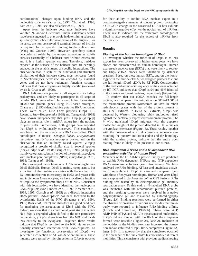
Cloning of the human homologue of Dbp5To investigate whether the function of Dbp5 in mRNAexport has been conserved in higher eukaryotes, we havecloned and characterized its human homologue. Humanexpressed sequence tags (ESTs) that were likely to repres-ent Dbp5 cDNA clones were identified by databasesearches. Based on these human ESTs, and on the homo-logy with the murine cDNA, we designed primers to clonethe full-length hDbp5 cDNA by RT–PCR. Considerationof the deduced amino acid sequence of the clones obtainedby RT–PCR indicates that hDbp5 is 94 and 46% identicalto the murine and yeast proteins, respectively (Figure 1A).
To confirm that our cDNA encodes the full-lengthprotein, we compared the migration on SDS–PAGE ofthe recombinant protein synthesizedin vitro in rabbitreticulocyte lysates with that of the protein present inHeLa cell extracts. In HeLa cell extracts, hDbp5 wasdetected by Western blot using rabbit antibodies raisedagainst the bacterially expressed recombinant protein. Thein vitro translated hDbp5 migrates with the apparentmolecular weight of the protein detected in HeLa nuclearor cytoplasmic extracts (Figure 1B). These results, togetherwith the presence of a Kozak consensus sequence sur-rounding the putative initiation codon and the homologywith the murine protein, indicate that the entire openreading frame is likely to be present in our cDNA.
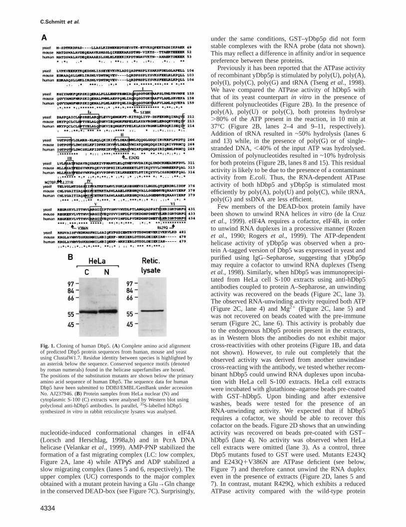
RNA-dependent ATPase and ATP-dependent RNAunwinding activities of human Dbp5Members of the DEAD-box protein family are predictedto exhibit RNA-dependent NTPase and NTP-dependentRNA-unwindase activities (see Introduction). We haveanalysed the RNA-binding, ATPase and unwindase activit-ies of recombinant hDbp5in vitro and compared themwith those of its yeast homologue. Human and yeast Dbp5were expressed inEscherichia colias GST fusions. RNAbinding was tested by an electrophoretic gel mobilityretardation assay. To this end, a32P-labelled RNA probewas incubated with the recombinant purified proteins,and the resulting complexes were resolved in a nativepolyacrylamide gel and visualized by autoradiography(Figure 2A). Binding reactions were performed in eitherthe absence or presence of various nucleotides that previ-ously were reported to influence RNA-binding affinity(Lorsch and Herschlag, 1998a,b). These were ATP,AMP-PNP, ATPγS and ADP. In the absence of nucleotides,hDbp5 did not interact with the RNA or the complexesformed were unstable (Figure 2A, lane 2). Inclusion ofnucleotides in the binding reactions increased the forma-tion and/or stabilized hDbp5–RNA complexes (Figure 2A,lanes 3–6). It is noteworthy that the complexes obtainedin the presence of the nucleotides tested exhibited differentmobilities. This is consistent with previous studies showing
C.Schmitt et al.
Fig. 1. Cloning of human Dbp5. (A) Complete amino acid alignmentof predicted Dbp5 protein sequences from human, mouse and yeastusing ClustalW1.7. Residue identity between species is highlighted byan asterisk below the sequence. Conserved sequence motifs (denotedby roman numerals) found in the helicase superfamilies are boxed.The positions of the substitution mutants are shown below the primaryamino acid sequence of human Dbp5. The sequence data for humanDbp5 have been submitted to DDBJ/EMBL/GenBank under accessionNo. AJ237946. (B) Protein samples from HeLa nuclear (N) andcytoplasmic S-100 (C) extracts were analysed by Western blot usingpolyclonal anti-hDbp5 antibodies. In parallel,35S-labelled hDbp5synthesizedin vitro in rabbit reticulocyte lysates was analysed.
nucleotide-induced conformational changes in eIF4A(Lorsch and Herschlag, 1998a,b) and in PcrA DNAhelicase (Velankaret al., 1999). AMP-PNP stabilized theformation of a fast migrating complex (LC: low complex,Figure 2A, lane 4) while ATPγS and ADP stabilized aslow migrating complex (lanes 5 and 6, respectively). Theupper complex (UC) corresponds to the major complexobtained with a mutant protein having a Glu→Gln changein the conserved DEAD-box (see Figure 7C). Surprisingly,
4334
under the same conditions, GST–yDbp5p did not formstable complexes with the RNA probe (data not shown).This may reflect a difference in affinity and/or in sequencepreference between these proteins.
Previously it has been reported that the ATPase activityof recombinant yDbp5p is stimulated by poly(U), poly(A),poly(I), poly(C), poly(G) and tRNA (Tsenget al., 1998).We have compared the ATPase activity of hDbp5 withthat of its yeast counterpartin vitro in the presence ofdifferent polynucleotides (Figure 2B). In the presence ofpoly(A), poly(U) or poly(C), both proteins hydrolyse.80% of the ATP present in the reaction, in 10 min at37°C (Figure 2B, lanes 2–4 and 9–11, respectively).Addition of tRNA resulted in ~50% hydrolysis (lanes 6and 13) while, in the presence of poly(G) or of single-stranded DNA,,40% of the input ATP was hydrolysed.Omission of polynucleotides resulted in ~10% hydrolysisfor both proteins (Figure 2B, lanes 8 and 15). This residualactivity is likely to be due to the presence of a contaminantactivity from E.coli. Thus, the RNA-dependent ATPaseactivity of both hDbp5 and yDbp5p is stimulated mostefficiently by poly(A), poly(U) and poly(C), while tRNA,poly(G) and ssDNA are less efficient.
Few members of the DEAD-box protein family havebeen shown to unwind RNA helicesin vitro (de la Cruzet al., 1999). eIF4A requires a cofactor, eIF4B, in orderto unwind RNA duplexes in a processive manner (Rozenet al., 1990; Rogerset al., 1999). The ATP-dependenthelicase activity of yDbp5p was observed when a pro-tein A-tagged version of Dbp5 was expressed in yeast andpurified using IgG–Sepharose, suggesting that yDbp5pmay require a cofactor to unwind RNA duplexes (Tsenget al., 1998). Similarly, when hDbp5 was immunoprecipi-tated from HeLa cell S-100 extracts using anti-hDbp5antibodies coupled to protein A–Sepharose, an unwindingactivity was recovered on the beads (Figure 2C, lane 3).The observed RNA-unwinding activity required both ATP(Figure 2C, lane 4) and Mg21 (Figure 2C, lane 5) andwas not recovered on beads coated with the pre-immuneserum (Figure 2C, lane 6). This activity is probably dueto the endogenous hDbp5 protein present in the extracts,as in Western blots the antibodies do not exhibit majorcross-reactivities with other proteins (Figure 1B, and datanot shown). However, to rule out completely that theobserved activity was derived from another unwindasecross-reacting with the antibody, we tested whether recom-binant hDbp5 could unwind RNA duplexes upon incuba-tion with HeLa cell S-100 extracts. HeLa cell extractswere incubated with glutathione–agarose beads pre-coatedwith GST–hDbp5. Upon binding and after extensivewashes, beads were tested for the presence of anRNA-unwinding activity. We expected that if hDbp5requires a cofactor, we should be able to recover thiscofactor on the beads. Figure 2D shows that an unwindingactivity was recovered on beads pre-coated with GST–hDbp5 (lane 4). No activity was observed when HeLacell extracts were omitted (lane 3). As a control, threeDbp5 mutants fused to GST were used. Mutants E243Qand E243Q1V386N are ATPase deficient (see below,Figure 7) and therefore cannot unwind the RNA duplexeven in the presence of extracts (Figure 2D, lanes 5 and7). In contrast, mutant R429Q, which exhibits a reducedATPase activity compared with the wild-type protein
CAN/Nup159 recruits Dbp5 to the NPC cytoplasmic fibrils
Fig. 2. RNA binding, RNA-dependent ATPase and ATP-dependent unwinding activities of recombinant hDbp5. (A) Recombinant hDbp5 binds RNAin a nucleotide-dependent manner. A gel mobility retardation assay was performed with purified recombinant GST–hDbp5. In lanes 3–6, thenucleotides indicated above the lanes were added. The concentration of the nucleotides in the binding reactions was 2 mM, and that of therecombinant protein 0.2 mg/ml. The positions of the free RNA probe (lanes 1 and 7) and of the hDbp5–RNA complexes (lanes 2–6) are indicated onthe left. The upper and lower complexes may represent two distinct conformations of hDbp5 bound to the RNA. (B) RNA-dependent ATPaseactivity of purified human and yeast Dbp5 proteins. In lanes 2–7 and 9–14, ATP hydrolysis was stimulated in the presence of the polynucleotidesindicated above the lanes. In lanes 8 and 15, no polynucleotide was added. Lane 1 shows the ATP input. The position of ATP and ADP is indicatedon the left of the panel. (C) hDbp5 immunoprecipitated from HeLa cell S-100 extracts unwinds RNA duplexesin vitro. Lane 1, RNA duplex; lane 2,the sample was boiled before loading onto the gel; lane 3, unwinding activity co-immunoprecipitated with anti-hDbp5 polyclonal antibodies; lanes 4and 5, no unwinding activity was observed when ATP or Mg21 were omitted; lane 6, background activity selected on beads pre-coated with the pre-immune serum. The RNA duplex and the monomer are indicated on the left of the gel. (D) Recombinant GST–hDbp5 unwinds RNA duplexes uponincubation with S-100 extracts. HeLa cell extracts were incubated with glutathione–agarose beads pre-coated with GST–hDbp5 (lane 4) or with GSTfused to Dbp5 mutants E243Q1V386N (lane 5), R429Q (lane 6) and E243Q (lane 7). Unwinding activity was only observed on beads coated withthe wild-type protein (lane 4) and with mutant R429Q. No unwinding activity was observed when the extracts were omitted (lane 3). The bandsbelow the duplex in lane 3 are likely to represent partial degradation of the RNA probe. Symbols are as in (C).
(Figure 7), unwound the RNA duplex upon incubationwith the extracts, albeit with a reduced efficiency(Figure 2D, lane 6 versus lane 4). Together, these resultssuggest that hDbp5 is an ATP-dependent RNA unwindasethat requires a cofactor to exhibit unwinding activity. Notethat the substrate employed in the assays shown inFigure 2C and D consisted of an RNA duplex of 25nucleotides (∆G° 5 approximately –44 kcal/mol at 37°C;Turneret al., 1988), flanked by 39-terminal extensions of~78 nucleotides of single-stranded RNA. Thus, Dbp5 doesnot require a 59 single-stranded region on the substrate.
Human Dbp5 is localized in the cytoplasm and atthe nuclear rimYeast Dbp5p has been localized both to the cytoplasmand to the nuclear envelope (Snay-Hodgeet al., 1998;Tseng et al., 1998; see Figure 5). To investigate thelocalization of hDbp5, we generated fusion proteins bytagging hDbp5 with the green fluorescent protein (GFP)at its N- or C-terminus. hDbp5–GFP fusions could bevizualized directly upon transfection. Figure 3A–C showsthe results obtained when GFP was fused to the N-terminus
4335
of hDbp5. In transfected HeLa cells, the majority of theGFP-tagged protein was detected in the cytoplasm, whilethe nucleoplasm was largely free from staining. Moreover,a fraction of the protein appeared to be concentratedaround the nucleus. A similar subcellular localization ofhDbp5 was found when the GFP tag was fused to itsC-terminus (Figure 3D). The nuclear rim staining becamemore apparent when cells were treated with digitonin priorto fixation (Figure 3D). The nuclear rim-associated fractionof Dbp5 co-localizes with the labelling produced bymonoclonal antibody 414 directed against nucleoporins(Figure 3E and F). Thus, hDbp5, like its yeast homologue,is predominantly cytoplasmic, but a fraction localizes tothe nuclear rim.
Identification of an evolutionarily conservedinteraction between Dbp5 and CAN/Nup159pThe nuclear rim association of Dbp5 in both HeLa andyeast cells suggests that a fraction of the protein interactswith components of the NPC. Furthermore, experimentsshown in Figure 2D suggest that Dbp5 requires a cofactorin order to unwind RNA duplexes. To identify Dbp5

C.Schmitt et al.
Fig. 3. Human Dbp5 is localized in the cytoplasm and nuclear rim.(A–C) HeLa cells were transfected with pEGFP-C1 hDbp5 using thecalcium phosphate method. Approximately 20 h after transfection,cells were fixed in formaldehyde and observed directly with thefluorescence microscope. The fusion protein is detected predominantlyin the cytoplasm. Arrows point to a rim staining at the nuclearperiphery. (D–F) HeLa cells transfected with pEFGP-N3 hDbp5 werepermeabilized with digitonin for 2 min before fixation informaldehyde. The cells were then incubated sequentially with ananti-nucleoporin monoclonal antibody (mAb414) and an appropriatesecondary antibody conjugated to Texas red. The superimposition of(D) and (E) confirms that GFP–Dbp5 co-localizes with nucleoporins.Bar, 10µm.
partners, we have used a novel approach, the tandemaffinity purification (TAP) strategy, that allows efficientpurification of protein complexes from yeast cell extracts(see Materials and methods). Because of the high degreeof similarity between human and yeast Dbp5, their similarlocalization and function (see below), we expected thatidentification of its interacting proteins in yeast wouldlead to the identification of Dbp5 partners in vertebratecells. The TAP tag was fused to the C-terminus of yDbp5protein by integrating a DNA cassette into the genome ofa haploid cell (Puiget al., 1998). The TAP tag consistsof a calmodulin-binding peptide followed by a TEVprotease cleavage site and two IgG-binding units ofStaphylococcus aureusprotein A (ProtA). Because thetagged protein is the only source of the essential yDbp5function, and the tagged strain did not display a stronggrowth phenotype, we conclude that the TAP tag did not
4336
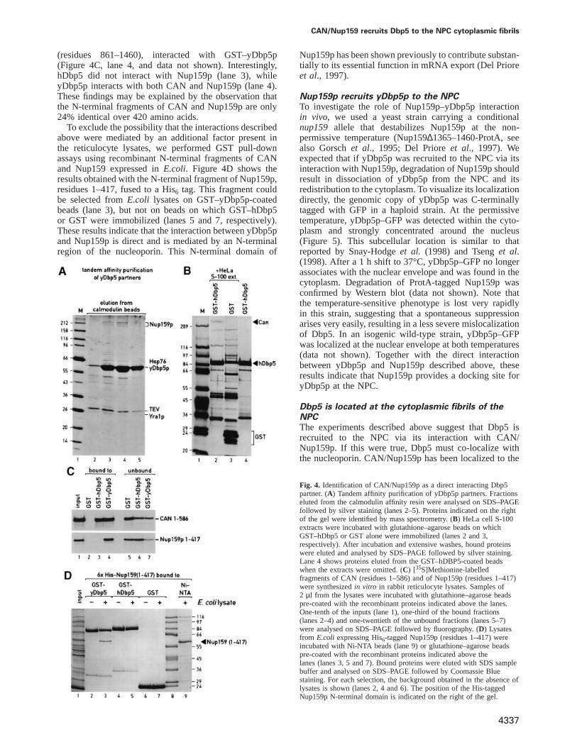
abolish Dbp5 protein function. Extracts were preparedfrom the tagged strain, and Dbp5 with its associatedproteins was purified following the two-step affinity puri-fication of the TAP method. Figure 4A shows the proteinseluted from the calmodulin affinity resin. Most of theputative interacting proteins were found in sub-stoichi-ometric amounts. This does not result from overexpressionof the Dbp5 protein that is expressed under the control ofits natural promoter, but rather suggests that Dbp5 is nota subunit of a stable multimeric complex. Bands obtainedin several selections were excised from the gel, the proteinswere in-gel digested with trypsin and the tryptic peptideswere identified by mass spectrometry. The peptidessequenced unambiguously identify Nup159p as the proteinmigrating with an apparent mol. wt of 220 kDa (Gorschet al., 1995; Kraemeret al., 1995). The sequenced peptideswere ALESVGSDTTFK, SLFVAASGSK and LFDVSAK(using the single-letter amino acid code). Two other bandswere identified as Hsp76 and Yra1p (Portmanet al.,1997). The interaction between Yra1p and yDbp5p wasnot investigated further.
To determine whether the interaction with Nup159pwas conserved, HeLa cell S-100 extracts were fractionatedon glutathione–agarose beads on which GST–hDbp5 orGST alone was immobilized. Analysis of the pattern ofproteins selected on GST–Dbp5 versus GST by SDS–PAGE revealed one striking difference in the region of220 kDa due to the presence of a protein in the hDbp5eluates (Figure 4B, lane 2) which is absent from the GSTeluates (lane 3). This band was observed consistently inseveral selections. Because of its size, and the resultsobtained with yDbp5p, this protein was predicted to beCAN (von Lindern et al., 1992; Kraemeret al., 1994),the putative homologue of Nup159p (Gorschet al., 1995;Kraemeret al., 1995; Belgarehet al., 1998, Hurwitzet al.,1998). The identity of this protein was confirmed byWestern blot using a polyclonal anti-CAN antibodydescribed by Fornerodet al. (1995) (data not shown).
Dbp5 interacts directly with an N-terminal regionof CAN/Nup159pTo determine the specificity of the interaction betweenCAN/Nup159p and Dbp5, we first delineated thedomains on CAN/Nup159p that interact with Dbp5.[35S]Methionine-labelled fragments from CAN andNup159p were synthesizedin vitro in rabbit reticulocytelysates and assayed for binding to glutathione–agarosebeads coated with either GST–hDbp5, GST–yDbp5p orGST alone. Preliminary experiments indicate that theN-terminal domain of CAN (residues 1–1058) and ashorter fragment encompassing residues 1–586, but notits C-terminal domain (residues 1690–2090), interactedwith hDbp5 (data not shown). Figure 4C shows that thefragment of CAN encompassing amino acids 1–586 boundto hDbp5- and yDbp5p-coated beads but not to the controlbeads having GST alone (lanes 3 and 4 versus lane 2).Further analysis indicates that the C-terminal boundaryof the Dbp5-binding domain is located between CANresidues 366 and 433, as fragment 1–433 bound to Dbp5as efficiently as fragment 1–586, while fragment 1–366no longer interacted with the helicase (data not shown).Similarly, an N-terminal fragment of Nup159p,comprising residues 1–417, but not a C-terminal fragment
CAN/Nup159 recruits Dbp5 to the NPC cytoplasmic fibrils
(residues 861–1460), interacted with GST–yDbp5p(Figure 4C, lane 4, and data not shown). Interestingly,hDbp5 did not interact with Nup159p (lane 3), whileyDbp5p interacts with both CAN and Nup159p (lane 4).These findings may be explained by the observation thatthe N-terminal fragments of CAN and Nup159p are only24% identical over 420 amino acids.
To exclude the possibility that the interactions describedabove were mediated by an additional factor present inthe reticulocyte lysates, we performed GST pull-downassays using recombinant N-terminal fragments of CANand Nup159 expressed inE.coli. Figure 4D shows theresults obtained with the N-terminal fragment of Nup159p,residues 1–417, fused to a His6 tag. This fragment couldbe selected fromE.coli lysates on GST–yDbp5p-coatedbeads (lane 3), but not on beads on which GST–hDbp5or GST were immobilized (lanes 5 and 7, respectively).These results indicate that the interaction between yDbp5pand Nup159p is direct and is mediated by an N-terminalregion of the nucleoporin. This N-terminal domain of
4337
Nup159p has been shown previously to contribute substan-tially to its essential function in mRNA export (Del Prioreet al., 1997).
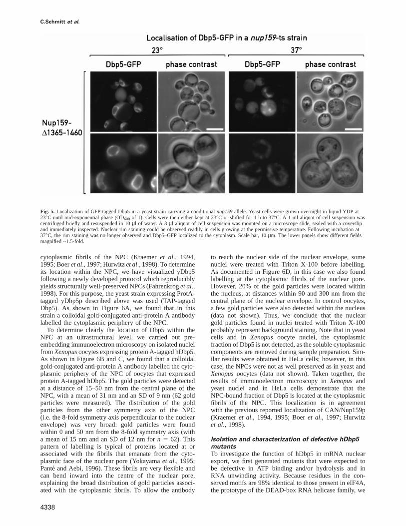
Nup159p recruits yDbp5p to the NPCTo investigate the role of Nup159p–yDbp5p interactionin vivo, we used a yeast strain carrying a conditionalnup159 allele that destabilizes Nup159p at the non-permissive temperature (Nup159∆1365–1460-ProtA, seealso Gorschet al., 1995; Del Prioreet al., 1997). Weexpected that if yDbp5p was recruited to the NPC via itsinteraction with Nup159p, degradation of Nup159p shouldresult in dissociation of yDbp5p from the NPC and itsredistribution to the cytoplasm. To visualize its localizationdirectly, the genomic copy of yDbp5p was C-terminallytagged with GFP in a haploid strain. At the permissivetemperature, yDbp5p–GFP was detected within the cyto-plasm and strongly concentrated around the nucleus(Figure 5). This subcellular location is similar to thatreported by Snay-Hodgeet al. (1998) and Tsenget al.(1998). After a 1 hshift to 37°C, yDbp5p–GFP no longerassociates with the nuclear envelope and was found in thecytoplasm. Degradation of ProtA-tagged Nup159p wasconfirmed by Western blot (data not shown). Note thatthe temperature-sensitive phenotype is lost very rapidlyin this strain, suggesting that a spontaneous suppressionarises very easily, resulting in a less severe mislocalizationof Dbp5. In an isogenic wild-type strain, yDbp5p–GFPwas localized at the nuclear envelope at both temperatures(data not shown). Together with the direct interactionbetween yDbp5p and Nup159p described above, theseresults indicate that Nup159p provides a docking site foryDbp5p at the NPC.
Dbp5 is located at the cytoplasmic fibrils of theNPCThe experiments described above suggest that Dbp5 isrecruited to the NPC via its interaction with CAN/Nup159p. If this were true, Dbp5 must co-localize withthe nucleoporin. CAN/Nup159p has been localized to the
Fig. 4. Identification of CAN/Nup159p as a direct interacting Dbp5partner. (A) Tandem affinity purification of yDbp5p partners. Fractionseluted from the calmodulin affinity resin were analysed on SDS–PAGEfollowed by silver staining (lanes 2–5). Proteins indicated on the rightof the gel were identified by mass spectrometry. (B) HeLa cell S-100extracts were incubated with glutathione–agarose beads on whichGST–hDbp5 or GST alone were immobilized (lanes 2 and 3,respectively). After incubation and extensive washes, bound proteinswere eluted and analysed by SDS–PAGE followed by silver staining.Lane 4 shows proteins eluted from the GST–hDBP5-coated beadswhen the extracts were omitted. (C) [35S]Methionine-labelledfragments of CAN (residues 1–586) and of Nup159p (residues 1–417)were synthesizedin vitro in rabbit reticulocyte lysates. Samples of2 µl from the lysates were incubated with glutathione–agarose beadspre-coated with the recombinant proteins indicated above the lanes.One-tenth of the inputs (lane 1), one-third of the bound fractions(lanes 2–4) and one-twentieth of the unbound fractions (lanes 5–7)were analysed on SDS–PAGE followed by fluorography. (D) Lysatesfrom E.coli expressing His6-tagged Nup159p (residues 1–417) wereincubated with Ni-NTA beads (lane 9) or glutathione–agarose beadspre-coated with the recombinant proteins indicated above thelanes (lanes 3, 5 and 7). Bound proteins were eluted with SDS samplebuffer and analysed on SDS–PAGE followed by Coomassie Bluestaining. For each selection, the background obtained in the absence oflysates is shown (lanes 2, 4 and 6). The position of the His-taggedNup159p N-terminal domain is indicated on the right of the gel.
C.Schmitt et al.
Fig. 5. Localization of GFP-tagged Dbp5 in a yeast strain carrying a conditionalnup159allele. Yeast cells were grown overnight in liquid YDP at23°C until mid-exponential phase (OD600 of 1). Cells were then either kept at 23°C or shifted for 1 h to 37°C. A 1 ml aliquot of cell suspension wascentrifuged briefly and resuspended in 10µl of water. A 3 µl aliquot of cell suspension was mounted on a microscope slide, sealed with a coverslipand immediately inspected. Nuclear rim staining could be observed readily in cells growing at the permissive temperature. Following incubation at37°C, the rim staining was no longer observed and Dbp5–GFP localized to the cytoplasm. Scale bar, 10µm. The lower panels show different fieldsmagnified ~1.5-fold.
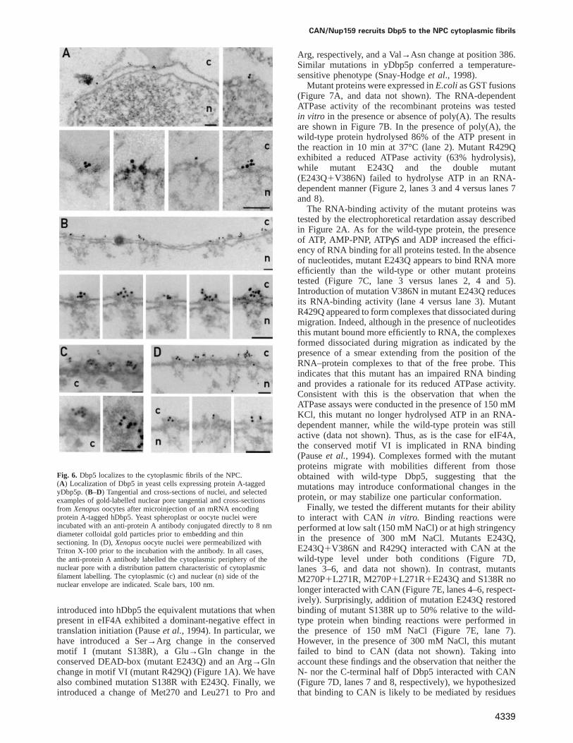
cytoplasmic fibrils of the NPC (Kraemeret al., 1994,1995; Boeret al., 1997; Hurwitzet al., 1998). To determineits location within the NPC, we have visualized yDbp5following a newly developed protocol which reproduciblyyields structurally well-preserved NPCs (Fahrenkroget al.,1998). For this purpose, the yeast strain expressing ProtA-tagged yDbp5p described above was used (TAP-taggedDbp5). As shown in Figure 6A, we found that in thisstrain a colloidal gold-conjugated anti-protein A antibodylabelled the cytoplasmic periphery of the NPC.
To determine clearly the location of Dbp5 within theNPC at an ultrastructural level, we carried out pre-embedding immunoelectron microscopy on isolated nucleifromXenopusoocytes expressing protein A-tagged hDbp5.As shown in Figure 6B and C, we found that a colloidalgold-conjugated anti-protein A antibody labelled the cyto-plasmic periphery of the NPC of oocytes that expressedprotein A-tagged hDbp5. The gold particles were detectedat a distance of 15–50 nm from the central plane of theNPC, with a mean of 31 nm and an SD of 9 nm (62 goldparticles were measured). The distribution of the goldparticles from the other symmetry axis of the NPC(i.e. the 8-fold symmetry axis perpendicular to the nuclearenvelope) was very broad: gold particles were foundwithin 0 and 50 nm from the 8-fold symmetry axis (witha mean of 15 nm and an SD of 12 nm forn 5 62). Thispattern of labelling is typical of proteins located at orassociated with the fibrils that emanate from the cyto-plasmic face of the nuclear pore (Yokayamaet al., 1995;Panteand Aebi, 1996). These fibrils are very flexible andcan bend inward into the centre of the nuclear pore,explaining the broad distribution of gold particles associ-ated with the cytoplasmic fibrils. To allow the antibody
4338
to reach the nuclear side of the nuclear envelope, somenuclei were treated with Triton X-100 before labelling.As documented in Figure 6D, in this case we also foundlabelling at the cytoplasmic fibrils of the nuclear pore.However, 20% of the gold particles were located withinthe nucleus, at distances within 90 and 300 nm from thecentral plane of the nuclear envelope. In control oocytes,a few gold particles were also detected within the nucleus(data not shown). Thus, we conclude that the nucleargold particles found in nuclei treated with Triton X-100probably represent background staining. Note that in yeastcells and in Xenopusoocyte nuclei, the cytoplasmicfraction of Dbp5 is not detected, as the soluble cytoplasmiccomponents are removed during sample preparation. Sim-ilar results were obtained in HeLa cells; however, in thiscase, the NPCs were not as well preserved as in yeast andXenopusoocytes (data not shown). Taken together, theresults of immunoelectron microscopy inXenopusandyeast nuclei and in HeLa cells demonstrate that theNPC-bound fraction of Dbp5 is located at the cytoplasmicfibrils of the NPC. This localization is in agreementwith the previous reported localization of CAN/Nup159p(Kraemeret al., 1994, 1995; Boeret al., 1997; Hurwitzet al., 1998).
Isolation and characterization of defective hDbp5mutantsTo investigate the function of hDbp5 in mRNA nuclearexport, we first generated mutants that were expected tobe defective in ATP binding and/or hydrolysis and inRNA unwinding activity. Because residues in the con-served motifs are 98% identical to those present in eIF4A,the prototype of the DEAD-box RNA helicase family, we
CAN/Nup159 recruits Dbp5 to the NPC cytoplasmic fibrils
Fig. 6. Dbp5 localizes to the cytoplasmic fibrils of the NPC.(A) Localization of Dbp5 in yeast cells expressing protein A-taggedyDbp5p. (B–D) Tangential and cross-sections of nuclei, and selectedexamples of gold-labelled nuclear pore tangential and cross-sectionsfrom Xenopusoocytes after microinjection of an mRNA encodingprotein A-tagged hDbp5. Yeast spheroplast or oocyte nuclei wereincubated with an anti-protein A antibody conjugated directly to 8 nmdiameter colloidal gold particles prior to embedding and thinsectioning. In (D),Xenopusoocyte nuclei were permeabilized withTriton X-100 prior to the incubation with the antibody. In all cases,the anti-protein A antibody labelled the cytoplasmic periphery of thenuclear pore with a distribution pattern characteristic of cytoplasmicfilament labelling. The cytoplasmic (c) and nuclear (n) side of thenuclear envelope are indicated. Scale bars, 100 nm.
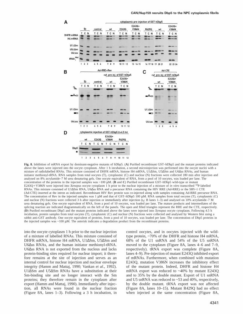
introduced into hDbp5 the equivalent mutations that whenpresent in eIF4A exhibited a dominant-negative effect intranslation initiation (Pauseet al., 1994). In particular, wehave introduced a Ser→Arg change in the conservedmotif I (mutant S138R), a Glu→Gln change in theconserved DEAD-box (mutant E243Q) and an Arg→Glnchange in motif VI (mutant R429Q) (Figure 1A). We havealso combined mutation S138R with E243Q. Finally, weintroduced a change of Met270 and Leu271 to Pro and
4339
Arg, respectively, and a Val→Asn change at position 386.Similar mutations in yDbp5p conferred a temperature-sensitive phenotype (Snay-Hodgeet al., 1998).
Mutant proteins were expressed inE.colias GST fusions(Figure 7A, and data not shown). The RNA-dependentATPase activity of the recombinant proteins was testedin vitro in the presence or absence of poly(A). The resultsare shown in Figure 7B. In the presence of poly(A), thewild-type protein hydrolysed 86% of the ATP present inthe reaction in 10 min at 37°C (lane 2). Mutant R429Qexhibited a reduced ATPase activity (63% hydrolysis),while mutant E243Q and the double mutant(E243Q1V386N) failed to hydrolyse ATP in an RNA-dependent manner (Figure 2, lanes 3 and 4 versus lanes 7and 8).
The RNA-binding activity of the mutant proteins wastested by the electrophoretical retardation assay describedin Figure 2A. As for the wild-type protein, the presenceof ATP, AMP-PNP, ATPγS and ADP increased the effici-ency of RNA binding for all proteins tested. In the absenceof nucleotides, mutant E243Q appears to bind RNA moreefficiently than the wild-type or other mutant proteinstested (Figure 7C, lane 3 versus lanes 2, 4 and 5).Introduction of mutation V386N in mutant E243Q reducesits RNA-binding activity (lane 4 versus lane 3). MutantR429Q appeared to form complexes that dissociated duringmigration. Indeed, although in the presence of nucleotidesthis mutant bound more efficiently to RNA, the complexesformed dissociated during migration as indicated by thepresence of a smear extending from the position of theRNA–protein complexes to that of the free probe. Thisindicates that this mutant has an impaired RNA bindingand provides a rationale for its reduced ATPase activity.Consistent with this is the observation that when theATPase assays were conducted in the presence of 150 mMKCl, this mutant no longer hydrolysed ATP in an RNA-dependent manner, while the wild-type protein was stillactive (data not shown). Thus, as is the case for eIF4A,the conserved motif VI is implicated in RNA binding(Pauseet al., 1994). Complexes formed with the mutantproteins migrate with mobilities different from thoseobtained with wild-type Dbp5, suggesting that themutations may introduce conformational changes in theprotein, or may stabilize one particular conformation.
Finally, we tested the different mutants for their abilityto interact with CAN in vitro. Binding reactions wereperformed at low salt (150 mM NaCl) or at high stringencyin the presence of 300 mM NaCl. Mutants E243Q,E243Q1V386N and R429Q interacted with CAN at thewild-type level under both conditions (Figure 7D,lanes 3–6, and data not shown). In contrast, mutantsM270P1L271R, M270P1L271R1E243Q and S138R nolonger interacted with CAN (Figure 7E, lanes 4–6, respect-ively). Surprisingly, addition of mutation E243Q restoredbinding of mutant S138R up to 50% relative to the wild-type protein when binding reactions were performed inthe presence of 150 mM NaCl (Figure 7E, lane 7).However, in the presence of 300 mM NaCl, this mutantfailed to bind to CAN (data not shown). Taking intoaccount these findings and the observation that neither theN- nor the C-terminal half of Dbp5 interacted with CAN(Figure 7D, lanes 7 and 8, respectively), we hypothesizedthat binding to CAN is likely to be mediated by residues

C.Schmitt et al.
Fig. 7. Characterization of hDbp5 mutants. (A) Visualization of the purified wild-type and mutant recombinant hDbp5 proteins by Coomassiestaining following SDS–PAGE. (B) The ATPase activity of the wild-type and mutant Dbp5 proteins was tested in the presence or absence of poly(A)as indicated above the lanes. Symbols are as in Figure 2B. (C) An electrophoretic mobility retardation assay was performed with a labelled RNAprobe and the purified recombinant proteins indicated above the lanes. For each protein, binding reactions were performed in the absence ofnucleotides (lanes 2–5) or in the presence of ATP (lanes 6–10), AMP-PNP (lanes 11–15), ATPγS (lanes 16–20) or ADP (lanes 21–25). Theconcentration of the nucleotides in the binding reactions was 2 mM, and that of the recombinant proteins 0.2 mg/ml. The positions of the free RNAprobe and of hDbp5–RNA complexes are shown on the left. (D–F) A [35S]methionine-labelled fragment of CAN (residues 1–586) was synthesizedin vitro in rabbit reticulocyte lysates. Samples of 2µl from the lysates were incubated with glutathione–agarose beads pre-coated with therecombinant proteins indicated above the lanes. In (F), binding of CAN to wild-type GST–hDbp5 was analysed in the presence of nucleotide asindicated. One-tenth of the input and one-quarter of the bound fractions were analysed on SDS–PAGE followed by fluorography.
exposed at the surface of the helicase core and is influencedby the overall conformation of the protein. To test thishypothesis, we analysed the interaction between Dbp5 andCAN in the presence of nucleotides which upon bindinginduce conformational changes in the helicase core. Asshown in Figure 7F, the presence of ATP, AMP-PNP andATPγS reduced the efficiency of CAN binding to Dbp5,while ADP had no effect. This suggest that Dbp5–CAN
4340
interaction may be regulated by nucleotide binding andhydrolysis.
Mutants E243Q and E243QFV386N inhibit themRNA export pathwayThe effect of the mutant proteins in RNA export wasexamined by microinjection intoXenopusoocytes. Purifiedrecombinant proteins (shown in Figure 7A) were injected
CAN/Nup159 recruits Dbp5 to the NPC cytoplasmic fibrils
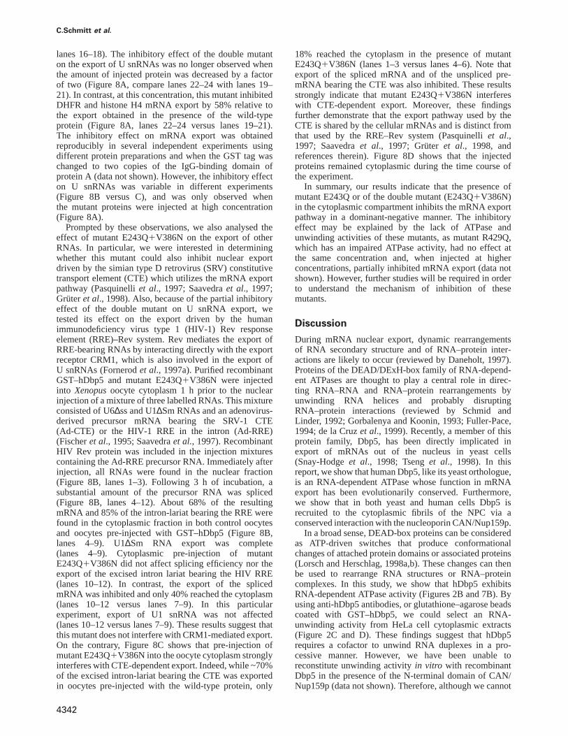
Fig. 8. Inhibition of mRNA export by dominant-negative mutants of hDbp5. (A) Purified recombinant GST–hDbp5 and the mutant proteins indicatedabove the lanes were injected into the oocyte cytoplasm. After 1 h incubation, a second microinjection was performed into the oocyte nuclei with amixture of radiolabelled RNAs. This mixture consisted of DHFR mRNA, histone H4 mRNA, U1∆Sm, U5∆Sm and U6∆ss RNAs, and humaninitiator methionyl-tRNA. RNA samples from total oocytes (T), cytoplasmic (C) and nuclear (N) fractions were collected 180 min after injection andanalysed on 8% acrylamide–7 M urea denaturing gels. One oocyte equivalent of RNA, from a pool of 10 oocytes, was loaded per lane. Theconcentration of the proteins in the injected samples was ~100µM. (B andC) Purified recombinant GST–hDbp5 wild-type or mutantE243Q1V386N were injected intoXenopusoocyte cytoplasm 1 h prior to the nuclear injection of a mixture ofin vitro transcribed32P-labelledRNAs. This mixture consisted of U1∆Sm RNA, U6∆ss RNA and a precursor RNA containing the HIV RRE (Ad-RRE) or the SRV-1 CTE(Ad-CTE) inserted at the intron as indicated. Recombinant HIV Rev protein was co-injected along with samples containing Ad-RRE precursor RNA.The concentration of Rev in the injected samples was 1µM and that of GST–hDbp5 100µM. RNA samples from total oocytes (T), cytoplasmic (C)and nuclear (N) fractions were collected 3 h after injection or immediately after injection (t0: B lanes 1–3) and analysed on 10% acrylamide–7 Murea denaturing gels. One oocyte equivalent of RNA, from a pool of 10 oocytes, was loaded per lane. The mature products and intermediates of thesplicing reaction are indicated diagrammatically on the left of the panels. The open and filled triangles represent the RRE and the CTE, respectively.(D) Purified recombinant Dbp5 and the mutant proteins indicated above the lanes were injected intoXenopusoocyte cytoplasm. Following 4.5 hincubation, protein samples from total oocytes (T), cytoplasmic (C) and nuclear (N) fractions were collected and analysed by Western blot using arabbit anti-GST antibody. One oocyte equivalent of proteins, from a pool of 10 oocytes, was loaded per lane. The concentration of Dbp5 proteins inthe injected samples was ~100µM. The asterisk indicates a degradation product from the recombinant proteins.
into the oocyte cytoplasm 1 h prior to the nuclear injectionof a mixture of labelled RNAs. This mixture consisted ofDHFR mRNA, histone H4 mRNA, U1∆Sm, U5∆Sm andU6∆ss RNAs, and the human initiator methionyl-tRNA.U6∆ss RNA is not exported from the nucleus and lacksprotein-binding sites required for nuclear import; it there-fore remains at the site of injection and serves as aninternal control for nuclear injection and nuclear envelopeintegrity (Hamm and Mattaj, 1990; Vankanet al., 1992).U1∆Sm and U5∆Sm RNAs have a substitution at theirSm-binding site and no longer interact with the Smproteins; they therefore remain in the cytoplasm afterexport (Hamm and Mattaj, 1990). Immediately after injec-tion, all RNAs were found in the nuclear fraction(Figure 8A, lanes 1–3). Following a 3 h incubation, in
4341
control oocytes, and in oocytes injected with the wild-type protein, ~70% of the DHFR and histone H4 mRNA,68% of the U1 snRNA and 54% of the U5 snRNAmoved to the cytoplasm (Figure 8A, lanes 4–6 and 7–9,respectively). tRNA export was complete (Figure 8A,lanes 4–9). Pre-injection of mutant E243Q inhibited exportof mRNAs. Furthermore, when combined with mutationE243Q, mutation V386N increases the inhibitory effectof the mutant protein. Indeed, DHFR and histone H4mRNA export was reduced to ~40% by mutant E243Qand to 35% by the double mutant. Export of U1 snRNAand U5 snRNA was reduced to ~53 and 40%, respectively,by the double mutant. tRNA export was not affected(Figure 8A, lanes 10–15). Mutant R429Q had no effectwhen injected at the same concentration (Figure 8A,
C.Schmitt et al.
lanes 16–18). The inhibitory effect of the double mutanton the export of U snRNAs was no longer observed whenthe amount of injected protein was decreased by a factorof two (Figure 8A, compare lanes 22–24 with lanes 19–21). In contrast, at this concentration, this mutant inhibitedDHFR and histone H4 mRNA export by 58% relative tothe export obtained in the presence of the wild-typeprotein (Figure 8A, lanes 22–24 versus lanes 19–21).The inhibitory effect on mRNA export was obtainedreproducibly in several independent experiments usingdifferent protein preparations and when the GST tag waschanged to two copies of the IgG-binding domain ofprotein A (data not shown). However, the inhibitory effecton U snRNAs was variable in different experiments(Figure 8B versus C), and was only observed whenthe mutant proteins were injected at high concentration(Figure 8A).
Prompted by these observations, we also analysed theeffect of mutant E243Q1V386N on the export of otherRNAs. In particular, we were interested in determiningwhether this mutant could also inhibit nuclear exportdriven by the simian type D retrovirus (SRV) constitutivetransport element (CTE) which utilizes the mRNA exportpathway (Pasquinelliet al., 1997; Saavedraet al., 1997;Gruter et al., 1998). Also, because of the partial inhibitoryeffect of the double mutant on U snRNA export, wetested its effect on the export driven by the humanimmunodeficiency virus type 1 (HIV-1) Rev responseelement (RRE)–Rev system. Rev mediates the export ofRRE-bearing RNAs by interacting directly with the exportreceptor CRM1, which is also involved in the export ofU snRNAs (Fornerodet al., 1997a). Purified recombinantGST–hDbp5 and mutant E243Q1V386N were injectedinto Xenopusoocyte cytoplasm 1 h prior to the nuclearinjection of a mixture of three labelled RNAs. This mixtureconsisted of U6∆ss and U1∆Sm RNAs and an adenovirus-derived precursor mRNA bearing the SRV-1 CTE(Ad-CTE) or the HIV-1 RRE in the intron (Ad-RRE)(Fischeret al., 1995; Saavedraet al., 1997). RecombinantHIV Rev protein was included in the injection mixturescontaining the Ad-RRE precursor RNA. Immediately afterinjection, all RNAs were found in the nuclear fraction(Figure 8B, lanes 1–3). Following 3 h of incubation, asubstantial amount of the precursor RNA was spliced(Figure 8B, lanes 4–12). About 68% of the resultingmRNA and 85% of the intron-lariat bearing the RRE werefound in the cytoplasmic fraction in both control oocytesand oocytes pre-injected with GST–hDbp5 (Figure 8B,lanes 4–9). U1∆Sm RNA export was complete(lanes 4–9). Cytoplasmic pre-injection of mutantE243Q1V386N did not affect splicing efficiency nor theexport of the excised intron lariat bearing the HIV RRE(lanes 10–12). In contrast, the export of the splicedmRNA was inhibited and only 40% reached the cytoplasm(lanes 10–12 versus lanes 7–9). In this particularexperiment, export of U1 snRNA was not affected(lanes 10–12 versus lanes 7–9). These results suggest thatthis mutant does not interfere with CRM1-mediated export.On the contrary, Figure 8C shows that pre-injection ofmutant E243Q1V386N into the oocyte cytoplasm stronglyinterferes with CTE-dependent export. Indeed, while ~70%of the excised intron-lariat bearing the CTE was exportedin oocytes pre-injected with the wild-type protein, only
4342
18% reached the cytoplasm in the presence of mutantE243Q1V386N (lanes 1–3 versus lanes 4–6). Note thatexport of the spliced mRNA and of the unspliced pre-mRNA bearing the CTE was also inhibited. These resultsstrongly indicate that mutant E243Q1V386N interfereswith CTE-dependent export. Moreover, these findingsfurther demonstrate that the export pathway used by theCTE is shared by the cellular mRNAs and is distinct fromthat used by the RRE–Rev system (Pasquinelliet al.,1997; Saavedraet al., 1997; Gru¨ter et al., 1998, andreferences therein). Figure 8D shows that the injectedproteins remained cytoplasmic during the time course ofthe experiment.
In summary, our results indicate that the presence ofmutant E243Q or of the double mutant (E243Q1V386N)in the cytoplasmic compartment inhibits the mRNA exportpathway in a dominant-negative manner. The inhibitoryeffect may be explained by the lack of ATPase andunwinding activities of these mutants, as mutant R429Q,which has an impaired ATPase activity, had no effect atthe same concentration and, when injected at higherconcentrations, partially inhibited mRNA export (data notshown). However, further studies will be required in orderto understand the mechanism of inhibition of thesemutants.
Discussion
During mRNA nuclear export, dynamic rearrangementsof RNA secondary structure and of RNA–protein inter-actions are likely to occur (reviewed by Daneholt, 1997).Proteins of the DEAD/DExH-box family of RNA-depend-ent ATPases are thought to play a central role in direc-ting RNA–RNA and RNA–protein rearrangements byunwinding RNA helices and probably disruptingRNA–protein interactions (reviewed by Schmid andLinder, 1992; Gorbalenya and Koonin, 1993; Fuller-Pace,1994; de la Cruzet al., 1999). Recently, a member of thisprotein family, Dbp5, has been directly implicated inexport of mRNAs out of the nucleus in yeast cells(Snay-Hodgeet al., 1998; Tsenget al., 1998). In thisreport, we show that human Dbp5, like its yeast orthologue,is an RNA-dependent ATPase whose function in mRNAexport has been evolutionarily conserved. Furthermore,we show that in both yeast and human cells Dbp5 isrecruited to the cytoplasmic fibrils of the NPC via aconserved interaction with the nucleoporin CAN/Nup159p.
In a broad sense, DEAD-box proteins can be consideredas ATP-driven switches that produce conformationalchanges of attached protein domains or associated proteins(Lorsch and Herschlag, 1998a,b). These changes can thenbe used to rearrange RNA structures or RNA–proteincomplexes. In this study, we show that hDbp5 exhibitsRNA-dependent ATPase activity (Figures 2B and 7B). Byusing anti-hDbp5 antibodies, or glutathione–agarose beadscoated with GST–hDbp5, we could select an RNA-unwinding activity from HeLa cell cytoplasmic extracts(Figure 2C and D). These findings suggest that hDbp5requires a cofactor to unwind RNA duplexes in a pro-cessive manner. However, we have been unable toreconstitute unwinding activityin vitro with recombinantDbp5 in the presence of the N-terminal domain of CAN/Nup159p (data not shown). Therefore, although we cannot
CAN/Nup159 recruits Dbp5 to the NPC cytoplasmic fibrils
rule out that the full-length CAN/Nup159p is sufficient toactivate Dbp5, we favour the hypothesis that an additionalcofactor may be implicated.
The demonstration that Dbp5 exhibits RNA-dependentATPase activity has significant impact on understandinghow Dbp5 may function in RNA export. Indeed, conforma-tional rearrangements of large RNPs during the transloca-tion through the NPC may depend on the hydrolysis ofATP by Dbp5. For instance, the ATP hydrolysis-drivenconformational changes of Dbp5 may be transduced intomovement and/or conformational changes of the emergingmRNP. Alternatively, Dbp5 conformational changes maybe transduced into movement and/or conformationalchanges of the cytoplasmic fibrils of the NPC. Independ-ently of the exact mechanism of action of Dbp5 duringexport, its direct implication in mRNA nuclear exportmay account, at least in part, for the energy requirementsof this process (Jarmolowskiet al., 1994).
The terminal step for the RNA export process mayoccur at the cytoplasmic fibrils of the NPCIn this report, we demonstrate that hDbp5 interacts withan N-terminal region of the nucleoporin CAN while yDbp5interacts with both CAN and its putative yeast homologueNup159p. Nup159p is an essential nucleoporin which,like CAN, has already been implicated in mRNA export(Gorschet al., 1995; van Deursenet al., 1996; Belgarehet al., 1998; Boeret al., 1998; Hurwitzet al., 1998). TheN-terminal domain of Nup159p is not essential for growth,but appears to contribute substantially to its function inmRNA export (Del Prioreet al., 1997). Indeed, in cellsexpressing a mutant protein lacking this domain (residues1–417), nuclear accumulation of polyadenylated RNAswas observed even at the permissive temperature. Thisdefect in mRNA export became more severe following ashift at 37°C. These findings indicate that the N-terminaldomain of Nup159p is required for efficient mRNA export(Del Prioreet al., 1997). Although, currently, it cannot beruled out that this domain recruits other factors requiredfor export, these observations suggest that recruitment ofyDbp5p to the cytoplasmic fibrils of the NPC increasesthe efficiency of the export process. However, they alsoindicate that in the absence of the N-terminal domain ofNup159p, yDbp5p can still accomplish its essential func-tion probably due to the high concentration of the proteinin the cytoplasm. Alternatively, yDbp5p may interactweakly with other components of the NPC, and theseinteractions may be temperature sensitive. Indeed, yDbp5pinteracts with Gle1p (F.Stutz, personal communication), anucleoporin showing genetic interaction with Nup159pand yDbp5p (see Snay-Hodgeet al., 1998).
Aside from interacting directly with Dbp5 (this work),CAN interacts with the mRNA export factor TAP (Katahiraet al., 1999; our unpublished data) and with the exportreceptor CRM1 (Fornerodet al., 1997b; Nevilleet al.,1997). Binding of CRM1 to CAN requires RanGTP andis stimulated by the export substrate (Askjaeret al., 1999).This indicates that the trimeric complex composed ofCRM1–RanGTP–NES-substrate would bind CAN imme-diately after translocation through the NPC. In thecytoplasmic side of the NPC, the RanGAP together withRanBP1 or RanBP2 would promote GTP hydrolysis byRan and thus the release of the export substrate (Bischoff
4343
and Gorlich, 1997; Paraskevaet al., 1999) and the dissoci-ation of CRM1 from CAN (Askjaer et al., 1999;Kehlenbachet al., 1999). The close proximity of RanGAPand RanBP2 at the tip of the cytoplasmic fibrils willrender this process extremely efficient. Thus, CAN mayact as a platform on which the export complexes aredismantled and from which export factors can be returnedimmediately back to the nucleus. Proteins of the DEAD/DExH-box family of RNA-dependent ATPases are primecandidates to play a role in this disassembling step. If thishypothetical scenario were true, other members of theDEAD/DExH-box family may be implicated in nucleartransport, as Dbp5 appears to be dedicated to the mRNAexport pathway (Figure 8). Finally, our data strengthenthe hypothesis that CAN is the functional homologue ofNup159p (Kraemeret al., 1994, 1995; Gorschet al., 1995;Belgarehet al., 1998; Hurvitzet al., 1998).
Subcellular localization and functional specificityof Dbp5Previously, Snay-Hodgeet al. (1998) and Tsenget al.(1998) have shown that a fraction of yDbp5p localizes tothe NPC. In this report, we show that both the localizationand function of hDbp5 have been evolutionarily conserved.Furthermore, we show that the association of Dbp5 withthe NPC is mediated by its direct interaction with CAN/Nup159p (Figures 4, 5 and 6). Although we could mapthe Dbp5-binding domain from CAN/Nup159p to theN-terminal region of the nucleoporin, the nucleoporin-binding site of Dbp5 could not be reduced to a shortsequence. Because mutations in the conserved motifs ofthe helicase core and the presence of nucleotides alter theinteraction between Dbp5 and CAN, we conclude thatthis interaction may depend on the conformation of thehelicase. This suggests that conformational changes onDbp5 may be transduced to the nucleoporin.
It has been proposed that the unique extensions thatmost proteins of the DEAD-box family have in additionto their conserved core may determine the subcellularlocation and the specificity of the enzyme (Wang andGuthrie, 1998). Dbp5 has a unique N-terminal extension;however, this extension is not required for the essentialfunction of the protein as it can be deleted withoutconferring an obvious phenotype (Snay-Hodgeet al.,1998). Thus, as is likely to be the case for eIF4A, weconclude that residues exposed at the surface of thehelicase core are responsible for determining the specificlocation and the functional specificity of the enzyme.
Dbp5 is also located within the cytoplasm (Figure 3).The role of the cytoplasmic pool of Dbp5 is unknown. Itis possible that recruitment of Dbp5 to the NPC is adynamic process and Dbp5 dissociates from CAN at onestep of the ATP binding and hydrolysis cycle (Figure 7F).The high concentration of Dbp5 in the cytoplasm willensure a permanent occupancy of its binding sites on thecytoplasmic fibrils. As mentioned above, this appears tobe required for efficient mRNA export (Del Prioreet al.,1997). It is also possible that Dbp5 binds to the exportedmRNPs and moves with them to the cytoplasm. However,further studies will be required in order to determinewhether the cytoplasmic pool of Dbp5 is associatedwith mRNPs.
In this study, we show that Dbp5 is involved in the

C.Schmitt et al.
export of mRNAs but does not play an important role intRNA export nor in CRM1-mediated export (Figure 8),raising the question of how its functional specificity isdetermined. In that respect, it is intriguing that Yra1p(Portman et al., 1997), an essential hnRNP-like yeastprotein, was selected with yDbp5p in our affinity purifica-tion (Figure 4A). Similarly, when HeLa cell nuclearextracts were fractionated on GST–hDbp5 affinitycolumns, we reproducibly have selected hnRNP C1/C2and hnRNP U (data not shown). Although we cannot ruleout that these proteins were tethered on RNA, it is strikingthat both yeast and human Dbp5 selected an hnRNPprotein. Thus it is possible that the substrate specificityof Dbp5 will be determined by its specific interactionwith proteins bound to the mRNP. Understanding themechanism of Dbp5 action in mRNA export and the basisof its functional specificity at the molecular level areimportant goals for the future.
Materials and methods
Cloning of full-length human hDbp5 cDNAFull-length human hDbp5 cDNA was obtained by PCR using Expand™high fidelity enzyme (Boehringer), human HeLa cDNA as a template(Clontech) and primers containing the appropriate restriction sites.Primers were designed based on the human ESTs and on the homologywith the murine cDNA. The accession number for the human Dbp5 ESTis W27441. PCR fragments were cloned into theXhoI–EcoRI sites ofvectors pGEXCS (Parkset al., 1994) and pRSETA (Invitrogen) togenerate plasmids pGEXCS-hDbp5 and pRSETA-hDbp5. PCR fragmentswere also cloned into theSphI–BamHI sites of a derivative of pQE70vector (Qiagen) having two IgG-binding domains of protein A fromS.aureus(zz-tag) inserted between theEcoRI andSphI sites (pQE70zz-hDBP5-His6). Dbp5 cDNA was also cloned into theNcoI–EcoRI sitesof a derivative of pBSSK1 vector (Stratagene) having theβ-globin59-untranslated region inserted between theHindIII and EcoRI sites.Plasmid pBSSK-hDbp5 was used as a template in thein vitro coupledtranscription–translation system. For expression in HeLa cells, hDbp5cDNA was cloned as aBglII–EcoRI fragment within theBamHI–EcoRIsites of vector pEGFP-C1 (Clontech) and as anEcoRI–BamHI fragmentinto theEcoRI–BamHI sites of vector pEGFP-N3 (Clontech).
Cloning of yeast Dbp5p, Nup159p and CANYeast Dbp5p and Nup159p were obtained using Expand™ high fidelityenzyme (Boehringer), with yeast genomic ssDNA as template andprimers containing the appropriate restriction sites. Primers were designedbased on the yeast genomic DNA (DDBJ/EMBL/GenBank accessionNos U28135 and L40634, respectively). Yeast Dbp5p was cloned intothe XhoI–EcoRI sites of plasmids pGEXCS and pRSETA. A fragmentof Nup159p encompassing amino acids 1–417 was cloned into theNcoI–HindIII sites of pRSETB. Plasmid pRSETB-Nup159 1–417 was used asa template in thein vitro coupled transcription–translation system. Full-length CAN cDNA (pBS-KS T7-CAN) was kindly provided by Dr GerardGrosveld. Fragments of CAN encompassing residues 1–586 and 1–1058were obtained by PCR using plasmid pBS-KS T7-CAN as template.PCR fragments were cloned into theNdeI–EcoRI sites of pRSETB.pRSETB-CAN plasmids were used as a template in thein vitrocoupled transcription–translation system. Fragments encompassing CANresidues 1–366 and 1–433 were obtained by linearizing plasmidpRSETB-CAN 1–586 withAccI and ScaI, respectively. The linearizedplasmids were used as templates in thein vitro coupled transcription–translation system.
Mutagenesis and expression of recombinant proteinsAll mutations were introduced using an oligonucleotide-directedin vitromutagenesis system from Stratagene (Quick-change site-directed muta-genesis) following the instructions of the manufacturer. Most mutantswere generated on pGEXCS-hDbp5 vector and subsequently subclonedinto pQE70zz-hDbp5-His6. Mutations were confirmed by restrictionmapping and by sequencing. The sequences of the oligodeoxyribonucleo-tides are available upon request.
The E.coli BL21(DE3) pLysS strain was used when proteins were
4344
expressed using the pGEXCS or the pRSET vectors, whileE.coliM15[pREP4] strain was used for expressing proteins cloned into thepQE vectors. Bacterial pellets were resuspendend in 23 phosphate-buffered saline (PBS), 10% glycerol, 1% Triton X-100, 1µg/ml aprotinin,1 µg/ml leupeptin and 1 mM phenylmethylsulfonyl fluoride (PMSF)supplemented with 10 mM imidazole for proteins expressed with a His6tag. Lysates were obtained by sonication of bacterial pellets in theappropriate lysis buffer and cleared by centrifugation at 35 000 r.p.m. at4°C for 90 min in a Beckman Ti60 rotor. Ni-NTA beads superflow(Qiagen) or glutathione–agarose beads (Sigma) were added to bacteriallysates and the mixtures were incubated at 4°C for 1 h. Beads werewashed four times with lysis buffer before pouring a column. Columnswere washed with three column bed volumes of lysis buffer withoutTriton X-100 before elution. Proteins bound to glutathione–agarose beadsor Ni-NTA beads were eluted with lysis buffer without Triton X-100supplemented with 20 mM glutathione or 0.5 M imidazole, respectively.The eluted proteins were dialysed against 1.53 PBS containing 5%glycerol. To raise antibodies, hDbp5 was expressed inE.coli BL21(DE3)pLysS using the pRSETA-hDbp5 construct. The protein was purified asdescribed above using a denaturing lysis buffer (8 M urea, 0.01 MTris–HCl pH 8.0, 0.1 M NaH2PO4, 10% glycerol, 10 mM imidazole and1 mM PMSF). The eluted protein was dialysed against PBS containing10% glycerol and used to raise a rabbit polyclonal antibody. For Westernblots, the crude serum was used in a 1:1000 dilution in PBS containing0.5% Triton X-100 and 5% non-fat milk.
In vitro translationFor generation of35S-labelledin vitro translated proteins, the combinedin vitro transcription–translation (TnT) kit from Promega was used.Reactions were carried out at 30°C for 2 h. Translation was checked bySDS–PAGE and subsequent fluorography using intensifing solutionsfrom Amersham (Amplify).In vitro translated proteins were used directlyin binding assays without further purification.
DNA templates for in vitro RNA synthesisDNA templates forin vitro RNA synthesis of Ad-CTE, Ad-RRE, DHFRmRNA, histone H4 mRNA, U5∆Sm, U1∆Sm and U6∆ss RNAs, andhuman methionyl-tRNA have been described previously (Jarmolowskiet al., 1994; Saavedraet al., 1997). Synthesis of Ad-CTE, Ad-RRE,DHFR mRNA, histone H4 mRNA, and U5∆Sm and U1∆Sm RNAs wasprimed with the m7GpppG cap dinucleotide, whereas synthesis of U6∆ssRNAs was primed withγ-mGTP.
RNA binding assayFor native gel assays, a 77 nucleotide RNA probe was employed.Synthesis and purification of the RNA probe were as previously described(Gruteret al., 1998). Homoribopolymers were purchased from Pharmaciaand had a mean size of 200 nucleotides. Reactions were carried out inbinding buffer: 10 mM HEPES pH 7.9, 50 mM KCl, 5 mM NaCl, 2 mMMgCl2, 0.1 mM EDTA, 10% glycerol, 0.5 mM dithiothreitol (DTT) and0.025% NP-40. Final sample volumes were 10µl. A 5 ng aliquot ofpoly(A) and 0.2µg of poly(C) and single-stranded herring sperm DNAwere used as unlabelled competitors in all binding reactions. Theconcentration of nucleotides in the binding reaction was 2 mM. Recom-binant proteins (2µg) were added to the reaction mixtures from a dilutedstock solution in the same buffer. After 30 min at room temperature,1 µl of a solution containing 0.05% bromophenol blue in binding bufferwas added to the reaction mixtures. Samples were applied to a 5% non-denaturing polyacrylamide gel (19:1 acryl:bisacryl ratio). Electrophoresiswas carried out at a constant voltage of 17 V/cm at 4°C in 0.53 TBEbuffer and complexes were visualized by autoradiography.
ATPase assayThe ATPase assays were carried out in the presence of 20 mM MES–KOH pH 6.0, 10 mM KOAc, 1% glycerol, 5 mM MgCl2, 1 mM DTT,0.1 mg/ml poly(A), 1 mg/ml bovine serum albumin (BSA), 1 mM ATPand 0.1µl of [α-32P]ATP (20µCi/µl, 3000 Ci/mmol). Reactions (10µl)were initiated by the addition of 5µg of purified GST–Dbp5 wild-typeor mutant. After incubation for 10 min at 37°C, reactions were stoppedby adding 2µl of proteinase K buffer (50 mM Tris–HCl pH 7.4, 5 mMEDTA, 1.5% SDS, 2 mg/ml proteinase K) and incubated for 20 min at37°C. Samples of 2µl were spotted directly onto a PEI–cellulose thin-layer chromatography plate which was developed in 0.6 M potassiumphosphate (pH 3.4). In Figure 2B, 1.25µg of protein was used.
Unwinding assayThe unwinding reactions were carried out on immobilized proteins. A20 µl aliquot of beads was mixed with 20µl of helicase buffer (20 mM
CAN/Nup159 recruits Dbp5 to the NPC cytoplasmic fibrils
HEPES–KOH pH 7.5, 100 mM KCl, 5% glycerol, 5 mM MgCl2 and1 mM DTT) supplemented with 2 mM ATP, 0.2 mM GTP, 40 U ofRNAsin, 5µg of tRNA and 3500 c.p.m. of32P-labelled dsRNA substrate.Reactions containing all components were incubated at 37°C for 50 min.Following phenol extraction, 10µl samples were analysed directly on12% SDS–PAGE (19:1 acryl:bisacryl ratio).
For the preparation of beads, anti-Dbp5 immune and pre-immune serawere incubated with protein A–Sepharose CL-4B beads (Pharmacia) in13 PBS and 10% BSA at 4°C for 1 h (1 ml ofserum/ml of packedbeads). Glutathione–agarose beads coated with GST–Dbp5 were preparedas described for the GST pull-down experiments. About 0.5µg ofrecombinant protein was bound perµl of packed beads. In the experimentshown in Figure 2C and D, 200µl of HeLa S-100 (Dignamet al., 1983;protein concentration 7 mg/ml) were incubated at 4°C for 2 h with 20µlof either protein A–Sepharose beads pre-coated with the immune or pre-immune sera, or 20µl of glutathione–agarose beads pre-coated withGST–Dbp5 (wild-type or the mutant proteins). Beads were washed fivetimes with 500µl of 200 mM KCl, 20 mM HEPES–KOH pH 7.5 and20% glycerol, twice with 500µl of the same buffer containing 100 mMKCl and 2 mg/ml BSA, and twice with helicase buffer.
Yeast strains cell culture and constructionsThe standard protocol for the TAP of Dbp5 and associated partners wasused (G.Rigaut and B.Se´raphin, unpublished). The experiments describedin Figure 5 were carried out in strains isogenic to W303a (MATaleu2-3,112 his3-11 ade2-1 trp1-1 ura3-1 CAN1-110). Strains werecultured using rich (YDP, yeast, peptone, dextrose) or defined media(SC, synthetic complete) lacking the appropriate amino acids. TheDPB5gene was tagged genomically at the 39 end with the GFP gene (GFP-S65T)using PCR targeting methods described in Wachet al. (1997). We haveused double modules with GFP as a reporter andKanMX6as a selectionmarker amplified from the plasmid pFA6a-GFP(S65T)-KanMX6. Theyeast strain carrying a conditionalnup159allele was obtained by insertingat the 1365 codon of theNUP159gene the TAP tag followed by a stopcodon and theURA3selectable marker according to the strategy of Puiget al. (1998). The strain obtained is temperature sensitive due toinstability of the mutant Nup159p at the non-permissive temperature(Gorschet al., 1995; Del Prioreet al., 1997; and data not shown). Theconstructed strains were verified either by PCR amplification on DNAfrom yeast colonies, using primers flanking the sites of integration forGFP-tagged strains, or by Western blotting for protein A-tagged strains.Temperature-sensitive mutants were grown at 23°C (permissive temper-ature) or shifted to 37°C (restrictive temperature).
GST pull-down assaysUnless indicated otherwise, 5µg of GST-tagged recombinant proteinsimmobilized on 20µl of packed glutathione–agarose beads were usedper binding reaction.
For pull-down assays within vitro translated proteins, followingbinding of GST-tagged recombinant proteins, beads were washed threetimes with 0.5 ml of IPP buffer (10 mM Tris–HCl pH 8.0, 150 mMNaCl, 5% glycerol) supplemented with 0.1% Triton X-100. Between2 and 5µl of in vitro synthesized protein in rabbit reticulocyte lysateswere used per binding reaction in a final volume of 200µl of IPPcontaining 0.1% Triton X-100. Binding was for 1 h at 4°C. Beads werewashed four times with 0.5 ml of IPP containing 0.1% Triton X-100.Bound proteins were eluted with SDS sample buffer and analysed bySDS–PAGE followed by fluorography. Alternatively, binding reactionsand washes were performed with IPP buffer containing 300 mM NaCl.
When HeLa extracts were used in the pull-down assays (Figure 4B),glutathione–agarose beads were washed twice with 0.5 ml of 0.5 MNaCl, 25 mM HEPES–KOH pH 7.9, and twice with 0.5 ml of bindingbuffer (100 mM KCl, 20 mM HEPES–KOH pH 7.9, 20% glycerol).HeLa S-100 extracts (1 mg of total protein), prepared as described byDignamet al. (1983), were incubated for 1 h at 4°Cwith 20 µl of beadsin a final volume of 100µl. The extracts previously were pre-clearedtwice on glutathione–agarose beads (100µl of packed beads/ml ofextract). Beads were rotated for 1 h at 4°C, recovered by gentlecentrifugation and washed six times with 200µl of ice-cold bindingbuffer containing 200 mM KCl. Bound proteins were eluted with 250 mMMgCl2. The eluted proteins were precipitated with 20% trichloroaceticacid (final concentration), resuspended in SDS sample buffer andanalysed by SDS–PAGE followed by silver staining.
Protein identificationProteins eluted from the affinity columns were analysed by SDS–PAGE. Bands of interest were excised and in-gel digested with trypsin
4345
(Shevchenkoet al., 1996). A 0.3µl aliquot of the total digest solutionwas analysed by peptide mass mapping on a Bruker REFLEX MALDItime-of-flight mass spectrometer (Bruker-Franzen, Bremen, Germany)using the fast evaporation technique for matrix preparation (Vormet al.,1994). A non-redundant protein database containing.300 000 entrieswas searched with the peptide masses. In cases when the identificationwas not certain, the peptide mixture was extracted, desalted on a 100 nlPoros R2 column, eluted directly into a nano-electrospray needle andanalysed on a triple quadrupole tandem mass spectrometer (API III,PE-Sciex, Ontario, Canada) (Wilm and Mann, 1996; Wilmet al., 1996).Proteins were identified using the sequence tag algorithm and thePeptideSearch program (Mann and Wilm, 1994). Tandem MS experi-ments were carried on a triple quadrupole mass spectrometer (API III,PE-Sciex, Ontario, Canada).
Immunofluorescence in HeLa cellsHeLa cells were grown as monolayers in minimum essential mediumsupplemented with 1% non-essential amino acids and 10% fetal calfserum (Gibco-BRL, Gaithersburg, MD). HeLa cells were transfectedwith either pEGFP-C1 hDbp5 or pEFGP-N3 hDbp5 using the calciumphosphate method. Approximately 20 h after transfection, cells werefixed in formaldehyde and observed directly with the fluorescencemicroscope. In Figure 3D–F, 20 h after transfection the cells wereincubated with 40µg/ml digitonin in HPEM buffer (65 mM PIPES,30 mM HEPES, 2 mM MgCl2, 10 mM EGTA, pH 6.9), containing1 mM PMSF for 2 min on ice. Subsequently the cells were fixed in3.7% formaldehyde in HPEM for 10 min.
Immunoelectron microscopy in yeast cells and XenopusoocytesFor immunoelectron microscopy, 8 nm diameter gold particles wereprepared and an anti-protein A antibody (Sigma, St Louis, MO) wasconjugated directly to the colloidal gold particles as described by Pante´et al. (1994). Localization of Dbp5 was performed inXenopusand inyeast by pre-embedding labelling. For expression in oocytes, Dbp5cDNA was cloned into theNcoI–BamHI sites of a derivative ofpBluescript RN3 kindly provided by Dr V.Doye. This vector contains azz-tag immediatelly downstream of theβ-globin 59-untranslated region(pRN3zz). Plasmid pRN3zz-hDbp5 was used as a template for thesynthesis of an mRNA which was microinjected into theXenopusoocytecytoplasm. Injected oocytes were incubated at room temperature for24 h. The nuclei were then dissected manually in low salt buffer (LSB:1 mM KCl, 0.5 mM MgCl2, 10 mM HEPES, pH 7.5), and cleaned ofyolk and other materials by sucking them up and down with a 5µlpipette in LSB. Freshly isolated nuclei were incubated in the anti-protein A antibody conjugated directly with colloidal gold for 1 h. Nextthe labelled nuclei were washed three times in LSB and prepared forembedding and thin sectioning exactly as described by Pante´ et al.(1994). For some experiments, isolated nuclei were treated with 0.1%Triton X-100 before labelling. For immunolabelling of yeast, cellsexpressing yDbp5p tagged with protein A were prepared and pre-embedded labelled according to the method developed by Fahrenkroget al. (1998).
Xenopus laevis oocyte microinjections and RNA analysisOocyte injections and analysis of microinjected RNA by denaturinggel electrophoresis and autoradiography analysis were performed aspreviously described (Jarmolowskiet al., 1994; Saavedraet al., 1997).Quantification was performed with a phosphoimager (Bio-Rad). Theconcentrations of recombinant proteins in the injected samples areindicated in the figure legends.
Acknowledgements
The technical support of Dora Brito, Nathalie Treichel and EmmanuelleRohrbach is gratefully acknowledged. We wish to thank Drs NahumSonenberg for the kind gift of a plasmid pET-hDbp5, Peter Philippsenfor plasmid pFA6a-GFP(S65T)-KanMX6G, Kenneth Ryan for plasmidpβglobin-RN3, Valerie Doye for plasmid pβglobin-RN3zz, Lionel Arnaudfor plamid pBSSK-βglobin, Dirk Gorlich for plamid pQE70zz, andGerard Grosveld for plasmid pBS-KS T7-CAN and for providing uswith anti-CAN antibodies. We are indepted to Lionel Arnaud, MariaTeresa Berciano, Maarten Fornerod, Miguel Lafarga and Patrick Meraldifor their invaluable help. We thank Dr Jeffrey Patton for critical readingof the manuscript, and Nicolas Ro¨ggli for the photographic work. Thisstudy was supported by the Junta Nacional de Investigac¸ao Cientıfica e
C.Schmitt et al.
Tecnologica (Programme PRAXIS XXI) Portugal, the German Ministryof Research and Technology (BMBF), the Swiss National ScienceFoundation, the State of Geneva and the Human Frontier ScienceProgram Organisation.
References
Askjaer,P.et al. (1999) RanGTP-regulated interactions of CRM1 withnucleoporins and a shuttling DEAD-box helicase.Mol. Cell. Biol.,in press.
Belgareh,N., Snay-Hodge,C., Pasteau,F., Dagher,S., Cole,C. and Doye,V.(1998) Functional characterization of a Nup159p-containing nuclearpore subcomplex.Mol. Biol. Cell., 9, 3475–3492.
Bischoff,F.R. and Go¨rlich,D. (1997) RanBP1 is crucial for the releaseof RanGTP from importinβ-related nuclear transport factors.FEBSLett., 419, 249–254.
Boer,J.M., Van Deursen,J.M.A., Croes,H.J., Fransen,J.A.M. andGrosveld,G.C. (1997) The nucleoporin CAN/Nup214 binds to boththe cytoplasmic and the nucleoplasmic sides of the nuclear porecomplex in overexpressing cells.Exp. Cell Res., 232, 182–185.
Boer,J.M, Bonten-Surtel,J. and Grosveld, G. (1998) Overexpression of thenucleoporin CAN/Nup214 induces growth arrest, nucleocytoplasmictransport defects, and apoptosis.Mol. Cell. Biol., 18, 1236–1247.
Chang,T.-H., Arenas,J. and Abelson,J. (1990) Identification of fiveputative yeast RNA helicase genes.Proc. Natl Acad. Sci. USA, 87,1571–1575.
Cho,H.-S., Ha,N.-C., Kang,L.-W., Chung,K.M., Back,S.H., Jang,S.K.and Oh,B.-H. (1998) Crytal structure of RNA helicase from genotype1b hepatitis C virus.J. Biol. Chem., 273, 15045–15052.
Daneholt,B. (1997) A look at messenger RNP moving through thenuclear pore.Cell, 88, 585–588.
de la Cruz,J., Kressler,D. and Linder,P. (1999) Unwinding RNA inSaccharomyces cerevisiae: DEAD-box proteins and related families.Trends Biochem. Sci., 24, 192–198.
Del Priore,V., Heath,C.V., Snay-Hodge,C.A., MacMillan,A., Gorsch,L.C.,Dagher,S. and Cole,C.N. (1997) A structure/function analysis ofRat7p/Nup159p, an essential nucleoporin ofSaccharomyces cerevisiae.J.Cell Sci., 110, 2987–2999.
Dignam,J., Lebowitz,R. and Roeder,R. (1983) Accurate transcriptioninitiation by RNA polymerase II in a soluble extract from mammaliannuclei.Nucleic Acids Res., 11, 1475–1589.
Fahrenkrog,B., Hurt,E., Aebi,U. and Pante´,N. (1998) Moleculararchitecture of the yeast nuclear pore complex: localization of Nsp1psubcomplexes.J. Cell Biol., 143, 577–588.
Fischer,U., Huber,J., Boelens,W.C., Mattaj,I.W. and Lu¨hrmann,R. (1995)The HIV-1 Rev activation domain is a nuclear export signal thataccesses an export pathway used by specific cellular RNAs.Cell, 82,475–483.
Fornerod,M.et al. (1995) Relocation of the carboxyterminal part ofCAN from the nuclear envelope to the nucleus as a result of leukemia-specific chromosome rearrangements.Oncogene, 10, 1739–1748.
Fornerod,M., Ohno,M., Yoshida,M. and Mattaj,I.W. (1997a) CRM1 isan export receptor for leucine-rich nuclear export signals.Cell, 90,1051–1060.
Fornerod,M., Vandeursen,J., van Baal,S., Reynolds,A., Davis,D., GopalMurti,K., Fransen,J. and Grosveld,G. (1997b) The human homologueof yeast Crm1 is in a dynamic subcomplex with CAN/Nup214 and anovel nuclear pore component Nup88.EMBO J., 16, 807–816.
Fuller-Pace,F.V. (1994) RNAs helicases: modulators of RNA structure.Trends Cell Biol., 4, 271–274.
Gorbalenya,A.E. and Koonin,E.V. (1994) Helicases: amino acid sequencecomparisons and structure–function relationships.Curr. Opin. Struct.Biol., 3, 419–429.
Gorsch,L.C., Dockendorff,T.C. and Cole,C.N. (1995) A conditional alleleof a novel repeat containing yeast nucleoporin Rat7/Nup159 causesboth rapid cessation of mRNA export and reversible clustering innuclear pore complexes.J. Cell. Biol., 4, 939–955.
Gruter,P., Tabernero,C., von Kobbe,C., Schmitt,C., Saavedra,C.,Bachi,A., Wilm,M., Felber,B.K. and Izaurralde,E. (1998) TAP, thehuman homologue of Mex67p, mediates CTE-dependent RNA exportfrom the nucleus.Mol. Cell, 1, 649–659.
Hamm,J. and Mattaj,I.W. (1990) Monomethylated cap structures facilitateRNA export from the nucleus.Cell, 63, 109–118.
Hurwitz,M.E., Strambio-de-Castillia,C. and Blobel,G. (1998) Two yeastnuclear pore complex proteins involved in mRNA export fromcytoplasmically oriented subcomplex.Proc. Natl Acad. Sci. USA, 95,11242–11245.
4346
Jarmolowski,A., Boelens,W., Izaurralde,E. and Mattaj,I.W. (1994)Nuclear export of different classes of RNA is mediated by specificfactors.J. Cell Biol., 124, 627–635.
Katahira,J., Strasser,K., Podtelejnikov,A., Mann,M., Jung,J.U. andHurt,E. (1999) The Mex67p-mediated nuclear mRNA export pathwayis conserved from yeast to human.EMBO J., 18, 2593–2609.
Kehlenbach,R.H., Dickmanns,A., Kehlenbach,A., Guan,T. and Gerace,L.(1999) A role for RanBP1 in the release of CRM1 from the nuclearpore complex in a terminal step of nuclear export.J. Cell Biol., 145,645–657.
Kim,J.L., Morgenstern,K.A., Griffith,J.P., Dwyer,M.D., Thomson,J.A.,Murcko,M.A., Lin,C. and Caron,P.R. (1998) Hepatitis C virus NS3RNA helicase domain with a bound oligonucleotide: the crystalstructure provides insights into the mode of unwinding.Structure, 6,89–100.
Kraemer,D., Wozniak,R.W., Blobel,G. and Radu,A. (1994) The humanCAN protein, a putative oncogene product associated with myeloidleukemogenesis, is a nuclear pore complex protein that faces thecytoplasm.Proc. Natl Acad. Sci. USA, 91, 1519–1523.
Kraemer,D.M., Strambio-de-Castillia,C., Blobel,G. and Rout,M.P. (1995)The essential yeast nucleoporin NUP159 is located on the cytoplasmicside of the nuclear pore complex and serves in karyopherin-mediatedbinding of transport substrate.J. Biol. Chem., 270, 19017–19021.
Linder,P., Lasko,P.F., Ashburner,M., Leroy,P., Nielsen,P.J., Nishi K. andSlonimski,P. (1989) Birth of the D-E-A-D box.Nature, 337, 121–122.
Lorsch,J.R. and Herschlag,D. (1998a) The DEAD box protein eIF4A. 1.A minimal kinetic and thermodynamic framework reveals coupledbinding of RNA and nucleotide.Biochemistry, 37, 2180–2193.
Lorsch,J.R. and Herschlag,D. (1998b) The DEAD box protein eIF4A.2. A cycle of nucleotide and RNA-dependent conformational changes.Biochemistry, 37, 2194–2206.
Mann,M. and Wilm,M. (1994) Error-tolerant identification of peptidesin sequence databases by peptide sequence tags.Anal. Chem., 66,4390–4399.
Neville,M., Stutz,F., Lee,L., Davis,L.I. and Rosbash,M. (1997) Theimportin-β family member Crm1p bridges the interaction betweenRev and the nuclear pore complex during nuclear export.Curr. Biol.,7, 767–775.
Pante,N. and Aebi,U. (1996) Import ligand complexes sequentially bindto two distinct nucleopore sites before translocation into the nucleus.Science, 273, 1729–1732.
Pante,N., Bastos,R., McMorrow,I., Burke,B. and Aebi,U. (1994)Interactions and three-dimensional localization of a group of porecomplex proteins. J. Cell Biol., 126, 603–617.
Paraskeva,E., Izaurralde,E., Bischoff,F.R., Huber,J., Kutay,U.,Hartmann,E., Lu¨hrmann,R. and Go¨rlich,D. (1999) CRM1-mediatedrecycling of snurportin 1 to the cytoplasm.J. Cell Biol., 145, 255–264.
Parks,T.D., Leuther,K.K., Howard,E.D., Johnston,S.A. andDougherty,W.G. (1994) Release of proteins and peptides from fusionproteins with a recombinant plant virus proteinase.Anal. Biochem.,216, 413–417.
Pasquinelli,A.E., Ernst,R.K., Lund,E., Grimm,C., Zapp,M.L., Rekosh,D.,Hammarskjold,M.-L. and Dahlberg,J.E. (1997) The constitutivetransport element (CTE) of Mason–Pfizer monkey virus (MPMV)accesses an RNA export pathway utilized by cellular messengerRNAs. EMBO J., 16, 7500–7510.
Pause,A., Me´thot,N. and Sonenberg,N. (1993) The HRIGRXXR regionof the DEAD box RNA helicase eukaryotic initiation factor 4A isrequired for RNA binding and ATP hydrolysis.Mol. Cell. Biol., 13,6789–6798.
Pause,A., Me´thot,N., Svitkin,Y., Merrick,W.C. and Sonenberg,N. (1994)Dominant negative mutants of mammalian translation initiation factoreIF-4A define a critical role for eIF-4A in cap-dependent and cap-independent initiation of translation.EMBO J., 13, 1205–1215.
Portman,D.S., O’Connor,P. and Dreyfuss,G. (1997)YRA1, an essentialSaccharomyces cerevisiaegene, encodes a novel nuclear protein withRNA annealing activity.RNA, 3, 527–537.
Puig,O., Rutz,B., Luukkonen,B.G., Kandels-Lewis,S., Bragado-Nilsson,E. and Se´raphin,B. (1998) New constructs and strategies forefficient PCR-based manipulations in yeast.Yeast, 14, 1139–1146
Rogers,G.W., Richter,N.J. and Merrick,W.C. (1999) Biochemical andkinetic characterization of the RNA helicase activity of eukaryoticinitiation factor 4A.J. Biol. Chem., 274, 12236–12244.
Rozen,F., Pelletier,J., Trachel,H. and Sonenberg,N. (1989) A lysinesubstitution in the ATP binding site of eucaryotic initiation factor 4Aabrogates nucleotides binding activity. Mol. Cell. Biol., 9, 4061–4063.
Rozen,F., Edery,I., Meerovitch,K., Dever,T.E., Merrick,W.C. and
CAN/Nup159 recruits Dbp5 to the NPC cytoplasmic fibrils
Sonenberg,N. (1990) Bidirectional RNA helicase activity of eukaryotictranslation initiation factors 4A and 4E.Mol. Cell. Biol., 10, 1134–1144.
Saavedra,C., Felber,B.K. and Izaurralde,E. (1997) The simian retrovirus-1 constitutive transport element (CTE), unlike HIV-1 RRE, utilisesfactors required for cellular RNA export.Curr. Biol., 7, 619–628.
Schmid,S.R. and Linder,P. (1992) D-E-A-D protein family of putativeRNA helicase.Mol. Microbiol., 6, 283–291.
Shevchenko,A., Wilm,M., Vorm,O. and Mann,M. (1996) Massspectrometric sequencing of proteins from silver-stainedpolyacrylamide gels.Anal. Chem., 68, 850–858.
Snay-Hodge,C.A., Colot,H.V., Goldstein,A.L. and Cole,C.N. (1998)Dbp5p/Rat8p is a yeast nuclear pore-associated DEAD-box proteinessential for RNA export.EMBO J., 17, 2663–2676.
Staley,J.P. and Guthrie,C. (1999) An RNA switch at the 59 splice siterequires ATP and the DEAD box protein Prp28p.Mol. Cell, 3, 55–64.
Subramanya,H.S., Bird,L.E., Brannigan,J.A. and Wigley,D.B. (1996)Crystal structure of a DExx box helicase.Nature, 384, 379–383.
Turner,D.H., Sugimoto,N. and Freier,S.M. (1988) RNA structureprediction.Annu. Rev. Biophys. Chem., 17, 167–192.
Tseng,S.S.-I., Weaver,P.L., Hitomi,M., Tartakoff,A.M. and Chang,T.-H.(1998) Dbp5p, a cytosolic RNA helicase, is required for poly(A)1
RNA export.EMBO J., 17, 2651–2662.Van Deursen,J., Boer,J., Kasper,L. and Grosveld,G. (1996) G2 arrest and
impaired nucleocytoplasmic transport in mouse embryos lacking theproto-oncogeneCAN/Nup214. EMBO J., 15, 5574–5583.
Vankan,P., McGuigan,C. and Mattaj,I.W. (1992) Domains of U4 and U6snRNAs required for snRNP assembly and splicing complementationin Xenopusoocytes.EMBO J., 11, 335–342.
Velankar,S.S., Soultanas,P., Dillinham,M.S., Subramanya,H.S. andWigley,D.B. (1999) Crystal structure of complexes of PcrA DNAhelicase with a DNA substrate indicate an inchworm mechanism.Cell, 97, 75–84.
Von Lindern,M., Fornerod,M., van Baal,S., Jaegle,M., de Wit,T., Bings,A. and Grosveld,G. (1992) The translocation (6;9), associated with aspecific subtype of acute myeloid leukemia, results in the fusion oftwo genes,dekandcan, and the expression of a chimeric, leukemia-specificdek–canmRNA. Mol. Cell. Biol., 12, 1687–1697.
Vorm,O., Roepstorff,P. and Mann,M. (1994) Improved resolution andvery high sensitivity in MALDI TOF of matrix surfaces made by fastevaporation.Anal. Chem., 66, 3281–3287.
Wach,A., Brachat,A., Alberti-Segui,C., Rebischung,C. and Philippsen,P.(1997) Heterologous HIS3 marker and GFP reporter modules forPCR-targeting inSaccharomyces cerevisiae. Yeast, 13, 1065–1075.
Walker,J.E., Saraste,M., Runswick,M.J. and Gay,N.J. (1982) Distantlyrelated sequences in theα- andβ-subunits of ATP synthetase, myosinkinases and other enzymes requiring a common nucleotide bindingfold. EMBO J., 1, 945–951.
Wang,Y. and Guthrie,C. (1998) PRP16, a DEAH-box RNA helicase, isrecruited to the spliceosome primarily via its nonconserved N-terminaldomain.RNA, 4, 1216–1229.
Wilm,M. and Mann,M. (1996) Analytical properties of the nanoelectrospray ion source.Anal. Chem., 66, 1–8.
Wilm,M., Shevchenko,A., Houthaeve,T., Breit,S., Schweigerer,L.,Fotsis,T. and Mann,M. (1996) Femtomole sequencing of proteins frompolyacrylamide gels by nano electrospray mass spectrometry.Nature,379, 466–469.
Yao,N., Hessen,T., Cable,M., Hong,Z., Kwong,A.D., Le,H.V. andWeber,P.C. (1997) Structure of the hepatitis C virus RNA helicasedomain.Nature Struct. Biol., 4, 463–467.
Yokoyama,N.et al. (1995) RanBP2, a giant nuclear pore protein whichbinds Ran/TC4.Nature, 376, 184–188.
Received April 20, 1999; revised and accepted June 7, 1999
4347