DAMPs and PDT-mediated photo-oxidative stress: exploring the unknown
11
Photochemical & Photobiological Sciences Dynamic Article Links Cite this: DOI: 10.1039/c0pp00294a www.rsc.org/pps PERSPECTIVE DAMPs and PDT-mediated photo-oxidative stress: exploring the unknown† Abhishek D. Garg, a Dmitri V. Krysko, b,c Peter Vandenabeele b,c and Patrizia Agostinis* a Received 2nd October 2010, Accepted 13th December 2010 DOI: 10.1039/c0pp00294a Damage-associated molecular patterns (DAMPs) or cell death associated molecular patterns (CDAMPs) are a subset of endogenous intracellular molecules that are normally hidden within living cells but become either passively released by primary and secondary necrotic cells or actively exposed and secreted by the dying cells. Once released, DAMPs are sensed by the innate immune system and act as activators of antigen-presenting cells (APCs) to stimulate innate and adaptive immunity. Cancer cells dying in response to a subset of conventional anticancer modalities exhibit a particular composition of DAMPs at their cell surface, which has been recently shown to be vital for the stimulation of the host immune system and the control of residual disease. Photodynamic therapy (PDT) for cancer has long been shown to be capable of killing malignant cells and concomitantly stimulate the host immune system, properties that are likely linked to its ability of inducing exposure/release of certain DAMPs. PDT, by evoking oxidative stress at specific subcellular sites through the light activation of organelle-associated photosensitizers, may be unique in incorporating tumour cells destruction and antitumor immune response in one therapeutic paradigm. Here we review the current knowledge about mechanisms and signalling cascades leading to the exposure of DAMPs at the cell surface or promoting their release, the cell death mechanism associated to these processes and its immunological consequences. We also discuss how certain PDT paradigms may yield therapies that optimally stimulate the immune system and lead to the discovery of new DAMPs. Introduction The immune system and the cells that help in its efficient working are a host’s best defence against “foreign” pathogens/particles/ antigens which might prove to be detrimental (or even fatal) if not controlled in time. Usually our immune system can carry out this process with panache due to its ability to proficiently detect various molecular/biochemical patterns exhibited by a “foreign aggres- sor”, e.g. ‘Pathogen-associated Molecular Patterns (PAMPs)’, expressed by various pathogens. 1,2 These molecular patterns can be quite diverse in their biochemical nature, ranging from lipids and proteins to lipoproteins/glycolipids and glycoproteins. 1,3 However, it has recently come to attention that, by using similar “molecular pattern detection senses”, the immune system can detect the ‘danger signals’ associated with stressed or damaged cells in the body and deal with them accordingly. 1–3 This process has been found in recent times to have implications in several medical disorders including autoimmune diseases and cancer. 1,2 a Department of Molecular Cell Biology, Faculty of Medicine, Catholic University of Leuven, Campus Gasthuisberg ON1, Herestraat 49, B-3000, Leuven, Belgium. E-mail: [email protected]; Tel: +32.16.345715 b Molecular Signalling and Cell Death Unit, Department for Molecular Biomedical Research, VIB, Belgium c Department of Biomedical Molecular Biology, Ghent University, Belgium † This article is published as part of a themed issue on immunological aspects and drug delivery technologies in PDT. One of the earliest ‘extensive’ discussions in the direction of ‘danger signals’ was instigated by Polly Matzinger, when in the course of outlining her opinions over the ‘self vs. non- self discrimination by immune system’ she mentioned that cells exposed to chemical/physical/biological stress have the ability to emit ‘danger signals’, which are usually intracellular molecules (cy- toplasmic, nuclear or membrane-associated) that are not secreted under normal conditions but, once released, could act as activators of antigen-presenting cells (APCs) like dendritic cells (DCs). 4 Over the years however, the term ‘danger signal’ has been upgraded to the umbrella term ‘Damage/Danger-associated Molecular Patterns (DAMPs)’ 2,5 to outline the ‘immuno-modulatory resem- blance’ of these molecules with the corresponding prokaryotic counterparts – PAMPs. Moreover, there have also been some additions to the definition of DAMPs/danger signals to ac- commodate the developments done over the years. As it stands currently, DAMPs are defined as mostly intracellular molecules (or molecules associated with cellular membranes in normal conditions) that are normally hidden within/with healthy cells, but tend to get exposed or secreted by damaged/dying cells such that once outside they have a propensity to acquire im- munostimulatory/immunomodulatory properties. 1,2 DAMPs that have been exposed or secreted have the ability, just like PAMPs, to interact with pattern-recognition receptors (PRRs) – both membrane-bound PRRs and intracellular PRRs. 1,2 However, it is worth mentioning here that this definition of DAMPs cannot This journal is © The Royal Society of Chemistry and Owner Societies 2011 Photochem. Photobiol. Sci. Downloaded by Katholieke Universiteit Leuven on 27 January 2011 Published on 24 January 2011 on http://pubs.rsc.org | doi:10.1039/C0PP00294A View Online
-
Upload
independent -
Category
Documents
-
view
4 -
download
0
Transcript of DAMPs and PDT-mediated photo-oxidative stress: exploring the unknown

Photochemical &Photobiological Sciences
Dynamic Article Links
Cite this: DOI: 10.1039/c0pp00294a
www.rsc.org/pps PERSPECTIVE
DAMPs and PDT-mediated photo-oxidative stress: exploring the unknown†
Abhishek D. Garg,a Dmitri V. Krysko,b,c Peter Vandenabeeleb,c and Patrizia Agostinis*a
Received 2nd October 2010, Accepted 13th December 2010DOI: 10.1039/c0pp00294a
Damage-associated molecular patterns (DAMPs) or cell death associated molecular patterns(CDAMPs) are a subset of endogenous intracellular molecules that are normally hidden within livingcells but become either passively released by primary and secondary necrotic cells or actively exposedand secreted by the dying cells. Once released, DAMPs are sensed by the innate immune system and actas activators of antigen-presenting cells (APCs) to stimulate innate and adaptive immunity. Cancer cellsdying in response to a subset of conventional anticancer modalities exhibit a particular composition ofDAMPs at their cell surface, which has been recently shown to be vital for the stimulation of the hostimmune system and the control of residual disease. Photodynamic therapy (PDT) for cancer has longbeen shown to be capable of killing malignant cells and concomitantly stimulate the host immunesystem, properties that are likely linked to its ability of inducing exposure/release of certain DAMPs.PDT, by evoking oxidative stress at specific subcellular sites through the light activation oforganelle-associated photosensitizers, may be unique in incorporating tumour cells destruction andantitumor immune response in one therapeutic paradigm. Here we review the current knowledge aboutmechanisms and signalling cascades leading to the exposure of DAMPs at the cell surface or promotingtheir release, the cell death mechanism associated to these processes and its immunologicalconsequences. We also discuss how certain PDT paradigms may yield therapies that optimally stimulatethe immune system and lead to the discovery of new DAMPs.
Introduction
The immune system and the cells that help in its efficient workingare a host’s best defence against “foreign” pathogens/particles/antigens which might prove to be detrimental (or even fatal) if notcontrolled in time. Usually our immune system can carry out thisprocess with panache due to its ability to proficiently detect variousmolecular/biochemical patterns exhibited by a “foreign aggres-sor”, e.g. ‘Pathogen-associated Molecular Patterns (PAMPs)’,expressed by various pathogens.1,2 These molecular patterns canbe quite diverse in their biochemical nature, ranging from lipidsand proteins to lipoproteins/glycolipids and glycoproteins.1,3
However, it has recently come to attention that, by using similar“molecular pattern detection senses”, the immune system candetect the ‘danger signals’ associated with stressed or damagedcells in the body and deal with them accordingly.1–3 This processhas been found in recent times to have implications in severalmedical disorders including autoimmune diseases and cancer.1,2
aDepartment of Molecular Cell Biology, Faculty of Medicine, CatholicUniversity of Leuven, Campus Gasthuisberg ON1, Herestraat 49,B-3000, Leuven, Belgium. E-mail: [email protected];Tel: +32.16.345715bMolecular Signalling and Cell Death Unit, Department for MolecularBiomedical Research, VIB, BelgiumcDepartment of Biomedical Molecular Biology, Ghent University, Belgium† This article is published as part of a themed issue on immunologicalaspects and drug delivery technologies in PDT.
One of the earliest ‘extensive’ discussions in the directionof ‘danger signals’ was instigated by Polly Matzinger, when inthe course of outlining her opinions over the ‘self vs. non-self discrimination by immune system’ she mentioned that cellsexposed to chemical/physical/biological stress have the ability toemit ‘danger signals’, which are usually intracellular molecules (cy-toplasmic, nuclear or membrane-associated) that are not secretedunder normal conditions but, once released, could act as activatorsof antigen-presenting cells (APCs) like dendritic cells (DCs).4 Overthe years however, the term ‘danger signal’ has been upgradedto the umbrella term ‘Damage/Danger-associated MolecularPatterns (DAMPs)’2,5 to outline the ‘immuno-modulatory resem-blance’ of these molecules with the corresponding prokaryoticcounterparts – PAMPs. Moreover, there have also been someadditions to the definition of DAMPs/danger signals to ac-commodate the developments done over the years. As it standscurrently, DAMPs are defined as mostly intracellular molecules(or molecules associated with cellular membranes in normalconditions) that are normally hidden within/with healthy cells,but tend to get exposed or secreted by damaged/dying cellssuch that once outside they have a propensity to acquire im-munostimulatory/immunomodulatory properties.1,2 DAMPs thathave been exposed or secreted have the ability, just like PAMPs,to interact with pattern-recognition receptors (PRRs) – bothmembrane-bound PRRs and intracellular PRRs.1,2 However, itis worth mentioning here that this definition of DAMPs cannot
This journal is © The Royal Society of Chemistry and Owner Societies 2011 Photochem. Photobiol. Sci.
Dow
nloa
ded
by K
atho
lieke
Uni
vers
iteit
Leu
ven
on 2
7 Ja
nuar
y 20
11Pu
blis
hed
on 2
4 Ja
nuar
y 20
11 o
n ht
tp://
pubs
.rsc
.org
| do
i:10.
1039
/C0P
P002
94A
View Online

be considered to be ‘exhaustive’ since it is currently incapableof accommodating certain controversial or less studied signalslike “whole organelle-based danger signals”, e.g. complete mi-tochondria (capable of activating the NLRP3 inflammasome),6,7
or danger signals originating from “non-intracellular pools”, e.g.those resulting from the extracellular matrix (ECM), and notto mention certain other types of danger signals/structures thathave not been characterized completely yet, e.g. tumour cell-derived exosomes (see Table 1). Thus, in future, if the existenceof these DAMPs is proven to be incontrovertible then a newdefinition might come into calling; till that happens, in the presentcontext (with respect to the well characterized immunomodulatoryactivities of other DAMPs) these danger signals might well beregarded as “atypical DAMPs”. Alternatively, one could alsodefine DAMPs in accordance with the cellular processes duringwhich they are generated, e.g. Cell Death Associated Molecularpatterns (CDAMPs), Apoptotic Cell Associated Molecular Pat-terns (ACAMPs) or cellular Stress Associated Molecular Patterns(SAMPs). These nomenclature/definition dilemmas bring us to anintriguing question, i.e. based on the type(s) of danger signals, canthe immune system differentiate between the sources where theyare coming from (i.e. injured tissue or stressed/dying/damagedcells)? Or does it always react to these danger signals in thesame way (irrespective of the source)? The latter possibility ishinted towards (but not confirmed) by the fact that the receptorsperceiving DAMPs are shared with those for PAMPs,1,2 howevermore research is required on the link between spectra of DAMPsand the cellular sources. Further research into this subject mightbe the only way to reach a proper nomenclature/classificationscheme for DAMPs.
In recent times, several developments have been made in thedirection of associating a particular DAMP or their spectraeither with biochemistry of a specific cell death pathway orwith particular chemotherapeutic stressors (or biological stress).These observations have not only influenced further explorationin the direction of immunogenic cell death but they have alsoled to a “race” in discovering/further characterizing new oralready known therapeutic agents/modalities that are capable ofsustaining a particular spectrum of DAMPs.2 One such therapeuticmodality, which has known association with certain DAMPs butneeds a fresh re-look on these lines is – photodynamic therapy(PDT).2,8–11 In the present review, we have discussed in detailhow PDT has the potential to not only help us discover newDAMPs or better understand the exposure of known ones but alsosolve certain glitches/conundrums (discussed later) associatedwith them. Moreover, we have also reported certain observationson the link between PDT and certain emerging DAMPs obtainedin our laboratory.
DAMPs and the crossroads between cell death andimmunogenicity
DAMPs tend to mediate the immunogenicity of dying/damagedcells such that the type, diversity and mode of emergence (expo-sure, secretion or release) of DAMPs are intricately associatedwith the biochemistry of the particular cell death pathway inquestion. Most prominent DAMPs associated with various celldeath pathways have been discussed in Table 1. In a ‘classical’
sense, the most immunogenic cell death pathway is consideredto be necrosis, since rapid loss of plasma membrane integrityoccurring during necrosis is always associated with the release ofvarious pro-inflammatory factors. On the other hand, apoptosisin most cases is considered to be ‘tolerogenic’ and ‘autophagic’cell death (i.e. cell death caused by autophagy) lacks a stronglydefinable immunogenicity.2,12–14 This sketch of cell death pathwayimmunogenicity has been modified by recent trends such asthe further characterization of the pro-inflammatory profile ofnecrosis and its diversity (Table 1), the emergence of ‘immunogenicapoptosis’ as a new ‘subset’ of apoptosis and autophagic cell deathhas shown the ability to release/expose DAMPs (discussed indetail elsewhere).2,12–14
In therapeutic terms, the immunogenicity of apoptosis ispreferable for application rather than necrosis (or for that mat-ter autophagic cell death) since necrosis can lead to harmfulimmunological reactions15 (on the other hand, the extent ofimmunological impact of autophagic cell death is as yet unchar-acterized, thereby making it an uncertain modality to use in thecontext of ‘immunochemotherapy’).2,8 Immunogenic apoptosis isa phenomenon that has emerged recently and tends to be more-or-less stressor dependent (as demonstrated for anthracyclines,oxaliplatin, UVC light, g-irradiation and bortezomib).2,12,14,16 Onebiochemical feature that has been found to be common amongstall these agents is the ability to induce Endoplasmic Reticulum(ER) directed stress either as a primary or secondary effect.2,12,14,16
Here, ER stress is a condition where the normal ER homeostasisis disturbed by chemical/biological/chemotherapeutic stressors(e.g. viral infections, oxidative injury, hypoglycaemia, hypoxia,thapsigargin) thereby leading to imbalance between protein fold-ing capacity and load.17 Moreover, in the case of agents likeanthracyclines, the involvement of ER stress probably associatedwith the production of reactive oxygen species or ROS has beenimplicated.14,16 Considering that ER stress has already been knownto associate with immunological signalling, these new observationslinking it with ‘critical’ DAMPs and immunogenic apoptosis areintriguing. For more on this link between ER stress-based celldeath, inflammation and DAMPs, please refer to the recent reviewsby Verfaillie et al.,17 Hummasti & Hotamisligil,18 Hotamisligil19
and Kaser & Blumberg.20
Biochemically speaking, immunogenic apoptosis is supposedto have all the main hallmarks of ‘physiological’ apoptosishowever the main difference is in the immunological profilewhich is immunostimulatory rather than tolerogenic. On theselines, immunogenic apoptosis can be considered to possesstwo main properties beyond ‘physiological’ apoptosis, i.e. (1)exposure/secretion of (critical) ‘immunogenic signals’ or DAMPsand (2) ability to activate the immune system (so as to causematuration of antigen-presenting cells which then might prime theadaptive immune cells against the target cell antigens).2,8,12,14,16,21
However exposure/secretion of ‘critical’ DAMPs is one of themost vital properties for immunogenic apoptosis as proved inthe case of apoptotic DAMPs like surface calreticulin (ecto-CRT), surface HSP90 (ecto-HSP90) and extracellular ATP (exo-ATP) (Table 1). The emergence of concepts associated withDAMPs have opened up new vistas which might have ef-fects on both sides of the line in terms of disease progres-sion, a subject that has been dealt with briefly in the nextsection.
Photochem. Photobiol. Sci. This journal is © The Royal Society of Chemistry and Owner Societies 2011
Dow
nloa
ded
by K
atho
lieke
Uni
vers
iteit
Leu
ven
on 2
7 Ja
nuar
y 20
11Pu
blis
hed
on 2
4 Ja
nuar
y 20
11 o
n ht
tp://
pubs
.rsc
.org
| do
i:10.
1039
/C0P
P002
94A
View Online

Table 1 Major Damage-associated Molecular Patterns (DAMPs) linked with necrosis, apoptosis and cell death accompanied by/caused by autophagyand their association with PDT
Molecule/Component Immunomodulatory activity Association with PDT Ref.
Surface (outer leaflet of plasma membrane) exposed DAMPsPhosphatidylserine(PS)
PS normally resides in the inner leaflet of the plasma membrane, howeverdamaged/dying cells “scramble” it to the outer leaflet where it acts as animportant ‘eat me’ signal and interacts with multiple immune cellsreceptors to help in efficient phagocytosis and induction ofanti-inflammatory response. Also interacts with opsonins like: Annexin-V,b2-glycoprotein (b2GP1), milk fat globule EGF/factor VIIC (MFG-E8)and growth arrest-specific gene 6 (Gas6).
Apoptosis induced viaPDT has been associatedwith PS exposure.
13,68
Heat Shock Proteins(HSPs)
HSPs normally reside in various intracellular regions/organelles, howeverunder certain stresses, they have been known to be exposed on thedamaged/dying cell’s surface and mediate immunomodulatory processes.More specifically, surface-exposed HSP70 and HSP90 have been found toaffect phagocytosis and antigen processing/presentation. HSP90 has beenfound to define immunogenicity of dying cells.
HSPs like HSP70, HSP60,GRP94 and GRP78 havebeen associated withPhotofrin-PDT (Fig. 1).
2,13,35,36
Calreticulin (CRT) CRT normally resides in diverse intracellular regions/organelles, howeverunder certain particular stresses (mostly ER-directed) its presence on theplasma membrane tends to enhance “strongly” (sometimes even before PSexposure). Once on the plasma membrane, ecto-CRT has been shown toact as an ‘eat me’ signal as well as assist in ‘increasing’ theimmunogenicity of the dying cells.
Hypericin-PDT can induceecto-CRT exposure (Garget al., unpublished data)(Fig. 2).
2,13
OxidizedPhosphatidylcholine(Ox-PtC)
Ox-PtC has been reported to emerge specifically on the surface of theapoptotic cells and postulated to play a role in their phagocytic clearance(possibly via CD36).
Not known 45,46
Extracellularly released/secreted DAMPsHeat Shock Proteins(HSPs)
HSPs have been known (more often) to be released passively from dyingcells, however there is an instance reported of active release via anon-classical secretory pathway as well. Secreted HSPs can act as carriersof crucial tumour antigens, helping in their proper uptake and processingby APCs. They can also trigger secretion of various pro-inflammatorycytokines from immune cells. Recently it was shown that extracellularHSP90b can inhibit activation of latent TGF-b1.
Passive release of HSP70has been associated withPhotofrin-PDT (Fig. 1).
8,35,69
Peroxiredoxin 1 (Prx1) Prx1 can be passively released by dying cells, however there is evidencetowards active secretion via non-classical secretory pathway in non-smallcell lung cancer cells. It was recently shown that “secreted” Prx1 canstimulate TLR4-dependent cytokine secretion (in DCs and macrophages).
Not known 70
Calreticulin (CRT) Extracellular CRT (exo-CRT) tends to act as an important chemotacticagent as well as assists in wound healing.
Not known 13
HMGB1 HMGB1 normally resides within the nucleus however it has been reportedthat necrotic and secondary necrotic cells might release HMGB1passively, while cells dying with autophagy under the stress ofrecombinant fusion protein diphtheria toxin-epidermal growth factor(DT-EGF) have been reported to secrete HMGB1 actively. HMGB1 has avery prominent cytokine-like property and tends to stimulate immunecells into producing various pro-inflammatory cytokines.
Not known 2,13
BCL2 BCL2 mediates various intracellular functions, especially those relating toregulation of cell death. It was recently reported that extracellular BCL2might act as a cytoprotective DAMP, thereby reducing apoptosis andtissue injury.
Not known 71
ATP, UTP ATP/UTP are normally confined pre-dominantly in the intracellularspace, however it has been reported that they might be released passivelyby necrotic cells and actively by apoptotic cells dying under particularstresses. Extracellular ATP and UTP have the ability to assist inchemoattraction of immune cells. In fact, ATP has been shown to activatethe NLRP3 inflammasome in macrophages/dendritic cells.
Hypericin-PDT can induceexo-ATP secretion (Garget al., unpublished data).
8,13
Annexin A1 Normally confined to internal space, annexin A1 has been observed to beexternalized on secondary necrotic cells (but not primary apoptotic) andcause prevention of cytokine production in macrophages. Annexin A1 isknown to be potently anti-inflammatory in nature.
Not known 72
This journal is © The Royal Society of Chemistry and Owner Societies 2011 Photochem. Photobiol. Sci.
Dow
nloa
ded
by K
atho
lieke
Uni
vers
iteit
Leu
ven
on 2
7 Ja
nuar
y 20
11Pu
blis
hed
on 2
4 Ja
nuar
y 20
11 o
n ht
tp://
pubs
.rsc
.org
| do
i:10.
1039
/C0P
P002
94A
View Online

Table 1 (Contd.)
Molecule/Component Immunomodulatory activity Association with PDT Ref.
Other majorextracellular DAMPsassociated withapoptosis
Endothelial Monocyte-activating Polypeptide II (EMAPII), Cross-linkeddimer of ribosomal protein S19 (dRP S19), lysophosphatidylcholine(LPC) and fragments of human tyrosyl tRNA synthetase (TyrRS) aresome of the ‘find me’ signals released from apoptotic cells (either activelyor passively depending on the conditions). Acting as a chemotactic factorfor attracting various immune cells is a major extracellular function ofmost of them. LPC can also assist in DC maturation.
Not known 2,13
Other majorextracellular DAMPsassociated withnecrosis
S100/Calgranulin protein family members (S100A8, S100A9, S100A12),Hepatoma-derived Growth Factor (HDGF), monosodium urate (MSU),SAP130, galectins, thioredoxin and cathelicidins are ‘find me’ signalspassively released by necrotic cells. These molecules mediate a number ofprocesses ranging from attracting various immune cells (chemotacticfactor) and interacting with vital immune cell receptors (likeTLR4/RAGE) to inducing secretion of pro-inflammatorycytokines/enzymes from immune cells.
Not known 2,13
End-stage products based DAMPsGenomic DNA,mRNA, snRNPs
They are usually released by dead cells (necrotic or secondary necrotic)and tend to activate various pro-inflammatory processes.
Not known 13
MitochondrialDAMPs (mtDAMPs)
DAMPs derived from mitochondria (like mitochondrial DNA andN-formyl peptides) have been shown to be capable of activating stronginflammatory response, however their relevance in a cell death modalityset-up is yet to be fully tested. Recently it was reported that even wholemitochondria released from necrotic cells are capable of activating theinflammasome.
Not known 7,73,74
“Atypical DAMPs” originating from Extracellular Matrix (ECM) componentsBiglycan Interacts with TLR2/4 and P2X4/P2X7 receptors on macrophages and
induces NLRP3/ASC inflammasome-dependent IL-1b production. It canalso interact with various cytokines.
Not known 75,76
Hyaluronan Similar to biglycan, hyaluronan has been reported to interact withTLR2/4 receptors as well as activate NLRP3 inflammasome-dependentIL-1b production.
Not known 76
Versican Versican was recently reported to interact with the TLR2/6/CD14complex thereby activating tumour-infiltrating myeloid cells and assistingin tumour metastasis.
Not known 76
Fibrinogen A known ligand for TLR4 that has been shown to activate macrophage’schemokine synthesis program.
Not known 76
Heparan Sulfatefragments
Acts as a TLR4 ligand on DCs and assists in their maturation as well aslater T-cell stimulation.
Not known 76
Fibronectin extradomain A
Known to activate expression of genes associated with TLR4-dependentinflammatory response.
Not known 76
Tenascin-C Promotes TLR4-dependent synthesis of inflammatory cytokines. Not known 77
“Uncharacterized” DAMPsTumour cell-derivedexosomes
Exosomes are small lipid-bilayer vesicles reported to be released fromtumour cells recently. Exosomes are considered to be stronglyimmuno-modulatory since they are enriched in heat shock proteins (likeHSP70 and HSP90), tetraspanins and MHC complexes. It has beenreported that DCs can use exosomes for proper cross-presentation andsubsequent T cell activation. In fact, it has been a common knowledge forsome time that DCs secrete exosomes and use them as “communicationvehicles” amongst themselves. However the exact “DAMP-ness” ofexosomes (with respect to dying/stressed cells) is yet to be characterizedfully.
Not known 78,79
Note: Here “passive” refers to release in presence of plasma membrane permeabilization while “active” means secretion/surface exposure in presence ofnegligible/non-significant plasma membrane permeabilization.
Photochem. Photobiol. Sci. This journal is © The Royal Society of Chemistry and Owner Societies 2011
Dow
nloa
ded
by K
atho
lieke
Uni
vers
iteit
Leu
ven
on 2
7 Ja
nuar
y 20
11Pu
blis
hed
on 2
4 Ja
nuar
y 20
11 o
n ht
tp://
pubs
.rsc
.org
| do
i:10.
1039
/C0P
P002
94A
View Online

DAMPs: from bench-side research to bed-side reality
DAMPs are exposed, secreted actively or released passively ina manner that depends on the type of damage or type ofstressors, as discussed previously. Once in the extracellular space,DAMPs mediate various immunological processes. Howeverthe point to ponder is whether the immunological processesactivated by DAMPs help ‘against’ a disease’s progression orfavour its progression (since DAMPs can be immunogenic/pro-inflammatory as well as anti-inflammatory)? Processes associatedwith certain DAMPs (e.g. in the case of exo-ATP and ecto-CRT) have been recently shown to be capable of uniting tumourcell kill and revival of anti-tumour immunity within a ‘singleparadigm’ thereby opening up a new area in our ‘therapeu-tic war’ against cancer.2 On the other hand however, certainDAMPs (e.g. HMGB1) have been suspected to promote thetumour-driven inflammatory environment as well as the processesdriving inflammatory or autoimmune diseases15 (discussed laterbriefly). Either way, the knowledge we gain about DAMPsand their mechanism/kinetics could help us in bettering ourtreatment strategies against certain diseased conditions in clinicalsettings.
Up front, knowledge about DAMPs could help us in makingproper decisions about which chemotherapeutic drugs should bemore or less preferred for a particular condition. Also, presence (orabsence) of particular DAMPs could serve the purpose of being‘biomarkers’ thereby assisting in diagnostics or prognostics eithertowards identification of the disease’s stage or towards the identi-fication of the extent of inflammation associated with that disease.For example, it was recently shown in stage IIIB colon cancer pa-tients that higher expression levels of the DAMP calreticulin wereassociated with increased tumour-infiltration by T-lymphocytes,both of which in turn where postulated to be associated withhigher 5 year survival rate of patients.22 Apart from this, knowledgeabout DAMPs can also help us in improving certain vaccinationstrategies employed against cancer. For instance, ‘autologous’(killed) tumour cell based-vaccines23,24 and ‘autologous’ dendriticcell (DC)-based vaccines25,26 have shown considerable promisemitigated by instances of failures within clinical settings.27,28 Cur-rently, such vaccine preparations mostly involve simple lysis or heatshock-based killing of tumour cells, treatments which might notbe ‘strongly’ immunogenic. However, our knowledge of treatmentstrategies that induce immunogenic apoptosis associated withcrucial DAMPs could help us in applying them in preparationof the above-mentioned vaccines so as to improve their adaptiveimmune system priming abilities. Also, one may go one step furtherand add a purified version of particular DAMPs to the vaccinepreparations in order to improve the ‘immunogenicity’ of thevaccine.
As evident from the above discussion, in terms of DAMPs,there are several things that are of utmost importance for furtherinvestigation to completely exploit their clinical potential, suchas: further search for agents/modalities capable of inducingimmunogenic cell death, characterization of new (hopefully morecrucial) DAMPs and identification of molecular pathways behindactive surface exposure/secretion of DAMPs. In the followingsections, the DAMPs associated with PDT and the possibilitiesoffered by PDT in bettering the DAMP-based paradigms havebeen discussed in detail.
DAMPs and PDT: a revival in the calling!
The link between PDT and DAMPs is in no way new, as there arealready ‘solid’ as well as promising observations available aboutnot only association of particular DAMPs with PDT but alsothe immunogenicity of PDT-treated cancer cells, as well as theability of PDT-treated cancer cells to help in ‘revival’ of anti-tumour immunity both in pre-clinical as well as clinical settings.8
For more on PDT and the immunological processes activated byit, please refer to reviews by Gollnick & Brackett,29 Garg et al.8 andCastano et al.30 It is well-known that PDT-killed tumour cells tendto induce stronger anti-tumour immunity in vivo than tumour celllysates produced via treatments like ionizing irradiation, freeze-thaw or UV irradiation.8 Based on these premises PDT-basedwhole tumour cell killed vaccines have been produced and haveshown good promise in pre-clinical models (and led to phase Iclinical trial on similar lines),11,29 thereby further substantiatingthe stand that PDT-treated cancer cells are highly immunogenic.Such observations were the primary reasons in the past which leadresearchers to probe the link between PDT and DAMPs.
It is well known that PDT is capable of inducing all threetypes of cell death modalities, i.e. necrosis, ‘autophagic’ cell deathand apoptosis,31 however what would be interesting to confirmis whether the concept of ‘PDT-induced cell death associatedDAMPs’ exists.8 For necrosis, (inducing) agent-to-agent variationshave not been reported to change the spectra of necrotic DAMPs.Thus, although PDT-induced necrosis would not be expectedto have significantly different spectra of DAMPs, it might wellbe associated with DAMPs that are more chemically modifiedunder oxidative stress. However, this latter possibility has notbeen investigated yet. It is known now that the immune cells interms of phagocytosis deal with necrosis in a different mannerthan apoptosis,32 however whether this also applies to antigenprocessing is a matter of debate. Thus in clinical settings, dueto very limited amount of data, it is tough to speculate orpropose if PDT-induced necrosis would be harmful or beneficial.If the DAMP spectra associated with PDT-induced necrosis isfound to be “biochemically different” from normal necrosis thenthere is a possibility of it being beneficial but this needs furtherinvestigation. Similarly, nothing substantial is known about thespectra of DAMPs associated with PDT-induced autophagic celldeath. On the other hand, PDT-treated apoptotic cells havebeen predominantly associated with heat shock protein (HSP)-based DAMPs. HSPs are considered to be a well-known groupof DAMPs33 due to their high hydrophobicity, a biochemicalcharacter considered to be critical for DAMP-like behaviour.3
HSPs are considered to be ‘strongly’ immunostimulatory in allextracellular forms, i.e. surface-exposed, secreted (actively orpassively) and in the form of passively-released complexes withlipid membranes/tumour-derived peptides (Table 1). For more onDAMP-like activity and immunomodulatory potential of HSPs,please see reviews by Pockley,34 Tsan & Gao33 and Garg et al.2,8
In the context of PDT, it has been shown that Photofrin-PDT-treated SCCVII cancer cells can surface expose molecules like ecto-HSP70, ecto-HSP60 and ecto-GRP94 (GRP – glucose-regulatedprotein) (Fig. 1) more robustly in apoptotic state rather thanin healthy conditions.35 Moreover, these PDT-treated cells werealso found to secrete exo-HSP70, a DAMP that according topresent knowledge is suspected to be released passively, i.e. after
This journal is © The Royal Society of Chemistry and Owner Societies 2011 Photochem. Photobiol. Sci.
Dow
nloa
ded
by K
atho
lieke
Uni
vers
iteit
Leu
ven
on 2
7 Ja
nuar
y 20
11Pu
blis
hed
on 2
4 Ja
nuar
y 20
11 o
n ht
tp://
pubs
.rsc
.org
| do
i:10.
1039
/C0P
P002
94A
View Online
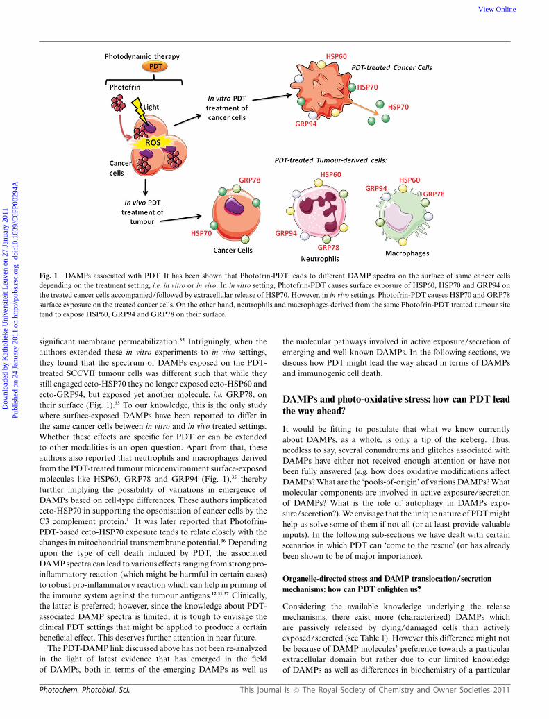
Fig. 1 DAMPs associated with PDT. It has been shown that Photofrin-PDT leads to different DAMP spectra on the surface of same cancer cellsdepending on the treatment setting, i.e. in vitro or in vivo. In in vitro setting, Photofrin-PDT causes surface exposure of HSP60, HSP70 and GRP94 onthe treated cancer cells accompanied/followed by extracellular release of HSP70. However, in in vivo settings, Photofrin-PDT causes HSP70 and GRP78surface exposure on the treated cancer cells. On the other hand, neutrophils and macrophages derived from the same Photofrin-PDT treated tumour sitetend to expose HSP60, GRP94 and GRP78 on their surface.
significant membrane permeabilization.35 Intriguingly, when theauthors extended these in vitro experiments to in vivo settings,they found that the spectrum of DAMPs exposed on the PDT-treated SCCVII tumour cells was different such that while theystill engaged ecto-HSP70 they no longer exposed ecto-HSP60 andecto-GRP94, but exposed yet another molecule, i.e. GRP78, ontheir surface (Fig. 1).35 To our knowledge, this is the only studywhere surface-exposed DAMPs have been reported to differ inthe same cancer cells between in vitro and in vivo treated settings.Whether these effects are specific for PDT or can be extendedto other modalities is an open question. Apart from that, theseauthors also reported that neutrophils and macrophages derivedfrom the PDT-treated tumour microenvironment surface-exposedmolecules like HSP60, GRP78 and GRP94 (Fig. 1),35 therebyfurther implying the possibility of variations in emergence ofDAMPs based on cell-type differences. These authors implicatedecto-HSP70 in supporting the opsonisation of cancer cells by theC3 complement protein.11 It was later reported that Photofrin-PDT-based ecto-HSP70 exposure tends to relate closely with thechanges in mitochondrial transmembrane potential.36 Dependingupon the type of cell death induced by PDT, the associatedDAMP spectra can lead to various effects ranging from strong pro-inflammatory reaction (which might be harmful in certain cases)to robust pro-inflammatory reaction which can help in priming ofthe immune system against the tumour antigens.12,31,37 Clinically,the latter is preferred; however, since the knowledge about PDT-associated DAMP spectra is limited, it is tough to envisage theclinical PDT settings that might be applied to produce a certainbeneficial effect. This deserves further attention in near future.
The PDT-DAMP link discussed above has not been re-analyzedin the light of latest evidence that has emerged in the fieldof DAMPs, both in terms of the emerging DAMPs as well as
the molecular pathways involved in active exposure/secretion ofemerging and well-known DAMPs. In the following sections, wediscuss how PDT might lead the way ahead in terms of DAMPsand immunogenic cell death.
DAMPs and photo-oxidative stress: how can PDT leadthe way ahead?
It would be fitting to postulate that what we know currentlyabout DAMPs, as a whole, is only a tip of the iceberg. Thus,needless to say, several conundrums and glitches associated withDAMPs have either not received enough attention or have notbeen fully answered (e.g. how does oxidative modifications affectDAMPs? What are the ‘pools-of-origin’ of various DAMPs? Whatmolecular components are involved in active exposure/secretionof DAMPs? What is the role of autophagy in DAMPs expo-sure/secretion?). We envisage that the unique nature of PDT mighthelp us solve some of them if not all (or at least provide valuableinputs). In the following sub-sections we have dealt with certainscenarios in which PDT can ‘come to the rescue’ (or has alreadybeen shown to be of major importance).
Organelle-directed stress and DAMP translocation/secretionmechanisms: how can PDT enlighten us?
Considering the available knowledge underlying the releasemechanisms, there exist more (characterized) DAMPs whichare passively released by dying/damaged cells than activelyexposed/secreted (see Table 1). However this difference might notbe because of DAMP molecules’ preference towards a particularextracellular domain but rather due to our limited knowledgeof DAMPs as well as differences in biochemistry of a particular
Photochem. Photobiol. Sci. This journal is © The Royal Society of Chemistry and Owner Societies 2011
Dow
nloa
ded
by K
atho
lieke
Uni
vers
iteit
Leu
ven
on 2
7 Ja
nuar
y 20
11Pu
blis
hed
on 2
4 Ja
nuar
y 20
11 o
n ht
tp://
pubs
.rsc
.org
| do
i:10.
1039
/C0P
P002
94A
View Online

Fig. 2 Photo-oxidative ER stress via hypericin-PDT causes surface exposure of calreticulin (ecto-CRT). T24 human bladder carcinoma cells were treatedwith Mitoxantrone/MTX (1 mM MTX for 4 h) and Hypericin-PDT (150 nM hypericin for 16 h and 2.16 J cm-2 irradiation with visible light followed byrecovery at 1 h post-PDT). MTX is the positive control for ecto-CRT exposure. After treatment, the cells were fixed and immunostained for ecto-CRT(primary antibody: rabbit anti-calreticulin polyclonal antibody, cell signaling; secondary antibody: Alexa Fluor-488 goat anti-rabbit IgG, Invitrogen)followed by DAPI-based staining for the nucleus. Scale bar = 15 mm.
cell death pathway. Nevertheless, while molecular pathways forsome actively translocated/secreted DAMPs have been describedyet for other they are either incomplete or less characterized.Ecto-CRT remains one of the DAMPs for which a molecularpathway has been described in some detail,14 but there are stillareas within that pathway that need further attention. One thingthat emerged from the studies on ecto-CRT is that the ‘pool-of-origin’ of DAMPs from within a sub-cellular location of a cellmight finally influence the type of pathway that the correspondingDAMP eventually follows. For example, ER stress in the case ofecto-CRT sets the tone for molecular components of the secretorypathway and the Unfolded Protein Response (UPR) signalling toplay a role in translocating CRT to the surface.14,16,21 However,in this case, the agents used for exerting ER stress-mediatedecto-CRT translocation (especially anthracyclines) are known toproduce ROS as a secondary effect of cellular damage such thattheir ‘action’ is not exclusively directed towards ER but also otherorganelles.38 Thus, to further characterize this pathway we needa stressor that has ROS-based ER stress as its primary effect.Similarly, in the case of other DAMPs like exo-ATP, ecto-/exo-HSP70, ecto-HSP90, not only the ‘pool-of-origin’ is elusive butalso the exact molecular components involved are unknown.
The uniqueness of PDT as a therapy could be of vitalimportance here, both for further characterization of molecularpathways behind DAMP translocation/secretion and for theidentification of their ‘pool-of-origin’. PDT and its effects heavilydepend upon the sub-cellular localization of the photosensitiser(PS) and luckily there is a significant panel of photosensitisersavailable with various different sub-cellular localization profiles.31
Moreover, ROS-based stress directed towards the organelle wherethe photosensitiser localizes is a primary effect of PDT, e.g.hypericin (localizes predominantly in the ER)-based PDT wouldproduce photo-oxidative ER (p-ox ER stress) stress while 5-ALA (localizes in the mitochondria)-based PDT would producephoto-oxidative mitochondrial stress.30,31 It is worth mentioninghere that correlation between a particular spectrum of DAMPsand stress on a particular sub-cellular site or organelle is arelatively new and highly unexplored concept. For instance, basedon the observations showing that ER-directed and ROS-basedstress causes surface exposure of ecto-CRT, it is presumablethat ecto-HSP70 exposed after Photofrin-PDT requires pathwaysoriginating from the subcellular sites where Photofrin-PDT exertsits primary photodamage,39,40 namely the ER, mitochondria or
Golgi complex. However, further investigations are required toestablish such correlations, since these can influence further studieson molecular pathways.
To this end, we have been investigating the DAMP spectraassociated with photo-oxidative ER stress (p-ox ER stress) exertedby especially hypericin-based PDT (Hyp-PDT) in our laboratory.41
We have observed that p-ox ER stress induces ‘pre-apoptotic’enhancement in surface exposure of calreticulin (ecto-CRT) (seeFig. 2) as quickly as 30 min after PDT in a dose-dependent manner,in T24 human bladder carcinoma cells (Garg et al., unpublishedresults). Interestingly, Photofrin-PDT, although shown to be ca-pable of exerting ROS-based ER stress,40 does not incite enhancedsurface exposure of ecto-CRT in the same cells (J. Golab, personalcommunication). This difference between photofrin-PDT andhypericin-PDT is rather intriguing and might be explained by themagnitude of p-ox ER stress generated since photofrin localizesin the ER to a lesser extent than hypericin, or by distinct PDT-mediated effects on processes required to translocate CRT fromthe ER to the plasma membrane. We also observed that Hyp-PDTinduces ‘pre-apoptotic’ active exo-ATP secretion and late stagepassive release of DAMPs like HSP70, HSP90 and CRT (Garget al., unpublished results). Overall these observations reveal thepotential of Hyp-PDT in causing exposure/secretion of ‘critical’DAMPs and also adds this modality in a rather ‘small club’ ofanti-cancerous therapeutic agents/modalities capable of exposingimmunogenic signals like ecto-CRT.14,16
Oxidative modifications of DAMPs: to be or not to be?
The possibility of immunomodulatory/immunostimulatory po-tential of a DAMP being neutralized via oxidative modificationsis a debate that has risen recently. Apparently, under naturalconditions, the sub-cellular location from where a DAMP isderived can finally decide the kind of modification it mightundergo once outside the cell.37,42 The redox variations withinthe cells and outside mostly are defined by the thiol redox statussuch that the cytosol is highly reducing (due to thiol-regulatingenzymes like thioredoxin-thioredoxin reductase; reduced andoxidized glutathione molecules maintained in a reducing 100 : 1ratio), lumens of all secretory pathway components including ERis highly oxidizing (reduced and oxidized glutathione moleculesmaintained in an oxidizing 3 : 1 ratio), nucleus is highly reduc-ing while the extracellular space is strongly oxidizing (due to
This journal is © The Royal Society of Chemistry and Owner Societies 2011 Photochem. Photobiol. Sci.
Dow
nloa
ded
by K
atho
lieke
Uni
vers
iteit
Leu
ven
on 2
7 Ja
nuar
y 20
11Pu
blis
hed
on 2
4 Ja
nuar
y 20
11 o
n ht
tp://
pubs
.rsc
.org
| do
i:10.
1039
/C0P
P002
94A
View Online

the number of reducing enzymes and abundance of oxidizingagents).37,42 Understandably, the proteins from these sub-cellularregions are accustomed to the redox status of that region suchthat proteins in the cytosol and nucleus are highly susceptibleto oxidation-based neutralization due to presence of cysteineresidues with free sulfhydryl groups. On the other hand, proteinsderived from secretory pathway components like the ER are notbecause their cysteine residues are linked by disulfide bonds,thereby providing extra stability in oxidizing environments.42
Thus DAMPs derived from nucleus and cytosol might be moresusceptible to extracellular space-based oxidation than thosederived from the Golgi or the ER, a claim substantiated by certainrecent observations. For instance, a predominantly cytosolicprotein, S100A8 (Table 1), which tends to act as a DAMPwhen secreted and stimulates recruitment of myeloid cells tothe site of inflammation, can be oxidatively neutralized suchthat the oxidized form is chemotactically inactive.43 Similarly,HMGB1, a well-known DAMP originating from the nucleus(Table 1), has been shown to be susceptible to oxidation-basedneutralization44 although not in the context of extracellular space-based oxidation but rather oxidation via ROS produced frommitochondria because of caspase activity. The latter work alsoreasoned that the type of cell death pathway may also govern theoxidative state of certain DAMPs, e.g. caspase activation duringapoptosis causes mitochondria-instigated HMGB1 oxidation44
but probably not under necrosis, although the mechanism ofthis differential outcome between apoptosis and necrosis remainsenigmatic. However, in the case of cells dying through necrosis,the extracellular oxidative environment comes into the picture,thereby making this a moot case.
Therefore the debate centers around some still unresolvedquestions like: does oxidation-based neutralization theory applyto all DAMPs? This discussion becomes intriguing if we draga ROS-based modality like PDT into the picture since ROSproduced by PDT might be expected to affect DAMPs. Whilethis might be considered puzzling, evidence derived from PDTtreatments both in vitro and in vivo points towards anythingbut inactivation of immunostimulation.8 PDT-treated cancer cellshave been reported to be ‘strongly’ immunogenic and PDT-derived HSPs are usually observed to remain immunostimulatory.8
In fact, it has been proposed that photo-oxidation via PDTmight actually be immunologically beneficial such that PDT-basedprotein unfolding events and membrane changes might revealcertain neo or cryptic antigens.8 Thus, although there are no suchindications from immunological data generated after PDT, we stillneed to investigate whether ROS-based modifications of certainDAMPs by PDT could inactivate them.
It is worth mentioning here that in the context of DAMPs,oxidation might not always be considered to be counter-productivesince its effects tend to be biomolecule-dependent and DAMPs (asmentioned previously) are not always protein based in nature.Lipid or lipid-based molecules (e.g. phospholipids) have alsobeen documented to have DAMP-like activity (Table 1). In fact,oxidized phospholipids (Ox-PL) have been documented to beattractive to both humoral and cellular innate immune responsessuch that their presence on apoptotic cells has been postulatedto assist in binding with macrophage scavenger receptors likeSR-B1 and CD3645,46 and in DC maturation regulation.47 Ox-PL have been reported to act as ligands for PRRs and have been
found to be capable of activating a TLR4-based signaling pathwayindependent of NFkB.48 In fact, it has been proposed that in thecase of phospholipids, oxidation leads to creation of ‘new’ epitopesrecognizable by antibodies.48 Observations on Ox-PL and theirbehavior have led the researchers to postulate the ‘Lipid WhiskerModel’, an extended variant of the Fluid Mosaic Model.49 Asper the Fluid Mosaic Model, the amphipathic phospholipids aresupposed to have their hydrophobic fatty acyl chains hidden withinthe membrane interior while the hydrophilic polar headgroups aresupposed to be oriented towards the aqueous environment.49 How-ever, as per the ‘Lipid Whisker Model’, when cellular membranesundergo lipid peroxidation (e.g. during apoptosis or inflammation)the resulting oxidation causes previously hydrophobic portionsof fatty acids to move towards the aqueous exterior therebyfacilitating their recognition by PRRs.49 More specifically, it hasbeen observed that the oxidized form of phosphatidylcholinespecifically tends to emerge on the apoptotic cells and possiblyhelps in their clearance by phagocytes.45 Researchers have alsoshown that phosphatidylserine (PS) the ‘classical’ lipid-based ‘eatme’ signal (Table 1) tends to bind better to CD36 phagocytosis-assisting receptor on macrophages when in oxidized form.50 Allthis taken together means these Ox-PLs present an interestingprospect that deserves further investigation. Interestingly, PDThas been known for some time to cause considerable lipidperoxidation, a process that has been found to play an importantrole in PDT-mediated cell death and inflammatory processes.51,52
Thus, it would be worth investigating if PDT-mediated lipidperoxidation enhances immunogenicity of PDT-treated cells andtheir phagocytosis.
Cell death accompanied by autophagy and DAMPs: how beneficialcan this link be?
Autophagy accompanying cell death has recently shown the poten-tial to cause ‘active’ secretion of HMGB1,53 a well-known DAMPand the ability to affect the proteome of the cell membrane.2,54
Interestingly, it was recently shown that cytosolic HMGB1is capable of regulating starvation-induced autophagy, therebyfurther shedding light on a possible link between autophagicprocesses and HMGB1.55 However, it has not been characterizedyet if in long run autophagy accompanying cell death helps, interms of immunological impact. PDT is one of those therapeuticmodalities which can convincingly induce cell death accompaniedby or caused by autophagy in cancer cells, a subject that has beena topic of many investigations in recent times.31,56–58 We envisagethat the study of DAMP spectra and immunogenicity in cancercells that, due to genetic defects in apoptotic pathway, undergoautophagic cell death after PDT could help us in further answeringthe conundrum of whether autophagy can help by enhancingimmunogenicity or reduce it.
DAMPs and their ‘alter egos’: where does PDT fit in the story?
The discussions above mostly concentrate on the ‘good’ facet ofDAMPs, i.e. the scenario where DAMPs could help us againstdiseases like cancer by pro-actively activating the immune systemagainst cancerous lesions. However in recent times it has beenrevealed that certain DAMPs (especially HMGB1) have an ‘alterego’ which might be responsible for them driving certain
Photochem. Photobiol. Sci. This journal is © The Royal Society of Chemistry and Owner Societies 2011
Dow
nloa
ded
by K
atho
lieke
Uni
vers
iteit
Leu
ven
on 2
7 Ja
nuar
y 20
11Pu
blis
hed
on 2
4 Ja
nuar
y 20
11 o
n ht
tp://
pubs
.rsc
.org
| do
i:10.
1039
/C0P
P002
94A
View Online

non-infectious inflammatory/autoimmune diseases, e.g.rheumatoid arthritis, systemic lupus erythematosus (SLE),59
scleroderma59 and renal autoimmune tissue injury.60 A link hasbeen postulated between the DAMP-associated pro-inflammatoryprogram and the progression of these diseases. However, it is ratherhard to fit PDT within this story since PDT that is associatedwith DAMPs exposure and secretion has actually been in use fortreatment of various non-infectious inflammatory/autoimmunediseases with positive results, e.g. psoriasis,61 psoriatic arthritis,62
rheumatoid arthritis,63,64 scleroderma,8,65 vulvar lichen sclerosus66
and autoimmune skin ulcers.67 PDT-based treatment of thesediseases has been considered to be promising, meaning that therelationship between DAMPs and inflammatory/autoimmunediseases needs a ‘fresh re-look’ on the lines of PDT. It is possiblethat in these settings PDT might be proving to be beneficial dueto ‘production’ of anti-inflammatory DAMPs or by inactivatingcertain ‘problematic’ inflammatory DAMPs (possibly via ROS),however both these possibilities need further investigation.Nevertheless, one point is clear with this backdrop: that PDThas considerable beneficial immunomodulatory potential interms of disease management even if the diseases are originallyinflammation-associated/-driven.
Concluding remarks
DAMP-related research has been around for a considerable timeand, while the complete extent and diversity up to which cells canexpose DAMPs is still to be characterized, several ‘new’ DAMPshave emerged recently. Moreover, major effort needs to be investedon getting a better knowledge about the link between a particularDAMP and a particular cell death pathway as well as the molecularpathways underlying the extracellular emergence of DAMPs.Considering the uniqueness of PDT in terms of its settings, andits proven immunogenic character, we can safely assume that PDTholds great promise not only in terms of ‘exposing’ as yet unknownDAMPs but also in terms of shedding more light on the molecularpathway underlying the translocation/secretion of these DAMPs.Moreover, urgent attention is needed to characterize the presenceof immunogenic apoptosis following PDT so that this treatmentmodality can be optimized for clinical induction of immunogeniccell death in tumours.
Abbreviations
APC Antigen-Presenting CellsATP Adenosine TriphosphateCDAMP Cell Death Associated Molecular PatternsCRT CalreticulinDAMP Damage-Associated Molecular PatternsDC Dendritic CellsECM Extracellular MatrixER Endoplasmic ReticulumHMGB1 High Mobility Group Box-1HSP Heat Shock ProteinsOx-PL Oxidized PhospholipidsPAMP Pathogen-Associated Molecular PatternsPDT Photodynamic Therapyp-ox ER stress Photo-oxidative Endoplasmic Reticulum stressPRR Pattern Recognition Receptors
PS PhotosensitiserROS Reactive Oxygen Species
Acknowledgements
The work from the author’s laboratories was supported by grantsfrom the K.U. Leuven (OT49/06) to P.A. and F.W.O Vlaanderen(G.0728.10). This paper presents research results of the IAP6/18,funded by the Interuniversity Attraction Poles Programme, ini-tiated by the Belgian State, Science Policy Office. D. V. K. is apostdoctoral research fellow of the Fund for Scientific ResearchFlanders (FWO-Vlaanderen), Belgium. Research in the Vanden-abeele group is supported by VIB, Ghent University (GROUP-IDconsortium of the UGhent MRP initiative), Research FoundationFlanders (FWO-Vlaanderen) (G.0875.11 and G.0973.11), FederalResearch Program IAP 6/18, European Research Program FP6ApopTrain (MRTN-CT-035624) and FP7 Apo-Sys 200767, andthe GROUP-ID consortium of the UGent MRP initiative. P.V.holds a Methusalem grant (BOF09/01M00709) from the FlemishGovernment. Fig. 1 was produced using Servier Medical Art(www.servier.com) for which the authors would like to acknowl-edge Servier.
References
1 M. E. Bianchi, DAMPs, PAMPs and alarmins: all we need to knowabout danger, J. Leukocyte Biol., 2006, 81, 1–5.
2 A. D. Garg, D. Nowis, J. Golab, P. Vandenabeele, D. V. Kryskoand P. Agostinis, Immunogenic cell death, DAMPs and anticancertherapeutics: an emerging amalgamation, Biochim. Biophys. Acta, 2010,1805, 53–71.
3 S. Y. Seong and P. Matzinger, Hydrophobicity: an ancient damage-associated molecular pattern that initiates innate immune responses,Nat. Rev. Immunol., 2004, 4, 469–478.
4 P. Matzinger, Tolerance, danger, and the extended family, Annu. Rev.Immunol., 1994, 12, 991–1045.
5 J. J. Maher, DAMPs ramp up drug toxicity, J. Clin. Invest., 2009, 119,246–249.
6 C. S. Calfee and M. A. Matthay, Clinical immunology: Culprits withevolutionary ties, Nature, 2010, 464, 41–42.
7 S. S. Iyer, W. P. Pulskens, J. J. Sadler, L. M. Butter, G. J. Teske, T. K.Ulland, S. C. Eisenbarth, S. Florquin, R. A. Flavell, J. C. Leemans andF. S. Sutterwala, Necrotic cells trigger a sterile inflammatory responsethrough the Nlrp3 inflammasome, Proc. Natl. Acad. Sci. U. S. A., 2009,106, 20388–20393.
8 A. D. Garg, D. Nowis, J. Golab and P. Agostinis, Photodynamictherapy: illuminating the road from cell death towards anti-tumourimmunity, Apoptosis, 2010, 15, 1050–1071.
9 D. V. Krysko and P. Vandenabeele, Clearance of dead cells: mechanisms,immune responses and implication in the development of diseases,Apoptosis, 2010, DOI: 10.1007/s10495-10010-10524-10496.
10 M. Korbelik, PDT-associated host response and its role in the therapyoutcome, Lasers Surg. Med., 2006, 38, 500–508.
11 M. Korbelik and J. Sun, Photodynamic therapy-generated vaccine forcancer therapy, Cancer Immunol. Immunother., 2005, 55, 900–909.
12 D. R. Green, T. Ferguson, L. Zitvogel and G. Kroemer, Immunogenicand tolerogenic cell death, Nat. Rev. Immunol., 2009, 9, 353–363.
13 L. Zitvogel, O. Kepp and G. Kroemer, Decoding cell death signals ininflammation and immunity, Cell, 2010, 140, 798–804.
14 L. Zitvogel, O. Kepp, L. Senovilla, L. Menger, N. Chaput andG. Kroemer, Immunogenic tumor cell death for optimal anticancertherapy: the calreticulin exposure pathway, Clin. Cancer Res., 2010, 16,3100–3104.
15 S. Demaria, E. Pikarsky, M. Karin, L. M. Coussens, Y. C. Chen,E. M. El-Omar, G. Trinchieri, S. M. Dubinett, J. T. Mao, E. Szabo,A. Krieg, G. J. Weiner, B. A. Fox, G. Coukos, E. Wang, R. T. Abraham,M. Carbone and M. T. Lotze, Cancer and inflammation: promise forbiologic therapy, J. Immunother., 2010, 33, 335–351.
This journal is © The Royal Society of Chemistry and Owner Societies 2011 Photochem. Photobiol. Sci.
Dow
nloa
ded
by K
atho
lieke
Uni
vers
iteit
Leu
ven
on 2
7 Ja
nuar
y 20
11Pu
blis
hed
on 2
4 Ja
nuar
y 20
11 o
n ht
tp://
pubs
.rsc
.org
| do
i:10.
1039
/C0P
P002
94A
View Online

16 T. Panaretakis, O. Kepp, U. Brockmeier, A. Tesniere, A. C. Bjorklund,D. C. Chapman, M. Durchschlag, N. Joza, G. Pierron, P. van Endert,J. Yuan, L. Zitvogel, F. Madeo, D. B. Williams and G. Kroemer,Mechanisms of pre-apoptotic calreticulin exposure in immunogeniccell death, EMBO J., 2009, 28, 578–590.
17 T. Verfaillie, A. D. Garg and P. Agostinis, Targeting ER stressinduced apoptosis and inflammation in cancer, Cancer Lett., 2010,DOI: 10.1016/j.canlet.2010.07.016.
18 S. Hummasti and G. S. Hotamisligil, Endoplasmic reticulumstress andinflammation in obesity and diabetes, Circ. Res., 2010, 107, 579–591.
19 G. S. Hotamisligil, Endoplasmic reticulum stress and the inflammatorybasis of metabolic disease, Cell, 2010, 140, 900–917.
20 A. Kaser and R. S. Blumberg, Endoplasmic reticulum stress andintestinal inflammation, Mucosal Immunol., 2009, 3, 11–16.
21 M. Obeid, A. Tesniere, F. Ghiringhelli, G. M. Fimia, L. Apetoh, J. L.Perfettini, M. Castedo, G. Mignot, T. Panaretakis, N. Casares, D.Metivier, N. Larochette, P. van Endert, F. Ciccosanti, M. Piacentini,L. Zitvogel and G. Kroemer, Calreticulin exposure dictates the im-munogenicity of cancer cell death, Nat. Med., 2006, 13, 54–61.
22 R. Q. Peng, Y. B. Chen, Y. Ding, R. Zhang, X. Zhang, X. J. Yu,Z. W. Zhou, Y. X. Zeng and X. S. Zhang, Expression of calreticulinis associated with infiltration of T-cells in stage IIIB colon cancer,World J. Gastroenterol., 2010, 16, 2428–2434.
23 D. Jocham, A. Richter, L. Hoffmann, K. Iwig, D. Fahlenkamp,G. Zakrzewski, E. Schmitt, T. Dannenberg, W. Lehmacher, J. vonWietersheim and C. Doehn, Adjuvant autologous renal tumour cellvaccine and risk of tumour progression in patients with renal-cellcarcinoma after radical nephrectomy: phase III, randomised controlledtrial, Lancet, 2004, 363, 594–599.
24 M. May, S. Brookman-May, B. Hoschke, C. Gilfrich, F. Kendel, S.Baxmann, S. Wittke, S. T. Kiessig, K. Miller and M. Johannsen,Ten-year survival analysis for renal carcinoma patients treated withan autologous tumour lysate vaccine in an adjuvant setting, CancerImmunol. Immunother., 2009, 59, 687–695.
25 E. A. Hirschowitz, T. Foody, R. Kryscio, L. Dickson, J. Sturgill andJ. Yannelli, Autologous dendritic cell vaccines for non-small-cell lungcancer, J. Clin. Oncol., 2004, 22, 2808–2815.
26 L. M. Liau, R. M. Prins, S. M. Kiertscher, S. K. Odesa, T. J. Kremen,A. J. Giovannone, J. W. Lin, D. J. Chute, P. S. Mischel, T. F. Cloughesyand M. D. Roth, Dendritic cell vaccination in glioblastoma patientsinduces systemic and intracranial T-cell responses modulated by thelocal central nervous system tumor microenvironment, Clin. CancerRes., 2005, 11, 5515–5525.
27 K. Palucka, H. Ueno, G. Zurawski, J. Fay and J. Banchereau, Buildingon dendritic cell subsets to improve cancer vaccines, Curr. Opin.Immunol., 2010, 22, 258–263.
28 C. L. Chiang, F. Benencia and G. Coukos, Whole tumor antigenvaccines, Semin. Immunol., 2010, 22, 132–143.
29 S. O. Gollnick and C. M. Brackett, Enhancement of anti-tumorimmunity by photodynamic therapy, Immunol. Res., 2009, 46, 216–226.
30 A. P. Castano, P. Mroz and M. R. Hamblin, Photodynamic therapyand anti-tumour immunity, Nat. Rev. Cancer, 2006, 6, 535–545.
31 E. Buytaert, M. Dewaele and P. Agostinis, Molecular effectors ofmultiple cell death pathways initiated by photodynamic therapy,Biochim. Biophys. Acta, 2007, 1776, 86–107.
32 G. Brouckaert, M. Kalai, D. V. Krysko, X. Saelens, D. Vercammen, M.Ndlovu, G. Haegeman, K. D’Herde and P. Vandenabeele, Phagocytosisof necrotic cells by macrophages is phosphatidylserine dependent anddoes not induce inflammatory cytokine production, Mol. Biol. Cell,2003, 15, 1089–1100.
33 M. F. Tsan and B. Gao, Heat shock proteins and immune system,J. Leukocyte Biol., 2009, 85, 905–910.
34 A. G. Pockley, Heat shock proteins as regulators of the immuneresponse, Lancet, 2003, 362, 469–476.
35 M. Korbelik, J. Sun and I. Cecic, Photodynamic therapy-induced cellsurface expression and release of heat shock proteins: relevance fortumor response, Cancer Res., 2005, 65, 1018–1026.
36 F. Zhou, D. Xing and W. R. Chen, Dynamics and mechanism of HSP70translocation induced by photodynamic therapy treatment, CancerLett., 2008, 264, 135–144.
37 S. Carta, P. Castellani, L. Delfino, S. Tassi, R. Vene and A. Rubartelli,DAMPs and inflammatory processes: the role of redox in the differentoutcomes, J. Leukocyte Biol., 2009, 86, 549–555.
38 G. Minotti, P. Menna, E. Salvatorelli, G. Cairo and L. Gianni,Anthracyclines: molecular advances and pharmacologic developments
in antitumor activity and cardiotoxicity, Pharmacol. Rev., 2004, 56,185–229.
39 Y. J. Hsieh, C. C. Wu, C. J. Chang and J. S. Yu, Subcellular localizationof Photofrin determines the death phenotype of human epidermoidcarcinoma A431 cells triggered by photodynamic therapy: when plasmamembranes are the main targets, J. Cell. Physiol., 2003, 194, 363–375.
40 A. Szokalska, M. Makowski, D. Nowis, G. M. Wilczynski, M. Kujawa,C. Wojcik, I. Mlynarczuk-Bialy, P. Salwa, J. Bil, S. Janowska, P.Agostinis, T. Verfaillie, M. Bugajski, J. Gietka, T. Issat, E. Glodkowska,P. Mrowka, T. Stoklosa, M. R. Hamblin, P. Mroz, M. Jakobisiakand J. Golab, Proteasome inhibition potentiates antitumor effectsof photodynamic therapy in mice through induction of endoplasmicreticulum stress and unfolded protein response, Cancer Res., 2009, 69,4235–4243.
41 A. D. Garg, T. Verfaillie, N. Rubio, G. B. Ferreira, C. Mathieuand P. Agostinis, Hypericin-PDT treatment of cancer cells leads tosurface exposure/extracellular release of DAMPs and activates humanimmature dendritic cells (abstract), Belg. J. Med. Oncol., 2010, 4, 93–94.
42 A. Rubartelli and M. T. Lotze, Inside, outside, upside down: damage-associated molecular-pattern molecules (DAMPs) and redox, TrendsImmunol., 2007, 28, 429–436.
43 C. A. Harrison, M. J. Raftery, J. Walsh, P. Alewood, S. E. Iismaa,S. Thliveris and C. L. Geczy, Oxidation regulates the inflammatoryproperties of the murine S100 protein S100A8, J. Biol. Chem., 1999,274, 8561–8569.
44 H. Kazama, J. E. Ricci, J. M. Herndon, G. Hoppe, D. R. Green andT. A. Ferguson, Induction of immunological tolerance by apoptoticcells requires caspase-dependent oxidation of high-mobility group box-1 protein, Immunity, 2008, 29, 21–32.
45 M. K. Chang, C. J. Binder, M. Torzewski and J. L. Witztum, C-reactiveprotein binds to both oxidized LDL and apoptotic cells throughrecognition of a common ligand: Phosphorylcholine of oxidizedphospholipids, Proc. Natl. Acad. Sci. U. S. A., 2002, 99, 13043–13048.
46 E. A. Podrez, E. Poliakov, Z. Shen, R. Zhang, Y. Deng, M. Sun, P. J.Finton, L. Shan, B. Gugiu, P. L. Fox, H. F. Hoff, R. G. Salomon andS. L. Hazen, Identification of a novel family of oxidized phospholipidsthat serve as ligands for the macrophage scavenger receptor CD36,J. Biol. Chem., 2002, 277, 38503–38516.
47 S. Bluml, S. Kirchberger, V. N. Bochkov, G. Kronke, K. Stuhlmeier,O. Majdic, G. J. Zlabinger, W. Knapp, B. R. Binder, J. Stockl andN. Leitinger, Oxidized phospholipids negatively regulate dendritic cellmaturation induced by TLRs and CD40, J. Immunol., 2005, 175, 501–508.
48 A. Furnkranz and N. Leitinger, Regulation of inflammatory responsesby oxidized phospholipids: structure-function relationships, Curr.Pharm. Des., 2004, 10, 915–921.
49 S. L. Hazen, Oxidized phospholipids as endogenous pattern recognitionligands in innate immunity, J. Biol. Chem., 2008, 283, 15527–15531.
50 M. E. Greenberg, M. Sun, R. Zhang, M. Febbraio, R. Silverstein andS. L. Hazen, Oxidized phosphatidylserine-CD36 interactions play anessential role in macrophage-dependent phagocytosis of apoptotic cells,J. Exp. Med., 2006, 203, 2613–2625.
51 D. Nowis, M. Makowski, T. Stoklosa, M. Legat, T. Issat and J. Golab,Direct tumor damage mechanisms of photodynamic therapy, ActaBiochim. Pol., 2005, 52, 339–352.
52 D. V. Sakharov, E. D. Elstak, B. Chernyak and K. W. Wirtz, Prolongedlipid oxidation after photodynamic treatment. Study with oxidation-sensitive probe C11-BODIPY581/591, FEBS Lett., 2005, 579, 1255–1260.
53 J. Thorburn, H. Horita, J. Redzic, K. Hansen, A. E. Frankel and A.Thorburn, Autophagy regulates selective HMGB1 release in tumorcells that are destined to die, Cell Death Differ., 2008, 16, 175–183.
54 L. Baricault, J. A. Fransen, M. Garcia, C. Sapin, P. Codogno, L. A.Ginsel and G. Trugnan, Rapid sequestration of DPP IV/CD26 andother cell surface proteins in an autophagic-like compartment in Caco-2 cells treated with forskolin, J. Cell. Sci., 1995, 108(Pt 5), 2109–2121.
55 D. Tang, R. Kang, K. M. Livesey, C. W. Cheh, A. Farkas, P. Loughran,G. Hoppe, M. E. Bianchi, K. J. Tracey, H. J. Zeh, 3rd and M. T. Lotze,Endogenous HMGB1 regulates autophagy, J. Cell Biol., 2010, 190,881–892.
56 M. Dewaele, W. Martinet, N. Rubio, T. Verfaillie, P. A. de Witte, J.Piette and P. Agostinis, Autophagy pathways activated in response toPDT contribute to cell resistance against ROS damage, J. Cell Mol.Med., 2010, DOI: 10.1111/j.1582-4934.2010.01118.x.
Photochem. Photobiol. Sci. This journal is © The Royal Society of Chemistry and Owner Societies 2011
Dow
nloa
ded
by K
atho
lieke
Uni
vers
iteit
Leu
ven
on 2
7 Ja
nuar
y 20
11Pu
blis
hed
on 2
4 Ja
nuar
y 20
11 o
n ht
tp://
pubs
.rsc
.org
| do
i:10.
1039
/C0P
P002
94A
View Online

57 M. Dewaele, H. Maes and P. Agostinis, ROS-mediated mechanismsof autophagy stimulation and their relevance in cancer therapy,Autophagy, 2010, 6, DOI: 10.4161/auto.6.7.12113.
58 J. J. Reiners, Jr., P. Agostinis, K. Berg, N. L. Oleinick and D.Kessel, Assessing autophagy in the context of photodynamic therapy,Autophagy, 2010, 6, 7–18.
59 G. P. Sims, D. C. Rowe, S. T. Rietdijk, R. Herbst and A. J. Coyle,HMGB1 and RAGE in inflammation and cancer, Annu. Rev. Immunol.,2010, 28, 367–388.
60 H. J. Anders and D. O. Schlondorff, Innate immune receptors andautophagy: implications for autoimmune kidney injury, Kidney Int.,2010, 78, 29–37.
61 T. Smits, M. M. Kleinpenning, P. E. van Erp, P. C. van de Kerkhof andM. J. Gerritsen, A placebo-controlled randomized study on the clinicaleffectiveness, immunohistochemical changes and protoporphyrin IXaccumulation in fractionated 5-aminolaevulinic acid-photodynamictherapy in patients with psoriasis, Br. J. Dermatol., 2006, 155, 429–436.
62 L. G. Ratkay, J. D. Waterfield and D. W. Hunt, Photodynamic therapyin immune (non-oncological) disorders: focus on benzoporphyrinderivatives, BioDrugs, 2000, 14, 127–135.
63 E. Torikai, Y. Kageyama, E. Kohno, T. Hirano, Y. Koide, S. Terakawaand A. Nagano, Photodynamic therapy using talaporfin sodium forsynovial membrane from rheumatoid arthritis patients and collagen-induced arthritis rats, Clin. Rheumatol., 2007, 27, 751–761.
64 A. Hansch, O. Frey, M. Gajda, G. Susanna, J. Boettcher, R. Brauerand W. A. Kaiser, Photodynamic treatment as a novel approach in thetherapy of arthritic joints, Lasers Surg. Med., 2008, 40, 265–272.
65 H. Takahashi, S. Komatsu, M. Ibe, A. Ishida-Yamamoto, S. Nakajima,I. Sakata and H. Iizuka, ATX-S10(Na)-PDT shows more potent effecton collagen metabolism of human normal and scleroderma dermalfibroblasts than ALA-PDT, Arch. Dermatol. Res., 2006, 298, 257–263.
66 A. Olejek, K. Steplewska, A. Gabriel, I. Kozak-Darmas, A. Jarek,S. Kellas-Sleczka, F. Bydlinski, K. Sieron-Stoltny, S. Horak, A.Chelmicki and A. Sieron, Efficacy of Photodynamic Therapy inVulvar Lichen Sclerosus Treatment Based on ImmunohistochemicalAnalysis of CD34, CD44, Myelin Basic Protein, and Ki67 Antibodies,Int. J. Gynecol. Cancer, 2010, 20, 879–887.
67 S. Motta and M. Monti, Photodynamic therapy-a promising treatmentoption for autoimmune skin ulcers: a case report, Photochem. Photo-biol. Sci., 2007, 6, 1150–1151.
68 M. Subbarayan, U. O. Hafeli, D. K. Feyes, J. Unnithan, S. N.Emancipator and H. Mukhtar, A simplified method for preparationof 99mTc-annexin V and its biologic evaluation for in vivo imaging of
apoptosis after photodynamic therapy, J. Nucl. Med., 2003, 44, 650–656.
69 S. Suzuki and A. B. Kulkarni, Extracellular heat shock proteinHSP90beta secreted by MG63 osteosarcoma cells inhibits activation oflatent TGF-beta1, Biochem. Biophys. Res. Commun., 2010, 398, 525–531.
70 J. R. Riddell, X. Y. Wang, H. Minderman and S. O. Gollnick,Peroxiredoxin 1 stimulates secretion of proinflammatory cytokines bybinding to TLR4, J. Immunol., 2009, 184, 1022–1030.
71 A. Iwata, V. Morgan-Stevenson, B. Schwartz, L. Liu, J. Tupper, X.Zhu, J. Harlan and R. Winn, Extracellular BCL2 proteins are danger-associated molecular patterns that reduce tissue damage in murinemodels of ischemia-reperfusion injury, PLoS One, 2010, 5, e9103.
72 K. E. Blume, S. Soeroes, M. Waibel, H. Keppeler, S. Wesselborg,M. Herrmann, K. Schulze-Osthoff and K. Lauber, Cell surfaceexternalization of annexin A1 as a failsafe mechanism preventinginflammatory responses during secondary necrosis, J. Immunol., 2009,183, 8138–8147.
73 A. A. Manfredi and P. Rovere-Querini, The mitochondrion–a Trojanhorse that kicks off inflammation?, N. Engl. J. Med., 2010, 362, 2132–2134.
74 Q. Zhang, M. Raoof, Y. Chen, Y. Sumi, T. Sursal, W. Junger, K. Brohi,K. Itagaki and C. J. Hauser, Circulating mitochondrial DAMPs causeinflammatory responses to injury, Nature, 2010, 464, 104–107.
75 A. Babelova, K. Moreth, W. Tsalastra-Greul, J. Zeng-Brouwers, O.Eickelberg, M. F. Young, P. Bruckner, J. Pfeilschifter, R. M. Schaefer,H. J. Grone and L. Schaefer, Biglycan, a danger signal that activates theNLRP3 inflammasome via toll-like and P2X receptors, J. Biol. Chem.,2009, 284, 24035–24048.
76 L. Schaefer, Extracellular matrix molecules: endogenous danger signalsas new drug targets in kidney diseases, Curr. Opin. Pharmacol., 2010,10, 185–190.
77 F. G. Goh, A. M. Piccinini, T. Krausgruber, I. A. Udalova and K. S.Midwood, Transcriptional regulation of the endogenous danger signaltenascin-C: a novel autocrine loop in inflammation, J. Immunol., 2010,184, 2655–2662.
78 T. E. Ichim, Z. Zhong, S. Kaushal, X. Zheng, X. Ren, X. Hao, J. A.Joyce, H. H. Hanley, N. H. Riordan, J. Koropatnick, V. Bogin, B. R.Minev, W. P. Min and R. H. Tullis, Exosomes as a tumor immuneescape mechanism: possible therapeutic implications, J. Transl. Med.,2008, 6, 37.
79 L. Zitvogel, A. Tesniere and G. Kroemer, Cancer despite immuno-surveillance: immunoselection and immunosubversion, Nat. Rev. Im-munol., 2006, 6, 715–727.
This journal is © The Royal Society of Chemistry and Owner Societies 2011 Photochem. Photobiol. Sci.
Dow
nloa
ded
by K
atho
lieke
Uni
vers
iteit
Leu
ven
on 2
7 Ja
nuar
y 20
11Pu
blis
hed
on 2
4 Ja
nuar
y 20
11 o
n ht
tp://
pubs
.rsc
.org
| do
i:10.
1039
/C0P
P002
94A
View Online