Cyclin E Associates with Components of the Pre-mRNA Splicing Machinery in Mammalian Cells
Upload
independentCategory
view
1download
0
Cyclin E is recruited to the nuclear matrix duringdifferentiation, but is not recruited in cancer cellsJennifer Munkley1, Nikki A. Copeland1, Victoria Moignard1, John R. P. Knight1,
Erin Greaves2, Simon A. Ramsbottom1, Mary E. Pownall1, Jennifer Southgate1,
Justin F.-X. Ainscough2 and Dawn Coverley1,*
1Department of Biology, University of York, YO10 5YW and 2School of Medicine, Leeds University,LS2 9JT, UK
Received August 4, 2010; Revised October 15, 2010; Accepted November 5, 2010
ABSTRACT
Cyclin E supports pre-replication complex (pre-RC)assembly, while cyclin A-associated kinase acti-vates DNA synthesis. We show that cyclin E, butnot A, is mounted upon the nuclear matrix insub-nuclear foci in differentiated vertebrate cells,but not in undifferentiated cells or cancer cells. Inmurine embryonic stem cells, Xenopus embryos andhuman urothelial cells, cyclin E is recruited to thenuclear matrix as cells differentiate and this canbe manipulated in vitro. This suggests that pre-RCassembly becomes spatially restricted as templateusage is defined. Furthermore, failure to become re-stricted may contribute to the plasticity of cancercells.
INTRODUCTION
Cyclin E is a G1 cyclin that associates preferentially withcyclin-dependent protein kinase 2 (CDK2). There are twomammalian E type cyclins, with similar expression,function and structure in complex with CDK2 (1).Cyclin E rises in mid G1, peaks after passage throughthe restriction point (R), and falls around G1/S (2).Initiation of mammalian DNA replication is controlledby CDK2, which, in association with cyclins E and A,acts on proteins that assemble at replication origins.Cyclin E plays a role in pre-replication complex(pre-RC) formation in higher eukaryotes, cooperatingwith CDC6 and CDT1 to support assembly of the minichromosome maintenance (MCM) complex (3–7). CyclinE is also required for centrosome duplication (8), where itsrole also appears to be distinct from that of cyclin A (3,9).
Despite increasingly well-defined roles for cyclin E,fibroblasts isolated from mice deficient in E1 and 2 areable to proliferate, indicating that cyclin E is redundantin the mitotic cell cycle. In fact, cyclin E appears to be
essential for MCM assembly only in cells preparing forS phase that is not preceded by mitosis, such asthose re-entering cycle from G0 or undergoingendoreduplication (5,10). By contrast, CDK2-deficientfibroblasts do not encounter similar problems (11), sug-gesting that cyclin E may act independently of CDK2under some circumstances. Consistent with this, akinase-deficient mutant partially corrects the phenotypeof cyclin E null cells, restoring MCM complex assembly(12). Similarly, the centrosomal function of cyclin E mayalso be CDK2 independent (8). Thus, while CDK2 is thepreferred binding partner of cyclin E and cyclin E canclearly activate CDK2, evidence points to additionalCDK-independent roles for cyclin E.DNA replication is constrained in nuclear space to
discrete sub-nuclear foci that are associated with anon-chromatin protein/RNA structure, that is definedas ‘nuclear matrix’ or ‘nucleoskeleton’, and can bevisualized by electron microscopy (13,14). Nuclearmatrix-associated DNA is enriched with replicationorigins during G1 phase, suggesting that recruitmentplays a role in initiation (15), while the pre-RC proteinsCDC6 and ORC1 both undergo transient association inmammalian cells (16). However, there is little evidence toindicate at what point in pre-RC assembly DNA replica-tion origins are recruited, or what the nuclearmatrix-associated receptors might be.In Xenopus egg extracts cyclin E is reported to bind to
chromatin, most likely via CDC6, which acts as achromatin-associated receptor (4). Similarly, work withmouse embryonic fibroblasts and HeLa cells indicatesthat cyclin E is loaded onto chromatin and that thisinvolves direct interaction with both CDT1 and MCMproteins (12). Thus, immobilization of cyclin E appearsto occur in association with pre-RC formation and theprevailing view is that this reflects association with chro-matin. However, the protocols used in most of thesestudies do not discriminate between chromatin andnuclear matrix, and the picture is further complicated by
*To whom correspondence should be addressed. Tel: +44 1904 328651; Fax: +01904 328505; Email: [email protected]
Published online 24 November 2010 Nucleic Acids Research, 2011, Vol. 39, No. 7 2671–2677doi:10.1093/nar/gkq1190
� The Author(s) 2010. Published by Oxford University Press.This is an Open Access article distributed under the terms of the Creative Commons Attribution Non-Commercial License (http://creativecommons.org/licenses/by-nc/2.5), which permits unrestricted non-commercial use, distribution, and reproduction in any medium, provided the original work is properly cited.

analyses of cell types of distinct species, differentiationand pathological state.Here we show that (i) cyclin E is immobilized in the
nucleus by association with the nuclear matrix, (ii) thatrecruitment occurs as cells differentiate and (iii) thatcyclin E is not attached to the nuclear matrix in cancercells. This is the first time that either of thereplication-associated cyclins has been linked with thenuclear matrix. These novel findings point to an importantrole for cyclin E in genome organization. They also arguethat despite the relative ease with which cancer cells can bemanipulated, they are a questionable choice for analysis ofnuclear matrix function or the spatial organization ofDNA replication.
MATERIALS AND METHODS
Cell culture
Cell lines were sourced from JCRB or ATCC and main-tained as recommended. Mouse cardiac fibroblasts ex-pressing Col1A1 (not shown) were derived from atriadissected from a C57/BL6 mouse heart. Mouse ES cells(KES1) were differentiated to embryoid bodies orembryoid body outgrowths as described (17). Xenopus
laevis embryos were produced as described (18). NHUcells were established from surgical samples collectedwith ethical review and informed patient consent andinduced to differentiate as described (19). NIH3T3 cellswere synchronised and labelled with Bromodeoxyuridine(BrdU) as described (3).
Nuclear fractionation
Nuclear fractionation was carried out as described (20).Typically, 25% of cell lysate in CSK buffer was removed(total) and the rest a diluted 2-fold with CSK, 0.2% TritonX-100 and separated by centrifugation into detergentsupernatant (SN) and pellet (P). Pellet was resuspendedin CSK with 0.5 M NaCl to produce salt wash or ‘w’.Pellet was further re-suspended in digestion buffer andincubated with or without DNase 1 (Roche), supple-mented with 0.5 M NaCl and separated by centrifugation.Cell equivalents of each fraction were analysed by westernblot. Mouse cyclin E was detected with rabbit polyclonalantibody 07-687 (Upstate) in Figure 1B and F, andverified with ab7959 (not shown); and with ab7959(Abcam) in Figures 1C and 3A. Human cyclin E wasdetected with mouse monoclonal antibody sc247 HE12(Santa Cruz). Xenopus cyclin E was detected with arabbit polyclonal antibody that was a kind gift from
Figure 1. Cyclin E is present in nuclear matrix-associated foci. (A) Immunofluorescent detection of cyclin E (green) and DNA (blue) in cyclingNIH3T3 cells after extraction with or without DNase. Bar is 5 mm. Histogram shows average fluorescence intensity derived from multiple images withSEM. (B) Western blot showing cyclin E and other proteins as indicated, in fractions generated by serial extraction of NIH3T3 cells. T= totalprotein. Cells were lysed in detergent containing buffer to generate pellet (P) and supernatant (SN). Pellet was washed with 0.5 M NaCl to generate‘w’ and extracted with or without DNase. All fractions are derived from cell equivalents. (C and D) As in B, showing cyclin E in early passagemurine cardiac fibroblasts and human lung cell line HFL1, respectively. (E) The proportion of NIH3T3 cells engaged in DNA synthesis between 14and 19 h after release from quiescence, measured by incorporation of BrdU. Inset shows BrdU (red) in nuclei (blue) at 19 h. (F) Western blotsshowing total cyclin E, CDC6 and histone H3 14–19 h after release from quiescence (upper panel). After extraction with DNase cyclin E remained inthe pellet (middle panel), despite complete release of histone H3 (lower panel), at all time points.
2672 Nucleic Acids Research, 2011, Vol. 39, No. 7
Tim Hunt. Cyclin A was detected with C4710 (Sigma),CDK2 with 7954-1 (Abcam), CDC6 with sc9964 (SantaCruz), lamin B2 with E3 (invitrogen) and histone H3 with1791 (Abcam).
Immunofluorescence
Cells on coverslips were washed in PBS and CSK, 0.1%Triton X-100 (detergent washed). For DNase 1 treatment,coverslips were washed as above with digestion buffer plusDNase 1 and incubated at 25�C for 45min. Cells werefixed in 4% paraformaldehyde for 20min and blockedwith antibody buffer (10% BSA, 0.02% SDS, 0.1%Triton X-100 in PBS). Human cyclin E was detectedwith mouse monoclonal antibody sc247 HE12 (SantaCruz) and mouse cyclin E with E4 (Santa Cruz). DNAwas counterstained with Hoechst 33258 (Sigma). Imageswere collected using a Zeiss Axiovert 200 M and Openlabimage acquisition software. Fluorescence intensity wasquantified from raw images acquired under identicalimaging parameters.
Gene expression
RNA was extracted using Trizol (Ambion) andreverse-transcribed with SuperScript III (Invitrogen).Primers and probes for quantitative RT–PCR are givenin Supplementary Table S2. GAPDH was used to normal-ize results, which are expressed as relative quantification(RQ).
RESULTS AND DISCUSSION
In murine NIH3T3 cells, cyclin E resists extraction withDNase despite efficient extraction of the majority ofnuclear DNA and is located within punctate nuclear foci(Figure 1A). Thus, cyclin E is associated withnuclease-resistant, non-chromatin nuclear structures thatare consistent with the nuclear matrix. Analysis of proteinfractions generated by sequential extraction with deter-gent, salt and nuclease confirm that none of theimmobilized cyclin E is released by DNase, despiterelease of more than half of the histone H3. Lamin B1,a nuclear protein, predicted to fractionate with the nuclearmatrix, is detected in the DNase pellet fraction, confirm-ing the validity of the fractionation protocol (Figure 1B,lanes 5 and 6). Similar results were obtained with otherdifferentiated cells, including primary murine cardiacfibroblasts (Figure 1C), human lung cell line HFL1(Figure 1D), in vitro differentiated mouse embryonicstem cells and normal human urothelial (NHU) cells (seelater). Four independent cyclin E antibodies gave similarresults (not shown). Therefore, immobilization of cyclin Eon the nuclear matrix occurs in primary and establishedcell lines of both human and murine origin. Under thesame conditions we did not detect CDK2 or cyclin A inthe nuclear matrix fraction (Figure 1B) and there havebeen no other reports of nuclear matrix-association foreither cyclin E or cyclin A. Therefore, although bothcyclins have been localized to sub-nuclear foci (21), ouranalysis suggests that cyclins E and A undergo fundamen-tally different types of interaction.
NIH3T3 cells synchronised by release from quiescencepass through R after �15 h and enter S phase at 18–19 h(3) (Figure 1E). Cyclin E protein levels remained similarthroughout this period, while CDC6 expression increasedbetween 14 and 15 h, indicating passage through R(Figure 1F). Throughout, cyclin E remained resistant tohigh salt (not shown) and DNase (Figure 1F), suggestingthat its association with the nuclear matrix is not regulatedduring passage through R, or entry to S phase.Much of the evidence identifying cyclin E as
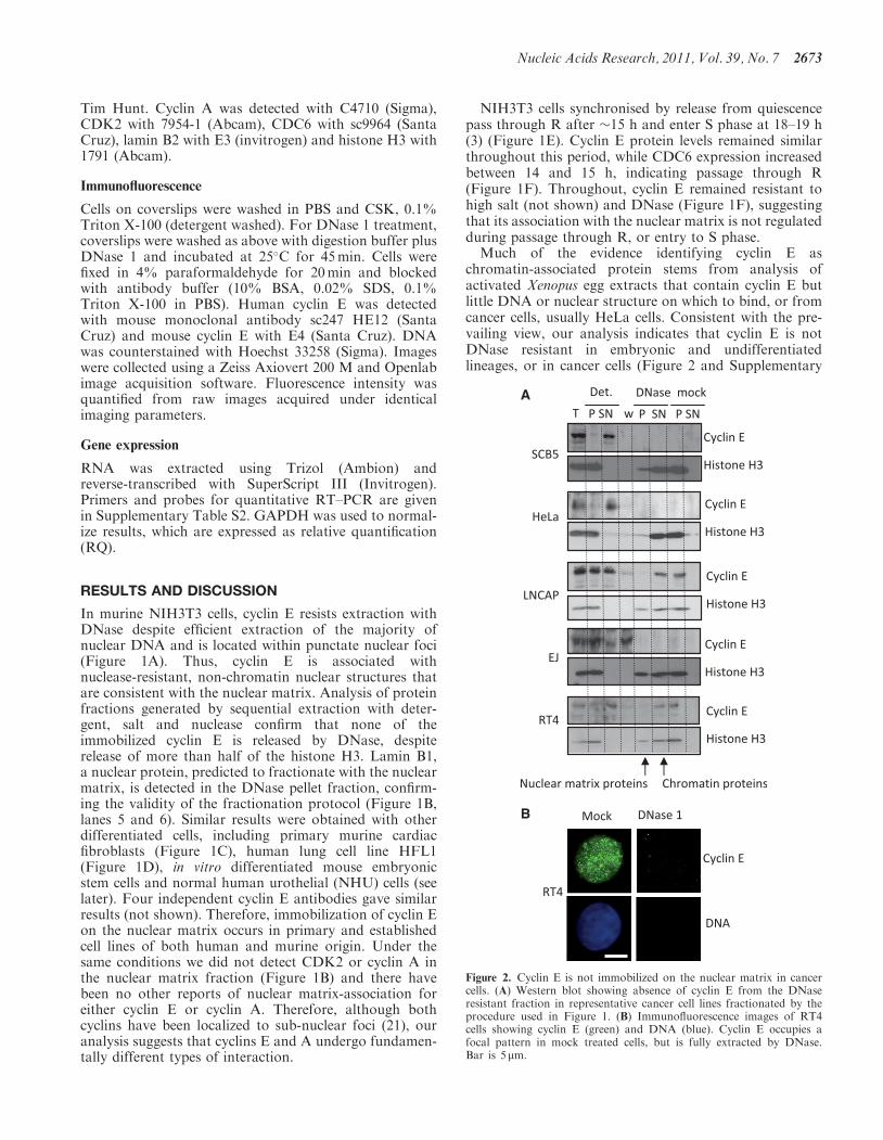
chromatin-associated protein stems from analysis ofactivated Xenopus egg extracts that contain cyclin E butlittle DNA or nuclear structure on which to bind, or fromcancer cells, usually HeLa cells. Consistent with the pre-vailing view, our analysis indicates that cyclin E is notDNase resistant in embryonic and undifferentiatedlineages, or in cancer cells (Figure 2 and Supplementary
A
B
Figure 2. Cyclin E is not immobilized on the nuclear matrix in cancercells. (A) Western blot showing absence of cyclin E from the DNaseresistant fraction in representative cancer cell lines fractionated by theprocedure used in Figure 1. (B) Immunofluorescence images of RT4cells showing cyclin E (green) and DNA (blue). Cyclin E occupies afocal pattern in mock treated cells, but is fully extracted by DNase.Bar is 5mm.
Nucleic Acids Research, 2011, Vol. 39, No. 7 2673

Table S1). In some cases, such as the prostate cancer lineLNCAP or the urothelial cancer line RT4, cyclin E resistsextraction with detergent (Figure 2A), but in all but one ofthe eight cancer cell lines we studied cyclin E was not re-sistant to DNase and is therefore not mounted on thenuclear matrix. Notably, cyclin E remained organizedinto sub-nuclear foci (Figure 2B), showing that attach-ment to an underlying architecture is not required tospecify foci.To reconcile these two sets of observations and test
whether both states are in fact encountered in vivo, weused three different experimental systems. These yieldedconsistent results and show clearly that recruitment to thenuclear matrix is established as cells differentiate.In undifferentiated murine embryonic stem (ES) cells
the majority of cyclin E was not resistant to DNase
(Figure 3A, lane 2 upper panels), but shifted into theDNase-resistant fraction (lane 4, lower panels) upon in-duction of differentiation (17). Reduced OCT4 andincreased GATA4 expression confirmed endodermal dif-ferentiation (Figure 3B), although markers for otherlineages were also present (not shown). Similar resultswere obtained with X. laevis embryos during developmentfrom fertilized egg to tadpole. The embryo is transcrip-tionally silent until the mid blastula transition (MBT)when zygotic transcription begins in a process thatinvolves chromatin remodelling and reorganisation of rep-lication origins (22). Prior to the MBT (embryo stages 2and 8, Figure 3C), cyclin E was fully solubilised by deter-gent (Figure 3D), while after the MBT (stages 10 and 39) aresistant fraction of cyclin E was evident. Further analysisshowed that in gastrulating embryos (stage 10), this was
Figure 3. Cyclin E is recruited to the nuclear matrix during embryonic stem cell differentiation and Xenopus laevis development. (A) Western blotshowing protein fractions derived from murine ES cells before and after embryoid body formation, and after 25 days in culture under differentiationconditions. Images show cell populations before extraction. Bar=50 mm. Cyclin E changes from detergent soluble in ES cells and embryoid bodies tomostly DNase resistant in differentiated cells despite efficient extraction of histone H3. (B) Quantitative RT–PCR of differentiation markers Oct4 andGATA4 in cells in A, with SEM. (C) Xenopus embryos at stages 2, 8, 10 and 39. (D) Western blots showing total cyclin E and the proportion that isdetergent-resistant (P) and detergent soluble (SN) at each stage. (E) Western blot showing detergent-resistant cyclin E after further incubationwithout (mock) or with DNase. Histone H3 was used to control for chromatin digestion. By stage 39 the detergent resistant fraction of cyclin E isDNase resistant.
2674 Nucleic Acids Research, 2011, Vol. 39, No. 7

released by DNase (Figure 3E), while in the tadpoles(stage 39) it was completely resistant (lane 1). Therefore,cyclin E is recruited to the nuclear matrix during Xenopusdevelopment. If cyclin E recruitment is interpreted as asurrogate for replication origin recruitment, the dataimply that this occurs sometime after the MBT. Thematrix-association of some key housekeeping genes, suchas the rRNA genes, is reorganised at the MBT as theybecome activated. Others such as the keratin domain,
which encodes genes expressed only in adult keratinocytes,are not specified until later in development (23). Thus theformation of functional links with the nuclear matrix maybe a staged process that occurs as template usage isdefined.Complementary analysis in a well-characterised model
of urothelial cell differentiation (19), revealed a similarpicture. In undifferentiated cycling and confluent popula-tions of NHU cells (Figure 4A) cyclin E was fully
A
B
D E
C
Figure 4. Recruitment of cyclin E to the nuclear matrix during differentiation of NHU cells (A) Cycling NHU cells were grown to confluence with-out differentiation or induced to differentiate over 7 days. (B) Cyclin E (green) and DNA (blue) in nuclei from cycling cells (Day 1, upper panels),differentiated (Day 7, middle panels) and confluent control cells (Day 7, lower panels), after extraction with detergent or DNase as indicated. Bar is10 mm. (C) Detergent and DNase extracted protein fractions from the populations shown in A. Cyclin E changes from DNase sensitive in undifferen-tiated NHU and confluent control cells to partly DNase resistant in differentiated NHU cells, despite efficient extraction of histone H3. (D) QuantitativeRT-PCR showing uroplakin 2 expression with SEM. (E) Model of cyclin E foci (green) associated with chromatin (blue background), unconstrainedby attachment to the nuclear matrix (gray lines) in undifferentiated cells, but mounted upon a nuclear matrix in differentiated cells.
Nucleic Acids Research, 2011, Vol. 39, No. 7 2675
extracted with DNase (Figure 4B and C, upper and lowerpanels). However in cells induced to differentiate, approxi-mately half of the cyclin E was recruited to the nuclearmatrix (Figure 4B and C, middle panels). The transition todifferentiated urothelium was verified visually (Figure 4A)and by analysis of uroplakin 2 expression (Figure 4D).These analyses in three diverse differentiating systems
show that cyclin E is a conditional nuclear matrix proteinthat is recruited during differentiation and imply aconserved matrix-associated function. We propose thatrecruitment to the nuclear matrix reflects changes in thelocation at which cyclin E executes its function in pre-RCassembly, and that this switches from being unconstrainedin nuclear space in undifferentiated cells, to being specifiedby immobilization on an underlying nuclear architecture(Figure 4E), potentially restricting the genome to celltype-specific patterns of DNA replication.Attachment of DNA to the nuclear matrix occurs via
sequences that anchor the genome in loops, while add-itional transient associations are thought to give rise toloops that are reeled through factories as DNA replicates(24). Changes in loop size have been observed directly andlinked to transcriptional and developmental decisions,suggesting that their selection may contribute to restric-tion of gene expression and limit the plasticity ofdifferentiating cells compared to stem and progenitorcells. Remodelling of attachments and establishment ofreplicon size occurs during passage through mitosis inXenopus (25), but little is known about how pre-RCs arerecruited to the matrix. Moreover, in the absence of amitotic transition the recruitment process may be subtlydifferent. It seems likely that the long G1 phase that cellsexperience upon exit from quiescence involves some re-building of the nuclear architecture, but it remains to beseen whether this underlies the specific requirement forcyclin E in non-mitotic cycles.The difference between differentiated adult cells and
cancer cells with respect to cyclin E has implications forthe functional organization of the nucleus and its relation-ship with disease. Deregulation of cyclin E confers genomeinstability (26), possibly through defects in pre-RCassembly (27). Thus, the link between cyclin E and trans-formation may partly reflect failure to correctly assembleon the nuclear matrix, helping to support an undifferenti-ated state.Changes in the form and texture of the nucleus have
long been used to diagnose and classify tumours andchanges in nuclear matrix composition have beenobserved in cancer cells (28). If, as we suggest, recruitmentof cyclin E to the nuclear matrix influences genome organ-ization, possibly conferring reduced plasticity, failure tobecome immobilized could have prognostic value.However, it will be necessary to distinguish betweendefects in cyclin E that impair recruitment, and widerproblems with the internal organization of the nucleus incancer cells.
SUPPLEMENTARY DATA
Supplementary Data are available at NAR Online.
ACKNOWLEDGEMENTS
We would like to thank T. Hunt and J. Gannon forXenopus cyclin E antibody, A. Surani and K. Hilton formurine ES cells and R. Wilson for critical comment.
FUNDING
Biotechnology and Biosciences Research Council(BBSRC); Yorkshire Cancer Research; York AgainstCancer (to J.S.); British Heart Foundation (to J.F.-X.A.). Funding for the open access charge: BBSRC;Yorkshire Cancer Research, partial.
Conflict of interest statement. None declared.
REFERENCES
1. Honda,R., Lowe,E.D., Dubinina,E., Skamnaki,V., Cook,A.,Brown,N.R. and Johnson,L.N. (2005) The structure of cyclin E1/CDK2: implications for CDK2 activation and CDK2-independentroles. EMBO J., 24, 452–463.
2. Ekholm,S.V., Zickert,P., Reed,S.I. and Zetterberg,A. (2001)Accumulation of cyclin E is not a prerequisite for passagethrough the restriction point. Mol. Cell. Biol., 21, 3256–3265.
3. Coverley,D., Laman,H. and Laskey,R.A. (2002) Distinct roles forcyclins E and A during DNA replication complex assembly andactivation. Nat. Cell Biol., 4, 523–528.
4. Furstenthal,L., Kaiser,B.K., Swanson,C. and Jackson,P.K. (2001)Cyclin E uses Cdc6 as a chromatin-associated receptor requiredfor DNA replication. J. Cell. Biol., 152, 1267–1278.
5. Geng,Y., Yu,Q., Sicinska,E., Das,M., Schneider,J.E.,Bhattacharya,S., Rideout,W.M., Bronson,R.T., Gardner,H. andSicinski,P. (2003) Cyclin E ablation in the mouse. Cell, 114,431–443.
6. Mailand,N. and Diffley,J.F. (2005) CDKs promote DNAreplication origin licensing in human cells by protecting Cdc6from APC/C-dependent proteolysis. Cell, 122, 915–926.
7. Su,T.T. and O’Farrell,P.H. (1998) Chromosome association ofminichromosome maintenance proteins in Drosophilaendoreduplication cycles. J. Cell. Biol., 140, 451–460.
8. Matsumoto,Y. and Maller,J.L. (2004) A centrosomal localizationsignal in cyclin E required for Cdk2-independent S phase entry.Science, 306, 885–888.
9. Hemerly,A.S., Prasanth,S.G., Siddiqui,K. and Stillman,B. (2009)Orc1 controls centriole and centrosome copy number in humancells. Science, 323, 789–793.
10. Parisi,T., Beck,A.R., Rougier,N., McNeil,T., Lucian,L., Werb,Z.and Amati,B. (2003) Cyclins E1 and E2 are required forendoreplication in placental trophoblast giant cells. EMBO J., 22,4794–4803.
11. Berthet,C., Aleem,E., Coppola,V., Tessarollo,L. and Kaldis,P.(2003) Cdk2 knockout mice are viable. Curr. Biol., 13, 1775–1785.
12. Geng,Y., Lee,Y.M., Welcker,M., Swanger,J., Zagozdzon,A.,Winer,J.D., Roberts,J.M., Kaldis,P., Clurman,B.E. and Sicinski,P.(2007) Kinase-independent function of cyclin E. Mol. Cell., 25,127–139.
13. Berezney,R. and Coffey,D.S. (1975) Nuclear protein matrix:association with newly synthesized DNA. Science, 189, 291–293.
14. Hozak,P., Hassan,A.B., Jackson,D.A. and Cook,P.R. (1993)Visualization of replication factories attached to thenucleoskeleton. Cell, 73, 361–373.
15. Djeliova,V., Russev,G. and Anachkova,B. (2001) Dynamics ofassociation of origins of DNA replication with the nuclear matrixduring the cell cycle. Nucleic Acids Res., 29, 3181–3187.
16. Fujita,M., Ishimi,Y., Nakamura,H., Kiyono,T. and Tsurumi,T.(2002) Nuclear organization of DNA replication initiationproteins in mammalian cells. J. Biol. Chem., 277, 10354–10361.
17. Turksen,K. (2006) Stem Cell Protocols, Methods in MolecularBiology. Humana Press Inc., Totowa, New Jersey.
2676 Nucleic Acids Research, 2011, Vol. 39, No. 7

18. Fisher,M.E., Isaacs,H.V. and Pownall,M.E. (2002) eFGF isrequired for activation of XmyoD expression in the myogenic celllineage of Xenopus laevis. Development, 129, 1307–1315.
19. Cross,W.R., Eardley,I., Leese,H.J. and Southgate,J. (2005) Abiomimetic tissue from cultured normal human urothelial cells:analysis of physiological function. Am. J. Physiol. Renal. Physiol.,289, F459–468.
20. Ainscough,J.F., Rahman,F.A., Sercombe,H., Sedo,A., Gerlach,B.and Coverley,D. (2007) C-terminal domains deliver the DNAreplication factor Ciz1 to the nuclear matrix. J. Cell. Sci., 120,115–124.
21. Erlandsson,F., Martinsson-Ahlzen,H.S., Wallin,K.L.,Hellstrom,A.C., Andersson,S. and Zetterberg,A. (2006) Parallelcyclin E and cyclin A expression in neoplastic lesions of theuterine cervix. Br. J. Cancer, 94, 1045–1050.
22. Hyrien,O., Maric,C. and Mechali,M. (1995) Transition inspecification of embryonic metazoan DNA replication origins.Science, 270, 994–997.
23. Vassetzky,Y., Hair,A. and Mechali,M. (2000) Rearrangement ofchromatin domains during development in Xenopus. Genes. Dev.,14, 1541–1552.
24. Ottaviani,D., Lever,E., Takousis,P. and Sheer,D. (2008)Anchoring the genome. Genome Biol., 9, 201.
25. Lemaitre,J.M., Danis,E., Pasero,P., Vassetzky,Y. and Mechali,M.(2005) Mitotic remodeling of the replicon and chromosomestructure. Cell, 123, 787–801.
26. Spruck,C.H., Won,K.-A. and Reed,S.I. (1999) Deregulated cyclinE induces chromosome instability. Nature, 401, 297–300.
27. Ekholm-Reed,S., Mendez,J., Tedesco,D., Zetterberg,A.,Stillman,B. and Reed,S.I. (2004) Deregulation of cyclin E inhuman cells interferes with prereplication complex assembly.J. Cell. Biol., 165, 789–800.
28. Zink,D., Fischer,A.H. and Nickerson,J.A. (2004) Nuclearstructure in cancer cells. Nat. Rev., 4, 677–687.
Nucleic Acids Research, 2011, Vol. 39, No. 7 2677
Copyright © 2022 FDOKUMEN


![Pyrazolo[3,4- c ]pyridazines as Novel and Selective Inhibitors of Cyclin-Dependent Kinases](https://static.fdokumen.com/doc/165x107/632451b94d8439cb620d53b7/pyrazolo34-c-pyridazines-as-novel-and-selective-inhibitors-of-cyclin-dependent.jpg)