Custom-Fit Ruthenium(II) Metallopeptides: A New Twist to DNA Binding With Coordination Compounds
8
DOI: 10.1002/chem.201301629 Custom-Fit Ruthenium(II) Metallopeptides: A New Twist to DNA Binding With Coordination Compounds Ilaria Gamba, [b] Iria SalvadɃ, [a] Gustavo Rama, [b] Miriam Bertazzon, [a] Mateo I. SƁnchez, [a] Vȷctor M. SƁnchez-Pedregal, [a] JosȖ Martȷnez-Costas, [c] Rosa F. Brissos, [d] Patrick Gamez , [d, e] JosȖ L. MascareÇas, [a] Miguel VƁzquez LɃpez,* [b] and M. Eugenio VƁzquez* [a] Introduction Mapping the human genome has provided a wealth of infor- mation and potential DNA targets of both therapeutic and diagnostic relevance that has stimulated the search for new DNA-binding agents. [1] In addition to the extensive develop- ment of small organic binders, the past few years have wit- nessed an increased interest on the application of coordina- tion compounds as DNA-targeted probes, reactive agents, and therapeutics. [2–4] Among them, ruthenium(II) polypyrid- yl mononuclear complexes have been exhaustively studied for their kinetic stability and convenient redox and optical properties. [5–7] Surprisingly, dinuclear ruthenium(II) deriva- tives, despite systematically displaying better DNA-binding properties than their mononuclear counterparts, [8] have scarcely been studied to date, [9–12] hampered by the stiff syn- thetic methodologies commonly used in the assembly of pol- ypyridyl chelators, which severely limits the structural modi- fications that can be efficiently accessed. [13] Given our expe- rience in the study of DNA recognition agents, particularly minor-groove binders, [14] and metallopeptides, [15] we sought to fill this gap with the development of custom-fit dinuclear ruthenium(II) metallopeptides, which could potentially com- bine the unique photophysical properties of the Ru II poly- pyridyl complexes with the synthetic versatility of solid- phase peptide synthesis methods that would allow the straightforward modification of the bis-chelating ligand for the modulation of the biophysical properties of the resulting complexes. [16] To this end, we reasoned that appropriate de- rivatization of the widely used 2,2’-bipyridine chelator in the form of a 9-fluorenylmethoxycarbonyl (Fmoc)-protected amino acid, might allow the straightforward solid-phase syn- thesis of metallopeptide precursor ligands, which could be easily tweaked to efficiently optimize the DNA-binding and biological properties of their resulting ruthenium(II) metal- lopeptides. Keywords: DNA recognition · fluo- rescence · metallopeptides · molec- ular recognition · supramolecular chemistry Abstract: A new bipyridine building block has been used for the solid-phase syn- thesis of dinuclear DNA-binding ruthenium(II) metallopeptides. Detailed spectro- scopic studies suggest that these compounds bind to the DNA by insertion into the DNA minor groove. Moreover, the potential of the solid-phase peptide synthesis approach is demonstrated by the straightforward synthesis of an octaarginine de- rivative that shows effective cellular internalization and cytotoxicity linked with strong DNA interaction, as evidenced by steady-state fluorescence spectroscopy and AFM studies. [a] I. SalvadɃ, M. Bertazzon, M. I. SƁnchez, Prof. V. M. SƁnchez-Pedregal, Prof. J. L. MascareÇas, Prof. M. E. VƁzquez Departamento de Quȷmica OrgƁnica Centro Singular de InvestigaciɃn en Quȷmica BiolɃxica e Materiais Moleculares Universidade de Santiago de Compostela 15782 Santiago de Compostela (Spain) E-mail: [email protected] [b] Dr. I. Gamba, G. Rama, Prof. M. VƁzquez LɃpez Departamento de Quȷmica InorgƁnica y Centro Singular de InvestigaciɃn en Quȷmica BiolɃxica e Materiais Moleculares Universidade de Santiago de Compostela 15782 Santiago de Compostela (Spain) E-mail : [email protected] [c] Prof. J. Martȷnez-Costas Departamento de Bioquȷmica y Biologȷa Molecular y Centro Singular de InvestigaciɃn en Quȷmica BiolɃxica e Materiais Moleculares Universidade de Santiago de Compostela 15782 Santiago de Compostela (Spain) [d] R.F. Brissos, Prof. P. Gamez Departament de Quȷmica InorgȤnica, QBI, Universitat de Barcelona 08028 Barcelona (Spain) [e] Prof. P. Gamez InstituciɃ Catalana de Recerca I Estudis AvanÅats (ICREA) 08010 Barcelona (Spain) Supporting information for this article is available on the WWW under http://dx.doi.org/10.1002/chem.201301629. Chem. Eur. J. 2013, 00,0–0 # 2013 Wiley-VCH Verlag GmbH & Co. KGaA, Weinheim These are not the final page numbers! ÞÞ &1& FULL PAPER
-
Upload
independent -
Category
Documents
-
view
3 -
download
0
Transcript of Custom-Fit Ruthenium(II) Metallopeptides: A New Twist to DNA Binding With Coordination Compounds
DOI: 10.1002/chem.201301629
Custom-Fit Ruthenium(II) Metallopeptides:A New Twist to DNA Binding With Coordination Compounds
Ilaria Gamba,[b] Iria Salvad�,[a] Gustavo Rama,[b] Miriam Bertazzon,[a]
Mateo I. S�nchez,[a] V�ctor M. S�nchez-Pedregal,[a] Jos� Mart�nez-Costas,[c]
Rosa F. Brissos,[d] Patrick Gamez ,[d, e] Jos� L. MascareÇas,[a]
Miguel V�zquez L�pez,*[b] and M. Eugenio V�zquez*[a]
Introduction
Mapping the human genome has provided a wealth of infor-mation and potential DNA targets of both therapeutic anddiagnostic relevance that has stimulated the search for newDNA-binding agents.[1] In addition to the extensive develop-
ment of small organic binders, the past few years have wit-nessed an increased interest on the application of coordina-tion compounds as DNA-targeted probes, reactive agents,and therapeutics.[2–4] Among them, ruthenium(II) polypyrid-yl mononuclear complexes have been exhaustively studiedfor their kinetic stability and convenient redox and opticalproperties.[5–7] Surprisingly, dinuclear ruthenium(II) deriva-tives, despite systematically displaying better DNA-bindingproperties than their mononuclear counterparts,[8] havescarcely been studied to date,[9–12] hampered by the stiff syn-thetic methodologies commonly used in the assembly of pol-ypyridyl chelators, which severely limits the structural modi-fications that can be efficiently accessed.[13] Given our expe-rience in the study of DNA recognition agents, particularlyminor-groove binders,[14] and metallopeptides,[15] we soughtto fill this gap with the development of custom-fit dinuclearruthenium(II) metallopeptides, which could potentially com-bine the unique photophysical properties of the RuII poly-pyridyl complexes with the synthetic versatility of solid-phase peptide synthesis methods that would allow thestraightforward modification of the bis-chelating ligand forthe modulation of the biophysical properties of the resultingcomplexes.[16] To this end, we reasoned that appropriate de-rivatization of the widely used 2,2’-bipyridine chelator in theform of a 9-fluorenylmethoxycarbonyl (Fmoc)-protectedamino acid, might allow the straightforward solid-phase syn-thesis of metallopeptide precursor ligands, which could beeasily tweaked to efficiently optimize the DNA-binding andbiological properties of their resulting ruthenium(II) metal-lopeptides.
Keywords: DNA recognition · fluo-rescence · metallopeptides · molec-ular recognition · supramolecularchemistry
Abstract: A new bipyridine building block has been used for the solid-phase syn-thesis of dinuclear DNA-binding ruthenium(II) metallopeptides. Detailed spectro-scopic studies suggest that these compounds bind to the DNA by insertion into theDNA minor groove. Moreover, the potential of the solid-phase peptide synthesisapproach is demonstrated by the straightforward synthesis of an octaarginine de-rivative that shows effective cellular internalization and cytotoxicity linked withstrong DNA interaction, as evidenced by steady-state fluorescence spectroscopyand AFM studies.
[a] I. Salvad�, M. Bertazzon, M. I. S�nchez,Prof. V. M. S�nchez-Pedregal, Prof. J. L. MascareÇas,Prof. M. E. V�zquezDepartamento de Qu�mica Org�nicaCentro Singular de Investigaci�n en Qu�mica Biol�xicae Materiais MolecularesUniversidade de Santiago de Compostela15782 Santiago de Compostela (Spain)E-mail : [email protected]
[b] Dr. I. Gamba, G. Rama, Prof. M. V�zquez L�pezDepartamento de Qu�mica Inorg�nica yCentro Singular de Investigaci�n en Qu�mica Biol�xicae Materiais MolecularesUniversidade de Santiago de Compostela15782 Santiago de Compostela (Spain)E-mail : [email protected]
[c] Prof. J. Mart�nez-CostasDepartamento de Bioqu�mica y Biolog�a Molecular y Centro Singularde Investigaci�n enQu�mica Biol�xica e Materiais MolecularesUniversidade de Santiago de Compostela15782 Santiago de Compostela (Spain)
[d] R. F. Brissos, Prof. P. GamezDepartament de Qu�mica Inorg�nica, QBI, Universitat de Barcelona08028 Barcelona (Spain)
[e] Prof. P. GamezInstituci� Catalana de Recerca I Estudis AvanÅats (ICREA)08010 Barcelona (Spain)
Supporting information for this article is available on the WWWunder http://dx.doi.org/10.1002/chem.201301629.
Chem. Eur. J. 2013, 00, 0 – 0 � 2013 Wiley-VCH Verlag GmbH & Co. KGaA, Weinheim
These are not the final page numbers! ��&1&
FULL PAPER
Results and Discussion
Metallopeptide design and synthesis : Based on previous re-ports studying related bipyridine amino acids,[17] we chosethe Fmoc-bAla-bpy-OH dipeptide (1, Scheme 1) as the
basic building block of our bis-chelating peptidic ligands.Following the synthesis of 1 using known procedures (seethe Supporting Information),[13] we designed two simplepeptide ligands containing two bAla-bpy coordinating unitsin tandem (Ac-bAla-bpy-bAla-bpy-NH2), or separated by ashort glycine residue linker (Ac-bAla-bpy-Gly-bAla-bpy-NH2), as simple test cases to demonstrate the flexibility ofthe solid-phase peptide synthesis (SPPS) methodology forthe straightforward structural variation of the peptidic li-gands. The peptides were synthesized following standardFmoc/tBu solid-phase protocols.[18] Once fully assembled(and while still attached to the solid support) the peptideswere coordinated with the enantiopure Hua and von Zelew-sky�s L-cis-[Ru ACHTUNGTRENNUNG(bpy)2(py)2]
2+ , or D-cis-[Ru ACHTUNGTRENNUNG(bpy)2(py)2]2+ re-
agents (bpy =2,2’-bipyridine, py =pyridine),[19] to give thedesired LL- or DD-Ru2 and LL- or DD-Ru2Gly dinuclearcomplexes attached to the solid support (Scheme 1). Acidiccleavage, followed by reverse-phase HPLC purification, af-forded the desired RuII metallopeptides. It should be high-lighted that the formation of the complexes in the solid
phase from bis-bipyridine RuII reagents outperforms thewidely used solution procedures,[20] allowing the use ofgreater excess of metal precursors and simplifying the subse-quent purification steps.[21] To our knowledge, these speciesrepresent the first reported examples of dinuclear RuII met-allopeptides.[10]
DNA binding studies : The basic DNA binding properties ofthe LL- and DD-Ru2 dinuclear complexes were initially as-sessed by displacement experiments with ethidium bro-mide.[22] Additionally, emission-quenching experimentsshowed that both LL- and DD-Ru2 were effectively protect-ed from luminescence deactivation with [Fe(CN)6]
4� in thepresence of calf-thymus DNA (ctDNA).[23] Thus, the ratio ofthe slopes in the absence and in presence of ctDNA, whichis used as a measure of the protection of DNA-bound com-plexes against the dynamic quenching by the [Fe(CN)6]
4�,resulting from electrostatic repulsion of the highly anionicferricyanide from the polyanionic DNA, is 12.1 for LL-Ru2
and 7.3 for DD-Ru2. These values are in the same range ofthose observed for other RuII complexes that interact withthe DNA (the Supporting information, Figure S2). Thesepreliminary experiments showed that, in contrast to themononuclear tris-bipyridyl complex,[24] both LL- and DD-Ru2 display significant DNA-binding affinities (see the Sup-porting Information, Figure S1). Encouraged by these re-sults, we decided to exploit the intrinsic emission of the RuII
metallopeptides and their environment-sensitive lumines-cence for studying their interaction with synthetic oligonu-cleotides,[25] which would allow us to obtain quantitative in-formation about their binding affinity towards particularDNA sequences. Thus, addition of successive aliquots of astock oligonucleotide solution to 1 mm solutions of LL- orDD-Ru2 in Tris-HCl buffer resulted in a progressive increasein the emission intensity of the RuII complexes, which wasfitted to a binding model corresponding to the formation ofspecific LL- and DD-Ru2/dsDNA complexes (Figure 1).
The resulting apparent dissociation constants were strong-ly dependent on the oligonucleotide sequences, so that thelower apparent dissociation constants—as well as larger in-creases in the emission intensity—were observed for the A/T-rich oligonucleotides, whereas G/C-containing oligos onlyinduced a modest increase in emission intensity and showedweaker apparent binding (Table 1). Moreover, LL-Ru2 con-sistently displayed stronger DNA binding affinities than itsenantiomeric DD-Ru2 counterpart; this preferential DNAbinding of the LL- enantiomer is consistent with earlier re-ports of chiral discrimination in homochiral dinuclear RuII
complexes, for which a preference of the LL-isomers forDNA binding was also observed.[26] In addition to the in-creased DNA affinity observed for the LL-Ru2 metallopep-tide, its luminescence intensity in the presence of a high-af-finity binding sequence (AAATTT) was much higher thanthat of the DD-Ru2 complex (Figure 1 c, solid and dashedlines, respectively), and centered at a much shorter emissionwavelength, hence suggesting a larger change in its immedi-ate environment upon binding. This observation, together
Scheme 1. Solid-phase peptide synthesis of the model LL- and DD-Ru2
dinuclear complexes. LL- and DD-Ru2Gly are synthesized following thesame procedure.
www.chemeurj.org � 2013 Wiley-VCH Verlag GmbH & Co. KGaA, Weinheim Chem. Eur. J. 0000, 00, 0 – 0
�� These are not the final page numbers!&2&
with the binding preference for A/T-rich sequences, wouldbe consistent with a deeper insertion mechanism of thiscomplex into the DNA minor groove.
Titrations with the LL- and DD-Ru2Gly dinuclear metal-lopeptides showed similar tendencies to those of their ho-mologous complexes lacking the Gly linker. Thus, the LL-Ru2Gly isomer displayed better apparent binding constantsthan DD-Ru2Gly, and both showed a clear preference forbinding to extended A/T-rich oligonucleotides. Curiously,the glycine linker had subtle effects on the binding of thesedinuclear metallopeptides, and although it had a slightlynegative effect on the DNA binding of the LL-Ru2Glyisomer compared with the model LL-Ru2 (KDACHTUNGTRENNUNG[AAATTT]�8.2 and 5.8 mm, respectively), it induced an opposite, slight-ly favorable effect, on the DNA binding of the DD-Ru2Glystereoisomer in comparison with the DD-Ru2 model com-pound (KDACHTUNGTRENNUNG[AAATTT]�31 and 14 mm, respectively), whichagain suggests some differences in the DNA binding modesof the isomeric complexes. As expected, no measurablebinding was detected in control experiments with single-
stranded DNA oligonucleotides. Circular dichroism (CD)experiments show an induced CD band at 320 nm by theLL-Ru2 isomer (the Supporting Information, Figure S4a),which has been also reported for other RuII complexes,[27]
but, more importantly, they show that the shape of the CDsignals between 200 and 300 nm, corresponding to theDNA, are not significantly perturbed, thus indicating the re-tention of the B-DNA conformation.[28]
Saturation transfer difference (STD) NMR experimentsalso support the binding to the DNA.[29] Thus, in the pres-ence of ctDNA both enantiomers, LL- and DD-Ru2, giverise to STD signals in the region of d=7.0–8.5 ppm of theirspectra (Figure 2 d and e), as expected for molecules that re-
versibly bind to the macromolecular receptor in the fast ex-change regime; these resonances are not found in the con-trol STD spectrum in the absence of ctDNA (Figure 2 b).Both the LL- and DD-Ru2 enantiomers gave similar intensi-ties in the STD experiments, in agreement with the similarvalues of their apparent binding affinities.
Structural modification of the basic metallopeptide core: In-troduction of an octaarginine peptide tail : Given the rela-tively good DNA-binding properties as well as the favorableluminescent emission displayed by the dinuclear rutheniumcomplexes, we were interested in studying their effect inliving cells, but unfortunately these compounds were not in-ternalized in Vero cells at concentrations of 5 or 25 mm andduring variable incubations times (30 min and 1 h). Consid-ering the effective cellular uptake induced by a number ofcationic peptides,[30] and oligoarginines in particular,[31] wedecided to take advantage of the flexibility afforded by the
Figure 1. Left: a) Luminescence spectra of 1 mm solutions of LL-Ru2 orDD-Ru2 in Tris-HCl buffer (20 mm), NaCl (100 mm), pH 7.5; b) LL-Ru2
in the presence of the GGCC hairpin oligonucleotide (20 mm);c) (c) LL-Ru2 in the presence of of the AAAATTT hairpin oligo(21 mm) ; a) DD-Ru2 in the presence of the AAAATTT hairpin oligo(21 mm). Right: Titration profiles of LL-Ru2 (1 mm) with AAAATTT (~);AAT (*); AAGCTT (*), and GGCC (~) oligonucleotides. Lines rep-resent the best fit to a 1:1 binding model. Hairpin DNA oligonucleotidesequences, AAAATTT: 5’-GGC AAAATTT CGTTTTTCG AAATTTTGCC-3’; AAT: 5’-GGCG AAT GCAGCTTTTTGCTGC ATT CGCC-3’;AAGCTT: 5’-GGC AAGCTT CGCTTTTTGCG AAGCTT GCC-3’;GGCC : 5’-GGCAGGCCCAGC TTTTT GCTGGGCCTGCC-3’.
Table 1. Dissociation constants of dinuclear LL-, DD-Ru2 and LL-, DD-Ru2Gly with selected oligonucleotides.
Oligo[a] DD-Ru2[b] LL-Ru2 DD-Ru2Gly LL-Ru2Gly
AAAATTT 31.0 (7.0) 5.8 (0.7) 13.9 (2.5) 8.2 (1.8)AAT 24.5 (3.5) 12.0 (0.6) 15.2 (2.6) 11.6 (1.6)AAGCTT 27.0 (6.0) 13.8 (0.7) 22.9 (6.8) 13.5 (2.2)GGCC >50 15.5 (3.0) >50 >50
KD (mm) were measured in Tris-HCl buffer (20 mm), NaCl (100 mm),pH 7.5 at 298 K. [a] Hairpin oligonucleotide binding sites. [b] Best fit ac-cording to a 1:1 model obtained for three independent titrations. The es-timated KD error is shown in brackets.
Figure 2. STD NMR spectra (600 MHz, H2O, 25 8C) of LL- and DD-Ru2
with calf-thymus DNA. a) Reference (off-resonance) 1H NMR spectrumof 150 mm DD-Ru2 ; b) STD NMR of DD-Ru2 (150 mm) ; c) Reference (off-resonance) 1H NMR spectrum of a mixture of DD-Ru2 (150 mm) withcalf-thymus DNA (150 mm) ; d) STD NMR spectrum of the same samplein (c); e) STD NMR of a mixture of LL-Ru2 (228 mm) and calf-thymusDNA (250 mm). Saturation time was 2 s at frequencies �40 ppm (refer-ence, off-resonance spectra) and +4.0 ppm (STD, on-resonance spectra).
Chem. Eur. J. 2013, 00, 0 – 0 � 2013 Wiley-VCH Verlag GmbH & Co. KGaA, Weinheim www.chemeurj.org
These are not the final page numbers! ��&3&
FULL PAPERCustom-Fit Ruthenium(II) Metallopeptides
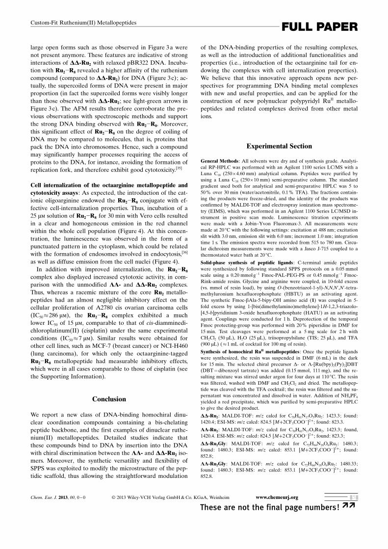
SPPS methodology to synthesize an octaarginine-dirutheni-um derivative (Ru2�R8). This oligocationic metallopeptidewas obtained following the same basic synthetic procedureoutlined earlier, with the difference that the two bAla-bpy-coordinating building blocks were appended at the N-termi-nus of a previously synthesized octaarginine peptide(Scheme 2). Unfortunately, attempts to prepare LL-Ru2�R8
or DD-Ru2�R8 in the solid support were unsuccessful, be-cause the high temperatures required for the coordinationof the relatively unreactive chiral Hua and von Zelewsky�sL-cis-[Ru ACHTUNGTRENNUNG(bpy)2(py)2]
2+ , or D-cis-[Ru ACHTUNGTRENNUNG(bpy)2(py)2]2+ reagents
resulted in a complex mixture of peptide degradation prod-ucts. Fortunately, the achiral [Ru ACHTUNGTRENNUNG(bpy)2(Cl)2] precursor com-plex required lower reaction temperatures, and allowed thesynthesis of the Ru2�R8 dinuclear complex as a diasteromer-ic mixture.[32]
Fluorescence titrations showed that the resulting octaargi-nine-dinuclear RuII metallopeptide Ru2�R8 displayed thesame A/T-rich sequence preference presented by the coreLL- and DD-Ru2 complexes. Moreover, the observed bind-ing affinity for A/T-rich sequences was increased by thismodification; probably because of the increased charge den-sity of the oligocationic Ru2�R8 complex, which presumablywould favorably contribute to the electrostatic componentof the interaction with the negatively-charged DNA phos-phate backbone. As expected, Ru2�R8 did not show any sig-nificant affinity for G/C-rich oligonucleotides. Moreover, thetitration curve for the G/C-rich hairpin exhibited a rathercomplex multiphasic profile (see the Supporting Informa-tion), consistent with the formation of multiple low-affinityand nonspecific complexes resulting from electrostatic inter-actions between the polyanionic DNA and the highly charg-ed conjugate.[33]
AFM studies of the DNA binding : The interaction of theRuII compounds DD-Ru2 and Ru2�R8 with DNA was further
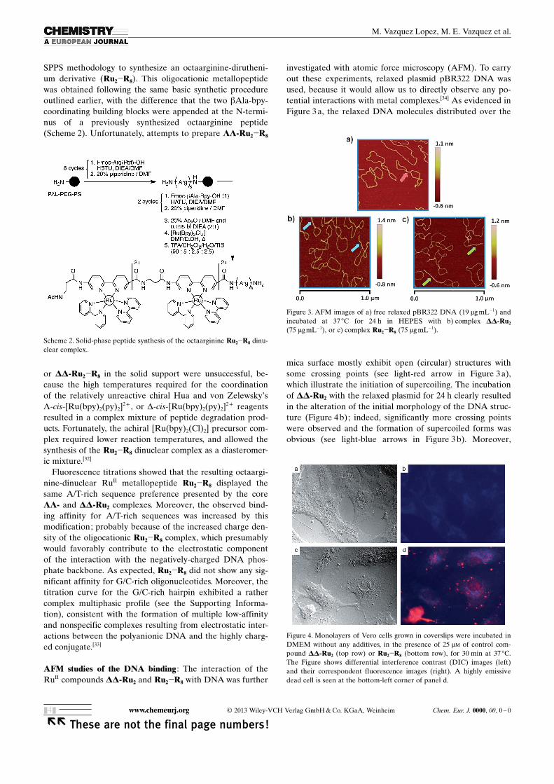
investigated with atomic force microscopy (AFM). To carryout these experiments, relaxed plasmid pBR322 DNA wasused, because it would allow us to directly observe any po-tential interactions with metal complexes.[34] As evidenced inFigure 3 a, the relaxed DNA molecules distributed over the
mica surface mostly exhibit open (circular) structures withsome crossing points (see light-red arrow in Figure 3 a),which illustrate the initiation of supercoiling. The incubationof DD-Ru2 with the relaxed plasmid for 24 h clearly resultedin the alteration of the initial morphology of the DNA struc-ture (Figure 4 b); indeed, significantly more crossing pointswere observed and the formation of supercoiled forms wasobvious (see light-blue arrows in Figure 3 b). Moreover,
Scheme 2. Solid-phase peptide synthesis of the octaarginine Ru2�R8 dinu-clear complex.
Figure 3. AFM images of a) free relaxed pBR322 DNA (19 mg mL�1) andincubated at 37 8C for 24 h in HEPES with b) complex DD-Ru2
(75 mgmL�1), or c) complex Ru2�R8 (75 mg mL�1).
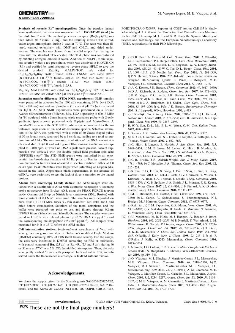
Figure 4. Monolayers of Vero cells grown in coverslips were incubated inDMEM without any additives, in the presence of 25 mm of control com-pound DD-Ru2 (top row) or Ru2�R8 (bottom row), for 30 min at 37 8C.The Figure shows differential interference contrast (DIC) images (left)and their correspondent fluorescence images (right). A highly emissivedead cell is seen at the bottom-left corner of panel d.
www.chemeurj.org � 2013 Wiley-VCH Verlag GmbH & Co. KGaA, Weinheim Chem. Eur. J. 0000, 00, 0 – 0
�� These are not the final page numbers!&4&
M. Vazquez Lopez, M. E. Vazquez et al.
large open forms such as those observed in Figure 3 a werenot present anymore. These features are indicative of stronginteractions of DD-Ru2 with relaxed pBR322 DNA. Incuba-tion with Ru2�R8 revealed a higher affinity of the rutheniumcompound (compared to DD-Ru2) for DNA (Figure 3 c); ac-tually, the supercoiled forms of DNA were present in majorproportion (in fact the supercoiled forms were visibly longerthan those observed with DD-Ru2 ; see light-green arrows inFigure 3 c). The AFM results therefore corroborate the pre-vious observations with spectroscopic methods and supportthe strong DNA binding observed with Ru2�R8. Moreover,this significant effect of Ru2�R8 on the degree of coiling ofDNA may be compared to molecules, that is, proteins thatpack the DNA into chromosomes. Hence, such a compoundmay significantly hamper processes requiring the access ofproteins to the DNA, for instance, avoiding the formation ofreplication fork, and therefore exhibit good cytotoxicity.[35]
Cell internalization of the octaarginine metallopeptide andcytotoxicity assays : As expected, the introduction of the cat-ionic oligoarginine endowed the Ru2�R8 conjugate with ef-fective cell-internalization properties. Thus, incubation of a25 mm solution of Ru2�R8 for 30 min with Vero cells resultedin a clear and homogeneous emission in the red channelwithin the whole cell population (Figure 4). At this concen-tration, the luminescence was observed in the form of apunctuated pattern in the cytoplasm, which could be relatedwith the formation of endosomes involved in endocytosis,[36]
as well as diffuse emission from the cell nuclei (Figure 4).In addition with improved internalization, the Ru2�R8
complex also displayed increased cytotoxic activity, in com-parison with the unmodified LL- and DD-Ru2 complexes.Thus, whereas a racemic mixture of the core Ru2 metallo-peptides had an almost negligible inhibitory effect on thecellular proliferation of A2780 cis ovarian carcinoma cells(IC50�286 mm), the Ru2�R8 complex exhibited a muchlower IC50 of 15 mm, comparable to that of cis-diamminedi-chloroplatinum(II) (cisplatin) under the same experimentalconditions (IC50�7 mm). Similar results were obtained forother cell lines, such as MCF-7 (breast cancer) or NCI-H460(lung carcinoma), for which only the octaarginine-taggedRu2�R8 metallopeptide had measurable inhibitory effects,which were in all cases comparable to those of cisplatin (seethe Supporting Information).
Conclusion
We report a new class of DNA-binding homochiral dinu-clear coordination compounds containing a bis-chelatingpeptide backbone, and the first examples of dinuclear ruthe-nium(II) metallopeptides. Detailed studies indicate thatthese compounds bind to DNA by insertion into the DNAwith chiral discrimination between the LL- and DD-Ru2 iso-mers. Moreover, the synthetic versatility and flexibility ofSPPS was exploited to modify the microstructure of the pep-tidic scaffold, thus allowing the straightforward modulation
of the DNA-binding properties of the resulting complexes,as well as the introduction of additional functionalities andproperties (i.e., introduction of the octaarginine tail for en-dowing the complexes with cell internalization properties).We believe that this innovative approach opens new per-spectives for programming DNA binding metal complexeswith new and useful properties, and can be applied for theconstruction of new polynuclear polypyridyl RuII metallo-peptides and related complexes derived from other metalions.
Experimental Section
General Methods : All solvents were dry and of synthesis grade. Analyti-cal RP-HPLC was performed with an Agilent 1100 series LC/MS with aLuna C18 (250 � 4.60 mm) analytical column. Peptides were purified byusing a Luna C18 (250 � 10 mm) semi-preparative column. The standardgradient used both for analytical and semi-preparative HPLC was 5 to50% over 30 min (water/acetonitrile, 0.1 % TFA). The fractions contain-ing the products were freeze-dried, and the identity of the products wasconfirmed by MALDI-TOF and electrospray ionization mass spectrome-try (EIMS), which was performed in an Agilent 1100 Series LC/MSD in-strument in positive scan mode. Luminescence titration experimentswere made with a Jobin–Yvon Fluoromax-3. All measurements weremade at 20 8C with the following settings: excitation at 488 nm; excitationslit width 3.0 nm, emission slit with 6.0 nm; increment 1.0 nm; integrationtime 1 s. The emission spectra were recorded from 515 to 780 nm. Circu-lar dichroism measurements were made with a Jasco J-715 coupled to athermostated water bath at 20 8C.
Solid-phase synthesis of peptidic ligands : C-terminal amide peptideswere synthesized by following standard SPPS protocols on a 0.05 mmolscale using a 0.20 mmol g�1 Fmoc-PAL-PEG-PS or 0.45 mmol g�1 Fmoc-Rink-amide resins. Glycine and arginine were coupled, in 10-fold excess(vs. mmol of resin load), by using O-(benzotriazol-1-yl)-N,N,N’,N’-tetra-methyluronium hexafluorophosphate (HBTU) as an activating agent.The synthetic Fmoc-bAla-5-bipy-OH amino acid (1) was coupled in 5-fold excess by using 1-[bis(dimethylamino)methylene]-1H-1,2,3-triazolo-ACHTUNGTRENNUNG[4,5-b]pyridinium 3-oxide hexafluorophosphate (HATU) as an activatingagent. Couplings were conducted for 1 h. Deprotection of the temporalFmoc protecting-group was performed with 20% piperidine in DMF for15 min. Test cleavages were performed at a 5 mg scale for 2 h withCH2Cl2 (50 mL), H2O (25 mL), triisopropylsilane (TIS; 25 mL), and TFA(900 mL) (�1 mL of cocktail for 100 mg of resin).
Synthesis of homochiral RuII metallopeptides : Once the peptide ligandswere synthesized, the resin was suspended in DMF (6 mL) in the darkfor 15 min. The selected chiral precursor D- or L-[Ru ACHTUNGTRENNUNG(bpy)2(Py)2]DBT(DBT =dibenzoyl tartrate) was added (0.15 mmol, 111 mg), and the re-sulting mixture was stirred under argon for four days at 110 8C. The resinwas filtered, washed with DMF and CH2Cl2 and dried. The metallopep-tide was cleaved with the TFA cocktail; the resin was filtered and the su-pernatant was concentrated and dissolved in water. Addition of NH4PF6
yielded a red precipitate, which was purified by semi-preparative HPLCto give the desired product.
DD-Ru2 : MALDI-TOF: m/z calcd for C70H61N17O5Ru2: 1423.3; found:1420.4; ESI-MS: m/z calcd: 824.5 [M+2CF3COO�]2+ ; found: 823.3.
LL-Ru2 : MALDI-TOF: m/z calcd for C70H61N17O5Ru2, 1423.3; found,1420.4. ESI-MS: m/z calcd: 824.5 [M+2 CF3COO�]2+ ; found: 823.3;
DD-Ru2Gly : MALDI-TOF: m/z calcd for C72H64N18O6Ru2: 1480.3;found: 1480.3; ESI-MS: m/z calcd: 853.1 [M+2CF3COO�]2+ ; found:852.8;
LL-Ru2Gly : MALDI-TOF: m/z calcd for C72H64N18O6Ru2: 1480.33;found: 1480.3; ESI-MS: m/z calcd: 853.1 [M+2CF3COO�]2+ ; found:852.8.
Chem. Eur. J. 2013, 00, 0 – 0 � 2013 Wiley-VCH Verlag GmbH & Co. KGaA, Weinheim www.chemeurj.org
These are not the final page numbers! ��&5&
FULL PAPERCustom-Fit Ruthenium(II) Metallopeptides
Synthesis of racemic RuII metallopeptides : Once the peptide ligandswere synthesized, the resin was suspended in 1:1 EtOH/DMF (6 mL) inthe dark for 15 min. The neutral precursor complex [Ru ACHTUNGTRENNUNG(bpy)2Cl2] wasthen added (0.15 mmol; 71 mg), and the resulting mixture was stirred,under argon atmosphere, during 3 days at 70 8C. The resin was then fil-tered, washed extensively with DMF and CH2Cl2 and dried undervacuum. The complex was cleaved from the solid support by treating theresin with the standard TFA cocktail. The TFA phase was concentratedby bubbling nitrogen, diluted in water. Addition of NH4PF6 to the aque-ous solution yields a red precipitate, which was dissolved in H2O/CH3CN(3:1) and purified by semi-preparative reverse-phase HPLC to give thedesired product. Ru2�R8 : MALDI-TOF: m/z calcd forC118H155N49O13Ru2: 2670.1; found: 2669.9; ESI-MS: m/z calcd 1079.7[M+5CF3COO�+4H+]3+ ; found =1081.2; ESI-MS: m/z calcd: 1117.7[M+6CF3COO�+5H+]3+ ; found: 1117.3; m/z calcd: 1155.7[M+7CF3COO�+6H+]3+ ; found: 1155.8.
Ru2�R8 : MALDI-TOF: m/z calcd for C70H61N17O5Ru2: 1423.31; found:1420.4; ESI-MS: m/z calcd: 824.5 [M+2CF3COO�]2+ ; found: 823.3.
Saturation transfer difference (STD) NMR experiments : NMR sampleswere prepared in aqueous buffer (500 mL) containing 10 % (v/v) D2O,NaCl (100 mm) and sodium phosphate (10 mm) at pH 7.5 (not correctedfor D2O). All STD NMR experiments were performed on a BrukerAvance 600 spectrometer (Billerica, MA, USA) operating at 600.13 MHzfor 1H, equipped with a 5 mm inverse triple resonance probe with Z-onlygradients. Spectra were processed with TopSpin and MestreNova. Apseudo-2D version of the STD NMR pulse sequence was used for the in-terleaved acquisition of on- and off-resonance spectra. Selective satura-tion of the DNA was performed with a train of 40 Gauss-shaped pulsesof 50 ms length each, separated by a 1 ms delay, leading to a total satura-tion time of 2.04 s. The on-resonance irradiation of the DNA was set at achemical shift of +1.0 and +4.0 ppm. Off-resonance irradiation was ap-plied at �40.0 ppm, at which no DNA signals were present. Solvent sup-pression was achieved with the double pulsed-field gradient spin-echo(DPFGSE) step.[37] The STD NMR spectra were multiplied by an expo-nential line-broadening function of 3.0 Hz prior to Fourier transforma-tion. Saturation transfer was observed in spectra irradiated either at 1.0or 4.0 ppm. Peak intensities were larger when saturating at 4.0 ppm (dis-cussed in the text). Appropriate blank experiments, in the absence ofctDNA, were performed to test the lack of direct saturation to the ligandprotons.
Atomic force microscopy (AFM) studies : The AFM images were ob-tained with a Multimode 8 AFM with electronic Nanoscope V scanningprobe microscope from Bruker AXS, using the PEAK FORCE tappingmode. Commercial Si-tip on Nitride lever cantilevers (SNL, Bruker) withforce constant of 0.4 Nm�1 were used. The samples were deposited onmica disks (PELCO Mica Discs, 9.9 mm diameter; Ted Pella, Inc.), anddried before visualization. Solutions of the metal complexes and thebuffer were prepared just prior to use, and filtered through 0.2 nmFP030/3 filters (Scheicher and Schuell, Germany). The samples were pre-pared in HEPES with relaxed plasmid pBR322 DNA (19 mg mL�1) andthe corresponding metallopeptide (75 � 10�3 mg mL�1). All samples wereincubated for 24 h at 37 8C before the AFM studies.
Cell internalization studies : Semi-confluent monolayers of Vero cellswere grown on glass coverslips in Dulbecco�s modified Eagle Medium(DMEM) containing 10 % of FBS (fetal bovine serum). For the assays,the cells were incubated in DMEM containing no FBS or antibiotics,with control compound Ru2 (25 mm) or Ru2�R8 (25 and 5 mm), during 60or 30 min at 37 8C in a 5% CO2 humidified atmosphere. Then, the cellswere gently washed 5 times with phosphate buffered saline PBS, and ob-served under the fluorescence microscope in DMEM without fixation.
Acknowledgements
We thank the support given by the Spanish grants SAF2010–20822-C02,CTQ2012–31341, CTQ2009–14431, CTQ2011–27929-C02–01, SAF2007–61015, and the Xunta de Galicia INCITE09 209 084PR, GRC2010/12,
PGIDIT08CSA-047209PR. Support of COST Action CM1105 is kindlyacknowledged. I. S. thanks the Fundaci�n Jos Otero–Carmela Mart�nezfor her PhD fellowship. M. I. S. and G. R. thank the Spanish Ministry ofScience and Technology, and the European Nanotechnology Laboratory(ENL), respectively, for their PhD fellowships.
[1] a) D. R. Boer, A. Canals, M. Coll, Dalton Trans. 2009, 3, 399 –414;b) R. Palchaudhuri, P. J. Hergenrother, Curr. Opin. Biotechnol. 2007,18, 497 – 503; c) S. M. Nelson, L. R. Ferguson, W. A. Denny, Mutat.Res. 2007, 623, 24– 40; d) W. C. Tse, D. L. Boger, Chem. Biol. 2004,11, 1607 – 1617; e) S. Neidle, Nat. Prod. Rep. 2001, 18, 291 – 309;f) P. N. Dervan, Science 1986, 232, 464 –471. For a recent review ondesigned DNA-binding agents: E. Pazos, J. Mosquera, M. E.V�zquez, J. L. MascareÇas, ChemBioChem 2011, 12, 1958 – 1973.
[2] a) A. C. Komor, J. K. Barton, Chem. Commun. 2013, 49, 3617 – 3630;b) D. A. Richards, A. Rodger, Chem. Soc. Rev. 2007, 36, 471 – 483;c) B. M. Zeglis, V. C. Pierre, J. K. Barton, Chem. Commun. 2007,4565 – 4579; d) K. L. Haas, K. Franz, Chem. Rev. 2009, 109, 4921 –4960; e) P. C. A. Bruijnincx, P. J. Sadler, Curr. Opin. Chem. Biol.2008, 12, 197 –206; f) A. Pyle, J. K. Barton, Bioinorganic Chemistry(Ed.: S. J Lippard), Wiley, Hoboken, 1990, p.413.
[3] a) J. Reedijk, Eur. J. Inorg. Chem. 2009, 1303 – 1312; b) L. Kelland,Nature Rev. Cancer 2007, 7, 573 – 584; c) E. R. Jamieson, S. J. Lip-pard, Chem. Rev. 1999, 99, 2467 –2498.
[4] R. W.-Y. Sun, D.-L. Ma, E. L.-M. Wong, C.-M. Che, Dalton Trans.2007, 4884 – 4892.
[5] J. Brunner, J. K. Barton, Biochemistry 2006, 45, 12295 –12302.[6] M. R. Gill, J. Garc�a-Lara, S. J. Foster, C. Smythe, G. Battaglia, J. A.
Thomas, Nature Chem. 2009, 1, 662 – 667.[7] a) C. Hiort, P. Lincoln, B. Nordn, J. Am. Chem. Soc. 1993, 115,
3448 – 3454; b) M. Eriksson, M. Leijon, C. Hiort, B. Nordn, A.Graeslund, J. Am. Chem. Soc. 1992, 114, 4933 –4934; c) A. Reymer,B. Nordn, Chem. Commun. 2012, 48, 4941 – 4943.
[8] a) C. R. Brodie, J. R. Aldrich-Wright, Eur. J. Inorg. Chem. 2007,4781 – 4793; b) C. Metcalfe, J. A. Thomas, Chem. Soc. Rev. 2003, 32,215 – 224.
[9] a) S. Sun, F. Li, F. Liu, X. Yang, J. Fan, F. Song, L. Sun, X. Peng,Dalton Trans. 2012, 41, 12434 –12438; b) V. Gonzalez, T. Wilson, I.Kurihara, A. Imai, J. A. Thomas, J. Otsuki, Chem. Commun. 2008,1868 – 1870; c) C. B. Spillane, J. A. Smith, J. L. Morgan, F. R. Keene,J. Biol. Inorg. Chem. 2007, 12, 819 – 824; d) F. Pierard, A. K.-D. Mes-maeker, Inorg. Chem. Commun. 2006, 9, 111 –126.
[10] a) M. P. Fitzsimons, J. K. Barton, J. Am. Chem. Soc. 1997, 119, 3379 –3380; b) L. Cardo, V. Sadovnikova, S. Phongtongpasuk, N. J.Hodges, M. J. Hannon, Chem. Commun. 2011, 47, 6575 –6577.
[11] a) Ref. [8a]; b) T. M. Pappenfus, K. R. Mann, Inorg. Chem. 2001, 40,6301 – 6307; c) Y. Nakabayashi, H. Inada, Y. Minoura, N. Iwamoto,O. Yamauchi, Inorg. Chim. Acta 2009, 362, 869 – 877.
[12] a) U. Mcdonnell, M. R. Hicks, M. J. Hannon, A. Rodger, J. Inorg.Biochem. 2008, 102, 2052 –2059; b) P. Nordell, F. Westerlund, L. M.Wilhelmsson, B. Nordn, P. Lincoln, Angew. Chem. 2007, 119, 2253 –2256; Angew. Chem. Int. Ed. 2007, 46, 2203 – 2206; c) O. Gijte,A. K.-D. Mesmaeker, J. Chem. Soc. Dalton Trans. 1999, 951 – 956;d) F. O�Reilly, J. Kelly, New. J. Chem. 1998, 22, 215 – 217; e) F.O�Reilly, J. Kelly, A. K-D. Mesmaeker, Chem. Commun. 1996,1013 – 1014.
[13] J. A. Smith, J. G. Collins, F. R. Keene in Metal Complex– DNA Inter-actions (Eds.: N. Hadjiliadis, E. Sletten), Wiley-Blackwell, Chiches-ter, 2009, pp. 319 –346.
[14] a) O. V�zquez, M. I. S�nchez, J. Mart�nez-Costas, J. L. MascareÇas,M. E. V�zquez, Chem. Commun. 2010, 46, 5518 –5520; b) O.V�zquez, M. I. S�nchez, J. Mart�nez-Costas, M. E. V�zquez, J. L.MascareÇas, Org. Lett. 2010, 12, 216 – 219; c) A. M. CaamaÇo, M. E.V�zquez, J. Mart�nez-Costas, L. Castedo, J. L. MascareÇas, Angew.Chem. 2000, 112, 3234 –3237; Angew. Chem. Int. Ed. 2000, 39, 3104 –3107; d) M. E. V�zquez, A. M. CaamaÇo, J. Mart�nez-Costas, L. Cas-tedo, J. L. MascareÇas, Angew. Chem. 2001, 113, 4859 – 4861; Angew.Chem. Int. Ed. 2001, 40, 4723 – 4725.
www.chemeurj.org � 2013 Wiley-VCH Verlag GmbH & Co. KGaA, Weinheim Chem. Eur. J. 0000, 00, 0 – 0
�� These are not the final page numbers!&6&
M. Vazquez Lopez, M. E. Vazquez et al.
[15] G. Rama, A. Ard�, J.-D. Marchal, I. Gamba, H. Ishida, J. Jimnez-Barbero, M. E. Vazquez, M. V�zquez L�pez, Chem. Eur. J. 2012, 18,7030 – 7035.
[16] B. Happ, A. Winter, M. D. Hager, U. S. Schubert, Chem. Soc. Rev.2012, 41, 2222 –2225.
[17] a) A. Torrado, B. Imperiali, J. Org. Chem. 1996, 61, 8940 – 8948;b) M. Kyakuno, S. Oishi, H. Ishida, Inorg. Chem. 2006, 45, 3756 –3765; c) M. Kyakuno, S. Oishi, H. Ishida, Chem. Lett. 2005, 34,1554 – 1555; d) H. Ishida, M. Kyakuno, S. Oishi, Biopolymers 2004,76, 69–82; e) M. Y. Ogawa, A. Gretchikhine, S. Soni, S. Davis,Inorg. Chem. 1995, 34, 6423 –6424.
[18] N. Sewald, H.-D. Jakubke in Peptides: Chemistry and Biology,Wiley-VCH, Weinheim, 2002, p.135.
[19] a) A. Papakyriakou, G. Malandrinos, A. Garoufis, J. Inorg. Biochem.2006, 100, 1842 –1848; b) X. Hua, A. von Zelewsky, Inorg. Chem.1995, 34, 5791 –5797; c) X. Hua, A. von Zelewsky, Inorg. Chem.1991, 30, 3796 – 3798; d) M. Brissard, O. Convert, M. Gruselle, C.Guyard-Duhayon, R. Thouvenot, Inorg. Chem. 2003, 42, 1378 –1385.
[20] a) M. R. Ghadiri, C. Soares, C. Choi, J. Am. Chem. Soc. 1992, 114,4000 – 4002; b) H. Ishida, Y. Maruyama, M. Kyakuno, Y. Kodera, T.Maeda, S. Oishi, ChemBioChem 2006, 7, 1567 –1570.
[21] a) K. Karidi, A. Garoufis, N. Hadjiliadis, J. Reedijk, Dalton Trans.2005, 728 – 734; b) K. Triantafillidi, K. Karidi, J. Malina, A. Garoufis,Dalton Trans. 2009, 6403 – 6415.
[22] a) Y.-M. Chen, Y.-J. Liu, Q. Li, K.-Z. Wang, J. Inorg. Biochem. 2009,103, 1395 – 1404; b) M.-J. Han, L.-H. Gao, Y.-Y. L, K.-Z. Wang, J.Phys. Chem. B 2006, 110, 2364 –2371.
[23] a) C. V. Kumar, J. K. Barton, N. J. Turro, J. Am. Chem. Soc. 1985,107, 5518 – 5523; b) W. J. Mei, J. Liu, K. C. Zheng, L. J. Lin, H.Chao, A. X. Li, F. C. Yun, L. N. Ji, Dalton Trans. 2003, 1352 –1359.
[24] a) P. Lincoln, B. Nordn, J. Phys. Chem. B 1998, 102, 9583 –9594;b) H. Gçrner, A. B. Tossi, C. Stradowski, D. Schulte-Frohlinde, J.Photochem. Photobiol. B Biol. 1988, 2, 67– 89.
[25] a) M. R. Gill, H. Derrat, C. G. W. Smythe, G. Battaglia, J. A.Thomas, ChemBioChem 2011, 12, 877 –880; b) F. Westerlund, P. Lin-coln, Biophys. Chem. 2007, 129, 11 –17; c) S. Arounaguiri, B. Maiya,Inorg. Chem. 1999, 38, 842 –843.
[26] N. Chitrapriya, Y. J. Jang, S. K. Kim, H. J. Lee, Inorg. Biochem.2011, 105, 1569 –1575.
[27] a) E. Corral, A. C. G. Hotze, H. den Dulk, A. Leczkowska, A.Rodger, M. J. Hannon, J. Reedijk, J. Biol. Inorg. Chem. 2009, 14,
439 – 448; b) K. Karidi, J. Reedijk, N. Hadjiliadis, A. Garoufis, J.Inorg. Biochem. 2007, 101, 1483 – 1491.
[28] S. Komeda, T. Moulaei, M. Chikuma, A. Odani, R. Kipping, N. P.Farrell, L. D. Williams, Nucleic Acids Res. 2011, 39, 325 –336.
[29] a) B. Mayer, B. Meyer, Angew. Chem. 1999, 111, 1902 – 1906;Angew. Chem. Int. Ed. 1999, 38, 1784 – 1788; b) B. Meyer, T. Peters,Angew. Chem. 2003, 115, 890 –918; Angew. Chem. Int. Ed. 2003, 42,864 – 890.
[30] a) K. Melikov, L. V. Chernomordik, Cell Mol. Life Sci. 2005, 62,2739 – 2749; b) C. N. Carrigan, B. Imperiali, Anal. Biochem. 2005,341, 290 – 298; c) S. Futaki, Adv. Drug Delivery Rev. 2005, 57, 547 –558.
[31] a) C.-H. Tung, R. Weissleder, Adv. Drug Delivery Rev. 2003, 55,281 – 294; b) E. A. Goun, T. H. Pillow, L. R. Jones, J. B. Rothbard,P. A. Wender, ChemBioChem 2006, 7, 1497 –1515.
[32] a) U. Mcdonnell, J. M. C. A. Kerchoffs, R. P. M. Castineiras, M. R.Hicks, A. C. G. Hotze, M. J. Hannon, A. Rodger, Dalton Trans.2008, 667 –675; b) A. C. G. Hotze, N. J. Hodges, R. E. Hayden, C.Sanchez-Cano, C. Paines, N. Male, M.-K. Tse, C. M. Bunce, J. K.Chipman, M. J. Hannon, Chem. Biol. 2008, 15, 1258 –1267; G. I.Pascu, A. C. G. Hotze, C. Sanchez-Cano, B. M. Kariuki, M. J.Hannon, Angew. Chem. 2007, 119, 4452 –4456; Angew. Chem. Int.Ed. 2007, 46, 4374 –4378.
[33] J. A. Holbrook, O. V. Tsodikov, R. M. Saecker, M. T. Record Jr, J.Mol. Biol. 2001, 310, 379 –401.
[34] G. B. Onoa, V. Moreno, Int. J. Pharm. 2002, 245, 55 –65.[35] a) M. J. Hannon, V. Moreno, M. J. Prieto, E. Moldrheim, E. Sletten,
I. Meistermann, C. J. Isaac, K. J. Sanders, A. Rodger, Angew. Chem.2001, 113, 903 –908; Angew. Chem. Int. Ed. 2001, 40, 879 –884; b) L.Childs, J. Malina, B. Rolfsnes, M. Pascu, M. Prieto, M. Broome, P.Rodger, E. M. Sletten, V. Moreno, A. Rodger, M. J. Hannon, Chem.Eur. J. 2006, 12, 4919 –4927.
[36] Endocytosis is one of the proposed mechanisms for the internaliza-tion of oligoarginine peptides into cells: C. A. Puckett, J. K. Barton,J. Am. Chem. Soc. 2009, 131, 8738 – 8739.
[37] T. L. Hwang, A. J. Shaka, J. Magn. Reson. Ser. A 1995, 112, 275 –279.
Received: April 28, 2013Published online: && &&, 2013
Chem. Eur. J. 2013, 00, 0 – 0 � 2013 Wiley-VCH Verlag GmbH & Co. KGaA, Weinheim www.chemeurj.org
These are not the final page numbers! ��&7&
FULL PAPERCustom-Fit Ruthenium(II) Metallopeptides

Supramolecular Chemistry
I. Gamba, I. Salvad�, G. Rama,M. Bertazzon, M. I. S�nchez,V. M. S�nchez-Pedregal,J. Mart�nez-Costas, R. F. Brissos,P. Gamez , J. L. MascareÇas,M. V�zquez L�pez,*M. E. V�zquez* . . . . . . . . . . . . . . &&&&—&&&&
Custom-Fit Ruthenium(II) Metallo-peptides: A New Twist to DNABinding With CoordinationCompounds
Metallopeptides made-to-measure : Anew bipyridine building block hasbeen used for the solid-phase synthesisof dinuclear DNA-binding rutheniu-m(II) metallopeptides that bind to theDNA. The potential of the solid-phasepeptide synthesis (SPPS) is demon-strated by the straightforward synthesisof an octaarginine derivative thatshows effective cellular internalizationand cytotoxicity linked with strongDNA interaction (see figure).
www.chemeurj.org � 2013 Wiley-VCH Verlag GmbH & Co. KGaA, Weinheim Chem. Eur. J. 0000, 00, 0 – 0
�� These are not the final page numbers!&8&
M. Vazquez Lopez, M. E. Vazquez et al.