Current and Emerging Technology for Continuous Glucose ...
19
sensors Review Current and Emerging Technology for Continuous Glucose Monitoring Cheng Chen 1,† , Xue-Ling Zhao 1,† , Zhan-Hong Li 1 , Zhi-Gang Zhu 1, *, Shao-Hong Qian 2 and Andrew J. Flewitt 3 1 School of Environmental and Materials Engineering, College of Engineering, Shanghai Polytechnic University, Shanghai 201209, China; [email protected] (C.C.); [email protected] (X.-L.Z.); [email protected] (Z.-H.L.) 2 Department of Ophthalmology, Eye and ENT Hospital, Shanghai Medical College, Fudan University, Shanghai 200231, China; [email protected] 3 Electrical Engineering Division, Department of Engineering, University of Cambridge, J J Thomson Avenue, Cambridge CB3 0FA, UK; [email protected] * Correspondence: [email protected]; Tel.: +86-21-5021-5021 (ext. 8325) † These authors contributed equally to this work. Academic Editors: Giovanni Sparacino, Andrea Facchinetti and J. Hans de Vries Received: 14 October 2016; Accepted: 20 December 2016; Published: 19 January 2017 Abstract: Diabetes has become a leading cause of death worldwide. Although there is no cure for diabetes, blood glucose monitoring combined with appropriate medication can enhance treatment efficiency, alleviate the symptoms, as well as diminish the complications. For point-of-care purposes, continuous glucose monitoring (CGM) devices are considered to be the best candidates for diabetes therapy. This review focuses on current growth areas of CGM technologies, specifically focusing on subcutaneous implantable electrochemical glucose sensors. The superiority of CGM systems is introduced firstly, and then the strategies for fabrication of minimally-invasive and non-invasive CGM biosensors are discussed, respectively. Finally, we briefly outline the current status and future perspective for CGM systems. Keywords: continuous glucose monitoring; glucose biosensor; implanted devices; mini-invasive; non-invasive 1. Introduction Diabetes mellitus is a worldwide epidemic disease affecting 422 million people, and is predicted to be the seventh leading cause of death if the current morbidity trends continue [1]. Blood glucose (BG) concentrations in diabetics can undulate significantly throughout a day, and they lead serious consequences including kidney failure, strokes, heart attacks, high blood pressure, blindness and coma [2,3]. BG levels could be finely controlled by insulin, and the abnormal concentration of BG is caused by the absence of insulin secretion (type I diabetes, T1D) or defective insulin secretion and action (type II diabetes, T2D). For maintaining BG within the euglycemic range, the BG concentration should be detected at least twice a day and four times a day for T2D patients and T1D patients, respectively, and combined therapies including drugs, exogenous insulin supply, diet and physical exercise [4–6]. The emergence of glucose sensors has provided patients the ability to self-monitor BG levels so as to manage insulin levels, and thus control the mortality of diabetes mellitus. Traditional glucose detecting devices primarily consist of glucose sensors based on electrochemical methods [7]. The frequent analysis of BG levels requires a small blood sample (<1 μL) obtained by a “finger-pricking” collection method, which is inconvenient and results in poor patient compliance. Such tests neglect nighttime variations and might cause approximation of BG variations. Moreover, instantaneous monitoring Sensors 2017, 17, 182; doi:10.3390/s17010182 www.mdpi.com/journal/sensors
-
Upload
khangminh22 -
Category
Documents
-
view
0 -
download
0
Transcript of Current and Emerging Technology for Continuous Glucose ...

sensors
Review
Current and Emerging Technology for ContinuousGlucose Monitoring
Cheng Chen 1,†, Xue-Ling Zhao 1,†, Zhan-Hong Li 1, Zhi-Gang Zhu 1,*, Shao-Hong Qian 2
and Andrew J. Flewitt 3
1 School of Environmental and Materials Engineering, College of Engineering,Shanghai Polytechnic University, Shanghai 201209, China; [email protected] (C.C.);[email protected] (X.-L.Z.); [email protected] (Z.-H.L.)
2 Department of Ophthalmology, Eye and ENT Hospital, Shanghai Medical College, Fudan University,Shanghai 200231, China; [email protected]
3 Electrical Engineering Division, Department of Engineering, University of Cambridge, J J Thomson Avenue,Cambridge CB3 0FA, UK; [email protected]
* Correspondence: [email protected]; Tel.: +86-21-5021-5021 (ext. 8325)† These authors contributed equally to this work.
Academic Editors: Giovanni Sparacino, Andrea Facchinetti and J. Hans de VriesReceived: 14 October 2016; Accepted: 20 December 2016; Published: 19 January 2017
Abstract: Diabetes has become a leading cause of death worldwide. Although there is no cure fordiabetes, blood glucose monitoring combined with appropriate medication can enhance treatmentefficiency, alleviate the symptoms, as well as diminish the complications. For point-of-care purposes,continuous glucose monitoring (CGM) devices are considered to be the best candidates for diabetestherapy. This review focuses on current growth areas of CGM technologies, specifically focusingon subcutaneous implantable electrochemical glucose sensors. The superiority of CGM systems isintroduced firstly, and then the strategies for fabrication of minimally-invasive and non-invasiveCGM biosensors are discussed, respectively. Finally, we briefly outline the current status and futureperspective for CGM systems.
Keywords: continuous glucose monitoring; glucose biosensor; implanted devices; mini-invasive;non-invasive
1. Introduction
Diabetes mellitus is a worldwide epidemic disease affecting 422 million people, and is predictedto be the seventh leading cause of death if the current morbidity trends continue [1]. Blood glucose(BG) concentrations in diabetics can undulate significantly throughout a day, and they lead seriousconsequences including kidney failure, strokes, heart attacks, high blood pressure, blindness andcoma [2,3]. BG levels could be finely controlled by insulin, and the abnormal concentration of BG iscaused by the absence of insulin secretion (type I diabetes, T1D) or defective insulin secretion and action(type II diabetes, T2D). For maintaining BG within the euglycemic range, the BG concentration shouldbe detected at least twice a day and four times a day for T2D patients and T1D patients, respectively,and combined therapies including drugs, exogenous insulin supply, diet and physical exercise [4–6].The emergence of glucose sensors has provided patients the ability to self-monitor BG levels so as tomanage insulin levels, and thus control the mortality of diabetes mellitus. Traditional glucose detectingdevices primarily consist of glucose sensors based on electrochemical methods [7]. The frequentanalysis of BG levels requires a small blood sample (<1 µL) obtained by a “finger-pricking” collectionmethod, which is inconvenient and results in poor patient compliance. Such tests neglect nighttimevariations and might cause approximation of BG variations. Moreover, instantaneous monitoring
Sensors 2017, 17, 182; doi:10.3390/s17010182 www.mdpi.com/journal/sensors
Sensors 2017, 17, 182 2 of 19
sensors cannot provide real-time BG information, and thus are unable to warn of hypoglycemic(low blood sugar, <3.0 mM) and hyperglycemic (high blood sugar, >11.1 mM) events in advance.
There are three generations in the development of glucose biosensors, the first-generation reliedon the use of the natural oxygen and the production-detection process of hydrogen peroxide, and thesecond-generation sensors employ a non-physiological electron acceptor to shuttle electrons and thussolve the oxygen deficiency. The market leading methods are based on second generation sensortechnology. The design of the third-generation glucose sensors aims to get rid of the leachable artificialmediators and even the glucose enzyme. Therefore, continuous glucose monitoring (CGM) devices areconsidered to be the ideal candidate for the next generation products to replace the currently used portableglucose meters [8]. CGM reports BG levels as trends of glucose fluctuations during a day (including theincreasing or decreasing of BG), and these data could be utilized for various applications, includingretrospective analysis [9], hypo/hyperglycemia detection and prediction [10,11]. Ideally, intelligentCGM devices could be linked to an insulin delivery pump to form an artificial pancreas [12], and thebenefit of CGM point-of-care tests for the self-management of diabetes patients is the reduced timelength spent in hypoglycemia and the increased time in euglycemia [13–15]. However, the currentgold standard glucose biosensors are invasive, and CGM systems still present a number of limitations,such as biofouling, fibrous encapsulation of the implanted electrode, inflammation, and loss of hostvasculature, which seriously affect the precision and accuracy of the BG results [16]. Non-invasivemethods, on the other hand, are increasingly prevalent due to the highly sensitivity and better patientcompliance contrary to invasive ones [17]. Since there is a delay as glucose is transported from theblood level to the interstitial fluid level, the real-time information detected from CGM sensors mightbe 15–20 min later than BG, so the lag-of-time of interstitial glucose (IG) relative to BG limits theresult reliability in hypoglycemic emergency situations. Although, many enhancements have beenimplemented to mitigate the inaccuracy of CGM, there is still lack of approval of CGM data foradjusting insulin, since the dosage of insulin is strongly dependent on the BG level and rate of changeof BG. Another barrier is the lifetime of the sensors, as loss of function happens to most implantableelectrodes within 7 days, and the calibration would also create a series of problems such as cost, toxicity,inconvenience and discomfort [18]. The integration of CGM with wireless devices could be an effectivemethod for developing closed loop systems like the Internet of Things. By 2017, there will be moremobile phones around world than people, and the CGM data could be sent to the smart phones foranalysis to then trigger the release of drugs [19].
This review focuses particularly on the progress in the development of CGM technologies,focusing specifically on subcutaneous implantable electrochemical glucose sensors, which are widelystudied and commercially available. We briefly introduce the superiority of CGM systems and discussthe challenges in the development and implementation of CGM devices in Section 1. Strategies for thefabrication of minimally-invasive CGM biosensors, including new materials, biocompatible coatingsand drug delivery systems, are presented in Section 2. The non-invasive technologies and concepts areintroduced in Section 3. Finally, we briefly outline the current status and future perspectives for CGMsystems in Section 4.
2. Mini-Invasive Biosensors for CGM
Dynamic glucose detection methods usually employ a tiny sensor inserted beneath the skin andmaintained for a certain period of time in contact with the interstitial fluid. Most minimally-invasivesystems for CGM are enzyme-based, whereas others are enzyme-free. For both enzyme and non-enzymemethods, numerous efforts have been focused on the preparation, functionalization and modification ofthe required electrodes [16,20,21].
2.1. Sensing Design and New Materials
Enzymatic electrodes use enzymes to catalyze reduction–oxidation (redox) reactions, and themovement of electrons could thus produce a concentration-dependent current or voltage that
Sensors 2017, 17, 182 3 of 19
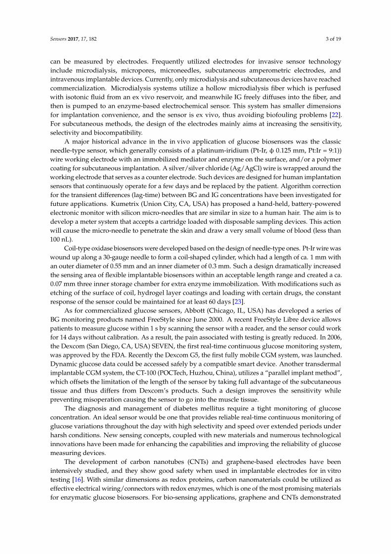
can be measured by electrodes. Frequently utilized electrodes for invasive sensor technologyinclude microdialysis, micropores, microneedles, subcutaneous amperometric electrodes, andintravenous implantable devices. Currently, only microdialysis and subcutaneous devices have reachedcommercialization. Microdialysis systems utilize a hollow microdialysis fiber which is perfusedwith isotonic fluid from an ex vivo reservoir, and meanwhile IG freely diffuses into the fiber, andthen is pumped to an enzyme-based electrochemical sensor. This system has smaller dimensionsfor implantation convenience, and the sensor is ex vivo, thus avoiding biofouling problems [22].For subcutaneous methods, the design of the electrodes mainly aims at increasing the sensitivity,selectivity and biocompatibility.
A major historical advance in the in vivo application of glucose biosensors was the classicneedle-type sensor, which generally consists of a platinum-iridium (Pt-Ir, φ 0.125 mm, Pt:Ir = 9:1))wire working electrode with an immobilized mediator and enzyme on the surface, and/or a polymercoating for subcutaneous implantation. A silver/silver chloride (Ag/AgCl) wire is wrapped around theworking electrode that serves as a counter electrode. Such devices are designed for human implantationsensors that continuously operate for a few days and be replaced by the patient. Algorithm correctionfor the transient differences (lag-time) between BG and IG concentrations have been investigated forfuture applications. Kumetrix (Union City, CA, USA) has proposed a hand-held, battery-poweredelectronic monitor with silicon micro-needles that are similar in size to a human hair. The aim is todevelop a meter system that accepts a cartridge loaded with disposable sampling devices. This actionwill cause the micro-needle to penetrate the skin and draw a very small volume of blood (less than100 nL).
Coil-type oxidase biosensors were developed based on the design of needle-type ones. Pt-Ir wire waswound up along a 30-gauge needle to form a coil-shaped cylinder, which had a length of ca. 1 mm withan outer diameter of 0.55 mm and an inner diameter of 0.3 mm. Such a design dramatically increasedthe sensing area of flexible implantable biosensors within an acceptable length range and created a ca.0.07 mm three inner storage chamber for extra enzyme immobilization. With modifications such asetching of the surface of coil, hydrogel layer coatings and loading with certain drugs, the constantresponse of the sensor could be maintained for at least 60 days [23].
As for commercialized glucose sensors, Abbott (Chicago, IL, USA) has developed a series ofBG monitoring products named FreeStyle since June 2000. A recent FreeStyle Libre device allowspatients to measure glucose within 1 s by scanning the sensor with a reader, and the sensor could workfor 14 days without calibration. As a result, the pain associated with testing is greatly reduced. In 2006,the Dexcom (San Diego, CA, USA) SEVEN, the first real-time continuous glucose monitoring system,was approved by the FDA. Recently the Dexcom G5, the first fully mobile CGM system, was launched.Dynamic glucose data could be accessed safely by a compatible smart device. Another transdermalimplantable CGM system, the CT-100 (POCTech, Huzhou, China), utilizes a “parallel implant method”,which offsets the limitation of the length of the sensor by taking full advantage of the subcutaneoustissue and thus differs from Dexcom’s products. Such a design improves the sensitivity whilepreventing misoperation causing the sensor to go into the muscle tissue.
The diagnosis and management of diabetes mellitus require a tight monitoring of glucoseconcentration. An ideal sensor would be one that provides reliable real-time continuous monitoring ofglucose variations throughout the day with high selectivity and speed over extended periods underharsh conditions. New sensing concepts, coupled with new materials and numerous technologicalinnovations have been made for enhancing the capabilities and improving the reliability of glucosemeasuring devices.
The development of carbon nanotubes (CNTs) and graphene-based electrodes have beenintensively studied, and they show good safety when used in implantable electrodes for in vitrotesting [16]. With similar dimensions as redox proteins, carbon nanomaterials could be utilized aseffective electrical wiring/connectors with redox enzymes, which is one of the most promising materialsfor enzymatic glucose biosensors. For bio-sensing applications, graphene and CNTs demonstrated

Sensors 2017, 17, 182 4 of 19
higher sensitivity and faster response time than traditional electrodes at extremely low workingpotentials. Our group has carried out a comprehensive study of CVD-synthesized CNT fibers used assensing electrodes to detect glucose solutions. The CNT fiber resembles an electric wire, relying onnanoscale surface topography and porosity, which can facilitate molecular-scale interactions withagents like enzymes to efficiently capture and promote electron transfer reactions [24,25]. A scalablesynthesis of a multifunctional conducting polyacrylic acid (PAA) hydrogel for glucose detection thatintegrates a multifunctional matrix that includes reduced graphene and lutetium phthalocyanine wasreported. This biosensor provided high sensitivity for the detection of glucose with a low detectionlimit [26]. However, better control of the physical and chemical properties is still needed for thefabrication of carbon nanomaterial-based biosensors, such as the miniaturization of the sensor, in vivostability, separation processes for different type of CNTs, etc. Other non-enzymatic sensors, such asmetallic oxide and strontium palladium perovskite also show enhanced sensitivity and improveddetection limits, and thus avoid the need for expensive enzymes in the system [27,28].
Microgels, glucose-responsive polymer gels, have several advantages as glucose sensing systemsdue to their porous structures for carrying drugs, semi-solid properties for easy engineering intovarious shapes (beads, films, fibers, etc.), and potential biocompatibility for implantation. Since thefirst preparation of boronic acid-functionalized glucose sensitive hydrogels [29], many polymergel-based glucose sensors have been developed based on the measurements of swelling pressure [30],fluorescence intensity [31], changes in reflection holograms [32,33] and diffraction of photonic colloidalcrystal arrays [34]. Li et al. recently developed a series of inorganic-polymer hybrid microgels for opticalglucose sensing, based on the immobilization of fluorescent quantum dots (QDs) or metal nanoparticles(NPs) into phenylboronic acid (PBA)-functionalized microgels [35]. Zhang et al. constructed an advancedglucose-sensitive platform on the basis of G1.0 PAMAM-functionalized microgels [36]. The beauty ofthis smart microgel, which differentiates it from other examples of traditional glucose sensors, is thatit provides minimally invasive implantation and a fluorescent signal by transdermal transmissionwithout any external links or electric power sources for CGM. A schematic illustration of G1.0PAMAM-functionalized microgels that can recognize glucose and emit blue fluorescence after injection,is depicted in Figure 1.
Sensors 2016, 16, 182 4 of 18
low working potentials. Our group has carried out a comprehensive study of CVD-synthesized CNT fibers used as sensing electrodes to detect glucose solutions. The CNT fiber resembles an electric wire, relying on nanoscale surface topography and porosity, which can facilitate molecular-scale interactions with agents like enzymes to efficiently capture and promote electron transfer reactions [24,25]. A scalable synthesis of a multifunctional conducting polyacrylic acid (PAA) hydrogel for glucose detection that integrates a multifunctional matrix that includes reduced graphene and lutetium phthalocyanine was reported. This biosensor provided high sensitivity for the detection of glucose with a low detection limit [26]. However, better control of the physical and chemical properties is still needed for the fabrication of carbon nanomaterial-based biosensors, such as the miniaturization of the sensor, in vivo stability, separation processes for different type of CNTs, etc. Other non-enzymatic sensors, such as metallic oxide and strontium palladium perovskite also show enhanced sensitivity and improved detection limits, and thus avoid the need for expensive enzymes in the system [27,28].
Microgels, glucose-responsive polymer gels, have several advantages as glucose sensing systems due to their porous structures for carrying drugs, semi-solid properties for easy engineering into various shapes (beads, films, fibers, etc.), and potential biocompatibility for implantation. Since the first preparation of boronic acid-functionalized glucose sensitive hydrogels [29], many polymer gel-based glucose sensors have been developed based on the measurements of swelling pressure [30], fluorescence intensity [31], changes in reflection holograms [32,33] and diffraction of photonic colloidal crystal arrays [34]. Li et al. recently developed a series of inorganic-polymer hybrid microgels for optical glucose sensing, based on the immobilization of fluorescent quantum dots (QDs) or metal nanoparticles (NPs) into phenylboronic acid (PBA)-functionalized microgels [35]. Zhang et al. constructed an advanced glucose-sensitive platform on the basis of G1.0 PAMAM-functionalized microgels [36]. The beauty of this smart microgel, which differentiates it from other examples of traditional glucose sensors, is that it provides minimally invasive implantation and a fluorescent signal by transdermal transmission without any external links or electric power sources for CGM. A schematic illustration of G1.0 PAMAM-functionalized microgels that can recognize glucose and emit blue fluorescence after injection, is depicted in Figure 1.
Figure 1. The schematic illustration of G1.0 PAMAM-functionalized microgels that can recognize glucose and emit blue fluorescence after injection. Reprinted with permission from [36].
Semiconductor quantum dots (QDs), as photoluminescent nanomaterials, have been intensively studied in sensing and cell imaging due to their excellent performance and size-tunable optical properties [37]. Glucose could be detected by indirect measurements using photoelectrochemical QD sensors if combined with suitable enzymes [38,39]. Tanne et al. reported the indirect sensitive detection of glucose by creating a signal chain from glucose via glucose oxidase and molecular oxygen via CdSe/ZnS QDs toward the electrode [40], as shown in Figure 2. On the basis of the
Figure 1. The schematic illustration of G1.0 PAMAM-functionalized microgels that can recognizeglucose and emit blue fluorescence after injection. Reprinted with permission from [36].
Semiconductor quantum dots (QDs), as photoluminescent nanomaterials, have been intensivelystudied in sensing and cell imaging due to their excellent performance and size-tunable opticalproperties [37]. Glucose could be detected by indirect measurements using photoelectrochemical
Sensors 2017, 17, 182 5 of 19
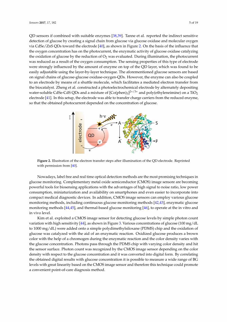
QD sensors if combined with suitable enzymes [38,39]. Tanne et al. reported the indirect sensitivedetection of glucose by creating a signal chain from glucose via glucose oxidase and molecular oxygenvia CdSe/ZnS QDs toward the electrode [40], as shown in Figure 2. On the basis of the influence thatthe oxygen concentration has on the photocurrent, the enzymatic activity of glucose oxidase catalyzingthe oxidation of glucose by the reduction of O2 was evaluated. During illumination, the photocurrentwas reduced as a result of the oxygen consumption. The sensing properties of this type of electrodewere strongly influenced by the amount of enzyme on top of the QD layer, which was found to beeasily adjustable using the layer-by-layer technique. The aforementioned glucose sensors are basedon signal chains of glucose-glucose oxidase-oxygen-QDs. However, the enzyme can also be coupledto an electrode by means of a shuttle molecule, which facilitates a mediated electron transfer fromthe biocatalyst. Zheng et al. constructed a photoelectrochemical electrode by alternately depositingwater-soluble CdSe-CdS QDs and a mixture of [Co(phen)3]2+/3+ and poly(ethyleneimine) on a TiO2
electrode [41]. In this setup, the electrode was able to transfer charge carriers from the reduced enzyme,so that the obtained photocurrent depended on the concentration of glucose.
Sensors 2016, 16, 182 5 of 18
influence that the oxygen concentration has on the photocurrent, the enzymatic activity of glucose oxidase catalyzing the oxidation of glucose by the reduction of O2 was evaluated. During illumination, the photocurrent was reduced as a result of the oxygen consumption. The sensing properties of this type of electrode were strongly influenced by the amount of enzyme on top of the QD layer, which was found to be easily adjustable using the layer-by-layer technique. The aforementioned glucose sensors are based on signal chains of glucose-glucose oxidase-oxygen-QDs. However, the enzyme can also be coupled to an electrode by means of a shuttle molecule, which facilitates a mediated electron transfer from the biocatalyst. Zheng et al. constructed a photoelectrochemical electrode by alternately depositing water-soluble CdSe-CdS QDs and a mixture of [Co(phen)3]2+/3+ and poly(ethyleneimine) on a TiO2 electrode [41]. In this setup, the electrode was able to transfer charge carriers from the reduced enzyme, so that the obtained photocurrent depended on the concentration of glucose.
Figure 2. Illustration of the electron transfer steps after illumination of the QD electrode. Reprinted with permission from [40].
Nowadays, label free and real time optical detection methods are the most promising techniques in glucose monitoring. Complementary metal oxide semiconductor (CMOS) image sensore are becoming powerful tools for biosensing applications with the advantages of high signal to noise ratio, low power consumption, miniaturization and availability on smartphones and even easier to incorporate into compact medical diagnostic devices. In addition, CMOS image sensors can employ various glucose monitoring methods, including continuous glucose monitoring methods [42,43], enzymatic glucose monitoring methods [44,45], and thermal-based glucose monitoring [46], to operate at the in vitro and in vivo level.
Figure 3. Schematic of PDMS chip utilization for monitoring of glucose solutions by a CMOS image sensor. Reprinted with permission from [44].
Figure 2. Illustration of the electron transfer steps after illumination of the QD electrode. Reprintedwith permission from [40].
Nowadays, label free and real time optical detection methods are the most promising techniques inglucose monitoring. Complementary metal oxide semiconductor (CMOS) image sensore are becomingpowerful tools for biosensing applications with the advantages of high signal to noise ratio, low powerconsumption, miniaturization and availability on smartphones and even easier to incorporate intocompact medical diagnostic devices. In addition, CMOS image sensors can employ various glucosemonitoring methods, including continuous glucose monitoring methods [42,43], enzymatic glucosemonitoring methods [44,45], and thermal-based glucose monitoring [46], to operate at the in vitro andin vivo level.
Kim et al. exploited a CMOS image sensor for detecting glucose levels by simple photon countvariation with high sensitivity [44], as shown in Figure 3. Various concentrations of glucose (100 mg/dLto 1000 mg/dL) were added onto a simple polydimethylsiloxane (PDMS) chip and the oxidation ofglucose was catalyzed with the aid of an enzymatic reaction. Oxidized glucose produces a browncolor with the help of a chromogen during the enzymatic reaction and the color density varies withthe glucose concentration. Photons pass through the PDMS chip with varying color density and hitthe sensor surface. Photon count was recognized by the CMOS image sensor depending on the colordensity with respect to the glucose concentration and it was converted into digital form. By correlatingthe obtained digital results with glucose concentration it is possible to measure a wide range of BGlevels with great linearity based on the CMOS image sensor and therefore this technique could promotea convenient point-of-care diagnosis method.

Sensors 2017, 17, 182 6 of 19
Sensors 2016, 16, 182 5 of 18
influence that the oxygen concentration has on the photocurrent, the enzymatic activity of glucose oxidase catalyzing the oxidation of glucose by the reduction of O2 was evaluated. During illumination, the photocurrent was reduced as a result of the oxygen consumption. The sensing properties of this type of electrode were strongly influenced by the amount of enzyme on top of the QD layer, which was found to be easily adjustable using the layer-by-layer technique. The aforementioned glucose sensors are based on signal chains of glucose-glucose oxidase-oxygen-QDs. However, the enzyme can also be coupled to an electrode by means of a shuttle molecule, which facilitates a mediated electron transfer from the biocatalyst. Zheng et al. constructed a photoelectrochemical electrode by alternately depositing water-soluble CdSe-CdS QDs and a mixture of [Co(phen)3]2+/3+ and poly(ethyleneimine) on a TiO2 electrode [41]. In this setup, the electrode was able to transfer charge carriers from the reduced enzyme, so that the obtained photocurrent depended on the concentration of glucose.
Figure 2. Illustration of the electron transfer steps after illumination of the QD electrode. Reprinted with permission from [40].
Nowadays, label free and real time optical detection methods are the most promising techniques in glucose monitoring. Complementary metal oxide semiconductor (CMOS) image sensore are becoming powerful tools for biosensing applications with the advantages of high signal to noise ratio, low power consumption, miniaturization and availability on smartphones and even easier to incorporate into compact medical diagnostic devices. In addition, CMOS image sensors can employ various glucose monitoring methods, including continuous glucose monitoring methods [42,43], enzymatic glucose monitoring methods [44,45], and thermal-based glucose monitoring [46], to operate at the in vitro and in vivo level.
Figure 3. Schematic of PDMS chip utilization for monitoring of glucose solutions by a CMOS image sensor. Reprinted with permission from [44]. Figure 3. Schematic of PDMS chip utilization for monitoring of glucose solutions by a CMOS imagesensor. Reprinted with permission from [44].
2.2. Novel Biocompatible Coatings for Sensors
The major challenge in the design of tissue-contacting materials for both bioaffinity andmedical implantation sensors is to endure protein adsorption between surfaces and interfaces.Most minimally-invasive biosensors are surrounded by protective coatings that can strongly interactwith proteins, thereby minimizing tissue reactions induced by device implantation [47]. To achievea protein-resistant surface, the structure design of the anti-fouling material should be hydrophilic,electrically neutral, and contain hydrogen bond acceptors instead of donors. These requirementsguide the design of anti-fouling formulations for sensing applications with novel structures that havenon-specific protein resistance [48]. A considerable number of effective candidates have been identified,including organic/inorganic composites and biofunctional polymer architectures. Sol-gel derivedsilicates have been demonstrated to be highly compatible with enzymes [49]. Silica-based hybridmaterials exhibit a fair biocompatibility both in vitro and in vivo, and are shown to be non-toxic andbiocompatible coatings for glucose oxidase-based sensors. Stable glucose responses were obtained forthe silica-polymer coated sensors both in buffered solutions containing bovine serum albumin and inserum [50,51]. Unfortunately, the anti-fouling properties of such systems were not discussed.
Various macromolecules, including natural, semi-synthetic, and synthetic materials are currentlyutilized in the fabrication of implantable CGM sensor coatings (cf. Table 1). Naturally occurring materialssuch as alginate, chitin, and chitosan and their derivatives are widely investigated for biosensors inthe form of sheets, membranes and coatings, which offer the advantage of being comparable tothe natural precursors and the biological environment is prepared to recognize and deal with theirmetabolites [52,53]. On the other hand, modified synthetic polymers with tailored structures andcoating properties are often superior to traditional macromolecules, especially for their reducedimmunogenicity. Commonly used biocomparable and biodegradable polymers includes poly(lacticco-glycolic acid) (PLGA), poly(ethylene glycol) (PEG), poly(hydroxyethyl methacrylate) (PHEMA),and poly(vinyl alcohol) (PVA), etc. [54]. Among the abovementioned polymers, HEMA-based materialsconstitute the first generation of efficient protein-resistant materials while PEG-derived materialsrepresent the second generation [55]. Currently, they are extensively utilized in various areas andcan be used to modify mini-invasive devices by the coating method. They are also very popular ashydrogel matrices, either alone or combined with other polymers [56].

Sensors 2017, 17, 182 7 of 19
Table 1. Biochemical aspects of commonly used polymers.
Polymer Characters Formation Applications
Natural
Alginate Immobilization of glucose oxidase Hydrogel and membrane Drug deliveryCollagen Extracellular matrix component Hydrogel, membrane and sponge Scaffolds
Semisynthetic
Chitin,Chitosan Hydrogel, membrane and fiber Anti-microbial and drug delivery
Synthetic
PLGA Negligible protein adsorption Micelle and hydrogel Coating, drug delivery and scaffoldsPHEMA Negligible protein adsorption Hydrogel Coating
PVA Geltaion and mechanical properties Hydrogel, membrane and sponge Coating and drug deliveryPEG Negligible protein adsorption Hydrogel and membrane Coating and drug delivery
PEGMA Negligible protein adsorption Hydrogel Coating
Hydrogels are hydrophilic, water-insoluble three-dimensional polymeric networks represented assemi-open structures comprising entangled chains, which adsorb and store large amounts of water andare highly permeable to small molecules. The use of hydrogel coatings allows the diffusion of glucosethrough the swollen hydrogel layer. The degree of glucose diffusion could be readily modulated byphysically/chemically control of the cross-linking density of the hydrogel, which consequently affectsthe water content and mechanical strength of polymer network. Both in vitro and in vivo animaltests have demonstrated better performance with biocompatible polymer hydrogel outer layers fora mini-invasive needle-type glucose sensor.
Despite oxidation problems that could alter the anti-fouling properties during long-term usage,PEG-derived anti-fouling matrices remain popular and have been widely investigated [57]. Similar toPEG, poly(ethylene glycol) methyl ether methacrylate (PEGMA) and PEGMA-analogous hydrogelswere also investigated for the design of the next generation of anti-biofouling materials [58]. The resultsrevealed that the molecular weight of the PEGMA homopolymer directly affected the overallhydrophilicity of the gel, which could be attributed to both the intermediate hydrophilic chain lengthand intermediate crosslinking degree. Furthermore, the results of micro-biofouling by bacteria andblood cells demonstrated that PEGMA hydrogels offered the perfect non-fouling ability, showingapplication potential for the coating of devices in contact with blood and the in vivo controlled releaseof drugs from nonfouling hydrogels.
2.3. Drug Deliver and Its System
Drug delivery is a potential field of application for CGM devices [59]. In recent years, architecturesfor sustained and controlled delivery of drugs has been extensively studied due to several problemsin common drug delivery systems, such as limited stability, inopportune leakage and poor solubility,especially for CGM systems. Modern closed-loop BG controlling systems have been developed byvarious organizations around the world, by merging glucose-sensing devices and insulin-deliverycarriers for optimized dosage of insulin according to detected glycemic levels (Figure 4). Such BGmanagement systems utilize a ‘Sense and Act’ feedback-loop design to realize appropriate correctiveaction by timely and optimal dosing. The pump dispenses the right amount of insulin as the sensorrelays the information. The development of these responsive drug delivery systems is anticipated todramatically change diabetes management and patient monitoring. It is expected that the release ofdifferent drugs could be controlled by a microprocessor in response to the signal from a biosensor viadifferent release kinetics.
Based on the biocoating, intelligent drug releasing functionality was also investigated to makethe system more integrated, and ensure the long-term usage of the device [60]. For instance, a PLGA-copolymer, PVA and PEG combined system which has been shown to achieve a continuous release ofdexamethasone for up to one month that decreases extravasation of leukocytes, and inhibits productionof proteolytic enzymes and chemoattractive factors was already discussed [61,62]. A masitinib-releasing

Sensors 2017, 17, 182 8 of 19
PLGA carrier delivers locally from soluble PEG-coated CGM sensors were developed to reduce thehost-implant response. The releasing action could be attributed to the CGM sensor output fluctuationsover time, which was monitored for 21 days. The results showed relatively improved performance interms of sensing ability in the drug-delivery implant group compared to the control group [63].
Sensors 2016, 16, 182 8 of 18
glucose oxidase on the biosensor. The result showed that the sensitivity of the as-prepared spiral sensor was 20 nA/(mmol/L)~30 nA/(mmol/L), which provided a linear detection of physiological glucose concentrations between 2 and 30 mmol/L. The sensors also showed good repeatability, selectivity, and stability, which could be tuned by adjusting the amount of the polyurethane (PU) semi-permeable membrane coating [64]. More recently, the PU coating was further modified by a PVA-PEG hydrogel that enabled zero-ordered release of dexamethasone, and the release duration was controlled as a function of film gelation density. The cumulative released amount could be adjusted by changes in the molecular weight of the PEG. As a matter of fact, PEG derivatives were revealed to be effective candidates for sensor coatings as well as controlled release of supposed drugs [58,65].
Figure 4. Illustration of closed-loop glycemic management system utilizing the ‘Sense and Act’ method for optimized insulin delivery.
3. Non-Invasive Technology
Non-invasive techniques have received significant research interest due to the highly sensitivity and better patient compliance, contrary to invasive ones. Typical non-invasive biosensors based on different approaches include iontophoretic extraction of glucose through the skin, surface plasmon resonance, Raman spectroscopy in aqueous humor, visible or near-infrared (NIR) spectroscopy, polarimetry, photo-acoustic probes, and fluorescence methods. Although results from these innovative devices require frequent calibration against direct BG data, they might be an alternative choice for future continuous glucose measurement. Consequently, the challenge of preparing accurate level sensors to biomonitor conveniently and painlessly BG information remains to be solved.
3.1. Tear Sensing Designs
Non-invasive, contact lenses are one of the most appealing ways to achieve constant contact with tear fluid and monitor glucose in tears. Such a method could be used for normal conditions as well as for clinical trials. During the course of treatment, the glucose could be continuously monitored. Contact lenses have to meet a certain number of criteria, and should be made of soft, comfortable materials with constant mechanical properties such as hydrogels or plasticized polymers that could withstand the eye movements and blinking. Moreover, the lenses also should be non-toxic and to allow for a certain amount of oxygen and tear fluid to pass through to the eye. Some interactions between contact lenses and the tear film were discussed. The presence of the contact lens immediately provides the potential for both biophysical and biochemical phenomena. The biophysical phenomena resulting from lens-tear interactions are often directly observable as the increased osmotic pressure caused by enhanced evaporation of the tear film. The biochemical
Figure 4. Illustration of closed-loop glycemic management system utilizing the ‘Sense and Act’ methodfor optimized insulin delivery.
Recently, a novel implantable electrochemical sensor was fabricated by utilizing a spiral Pt-Iralloy electrode to increase the active area of the working electrode as well as enhance the loading ofglucose oxidase on the biosensor. The result showed that the sensitivity of the as-prepared spiral sensorwas 20 nA/(mmol/L)~30 nA/(mmol/L), which provided a linear detection of physiological glucoseconcentrations between 2 and 30 mmol/L. The sensors also showed good repeatability, selectivity, andstability, which could be tuned by adjusting the amount of the polyurethane (PU) semi-permeablemembrane coating [64]. More recently, the PU coating was further modified by a PVA-PEG hydrogelthat enabled zero-ordered release of dexamethasone, and the release duration was controlled asa function of film gelation density. The cumulative released amount could be adjusted by changes inthe molecular weight of the PEG. As a matter of fact, PEG derivatives were revealed to be effectivecandidates for sensor coatings as well as controlled release of supposed drugs [58,65].
3. Non-Invasive Technology
Non-invasive techniques have received significant research interest due to the highly sensitivityand better patient compliance, contrary to invasive ones. Typical non-invasive biosensors based ondifferent approaches include iontophoretic extraction of glucose through the skin, surface plasmonresonance, Raman spectroscopy in aqueous humor, visible or near-infrared (NIR) spectroscopy,polarimetry, photo-acoustic probes, and fluorescence methods. Although results from these innovativedevices require frequent calibration against direct BG data, they might be an alternative choice forfuture continuous glucose measurement. Consequently, the challenge of preparing accurate levelsensors to biomonitor conveniently and painlessly BG information remains to be solved.
3.1. Tear Sensing Designs
Non-invasive, contact lenses are one of the most appealing ways to achieve constant contact withtear fluid and monitor glucose in tears. Such a method could be used for normal conditions as wellas for clinical trials. During the course of treatment, the glucose could be continuously monitored.Contact lenses have to meet a certain number of criteria, and should be made of soft, comfortablematerials with constant mechanical properties such as hydrogels or plasticized polymers that could
Sensors 2017, 17, 182 9 of 19
withstand the eye movements and blinking. Moreover, the lenses also should be non-toxic and to allowfor a certain amount of oxygen and tear fluid to pass through to the eye. Some interactions betweencontact lenses and the tear film were discussed. The presence of the contact lens immediately providesthe potential for both biophysical and biochemical phenomena. The biophysical phenomena resultingfrom lens-tear interactions are often directly observable as the increased osmotic pressure caused byenhanced evaporation of the tear film. The biochemical outcomes revealed the difference in electrolyteconcentration between the anterior and posterior tear films [66,67].
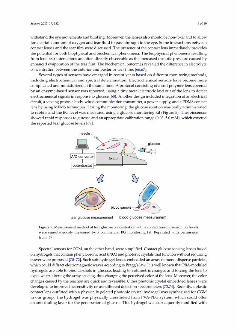
Several types of sensors have emerged in recent years based on different monitoring methods,including electrochemical and spectral determination. Electrochemical sensors have become morecomplicated and miniaturized at the same time. A protocol consisting of a soft polymer lens coveredby an enzyme-based sensor was reported, using a tiny metal electrode laid out of the lens to detectelectrochemical signals in response to glucose [68]. Another design included integration of an electricalcircuit, a sensing probe, a body-wired communication transmitter, a power supply, and a PDMS contactlens by using MEMS techniques. During the monitoring, the glucose solution was orally administratedto rabbits and the BG level was measured using a glucose monitoring kit (Figure 5). This biosensorshowed rapid responses to glucose and an appropriate calibration range (0.03–5.0 mM), which coveredthe reported tear glucose levels [69].
Sensors 2016, 16, 182 9 of 18
outcomes revealed the difference in electrolyte concentration between the anterior and posterior tear films [66,67].
Several types of sensors have emerged in recent years based on different monitoring methods, including electrochemical and spectral determination. Electrochemical sensors have become more complicated and miniaturized at the same time. A protocol consisting of a soft polymer lens covered by an enzyme-based sensor was reported, using a tiny metal electrode laid out of the lens to detect electrochemical signals in response to glucose [68]. Another design included integration of an electrical circuit, a sensing probe, a body-wired communication transmitter, a power supply, and a PDMS contact lens by using MEMS techniques. During the monitoring, the glucose solution was orally administrated to rabbits and the BG level was measured using a glucose monitoring kit (Figure 5). This biosensor showed rapid responses to glucose and an appropriate calibration range (0.03–5.0 mM), which covered the reported tear glucose levels [69].
Figure 5. Measurement method of tear glucose concentration with a contact lens biosensor. BG levels were simultaneously measured by a commercial BG monitoring kit. Reprinted with permission from [69].
Spectral sensors for CGM, on the other hand, were simplified. Contact glucose-sensing lenses based on hydrogels that contain phenylboronic acid (PBA) and photonic crystals that function without requiring power were proposed [70–72]. Such soft hydrogel lenses embedded an array of mono-disperse particles, which could diffract electromagnetic waves according to Bragg’s law. It is well known that PBA-modified hydrogels are able to bind cis-diols in glucose, leading to volumetric changes and forcing the lens to expel water, altering the array spacing, thus changing the perceived color of the lens. Moreover, the color changes caused by the reaction are quick and reversible. Other photonic crystal embedded lenses were developed to improve the sensitivity or use different detection spectrometers [73,74]. Recently, a plastic contact lens outfitted with a physically gelated photonic crystal hydrogel was synthesized for CGM in our group. The hydrogel was physically crosslinked from PVA-PEG system, which could offer an anti-fouling layer for the penetration of glucose. This hydrogel was subsequently modified with 4-boronobenzaldehyde, inproving the sensitivity for glucose, and a prototype of such a lens is shown in Figure 6a. The sensing behavior of the lens was monitored by a reflection photospectrometer, and the results showed that the diffracted wavelength was relatively shifted with the increasing glucose concentration in artificial tear solution (Figure 6b). Despite the exciting design, it remains difficult to measure glucose concentrations in tears using contact lenses. Because of the very low amount of glucose present in healthy control
Figure 5. Measurement method of tear glucose concentration with a contact lens biosensor. BG levelswere simultaneously measured by a commercial BG monitoring kit. Reprinted with permissionfrom [69].
Spectral sensors for CGM, on the other hand, were simplified. Contact glucose-sensing lenses basedon hydrogels that contain phenylboronic acid (PBA) and photonic crystals that function without requiringpower were proposed [70–72]. Such soft hydrogel lenses embedded an array of mono-disperse particles,which could diffract electromagnetic waves according to Bragg’s law. It is well known that PBA-modifiedhydrogels are able to bind cis-diols in glucose, leading to volumetric changes and forcing the lens toexpel water, altering the array spacing, thus changing the perceived color of the lens. Moreover, the colorchanges caused by the reaction are quick and reversible. Other photonic crystal embedded lenses weredeveloped to improve the sensitivity or use different detection spectrometers [73,74]. Recently, a plasticcontact lens outfitted with a physically gelated photonic crystal hydrogel was synthesized for CGMin our group. The hydrogel was physically crosslinked from PVA-PEG system, which could offeran anti-fouling layer for the penetration of glucose. This hydrogel was subsequently modified with

Sensors 2017, 17, 182 10 of 19
4-boronobenzaldehyde, inproving the sensitivity for glucose, and a prototype of such a lens is shown inFigure 6a. The sensing behavior of the lens was monitored by a reflection photospectrometer, and theresults showed that the diffracted wavelength was relatively shifted with the increasing glucoseconcentration in artificial tear solution (Figure 6b). Despite the exciting design, it remains difficultto measure glucose concentrations in tears using contact lenses. Because of the very low amount ofglucose present in healthy control subjects (3.59 mM) and for diabetic subjects (4.69 mM), the tearglucose levels measured appear to vary with the volume of the aqueous tear fraction collected.
Sensors 2016, 16, 182 10 of 18
subjects (3.59 mM) and for diabetic subjects (4.69 mM), the tear glucose levels measured appear to vary with the volume of the aqueous tear fraction collected.
(a) (b)
Figure 6. (a) Diagram and photograph (insert) of a physical hydrogel photonic crystal sensing lens; (b) Diffraction wavelength shifts with the variation of the glucose concentration in artificial tear solution.
3.2. Salivary Biosensors
Many authors have found higher glucose salivary levels in diabetic patients than in non-diabetics [75–78]. Such investigations mainly aimed at exploring whether diabetic control could be monitored by a non-invasive salivary glucose measurement method. Saliva is a great diagnostic fluid providing an alternative to direct blood analysis via the permeation of blood constituents without any skin-piercing for blood sampling [79]. Actually, saliva glucose concentrations range approximately from 20 to 200 mmol/L in normal and diabetic individuals, and closely follow circadian BG fluctuations [80]. Saliva and BG levels correlate reasonably well in a sample of individuals [81–83], however, a much stronger correlation is observed within the same individual, enabling BG concentrations to be estimated from saliva glucose measurements [84].
Various attempts have been made by researchers to develop glucose monitoring systems for salivary measurements. Lambert et al. used conventional measurement through colorimetric assays [85]. Lipson et al. detected the saliva glucose level through NIR/Raman-based spectroscopic techniques [86], and Yamaguchi et al. used Clarke-type electrode-based amperometric measurements, etc. [80]. Taking clues from these developments, Soni et al. developed an optical biosensor for direct determination of salivary glucose by using glucose oxidase enzyme immobilized on filter paper strips (specific activity 1.4 U/strip) and then reacting it with synthetic glucose samples in the presence of a co-immobilized color pH indicator [83]. The filter paper changed color based on the concentration of glucose in the reaction medium and hence, by scanning this color change (using RGB profiling) through an office scanner and using open source image processing software (GIMP), the concentration of glucose in the reaction medium could be deduced.
Wearable sensors have recently attracted considerable interest owing to their promise for real-time monitoring of the wearer’s health and fitness in a wide range of biomedical, sport and military scenarios [87,88]. Recent efforts have led to wearable biosensors for detecting chemical biomarkers in human fluids that can be obtained non-invasively, e.g., tears, sweat or saliva [89–92]. Kim et al. reported a mouth-guard biosensor for continuous monitoring of salivary lactate and other chemical components [79,93]. A mouth-guard glucose sensor produced using micro electromechanical systems (MEMS) techniques would offer promise as a minimally-invasive, painless, continuous, custom-fitted and wireless solution for self-monitoring of glucose. Mitsubayashi et al. have developed a detachable “Cavitas sensor” used in the human oral cavity for non-invasive monitoring of saliva glucose [94], as shown in Figure 7. The mouth-guard glucose sensor consisted of a platinum and silver/silver chloride electrode, with glucose oxidase immobilized by entrapment with poly(MPC-co-EHMA) (PMEH), on a custom-fitted monolithic
Figure 6. (a) Diagram and photograph (insert) of a physical hydrogel photonic crystal sensing lens;(b) Diffraction wavelength shifts with the variation of the glucose concentration in artificial tear solution.
3.2. Salivary Biosensors
Many authors have found higher glucose salivary levels in diabetic patients than innon-diabetics [75–78]. Such investigations mainly aimed at exploring whether diabetic control could bemonitored by a non-invasive salivary glucose measurement method. Saliva is a great diagnostic fluidproviding an alternative to direct blood analysis via the permeation of blood constituents without anyskin-piercing for blood sampling [79]. Actually, saliva glucose concentrations range approximately from20 to 200 mmol/L in normal and diabetic individuals, and closely follow circadian BG fluctuations [80].Saliva and BG levels correlate reasonably well in a sample of individuals [81–83], however, a muchstronger correlation is observed within the same individual, enabling BG concentrations to be estimatedfrom saliva glucose measurements [84].
Various attempts have been made by researchers to develop glucose monitoring systems forsalivary measurements. Lambert et al. used conventional measurement through colorimetric assays [85].Lipson et al. detected the saliva glucose level through NIR/Raman-based spectroscopic techniques [86],and Yamaguchi et al. used Clarke-type electrode-based amperometric measurements, etc. [80]. Taking cluesfrom these developments, Soni et al. developed an optical biosensor for direct determination of salivaryglucose by using glucose oxidase enzyme immobilized on filter paper strips (specific activity 1.4 U/strip)and then reacting it with synthetic glucose samples in the presence of a co-immobilized color pHindicator [83]. The filter paper changed color based on the concentration of glucose in the reactionmedium and hence, by scanning this color change (using RGB profiling) through an office scannerand using open source image processing software (GIMP), the concentration of glucose in the reactionmedium could be deduced.
Wearable sensors have recently attracted considerable interest owing to their promise for real-timemonitoring of the wearer’s health and fitness in a wide range of biomedical, sport and militaryscenarios [87,88]. Recent efforts have led to wearable biosensors for detecting chemical biomarkersin human fluids that can be obtained non-invasively, e.g., tears, sweat or saliva [89–92]. Kim et al.reported a mouth-guard biosensor for continuous monitoring of salivary lactate and other chemicalcomponents [79,93]. A mouth-guard glucose sensor produced using micro electromechanical systems

Sensors 2017, 17, 182 11 of 19
(MEMS) techniques would offer promise as a minimally-invasive, painless, continuous, custom-fittedand wireless solution for self-monitoring of glucose. Mitsubayashi et al. have developed a detachable“Cavitas sensor” used in the human oral cavity for non-invasive monitoring of saliva glucose [94],as shown in Figure 7. The mouth-guard glucose sensor consisted of a platinum and silver/silverchloride electrode, with glucose oxidase immobilized by entrapment with poly(MPC-co-EHMA)(PMEH), on a custom-fitted monolithic mouth-guard support with a wireless transmitter, therebyenabling telemetric measurement of saliva glucose.
Sensors 2016, 16, 182 11 of 18
mouth-guard support with a wireless transmitter, thereby enabling telemetric measurement of saliva glucose.
Figure 7. (A) Schematic image of the glucose biosensor on the polyethylene terephthalate glycol mouthguard support. Pt and Ag electrodes were formed on the PETG through a sputtering process. Each electrode sensor consisted of a 0.20 mm2 Pt working electrode and a 4.0 mm2 Ag/AgCl reference/counter electrode, both insulated with PDMS on a 0.5 mm thick PETG layer. 30 units of GOD were applied to the sensing region of the working electrode. In order to optimize enzyme entrapment, 2.0 mL of 1.0 wt% PMEH solution was spread over the sensing region to form the PMEH overcoat; (B) Schematic image of the mouth-guard biosensor custom-fit to the patient’s dentition. The device consists of a glucose sensor and wireless transmitter incorporating a potentiostat for stable glucose measurement. The sensor was designed to fit the mandibular dentition from the first premolar up to the third molar. The wireless transmitter was neatly encased in PETG. Reprinted with permission from [94].
3.3. Other Methods
Recently continuous glucose monitoring devices have become available and could provide more detailed data on glucose excursions. In future applications such continuous glucose sensors may become a critical component of closed loop insulin delivery systems and, as such, must be selective, rapid, predictable and acceptable for continuous patient use. Many potential sensing modalities are being pursued for this, including optical techniques.
Optical sensors use light of variable frequencies to detect glucose, utilizing different interaction properties of light with glucose molecules in a concentration-dependent manner [95]. Compared to conventional glucose sensors, optical sensors benefit from the absence of electromagnetic interference, simple design and handling as well as low cost. Miniaturization of optical sensors is easily achievable without compromising performance and dramatically reduces repulsion reactions. Evidently, optical sensing technology can provide an interesting alternative to more established electrochemical sensors and will contribute to better flexibility concerning the design of CGM systems, their cost, materials used, etc. [96]. Based on this optical transducing technology, numerous optics-based CGM systems have been developed, such as fluorescence, infrared absorption spectroscopy, and Raman spectroscopy.
Figure 7. (A) Schematic image of the glucose biosensor on the polyethylene terephthalate glycolmouthguard support. Pt and Ag electrodes were formed on the PETG through a sputtering process.Each electrode sensor consisted of a 0.20 mm2 Pt working electrode and a 4.0 mm2 Ag/AgClreference/counter electrode, both insulated with PDMS on a 0.5 mm thick PETG layer. 30 units of GODwere applied to the sensing region of the working electrode. In order to optimize enzyme entrapment,2.0 mL of 1.0 wt% PMEH solution was spread over the sensing region to form the PMEH overcoat;(B) Schematic image of the mouth-guard biosensor custom-fit to the patient’s dentition. The deviceconsists of a glucose sensor and wireless transmitter incorporating a potentiostat for stable glucosemeasurement. The sensor was designed to fit the mandibular dentition from the first premolar upto the third molar. The wireless transmitter was neatly encased in PETG. Reprinted with permissionfrom [94].
3.3. Other Methods
Recently continuous glucose monitoring devices have become available and could provide moredetailed data on glucose excursions. In future applications such continuous glucose sensors maybecome a critical component of closed loop insulin delivery systems and, as such, must be selective,rapid, predictable and acceptable for continuous patient use. Many potential sensing modalities arebeing pursued for this, including optical techniques.
Optical sensors use light of variable frequencies to detect glucose, utilizing different interactionproperties of light with glucose molecules in a concentration-dependent manner [95]. Compared toconventional glucose sensors, optical sensors benefit from the absence of electromagnetic interference,
Sensors 2017, 17, 182 12 of 19
simple design and handling as well as low cost. Miniaturization of optical sensors is easily achievablewithout compromising performance and dramatically reduces repulsion reactions. Evidently, opticalsensing technology can provide an interesting alternative to more established electrochemical sensorsand will contribute to better flexibility concerning the design of CGM systems, their cost, materialsused, etc. [96]. Based on this optical transducing technology, numerous optics-based CGM systemshave been developed, such as fluorescence, infrared absorption spectroscopy, and Raman spectroscopy.
Fluorescence uses the principle of varying light emission from molecules in different states. It isfast, reagentless and extremely sensitive. Many fluorescence-based glucose sensors are based upon theaffinity sensor principle where glucose and a fluorescein-labelled analogue bind competitively witha receptor site specific for both ligands [97,98]. Other fluorescence techniques use enzyme- catalysedreactions to change fluorescent status. Fluorescein may be bound to glucose oxidase, allowing energytransfer between the flavin group of GOx and the fluorescein. The GOx enzyme may be furtherutilized by measuring oxygen consumption or hydrogen peroxide production by fluorescence [99,100].GOx can also be used as glucose-binding protein, utilizing its intrinsic fluorescent properties and maybe used without its flavin group with a fluorescent label [101].
Near infrared (NIR) light (wavelength 0.7–1.4 µm) provides an optical window in which 90%–95%of light passes through the stratum corneum and epidermis into the subcutaneous space independentof skin pigmentation. It is already successfully used to non-invasively monitor the concentration ofoxygenated and deoxygenated haemoglobin in the preterm infant brain [102]. BG has been measuredacross the oral mucosa using NIR [103]. The oral mucosa is an obvious candidate for sensing, as itis well vascularized and may be trans-illuminated. However, any oral measurement includes saliva,which may contain a different glucose concentration, and residual food may contain interferents.NIR spectroscopy has been measured across the tongue [104]. The standard error of prediction was3.5 mmol/L, with significant variability caused by variability in tissue fat. NIR therefore ideallyrequires consistent tissue fat in addition to a path length in the order of 5 mm [105]. In order to solvesome of the problems posed by transdermal optical techniques, NIR has been proposed in combinationwith techniques for sampling ISF, including sonophoresis [106].
Raman spectroscopy assesses scattering of single wavelength light. This is dependent on rotationalor vibrational energy states within a molecule and highly specific absorption bands are seen withRaman spectroscopy which can be used to identify and quantify molecules. It has the benefit ofreduced interference from water compared with MIR or NIR spectroscopy. However, the Ramansignal is weaker than that of other technologies, requiring powerful detectors for physiologicalconcentrations of glucose. The Raman signal is also susceptible to turbidity, haematocrit, skin thicknessand melanin. Sufficient sensitivity can be achieved by surface-enhanced Raman spectroscopy (SERS).The electromagnetic mechanism of SERS is that when an electromagnetic wave interacts with a metalsurface, the fields at the surface are different than those observed in the far field. If the surface isrough, the wave may excite localized surface plasmons on the surface, resulting in amplification ofthe electromagnetic fields near the surface. If one assumes that there is enhancement of the intensityof the incident and scattered fields (albeit at different wavelengths), then the possibility of a largeenhancement of Raman scattering intensity arises [107]. And now some reports utilized the SERSsignal probes served as the molecular recognition agent in response to the concentration of glucose.Torul et al. fabricated a SERS substrate by modifying two component self-assembled monolayers(SAMS) on the surface of gold nanorod particles [108]. The variation of the SERS signal of SAMSshowed the response to the concentration of glucose, and a low detection limit of 0.5 mM was obtained.Kong et al. proposed a novel glucose binding mechanism by using phenylboronic acid as the receptorfor saccharide and forming a glucose-alkyne-boronic acid complex on SERS substrate, which exhibiteda new Raman peak at 1996 cm−1 [109]. Thus, the novel technology offered a high sensitivity for SERSglucose sensing.

Sensors 2017, 17, 182 13 of 19
4. Future Perspectives
The worldwide increase in the number of diabetic patients has encouraged scientists to put greatefforts into the field of continuous glucose monitoring (CGM) devices, which would dramatically alterthe treatment of diabetes as well as the life quality of diabetic patients. So far, there are only a fewcommercial CGM devices, developed by Medtronic (Minneapolis, MN, USA) and Dexcom, which havebeen approved by the FDA and their current lifetimes are less than one week. No commercial CGMdevices are available for long-term monitoring, however, it is extremely important for T1D, whichaccounts for around 10% of diabetic patients. The existing studies of long-term mini-invasive biosensorsremain challenging, mainly due to the foreign body responses such as biofouling, fibrous capsuleand inflammation. To overcome foreign body responses and improve the life-time of implantablebiosensors, we believe the following issues should be addressed: (1) improving the loading amountand stability of enzymes. The coil type electrode design is one way to load more enzyme to reducethe effects from accumulation of H2O2, glucose oxidase molecular cloning or encapsulation in silicasol-gel may be another way to reduce the degradation of cross-linked enzymes [110]; (2) a numberof interferences (e.g., acetaminophen, ascorbic acid, and uric acid) can affect sensor response as theyare electroactive during the oxidation of hydrogen peroxide, although semi-permeable membraneswith designed porosity, like polyurethane, are able to alleviate this effect, At the meantime, decreasingthe work potential through electrode design is another route to reduce interference; (3) to reduceforeign body responses once the biosensors are implanted, the synthesis of anti-fouling films isa common strategy to improve glucose sensor functionality against the initial adhesion of proteins.The introducing of biocompatible hydrogel coatings or collagen encapsulation for sensing devices areother key approaches to improve the lifetime. The use of small anti-inflammatory and/or antigenicmolecules, such as dexamethasone, to influence tissue integration has been intensively studied. Here,the drug delivery system with biocompatible and controllable particles, like poly(lactic-co-glycolic acid)is pursued to limit inflammation and take advantage of the wound healing properties of this material.
Despite the fact that the results from non-invasive devices require frequent calibration againstinvasive ones, the innovative design might be an alternative choice for glucose measurements thantraditional implanted electrochemical biosensors. Two possible techniques have the most chancefor non-invasive glucose monitoring, one is tear glucose sensors and another is salivary biosensors.Detecting glucose in tear fluid has been established for over twenty years, and the major challengefor traditional contact lens biosensors is that the amount tear fluid is not high enough to cover theelectrode to construct an electrochemical cell, and this thus leads to less accuracy in this type ofsensor. Compared with electrochemical sensors, a plastic contact lens outfitted with a physicallygelated photonic crystal hydrogel maybe a possible route for CGM in the future. A salivary biosensorincorporating Pt and Ag/AgCl electrodes on a mouth-guard support as dental material with an enzymemembrane was developed and tested by Mitsubayashi’s group [94], and this invisible mouth-guardbiosensor integrated with a low energy wireless module could be an ideal candidate for future real-timemonitoring of salivary glucose levels.
Non-enzymatic sensor show enhanced sensitivity and detection limits, while avoiding theexpensive and fragile enzymes in the system, and thus it is also a promising choice for the nextgeneration of CGM. Although there are large number of research work on this area, the clinic productsare not yet available in the market. The ultimate goal for treating diabetic patients is to construct anartificial pancreas, combining CGM and insulin dosage system, the life time of CGM is the greatestchallenge, and are required for deeply and comprehensively studied in the future.
Acknowledgments: This work was supported by the National Natural Science Foundation of China (61471233,21504051), the Program for Professor of Special Appointment (Eastern Scholar) at SIHL, the Sailing Project andBasic Research Program from Science and Technology Commission of Shanghai Municipality (14YF1410600,14JC1406402), Shuguang and ChenGuang project supported by Shanghai Municipal Education Commissionand Shanghai Education Development Foundation (14SG52, 13CG62), the key subject of Shanghai PolytechnicUniversity (Material Science and Engineering, XXKZD1601).
Sensors 2017, 17, 182 14 of 19
Conflicts of Interest: The authors declare no conflict of interest.
References
1. World Health Organization (WHO). Global Report on Diabetes 2016; WHO: Geneva, Switzerland, 2016.2. Centers for Disease Control and Prevention. National Diabetes Fact Sheet: National Estimates and General
Information on Diabetes and Prediabetes in the United States; Centers for Disease Control and Prevention: Atlanta,GA, USA, 2011.
3. Wild, S.; Roglic, G.; Green, A.; Sicree, R.; King, H. Global prevalence of diabetes: Estimates for the year 2000and projections for 2030. Diabetes Care 2004, 27, 1047–1053. [CrossRef] [PubMed]
4. Chase, H.P.; Beck, R.W.; Xing, D.; Tamborlane, W.V.; Coffey, J.; Fox, L.A.; Ives, B.; Keady, J.; Kollman, C.;Laffel, L.; et al. Continuous glucose monitoring in youth with type 1 diabetes: 12-month follow-up of theJuvenile Diabetes Research Foundation continuous glucose monitoring randomized trial. Diabetes Technol. Ther.2010, 12, 507–515. [CrossRef] [PubMed]
5. Vigersky, R.A. The benefits, limitations, and cost-effectiveness of advanced technologies in the managementof patients with diabetes mellitus. J. Diabetes Sci. Technol. 2015, 9, 320–330. [CrossRef] [PubMed]
6. Ehrhardt, N.M.; Chellappa, M.; Walker, M.S.; Fonda, S.J.; Vigersky, R.A. The effect of real-time continuousglucose monitoring on glycemic control in patients with type 2 diabetes mellitus. J. Diabetes Sci. Technol.2011, 5, 668–675. [CrossRef] [PubMed]
7. Clark, L.C.; Lyon, C. Electrode systems for continuous monitoring in cardiovascular surgery. Ann. N. Y.Acad. Sci. 1962, 102, 29–45. [CrossRef] [PubMed]
8. Dovc, K.; Bratina, N.; Battelino, T. A new horizon for glucose monitoring. Horm. Res. Paediatr. 2015, 83,149–156. [CrossRef] [PubMed]
9. Mauras, N.; Fox, L.; Englert, K.; Beck, R.W. Continuous glucose monitoring in type 1 diabetes. Endocrine2013, 43, 41–50. [CrossRef] [PubMed]
10. McGarraugh, G.; Bergenstal, R. Detection of hypoglycemia with continuous interstitial and traditional bloodglucose monitoring using the FreeStyle navigator continuous glucose monitoring system. Diabetes Technol. Ther.2009, 11, 145–150. [CrossRef] [PubMed]
11. Sparacino, G.; Facchinetti, A.; Cobelli, C. “Smart” continuous glucose monitoring sensors: Online signalprocessing issues. Sensors 2010, 10, 6751–6772. [CrossRef] [PubMed]
12. Cobelli, C.; Renard, E.; Kovatchev, B. Artificial pancreas: Past, present, future. Diabetes 2011, 60, 2672–2682.[CrossRef] [PubMed]
13. Langendam, M.; Luijf, Y.M.; Hooft, L.; DeVries, J.H.; Mudde, A.H.; Scholten, R.J.P.M. Continuous glucosemonitoring systems for type 1 diabetes mellitus. Cochrane Database Syst. Rev. 2012, 1, CD008101. [PubMed]
14. McQueen, R.B.; Ellis, S.L.; Campbell, J.D.; Nair, K.V.; Sullivan, P.W. Cost-effectiveness of continuous glucosemonitoring and intensive insulin therapy for type 1 diabetes. Cost Eff. Resour. Alloc. 2011, 9, 13. [CrossRef][PubMed]
15. Szypowska, A.; Ramotowska, A.; Dzygało, K.; Golicki, D. Beneficial effect of real-time continuous glucosemonitoring system on glycemic control in type 1 diabetic patients: Systematic review and meta-analysis ofrandomized trials. Eur. J. Endocrinol. 2012, 166, 567–574. [CrossRef] [PubMed]
16. Zhu, Z.G.; Garcia-Gancedo, L.; Flewitt, A.J.; Xie, H.Q.; Moussy, F.; Milne, W.I. A Critical Review of GlucoseBiosensors Based on Carbon Nanomaterials: Carbon Nanotubes and Graphene. Sensors 2012, 12, 5996–6022.[CrossRef] [PubMed]
17. Aggidis, A.G.A.; Newman, J.D.; Aggidis, G.A. Investigating pipeline and state of the art blood glucosebiosensors to formulate next steps. Biosens. Bioelectron. 2015, 74, 243–262. [CrossRef] [PubMed]
18. Bailey, T.; Bode, B.W.; Christiansen, M.P.; Klaff, L.J.; Alva, S. The performance and usability of a factory-calibrated flash glucose monitoring system. Diabetes Technol. Ther. 2015, 17, 787–794. [CrossRef] [PubMed]
19. Wu, C.J.; Wu, S.Y.; Chen, P.C.; Lin, Y.S. An Innovative Smartphone-Based Otorhinoendoscope and Its Applicationin Mobile Health and Teleotolaryngology. J. Med. Internet. Res. 2014, 16, e71. [CrossRef] [PubMed]
20. Zhu, Z.G.; Garcia-Gancedo, L.; Chen, C.; Zhu, X.R.; Xie, H.Q.; Flewitt, A.J.; Milne, W.I. Enzyme-free glucosebiosensor based on low density CNT forest grown directly on a Si/SiO2 substrate. Sens. Actuators B Chem.2013, 178, 586–592. [CrossRef]

Sensors 2017, 17, 182 15 of 19
21. Wang, H.C.; Zhou, H.; Chen, B.Q.; Mendes, P.M.; Fossey, J.S.; James, T.D.; Long, Y.T. A bis-boronic acidmodified electrode for the sensitive and selective determination of glucose concentrations. Analyst 2013, 138,7146–7151. [CrossRef] [PubMed]
22. Moon, B.; de Vries, M.G.; Cordeiro, C.A.; Westerink, B.H.C.; Verpoorte, E. Microdialysis-Coupled EnzymaticMicroreactor for in vivo Glucose Monitoring in Rats. Anal. Chem. 2013, 85, 10949–10955. [CrossRef] [PubMed]
23. Yu, B.; Long, N.; Moussy, Y.; Moussy, F. A long-term flexible minimally-invasive implantable glucosebiosensor based on an epoxy-enhanced polyurethane membrane. Biosens. Bioelectron. 2006, 21, 2275–2282.[CrossRef] [PubMed]
24. Zhu, Z.G.; Song, W.H.; Burugapalli, K.; Moussy, F. Nano-yarn carbon nanotube fiber based enzymaticglucose biosensor. Nanotechnology 2010, 21, 165501–165510. [CrossRef] [PubMed]
25. Zhu, Z.G.; Garcia-Gancedo, L.; Flewitt, A.J.; Milne, W.I.; Moussy, F. Design of carbon nanotube fibermicroelectrode for glucose biosensing. J. Chem. Technol. Biotechnol. 2012, 87, 256–262. [CrossRef]
26. Al-Sagura, H.; Komathia, S.; Khanb, M.A.; Gurekc, A.G.; Hassana, A. A novel glucose sensor using lutetiumphthalocyanine as redox mediator in reduced graphene oxide conducting polymer multifunctional hydrogel.Biosens. Bioelectron. 2006. [CrossRef] [PubMed]
27. Zhu, Z.G.; Chen, C.; Zhu, X.R.; Xie, R.S.; Flewitt, A.J.; Milne, W.I. Effects of Ni Deposition on the ElectrochemicalProperties of CNT/Ni Electrode and Its Application for Glucose Sensing. J. Nanosci. Nanotechnol. 2015, 15,3196–3199. [CrossRef] [PubMed]
28. El-Ads, E.H.; Galala, A.; Atta, N.F. The effect of A-site doping in a strontium palladium perovskite and itsapplications for non-enzymatic glucose sensing. RSC Adv. 2016, 6, 16183–16196. [CrossRef]
29. Kataoka, K.; Miyazaki, H.; Bunya, M.; Okano, T.; Sakurai, Y. Totally synthetic polymer gels responding toexternal glucose concentration: Their preparation and application to on–off regulation of insulin release.J. Am. Chem. Soc. 1998, 120, 12694–12695. [CrossRef]
30. Horkay, F.; Cho, S.H.; Tathireddy, P.; Rieth, L.; Solzbacher, F.; Magda, J. Thermodynamic analysis of theselectivity enhancement obtained by using smart hydrogels that are zwitterionic when detecting glucosewith boronic acid moieties. Sens. Actuators B Chem. 2011, 160, 1363–1371. [CrossRef] [PubMed]
31. Gamsey, S.; Suri, J.T.; Wessling, R.A.; Singaram, B. Continuous glucose detection using boronic acid-substitutedviologens in fluorescent hydrogels: Linker effects and extension to fiber optics. Langmuir 2006, 22, 9067–9074.[CrossRef] [PubMed]
32. Lee, M.C.; Kabilan, S.; Hussain, A.; Yang, X.; Blyth, J.; Lowe, C.R. Glucose-sensitive holographic sensors formonitoring bacterial growth. Anal. Chem. 2004, 76, 5748–5755. [CrossRef] [PubMed]
33. Yang, X.; Pan, X.; Blyth, J.; Lowe, C.R. Towards the real-time monitoring of glucose in tear fluid: Holographicglucose sensors with reduced interference from lactate and pH. Biosens. Bioelectron. 2008, 23, 899–905.[CrossRef] [PubMed]
34. Liu, Y.; Zhang, Y.; Guan, Y. New polymerized crystalline colloidal array for glucose sensing. Chem. Commun.2009, 1, 1867–1869. [CrossRef] [PubMed]
35. Li, Y.Y.; Zhou, S.Q. A simple method to fabricate fluorescent glucose sensor based on dye-complexedmicrogels. Sens. Actuators B Chem. 2013, 177, 1363–1371. [CrossRef]
36. Zhang, X.J.; Gao, C.M.; Lu, S.Y.; Duan, H.G.; Jing, N.N.; Dong, D.; Shi, C.F.; Liu, M.Z. Anti-photobleachingflower-like microgels as optical nanobiosensors with high selectivity at physiological conditions for continuousglucose monitoring. J. Mater. Chem. B 2014, 2, 5452–5460. [CrossRef]
37. Rosenthal, S.J.; Chang, J.C.; Kovtun, O.; McBride, J.R.; Tomlinson, I.D. Biocompatible quantum dots forbiological applications. Chem. Biol. 2011, 18, 10–24. [CrossRef] [PubMed]
38. Schubert, K.; Khalid, W.; Yue, Z.; Parak, W.J.; Lisdat, F. Quantum-dot-modified electrode in combination withNADH-dependent dehydrogenase reactions for substrate analysis. Langmuir 2010, 26, 1395–1400. [CrossRef][PubMed]
39. Tang, L.; Zhu, Y.; Yang, X.; Sun, J.; Li, C. Self-assembled CNTs/CdS/dehydrogenase hybrid-basedamperometric biosensor triggered by photovoltaic effect. Biosens. Bioelectron. 2008, 24, 319–323. [CrossRef][PubMed]
40. Tanne, J.; Schafer, D.; Khalid, W.; Parak, W.J.; Lisdat, F. Light-controlled bioelectrochemical sensor based onCdSe/ZnS quantum dots. Anal. Chem. 2011, 83, 7778–7785. [CrossRef] [PubMed]
41. Zheng, M.; Cui, Y.; Li, X.; Liu, S.; Tang, Z.J. Photoelectrochemical sensing of glucose based on quantum dotand enzyme nanocomposites. Electroanal. Chem. 2011, 656, 167–173. [CrossRef]

Sensors 2017, 17, 182 16 of 19
42. Alrouq, F.A.; Al-Masri, A.A.; AL-Dokhi, L.M.; Alregaiey, K.A.; Bayoumy, N.M. Study of the association ofadrenomedullin and basic-fibroblast growth factors with the peripheral arterial blood flow and endothelialdysfunction biomarkers in type 2 diabetic patients with peripheral vascular insufficiency. J. Biomed. Sci. 2014,21, 94–102. [CrossRef] [PubMed]
43. Rosenbloom, A.J.; Gandhi, H.R.; Subrebost, G.L. Microdialysis coupled with an embedded systems controllerand CMOS image sensor. In Proceedings of the 2009 Annual International Conference of the IEEE Engineeringin Medicine and Biology Society, Minneapolis, MN, USA, 3–6 September 2009; pp. 1230–1233.
44. Devadhasan, J.P.; Kim, S. Toward CMOS image sensor based glucose monitoring. Analyst 2012, 137,3917–3920. [CrossRef] [PubMed]
45. Devadhasan, J.P.; Kim, S.; Choi, C.S. CMOS image sensors as an efficient platform for glucose monitoring.Analyst 2013, 138, 5679–5684. [CrossRef] [PubMed]
46. Wang, P.Y.; Lu, M.S.C. CMOS thermal sensor arrays for enzymatic glucose detection. IEEE Sens. J. 2011, 11,3469–3475. [CrossRef]
47. Wickramasinghe, Y.; Yang, Y.; Spencer, S.A. Current Problems and Potential Techniques in In Vivo GlucoseMonitoring. J. Fluoresc. 2004, 14, 513–520. [CrossRef] [PubMed]
48. Chou, Y.N.; Chang, Y.; Wen, T.C. Applying Thermosettable Zwitterionic Copolymers as General Fouling-Resistant and Thermal-Tolerant Biomaterial Interfaces. ACS Appl. Mater. Interf. 2015, 7, 10096–10107.[CrossRef] [PubMed]
49. Mohsen, D.S.; Pazuki, G.; Vossoughi, M. PEGylated silica-enzyme nanoconjugates: A new frontier in largescale separation of α-amylase. Sci. Rep. 2015, 5, 18221. [CrossRef] [PubMed]
50. Gerritsen, M.; Kros, A.; Sprakel, V.; Lutterman, J.A.; Nolte, R.J.M.; Jansen, J.A. Biocompatibility evaluation ofsol-gel coatings for subcutaneously implantable glucose sensors. Biomaterials 2000, 21, 71–78. [CrossRef]
51. Kros, A.; Gerritsen, M.; Sprakel, V.; Lutterman, J.A.; Sommerdijk, N.; Jansen, J.A.; Nolte, R.J.M. Silica-basedhybrid materials as biocompatible coatings for glucose sensors. Sens. Actuators B Chem. 2001, 81, 68–75.[CrossRef]
52. Jayakumar, R.; Prabaharan, M.; Nair, S.V.; Tamura, H. Novel chitin and chitosan nanofibers in biomedicalapplications. Biotechnol. Adv. 2010, 28, 142–150. [CrossRef] [PubMed]
53. Jayakumar, R.; Prabaharan, M.; Kumar, R.T.S.; Nair, S.V.; Tamura, H. Biomaterials based on chitin andchitosan in wound dressing applications. Biotechnol. Adv. 2011, 29, 322–337. [CrossRef] [PubMed]
54. Morais, J.M.; Papadimitrakopoulos, F.; Burgess, D.J. Biomaterials/Tissue Interactions: Possible Solutions toOvercome Foreign Body Response. AAPS J. 2010, 12, 188–196. [CrossRef] [PubMed]
55. Quinn, C.A.P.; Conner, R.E.; Heller, A. Biocompatible, glucose-perme able hydrogel for in situ coating ofimplantable biosensors. Biomaterials 1997, 18, 1665–1670. [CrossRef]
56. Chen, C.; Zhao, X.L.; Bao, H.; Liang, X.W.; Zhu, Z.G.; Zhu, Y.H. Polymerized Crystalline Colloidal ArrayPhotonic Crystal with Enhanced Mechanical Property. Chem. Lett. 2015, 44, 1566–1568. [CrossRef]
57. Heo, Y.J.; Takeucho, S. Towards Smart Tattoos: Implantable Biosensors for Continuous Glucose Monitoring.Adv. Healthc. Mater. 2013, 2, 43–56. [CrossRef] [PubMed]
58. Yeh, C.C.; Venault, A.; Chang, Y. Structural effect of poly(ethylene glycol) segmental length on biofoulingand hemocompatibility. Polym. J. 2016, 48, 1–8. [CrossRef]
59. Wang, J. In vivo glucose monitoring: Towards ‘Sense and Act’ feedback-loop individualized medical systems.Talanta 2008, 75, 636–641. [CrossRef] [PubMed]
60. Kotanen, C.N.; Moussy, F.J.; Carrara, S.; Guiseppi-Elie, A. Implantable enzyme amperometric biosensors.Biosens. Bioelectron. 2012, 35, 14–26. [CrossRef] [PubMed]
61. Hickey, T.; Kreutzer, D.; Burgess, D.J.; Moussy, F. Dexamethasone/PLGA microspheres for continuousdelivery of an anti-inflammatory drug for implantable medical devices. Biomaterials 2002, 23, 1649–1656.[CrossRef]
62. Heo, D.N.; Song, S.J.; Kim, H.J.; Lee, Y.J.; Ko, W.K.; Lee, S.J.; Lee, D.H.; Park, S.J.; Zhang, L.J.; Kang, J.Y.; et al.Multifunctional hydrogel coatings on the surface of neural cuff electrode for improving electrode-nervetissue interfaces. Acta Biomater. 2016, 39, 25–33. [CrossRef] [PubMed]
63. Avula, M.; Jones, D.; Rao, A.N.; McClain, D.; McGill, L.D.; Grainger, D.W.; Solzbacher, F. Local release ofmasitinib alters in vivo implantable continuous glucose sensor performance. Biosens. Bioelectron. 2016, 77,149–156. [CrossRef] [PubMed]
Sensors 2017, 17, 182 17 of 19
64. Yu, J.Y.; Zhu, Z.G.; Chen, C.; Li, Z.H.; Chen, Y.X. The implantable glucose biosensor based on the spiral-typePt-Ir electrode. Chin. J. Sens. Actuators 2016, 29, 9–14.
65. Yu, D.H.; Sun, C.L.; Zheng, Z.Z.; Wang, X.L.; Chen, D.Y.; Wu, H.; Wang, X.Q.; Shi, F.X. Inner ear delivery ofdexamethasone using injectable silk-polyethylene glycol (PEG) hydrogel. Int. J. Pharm. 2016, 503, 229–237.[CrossRef] [PubMed]
66. O’Donnell, C.; Efron, N. Diabetes and contact lens wear. Clin. Exp. Optom. 2012, 95, 328–337. [CrossRef][PubMed]
67. Mann, A.; Tighe, B. Contact lens interactions with the tear film. Exp. Eye Res. 2013, 117, 88–98. [CrossRef][PubMed]
68. Yao, H.F.; Shum, A.J.; Cowan, M.; Lähdesmäki, I.; Parviz, B.A. A contact lens with embedded sensor formonitoring tear glucose level. Biosens. Bioelectron. 2011, 26, 3290–3296. [CrossRef] [PubMed]
69. Chu, M.X.; Miyajima, K.; Takahashi, D.; Arakawa, T.; Sano, K.; Sawada, X.; Kudo, H.; Iwasaki, Y.; Akiyoshi, K.;Mochizuki, M.; et al. Soft contact lens biosensor for in situ monitoring of tear glucose as non-invasive bloodsugar assessment. Talanta 2011, 83, 960–965. [CrossRef] [PubMed]
70. Alexeev, V.L.; Sharma, A.C.; Goponenko, A.V.; Das, S.; Lednev, I.K.; Wilcox, C.S.; Finegold, D.N.; Asher, S.A.High Ionic Strength Glucose-Sensing Photonic Crystal. Anal. Chem. 2003, 75, 2316–2323. [CrossRef] [PubMed]
71. Ben-Moshe, M.; Alexeev, V.L.; Asher, S.A. Fast Responsive Crystalline Colloidal Array Photonic CrystalGlucose Sensors. Anal. Chem. 2006, 78, 5149–5157. [CrossRef] [PubMed]
72. Zhang, C.J.; Cano, G.G.; Braun, P.V. Linear and Fast Hydrogel Glucose Sensor Materials Enabled by VolumeResetting Agents. Adv. Mater. 2014, 26, 5678–5683. [CrossRef] [PubMed]
73. Hu, Y.M.; Jiang, X.M.; Zhang, L.Y.; Fan, J.; Wu, W.T. Construction of near-infrared photonic crystalglucose-sensing materials for ratiometric sensing of glucose in tears. Biosens. Bioelectron. 2013, 48, 94–99.[CrossRef] [PubMed]
74. Zhang, J.; Zhu, J.; Akhter, K.F.; Thomas, A.A. Encapsulation of BSA within Gelatin Nanoparticles-ladenBiopolymer Film. Mater. Res. Soc. Symp. Proc. 2010. [CrossRef]
75. Amer, S.; Yousuf, M.; Siddqiui, P.Q.; Alam, J. Salivary glucose concentrations in patients with diabetesmellitus-a minimally invasive technique for monitoring blood glucose levels. Pak. J. Pharm. Sci. 2001, 14,33–37. [PubMed]
76. Chavez, E.M.; Borrell, L.N.; Taylor, G.W.; Ship, J.A. A longitudinal analysis of salivary flow in control subjectsand older adults with type 2 diabetes. Oral Surg. Oral Med. Oral Pathol. Oral Radiol. Endod. 2001, 91, 166–173.[CrossRef] [PubMed]
77. Lopez, M.E.; Colloca, M.E.; Paez, R.G.; Schallmach, J.N.; Koss, M.A.; Chervonagura, A. Salivary characteristicsof diabetic children. Braz. Dent. J. 2003, 14, 26–31. [CrossRef] [PubMed]
78. Aydin, S. A comparison of ghrelin, glucose, alpha-amylase and protein levels in saliva from diabetics.J. Biochem. Mol. Biol. 2007, 40, 29–35. [CrossRef] [PubMed]
79. Kim, J.; Imani, S.; de Araujo, W.R.; Warchall, J.; Valdes-Ramirez, G.; Paixao, T.R.L.C.; Mercier, P.P.; Wang, J.Wearable salivary uric acid mouthguard biosensor with integrated wireless electronics. Biosens. Bioelectron.2015, 74, 1061–1068. [CrossRef] [PubMed]
80. Yamaguchi, M.; Mitsumori, M.; Kano, Y. Noninvasively measuring blood glucose using saliva. IEEE Eng.Med. Biol. Mag. 1998, 17, 59–63. [CrossRef] [PubMed]
81. Abikshyeet, P.; Ramesh, V.; Oza, N. Glucose estimation in the salivary secretion of diabetes mellitus patients.Targets Ther. 2012, 5, 149–154.
82. Liu, C.; Sheng, Y.; Sun, Y.; Feng, J.; Wang, S.; Zhang, J.; Xu, J.; Jiang, D. A glucose oxidase-coupled DNAzymesensor for glucose detection in tears and saliva. Biosens. Bioelectron. 2015, 70, 455–461. [CrossRef] [PubMed]
83. Soni, A.; Jha, S.K. A paper strip based non-invasive glucose biosensor for salivary analysis. Biosens. Bioelectron.2015, 67, 763–768. [CrossRef] [PubMed]
84. Zhang, W.; Du, Y.; Wang, M.L. On-chip highly sensitive saliva glucose sensing using multilayer filmscomposed of single-walled carbon nanotubes, gold nanoparticles, and glucose oxidase. Sens. Bio-Sens. Res.2015, 4, 96–102. [CrossRef]
85. Lambert, J.L.; Borchert, M.S. Assessijng Blood Brain Barrier Dynamics or Identifying or Measuring SelectedSubstances or Toxins in a Subject by Analyzing Raman Spectrum Signals of Selected Regions in the Eye.U.S. Patent 6,574,501, 3 June 2003.
Sensors 2017, 17, 182 18 of 19
86. Lipson, J.; Bernhardt, J.; Block, U.; Freeman, W.R.; Hofmeister, R.; Hristakeva, M.; Lenosky, T.; McNamara, R.;Petrasek, D.; Veltkamp, D. Requirements for calibration in naninvaive glucose monitoring by Ramanspectroscopy. J. Diabetes Sci. Technol. 2009, 3, 233–241. [CrossRef] [PubMed]
87. Bonato, P. Wearable sensors and systems. IEEE Eng. Med. Biol. Mag. 2010, 293, 25–36. [CrossRef] [PubMed]88. Buller, M.; Welles, A.; Jenkins, O.C.; Hoyt, R. Extreme health sensing: The challenges, technologies, and
strategies for active health sustainment of military personnel during training and combat missions. Proc. SPIE2010. [CrossRef]
89. Jia, W.; Bandodkar, A.J.; Valdes-Ramirez, G.; Windmiller, J.R.; Yang, Z.; Ramirez, J.; Chan, G.; Wang, J.Electrochemical Tattoo Biosensors for Real-Time Noninvasive Lactate Monitoring in Human Perspiration.Anal. Chem. 2013, 85, 6553–6560. [CrossRef] [PubMed]
90. Thomas, N.; Lahdesmaki, I.; Parviz, B.A. A contact lens with an integrated lactate sensor. Sens. ActuatorsB Chem. 2012, 162, 128–134. [CrossRef]
91. Schabmueller, C.G.J.; Loppow, D.; Piechotta, G.; Schutze, B.; Albers, J.; Hintsche, R. Micromachined sensorfor lactate monitoring in saliva. Biosens. Bioelectron. 2006, 21, 1770–1776. [CrossRef] [PubMed]
92. Claver, J.B.; Miron, M.C.V.; Capitan-Vallvey, L.F. Disposable electrochemiluminescent biosensor for lactatedetermination in saliva. Analyst 2009, 134, 1423–1432. [CrossRef] [PubMed]
93. Kim, J.; Valdes-Ramirez, G.; Bandodkar, A.J.; Jia, W.Z.; Martinez, A.G.; Ramirez, J.; Mercier, P.; Wang, J.Non-invasive mouthguard biosensor for continuous salivary monitoring of metabolites. Analyst 2014, 139,1632–1636. [CrossRef] [PubMed]
94. Arakawa, T.; Kuroki, Y.; Nitta, H.; Chouhan, P.; Toma, K.; Sawada, S.; Takeuchi, S.; Sekita, T.; Akiyoshi, K.;Minakuchi, S.; et al. Mouthguard biosensor with telemetry system for monitoring of saliva glucose: A novelcavitas sensor. Biosens. Bioelectron. 2016, 84, 106–111. [CrossRef] [PubMed]
95. Luchansky, M.S.; Bailey, R.C. High-Q Optical Sensors for Chemical and Biological Analysis. Anal. Chem.2012, 84, 793–821. [CrossRef] [PubMed]
96. Paek, S.H.; Cho, I.H.; Kim, D.H.; Jeon, J.W.; Lim, G.S.; Paek, S.H. Label-free, needle-type biosensor forcontinuous glucose monitoring based on competitive binding. Biosens. Bioelectron. 2013, 40, 38–44. [CrossRef][PubMed]
97. Cummins, B.; Garza, J.; Cote, G. Optimization of a Concanavalin A-Based Glucose Sensor Using FluorescenceAnisotropy. Anal. Chem. 2013, 85, 5397–5404. [CrossRef] [PubMed]
98. Shen, P.F.; Xia, Y.S. Synthesis-Modification Integration: One-Step Fabrication of Boronic Acid FunctionalizedCarbon Dots for Fluorescent Blood Sugar Sensing. Anal. Chem. 2014, 86, 5323–5329. [CrossRef] [PubMed]
99. Srinivasan, G.; Chen, J.; Parisi, J.; Bruckner, C.; Yao, X.D.; Lei, Y. An Injectable PEG-BSA-Coumarin-GOxHydrogel for Fluorescence Turn-on Glucose Detection. Appl. Biochem. Biotechnol. 2015, 5, 1115–1126. [CrossRef][PubMed]
100. Srivastava, R.; Jayant, R.D.; Chaudhary, A.; Mcshane, M.J. “Smart Tattoo” Glucose Biosensors and Effect ofCoencapsulated Anti-inflammatory Agents. J. Diabetes Sci. Technol. 2011, 1, 76–85. [CrossRef]
101. Wang, H.C.; Lee, A.R. Recent developments in blood glucose sensors. J. Food. Drug. Anal. 2015, 2, 191–200.[CrossRef]
102. Ferrari, M.; Quaresima, V. A brief review on the history of human functional near-infrared spectroscopy(fNIRS) development and fields of application. Neurolmage 2012, 63, 921–935. [CrossRef] [PubMed]
103. So, C.F.; Choi, K.S.; Wong, T.K.S.; Chung, J.W.Y. Recent advances in noninvasive glucose monitoring.Med. Devices 2012, 5, 45–52.
104. Vashist, S.K. Non-invasive glucose monitoring technology in diabetes management: A review. Anal. Chim. Acta2012, 750, 16–27. [CrossRef] [PubMed]
105. Hazen, K.H.; Arnold, M.A.; Small, G.W. Measurement of glucose in water with first-overtone near-infraredspectra. Appl. Spectrosc. 1998, 52, 1597–1605. [CrossRef]
106. Genina, E.A.; Bashkatov, A.N.; Sinichkin, Y.P.; Yanina, I.Y.; Tuchin, V.V. Optical clearing of biological tissues:Prospects of application in medical diagnostics and phototherapy. J. Biomed. Photonics Eng. 2015, 1, 24–58.[CrossRef]
107. Stiles, P.L.; Dieringer, J.A.; Shah, N.C.; Van Duyne, R.P. Surface-enhanced Raman spectroscopy. Anal. Chem.2008, 1, 601–626. [CrossRef] [PubMed]

Sensors 2017, 17, 182 19 of 19
108. Torul, H.; Ciftci, H.; Dudak, F.C.; Adiguzel, Y.; Kulah, H.; Boyaci, I.H.; Tamer, U. Glucose determinationbased on a two component self-assembled monolayer functionalized surfaceenhanced Raman spectroscopy(SERS) probe. Anal. Methods 2014, 6, 5097–5104. [CrossRef]
109. Kong, K.V.; Ho, C.J.H.; Gong, F.X.; Lau, W.K.O. Sensitive SERS glucose sensing in biological media usingalkyne functionalized boronic acid on planar substrates. Biosens. Bioelectron. 2014, 56, 186–191. [CrossRef][PubMed]
110. Harris, J.M.; Reyes, C.; Lopez, G.P. Common causes of glucose oxidase instability in in vivo biosensing:A brief review. J. Diabetes Sci. Technol. 2013, 7, 1030–1038. [CrossRef] [PubMed]
© 2017 by the authors; licensee MDPI, Basel, Switzerland. This article is an open accessarticle distributed under the terms and conditions of the Creative Commons Attribution(CC BY) license (http://creativecommons.org/licenses/by/4.0/).