Cryptic species revealed by molecular phylogenetic analysis of sequences obtained from basidiomata...
15
Cryptic species revealed by molecular phylogenetic analysis of sequences obtained from basidiomata of Tulasnella Darı ´o Cruz 1 Juan Pablo Sua ´rez Departamento de Ciencias Naturales, Universidad Te ´cnica Particular de Loja, San Cayetano Alto s/n C.P. 11 01 608, Loja, Ecuador Ingrid Kottke Departamento de Ciencias Naturales, Universidad Te ´cnica Particular de Loja, San Cayetano Alto s/n C.P. 11 01 608, Loja, Ecuador Institute of Evolution and Ecology, Evolutionary Ecology of Plants, Eberhard-Karls-University Tu ¨ bingen, Auf der Morgenstelle 1, D-72076 Tu ¨ bingen, Germany Meike Piepenbring Institute of Ecology, Evolution and Diversity, Goethe- University Frankfurt am Main, Max-von-Laue-Str. 13, D-60438 Frankfurt am Main, Germany Abstract: Delimitation of species and the search for a proper threshold for defining phylogenetic species in fungi are under discussion. In this study, morpho- logical and molecular data are correlated to delimit species of Tulasnella, the most important mycobionts of Orchidaceae, which suffer from poor taxonomy. Resupinate basidiomata of Tulasnella species were collected in Ecuador and Germany, and 11 specimens (seven from Ecuador, four from Germany) were assigned to traditional species concepts by use of morphological keys. The specimens were compared by micro-anatomical examination with 75 specimens of Tulasnella borrowed from fungaria to obtain better insights on variation of characters. Sequences of the ITS region (127) were obtained after cloning from the fresh basidiomata and from pure cultures. Proportional variability of ITS sequences was analyzed within and among the cultures and the specimens designated to different morphospecies. Results sug- gested an intragenomic variation of less than 2%, an intraspecific variation of up to 4% and an interspe- cific divergence of more than 9% in Tulasnella. Cryptic species in Tulasnella, mostly from Ecuador, were revealed by phylogenetic analyses with 4% intraspecific divergence as a minimum threshold for delimiting species. Conventional diagnostic morpho- logical characters appeared insufficient for species characterization. Arguments are presented for molec- ular delimitation of the established species Tulasnella albida, T. asymmetrica, T. eichleriana, T. cf. pinicola, T. tomaculum and T. violea. Key words: interspecific divergence, intrage- nomic variability, intraspecific variability, ITS-5.8S region, morphospecies, teleomorphic states INTRODUCTION Species concepts corresponding to phylogenetic units often combine morphological information as primary species hypotheses and molecular data (Walker et al. 2007, Stielow et al. 2011) or other evidence such as geographical or ecological data to reach conclusive secondary species hypotheses (Puillandre et al. 2012). However, in many groups of fungi morphological characters are difficult to define and are insufficient for species recognition (Gazis et al. 2011). Cryptic species thus may be frequently misclassified (Bickford et al. 2007). Species of Tulasnellaceae are especially difficult to organize taxonomically due to the ambiguous morphological and molecular character- istics (Moncalvo et al. 2006, Hibbett et al. 2007). The genus Tulasnella J. Schro ¨t. (1888) is globally distributed and includes species reported as sapro- trophs on decayed wood (Roberts 1999, Greslebin and Rajchenberg 2001, Cruz et al. 2011) and mycobionts of Orchidaceae (Warcup and Talbot 1967, 1971, 1980; Cameron et al. 2006; Sua ´rez et al. 2006; Smith and Read 2008; Jacquemyn et al. 2010, 2011; Martos et al. 2012) or liverworts (Kottke et al. 2003). Their inconspicuous basidiomata are difficult to find, and the few descriptions available, mostly from the northern hemisphere, mention lack of distinctive macromorphological characteristics and high variation in shape and overlapping sizes of microscopic structures (Roberts 1994b). Morphologi- cal descriptions of only 12 species of Tulasnellaceae are available from five Neotropical countries including Colombia (Martin 1939), Guatemala (Lowy 1964), Argentina (Greslebin and Rajchenberg 2001), Jamaica (Roberts 2006) and Ecuador (Cruz et al. 2011). Hitherto, most DNA sequences of Tulasnella were obtained by direct PCR amplification from orchid roots or liverworts (Kottke et al. 2003; McCormick et al. 2004; Sua ´rez et al. 2006; Taylor and McCormick 2008; Preußing et al. 2010; Jacquemyn et al. 2010, 2011; Girlanda et al. 2011; Martos et al. 2012) or from axenic, anamorphic cultures (McCormick et al. 2004, Sua ´rez et al. 2006). Corresponding morphological data of Submitted 9 Dec 2012; accepted for publication 6 Jan 2014. 1 Corresponding author: [email protected] Mycologia, 106(4), 2014, pp. 708–722. DOI: 10.3852/12-386 # 2014 by The Mycological Society of America, Lawrence, KS 66044-8897 708
Transcript of Cryptic species revealed by molecular phylogenetic analysis of sequences obtained from basidiomata...
Cryptic species revealed by molecular phylogenetic analysisof sequences obtained from basidiomata of Tulasnella
Darıo Cruz1
Juan Pablo SuarezDepartamento de Ciencias Naturales, UniversidadTecnica Particular de Loja, San Cayetano Alto s/n C.P.11 01 608, Loja, Ecuador
Ingrid KottkeDepartamento de Ciencias Naturales, UniversidadTecnica Particular de Loja, San Cayetano Alto s/n C.P.11 01 608, Loja, Ecuador
Institute of Evolution and Ecology, EvolutionaryEcology of Plants, Eberhard-Karls-University Tubingen,Auf der Morgenstelle 1, D-72076 Tubingen, Germany
Meike PiepenbringInstitute of Ecology, Evolution and Diversity, Goethe-University Frankfurt am Main, Max-von-Laue-Str. 13,D-60438 Frankfurt am Main, Germany
Abstract: Delimitation of species and the search fora proper threshold for defining phylogenetic speciesin fungi are under discussion. In this study, morpho-logical and molecular data are correlated to delimitspecies of Tulasnella, the most important mycobiontsof Orchidaceae, which suffer from poor taxonomy.Resupinate basidiomata of Tulasnella species werecollected in Ecuador and Germany, and 11 specimens(seven from Ecuador, four from Germany) wereassigned to traditional species concepts by use ofmorphological keys. The specimens were comparedby micro-anatomical examination with 75 specimensof Tulasnella borrowed from fungaria to obtain betterinsights on variation of characters. Sequences of theITS region (127) were obtained after cloning fromthe fresh basidiomata and from pure cultures.Proportional variability of ITS sequences was analyzedwithin and among the cultures and the specimensdesignated to different morphospecies. Results sug-gested an intragenomic variation of less than 2%, anintraspecific variation of up to 4% and an interspe-cific divergence of more than 9% in Tulasnella.Cryptic species in Tulasnella, mostly from Ecuador,were revealed by phylogenetic analyses with 4%
intraspecific divergence as a minimum threshold fordelimiting species. Conventional diagnostic morpho-logical characters appeared insufficient for speciescharacterization. Arguments are presented for molec-ular delimitation of the established species Tulasnella
albida, T. asymmetrica, T. eichleriana, T. cf. pinicola,T. tomaculum and T. violea.
Key words: interspecific divergence, intrage-nomic variability, intraspecific variability, ITS-5.8Sregion, morphospecies, teleomorphic states
INTRODUCTION
Species concepts corresponding to phylogenetic unitsoften combine morphological information as primaryspecies hypotheses and molecular data (Walker et al.2007, Stielow et al. 2011) or other evidence such asgeographical or ecological data to reach conclusivesecondary species hypotheses (Puillandre et al. 2012).However, in many groups of fungi morphologicalcharacters are difficult to define and are insufficientfor species recognition (Gazis et al. 2011). Crypticspecies thus may be frequently misclassified (Bickfordet al. 2007). Species of Tulasnellaceae are especiallydifficult to organize taxonomically due to theambiguous morphological and molecular character-istics (Moncalvo et al. 2006, Hibbett et al. 2007).
The genus Tulasnella J. Schrot. (1888) is globallydistributed and includes species reported as sapro-trophs on decayed wood (Roberts 1999, Greslebinand Rajchenberg 2001, Cruz et al. 2011) andmycobionts of Orchidaceae (Warcup and Talbot1967, 1971, 1980; Cameron et al. 2006; Suarez et al.2006; Smith and Read 2008; Jacquemyn et al. 2010,2011; Martos et al. 2012) or liverworts (Kottke et al.2003). Their inconspicuous basidiomata are difficultto find, and the few descriptions available, mostlyfrom the northern hemisphere, mention lack ofdistinctive macromorphological characteristics andhigh variation in shape and overlapping sizes ofmicroscopic structures (Roberts 1994b). Morphologi-cal descriptions of only 12 species of Tulasnellaceae areavailable from five Neotropical countries includingColombia (Martin 1939), Guatemala (Lowy 1964),Argentina (Greslebin and Rajchenberg 2001), Jamaica(Roberts 2006) and Ecuador (Cruz et al. 2011).Hitherto, most DNA sequences of Tulasnella wereobtained by direct PCR amplification from orchidroots or liverworts (Kottke et al. 2003; McCormick et al.2004; Suarez et al. 2006; Taylor and McCormick 2008;Preußing et al. 2010; Jacquemyn et al. 2010, 2011;Girlanda et al. 2011; Martos et al. 2012) or from axenic,anamorphic cultures (McCormick et al. 2004, Suarezet al. 2006). Corresponding morphological data of
Submitted 9 Dec 2012; accepted for publication 6 Jan 2014.1 Corresponding author: [email protected]
Mycologia, 106(4), 2014, pp. 708–722. DOI: 10.3852/12-386# 2014 by The Mycological Society of America, Lawrence, KS 66044-8897
708

teleomorphic stages are lacking, leading to inconsis-tencies among morphological and phylogeneticspecies concepts of Tulasnella (McCormick et al.2004, Suarez et al. 2006, Taylor and McCormick2008). Tulasnella, as with other cantharelloid fungi,exhibits exceptionally high molecular variability innuclear ribosomal DNA regions (Moncalvo et al.2006). The internal transcribed spacer (ITS), nowthe official marker for species delimitation in Fungi(Schoch et al. 2012), is exceptionally variable,impeding unambiguous sequence alignments (Suarezet al. 2006, Shefferson et al. 2007, Taylor andMcCormick 2008, Nontachaiyapoom et al. 2010,Cruz et al. 2011). Beyond that sequence similaritythresholds delimiting phylogenetic species need tobe evaluated, as has been accomplished by identify-ing the gap between intraspecific variation andinterspecific divergence for other organisms (Meieret al. 2008, Ni et al. 2012, Puillandre et al. 2012).This has been named the ‘‘barcode gap’’ (Meyer andPaulay 2005). The species boundary on the ITSregion in Fungi is typically defined at 3% sequencedivergence (Nilsson et al. 2008), and 3–5% diver-gence was proposed for delimiting Tulasnella spp.based on molecular data from mycorrhiza isolates(Girlanda et al. 2011, Jacquemyn et al. 2011, Martoset al. 2012). Our own study of a limited number ofbasidiomata of one Tulasnella sp. collected inEcuador revealed less than 1% variability amongspecimens except one clone of 5.1% divergence(Cruz et al. 2011).
The aim of the present study was to examine inmore detail the variation within and divergenceamong species by combining molecular and morpho-logical data of basidiomata of Tulasnella spp. newlycollected in southern Ecuador and in temperateforests in Germany, including fungarium specimensand axenic, anamorphic cultures. Integration of themolecular and morphological data suggests improve-ment of the species concepts in Tulasnella and revealsseveral cryptic species.
MATERIALS AND METHODS
Sampling.—Sampling of resupinate fungi with tulasnelloidcharacteristics (Roberts 1999, Cruz et al. 2011) on decayedwood or fallen branches was carried out in the evergreenupper montane tropical rainforest in Ecuador and temper-ate forests near Frankfurt am Main, Germany, 2007–2010.Sampling sites in Ecuador include the Reserva Biologica SanFrancisco (RBSF), on the eastern slope of the Cordillera ElConsuelo in the northern Andes, Zamora Chinchipeprovince (3u589S, 79u049W, 1900–2500 m), and a fragmentof regenerated, approximately 35 y old forest on the easternslope of the Cordillera Real in the northern Andes, Zamora-Chinchipe province (3u599170S, 79u6940W, 2280 m). The
most species-rich plant families in these forests areEricaceae, Melastomataceae, Orchidaceae and Rubiaceae(Homeier and Werner 2008). Details of the sampling areaare in Beck et al. (2008). Sampling in Germany was carriedout in regenerated forests in Frankfurt am Main, Ginnheimforest, (8u389E, 50u089N, 112 m), Stadtwald, Louisa forest,(8u409E, 50u59N, 149 m), and Apfelbach, Groß Gerau,(8u299E, 49u559N, 140 m).
Seven Tulasnella specimens were collected in Ecuadorand four specimens in Germany. Names of speciescorrespond to preliminary identifications based on mor-phology (TABLE I). All samples of Tulasnella spp. fromEcuador and Germany were dried and deposited in theHerbarium of the Universidad Tecnica Particular de Loja(HUTPL), Loja, Ecuador.
Fungarium material.—Tulasnella specimens, 75 in total,were loaned from the following fungaria: specimensbelonging to Franz Oberwinkler (FO) are deposited atEberhard-Karls-Universitat Tubingen (TUB). Further spec-imens are from National Botanic Garden of Belgium (BR)and Kew Royal Botanic Gardens (K). The specimensbelonging of Roland Kirschner (RoKi) are deposited inthe National Museum of Natural Science of Taiwan (TNM).
Cultures.—Six axenic cultures of Tulasnella spp. wereanalyzed; these include three isolates from Australiaidentified by Warcup and Talbot (1967) and Warcup(1973) as Tulasnella asymmetrica Warcup & P.H.B. Talbot(MAFF 305806, MAFF 305808, MAFF 305809) and threeisolates from Germany contributed by Franz Oberwinkler,Tulasnella sp. FO 35532, Tulasnella sp. FO 24380a andTulasnella sp. FO 24462a. All strains were obtained from theculture collection of the Institute of Ecology, EvolutionaryEcology of Plants, Eberhard-Karls-University Tubingen,Germany.
Two cultures were obtained in the context of the presentstudy from fresh basidiomata sampled in Germany desig-nated to Tulasnella cf. pinicola Bres., DC309 and T. violea(Quel.) Bourdot & Galzin, DC292. Isolation was achieved byfixing small pieces of basidiomata to the lid of Petri disheswhere they released basidiospores onto solid malt extractagar (MEA) containing 0.1 g/L chloramphenicol forgermination.
Morphology and numerical analysis of basidiomata.—Free-hand sections of basidiomata of the fresh samples andfungarium material were stained with 1% phloxine. Sectionswere examined at 100–10003 magnification with a LeitzSM-LUX or Zeiss Axioskop 2 microscope. A provisionalmorphospecies designation was obtained by using the keyprovided by Roberts (1999). Morphological descriptions willbe presented and discussed in detail elsewhere.
Hyphal diameter, length/width of basidia, sterigmata andspores were measured based on 30 spores and at least 25other structures per specimen. The shapes of the basidio-spores, globose, subglobose, elliptical, oblong, subcylindri-cal, allantoid, classified according to Largent et al. (1973),and the Q values (length/width of spores; Tulloss andLindgren 2005) of each specimen were used to calculate adendrogram based on Euclidean distances (Sokal and
CRUZ ET AL.: CRYPTIC SPECIES REVEALED 709

Sneath 1963). Between-linkage clustering was applied, andall variables changed to z-scores to yield equal metrics andequal weighting (SUPPLEMENTARY FIG. 1). The means of thelength and width of the basidiospores of each specimenwere compared by analysis of correlation with simple linearregression (FIG. 1). All numerical analysis were performedwith the statistical software SPSS 11.0 (IBM Corp., Somers,New York).
DNA isolation, PCR, cloning and sequencing.—DNA wasextracted from fresh basidiomata, dried fungarium materialand axenic cultures with a DNeasy Plant Mini Kit (QIAGEN).The DNA amplification, PCR, and cloning conditions aresimilar to those explained by Cruz et al. (2011), except thatthe number of colonies checked for cloned ITS ampliconswas 10 or 20, (labeled C01–C20 per specimen frombasidiomata and C01–C10 per axenic culture). A nestedPCR was not necessary for DNA amplification from axeniccultures. The success of each PCR was visualized in 1%
agarose containing GelRed 33 nucleic acid stain solution asrecommended by the producer (Biotum, Hayward, Califor-
nia). Products were cloned with Zero BluntH TOPOH PCRCloning Kit (Invitrogen). Sequencing was carried out withBig Dye Terminator 3.1 and analyzed by a 3500RUO GeneticAnalyzer (Applied Biosystems). The sequences obtained inthis study are available from GenBank (SUPPLEMENTARY
TABLE II). Unfortunately it was not possible to obtainsequences from fungarium specimens, which were 20–40 yold and frequently in poor condition, with collapsed anddegraded structures.
Alignment of DNA sequence data and phylogenetic analysis.—The sequence chromatograms were verified with thesoftware Sequencher 4.6 (Gene Codes Corp., Ann Arbor,Michigan). Our 121 new sequences and six sequencesobtained from basidiomata provided by M. Bidartondo(Tulasnella albida K[M]118140, K[M]120788; T. eichlerianaK[M]143600; T. tomaculum K[M]123675 and T. violeaK[M]155209, K[M]165206; all named according to theirmorphology) were aligned separately or with the mostsimilar Tulasnella sequences available from GenBank(http://www.ncbi.nlm.nih.gov/). All ITS sequences were
TABLE I. Data on basidiomata for which new sequences (clones) were obtained
Specimen/voucher
Location and date ofcollection by DCa Substrate
Preliminaryidentification
Phylogeneticclassification Cultureb
(DC157) ECUADORCordillera Real,16 Jul 2009
On decaying fallenbranch
Tulasnella sp. ECU1 T. sp. ECU1 (3 molecularclones), and T. sp.ECU3 (2 clones)
No
(DC177) ECUADORCordillera Real,27 Oct. 2009
On fallen branch T. violea T. violea (3 molecularclones)
No
(DC185) ECUADORCordillera Real,16 Jul 2009
On decaying fallenbranch
T. cf. eichleriana T. sp. ECU6 (2 molecularclones)
No
(DC225) ECUADOR RBSF,27 Oct 2009
On decaying fallenbranch
Tulasnella sp. ECU1 T. sp. ECU1 (6 molecularclones), and T. sp.ECU5 (2 clones)
No
(DC245) ECUADOR RBSF,11 Nov 2009
On decayed wood Tulasnella sp. ECU1 T. sp. ECU1 (2 molecularclones)
No
(DC262) ECUADOR RBSF,13 Nov 2009
On fallen branch T. cf. eichleriana T. sp. ECU6(9 molecular clones)
No
(DC271) ECUADOR RBSF,19 Nov 2009
On fallen branch T. cf. eichleriana T. sp. ECU4 (5 molecularclones)
No
(DC292) GERMANYGinnheim,21 Mar 2010
On decaying wood T. violea T. violea (14 molecularclones)
Yes (7molecularclones)
(DC293) GERMANYStadtwald,11 Apr 2010
On decayed wood T. violea T. violea (3 molecularclones)
No
(DC294) GERMANYStadtwald,11 Apr 2010
On decayed wood T. eichleriana T. eichleriana (8molecular clones)
No
(DC309) GERMANYApfelbach,27 Dec 2010
On decaying wood T. cf. pinicola T. cf. pinicola Yes (8molecularclones)
a DC 5 collector Darıo Cruz.b Two pure cultures isolated from basidiomata are indicated. The preliminary identification is based on morphology and the
phylogenetic classification on molecular sequences obtained from clones.
710 MYCOLOGIA

aligned following the multiple analysis method (Lee 2001),using G-INS-i strategy as implemented in MAFFT 5.667(Katoh et al. 2002), Clustal W (Thompson et al. 1994), andMUSCLE 3.2 (Edgar 2004). The alignments were comparedwith the program SuiteMSA-1.2 (Anderson et al. 2011) toidentify inconsistencies among MSAs (multiple sequencealignments) to choose the best alignment. Putative chimerasequences were analyzed in fragments of 100 bp (base pairs)with BLAST.
Two steps were necessary to calculate the phylogenetictrees because of the heterogeneity of Tulasnella sequences
(Sharon et al. 2008). A similar method was successfullyapplied by Suarez et al. (2006), Shefferson et al. (2007),Taylor and McCormick (2008), Nontachaiyapoom et al.(2010) and Cruz et al. (2011). First, a phylogenetic tree wascreated with the data of the 5.8S region only, about 165 bp,including all new sequences plus six sequences of Cruz et al.(2011) (SUPPLEMENTARY FIG. 2). This tree was used to definewell supported groups within which the entire ITS region(approximately 536–878 bp) was realigned. Four separatetrees were calculated, including similar sequences fromGenBank (FIGS. 2–5).
FIG. 1. Dispersion diagram of average dimensions of basidiospores from specimens of Tulasnella spp. Symbols representspecies that specimens were assigned to based on morphology. Symbols of specimens assigned to T. eichleriana are grouped byblack line; those of T. violea by gray line.
CRUZ ET AL.: CRYPTIC SPECIES REVEALED 711

FIG. 2. Phylogenetic tree, group I, predefined by the 5.8S phylogeny (SUPPLEMENTARY FIG. 2). Phylogeny of the realignedITS-5.8S region includes new sequences of Tulasnella spp. and the most similar sequences from GenBank (NCBI). Referencesto new sequences with information about teleomorphic states are in boldface. Values close to the nodes correspond tomaximum likelihood bootstrap values (left) and Bayesian posterior probabilities (right; converted to percent). Only valueslarger than 70% are shown. Tree is rooted at its midpoint. GER 5 Germany.
712 MYCOLOGIA
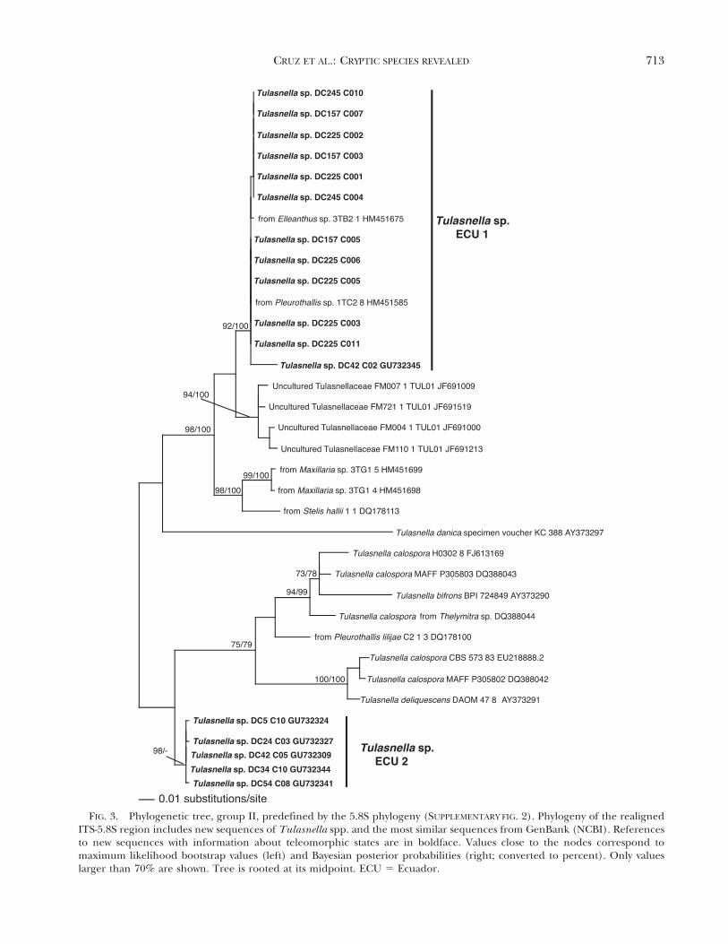
FIG. 3. Phylogenetic tree, group II, predefined by the 5.8S phylogeny (SUPPLEMENTARY FIG. 2). Phylogeny of the realignedITS-5.8S region includes new sequences of Tulasnella spp. and the most similar sequences from GenBank (NCBI). Referencesto new sequences with information about teleomorphic states are in boldface. Values close to the nodes correspond tomaximum likelihood bootstrap values (left) and Bayesian posterior probabilities (right; converted to percent). Only valueslarger than 70% are shown. Tree is rooted at its midpoint. ECU 5 Ecuador.
CRUZ ET AL.: CRYPTIC SPECIES REVEALED 713
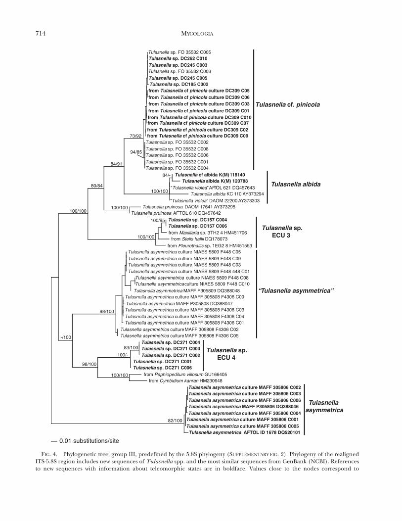
FIG. 4. Phylogenetic tree, group III, predefined by the 5.8S phylogeny (SUPPLEMENTARY FIG. 2). Phylogeny of the realignedITS-5.8S region includes new sequences of Tulasnella spp. and the most similar sequences from GenBank (NCBI). Referencesto new sequences with information about teleomorphic states are in boldface. Values close to the nodes correspond to
714 MYCOLOGIA

Phylogenetic calculations were performed by the pro-grams RAxML 7.0.4 (Stamatakis 2006) and MrBayes 3.1.2(Huelsenbeck and Ronquist 2001). Maximum likelihood(ML) analysis in RAxML used the GTRMIX DNA substitutionmodel with 1000 bootstrap (BS) replicates (Felsenstein1985). For the Bayesian posterior probability (PP) analysis,we used the most complex substitution model available(GTR+I+G) following Whelan et al. (2001), Douady et al.(2003) and Huelsenbeck and Rannala (2004), includingtwo runs each involving four incrementally heated Markovchains over 4 000 000 generations and using randomstarting trees. Trees were sampled every 100 generationsresulting in a total of 40 000 trees from which the last 24 000were used to compute a 50% majority-rule consensus tree.Stationarity of the process and effective sample size (ESS)values were checked visually with the software Tracer 1.5(Drummond and Rambaut 2007).
All trees presented here correspond to the best treesresulting from ML analysis. In total five phylogenetic trees(FIGS. 2–5, SUPPLEMENTARY FIG. 2) were calculated with 127sequences consistently aligned in MAFFT (Katoh et al.2002).
The pairwise differences of the ITS sequences werecalculated with MEGA 5 software (Tamura et al. 2011) toevaluate the intragenomic variation from cultures as well asintraspecific and interspecific variation among specimens(basidiomata) and phylogenetic species. For genetic diver-gence estimation we employed the Kimura-2-parameterdistances (Kimura 1980) with gap deletion. The ‘‘barcodegap’’ (Meyer and Paulay 2005) was estimated between themaximal intraspecific variation and the minimal interspe-cific variation (Meier et al. 2008).
RESULTS
Numerical analysis of spore shapes and sizes.—The 91specimens were provisionally assigned to six species ofTulasnella (T. albida, T. eichleriana, T. pinicola, T.pruinosa, T. violea, T. sp.) based on morphology andthe measurements of the structures (SUPPLEMENTARY
TABLE I). The dendrogram based on shape and size ofbasidiospores revealed six groups, most of themheterogeneous with respect to the provisional, mor-phological species concepts (SUPPLEMENTARY FIG. 1).Classification of spore shapes is also largely inconsis-tent with the numerical classification in the dendro-gram. Only the group of unnamed Tulasnella sp.,consisting of the specimens DC5, DC24, DC34, DC42,DC54, DC157, DC225 and DC245 sampled in Ecua-dor, is consistent in shape and size for all specimens.
The dispersion diagram, based on correlation ofwidth and length of basidiospores, separated T.eichleriana and T. violea specimens, but the othersformed a mixed group (FIG. 1). Basidiospore sizedifferences are obviously insufficient to distinguishclearly between T. albida Bourdot & Galzin, T.pinicola, T. pruinosa and T. sp.
Phylogenetic and pairwise distance analysis.—Thephylogenetic tree based on 127 sequences of theconserved 5.8S region is composed of four groups (I–IV), well supported (73–100% BS and PP) bymaximum likelihood bootstrap values and Bayesianposterior probabilities respectively (SUPPLEMENTARY
FIG. 2). With Bayesian analysis we obtained ESS valuesof 8749.9 for Group I, 1196.7 for Group II, 364.6 forGroup III and 370.9 for Group IV. The standarddeviations of split frequencies, determined byMrBayes 3.1.2 at the end of the Bayesian analyses,were 0.001 for Group I, 0.003 for Group II, 0.009 forGroup III and 0.01for Group IV. Within these groups,phylogenetic trees revealed well supported clades(FIGS. 2–5).
Group I (FIG. 2) called Tulasnella sp. GER includessequences of three clones from specimen DC294sampled in Germany and two sequences of myco-bionts of Prostechea sp. (Orchidaceae) sampled inEcuador. Further orchid mycobionts yielded closelyrelated sequences. Other clones from the specimenDC294 are located in the T. eichleriana clade ofGroup IV (FIG. 5).
Group II (FIG. 3) contains two separated clades ofspecimens sampled in Ecuador, Tulasnella sp. ECU 1and T. sp. ECU 2, without close affinities to knownmorphological or sequenced Tulasnella species.Tulasnella sp. ECU 1 also includes sequences oforchid mycobionts from Ecuador (Elleanthus sp. andPleurothallis sp.). The clade T. sp. ECU 2 includessequences from specimens of a Tulasnella sp.presented by Cruz et al. (2011), and it is closelyrelated to sequences isolated from Pleurothallis lilijaeFoldats and other orchids, as well as sequences ofTulasnella spp. named T. bifrons Bourdot & Galzin, T.calospora (Boud.) Juel and T. deliquescens (Juel) Juel.
Group III (FIG. 4) contains two distant clades forsequences obtained from cultures named T. asymme-trica. The clade of Tulasnella cf. pinicola is mainlyformed by sequences obtained from a pure culture
r
maximum likelihood bootstrap values (left) and Bayesian posterior probabilities (right; converted to percent). Only valueslarger than 70% are shown. Dubious species names are between inverted commas. Tree is rooted at its midpoint. ECU5 Ecuador.
CRUZ ET AL.: CRYPTIC SPECIES REVEALED 715
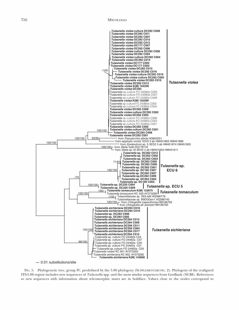
FIG. 5. Phylogenetic tree, group IV, predefined by the 5.8S phylogeny (SUPPLEMENTARY FIG. 2). Phylogeny of the realignedITS-5.8S region includes new sequences of Tulasnella spp. and the most similar sequences from GenBank (NCBI). Referencesto new sequences with information about teleomorphic states are in boldface. Values close to the nodes correspond to
716 MYCOLOGIA
isolated from a basidioma morphologically identifiedas T. cf. pinicola by us (DC309) and culture FO 35532from a basidioma of Tulasnella sp. (non vid.) closelyrelated to the clade T. albida. From basidioma DC309no sequence was obtained directly. Two furtherclades, T. sp. ECU 3 and T. sp. ECU 4, are withoutknown affinities. Clade T. sp. ECU 3 consists of twoclones from specimen DC157 (C004, C006) andsequences obtained from species of Orchidaceaefrom Ecuador (Maxillaria sp., Stelis hallii, Pleurothal-lis sp.). Other clones from specimen DC157 form partof the clade T. sp. ECU 1 in Group II (FIG. 3).Specimen DC271 in clade T. sp. ECU 4 wasdetermined provisionally to be T. eichleriana accord-ing to morphology.
Group IV (FIG. 5) contains sequences consideredto represent T. violea, obtained from five basidiomatafrom Ecuador (DC177), Germany (DC292, DC293),England (K[M] 15529) or Wales (K[M] 164256), aswell as sequences obtained from two pure cultures,one isolated from the basidioma T. violea DC292 andthe culture FO 24380a isolated from a specimen ofTulasnella sp. (non vid.). The clade delegated to T.eichleriana consists of sequences from basidioma
DC294 and culture FO 24462a obtained from abasidioma of Tulasnella sp. (non vid.). Clade T. sp.ECU6 with sequences from basidiomata lacks mor-phological identification, although specimen DC262was preliminarily determined to be T. eichleriana.Clade T. sp. ECU 5 contains only two clones fromspecimen DC225 (C004, C009), but most of theclones from this specimen fall into clade T. sp. ECU 1of Group II (FIG. 3).
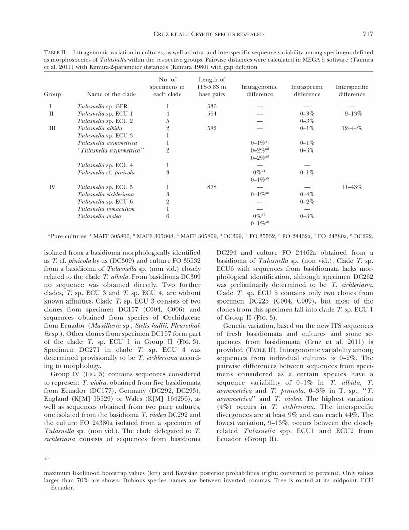
Genetic variation, based on the new ITS sequencesof fresh basidiomata and cultures and some se-quences from basidiomata (Cruz et al. 2011) isprovided (TABLE II). Intragenomic variability amongsequences from individual cultures is 0–2%. Thepairwise differences between sequences from speci-mens considered as a certain species have asequence variability of 0–1% in T. albida, T.asymmetrica and T. pinicola, 0–3% in T. sp., ‘‘T.asymmetrica’’ and T. violea. The highest variation(4%) occurs in T. eichleriana. The interspecificdivergences are at least 9% and can reach 44%. Thelowest variation, 9–13%, occurs between the closelyrelated Tulasnella spp. ECU1 and ECU2 fromEcuador (Group II).
r
maximum likelihood bootstrap values (left) and Bayesian posterior probabilities (right; converted to percent). Only valueslarger than 70% are shown. Dubious species names are between inverted commas. Tree is rooted at its midpoint. ECU5 Ecuador.
TABLE II. Intragenomic variation in cultures, as well as intra- and interspecific sequence variability among specimens definedas morphospecies of Tulasnella within the respective groups. Pairwise distances were calculated in MEGA 5 software (Tamuraet al. 2011) with Kimura-2-parameter distances (Kimura 1980) with gap deletion
Group Name of the clade
No. ofspecimens in
each clade
Length ofITS-5.8S inbase pairs
Intragenomicdifference
Intraspecificdifference
Interspecificdifference
I Tulasnella sp. GER 1 536 — — —II Tulasnella sp. ECU 1 4 564 — 0–3% 9–13%
Tulasnella sp. ECU 2 5 — 0–3%
III Tulasnella albida 2 582 — 0–1% 12–44%
Tulasnella sp. ECU 3 1 — —Tulasnella asymmetrica 1 0–1%a1 0–1%
‘‘Tulasnella asymmetrica’’ 2 0–2%a2 0–3%
0–2%a3
Tulasnella sp. ECU 4 1 — —Tulasnella cf. pinicola 3 0%a4 0–1%
0–1%a5
IV Tulasnella sp. ECU 5 1 878 — — 11–43%
Tulasnella eichleriana 3 0–1%a6 0–4%
Tulasnella sp. ECU 6 2 — 0–2%
Tulasnella tomaculum 1 — —Tulasnella violea 6 0%a7 0–3%
0–1%a8
a Pure cultures: 1 MAFF 305806, 2 MAFF 305808, 3 MAFF 305809, 4 DC309, 5 FO 35532, 6 FO 24462a, 7 FO 24380a, 8 DC292.
CRUZ ET AL.: CRYPTIC SPECIES REVEALED 717
The supported clades in each group containingsequences with up to 4% of proportional differencesare considered phylogenetic species and are namedaccording to the dominant species where possible.Clades lacking named species of Tulasnella werenamed according to the country of origin (ECUEcuador, GER Germany).
DISCUSSION
Morphology.—Genus Tulasnella has been studiedmorphologically and illustrated by Bourdot andGalzin (1909, 1928); Rogers (1933); Olive (1957);Warcup and Talbot (1967, 1971, 1980); Julich andJulich (1976); Roberts (1992, 1993a, 1993b, 1994a,1994b, 1999), van de Put and Antonissen (1996) andOrdynets (2012). Even with different species concepts(Bourdot and Galzin, 1909, 1928; Rogers 1933), sizeand shape of spores were emphasized as the principalcriteria for delimiting species. Our data on the sizeand shape of hyphae, basidia, sterigmata and basid-iospores, based on samples collected in Ecuador andGermany, distinguished T. eichleriana, T. cf. pinicola,T. sp. and T. violea. However, the morphologicalstructures of Tulasnella often overlap in shape andsize among species, making the correct identificationproblematic (Roberts 1994b). This situation is dem-onstrated here for basidiospores of Tulasnella albida,T. pruinosa and T. pinicola. Our analysis suggests thatmorphological characteristics alone are insufficient todelimit species of Tulasnella, a situation ubiquitouswithin the Basidiomycota.
Phylogeny.—The molecular data obtained from cul-tures and basidiomata of Tulasnella spp. let usidentify genetic distances within and between speciesand to compare thresholds at different levels.Sequences of Tulasnella spp. obtained from clonesof pure cultures in this study showed up to 2%
intragenomic variation (TABLE II). The result is closeto the 1.8% of intragenomic variation found for manyisolates of Tulasnella spp. in consensus analysis ofmultiple loci (Linde et al. 2013). A similar and evenhigher variation was observed for species of Laetiporusafter cloning the ITS region from pure cultures(Lindner and Banik 2011). The sequencing artifactproduced during PCR was reported to be less than 1%
(Acinas et al. 2005). We cannot discard Taq misread-ing during PCR amplification, which could inflate theintragenomic variation, but we used high-fidelity Taqto minimize PCR misreading (Simon and Weiss2008); so this is unlikely to be the only source ofvariation.
The 4% of intraspecific variation indicates thatvariation among Tulasnella species can be higher
than 3%, the widely accepted divergence thresholdfor defining phylogenetic species in other groups offungi (Nilsson et al. 2008) or for estimating diversity,for example in Sebacinales (Setaro et al. 2011). Theyare, however, within the range of the less than 3–5%
threshold for defining operational taxonomic units(OTUs) molecularly in Tulasnella as applied anddiscussed by Girlanda et al. (2011), Jacquemyn et al.(2011), Martos et al. (2012) and Linde et al. (2013).Here the so-called barcode gap used to delimitspecies in other organisms (Meyer and Paulay 2005,Meier et al. 2008, Ni et al. 2012, Puillandre et al.2012) was detected between the maximum intraspe-cific variation of 4% (0.8% mean, SD 1.2%) and theminimum 9% interspecific divergence. If we apply therule 103, using the mean of intraspecific variation tofind a threshold suitable to delimit species (Hebert etal. 2004), our threshold to delimit species inTulasnella would be 8%, a value lower than theminimum 9% interspecific variation we observed.More extensive and detailed studies of other speciesof Tulasnella are necessary to determine whether auniversal barcode gap exists in Tulasnella.
The phylogenetic trees revealed cryptic species ofTulasnella. Tulasnella sp. ECU 1 in Group II isindistinguishable morphologically from T. sp. ECU 2(Cruz et al. 2011). Tulasnella sp. ECU 4 and T. sp.ECU 6 in groups III and IV were both determined tobe T. eichleriana (FIG. 3). A further species is revealedfrom clones of a specimen sampled in Germany (T.sp. GER; FIG. 2).
Six of the seven specimens from Ecuador mayrepresent new species or known species lackingmolecular data. To our knowledge, molecular se-quences including several genes are available in theNCBI nucleotide databases for no more than 13described Tulasnella species (GenBank; http://www.ncbi.nlm.nih.gov/). Many sequences obtained frombasidiomata of Tulasnella spp. in clades T. sp. GER(FIG. 2), T. sp. ECU 1 (FIG. 3) and T. sp. ECU 3(FIG. 4) were grouped together with sequencesobtained from orchid mycorrhiza sampled from thesame site in Ecuador by Suarez et al. (2006) and P.Herrera (pers comm). These data suggest highmycorrhizal diversity within orchid-associated Tulas-nella spp.. Considering the dependence of orchids onmycobionts, and Tulasnella in particular, many moreTulasnella species are expected for Ecuador andother regions that are species rich in the Orchida-ceae. Suarez et al. (2008) already estimated a highnumber of Tulasnella species based on nucLSU D1/D2 sequence data.
In the present study we obtained several cladeswhere our sequences clustered with sequences as-signed to certain species of Tulasnella. We critically
718 MYCOLOGIA
examined these clades to decide about correctnaming. The clade T. eichleriana supported with MLBS (99%) and Bayesian PP (100%) (FIG. 5) is thoughtto represent this species. All sequences in this cladewere obtained from pure cultures or specimensisolated or collected in European countries. Thestrain T. eichleriana KC 852 (AY373292) fromEngland (McCormick et al. 2004) was isolated fromthe basidioma identified as T. eichleriana K(M) 30990(non vid.). New sequences were obtained from abasidioma DC294, from Germany, morphologicallyidentified as T. eichleriana by us. The type material ofT. eichleriana (non vid.) was collected in Poland anddescribed by Bresadola (1903). The congruence ofmorphological, phylogenetic and geographic datasupports our conclusion. The clade containingsequences of T. asymmetrica in Group III (FIG. 4),well supported by ML, BS (82%) and Bayesian PP(100%), is considered the correct clade for thisspecies. The grouping of sequences from strains ofculture 085 Warcup and Talbot (5 MAFF 305806)designated type material in the description by Warcupand Talbot (1967) supports this assignment. Theclade ‘‘Tulasnella asymmetrica’’ (Group III) is con-sidered to be named erroneously. This clade differsfrom the correct T. asymmetrica clade (FIG. 4) bymore than 12% sequence divergence with sequencesobtained from the type culture of T. asymmetrica 085Warcup and Talbot (5 MAFF 305806). This cladeincludes sequences obtained from the pure cultures0302 and 0591 Warcup and Talbot (5 MAFF 305808and MAFF 305809 respectively), which lack informa-tion on their teleomorphs. Warcup and Talbot (1967)did not include these cultures in the morphologicaldescription of the species. Specimens in the ‘‘T.asymmetrica’’ and T. asymmetrica clades are related toeach other morphologically by similar growth of theiranamorphs in culture (pers obs), so ‘‘T. asymmetrica’’probably represents a cryptic species.
We propose a clade for Tulasnella violea in GroupIV (FIG. 5), including sequences from five basidio-mata that correspond to the morphological conceptof this species (Bourdot and Galzin 1909; Roberts1994b, 1999). However, a sequence obtained from apure culture isolated from the basidioma named ‘‘T.violea’’ KC 851 AY373293 England by McCormick etal. (2004) is grouped within the clade of T. eichleriana(Group IV). Either the specimen was misidentified asT. violea or the culture from which the sequence wasobtained by McCormick et al. (2004) does notcorrespond to the basidioma morphologically ob-served. The sequence ‘‘T. violea’’ KC 851 AY373293is identical to sequence T. eichleriana AY373292 KC852 (McCormick et al. 2004), as already noted bySharon et al. (2008). The clades named T. albida and
T. cf. pinicola in Group III and T. tomaculum inGroup IV are designated with the same nameaccording to sequences obtained from basidiomataassigned to these species and their affinity tosequences obtained from pure culture isolates frombasidiomata named as these respective species(McCormick et al. 2004).
Several phylogenetic inconsistencies observed forsequences of Tulasnella species are not clarified bythe data and interpretation compiled in this study.Some sequences obtained from one individualbasidioma clustered in clades other than the recog-nized morphospecies. For example, sequences DC185C002 and DC262 C010 obtained from two specimensof Tulasnella sp. ECU6 are grouped within the cladeof T. cf. pinicola Group III (FIG. 4), sequences DC245C003 and C005 obtained from a specimen Tulasnellasp. ECU 1 are grouped within the clade of T. cf.pinicola Group III (FIG. 4) and sequences DC293C003 and C006 obtained from a specimen ofTulasnella violea are grouped within the clade T.eichleriana Group IV (FIG. 5). A potential explanationmay be that several species of Tulasnella are growingintermixed. We observed several times that more thanone morphospecies of Tulasnella can grow on thesame piece of wood or the same substrate. Roberts(1993a, b; 1994a, b; 1999) described several speci-mens of Tulasnella where different species ofTulasnella or other resupinate fungi were growingintermixed on the same substrate. In tropical habitats,where orchids are abundant and harbor a multitudeof Tulasnella mycobionts (Kottke et al. 2013), it is arealistic possibility that more than one species ofTulasnella establishes on the same substrate. Whenthe total DNA is extracted from one extremely thinbasidioma of Tulasnella, DNA of more than onespecies of Tulasnella is frequently found. Furtherstudies incorporating, for example, the analysis ofsecondary structure of the ITS2 region as alreadyanalyzed in Lycoperdaceae (Basidiomycota) by Kru-ger and Cargas (2008) may help confirm the speciesconcept and determine whether the sequences indifferent clades obtained in this study from the samebasidioma are interrelated as conspecific. In addition,mating compatibility tests could be conducted (Lom-bard et al. 2010). However, both methods are difficultto perform with Tulasnella spp., the first because ofthe high variability of the ITS region (Suarez perscomm) and the second because of difficulties inisolation and slow growth of cultures.
The integration of morphological and moleculardata as applied in this study highlights the geneticand morphological variability in Tulasnella spp.,identified cryptic species and improved the interpre-tation of species based on morphology and phyloge-
CRUZ ET AL.: CRYPTIC SPECIES REVEALED 719
netic concepts. The combination of morphologicaland molecular criteria lets us expand our visiontoward a more realistic interpretation of diversity inthis neglected group of fungi.
ACKNOWLEDGMENTS
The authors thank Martin Bidartondo for providing sixsequences of Tulasnella included in the phylogeneticanalysis. We also appreciate the help by Roland Kirschnerwho let us revise his personal collection of Tulasnella andFranz Oberwinkler who provided samples of Tulasnellacollected by him, deposited at the Eberhard-Karls Universityof Tubingen, Germany. We are grateful for the help ofAndreas Beck, Ralph Mangelsdorff for critical comments onthe manuscript and Jascha Weisenborn for help andassistance in the laboratory in Germany. The research wasgenerously supported by Deutsche Forschungsgemeinschaft(DFG RU 816) and German Academic Exchange Service(DAAD). Nature and Culture International (NCI) providedresearch facilities at RBSF.
LITERATURE CITED
Acinas SG, Sarma-Rupavtarm R, Klepac-Ceraj V, Polz MF.2005. PCR-induced sequence artifacts and bias: insightsfrom comparison of two 16S rRNA clone libraries con-structed from the same sample. Appl Environ Microbiol71:966–8969, doi:10.1128/AEM.71.12.8966-8969.2005
Anderson C, Strope CL, Moriyama EN. 2011. Suite MSA:visual tools for multiple sequence alignment compar-ison and molecular sequence simulation. BMC Bioin-formatics 12:184, doi:10.1186/1471-2105-12-184
Beck E, Makeschin F, Haubrich F, Richter M, Bendix J,Valarezo C. 2008. The ecosystem (Reserva BiologicaSan Francisco). In: Beck E, Bendix J, Kottke I,Makeschin F, Mosandl R, eds. Gradients in a tropicalmountain ecosystem of Ecuador. Ecological studiesNo. 198. Berlin, Heidelberg, New York: Springer. p 1–14.
Bickford D, Lohman DL, Sodhi NS, Ng KL, Meier N,Winker K, Ingram KK, Das I. 2007. Cryptic species as awindow on diversity and conservation. Trends Ecol Evol22:148–155, doi:10.1016/j.tree.2006.11.004
Bourdot H, Galzin A. 1909. Hymenomycetes de France I.Heterobasidies. Bull Soc Mycol Fr 25:15–36.
———, ———. 1928. Hymenomycetes de France. Hetero-basidies III. Tulasnellaceae. Bibliotheque Mycologique.J. Cramer, ed. Vol. 23 (reprint 1969).
Bresadola J. 1903. Fungi polonici. a cl. viro B. Eichler lecti.Ann Mycol 1:65–131.
Cameron DD, Leake JR, Read DJ. 2006. Mutualisticmycorrhiza in orchids: evidence from plant-funguscarbon and nitrogen transfers in the green-leavedterrestrial orchid Goodyera repens. New Phytol 171:405–416, doi:10.1111/j.1469-8137.2006.01767.x
Cruz D, Suarez JP, Kottke I, Piepenbring M, Oberwinker F.2011. Defining species in Tulasnella by correlatingmorphology and nrDNA ITS-5.8s sequence data of
basidiomata from a tropical Andean forest. Mycol Prog10:229–238, doi:10.1007/s11557-010-0692-3
Douady CJ, Delsuc F, Boucher Y, Doolittle WF, Douzery EJP.2003. Comparison of Bayesian and maximum likeli-hood bootstrap measures of phylogenetic reliability.Mol Biol Evol 20:248–254, doi:10.1093/molbev/msg042
Drummond AJ, Rambaut A. 2007. BEAST: Bayesian evolu-tionary analysis by sampling trees. BMC Evol Biol 7:214,doi:10.1186/1471-2148-7-214
Edgar RC. 2004. MUSCLE: a multiple sequence alignmentmethod with reduced time and space complexity. BMCBioinf 5:113, doi:10.1186/1471-2105-5-113
Felsenstein J. 1985. Confidence limits on phylogenies: anapproach using the bootstrap. Evolution 39:783–791,doi:10.2307/2408678
Gazis R, Rehner S, Chaverri P. 2011. Species delimitation infungal endophyte diversity studies and its implicationsin ecological and biogeographic inferences. Mol Ecol20:3001–3013, doi:10.1111/j.1365-294X.2011.05110.x
Girlanda M, Segreto R, Cafasso D, Liebel HB, Rodda M,Ercole E, Cozzolino S, Gebauer G, Perotto AS. 2011.Photosynthetic Mediterranean meadow orchids featurepartial mycoheterotrophy and specific mycorrhizalassociations. Am J Bot 98:1148–1163, doi:10.3732/ajb.1000486
Greslebin GA, Rajchenberg M. 2001. The genus Tulasnellawith a new species in the Patagonian Andes forests ofArgentina. Mycol Res 105:1149–1151, doi:10.1016/S0953-7562(08)61980-2
Hebert PDN, Stockle MY, Zemlak TS, Francis CM. 2004.Identification of birds through DNA barcodes. PLoSBiol 2:312, doi:10.1371/journal.pbio.0020312
Hibbett DS, Binder MJF, Bischoff M, Blackwell PF, CannonOE, Eriksson S, Huhndorf et al. 2007. A higher-levelphylogenetic classification of the Fungi. Mycol Res 111:509–547, doi:10.1016/j.mycres.2007.03.004
Homeier J, Werner FA. 2008. Spermatophyta checklist—Reserva Biologica San Francisco (Prov. Zamora-Chinch-ipe, s. Ecuador). In: Liede-Schumann S, Breckle SW,eds. Provisional checklist of flora and fauna of the SanFrancisco Valley and its surroundings. Ecotropicalmonographs No. 4. Ulm, Germany, Society of TropicalEcology. p 15–58.
Huelsenbeck JP, Rannala B. 2004. Frequentist properties ofBayesian posterior probabilities of phylogenetic treesunder simple and complex substitution models. SystBiol 53:904–913, doi:10.1080/10635150490522629
———, Ronquist FR. 2001. MrBayes: Bayesian inference ofphylogenetic trees. Bioinformatics 17:754–755, doi:10.1093/bioinformatics/17.8.754
Jacquemyn H, Honnay O, Cammue BPA, Brys R, Lievens B.2010. Low specificity and nested subset structurecharacterize mycorrhizal associations in five closelyrelated species of the genus Orchis. Mol Ecol 19:4086–4095, doi:10.1111/j.1365-294X.2010.04785.x
———, Merckx V, Brys R, Tyteca D, Cammue BPA, HonnayO, Lievens B. 2011. Analysis of network architecturereveals phylogenetic constraints on mycorrhizal speci-
720 MYCOLOGIA
ficity in the genus Orchis (Orchidaceae). New Phytol192:518–528, doi:10.1111/j.1469-8137.2011.03796.x
Julich W, Julich U. 1976. A contribution toward a revision ofthe genus Tulasnella. Persoonia 2:49–64.
Katoh K, Misawa K, Kuma K, Miyata T. 2002. MAFFT: anovel method for rapid multiple sequence alignmentbased on fast Fourier transformation. Nucleic Acids Res30:3059–3066, doi:10.1093/nar/gkf436
Kimura M. 1980. A simple method for estimating evolu-tionary rate of base substitutions through comparativestudies of nucleotide sequences. J Mol Evol 16:111–120,doi:10.1007/BF01731581
Kottke I, Beiter A, Weiß M, Haug I, Oberwinkler F, Nebel M.2003. Heterobasidiomycetes form symbiotic associa-tions with hepatics: Jungermanniales have sebacinoidmycobionts while Aneura pinguis (Metzgeriales) isassociated with a Tulasnella species. Mycol Res 107:957–968, doi:10.1017/S0953756203008141
———, Setaro S, Haug I, Herrera P, Cruz D, Fries A, GawlikJ, Homeier J, Werner F, Gerique A, Suarez JP. 2013.Mycorrhiza networks promote biodiversity and stabilizethe tropical mountain rain forest ecosystem—perspec-tives for understanding complex communities. In:Ecosystem services, biodiversity and environmentalchange in a tropical mountain ecosystem of southEcuador. Ecological studies. Heidelburg, Germany:Springer Verlag.
Kruger D, Gargas A. 2008. Secondary structure of ITS2rRNA provides taxonomic characters for systematicstudies—a case in Lycoperdaceae (Basidiomycota).Mycol Res 112:316–330.
Largent DL, Johnson D, Watling R. 1973. How to identifymushrooms to genus III: microscopic features. Eureka,California: Mad River Press. 148 p.
Lee MSY. 2001. Unalignable sequences and molecularevolution. Trends Ecol Evol 16:681–685, doi:10.1016/S0169-5347(01)02313-8
Linde CC, Phillips RD, Crisp MD, Peakall R. 2013.Congruent species delineation of Tulasnella usingmultiple loci and methods. New Phytol, doi:10.1111/nph.12492
Lindner DL, Banik MT. 2011. Intragenomic variation in theITS rDNA region obscures phylogenetic relationshipsand inflates estimates of operational taxonomic units ingenus Laetiporus. Mycologia 103:731–740, doi:10.3852/10-331
Lombard L, Crous PW, Wingfield BD, Wingfield MJ. 2010.Multigene phylogeny and mating tests reveal threecryptic species related to Calonectria pauciramosa. StudMycol 66:15–30, doi:10.3114/sim.2010.66.02
Lowy B. 1964. A new genus of the Tulasnellaceae. Mycologia56:696–700, doi:10.2307/3756621
Martin GW. 1939. New or noteworthy fungi from Panamaand Colombia III. Mycologia 31:239–249, doi:10.2307/3754518
Martos F, Munoz F, Pailler T, Kottke I, Gonneau C, SelosseMA. 2012. The role of epiphytism in architecture andevolutionary constraint within mycorrhizal networks oftropical orchids. Mol Ecol 21:5098–5109, doi:10.1111/j.1365-294X.2012.05692.x
McCormick MK, Whigham DF, O’Neill J. 2004. Mycorrhizaldiversity in photosynthetic terrestrial orchids. NewPhytol 163:425–438, doi:10.1111/j.1469-8137.2004.01114.x
Meier R, Zhang G, Ali F. 2008. The use of mean instead ofsmallest interspecific distances exaggerates the size ofthe ‘‘barcoding gap’’ and leads to misidentification.Syst Biol 57:809–813, doi:10.1080/10635150802406343
Meyer CP, Paulay G. 2005. DNA barcoding: error ratesbased on comprehensive sampling. PLoS Biol 3:2229–2238.
Moncalvo JM, Nilsson RH, Koster B, Dunham SM, BernauerT, Matheny PB, Porter TM, et al. 2006. The canthar-elloid clade: dealing with incongruent gene trees andphylogenetic reconstruction methods. Mycologia 98:937–948, doi:10.3852/mycologia.98.6.937
Ni L, Li Q, Kong L, Huang S, Li L. 2012. DNA barcodingand phylogeny in the family Mactridae (Bivalvia:Heterodonta): evidence for cryptic species. BiochemSys Ecol 44:164–172, doi:10.1016/j.bse.2012.05.008
Nilsson RH, Kristiansson E, Ryberg M, Hallenberg N,Larsson KH. 2008. Intraspecific ITS variability in thekingdom Fungi as expressed in the internal sequencedatabases and its implications for molecular speciesidentification. Evol Bioinform 4:193–201.
Nontachaiyapoom S, Sasirat S, Manoch L. 2010. Isolationand identification of Rhizoctonia-like fungi from rootsof three orchid genera, Paphiopedilum, Dendrobium andCymbidium, collected in Chiang Rai and Chiang Maiprovinces of Thailand. Mycorrhiza 20:459–471,doi:10.1007/s00572-010-0297-3
Olive LS. 1957. Tulasnellaceae of Tahiti. A revision of thefamily. Mycologia 49:663–679, doi:10.2307/3755985
Ordynets O. 2012. New records of corticioid fungi withheterobasidia from Ukraine. Turk J Bot, doi:10.3906/bot-1109-1
Preußing M, Nebel M, Oberwinkler F, Weiß M. 2010.Diverging diversity patterns in the Tulasnella (Basid-iomycota, Tulasnellales) mycobionts of Aneura pin-guis (Marchantiophyta, Metzgeriales) from Europeand Ecuador. Mycorrhiza 20:147–59, doi:10.1007/s00572-009-0275-9
Puillandre N, Modica MV, Zhang Y, Sirovich L, BoisselierMC, Cruaud C, Holford M, Samadi S. 2012. Large-scalespecies delimitation method for hyperdiverse groups.Mol Ecol 21:2671–2691, doi:10.1111/j.1365-294X.2012.05559.x
Roberts P. 1992. Spiral-spored Tulasnella species fromDevon and the New Forest. Mycol Res 96:233–236,doi:10.1016/S0953-7562(09)80973-8
———. 1993a. The genus Tulasnella in Norway. Windahlia20:67–73.
———. 1993b. Allantoid-spored Tulasnella speciesfrom Devon. Mycol Res 97:213–220, doi:10.1016/S0953-7562(09)80243-8
———. 1994a. Long-spored Tulasnella species from Devon,with additional notes on allantoid-spored species. MycolRes 98:1235–1244, doi:10.1016/S0953-7562(09)80293-1
———. 1994b. Globose and ellipsoid-spored Tulasnellaspecies from Devon and Surrey, with a key to the genus
CRUZ ET AL.: CRYPTIC SPECIES REVEALED 721
in Europe. Mycol Res 98:1431–1452, doi:10.1016/S0953-7562(09)81075-7
———. 1999. Rhizoctonia-forming Fungi: a taxonomicguide. Kew, UK: Royal Botanic Gardens. 246 p.
———. 2006. Caribbean heterobasidiomycetes 2: Jamaica.Mycotaxon 96:83–107.
Rogers DP. 1933. A taxonomic review of the Tulasnellaceae.Ann Mycology 31:181–203.
Schoch C, Seifert K, Huhndorf S, Robert V, Spouge J, et al.2012. The nuclear ribosomal internal transcribedspacer (ITS) region as a universal DNA barcode markerfor Fungi. Proc Natl Acad Sci USA 109:6241–6246,doi:10.1073/pnas.1117018109
Setaro S, Garnica S, Herrera PI, Suarez JP, Goker M. 2011. Aclustering optimization strategy to estimate speciesrichness of Sebacinales in the tropical Andes basedon molecular sequences from distinct DNA regions.Biodivers Conserv, doi:10.1007/s10531-011-0205
Sharon M, Kuninaga S, Hyacumachi M, Naito S, Sneh B.2008. Classification of Rhizoctonia spp. using rDNA-ITSsequence analysis supports the genetic basis of theclassical anastomosis grouping. Mycoscience 49:93–114,doi:10.1007/S10267-007-0394-0
Shefferson RP, Taylor DL, Weiß M, Garnica S, McCormickMK, Adams S, Gray HM, McFarland JW, Kull T, Tali K,Yukawa T, Kawahara T, Miyoshi K, Lee Y. 2007. Theevolutionary history of mycorrhizal specificity amonglady’s slipper orchids. Evolution 61:1380–1390,doi:10.1111/j.1558-5646.2007.00112.x
Simon UK, Weiss M. 2008. Intragenomic variation of fungalribosomal genes is higher than previously thought. MolBiol Evol 25:2251–2254, doi:10.1093/molbev/msn188
Smith S, Read D. 2008. Mycorrhizal symbiosis. 3rd ed. SanDiego, California: Academic Press. 800 p.
Sokal RR, Sneath PHA. 1963. Principles of numericaltaxonomy. San Francisco: WH Freeman & Co.
Stamatakis A. 2006. RAxML-VI-HPC: maximum Likelihood-based phylogenetic analyses with thousands of taxa andmixed models. Bioinformatics 22: 2688– 2690,doi:10.1093/bioinformatics/btl446
Stielow B, Bratek Z, Orczan AKI, Rudnoy S, Hensel G,Hoffmann P, Klenk HP, Goker. 2011. Species delimi-tation in taxonomically difficult fungi: the case ofHymenogaster. PLoS ONE 6(1):e15614, doi:10.1371/journal.pone.0015614
Suarez JP, Weiss M, Abele A, Garnica S, Oberwinkler F,Kottke I. 2006. Diverse tulasnelloid fungi form mycor-rhizas with epiphytic orchids in an Andean cloud
forest. Mycol Res 110:1257–1270, doi:10.1016/j.mycres.2006.08.004
———, ———, ———, Oberwinkler F, Kottke I. 2008.Members of Sebacinales subgroup B form mycorrhizaewith epiphytic orchids in a Neotropical mountain rain-forest. Mycol Prog 7:75–85, doi:10.1007/s11557-008-0554-4
Tamura K, Peterson D, Peterson N, Stecher G, Nei M,Kumar S. 2011. MEGA 5: Molecular evolutionarygenetics analysis using maximum likelihood, evolution-ary distance and maximum parsimony methods. MolBiol Evol 28:2731–2739, doi:10.1093/molbev/msr121
Taylor DL, McCormick MK. 2008. Internal transcribedspacer primers and sequences for improved character-ization of basidiomycetous orchid mycorrhizas. NewPhytol 177:1020–1033, doi:10.1111/j.1469-8137.2007.02320.x
Thompson JD, Higgins DG, Gibson TJ. 1994. Clustal W:improving the sensitivity of progressive multiple se-quence alignment through sequence weighting, posi-tion-specific gap penalties and weight matrix choice.Nucleic Acids Res 22:4673–4680, doi:10.1093/nar/22.22.4673
Tulloss RE, Lindgren JE. 2005. Amanita aprica a new toxicspecies from western North America. Mycotaxon 91:193–205.
van de Put K, Antonissen I. 1996. Tulasnella’s uitVlaanderen. Sterbeeckia 17:44–69.
Walker C, Vestberg M, Demircik F, Stockinger H, Saito M,Sawaki H, Nishmura I, Schußler A. 2007. Molecularphylogeny and new taxa in the Archaeosporales(Glomeromycota): Ambispora fennica gen. sp nov.,Ambisporaceae fam. nov. and emendation of Archae-ospora and Archaeosporaceae. Mycol Res 111:137–153,doi:10.1016/j.mycres.2006.11.008
Warcup, Talbot PHB. 1967. Perfect states of Rhizoctoniasassociated with orchids I. New Phytol 66:631–641,doi:10.1111/j.1469-8137.1967.tb05434.x
———, ———. 1971. Perfect states of Rhizoctonias associ-ated with orchids II. New Phytol 76:35–40, doi:10.1111/j.1469-8137.1971.tb02506.x
———, ———. 1980. Perfect states of Rhizoctonias associ-ated with orchids III. New Phytol 86:267–272,doi:10.1111/j.1469-8137.1980.tb00787.x
Warcup JH. 1973. Symbiotic germination of some Australianterrestrial orchids. New Phytol 72:387–392, doi:10.1111/j.1469-8137.1973.tb02046.x
Whelan S, Lio P, Goldman N. 2001. Molecular phylogenetics:state-of-the-art methods for looking into the past. Trendsgenet 17:262–272, doi:10.1016/S0168-9525(01)02272-7
722 MYCOLOGIA