'AIRHOUSE': A New Natural Ventilation Concept in the Tropics
Upload
independentCategory
view
5download
0
STUDIES IN MYCOLOGY 50: 457–469. 2004.
457
Cryptic speciation and host specificity among Mycosphaerella spp. occurring on Australian Acacia species grown as exotics in the tropics
Pedro W. Crous1*, Johannes Z. Groenewald1, Krisna Pongpanich2, Winanda Himaman2, Mahdi Ar-zanlou1and Michael J. Wingfield3
1Centraalbureau voor Schimmelcultures, Fungal Biodiversity Centre, Uppsalalaan 8, 3584 CT Utrecht, The Netherlands; 2Forest Entomology and Microbiology Group, National Park, Wildlife and Plant Conservation Department, Chatuchak, Bangkok, 10900, Thailand; 3Department of Microbiology and Plant Pathology, Forestry and Agricultural Biotechnology Institute (FABI), University of Pretoria, Pretoria, 0002, South Africa *Correspondence: Pedro W. Crous, [email protected] Abstract: Species of Mycosphaerella and their anamorphs represent serious pathogens of two phyllodenous species of Acacia, A. mangium and A. crassicarpa. In recent years, these fungi have been collected during surveys in South America and South-East Asia, where these trees are widely planted as exotics. In this study, the Mycosphaerella spp. and their anamorphs were identified based on morphological and cultural characteristics. Identifications were confirmed using comparisons of DNA sequences for the internal transcribed spacers (ITS1 & ITS2), the 5.8S rRNA gene, elongation factor 1-�, histone 3, actin and calmodulin gene regions. The data revealed six new taxa, of which three are named in this study, along with their anamorphs. Cercospora acaciae-mangii, which is morphologically part of the C. apii sensu lato species complex, is distinguished based on its distinct phylogeny. Mycosphaerella acaciigena, collected in Venezuela, is distinguished from M. konae and M. heimii, and described as new. Mycosphaerella thailandica, a new species occurring on Acacia and Musa, is shown to be a sibling species to M. colombiensis, a foliar pathogen of Eucalyptus. Mycosphaerella citri, an important leaf and fruit pathogen of Citrus (Rutaceae), is shown to also occur on Musa (Musaceae) and Acacia (Leguminosae).
Taxonomic novelties: Cercospora acaciae-mangii Crous, Pongpanich & M.J. Wingf. sp. nov., Mycosphaerella acaciigena Crous & M.J. Wingf. sp. nov. (anamorph Pseudocercospora acaciigena Crous & M.J. Wingf. sp. nov.), Mycosphaerella thailandica Crous, Himaman & M.J. Wingf. sp. nov. (anamorph Pseudocercospora thailandica Crous, Himaman & M.J. Wingf. sp. nov.). Key words: Acacia, Ascomycetes, Cercospora, Mycosphaerella, Pseudocercospora, Stenella, systematics.
INTRODUCTION Plantations of exotic tree species in the tropics and Southern Hemisphere sustain important industries producing solid wood products and pulp. In many situations, they provide an alternative to logging of native forest trees and they contribute substantially to the economies of many developing countries. The most extensively planted trees in these plantations are species of Pinus L., Eucalyptus L’Herit. and Acacia L. Australian Acacia species have been planted as exot-ics in the tropics and Southern Hemisphere for many years. Until relatively recently, however, these have been less extensively planted than Pinus or Eucalyptus spp. In areas with temperate climates, Acacia spp. with pinnate leaves such as Acacia mearnsii De Wild. and A. dealbata Link are planted, although on a lim-ited scale. More recently, phyllodenous Acacia spp. such as Acacia mangium Willd., A. crassicarpa A. Cunn. ex Benth. and A. auriculiformis A. Cunn. ex Benth. have been planted extensively in plantations in the tropics (Old et al. 2000).
The success of exotic plantation forestry can, to some extent, be attributed to the separation of trees from their natural enemies (Wingfield et al. 2001). In terms of Acacia spp., virtually nothing is known regarding the diseases that affect these trees, particu-larly where they are planted as exotics. A preliminary synthesis of the diseases of phyllodenous Acacia spp. was made by Old et al. (2000), and from this study it was clear that many pathogens were poorly defined and required rigorous taxonomic study. Leaf and shoot pathogens belonging to the genus Mycosphaerella Johanson, have had a very distinct impact on plantations in the tropics and Southern Hemisphere. The pine pathogen Dothistroma septo-sporum (Dorog.) M. Morelet (teleomorph M. pini E. Rostrup) that has devastated plantings of P. radiata D. Don in many Southern Hemisphere countries is one example (Stone et al. 2003). Likewise, species of Mycosphaerella have had a very marked impact on Eucalyptus species planted in this area. For example, Mycosphaerella leaf blight resulted in the abandon-ment of E. globulus Labill. as a plantation species in South Africa (Purnell & Lundquist 1986), and this and

CROUS ET AL.
458
other species in the genus continue to seriously threaten Eucalyptus plantings (Crous 1998). Species of Mycosphaerella and its anamorphs have been recorded on phyllodenous Acacia spp. grown in the tropics (Old et al. 1996). These fungi have tenta-tively been recognised as members of two anamorph genera of Mycosphaerella, namely Cercospora Fre-sen. and Pseudocercospora Speg. (Old et al. 1996, Cannon et al. 1997). However, no intensive taxonomic studies have been conducted on these fungi, and the names used are tentative. Although the disease is known to occur widely on species of Acacia (Fig. 1), the correct identity of the causal organisms remains unresolved. This again has negative implications for disease management and quarantine programmes, which are aimed at restricting the movement of patho-gens between countries.
This present study results from a collection of Mycosphaerella species and their anamorphs on two phyllodenous species of Acacia, A. mangium and A. crassicarpa, which are widely planted as exotics in the tropics and the Southern Hemisphere. These fungi have been collected in surveys in South America and South-East Asia during the course of the past four years. Their identification will hopefully contribute to a better understanding of their biology and the dis-eases that they cause. Identification of species in-cluded both morphological and cultural characteris-tics. More importantly for this group of fungi, how-ever, identifications were also confirmed using com-parisions of DNA sequences for the internal tran-scribed spacer (ITS1 & ITS2) and the 5.8S regions of the ribosomal RNA operon, as well as the elongation factor 1-�, histone, actin and calmodulin gene regions.
Fig. 1A–D. Typical Mycosphaerella leaf blotch symptoms on Acacia mangium leaves collected in Thailand.
MYCOSPHAERELLA SPP. OCCURRING ON ACACIA
459
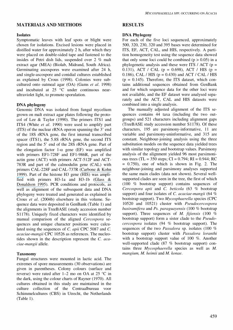
MATERIALS AND METHODS Isolates Symptomatic leaves with leaf spots or blight were chosen for isolations. Excised lesions were placed in distilled water for approximately 2 h, after which they were placed on double-sided tape and fastened to the insides of Petri dish lids, suspended over 2 % malt extract agar (MEA) (Biolab, Midrand, South Africa). Germinating ascospores were examined after 24 h, and single-ascospore and conidial cultures established as explained by Crous (1998). Colonies were sub-cultured onto oatmeal agar (OA) (Gams et al. 1998) and incubated at 25 �C under continuous near-ultraviolet light, to promote sporulation. DNA phylogeny Genomic DNA was isolated from fungal mycelium grown on malt extract agar plates following the proto-col of Lee & Taylor (1990). The primers ITS1 and ITS4 (White et al. 1990) were used to amplify part (ITS) of the nuclear rRNA operon spanning the 3’ end of the 18S rRNA gene, the first internal transcribed spacer (ITS1), the 5.8S rRNA gene, the second ITS region and the 5’ end of the 28S rRNA gene. Part of the elongation factor 1-� gene (EF) was amplified with primers EF1-728F and EF1-986R, part of the actin gene (ACT) with primers ACT-512F and ACT-783R and part of the calmodulin gene (CAL) with primers CAL-228F and CAL-737R (Carbone & Kohn 1999). Part of the histone H3 gene (HIS) was ampli-fied with primers H3-1a and H3-1b (Glass & Donaldson 1995). PCR conditions and protocols, as well as alignment of the subsequent data and DNA phylogeny were treated and generated as explained in Crous et al. (2004b) elsewhere in this volume. Se-quence data were deposited in GenBank (Table 1) and the alignments in TreeBASE (study accession number S1178). Uniquely fixed characters were identified by manual comparison of the aligned Cercospora se-quences and unique character positions were calcu-lated using the sequences of C. apii CPC 5087 and C. acaciae-mangii CPC 10526 as references. The nucleo-tides shown in the description represent the C. aca-ciae-mangii allele. Taxonomy Fungal structures were mounted in lactic acid. The extremes of spore measurements (30 observations) are given in parentheses. Colony colours (surface and reverse) were rated after 1–2 mo on OA at 25 °C in the dark, using the colour charts of Rayner (1970). All cultures obtained in this study are maintained in the culture collection of the Centraalbureau voor Schimmelcultures (CBS) in Utrecht, the Netherlands (Table 1).
RESULTS DNA Phylogeny For each of the five loci sequenced, approximately 500, 320, 230, 320 and 395 bases were determined for ITS, EF, ACT, CAL, and HIS, respectively. A parti-tion homogeneity test using the sequence data showed that only some loci could be combined (p > 0.05) in a phylogenetic analysis and these were ITS / ACT (p = 0.131), ACT / CAL (p = 0.698), ACT / HIS (p = 0.186), CAL / HIS (p = 0.430) and ACT / CAL / HIS (p = 0.145). Therefore, the ITS dataset, which con-tains additional sequences obtained from GenBank and for which sequence data for the other loci were not available, and the EF dataset were analysed sepa-rately and the ACT, CAL and HIS datasets were combined into a single analysis. The manually adjusted alignment of the ITS se-quences contains 44 taxa (including the two out-groups) and 521 characters including alignment gaps (TreeBASE study accession number S1178). Of these characters, 195 are parsimony-informative, 11 are variable and parsimony-uninformative, and 315 are constant. Neighbour-joining analysis using the three substitution models on the sequence data yielded trees with similar topology and bootstrap values. Parsimony analysis of the alignment yielded 96 most parsimoni-ous trees (TL = 350 steps; CI = 0.794; RI = 0.944; RC = 0.750), one of which is shown in Fig. 2. The neighbour-joining and parsimony analyses supported the same main clades (data not shown). Several well-supported clades are seen in the tree, the first of which (100 % bootstrap support) contains sequences of Cercospora apii and C. beticola (63 % bootstrap support) and four isolates of C. acaciae-mangii (64 % bootstrap support). Two Mycosphaerella species (CPC 10520 and 10521) cluster with Pseudocercospora basiramifera and Ps. paraguayensis (100 % bootstrap support). Three sequences of M. fijiensis (100 % bootstrap support) form a sister clade to the Pseudo-cercospora isolates (94 % bootstrap support). The sequences of the two Passalora sp. isolates (100 % bootstrap support) cluster with Passalora loranthi with a bootstrap support value of 100 %. Another well-supported clade (87 % bootstrap support) con-tains three Mycosphaerella species as well as M. mangium, M. heimii and M. konae.

CROUS ET AL.
460
Table 1. Isolates of Mycosphaerella spp. and their anamorphs included for sequence analysis. Species Accession number1 Host Country Collector GenBank numbers
(ITS, EF 1-����, ACT, CAL, HIS) Cladosporium cladosporioides ATCC 66669 / CPC 5100 Creosote-treated southern pine pole U.S.A. – AY251070, AY752164, AY752192, AY752223, AY752254 Cladosporium herbarum ATCC 66670 / CPC 5101 CCA-treated Douglas-fir pole U.S.A. – AY251078, AY752165, AY752193, AY752224, AY752255 Cercospora apii CBS 536.71 / CPC 5087 Apium graveolens Romania – AY752133, AY752166, AY752194, AY752225, AY752256 CPC 5123 A. graveolens New Zealand C.F. Hill AY752134, AY752167, AY752195, AY752226, AY752257 Cercospora beticola CBS 116.47 / CPC 5074 Beta vulgaris Netherlands – AY752135, AY752168, AY752196, AY752227, AY752258 CBS 122.31 / CPC 5072 Beta vulgaris Germany – AY752136, AY752169, AY752197, AY752228, AY752259 CPC 5125 Beta vulgaris New Zealand C.F. Hill AY752137, AY752170, AY752198, AY752229, AY752260 CPC 5128 Beta vulgaris New Zealand C.F. Hill AY752138, AY752171, AY752199, AY752230, AY752261 Cercospora acaciae-mangii CPC 10550 Acacia mangium Thailand – AY752139, AY752172, AY752200, AY752231, AY752262 CPC 10551 A. mangium Thailand – AY752140, AY752173, AY752201, AY752232, AY752263 CPC 10552 A. mangium Thailand – –, AY752174, AY752202, AY752233, AY752264 CPC 10553 A. mangium Thailand – –, AY752175, AY752203, AY752234, AY752265 CPC 10526 A. mangium Thailand M.J. Wingfield AY752141, AY752176, AY752204, AY752235, AY752266 CPC 10527 A. mangium Thailand M.J. Wingfield AY752142, AY752177, AY752205, AY752236, AY752267 Mycosphaerella acaciigena CPC 3837 Acacia sp. Venezuela M.J. Wingfield AY752143 (ITS only) Mycosphaerella citri X126 Citrus sp. Florida – AY752144, AY752178, AY752206, AY752237, AY752268 CPC 10522 A. mangium Thailand M.J. Wingfield AY752145, AY752179, AY752207, AY752238, AY752269 X115 / rCRB2 / CBS 116426 Musa sp. Florida J. Cavaletto AY752146, AY752180, AY752208, AY752239, AY752270 Mycosphaerella colombiensis CBS 110967 / CPC 1104 Eucalyptus urophylla Colombia M.J. Wingfield AY752147, AY752181, AY752209, AY752240, AY752271 CBS 110968 / CPC 1105 E. urophylla Colombia M.J. Wingfield AY752148, AY752182, AY752210, AY752241, AY752272 CBS 110969 / CPC 1106 E. urophylla Colombia M.J. Wingfield AY752149, AY752183, AY752211, AY752242, AY752273 Mycosphearella fijiensis X300 Musa sp. Tonga F. Sumich AY752150 (ITS only) Mycosphaerella konae CPC 2123 Leucadendron sp. Hawaii P.W. Crous AY260086, AY752184, AY752212, AY752243, AY752274 CPC 2125 Leucadendron sp. Hawaii P.W. Crous AY260085, AY752185, AY752213, AY752244, AY752275 Mycosphaerella sp. CPC 10516 A. mangium Thailand M.J. Wingfield AY752151, AY752186, AY752214, AY752245, AY752276 CPC 10518 A. mangium Thailand M.J. Wingfield AY752152, AY752187, AY752215, AY752246, AY752277 CPC 10520 Acacia aulacocarpa Thailand M.J. Wingfield AY752153 (ITS only) CPC 10521 A. aulacocarpa Thailand M.J. Wingfield AY752154 (ITS only) CPC 10524 A. mangium Thailand M.J. Wingfield AY752155, AY752188, AY752216, AY752247, AY752278 Mycosphaerella thailandica CPC 10547 A. mangium Thailand – AY752156, –, AY752217, AY752248, AY752279 CPC 10548 A. mangium Thailand – AY752157, –, AY752218, AY752249, AY752280 CPC 10549 A. mangium Thailand – AY752158, –, AY752219, AY752250, AY752281 CPC 10621 A. mangium Thailand – AY752159, AY752189, AY752220, AY752251, AY752282 X51 Musa sp. Windward Isles E. Reid AY752160, AY752190, AY752221, AY752252, AY752283 X58 Musa sp. Windward Isles E. Reid AY752161, AY752191, AY752222, AY752253, AY752284 Passalora sp. CPC 11147 Acacia crassicarpa Indonesia M.J. Wingfield AY752162 (ITS only) CPC 11150 A. crassicarpa Indonesia M.J. Wingfield AY752163 (ITS only) 1CBS: Centraalbureau voor Schimmelcultures, Utrecht, The Netherlands; C.P.C.: Culture collection of Pedro Crous, housed at CBS; ATCC: American Type Culture Collection, Vir-ginia, U.S.A; 2Ex-type cultures.3ITS: internal transcribed spacer region, EF 1-�: elongation factor 1-alpha, ACT: actin, CAL: calmodulin, HIS: histone 3-a.

MYCOSPHAERELLA SPP. OCCURRING ON ACACIA
461
10 changes
Cladosporium cladosporioides CPC 5100 AY251070Cladosporium herbarum CPC 5101 AY251078
Cercospora beticola CPC 5128
Cercospora apii CPC 5123Cercospora beticola CPC 5125Cercospora apii CPC 5087
Cercospora beticola CPC 5072Cercospora beticola CPC 5074
CPC 10526CPC 10527CPC 10550
CPC 10551CPC 10520
CPC 10521Pseudocercospora basiramifera AF222837Pseudocercospora paraguayensis AF222845
X300AY266150
AY266152Passalora loranthi AY348311CPC 11147
CPC 11150Mycosphaerella acaciigena CPC 3837
Mycosphaerella sp. CPC 10516Mycosphaerella sp. CPC 10518Mycosphaerella sp. CPC 10524
Mycosphaerella heimii AF222841AY260086
AY260085CPC 10547CPC 10548
CPC 10549CPC 10621
AF309612X51
X58
CBS 110967CBS 110968
CBS 110969CPC 10522AF181703
AF181704X126
X115
Mycosphaerella citri
Mycosphaerella colombiensis
Mycosphaerella thailandica
Mycosphaerella konae
Passalora sp.
Mycosphaerella fijiensis
Mycosphaerella sp.
Cercospora acaciae-mangii
100
98
100
87
51
87 98
83
100
94
89
100100
100
94
100
64
62
64
63
100
Fig. 2. One of 96 most parsimonious trees obtained from a heuristic search with 100 random taxon additions of the ITS sequence alignment. The scale bar shows 10 changes; bootstrap support values from 1000 replicates are shown at the nodes. Thickened lines indicate the strict consensus branches. The tree was rooted to two Cladosporium species. The sequences of M. thailandica and M. colombiensis all cluster in the same clade (87 % bootstrap support), with only isolates X51 and X58 forming a distinct group (98 % bootstrap support). Five sequences of M. citri, two of which were obtained from GenBank, also formed a well-supported (100 % bootstrap support) clade.
10 changes
Mycosphaerella citri
Mycosphaerella konae
Mycosphaerella sp.
Cercospora acaciae-mangii86
Cladosporium cladosporioides CPC 5100
Cladosporium herbarum CPC 5101
Cercospora beticola CPC 5128
Cercospora apii CPC 5123
Cercospora beticola CPC 5125
Cercospora apii CPC 5087
Cercospora beticola CPC 5072
Cercospora beticola CPC 5074
CPC 10526
CPC 10527
CPC 10550
CPC 10551
CPC 10552
CPC 10553
X126
X115
CPC 10522
CPC 2123
CPC 2125
CPC 10516
CPC 10518
CPC 10524
Mycosphaerella thailandica CPC 10621
Mycosphaerella colombiensis CBS 110967
Mycosphaerella colombiensis CBS 110968
Mycosphaerella colombiensis CBS 110969
Mycosphaerella thailandica X51
Mycosphaerella thailandica X58
100
100
91
99
100
94
100
100
100
Fig. 3. Single most parsimonious tree obtained from a heuristic search with 100 random taxon additions of the EF 1-� sequence alignment. The scale bar shows 10 changes; bootstrap support values from 1000 replicates are shown at the nodes. The tree was rooted to two Cladospo-rium species. The manually adjusted EF sequence alignment (TreeBASE study accession number S1178) contains 28 taxa (including the two outgroups) and 300 charac-ters including alignment gaps; of these characters 233 are parsimony-informative, 24 are variable and parsi-mony-uninformative, and 43 are constant. Neighbour-joining analysis using the three substitution models on the sequence data yielded trees with similar topology (data not shown). Between the neighbour-joining and parsimony analyses, the trees supported the same main clades (data not shown). Parsimony analysis of the alignment yielded a single most parsimonious tree (TL = 611 steps; CI = 0.876; RI = 0.966; RC = 0.846), which is shown in Fig. 3.

CROUS ET AL.
462
10 changes
Mycosphaerella citri
Mycosphaerella colombiensis
Mycosphaerella thailandica
Mycosphaerella konae
Mycosphaerella sp.
Cercospora acaciae-mangii
100
Cladosporium cladosporioides CPC 5100
Cladosporium herbarum CPC 5101
Cercospora apii CPC 5087
Cercospora apii CPC 5123
Cercospora beticola CPC 5125
Cercospora beticola CPC 5128
Cercospora beticola CPC 5072
Cercospora beticola CPC 5074
CPC 10550
CPC 10552
CPC 10526
CPC 10527
CPC 10551
CPC 10553
X126
X115
CPC 10522
Mycosphaerella sp. CPC 10518
CPC 2123
CPC 2125
CPC 10516
CPC 10524
CPC 10621
X51
X58
CPC 10547
CPC 10548
CPC 10549
CBS 110967
CBS 110968
CBS 110969
97
70
56
100
100
9992
94
100
72
100
92
51
96
87
51
Fig. 4. One of 18 most parsimonious trees obtained from a heuristic search with 100 random taxon additions of the combined ACT, CAL and HIS sequence alignment. The scale bar shows 10 changes and bootstrap support values from 1000 replicates are shown at the nodes. Thickened lines indicate the strict consensus branches. The tree was rooted to two Cladosporium species Several well-supported clades are seen in the tree, one of which (100 % bootstrap support) contains se-quences of C. apii and C. beticola (99 %) and isolates of C. acaciae-mangii (91 %). The M. citri clade (100 %) contains three isolates, two of which are more closely related, grouping with a bootstrap support value of 94 %. The three Mycosphaerella spp. form a well-supported clade (100 %) together with the M. konae isolates. Three sequences of each of M. thai-landica and M. colombiensis all cluster in the same clade (100 %). The EF dataset failed to separate M. konae and the Mycosphaerella sp., as well as M. thailandica and M. colombiensis.
For ACT, CAL and HIS, respectively 209, 312 and 388 bases (including alignment gaps) were included in the manually adjusted alignment consisting of all three loci for 44 taxa (including the two outgroups). The combined data set (TreeBASE study accession num-ber S1178) used for phylogenetic analysis contains a total of 909 characters, of which 306 are parsimony-informative, 54 were variable and parsimony-uninformative, and 549 were constant. The topology of the trees generated with neighbour-joining analysis using the three substitution models were identical (data not shown). Parsimony analysis of the combined data yielded 18 most parsimonious trees, one of which is shown in Fig. 4. Between the neighbour-joining and parsimony analyses, the trees differed only in the placement of the M. citri clade (data not shown). Distance analysis grouped the M. citri clade with the Cercospora clade (bootstrap support value of ap-proximately 70 % irrespective of which substitution model is used), whereas it groups (94 %) with the clades containing the other Mycosphaerella species when a parsimony analysis is performed. As with the ITS and EF trees, a clear separation is found between the clade containing C. apii/C. beticola isolates (87 %) and C. acaciae-mangii (92 %). The M. citri clade (100 %) contains three isolates, two of which once again are more closely related, and is supported by a lower bootstrap support value of 72 %. The clade containing the three Mycosphaerella sp. and two M. konae iso-lates is also well-supported (100 %), with M. konae clustering with a bootstrap support of 92 % and the isolate CPC 10518 sitting outside of the cluster (99 %) formed by the rest of the isolates in this clade. An-other well-supported clade (70 %) in this tree contains the M. thailandica (70 %) and M. colombiensis (97 %) isolates. Taxonomy Cercospora acaciae-mangii Crous, Pongpanich & M.J. Wingf., sp. nov. MycoBank MB500118. Teleomorph: Mycosphaerella sp. Fig. 5. Etymology: Named after its host Acacia mangium. Maculae amphigenae, medio-brunneae, inter marginem et costam, margine atro-brunneo, leviter elevato cinctae. Stromata nulla vel bene evoluta, brunnea, ad 30 µm diam. Conidiophora medio-brunnea, levia, longa, fasciculata (3–20), recta vel apice geniculato-sinuoso. Cellulae conidiogenae integratae, terminales vel intercalares, ad 100 µm longae, sympodiales; cicatrices conidiales incrassatae, fuscatae, refractivae, ad 3 µm latae. Conidia solitaria, hyalina, levia, aciculares, 50–350 � 3.5–5 µm, pluriseptata, basi (in hilo) incassata, fuscata, refractiva. Cercosporae apii similis, sed hospite Acacia et nonnullis nucleotideis differens: elongation factor 1-alpha (EF) in positionibus 42 (T), 47 (C), 144 (C), 198 (G), 217 (A), 224 (A), 235 (A), 245 (C), 257 (G); actinum (ACT) in positionibus 70 (T), 172 (A), 175 (A); calmodulinum (CAL) in positionibus 37

MYCOSPHAERELLA SPP. OCCURRING ON ACACIA
463
(C), 81 (A), 109 (C), 114 (C), 117 (A), 148 (A), 149 (T), 189 (T), 270 (G), 279 (T); histonum H3 (HIS) in positionibus 112 (A), 114 (T), nucleotide deleto inter positiones 122 et 123, 135 (G), 148 (C), 151 (T), 381 (C). Leaf spots amphigenous, covering up to half of the leaf lamina from the margin to the mid rib; infections intermixed with that of M. thailandica; lesions me-dium brown, surrounded by a raised, dark brown border. Stromata lacking to well developed, brown, up to 30 µm diam, giving rise to conidiophores. Conidio-phores medium brown, smooth, long, flexuous, in fascicles that vary in number from 3–20, straight, or with upper part geniculate-sinuous. Conidiogenous cells integrated, terminal or intercalary, up to 100 µm long, proliferating sympodially, loci thickened, dark-ened, refractive, up to 3 µm wide. Conidia solitary, hyaline, smooth, acicular, 50–350 � 3.5–5 µm, multi-septate, with a thickened, darkened, refractive scar. Morphologically indistinguishable from C. apii s. l. (Crous & Braun 2003). Holotype: Thailand, Chachoengsao Province, Sanamchaik-het, on leaves of A. mangium, 28 May 2003, K. Pong-panich, holotype herb. CBS 9874; culture ex-type CBS 116365 = CPC 10526. Host: Acacia mangium. Cultures: Colonies irregular, fast growing, covering the dish after 1 mo; aerial mycelium fluffy to woolly, surface white to pale olivaceous-grey (21””’d), with patches of grey-olivaceous (21””b) sporulation; re-verse iron-grey (25””’k). Distribution: Thailand.
Fig. 5. Asci and ascospores of a Mycosphaerella sp. com-monly found associated with fascicles of Cercospora acaciae-mangii. Scale bar = 10 µm. Notes: When leaf tissues were treated for ascospore discharge, several ascospores of a Mycosphaerella sp.
were obtained that gave rise to a Cercospora ana-morph. Upon germination, however, these ascospores could not with certainty be traced back to the My-cosphaerella state, as they were only harvested after 48 h, and had hence started to distort. The formal naming of the Mycosphaerella teleomorph thus awaits further collections of fresh material. A probable candidate which occurred on the lesions from which the cultures were derived has the following morphol-ogy: Ascomata pseudothecial, amphigenous, erum-pent, black, aggregated in moderately dense clusters, globose, up to 90 µm diam; apical ostiole 5–10 µm diam; wall of 2–3 layers of medium brown textura angularis. Asci fasciculate, bitunicate, subsessile, obovoid to narrowly ellipsoid or subcylindrical, straight or slightly incurved, 8-spored, 30–40 � 7–9 µm. Ascospores tri- to multiseriate, overlapping, hyaline, non-guttulate, thin-walled, curved, fusoid-ellipsoidal with obtuse ends, medianly 1-septate, widest at the median, unconstricted septum, tapering towards both ends, (10–)12–13(–15) � (2–)2.5–3 µm in vivo. The Cercospora anamorph closely matched others within the C. apii s. l. complex (Crous & Braun 2003), but could be separated phylogenetically, and is thus described as C. acaciae-mangii. Additional specimens and cultures examined: Thailand, Chachoengsao Province, Sanamchaikhet, on leaves of A. mangium, 28 May 2003, K. Pongpanich, herb. CBS 9876, CPC 10550, 10526–10528 (single-ascospore isolates), CPC 10551–10553 (single-conidial isolates of C. acaciae-mangii). Mycosphaerella acaciigena Crous & M.J. Wingf., sp. nov. MycoBank MB500119. Figs 6–9. Anamorph: Pseudocercospora acaciigena Crous & M.J. Wingf., sp. nov. Etymology: Named after the host genus, Acacia. Mycosphaerella heimii similis sed ascosporis ad septum modice constrictis differens. Leaf spots amphigenous, elongated along the length of the leaf, not confined to the margins, variable in width, up to 2 cm diam, medium brown, surrounded by a raised, dark brown border. Ascomata pseudothe-cial, amphigenous, erumpent, black, aggregated in clusters of up to 100, forming black spots up to 1 mm diam on the lesions; ascomata globose, up to 80 µm diam; apical ostiole 5–10 µm diam; wall of 2–3 layers of medium brown textura angularis. Asci fasciculate, bitunicate, subsessile, obovoid to narrowly ellipsoid, straight or slightly incurved, 8-spored, 25–40 � 8–11 µm. Ascospores tri- to multiseriate, overlapping, hyaline, non-guttulate, thin-walled, straight, fusoid-

CROUS ET AL.
464
ellipsoidal with obtuse ends, medianly 1-septate, widest in the middle of the apical cell, slightly con-stricted at the septum, tapering towards both ends, but more prominently towards the lower end, (8–)9–10(–11) � (2.5–)3 µm in vivo. Spermogonia intermixed with and similar to the ascomata in general morphol-ogy. Spermatia rod-shaped, hyaline, 3–6 � 1 µm in vivo.
Figs 6–9. Mycosphaerella acaciigena and its Pseudocerco-spora anamorph. 6. Asci. 7. Ascospores. 8. Spermatia. 9. Conidiophores and conidia. Scale bar = 10 µm. Pseudocercospora acaciigena Crous & M.J. Wingf., sp. nov. MycoBank MB500120. Differt a P. thailandica conidiis longioribus, ad 15-septatis; a P. acaciae-confusae, P. hyaloconidiophora et P. acaciae conidiis obclavatis, pallide brunneis, 2–2.5(–3) µm latis. Conidiomata amphigenous, pale brown, up to 80 µm diam; stromata well developed, brown, up to 60 µm wide and 30 µm high. Mycelium predominantly inter-nal, consisting of smooth, branched, septate, pale brown, 3–4 µm wide hyphae. Conidiophores aggre-gated in dense fascicles arising from the upper cells of the stroma; conidiophores pale brown, smooth, un-branched or branched, 0–3-septate, subcylindrical, straight to geniculate-sinuous, 15–30 � 3–5 µm.
Conidiogenous cells terminal, pale brown, smooth, subcylindrical, tapering to flat tipped apical loci, proliferating sympodially, or several times percur-rently, 15–20 � 3–4 µm; conidial scars inconspicuous. Conidia solitary, pale brown, smooth, guttulate, narrowly obclavate, apex subobtuse, base long ob-conically subtruncate, straight to curved, 3–15-septate, (40–)50–75(–80) � 2–2.5(–3) µm in vivo; hila incon-spicuous. Holotype: Venezuela, Acarigua, on leaves of A. mangium, May 2000, M.J. Wingfield, herb. CBS 9873, holotype of M. acaciigena and P. acaciigena; cultures ex-type CBS 115432, 112515, 112516 = CPC 3836–3838. Cultures: Colonies on OA with thin yellow-brown line of pigment diffusing into the agar; margin thin, smooth, slimy, white (1–2 mm wide); surface pale olivaceous-grey (21””’d), with sparse aerial myce-lium. On MEA margin smooth, regular, aerial myce-lium sparse; surface colour variable, predominantly pale olivaceous-grey (23””’d), with patches of smoke-grey (19””d) and olivaceous-grey (21””’i); reverse olivaceous-grey (21””’i). Host: Acacia mangium (Leguminosae). Distribution: Venezuela. Notes: The dense black clusters of raised ascomata on both sides of the leaf lamina is a very characteristic feature of this species. The holotype specimen of M. acaciigena is also colonized by a species of Cerco-spora. The latter appears to be distinct from the C. apii s. l. complex, as conidia tend to have more rounded bases, and be more subcylindrical in shape and shorter than the typical conidia of C. apii, which have more truncate bases, and are longer and acicular in shape. A few conidia of a Stenella sp. were also found to be present, though fructification was sparse. As no cultures of the latter two fungi were obtained, they are not treated further and await additional col-lections. Mycosphaerella citri Whiteside, Phytopathology 62: 263. 1972. Fig. 10. Anamorph: Stenella citri-grisea (F.E. Fisher) Sivan., In: Sivanesan, Bitunicate ascomycetes and their ana-morphs: 226. 1984. � Cercospora citri-grisea F.E. Fisher, Phytopa- thology 51: 300. 1961. Leaf spots amphigenous, covering up to half of the leaf lamina from the margin to the mid rib; infections intermixed with that of M. thailandica and C. acaciae-mangii; lesions medium brown, surrounded by a raised, dark brown border. Mycelium consisting of verruculose, branched, septate, red-brown to medium

MYCOSPHAERELLA SPP. OCCURRING ON ACACIA
465
brown hyphae, 2–3 µm wide. Conidiophores arising singly from superficial mycelium, red-brown to me-dium brown, verruculose, subcylindrical to irregular, 1–3-septate, straight to variously curved, 5–20 � 2.5–4 µm. Conidiogenous cells terminal, verruculose, me-dium brown, unbranched, tapering to rounded apices with flat, thickened, darkened, refractive loci, prolifer-ating sympodially, 5–10 � 2.5–4 µm. Conidia solitary, medium brown to red-brown, verruculose, narrowly obclavate, apex subobtuse, base long obconically subtruncate, straight to curved, (0–)3–5(–10)-septate, (10–)35–65(–120) � (2–)2.5(–3) µm in vivo (descrip-tion based on Acacia isolate CPC 10522 = CBS 116366). Cultures: Colonies with smooth, regular margins, moderately fast growing, covering the dish after 2 mo; aerial mycelium moderate, surface olivaceous-grey (21 ””’i), reverse greenish black (33 ””’k); cultures fertile.
Fig. 10. Conidiophores and conidia of Stenella citri-grisea formed in vitro from isolate CBS 116366. Scale bar = 10 µm. Hosts: Acacia mangium, Musa sp., and species of Aeglopsis Swingle, Citrus, Fortunella Swingle, Murraya L., Poncirus Rafin. (Rutaceae) (Pretorius et al. 2003). Distribution: Thailand (Acacia), on Rutaceae in Bra-zil, Costa Rica, Cuba, Dominican Republic, El-Salvador, Gabon, Haiti, Hong Kong, Japan, Puerto Rico, Surinam, Taiwan, Thailand, USA (FL, HI, TX), Venezuela, Virgin Islands (Pretorius et al. 2003).
Notes: In culture, conidia of CBS 116366 closely resembled the morphology of isolates described from Citrus (Fisher 1961, Sivanesan 1984). Culture examined: Thailand, Chachoengsao Province, Sanamchaikhet, on leaves of A. mangium, 28 May 2003, K. Pongpanich, CBS 116366 = CPC 10522 (single-ascospore isolate). Mycosphaerella thailandica Crous, Himaman & M.J. Wingf., sp. nov. MycoBank MB500121. Figs 11–15. Anamorph: Pseudocercospora thailandica Crous, Himaman & M.J. Wingf., sp. nov. Etymology: Named after its country of origin, Thai-land. Mycosphaerellae colombiensi similis, sed ascosporis ad septum modice constrictis differens; ascosporae modo C germinantes. Leaf spots amphigenous, irregular blotches covering large parts of the leaf lamina; associated symptoms include tip blight, or lesions all along the margin of the leaf, frequently extending to the middle of the leaf lamina; lesions medium brown, surrounded by a raised, dark brown border. Ascomata pseudothecial, amphigenous, subepidermal, becoming erumpent, black, globose, up to 80 µm diam; apical ostiole 5–10 µm diam; wall of 2–3 layers of medium brown textura angularis. Asci fasciculate, bitunicate, subsessile, obovoid to narrowly ellipsoid, straight or slightly incurved, 8-spored, 30–40 � 6–8 µm. Ascospores tri- to multiseriate, overlapping, hyaline, guttulate, thin-walled, straight to slightly curved, fusoid-ellipsoidal with obtuse ends, medianly 1-septate, widest in mid-dle of the apical cell, slightly constricted at the sep-tum, tapering towards both ends, but more promi-nently towards the lower end, (9–)10–11(–12) � (2–)2.5–3 µm in vivo. Spermogonia intermixed with and similar to the ascomata in general morphology. Spermatia rod-shaped, hyaline, 3–5 � 1 µm in vivo. Pseudocercospora thailandica Crous, Himaman & M.J. Wingf., sp. nov. MycoBank MB500122. Differt a P. acaciigena conidiis brevioribus, ad 6-septatis; a P. acaciae-confusae, P. hyaloconidiophora et P. acaciae conidiis obclavatis-subcylindraceis, pallide brunneis, 2-2.5(–3) µm latis. Conidiomata amphigenous, pale brown, up to 60 µm diam; stromata well developed, brown, up to 25 µm wide and 30 µm high. Mycelium predominantly inter-nal, consisting of smooth, branched, septate, medium brown, 3–4 µm wide hyphae. Conidiophores aggre-gated in dense fascicles arising from the upper cells of the stroma; conidiophores pale brown, smooth, un-

CROUS ET AL.
466
branched, 0–2-septate, subcylindrical, straight to variously curved, 10–20 � 5–6 µm. Conidiogenous cells terminal, pale brown, smooth, subcylindrical, tapering to flat tipped apical loci, proliferating sympo-dially, 10–15 � 3–5 µm; conidial scars inconspicuous. Conidia solitary, pale brown, smooth, guttulate, narrowly obclavate to subcylindrical, apex subobtuse, base long obconically subtruncate, straight to curved, 3–6-septate, (25–)30–45(–60) � 2–2.5(–3) µm in vivo; hila inconspicuous.
Figs 11–15. Mycosphaerella thailandica and its Pseudocer-cospora anamorph. 11. Asci. 12. Ascospores. 13. Spermatia. 14. Germinating ascospores on MEA. 15. Conidiophores and conidia. Scale bar = 10 µm. Holotype: Thailand, Chachoengsao Province, Sanamchaik-het, on leaves of A. mangium, 28 May 2003, K. Pong-panich, herb. CBS 9875, holotype of both M. thailandica and P. thailandica, cultures ex-type CBS 116367 = CPC 10547–10549. Ascospore germination on MEA after 24 h: Germinat-ing with germ tubes parallel to the long axis of the ascospore, constricted at the original septum, asco-spores becoming 2.5–3 µm wide, developing several lateral branches. Cultures: Colonies slightly erumpent, having smooth, regular margins, fast growing, covering the dish after 60 d; aerial mycelium fluffy, surface grey-olivaceous (21 ””b), reverse olivaceous-black (25 ””k); cultures sterile. Host: Acacia mangium. Distribution: Thailand. Notes: Morphologically M. acaciigena is similar to M. thailandica, except that the Pseudocercospora conidia of M. acaciigena tend to be longer, and ascomata of M. acaciigena are arranged in dense, superficial clusters, which differ from what was observed on the
type of M. thailandica. However, additional speci-mens studied from Thailand (herb. CBS 9879, Mar. 2003) also tend to have ascomata arranged in clusters, though not as pronounced as observed for M. aca-ciigena. This could indicate that the clustering is a result of the host tissue, or that M. acaciigena also occurs in Thailand. Further collections and cultures would be required, however, to resolve this issue. Mycosphaerella thailandica is morphologically similar to M. colombiensis Crous & M.J. Wingf., which is a pathogen of Eucalyptus (Crous 1998). Although the latter two species can be distinguished based on ascospore morphology and germination patterns. Additional specimens and cultures of unidentified spp. examined: Thailand, Chachoengsao Province, Sanam-chaikhet, on leaves of A. mangium, May 2002, W. Hi-maman, herb. CBS 9878; Chachoengsao Province, Sanam-chaikhet, on leaves of A. mangium, Mar. 2003, K. Pong-panich, herb. CBS 9879; Chachoengsao Province, Sanam-chaikhet, on leaves of A. mangium, 2003, K. Pongpanich, ascospore cultures CPC 10516–10525, 10621–10625. Cultures of unidentified Mycosphaerella spp. examined: Thailand, Chachoengsao Province, Sanamchaikhet, on leaves of A. mangium, 28 May 2003, K. Pongpanich, CPC 10516, 10518, 10524 (single-ascospore isolates of My-cosphaerella sp. in the M. konae clade); CPC 10520, 10521 (single-ascospore isolates of Mycosphaerella sp. in the M. basiramifera clade). DISCUSSION Results of this study have clearly emphasised the paucity of knowledge regarding the taxonomy of leaf pathogens of Acacia spp. that are of considerable economic importance to the forestry industry. In the review of diseases of Acacia spp. grown in plantations in the tropics, Old et al. (2000) noted that two species, tentatively identified as species of Cercospora and Pseudocercospora, occur on A. mangium, A. auriculi-formis and A. crassicarpa. In this study we have described three species of Mycosphaerella, and one that is currently known only from its anamorph. We have also identified at least three other, as yet unde-scribed species from these trees in various tropical countries. Several of these fungi are peripherally similar to each other and this probably explains why they have not previously been recognised. In their revision of the genus Cercospora, Crous & Braun (2003) regarded 281 names to be synonymous with the older C. apii, and treated these as part of the C. apii s. l. species complex. Currently there are no Mycosphaerella teleomorphs known within this com-plex. The collection of a Mycosphaerella sp. that gave rise to a Cercospora anamorph matching the descrip-tion of C. apii s. l. in the present study is thus an exciting development. Isolates were obtained from

MYCOSPHAERELLA SPP. OCCURRING ON ACACIA
467
single conidia, as well as single ascospores. Compari-son of DNA sequence data for several genes (Figs 2–4) showed that these ascospore and conidial isolates cluster closely together within the C. apii clade, but that they represent a distinct lineage. We have de-scribed these as morphologically similar to C. apii, but representing a phylogenetically distinct species, named C. acaciae-mangii. These isolates will add a valuable indication of the variation that can be ex-pected within the C. apii s. l. species complex. They will also promote our understanding of the species limits and genetic entities within this complex. The Pseudocercospora anamorphs of M. aca-ciigena and M. thailandica are morphologically very similar, differing chiefly in conidial size and septation, and are quite distinct from P. acaciae-confusae (Sa-wada) Goh & W.H. Hsieh, which has pale yellowish brown, cylindrical conidia, and causes irregularly angular spots 0.5–2 mm diam (Hsieh & Goh 1990). Pseudocercospora hyaloconidiophora Goh & W.H. Hsieh is distinguished by having hyaline conidio-phores and conidia (Hsieh & Goh 1990). Furthermore, P. acaciae Kamal & R.P. Singh is distinguished by its very long (up to 270 µm), thick-walled, smooth co-nidiophores, and obclavate conidia that are much wider than observed in the present collections (21.5–70 � 7–11 µm) (Kamal & Singh 1980). Mycosphaerella acaciigena, which was collected in Venezuela, is morphologically similar, but phy-logenetically distinct from the M. heimii Crous/M. konae Crous, Joanne E. Taylor & M.E. Palm species complex (Crous 1998, Crous et al. 2004a). Several other isolates obtained from Thailand (CPC 10516, 10518, 10524), could, however, represent one of the latter species, and this will be resolved once fertile collections have been obtained for morphological comparison. Isolates CPC 10520 and CPC 10521 appear to represent another, undescribed species closely related to P. basiramifera Crous/P. paraguay-ensis (Kobayashi) Crous (Fig. 2). The Passalora sp. (CPC 11147, 11150) from A. crassicarpa which clusters with Cercospora loranthi McAlpine (= Pas-salora fide V. Beilharz, in press), is clearly distin-guishable based on morphological and phylogenetic differences. This species is treated elsewhere in this volume (Beilharz et al. 2004). Mycosphaerella thailandica is morphologically very similar to M. colombiensis, which is a leaf patho-gen of Eucalyptus in Colombia (Crous 1998). Mor-phologically, the two species can be distinguished by the constricted ascospores of M. thailandica, while those of M. colombiensis are not constricted. In the ITS dataset (Fig. 2), these species cluster together. However, in both the EF-1�, and combined actin, calmodulin & histone datasets (Figs 3, 4), it is clear that M. thailandica is a cryptic species closely related to, but distinct from M. colombiensis.
Mycosphaerella citri is an important foliar and fruit pathogen of Citrus, causing premature leaf drop, as well as reduced tree vigour, yield and fruit size (Mondal et al. 2003). In a recent phylogenetic study of the genus Cercospora, Goodwin et al. (2001) included one isolate from a Musa sp. (rCRB2 = CBS 116426), which, although identified as M. fijiensis M. Morelet, clustered with an isolate of M. citri. They subse-quently concluded that the isolate was either misiden-tified or contaminated. The same isolate was obtained from Dr S.B. Goodwin for inclusion in the present study. We can now confirm that this isolate represents M. citri, and not M. fijiensis. Furthermore, an ex-ascospore isolate obtained from leaves of Acacia mangium in Thailand in the present study, also repre-sented M. citri. As far as we are aware, this is the first record confirmed based on DNA sequence data, of a serious Mycosphaerella pathogen having alternative hosts. Species of Acacia, Citrus, and Musa are all native to parts of South-East Asia, and this might explain the host-sharing observed here. The fact that these trees are also widely planted as exotics in tropi-cal and sub-tropical parts of the world, and that the important pathogen M. citri could infect three unre-lated hosts, is cause for considerable concern. An examination of the various gene trees generated in the current study support the view of Pretorius et al. (2003) that M. citri is more variable than previously believed. Furthermore, our results show that speci-ation is occurring in M. citri. Although the isolates occurring on Musa and Acacia appear to fall within the morphological variation accepted for M. citri, this appears to be changing. We expect that in the future, this species will evolve into separate, cryptic species or lineages depending on its host. Host sharing was also found in the M. colombien-sis/thailandica complex, where M. thailandica, occurs on Acacia and Musa. However, in this case, lineages are more distinct than those in the M. citri complex, and the fungus on Acacia and Musa could thus be named as M. thailandica. In the Cercospora apii s. l. complex, C. acaciae-mangii represents an additional example of a morphologically similar species, which can be separated based on its host and phylogeny. Ironically, in all three examples where host sharing has been observed, isolates were obtained from asco-spores, again suggesting that the presence of the teleomorph enhances speciation. Other taxa in the C. apii s. l. complex lack teleomorphs, and still cluster together in clades emerging from comparisons of the various gene regions sequenced, despite their different hosts. An intriguing question relating to the fungi de-scribed in this study is where they might have origi-nated. The host trees are native to tropical parts of Australia and Papua New Guinea, and it is logical to assume that the fungi have been introduced into plantation areas from one or more of these native tree

CROUS ET AL.
468
populations. Alternatively, and as illustrated, they could have jumped from completely unrelated hosts. The two undescribed cercosporoid fungi reported by Old et al. (2000) were both found in Northern Austra-lia (Old et al. 1996, Cannon et al. 1997), and match the description of the fungi described here. The re-maining species might have evolved together with the Acacia spp. on which they occur. However, there is growing evidence to show that pathogens of Eucalyp-tus have adapted from native plants to infect these important plantation trees (Wingfield et al. 2001). There are many native species of Acacia and trees of related genera in areas where Australian Acacia spp. are being propagated commercially. It seems likely that both fungi occurring on Acacia spp. in their native environment, and others that have more re-cently adapted to infect these trees as exotics will be encountered. The latter group of new pathogens could seriously threaten the trees in their native environ-ment, if they were to be transferred back to these areas. This situation would be similar to that found with Eucalyptus rust caused by Puccinia psidii G. Winter, which is native in Latin America on various Myrta-ceae, and has adapted to infect Eucalyptus in that area (Coutinho et al. 1998). This rust fungus is presently considered to be one of the most serious threats to Eucalyptus in areas such as Australia where there are no rust pathogens of these trees. Mycosphaerella spp. and their anamorphs include some of the most important leaf and shoot pathogens of forest plantation trees, fruit trees and shrubs (Old et al. 2000, Park et al. 2000, Stone et al. 2003, Crous et al. 2004a, b). In the case of Eucalyptus, plantations in the tropics and the Southern Hemisphere have been seriously damaged by these fungi (Crous 1998). We might thus expect the same situation for Acacia spp. in the future. It is thus imperative that these fungi are correctly characterised and named. Management strategies to reduce the impact of the diseases associ-ated with these fungi will rest strongly on a clear understanding of the relative importance of the vari-ous species. Likewise, quarantine measures aimed at excluding these fungi from new areas will depend on our ability to identify them. REFERENCES Beilharz VC, Pascoe IG, Wingfield MJ, Tjahjono B, Crous
PW (2004). Passalora perplexa, an important pleoana-morphic leaf blight pathogen of Acacia crassicarpa in Australia and Indonesia. Studies in Mycology 50: 471–479.
Cannon P, Pascoe I, Beilharz V, Yuan ZQ (1997). Report on fungi from diseased acacia samples examined at the Institute of Horticultural development, Knoxfield. In: Diseases of tropical acacias (Old KM, Lee SS, Sharma JK, eds). Proceedings of an international workshop,
Subanjeriji (South Sumatra), 28 April – 3 May 1996. CIFOR Special Publication: 108–113.
Carbone I, Kohn LM (1999). A method for designing primer sets for speciation studies in filamentous asco-mycetes. Mycologia 91: 553–556.
Coutinho TA, Wingfield MJ, Alfenas AC, Crous PW (1998). Eucalyptus rust: a disease with the potential for serious international implications. Plant Disease 82: 819–825.
Crous PW (1998). Mycosphaerella spp. and their ana-morphs associated with leaf spot diseases of Eucalyptus. Mycologia Memoir 21: 1–170.
Crous PW, Braun U (2003). Mycosphaerella and its ana-morphs. 1. Names published in Cercospora and Passa-lora. CBS Biodiversity Series 1: 1–571.
Crous PW, Denman S, Taylor JE, Swart L, Palm ME (2004a). Cultivation and diseases of Proteaceae: Leuca-dendron, Leucospermum and Protea. CBS Biodiversity Series 2: 1–228.
Crous PW, Groenewald JZ, Mansilla JP, Hunter GC, Wingfield MJ (2004b). Phylogenetic reassessment of Mycosphaerella spp. and their anamorphs occurring on Eucalyptus. Studies in Mycology 50: 195–214.
Fisher F (1961). Greasy spot and tar spot of Citrus in Florida. Phytopathology 51: 297–303.
Gams W, Hoekstra ES, Aptroot A (eds) (1998). CBS course of mycology. Centraalbureau voor Schimmelcultures, Baarn, The Netherlands.
Glass NL, Donaldson G (1995). Development of primer sets designed for use with PCR to amplify conserved genes from filamentous ascomycetes. Applied and Envi-ronmental Microbiology 61: 1323–1330.
Goodwin SB, Dunkle LD, Zismann VL (2001). Phyloge-netic analysis of Cercospora and Mycosphaerella based on the internal transcribed spacer region of ribosomal DNA. Phytopathology 91: 648–658.
Hsieh WH, Goh TK (1990). Cercospora and similar fungi from Taiwan. Maw Chang Book Company, Taipei, Taiwan.
Kamal, Singh RP (1980). Fungi of Gorakhpur. XIX. Pseu-docercospora. Sydowia 33: 157–161.
Lee SB, Taylor JW (1990). Isolation of DNA from fungal mycelia and single spores. In PCR protocols: a guide to methods and applications. (MA Innis, DH Gelfand, JJ Sninsky, TJ White, eds): 282–287. Academic Press, San Diego.
Mondal SN, Gottwald TR, Timmer LW (2003). Environ-mental factors affecting the release and dispersal of as-cospores of Mycosphaerella citri. Phytopathology 93: 1031–1036.
Old KM, Harwood CE, Robson KJ, Haines MW, Solomon DJ (1996). Foliar pathogens of tropical acacias in Aus-tralia. In: Impact of diseases and insect pests in tropical forests (Nair KSS, Sharma JK, Varma RV, eds). Pro-ceedings of IUFRO Symposium, 23–26 Nov. 1993. Peechi, India: 11–19.
Old KM, Lee SS, Sharma JK, Yuan ZQ (2000). A manual of diseases of tropical acacias in Australia, South-East Asia and India. Centre for International Forestry Re-search, Jakarta, Indonesia.
Park RF, Keane PJ, Wingfield MJ, Crous PW (2000). Fungal diseases of eucalypt foliage. In: Diseases and pathogens of eucalypts (Keane PJ, Kile GA, Podger FD,

MYCOSPHAERELLA SPP. OCCURRING ON ACACIA
469
Brown BN, eds). CSIRO publishing, Australia: 153–239.
Pretorius MC, Crous PW, Groenewald JZ, Braun U (2003). Phylogeny of some cercosporoid fungi from Citrus. Sy-dowia 55: 286–305.
Purnell RC, Lundquist JE (1986). Provenance variation of Eucalyptus nitens on the eastern Transvaal highveld in South Africa. South African Forestry Journal 138: 23–31.
Rayner RW (1970). A mycological colour chart. CMI and British Mycological Society. Kew, Surrey, England.
Sivanesan A (1984). The bitunicate ascomycetes. J. Cramer, Vaduz, Lichtenstein.
Stone C, Chrisholm LA, McDonald S (2003). Spectral reflectance characteristics of Pinus radiata needles af-fected by Dothistroma needle blight. Canadian Journal of Botany 81: 560–569.
White TJ, Bruns T, Lee S, Taylor J (1990). Amplification and direct sequencing of fungal ribosomal RNA genes for phylogenetics. In PCR protocols: a guide to methods and applications. (MA Innis, DH Gelfand, JJ Sninsky, TJ White, eds): 282–287. Academic Press, San Diego.
Wingfield MJ, Slippers B, Roux J, Wingfield BD (2001). Worldwide movement of exotic forest fungi, especially in the tropics and the Southern Hemisphere. BioScience 51: 134–140.

CROUS ET AL.
470
Copyright © 2022 FDOKUMEN