Critical Role of cRel Subunit of NF- B in Sepsis Survival
7
INFECTION AND IMMUNITY, May 2011, p. 1848–1854 Vol. 79, No. 5 0019-9567/11/$12.00 doi:10.1128/IAI.00021-11 Copyright © 2011, American Society for Microbiology. All Rights Reserved. Critical Role of cRel Subunit of NF-B in Sepsis Survival † Emilie Courtine, 1,2 Fre ´de ´ric Pe `ne, 1,2,3 Nicolas Cagnard, 6 Julie Toubiana, 1,2 Catherine Fitting, 4 Jessy Brocheton, 2,5 Christophe Rousseau, 1,2 Steve Gerondakis, 7,8,9 Jean-Daniel Chiche, 1,2,3 Fatah Ouaaz, 1,2 # and Jean-Paul Mira 1,2,3 #* De ´partement de Biologie Cellulaire des Interactions Ho ˆte-Pathoge `ne, Institut Cochin, Paris, France 1 ; Universite ´ Paris Descartes, CNRS (UMR 8104), and INSERM U1016, Paris, France 2 ; Ho ˆpital Cochin, Re ´animation Me ´dicale, AP-HP, Paris, France 3 ; Unite ´ Cytokines et Inflammation Institut Pasteur, Paris, France 4 ; Equipe Se ´quence-Transcriptome, Institut Cochin, Paris, France 5 ; Plateforme Bioinformatique Paris Descartes, Paris, France 6 ; The Burnet Institute, Melbourne, Victoria 3004, Australia 7 ; Department of Clinical Hematology, Monash University, Alfred Medical Research and Education Precinct, Prahran, Victoria 3004, Australia 8 ; and Department of Immunology, Monash University, Alfred Medical Research and Education Precinct, Prahran, Victoria 3004, Australia 9 Received 6 January 2011/Returned for modification 1 February 2011/Accepted 14 February 2011 NF-B is a critical regulator of gene expression during severe infections. NF-B comprises homo- and heterodimers of proteins from the Rel family. Among them, p50 and p65 have been clearly implicated in the pathophysiology of sepsis. In contrast, the role of cRel in sepsis is still controversial and has been poorly studied in single-pathogen infections. We aimed to investigate the consequences of cRel deficiency in a cecal ligation and puncture (CLP) model of sepsis. We have approached the underlying mechanisms of host defense by analyzing bacterial clearance, systemic inflammation, and the distribution of spleen dendritic cell subsets. Moreover, by using a genome-wide technology, we have also analyzed the CLP- induced modifications in gene expression profiles both in wild-type (wt) and in rel / mice. The absence of cRel enhances mortality due to polymicrobial sepsis. Despite normal pathogen clearance, cRel defi- ciency leads to an altered systemic inflammatory response associated with a sustained loss of the spleen lymphoid dendritic cells. Furthermore, a whole-blood microarray study reveals that the differential outcome between wt and rel / mice during sepsis is preceded by remarkable changes in the expression of hundreds of genes involved in aspects of host-pathogen interaction, such as host survival and lipid metabolism. In conclusion, cRel is a key NF-B member required for host antimicrobial defenses and a regulatory transcription subunit that controls the inflammatory and immune responses in severe infection. Severe sepsis is a complex syndrome characterized by an amplified host inflammatory response leading to organ dys- function. Host-pathogen interactions mediated through Toll- like receptors (TLRs) stimulate the production of proinflam- matory cytokines, chemokines, and adhesion and immune activation molecules (23, 35). This proinflammatory response is followed by a compensatory immunosuppressive response that combines various quantitative and functional defects of immune cells (17). Pathogen-induced cellular modifications are accompanied by marked changes in host gene expression (29), with NF-B transcription factors playing a pivotal role in this modulation. During severe infections, NF-B is crucial for the regulation of immune/inflammatory responses and the control of cell pro- liferation/apoptosis (2, 3, 27). NF-B transcription factors con- sist of homo- and heterodimers of the Rel protein family; p65 (RelA), RelB, cRel, p50, p52, p65, RelB, and cRel are tran- scriptionally active members of the NF-B family, whereas p50 and p52 serve primarily as nontransactivating DNA binding subunits (13). The major importance of the p50 and p65 sub- units in the pathophysiology of sepsis has been highlighted in both animal models and human pathology (6). In contrast, the role and the significance of cRel during infectious challenges remain poorly defined. Mice deficient for the cRel subunit (rel / mice) exhibit compromised immune function and a variable response to individual pathogen infections (12, 25). Despite the fact that the immune system in the main develops normally in rel / mice, these animals display defects in lymphocyte proliferation and humoral immunity (5, 7, 25). In animal models of sepsis, rel / mice show normal resistance to Trichuris muris (1) and lymphocytic choriomeningitis virus (9). In contrast, they are susceptible to Leishmania major (14), Toxoplasma gondii (28), influenza virus (15), and Listeria monocytogenes (9). Despite these reports, the contribution of cRel to innate host responses directed toward common bacterial infections has not been studied, and the potential function of this NF-B member in the defense against acute bacterial infection has yet to be investigated in vivo. In this report, we have investigated the consequences that * Corresponding author. Present address: Cochin University Hospi- tal, Medical Intensive Care Unit, 27 rue du Faubourg St. Jacques, 75014 Paris, France. Phone: 33 1 58 41 25 01. Fax: 33 1 58 41 25 05. E-mail: [email protected]. † Supplemental material for this article may be found at http://iai .asm.org/. # J.-P.M. and F.O. contributed equally to the design of this study. Published ahead of print on 22 February 2011. 1848
-
Upload
independent -
Category
Documents
-
view
3 -
download
0
Transcript of Critical Role of cRel Subunit of NF- B in Sepsis Survival

INFECTION AND IMMUNITY, May 2011, p. 1848–1854 Vol. 79, No. 50019-9567/11/$12.00 doi:10.1128/IAI.00021-11Copyright © 2011, American Society for Microbiology. All Rights Reserved.
Critical Role of cRel Subunit of NF-�B in Sepsis Survival�†Emilie Courtine,1,2 Frederic Pene,1,2,3 Nicolas Cagnard,6 Julie Toubiana,1,2 Catherine Fitting,4
Jessy Brocheton,2,5 Christophe Rousseau,1,2 Steve Gerondakis,7,8,9 Jean-Daniel Chiche,1,2,3
Fatah Ouaaz,1,2# and Jean-Paul Mira1,2,3#*Departement de Biologie Cellulaire des Interactions Hote-Pathogene, Institut Cochin, Paris, France1; Universite Paris Descartes,
CNRS (UMR 8104), and INSERM U1016, Paris, France2; Hopital Cochin, Reanimation Medicale, AP-HP, Paris, France3;Unite Cytokines et Inflammation Institut Pasteur, Paris, France4; Equipe Sequence-Transcriptome, Institut Cochin,
Paris, France5; Plateforme Bioinformatique Paris Descartes, Paris, France6; The Burnet Institute, Melbourne,Victoria 3004, Australia7; Department of Clinical Hematology, Monash University,
Alfred Medical Research and Education Precinct, Prahran, Victoria 3004,Australia8; and Department of Immunology, Monash University,
Alfred Medical Research and Education Precinct,Prahran, Victoria 3004, Australia9
Received 6 January 2011/Returned for modification 1 February 2011/Accepted 14 February 2011
NF-�B is a critical regulator of gene expression during severe infections. NF-�B comprises homo- andheterodimers of proteins from the Rel family. Among them, p50 and p65 have been clearly implicated inthe pathophysiology of sepsis. In contrast, the role of cRel in sepsis is still controversial and has beenpoorly studied in single-pathogen infections. We aimed to investigate the consequences of cRel deficiencyin a cecal ligation and puncture (CLP) model of sepsis. We have approached the underlying mechanismsof host defense by analyzing bacterial clearance, systemic inflammation, and the distribution of spleendendritic cell subsets. Moreover, by using a genome-wide technology, we have also analyzed the CLP-induced modifications in gene expression profiles both in wild-type (wt) and in rel�/� mice. The absenceof cRel enhances mortality due to polymicrobial sepsis. Despite normal pathogen clearance, cRel defi-ciency leads to an altered systemic inflammatory response associated with a sustained loss of the spleenlymphoid dendritic cells. Furthermore, a whole-blood microarray study reveals that the differentialoutcome between wt and rel�/� mice during sepsis is preceded by remarkable changes in the expressionof hundreds of genes involved in aspects of host-pathogen interaction, such as host survival and lipidmetabolism. In conclusion, cRel is a key NF-�B member required for host antimicrobial defenses and aregulatory transcription subunit that controls the inflammatory and immune responses in severeinfection.
Severe sepsis is a complex syndrome characterized by anamplified host inflammatory response leading to organ dys-function. Host-pathogen interactions mediated through Toll-like receptors (TLRs) stimulate the production of proinflam-matory cytokines, chemokines, and adhesion and immuneactivation molecules (23, 35). This proinflammatory responseis followed by a compensatory immunosuppressive responsethat combines various quantitative and functional defects ofimmune cells (17). Pathogen-induced cellular modificationsare accompanied by marked changes in host gene expression(29), with NF-�B transcription factors playing a pivotal role inthis modulation.
During severe infections, NF-�B is crucial for the regulationof immune/inflammatory responses and the control of cell pro-liferation/apoptosis (2, 3, 27). NF-�B transcription factors con-sist of homo- and heterodimers of the Rel protein family; p65
(RelA), RelB, cRel, p50, p52, p65, RelB, and cRel are tran-scriptionally active members of the NF-�B family, whereas p50and p52 serve primarily as nontransactivating DNA bindingsubunits (13). The major importance of the p50 and p65 sub-units in the pathophysiology of sepsis has been highlighted inboth animal models and human pathology (6). In contrast, therole and the significance of cRel during infectious challengesremain poorly defined.
Mice deficient for the cRel subunit (rel�/� mice) exhibitcompromised immune function and a variable response toindividual pathogen infections (12, 25). Despite the fact thatthe immune system in the main develops normally in rel�/�
mice, these animals display defects in lymphocyte proliferationand humoral immunity (5, 7, 25). In animal models of sepsis,rel�/� mice show normal resistance to Trichuris muris (1) andlymphocytic choriomeningitis virus (9). In contrast, they aresusceptible to Leishmania major (14), Toxoplasma gondii (28),influenza virus (15), and Listeria monocytogenes (9). Despitethese reports, the contribution of cRel to innate host responsesdirected toward common bacterial infections has not beenstudied, and the potential function of this NF-�B member inthe defense against acute bacterial infection has yet to beinvestigated in vivo.
In this report, we have investigated the consequences that
* Corresponding author. Present address: Cochin University Hospi-tal, Medical Intensive Care Unit, 27 rue du Faubourg St. Jacques,75014 Paris, France. Phone: 33 1 58 41 25 01. Fax: 33 1 58 41 25 05.E-mail: [email protected].
† Supplemental material for this article may be found at http://iai.asm.org/.
# J.-P.M. and F.O. contributed equally to the design of this study.� Published ahead of print on 22 February 2011.
1848
cRel subunit deficiency has for survival and the host systemicinflammatory response in mice subjected to a model of poly-microbial peritonitis.
MATERIALS AND METHODS
Mice. Wild-type (wt) C57BL6/J female mice, 8 to 12 weeks old, were pur-chased from Charles River Laboratories. rel�/� mice were kindly provided by S.Gerondakis (The Burnet Institute, Melbourne, Australia). Experiments wereconducted in accordance with guidelines of the Cochin Institute, in compliancewith the European animal welfare regulation.
Abs and reagents. The following antibodies (Abs) were purchased: anti-p65,anti-c-Rel, or anti-p50 polyclonal Abs (Santa Cruz Biotechnology); anti-CD11cmagnetic beads and CD11c, CD8�, CD11b, and anti-CD16/CD32 Fc blockfluorescent antibodies (Miltenyi Biotec); and horseradish peroxidase (HRP)-conjugated goat anti-rabbit IgG and goat anti-mouse-IgG (Pierce). Tienam (imi-penem-cilastatin; used at 25 mg/kg in 0.5 ml saline) (Merck Sharp & Dohme),lipopolysaccharide (LPS) (Escherichia coli K-12 ultrapure LPS; Invivogen). SDS,glycerol orthovanadate, sodium pyrophosphate, and �-glycerophosphate werepurchased from Sigma. Nonidet P-40 and Complete proteinase inhibitor cocktailtablets 25X were from Roche. Sepharose beads were streptavidin high perfor-mance from GE Healthcare. ECL Plus Western blotting detection reagents werepurchased from Amersham and X-ray film from Pierce (CL-XPosure film;Pierce). The NF-�B consensus probes used for electrophoretic mobility shiftassay (EMSA) and DNA pulldown assay were provided by Promega.
Model of polymicrobial sepsis. We used a sublethal model of cecal ligatureand puncture (CLP). Briefly, mice were anesthetized by an intraperitoneal in-jection of ketamine and xylazine. After a midline incision (�1 cm), the cecumwas exposed, ligatured at its external third, and punctured through and throughwith a 21-gauge needle. The incision was sutured in layers, and animals wereresuscitated with an intraperitoneal injection of 1 ml saline. Controls weresham-operated mice undergoing abdominal surgery, but with only exposition ofcecum without CLP. Six hours following surgery and then every 12 h for 3 days,mice received an intraperitoneal injection of antibiotics (imipenem-cilastatin[Tienam; Merck Sharp & Dohme]; 25 mg/kg in 0.5 ml saline).
Assessment of bacterial dissemination. At different time points after surgery,mice were humanely killed, and blood samples of 0.5 to 1.0 ml were obtained bycardiac puncture. Bacterial dissemination was indirectly assessed through spleen,liver, and blood bacterial cultures to quantify the number of CFU. Spleen andliver were removed and mechanically homogenized under sterile conditions.Blood, spleen, and liver homogenates were subjected to serial 10-fold dilutions.Bacteria were quantified in tryptic soy agar (for spleen homogenates) or inTodd-Hewitt (TH) agar (for blood) after 24 h.
Determination of cytokines levels in mouse sera. Plasma interleukin-1� (IL-1�), IL-6, IL-12, gamma interferon (IFN-�), tumor necrosis factor alpha (TNF-�), and IL-10 concentrations were quantified using quantitative multiplex assays(Bio-Plex cytokines; Bio-Rad) according to the manufacturer’s instructions. Du-plicates and means were obtained from 3 to 13 mice.
Preparation of DCs. Bone marrow-derived dendritic cells (BMDCs) weremade from BM cells flushed from femurs and tibias (21). Cells were seeded in24-well plates at the concentration of 1 � 106/ml in RPMI medium supple-mented with 5% fetal calf serum (FCS) and antibiotics in the presence ofsupernatant (4% vol/vol) from J558 cells transduced with murine granulocyte-macrophage colony-stimulating factor (GM-CSF). Cells were cultured for 6 days,with medium replacement at days 3 and 6. Spleen dendritic cells were preparedfrom collagenase-digested spleens. Cell suspensions were incubated with anti-CD11c magnetic beads to collect purified CD11c� DCs (magnetic-activated cellseparation CD11c isolation kit; Miltenyi Biotech). The purity of the positivefraction was generally 80% as confirmed by CD11c staining. Purified CD11c�
DCs were then stained with fluorescence-coupled CD11c and CD8� antibodiesto analyze the distribution of subsets by flow cytometry.
Cell stimulation and nuclear extract preparation. Briefly, BMDCs were leftuntreated or stimulated with LPS (1 g/ml) for 2 h. The cytosolic fraction wasextracted in buffer A (10 mM HEPES-NaOH [pH 7.6], 3 mM MgCl2, 10 mMKCl, 5% glycerol, 0,1% Nonidet P-40) containing 1 mM vanadate, 10 mM NaF,1 mM sodium pyrophosphate, 25 mM �-glycerophosphate, and Complete pro-teinase inhibitor cocktail tablets 25X. Cells were incubated for 10 min at �4°Cand centrifuged at 10,000 rpm for 2 min. Nuclear proteins were obtained bytreatment of the pellet with buffer B (1 mM vanadate, 10 mM NaF, 1 mM sodiumpyrophosphate, 25 mM �-glycerophosphate, and 300 mM KCl) and centrifugedafter 30 min at �4°C and 15,000 rpm for 20 min. Finally, the nuclear fraction wasdiluted in buffer A.
EMSA and DNA pulldown assay. EMSA and DNA pulldown assay were usedto analyze the NF-�B DNA binding activity as previously described (11, 24). Theprobes used were nfkb wt (5�-AGTTGAGGGGACTTTCCCAGGC-3� and 5�-GCCTGGGAAAGTCCCCTCAACT-3�) and nfkb mut (5�-AGTTGAGGCGACTTTCCCAGGC-3� and 5�-GCCTGGGAAAGTCGCCTCAACT-3�). For su-pershift assays in EMSA, 1 g of anti-RelA, anti-cRel, or anti-p50 polyclonal Abwas added to the reaction mixture. For DNA pulldown experiments, nuclearextracts were incubated with 1 g of double-strand consensus or control 5 �-biotinylated probe for 1 h at �4°C. Twenty-five microliters of Sepharose beadswas used to capture the biotinylated probe (15 min at �4°C). Proteins associatedwith the probe were eluted at 95°C for 5 min in Laemmli Buffer (Bio-Rad).
Western Blot analysis. Samples were loaded onto 10% denatured polyacryl-amide gels with 1� Tris-glycine (TG)–1% SDS. The migrated proteins weretransferred to nitrocellulose membrane (38). The membrane was incubated inblocking buffer (1� Tris-buffered saline [TBS] and 0.1% Tween 20 with 5%nonfat dry milk) for 1 h at room temperature. Incubation with primary Abovernight at 4°C was followed by incubation with HRP-conjugated secondary Abfor 1 h at room temperature. Proteins were detected by addition of ECL Plus andexposure to X-ray film.
Microarray analysis. Whole-blood samples were collected at day 1 after sepsisinduction from sham-operated and CLP-treated rel�/� and wt mice. RNA ex-traction was performed with a TRIzol Plus RNA purification kit (Invitrogen)according to the manufacturer’s protocol and analyzed using an Agilent 2100Bioanalyzer (Agilent Technologies). RNA was used to make biotin-labeledcRNA using the SuperScript double-stranded cDNA synthesis kit (Invitrogen)and BioArray high-yield RNA transcript labeling kit (Enzo). Biotin-labeledcRNA was fragmented before being hybridized to the GeneChip mouse gene 1.0ST array (Affymetrix), which contains 28,944 murine genes and expressed se-quence tags (ESTs). The arrays were analyzed with a confocal scanner and withthe MicroArray Suite 5.0 Gene Expression analysis program (Affymetrix). Flu-orescence data were imported into two analysis software programs, AffymetrixExpression Console and R Bioconductor (http://www.bioconductor.org). Geneexpression levels were calculated using the RMA algorithm in Expression Con-sole, and flags were computed using a custom algorithm within. To limit poten-tially biased measurement (background or saturating), all probes for whichnormalized intensity measures were outside a confidence interval were flagged 0.The confidence interval was a 2-fold standard deviation from the mean intensityof each microarray. Three probe lists were used for each comparison accordingto flagged measurement in the relevant chips. The “PP” list is made of probesflagged only as “Present” for all microarrays involved in the comparison. The“P50%” list was created by filtering probes flagged as “Present” for at least halfof the chips. The “All” list is made of all probes without any filter. Data weresubsequently subjected to Ingenuity pathway analysis (IPA) (Ingenuity Systems,Inc., Redwood City, CA) to model relationships among genes and proteins andto construct putative pathways and relevant biological processes.
Statistical analysis. Survival curves were analyzed using the Kaplan-Meiermethod and compared using the log rank test. Continuous variables were ex-pressed as median and interquartile range and displayed using box plots orscatter plots. They were represented as mean � standard deviation (SD) andcompared using the Student t test. Categorical variables were compared using theFisher exact test. P values lower than 0.05 indicated statistically significant dif-ferences. For microarray analysis, independent samples were compared by com-puting fold ratios and filtered at 2-fold. Cluster analysis was performed byhierarchical clustering, using the Spearman correlation similarity measure andaverage linkage algorithm.
Microarray data accession number. The microarray data have been depositedinto ArrayExpress under accession number E-MEXP-2944.
RESULTS
LPS activates the p50, p65, and cRel subunits of NF-�B. Tostudy the involvement of cRel in antimicrobial immune cellresponses, the subunit composition of �B binding complexeswas first analyzed in LPS-stimulated BMDCs. As shown in Fig.1A, NF-�B DNA binding activity measured by EMSA wasstrongly induced in BMDCs upon 2 h of stimulation with LPS.The abundance of the �B DNA binding complexes dramati-cally decreased with the addition of antisera against cRel, p65,and p50, demonstrating the presence of these three subunits,including cRel, within the LPS-induced NF-�B complexes (Fig.
VOL. 79, 2011 cRel SUBUNIT OF NF-�B IN POLYMICROBIAL SEPSIS 1849
1A). Further evidence for the presence of cRel and p50 inthese complexes was the presence of specific cRel and p50bands in the supershift experiment. These results were vali-dated by DNA pulldown assay, illustrated in Fig. 1B. Together,these results indicated that besides the activation of the pro-totypic p65 and p50 proteins, LPS-activated NF-�B complexesinclude cRel.
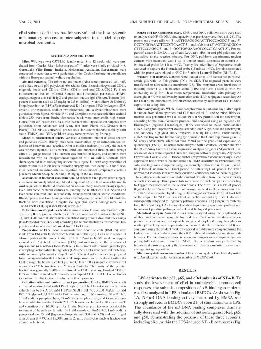
Sublethal polymicrobial sepsis induces increased mortalityin rel�/� mice. In order to investigate the importance of cRelin the host response to acute infection, survival of wild-type(wt) and rel�/� mice was evaluated using a model of sublethalpolymicrobial sepsis. As shown in Fig. 2, the incidence ofmortality was dramatically and significantly increased in rel�/�
mice (12/20; 60%) compared to wt animals (4/18; 22%) (P �0.05), suggesting for the first time the critical role of the cRelsubunit in survival of polymicrobial sepsis.
Absence of cRel and bacterial clearance. As bacterial clear-ance is essential for survival of infection, the consequences of
a lack of rel on systemic bacterial dissemination were investi-gated. As illustrated in Fig. 3, wt and rel-deficient mice exhib-ited similar bacterial counts in liver, spleen, and blood, indi-cating that the increased mortality seen in rel�/� mice was notrelated to impaired pathogen clearance.
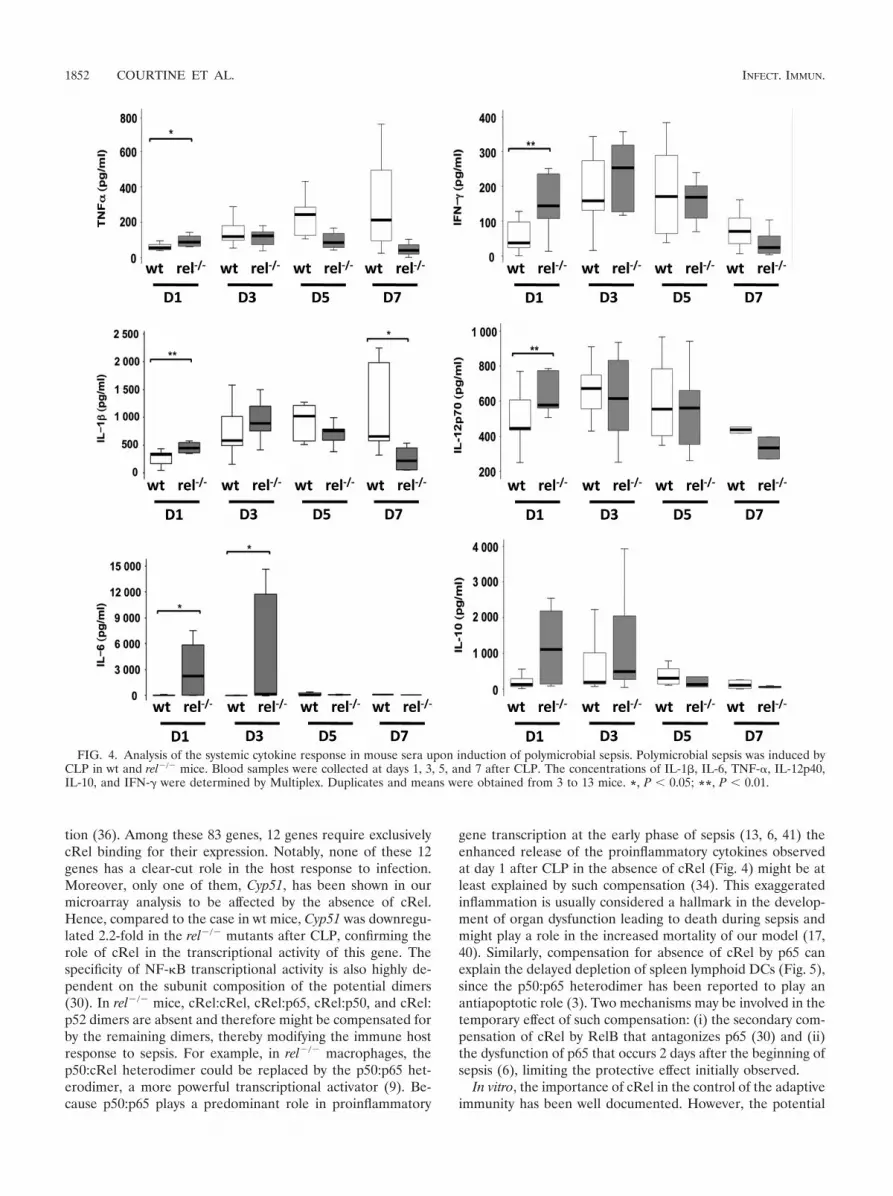
cRel deficiency leads to an altered inflammatory response.An imbalance of pro- and anti-inflammatory cytokine re-sponses has been linked to the risk of death in both experi-mental and clinical sepsis models. Hence, we measured plasmalevels of several cytokines at days 1, 3, 5, and 7 after CLP. Inwt mice, induction of sepsis led to a moderate release of pro-inflammatory cytokines at day 1, followed by a substantialincrease at days 3 and 5 for TNF-�, IL-I�, and IFN-� (Fig. 4).Both IL-6 and IL-10 levels remained unchanged during thesepsis course. Compared to wt mice, in the early phase, rel-deficient mice had a significantly enhanced release of proin-flammatory cytokines (TNF-�, IL-1�, IL-6, IL-12p70, andIFN-�). In contrast, at day 7, rel�/� mice exhibited a significantlower plasma level of IL-1�.
Absence of cRel leads to sustained depletion of spleen den-dritic cells upon polymicrobial sepsis. DCs link innate immu-nity and adaptive immunity and are critical in mounting andcoordinating host immune responses against infection. More-over, the importance of DCs in both the early and late phasesof sepsis has been established (4, 31), with these cells havingbeen reported to be required for survival of polymicrobialsepsis (37). In wt mice, a selective depletion of the spleenlymphoid DC subset CD11c� CD8�� was found at day 1,followed by a full replenishment at day 7 after CLP. Althoughdelayed, sustained spleen DC depletion was observed in rel-deficient mice from day 3 until day 14 after surgery (Fig. 5 anddata not shown). Recovery time in rel�/� mice was not estab-lished.
Genome-wide analysis of the host systemic response to poly-microbial sepsis. To further explore the increased mortality ratein the rel�/� animals, blood from both wt and rel-deficient micewas subjected to global gene expression analysis at day 1. First,microarray analysis did not reveal major differences in the globalgene expression in sham-operated wt and rel�/� mice (data not
FIG. 1. NF-�B/Rel subunit composition in dendritic cells uponLPS stimulation. BMDCs were left untreated (phosphate-buffered sa-line [PBS]) or stimulated for 2 h with LPS. Nuclear extracts wereprepared and analyzed by EMSA (A) and DNA pulldown assay (B) forbinding to the consensus �B site. Nuclear extracts were also preincu-bated with anti-p65, anti-p50, and anti-cRel antibodies prior to incu-bation with the �B site oligonucleotide probe. The arrows correspondto the supershifted bands with anti-cRel and anti-p50 antibodies (A).
FIG. 2. Survival rates of rel�/� and wild-type animals with polymicrobial sepsis. Sublethal polymicrobial sepsis was induced by CLP in wild-typeand rel�/� mice. Survival of wt (n 18) or rel�/� (n 20) mice was monitored for 12 days (log rank test, P � 0.05).
1850 COURTINE ET AL. INFECT. IMMUN.

shown). CLP significantly altered the expression of 3,414 probesets in wt mice and 3,169 in rel�/� mice (Fig. 6). Among those, theexpression of 1,897 probe sets was modified in both groups ofmice and corresponded to the core response (1,051 versus 1,026upregulated and 846 versus 871 downregulated in the rel�/� andwt mice, respectively). Heat map analysis indicated that thesecommon probe sets consisted of genes involved mainly with theinflammatory response for the upregulated core response andcellular development for the downregulated common probe sets(see Tables S1 and S2 in the supplemental material). Expressionof 1,517 probe sets was specifically modified in wt mice (Fig. 6).Functional grouping of these probe sets showed that organismsurvival and the cell cycle were the main functions for the up- anddownregulated genes, respectively (see Tables S3 and S4 in thesupplemental material). Finally, 1,272 probe sets were specificallymodulated in the absence of cRel. Their corresponding functionsconcerned lipid metabolism for the upregulated genes and aminoacid metabolism for the downregulated ones (see Tables S5 andS6 in the supplemental material).
DISCUSSION
Using a sublethal polymicrobial model of sepsis, we reportfor the first time that cRel deficiency leads to an increased rateof mortality, an altered systemic host response with an earlyhyperinflammatory profile, and a sustained depletion of spleenlymphoid DCs. Furthermore, we show that the absence of cRelleads to a dramatic modification of CLP-induced gene expres-sion.
The phenotypic characteristics of rel�/� mice observed inthis model of polymicrobial sepsis could either result directlyfrom the downregulation of cRel-dependent gene expressionor be a consequence of altered NF-�B-dependent transcriptionarising from the replacement of cRel by other members of theRel family in the activated NF-�B dimers. Few studies haveanalyzed the direct impact of cRel on gene expression. Bycombining genome-scale location and specific NF-�B subunitantibodies in U937 cells, Schreiber and coworkers have shownthat cRel binds to 83 �B target genes following LPS stimula-
FIG. 3. Assessment of bacterial load in mice upon induction of polymicrobial sepsis. The bacterial counts in wt (black circles) and rel�/� (whitecircles) mice were determined at days 1, 3, 5, and 7 after CLP. Bacterial dissemination was assessed through quantitative liver, spleen, and bloodbacterial cultures. Bacterial counts were compared by Student’s two-tailed t test.
VOL. 79, 2011 cRel SUBUNIT OF NF-�B IN POLYMICROBIAL SEPSIS 1851
tion (36). Among these 83 genes, 12 genes require exclusivelycRel binding for their expression. Notably, none of these 12genes has a clear-cut role in the host response to infection.Moreover, only one of them, Cyp51, has been shown in ourmicroarray analysis to be affected by the absence of cRel.Hence, compared to the case in wt mice, Cyp51 was downregu-lated 2.2-fold in the rel�/� mutants after CLP, confirming therole of cRel in the transcriptional activity of this gene. Thespecificity of NF-�B transcriptional activity is also highly de-pendent on the subunit composition of the potential dimers(30). In rel�/� mice, cRel:cRel, cRel:p65, cRel:p50, and cRel:p52 dimers are absent and therefore might be compensated forby the remaining dimers, thereby modifying the immune hostresponse to sepsis. For example, in rel�/� macrophages, thep50:cRel heterodimer could be replaced by the p50:p65 het-erodimer, a more powerful transcriptional activator (9). Be-cause p50:p65 plays a predominant role in proinflammatory
gene transcription at the early phase of sepsis (13, 6, 41) theenhanced release of the proinflammatory cytokines observedat day 1 after CLP in the absence of cRel (Fig. 4) might be atleast explained by such compensation (34). This exaggeratedinflammation is usually considered a hallmark in the develop-ment of organ dysfunction leading to death during sepsis andmight play a role in the increased mortality of our model (17,40). Similarly, compensation for absence of cRel by p65 canexplain the delayed depletion of spleen lymphoid DCs (Fig. 5),since the p50:p65 heterodimer has been reported to play anantiapoptotic role (3). Two mechanisms may be involved in thetemporary effect of such compensation: (i) the secondary com-pensation of cRel by RelB that antagonizes p65 (30) and (ii)the dysfunction of p65 that occurs 2 days after the beginning ofsepsis (6), limiting the protective effect initially observed.
In vitro, the importance of cRel in the control of the adaptiveimmunity has been well documented. However, the potential
FIG. 4. Analysis of the systemic cytokine response in mouse sera upon induction of polymicrobial sepsis. Polymicrobial sepsis was induced byCLP in wt and rel�/� mice. Blood samples were collected at days 1, 3, 5, and 7 after CLP. The concentrations of IL-1�, IL-6, TNF-�, IL-12p40,IL-10, and IFN-� were determined by Multiplex. Duplicates and means were obtained from 3 to 13 mice. *, P � 0.05; **, P � 0.01.
1852 COURTINE ET AL. INFECT. IMMUN.
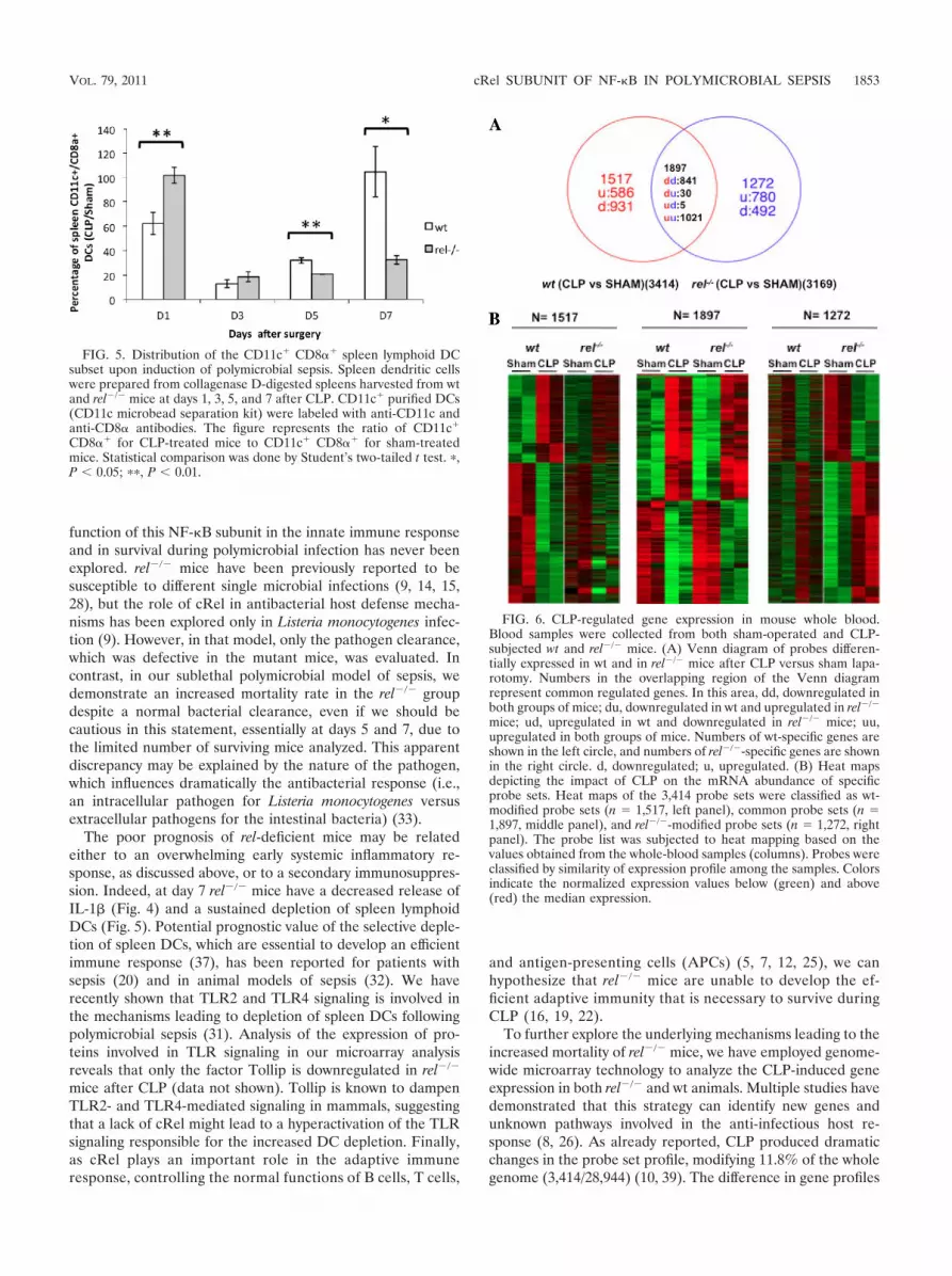
function of this NF-�B subunit in the innate immune responseand in survival during polymicrobial infection has never beenexplored. rel�/� mice have been previously reported to besusceptible to different single microbial infections (9, 14, 15,28), but the role of cRel in antibacterial host defense mecha-nisms has been explored only in Listeria monocytogenes infec-tion (9). However, in that model, only the pathogen clearance,which was defective in the mutant mice, was evaluated. Incontrast, in our sublethal polymicrobial model of sepsis, wedemonstrate an increased mortality rate in the rel�/� groupdespite a normal bacterial clearance, even if we should becautious in this statement, essentially at days 5 and 7, due tothe limited number of surviving mice analyzed. This apparentdiscrepancy may be explained by the nature of the pathogen,which influences dramatically the antibacterial response (i.e.,an intracellular pathogen for Listeria monocytogenes versusextracellular pathogens for the intestinal bacteria) (33).
The poor prognosis of rel-deficient mice may be relatedeither to an overwhelming early systemic inflammatory re-sponse, as discussed above, or to a secondary immunosuppres-sion. Indeed, at day 7 rel�/� mice have a decreased release ofIL-1� (Fig. 4) and a sustained depletion of spleen lymphoidDCs (Fig. 5). Potential prognostic value of the selective deple-tion of spleen DCs, which are essential to develop an efficientimmune response (37), has been reported for patients withsepsis (20) and in animal models of sepsis (32). We haverecently shown that TLR2 and TLR4 signaling is involved inthe mechanisms leading to depletion of spleen DCs followingpolymicrobial sepsis (31). Analysis of the expression of pro-teins involved in TLR signaling in our microarray analysisreveals that only the factor Tollip is downregulated in rel�/�
mice after CLP (data not shown). Tollip is known to dampenTLR2- and TLR4-mediated signaling in mammals, suggestingthat a lack of cRel might lead to a hyperactivation of the TLRsignaling responsible for the increased DC depletion. Finally,as cRel plays an important role in the adaptive immuneresponse, controlling the normal functions of B cells, T cells,
and antigen-presenting cells (APCs) (5, 7, 12, 25), we canhypothesize that rel�/� mice are unable to develop the ef-ficient adaptive immunity that is necessary to survive duringCLP (16, 19, 22).
To further explore the underlying mechanisms leading to theincreased mortality of rel�/� mice, we have employed genome-wide microarray technology to analyze the CLP-induced geneexpression in both rel�/� and wt animals. Multiple studies havedemonstrated that this strategy can identify new genes andunknown pathways involved in the anti-infectious host re-sponse (8, 26). As already reported, CLP produced dramaticchanges in the probe set profile, modifying 11.8% of the wholegenome (3,414/28,944) (10, 39). The difference in gene profiles
FIG. 5. Distribution of the CD11c� CD8�� spleen lymphoid DCsubset upon induction of polymicrobial sepsis. Spleen dendritic cellswere prepared from collagenase D-digested spleens harvested from wtand rel�/� mice at days 1, 3, 5, and 7 after CLP. CD11c� purified DCs(CD11c microbead separation kit) were labeled with anti-CD11c andanti-CD8� antibodies. The figure represents the ratio of CD11c�
CD8�� for CLP-treated mice to CD11c� CD8�� for sham-treatedmice. Statistical comparison was done by Student’s two-tailed t test. �,P � 0.05; ��, P � 0.01.
FIG. 6. CLP-regulated gene expression in mouse whole blood.Blood samples were collected from both sham-operated and CLP-subjected wt and rel�/� mice. (A) Venn diagram of probes differen-tially expressed in wt and in rel�/� mice after CLP versus sham lapa-rotomy. Numbers in the overlapping region of the Venn diagramrepresent common regulated genes. In this area, dd, downregulated inboth groups of mice; du, downregulated in wt and upregulated in rel�/�
mice; ud, upregulated in wt and downregulated in rel�/� mice; uu,upregulated in both groups of mice. Numbers of wt-specific genes areshown in the left circle, and numbers of rel�/�-specific genes are shownin the right circle. d, downregulated; u, upregulated. (B) Heat mapsdepicting the impact of CLP on the mRNA abundance of specificprobe sets. Heat maps of the 3,414 probe sets were classified as wt-modified probe sets (n 1,517, left panel), common probe sets (n 1,897, middle panel), and rel�/�-modified probe sets (n 1,272, rightpanel). The probe list was subjected to heat mapping based on thevalues obtained from the whole-blood samples (columns). Probes wereclassified by similarity of expression profile among the samples. Colorsindicate the normalized expression values below (green) and above(red) the median expression.
VOL. 79, 2011 cRel SUBUNIT OF NF-�B IN POLYMICROBIAL SEPSIS 1853

between wt and rel�/� mice is probably a key to understandingthe mechanisms responsible for the differential outcomes ofsepsis. Ingenuity analysis of gene expression profiles high-lighted a subgroup of 63 genes involved in “organism survival,”which are upregulated in wt but not in rel�/� animals (Fig. 6;see Table S3 in the supplemental material). Among thosegenes, Bcl-2l 11, a member of the Bcl2 family, is upregulatedafter CLP only in wt mice. This result is consistent with thedeleterious role of apoptosis in sepsis outcome (18). In addi-tion, gene ontology revealed potential regulation by cRel ofnew genes involved in previously identified pathways such asthe cell cycle and amino acid metabolism. The discovery ofthese new genes and gene families has been made possiblethrough our microarray study and may constitute the basis foradditional experiments useful in providing new insights intothe biology of cRel and its role in gene transcription. However,as the host response to sepsis is highly compartmentalized withtissue specificity of gene expression (9), our data should beconsidered only for the systemic response to CLP.
In conclusion, the present study highlights for the first timethe critical role of cRel in the antimicrobial host defense. Theidentification of cRel target genes associated with inflamma-tion and survival of sepsis might lead to potential NF-�B-targeted immunomodulation in severe infection. However, theclinical relevance in humans has to be validated.
ACKNOWLEDGMENTS
The Cochin Association for Research on Inflammation Sepsis andMolecular Advances (CARISMA) supported this work. The fundershad no role in study design, data collection and analysis, decision topublish, or preparation of the manuscript.
The authors had no financial conflicts of interest.We thank members of the transcriptomic (Franck Letourneur), flow
cytometry (Brigitte Chanaud and Laurence Stouvenel), and animal(Veronique Fauveau) facilities at the Cochin Institute for their helpfulassistance.
REFERENCES
1. Artis, D., et al. 2002. Differential requirement for NF-kappa B family mem-bers in control of helminth infection and intestinal inflammation. J. Immu-nol. 169:4481–4487.
2. Baldwin, A. S. J. 1996. The NF-kappa B and I kappa B proteins: newdiscoveries and insights. Annu. Rev. Immunol. 14:649–683.
3. Beg, A. A., W. C. Sha, R. T. Bronson, S. Ghosh, and D. Baltimore. 1995.Embryonic lethality and liver degeneration in mice lacking the RelA com-ponent of NF-kappa B. Nature 376:167–170.
4. Benjamim, C. F., S. K. Lundy, N. W. Lukacs, C. M. Hogaboam, and S. L.Kunkel. 2005. Reversal of long-term sepsis-induced immunosuppression bydendritic cells. Blood 105:3588–3595.
5. Boffa, D. J., et al. 2003. Selective loss of c-Rel compromises dendritic cellactivation of T lymphocytes. Cell. Immunol. 222:105–115.
6. Bohrer, H., et al. 1997. Role of NFkappaB in the mortality of sepsis. J. Clin.Invest. 100:972–985.
7. Bunting, K., et al. 2007. Genome-wide analysis of gene expression in T cellsto identify targets of the NF-kappa B transcription factor c-Rel. J. Immunol.178:7097–7109.
8. Calvano, S. E., et al. 2005. A network-based analysis of systemic inflamma-tion in humans. Nature 437:1032–1037.
9. Carrasco, D., et al. 1998. Multiple hemopoietic defects and lymphoid hyper-plasia in mice lacking the transcriptional activation domain of the c-Relprotein. J. Exp. Med. 187:973–984.
10. Cobb, J. P., et al. 2002. Sepsis gene expression profiling: murine spleniccompared with hepatic responses determined by using complementary DNAmicroarrays. Crit. Care Med. 30:2711–2721.
11. Fried, M., and D. M. Crothers. 1981. Equilibria and kinetics of lac repressor-operator interactions by polyacrylamide gel electrophoresis. Nucleic AcidsRes. 9:6505–6525.
12. Gerondakis, S., et al. 2006. Unravelling the complexities of the NF-kappaBsignalling pathway using mouse knockout and transgenic models. Oncogene25:6781–6799.
13. Ghosh, S., M. J. May, and E. B. Kopp. 1998. NF-kappa B and Rel proteins:evolutionarily conserved mediators of immune responses. Annu. Rev. Im-munol. 16:225–260.
14. Grigoriadis, G., et al. 1996. The Rel subunit of NF-kappaB-like transcriptionfactors is a positive and negative regulator of macrophage gene expression:distinct roles for Rel in different macrophage populations. EMBO J. 15:7099–7107.
15. Harling-McNabb, L., et al. 1999. Mice lacking the transcription factor sub-unit Rel can clear an influenza infection and have functional anti-viralcytotoxic T cells but do not develop an optimal antibody response. Int.Immunol. 11:1431–1439.
16. Hotchkiss, R. S., et al. 2000. Caspase inhibitors improve survival in sepsis: acritical role of the lymphocyte. Nat. Immunol. 1:496–501.
17. Hotchkiss, R. S., and I. E. Karl. 2003. The pathophysiology and treatment ofsepsis. N. Engl. J. Med. 348:138–150.
18. Hotchkiss, R. S., and S. Opal. 2010. Immunotherapy for sepsis—a newapproach against an ancient foe. N. Engl. J. Med. 363:87–89.
19. Hotchkiss, R. S., et al. 1999. Prevention of lymphocyte cell death in sepsisimproves survival in mice. Proc. Natl. Acad. Sci. U. S. A. 96:14541–14546.
20. Hotchkiss, R. S., et al. 2002. Depletion of dendritic cells, but not macro-phages, in patients with sepsis. J. Immunol. 168:2493–2500.
21. Inaba, K., et al. 1992. Generation of large numbers of dendritic cells frommouse bone marrow cultures supplemented with granulocyte/macrophagecolony-stimulating factor. J. Exp. Med. 176:1693–1702.
22. Inoue, S., et al. 2010. IL-15 prevents apoptosis, reverses innate and adaptiveimmune dysfunction, and improves survival in sepsis. J. Immunol. 184:1401–1409.
23. Iwasaki, A., and R. Medzhitov. 2004. Toll-like receptor control of the adap-tive immune responses. Nat. Immunol. 5:987–995.
24. Kadonaga, J. T., and R. Tjian. 1986. Affinity purification of sequence-specificDNA binding proteins. Proc. Natl. Acad. Sci. U. S. A. 83:5889–5893.
25. Kontgen, F., et al. 1995. Mice lacking the c-rel proto-oncogene exhibitdefects in lymphocyte proliferation, humoral immunity, and interleukin-2expression. Genes Dev. 9:1965–1977.
26. Lewis, C. C., et al. 2008. Disease-specific gene expression profiling in mul-tiple models of lung disease. Am. J. Respir. Crit. Care Med. 177:376–387.
27. Li, Q., and I. M. Verma. 2002. NF-kappaB regulation in the immune system.Nat. Rev. Immunol. 2:725–734.
28. Mason, N. J., H. C. Liou, and C. A. Hunter. 2004. T cell-intrinsic expressionof c-Rel regulates Th1 cell responses essential for resistance to Toxoplasmagondii. J. Immunol. 172:3704–3711.
29. Medzhitov, R. 2007. Recognition of microorganisms and activation of theimmune response. Nature 449:819–826.
30. O’Dea, E., and A. Hoffmann. 2010. The regulatory logic of the NF-kappaBsignaling system. Cold Spring Harbor Perspect. Biol. 2:a000216.
31. Pene, F., et al. 2009. Toll-like receptors 2 and 4 contribute to sepsis-induceddepletion of spleen dendritic cells. Infect. Immun. 77:5651–5658.
32. Pene, F., et al. 2008. Dendritic cells modulate lung response to Pseudomonasaeruginosa in a murine model of sepsis-induced immune dysfunction. J. Im-munol. 181:8513–8520.
33. Pizarro-Cerda, J., and P. Cossart. 2009. Listeria monocytogenes membranetrafficking and lifestyle: the exception or the rule? Annu. Rev. Cell Dev. Biol.25:649–670.
34. Saccani, S., S. Pantano, and G. Natoli. 2003. Modulation of NF-kappaBactivity by exchange of dimers. Mol. Cell 11:1563–1574.
35. Sadikot, R. T., T. S. Blackwell, J. W. Christman, and A. S. Prince. 2005.Pathogen-host interactions in Pseudomonas aeruginosa pneumonia. Am. J.Respir. Crit. Care Med. 171:1209–1223.
36. Schreiber, J., et al. 2006. Coordinated binding of NF-kappaB family mem-bers in the response of human cells to lipopolysaccharide. Proc. Natl. Acad.Sci. U. S. A. 103:5899–5904.
37. Scumpia, P. O., et al. 2005. CD11c� dendritic cells are required for survivalin murine polymicrobial sepsis. J. Immunol. 175:3282–3286.
38. Towbin, H., T. Staehelin, and J. Gordon. 1979. Electrophoretic transfer ofproteins from polyacrylamide gels to nitrocellulose sheets: procedure andsome applications. Proc. Natl. Acad. Sci. U. S. A. 76:4350–4354.
39. Wagner, T. H., et al. 2007. Surviving sepsis: bcl-2 overexpression modulatessplenocyte transcriptional responses in vivo. Am. J. Physiol. Regul. Integr.Comp. Physiol. 292:R1751–R1759.
40. Walley, K. R., N. W. Lukacs, T. J. Standiford, R. M. Strieter, and S. L.Kunkel. 1996. Balance of inflammatory cytokines related to severity andmortality of murine sepsis. Infect. Immun. 64:4733–4738.
41. Wang, J., et al. 2007. Distinct roles of different NF-kappa B subunits inregulating inflammatory and T cell stimulatory gene expression in dendriticcells. J. Immunol. 178:6777–6788.
Editor: A. J. Baumler
1854 COURTINE ET AL. INFECT. IMMUN.