Identification of genes, including the gene encoding p27Kip1, regulated by serine 276...
11
Identification of genes, including the gene encoding p27Kip1, regulated by serine 276 phosphorylation of the p65 subunit of NF-jB Ratna Chakraborty Prasad b , Xiaohui L. Wang b , Brian K. Law a , Bradley Davis a , Gail Green a , Braden Boone c , Lauren Sims c , Mary Law a, * a Department of Pharmacology and Therapeutics, Shands Cancer Center, University of Florida, 1376 Mowry Road, Room 275-G, Gainesville, FL 32610, USA b Department of Molecular Physiology & Biophysics, Vanderbilt University School of Medicine, Nashville, TN 37232-0615, USA c Vanderbilt Ingram Cancer Center, Vanderbilt University School of Medicine, Nashville, TN 37232-0615, USA article info Article history: Received 4 September 2008 Received in revised form 7 October 2008 Accepted 8 October 2008 Keywords: NF-jB p65 Serine 276 p27 TNF-a abstract Phosphorylation of the p65 subunit of NF-jB is required for its transcriptional activity. Recent reports show that phosphorylation of p65 at serine 276 regulates only a subset of genes, such as those encoding IL-6, IL-8, Gro-b, and ICAM-1. In order to identify additional genes regulated by serine 276 phosphorylation, HepG2 hepatoma cells were infected with adenoviruses encoding either wild-type p65 or the S276A mutant of p65, followed by DNA microarray analysis. The results show that mutation of serine 276 affected the expression of several genes that encode proteins involved in cell cycle regulation, signal transduction, transcription, and metabolism. Notably, expression of S276A increased the mRNA and pro- tein level of p27, a cell cycle inhibitory protein, which led to an increased association of p27 with cdk2, and inhibition of cdk2 activity. Furthermore, while wild-type NF-jB is known to increase cell proliferation in a number of different cancer cell lines, our data shows that S276A inhibited cell proliferation. Evidence is mounting that NF-jB plays a pivotal role in oncogenesis. Therapeutic agents that regulate the phosphorylation of serine 276 and p27 gene expression, therefore, may be useful as anti-cancer agents in the future. Ó 2008 Elsevier Ireland Ltd. All rights reserved. 1. Introduction Nuclear factor jB (NF-jB) is an inducible transcription factor critical for the expression of a variety of genes in- volved in inflammation, immunity, apoptosis and cell pro- liferation [1,2]. The most abundant form of this transcription factor is a heterodimer consisting of a p50 and p65 subunit. In most unstimulated cells, NF-jB is sequestered in the cytoplasm by the inhibitory protein, IjB. Upon stimulation of cells with various agents, includ- ing TNF-a, phorbol esters and lipopolysaccharide, IjB is phosphorylated and degraded, thus exposing the nuclear localization signal of NF-jB and allowing it to translocate to the nucleus and regulate gene expression [1,2]. The p65 subunit of NF-jB has been studied extensively since it is ubiquitously expressed and has an extremely po- tent transcriptional activation domain [3]. An emerging area of interest is the role of p65 phosphorylation for the regulation of NF-jB activity. The phosphorylation of serine 276, which lies in the rel homology domain of p65, has been examined in some detail. Phosphorylation of this site is induced by lipopolysaccharide and TNF-a and is essen- tial for the interaction of p65 with coregulators, including CBP [4–6]. Mutation of serine 276 to alanine affects the regulation of certain gene promoters [4,7]. For example, in RelA / MEFs, stable expression of wt p65 induces the transcription of several genes including intercellular adhe- sion molecule (ICAM-1), while stable expression of the S276A mutant of p65 does not induce ICAM-1 gene expres- sion [7]. Both wt p65 and S276A, however, induce the expression of other genes, including the gene encoding manganese superoxide dismutase (MnSOD), to the same 0304-3835/$ - see front matter Ó 2008 Elsevier Ireland Ltd. All rights reserved. doi:10.1016/j.canlet.2008.10.007 * Corresponding author. Tel.: +1 352 273 8180; fax: +1 352 273 8285. E-mail address: marylaw@ufl.edu (M. Law). Cancer Letters 275 (2009) 139–149 Contents lists available at ScienceDirect Cancer Letters journal homepage: www.elsevier.com/locate/canlet
Transcript of Identification of genes, including the gene encoding p27Kip1, regulated by serine 276...
Cancer Letters 275 (2009) 139–149
Contents lists available at ScienceDirect
Cancer Letters
journal homepage: www.elsevier .com/locate /canlet
Identification of genes, including the gene encoding p27Kip1, regulatedby serine 276 phosphorylation of the p65 subunit of NF-jB
Ratna Chakraborty Prasad b, Xiaohui L. Wang b, Brian K. Law a, Bradley Davis a, Gail Green a,Braden Boone c, Lauren Sims c, Mary Law a,*
a Department of Pharmacology and Therapeutics, Shands Cancer Center, University of Florida, 1376 Mowry Road, Room 275-G, Gainesville, FL 32610, USAb Department of Molecular Physiology & Biophysics, Vanderbilt University School of Medicine, Nashville, TN 37232-0615, USAc Vanderbilt Ingram Cancer Center, Vanderbilt University School of Medicine, Nashville, TN 37232-0615, USA
a r t i c l e i n f o a b s t r a c t
Article history:Received 4 September 2008Received in revised form 7 October 2008Accepted 8 October 2008
Keywords:NF-jBp65Serine 276p27TNF-a
0304-3835/$ - see front matter � 2008 Elsevier Ireldoi:10.1016/j.canlet.2008.10.007
* Corresponding author. Tel.: +1 352 273 8180; faE-mail address: [email protected] (M. Law).
Phosphorylation of the p65 subunit of NF-jB is required for its transcriptional activity.Recent reports show that phosphorylation of p65 at serine 276 regulates only a subset ofgenes, such as those encoding IL-6, IL-8, Gro-b, and ICAM-1. In order to identify additionalgenes regulated by serine 276 phosphorylation, HepG2 hepatoma cells were infected withadenoviruses encoding either wild-type p65 or the S276A mutant of p65, followed by DNAmicroarray analysis. The results show that mutation of serine 276 affected the expressionof several genes that encode proteins involved in cell cycle regulation, signal transduction,transcription, and metabolism. Notably, expression of S276A increased the mRNA and pro-tein level of p27, a cell cycle inhibitory protein, which led to an increased association of p27with cdk2, and inhibition of cdk2 activity. Furthermore, while wild-type NF-jB is known toincrease cell proliferation in a number of different cancer cell lines, our data shows thatS276A inhibited cell proliferation. Evidence is mounting that NF-jB plays a pivotal rolein oncogenesis. Therapeutic agents that regulate the phosphorylation of serine 276 andp27 gene expression, therefore, may be useful as anti-cancer agents in the future.
� 2008 Elsevier Ireland Ltd. All rights reserved.
1. Introduction
Nuclear factor jB (NF-jB) is an inducible transcriptionfactor critical for the expression of a variety of genes in-volved in inflammation, immunity, apoptosis and cell pro-liferation [1,2]. The most abundant form of thistranscription factor is a heterodimer consisting of a p50and p65 subunit. In most unstimulated cells, NF-jB issequestered in the cytoplasm by the inhibitory protein,IjB. Upon stimulation of cells with various agents, includ-ing TNF-a, phorbol esters and lipopolysaccharide, IjB isphosphorylated and degraded, thus exposing the nuclearlocalization signal of NF-jB and allowing it to translocateto the nucleus and regulate gene expression [1,2].
and Ltd. All rights reserved.
x: +1 352 273 8285.
The p65 subunit of NF-jB has been studied extensivelysince it is ubiquitously expressed and has an extremely po-tent transcriptional activation domain [3]. An emergingarea of interest is the role of p65 phosphorylation for theregulation of NF-jB activity. The phosphorylation of serine276, which lies in the rel homology domain of p65, hasbeen examined in some detail. Phosphorylation of this siteis induced by lipopolysaccharide and TNF-a and is essen-tial for the interaction of p65 with coregulators, includingCBP [4–6]. Mutation of serine 276 to alanine affects theregulation of certain gene promoters [4,7]. For example,in RelA�/� MEFs, stable expression of wt p65 induces thetranscription of several genes including intercellular adhe-sion molecule (ICAM-1), while stable expression of theS276A mutant of p65 does not induce ICAM-1 gene expres-sion [7]. Both wt p65 and S276A, however, induce theexpression of other genes, including the gene encodingmanganese superoxide dismutase (MnSOD), to the same
140 R.C. Prasad et al. / Cancer Letters 275 (2009) 139–149
extent. Since ICAM-1 and MnSOD have different jB DNAcis-elements, it was concluded that phosphorylation of ser-ine 276 modulates the transcriptional activity of NF-jB in acis-acting element and promoter-specific context [7]. Thissuggests that the interaction of p65 with co-activators orother transcription components could be modulated in aDNA site-specific context. These observations also supportthe notion that the phosphorylation status of p65 is animportant determinant of differential gene expression.
The phosphorylation of p65 on serine 276 both posi-tively and negatively regulates gene expression. For in-stance, genes encoding chemokine CXC ligand 9, E-selectin, interleukins-6 and -8, and vascular cell adhesionmolecule 1 are upregulated by an interaction of p65 withCBP/p300 [8–12]. The gene encoding phosphoenolpyruvatecarboxykinase, on the other hand, is downregulated by p65phosphorylation on serine 276, and the S276A mutant ofp65 abolishes this effect [13].
The phosphorylation of p65 on serine 276 likely regu-lates the transcription of gene promoters through multiplemechanisms. Besides coactivators, p65 phosphorylated onserine 276 interacts with the positive transcription elonga-tion factor b (P-TEFb), which is a complex containing cy-clin-dependent kinase 9 and cyclin T1 subunits [14]. Itwas shown that the P-TEFb-phospho-Ser276 p65 complexbinds primarily to a subset of gene promoters that are rap-idly induced by TNF-a and that this complex is required fortranscription of Gro-b and IL-8, but not IjBa [14].
It is important to identify genes that are regulated bythe serine 276 phosphorylation of p65 because it may bepossible to develop more specific NF-jB inhibitors that tar-get the regulation of only certain subsets of genes. In orderto further identify genes regulated by serine 276 phos-phorylation, adenoviruses encoding either wild-type orthe S276A mutant of p65 were constructed. RNA obtainedfrom cells infected with these viruses was used in DNAmicroarray analysis to identify genes regulated by wild-type and mutant p65. The data reveal that p65 andS276A differentially regulate gene expression. Further-more, S276A abrogates the expression of several genes reg-ulated by TNF-a, thus acting in a dominant-negativefashion in some cases. Strikingly, S276A upregulates theexpression of several genes that negatively regulate thecell cycle, including the gene encoding cyclin-dependentkinase (Cdk) inhibitors p27 and p18, and two BTG familymembers, BTG2 and BTG3. While we were unable to detectp18 or BTG protein by immunoblot analysis in HepG2 cells,p27 protein levels increased in response to S276A expres-sion. The increased p27 protein inhibited cdk2 kinaseactivity and cell proliferation. Furthermore, TNF-a, a phys-iological activator of NF-jB, downregulated p27 at boththe gene and protein level. These results suggest that p65phosphorylated on serine 276 may function to promotecell proliferation by repressing p27 expression.
2. Materials and methods
2.1. DNA constructs
The p27 promoter/luciferase promoter construct was akind gift from Dr. Albert Nordin. Construction of expres-
sion vectors encoding p65 and S276A was described previ-ously [13].
2.2. Adenovirus construction
The p65-encoding adenovirus was a kind gift from Dr.Craig Logsdon. The cDNA encoding S276A was subclonedinto the pShuttle-CMV vector and the cDNA encodingp27 was subcloned into the pAd-Track-CMV vector. Adeno-virus was prepared using the pAd-Easy system [15]. Ade-noviruses were propagated in HEK 293 cells and purifiedusing the BD Adeno-X Virus Purification Kit (BD Biosci-ences, San Jose, CA). The number of virus particles per mlwas calculated using the equation: virus particle/ml = OD260 � cuvette dilution � 1012. The multiplicity ofinfection (MOI) was calculated as the number of virus par-ticles per cell. HepG2 or MDA-MB-231 target cells wereincubated for 48 h with either GFP-, p65- or S276A-encod-ing adenoviruses before experimentation to assure optimalexpression of these proteins.
2.3. Cell culture and transient transfection
HepG2 hepatoma cells were grown to confluence inDulbecco’s modified Eagle’s medium (DMEM) containing10% (v/v) fetal bovine serum. Transient transfections wereperformed using FuGENE (Roche Applied Science, India-napolis, IN) according to the manufacturer’s instructions.Cells were incubated for 48 h posttransfection, and cell ex-tracts were prepared and subjected to luciferase assays.Background readings were subtracted from the luciferaseassay values. Luciferase activity was normalized for theprotein concentration in the cell lysate using the Bio-Radprotein assay reagent.
2.4. DNA microarray analysis
2.4.1. Microarray constructionMicroarrays containing 11,520 cloned genes from the
human sequence verified clone set from Research Geneticswere generated by the Vanderbilt Microarray Shared Re-source. Full gene lists and protocols are available athttp://array.mc.vanderbilt.edu. Briefly, all cDNA cloneswere amplified and verified by gel electrophoresis. Thedried PCR products were then resuspended in 40 ll VMSRbuffer A (Vanderbilt Microarray Shared Resource, Nash-ville, TN) resulting in an average concentration of 400–700 lg/ml per product. The PCR products were then trans-posed to 384 well plates and robotically arrayed using aBioRobotics MicroGrid II microarray printing robot (Apo-gent Discoveries, Hudson, NH) onto polylysine-coatedglass slides (Cel Associates, Pearland, TX). The microarrayswere then crosslinked using 80 mJ of ultraviolet energy(Stratgene Stratalinker, LaJolla, CA). Following crosslinking,the arrays were baked for two hours at 70 �C in a standardoven and stored under low humidity conditions until use.
2.5. RNA labeling and mitochip hybridization
To produce targets for hybridization to the microarrays,30 lg of total RNA was labeled with fluorescent nucleo-

Table 1Genes up- or down-regulated greater than twofold by either p65 or S276A.
GenBankAccessionNo.
Gene name Fold change
S276A p65
Signal transductionNM_000118 Endoglin 3.21 2.85NM_003713 Phosphatidic acid phosphatase 3.27 2.04
type 2B
Transcriptional regulationNM_001328 C-terminal binding protein 1 2.53 2.32
Glucose/lipid/protein metabolismNM_001701 Bile acid Coenzyme A: �2.39 �2.39
amino acid N-acyltransferase
Membrane transportersNM_012449 Six transmembrane epithelial �2.56 �2.44
antigen of the prostate
Cell adhesion/motilityNM_002026 Fibronectin 1 2.07 3.34NM_002203 Integrin, alpha 2 �3.10 �2.14
OtherNM_006383 DNA-dependent protein kinase
catalytic subunit-interacting protein3.73 2.01
NM_005620 S100 calcium binding protein A11 �2.05 �3.61NM_007350 Pleckstrin homology-like domain, �2.60 �2.05
family A, member 1
R.C. Prasad et al. / Cancer Letters 275 (2009) 139–149 141
tides by reverse transcription. The RNA was mixed with2 lg of anchored oligo-dT (50-TTTTTTTTTTTTTTTTTT VN-30) and incubated at 70 �C for 5 min followed by 10 minat 25 �C. The denatured and annealed RNA was then re-verse transcribed in a 30 ll reaction mix containing reac-tion buffer (50 mM Tris–HCl, 75 mM KCl, 3 mM MgCl2,pH 8.3), 10 mM dithiothreitol, 500 lM dATP, 500 lM dGTP,500 lM dTTP, 120 lM dCTP, 20 U SuperScript reversetranscriptase (Life Technologies, Gaithersburg, MD) andeither 120 lM Cy5-dCTP (control samples) or 120 lMCy3-dCTP (experimental samples). The reactions wereincubated at 42 �C for 2 h. Following incubation, 15 ll of0.1 M NaOH was added to degrade the remaining templateRNA and the sample incubated at 70 �C for 10 min. Thereaction was neutralized by the addition of 15 ll of 0.1 MHCl followed by 440 ll of TE buffer (10 mM Tris, 1 mMEDTA, pH 7.4). The synthesized cDNA was purified usinga silica-based DNA binding column (Qiagen). After purifi-cation, the target samples were desiccated and the finalprobe volume was adjusted to 55 ll hybridization buffer(25% formamide, 5� SSC, 0.1% SDS, 10 lg yeast tRNA,10 lg poly A RNA, 1 lg human COT-1 DNA). The targetwas denatured at 95 �C for 2 min and added to the array.The probe and array were covered by a 24 mm � 60 mmcoverslip (Lifterslip, Erie Scientific, Portsmouth, NH) andplaced in a humidified hybridization chamber (Corning,Acton, MA) and incubated at 65 �C for 12–16 h. Followinghybridization, the arrays were washed with successive 5-min washes in 2� SSC, 0.1% SDS; 1� SSC; and 0.1� SSC.After the final wash, the arrays were dried by centrifuga-tion at 20g and scanned using the Axon 4000B array scan-ner (Axon Instruments, Union City, CA).
2.6. Data analysis
Microarrays were scanned using an Axon 4000B scan-ner and the raw data generated by the accompanying soft-ware, GenePix 4.0. Raw data were then analyzed usingGeneTraffic 2.6 by Iobion Informatics, LLC. All 11,850 spotswere first filtered and removed from analysis if the follow-ing criteria were not met:
1. Cy5 signal to background intensity ratio greater than 1.52. Cy3 signal to background intensity ratio greater than 1.53. Cy5 signal greater than 2004. Cy3 signal greater than 200
The arrays were performed in triplicate. Data were nor-malized using the Lowess sub-grid method. Lists of fea-tures with a fold change of greater than two and acovariance of less than 30% (across the triplicate experi-ments) were generated for each treatment. These listswere then compared to each other to identify genes withinteresting expression profiles across each category.
2.7. cDNA synthesis and quantitative real-time PCR
Total RNA was isolated from HepG2 cells with Tri-Re-agent (Molecular Research Center, Inc., Cincinnati, OH)using the instructions provided by the manufacturer.RNA was initially reverse transcribed to first-strand cDNA
by PCR, using the conditions described below. PCR was per-formed using p27 and cyclophilin primers in a total vol-ume of 50 ll in the presence of SYBR Green PCR MasterMix (Applied Biosystems). Primers used to amplify p27are as follows: 50-GCAGAGACATGGAAGAGGCG-30 and 50-CCTTCTCCACCTCTTGCCAC-30. Primers used to amplifycyclophilin are as follows: 50-AAGGTGAAAGAAGGCATGAG-CA-30 and 50-AGTTGTCCACAGTCGGAGATGG-30. The reac-tions were performed under the following conditions:48 �C for 30 min (this step was included to synthesizecDNA), 95 �C for 10 min, 40 cycles at 95 �C for 15 s and60 �C for 1 min. All reactions were performed using theABI Prism 7700 sequence detection system, and the fluo-rescence was determined three times during each cycle.The data were analyzed via the Sequence Detection Systemversion 1.7 software. Normalized fluorescence was plottedagainst cycle number (amplification plot), and the thresh-old suggested by the software was used to calculate the cy-cle at threshold (Ct). Results of the real-time PCR wereexpressed as Ct, and the level of expression of p27 wasindicated by the number of cycles required to achieve thethreshold level of amplification.
2.8. Chromatin immunoprecipitation (ChIP) assays
HepG2 cells (1 � 108 cells per treatment) were kept inserum-free DMEM at 37 �C in 5% CO2 for about 48 h. Aftera 1-h TNF-a treatment, protein–DNA cross-linking wasperformed with 1% formaldehyde in serum-free DMEM atroom temperature for 10 min. The cells were then har-vested into PBS buffer (ice-cold 1� PBS with proteaseinhibitors: 1 mM phenylmethylsulfonyl fluoride, 1 lg/mlaprotinin, 1 lg/ml leupeptin, 1 lg/ml pepstatin). The cellswere then pelleted and resuspended in SDS lysis buffer

Table 2Genes up- or down-regulated by p65.
GenBank Accession No. Gene name Fold change
S276A p65
Signal transductionNM_002996 Chemokine (C-X3-C motif) ligand 1 �1.05 2.80NM_003810 Tumor necrosis factor (ligand) superfamily, member 10 �1.18 3.07NM_002648 Pim-1 oncogene �1.28 2.06NM_012425 Ras suppressor protein 1 �1.09 �2.00
Cell cycle regulationNM_004748 Cell cycle progression 8 protein 1.18 �2.15Glucose/lipid/protein metabolismNM_001902 Cystathionase O-acyltransferase 3 1.09 �2.14
Membrane transportersNM_000166 Gap junction protein, beta 1 �1.22 2.13XM_010822 Putative L-type neutral amino acid transporter 1.43 �2.00NM_007107 Signal sequence receptor, gamma 1.24 �2.17NM_005827 Solute carrier family 35, member B1 1.10 �2.45NM_006516 Solute carrier family 2 (GLUT1) �1.05 �2.45
Serine/cysteine protease inhibitorsNM_000185 Serine proteinase inhibitor, clade D, member 1 �1.47 2.12NM_000354 Serine proteinase inhibitor, clade A, member 7 1.04 2.07NM_000488 Serine proteinase inhibitor, clade C, member 1 �1.16 �2.11
Electron transport/redox regulationNM_002064 Glutaredoxin 1.05 �2.02NM_018947 Cytochrome c 1.04 �2.40
OtherNM_017660 p66 alpha �1.25 4.01NM_007021 Decidual protein induced by progesterone �1.51 3.66NM_016305 Synovial sarcoma translocation gene 1.09 3.20NM_153186 Kidney ankyrin repeat-containing protein 1.06 2.50NM_002960 S100 calcium binding protein A3 1.17 2.29NM_021175 Hepcidin antimicrobial peptide �1.24 2.08NM_005065 Sel-1 suppressor of lin-12-like 1.02 �2.00NM_016009 SH3-domain GRB2-like endophilin B1 �1.17 �2.02NM_006947 Signal recognition particle 72 kDa 1.19 �2.03NM_000270 Nucleoside phosphorylase �1.07 �2.07NM_003758 Eukaryotic translation initiation factor 3, subunit 1 alpha �1.09 �2.11NM_006010 Arginine-rich, mutated in early stage tumors 1.49 �2.14NM_002970 Spermidine/spermine N1-acetyltransferase 1.19 �2.14NM_000214 Jagged 1 �1.22 �2.19NM_003299 Tumor rejection antigen (gp96) 1 1.00 �2.28NM_021237 Selenoprotein K 1.65 �2.30NM_002539 Ornithine decarboxylase 1 1.07 �2.34NM_004184 Tryptophanyl-tRNA synthetase 1.19 �2.70NM_012328 DnaJ (Hsp40) homolog, subfamily B, member 9 1.01 �2.64NM_004911 Protein disulfide isomerase related protein 1.15 �2.78
142 R.C. Prasad et al. / Cancer Letters 275 (2009) 139–149
(1% SDS, 10 mM EDTA, 50 mM Tris–HCl, pH 8, with prote-ase inhibitors). After incubation on ice for 20 min, the ly-sates were mixed with 25-lm-diameter glass beads (MO-SCI Corp., Rolla, MO) and sonicated (VirSonic; 2.5-mmtip) at 4 �C 10 times with 15-s pulses at a setting of 6 (out-put of 6 watts) to shear chromatin to �500-bp fragments.The supernatant from each sample was divided into ali-quots for a subsequent tenfold dilution in ChIP dilutionbuffer (0.01% SDS, 1.1% Triton X-100, 1.2 mM EDTA, pH 8,16.7 mM Tris–HCl, pH 8, with protease inhibitors). Eachsample was pre-cleared to reduce nonspecific backgroundusing 40 ll of a 1:1 protein A/G-agarose slurry (Santa CruzBiotechnology, Santa Cruz, CA) supplemented with 100 lg/ml salmon sperm DNA (Stratagene, La Jolla, CA) for 3 h at4 �C on a rotating wheel. The beads were then pelleted,and the supernatant was transferred to a new tube. Theprecleared extracts were incubated overnight with 4 lg
of the antibody or without antibody (as a negative control)at 4 �C on a rotating wheel. Immune complexes were col-lected using 40 ll of 1:1 protein-A/G-agarose slurry plus100 lg/ml salmon sperm DNA, with constant rotation for3 h at 4 �C. The agarose beads were pelleted at 1000g for2 min and washed for 10 min at room temperature with1 ml of each of the following buffers in succession: low saltwash buffer (0.1% SDS, 1% Triton X-100, 2 mM EDTA,20 mM Tris–HCl, 150 mM NaCl, pH 8), high salt wash buf-fer (0.1% SDS, 1% Triton X-100, 2 mM EDTA, pH 8, 20 mMTris–HCl, pH 8, 500 mM NaCl, pH 8), LiCl wash buffer(0.25 M LiCl, 1% Nonidet P-40, 1% sodium deoxycholate,1 mM EDTA, 10 mM Tris–HCl, pH 8), and two times withTE buffer (10 mM Tris–HCl, 1 mM EDTA, pH 8). Chromatincomplexes were eluted from the beads by two 1-h incuba-tions with 160 ll of elution buffer (1% SDS, 0.1 M NaHCO3)at room temperature. The DNA–protein cross-links were
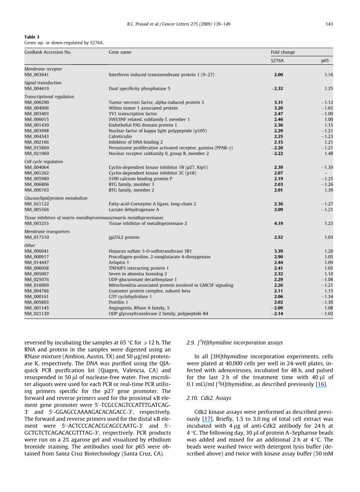
Table 3Genes up- or down-regulated by S276A.
GenBank Accession No. Gene name Fold change
S276A p65
Membrane receptorNM_003641 Interferon induced transmembrane protein 1 (9–27) 2.00 1.16
Signal transductionNM_004419 Dual specificity phosphatase 5 �2.32 1.25
Transcriptional regulationNM_006290 Tumor necrosis factor, alpha-induced protein 3 3.31 �1.12NM_004906 Wilms tumor 1 associated protein 3.20 �1.65NM_003403 YY1 transcription factor 2.47 �1.09NM_006015 SWI/SNF related, subfamily f, member 1 2.46 1.00NM_001430 Endothelial PAS domain protein 1 2.36 1.15NM_003998 Nuclear factor of kappa light polypeptide (p105) 2.29 �1.21NM_004343 Calreticulin 2.25 �1.23NM_002166 Inhibitor of DNA binding 2 2.15 1.21NM_015869 Peroxisome proliferative activated receptor, gamma (PPAR-c) �2.20 �1.21NM_021969 Nuclear receptor subfamily 0, group B, member 2 �2.22 1.48
Cell cycle regulationNM_004064 Cyclin-dependent kinase inhibitor 1B (p27, Kip1) 2.39 �1.39NM_001262 Cyclin-dependent kinase inhibitor 2C (p18) 2.07 –NM_005980 S100 calcium binding protein P 2.19 �1.25NM_006806 BTG family, member 3 2.03 �1.26NM_006763 BTG family, member 2 2.01 1.39
Glucose/lipid/protein metabolismNM_021122 Fatty-acid-Coenzyme A ligase, long-chain 2 2.36 �1.27NM_005566 Lactate dehydrogenase A 2.09 �1.21
Tissue inhibitors of matrix metalloproteinases/matrix metalloproteinasesNM_003255 Tissue inhibitor of metalloproteinase 2 4.19 1.23
Membrane transportersNM_017510 gp25L2 protein 2.52 1.03
OtherNM_006041 Heparan sulfate 3-O-sulfotransferase 3B1 3.39 1.20NM_000917 Procollagen-proline, 2-oxoglutarate 4-dioxygenase 2.90 1.05NM_014447 Arfaptin 1 2.44 1.09NM_006058 TNFAIP3 interacting protein 1 2.41 1.05NM_005067 Seven in absentia homolog 2 2.32 1.10NM_025076 UDP-glucuronate decarboxylase 1 2.29 �1.08NM_016069 Mitochondria-associated protein involved in GMCSF signaling 2.26 �1.21NM_004766 Coatomer protein complex, subunit beta 2.11 1.15NM_000161 GTP cyclohydrolase 1 2.06 �1.34NM_005803 Flotillin 1 2.02 �1.30NM_001145 Angiogenin, RNase A family, 5 �2.09 1.08NM_021139 UDP glycosyltransferase 2 family, polypeptide B4 �2.14 �1.02
R.C. Prasad et al. / Cancer Letters 275 (2009) 139–149 143
reversed by incubating the samples at 65 �C for P12 h. TheRNA and protein in the samples were digested using anRNase mixture (Ambion, Austin, TX) and 50 lg/ml protein-ase K, respectively. The DNA was purified using the QIA-quick PCR purification kit (Qiagen, Valencia, CA) andresuspended in 50 ll of nuclease-free water. Five microli-ter aliquots were used for each PCR or real-time PCR utiliz-ing primers specific for the p27 gene promoter. Theforward and reverse primers used for the proximal jB ele-ment gene promoter were 50-TCGCCAGTCCATTTGATCAG-30 and 50-GGAGCCAAAAGACACAGACC-30, respectively.The forward and reverse primers used for the distal jB ele-ment were 50-ACTCCCACACGCAGCCAATG-30 and 50-GCTGTCTCAGACACGTTTAG-30, respectively. PCR productswere run on a 2% agarose gel and visualized by ethidiumbromide staining. The antibodies used for p65 were ob-tained from Santa Cruz Biotechnology (Santa Cruz, CA).
2.9. [3H]thymidine incorporation assays
In all [3H]thymidine incorporation experiments, cellswere plated at 40,000 cells per well in 24-well plates, in-fected with adenoviruses, incubated for 48 h, and pulsedfor the last 2 h of the treatment time with 40 ll of0.1 mCi/ml [3H]thymidine, as described previously [16].
2.10. Cdk2. Assays
Cdk2 kinase assays were performed as described previ-ously [17]. Briefly, 1.5 to 3.0 mg of total cell extract wasincubated with 4 lg of anti-Cdk2 antibody for 24 h at4 �C. The following day, 30 ll of protein A–Sepharose beadswas added and mixed for an additional 2 h at 4 �C. Thebeads were washed twice with detergent lysis buffer (de-scribed above) and twice with kinase assay buffer (50 mM
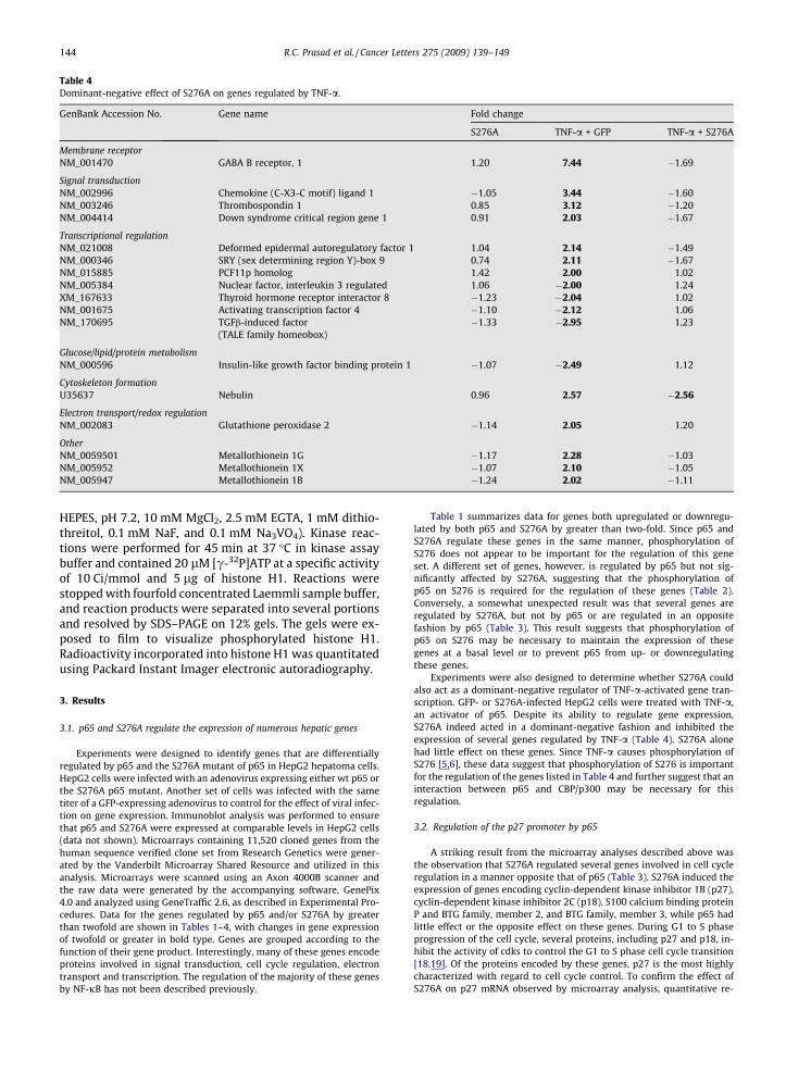
Table 4Dominant-negative effect of S276A on genes regulated by TNF-a.
GenBank Accession No. Gene name Fold change
S276A TNF-a + GFP TNF-a + S276A
Membrane receptorNM_001470 GABA B receptor, 1 1.20 7.44 �1.69
Signal transductionNM_002996 Chemokine (C-X3-C motif) ligand 1 �1.05 3.44 �1.60NM_003246 Thrombospondin 1 0.85 3.12 �1.20NM_004414 Down syndrome critical region gene 1 0.91 2.03 �1.67
Transcriptional regulationNM_021008 Deformed epidermal autoregulatory factor 1 1.04 2.14 �1.49NM_000346 SRY (sex determining region Y)-box 9 0.74 2.11 �1.67NM_015885 PCF11p homolog 1.42 2.00 1.02NM_005384 Nuclear factor, interleukin 3 regulated 1.06 �2.00 1.24XM_167633 Thyroid hormone receptor interactor 8 �1.23 �2.04 1.02NM_001675 Activating transcription factor 4 �1.10 �2.12 1.06NM_170695 TGFb-induced factor �1.33 �2.95 1.23
(TALE family homeobox)
Glucose/lipid/protein metabolismNM_000596 Insulin-like growth factor binding protein 1 �1.07 �2.49 1.12
Cytoskeleton formationU35637 Nebulin 0.96 2.57 �2.56
Electron transport/redox regulationNM_002083 Glutathione peroxidase 2 �1.14 2.05 1.20
OtherNM_0059501 Metallothionein 1G �1.17 2.28 �1.03NM_005952 Metallothionein 1X �1.07 2.10 �1.05NM_005947 Metallothionein 1B �1.24 2.02 �1.11
144 R.C. Prasad et al. / Cancer Letters 275 (2009) 139–149
HEPES, pH 7.2, 10 mM MgCl2, 2.5 mM EGTA, 1 mM dithio-threitol, 0.1 mM NaF, and 0.1 mM Na3VO4). Kinase reac-tions were performed for 45 min at 37 �C in kinase assaybuffer and contained 20 lM [c-32P]ATP at a specific activityof 10 Ci/mmol and 5 lg of histone H1. Reactions werestopped with fourfold concentrated Laemmli sample buffer,and reaction products were separated into several portionsand resolved by SDS–PAGE on 12% gels. The gels were ex-posed to film to visualize phosphorylated histone H1.Radioactivity incorporated into histone H1 was quantitatedusing Packard Instant Imager electronic autoradiography.
3. Results
3.1. p65 and S276A regulate the expression of numerous hepatic genes
Experiments were designed to identify genes that are differentiallyregulated by p65 and the S276A mutant of p65 in HepG2 hepatoma cells.HepG2 cells were infected with an adenovirus expressing either wt p65 orthe S276A p65 mutant. Another set of cells was infected with the sametiter of a GFP-expressing adenovirus to control for the effect of viral infec-tion on gene expression. Immunoblot analysis was performed to ensurethat p65 and S276A were expressed at comparable levels in HepG2 cells(data not shown). Microarrays containing 11,520 cloned genes from thehuman sequence verified clone set from Research Genetics were gener-ated by the Vanderbilt Microarray Shared Resource and utilized in thisanalysis. Microarrays were scanned using an Axon 4000B scanner andthe raw data were generated by the accompanying software, GenePix4.0 and analyzed using GeneTraffic 2.6, as described in Experimental Pro-cedures. Data for the genes regulated by p65 and/or S276A by greaterthan twofold are shown in Tables 1–4, with changes in gene expressionof twofold or greater in bold type. Genes are grouped according to thefunction of their gene product. Interestingly, many of these genes encodeproteins involved in signal transduction, cell cycle regulation, electrontransport and transcription. The regulation of the majority of these genesby NF-jB has not been described previously.
Table 1 summarizes data for genes both upregulated or downregu-lated by both p65 and S276A by greater than two-fold. Since p65 andS276A regulate these genes in the same manner, phosphorylation ofS276 does not appear to be important for the regulation of this geneset. A different set of genes, however, is regulated by p65 but not sig-nificantly affected by S276A, suggesting that the phosphorylation ofp65 on S276 is required for the regulation of these genes (Table 2).Conversely, a somewhat unexpected result was that several genes areregulated by S276A, but not by p65 or are regulated in an oppositefashion by p65 (Table 3). This result suggests that phosphorylation ofp65 on S276 may be necessary to maintain the expression of thesegenes at a basal level or to prevent p65 from up- or downregulatingthese genes.
Experiments were also designed to determine whether S276A couldalso act as a dominant-negative regulator of TNF-a-activated gene tran-scription. GFP- or S276A-infected HepG2 cells were treated with TNF-a,an activator of p65. Despite its ability to regulate gene expression,S276A indeed acted in a dominant-negative fashion and inhibited theexpression of several genes regulated by TNF-a (Table 4). S276A alonehad little effect on these genes. Since TNF-a causes phosphorylation ofS276 [5,6], these data suggest that phosphorylation of S276 is importantfor the regulation of the genes listed in Table 4 and further suggest that aninteraction between p65 and CBP/p300 may be necessary for thisregulation.
3.2. Regulation of the p27 promoter by p65
A striking result from the microarray analyses described above wasthe observation that S276A regulated several genes involved in cell cycleregulation in a manner opposite that of p65 (Table 3). S276A induced theexpression of genes encoding cyclin-dependent kinase inhibitor 1B (p27),cyclin-dependent kinase inhibitor 2C (p18), S100 calcium binding proteinP and BTG family, member 2, and BTG family, member 3, while p65 hadlittle effect or the opposite effect on these genes. During G1 to S phaseprogression of the cell cycle, several proteins, including p27 and p18, in-hibit the activity of cdks to control the G1 to S phase cell cycle transition[18,19]. Of the proteins encoded by these genes, p27 is the most highlycharacterized with regard to cell cycle control. To confirm the effect ofS276A on p27 mRNA observed by microarray analysis, quantitative re-

GFP p65 S276A0
100
200
Adenovirus
p27
mR
NA
(% o
fGFP
con
trol
) *
Vector Control p65-100 ng p65-200 ng S276A-100 ng S276A-200 ng0
2500
5000
7500
10000
12500
Transfected cDNA
p27
prom
oter
activ
ity (R
LU/μ
g pr
otei
n)
*
*
p27 Promoter - pκB p27 Promoter - dκB
Input
No Ab
p65
TNF-α − + − +
-230 -116 -1375 -1270
Fig. 1. p65 binds to the p27 promoter and represses p27 promoter activity. In panel A, HepG2 cells were infected with adenoviruses encoding GFP, p65, orS276A at an MOI of 50. Total RNA was isolated and p27 and cyclophilin mRNA were measured by real-time RT-PCR. p27 mRNA is normalized to that ofcyclophilin and the amount of p27 mRNA detected in GFP-expressing cells is arbitrarily set at 100%. The graph represents three experiments ±S.E. (*p < 0.01,Students t test). In panel B, HepG2 cells were transiently transfected by a reporter construct consisting of �1609 to +178 of the p27 gene promoter upstreamof the luciferase reporter gene. Cells were also co-transfected with either 100 or 200 ng of an expression vector encoding either p65 or S276A. DNA amountswere normalized using the plasmid pRc-RSV. Luciferase assays were performed and the results plotted as relative luminescence units per microgram of cellprotein assayed. The data represent the average of triplicate determinations ±S.E. (*p < 0.01, Students t test) and is representative of three independentexperiments. In panel C, the association of p65 with the endogenous p27 gene promoter was measured by ChIP assay in HepG2 cells. HepG2 cells weretreated in the presence or absence of 1 ng/ml TNF-a for 24 h prior to the assay. The No Ab lane shows the results of immunoprecipitations performed inparallel without the application of primary antibodies. Input lanes show the results from samples not subjected to immunoprecipitation. Antibodiesdirected against endogenous p65 were used to measure the association of endogenous p65 with the p27 gene promoter. Primers designed to produce PCRproducts including the proximal jB (pjB) and distal jB (pjB) elements were used in these experiments.
R.C. Prasad et al. / Cancer Letters 275 (2009) 139–149 145
verse transcriptase PCR was performed. PCR data confirmed the resultsobserved with microarray analysis (Fig. 1A). Furthermore, microarraydata showed that the physiological activator, TNF-a, which causes p65phosphorylation at serine 276, repressed p27 gene expression 1.86 fold(data not shown).
Since p27 is an important regulator of the cell cycle, the effect of S276Aon p27 gene expression was further pursued. Reporter assays were per-formed using a p27-luciferase reporter construct, which consists of from-1609 to +178 of the p27 promoter upstream of the luciferase reportergene, to determine if p65 and S276A have effects on p27 gene promoter
HepG2 MDA-MB 231
p65
p27
β-tubulin
p65 S276A
p65
p27
β-tubulin
p65 S276AGFP GFP
Con
t
TNF-
α
PMA
pVCon
tp27
β-Actin
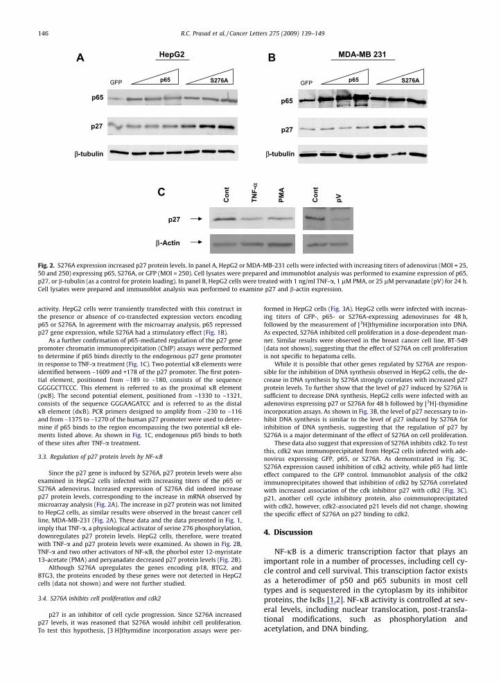
Fig. 2. S276A expression increased p27 protein levels. In panel A, HepG2 or MDA-MB-231 cells were infected with increasing titers of adenovirus (MOI = 25,50 and 250) expressing p65, S276A, or GFP (MOI = 250). Cell lysates were prepared and immunoblot analysis was performed to examine expression of p65,p27, or b-tubulin (as a control for protein loading). In panel B, HepG2 cells were treated with 1 ng/ml TNF-a, 1 lM PMA, or 25 lM pervanadate (pV) for 24 h.Cell lysates were prepared and immunoblot analysis was performed to examine p27 and b-actin expression.
146 R.C. Prasad et al. / Cancer Letters 275 (2009) 139–149
activity. HepG2 cells were transiently transfected with this construct inthe presence or absence of co-transfected expression vectors encodingp65 or S276A. In agreement with the microarray analysis, p65 repressedp27 gene expression, while S276A had a stimulatory effect (Fig. 1B).
As a further confirmation of p65-mediated regulation of the p27 genepromoter chromatin immunoprecipitation (ChIP) assays were performedto determine if p65 binds directly to the endogenous p27 gene promoterin response to TNF-a treatment (Fig. 1C). Two potential jB elements wereidentified between –1609 and +178 of the p27 promoter. The first poten-tial element, positioned from –189 to –180, consists of the sequenceGGGGCTTCCC. This element is referred to as the proximal jB element(pjB). The second potential element, positioned from –1330 to –1321,consists of the sequence GGGAAGATCC and is referred to as the distaljB element (djB). PCR primers designed to amplify from –230 to –116and from –1375 to –1270 of the human p27 promoter were used to deter-mine if p65 binds to the region encompassing the two potential jB ele-ments listed above. As shown in Fig. 1C, endogenous p65 binds to bothof these sites after TNF-a treatment.
3.3. Regulation of p27 protein levels by NF-jB
Since the p27 gene is induced by S276A, p27 protein levels were alsoexamined in HepG2 cells infected with increasing titers of the p65 orS276A adenovirus. Increased expression of S276A did indeed increasep27 protein levels, corresponding to the increase in mRNA observed bymicroarray analysis (Fig. 2A). The increase in p27 protein was not limitedto HepG2 cells, as similar results were observed in the breast cancer cellline, MDA-MB-231 (Fig. 2A). These data and the data presented in Fig. 1,imply that TNF-a, a physiological activator of serine 276 phosphorylation,downregulates p27 protein levels. HepG2 cells, therefore, were treatedwith TNF-a and p27 protein levels were examined. As shown in Fig. 2B,TNF-a and two other activators of NF-jB, the phorbol ester 12-myristate13-acetate (PMA) and peryanadate decreased p27 protein levels (Fig. 2B).
Although S276A upregulates the genes encoding p18, BTG2, andBTG3, the proteins encoded by these genes were not detected in HepG2cells (data not shown) and were not further studied.
3.4. S276A inhibits cell proliferation and cdk2
p27 is an inhibitor of cell cycle progression. Since S276A increasedp27 levels, it was reasoned that S276A would inhibit cell proliferation.To test this hypothesis, [3 H]thymidine incorporation assays were per-
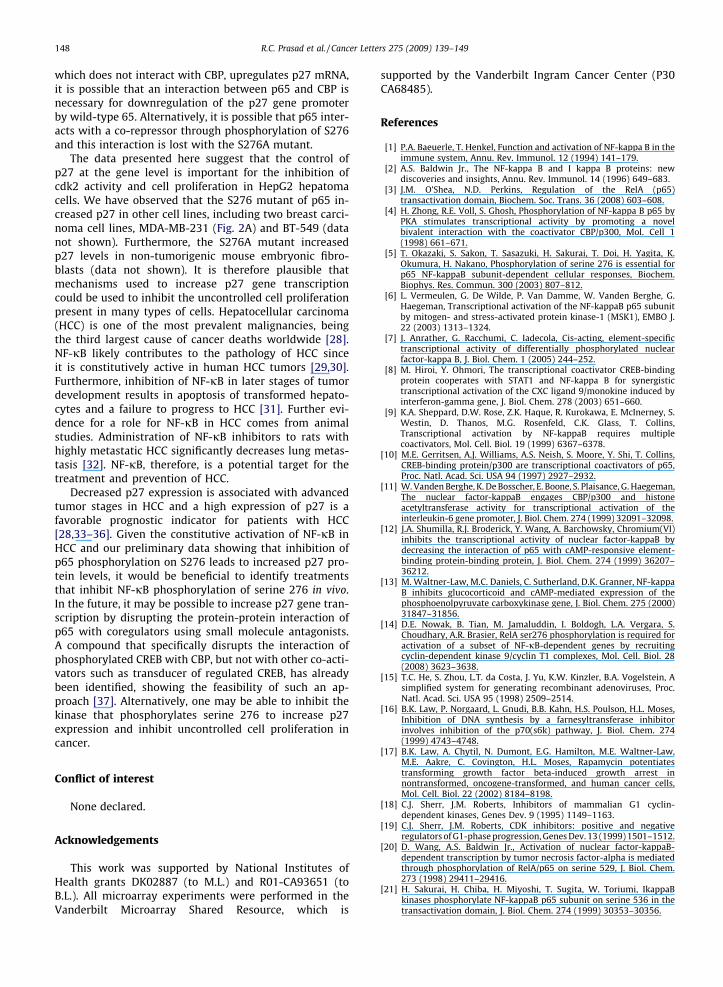
formed in HepG2 cells (Fig. 3A). HepG2 cells were infected with increas-ing titers of GFP-, p65- or S276A-expressing adenoviruses for 48 h,followed by the measurement of [3H]thymidine incorporation into DNA.As expected, S276A inhibited cell proliferation in a dose-dependent man-ner. Similar results were observed in the breast cancer cell line, BT-549(data not shown), suggesting that the effect of S276A on cell proliferationis not specific to hepatoma cells.
While it is possible that other genes regulated by S276A are respon-sible for the inhibition of DNA synthesis observed in HepG2 cells, the de-crease in DNA synthesis by S276A strongly correlates with increased p27protein levels. To further show that the level of p27 induced by S276A issufficient to decrease DNA synthesis, HepG2 cells were infected with anadenovirus expressing p27 or S276A for 48 h followed by [3H]-thymidineincorporation assays. As shown in Fig. 3B, the level of p27 necessary to in-hibit DNA synthesis is similar to the level of p27 induced by S276A forinhibition of DNA synthesis, suggesting that the regulation of p27 byS276A is a major determinant of the effect of S276A on cell proliferation.
These data also suggest that expression of S276A inhibits cdk2. To testthis, cdk2 was immunoprecipitated from HepG2 cells infected with ade-novirus expressing GFP, p65, or S276A. As demonstrated in Fig. 3C,S276A expression caused inhibition of cdk2 activity, while p65 had littleeffect compared to the GFP control. Immunoblot analysis of the cdk2immunoprecipitates showed that inhibition of cdk2 by S276A correlatedwith increased association of the cdk inhibitor p27 with cdk2 (Fig. 3C).p21, another cell cycle inhibitory protein, also coimmunoprecipitatedwith cdk2, however, cdk2-associated p21 levels did not change, showingthe specific effect of S276A on p27 binding to cdk2.
4. Discussion
NF-jB is a dimeric transcription factor that plays animportant role in a number of processes, including cell cy-cle control and cell survival. This transciption factor existsas a heterodimer of p50 and p65 subunits in most celltypes and is sequestered in the cytoplasm by its inhibitorproteins, the IjBs [1,2]. NF-jB activity is controlled at sev-eral levels, including nuclear translocation, post-transla-tional modifications, such as phosphorylation andacetylation, and DNA binding.

R.C. Prasad et al. / Cancer Letters 275 (2009) 139–149 147
The role of NF-jB phosphorylation for the regulation oftranscription is likely to be complex, as several serine res-idues of the p65 subunit are phosphorylated, and phos-phorylation of these sites affects the activity of NF-jBand its interaction with co-regulators [4,5,7,20–27]. Aneven greater degree of complexity occurs at the level ofNF-jB binding to DNA. The DNA microarray data presentedin this study reflect the complex nature of p65-mediatedgene regulation and the role of S276 phosphorylation.While S276A and p65 regulate several of the same genes
50 250 50 250 50 2500
25
50
75
100
125GFP p65 S276A
Virus Titer (MOI)
3 H-T
hym
idin
e In
corp
orat
ion
(%of
GFP
Con
trol
)
*
*
50 250 50 250 50 250
GFP p27 S276AMOI:
p27
-Actin
50 250 50 250 50 2500
25
50
75
100GFP p27 S276A
Virus Titer (MOI)
3 H-T
hym
idin
e In
corp
orat
ion
(% o
fGFP
Con
trol
)
*
*
*
*
P-HistoneH1
Cdk2IP
IB:
p27
GFP p65 S276A0
50
100
150
Adenovirus
Cdk
2 A
ctiv
ity (%
Con
trol
)
p21
Cdk21 2 3
p65
A
B
C
similarly, p65 regulates genes that are not regulated byS276A (Table 2). S276A is also capable of acting in a dom-inant-negative fashion by inhibiting the regulation of cer-tain genes by TNF-a (Table 4). Intriguingly, S276Aregulates a set of genes that are not highly regulated byp65 (Table 3). One possible explanation for this observa-tion is that S276A upregulates other transcription factorsthat regulate the expression of these genes. Alternatively,phosphorylation of wt p65 may function to repress ormaintain the expression of these genes at a basal level.
Anrather et al. [7] recently demonstrated that mutationof serine 276 inhibits the ability of p65 to activate tran-scription through two different cis-acting elements. Theseconsensus sequences consist of KGRAHWTYCC (R = purine,Y = pyrimidine, K = G or T, H = not G, W = A or T,), a sitepresent in several genes, including IL-6 and ICAM-1, andGGRWWWYYYY, a site present in IL-2a and porcineELAM-1. Mutation of serine 276 in p65, however, has no af-fect on the recognition of the sequence GGGRATTYCC,which is present in genes including MHC class I and humanELAM-1 [7]. It thus appears that site-specific phosphoryla-tion of p65 directs NF-jB to a particular cis-acting elementto regulate specific subsets of genes. This point is under-scored by our observation that S276A is capable of regulat-ing a set of genes in a manner similar to wild-type p65 butdoes not regulate all of the genes affected by p65 (Table 1).The p27 promoter has two jB elements. The downstreamjB element has the sequence of GGGGCTTCCC, while theupstream sequence is GGGAAGATCC. Both of these DNAelements display characteristics consistent with the con-sensus sites regulated by serine 276 phosphorylation.
Most studies of p27 show that it is regulated at the pro-tein level by several mechanisms, including proteasomaldegradation. Our data using DNA microarray, real-timePCR and transient transfection all indicate that TNF-aand NF-jB regulate p27 at the gene level. Since S276A,
Fig. 3. S276A inhibited cell proliferation and cdk2 activity. To examinethe effects of S276A on cell proliferation (panel A), HepG2 cells weresubjected to [3H]thymidine incorporation assays 48 h after viral infectionwith either the GFP-, p65-, or S276A-expressing adenovirus, as describedin Experimental Procedures. The data presented are the result of threereplicate determinations ±S.E. (*p < 0.05, Students t test) and are repre-sentative of three different experiments. In panel B, HepG2 cells wereinfected with an adenovirus expressing GFP, p27, or S276A (MOI = 50 or250) and were subjected to [3H]thymidine incorporation assays 48 h afterviral infection. The data presented are the result of three replicatedeterminations ±S.E. (*p < 0.05, Students t test) and are representative ofthree different experiments. p27 expression was confirmed by immuno-blot analysis of lysates prepared from cells infected with the GFP, p27, orS276A adenovirus. Cdk2 assays are shown in panel C. HepG2 wereinfected with adenoviruses expressing GFP (lane 1), p65 (lane 2), orS276A (lane 3) at an MOI of 50. To measure cdk2 activity, cdk2 wasimmunoprecipitated 48 h post-infection and the immunoprecipitateswere incubated with [c-32P]ATP and histone H1, as a cdk2 substrate, asdescribed in the Experimental Procedures section. Phosphorylated his-tone is indicated by the arrow (panel C). Immunoblot analysis (IB) wasperformed to show equivalent immunoprecipitation of cdk2 from eachsample. p21 and p27 immunoblot analysis was also performed toexamine the association of these proteins with cdk2. Proteins from thesame crude cell lysates used for cdk2 immunoprecipitation were probedwith a p65-specific antibody in immunoblot analyses to confirm that p65and S276A were expressed at comparable levels. The results shown arerepresentative of three independent experiments.
3
148 R.C. Prasad et al. / Cancer Letters 275 (2009) 139–149
which does not interact with CBP, upregulates p27 mRNA,it is possible that an interaction between p65 and CBP isnecessary for downregulation of the p27 gene promoterby wild-type 65. Alternatively, it is possible that p65 inter-acts with a co-repressor through phosphorylation of S276and this interaction is lost with the S276A mutant.
The data presented here suggest that the control ofp27 at the gene level is important for the inhibition ofcdk2 activity and cell proliferation in HepG2 hepatomacells. We have observed that the S276 mutant of p65 in-creased p27 in other cell lines, including two breast carci-noma cell lines, MDA-MB-231 (Fig. 2A) and BT-549 (datanot shown). Furthermore, the S276A mutant increasedp27 levels in non-tumorigenic mouse embryonic fibro-blasts (data not shown). It is therefore plausible thatmechanisms used to increase p27 gene transcriptioncould be used to inhibit the uncontrolled cell proliferationpresent in many types of cells. Hepatocellular carcinoma(HCC) is one of the most prevalent malignancies, beingthe third largest cause of cancer deaths worldwide [28].NF-jB likely contributes to the pathology of HCC sinceit is constitutively active in human HCC tumors [29,30].Furthermore, inhibition of NF-jB in later stages of tumordevelopment results in apoptosis of transformed hepato-cytes and a failure to progress to HCC [31]. Further evi-dence for a role for NF-jB in HCC comes from animalstudies. Administration of NF-jB inhibitors to rats withhighly metastatic HCC significantly decreases lung metas-tasis [32]. NF-jB, therefore, is a potential target for thetreatment and prevention of HCC.
Decreased p27 expression is associated with advancedtumor stages in HCC and a high expression of p27 is afavorable prognostic indicator for patients with HCC[28,33–36]. Given the constitutive activation of NF-jB inHCC and our preliminary data showing that inhibition ofp65 phosphorylation on S276 leads to increased p27 pro-tein levels, it would be beneficial to identify treatmentsthat inhibit NF-jB phosphorylation of serine 276 in vivo.In the future, it may be possible to increase p27 gene tran-scription by disrupting the protein-protein interaction ofp65 with coregulators using small molecule antagonists.A compound that specifically disrupts the interaction ofphosphorylated CREB with CBP, but not with other co-acti-vators such as transducer of regulated CREB, has alreadybeen identified, showing the feasibility of such an ap-proach [37]. Alternatively, one may be able to inhibit thekinase that phosphorylates serine 276 to increase p27expression and inhibit uncontrolled cell proliferation incancer.
Conflict of interest
None declared.
Acknowledgements
This work was supported by National Institutes ofHealth grants DK02887 (to M.L.) and R01-CA93651 (toB.L.). All microarray experiments were performed in theVanderbilt Microarray Shared Resource, which is
supported by the Vanderbilt Ingram Cancer Center (P30CA68485).
References
[1] P.A. Baeuerle, T. Henkel, Function and activation of NF-kappa B in theimmune system, Annu. Rev. Immunol. 12 (1994) 141–179.
[2] A.S. Baldwin Jr., The NF-kappa B and I kappa B proteins: newdiscoveries and insights, Annu. Rev. Immunol. 14 (1996) 649–683.
[3] J.M. O’Shea, N.D. Perkins, Regulation of the RelA (p65)transactivation domain, Biochem. Soc. Trans. 36 (2008) 603–608.
[4] H. Zhong, R.E. Voll, S. Ghosh, Phosphorylation of NF-kappa B p65 byPKA stimulates transcriptional activity by promoting a novelbivalent interaction with the coactivator CBP/p300, Mol. Cell 1(1998) 661–671.
[5] T. Okazaki, S. Sakon, T. Sasazuki, H. Sakurai, T. Doi, H. Yagita, K.Okumura, H. Nakano, Phosphorylation of serine 276 is essential forp65 NF-kappaB subunit-dependent cellular responses, Biochem.Biophys. Res. Commun. 300 (2003) 807–812.
[6] L. Vermeulen, G. De Wilde, P. Van Damme, W. Vanden Berghe, G.Haegeman, Transcriptional activation of the NF-kappaB p65 subunitby mitogen- and stress-activated protein kinase-1 (MSK1), EMBO J.22 (2003) 1313–1324.
[7] J. Anrather, G. Racchumi, C. Iadecola, Cis-acting, element-specifictranscriptional activity of differentially phosphorylated nuclearfactor-kappa B, J. Biol. Chem. 1 (2005) 244–252.
[8] M. Hiroi, Y. Ohmori, The transcriptional coactivator CREB-bindingprotein cooperates with STAT1 and NF-kappa B for synergistictranscriptional activation of the CXC ligand 9/monokine induced byinterferon-gamma gene, J. Biol. Chem. 278 (2003) 651–660.
[9] K.A. Sheppard, D.W. Rose, Z.K. Haque, R. Kurokawa, E. McInerney, S.Westin, D. Thanos, M.G. Rosenfeld, C.K. Glass, T. Collins,Transcriptional activation by NF-kappaB requires multiplecoactivators, Mol. Cell. Biol. 19 (1999) 6367–6378.
[10] M.E. Gerritsen, A.J. Williams, A.S. Neish, S. Moore, Y. Shi, T. Collins,CREB-binding protein/p300 are transcriptional coactivators of p65,Proc. Natl. Acad. Sci. USA 94 (1997) 2927–2932.
[11] W. Vanden Berghe, K. De Bosscher, E. Boone, S. Plaisance, G. Haegeman,The nuclear factor-kappaB engages CBP/p300 and histoneacetyltransferase activity for transcriptional activation of theinterleukin-6 gene promoter, J. Biol. Chem. 274 (1999) 32091–32098.
[12] J.A. Shumilla, R.J. Broderick, Y. Wang, A. Barchowsky, Chromium(VI)inhibits the transcriptional activity of nuclear factor-kappaB bydecreasing the interaction of p65 with cAMP-responsive element-binding protein-binding protein, J. Biol. Chem. 274 (1999) 36207–36212.
[13] M. Waltner-Law, M.C. Daniels, C. Sutherland, D.K. Granner, NF-kappaB inhibits glucocorticoid and cAMP-mediated expression of thephosphoenolpyruvate carboxykinase gene, J. Biol. Chem. 275 (2000)31847–31856.
[14] D.E. Nowak, B. Tian, M. Jamaluddin, I. Boldogh, L.A. Vergara, S.Choudhary, A.R. Brasier, RelA ser276 phosphorylation is required foractivation of a subset of NF-jB-dependent genes by recruitingcyclin-dependent kinase 9/cyclin T1 complexes, Mol. Cell. Biol. 28(2008) 3623–3638.
[15] T.C. He, S. Zhou, L.T. da Costa, J. Yu, K.W. Kinzler, B.A. Vogelstein, Asimplified system for generating recombinant adenoviruses, Proc.Natl. Acad. Sci. USA 95 (1998) 2509–2514.
[16] B.K. Law, P. Norgaard, L. Gnudi, B.B. Kahn, H.S. Poulson, H.L. Moses,Inhibition of DNA synthesis by a farnesyltransferase inhibitorinvolves inhibition of the p70(s6k) pathway, J. Biol. Chem. 274(1999) 4743–4748.
[17] B.K. Law, A. Chytil, N. Dumont, E.G. Hamilton, M.E. Waltner-Law,M.E. Aakre, C. Covington, H.L. Moses, Rapamycin potentiatestransforming growth factor beta-induced growth arrest innontransformed, oncogene-transformed, and human cancer cells,Mol. Cell. Biol. 22 (2002) 8184–8198.
[18] C.J. Sherr, J.M. Roberts, Inhibitors of mammalian G1 cyclin-dependent kinases, Genes Dev. 9 (1995) 1149–1163.
[19] C.J. Sherr, J.M. Roberts, CDK inhibitors: positive and negativeregulators of G1-phase progression, Genes Dev. 13 (1999) 1501–1512.
[20] D. Wang, A.S. Baldwin Jr., Activation of nuclear factor-kappaB-dependent transcription by tumor necrosis factor-alpha is mediatedthrough phosphorylation of RelA/p65 on serine 529, J. Biol. Chem.273 (1998) 29411–29416.
[21] H. Sakurai, H. Chiba, H. Miyoshi, T. Sugita, W. Toriumi, IkappaBkinases phosphorylate NF-kappaB p65 subunit on serine 536 in thetransactivation domain, J. Biol. Chem. 274 (1999) 30353–30356.

R.C. Prasad et al. / Cancer Letters 275 (2009) 139–149 149
[22] H. Sakurai, S. Suzuki, N. Kawasaki, H. Nakano, T. Okazaki, A. Chino, T.Doi, I. Saiki, Tumor necrosis factor-alpha-induced IKKphosphorylation of NF-kappaB p65 on serine 536 is mediatedthrough the TRAF2, TRAF5, and TAK1 signaling pathway, J. Biol.Chem. 278 (2003) 36916–36923.
[23] D. Wang, S.D. Westerheide, J.L. Hanson, A.S. Baldwin Jr., Tumornecrosis factor alpha-induced phosphorylation of RelA/p65 onSer529 is controlled by casein kinase II, J. Biol. Chem. 275 (2000)32592–32597.
[24] X. Jiang, N. Takahashi, N. Matsui, T. Tetsuka, T. Okamoto, The NF-kappa B activation in lymphotoxin beta receptor signaling dependson the phosphorylation of p65 at serine 536, J. Biol. Chem. 278(2003) 919–926.
[25] A. Duran, M.T. Diaz-Meco, J. Moscat, Essential role of RelA Ser311phosphorylation by zetaPKC in NF-kappaB transcriptional activation,EMBO J. 22 (2003) 3910–3918.
[26] H. Buss, A. Dorrie, M. Lienhard Schmitz, R. Frank, M. Livingstone, K.Resch, M. Kracht, Phosphorylation of serine 468 by GSK-3betanegatively regulates basal p65 NF-kappaB activity, J. Biol. Chem. 48(2004) 49571–49574.
[27] J. Hu, H. Nakano, H. Sakurai, N.H. Colburn, Insufficient p65phosphorylation at S536 specifically contributes to the lack of NF-kappaB activation and transformation in resistant JB6 cells,Carcinogenesis 10 (2004) 1991–2003.
[28] P.P. Claudio, G. Russo, C.A. Kumar, C. Minimo, A. Farina, S. Tutton, G.Nuzzo, F. Giuliante, G. Angeloni, V. Maria, F.M. Vecchio, C.D. Campli,A. Giordano, PRb2/p130, vascular endothelial growth factor,p27(KIP1), and proliferating cell nuclear antigen expression inhepatocellular carcinoma: their clinical significance, Clin. CancerRes. 10 (2004) 3509–3517.
[29] D.I. Tai, S.L. Tsai, Y.H. Chang, S.N. Huang, T.C. Chen, K.S. Chang, Y.F.Liaw, Constitutive activation of nuclear factor kappaB inhepatocellular carcinoma, Cancer 89 (2000) 2274–2281.
[30] S.P. Guo, W.L. Wang, Y.Q. Zhai, Y.L. Zhao, Expression of nuclearfactor-kappa B in hepatocellular carcinoma and its relation with theX protein of hepatitis B virus, World J. Gastroenterol. 7 (2001) 340–344.
[31] E. Pikarsky, R.M. Porat, I. Stein, R. Abramovitch, S. Amit, S. Kasem, E.Gutkovich-Pyest, S. Urieli-Shoval, E. Galun, Y. Ben-Neriah, NF-kappaB functions as a tumour promoter in inflammation-associated cancer, Nature 431 (2004) 461–466.
[32] M. Futakuchi, K. Ogawa, S. Tamano, S. Takahashi, T. Shirai,Suppression of metastasis by nuclear factor kappaB inhibitors inan in vivo lung metastasis model of chemically inducedhepatocellular carcinoma, Cancer Sci. 95 (2004) 18–24.
[33] K.J. Nan, Z. Jing, L. Gong, Expression and altered subcellularlocalization of the cyclin-dependent kinase inhibitor p27Kip1 inhepatocellular carcinoma, World J. Gastroenterol. 10 (2004) 1425–1430.
[34] Y. Ito, N. Matsuura, M. Sakon, E. Miyoshi, K. Noda, T. Takeda, K.Umeshita, H. Nagano, S. Nakamori, K. Dono, M. Tsujimoto, M.Nakahara, K. Nakao, N. Taniguchi, M. Monden, Expression andprognostic roles of the G1-S modulators in hepatocellularcarcinoma: p27 independently predicts the recurrence, Hepatology30 (1999) 90–99.
[35] A.M. Hui, L. Sun, Y. Kanai, M. Sakamoto, S. Hirohashi, Reducedp27Kip1 expression in hepatocellular carcinomas, Cancer Lett. 132(1998) 67–73.
[36] A. Tannapfel, D. Grund, A. Katalinic, D. Uhlmann, F. Kockerling, U.Haugwitz, M. Wasner, J. Hauss, K. ngeland, C. Wittekind, Decreasedexpression of p27 protein is associated with advanced tumor stagein hepatocellular carcinoma, Int. J. Cancer 89 (2000) 350–355.
[37] J.L. Best, C.A. Amezcua, B. Mayr, L. Flechner, C.M. Murawsky, B.Emerson, T. Zor, K.H. Gardner, M. Montminy, Identification of small-molecule antagonists that inhibit an activator: coactivatorinteraction, Proc. Natl. Acad. Sci. USA 101 (2004) 17622–17627.