Critical assessment of relevant methods in the field of ...
33
REVIEW Critical assessment of relevant methods in the field of biosensors with direct optical detection based on fibers and waveguides using plasmonic, resonance, and interference effects Günter Gauglitz 1 # The Author(s) 2020 Abstract Direct optical detection has proven to be a highly interesting tool in biomolecular interaction analysis to be used in drug discovery, ligand/receptor interactions, environmental analysis, clinical diagnostics, screening of large data volumes in immu- nology, cancer therapy, or personalized medicine. In this review, the fundamental optical principles and applications are reviewed. Devices are based on concepts such as refractometry, evanescent field, waveguides modes, reflectometry, resonance and/or interference. They are realized in ring resonators; prism couplers; surface plasmon resonance; resonant mirror; Bragg grating; grating couplers; photonic crystals, Mach-Zehnder, Young, Hartman interferometers; backscattering; ellipsometry; or reflectance interferometry. The physical theories of various optical principles have already been reviewed in detail elsewhere and are therefore only cited. This review provides an overall survey on the application of these methods in direct optical biosensing. The “historical” development of the main principles is given to understand the various, and sometimes only slightly modified variations published as “new” methods or the use of a new acronym and commercialization by different companies. Improvement of optics is only one way to increase the quality of biosensors. Additional essential aspects are the surface modification of transducers, immobilization strategies, selection of recognition elements, the influence of non-specific interaction, selectivity, and sensitivity. Furthermore, papers use for reporting minimal amounts of detectable analyte terms such as value of mass, moles, grams, or mol/L which are difficult to compare. Both these essential aspects (i.e., biochemistry and the presentation of LOD values) can be discussed only in brief (but references are provided) in order to prevent the paper from becoming too long. The review will concentrate on a comparison of the optical methods, their application, and the resulting bioanalytical quality. Keywords Biosensor . Evanescent field . Resonance . Refractometry . Reflectometry . Interference Introduction The measurement of molecule interactions in medicine, biol- ogy, biochemistry, and diagnostics has been of high impor- tance. Many years ago, radio-labelling has been used to report the binding of a ligand to its receptor. One analytical development-pushing application has been drug discovery [1] to determine affinity, activity, toxicity or availability of candidates in the process of ligand/receptor interactions. Especially screening applications have driven research be- yond ELISAs to receive thermodynamic as well as kinetic data in biomolecular interaction analysis (BIA) [2–4]. Primary screening of antibodies and selection of alternative binders out of cell cultures at extremely low concentrations with high throughput are present high topics. In the future, screening of large data volumes will get interest in immunol- ogy and cancer therapy. This aims to personalize medicine, and methods without labelling will be of interest in Clustered Regularly Interspaced Short Palindromic Repeats genome editing (CRISPR/Cas) methods in molecular biology [5]. The huge field of biomolecular interaction analysis and its application to urgent problems in the environment, biology, medicine, and health care has induced an extreme number of Published in the topical collection Advances in Direct Optical Detection with guest editors Antje J. Baeumner, Günter Gauglitz, and Jiri Homola. * Günter Gauglitz [email protected] 1 Institute of Physical and Theoretical Chemistry, Eberhard Karls Universität, Auf der Morgenstelle 18, 72076 Tübingen, Germany https://doi.org/10.1007/s00216-020-02581-0 Analytical and Bioanalytical Chemistry (2020) 412:3317–3349 Received: 20 December 2019 /Revised: 28 February 2020 /Accepted: 4 March 2020 /Published online: 20 April 2020
-
Upload
khangminh22 -
Category
Documents
-
view
0 -
download
0
Transcript of Critical assessment of relevant methods in the field of ...
REVIEW
Critical assessment of relevant methods in the field of biosensorswith direct optical detection based on fibers and waveguides usingplasmonic, resonance, and interference effects
Günter Gauglitz1
# The Author(s) 2020
AbstractDirect optical detection has proven to be a highly interesting tool in biomolecular interaction analysis to be used in drugdiscovery, ligand/receptor interactions, environmental analysis, clinical diagnostics, screening of large data volumes in immu-nology, cancer therapy, or personalized medicine. In this review, the fundamental optical principles and applications arereviewed. Devices are based on concepts such as refractometry, evanescent field, waveguides modes, reflectometry, resonanceand/or interference. They are realized in ring resonators; prism couplers; surface plasmon resonance; resonant mirror; Bragggrating; grating couplers; photonic crystals, Mach-Zehnder, Young, Hartman interferometers; backscattering; ellipsometry; orreflectance interferometry. The physical theories of various optical principles have already been reviewed in detail elsewhere andare therefore only cited. This review provides an overall survey on the application of these methods in direct optical biosensing.The “historical” development of the main principles is given to understand the various, and sometimes only slightly modifiedvariations published as “new”methods or the use of a new acronym and commercialization by different companies. Improvementof optics is only one way to increase the quality of biosensors. Additional essential aspects are the surface modification oftransducers, immobilization strategies, selection of recognition elements, the influence of non-specific interaction, selectivity, andsensitivity. Furthermore, papers use for reporting minimal amounts of detectable analyte terms such as value of mass, moles,grams, or mol/L which are difficult to compare. Both these essential aspects (i.e., biochemistry and the presentation of LODvalues) can be discussed only in brief (but references are provided) in order to prevent the paper from becoming too long. Thereview will concentrate on a comparison of the optical methods, their application, and the resulting bioanalytical quality.
Keywords Biosensor . Evanescent field . Resonance . Refractometry . Reflectometry . Interference
Introduction
The measurement of molecule interactions in medicine, biol-ogy, biochemistry, and diagnostics has been of high impor-tance. Many years ago, radio-labelling has been used to reportthe binding of a ligand to its receptor. One analyticaldevelopment-pushing application has been drug discovery
[1] to determine affinity, activity, toxicity or availability ofcandidates in the process of ligand/receptor interactions.Especially screening applications have driven research be-yond ELISAs to receive thermodynamic as well as kineticdata in biomolecular interaction analysis (BIA) [2–4].Primary screening of antibodies and selection of alternativebinders out of cell cultures at extremely low concentrationswith high throughput are present high topics. In the future,screening of large data volumes will get interest in immunol-ogy and cancer therapy. This aims to personalize medicine,and methods without labelling will be of interest in ClusteredRegularly Interspaced Short Palindromic Repeats genomeediting (CRISPR/Cas) methods in molecular biology [5].
The huge field of biomolecular interaction analysis and itsapplication to urgent problems in the environment, biology,medicine, and health care has induced an extreme number of
Published in the topical collection Advances in Direct Optical Detectionwith guest editors Antje J. Baeumner, Günter Gauglitz, and Jiri Homola.
* Günter [email protected]
1 Institute of Physical and Theoretical Chemistry, Eberhard KarlsUniversität, Auf der Morgenstelle 18, 72076 Tübingen, Germany
https://doi.org/10.1007/s00216-020-02581-0Analytical and Bioanalytical Chemistry (2020) 412:3317–3349
Received: 20 December 2019 /Revised: 28 February 2020 /Accepted: 4 March 2020 /Published online: 20 April 2020

publications in classical analytics like MS, NMR, or hyphen-ated techniques with separation science as well as reportingbiosensors based on mass-sensitive, electronic, electrochemi-cal, or optical devices. The scope of this review is to try toname the problems using biosensors coming from biochemis-try, surface chemistry, transport processes in sample cells,microfluidics, and detection. For space reasons, not all prob-lems can be discussed in detail. Therefore, after a brief surveyof potential sensor principles, the review has to focus on op-tical detection and especially on direct optical detection.Thereby, the physical and optical basics will be referencedand not discussed using formulas. The main aim is to classifythese optical principles according to plasmonic, resonance, orinterferometric effects to give the reader a systematic pictureof the huge number of devices, many with just minimal mod-ification of the original optical principle. For this reason, alsothe “historical” development is sketched in most methods. InChapter conclusion, Table 1 shows the essential parameters.
Biomolecular interaction analysis
A successful approach to achieve information without radio-labelling is isothermal titration calorimetry (ITC) which yieldsthermodynamic data such as enthalpy of binding or entropy ofbinding of especially large biomolecules. Typically, protein/protein interactions are examined [68]. The application of ITCfor the formation or disassociation of molecular complexeshas developed since first publications in 1990 [69]. Since thatyear, the number of publications has increased, and publica-tions cover especially the field of protein chemistry. Researchand technical development from 2011 to 2015 has beenreviewed, providing information on methodological advancesand interpretation of single and multiple binding sites [70].Besides thermodynamic information on binding constants,e.g., enzymes, of substrate reactions and inhibitory constants,kinetic data is also of interest. The possibilities are introducedin [71]. Modern ITC instrumentation allows measurement ofvery small heat powers and provides a tool for biology tostudy association processes involving liquid membrane pro-teins, nucleic acids, macromolecular assemblies, and a greatvariety of ligands. A joint method for thermodynamic andkinetic data achieved by ITC is described in [72].Miniaturized calorimeter with an elaborated temperature con-trol inside the system was developed for microbiological ap-plications [73]. Despite the instrumental and methodologicaldevelopment, the ITC is a calorimetry not easy to handle andlacks screening possibilities. Thus, one could realize increas-ing interest to have another method to determine thermody-namic and kinetic data of the biomolecular interaction process.
Since approx. the year 2000, biosensors as a tool for quan-tifying ligand/receptor interactions in homogeneous phase andat heterogeneous interface came more and more into focus.
There is a huge variety of biosensor types, ranging frommass-sensitive (quartz-microbalances (QCM), surface acousticwaves (SAW), or cantilever (CL) systems) to electrochemicaland optical ones. Recently, a survey on sensors in general andtheir application has been published [74, 75]. All these possi-ble biosensors depend on sampling, sample preparation, suit-ability for microfluidics setups, potential parallelization, andminiaturization and finally in the case of direct detection with-out label on avoiding rival non-specific binding in the biomo-lecular interaction process. Because of these many require-ments considering biosensing, first, a brief survey on non-optical method is given with reference to quality and applica-bility to problems in biochemistry, biology or medicine, just todemonstrate the huge variety of methods.
QCM, SAW [76], and CL [77, 78] based sensors are espe-cially suitable to monitor mass-sensitive effects. Therefore,their applications are well known for measuring gas concen-trations. However, the signal depends on liquids extremely onviscosity. The Sauerbrey equation is not anymore applicablein its simple form. Interfacial properties of solid-liquid inter-face have to be considered [79]. Nevertheless, QCMs are usedfor biosensing [80, 81] and even cell behavior is examined[82]. Picomolar specific biomarker target detection wasachieved for miss-matches of non-coding RNA [83] or usingmicro-cantilever arrays for early liver cancer diagnosis [84].However, the mechanical sensitivity to damage prevents out-of-lab applications. For many years, electrochemical sensorswere preferably used in biosensing. Especially the possibilityto fabricate large sensor arrays with many spots or readers formicrotiter plates supported many applications [85, 86]. Thedevelopment of impedance instrumentation, which allowedparallel measurement of signal and phase, pushed electro-chemical biosensors [87, 88]. Applications of DNA-basedelectrochemical sensors are numerous [89]. Recently the liter-ature on bioanalytics usingmicroelectrodes has been reviewed[90]. A smart and interesting combination of electrochemistryand chemiluminescence results in many advantages such asremarkably lower limits of detection, higher sensitivity, and awide dynamic range. This electrochemiluminescence (ECL)[91] offers many applications like measuring toxins, pesti-cides or drugs in food [92]. It has improved with newgraphene electrodes [93]. Comparable with electrochemicalbiosensors a huge amount of publications deals with detectionprinciples and applications of optical biosensors. A very de-tailed review on optical biosensors provide — apart from thedefinition of biosensors — descriptions of different recogni-tion elements as enzymatic biosensors, immunosensor, ligand/receptor interactions, and nucleic acid assay and even wholecells. Furthermore, the paper provides an in-depth survey onmethods and applications [94]. Recently, a review on biolog-ical and synthetic materials as recognition elements for foodsafety analysis has been published [95, 96] can help to select asuitable recognition element. Because of the wide field of
Gauglitz G.3318

detection principles and applications the review aims to focuson optical biosensing and within these many methods is re-stricted to biosensors based on direct optical detection.
Optical biosensors
General considerations
Of the optical biosensors, at first, fluorescence assays (using amarker or a label) were predominantly used, taking into ac-count that problems with photostability and influences on thebioactivity of fluorophore-labeled partners could prevent toobtain kinetic data apart from equilibrium constants.However, regarding the possibility of measuring multiple in-teractions in parallel, microarrays based on methods usinglabels proved their advantages [97, 98]. The detection of bio-molecular interaction in bulk or homogeneous phase on top ofthe transducer is only possible with signal changes caused byvariation of the fluorescence intensity or change of fluores-cence wavelength of the monitored complex. In addition, thequenching of fluorescence might be used during the interac-tion process. However, this quenching also can be caused bysimple changes in the oxygen concentration of the solution,for example. Therefore, this effect is rather non-specific. Abetter chance offers (Förster) fluorescence resonance energytransfer (FRET) where the ligand as a labeled donor moleculeand a fluorescent receptor interact. The receptor absorbs at thefluorescence wavelength of the receptor in the case of a closeneighborhood (up to 10 nm) by dipole interaction.Fluorescence intensity of the receptor increases [99, 100].However, even this effect depends often on environmentalconditions. Reviews on fluorescence biosensors have beenpublished some years ago [101–105]. Another possibility inthe homogeneous phase is the measurement of the light scat-tering which will differ dependent on the size of the complexmeasured. However, in this case, the interaction between alarge receptor and a small ligand will give a poor variationof the signal. Only interactions with recognizable differencesin size between ligand/receptor and the complex can be mon-itored [106, 107]. Accordingly, normally in optical biosens-ing, a heterogeneous phase device is used.
Soon, after 2000, the advantages of direct optical detectionwere discussed, especially in the case of drug discovery tech-nologies [108]. Rather early upcoming new interesting fieldwere considered in which direct optical sensing promised ad-vantages [109]. Faster assay development times, accurate andhigh information content data, and less interference from la-bels were considered as an advantage and as a perspective[110]. In the following, a large number of optical devices weredeveloped. An early review about themany possible label-freebiosensor structures also exists [111]. The principles of theseoptical methods are discussed and compared in [112].
Progress in material fabrication and novel substrate with en-hanced optical response properties and potential applicationfor rapid analytical measurement of target interactions fromproteins to DNA and viruses are demonstrated in a reviewarticle on emerging applications [113]. Looking especially atsmall molecules, many techniques, including surface-enhanced Raman spectroscopy, have been reviewed in a re-cent article, together with potential evaluation techniques[114]. Raman and especially using the SERS (surface-enhanced Raman spectroscopy) instrumentation has becomevery interesting because of new developments resulting ineasy to use and at low costs [115]. Nevertheless, Raman can-not be the topic of this review.
The direct optical detection techniques perform spectrosco-py on biomolecules at the surface of the transducer.Accordingly, the measurement not only depends on the trans-duction method but also on competition between specific andnon-specific interactions. Labelling can reduce the problemswith non-specific interaction, but in the case of direct opticaldetection, this problem always arises [116]. Therefore, nor-mally between the transducer and the recognition sites (re-sponsible for the amount of biomolecular interaction) abiolayer is added, which reduces and/or prevents non-specific binding and allows the immobilization of as manyrecognition sites as possible [117]. Besides, nonspecific inter-action, the performance of direct optical sensors is impairedfor very small analytes, which do not provoke recognizablesignal changes when interacting with recognition elements atthe sensor surface. This problem can be overcome by usingeither competitive or binding inhibition assay formats [118].
In general, the optical techniques, which will bediscussed here, use the influence on the propagation ofelectromagnetic radiation in a waveguide or fiber or ef-fects on the reflection of electromagnetic radiation at theinterface including resonance and interference effects. Inprinciple, all direct optical detection techniques measurethe product (n × d) of refractive index n and the physicalthickness of an interaction layer d. Depending on thetransduction method used and on the setup of the mea-surement cell, the readout is dominated by the influenceson the refractive index during bio-recognition or on thechanges of the physical thickness of the examined layerduring interaction. Thus, it is possible to divide directoptical detection techniques into refractometric- andreflectometric-based fundamentals. In the case of refrac-tometric dominance, the so-called evanescent field outsidethe waveguide is influenced by the optical density on topof this waveguide (mostly by the refractive index)[119–121]). Whereas interferometric methods monitorchanges in the interaction layer homogeneously acrossthe total radiation pathway, the refractive index exponen-tially decays with distance to the waveguide transducerwithin a few hundreds of nanometers. Thus, large
Critical assessment of relevant methods in the field of biosensors with direct optical detection based on... 3319

shielding layers and/or large interacting molecules (cells)cause problems (see LRSPR). Furthermore, the tempera-ture dependence of refractive index should be consideredin referencing.
The main aim of this review is a survey on resonance andinterferometric methods used presently in biosensing, withsome trends in recent literature. Especially the waveguide-based optical methods predominantly rely on changes of therefractive index in the sample. Among the interferometricmethods, some rely on evanescent field techniques, whichcombine refractometry with interference; some use resonatorsystems which also include interferometry; and finally, thetypical interference reflectometric methods have to be men-tioned. In total, an extremely large number of realization of thebasic optical principles exist. Sometimes modifications arerather small and specific. The quality of the method regardingthe limit of detection, reproducibility, or sensitivity dependson the application inmany publications. Thus, a comparison issometimes difficult just considering the optical transductionprinciple. Furthermore, besides the kinetics at the recognitionsites, the mass transport from the bulk to the recognition sitesplays an interesting role in dependence on the loading withrecognition sites [122, 123]. Thus, aspects of biomolecularinteraction analysis have to be considered [2], understandingthe ratio of transport limited interaction to kinetics at the sur-face in dependence on the loading of the surface with recog-nition elements versus concentration of ligand in the homoge-neous phase [3, 124]. These aspects will be more consideredin interferometric applications.
Fiber- or waveguide-based biosensors
In optic communication the fibers find wide usage to transportelectromagnetic radiation between the two ends of the fiber totransmit the incident radiation of a light source to a detector.Besides this usage in biosensor applications, another propertyof fibers is used. The inside radiation pathway is determined bytotal internal reflection. Thus, fibers act as a waveguide.Because of quantum optics to this electric field vector an exter-nal electric field vector couples forming an evanescent fieldoutside into the bulk (cladding or sample) close to the core ofthe fiber/waveguide. Whereas in fibers the core is surroundedby a transparent cladding, for waveguides higher refractive in-dex material is structured onto a substrate and covered by a thinlayer or contacts directly the sample; both having lower refrac-tive index values. Both the guided wave (at total reflectanceconditions) and the resulting evanescent field depend on thecore of the fiber/waveguide and on the cladding/substrate.The theory of waveguiding is discussed in many textbooksand elsewhere [119, 125]. Any influence on the refractive indexwithin this evanescent field will influence the guided wave,since the electric field of the evanescent wave couples back tothe electric field vector of the guided wave and result in an
effective refractive index. The principle of such a transduceris to find possibilities to readout this effective refractive indexand its changes by activities close to the waveguide. Differenttypes of structures of waveguides such as slap, buried, diffused,strip-loaded, ridge, rib, or even ARROW waveguides arediscussed in principle in [126]. A survey on various realiza-tions, especially with a view to the influence on the two modesTM and TE (transversal magnetic/electric) of electromagneticradiation in the waveguiding optics, is given in [127].
In summary, in a fiber or waveguide, radiation propagatesvia total internal reflectance. For the following discussion ofoptical sensor principles, the general questions are as follows:(1) How can radiation be brought into the fiber/waveguide topropagate via total internal reflectance? (2) Which externaleffects influence this internal propagation? and (3) How doesthe readout of the influence on this propagation of radiationwork? Therefore, for such types of transducers the followingpoints have to be considered: the in-coupling of radiation (an-gle, wavelength, state of polarization), the properties of thegenerated evanescent field, and for the readout the achievedintensity, out-coupling angle, wavelength, state of polariza-tion, change in phase of the radiation. Classification accordingto waveguide, resonance, or interference is rather difficult,since many of the methods use optics relying on differentmethods, sometimes in combination.
Essential is the in-coupling of electromagnetic radiationinto the waveguide which results in total reflectance condi-tions of the guided wave and reducing losses during in-cou-pling. In-coupling can be achieved via a lens as an end-firecoupling, or simply by butt-end coupling, or via a prism or agrating or even by using the coupling of two waveguides viatheir interacting evanescent fields. The modes of the guidedwave may differently depend on the value of the refractiveindex of the core and the surrounding of the waveguide, onthe material, and on the influence of external refractive indexchanges via the evanescent fields on the guided wave (opti-cally isotropic/anisotropic). Accordingly, the phase of themodes can depend on the diameter dimensions of the wave-guide, forming mono-mode or multi-mode propagation. If thetwo modes have different propagation conditions, a phaseshift between both will occur. Internal and external structuringof the waveguide is possible, will influence the phase condi-tions of the modes, and may cause resonant and interferenceconditions inside the waveguide. Accordingly, a very largenumber of possible readout realizations can be found in liter-ature for direct optical detection; however, it should be kept inmind that besides all realizations of optical theory, the qualityof a biosensor is certainly application-driven and depends to alarge degree on the quality of the biochemistry in order toobtain an optimized biosensor. In the following the main ap-proaches for in-coupling of radiation, to influence the propa-gation in the fiber/waveguide, and for readout information arediscussed.
Gauglitz G.3320

Fibers and waveguides without structure
The interaction of the evanescent field coupled to theguided wave internally a fiber or a waveguide was usedfor sensing already at an early time (EFAS: evanescentfield absorbance sensor). Attenuation of the waveguidecould be measured if the interaction distance was long[128]. Improvement also for measurements in the NIRwas achieved by long path integrated optical sensor chips[129]. These realizations of a fiber/waveguide sensor werenot followed in the future because of mechanical instabil-ity and coupling problems with the micro-chips. Thus,future research concentrated on fiber/waveguide modifica-tions and methods of better readout.
Coated fibers A possibility is the use of optical fiber sens-ing based on Brillouin scattering with different ap-proaches such as Brillouin optical frequency correlationdomain analysis or even correlation domain reflectometry[130]. Radiation interacts with the material waves in amedium in dependence on the material properties and is(back-)scattered by periodic fluctuations of phonons.These can be influenced by temperature or strain.Accordingly, the elastic behavior of thin films can bemeasured (potential application in garments). Various ap-proaches of optical frequency domain reflectometry arereviewed in [6]. The latter systems are rather complexand are not yet applied widely. They might become inter-esting application in fibers imbedded in clothing.
There is a large variety of realizations of optical fibersor waveguides with polymer cladding or metal clads.Details are discussed elsewhere. The possibility to usemetal cladding on fibers or waveguides for chemo- orbiosensing had been introduced rather early [7].Waveguides coated with a thin gold layer and a bufferlayer between the waveguide and the metal film offeredthe chance of surface plasmon resonance (SPR) [131].This buffer layer is necessary to reach the in-couplingcondition for total internal reflectance. Various types ofmetal clad waveguides had been compared [132]. Label-free biosensing platforms based on planar optical wave-guides have been discussed with respect to their operationprinciples and performance characteristics [133], four de-vices for generating SPR using optical fiber are comparedfor biosensor applications [134]. A survey on modifica-tions of optical fibers and applications in various fields —especially diagnostics — in combination with a discussionof future challenges are given in [135]. Nanowires, nano-particles, and nanoholes are used for biosensing [136]. Alarge number of types of fiber sensors are reviewed [137]and challenges and prospects are discussed recently [138].Nanoparticles are used directly for detection also for sig-nal enhancement. Their applications in various detection
methods are compared in the case of gold nanoparticlesrecently [8].
Ring resonator The signal is restricted in all mentioned instru-mental developments by the interaction length. Therefore,ring resonator systems were considered to overcome theselimitations. In part of [111] an extensive review is given. Attotal reflectance conditions, electromagnetic radiation travelsin a ring micro-waveguide in substrate and the evanescentfield forms the so-called whispering gallery modes character-ized by a number of wavelengths in this orbit. Extremelysensitive to waveguide and outside refractive index, a resona-tor with a quality factor is formed. Based on the first experi-ments [139], soon, the first application to biosensing was pub-lished [140, 141]. The first years of development and applica-tion as well as some configurations are given in [9]. Recentactivities are demonstrated in [142]. In many publications, thehigh sensitivity is argued as an advantage of such ring reso-nator systems [143]. In [10], optical biosensors based on inte-grated photonic devices with a special view on silicon-on-insulator ring resonators are reviewed with respect to sensingmechanism, sensor design, and biofunctionalization. Even ahigh-quality factor (low loss within the ring) with a detectablesmall wavelength shift cannot compensate for the small cou-pling area and biomolecular interaction conditions. Thus, thepresented limits of detection (interleukin 6–100 pM) are com-parable with other direct optical sensors. These scalable andcost-effective on-chip biosensors can be interesting for a broadmarket in the future. In [144], biosensors based on siliconphotonics (among ring resonators) are compared with respectto chip-scale integration and miniaturization with potential forlow-cost, high yield and portability in applications also forpoint-of-care diagnosis.
Difference interferometer In 1991, the term difference inter-ferometer was introduced as a new type of integrated opticalinterferometer, using a mono-mode SiO2-TiO2 waveguide inwhich the TE and TM modes are coherently excited. Thetime-dependent phase difference is measured in dependenceon the interaction of the waveguide with a sample. The prop-erties of this difference interferometer as a differential refrac-tometer were applied first as a humidity sensor [145], and laterfor applications as a biochemical sensor, beginning with mon-itoring avidin-biotin-BSA affinity reactions [146]. Avidin andbiotin have a very high equilibrium constant and are easilydetermined even at lower concentrations. Many publicationsuse this equilibrium as a first test for biomolecular interactionand give nice limits of detection which are not at all attainablewith relevant analytical problems. Thus, concentrations of50 μg/L biotin BSA could be detected on streptavidin layers.The use of Wollaston prisms in this difference interferometerwas to separate TE and TM mode propagation, and the theo-retical background is given in [147, 148]. The difference
Critical assessment of relevant methods in the field of biosensors with direct optical detection based on... 3321

interferometer was also applied to direct affinity sensor mea-surements. Limits of detection of anti-h-IgG with 10−11 M areachieved because of the high molecular weight of more than100 KDa. In the following years, the complex readout byWollaston prism was complemented by the interference ofout-coupled modes TE and TM forming interference fringeswith a polarizer from the surface-relief grating [11]. Anotherapproach was a dual-wavelength difference interferometer[149], in which end-fire coupling with Wollaston andpolarizer form time-dependent spatial interference fringeswhich are recorded by a CCD. Additional advantage is adual-wavelength operation which allows the separation ofsurface-mass-density changes and sample’s refractive indexchanges or temperature fluctuations. Readout of differenceinterferometers is the phase difference of the two modes.The dependency of the modes in the case of polymer coatingswas simulated and measured, even for a multilayer systembeing a bimodal waveguide [150]. Sensitivity and selectivityof this difference interferometry is discussed in comparisonwith SPR and input grating couplers [12]. A monolayer cov-erage for IgG-complex is determined to 5 × 10⁻⁹ g mm−2.
Surface plasmon resonance In a high percentage of researcharticles describing direct optical detection for biomolecularinteraction processes, surface plasmon resonance (SPR) isused in various modifications as a successful tool. SPR wasfirst introduced to biosensing and gas detection in 1983 [151].Electromagnetic radiation is in-coupled by a prism at totalinternal reflection conditions. The prism is coated with a thingold film of approx. 50 nm. At resonance conditions (suitablewavelength and / or angle of incidence of radiation) the TMmode (transverse magnetic mode propagation [119]) excitessurface plasmons in the metal film (near the metal surface) andforms an evanescent field, reaching into the volume close tothe surface of the metal film (opposite interface to the incidentone at interface metal/prism). The intensity of the reflectedelectromagnetic radiation is reduced under resonance condi-tions, and a “dip” is formed in the “reflection spectrum” [119,152]. This type of “waveguide” based sensor has been alsonamed as a prism coupler [153].
Any change in the refractive index in the sample cell closeto the interface of the metal film varies the resonance condi-tion and therefore the position of the dip in the “spectrum.”Since this method was commercialized at an already earlytime (https://www.gelifesciences.com/en/gb/solutions/protein-research/products-and-technologies/spr-systems,https://www.gelifesciences.com/en/us/solutions/Protein-Research/Knowledge-center/Surface-plasmon-resonance/Surface-plasmon-resonance), there exist a large number ofpublications covering the application of this method(originally pSPR: propagating SPR). A large number ofthese applications were described in reviews [154]. pSPR asa normal approach uses a thin metal film. The localized SPR
(lSPR) uses nanoparticles on a glass layer. The pSPR setupshows an influence on the amount of reflectance, whereas thelSPR is usually measured in transmittance. Both approachesare comparedwith each other based on theoretical calculationsand experiments [155]. The pSPR system is significantly bet-ter compared with the lSPR with regard to the measurement ofthe bulk refractive index. However, lSPR improves the mea-surement of small molecules when smaller nanoparticles areused (signal depends on the nanoparticle size). Among SPRused for biosensing, there are four typical types: the conven-tional pSPR, the long-range SPR (LRSPR), the classicalplasmon-waveguide resonance (CPWR) and the waveguide-coupled SPR (WCSPR). All these rely on attenuated totalreflection; their sensitivities are compared in [156]. Recently,the fundamentals and upcoming technological advances andtheir applications have been discussed [15, 157], even in com-parison with other direct optical sensors. SPR has become agold standard for biomedical diagnostics including point-of-care diagnostics. SPR sensing of nucleic acids was reviewed[16], demonstrating the concept of such SPR biosensors incase of nucleic acid detection, the immobilization techniques,fabrication of arrays and quantification strategies in medicaldiagnostics [158], food safety [159], and environmental mon-itoring. Improvements in lSPR can be demonstrated in thecase of DNA hybridization [160]. In the case of biologicalapplications, localized surface plasmon resonance, imaging,and microscopy have gained interest. Recent advances inthese methods regarding the optical platforms and the func-tional coatings and directing to the detection of bacterial cellsare discussed with respect to many biomolecular interactionssuch as drug-receptor, protein-protein, protein-DNA, or evenprotein-cell measurements [17]. The use of portable systemsin direct detection of analytes in blood or in diagnostics isadvantageous as well as the improvement of the in-couplingof radiation into the metal film. Advantages of compactgrating-coupled SPR are demonstrated (GCSPR) [161].
As soon as surface plasmon resonance was accepted as avery good method for measuring concentrations ofbiosamples, fiber optics was considered as a new miniaturizedapproach [162]. As an interesting modification, a bifurcatedfiber tip coated with a gold film, allowing tip-based surfaceplasmon resonance. The fiber is dipped, e.g., into the wells ofa microtiter plate, and interactions between the tip-immobilized recognition elements and the analytes in thewells are evaluated in the same way as normal SPR. ThisFO-SPR is commercialized by Fox Biosystems and the ap-proach is comparable with the commercialized biolayer inter-ferometry (see chapter 3.4.3) (http://www.foxbiosystems.com/). The possibility of miniaturization of such fiber-opticSPR systems could be demonstrated [163]. The systems wereimproved by model numerical calculations, proving experi-mental results in terms of geometrical structure and materialsin the dynamic range [164]. DNA hybridization [165] was
Gauglitz G.3322

validated using a commercial Biacore 3000 system as SPRreference. Using nano-beads in the assay as enhancement,the measurement of allergen could be validated versusELISA [166]. DeterminingAlzheimer’s disease via fibrinogenis another application, where the silica core is coated withsilver aluminum and nickel [167]. Even pathogens can bedetected by combining SPR fiber microdevices with a poly-mer chain reaction (PCR) chamber [168]. Localized SPR canbe used in arrays of vertical gold-SiO2-gold dimers, e.g., for atestosterone biosensor [169]. In recent years mainly furtheroptical developments and characterization approaches for im-proving these miniaturized systems are published in opticsjournals.
It was even possible to investigate cells [18, 170]. Thecommercialized Bionavis SPR claims high-quality measure-ments of surface interactions as well as layer properties andenabling measurement of living cells which is achieved byMulti-Parametric Surface Plasmon Resonance (MP-SPR)(http://www.bionavis.com/en/).
However, since the evanescent field decays within 300 to400 nm in the bulk, measurement of cells with conventionalSPR is problematic. For this reason, long-range SPR has beenintroduced, where on the glass substrate of the prism a 1299-mmTeflon layer is coated, on which a gold film of only 25 nmis placed [171]. Thus, the penetration depth of the evanescentfield is extended to several micrometers. As an imaging sys-tem, SPR can be used in cell-based clinical diagnosis [172] orfor monitoring dynamics of cell processes using awavelength-scanning SPR microscope [173]. Imaging LSPRopens the possibility to examine intact cells [174]. Beside thedependence of refractive index on temperature and the decayof the evanescent field into the bulk it has to be considered thatin a metal film SPR signal is not localized, but continues forseveral micrometers. Therefore, for imaging setups, crosstalkbetween spots or channels may be a problem. It can be solvedby localized SPR or by using nanostructures which are func-tionalized with specific recognition structures for the detectionof certain analytes in solution and in combination with so-called GRIN lenses (gradient index lenses) to allow easy op-tical readout in the far-field modified setup by effects in thenear field of the structures. GRIN lenses achieve their focus-ing properties by spatially varying internal refractive indexand image directly the metallic nanostructures as an objectivebeing automatically in focus [19]. Compared with standardmicroscope objectives, this configuration is more compactand offers advantages in such imaging setups [175]. For im-aging systems, the measurement of more than 100 spots inparallel is expected. Thus, most “imaging” systems are inreality just multiplex systems, such as the Bruker SPR andthe Sierra SPR-32 system, which enable high-throughput sur-face plasmon resonance analysis of molecular interactions at32 individually addressable detection spots (https://www.bruker.com/products/surface-plasmon-resonance/sierra-spr-
32/overview.html) or the Biacore 8K as a high-throughput,high-sensitivity SPR systemwith 8 channels for high through-put and small molecule (https://www.gelifesciences.com/en/us/shop/protein-analysis/spr-label-free-analysis/systems/biacore-8k-p-05540, https://www.gelifesciences.com/en/us/shop/protein-analysis/spr-label-free-analysis/systems/biacore-8k-p-05540#related-documents).
SPR is the most-cited method in direct optical sensing.In recent literature, one interesting communication can befound on smartphone-based SPR [176]. This publicationtries to give a status of commercialized SPR biosensortechnology, also. At status year 2018, the companies offer-ing SPR instrumentation are named with the designation ofsold instruments. Discussion of ultrasensitive SPR [177,178] is another new topic. An interesting aspect in themonitoring of cell-based assays is the combination of im-pedance analysis and SPR. Time-resolved measurement ofcell adhesion and differentiation become possible [179].Recently the application of SPR in medical diagnostics isdemonstrated [13] and perspectives for small moleculescreening are discussed [14]. Mimotopes demonstratenew recognition elements [180]. Their use allows analysisof binding kinetics and interesting perspectives for myco-toxin detection. An extremely sensitive SPR based biosen-sor, offering increased productivity in fragment drug dis-covery and measuring small molecules is the commercialBiacore S200 detecting approx. 0.01 pg/mm2 (https://www.gelifesciences.com/en/us/shop/protein-analysis/spr-label-free-analysis/systems/biacore-s200-p-05541).
SPR depends mainly on the refractive index. Its changesare influenced by the interaction processes during biochemicalreactions. Typically, changes in pg/mm2 transducer interfacecan be detected. However, the refractive index is temperature-dependent. Thus, in addition to the problem of specific dis-crimination of specific/non-specific interaction, minimalchanges in temperature influence the readout of the SPR sig-nal. Accordingly, high-temperature control (< 0.01 K) and/orsophisticated referencing are essential. These problems withthe high dependence on temperature applies to all evanescentfield techniques and can be called a disadvantage of this typeof direct optical sensing. Especially in Homola’s publicationexamples for this necessary referencing are discussed and so-lutions given.
Since commercialized SPR supplies software special carehas to be taken to know how to properly perform, analyze, andpresent biosensor data. Understanding of biomolecular pro-cesses in the homogeneous phase close to the transducer,transport processes to the surface and kinetics at the recogni-tion site are prerequisites for valid data [2, 116, 181].Screening more than 1000 biosensor citations the reviewersfind that the quality of the biosensor work in these articles isoften pretty poor [182]. This review of 2006 could be repeatednowadays with really no improved results.
Critical assessment of relevant methods in the field of biosensors with direct optical detection based on... 3323
Nanofibers Optical micro/nanofibers (OMNFs) improve thesensitivity by a large fraction of evanescent fields and highsurface field intensity. Using biotin-streptavidin the depen-dence on fiber diameter is examined and simulated [183].Compared with former approaches for LSPR and fibers [22,184] lower limits of detection were achieved. Integratingnanofibers into miniaturized analytical systems promises toolsenabling screening, diagnosis, and effective disease manage-ment in cancer diagnostic [185].
Resonant Mirror As in the case of SPR with resonance be-tween incident radiation and plasmons in the metal film,waveguide structures without this metal film can demonstratea resonance like behavior. This approach is the so-called res-onant mirror. Radiation incident above critical angle (mostlyvia a prism) forms an evanescent field at the interface of thehigh-index substrate to a low index spacer layer. It is coupledinto a very thin mono-mode waveguide placed beneath thespacer layer, when the propagation constants in substrateand waveguide match. The waveguide is the resonant cavity.For resonance detection, a reference phase has to be provided.This can be achieved using the TE mode as a reference to theTM mode and vice versa. The resonant cavity of the wave-guide will influence the TM and the TE mode differently, anda readout after some distance will change the polarization stateof polarized in-coupled light [112, 186, 187]. Real-time anal-ysis was first done with a demonstrator [188]. Binding studieswere done with this method using a former instrument byAffinity Sensors Ltd., Cambridge, UK (IAsys 1995) [23].Prism and grating couplers have been compared [153]. A re-view of biochemical sensors based on Resonant Mirror isgiven in [24].
Structured fibers and waveguides
Structured fibers and waveguides with internal gradientThese devices can be realised as Bragg gratings or ChirpedBragg gratings. Bragg gratings in fibers were first consideredto be interesting for telecommunications [189]. Gratings withvariation of refractive index were embedded into the fibersthrough optical processes. These variations inside the core ofthe fiber select a frequency to be reflected inside the fiber. Thiscan be considered as a certain type of resonator, and later wasused not only in combination with external interferometers forreadout but also using these internal gratings in various setupsas internal interferometers. Radiating the fiber with whitelight, a dip in intensity is formed within the bandwidth ofthe transmitted radiation, whereas the back-propagating radi-ation exhibits a single line, but mostly with sidebands. Thepossible fabrication approaches of such fiber Bragg gratingsare exhaustively reviewed in [190]. Apart from the variousfabrication techniques, in this paper, a detailed discussion onthe form of the reflection spectrum, the coupling conditions,
and different approaches for chirp as well as tilted gratings isgiven. In addition to use in telecommunications, soon mea-surements of temperature and strain control became interest-ing. Due to this interest also in new applications, quite a fewtutorials and reviews, and even extensive books could befound in literature, covering this new type of fiber opticaltechnology. Mainly considering the interests in Bragg gratingsbeing sensitive to temperature, axial strain, and pressure, thefundamentals are described in [191]. In a book chapter onoptical interferometry [192] Bragg gratings are also consid-ered as intrinsic reflectors in the fiber to construct varioustypes of fiber interferometers such as Michelson or Fabry-Perot interferometers. The combinations of Bragg fibers andinterferometric readout, the materials, the fabrication andsensing applications for new smart optical fibers systems havebeen discussed recently in [193]. A classical book on FiberBragg gratings covers fabrication, theory, and characteriza-tion. It is also obtainable in Google Books [194].
Until 2000, the focus of Bragg grating development was onapplication in telecommunications, and on temperature andstrain measurements. Then, the first application was publishedusing long-period fiber Bragg gratings in immunoassays, es-pecially for the measurement of antibody-antigen interaction.Typically, in these first experiments, the problem of competi-tion between specific and non-specific interaction was notexamined in detail [195]. Bragg gratings can be combinedwith surface plasmon polaritons (SPP). Two different ap-proaches are discussed; the first is a Bragg grating fiber withcladding, and around the cladding a thin metal film. The sec-ond approach is a capillary, where the wall is coated with athin metal film, and the glass of the capillary contains Bragggrating. In the title, “biomedical application” is mentioned, butis not discussed in the paper [196]. However, interesting is therealization of waveguide/capillary structure. Some further bio-chemical applications are mentioned in [111].
The necessity of sophisticated biofunctionalization is dem-onstrated in [197] where proteins were immobilized via onlyionic bonding, combined with avidin/biotin linkage, and, fi-nally, covalent bindings combined with an avidin/biotin link-age. As a probe protein, bovine serum albumin (BSA) wasused. This early stage of biosensing demonstrates that largemolecules and interactions with extraordinarily high bindingconstants were used first in optics and physics. The results ofsmall-biomolecule immunosensing with plasmonic opticalBragg grating sensors were compared with results ofenzyme-linked immunosorbent assay (ELISA). In this case,a surface plasmon resonance optical fiber biosensor based ontilted fiber Bragg grating technology was used for direct opti-cal detection [198]. More sophisticated surface chemistry wasused in the case of the detection of thrombin [25]. Comparablewith other assay approaches on the Bragg fiber, (3-aminopropyl)triethoxysilane (APTES) with aptamers wasimmobilized and allowed good thrombin detection.
Gauglitz G.3324

However, the observed “binding curves” and analytical datadid not achieve the quality of other optical biosensors. Insteadof fibers, waveguides as silicon photonic biosensors in a slotwaveguide are also used [26], where the Bragg gratings areformed with a sidewall structuring on the outside of the wave-guide within a microfluidic channel. However, the results arenot convincing with regard to biosensor quality.
Chirped Bragg grating fibers could show very interestingproperties. Either the periodicity of the refractive index mod-ulation is not constant, but gradually increasing, or in somedistance within the fiber core gratings with different grating,constants are embedded. In telecommunications, the selectionof different frequencies which could be correlated to differentinteraction processes on the different grating areas are pub-lished, but not applied to real biosensor approaches. A reviewof this chirped fiber Bragg grating [199] refers only to themeasurement of muscular activity associated with peristalsis.The development could be interesting for sensor arrays. Aboutsuch arrays and the possibility of spatial multiplexing, firstpublications could be found in 1995 [200]. To increase thesensitivity of fiber Bragg grating sensors when measuringthe refractive index, tapered fiber optical interferometer (with-out cladding) between two fiber grating areas was consideredto have high sensitivity. This can be called a fiber Fabri-Perotinterferometer, first used as a gas pressure high-temperaturesensor [201]. This approach was used to detect biomarkers forbreast cancer [27] to calibrate HER2 biomarkers, surfacefunctionalization is improved (APTES, cross-linking glutaral-dehyde, immobilization of HER2 antibody, blocking by bo-vine serum albumin) at minimized non-specific interaction.The lowest detectable concentration is 2 μg/L, whereas thecut-off level is 15 μg/L serum [28]. An interesting approachis the combination of optical and opto-acoustic microscopy toimage thin samples to make it more accessible to the biomed-ical community [202]. In this opto-acousting microscopy, aprotein transmission mode phase-shifted fiber Bragg gratinginterferometry was used. No interesting biosensor applicationscan be found in the literature regarding this method. However,the original use of fiber Bragg gratings to measure bone de-formation under load could be interesting for the elucidationof biomechanics of the bone tissue to understand the mecha-nism of normal remodeling and repair processes, and alsoeffects in bone metabolic diseases and injuries [203].Recently, such fiber Bragg gratings were embedded in smartgarments to measure body postures at different joint positions[204]. Future interesting applications can be expected in thearea of biosensing as in the case of SPR fibers.
Waveguides with external periodic structure
An extensive amount of research has been done in the area ofreflected diffraction gratings with regard to on-chip opticaluse, aiming at sensing applications. The conventional prism
coupler was experimentally replaced by a grating couplerwhich in general could be called a resonant waveguide grating[205]. The propagation of the guided waves in a waveguide orfiber has been considered according to the theory of periodicdielectric waveguides [206]. It depends on the refractive indexin the environment, but also on periodic variations in theboundary, given, e.g., by groove profiles from edging anddepending on angle of incidence or reflected radiation as wellas waveguide properties. Based on fundamental consider-ations, a large number of different realizations have been pub-lished in the last decades. Recently, a review has tried to clas-sify, to give recent advances, to show numerical modeling,and to survey fabrication techniques of such generally calledresonant waveguide gratings (RWG) [207]. Interesting is anintegrated-optical Bragg-reflector using a waveguide with re-lief grating separated from an effective refractive index–shifting element (a dielectric plate with refractive index).Since a membrane can vary the distance electro-mechanically tuning of the Bragg-wavelength becomes possi-ble [208, 209]. A potential biosensor application has not beenconsidered, yet.
Incident radiation and readout perpendicular to structure In2002, a modification of a structured waveguide was intro-duced. It contains a sub-wavelength structured surface(SWS) which creates upon perpendicular illumination withwhite light a sharp optical resonant reflection at a particularwavelength. It is an unconventional diffractive optical set-up.I can be used as a microarray platform, even at normal micro-titer plate size [210]. It is called colorimetric resonant reflec-tion. For biomolecular interaction, detection of the term“BIND”was introduced and testedwith a polyelectrolyte mul-tilayer on PEG-biotin surfaces [211, 212]. This method wascommercialized by SRU Biosystems, Woburn, Massachusetts[29] as the BIND system and introduced for 96-well BINDmicroplates with 8-channel optical fiber probe. In 2010, SRUBiosystems announced introduction of BIND® SCANNERfor primary and stem cell applications [30]. BIND is not any-more on the market.
Photonic crystalsAwaveguide with grating can be consideredas a simple, one-dimensional “photonic crystal”. The basicidea was to design materials which can be compared withordinary semiconductor crystals that affect the properties ofelectrons. This is achieved by using a periodic dielectric struc-ture with a periodicity in the order of a wavelength and forms aphotonic bandgap. This is achieved by constructing a crystalconsisting of a periodic array of microscopic uniform dielec-tric sites. Photons can be described in this crystal in terms ofband structure. The basic concepts and the photon phenomenawhich can be achieved are discussed in [213]. A “photonicdefect” within the bandgap can be introduced by locallydisturbing the periodic structure of the photonic crystal. The
Critical assessment of relevant methods in the field of biosensors with direct optical detection based on... 3325
result is a defect mode. Radiation resonant with the defectmode can propagate in the photonic crystal, and a relativelysharp peak is readout related to the bandgap. This spectralposition depends highly on changes in the local environmentaround the local defect. Some possible realizations of produc-ing photonic crystals with micro cavities are demonstrated in[111]. This can be realized in a photonic crystal fiber whereradiation is guided within a periodic array of microscopic“tubes” running along the entire fiber length. These are de-scribed in [214] regarding fabrication techniques and lightguidance in the fiber. A first pseudo biosensor application ismentioned using silica based fibers filled with dye-DNA so-lution and measuring the transmittance [215]. Coremicrostructured polymer optical fibers can also be used. Thedifference between water and air filled core are demonstrated[216]. Biochemical sensing is achieved by immobilizingmonolayer of poly-L-lysine and double stranded DNA onthe sides of the holes of a photonic crystal fiber [31]. In[217] in comparison with Bragg gratings the photonic crystalfiber grating is theoretically treated using coupled-mode the-ory and numerical simulation to explain effects of refractiveindex, strain, temperature and biomolecules on top of the fiber.Photonic crystals are used in the study of matches of DNA inFRET applications to discriminate single base-pair mis-matches [218].
Instead of fibers surface structures can be fabricated onbulk glass or polymer to form slab waveguides. Such a 2Dphotonic crystal slab with a thickness of the order of the lightwavelength is introduced by [219]. The thickness of the pho-tonic crystal slab is just 0.3 μm, and the internal air rods are0.3 μm in diameter. On top and at the bottom of this squareslab, air is forming a clad, and also air is inside the rods. Intotal, a microcavity array is achieved. This slab is irradiatedfrom the small side, perpendicular to the rods. A defect isintroduced by reducing the center pore diameter. Such a con-figuration gives rise to a resonance in the bandgap. Anychange in the refractive index in these cavities or rods causesa shift in the resonance wavelength which can transmit thesystem. This is demonstrated for DNA or proteins in themicrocavities in [220]. A modern approach to fabricate suchphotonic crystal structures is given by [221], whereby saw-tooth-like anodization new types of photonic crystal structurescan be produced based on nanoporous anodic alumina. Thiscan be used as a very effective biosensing platform. In recentyears, a large number of publications about the use of photoniccrystal surfaces in biological applications have been pub-lished. A recent review is given in [222]. Further applicationsof nanoporous anodic alumina are given in the chapter onreflectometric interference. The combination of photonic crys-tals and plasmonic nanostructures can be of interest in thefuture— this 3D photonic crystal incorporated with plasmon-ic nanoparticles are discussed as recent advances with futureperspectives [223]. The combination of a hexagonal photonic
crystal fiber with a dual optofluidic channel based on the SPReffect is proposed for biosensing and food safety [224].
Interestingly, Cunningham started to use the term photoniccrystals also for fractured slab waveguides which had beenconsidered simply as one-dimensional gratings. Via a moldingprocess, a grating structure was produced on a transparentpolyester sheet. Next, the lower refractive index polymer grat-ing structure was coated with a thin film of high refractiveindex TiO2 to receive the final sensor structure. This structureis cut from the polyester sheet and attached to the bottom of astandard microplate. This was later used in the BIND reader ofSRU Biosystems mentioned above [225]. Such a system wasused later on to measure cell adhesion molecules, plasmamembrane-bound adenine nucleotide translocators andmetalloprotease as interesting experiments in neurosciences[226] in different configurations. In recent years, such one-dimensional photonic crystals were used to detect coloniesof E.coli [32], and for protein biomarker detection inmicrofluidic cartridges in lab-on-chip setups [227].
Input/output grating coupler Various grating couplers of theLukosz group were another development and were discussedas another device comparable with Bragg reflectors wheretransmission or reflectance sensitively depended on the effec-tive refractive index within a fiber. In the case of the inputgrating coupler, the grating was embedded in the surface ofa waveguide and the measured power of the in-coupled modebehind the waveguide is affected by the refractive index ofvolume near the grating. The first experiments are given in[228] for integrated optical switches and measurements ofhumidity and gases [229]. Further development of this sensorprinciple was influenced by the optimization of the embossingof gratings in the inorganic material [230, 231]. Experiencesof simple waveguide production caused some improvement ofgrating couplers fabricated from plastics [232]. Further im-provements were achieved using films with Ta2O5, or evenwith a polycarbonate TiO2 waveguide sensor chip [233].
In parallel, Kunz especially worked on waveguide materialand the possibility to modify the grating. Non-uniformity ofthe waveguides results in a spatially varying thickness of theguiding layer [234]. Accordingly, a grating of the effectiverefractive index is produced, and in-coupling/out-couplingconditions vary across the grating of the waveguide [235].This means, the grooves of the grating were not embeddedparallel, but more as spatially dependent in distance betweenthe grooves. Kunz called it GREFIN (gradient effective in-dex). A similar effect can be achieved varying the thicknessof the waveguide perpendicular to the direction of guidedradiation. The necessary goniometer for optimum in-coupling was miniaturized [236]. The aspects of differenttypes of smart planar optical transducer chips were discussedand reviewed for different applications in theory and withexperiments, mentioning the use of a “chemical disc” [237].
Gauglitz G.3326

The theoretical background of integrated optical chips for thelabel-free sensing have been discussed with respect to evanes-cent field penetration depth, bulk volume refractometry, thinand thick layer sensing and particle sensing in an overview[238]. Soon the interest was directed to biosensor application.The input grating coupler was used to observe an enzymaticreaction [239] or to measure protein adsorption (humanimmunoglobulin G, h-IgG) [240, 241]. Finally the implemen-tation of integrated input grating couplers as directimmunosensors is discussed with optical requirements andbiochemical experiments [242]. The minimal detectable anti-body concentration (rabbit anti-h-IgG) was 2 nM or350 ng ml-1. In 2000 a patent was filed by Tiefenthaler[243] and Artificial Sensing Instruments (ASI) in Zürich com-mercialized a BIOS-1 instrument. Via a goniometer set upchanges in the in-coupling angle to the grating was monitored.Nowadays the company is present in the internet with the aimto develop chips and instruments for biochemical applications,however, no product is presented [244] anymore.
An interesting alternative to input couplers is demonstratedas an output grating coupler with “reversed” path of radiation.Laser radiation is end-fire coupled into the planar waveguide.At the grating the out-coupled beam is focused on a position-sensitive detector, since the output angle varies with refractiveindex in the bulk next to the grating [245]. The first results formeasuring antigen/antibody interactions are given in [246].The results are compared for input, output coupler and surfaceplasmon resonance regarding resolution of the shifts in theresonance curves in dependence on changes in refractive in-dex [247]. The results of the Lukosz group are summarized in[33] discussing the different approaches of couplers and dif-ference interferometers. For input/output couplers, prism cou-plers, and surface plasmon sensors the minimal observableresolution of refractive index changes are calculated and ex-perimentally determined for anti-h-IgG (dips for SPR normal-ly broader, SPR more sensitive). It is stated that calculatedresolution might be too optimistic since effects like scattering,spatial inhomogeneities of chemo-responsive layer and its sta-bility are not considered.
The mechanical restriction of adjusting the in-coupling an-gle has been overcome by using a reflected mode operation.Convergent or divergent beams, respectively, are irradiatedonto the grating. The position of reflected radiation is ana-lyzed with a CCD array (in- and out-coupling) [248]. Thisapproach was used for another interesting application. Theanalyte gradient across the height within a sample cell in de-pendence on vertical concentration and determination of aninterface between different solvents was determined [249].
Based on the principle of input grating couplers in parallelto the development in the Lukosz group a similar setup wasintroduced as called optical waveguide light-mode spectros-copy (OWLS) [250]. The incident radiation is diffracted by anoptical grating at the surface and starts to propagate via total
internal reflection inside the waveguide film at a well-definedincident angle. The phase shift during one internal reflectionequals zero, and the guided mode is excited. It generates anevanescent field, penetrating into the bulk. Next to the wave-guide, the guided mode excites a sharp peak (could be TMand/or TEmode) which can be readout at the end of the wave-guide. An instrument prototype is mentioned in [251]. Thespecific grating material and the measurement of protein/DNA interactions, lipid bi-layers, and even interaction withcells are reported. The method was commercialized byMicroVacuum Ltd. [252]. This instrument was applied to in-vestigate membrane-bound ion channel activities [253] andthe adsorption of charged metal nanoparticles as nanostruc-tured material for bioassays [254]. The adsorption and desorp-tion kinetics of flagellin at various conditions were recentlypublished as an approach to determine orientation and surfacecoverage [255]. Essential for any biomolecular interactionanalysis is the fluid handling, the transport processes and thediffusion to and from the interface. These considerations areessential, especially in case of fluid handling in cell-basedassays [256], and are discussed in detail for such instruments.Recently, the instrument has been used as a label-free biosen-sor in Agro-environmental and food safety [34]. OWLS hasbeen compared with quartz crystal micro balance results forreal-time direct detection of probiotic bacteria in fermenteddairy products [257]. It is stated that OWLS is superior toQCM. Interesting is the combination of OWLS and electro-chemistry to monitor the adsorbed mass of charged moleculesand to study the reversibility of a adsorption processes [258].
Another possibility of grating coupler or resonant wave-guide grating is used with the EPIC system, first commercial-ized by Corning [37]. In this resonant waveguide grating bio-sensor, in-coupling and out-coupling is used as discussed forliving cell sensing [259], and applied to G protein–coupledreceptors (GPCR) [260]. This biochemical detection can becombined with a microfluidic cell and several responsesthrough the activation of protease-activated receptor can bemonitored [261]. Thus, this system is discussed to be suitablefor high-throughput screening. Potential realizations of parallelbiosensing are discussed in [36]. For some years, PerkinElmeroffers the EnSpireMulti-mode plate reader with Corning EPIClabel-free technology [262]. The EnSpire Label-free platformcan be combined with traditional measurement technologiessuch as fluorescence, ultrasensitive luminescence, or eventime-resolved fluorescence [263]. A large number of drugs onthe market target G protein–coupled receptors (GPCRs). Themonitoring of label-free cell-based assays come into focus ofresearch [264]. Some new applications in drug discovery arepresented in [265]. In a technical note Perkin Elmer comparesthe performance of the EnSpire Multimode Plate Reader andthe Corning® Epic® System [266].
A novel transducer based on gratings was introduced bycoating the surface of a chip by an extremely thin waveguide
Critical assessment of relevant methods in the field of biosensors with direct optical detection based on... 3327

film of amorphous TiO2. This is structured with a sub-microngrating relief which is composed of two superimposeduniformed diffraction gratings of different periodicities. Thisbidiffractive grating serves as both an input and an output portfor coupling and decoupling radiation beams to and from aplanar waveguide. The bidiffractive grating forms a frequencyspectrum which contains two fundamental spatial harmonics[267]. The operation principle is described in [268], and ex-hibits high sensitivity whereby two fundamental modes areused and the difference angle of the two decoupled modes ismeasured interferometrically. The direct thyroid-stimulatinghormone (TSH) shows a detection limit of 10−9 mol L−1. Thiscan be correlated with a surface coverage of 24 pg mm−2. In aclose cooperation between research, industry, and naval medi-cal research command, a bidiffractive grating biosensor wasfurther developed to allow immunoassays for biological threatagents [269].
For the in situ analysis molecular interaction in biologicalsamples, a new method was introduced, called focalmolography. It visualizes specific biomolecular interaction inreal time. The fundamental approach is explained in [35]. Thesensor chip is based on a single-mode optical waveguide witha grating coupler. Molography is a molecular nanotechnologyfor the examination of molecular interactions. Molecules aredetected using holography. With these methods, biospecificinteraction of biopolymers with an analyte can be visualizedusing a microscope. Biomolecules are immobilized on a chipin a refraction structure; interaction with a ligand changes therefractive index of the refractive structure, and a coherent op-tical element as”mologram” is formed. Holography uses pho-tolithography, molecular self-assembly, and laser optics. Amologram is produced by lithography on a photo-reactivebiocompatible polymer layer which is formed by self-organization of a wave-guided layer at high refractive index.The mologram is irradiated by the evanescent field of thepropagating laser light within the waveguide.Without analyte,no refraction takes place. However, if the analyte binds to themologram, one finds a holographic structure of the mologramand a focusing of the radiation into a photodetector array[270]. In an extensive paper, the refined theoretical modelsand measurements of diffraction-molographic foci are pre-sented. It is claimed that the resolution in real-time bindingexperiments is comparable with that of the best SPR sensorswithout the need of temperature stabilization or drift correc-tion. The method even allows the label-free detection of low-molecular weight compounds in an endpoint format [271].
Optical biosensor using interferometry with fibersor waveguides
In the chapters on structured and non-structured fibers orwaveguides, the influence of a biomolecular interaction pro-cess on the phase of modes, on resonance conditions, and,
e.g., grating constants, has been discussed. In some opticalrealizations, these were combined with interference effects.These could be called single-pathway interferometers sincethey did not use a reference arm. Devices such as Bragg fibersdemonstrated an internal pattern by interference of multiplereflected radiation, whereas in the case of difference interfer-ometers two modes of polarized radiation were measured. Inthis chapter, methods are considered, which readout an inter-ference pattern, especially. Some of these methods are com-pared with the discussed waveguide-based methods in [272]or [112]. Some of the interferometric methods have beenreviewed in [111, 126, 273].
Single pathway
Backscattering Another class of biosensors is called backscat-tering interferometry (BI) sensor. A single-wavelength laser isfocused on a small sensing area, and a detector analyzes thereflected intensity. An interference pattern is produced at thedetector, depending on the sub-wavelength structures on thesensing surface. Backscattering has developed as a label-freedetection method in the following fields: (a) measurement ofsmall refractive index changes in fused-silica capillaries, (b)monitoring of biomolecular interaction in microfluidic chan-nels, (c) demonstration of bioreactions on porous silicon sen-sor surfaces, and (d) application to the so-called biologicalcompact disc.
One of the first applications of backscattering was the mea-surement of biomolecular interaction on porous silicon-basedoptical systems. The surface is modified in the pores usingbiomolecular recognition elements. Incident white light ontop and at the bottom of the optical interference layer resultsin Fabry-Perot fringes as an interference pattern [274]. A moreinteresting approach was the measurement of backscatteringin capillaries. First, a tube of capillary dimensions was exam-ined; it produced an interference pattern irradiated by an un-focused He-Ne laser beam (the curvature of the capillary pro-cess produces beams with varying pathlengths). The interfer-ence fringes are directly related to the refractive index of thefluid in the tube. Such measurements are considered toachieve very low limits of detection - even zettamols are men-tioned. However, it must be taken into account that the samplevolume is just 350 pL [275]. This principle was applied to themeasurement of refractive indices in packed-capillary high-performance liquid chromatography columns, in nanoscaleliquid chromatography [276]. More advanced devices are de-scribed in [277] later. One can immobilize on the surface in-side fused-silica capillary tubes recognition elements. Thisallows micro-interferometric backscatter detection [278].Results for going from capillaries to microfluidic channelsare reported for measurements of IgG and calmodulin at verylow concentrations (nM) [279]. For binding small molecules,aptamers are considered to be helpful. Accordingly,
Gauglitz G.3328
backscattering was used to find binding constants, and to ex-amine assays for bisphenol-A at nanomol concentrations [38].Even for liquid crystals the method of backscattering wasachieved successfully. The performance of such devices wastested in human serum where glucose was detected in compe-tition with cholesterol, other proteins, and triglycerides(8 μM) [280].
To overcome temperature problems, a compensated back-scattering interferometer is introduced. Two adjacent regionsof the same multifluidic channel are simultaneously interro-gated. The shift of interference pattern along the microfluidicchannel from two adjacent regions of the channel are used toincrease signal-to-noise ratio (nL cell volume, 1,5 fmol Ca-recoverin) [39]. Backscattering was also applied to achieveTaylor dispersion analysis as a simple and absolute methodfor the determination of diffusion coefficient and the hydro-dynamic radius. Instead of normal UV-Vis detection, the mea-surement of refractive index is a potential alternative detector[281]. Also, characterization of polysaccharides by Taylordispersion analysis is reported to achieve a powerful sizingtechnique for macromolecules between nanometers and mi-crons [282]. A modification of the porous silicon technique isa chip with a stamped pattern which contains a gold particlessurface in stripes; BSA is binding to the gold particle, and themicro-patterns of such beads function as a type of refractiongrating [283]. This so-called backscattering interferometry inrectangular channels (BIRC) is used in nanoscale interferom-etry [284].
Grating coupled interferometryAn interesting combination ofwaveguide with gratings is given by placing on top of thewaveguide 3 gratings, the first for in-coupling, the secondafter the measurement spot for in-coupled modulated refer-ence light and a third for readout [285]. It can be consideredalso as a mofification of a grating coupler. Small moleculescan be detected like epigallocatechin-gallate (458.3 D) [40].Interaction of microvesicles with coated fibronectin in [286].Furthermore membrane vesicles have been examined in detail[41]. This optical princliple is commercialized by Creoptixand called WAVE (https://www.creoptix.com/images/pdf/CreoptixWAVE_Brochure.pdf), which is suitable to detectsmall molecules and membrane proteines.
Spinning disc The idea of backscattering by nanostructuredsurfaces was used for a new class of analytical sensors todetect immobilized biomolecules with high speed and highsensitivity by using a spinning-disc interferometer. Goldridges are evaporated on either a silicon wafer or dielectricmirror disc. Commercially available compact discs consist oftracks of pits with a width of half a micron separated approx-imately 1.6 μm. Information on this disc is readout by focus-ing a laser spot onto these pits and absorbing the far-fielddiffraction. Any biomolecular interaction on these gold ridges
causes a phase shift if the interference pattern [287]. The ad-ditional big cyclic bands on the disc can also be used forinternal reference and for parallel detection of different inter-action processes [288]. This special type of microarray on astandard digital versatile disc allows the detection of salmo-nella (including some serovars with selectivity >96% andcampylobacters) [289]. The fabrication of bio-gratings(diffractive gratings of bio-receptors) and their characteriza-tion as well as the use of commercially available disc drives isreported in [290]. Such discs can be used to transfer lateralflow strategies for fast bio-sensing at high speed to such ro-tating discs. The rotation of the discs creates the lateral flow ofthe target solution. The application of BioCD to POCT ingiven in [291]. In [292], the approach is tested in a fluores-cence assay. However, using backscattering it could be trans-ferred to direct optical detection. For the next years increasinginterest in such disks and commercialization can be expected.
Bimodal waveguide For another type of waveguide-based in-terferometer a single arm is embedded into the waveguide.The waveguide is separated in three parts: Into the first onea coherent source is coupled in; after a certain distance, theguided beam reaches a modal splitter which splits the firstguided mode into two transversal modes, the fundamentaland the first-order modes (second part), which are propagatinguntil the output of the chip. Such a setup can be used as animmunosensor for rapid diagnosis of bacterial infections[293]. The possibility to use a bimodal waveguide sensor ina point-of-care is discussed in [294]. It meets the requirementsof portability and disposability. The possibility to produce abimodal waveguide interferometer–based refractive indexsensor on a low-cost polymer platform is reported with a longlist of references in [42]. For the detection of serious hospitaldiseases such as methicillin-resistant Staphylococcus aureus(MRSA) a bimodal interferometer provides a rapid method ofidentification of pathogens [295].
Dual pathway
Mach-Zehnder interferometer Based on experiments ofYoung with double slits a device was developed 1856 byJamin [296] using interferometry. Some years later Mach[297] and Zehnder [298] proposed a new type of interferom-eter which had better light paths than the Michelson interfer-ometer. The free-space optics were substituted by integratingthe interferometric structure in planar waveguides, which werebyproducts in telecommunication and semiconductor indus-tries, which supplied cheap chips [299]. A first one was real-ized with two grating couplers, a two-channel flow-throughcuvette and two interferograms for measuring immunoreac-tion. A byproduct of telecommunication was used for a chipwhere one arm was split in two being parallel and joining thetwo into the output channel. One arm was exposed to a
Critical assessment of relevant methods in the field of biosensors with direct optical detection based on... 3329

cuvette, an antigen-antibody interaction is measured [300] andcharacterized by TM field simulations [301]. Using the grat-ing coupler device, immunoreactions down to concentrationsof 10−11 M of 40 kDa protein could be measured [43]. Thetelecommunication chip was further developed [302], andnew integrated optical substrates for immunoanalytical appli-cations were developed (calculated 10−9 mol L−1 pesticidewith binding inhibition test) [44].
Based on semiconductor and TiO2/SiO2 waveguides,one Mach-Zehnder arm was covered with a sensor pad,the second arm with a phase modulator [303]. This phasemodulator in the reference arm allows setting the operat-ing point of the interferometer to the point of maximumsensitivity. In the following, many groups worked on theminiaturization and optimization of sensor Mach-Zehnderstructures using focusing grating couples [304] or addinga third arm in the interferometric system to resolve refrac-tive index changes of 10−5 for affinity experiments [305].Limits of detection values for simazine immunosensorsusing an improved integrated optical Mach-Zehnder inter-ferometer were achieved (0.1 μg L−1 as LOD) [45]. Thechips were pig-tailed for in- and output, the phase modu-lation was optimized, and intense simulations and exper-iments with respect to the overall system containing thelight source, the sensing unit and the electro-optical phasemodulation were performed [306]. Lab-on-a-chipmicrosystems using standard CMOS compatible processeswere used to fabricate integrated Mach-Zehnder interfer-ometers with high surface sensitivity and mono-mode be-havior [307].
Different types of interferometers, application ofmicrodisc, ring resonators, surface plasmon resonance, andBragg gratings are experimentally and theoretically compared[308]. The feasibility of sensing even proteins was also dem-onstrated [309]. Electro-optical, acousto-optical, thermo-optical or magneto-optical working principles had been intro-duced for improving the sensitivity ofMach-Zehnder chips. In[310], the periodical wavelength modulation principle is in-troduced which resolves refractive index changes in a pub-lished detection limit of 2 × 10⁻⁷ RIU. By integrating fourasymmetric Mach-Zehnder interferometers in a waveguidestructure, low limits of detection (7 × 10⁻⁷ RIU) and high se-lectivity of aflatoxineM1 on a miniaturized chip with a size of1 Cent coin was achieved [311]. DNA hybridization was stud-ied with sensing down to some 100 fM [46]. In recent years,quite a few modifications of the original Mach-Zehnder chipswith implementation of phase modulation techniques, im-provement of in-coupling and out-coupling have been pub-lished. By this means, real-time detection of tuberculosis inhuman urine samples by using a nanophotonic point-of-careplatform [312]. Another interesting approach for an opticalmicrofiber reader based on a Mach-Zehnder interferometerhas been published [313]. Some novel integrated plasmo-
photonic Mach-Zehnder interferometer structures have beendesigned and experimentally evaluated recently [314]. Thedifferent possible limiting factors for the limit of detection ofsuch devices are examined and discussed [315]. In the pastyear, Optics Express has published some articles dealing withstate-of-the-art Mach-Zehnder interferometers and their im-provements. Learning from telecommunications, a coherentphase readout by directional coupler and differential detectordetection [316], an optimizing optical attenuation with vary-ing electrical power in the thermos-optic phase shifter on thereference arm of the Mach-Zehnder chip [317] or finallyimplementing an electro-optic comb sensor with some elec-tronics for the phase modulation [318] were fabricated.
Overall, very good limits of detection are published forMach-Zehnder chips, sometimes only as a result of simula-tions and calculating with Maxwell equations the influence onthe evanescent field via refractive index in dependence on thestructure type of the waveguides. However, especially minia-turizedMach-Zehnder chips are mechanically rather sensitive.Furthermore, real samples in biochemical or medicine sam-ples do not at all reach the theoretical values. A latest appli-cation of a broadband Mach-Zehnder interferometer with on-chip spectrum analyzers and mode-filtering components hasbeen published for multiplex diagnostics. A photograph of thereader, a diagram of on-chip components, and the results ofthe biomolecular interaction in case of C-reactive protein isgiven [319]. An interesting idea is the migration of the Mach-Zehnder chemical sensor and biosensor to the mid-infraredregion (MIR) as has been done in the application of the newdevice for the detection of the herbicide simazine [320, 321].By these means, selectivity information of MIR and sensitiv-ity of Mach-Zehnder can be combined.
Young interferometer In parallel, the integrated optical con-figuration of a Young interferometer has been developed[322]. As in a Mach-Zehnder chip, the beam is splitted byan optical Y branch. However, the two arms are not unifiedon the chip, but give in a free-space arrangement an interfer-ence pattern on a CCD array. Compared with conventionalMach-Zehnder interferometers, which normally only resultin one intensity value, a total intensity fringe can be monitored[323]. The Young interferometer can be used for bioreactions,and the limit of detection is 750 fg/mm2 for biomolecules onthe arm [324]. A multichannel Young interferometer usingmicrofluidics on a chip can be used for monitoringimmunoreactions, which reduces the protein mass coverageresolutions to 20 fg/mm2 by resolving refractive indices of 6 ×10⁻⁸ [47]. Stepwise binding kinetics are given for IgG andimmobilized to protein G [325]. A more detailed examinationof interaction processes in comparison with ELISAs is givenin [326]. Of interest is the detection of small molecules and amultianalyte approach to discriminate biomolecules. For thesmall molecule detection, a molecular imprinted polymer was
Gauglitz G.3330

used as a recognition element, and for the multianalyte bio-sensing, three different antibodies were immobilized to thechip. Interesting is the disposable polymeric Young interfer-ometer sensor chip [327].
Hartman interferometer In parallel to Mach-Zehnder devel-opments, Hartman filed US patents [328, 329] whereby inte-grated optics was used. Such a multichannel integrated opticalsensor configuration was used for preliminary testing mono-clonal antibody for salmonella as a foodborne pathogen [330].A modified version was then used to measure sensitive immu-noassays with human whole blood, using human chorionicgonadotropin as a model system (hCG). Measurements of0.5 μg/L were possible [331]. As realizations of Mach-Zehnder, Young, or Hartman integrated optics, a large varietyof different waveguide-based interferometers were developed,either with Y-branched systems, free-space detection, gratinginput/output coupling, readout of fringes with a CCD, andfinally with phase modulation in one of the interferometerarms.
Dual polarization interferometryAnother approach is to placetwo waveguides on top of each other; a laser illuminates thestacks, passes both waveguides, excites the structure, and dif-fracts into free space. Since the waveguides are close togetherwithin a few 100 μm of the end of the stack, the diffractedwavelength generates the well-known pattern of Young inter-ference fringe in the far field. This setup is called dual polar-ization interferometer [332]. Such a setup is applied to proteinabsorption systems [333] with an AnaLight Bio200 instru-ment from Farfield sensors Ltd. (nowadays not on the marketanymore) to study a model protein system such as biotin/streptavidin. Changes in the thickness of the layer were exam-ined [334]. Dual polarization interferometry was also used tocharacterize structural features of proteins and to identify thebinding site of matrilin-A-domain to collagen as well as tomeasure structural changes induced by the presence of zincion [50]. In principle, this interferometric method was used toinvestigate properties of surface coverage and to determine thethickness of ultrathin adsorbed globular protein layers to sur-faces, especially in comparison with neutron reflectivity[335]. Also, polymeric dual-slab waveguide interferometerscan be used for probing binding events on the waveguidesurface. With dual polarization interferometry, refractive in-dex bound to 10−5 RIU and detection of 4 pg/mm2 areachieved [49]. For the characterization of thin films, themethods of dual polarization interferometry, ellipsometry,and optical waveguide light-mode spectroscopy are com-pared. Interesting is the application to inhomogeneous filmsin [336]. Recently, small molecules were quantified in an im-munoassay. Preliminary results have been reported for detect-ing aflatoxin B1 in direct immunoassay with specific antibod-ies down to 10 ppt of aflatoxin B1 [48].
Modern strategies of fabrication, the use of well-knowntechniques in semiconductor processing and the integration ofoptics in waveguide-based chips make these different interfer-ometric methods to very interesting systems. Interferometryresults in low limits of detection. The complex electronic phasemodulation and interesting channel structure make the systemcomplex, though certainly be overcome in the next years.Grating coupler, photonic crystals, Young interferometers, andMach-Zehnder interferometers are compared in brief [337].RIU values of 10−7 RIU and surface coverage in the area ofpg mm−2 are reported.
Biosensors monitoring interferometric effects in thinlayers
Ellipsometry
The optical method of ellipsometry allows the determinationof physical thickness and the refractive index of a thin layerindependently. It is predominantly used to categorize semi-conductor wafers as basic material to produce any kind ofelectronic equipment. The increasing interest in quality con-trol of such wafer material has made ellipsometry instrumentsmore available. Two modes of polarized light are incident onthe thin layer surface, and are reflected after multiple reflec-tions within the thin layer. In dependence on the wavelength,the ratio of the resulting amplitudes of the modes and theirphase difference give two experimental “spectra”. To thesedata, a model is fitted which provides the refractive indexand the physical thickness of the layer [338, 339]. The appli-cation of spectro-ellipsometry to biofilms started with [340].By these means, the form of biomembranes could be exam-ined and it was possible to differentiate between liposomemembranes forming lipid biolayers or micelles. Recently, theellipsometry of functional organic surfaces and films has beenreviewed, either with respect to adsorption of proteins to solidsurfaces [341] or discussing the theoretical and experimentalconcepts and their limitations to achieve even for anisotropiclayers the shape of surface structure at nm-scale [342]. Manyyears ago, biosensors based on imaging ellipsometry wereintroduced [53]. Protein patterns were achieved. The charac-terization of immobilization of biomolecules at interfaces toachieve optimum biosensor systems for simple immunoassays[343] came into focus. The imaging technique is discussed in[54] in detail. Applications to a fast and sensitive approach inbiosensing can be found in [51]. As an example the label-freedetection of hepatitis B virus [52] is reported.
Normally, external reflection is used in ellipsometry. It canbe combined with surface plasmon enhancing layers by usinga thin metal film between the biolayer and the prism. By thismeans, ellipsometry is used to achieve high-performancephase-sensitive SPR [344]. Ellipsometry can also be consid-ered as a novel technique to study solid-supported lipid model
Critical assessment of relevant methods in the field of biosensors with direct optical detection based on... 3331

systems and, e.g., to decode the effect of anti-cancer agents onlipids. This is demonstrated with erufosine which is known asa membrane-acting anti-tumor agent [345]. Using internal in-stead of external reflection in normal ellipsometry can be ofinterest. This approach has been reviewed recently, summariz-ing the principle, the equipment setup, and the current appli-cations of this total internal reflection imaging ellipsometry(TIRIE) [346]. Accordingly, this method can be combinedwith localized surface plasmon resonance (LSPR) in form ofnanostructured gold films for the detection of aflatoxin B1 andM1 in direct assays with specific aptamers [347]. Of interest isa recent application of ellipsometry to control the growing of agrafted polymer film during amplification by polymerizationfor the human genomic DNA detection without PCR sincemale and female samples could be quantitatively distin-guished [348]. Overall, ellipsometry can be a tool used inmeasuring biomolecular interaction. However, even nowa-days ellipsometry is a complex method which requires theo-retical understanding and a fit of model to experimental data.Especially at very thin layers the model does not give a finalphysical answer since in the mathematical solution, refractiveindex and physical thickness have a strong correlation. Forthis reason, ellipsometry is preferably used to characterizebiosurfaces and is not yet a tool in normal biosensing, and isinfluenced by temperature effects.
Reflectometric interference spectroscopy
In ellipsometry, both polarization states of radiation are usedto determine refractive index and physical thickness of a thinlayer, normally, incident at a certain angle to the surface. Amore simple and robust approach is just measuring reflec-tance, as introduced by Fabry and Perot [349], of twosuperimposed beams being reflected at two interfaces of thinlayer. At the beginning, this method was used to measuretemperature or the thickness of thin layers until in 1912Buisson and Fabry presented interference measurements onthe width of spectral lines of rare earth gases. They comparedtheir own interferometer with other multiple beam instru-ments. A long time after, this simple method of interferometrywas compared with, e.g., Mach-Zehnder interferometer, andwas used for wavelength control and even wavelength stabi-lization. The first use of Fabry-Perot interferometry was todetermine refractive index in micro cuvettes (pathlength100 μm). This was demonstrated as direct optical detector inHPLC measurements [121] in wavelength regions whereanalytes do not absorb and show just refractive indices likesugars. Only a few years later, interferometry at thin layerswas introduced to chemical and biochemical sensing [350].Theory and experimental data are discussed for this type ofwhite light interference with multiple reflections in combina-tion with a flow injection analysis setup [351] and namedReflectometric Interference Spectroscopy (RIfS). The incident
and reflected radiation is along the optical axis to wimplifyoptics. The shift of the interference spectrum with varyingphysical thickness of the layer was used to monitor interac-tions either with gases or with biomolecules. Binding curvescould be determined since the method allowed simple time-resolved measurements even without any kind of thermostat-ing. The optimization of the layer system with respect to rel-ative refractive indices of the biomolecular recognition ele-ments, the bulk, the transducer, and the thin interface werediscussed [352]. Signal was enhanced coating to the glasstransducer an interference layer of a few nm of Ta2O5 on topof 500 nm of SiO2. By this means, good results for atrazinecould be achieved using a binding inhibition assay and resolv-able changes of less than 3 pm [353]. RIfS not only allows ininteraction measurements to obtain equilibrium values forthermodynamics, but also rate constants for kinetics. A quan-tification of even small molecules is possible for high equilib-rium canstants [55]. This is demonstrated for the determina-tion of association and dissociation rate constants as well asfor the equilibrium constant for a number of phosphate diesteroligonucleotides to determine mismatches. Thus, possibilitiesare found to improve metabolic stability and pharmacokineticproperties, and to increase the affinity of the antisense/senseinteraction which is interesting for multidrug resistance [354].RIfS was also demonstrated as simple method to detectsubmonolayer coverage of untagged DNA oligonucleotides[355]. The determination of affinity constants of oligonucleo-tide duplex formation is relevant for the understanding of hy-bridization. Nuclease stable double stranded oligomers andhigh affinity can be created by locked nucleic acid (LNA).RIfS allows the determination affinity constants for the differ-ent DNA/DNA and DNA/L-DNA strands [356].
In environmental analysis, limits of detection below 1 μg/Lhave to be achieved. This is normally done by using labeledreagents. Nevertheless, really good antibodes allow evenmea-surements with direct optical methods such as reported in[357] where benzo[α]pyrene was determined at LOD ofaround 1 μg/L. A binding inhibition assay was used and op-timized by determining thermodynamic and kinetic constantsof ligand recognition interactions. Another complex matrix inenvironmental or food samples is milk which has gained in-creasing importance in the last years. The problem is non-specific interaction caused by the components of milk whichcan reduce the quality of direction optical sensing approaches.However, it was possible to establish an assay for the hormonetestosterone, with limits of quantification of 130 ng/L [56]. Inaggressive matrices, antibodies cannot be used as a recogni-tion element. Instead, molecular imprinted polymers (MIPs)have been considered as good recognition elements. However,good recognition not only at surface but also in volume (largernumber of recognition sites) requires a rigid polymer for keep-ing selectivity. Accordingly, response times are drastically in-creased. Good accessibility in volume requires non-rigid
Gauglitz G.3332
polymer with loss of selectivity. To overcome this problem,molecular imprinted nanospheres have been introduced as anapproach for a robust and label-free detection of small mole-cules. This is especially essential in fermentation control, notlooking for pH, temperature or CO2, but rather for controllingthe expected fermentation product. This was demonstrated forphenylalanine alanide [358] or for monitoring penicillin Gproduction which is a typical system in fermentation [359].Further applications are the quantification of sarcosine [360],the determination of myoglobin at 0.1 mg L-1 byimmobilizing the antibody via histidine-tagged recombinantprotein A [361], or the analysis of biological toxins and E. coli([362]. By optimizing the layer system C-reactive protein wasmeasured [363].
Plaque accumulation, leading to inflammatory processesand failure of dental implants, depends on pellicle formationon which salivary proteins in adsorption and disassociationprocesses were monitored by time-resolved reflectometric in-terference spectroscopy. Two established promising candi-dates for biological coatings of titanium dental implants[364] could be used as a sensor system. Biodegradation ofthin films is of interest. Accordingly, polymeric thin filmsare examined [365]. Interesting is to enlarge the thickness ofthe effective substrate by different sphere diameters of nano-particles. Modulation is increased by this colloidal film, andthe result is demonstrated by monitoring the digestive processof gelatin by trypsin [366].
Another complex matrix is blood or serum. Instead of re-cording a spectrum and calculating the concentration from theshift by biomolecular interaction, the setup can be simplifiedusing a LED selected for optimum wavelength dependent onthe layer system. With such instruments, Salmonella can bequantified in a direct assay at a limit of detection of 1.2 mg L−1
[367]. Cystatin C is an improved marker for renal failure. Abinding inhibition assay allows the determination of50 μg L−1, which well covers the relevant clinical range[368] Depressive disorders are of global interest. For this rea-son, as a model system, the drug amitriptyline was measuredin human serum (LOD 540 ng L−1) at levels which are inter-esting for therapeutic concentration ranges [369].
Another possibility to increase the signal modulation is thedistribution of pores with a different size and depth of poroussilica SI chip and use of Fabry-Perot reflectance fringescaused by the reflectance at the top surface and at the bottomof the pores. Accordingly, such porous surfaces result in a0.1 pg mm2 resolution [57]. Such nanoporous anodic alumina(NAA) surfaces with high modulation of the Fabry-Perotreflectometric interference spectroscopy are especially pro-posed by the group of Losic [370]. This approach is also usedfor functionalization of nanoporous silicon and microporoussilicon with different silane or polyethylene glycol derivatives,and filling the pores with collagen or BSA as linkers or pro-teins. This opens possibilities in drug delivery and cell biology
[371]. In case the anodization profile is modified and formswithin the pore structures, distributed Bragg reflectors for theselective detection of vitamin C molecules in combinationwith RIfS can be discussed [372]. With the same concept,the binding affinity between human serum albumin (HAS)and indomethacin molecules can be monitored time-resolvedwith RIfS. Modification of the pores allows the establishmentof binding affinity between a set of drugs such as indometha-cin, cumarin, sulfadimethoxine, warfarin, and salicylic acid[373]. Another possibility the formation of ZnO2-based hy-brid thin films and mesoporous silica coating all on a glasssubstrate. It allows themeasurement by RIfS the concentrationof organic compounds down to a few parts per million [374].Approaches to engineer the surface chemistry for interfero-metric sensing platforms based on this nanoporous anodicalumina is given in [375] whereby RIfS is monitoring in realtime. Protein-modified nanoporous anodic alumina platformscan be finally used to observe binding interactions of bloodproteins. An example is an assessment of the binding affinitybetween Hg and transferrin-functionalized pores [375].Finally, streptavidin-biotin complexes are used to measurethrombin [376]. The innovative strategy for optimizing hier-archical structures of NAA is reviewed and evaluated as inter-ferometric bilayer [377]. This allows the fabrication of a vari-ety of functionalized systems with sophisticated multilayerstructures within the pores and the possibility to monitor ions[378]. Certainly, an increase in selectivity can be achieved bythese porous structures since their thick layer increases theinteraction sites resulting in higher modulation signals.However, approaching layer thicknesses in the area of mi-crometer, the increased modulation frequency requires a goodwavelength resolution to get the necessary resolution for cal-culating the shift by the interaction process from the spectrumshift.
Whereas SPR can be easily combined with MS in aMALDI-TOFmass spectrometric arrangement where the goldlayer supports laser desorption, such an approach is consid-ered more difficult for RIfS. However, using an ITO-coated(indium tin oxide) glass support allows the combination withmass spectrometers. This could be demonstrated for the deter-mination of quantitative and qualitative binding processes ofmixtures of vancomycin derivatives [379]. In contrast to SPR,with RIfS, there are no problems to combine reflectometrywith electrophoretic flow conditions. Thus, it was possible touse RIfS for the detection of relevant biomolecular interac-tions under electrophoretic flow conditions. This is demon-strated for an electropherogram of a DNA sample obtainedusing a functionalized LNA (locked nuclear acid) surface witha RIfS detector [380]. Furthermore, RIfS can be combinedwith fluorescence measurement. An example is the study ofligand-receptor interactions in the plane of membranes in thecase of IFNα2 (cytokine human interferon alpha-2) with theextracellular domains of its receptor subunits ifnar1-EC [381].
Critical assessment of relevant methods in the field of biosensors with direct optical detection based on... 3333

The capability of measuring cell adhesion using RIfS isdemonstrated in [58]. The simple optical setup is advanta-geous compared with other interferometer systems and is bythese multiple reflections very useful for measuring verticallyindependent even large molecules or cells or membranes.Unlike in evanescent field detection methods, the interactionbetween radiation and matter is independent of radiation prop-agation [382]. Reflectometric interference spectroscopy iscombined with quartz micro balance to have a useful tool toquantitatively analyze molecular adsorption of vesicles onvarious surfaces [59].
Endocrine-disrupting chemicals (EDCs) can potentially in-terfere with the human hormone system. One of the maintargets of EDCs is the nuclear receptor superfamily of proteinsplaying a crucial role in the human hormone system. Thequestion is whether a developed biosensor not only allowsthe differentiation between ligands and non-ligands of a re-ceptor but also can determine the potential of these ligands toinfluence conformational changes in the receptor. In conse-quence, this may lead to activation or inhibition of receptor-dependent pathways. Now, EDCs will interact with these re-ceptors in a different way. Measuring the concentrations of thedifferent EDCs does not give information about their effect onthe receptor. One has to determine the dose. This can be calledeffect-based analytics, and has found interest in recent years. Itcould be demonstrated that the RIfS is able to discriminatebetween agonistic and antagonistic effects of potential ligands[383, 384]. A disadvantage of measurements in the UV/Visregion is the lack in selectivity. However, in the mid-infraredwavelength range, a sensor provides additional informationvia weak absorption spectra (fingerprints). Originally poorspectra can be magnified by surface-enhanced infrared ab-sorption (SEIRA). Thus, the transfer of RIfS from the visibleto the mid-infrared could be of advantage [385, 386].
Biolayer interferometry
In the case of RIfS via fiber optics, radiation is sent to a surfaceand reflected radiation is collected and guided to a detector(camera). This allows flow injection analysis with a perma-nent flow passing the cell on top of the transducer with definedconditions for association and dissociation kinetics. In con-trast, in the so-called biolayer interferometry (BLI), the tip ofthe fiber is modified with recognition elements, dips into so-lution, and measures RIfS at the tip of the fiber. The principlehas been commercialized as Octet from Fortebio whereparallelization is achieved by using a microtiter plate in whichan array of fibers is dipping in. As an advantage is given thatno fluidics is necessary and “homogenization” is achieved bythe so-called orbital flow-through shaking of the microtiterplate. Sometimes it is mentioned that by this type of fluidcontrol, the dissociation rate constants can be influenced,and it must be considered that more mechanics is necessary
to lower the fibers correctly into the wells [66]. Furthermore,problems by blocking of the needles is discussed. This setupallows array-based measurements as has been performed inepitope discrimination for the selection of monoclonal anti-bodies with functional activity [387]. BLI can also be useful topredict an ELISA performance of antibody pairs when anantigen contains many repeats in its sequence. This can beof interest for many targets using BLI as a predictive tool[63]. The possibility to use a commercial instrument resultsin many applications such as the quantification of monoclonalantibody cell culture titer, providing a fast and cost-effectivealternative assay in cell culture harvest [388]. By usingLangmuir Blodgett methods, uniform large-area 2-Dmaterialsare achieved, and these films can be used for biolayer inter-ferometry. The detection of specific binding of pathogen-derived proteins is reported [67]. BLI also allows the charac-terization of biofilms in real time which is usually a challenge.Thereby, the initial rate and the final biofilm deposition onsurfaces can be examined to find out the effect of antibioticssuch as carbenicillin, tetracycline, chloramphenicol, and colis-tin. These antibiotics readily cause the formation of filamen-tous bacteria at concentrations both above and below theirminimum inhibitory concentration (MIC) of an antimicrobial[64]. Pseudomona aeruginosa efficiently adheres to humantissues, including the lungs and skin. This causes infectionsthat are difficult to treat. A main component of the extracellu-lar matrix is laminin, and the specifics of interaction could beconfirmed using BLI. In a recent scientific report, the conse-quences are discussed based on the BLI obtained data [65].
Another new type of label-free biosensor uses spectral cor-relation methods which correlate signals between two coupledinterferometers— it is called a picoscope. One of the interfer-ometers is the glass slide with the recognition spots or wells,the second one is a scanning interferometer that employs pe-riodical modulation of path differences of the interferingbeams [389]. The quality of expoxilated or biotinylated sensorchips used in this type of spectro-correlation interferometry isreported with LOD values in the area of mg/L [390].
Imaging
Rather early in the area of direct optical screening startedapproaches to parallelize transducers. In view of drug screen-ing, selection of alternative binders out of cell cultures, andpersonalized medicine imaging methods are of high interest.Imaging is available in resonance and interferometricmethods. Boundary conditions are necessary parallelization,crosstalk between spots, and achievable lateral optical resolu-tion. One of the first attempts was to use a liquid crystal-basedLot type filter which selects one wavelength after the otherincident in parallel for 40 wells for RIfS. For better Signal/Noise ratio the sampling time of the reflected radiation was30 ms. Within 300 s, about 20 thickness values for
Gauglitz G.3334
reflectometric interference for each well could be recordedgiving the binding curves [391]. For a prototype, the Lyotfilter was substituted by a filter wheel which selected 7 wave-lengths out of the white light. Screening experiments weredone selecting thrombin inhibitors in a 96-microtiter plate.This setup can be called the start of imaging technology forRIfS [392].
A SRIB system (spectral reflectance imaging biosensor) ofanother group uses a little different approach. A laser as lightsource irradiates via a beam splitter the glass slide and thereflected radiation of hundreds of spots is monitored with aCCD. Surface-bound concentrations and masses of adsorbedlayers of ssDNA, BSA, and IgG are measured [393, 394]. Thissystem was renamed to IRIS (interferometric reflectance im-aging sensor), and instead of the laser, 3 LEDs were used toget the interferometric information. A comparable sensitivityas for the Biacore system is reported for protein-protein,DNA-DNA, or antibody-antigen interactions [60]. Instead offew wavelengths as in the single spot RIfS without beamsplitter, just one wavelength supplies enough information alsoin imaging as long this wavelength is optimized to the trans-ducer system. The quality was tested in a setup with flow celland tubing pump using a laser and a CCD. It is called pi-RIfS,since polarized light was tested. In a 5 × 5 microarray theantigen-antibody interactions relevant for diagnosis of theantiphospholipid syndrome as an autoimmune disease weresuccessfully tested [61]. The mentioned imaging instrumentswill allow the realization of versatile tools for fragment-basedscreening and the future drug design process [395].
In the next step, 1-Lambda RIfS (iRIfS), based on the pi-RIfS system, was used with a flow cell with 50 μL whichallows reagent volumes of less than 300 μL. Since polarizedradiation did not show advantages, a laser or a simple LED aslight source was used. More than 10,000 spots at sizes of 200pixels (spot diameter approx. 100 μm) can be monitored.Multistep assays were used to demonstrate feasibility [62].The iRIf and a modified prototype called SCORE were usedas a platform for copying microarrays [396, 397]. This systemwas also applied tomultiplexed antibody detection from bloodsera by immobilization of in vitro expressed antigens [398].
SPR imaging (SPRi) is a straight forward format for two-dimensional (2D) array sensing which started in 1988 withsurface plasmon microscopy [20]. In the literature, one findsSPRi based on reflectivity, angle, wavelength, phase, or po-larization interrogation. These different techniques arediscussed in detail in [399]. An application is the measurementof whole cells using the specific binding of cell surface anti-gens expressed on the surface of cancer cells and specificligands deposited on a sensor chip in a label-free approachusing an IBIS MX96 SPR imager [21]. This instrument(https://www.ibis-spr.nl/product/ibis-mx96/) is based onangle-resolved SPRi as developed by Beusink [400]. Furtherdevelopment goes to Microscope Objective-Based,
Nanoparticle/Nanostructure-Based, and Smartphone-BasedSPRi. The novel use of resolution-optimized prism-based sur-face plasmon resonance imaging (RO-SPRI) and data process-ing is described for the detection of the foodborne pathogensListeria monocytogenes and Listeria innocua [401]. This pa-per documents the complexity of modern imaging. Other SPRmultiplexing instrumentation has been mentioned inChapter 3.2.1.4 (https://www.bruker.com/products/surface-plasmon-resonance/sierra-spr-32/overview.html, https://www.gelifesciences.com/en/us/shop/protein-analysis/spr-label-free-analysis/systems/biacore-8k-p-05540, https://www.gelifesciences.com/en/us/shop/protein-analysis/spr-label-free-analysis/systems/biacore-8k-p-05540#related-documents).
Detection methods reviewed and compared
In the previous chapters in this paper, reviews have been citedwhich discuss developments of basic optical detection tech-niques and group detection methods or compare specifica-tions. They provide detailed information about the underlyingphysics and optics of these methods and discuss applications.These are listed and classified in this review together withsome interesting technical reports comparing instrumental pa-rameters of experimental models in labs, prototypes or com-mercial instruments.
These reviews started two decades ago.Many of the opticalprobes and transducers were compared with respect to opticaleffects, substrate materials, spatial resolution, and interactionareas. Optics was assessed in comparison with biochemicaland surface properties, diffusion control, interaction kinetics,and fluidic requirements [402]. Reflectometric interference,SPR and resonant mirror (IAsys) were compared regardingthe determination of rate constants and binding curves [403].For grating couplers (ASI, Zürich), interferometric biosensors(Young interferometer IBS 201, Freiburg), and reflectometricinterference spectroscopy (BIAffinity, Analytik Jena), themeasurements of clinical samples are compared [404]. A firstreview on direct optical detection methods was published[405] and updated [406]. For integrated optical sensor plat-forms, the refractive index units are compared [127].Strategies for label-free optical detection are given in detailin [407]. Development and applications of input/output cou-plers have been reviewed [408]. Plasmon-enhanced opticalsensors are discussed in comparison with surface-enhancedRaman approaches [409]. Similar discussions with added in-terferometry can be found in [114]. More general reviews dealwith SPR, interferometers, waveguide-based sensors, ring res-onators, photonic crystals, and Bragg gratings [111]. A similarfield of optical methods (interferometers, waveguide sensors,and grating couplers) is reviewed in detail in [126]. Anotherrecent review covers SPR, grating couplers, photonic crystals,ring resonators, and interferometers [410]. Fabry-Perot
Critical assessment of relevant methods in the field of biosensors with direct optical detection based on... 3335
interferometric fiber-optic sensors are discussed with applica-tions in [411]. These are compared with Mach-Zehnder andBragg grating [412]. SPR, optical waveguide light-modespectroscopy, and dual polarization interferometry arediscussed with respect to characterization of peptide binding,membrane-mediated events and kinetic analysis of bindingmechanism [413]. Now, in the present review, both refracto-metric and reflectometric realizations of transduction are han-dled in their entirety.
Recently, two studies of routinely used biosensor platformshave been published. In the first one, the determination ofhigh-affinity antibody-antigen binding kinetics has been eval-uated in detail. The authors used a Biacore T100, the ProteOnXPR36, the Octet RED384, and the IBIS MX96. Three ofthese are based on SPR technology; the Octet uses BLI. Allthese instruments have their advantages and disadvantagesregarding throughput, consistency of data with high quality,and fluidics. Details are given in [414]; company links areprovided in [415]. The second study compares Biacore3000, Octet RED96, IBIS MX96, and 2 imaging RIfS instru-ments [416]. Some instruments allow multiplex measure-ments (less than 100 spots in parallel), only for the imagingiRIfS more than 1000 spots can be measured in parallel.Typical limits of detection, throughput, and amount of neces-sary reagents are listed. In both studies, the best choice de-pends on the analytical problem, and shows advantages ordisadvantages for SPR, BLI, and iRIfS regarding throughput,necessary chip quality and material, association/dissociationkinetics, necessary sample preparation, and temperaturecontrol.
Conclusion
Most of the cited publications provide data such as limit ofdetection or minimal detectable material coverage on thetransducer. However, sometimes they are given as detectableMol (number of particles), as detectable mass (without pro-viding area on the transducer), or correctly in mol/L as a con-centration value. The latter allows a good comparison and canbe discussed independently of the optical method, applicationproblem, recognition elements (loading of the surface) or ma-trix (problematic blood, milk) used. Especially in presenta-tions of new devices (with mostly just marginal differencesto the original standard optical concept) the authors presenteither very small arbitrary units of refractive index or the re-sults of theoretical simulations using the optical parameters.Published very small LOD values can be the result of verysmall sample cell volumes, e.g., a few picoliters.Measurements with gases (with estimation of values in caseof biomolecular interactions) or “model measurements” usingthe biotin/avidin interaction are used to prove this “new”method. An attempt is made in Table 1 to list at least published
specifications for the different detection methods regardingthe possibility to detect small molecules or cells, given limitsof detection in various matrices, and the capability ofmultiplexing or imaging.
The quality of the optical detection method is one aspect.However, direct optical detection normally takes place at asurface and results in a heterogeneous assay. Thus, the otheraspect of biochemical assay has at least the same importance.As mentioned for various applications, the quality of thebiolayer, the affinity of the recognition element, the equilibri-um constant of the interaction recognition element/analyte, thesize of analyte, the viscosity of the matrix, the loading of thesurface with recognition elements, the transport process of theanalyte to the surface-immobilized recognition element (rate-determining step in consecutive reaction transport to surface/binding kinetics at surface), the quality of the shielding layerto reduce/avoid non-specific interaction, the selectivity,backbonding, and microfluidics (flow rate, transversal homo-geneity) influence the observable signal. The assessment of allthese dependencies are worth being covered in detail beyondthis review on direct optical detection methods in a furthercritical review. Some of these dependencies have beendiscussed in previous papers [3, 117, 181, 182, 417].
Commercial biosensors platforms provide software for theevaluation of data and for the determination of kinetic con-stants which can be used as a black box. Misuse can result inwrong results. For a correct evaluation, the conditions asdiscussed above and proposed kinetic formalism must be con-sidered [417]. Accordingly, software as provided by the com-mercial instruments, and by Scrubber (http://www.biologic.com.au/scrubber.html) or Anabel [418], can be used only ifthe biomolecular interaction conditions are understood.
Thus, most direct optical methods allow to quantify surfaceloadings of about 1 pg/mm2 or, by plasmonic enhancement,perhaps 2 orders of magnitude less. Accordingly, values of0.01 μg/L in solution for normal small organic moleculescan be expected. Table 1 tries to summarize the different typesof forms for the limit of detection. Since generally the infor-mation on biochemical conditions is insufficient in most pa-pers, a calculation to either quantifiable concentration inmol/L or mass pro detection area g/mm2 is difficult. To en-force that such information is provided should be an objectiveof the biosensor community. The increasing interest to mea-sure cell or membrane interactions cause problems in case ofrefractometric methods, since the evanescent field decays,even in case of LRSPR. In the case of multianalyte measure-ments or high-throughput screening, crosstalk between differ-ent spots, as it occurs in refractometry by plasmonic effects orpropagation in waveguides, must be avoided.
In general, the direct optical detection methods measure theproduct refractive index n times the physical thickness d at theheterogeneous phase at the transducer. Both depend on tem-perature inversely. Evanescent field methods predominantly
Gauglitz G.3336
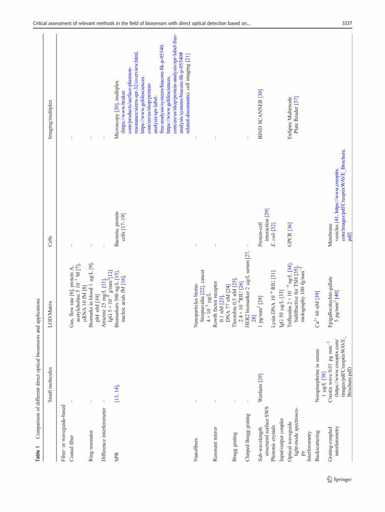
Table1
Com
parisonof
differentd
irecto
pticalbiosensorsandapplications
Smallm
olecules
LOD/M
atrix
Cells
Imaging/multip
lex
Fiber-or
waveguide-based
Coatedfiber
–Gas,flowrate[6],proteinA,
acetylcholine510
−3M
[7],
μRNA10
fM[8]
––
Ringresonator
–Biomarks
inblood1μg/L[9],
pM–nM
[10]
––
Differenceinterferom
eter
–Atrazine25
mg/L[11],
IgG5×10
−9g/mm
2[12]
––
SPR
[13,14],
Biomarkers500ng/L
[15],
nucleicacidsfM
[16],
Bacteria,protein
cells
[17–19]
Microscopy[20],m
ultip
lex
(http
s://w
ww.bruker.
com/products/surface-plasmon-
resonance/sierra-spr-32/overview
.htm
l,https://w
ww.gelifesciences.
com/en/us/shop/protein-
analysis/spr-label-
free-analysis/system
s/biacore-8k-p-05540,
https://w
ww.gelifesciences.
com/en/us/shop/protein-analysis/spr-label-free-
analysis/systems/biacore-8k-p-05540#
related-documents),cellim
aging[21]
Nanofibers
–Nanoparticlesbiotin-
Streptavidin[22],cancer
4×10
−5μg/L
––
Resonantm
irror
–Row
thfactor
receptor
0.1nM
[23],
DNA77
nM[24]
––
Bragg
grating
–Throm
bin0.5nM
[25],
2.4×10
−4RIU
[26]
Chirped
Bragg
grating
–HER2biom
arker2μg/Lserum
[27,
28]
––
Sub-wavelength
structured
surfaceSWS
Warfarin[29]
1pg/m
m2[29]
Protein-cell
interaction[29]
BIN
DSC
ANNER[30]
Photoniccrystals
Lysin-D
NA10
−4RIU
[31]
E.coli[32]
Input/o
utputcoupler
IgG50
μg/L[33]
Opticalwaveguide
light-m
odespectrosco-
py
Trifluralin
2×10
—4ng/L
[34],
bidiffractiveforTSH
[35],
molography100fg/m
m−2
GPC
R[36]
EnS
pire
Multim
ode
PlateReader[37]
Interferom
etry
Backscattering
Norepinephrinein
serum
1μg/L[38]
Ca2
+60
nM[39]
Gratin
g-coupled
interferom
etry
Creotix
wave0.01
pgmm
−2
(http
s://w
ww.creoptix
.com
/im
ages/pdf/Creoptix
WAVE_
Brochure.pdf)
Epigallo
catechin-gallate
5pg/m
m2[40]
Mem
brane
vesicles
[41,https://w
ww.creoptix
.com/im
ages/pdf/Creoptix
WAVE_B
rochure.
pdf]
Critical assessment of relevant methods in the field of biosensors with direct optical detection based on... 3337

measure the refractive index and its changes. Thus, they es-sentially need extreme referencing of temperature influence.Reflectometric methods which measure the superposition ofthe reflected beams at the boundaries of the biological inter-layer and its changes have no problems to detect even cells (nodecay as for evanescent fields) and show negligible tempera-ture dependence (decrease of refractive index with tempera-ture is compensated by increase of layer thickness).Reflectometric interference is advantageous in measuringlarge-size analytes and with respect to temperature control.
Overall, it can be said that in principle most of the directoptical methods end in the same optophysical range. The ap-plication and the matrix of the sample determine the selectionof the optimum optical method, the biomolecular interactionprocess, and the necessary assay format. These parametersgovern the bioanalytical quality.
This review wants to point out that for many applicationsnumerous slight modifications of the basic optical principlesclaiming optimisation canbe found in the literature. Therefore,each upcoming “new” method has to be critically consideredwith respect to improvement obtained bymore complex opticsor biochemistry. At present, for all applications, a direct opti-cal detection method or even some commercial platforms areavailable, which are competitive to other detection methods.Developments in optics and electronics in the last decade havepromoted miniaturization and parallelization. Thus, for manyapplications direct optical detection is the method of choice.
Such biosensors are part of Analytics 4.0 [419]. They allowbetter process control, are essential for personalized medicine,will be interesting for citizen science, supply the necessarymegadata for artificial intelligence [420], will help to meetchallenges in analytical chemistry in the future and will pre-pare it for the Internet of Things [421]. It will be interestingwhether for biosensors the ideas of digital twins inmanufacturing or simulation processes can provide bettercomparable standardized information on results of biomolec-ular interaction analysis [422] in research and quality control .
Acknowledgments The author gratefully acknowledges extensive dis-cussions with MSc Johanna Hutterer, Dr. Lothar Leidner, and Dr.Günther Proll.
Funding Information Open Access funding provided by Projekt DEAL.
Compliance with ethical standards
Conflict of interest The author declares that there is no conflict ofinterest.
Open Access This article is licensed under a Creative CommonsAttribution 4.0 International License, which permits use, sharing, adap-tation, distribution and reproduction in any medium or format, as long asyou give appropriate credit to the original author(s) and the source, pro-vide a link to the Creative Commons licence, and indicate if changes weremade. The images or other third party material in this article are included
Tab
le1
(contin
ued)
Smallm
olecules
LOD/M
atrix
Cells
Imaging/multip
lex
Mach-Zehnder
interferom
eter
+40
kDa10
−11M
[43],pesticide
10−9
mol/L
[44],sim
azine
0.1μg/L[45],D
NAfM
[46]
Young
interferom
eter
Affinity
reactio
n750fg/m
m2[324],
refractiv
eindex6×10
−84channel[47]
Dualp
olarization
interferom
eter
Aflatoxin
0.01
μg/L[48]
IgG4pg/m
m2[49],
Matrilin
-A-dom
ainto
collagen[50]
Ellipsom
etry
IgG15
μg/L[51]
HepatitisB[52]
BIA
[53,54]
Reflectom
etric
interference
spectroscopy
Biotin
40nM
[55]
Testosterone
130ng/L
[56],protein
1pg/m
m−2
[57],
Celladhesion[58],layer,
vesicles
[59],
DNA5.2pg/m
m2[60],
antip
hospholip
idsyndrome[61],
peptidelib
raries
[62]
Biolayerinterferom
etry
HRP2
[63]
Cellw
all–targeting[64],P
seudom
ona
aeruginosa
[65]
Multip
lex[66],L
angm
uir-Blogett[67]
Gauglitz G.3338
in the article's Creative Commons licence, unless indicated otherwise in acredit line to the material. If material is not included in the article'sCreative Commons licence and your intended use is not permitted bystatutory regulation or exceeds the permitted use, you will need to obtainpermission directly from the copyright holder. To view a copy of thislicence, visit http://creativecommons.org/licenses/by/4.0/.
References
1. Cooper MA. Optical biosensors in drug discovery. Nat Rev.2002;1:515–28.
2. Eddowes MJ. Direct immunochemical sensing: chemical princi-ples and fundamental limitations. Biosensors. 1987;3:1–15.
3. Piehler J, Brecht A, Giersch T, HockB, Gauglitz G. Assessment ofaffinity constants by rapid solid phase detection of equilibriumbinding in a flow system. J Immunol Methods. 1997;201:189–206.
4. Day YSN, Baird CL, Rich RL, Myszka D. Direct comparison ofbinding equilibrium, thermodynamic, and rate constants deter-mined by surface- and solution-based biophysical methods.Protein Sci. 2002;11:1017–27.
5. Yang HT, Jaeger M, Walker A, Wei D, Leiker K, Tao WT. Breakbreast cancer addiction by crispr/cas9 genome editing. J Cancer.2018;9(2):1–13.
6. Ding ZY, Wang CH, Kiu K, Jiang JF, Yang D, Pan GY, et al.Distributed optical Fiber sensors based on optical frequency do-main Reflectometry: a review. Sensors. 2018;18:1072.
7. Zourob M, Goddard NJ. Metal clad leaky waveguides for chem-ical and biosensing applications. Biosens Bioelectron. 2005;20(9):1718–27.
8. Coutinho C, Somoza A. MicroRNA sensors based on gold nano-particles. Anal Bioanal Chem. 2019;411:1807–24.
9. Sun YZ, Fan XD. Optical ring resonators for biochemical andchemical sensing. Anal Bioanal Chem. 2011;399(1):205–11.
10. Steglich P, Hülsemann M, Dietzel B, Mai A. Optical biosensorsbased on silicon-on-insulator ring resonators: a review.Molecules.2019;24:519.
11. Lukosz W, Stamm C, Moser HR, Ryf R, Dübendorfer J.Difference interferometer with new phase-measurement methodas integrated-optical refractometer, humidity sensor and biosen-sor. Sensors Actuators B Chem. 1997;38–39:316–23.
12. Huber W, Barner R, Fattinger C, Hübscher J, Koller H, Müller F,et al. Direct optical immunosensing (sensitivity and selectivity).Sensors Actuators B Chem. 1992;6:122–6.
13. Masson JF. Surface plasmon resonance clinical biosensors formedical diagnostics. ACS Sens. 2017;2(1):16–30.
14. Kaminski T, Geschwindner S. Perspectives on optical biosensorutility in small-molecule screening. Expert Opin Drug Discov.2017;12(11):1083–6.
15. Špačková B,Wrobel P, BockováM, Homola J. Optical biosensorsbased on Plasmonic nanostructures: a review. Proc IEEE.2016;104(12):2380–408.
16. Šípová H, Homola J. Surface plasmon resonance sensing ofnucleic acids: a review. Anal Chim Acta. 2013;773:9–23.
17. Bocková M, Slabý J, Špringer T, Homola J. Advances in SurfacePlasmon Resonance Imaging and Microscopy and TheirBiological Applications. Annu Rev Anal Chem. 2019;12:151–76.
18. Chabot V, Miron Y, Gradbois M, Charette PG. Long range surfaceplasmon resonance for increased sensitivity in living cell biosens-ing through greater probing depth. Sens Actuator. 2012;174:94–101.
19. Moore DT. Gradient-index optics: a review. Appl Opt. 1980;19:1035–8.
20. Rothenhäusler B, KnollW. Surf ace-plasm onmicroscopy. Nature.1988;332:615–7.
21. Stojanović I, Schasfoort RBM, Terstappen L. Analysis of cellsurface antigens by Surface Plasmon Resonance imaging.Biosens Bioelectron. 2014;52:36–43.
22. Chau LK, Lin YF, Cheng SF, Lin TJ. Fiber-optic chemical andbiochemical probes based on localized surface plasmon reso-nance. Sens Actuators B Chem. 2006;113:100–5.
23. Moroncini G, Cuccioloni M, Mozzicafreddo M, Pozniak KN,Grieco A, Paolini C, et al. Characterization of binding and quan-tification of human autoantibodies to PDGFRα using a biosensor-based approach. Anal Biochem. 2017;528:26–33.
24. De Tommasi E, De Stefano L, Rea I, Di SarnoV, Rotiroti L, ArcariP, et al. Porous Silicon Based Resonant Mirrors for BiochemicalSensing. Sensors. 2009;8:6549–56.
25. Bekmurzayeva A, Dukenbayev K, Shaimerdenova M,Bekniyazov I, Ayupova T, Sypabekova M, et al. Etched FiberBragg Grating Biosensor Functionalized with Aptamers forDetection of Thrombin. Sensors. 2018;18:4298.
26. Xu W, Flueckiger J, Schmidt S, Grist S, Fard ST, Kirk J, et al. Asilicon photonic biosensor using phase-shifted Bragg gratings inslot waveguide. J Biophotonics. 2013;6(19):821–8.
27. Sun DD, Ran Y, Wang GJ. Label-Free Detection of CancerBiomarkers Using an in-Line Taper Fiber-Optic Interferometerand a Fiber Bragg Grating. Sensors. 2017;17:2559.
28. Lee MH, Jung SY, Kang SH, Song EJ, Park IH, Kong SY, et al.The Significance of Serum HER2 Levels at Diagnosis on IntrinsicSubtype-Specific Outcome of Operable Breast Cancer Patients.PLoS One. 2016;11(10):e0163370.
29. Cunningham BT, Li P, Schulz S, Lin B, Baird C, Gerstenmaier J,et al. Label-free assays on the BIND system. J Biomol Screen.2004;9:481–90.
30. https://www.biocompare.com/Life-Science-News/64941-SRU-Biosystems-Announces-Introduction-Of-BIND-SCANNER-For-Primary-And-Stem-Cell-Applications/ (accessed 12.11.2019)
31. Rindorf L, Jensen JB, Dufva M, Pedersen LH. Høiby, Bang O.Photonic crystal fiber long-period gratings for biochemical sens-ing. Opt Express. 2006;14(18):8224–31.
32. Hao JJ; Xie X, Gu KD, Du WC, Liu YJ, Yang HW. Research onPhotonic Crystal-Based Biosensor for Detection of Escherichiacoli Colony. Plasmonics 2019; Ahead of Print.
33. Lukosz W. Integrated optical chemical and direct biochemicalsensors. Sensors and Actuators B. 1995;29:37–50.
34. Adányi N, Szendrö I, Székács A. OWLS Based Nanosensors forAgro-Environmental and Food Safety. J Adv Agric Technol.2017;4(4):335–9.
35. Gatterdam V, Frutiger A, Stengele KP, Heindl D, Lübbers T,Vörös J, et al. Focal molography is a new method for the in situanalysis of molecular interactions in biological samples. NatNanotechnol. 2017;12:1089–95.
36. Fang Y. Non-invasive optical biosensor for probing cell signaling.Sensors. 2007;7:2316–29.
37. https://www.corning.com/emea/de/products/life-sciences/products/label-free-detection.html and download file CLS-ES-038 REV4 DL.pdf
38. Kammer MN, Olmsted IR, Kussrow AK, Morris MJ, JacksonGW, Bornhop DJ. Characterizing aptamer small molecule interac-tions with backscattering interferometry. Analyst. 2014;139:5879–84.
39. Kammer MN, Kussrow AK, Olmsted IR, Bornhop DJ. A highlycompensated interferometer for biochemical analysis. ACS Sens.2018;3:1546–52.
40. Peter B, Saftics A, Kovacs B, Kurunczi S, Horvath R. Oxidizationincreases the binding of EGCG to serum albumin revealed by
Critical assessment of relevant methods in the field of biosensors with direct optical detection based on... 3339
kinetic data from labelfree optical biosensor with reference chan-nel. Analyst. 2020;145:588–95.
41. GyörgyB, Szabó TG, PásztóiM, et al. Membrane vesicles, currentstate-of-the-art: emerging role of extracellular vesicles. Cell MolLife Sci. 2011;68:2667–88.
42. Liang YX, Zhao MS, Wu ZL, Morthier G. Bimodal waveguideinterferometer ri sensor fabricated on low-cost polymer platform.IEEE Photonics J. 2019;11(2):6801108.
43. Heideman RG, Kooyman RPH, Geve J. Performance of a highlysensitive optical waveguide Mach-Zehnder interferometerimmunosensor. Sens Actuator B. 1993;10:209–17.
44. Ingenhoff J, Gauglitz G. Design of new integrated optical sub-strates for immunoanalytical applications. Fresenius J AnalChem. 1994;349:355–9.
45. Drapp B, Piehler J, Brecht A, Gauglitz G, Luff BJ, Wilkinson JS,et al. Integrated Optical Mach-Zehnder Interferometers asSimazine Immunoprobes. Sens Actua. 1997;B39:277–82.
46. Murib MS, Martens D, Bienstman P. Label-free real-time opticalmonitoring of DNA hybridization using SiNMach-Zehnder inter-ferometer-based integrated biosensing platform. J Biomed Opt.2018;23(12):127002.
47. Ymeti A, Kanger JS, Geve J, Besselink GAJ, Lambeck PV, WijnR, et al. Integration of microfluidics with a four-channel integratedoptical Young interferometer immunosensor. BiosensBioelectronic. 2005;20:1417–21.
48. Nabok A, Al-Jawdah AM, Tsargorodska A. Development of pla-nar waveguide-based immunosensor for detection of low molec-ular weight molecules such as mycotoxins. Sens Actuator B.2017;247:975–80.
49. Wang M, Uusitalo S, Liedert C, Hiltunen J, Hakalahti L, MyllyläR. Polymeric dual-slab waveguide interferometer for biochemicalsensing applications. Appl Opt. 2012;51(12):1886–93.
50. Fresquet M, Jowitt TA, Ylöstalo J, Coffey P, Meadows RS, Ala-Kokko L, et al. Structural and Functional Characterization ofRecombinant Matrilin-3 A-domain and Implications for HumanGenetic Bone Diseases. J Biol Chem. 2007;282(48):34634–43.
51. Li KW, Wang SA, Wang LM, Yu H, Jing N, Xue R, Wang ZB.Fast and sensitive ellipsometry-based biosensing. Sensors.2018;18(15). https://doi.org/10.3390/s18010015.
52. Diware MS, Cho HM, Chegal W, Cho YJ, O SW, Paek SH, et al.Label-free detection of hepatitis B virus using solution immersedsilicon sensors. Biointerphases. 2017;12:01A402.
53. Jin G, Tengvall P, Lundström I, Arwin H. A biosensor conceptbased on imaging ellipsometry for visualization of biomolecularinteractions. Anal Biochem. 1995;232:69–72.
54. Jin G, Jansson R, Arwin H. Imaging ellipsometry revisited: devel-opments for visualization of thin transparent layers on silicon sub-strates. Rev. Sci Instrum. 1996;67:2930–6.
55. Piehler J, Brecht A, Gauglitz G. Affinity Detection of LowMolecular Weight Analytes. Anal Chem. 1996;68:139–43.
56. Rau S, Gauglitz G. Reflectometric interference spectroscopy(RIfS) as a new tool to measure in the complex matrix milk atlow analyte concentration. Anal Bioanal Chem. 2012;402(1):529–36.
57. Lee JC, An JY, Kim BW. Application of anodized aluminumoxide as a biochip substrate for a Fabry-Perot interferometer. JChem Technol Biotechnol. 2007;82:1045–52.
58. Möhrle BP, Köhler K, Jaehrling J, Brock R,Gauglitz G. Label-freecharacterization of cell adhesion using reflectometric interferencespectroscopy (RIfS). Anal Bioanal Chem. 2006;384:407–13.
59. Kojima T. Combined Reflectometric Interference Spectroscopyand Quartz Crystal Microbalance Detect Differential Adsorptionof Lipid Vesicles with Different Phase Transition Temperatures onSiO2, TiO2, and Au Surfaces. Anal Chem. 2017;89:13596–13,602.
60. Daaboul GG, Vedula RS, Ahn S, Lopez CA, Reddington A,Özkumur E, et al. LED-based Interferometric ReflectanceImaging Sensor for quantitative dynamic monitoring of biomolec-ular interactions. Biosens Bioelectron. 2011;26:2221–7.
61. Bleher O, Schindler A, YinMX, Holmes AB, Luppa PB, GauglitzG, et al. Development of a new parallelized, optical biosensorplatform for label-free detection of autoimmunity-related antibod-ies. Anal Bioanal Chem. 2014;406:3305–14.
62. Burger J, Rath C,Woehrle J, Meyer PA, Ammar NB, Kilb N, et al.Low-Volume Label-Free Detection of Molecule-ProteinInteractions on Microarrays by Imaging ReflectometricInterferometry. SLAS Technol. 2017;22(4):437–46. https://doi.org/10.1177/221106821667512.
63. Markwalter CF, Jang IK, Burton RA, Domingo GJ, Wright DW.Biolayer interferometry predicts ELISA performance of monoclo-nal antibody pairs for Plasmodium falciparum histidine-rich pro-tein 2. Anal Biochem. 2017;534:10–3.
64. Jin Y, Zheng H, Ibanez AC, Patil PD, Lv S, Luo M, et al. Cell-wall-targeting antibiotics cause lag-phase bacteria to form surface-mediated filaments promoting the formation of biofilms and ag-gregates. Chembiochem. 2019;20:1–12.
65. Paulsson M, Su YC, Ringwood T, Uddén F, Riesbeck K.Pseudomonas aeruginosa uses multiple receptors for adherenceto laminin during infection of the respiratory tract and skinwounds. Scientific reports. 2019;9:18168.
66. Fortebio: https://www.moleculardevices.com/products/biologics/label-free-bli-detection.
67. Murray DJ, Kim JH, Grzincic EM, Kim SC, Abate AR,Zuckerman RN. Uniform, Large-Area, Highly Ordered PeptoidMonolayer and Bilayer Films for Sensing Applications.Langmuir. 2019;35:13671–80.
68. Pierce MM, Raman CS, Nall BT. Isothermal titration Calorimetryof protein-protein interactions. Methods. 1999;19:213–21.
69. Freire E, Mayorga OL, Straume M. Isothermal titration calorime-try. Anal Chem. 1990;62:950A–69A.
70. Falconer RJ. Applications of isothermal titration calorimetry – theresearch and technical developments from 2011 to 2015. J MolRecognit. 2016;29:504–15.
71. Su HX, Xu YC. Application of ITC-based characterization ofthermodynamic and kinetic Association of Ligands with Proteinsin drug design. Front Pharmacol. 2018;9:1133.
72. Burnouf D, Ennifar E, Guedich S, Puffer B, Hoffmann G, Bec G,et al. kinITC: a new method for obtaining joint thermodynamicand kinetic data by isothermal titration Calorimetry. J Am ChemSoc. 2012;134:559–68.
73. Lerchner J, Wolf A, Schneider HJ, Mertens F, Kessler E, Baier V,et al. Nano-calorimetry of small-sized biological samples.Thermochim Acta. 2008;477(1–2):48–53.
74. Barsan N, Gauglitz G, Oprea A, Ostertag E, Proll G, Rebner K,Schierbaum K, Schleifenbaum F, Weimar U. Chemical andBiochemical Sensors – 1. Fundamentals. In Ullmann’sEncyclopedia of Industrial Chemistry. VCH Verlagsgesellschaft;2016.
75. Barsan N, Gauglitz G, Oprea A, Ostertag E, Proll G, Rebner K,Schierbaum K, Schleifenbaum F, Weimar U. Chemical andBiochemical Sensors – 2. Applications. In Ullmann’sEncyclopedia of Industrial Chemistry, VCH Verlagsgesellschaft;2016.
76. Länge K, Gruhl FJ, Rapp M. Surface Acoustic Wave (SAW)Biosensors: COUPLING OF SENSING LAYERS ANDMEASurement. In Microfluidic diagnostics: methods and proto-cols, methods in molecular biology (Eds. Jenkins G, MansfieldCD) Springer Science+Business Media 2013, Vol 949.
77. Fritz J. Cantilever biosensors. Analyst. 2008;133:855–63.78. Álvarez M, Carrascosa LG, Zinoviev K, Plaza JA, Lechuga LM.
Biosensors based on Cantilevers. In: Rasooly A, Herold KE,
Gauglitz G.3340

editors. Biosensors and Biodetection. Methods in MolecularBiology, vol. 504: Humana Press; 2009. p. 51–71.
79. Qiao XX, Zhang XJ, Tian Y, Meng YG. Progresses on the theoryand application of quartz crystal microbalance. Appl Phys Rev.2016;3:031106.
80. Noi K, Iwata A, Kato F, Ogi H. Ultrahigh-frequency, wirelessMEMS QCM biosensor for direct, label-free detection of bio-markers in a large amount of contaminants. Anal Chem.2019;91:9398–402.
81. Afzal A, Mujahid A, Schirhagl R, Bajwa SZ, Latif U, Feroz S.Gravimetric viral diagnostics: QCM based biosensors for earlydetection of viruses. Chemosensor. 2017;5:7.
82. Chen JY, Penn LS, Xi J. Quartz crystal microbalance: sensing cell-substrate adhesion and beyond. Biosens Bioelectron. 2018;99:593–602.
83. Noy P, Steiner R, Voelkle J, Hegner M, Fattinger C. Instrument forLabel-Free Detection of Noncoding RNAs. J Sens 2012; 208079,5 pages.
84. Wang JJ, Wang LH, Zhu YF, Zhang JY, Liao J, Wang SP, et al. Ahigh accuracy cantilever array sensor for early liver cancer diag-nosis. Biomed Microdevices. 2016;18:110.
85. Electrochemical and Mechanical Detectors, Lateral Flow AndLigands For Biosensors. In: Biosensors and biodection: methodsand protocols Vol. 2 (Eds. Rasooly A, Herold K) (Methods inMolecular Biology, Vol. 504) 2009, Humana Press.
86. Warsinke A. Electrochemical Biochips for protein analysis. In:Renneberg R, Lisdat F (eds) Biosensing for the 21st century(Advances in Biochemical Engineering/Biotechnology 109 (Ed.Scheper T) 2007, Springer-Verlag Berlin Heidelberg 2007, Vol.109.
87. Pänke O, Balkenhohl T, Kafka J, Schäfer S, Lisdat F. Impedancespectroscopy and biosensing. In Scheper T (eds) Advances inbiochemical engineering/biotechnology 109 Springer-VerlagBerlin Heidelberg 2008, Vol. 109, (195–237).
88. Lindholm-Sethson B, Nyström J, Malmsten M, Ringstad L,Nelson A, Geladi P. Electrochemical impedance spectroscopy inlabel-free biosensor applications: multivariate data analysis for anobjective interpretation. Anal Bioanal Chem. 2010;398:2341–9.
89. Diculescu VC, Chiorcea-Paquim AM, Oliveira-Brett AM.Applications of a DNA-Electrochemical biosensor 2016 TRAC-trend. Anal Chem. 2016;79:23–36.
90. Moussa S,Mauzeroll J. Review –microelectrodes: an overview ofprobe development and bioelectrochemistry applications from2013 to 2018. J Electrochem Soc. 2019;166(6):G25–38.
91. Hu LZ, Xi GB. Applications and trends in electrochemiluminescence.Chem Soc Rev. 2010;39:3275–304.
92. Hao N, Wang K. Recent devellpment of electrochemiluminescencesensors for food analysis. Anal Bioanal Chem. 2016;408:7035–48.
93. Wei L, Zhang YH, Eziz N, Yang YR, Li GX, Guan M. An ultra-sensitive electrochemiluminescence immunosensor for alpha-fetoprotein based on a poly(aniline-luminol)/graphene oxidenanocomposite. Anal Bioanal Chem. 2019;411:5175–86.
94. Borisov SM, Wolfbeis OS. Optical Biosensors. Chem Rev.2008;108:423–61.
95. Hermann CA, Duerkop A, Baeumner AJ. Food safety analysisenabled through biological and synthetic materials: a critical re-view of current trends. Anal Chem. 2019;91:569–87.
96. Morales MA, Halpern JM. Guide to selecting a biorecognitionelement for biosensors. Bioconjug Chem. 2018;29:3231–9.
97. Seidel M, Niessner R. Automated analytical microarrays: a criticalreview. Anal Bioanal Chem. 2008;391:1521–154.
98. Bally M, Halter M, Vörös J, Grandin HM. Review - optical mi-croarray biosensing techniques. Surf Interface Anal. 2006;38:1442–58.
99. Förster T. Energiewanderung und Fluoreszenz. DieNaturwissenschaften. 1946;6:166–75.
100. Lakowicz JR. Principles of Fluorescence Spectroscopy. 3rd ed.Heidelberg: Springer Verlag; 2006.
101. Wolfbeis OS. An overview of nanoparticles commonly used influorescent bioimaging. Chem Soc Rev. 2015;44(14):4743–68.
102. Ligler FS, Taitt C. Optical Biosensors, Today and Tomorrow. 2nded: Elsevier Science; 2008.
103. Ligler FS. Fluorescence-based optical biosensors. In: Pavesi L,Fauchet PM, editors. Biophotonics. biological and medical phys-ics, biomedical engineering. Berlin, Heidelberg: Springer; 2008.
104. Nagl S, Wolfbeis OS. Optical multiple chemical sensing: statusand current challenges. Analyst. 2007;132(6):507–11.
105. Klimant I, Huber C, Liebsch G, Neurauter G, Stangelmayer A,Wolfbeis O. Dual Lifetime Referencing (DLR) - a new scheme forconverting fluorescence intensity into a frequency-domain ortime-domain information. Springer Series on Fluorescence 2001,New Trends in Fluorescence Spectroscopy:257–274.
106. van de Hulst HC. Light scattering by small particles. New Yorrk:Wiley; 1957.
107. Wang JJ, Huang XY, Liu H, Dong CQ, Ren JC. Fluorescence andscattering light cross correlation spectroscopy and its applicationsin homogeneous immunoassay. Anal Chem. 2017;89(10):5230–7.
108. Cooper MA. Label-free screening of bio-molecular interactions.Anal Bioanal Chem. 2003;377:834–42.
109. Perkel JM. Who needs labels? Macromolecular interaction sanslabels. Science. 2009;19:1561.
110. Cooper MA. Optical biosensors: where next and how soon? DrugDiscov Today. 2006;11(23/24):1061–7.
111. Fan XD, White IM, Shopova SI, Zhu HY, Suter JD, Sun YZ.Sensitive optical biosensors for unlabelled targets: a review. AnalChim Acta. 2008;620:8–26.
112. Gauglitz G, Goodard NJ. Direct Optical Detection in Bioanalytics.In: Gauglitz G, Moore DS, editors. Handbook of Spectroscopy,vol. 3. 2nd ed: WILEY-VCH; 2014. p. 1115–58.
113. Zanchetta G, Lanfranco R, Giavazzi F, Bellini T, Buscaglia M.Emerging applications of label-free optical biosensors.Nanophotonics. 2017;6(4):627–45.
114. Peltomaa R, Glahn-Martínez B, Benito-Peña E, Moreno-BondiMC. Optical biosensors for label-free detection of small mole-cules. Sensors. 2018;18:4126. https://doi.org/10.3390/s18124126.
115. Moore TJ, Moody AS, Payne TD, Sarabia GM, Daniel AR,Sharma B. In vitro and in vivo SERS biosensing for disease diag-nosis. Biosensors. 2018;8(2):46/1–46/25.
116. Ehni M, Pröll F. Immunoassays. In: Gauglitz G, Moore DS, edi-tors. Handbook of Spectroscopy, vol. 3. 2nd ed: WILEY-VCH;2014. p. 1313–34.
117. Piehler J, Brecht A, Geckeler KE, Gauglitz G. Surface modifica-tion for direct Immunoprobes. Biosens Bioelectron. 1996;11:579–90.
118. Gauglitz G, Proll G. On-Site Analysis. In: Popp J, Tuchin VV,Chiou A, Heinemann SH, editors. Handbook of Biophotonics,vol. 3: WILEY-VCH; 2012. p. 141–72.
119. Hecht E. Optics. 5th ed: Pearson; 2016.120. Gauglitz G, NahmW. Observation of spectral interferences for the
determination of volume and surface effects of thin polymer films.Fresenius Z Anal Chem. 1991;341:279–83.
121. Gauglitz G, Krause-Bonte J, Schlemmer H, Matthes A. Spectralinterference Refractometry by diode Array spectrometry. AnalChem. 1988;60:2609–12.
122. Špačková B, Lynn NS Jr, Slabý J, Šípová H, Homola J. A route tosuperior performance of a nanoplasmonic biosensor: considerationof both photonic and mass transport aspects. ACS Photonics.2018;5:1019–25.
123. De Avila BEF, Watkins HM, Pingarron JM, Plaxco KW, PalleschiG, Ricci F. Determinants of the detection limit and specificity ofsurface-based biosensors. Anal Chem. 2013;85:6593–7.
Critical assessment of relevant methods in the field of biosensors with direct optical detection based on... 3341

124. Piehler J, Brecht A, Gauglitz G, Maul C, Grabley S, Zerlin M.Specific binding of low molecular weight ligands with direct op-tical detection. Biosens Bioelectron. 1997;12:531–8.
125. Snyder AW, Love J. Optical Waveguide Theory: Springer; 1983.126. Kozma P, Kehl F, Ehrentreich-Förster E, Stamm C, Bier FF.
Integrated planar optical waveguide interferometer biosensors: acomparative review. Biosens Bioelectron. 2014;58:287–307.
127. Lambeck PV. Integrated optical sensors for the chemical domain(review article). Meas Sci Technol. 2006;17:R93–R116.
128. Conzen JP, Bürck J, Ache HJ. Characterization of a fiber-opticevanescent wave absorbance sensor for nonpolar organic com-pounds. Appl Spectrosc. 1993;47(6):753–63.
129. Bürck J, Zimmermann B, Mayer J, Ache HJ. Integrated opticalNIR-evanescent wave absorbance sensor for chemical analysis.Fresenius J Anal Chem. 1996;354:284–90.
130. Mizuno Y, Hayashi N, Fukuda H, Nakamura K. Phase-detectedBrillouin optical correlation-domain reflectometry. Opt Rev.2018;25:473–85.
131. Piraud C, Mwarania E, Wylangowski G, Wilkinson J.Optoelectrochemical thin-film chlorine sensor employing evanes-cent fields on planar optical waveguides. Anal Chem. 1992;64:651–5.
132. Saeed A, Mehmood S, Yasar M, Quddusi HM, Bhatti AS.Comparison of E. coli detection in single and bi-metal clad wave-guide biosensors. Sensors Actuators A Phys. 2011;168(1):101–4.
133. Li BH, Ju HK. Label-free optical biosensors based on a planaroptical waveguide. Biochip J. 2013;7(4):295–318.
134. Yong Z, Tong RJ, Xia F, Yun P. Current status of optical fiberbiosensor based on surface plasmon resonance. BiosensBioelectron. 2019;142:111505.
135. Shukla SK, Kushwaha CS, Guner T, Demir MM. Chemicallymodified optical fibers in advanced technology: an overview.Opt Laser Technol. 2019;115:404–32.
136. Barrios CA, Canalejas-Tejero V, Herranz S, Urraca J, Moreno-Bondi MC, Avella-Oliver M, et al. Aluminum nanoholes for op-tical biosensing. Biosensors. 2015;5(3):417–31.
137. Lou JY, Wang YP, Tong LM.Microfiber optical sensors: a review.Sensors. 2014;14:5823–44.
138. Tong LM. Micro/Nanofibre optical sensors: challenges and pros-pects. Sensors. 2018;18:903.
139. Serpengüzel A, Arnold S, Griffel G. Excitation of resonances ofmicrospheres on an optical fiber. Opt Lett. 1995;20(7):654–6.
140. Vollmer F, Braun D, Libchaber A. Protein detection by opticalshift of a resonant microcavity. Appl Phys Lett. 2002;80(21):654–6.
141. Mancuso M, Goddard JM, Erickson D. Nanoporous polymer ringresonators for biosensing. Opt Express. 2012;20(1):245–55.
142. CalvoM, Guerber S, Beaudin G, CanvaM, Romeo PR, Baudot C,et al. Ring resonator designed for biosensing applicationsmanufactured on 300 mm SOI in an industrial environment. JpnJ Appl Phys. 2019;58(SB):SBBE02.
143. Tavousi A, Rakhashani MR, Mansouri-Birjandi MA. High sensi-tivity label-free refractometer based biosensor applicable toglycated hemoglobin detection in human blood using all-circularphotonic crystal ring resonators. Opt Commun. 2018;429:166–74.
144. Luan EX, Shoman H, Ratner DM, Cheug KC, Chrostowski L.Silicon photonic biosensors using label-free detection. Sensors.2018;18:3519.
145. Lukosz W, Stamm C. Integrated Optical interferometer as relativehumidity sensor and differential refractometer. Sensors ActuatorsA Phys. 1991;25–27:185–8.
146. Stamm C, Lukosz W. Integrated optical difference interferometeras biochemical sensor. Sensors Actuators B Chem. 1994;18:183–7.
147. Schlatter D, Barner R, Fattinger C, Huber W, Hübscher J, Hurst J,et al. The difference interferometer: application as a direct affinitysensor. Biosens Bioelectron. 1993;8:109–16.
148. Fattinger C, Koller H, Schlatter D, Wehrli P. The difference inter-ferometer: a highly sensitive optical probe for quantification ofmolecular surface concentration. Biosens Bioelectron. 1993;8:99–107.
149. Stamm C, Dangel R, Lukosz W. Biosensing with the integrated-optical difference interferometer: dual-wavelength operation. OptCommun. 1998;153:347–59.
150. Gut K. A suggestion of utilizing bimodal layers of the polymerSU8 for the purpose of monitoring the changes in the refractiveindex. Opt Appl. 2012;17(2):407–16.
151. Liedberg B, Nylander C, Lundström I. Surface plasmon resonancefor gas detection and biosensing. Sensors Actuators. 1983;4:299–302.
152. Kretschmann E, Raether H. Radiative decay of nonradiative sur-face plasmons excited by light. Z Naturforsch A. 1968;23:2135–6.
153. Lukosz W, Tiefenthaler K. Sensitivity of integrated optical gratingand prism couplers as (bio)chemical sensors. Sensors Actuators.1988;15:273–84.
154. Homola J, Yee SS, Gauglitz G. Surface plasmon resonance sen-sors: review. Sensors Actuators B Chem. 1999;B54(1–2):3–15.
155. Jatschka J, Dathe A, Csáka A, FritzscheW, Stranik O. Propagatingand localized surface plasmon resonance sensing – a critical com-parison based on measurements and theory. Sens Biosensing Res.2016;7:62–70.
156. Chien FC, Chen SJ. A sensitivity comparison of optical biosensorsbased on four different surface plasmon resonance modes. BiosensBioelectron. 2004;20:633–42.
157. Singh P. SPR biosensors: historical perspectives and current chal-lenges. Sensors Actuators B Chem. 2016;229:110–30.
158. Souto DEP, Volpe J, Conçalves CC, Ramos CHI, Kubota LT. Abrief review on the strategy of developing SPR-based biosensorsfor application to the diagnosis of neglected tropical diseases.Talanta. 2019;205:120122.
159. Zhou JR, Qi QQ, Wang C, Q YF, Liu GM, Wang YB, et al.Surface plasmon resonance (SPR) biosensors for food allergendetection in food. Biosens Bioelectron. 2019;142:111449.
160. Soares L, Csáki A, Jatschka J, Fritzsche W, Flores O, Ricardo F,et al. Localized surface plasmon resonance (LSPR) biosensingusing gold nanotriagnles: detection of DNA hybridization eventsat room temperature. Analyst. 2014;139:4964–73.
161. Kotlarek D, Vorobii M, Ogieglo W, Knoll W, Rodriguez-Emmenegger C, Jakub D. Compact Grating-Coupled Biosensorfor the Analysis of Thrombin. ACS Sens. 2019;4:2109–16.
162. Garcés I, Aldea C, Mateo J. Four-layer chemical fiber opticplasmon-based sensor. Sens Actuator B Chem. 1992;7:77I–4.
163. Slavik R, Homola J, Čtyroký J. Miniaturization of fiber opticsurface plasmon resonance sensor. Sens Actuator B Chem.1998;51:311–5.
164. Xu Y, Jones NB, Fothergill JC, Hanning CD. Analytical estimatesof the characteristics of surface plasmon resonance fibre-opticsensors. J Mod Opt. 2000;47(6):1099–110.
165. Pollet J, Delport F, Janssen KPF, Tran DT, Wouters J, Verbiest T,et al. Fast and accurate peanut allergen detection with nanobeadenhanced optical fiber SPR biosensor. Talanta. 2011;83:1436–41.
166. Pollet J, Delport F, Janssen KPF, Jans K, Maes G, Pfeifer H. Fiberoptic SPR biosensing of DNA hybridization and DNA–proteininteractions. Biosens Bioelectron. 2009;25:864–9.
167. Kim JS, Kim SJ, Nguyen TT, Lee R, Li TH, Yun CY, et al. Label-Free Quantitative Immunoassay of Fibrinogen in AlzheimerDisease Patient Plasma Using Fiber Optical Surface PlasmonResonance. J Electron Mater. 2016;45(5):2354–60.
168. Nguyen TT, Trinh KTL, Yoon WJ, Lee NY. Integration of amicrofluidic polymerase chain reaction device and surface
Gauglitz G.3342

plasmon resonance fiber sensor into an inline all-in-one platformfor pathogenic bacteria detection. Sens Actuator B Chem.2017;242:1–8.
169. Horrer A, Krieg K, Freudenberg K, Rau S, Leidner L, Gauglitz G,et al. Plasmonic vertical dimter arrays as elements for biosensing.Anal Bioanal Chem. 2015;407:8225–31.
170. Vala M, Robelek R, Bocková M, Wegener J, Homola J. Real-timelabel-free monitoring of the cellular response to osmotic stressusing conventional and long-range surface plasmon. BiosensBioelectron. 2013;40:417–21.
171. Vala M, Etheridge S, Roach JA, Homola J. Long-range surfaceplasmons for sensitive detection of bacterial analytes. SensActuator B Chem. 2009;139:59–63.
172. Yanase Y, Hiragun T, Ishii K, Kawaguchi T, Yanase T, Kawai M,et al. Surface plasmon resonance for cell-based clinical diagnosis.Sensors. 2014;14:4948–59.
173. Zeng YJ, Zhou J, Wang XL, Cai ZW, Shao YH. Wavelength-scanning surface plasmon resonance microscopy: a novel toolfor real time sensing of cell-substrate interactions. BiosensBioelectron. 2019;145:111717.
174. Su YW, Wang W. Surface plasmon resonance sensing: from puri-fied biomolecules to intact cells. Anal Bioanal Chem.2018;410(17):3943–51.
175. Horrer A, Haas J, Freudenberger K, Gauglitz G, Kern DP,Fleischer M. Compact plasmonic optical biosensors based onnanostructured gradient index lenses integrated into microfluidiccells. Nanocale. 2017;9:17378–17,386.
176. Nivedha S, Babu PR, Senthilnathan K. Surface plasmon reso-nance: physics and technology. Curr Sc. 2018;115(1):56–63.
177. Scherbahn V, Nizamov S, Mirsky VM. Toward ultrasensitive sur-face plasmon resonance sensors. Springer Ser Chem SensorsBiosensors. 2018;16(Label-Free Biosensing):409–48.
178. Fathi F, Rashidi M-R, Omidi Y. Ultrasensitive detection by metalnanoparticles-mediated enhanced SPR biosensors. Talanta.2019;192:118–27.
179. Wegener MS, Robelek R. Label-free monitoring of cell-based as-says: comgining impedance analysis with SPR for multiparametriccell profiling. Biosens Bioelectron. 2013;49:63–70.
180. Peltomaa R, Agudo-Maestro I, Más V, Barderas R, Benito-Peña E.Development and comparison of mimotope-based immunoassayfor the analysis of fumonisin B1. Anal Bioanal Chem. 2019;411:6801–11.
181. O’Shannessy DJ. Determination of kinetic rate and equilibriumbinding constants for macromolecular interactions: a critique ofthe surface plasmon resonance literature. Curr Opin Biotechnol.1994;5:65–71.
182. Rich RL, Myszka DG. Survey of the year 2005 commercial opti-cal biosensor literature. J MOL RECOGNIT. 2006;19:478–534.
183. Li K, Zhou W, Zeng S. Optical micro/nanofiber-based localizedsurface plasmon resonance biosensors: fiber diameter dependence.Sensors. 2018;18:3295.
184. Cheng SF, Chau LK. Colloidal gold-modified optical fiber forchemical and biochemical sensing. Anal Chem. 2003;75:16–21.
185. Wongkaew N. Nanofiber-integrated miniaturized systems: an in-telligent platform for cancer diagnosis. Anal Bioanal Chem.2019;411:4251–64.
186. Cush R, Cronin JM, Stewart WJ, Maule CH, Molloy J, GoddardNJ. The resonant mirror: a novel optical biosensor for direct sens-ing of biomolecular interactions part I: principle of operation andassociated instrumentation. Biosens Bioelectron. 1993;8:347–53.
187. Buckle PE, Davies RJ, Kinning T, YeungD, Edwards PR, Pollard-Knight D. The resonant mirror: a novel optical sensor for directsensing of biomolecular interactions Part II: Applications. BiosensBioelectron. 1993;8:355–63.
188. Goddard NJ, Pollard-Knight D, Maule CH. Real-time biomolecu-lar interaction analysis using the resonant mirror sensor. Analyst.1994;119:583–8.
189. Hill KO, Fujii Y, Johnson DC, Kawasaki BS. Potosensitivity inoptical fiber waveguides: application to reflection filter fabrica-tion. Appl Phys Lett. 1978;32(10):647–9.
190. Othonos A. Fiber Bragg gratings. Rev. Sci Instrum. 1997;68(12):4309–41.
191. Caucheteur C, Guo T, Albert J. Polarization-assisted fiber bragggrating sensors: tutorial and review. J Lightwave Technol.2017;35(16):3311–22.
192. Wang LT, Fang N. Applications of Fiber-Optic InterferometryTechnology in Sensor Fields. In: Banishev AA, Bhowmick M,Wang J, editors. Optical Interferometry: InTechOpen Limited;2017.
193. Liu ZY, Zhang ZF, Tam HY, Tao XM. Multifunctional smart op-tical fibers: materials, fabrication, and sensing applications.Photonics. 2019;6:48.
194. Kashyap R. Fiber Bragg Gratings. 2nd ed. San Diego CA:Academic Press, Elsevier; 1999, 2010.
195. DeLisa MP, Zhang Z, Shiloach M, Pilevar S, Davis CC, Sirkis JS,et al. Evanescent Wave Long-Period Fiber Bragg Grating as anImmobilized Antibody Biosensor. Anal Chem. 2000;72:2895–900.
196. Nemova G, Kashyap R. Novel fiber Bragg grating assistedplasmon-polariton for bio-medical refractive-index sensors. JMater Sci: Mater Electron. 2007;18:S327–30.
197. Maguis S, Laffont G, Ferdinand P, Carbonnier B, Kham K,Mekhalif T, et al. Biofunctionalized tilted Fiber Bragg Gratingsfor label-free immunosensing. Opt Express. 2008;16(23):19049–62.
198. Ribaut C, Voisin V, Malachovská V, Dubois V, Mégret P, WattiezR, et al. Small biomolecule immunosensing with plasmonic opti-cal fiber grating sensor. BIosens Bioselectron. 2016;72:315–22.
199. Tosi D. Review of Chirped Fiber Bragg Grating (CFBG) Fiber-Optic Sensors and Their Applications. Sensors. 2018;18:2147.
200. Kalli K, Brady GP, Webb DJ, Jackson DA. Wavelength-divisionand spatial multiplexing using tandem interferometers for Bragggrating sensor networks. Opt Lett. 1995;20(24):2544–6.
201. Liang H, Jia PG, Liu J, Fang GC, Li Z, Hong YP, et al.Diaphragm-Free Fiber-Optic Fabry-Perot Interferometric GasPressure Sensor for High Temperature Application. Sensors.2018;18:1011.
202. Shnaiderman R, Wissmeyer G, Seeger M, Soliman D, Estrada H,Razansky D, et al. Fiber interferometer for hybrid optical andoptoacoustic intravital microscopy. Optica. 2017;4(10):1180–7.
203. Carvalho L, Alberto NJ, Gomes PS, Nogeuira RN, Pinto JL,Fernandes MH. In the trail of a new bio-sensor for measuringstrain in bone: osteoblastic biocompatibility. BiosensBioelectron. 2011;26:4046–52.
204. Abro ZA, Zhang YF, H CY, Lakho RA, Chen NY. Developmentof a smart garment for monitoring body postures based on FBGand flex sensing technologies. Sens Actuator A Phys. 2018;272:153–60.
205. Dakks ML, Kuhn L, Heidrich PF, Scott BA. Grating coupler forefficient excitation of optical guided waves in thin films. ApplPhys Lett. 1970;16:523.
206. Peng ST, Tamir T, Bertoni HL. Theory of periodic dielectric wave-guides. IEEE Trans Microw Theory Tech. 1975;23:123–33.
207. Quaranta G, Bassed G, Martin OJF, Gallinet B. Recent Advancesin Resonant Waveguide Gratings. Laser Photonics Rev. 2018;12:1800017.
208. Gabathuler W, Lukosz W. Electro-nanomechanically wavelength-tunable integrated-optical Bragg reflectors. Opt Commun.1997;135:385–93.
Critical assessment of relevant methods in the field of biosensors with direct optical detection based on... 3343

209. Gabathuler W, Lukosz W. Electro-nanomechanically wavelength-tunable integrated-optical Bragg reflectors. Part II: Stable deviceoperation. Opt Commun. 1998;4798:258–64.
210. Cunningham B, Li P, Lin B, Pepper J. Colorimetric resonant re-flection as a direct biochemical assay technique. Sens Actuator BChem. 2002;81:316–28.
211. Cunningham BT, Lin B, Qiu J, Li P, Pepper J, Hugh B. A plasticcolorimetric optical biosensor for multiparallel detection oflabelfree biochemical interactions. Sens Actuator B Chem.2002;85:219–26.
212. Cunningham B, Qiu J, Li P, Lin B. Enhancing the surface sensi-tivity of colorimetric resonant optical biosensors. Sens Actuator BChem. 2002;87:365–70.
213. Joannopoulos JD, Johnson SG,Winn JN. Photonic crystals: mold-ing the flow of light. Princeton NJ: Princeton University Press;2008.
214. Russell P. Review: photonic crystal fibers. Science. 2003;299:358–62.
215. Jensen JB, Pedersen LH, Hoiby PE, Nielsen LB, Hansen TP,Folkenberg JR, et al. Photonic crystal fiber based evanescent-wave sensor for detection of biomolecules in aqueous solutions.Opt Lett. 2004;29:1974–6.
216. Cox FM, Argyros A, Large MCJ. Ultracompact biochemical sen-sor built with two-dimensional photonic crystal microcavity. Opt.Express. 2006;14:4135.
217. Rindorf L, Bang O. Sensitivity of photonic crystal fiber gratingsensors: biosensing, refractive index, strain, and temperature sens-ing. J Opt Soc Am B. 2008;25(3):310–24.
218. Li MZ, He F, Liao Q, Liu J, Xu L, Jiang L, et al. UltrasensitiveDNA Detection Using Photonic Crystals. Angew Chem.2008;47(38):7258–62.
219. Akahane Y, Asano T, Song B, Noda S. High-Q photonicnanocavity in a two-dimensional photonic crystal. Nature.2003;425:944–7.
220. Mindy L, Fauchet PM. Two-dimensional silicon photonic crystalbased biosensing platform for protein detection. Opt Express.2007;15(8):4530–5.
221. Law CS, Lim SY, Abell AD, Santos A. Real-Time BindingMonitoring between Human Blood Proteins and Heavy MetalIons in Nanoporous Anodic Alumina Photonic Crystals. AnalChem. 2018;90:10039–10,048.
222. Cunningham BT, Zhang M, Zhuo Y, Kwon L, Race C. Recentadvances in biosensing with photonic crystal surfaces: a review.IEEE Sens J. 2016;16(10):3349–66.
223. Liu B, Monshat H, Gu ZZ, Lu M, Zhao XW. Recent advances inmerging photonic crystals and plasmonics for bioanalytical appli-cations. Analyst. 2018;143(11):2448–58.
224. Chen N, ChangM, Lu X, Zhou J, ZhangX. Photonic Crystal FiberPlasmonic Sensor Based on Dual Optofluidic Channel. Sensors.2019;19:5150.
225. Cunningham BT, Chan L, Mathias PC, Ganesh N. Photonic crys-tals: a platform for label-free and enhanced fluorescence biomo-lecular and cellular assays. Symp AA Mater Opt Sensors BiomedAppl. 2008;1133:AA04–1.
226. Loers G,Makhina T, BorkU, Dörnder A, Schachner M, Kleene R.The Interaction between Cell Adhesion Molecule L1, MatrixMetalloproteinase 14, and Adenine Nucleotide Translocator atthe Plasma Membrane Regulates L1-Mediated NeuriteOutgrowth of Murine Cerebellar Neurons. J Neurosci.2012;32(11):3917–30.
227. Che C, Li N, Long KD, Aguirre MA, Canady TD, Huang Q,Demircie U,Cunningham BT. Activate capture and digitalcounting (AC +DC) assay for protein biomarker detection inte-grated with a self-powered microfluidic cartridge. Lab on a Chip2019; Ahead of Print
228. Tiefenthaler K, Lukusz W. Integrated optical switches and gassensors. Opt Lett. 1984;9(4):137–9.
229. Tiefenthaler K. Grating couplers as integrated optical humidityand gas sensors. Thin Solid Films. 1985;126(3, 4):205–11.
230. Lukosz W, Tiefenthaler K. Embossing technique for fabricatingintegrated optical components in hard inorganic waveguiding ma-terials. Opt Lett. 1983;8(10):537–9.
231. Heuberger K, Lukosz W. Embossing technique for fabricatingsurface relief gratings on hard oxide waveguides. Appl Opt.1986;25:1499–504.
232. Kunz RE, Edlinger J, Curtis BJ, Gale MT, Kempen LU, RudigierH, et al. Grating Couplers in tapered waveguides for integratedoptical sensing. Proc SPIE Int Soc Opt Eng. 1994;2068:313–25.
233. Dübendorfer J, Kunz RE, Jobst G, Moser I, Urban G. Integratedoptical pH sensor using replicated chirped grating coupler sensorchips. Sens Actuator B Chem. 1998;50:210–9.
234. Cotttier K, Kunz RE, Voirin G, Wiki M; Thickness-modulatedwaveguides for integrated optical sensing Proceedings of SPIE-The International Society for Optical Engineering (2002),4616(Optical Fibers and Sensors for Medical Applications II),53–63.
235. Kunz RE. Gradient effective index waveguide sensors. SensActuator B Chem. 1993;11:167–76.
236. Kunz RE, Dübendorfer J. Novel miniature integrated optical go-niometers. Sens Actuator A Phys. 1997;60:23–8.
237. Kunz E. Miniature integrated optical modules for chemical andbiochemical sensing. Sens Actuator B Chem. 1997;38–39:13–28.
238. Kunz RE, Cottier K. Optimizing integrated optical chips for label-free (bio-)chemical sensing. Anal Bioanal Chem. 2006;384:180–90.
239. Seifert M, Tiefenthaler K, Heuberger K, Lukosz W, Mosbach K.An integrated Optical Biosensor (IOBS). Anal Lett. 1986;19(1, 2):205–16.
240. Nellen PM, Tiefenthaler K, Lukosz W. Integrated optical inputgrating couplers as biochemical sensors. Sens Actuator. 1988;15:285–95.
241. Nellen PM, LukoszW. Integrated optical input grating couplers aschemo- and immunosensors. Sens Actuator. 1990;B1:592–6.
242. Nellen PM, Lukosz W. Model experiments with integrated opticalinput grating couplers as direct immunosensors. BiosensBioelectron. 1991;6:517–25.
243. Tiefenthaler K, Lukosz W. Optical sensor for selectively detectingsubstances and for detection of the variation of refractive index insubstances. PCT Int Appl 1986;WO8607149 A119861204:52.
244. http://www.asiag.ch/, https://ch.kompass.com/c/artificial-sensing-instruments-asi-ag/ch068415/ (assessed 15.12.2019)
245. LukoszW, Nellen PM, StammC,Weiss P. Output grating couplerson planar waveguides as integrated optical chemical sensors. SensActuator. 1990;B1:585–8.
246. LukoszW, Clerc D, Nellen PM, StammC,Weiss P. Output gratingcouplers on planar optical waveguides as direct immunosensors.Biosens Bioelectron. 1991;6:227–32.
247. Lukosz W. Principles and sensitivities of integrated optical andsurface plasmon sensors for direct affinity sensing andimmunosensing. Biosens Bioelectron. 1991;6:215–25.
248. Brandenburg A, Gombert A. Grating couplers as chemical sen-sors: a new optical configuration. Sens Actuator B Chem.1993;17:35–40.
249. Leidner L, Gauglitz G. Development of a modified grating couplerin application to geosciences. Anal Bioanal Chem. 2011;400:2783–91.
250. Ramsden JJ. Optical Biosensors. J Mol Recognit. 1997;10:109–20.
251. Vörös J, Ramsden JJ, Csúcs G, Szendrö I, De Paul SM, Textor M,et al. Optical grating coupler biosensors. Biomaterials. 2002;23:3699–710.
Gauglitz G.3344

252. Quartz Crystal Microbalance & OWLS Biosensors, https://microvacuum.com/, http://www.owls-sensors.com/technology-overview.aspx, https://www.owls-sensors.com/owls-system(accessed 10th October 2019)
253. Székács I, Kaszás N, Gróf P, Erdélyi K, Szendrö I, Mihalik B,Pataki A, Antoni FA, Madarász E. Optical waveguide lightmodespectroscopic techniques for investigating membrane-bound ionchannel activities. PloS One 2013;https://doi.org/10.1371/journal.pone.0081398.
254. Peter B, Kurunczi S, Patko D, Lagzi I, Kowalczyk B, Rácz Z, et al.Label-Free in Situ Optical Monitoring of the Adorption ofOppositely Charged Metal Nanoparticles. Langmuir. 2014;30:13478–13,482.
255. Kovacs B, Saftics A, Biro A, Kurunczi S, Szalontai B, Kakasi B,et al. Kinetcis and structure of self-assembled flagellin monolayerson hydrophobic surfaces in the presence of Hofmeister salts: ex-perimental measurement of the protein interfacial tension at thenanometer scale. J Phys Chem C. 2018;122:21375–21,386.
256. Orgovan N, Patko D, Hos C, Kuruczi S, Szabó B, Ramsden JJ,et al. Sample handling in surface sensitive chemical and biologicalsensing: a practical review of basic fluidics and analyte transport.Adv Colloid Interfac. 2014;211:1–16.
257. Szalontai H, Adányi N, Kiss A. Comparative determination of twoprobiotics by QCM and OWLS-based immunosensors. NewBiotechnol. 2014;31(5):395–401.
258. Juan-Colás J, Johnson S, Krauss TF. Dual-mode electro-opticaltechniques for biosensing applications: a review. Sensors.2017;17:2047.
259. Fang Y, Ferrie AM, Fontaine NH, Mauro J, Balakrishnan J.Resonant waveguide grating biosensor for living cell sensing.Biophys J. 2006;91:1925–40.
260. Fang Y, Frutos AG, Verklereen R. Label-free cell-based assays forGPCR screening. CombChemHigh Throughput Screen. 2008;11:357–69.
261. Goral V,WuQ, Sun HY, FangY. Label-free optical biosensor withmicrofluidics for sensing ligand-directed functional selectivity ontrafficking of thrombin receptor. FEBS Lett. 2011;585:1054–60.
262. PerkinElmer EnSpire Multimode Plate Reader Label-free System:http://ir.perkinelmer.com/news-releases/news-release-details/perkinelmer-launches-enspirer-multimode-plate-reader-label-free(accessed 27.11.2019).
263. EnSp i re Mul t imode Pla t e Reader wi th download009446A_01_TCH_Enspire.pdf (accessed 27.11.2019).
264. Grundmann M, Kostenis E. Label-Free Biosensor Assays inGPCR Screening. In: Prazeres D, Martins S, editors. G protein-coupled receptor screening assays. Methods in MolecularBiology, vol. 1272. New York, NY: Humana Press; 2015.
265. Grundmann M. Label-free dynamic mass redistribution and bio-impedance methods for drug discovery. Curr Prot Pharmacol.2017;77:9.24.1–9.24.21.
266. Perkin Elmer, 009446A_01_TCH_Enspire.pdf267. Fattinger C, Mangold C, Gale MT, Schuetz H. Bidiffractive grat-
ing coupler: universal transducer for optical interface analytics.Opt Eng (Bellingham). 1995;34:2744–53.
268. Spinke J, Oranth N, Fattinger C, Koller H, Mangold C, VoegelinD. The bidiffract ive tra t ing coupler: applicat ion toimmunosensing. Sens Actuator B Chem. 1997;38–39:256–60.
269. O’Brien T, Johnson LH III, Alrich JL, Allen SG, Liang LT,Plummer AL, et al. The development of immunoassays for fourbiological threat agents in a bidiffractive grating biosensor.Biosens Bioelectron. 2000;14:815–28.
270. Fattinger C. Focal molography: coherent microscopic detection ofbiomolecular interaction. Phys Rev X. 2014;4:031024.
271. Frutiger A, Blickenstorfer Y, Bischof S, Forró C, Lauer M,Gatterdam V, et al. Principles for sensitive and robust biomolecu-lar interaction analysis: the limits of detection and resoluation of
diffraction-limited focal molography. Phys Rev Appl. 2019;11:014056.
272. Ince R, Narayanaswamy R. Analysis of the performance of inter-ferometry, surface plasmonresonance and luminescence as biosen-sors and chemosensors. Anal Chim Acta. 2006;569:1–20.
273. Kussrow A, Enders CS, Bornhop DJ. Interferometric methods forlabel-free molecular interaction studies. Anal Chem. 2012;84:779–92.
274. Lin VSY, Motesharei K, Dancil KPS, Sailor MJ, Ghadiri MR. Aporous silicon-based optical interferometric biosensor. Science.1997;278(5339):840–3.
275. Bornhop DJ. Microvolume index of refraction determinations byinterferometric backscatter. Appl Opt. 1995;34(18):3234–9.
276. Kenmore CK, Erskine SR, Bornhop DJ. Refractive-index detec-tion by interferometric backscatter in packed-capillary high-per-formance l iquid chromatography. J Chromatogr A.1997;762(219):225.
277. Wang ZL, BornhopDJ. Dual-capillary backscatter interfereometryfor high-sensitivity nanoliter-volume refractive index detectionwith density gradient compesation. Anal Chem. 2005;77:7872–7.
278. Sørensen HS, Larsen NB, Latham JC, Bornhop DJ, Andersen PE.Highly sensitive biosensing based on interference from light scat-tering in capillary tubes. Appl Phys Lett. 2006;89:151108.
279. Bornhop DJ, Latham JC, Kussrow A, Jones RD, Sørensen HS.Free-solution, label-free molecular interactions studied by back-scattering interferometry. Science. 2007;317:1732–6.
280. Khan M, Park SY. Liquid crystal-based biosensor with backscat-tering interferometry: a quantitative approach. BiosensBioelectron. 2017;87:976–83.
281. Saetear P, Chamieh J, Kammer MN, Manuel TJ, Biron JP,Bornhop DJ, et al. Taylor Dispersion analysis of polysaccharidesusing backscattering interferometry. Anal Chem. 2017;89:6710–8.
282. Leclercq L, Saetear P, Rolland-Sabaté A, Biron JP, Chamieh J,Cipelletti L, et al. Size-based characterization of polysaccharidesby Taylor dispersion analysis with photochemical oxidation orbackscattering interferometry detections. Macromolecules.2019;52:4421–31.
283. Chang CL, Acharya G, Savran CA. In situ assembled diffractiongrating for biomolecular detection. Appl Phys Lett. 2007;90:233901.
284. Markov DA, Swinney K, Bornhop DJ. Label-free molecular in-teraction determinations with nanoscale interferometry. J AmChem Soc. 2004;126(50):16659–16,664.
285. Kozma P, Hámori A, Kurunczia S, Cottier K, Horvath R. Gratingcoupled optical waveguide interferometer for label-free biosens-ing. Sens Actuator B Chem. 2011;155:446–50.
286. Patko D, Gyorgy B, Nemeth A, Szabó-Taylor KE, Kittel A, BuzasEI, et al. Label-free optical monitoring of surface adhesion ofextracellular vesicles by grating coupled interferometry. SensActuator B Chem. 2013;188:697–701.
287. Varma MM, Inerowicz HD, Regnier FE, Nolte DD. High-speedlabel-free detection by spinning-disk micro-interferometry.Biosens Bioelectron. 2004;19:1371–6.
288. ZhaoM, Nolte D, ChoW, Regnier F, VarmaM, Lawrence G, et al.High-Speed interferometric Detection of Label-FreeImmunoassays on the Biological Compact Disc. Clin Chem.2006;52(11):2135–40.
289. Tortajada-Genaro LA, Rodrigo A, Hevia E, Mena S, Niñoles R,Maquieira Á. Microarray on digital versatile disc for identificationand genotyping of Salmonella and Campylobacter in meat prod-ucts. Anal Bioanal Chem. 2015;407:7285–94.
290. Avella-Oliver M, Carrascosa J, Puchades R, Maquieira Á.Diffractive Protein Gratings as Optically Active Transducers forHigh-Throughput Label-free Immunosensing. Anal Chem.2017;89:9002–8.
Critical assessment of relevant methods in the field of biosensors with direct optical detection based on... 3345

291. Choi H, Chang C, Savran C, Nolte D. Diffraction-based BioCDbiosensor for point-of-care diagnostics. Proc. SPIE 10501, Opticaldiagnostics and sensing XVIII: Toward point-of-care diagnostics,105011K (20 February 2018)
292. Cho DG, Yoo H, Lee H, Choi YK, Lee M, Ahn DJ, et al. High-speed lateral flow strategy for a fast biosensing with an improvedselectivity and binding affinity. Sensors. 2018;18:1507.
293. Maldonado J, González-Guerrero AB, Domínguez C. Label-freebiomodal waveguide immunosensor for rapid diagnosis of bacte-rial infections in cirrhotic patients. Biosens Bioelectron. 2016;85:310–6.
294. Herranz S, Gavela AF, Lechuga LM. Label-free biosensors basedon bimodal waveguide (BiMW) interferometers. In: Rasooly A,Prickril B, editors. Biosensors and biodetection. Methods inMolecular Biology, vol. 1571. New York, NY: Humana Press;2017.
295. Maldonado J, EstévezMC, Fernández-Gavela A, González-LopezJ, González-Guerrero AB, Lechuga LM. Label-free detection ofnosocomial bacteria using a nanophotonic interferometric biosen-sor. Analyst 2019; https://doi.org/10.1039/c9an01485c.
296. Jamin JC. Neuer Interferential-Refractor. Annalen der Physik undChemie. 1856;174:345–9.
297. Mach L. Über einen Interferenzrefraktor. Zeitschrift fürInstrumentenkunde. 1892;12:89–93.
298. Zehnder L. Ein neuer Interferenzrefraktor. Zeitschrift fürInstrumentenkunde. 1891;11:275–85.
299. Heideman RG, Kooyman RPH, Greve J. Development of an op-tical waveguide interferometric immunosensor. Sens Actuator B.1991;4:297–9.
300. Brecht A, Ingenhoff J, Gauglitz G. Direct monitoring of antigen-antibody-interactions by spectral interferometry. Sens Actua.1992;6B:96–100.
301. Ingenhoff J, Gauglitz G, Wolf B. Simulation of transversal modalfields in integrated optic components. Anal Chim Acta. 1992;265:267–75.
302. Ingenhoff J, Drapp B, Gauglitz G. Biosensors using integratedoptical devices. Fresenius J Anal Chem. 1993;346:580–3.
303. Maisenhölder B, Zappe HP, Kunz RE, Riel P, Moser M, EdlingerJ. A GaAs/A1GaAs-based refractometer platform for integratedoptical sensing applications. Sens Actuator B. 1997;38–29:324–9.
304. Kempen LU, Kunz RE. ReplciatedMach-Zehnder interferometerswith focusing grating couplers for sensing applications. SensActuator B Chem. 1997;38–39:295–9.
305. Brosinger F, Freimuth H, Lacher M, Ehrfeld W, Gedig E,Katerkamp A, et al. A label-free affinity sensor with compensa-tion of unspecific protein interaction by a highly sensitive inte-grated optical Mach-Zehnder interferometer on silicon. SensActuator B Chem. 1997;44:350–5.
306. Heideman RG, Lambeck PV. Remote opto-chemical sensing withextreme sensitivity: design, fabrication and performance of a pig-tailed integration optical phase-modulatedMach-Zehnder interfer-ometer system. Sens Actuator B Chem. 1999;61:100–27.
307. Sepúlveda B, Armelles G, Lechuga LM. Magneto-optical phasemodulation in integrated Mach-Zehnder interferometric sensors.Sens Actuator A Phys. 2017;134:339–47.
308. Passaro VMN, Dell’Olio F, Casamassima B, De Leonardis F.Guided-Wave Optical Biosensors. Sensors. 2007;7:508–36.
309. Dér A, Valkai S, Mathesz A, Andó A, Andó I, Wolff EK, et al.Protein-based all optical sensor device. Sens Actuator B.2010;151:26–9.
310. Dante S, Duval D, Spúlveda B, González-Guerrero AB, SendraJR, Lechuga LM. All-optical phase modulation for integrated in-terferometric biosensors. Opt Express. 2012;20(7).
311. Chalyan T, Guider R, Pasquardini L, Zanetti M, Falke F,Schreuder E, Heideman RG, Pederzolli C, Pavesi L.Asymmetric Mach-Zehnder Interferometer Based Biosensors for
Aflatoxin M1 Detection. Biosensors 2016;6 (1). https://doi.org/10.3390/bios6010001.
312. Ramirez-Priego P, Martens D, Elamin AA, Soetaert P, Van RoyW,Vos R, et al. Label-Free and real-Time Detection of Tuberculosisin Human Urine Samples Using a Nanophotonic Pint-of-CarePlatform. ACS Sens. 2018;3:2079–86.
313. Sun LP, Huang Y, Huang TS, Yuan ZH, Lin WF, Sun Z, et al.Optical Microfiber Reader for Enzyme-Linked ImmunosorbentAssay. Anal Chem. 2019;91:14141–14,148.
314. Chatzianagnostou E, Manolis A, Dabos G, Ketzaki D, Miliou A,Pleros N, et al. Scaling the Sensitivity of Integrated Plasmo-Photonic Interferometric Sensors. 2019;6:1664–73.
315. Martens D, Bienstman P. Study on the limit of detection in MZI-based biosensor systems. Sci Rep. 2019;9:5767.
316. Molina-Fernández I, Leuermann J, Ortega-Moñux A,Wangüemert-Pérez JG, Halir R. Fundamental limit of detectionof photonic biosensors with coherent phase read-out. OptExpress. 2019;27(9):12616.
317. Manolis A, Chatzianagnostou E, Dabos G, Pleros N, Chmielak B,Giesecke AL, et al. Plasmonic co-integrated with silicon nitridephotonics for high-sensitivity interferometric biosensing. OptExpress. 2019;27(12):17102.
318. Knoerzer M, Szydzik C, Ren GH, Huertas CS, Palmer S, Tang P,et al. Optical frequency comb based system for photonic refractiveindex sensor interrogation. Opt Express. 2019;27(15):21532.
319. Misiakos K, Makarona E, Hoekman M, Fyrogenis R, TukkiniemiK, Jobst G, et al. All-silicon spectrally resolved interferometriccircuit for multiplexed diagnostics: a monolithic lab-on-a-chip in-tegrating all active and passive components. ACS Photonics.2019;6:1694–705.
320. Leidner L, Ewald M, Sieger M, Mizaikoff B, Gauglitz G.Migrating the Mach-Zehnder chemical and bio-sensor to themid-infrared region. Proc. SPIE 8774, Optical Sensors 2013:87740S.
321. Sieger M, Balluff F, Wang X, Kim SS, Leidner L, Gauglitz G,et al. On-Chip integrated mid-infrared GaAs/AlGaAs Mach−Zehnder interferometer. Anal Chem. 2013;85:3050–2.
322. Brandenburg A, Henniger R. Integrated optical Young interferom-eter. Appl Opt. 1994;33(25):5941–7.
323. Brandenburg A. Differential refractometry by an integrated-optical Young interferometer. Sens Actuator. 1997;38/39:266–71.
324. Brandenburg A, Krauter R, Künzel C, Stefan M, Schulte H.Interferometric sensor for detection of surface-bound bioreactions.Appl Opt. 2000;39(34):6396–405.
325. Schmitt K, Schirmer B, Brandenburg A. Label-free detection ofbiomolecules by waveguide interferometry. Proc SPIE.2005;5855.
326. Hoffmann C, Schmitt K, Brandenburg A, Hartmann S. Rapidprotein expression analysis with an interferometric biosensor formonitoring protein production. Anal Bioanal Chem. 2007;387:1921–32.
327. Aikio S, Zeilinger M, Hiltunen J, Hakalahti L, Hiitola-Keinänen J,Hiltunen M, et al. Disposable (bio)chemical integrated opticalwaveguide sensors implemented on roll-to-roll produced plat-forms. RSC Adv. 2016;6:50414–50,422.
328. Hartman NF. Optical sensing apparatus and method. US Patent.1990;4:940,328.
329. Hartman NF. Integrated optic interferometric sensor. US Patent.1997;5623561.
330. Hartman NF, Wyvill JC, Campbell DP, Edmonds P. Rapid re-sponse biosensor for detection and identification of commonfoodborne pathogens. SPIE. 1995n;2345:128,137.
331. Schneider BH, Edwards JG, HartmanNF. Hartman interferometer:versatile integrated optic sensor for label-free, real-time quantifi-cation of nucleic acids, proteins, and pathogens. Clin Chem.1997;43(9):1757–63.
Gauglitz G.3346
332. Ronan G. Doubling up – Dual polarization interferometry deter-mines protein structure and function. SPIE’s oemagazine. 2004:17–20.
333. Cross GH, Reeves AA, Brand S, Popplewell JF, Peel LL, SwannMJ, et al. A new quantitative optical biosensor for protein charac-terization. Biosens Bioselectron. 2003;19:383–90.
334. SwannMJ, Peel LL, Carrington S, Freeman NJ. Dual-polarizationinterferometry: an analytical technique to measure changes in pro-tein structure in real time, to determine the stoichiometry of bind-ing events, and to differentiate between specific and nonspecificinteractions. Anal Biochem. 2004;329:190–8.
335. Coswill BJ, Coffey PD, Yaseen M, Waigh TA, Freeman NJ, LuJR. Measurement of the thickness of ultra-thin adsorbed globularprotein layers with dual-polarisation interferometry: a comparisonwith neutron reflectivity. Soft Matter. 2011;7:7223–30.
336. Coffey PD, Swann MJ, Waigh TA, Mu QS, Lu JR. The structureand mass of heterogeneous thin films measured with dual polari-zation interferometry and ellipsometry. RSC Advances. 2013;3:3316–24.
337. Duval D, Gonzálesz-Guerrero AB, Dante S, Osmond J, Monge R,Fernández LJ, et al. Nanophotonic lab-on-a-chip platforms includ-ing novel bimodal interferometers, microfluidics and grating cou-plers. Lab Chip. 2012;12:1987–94.
338. Azzam RMA, Bashara NM. Ellipsometry and Polarized Light:North-Holland Pub. Co; 1977.
339. Tompkins HG, Irene EA. Handbook of Ellipsometry: WillamAndrew Inc; 2005.
340. Brecht A, Gauglitz G, Striebel C. Characterization ofBiomembranes by Spectral Ellipsometry. Biosens Bioelectron.1994;9:139–46.
341. Arwin H. Adsorption of proteins at solid surfaces. In: Hinrichs K,Eichhorn KJ, editors. Springer Series in Surface SciencesEllipsometry of functional organic surfaces and films, vol. 52:Springer; 2018. p. 31–48.
342. Cobet C. Ellipsometry: A survey of concept. In: Hinrichs K,Eichhorn KJ, editors. Springer Series in Surface SciencesEllipsometry of functional organic surfaces and films, vol. 52:Springer; 2018. p. 1–27.
343. Wang ZH, Jin G. Covalent immobilization of proteins for thebiosensor based on imaging ellipsometry. J Immunol Methods.2004;285:237–43.
344. Yuan W, Ho HP, Wu SY, Suen YK, Kong SK. Polarization-sensitive surface plasmon resonance enhanced ellipsometry bio-sensor using the photoelasticmodulation technique. Sens ActuatorA Phys. 2009;151:23–8.
345. Tanovska M, Rahmani M, Vladimirova-Mihaleva L, Berger M,Neshev D, Momchilova A, et al. An ellipsometric study of inter-action of anti-cancer agent erufosine on lipid model sysems. AIPConf Proc. 2019;2075(1):10.1063.
346. Wang T, Ma K, Liu W, Jin G, Niu Y. Total internal reflectionimaging ellipsometry biosensor: Ist principle and applications.AIP Conference Proceeings. 2019;2110:020007.
347. Al-Rubaye AG, Nabok A, Catanante G, Marty JL, Takács E,Székács A. Label-free optical detection of mycotoxins using spe-cific aptamers immobilized on gold nanostructures. Toxins.2018;10:291.
348. He P, Lou XH, Woody SM, He L. Amplification-by-polyermization in biosensing for human genomic DNAdetection.ACS Sens. 2019;4:992–1000.
349. Fabry C, Perot A. Méthodes interférentielles pour la mesure desgrandes épaisseurs et la comparaison des longueurs d’onde. Ann.Chim. Phys. 1899;16:289–338.
350. Brecht A, Gauglitz G, Nahm W. Interferometric measurementsused in chemical and biochemical sensors. Analusis. 1992;20:135–40.
351. Brecht A, Gauglitz G, Kraus G, Nahm W. Chemical andBiochemical Sensors Based on Interferometry at Thin Layers.Sens Actuator. 1993;11B:21–7.
352. Brecht A, Gauglitz G. Optimized layer systems forimmunosensors based on the RIFS transducer. Fresenius J AnalChem. 1994;349:360–6.
353. Brecht A, Piehler J, Lang G, Gauglitz G. A direct opticalimmunosensor for atrazine detection. Anal Chim Acta.1995;311:289–99.
354. Sauer M, Brecht A, Charisse K, Stemmler I, Gauglitz G, Bayer E.Interaction of chemically modified antisense oligonucleotides withsense DNA: a label free interaction study. Anal Chem. 1999;71:2850–7.
355. Lu JH, Strohsahl M, Miller BL, Rothberg LJ. Reflective interfer-ometric detection of label-free oligonucleotides. Anal Chem.2004;76(15):4416–20.
356. Möhrle BP, Kumpf M, Gauglitz G. Determination of affinity con-stants of locked nucleic acid (LNA) and DNA duplex formationusing label-free sensor technology. Analyst. 2005;130:1634–8.
357. Länge K, Griffin G, Vo-Dinh T. G. Gauglitz G. Characterization ofantibodies against benzo[a]pyrene with thermodynamic and kinet-ic constants. Talanta. 2002;56:1153–61.
358. Kolarov F, Niedergall K, Bach M, et al. Optical sensors withmolecularly imprinted nanospheres: a promising approach for ro-bust and label-free detection of small molecules. Anal BioanalChem. 2012;402:3245–52.
359. Weber P, Riegger BR, Niedergall K, Tovar GEM, Bach M,Gauglitz G. Nano-MIP based sensor for penicillin G: Sensitivelayer and analytical validation. Sens Actuator B Chem.2018;267:26–33. https://doi.org/10.1016/j.snb.2018.03.142.
360. Diltemiz SE, Ulsu O. A Reflectometric InterferometricNanosensor for Sarcosine. Biotechnol Prog. 2015;31:55–61.
361. Choi HW, Sakata Y, Ooya T, Takeuchi T. Reflectometric interfer-ence spectroscopy-based immunosensing using immobilized anti-body via His-tagged recombinant protein A. J Biosci Bioeng.2015;119(2):195–9.
362. Nagatsuka T, Uzawa H, Tanaka D, Oba Y, Nishida Y, Iwasa T,et al. Preparation of silicon nitride biochips for reflectometric in-terference spectroscopic (RIfS) analysis of biological toxins andE. coliO157:H7 strain. Sens Actuator B Chem. 2017;246:937–42.
363. Murata A, Ooya T, Takeuchi T. Two-layer reflectometric interfer-ence spectroscopy-based immunosensing for C-reactive protein.Microchim Acta. 2015;182:307–13.
364. Länge K, Herold M, Scheideler L, Geis-Gerstorfe Jr, Wendel HP,Gauglitz G. Investigation of initial pellicle formation on modifiedtitanium dioxide (TiO2) surfaces by reflectometric interferencespectroscopy (RIfS) in a model system. Dental Mater.2004;20(9):814–22.
365. Ooya T, Sakata Y, Choi HW, Takeuchi T. Reflectometric interfer-ence spectroscopy-based sensing for evaluating biodegradabilityof polymeric thin films. Acta Biomater. 2016;38:163–7.
366. Su QQ, Wu F, Xu PF, Dong A, Chang L, Wan YZ, et al.Interference Effect of Silica Colloidal Crystal Films nd TheirApplications to Biosensing. Anal Chem. 2019;91:6080–7.
367. Ewald M, Le Blanc AF, Gauglitz G, Proll G. A robust sensorplatform for label-free detection of anti-Salmonella antibodiesusing undiluted animal sera. Anal Bioanal Chem. 2013;405:6461–9.
368. Bleher O, Ehni M, Gauglitz G. Label-free quantification ofcystatin C as an improved marker for renal failure. Anal BioanalChem. 2012;402:349–56.
369. Krieg AK, Gauglitz G. Ultrasensitive Label-Free Immunoassayfor Optical Determination of Amitriptyline and Related TricyclicAntidepressants in Human Serum. Anal Che. 2015;87:8845–50.
Critical assessment of relevant methods in the field of biosensors with direct optical detection based on... 3347

370. Kumeria T, Santos A, Losic D. Ultrasensitive NanoporousInterferometric Sensor for Label-Free Detection of Gold(III)Ions. ACS Appl Mater Interfaces. 2013;5:11783–11,790.
371. Baranowska M, Slota AJ, Eravuchira PJ, Alba M, Formentin P,Pallarès J, et al. Protein attachment to silane-functionalized poroussilicon: a comparison of electrostatic and covalent attachment. JColloid Interface Sci. 2015;452:180–9.
372. Chen YT, Santos A, Wang Y, Kumeria T, Li JS, Wang CH.Biomimetic Nanoporous Andoic Alumina Distributed BraggReflectors in the Form of Films and Microsized Particles forSensing Applications. ACS Appl Mater Interfaces. 2015;7:19816–19,824.
373. Nemati M, Santos A, Law CS, Losic D. Assessment of BindingAffinity between Drugs and Human Serum Albumin UsingNanoporous Anodic Alumina Photonic Crystals. Anal Chem.2016;88:5971–80.
374. Sebök D, Janovák L, Kovásc D, Sápi A, Dobó DG, Kukovecz Á,et al. Room temperature ethanol sensor with sub-ppm detectionlimit: improving the optical response by using mesoporous silicafoam. Sens Actuators B Chem. 2017;243:1205–13.
375. LawCSW, Sylvia GM, NematiM,Yu JX, Losic D. Engineering ofSurface Chemistry for Enhanced Sensitivity in NanoporousInterferometric Sensing Platforms. ACS Appl Mater Interfaces.2017;9:8929–40.
376. Pol L, Eckstein C, Acosta LK, Xifré-Pérez E, Ferré-Borrull J,Marsal LF. Real-Time Monitoring of Biotinylated MoleculesDetection Dynamics in Nanoporous Anodic Alumina for Bio-Sensing. Nanomaterials. 2019;9:478. https://doi.org/10.3390/nano9030478.
377. Nemati M, Santos A, Losic D. Fabrication and Optimization ofBilayered Nanoporous Anodic Alumnina Structures as Multi-Point Interferometric Sensing Platform. Sensors. 2018;18:470.https://doi.org/10.3390/s18020470.
378. Kaur S, Law CS, Williamson NH, Kempson I, Popat A, KumeriaT, e t a l . Env i ronmen ta l Coppe r Senso r Based onPolyethylenimine-Functionalized Nanoporous Anodic AluminaInterferometers. Anal Chem. 2019;91:5011–20.
379. Mehlmann M, Garvin AM, Steinwand M, et al. Reflectometricinterference spectroscopy combined with MALDI−TOF massspectrometry to determine quantitative and qualitative binding ofmixtures of vancomycin derivatives. Anal Bioanal Chem.2005;382:1942–8.
380. Kumpf M, Gauglitz G. Biomolecular interaction analysis underelectrophoretic flow conditions. Anal Bioanal Chem. 2006;384:1129–33.
381. Gavutis M, Lata S, Lamken P, Müller P, Piehler J. Lateral Ligand-Receptor Interactions on Membranes Probed by SimultaneousFluorescence-Interference Detection. Biophys J. 2005;88:4289–302.
382. Chen LH, Chan CC, Menon R, Balamurali P, Wong WC, AngXM, et al. Fabry-Perot fiber-optic immunosensor based onsuspended layer-by-layer (chitosan/polystyrene sulfonate) mem-brane. Sens Actuator B. 2013;188:185–92.
383. Fechner P, Pröll F, Carlquist M, et al. An advanced biosensor forthe prediction of estrogenic effects of endocrine-disruptingchemicals on the estrogen receptor alpha. Anal Bioanal Chem.2009;393:1579–85.
384. Fechner P, Gauglitz G, Gustafsson JA. Nuclear receptors in ana-lytics - a fruitful joint venture or a wasteful futility? TrAC.2010;29(4):297–305.
385. Leopold N, Busche S, Gauglitz G, Lendl B. IR absorption andreflectometric interference spectroscopy (RIfS) combined to anew sensing approach for gas analytes absorbed into thin polymerfilms. Spectrochim Acta A. 2009;72(5):994–9.
386. Leidner L, Stäb J, Adam JT, Gauglitz G. Surface-enhanced infra-red absorption studies toward a new optical biosensor. Beilstein JNanotechnol. 2016;7:1736–42.
387. Abdiche YN, Miles A, Eckman J, Foletti D, van Blarcom TJ,Yeung YA, et al. High-throughput epitope binning assays onlabel-free array-based biosensors can yield exquisite epitope dis-crimination that facilitates the selection of monoclonal antibodieswith functional activity. PLoS ONE. 2014;9(3):e92451.
388. Swartz AR, Chen W. Rapid Quantification of MonoclonalAntibody Titer in Cell Culture Harvests by Antibody-Induced Z-ELP-E2 Nanoparticle Cross-Linking. Anal Chem. 2018;90:14447–14,452.
389. Nikitin PI. Picoscopes, New Label-Free Biosensors. In:Marks RS,Cullen DC, Karube I, Lowe CR, Weetall HH, editors. Handbookof Biosensors and Biochips: John Wiley & Sons, Ltd; 2007.
390. Burenin AG, Urusov AE, Betin AV, Orlov AV, Nikitin MP,Ksenevich TI, et al. Direct immunosensing by spectral correlationinterferometry: assay characteristics versus antibody immobiliza-tion chemistry. Anal Bioanal Chem. 2015;407:3955–64.
391. Rothmund M, Schütz A, Brecht A, et al. Label free binding assaywith spectroscopic detection for pharmaceutical screening.Fresenius J Anal Chem. 1997;359:15–22.
392. Birkert O, Gauglitz G. Development of an assay for label-freehigh-throughput screening of thrombin inhibitors by use ofreflectometric interference spectroscopy. Anal Bioanal Chem.2002;372:141–7.
393. Özkumur E, Yalçın A, Cretich M, Lopez CA, Bergstein DA,Goldberg BB, et al. Quantification of DNA and protein adsorptionby optical phase shift. Biosens Bioelectron. 2009;25:167–72.
394. Özkumur E, Ahn S, Yalçın A, Lopez CA, Çevik E, Irani RJ, et al.Label-free microarray imaging for direct detection of DNA hy-bridization and single-nucleotide mismatches. BiosensBioelectron. 2010;25:1789–95.
395. Pröll F, Fechner P, Proll G. Direct optical detection in fragment-based screening. Anal Bioanal Chem. 2009;393(6, 7):1557–62.
396. Krämer A, Wöhrle J, Meyer PA, Urban GA, Roth G. How to copyand paste DNA microarrays. SCI REP-UK. 2019;9:13940.
397. Kilb N, Herz T, Burger J, Woehrle J, Meyer PA, Roth G. Proteinmicroarray copying: easy on-demand protein microarray genera-tion compatible with fluorescence and label-free real-time analy-sis. Chem Bio Chem. 2019;20:1554–62.
398. Bender J, et al. Multiplexed antibody detection from blood sera byimmobilization of in vitro expressed antigens and label-free read-out via imaging reflectometric interferometry (iRIf). BiosensBioelectron. 2018;115:97–103.
399. Wang D, Loo JFC, Che J, Yam Y, Chen SC, He H, et al. Recentadvances in surface plasmon resonance imaging sensors. Sensors.2019;19:1266.
400. Beusink JB, Lokate AM, Besselink GA, Pruijn GJ, SchasfoortRB. Angle-scanning SPR imaging for detection ofbiomolecularinteractions on microarrays. Biosens Bioelectron. 2008;23:839–44.
401. BouladeM,Morlay A, Piat F, Roupioz Y, Livache T, Charette PG,et al. Early detection of bacteria using SPR imaging and eventcounting: experiments with Listeria monocytogenes and Listeriainnocua. RSC Adv. 2019;9:15554.
402. Brecht A, Gauglitz G. Optical Probes and Transducers. BiosensBioelectron. 1995;10:923–36.
403. Hänel C, Gauglitz G. Comparison of reflectometric interferencespectroscopy with other instruments for label-free optical detec-tion. Anal Bioanal Chem. 2002;372:91–100.
404. Nagel T, Ehrentreich-Förster E, SinghM, Schmitt K, BrandenburgA, Berk A, et al. Direct detection of tuberculosis infection in bloodserum using three optical label-free approaches. Sens Actuators BChem. 2008;129:934–40.
Gauglitz G.3348

405. Gauglitz G. Direct optical sensors: principles and selected appli-cations. Anal Bioanal Chem. 2005;381:141–55.
406. Gauglitz G. Direct optical detection in bioanalysis: an update.Anal Bioanal Chem. 2010;398(6):2363–72.
407. Gauglitz G, Proll G. Strategies for label-free optical detection. AdvBiochem Engin/Biotechnol. 2008;109:395–432.
408. Schmitt K, Oehse K, Sulz G, Hoffmann C. Evanescent field sen-sors based on tantalum pentoxide waveguides – a review. Sensors.2008;8:711–38.
409. Li M, Cushing SK, Wu NQ. Plasmon-enhanced optical sensors: areview. Analyst. 2015;140:386–406.
410. Angelopoulou M, Kakabakos S, Petrou P. Label-free biosensorsbased onto monolithically integrated onto silicon optical transduc-ers. Chemosensors. 2018;6:52,1–27.
411. Huang YW, Tao J, Huang XG. Research progress on F-P interfer-ence-based fiber-optic sensors. Sensors. 2016;16:1424.
412. Xie WG, Zhang YN, Wang PZ, Wang JZ. Optical fiber sensorsbased on fiber ring laser demodulation technology. Sensors.2018;18:505.
413. Lee TH, Hirst DJ, Aguilar MI. New insights into the molecularmechanisms of biomembrane structural changes and interactionsby optical biosensor technology. BBA-Biomembranes.1848;2015:1868–188.
414. Yang D, Singh A, Wu H, Kroe-Barret R. Determination of High-affinity antibody-antigen binding kinetics using four biosensorplatforms. J Visualized Experiments. 2017;122:55659/1–e55659/16.
415. Companies offering instruments for direct optical imaging: https://www.bio-rad.com/en-cn/product/proteon-xpr36-protein-interaction-array-system?ID=ea380548-08ca-4b4e-896b-
87e5580ac411, https://www.gelifesciences.com/en/gb/solutions/protein-research/products-and-technologies/spr-systems; https://www.ibis-spr.nl/product/ibis-mx96/; https://carterra-bio.com/lsa/,https://www.moleculardevices.com/products/biologics/label-free-bli-detection/high-throughput-octet-systems#gref.
416. Rath C, Burger J, Norvala L, Kraemer SD,GenschN, der Kooif A,et al. Comparison of different label-free imaging high-throughputbiosensing systems for aptamer binding measurements usingthrombin aptamers. Anal Biochem. 2019;583.
417. Rich RL, Myszka DG. Survey of the 2009 commercial opticalbiosensor literature. J Mol Recognit. 2011;24:892–914.
418. Krämer KD,Wöhrle J, Rath C, Roth G. Anabel: An online tool forthe real-time kinetic analysis of binding events. Bioinform BiolInsights. 2019;13:1177932218821383 https://www.ncbi.nlm.nih.gov/pmc/articles/PMC6328958/.
419. Mayer M, Baeumner AJ. ABC Spotlight on analytics 4.0. AnalBioanal Chem. 2018;410:5095–7.
420. Gauglitz G. Artificial vs. human intelligence in analytics. AnalBioanal Chem. 2019;411:5631–2.
421. Mayer M, Baeumner AJ. A megatrend challenging analyticalchemistry: biosensor and chemosensor concepts ready for the in-ternet of things. Chem Rev. 2019;119:7996–8027.
422. Eifert T, Eisen K, Maiwald M, Herwig Ch. Current and futurerequirements to industrial analytical infrastructure-part 2: smartsensors. Anal Bioanal Chem 2020; ahead of print
Publisher’s note Springer Nature remains neutral with regard to jurisdic-tional claims in published maps and institutional affiliations.
Critical assessment of relevant methods in the field of biosensors with direct optical detection based on... 3349