Corrigendum to “Profiling of E-cadherin, β-catenin and Ca 2+ in embryo–uterine interactions at...
8
Profiling of E-cadherin, b-catenin and Ca 2+ in embryo–uterine interactions at implantation Rajesh Kumar Jha, Shiny Titus, Deeksha Saxena, Pradeep G. Kumar * , Malini Laloraya * Rajiv Gandhi Centre for Biotechnology, Thycaud, Poojappura, Thiruvananthapuram 695 014, Kerala, India Received 3 August 2006; revised 6 September 2006; accepted 9 September 2006 Available online 18 September 2006 Edited by Ned Mantei Abstract Establishment of early pregnancy is promoted by a complex network of signalling molecules that mediate cell-to-cell and cell-to-extracellular matrix communications between the receptive endometrium and the invasive trophectoderm. In this study, we have attempted to evaluate the expression profiles of cadherin and catenin during embryo implantation in the mouse. Western blotting studies along with immunocytochemical analy- sis revealed that E-cadherin is expressed rather ubiquitously in the uterine epithelial cells, distinct enrichment is observed on the apical membrane in the endometrium of peri-implantation uterus specifically at the implantation sites and not at the inter-implanation sites. b-Catenin also is upregulated and is specifically restricted to apical membrane of epithelial cells of implantation sites. Progesterone induced expression of E-cad- herin and 17b-estradiol regulated the expression of catenin in implantation-delayed uteri. Interestingly, estradiol imparted neg- ative modulation on cadherin expression when co-administered with progesterone. On the contrary, trophoblast exhibits a strik- ing down regulation of cadherin, catenin and Ca 2+ at peri implanting stage. These observations suggest that the tropho- blasts exhibited an invasive phenotype while the endometrial epi- thelium displayed an adhesive phenotype during the window of implantation. Thus, embryo implantation presents an instance where two interacting surfaces showed mutually complementing interaction phenotypes. Ó 2006 Federation of European Biochemical Societies. Pub- lished by Elsevier B.V. All rights reserved. Keywords: Implantation; Trophoblast; E-cadherin; b-Catenin; Calcium; Uterine–embryo adhesion 1. Introduction Implantation of the early embryo in the uterus requires that the uterus reaches the receptive stage and the trophoblast reaches the invasive stage synchronously. Uterine receptivity represents a change and/or loss in the expression of general epithelial phenotype of uterine epithelial cells [1–3] and is marked by reduced thickness of glycocalyx of uterine epithelial cells, changes in cell surfaces [4–6] and the biosynthesis and expression of new cell surface proteins [7–10]. The endometrial receptivity is maintained only for a limited time period defined as the implantation window and is regulated by estrogen and progesterone [11]. The acquisition of receptivity by the uterine epithelium and the expression of the invasive phenotype by the trophoblast involve some of the molecular events of epithelial– mesenchymal (E–M) transformation [12–14] including the expression of developmentally regulated complex repertoire of cell–cell adhesion receptors [15], providing tremendous potential for recognition and signaling specificity. The association of cadherins with catenins is essential for calcium-dependent intercellular adhesiveness [16–18]. E-cad- herins and catenins are also of central importance for the establishment of the epithelial phenotype and for the preven- tion of invasiveness [19]. Since implantation is an event in which E–M transition and trophoblast invasion occur side by side, in this paper we have evaluated the expression of E- cadherin and catenin in the uterus and embryo during the early stages of embryo–uterine interaction. 2. Materials and methods 2.1. Reagents Anti-E-cadherin (affinity purified monoclonal antibody, developed against a 16.5 kDa C-terminal protein fragment corresponding to amino acids 735–883 of human E-cadherin) and anti-b-catenin (affinity purified monoclonal antibody raised against a 23 kDa protein fragment corre- sponding to residues 571–781 of mouse b-catenin) were purchased from Transduction laboratories, Lexington, USA. FURA 2AM was from Molecular probes (Eugene, USA). Anti-mouse IgG-TRITC (whole molecule), anti-mouse IgG-FITC (Fab-specific) and N,N 0 methylene bis acrylamide, 17b-estradiol, progesterone, pregnant mare serum gon- adotropin (PMSG), human chorionic gonadotropin (hCG) and corn oil were purchased from Sigma Chemicals (St. Louis, MO, USA). PVDF membrane (0.2 lm) was purchased from BioRad (CA, USA). ECL kit and anti-mouse IgG-peroxidase linked species-specific whole antibody (from sheep) were purchased from Amersham International (Bucking- hamshire, UK). ICI 182780 and tamoxifen were from ICI-Pharma, Che- sire, UK. RU 486 was a kind gift from Roussel Uclaf, France. 2.2. Animals Mature inbred female mice (Swiss strain, 2–3 months old) housed in temperature-controlled (27 ± 1 °C) rooms in a light:dark regimen of 14 h:10 h and provided with food and water ad libitum were used for studies. For studies involving pregnant mice, sexually mature virgin female mice showing regular estrous cycle were housed with males of proven fertility. Mating was confirmed by the presence of vaginal plug. This day was designated as day 1 of pregnancy. Animals were sacri- ficed on different days by cervical dislocation. The uteri from five ani- mals of the same stage were pooled and were processed for SDS– PAGE and western blot analysis as described below. Each experiment was repeated five times using different sets of animals. Abbreviations: E-M, epithelial–mesenchymal; PBS, phosphate buffered saline; FITC, fluorescein isothiocyanate; TRITC, tetramethyl rho- damine isothiocyanate; BSA, bovine serum albumin; N, cad-neural cadherin * Corresponding authors. Fax: +91 471 234 8096. E-mail address: [email protected] (P.G. Kumar). 0014-5793/$32.00 Ó 2006 Federation of European Biochemical Societies. Published by Elsevier B.V. All rights reserved. doi:10.1016/j.febslet.2006.09.014 FEBS Letters 580 (2006) 5653–5660
-
Upload
independent -
Category
Documents
-
view
1 -
download
0
Transcript of Corrigendum to “Profiling of E-cadherin, β-catenin and Ca 2+ in embryo–uterine interactions at...

FEBS Letters 580 (2006) 5653–5660
Profiling of E-cadherin, b-catenin and Ca2+ inembryo–uterine interactions at implantation
Rajesh Kumar Jha, Shiny Titus, Deeksha Saxena, Pradeep G. Kumar*, Malini Laloraya*
Rajiv Gandhi Centre for Biotechnology, Thycaud, Poojappura, Thiruvananthapuram 695 014, Kerala, India
Received 3 August 2006; revised 6 September 2006; accepted 9 September 2006
Available online 18 September 2006
Edited by Ned Mantei
Abstract Establishment of early pregnancy is promoted by acomplex network of signalling molecules that mediate cell-to-celland cell-to-extracellular matrix communications between thereceptive endometrium and the invasive trophectoderm. In thisstudy, we have attempted to evaluate the expression profiles ofcadherin and catenin during embryo implantation in the mouse.Western blotting studies along with immunocytochemical analy-sis revealed that E-cadherin is expressed rather ubiquitously inthe uterine epithelial cells, distinct enrichment is observed onthe apical membrane in the endometrium of peri-implantationuterus specifically at the implantation sites and not at theinter-implanation sites. b-Catenin also is upregulated and isspecifically restricted to apical membrane of epithelial cells ofimplantation sites. Progesterone induced expression of E-cad-herin and 17b-estradiol regulated the expression of catenin inimplantation-delayed uteri. Interestingly, estradiol imparted neg-ative modulation on cadherin expression when co-administeredwith progesterone. On the contrary, trophoblast exhibits a strik-ing down regulation of cadherin, catenin and Ca2+ at periimplanting stage. These observations suggest that the tropho-blasts exhibited an invasive phenotype while the endometrial epi-thelium displayed an adhesive phenotype during the window ofimplantation. Thus, embryo implantation presents an instancewhere two interacting surfaces showed mutually complementinginteraction phenotypes.� 2006 Federation of European Biochemical Societies. Pub-lished by Elsevier B.V. All rights reserved.
Keywords: Implantation; Trophoblast; E-cadherin; b-Catenin;Calcium; Uterine–embryo adhesion
1. Introduction
Implantation of the early embryo in the uterus requires that
the uterus reaches the receptive stage and the trophoblast
reaches the invasive stage synchronously. Uterine receptivity
represents a change and/or loss in the expression of general
epithelial phenotype of uterine epithelial cells [1–3] and is
marked by reduced thickness of glycocalyx of uterine epithelial
cells, changes in cell surfaces [4–6] and the biosynthesis and
Abbreviations: E-M, epithelial–mesenchymal; PBS, phosphate bufferedsaline; FITC, fluorescein isothiocyanate; TRITC, tetramethyl rho-damine isothiocyanate; BSA, bovine serum albumin; N, cad-neuralcadherin
*Corresponding authors. Fax: +91 471 234 8096.E-mail address: [email protected] (P.G. Kumar).
0014-5793/$32.00 � 2006 Federation of European Biochemical Societies. Pu
doi:10.1016/j.febslet.2006.09.014
expression of new cell surface proteins [7–10]. The endometrial
receptivity is maintained only for a limited time period defined
as the implantation window and is regulated by estrogen and
progesterone [11]. The acquisition of receptivity by the uterine
epithelium and the expression of the invasive phenotype by the
trophoblast involve some of the molecular events of epithelial–
mesenchymal (E–M) transformation [12–14] including the
expression of developmentally regulated complex repertoire
of cell–cell adhesion receptors [15], providing tremendous
potential for recognition and signaling specificity.
The association of cadherins with catenins is essential for
calcium-dependent intercellular adhesiveness [16–18]. E-cad-
herins and catenins are also of central importance for the
establishment of the epithelial phenotype and for the preven-
tion of invasiveness [19]. Since implantation is an event in
which E–M transition and trophoblast invasion occur side
by side, in this paper we have evaluated the expression of E-
cadherin and catenin in the uterus and embryo during the early
stages of embryo–uterine interaction.
2. Materials and methods
2.1. ReagentsAnti-E-cadherin (affinity purified monoclonal antibody, developed
against a 16.5 kDa C-terminal protein fragment corresponding to aminoacids 735–883 of human E-cadherin) and anti-b-catenin (affinity purifiedmonoclonal antibody raised against a 23 kDa protein fragment corre-sponding to residues 571–781 of mouse b-catenin) were purchased fromTransduction laboratories, Lexington, USA. FURA 2AM was fromMolecular probes (Eugene, USA). Anti-mouse IgG-TRITC (wholemolecule), anti-mouse IgG-FITC (Fab-specific) and N,N 0 methylenebis acrylamide, 17b-estradiol, progesterone, pregnant mare serum gon-adotropin (PMSG), human chorionic gonadotropin (hCG) and corn oilwere purchased from Sigma Chemicals (St. Louis, MO, USA). PVDFmembrane (0.2 lm) was purchased from BioRad (CA, USA). ECL kitand anti-mouse IgG-peroxidase linked species-specific whole antibody(from sheep) were purchased from Amersham International (Bucking-hamshire, UK). ICI 182780 and tamoxifen were from ICI-Pharma, Che-sire, UK. RU 486 was a kind gift from Roussel Uclaf, France.
2.2. AnimalsMature inbred female mice (Swiss strain, 2–3 months old) housed in
temperature-controlled (27 ± 1 �C) rooms in a light:dark regimen of14 h:10 h and provided with food and water ad libitum were usedfor studies. For studies involving pregnant mice, sexually mature virginfemale mice showing regular estrous cycle were housed with males ofproven fertility. Mating was confirmed by the presence of vaginal plug.This day was designated as day 1 of pregnancy. Animals were sacri-ficed on different days by cervical dislocation. The uteri from five ani-mals of the same stage were pooled and were processed for SDS–PAGE and western blot analysis as described below. Each experimentwas repeated five times using different sets of animals.
blished by Elsevier B.V. All rights reserved.
5654 R.K. Jha et al. / FEBS Letters 580 (2006) 5653–5660
2.3. Effect of 17b-estradiol and progesterone on the expression ofcadherin and catenin in pregnant uteri
The adult female mice (3–4 months old) showing regular cycle wereused for the experiments. The females were housed with the male ofproven fertility. The presence of vaginal plug the following morningconfirmed the mating and that day was designated as day 1 of preg-nancy. The animals were sacrificed on day 4 (pre-implantation), day5 (peri-implantation) and day 6 (post-implantation) of pregnancy.Uteri were excised and the blasocysts were recovered by retrogradeflushing through the oviducal end of the uterine horns. The uterinehorns were washed thoroughly in phosphate buffered saline and wereprocessed for protein extraction and immunohistochemical studies.
To induce and maintain the delayed implantation, mice were bilater-ally ovariectomized at 4.00 PM on day 3 of pregnancy under atropine(2 lg/kg body weight) and thiopentone (3 mg/kg body weight) anesthe-sia under aseptic conditions. The animals were divided into threegroups of five animals each. Animals of all the groups received proges-terone (1 mg/20 g body weight, dissolved in corn oil) at 7.00 PM fromthe day 3 to day 7 or pregnancy. Group 2 animals received 17b-estra-diol (0.1 lg/20 g body weight) along with the scheduled dosage of pro-gesterone on 7th day of pregnancy. Group 3 animals received RU 486(400 lg/20 g body weight) 30 min prior to the scheduled administrationof progesterone (1 mg/20 g body weight). This was followed by theadministration of 17b-estradiol (0.1 lg/20 g body weight) and proges-terone (1 mg/20 g body weight) in a manner similar to that of group2 animals.
The drugs were delivered through subcutaneous injections at 7.00PM for five consecutive days, starting from day 3 till day 7 of preg-nancy, and the animals were sacrificed at 9.00 AM on the next dayby cervical dislocation. The uteri were taken out, cleared of adherentfat and washed thoroughly in phosphate buffered saline (PBS, pH7.4) and the tissue was processed as required. The uteri were subjectedto retrograde flushing to expel the embryos and were processed forfurther experimentation.
2.4. SDS–PAGE and Western blottingPregnant female mice at the pre-, peri and post-implantation stages
were sacrificed at various days using CO2 euthanasia and the uteri wereexcised. The uteri were cleared of blood and adhering fat. Tissues werehomogenized (2 cycles of 1 min at 15000 rpm) in PBS with 0.05% phen-yl methyl sulfonyl fluoride and the homogenate was subjected to a cen-trifugation at 10000 rpm for 25 min at 4 �C. Protein concentrationswere determined by BCA protein assay reagent (Pierce, Rockford,USA) using the standard protocol and the protein concentrations wereadjusted to 10 lg/ll. The protein samples were denatured with Lae-mmli sample buffer in 1:4 proportion. Approximately 25 lg protein/well was loaded on to 7–15% gradient gel. Gels were run on a MiniProtean II vertical electrophoretic system (BioRad) at 100 V for 3 haccording to Laemmli [20]. The separated proteins were electroblottedon to PVDF membrane (0.2 lm; BioRad) in presence of 40% v/v meth-anol, 25 mM Tris, pH 8.2, 190 mM glycine at 30 V for 12–16 h usingMini Trans Blot cell (BioRad) after Towbin et al. [21] after whichthe membranes were air dried.
For development of the blots, the membranes were pre-wet in meth-anol and were then incubated in Superblock� blocking buffer in PBS(Pierce) for 1 h at 37 �C followed by extensive washing in phosphatebuffer saline – 0.1% Tween 20 (PBS-T) at room temperature. The mem-branes were then incubated for 2 h at 20 �C in monoclonal anti-E-cad-herin (IgG2a) at a concentration of 1.25 lg and anti-b-catenin at aconcentration of 1.25 lg by diluting in Superblock (Pierce). The mem-branes were then incubated in anti-mouse IgG-peroxidase linked spe-cies-specific whole antibody (from sheep) (Amersham International)at a dilution of 1:5000 for 1 h at 20 �C. The membranes were washedextensively as per the above mentioned protocol. The immunoblotswere developed using an enhanced chemiluminescence protocol usingECL kit (Amersham) and the images were captured on Hyperfilm-ECL. The experiment was repeated five times and the data presentedare representation of our observation.
2.5. Immunohistochemistry of uterine sectionsEarly implantation sites were visualized by injecting Evan’s Blue dye
in the jugular vein of the animal (0.1% Evan’s Blue in PBS) and killed5 min later to identify the blue bands (implantation sites) along theuterus. Uteri of different stages of pregnancy were fixed in 4% parafor-
maldehyde at 40 �C for 2 h. Tissues were dehydrated, cleared in xylene,embedded in paraffin wax and were sectioned (6 lm thick) on a labo-ratory microtome. The sections were deparaffinized in xylene, rehy-drated and then rinsed in PBS. Slides were autoclaved for 10 minafter placing them in a coplin jar with sodium citrate buffer (pH 6).After cooling, slides were rinsed in PBS, incubated with blocking solu-tion [5% normal goat serum in 0.1% bovine serum albumin (BSA)] for1 h. The washed slides were incubated overnight with the appropriateprimary antibody diluted 1:2500 (2.5 lg). Immunostaining was per-formed using anti-rabbit IgG conjugated with fluorescein isothiocya-nate (FITC) and were counterstained with propidium iodide as anuclear stain. The sections were imaged under a Leica SP2 AOBS con-focal laser scan microscope. Four consecutive sections from each of theuterine horns were selected for this study. The experiments were re-peated on six animals.
2.6. Blastocyst collectionThe sexual cycle of adult female mice was monitored for two consec-
utive periods and the females showing regular cycling were housedwith fertile males on the day of proestrous. Mating was confirmedby the presence of vaginal plug or the appearance of sperm in the vag-inal flushing. This day of mating is counted as day 1 of pregnancy. Ani-mals were killed on different days of pregnancy by cervical dislocation.The blastocysts were collected at 4:00 PM on day 4 (pre implantationstage) and 5:00 AM on day 5 (peri-implantation stage) from the uteriby flushing through tubal end. The post-implantation blastocyst werecollected at 10:00 AM of day 5 (post-implantation stage) by first flush-ing the uterus with PBS to remove unattached embryos followed bylongitudinal slitting of the uterus and recovery of the blastocysts fromthe uterine interior with a fine spatula under a stereo microscope. Theblastocyst were pooled and suspended in PBS (approximately 10blastocyst/100 ll of PBS) in watch glass. The watch glasses wereplaced in CO2 incubator and the blastocysts were allowed to standfor 30 min.
2.7. Localization of E-cadherin and b-catenin on blastocystsBlastocysts of different stages collected in watch glasses were trans-
ferred onto poly DD-lysine coated watch glasses (approximately 10 blas-tocyst in 100 ll of PBS in each watch glass). These embryos were fixedin neutral formalin (10%) in PBS (pH 7.4) for 30 min. The embryoswere neutralized in 50 mM NH4Cl in PBS for 10 min and permeabili-zed with 0.25% Triton X-100 in PBS for 10 min. Embryos were incu-bated in 2 drops of blocking solution (5% normal goat serum and1% BSA in PBS) for 30 min. After incubation, they were rinsed withPBS three times and were incubated with anti-E-cadherin or anti-b-catenin antibody (1.25 lg) for 12–18 h at 37 �C in a CO2 incubator.After incubation, they were washed with PBS. The blastocysts probedwith anti-E-cadherin antibody was incubated with anti-IgG-TRITC(1:200 dilution), and those probed with anti-b-catenin antibody wasincubated with anti-IgG-FITC (1:200 dilution) in dark for 30 min.The unbound secondary antibodies were washed off by repeated bufferreplacements. The embryos were picked up by gentle suction and wereplaced in a fresh watch glass containing 200 ll of PBS. Negative con-trols were performed by excluding primary antibody and processingthe blastocysts as stated above. The blastocysts were excited at495 nm using Nikon B-2A filter for FITC conjugates and at 525 nmusing Nikon G-2A for tetramethyl rhodamine isothiocyanate (TRITC)conjugates. All observations were made under a Nikon microscope andthe blastocysts photographed using a Nikon F-300 camera. The experi-ment was repeated in triplicates.
2.8. Localization of calciumThe blastocysts of different stages were cleaned up by repeated buffer
replacement under sterile conditions. For the detection of intracellularcalcium, the blastocysts of different stages of implantation were incu-bated with 5 lM FURA-2 AM (for 10 blastocyst suspended in100 ll buffer) for 10 min in watch glasses. After incubation, the blasto-cysts were washed many times with fresh buffer to remove unboundlabel. All observations were made on a Nikon Diaphot TMD invertedmicroscope with a CCTV monitoring unit. Fluorescence photographswere taken under a Nikon epi-fluorescence attachment with an excita-tion filter UV-1A (365 nm) and recording the blue fluorescence (emis-sion 450 nm). These experiments were repeated three times with freshsets of blastocysts.

R.K. Jha et al. / FEBS Letters 580 (2006) 5653–5660 5655
2.9. Statistical analysisWestern blots were digitized on a Chemimager 4400 (Alpha Innotech)
and the band intensities were computed following a background sub-traction using Phoretix 1D Advanced software (Nonlinear Systems).Each blot was analyzed three times and the average intensities calcu-lated. Band intensities from five replicates were pooled and averagedand the standard error of the mean (S.E.M) was calculated. The datawere subjected to a one-way ANOVA using Sigma Plot (version 4.0).
3. Results
3.1. E-Cadherin and b-catenin expression in the uterus
The timings selected for collecting the uteri at the pre-, peri-
and post-implantation stages were previously standardized in
our laboratory [22]. Pre-implantation uteri expressed moderate
levels of E-cadherin, which was enhanced significantly
(P < 0.001) in the peri-implantation uteri. The post-implanta-
tion stage uteri showed a significant reduction (P < 0.001) in
the expression levels of this protein (Fig. 1A).
On immunoblots, anti-b-catenin antibody recognized a
92 kDa band in all the three stages of the uteri (Fig. 1B). The
expression levels of b-catenin were significantly higher during
the peri-implantation stage when compared with the pre-
implantation (P < 0.001) and post-implantation (P < 0.005)
stages. b-Actin levels were determined to serve as loading con-
trol (Fig. 1C).
Immunocytochemical analysis on sections of uteri at pre-,
peri- and post-implantation stages showed an uneven distribu-
tion of E-cadherin on the endometrium (Fig. 2). A well devel-
oped endometrial epithelium with prominent columnar cells
with differentiated apical sides was visible in sections of pre-
implantation uteri. These cells showed moderate localization
of E-cadherin on their apical surfaces, but the basal and lateral
membranes showed very weak localization (Fig. 2A–C). The
peri-implantation uteri also showed a similar structure of the
endometrium, but these cells showed very strong localization
of E-cadherin over their apical poles in sections of the implan-
tation zone. Further, there was strong localization of E-cad-
herin in the stromal cells as well (Fig. 2D–F). However, the
inter-implantation zones lacked the accumulation of E-cad-
herin on the apical poles of the endometrial epithelial cells
(Fig. 2G–I). On the contrary, the post-implantation uteri
showed a more resorbed endometrial epithelium, with declin-
Fig. 1. Expression of E-cadherin (A) and b-catenin (B) in the mouse uterimplantation (post) time points of the window of implantation as detected by Wappearing below the bands denote mean ± S.E.M of the band intensities com
ing levels of E-cadherin on the apical poles and in the uterine
stroma (Fig. 2J–L).
Immunocytochemical studies revealed a weak localization of
b-catenin in the cytoplasm of stromal cells. We observed a
fairly strong localization of b-catenin in the nuclei of both
endometrial epithelial and stromal cells (Fig. 3A–C). But, the
peri-implantation uteri showed a heavy localization of b-cate-
nin in the endometrial epithelial cells towards the apical pole
and a diffuse localization in the stromal cells (Fig. 3D–F).
The inter-implantation zones were very much comparable to
the pre-implantation uteri in the staining for b-catenin
(Fig. 3G–I). Further, the post-implantation uteri showed a rel-
atively weak and diffuse localization of b-catenin in the endo-
metrial epithelial and stromal cells. At this stage, there was a
predominance of b-catenin in the cytosol, though nuclei also
had detectable levels of this molecule (Fig. 3J–L). Negative
controls with the detection performed in the absence of pri-
mary antibody did not show detectable levels of signals (results
not presented).
3.2. Expression of E-cadherin and b-catenin in the uteri of mice
with delayed implantation
Ovariectomized pregnant female mice in which implantation
was delayed and pregnancy sustained for 7 days by administra-
tion of progresterone showed detectable levels of expression
of E-cadherin in the uteri (Fig. 4). Administration of a combi-
nation of estradiol and progesterone on the 7th day of delayed
implantation reduced the expression levels of cadherin approx-
imately twofold compared to that observed in progesterone-
treated animals indicating that estradiol had antagonisitc
effect in E-cadherin expression in a progesterone-treated
background (Fig. 4). Administration of RU 486 in animals
treated with P+E brought about a highly significant suppres-
sion of the E-cadherin expression (P < 0.0001), further con-
firming that E-cadherin expression in mouse uterus is
regulated both by progesterone and estradiol in a mutually
opposing fashion.
Implantation-delayed pregnant mice which were receiving
supplementation of progesterone for sustaining pregnancy
expressed moderate levels of b-catenin (lane P), which could
be enhanced eightfolds with estradiol administration (lane
P+E). The expression of b-catenin was elevated further in
the uteri of P+E-treated animals that received Ru-486 as well
us during pre-implantation (pre), peri-implantation (peri) and post-estern blot analysis. b-Actin was used as a loading control (C). Values
puted using a densitometric image analyzer.

Fig. 2. Immunocytochemical localization of E-cadherin (A) and b-catenin (B) in mouse uterus during early pregnancy. E-cadherin (green),propidium iodide staining (red) and an overlay (merged) are shown. Endometrial epithelium and adjoining stromal layer from pre-implantation uteri(A–C), implantation site of peri-implantation uterus (D–F), inter-implantation site of peri-implantation uterus (G–I) and implantation zone of post-implantation uterus (J–L) were imaged. Endometrial epithelial cells are marked with solid arrows. S-Stromal cells.
5656 R.K. Jha et al. / FEBS Letters 580 (2006) 5653–5660
(lane P+E+Ru). The results are summarized in Fig. 4.
Negative controls did not show detectable bands signifying
the specificity of the primary antibodies used (images not pre-
sented).
3.3. E-cadherin, b-catenin and Ca2+ distribution in implanting
embryos
E-cadherin was differentially expressed at the pre-, peri- and
post-implantation blastocysts. The pre-implantation blastocyst
showed E-cadherin localization mainly in the cells of the inner
cell mass (Fig. 5A). At peri-implantation stage, the blastocyst
showed a total lack of E-cadherin expression (Fig. 5B).
Post-implantation embryos showed E-cadherin localization
in the inner cell mass area and trophectodermal cells
(Fig. 5C).
The b-catenin expression resembled the pattern of cadherin
expression. Thus, preimplantation embryos were characterized
by catenin expression in the blastomeres (Fig. 5D) while a sig-
nificant downregulation of b catenin was seen in peri-implant-
ing embryos (Fig. 5E). A weak and diffuse fluorescence was
seen in the post implantation embryos (Fig. 5F). Negative con-
trols did not show any detectable level of fluorescence (results
not presented).
Ca2+ levels were studied using FURA 2-AM as the calcium
indicator. Preimplantation embryos possessed heavy load of
Ca2+ as can be noted from the intense blue fluorescence in
the blastomeres (Fig. 5G). The intracellular Ca2+-levels de-
creased substantially at peri-implantation stage (Fig. 5H) while
post implantation embryos showed a rise in Ca2+ levels
(Fig. 5I).

Fig. 3. Immunocytochemical localization of b-catenin in uterus during pregnancy. E-cadherin (green), propidium iodide staining (red) and anoverlay (merged) are shown. Endometrial epithelium and adjoining stromal layer from pre-implantation uteri (A–C), implantation site of peri-implantation uterus (D–F), inter-implantation site of peri-implantation uterus (G–I) and implantation zone of post-implantation uterus (J–L) wereimaged. Endometrial epithelial cells are marked with solid arrows. S-Stromal cells.
R.K. Jha et al. / FEBS Letters 580 (2006) 5653–5660 5657
4. Discussion
Differential expression of proteins during the window of
implantation would be critical for mediating the complex cell
communication processes between the embryo and uterus that
lead to successful implantation. Global analysis of the gene
expression during the window of implantation has revealed
numerous processes to be occurring simultaneously or sequen-
tially, which include cell cycle regulation, angiogenesis, im-
mune modulation of implantation, defense mechanisms put
into place by antibacterial agents and detoxicants, secretion
of unique products, transport of ions and water, growth factor
actions, steroid hormone action and metabolism, and produc-
tion of extracellular matrix proteins, unique cell surface glyco-
proteins, cytokines, lipid mediators, adhesion molecules and a
variety of transcription factors [23,24]. We observed an upreg-
ulation of cadherin in peri-implantation uterine epithelial cells,
with the apical membranes showing enrichment of cadherin
moieties in comparison to the basal and lateral membrane
(Fig. 2). Thus, the pattern observed in mouse matches the
expression profiles of E-cadherin in rat uterus during early
pregnancy around the time of implantation [25]. This pattern
is strikingly different from the temporal redistribution of E-
cadherin in receptive rabbit uterus displaying maximal locali-
zation at the basal plasma membrane [26,27]. It has been
shown that calcitonin (CT) is transiently expressed in the
receptive rat and human endometrial epithelia within the win-
dow of implantation and that attenuation of uterine CT

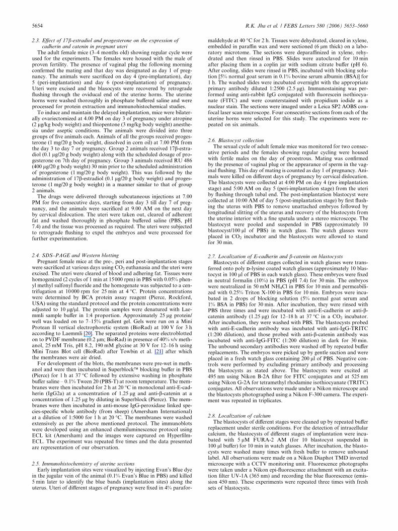
Fig. 4. Western Blot analysis of the E-cadherin and b-cateninexpression in implantation-delayed mice uteri after treatment withsex steroids and their inhibitors. Uterine tissue extracts from animalstreated with progesterone (P), progesterone+estradiol (P+E) andprogesterone+estradiol+RU39184 (P+E+Ru) were studied. Valuesappearing below the bands denote mean ± S.E.M of the bandintensities computed using a densitometric image analyzer.
5658 R.K. Jha et al. / FEBS Letters 580 (2006) 5653–5660
expression using antisense methods led to the disappearance of
E-cadherin from cell–cell contact sites and to severe impair-
ment in implantation in the rat [28]. However, HEC monolay-
ers which represent polarized, non adhesive phenotype showed
E-cadherin staining at the lateral membrane being confined to
the sites of cell-to-cell contacts and absent from the apical cell
pole, while RL monolayers which shows structural features of
Fig. 5. Localization of E-cadherin (A–C), b-catenin (D–F) and intracellulaBlastocysts were collected from day 4 (A, D and G), day 5 [5:00 AM] (B, E
non-polarized epithelial cells and functional features of adhe-
siveness are clearly positive for E-cadherin on the apical mem-
brane surfaces [12]. Thus, mouse uterine endometrium shows
signs of transformation into non-polarized and adhesive struc-
tures. Adhesion between uterine epithelium and trophoblast is
thought to be mediated by cell adhesion molecule, s-adenosyl
methionine (SAMs), lectin-like molecules, glycosyl transferase
or others [29,30].
Since the elevated expression of E-cadherin in the peri-
implantation uteri (Fig. 1) suggested that it could be under
the control of 17b-estradiol and/or progesterone, we evaluated
the profiles of E-cadherin in the uteri of implantation-delayed
animals supplemented with hormones and hormone antago-
nists. RU486 induced strong suppression in E-cadherin expres-
sion in the uteri of implantation-delayed mice maintained on
progesterone (Fig. 4) indicating that progesterone has stimula-
tory effect on E-cadherin expression in the uterus. Confocal
microscopic examination revealed a strong localization of E-
cadherin on the apical membrane surfaces of peri-implantation
uteri at the implantation sites, which was absent in the inter-
implantation sites and was less pronounced in stages bracket-
ing the peri-implantation time window (Fig. 2). b-Catenin is
seen to be upregulated predominantly in the endometrial epi-
thelial cells of the uterus at peri-implantation stage (Figs. 1
and 3), which might mediate connection between E-cadherin
and the actin filament network as part of a higher order
submembranous protein complex [31]. b-Catenin expression
r calcium (G–I) in blastocysts during various stages of development.and H) and day 5 [10:00 AM] (C, F and I). 108·.
R.K. Jha et al. / FEBS Letters 580 (2006) 5653–5660 5659
was augmented in the implantation-delayed mouse uterus by
17b-estradiol (Fig. 4). Neutralizing progesterone action by
administering RU486 in P+E-treated mice elevated the expres-
sion levels of b-catenin (Fig. 4), indicating that progesterone
suppresses 17b-estradiol-mediated activation of b-catenin
expression. The cadherin-catenin complex can function as
early recognition mechanisms between endothelial cells and
form loose junctions as in early confluent cells [32]. Studies
on the molecular organization of the E-cadherin-catenin com-
plex in culture cells [33] in vitro using recombinant proteins
[34] clearly demonstrate the central role of b-catenin in com-
plex formation. In human epithelial cancer cell line, HSC39,
it has been shown that the lack of cadherin–catenin complex
formation due to the expression of a truncated b-catenin cor-
relates with the loss of cell adhesion [35]. Expression of b-cate-
nin in these cells restores both complex formation and cell
adhesion [36].
E-cadherin is expressed in the blastomeres of mouse em-
bryos [37] and human embryos [38]. It plays an indispensable
role in their compaction at 8–16-cell stages [39–43]. Thus, func-
tionally interfering anti-E-cadherin antibodies caused early
embryos to decompact [40,43]. Further, homozygous negative
embryos for E-cadherin died around the time of implantation
probably due to their failure to form a trophoectodermal epi-
thelium or blastocyst cavity [44].
Lack of b-catenin causes primary defect in the embryonic
ectoderm cell layer of day 6.5–7.5 post-coital embryos that re-
sults in embryonic lethality. Thus b-catenin exerts a signaling
function in mouse embryonic ectoderm in addition to its role
in cadherin–catenin complex formation [45]. E-cadherin and
b-catenin expression are often down regulated in highly inva-
sive and poorly differentiated carcinoma. E-cadherin was
reported to be down regulated in epithelial-mesenchymal
transformation as well as in invasive tumor cells [19,46,47].
Expression of zonula occludens-1 (ZO-1) and E-cadherin,
two molecules associated with epithelial cells junctions in the
mouse uterus has been found during the peri-implantation
stage [48]. The pre-implantation embryos showed E-cadherin
staining and this is consistent with the view that maternal E-
cadherin present in early mouse embryos [49] suffices for ini-
tial compaction of morula but not for further pre-implanta-
tion development to occur [50]. The downregulation of
cadherin in peri-implantation embryos might necessitate the
downregulation of catenin as well, since the possibility of
the formation of cadherin–catenin complex to connect to actin
network is poor. Aberrant activation of b-catenin by LiCl, a
well-known glycogen synthase kinase-3 inhibitor, significantly
inhibited blastocyst hatching and subsequent adhesion and
outgrowth on fibronectin [51]. Further, the down regulation
of cadherin in the embryos might rule out the possibility of
a calcium-dependent homophilic E-cadherin–cadherin interac-
tion.
Human E-cadherin-Fc fusion protein binds directly to aEb7-
integrin [52]. Further, intra epithelial lymphocytes or transfec-
ted JY1 cells expressing the aEb7-integrin adhere strongly to
purified E-cadherin-Fc coated on plastic and the adhesion
can be inhibited by antibodies to aEb7-integrin or E-cadherin.
Binding of aEb7-integrin to cadherin is selective since cell
adhesion to P-cadherin-Fc through aEb7-integrin requires
>100-fold more fusion protein than to E-cadherin-Fc. Thus
it appears that E-cadherins are a direct counter receptor for
the aEb7-integrin [52]. Binding of aEb7-integrin requires the
presence of the first two N-terminal domains of E-cadherin
[53]. Although aEb7-integrin is not yet detected in peri-implan-
tation embryos, mRNAs for a variety of integrins including
a2, a6A and a7 isoforms are detected in late blastocyst, coin-
cident with endoderm differentiation and development of
attachment competence [54]. Further, the deletion of b1 inte-
grins in mice results in inner cell mass failure and peri implan-
tation lethality [55], suggesting a pivotal role of integrins on
peri-implantation embryos in mediating adhesive interactions
during implantation. Thus, the upregulation of cadherin and
catenin in the epithelial cells of peri-implantation uteri and
the down regulation of cadherin, catenin and Ca2+ in invasive
trophoblast appear to be associated with embryo–uterine inter-
actions during early pregnancy.
Acknowledgements: This work was supported by grants from IndianCouncil of Medical Research (# 3/1/2/6/99-RHN) and from Councilof Scientific and Industrial Research (# 13(7221-A)/97-Pool). J.K.R.and T.S. were supported by Indian Council of Medical Researchthrough grants 3/1/2/ 9/03-RHN and 3/1/3/25/05-RHN respectively. JijiV at the Confocal Imaging Core at Rajiv Gandhi Centre for Biotech-nology provided technical assistance in imaging.
References
[1] Denker, H.-W. (1986) Epithel–Epithel interaKtionen bei derEmbryo-implantation: Ansatze zur Losung eines zellbiologischenParadoxons. Anat. Anz. Suppl. 160, 93–114.
[2] Denker, H.-W. (1990) Trophoblast endometrial interaction atembryo implantation: a cell bilogical paradox. Trophoblast Res.4, 3–29.
[3] Denker, H.-W. (1994) Endometrial receptivity: a cell biologicalaspects of an unusual epithelial. Ann. Anat. 176, 53–60.
[4] Anderson, T., Simon, J. and Hodgen, G. (1990) Histochemicalcharacteristics of the endometrial surface related temporally toimplantation in the non human primate (Macaca fasicularis) in:Trophoblast Invasion and Endometrial Receptivity. NovelAspects of the Cell Biology of Embryo Implantation (Denker,H. and Aplin, J., Eds.), pp. 273–284, Plenum Medical BookComp, New York and London.
[5] Enders, A. and Schlafke, S. (1977) Alteration in uterine luminalsurface at the implantation site. J. Cell Biol. 75, 70a.
[6] Morris, J. and Potter, S. (1990) An in vitro model for studyinginteractions between mouse trophoblast and uterine epithelialcells in: Novel Aspects of the Cell Biology of Implantation(Denker, H. and Aplin, J., Eds.), pp. 51–69, Plenum MedicalBook Comp, New York and London.
[7] Anderson, T., Sieg, S. and Hodgen, G. (1988) MembraneComposition of the Endometrial Epithelium: Molecular Markersof Uterine Receptivity to Implantation, Excerpta Medica,Amsterdam.
[8] Hoffman, L., Winfrey, V., Anderson, T. and Olson, G. (1990)Uterine Receptivity to Implantation in the Rabbit: Evidence for a42 kDa Glycoprotein as a Marker of Receptivity, Plenum MedicalBook Comp, New York and London.
[9] Kimber, S. and Lindenberg, S. (1990) Hormonal control ofcarbohydrate epitope involved in implantation in mice. J. Reprod.Immunol. 89, 13–21.
[10] Lampelo, S., Ricketts, A. and Bullock, D. (1985) Purification ofrabbit endometrial plasma membranes from receptive and nonreceptive uteri. J. Reprod. Fertil. 75, 475–484.
[11] Psychoyos A. (1994) The implantation window: basic and clinicalaspects. In: Mori, T., Aono, T., Tominaga , T., and Hiroi, H.(Eds.), Ares Serono Symposia. Perspectives on Assisted Repro-duction, Vol. 4, pp. 57–63. Chritengraf, Rome.
[12] Thie, M., Fuchs, P. and Denker, H.-W. (1996) Epithelial cellpolarity and embryo implantation in mammals. Int. J. Dev. Biol.40, 389–393.
[13] Hay, E. (1995) An overview of epithelio-mesenchymal transfor-mation. Acta Anat. 154, 8–20.

5660 R.K. Jha et al. / FEBS Letters 580 (2006) 5653–5660
[14] Vicovac, L. and Aplin, J. (1996) Epithelial–mesenchymal transi-tion during trophoblast differentiation. Acta Anat. 156, 202–216.
[15] Hynes, R. and Lander, A. (1992) Contact and adhesive specific-ities in the association, migrations and targeting of cells andaxons. Cell 68, 303–322.
[16] Kintner, C. (1992) Regulation of embryonic cell adhesion by thecadherin cytoplasmic domain. Cell 69, 225–236.
[17] Nagafuchi, A. and Takeichi, M. (1988) Cell binding function of Ecadherin is regulated by the cytoplasmic domain. EMBO J. 7,3679–3684.
[18] Ozawa, M., Ringwald, M. and Kemler, R. (1990) Uvomorulin–catenin complex formation is regulated by a specific domain incytoplasmic region of the cell adhesion molecule. Proc. Natl.Acad. Sci. USA 87, 4246–4250.
[19] Behrens, J. (1994) Cadherins as determinants of tissue morphol-ogy and suppressor of invasion. Acta. Anat. 149, 165–169.
[20] Laemmli, U. (1970) Cleavage of structural proteins duringthe assembly of the head of bacteriophage T4. Nature 227, 680–685.
[21] Towbin, J., Staehelin, T. and Gordon, J. (1979) Electrophoretictransfer of proteins from polyacrylamide to nitrocellulose sheets.Proc. Natl. Acad. Sci. USA 76, 4350–4354.
[22] Thomas, M., Jain, S., Kumar, G. and Laloraya, M. (1997) Aprogrammed oxyradical burst causes hatching of mouse blasto-cysts. J. Cell Sci. 110, 1597–1602.
[23] Giudice, L. (2004) Microarray expression profiling reveals candi-date genes for human uterine receptivity. Am. J. Pharmacogen. 4,299–312.
[24] Dey, S., Lim, H., Das, S., Reese, J., Paria, B., Daikoku, T. andWang, H. (2004) Molecular cues to implantation. Endocr. Rev.25, 341–373.
[25] Slater, M., Murphy, C. and Barden, J. (2002) Tenascin, E-cadherin and P2X calcium channel receptor expression isincreased during rat blastocyst implantation. Histochem. J. 34,13–19.
[26] Donner, A., Classen-Linke, I., Frixen, U., Behrens, J. andDenker, H.-W. (1991) Anderungen im Verteilungsmuster vonMarker-proteinen fur das basolaterale Membran-Kompartimentdes Uterusepithels in der Periimplantationsphase: Desmoplakinund Uvomorulin. Anat. Anz. 172, 33.
[27] Donner, A., Beherens, J., Frixen, U. and Denker, H.-W. (1992)Uvomorulin, actin and the shift in uterine epithelial polarity atembryo implantation. Eur. J. Cell Biol. 57, 16.
[28] Li, Q., Wang, J., Armant, D., Bagchi, M. and Bagchi, I. (2002)Calcitonin down-regulates E-cadherin expression in rodent uter-ine epithelium during implantation. J. Biol. Chem. 277, 46447–46455.
[29] Carson, D., Wilson, O. and Dutt, A. (1990) Glycoconjugateexpression and interaction at the cell surface of the mouse uterineepithelial cell and periimplantation-stage embryos in: TrophoblastInvasion in Endometrial Receptivity. Novel Aspects of the CellBiology of Embryo Implantation (Denker, H. and Aplin, J., Eds.),pp. 211–241, Plenum Medical Book Comp., New York.
[30] Chavez, D. (1990) Possible involvement of DD-galactose in theimplantation process in: Trophoblast Invasion and EndometriumReceptivity (Denker, H. and Aplin, J., Eds.), pp. 259–272, PlenumMedical Book Comp., New York.
[31] Kemler, R. (1993) From cadherins to catenins: cytoplasmicprotein interactions and regulation of cell adhesion. TrendsGenet. 9, 317–321.
[32] Thie, M., Fuchs, P., Butz, S., Sieckmann, F., Hoschutzky, H.,Kemler, R. and Denker, H.-W. (1996) Adhesiveness of the apicalsurface of uterine epithelial cells: the role of junctional complexintegrity. Eur. J. Cell Biol. 70, 221–232.
[33] Ozawa, M. and Kemler, R. (1992) Molecular organization ofUvomorulin-catenin complex. J. Cell Biol. 116, 989–996.
[34] Aberle, H., Butz, S., Stappert, J., Weissig, H., Kemler, R. andHoschuetzky, H. (1994) Assembly of cadherin catenin complexin vitro with recombinant proteins. J. Cell Sci. 107, 3655–3663.
[35] Oyama, T., Kanai, I., Ochiai, A., Akimoto, S., Oda, T.,Yanagihara, K., Nagafuchi, A., Tsukita, S., Shibamoto, S., Ito,F., Takeichi, M., Matsuda, H. and Hirohashi, S. (1994) Atruncated bcatenin disrupts the interaction between E-cadherin
and alpha catenin: A cause of loss of intercellular adhesiveness inhuman cancer cell lines. Cancer Res. 54, 6282–6287.
[36] Kawanishi, J., Kato, J., Sasaki, K., Watanabe, N. and Niitsu, Y.(1995) Loss of E-cadherin-dependent cell-cell adhesion due tomutation of the bcatenin gene in a human cancer cell line HSC-39.Mol. Cell Biol. 15, 1175–1181.
[37] Ogou, S., Okada, T. and Takeichi, M. (1982) Cleavage stagemouse embryos share a common cell adhesion system withteratocarcinoma cells. Devl. Biol. 92, 521–528.
[38] Bloor, D., Metcalfe, A., Rutherford, A., Brison, D. and Kimber,S. (2002) Expression of cell adhesion molecules during humanpreimplantation embryo development. Mol. Hum. Reprod. 8,237–245.
[39] Damsky, C., Richa, J., Solter, D., Knusen, K. and Buck, C. (1983)Identification and purification of cell surface glycoprotein medi-ating intercellular adhesion in embryonic and adult tissue. Cell 34,445–466.
[40] Hyafil, F., Morello, D., Babinet, C. and Jacob, F. (1980) A cellsurface glycoprotein involved in the compaction of embryoniccarcinoma cell and cleavage stage embryos. Cell 21, 927–934.
[41] Johnson, M., Maro, B. and Takeichi, M. (1986) The role of celladhesion in synchronization and orientation of polarization in 8-cell mouse blastomeres. J. Embryol. Exp. Morph. 93, 239–255.
[42] Shirayoshi, Y., Okada, T. and Takeichi, M. (1983) The calciumdependent cell-cell adhesion system regulates inner cell massformation and cell surface polarization in early mouse develop-ment. Cell 35, 631–638.
[43] Vestweber, D. and Kemler, R. (1984) Rabbit antiserum against apurified surface glycoprotein decompacts mouse preimplantationembryo and react with specific adult tissues. Exp. Cell Res. 152,169–178.
[44] Larue, L., Ohsugi, M., Hirchenhain, J. and Kemler, R. (1994) Ecadherin null mutant embryo fail to form a trophoectodermepithelium. Proc. Natl. Acad. Sci. USA 91, 8263–8267.
[45] Haegel, H., Larue, L., Ohsugi, M., Fedorov, L., Herrenknecht, K.and Kemler, R. (1995) Lack of b-catenin affects mouse develop-ment at gastrulation. Development 121, 3529–3537.
[46] Birchmeier, W., Behrens, J., Weidner, K., Frixen, U. andSchipper, J. (1991) Dominant and recessive genes involved inthe tumor cell invasion. Curr. Opin. Cell Biol. 107, 1575–1587.
[47] Gumbiner, B., Stevenson, B. and Grimaldi, A. (1988) The role ofcell adhesion molecule uvomorulin in the formation and mainta-inence of epithelial junctional complexes. J. Cell Biol. 107, 1575–1587.
[48] Paria, B., Zhao, X., Das, S., Dey, S. and Yoshinaga, K. (1999)Zonula occludens-1 and E-cadherin are coordinately expressed inthe mouse uterus with the initiation of implantation and decid-ualization. Dev. Biol. 208, 488–501.
[49] Sefton, M., Johnson, M. and Clayton, L. (1992) Synthesis andphosphorylation of uvomorulin during early mouse development.Development 115, 313–318.
[50] Riethmacher, D., Brinkmann, V. and Birchmeier, C. (1995) Atargeted mutation in the mouse E-cadherin gene results indefective preimplantation development. Proc. Natl. Acad. Sci.USA 92, 855–859.
[51] Li, J., Zhang, J., Cao, Y., Zhou, J., Liu, W., Fan, X. and Duan, E.(2005) Inhibition of the beta-catenin signaling pathway inblastocyst and uterus during the window of implantation in mice.Biol. Reprod. 72, 700–706.
[52] Higgins, J., Mandlebrot, D., Shaw, S., Russell, G., Murphy, E.,Chen, Y., Nelson, W., Parker, C. and Brenner, M. (1998) Directedand regulated interaction of integrin alphaEbeta7 with E cad-herin. J. Cell Biol. 140, 197–210.
[53] Karecla, P., Green, S., Bowden, S., Coadwell, J. and Kilshaw, P.(1996) Identification of a binding site for integrin alpha E beta 7 inthe N-terminal domain of E-cadherin. J. Biol. Chem. 271, 30909–30915.
[54] Sutherland, A., Calacro, P. and Damsky, C. (1993) Developmen-tal regulation of integrin expression at the time of implantation inthe mouse embryo. Development 119, 1175–1186.
[55] Stephens, L., Sutherland, A., Klimanskaya, I., Andrieux, A.,Meneses, J., Pedersen, R. and Damsky, C. (1995) Deletion of beta1 integrins in mice results in inner cell mass failure and periimplantation lethality. Genes Dev. 9, 1883–1895.