
Correlation of DNA Ploidy with Progression of Cervical Cancer
8
Hindawi Publishing Corporation Journal of Cancer Epidemiology Volume 2008, Article ID 298495, 7 pages doi:10.1155/2008/298495 Clinical Study Correlation of DNA Ploidy with Progression of Cervical Cancer M. Singh, 1 S. Mehrotra, 2 N. Kalra, 1 U. Singh, 2 and Y. Shukla 1 1 Industrial Toxicology Research Center, P.O. Box 80, M.G. Marg, Lucknow 226001, India 2 Department of Obstetrics & Gynecology, Queen Merry Hospital, King George’s Medical University, Lucknow 226003, India Correspondence should be addressed to Y. Shukla, [email protected] Received 7 April 2007; Revised 26 July 2007; Accepted 24 November 2007 Recommended by Qingyi Wei The majority of squamous cell carcinomas of cervix are preceded by visible changes in the cervix, most often detected by cervical smear. As cervical cancer is preceded by long precancerous stages, identification of the high-risk population through detection of DNA ploidy may be of importance in effective management of this disease. Here we attempted to correlate aneuploid DNA patterns and their influence on biological behavior of flow-cytometry analysis of DNA ploidy which was carried out in cytologically diagnosed cases of mild (79), moderate (36), and severe (12) dysplasia, as well as “atypical squamous cells of unknown significance (ASCUS)” (57) along with controls (69), in order to understand its importance in malignant progression of disease. Cytologically diagnosed dysplasias, which were employed for DNA ploidy studies, 39 mild, 28 moderate, and 11 severe dysplasia cases were found to be aneuploid. Out of the 69 control subjects, 6 cases showed aneuploidy pattern and the rest 63 subjects were diploid. An aneuploidy pattern was observed in 8 out of 57 cases of cytologically evaluated ASCUS. The results of the followup studies showed that aberrant DNA content reliably predicts the occurrence of squamous cell carcinoma in cervical smear. Flow cytometric analysis of DNA ploidy may provide a strategic diagnostic tool for early detection of carcinoma cervix. Therefore, it is a concept of an HPV screening with reflex cytology in combination with DNA flow cytometry to detect progressive lesions with the greatest possible sensitivity and specificity. Copyright © 2008 M. Singh et al. This is an open access article distributed under the Creative Commons Attribution License, which permits unrestricted use, distribution, and reproduction in any medium, provided the original work is properly cited. 1. INTRODUCTION Cervical cancer is the second most common malignancy in women worldwide. It is estimated that, globally, 466,000 new cases of cervical cancer are diagnosed each year. As per estimate 231,000 women in a year die of cervical cancer and 80% of them are from the developing world [1]. Use of cytological screening technique, Papanicolaou (Pap) test, has significantly increased the detection of cervical cancer [2]. Cancer of cervix emerges from a defined series of preneoplastic lesions with increasing cellular dysplasia, referred to as cervical intraepithelial neoplasia (CIN) grade I, II, and III. However, CIN lesions frequently regress, and not all CIN III lesions, progress to invasive cancer [3]. The group of CIN is very heterogeneous, particularly in their clinical behavior. Persistent infection with high- risk-type human papilloma viruses (HPVs) is the strongest independent risk factor for cervical cancer [4]. CIN lesions induced by HPV infection and genomes of these viruses were detected in the vast majority of cervical cancer specimens [5–7]. During the course of tumor initiation in an HPV- infected cervical epithelial cell, an aneuploid DNA pattern develops [8], which along with its descendants shows genetic instability. Aneuploidy reflects a situation of uncontrolled increase of DNA and loss of essential information [9] and plays an important role in neoplastic transformation. Increased aneuploid DNA value with the increase in grades of cervical dysplasia has long been considered to be a specific prognostic marker of malignancy [10]. However, at present, no well-established technique, which, qualitatively, predicts the clinical outcome of cervical dysplasia except Pap smear screening, is available. Approximately 15–30% of all women who developed low-grade squamous intraepithelial lesion (LSIL) have a probability of developing moderate- to-severe cervical intraepithelial neoplasia (CIN II and III) identified on a subsequent cervical biopsy [11]. To prevent cervical malignancy and its precursors, several adjuvant diagnostic methods have been proposed to strengthen the accuracy of cytological and histological diagnosis [12]. As chromosomal aneuploidy has been used as an early key event
-
Upload
independent -
Category
Documents
-
view
4 -
download
0
Transcript of Correlation of DNA Ploidy with Progression of Cervical Cancer
Hindawi Publishing CorporationJournal of Cancer EpidemiologyVolume 2008, Article ID 298495, 7 pagesdoi:10.1155/2008/298495
Clinical StudyCorrelation of DNA Ploidy with Progression of Cervical Cancer
M. Singh,1 S. Mehrotra,2 N. Kalra,1 U. Singh,2 and Y. Shukla1
1 Industrial Toxicology Research Center, P.O. Box 80, M.G. Marg, Lucknow 226001, India2 Department of Obstetrics & Gynecology, Queen Merry Hospital, King George’s Medical University, Lucknow 226003, India
Correspondence should be addressed to Y. Shukla, [email protected]
Received 7 April 2007; Revised 26 July 2007; Accepted 24 November 2007
Recommended by Qingyi Wei
The majority of squamous cell carcinomas of cervix are preceded by visible changes in the cervix, most often detected by cervicalsmear. As cervical cancer is preceded by long precancerous stages, identification of the high-risk population through detectionof DNA ploidy may be of importance in effective management of this disease. Here we attempted to correlate aneuploid DNApatterns and their influence on biological behavior of flow-cytometry analysis of DNA ploidy which was carried out in cytologicallydiagnosed cases of mild (79), moderate (36), and severe (12) dysplasia, as well as “atypical squamous cells of unknown significance(ASCUS)” (57) along with controls (69), in order to understand its importance in malignant progression of disease. Cytologicallydiagnosed dysplasias, which were employed for DNA ploidy studies, 39 mild, 28 moderate, and 11 severe dysplasia cases werefound to be aneuploid. Out of the 69 control subjects, 6 cases showed aneuploidy pattern and the rest 63 subjects were diploid. Ananeuploidy pattern was observed in 8 out of 57 cases of cytologically evaluated ASCUS. The results of the followup studies showedthat aberrant DNA content reliably predicts the occurrence of squamous cell carcinoma in cervical smear. Flow cytometric analysisof DNA ploidy may provide a strategic diagnostic tool for early detection of carcinoma cervix. Therefore, it is a concept of an HPVscreening with reflex cytology in combination with DNA flow cytometry to detect progressive lesions with the greatest possiblesensitivity and specificity.
Copyright © 2008 M. Singh et al. This is an open access article distributed under the Creative Commons Attribution License,which permits unrestricted use, distribution, and reproduction in any medium, provided the original work is properly cited.
1. INTRODUCTION
Cervical cancer is the second most common malignancy inwomen worldwide. It is estimated that, globally, 466,000new cases of cervical cancer are diagnosed each year. As perestimate 231,000 women in a year die of cervical cancerand 80% of them are from the developing world [1].Use of cytological screening technique, Papanicolaou (Pap)test, has significantly increased the detection of cervicalcancer [2]. Cancer of cervix emerges from a defined seriesof preneoplastic lesions with increasing cellular dysplasia,referred to as cervical intraepithelial neoplasia (CIN) gradeI, II, and III. However, CIN lesions frequently regress,and not all CIN III lesions, progress to invasive cancer[3]. The group of CIN is very heterogeneous, particularlyin their clinical behavior. Persistent infection with high-risk-type human papilloma viruses (HPVs) is the strongestindependent risk factor for cervical cancer [4]. CIN lesionsinduced by HPV infection and genomes of these viruses weredetected in the vast majority of cervical cancer specimens
[5–7]. During the course of tumor initiation in an HPV-infected cervical epithelial cell, an aneuploid DNA patterndevelops [8], which along with its descendants shows geneticinstability. Aneuploidy reflects a situation of uncontrolledincrease of DNA and loss of essential information [9]and plays an important role in neoplastic transformation.Increased aneuploid DNA value with the increase in gradesof cervical dysplasia has long been considered to be aspecific prognostic marker of malignancy [10]. However, atpresent, no well-established technique, which, qualitatively,predicts the clinical outcome of cervical dysplasia except Papsmear screening, is available. Approximately 15–30% of allwomen who developed low-grade squamous intraepitheliallesion (LSIL) have a probability of developing moderate-to-severe cervical intraepithelial neoplasia (CIN II and III)identified on a subsequent cervical biopsy [11]. To preventcervical malignancy and its precursors, several adjuvantdiagnostic methods have been proposed to strengthen theaccuracy of cytological and histological diagnosis [12]. Aschromosomal aneuploidy has been used as an early key event
2 Journal of Cancer Epidemiology
in tumorigenesis caused by genetic instability [13, 14], thecytometric equivalence of chromosomal aneuploidy detectedby DNA flow-cytometry may serve as a marker of neoplasiaand provide valuable information for the diagnosis andunderstanding of pathogenesis of cervical cancer. Since aneu-ploid pattern of DNA is a useful parameter for the predictionof progression and persistence of “atypical squamous cellsof unknown significance (ASCUS)”, low-grade squamousintraepithelial lesion (LSIL/mild dysplasia/CIN I), and high-grade squamous intraepithelial lesion (HSIL/moderate andsevere dysplasia/CIN II and III) in clinical practice, thepresent study was designed to investigate whether alterationin cellular/nuclear content predicts the occurrence of cancerin cervical smears. We have thus estimated their DNAcontent using flow cytometry and compared it with thecytological findings.
2. MATERIALS AND METHODS
2.1. Subjects and sampling
The cases were derived from the ongoing outpatient cytologyscreening at Department Obstetrics and Gynecology, KingGeorge’s Medical University (Lucknow, India) for the presentstudy. Cytologically diagnosed 127 cases of dysplasia (79mild, 36 moderate, and 12 severe), 57 cases of “atypicalsquamous cells of unknown significance (ASCUS)” and69 controls, which enrolled during the period of 2004–2006, were recruited from a prospective followup study tounderstand the biological behavior and the natural historyof cervical precancerous lesions. None of the patients hadany previous history of CIN or had been treated for CIN. Allthe control women had normal smears. Age of the subjectsranged from 30 to 75 years.
An experienced gynecologist collected cervical sampleswith blunt end of Ayer’s spatula. Two fresh spatulas wereused for each preparation. Smears were fixed in alcohol andstained according to Pap method. The blunt end of thesecond spatula was transferred into a cryo tube immediately,containing 3 mL chilled PBS (7.2 pH) and quick frozenat −20◦C until further use for flow-cytometric analysis.Patients were followed up by cytological as well as DNA flowcytometric analysis.
2.2. Criteria for cytological classification
Cytological screening was performed with conventional Papstaining by trained cytologists. All slides were evaluatedaccording to the criteria of the Bethesda System [15].Precancerous lesions of the uterine cervix were classifiedas normal, ASCUS, LSIL (mild dysplasia/CIN grade I),and HSIL (moderate dysplasia/CIN grade II, severe dyspla-sia/CIN grade III and carcinoma in situ/CIS).
2.3. Criteria for management of precancerous lesions
To understand the biological behavior of cervical precancer-ous lesions, the following criteria were followed.
(i) The cases initially diagnosed as LSIL (CIN I) andHSIL (CIN II and III) were made to undergo a secondPap smear test after 30 days.
(ii) Cytological confirmed dysplasia cases were enrolledfor the followup with the consent of the patients.
(iii) Cases initially diagnosed as CIN III were subjectedto colposcopy directed biopsy on day 30 and weredirected to gynecologist for further management. Acytological control examination was done after every6 and 3 months for LSIL and HSIL subjects, respec-tively, and were categorized as “returned to nor-mal” (after the morphology normalized); progressed(when presented with higher grade of dysplasia); andwith no change (when remained at the same grade ofdysplasia). The samples were also tested for the DNAploidy pattern.
(iv) ASCUS and all aneuploid cases from control groupwere also followed for a period of one year.
2.4. Flow-cytometric DNA analysis
To determine the cellular DNA content, the protocol ofNicoletti et al. was followed [16]. In brief, cells were fixedwith chilled ethanol for 30 minutes at 4◦C temperature. Afteradding propidium iodide (Sigma-Aldrich, Miss, USA), themixture was stored in dark for 1 hour at room temperature.Lymphocytes were used for calibration. The maximumpermitted coefficient of variation (CV) for calibration was2%. A total of 15,000 cells were counted in each sample. DNAhistogram cell-cycle analysis was performed as described byRabinovitch [17], using ModFit LT for Mac V2.0 softwareafter excluding debris by using electronic gate. The events inG0-G1, S, and G2-M phases of the cell cycle were counted.Cases were regarded as acceptable for analysis if the CV ofthe G0-G1 peak was 7.0. Lesions were classified as diploid,tetraploid, or aneuploid. If two distinct G0-G1 peaks werepresent with a DNA index of ≥1.15 (each containing ≥10%of total cell population), the histogram was considered asaneuploid (DNA content). By convention, the first G0-G1peak represented the diploid peak. Diploid and tetraploidlesions were classified as nonaneuploid. Histogram patternswere termed diploid if there was a single G0-G1 peak. Ahistogram was classified as tetraploid only if a large peakobserved in G2-M (DNA index of 1.80–2.2), containing≥20% of the total curve and was also associated with acorresponding 8 N peak.
2.5. Statistical analysis
EPI-INFO software was used for calculating the odd ratioand χ2 (chi-square) values. A P-value of .05 or less wasconsidered statistically significant (95% CI).
3. RESULTS
The cervical smears were divided into four groups (seeFigure 1). The first group qualified as normal and grouped69 cases. The second group (57 cases) qualified as ASCUS.
M. Singh et al. 3
(a) (b)
(c) (d) (e)
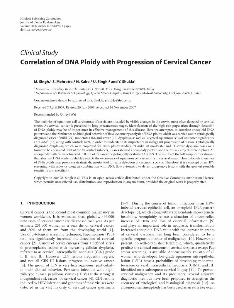
Figure 1: Showing representative cytological images of cervical smears stained according to Pap staining method (magnification: 40X): (a)showing cells with normal morphology, (b) showing “atypical squamous cells of unknown significance,” (c) showing cells of mild dysplasia(LSIL/CIN I), (d) showing cells of moderate dysplasia (HSIL/CIN II), (e) showing cells of severe dysplasia (HSIL/CIN III). LSIL: low-gradesquamous intraepithelial lesion; HSIL: high-grade squamous intraepithelial lesion; CIN: cervical intraepithelial neoplasia.
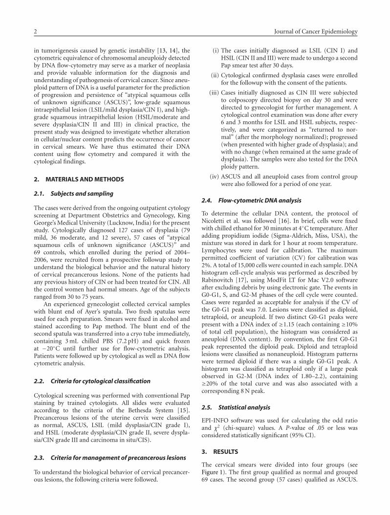
Table 1: DNA ploidy pattern in control, ASCUS, and various grades of dysplasia cases.
DNAploidy (n)
Controln (%)
ASCUSn (%)
OR(95% CI)
Milddysplasian (%)
OR(95% CI)
Moderatedysplasian (%)
OR(95% CI)
Severedysplasian (%)
OR(95% CI)
Aneuploidy 6 8 1.71NS 39 10.2∗ 28 36.75∗ 11 115.50∗
(92) (8.69%) (14.03%) (0.47–6.26) (49.36%) (3.71–29.7) (77.77%) (10.35–140.74) (91.66%) (11.75–5041.27)
Diploidy 63 49 1.00 40 1.00 8 1.00 1 1.00
(161) (91.03%) (85.96%) (50.36%) (22.22%) (8.33%)
Total (253) 69 57 79 36 12
Odd ratios (OR) were computed considering control cases as the base line category for the ASCUS and dysplasia groups.Not significant NSP ≥ .05, Statically significant ∗P ≤ .001, n: Number of cases.
The third and fourth groups comprised of LSIL (79 cases)and HSIL (48 cases; 36 cases in moderate dysplasia and 12cases in severe dysplasia).
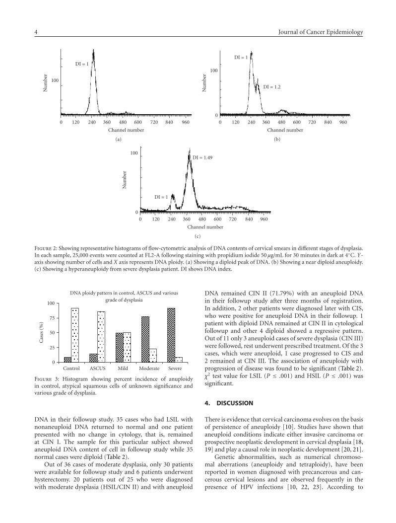
A comparison of DNA content analysis with behaviorof LSIL or HSIL in patients having cytologically diagnosedvarious grades of dysplasia along with the control andASCUS groups were made (Table 1). 127 cases of differentgrades of dysplasia were employed for analysis of DNA ploidyout of which 78 (62.99%) cases were aneuploid (39 mild, 28moderate, and 11 cases of severe dysplasia) and remaining49 (37.01%) cases (40 mild, 8 moderate, and 1 severe)were nonaneuploid, that is, diploid (see Figure 2). In thecontrol and ASCUS groups, 6 (8.70%) and 8 (14.04%) caseswere found to be aneuploid, respectively (see Figure 2). Theassociation of aneuploidy with increasing grades of dysplasiawas found to be significant (P ≤ .001; Table 1, see Figure 3).
All these cases with aneuploid DNA content and cyto-logically diagnosed ASCUS and various grades of dysplasia
were followed up for 1 year at aforementioned intervals, inorder to study the behavior of aneuploid DNA in progres-sion/regression/persistence of disease. Among 57 patientswith ASCUS, 49 (85.96%) diploid cases had smears thatreturned to normal cytology and remaining 8 (14.04%)aneuploid cases showed signs of progression/regression. Onepatient presented with HSIL and three with LSIL afterone year period of followup. Remaining four aneuploidcases of ASCUS group regressed to normal, as evidentfrom diploid DNA. Out of six aneuploid cases with normalcytological diagnosis, 4 cases were followed, where only onecase represented progression of disease from normal to milddysplasia after a year.
A total of 79 patients who had diagnosis of LSIL, 75 caseswere followed up. Out of 39 aneuploid cases, 7 developedmoderate dysplasia (CIN II) and 30 cases presented with nochange in cytology (CIN I), after a period of six monthsfrom the date of registration. All cases showed aneuploid
4 Journal of Cancer Epidemiology
0 120 240 360 480 600 720 840 960
Channel number
DI = 1
100
Nu
mbe
r
(a)
0 120 240 360 480 600 720 840 960
Channel number
DI = 1
DI = 1.2
0
100
Nu
mbe
r
(b)
0 120 240 360 480 600 720 840 960
Channel number
DI = 1
DI = 1.49
0
100
Nu
mbe
r
(c)
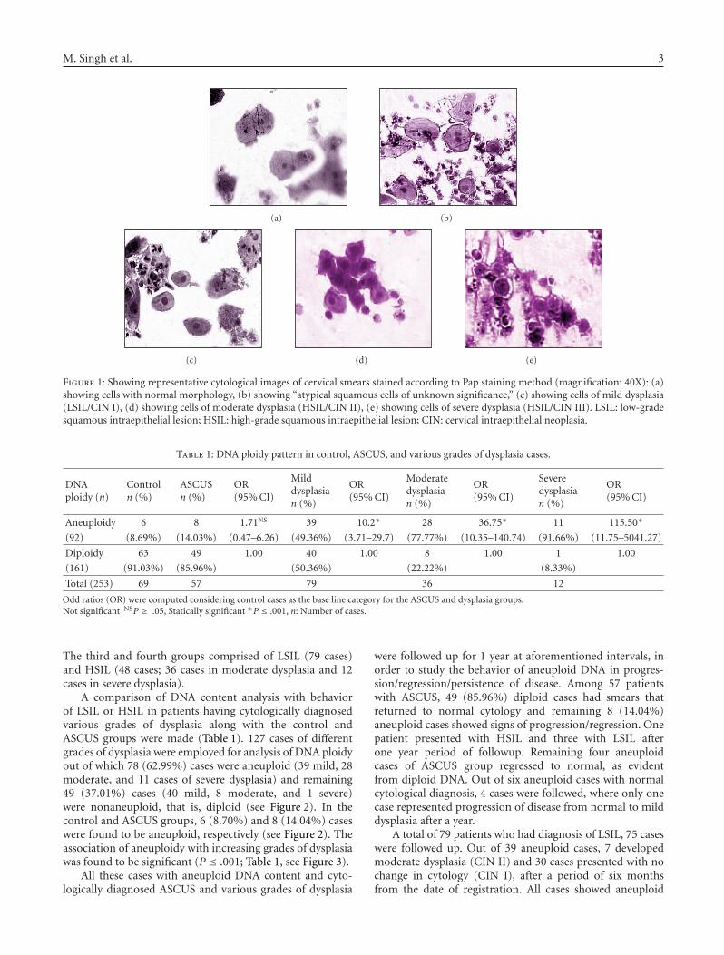
Figure 2: Showing representative histograms of flow-cytometric analysis of DNA contents of cervical smears in different stages of dysplasia.In each sample, 25,000 events were counted at FL2-A following staining with propidium iodide 50 μg/mL for 30 minutes in dark at 4◦C. Y-axis showing number of cells and X axis represents DNA ploidy. (a) Showing a diploid peak of DNA. (b) Showing a near diploid aneuploidy.(c) Showing a hyperaneuploidy from severe dysplasia patient. DI shows DNA index.
Control ASCUS Mild Moderate Severe
DNA ploidy pattern in control, ASCUS and variousgrade of dysplasia
100
75
50
25
0
Cas
es(%
)
Figure 3: Histogram showing percent incidence of anuploidyin control, atypical squamous cells of unknown significance andvarious grade of dysplasia.
DNA in their followup study. 35 cases who had LSIL withnonaneuploid DNA returned to normal and one patientpresented with no change in cytology, that is, remainedat CIN I. The sample for this particular subject showedaneuploid DNA content of cell in followup study while 35normal cases were diploid (Table 2).
Out of 36 cases of moderate dysplasia, only 30 patientswere available for followup study and 6 patients underwenthysterectomy. 20 patients out of 25 who were diagnosedwith moderate dysplasia (HSIL/CIN II) and with aneuploid
DNA remained CIN II (71.79%) with an aneuploid DNAin their followup study after three months of registration.In addition, 2 other patients were diagnosed later with CIS,who were positive for aneuploid DNA in their followup. 1patient with diploid DNA remained at CIN II in cytologicalfollowup and other 4 diploid showed a regressive pattern.Out of 11 only 3 aneuploid cases of severe dysplasia (CIN III)were followed, rest underwent prescribed treatment. Of the 3cases, which were aneuploid, 1 case progressed to CIS and2 remained at CIN III. The association of aneuploidy withprogression of disease was found to be significant (Table 2).χ2 test value for LSIL (P ≤ .001) and HSIL (P ≤ .001) wassignificant.
4. DISCUSSION
There is evidence that cervical carcinoma evolves on the basisof persistence of aneuploidy [10]. Studies have shown thataneuploid conditions indicate either invasive carcinoma orprospective neoplastic development in cervical dysplasia [18,19] and play a causal role in neoplastic development [20, 21].
Genetic abnormalities, such as numerical chromoso-mal aberrations (aneuploidy and tetraploidy), have beenreported in women diagnosed with precancerous and can-cerous cervical lesions and are observed frequently in thepresence of HPV infections [10, 22, 23]. According to
M. Singh et al. 5
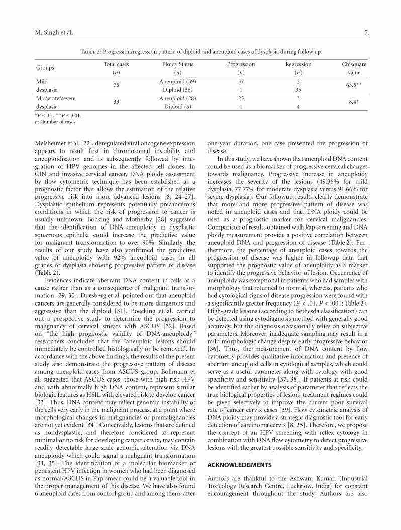
Table 2: Progression/regression pattern of diploid and aneuploid cases of dysplasia during follow up.
GroupsTotal cases Ploidy Status Progression Regression Chisquare
(n) (n) (n) (n) value
Mild75
Aneuploid (39) 37 263.5∗∗
dysplasia Diploid (36) 1 35
Moderate/severe33
Aneuploid (28) 25 38.4∗
dysplasia Diploid (5) 1 4∗P ≤ .01, ∗∗P ≤ .001.n: Number of cases.
Melsheimer et al. [22], deregulated viral oncogene expressionappears to result first in chromosomal instability andaneuploidization and is subsequently followed by inte-gration of HPV genomes in the affected cell clones. InCIN and invasive cervical cancer, DNA ploidy assessmentby flow cytometric technique has been established as aprognostic factor that allows the estimation of the relativeprogressive risk into more advanced lesions [8, 24–27].Dysplastic epithelium represents potentially precancerousconditions in which the risk of progression to cancer isusually unknown. Bocking and Motherby [28] suggestedthat the identification of DNA aneuploidy in dysplasticsquamous epithelia could increase the predictive valuefor malignant transformation to over 90%. Similarly, theresults of our study have also confirmed the predictivevalue of aneuploidy with 92% aneuploid cases in allgrades of dysplasia showing progressive pattern of disease(Table 2).
Evidences indicate aberrant DNA content in cells as acause rather than as a consequence of malignant transfor-mation [29, 30]. Duesberg et al. pointed out that aneuploidcancers are generally considered to be more dangerous andaggressive than the diploid [31]. Boecking et al. carriedout a prospective study to determine the progression tomalignancy of cervical smears with ASCUS [32]. Basedon “the high prognostic validity of DNA-aneuploidy”researchers concluded that the “aneuploid lesions shouldimmediately be controlled histologically or be removed”. Inaccordance with the above findings, the results of the presentstudy also demonstrate the progressive pattern of diseaseamong aneuploid cases from ASCUS group. Bollmann etal. suggested that ASCUS cases, those with high-risk HPVand with abnormally high DNA content, represent similarbiologic features as HSIL with elevated risk to develop cancer[33]. Thus, DNA content may reflect genomic instability ofthe cells very early in the malignant process, at a point wheremorphological changes in malignancies or premalignanciesare not yet evident [34]. Conceivably, lesions that are definedas nondysplastic, and therefore considered to representminimal or no risk for developing cancer cervix, may containreadily detectable large-scale genomic alteration viz DNAaneuploidy which could signal a malignant transformation[34, 35]. The identification of a molecular biomarker ofpersistent HPV infection in women who had been diagnosedas normal/ASCUS in Pap smear could be a valuable tool inthe proper management of this disease. We have also found6 aneuploid cases from control group and among them, after
one-year duration, one case presented the progression ofdisease.
In this study, we have shown that aneuploid DNA contentcould be used as a biomarker of progressive cervical changestowards malignancy. Progressive increase in aneuploidyincreases the severity of the lesions (49.36% for milddysplasia, 77.77% for moderate dysplasia versus 91.66% forsevere dysplasia). Our followup results clearly demonstratethat more and more progressive pattern of disease wasnoted in aneuploid cases and that DNA ploidy could beused as a prognostic marker for cervical malignancies.Comparison of results obtained with Pap screening and DNAploidy measurement provide a positive correlation betweenaneuploid DNA and progression of disease (Table 2). Fur-thermore, the percentage of aneuploid cases towards theprogression of disease was higher in followup data thatsupported the prognostic value of aneuploidy as a markerto identify the progressive behavior of lesion. Occurrence ofaneuploidy was exceptional in patients who had samples withmorphology that returned to normal, whereas, patients whohad cytological signs of disease progression were found witha significantly greater frequency (P < .01, P < .001; Table 2).High-grade lesions (according to Bethesda classification) canbe detected using cytodiagnosis method with generally goodaccuracy, but the diagnosis occasionally relies on subjectiveparameters. Moreover, inadequate sampling may result in amild morphologic change despite early progressive behavior[36]. Thus, the measurement of DNA content by flowcytometry provides qualitative information and presence ofaberrant aneuploid cells in cytological samples, which couldserve as a useful parameter along with cytology with goodspecificity and sensitivity [37, 38]. If patients at risk couldbe identified earlier by analysis of parameter that reflects thetrue biological properties of lesion, treatment regimes couldbe given selectively to improve the current poor survivalrate of cancer cervix cases [39]. Flow cytometric analysis ofDNA ploidy may provide a strategic diagnostic tool for earlydetection of carcinoma cervix [8, 25]. Therefore, we proposethe concept of an HPV screening with reflex cytology incombination with DNA flow cytometry to detect progressivelesions with the greatest possible sensitivity and specificity.
ACKNOWLEDGMENTS
Authors are thankful to the Ashwani Kumar, (IndustrialToxicology Research Centre, Lucknow, India) for constantencouragement throughout the study. Authors are also
6 Journal of Cancer Epidemiology
thankful to Vinita Das and Urmila Singh (Department ofObstetrics and Gynecology, King George’s Medical Univer-sity, Lucknow, India) for taking keen interest in the study.Authors also wish to acknowledge Neeraj Mathur (ITRC) forstatistical analysis of the data. Authors are deeply thankfulto Indian Council for Medical Research (New Delhi, India)for providing Research Associateship to M. Singh and SeniorResearch Fellowship to N. Kalra.
REFERENCES
[1] D. M. Parkin, P. Pisani, and J. Ferlay, “Estimates of theworldwide incidence of eighteen major cancers in 1985,”International Journal of Cancer, vol. 54, no. 4, pp. 594–606,1993.
[2] J. Tiffen and S. M. Mahon, “Cervical cancer: what shouldwe tell women about screening?” Clinical Journal of OncologyNursing, vol. 10, no. 4, pp. 527–531, 2006.
[3] A. G. Ostor, “Studies on 200 cases of early squamous cellcarcinoma of the cervix,” International Journal of GynecologicalPathology, vol. 12, no. 3, pp. 193–207, 1993.
[4] K. Lukaszuk, J. Liss, I. Wozniak, J. Emerich, and C.Wojcikowski, “Human papillomavirus type 16 status incervical carcinoma cell DNA assayed by multiplex PCR,”Journal of Clinical Microbiology, vol. 41, no. 2, pp. 608–612,2003.
[5] H. Z. Hausen, “Papillomavirus infections—a major cause ofhuman cancers,” Biochimica et Biophysica Acta, vol. 1288, pp.55–78, 1996.
[6] M. Schiffman, R. Herrero, A. Hildesheim, et al., “HPV DNAtesting in cervical cancer screening: results from women ina high risk province of Costa Rica,” Journal of the AmericanMedical Association, vol. 283, no. 1, pp. 87–93, 2000.
[7] M. Schiffman, A. Hildesheim, R. Herrero, and C. Bratti,“Human papillomavirus testing as a screening tool for cervicalcancer,” The Journal of the American Medical Association,vol. 283, no. 19, pp. 2525–2526, 2000.
[8] P. Melsheimer, R. Klaes, M. von Knebel Doeberitz, and G.Bastert, “Prospective clinical study comparing DNA flowcytometry and HPV typing as predictive tests for persistenceand progression of CIN I/II,” Communications in ClinicalCytometry, vol. 46, no. 3, pp. 166–171, 2001.
[9] H. zur Hausen, “Human papillomaviruses in the pathogenesisof anogenital cancer,” Virology, vol. 184, no. 1, pp. 9–13, 1991.
[10] V. Kashyap and B. C. Das, “DNA aneuploidy and infection ofhuman papillomavirus type 16 in preneoplastic lesions of theuterine cervix: correlation with progression to malignancy,”Cancer Letters, vol. 123, no. 1, pp. 47–52, 1998.
[11] N. M. Lonky, M. Sadeghi, G. W. Tsadik, and D. Petitti, “Theclinical significance of the poor correlation of cervical dys-plasia and cervical malignancy with referral cytologic results,”American Journal of Obstetrics and Gynecology, vol. 181, no. 3,pp. 560–566, 1999.
[12] S. C. Cotton, L. Sharp, J. Little, et al., “Trial of manage-ment of borderline and other low-grade abnormal smears(TOMBOLA): trial design,” Contemporary Clinical Trials,vol. 27, no. 5, pp. 449–471, 2006.
[13] F. Giroud, G. Haroske, A. Reith, and A. Bocking, “ESACPconsensus report on diagnostic DNA image cytometry. PartII. Specific recommendations for quality assurance,” AnalyticalCellular Pathology, vol. 17, pp. 201–208, 1998.
[14] G. Haroske, J. P. A. Baak, H. Danielsen, et al., “Fourthupdated ESACP consensus report on diagnostic DNA imagecytometry,” Analytical Cellular Pathology, vol. 23, no. 2, pp.89–95, 2001.
[15] D. Solomon, D. Davey, R. Kurman, et al., “The 2001 Bethesdasystem: terminology for reporting results of cervical cytology,”Journal of the American Medical Association, vol. 287, no. 16,pp. 2114–2119, 2002.
[16] I. Nicoletti, G. Migliorati, M. C. Pagliacci, F. Grignani, andC. Riccardi, “A rapid and simple method for measuringthymocyte apoptosis by propidium iodide staining and flowcytometry,” Journal of Immunological Methods, vol. 139, no. 2,pp. 271–279, 1991.
[17] P. S. Rabinovitch, “DNA content histogram and cell cycleanalysis,” Methods in Cell Biology, vol. 41, pp. 263–296, 1994.
[18] V. Kashyap, D. K. Das, and U. K. Luthra, “Microphotometricnuclear DNA analysis in cervical dysplasia of the uterinecervix: its relation to the progression to malignancy andregression to normalcy,” Neoplasma, vol. 37, no. 5, pp. 497–500, 1990.
[19] R. Bollmann, M. Bollmann, D. E. Henson, and M. Bodo,“DNA cytometry confirms the utility of the Bethesda systemfor the classification of Papanicolaou smears,” Cancer, vol. 93,no. 3, pp. 222–228, 2001.
[20] T. Webb, “When theories collide: experts develop differentmodels for carcinogenesis,” Journal of the National CancerInstitute, vol. 93, no. 2, pp. 92–94, 2001.
[21] A. G. Hanselaar, A. Bocking, H. Gundlach, et al., “Summarystatement on quantitative cytochemistry (DNA and molecularbiology): task force 8,” Acta Cytologica, vol. 45, no. 4, pp. 499–501, 2001, International Consensus Conference on the FightAgainst Cervical Cancer, IAC Task Force 8 Summary, Chicago,Illinois, USA.
[22] P. Melsheimer, S. Vinokurova, N. Wentzensen, G. Bastert, andM. von Knebel Doeberitz, “DNA aneuploidy and integrationof human papillomavirus type 16 E6/E7 oncogenes in intraep-ithelial neoplasia and invasive squamous cell carcinoma of thecervix uteri,” Clinical Cancer Research, vol. 10, no. 9, pp. 3059–3063, 2004.
[23] A. J. Olaharski and D. A. Eastmond, “Elevated levels oftetraploid cervical cells in human papillomavirus-positivePapanicolaou smears diagnosed as atypical squamous cells ofundetermined significance,” Cancer, vol. 102, no. 3, pp. 192–199, 2004.
[24] M. Anton, R. Nenutil, A. Rejthar, J. Kopocny, B. Ptackova, andJ. Zaloudik, “DNA flow cytometry: a predictor of a high-riskgroup in cervical cancer,” Cancer Detection and Prevention,vol. 21, no. 3, pp. 242–246, 1997.
[25] P. Melsheimer, S. Vinokurova, N. Wentzensen, G. Bastert, andM. von Knebel Doeberitz, “DNA aneuploidy and integrationof human papillomavirus type 16 E6/E7 oncogenes in intraep-ithelial neoplasia and invasive squamous cell carcinoma of thecervix uteri,” Clinical Cancer Research, vol. 10, no. 9, pp. 3059–3063, 2004.
[26] R. De Vita, A. Calugi, F. Maggi, F. Mauro, L. Montevecchi,and A. Vecchione, “Flow cytometric DNA analysis of thehuman cervix affected by human papillomavirus and/orintraepithelial neoplasia,” Analytical and Quantitative Cytologyand Histology, vol. 12, no. 5, pp. 306–313, 1990.
[27] L.-C. Horn, G. Raptis, and H. Nenning, “DNA cytometricanalysis of surgically treated squamous cell cancer of theuterine cervix, stage pT1b1-pT2b,” Analytical and Quantitative
M. Singh et al. 7
Cytology and Histology, vol. 24, no. 1, pp. 23–29, 2002.
[28] A. Bocking and H. Motherby, “Assessment of cervical dysplasiawith DNA image cytometry,” Der Pathologe, vol. 20, no. 1, pp.25–33, 1999.
[29] S. Sen, “Aneuploidy and cancer,” Current Opinion in Oncology,vol. 12, no. 1, pp. 82–88, 2000.
[30] J. Sudbo, W. Kildal, B. Risberg, H. S. Koppang, H. E. Danielsen,and A. Reith, “DNA content as a prognostic marker in patientswith oral leukoplakia,” New England Journal of Medicine,vol. 344, no. 17, pp. 1270–1278, 2001.
[31] P. Duesberg, R. Li, and D. Rasnick, “Aneuploidy approachinga perfect score in predicting and preventing cancer: highlightsfrom a conference held in Oakland in January 2004,” CellCycle, vol. 3, no. 6, pp. 823–828, 2004.
[32] A. Bocking, F. Giroud, and A. Reith, “Consensus report of theESACP task force on standardization of diagnostic DNA imagecytometry,” Analytical Cellular Pathology, vol. 8, no. 1, pp. 67–74, 1995.
[33] R. Bollmann, G. Mehes, R. Torka, N. Speich, C. Schmitt,and M. Bollmann, “Determination of features indicatingprogression in atypical squamous cells with undeterminedsignificance: human papillomavirus typing and DNA ploidyanalysis from liquid-based cytologic samples,” Cancer, vol. 99,no. 2, pp. 113–117, 2003.
[34] V. Q. Nguyen, H. J. Grote, N. Pomjanski, K. Knops, andA. Bocking, “Interobserver reproducibility of DNA-image-cytometry in ASCUS or higher cervical cytology,” CellularOncology, vol. 26, no. 3, pp. 143–150, 2004.
[35] A. Bocking and V. Q. Nguyen, “Diagnostic and prognostic useof DNA image cytometry in cervical squamous intraepitheliallesions and invasive carcinoma,” Cancer, vol. 102, no. 1, pp.41–54, 2004.
[36] J. H. Robertson and B. Woodend, “Negative cytology preced-ing cervical cancer: causes and prevention,” Journal of ClinicalPathology, vol. 46, no. 8, pp. 700–702, 1993.
[37] A. Krishan, P. Ganjei-Azar, M. Jorda, R. M. Hamelik, I. M.Reis, and M. Nadji, “Detection of tumor cells in body cavityfluids by flow cytometric and immunocytochemical analysis,”Diagnostic Cytopathology, vol. 34, no. 8, pp. 528–541, 2006.
[38] N. U. Kumar, P. Dey, A. K. Mondal, S. K. Singh, and H.Vohra, “DNA flow cytometry and bladder irrigation cytologyin detection of bladder carcinoma,” Diagnostic Cytopathology,vol. 24, no. 3, pp. 153–156, 2001.
[39] O. Reich, P. Purstner, P. Klaritsch, J. Haas, M. Lahousen, K.Tamussino, and R. Winter, “Prognostic significance of pre-operative DNA flow cytometry in surgically-treated cervicalcancer,” European Journal of Gynaecological Oncology, vol. 24,no. 1, pp. 13–17, 2003.

Submit your manuscripts athttp://www.hindawi.com
Stem CellsInternational
Hindawi Publishing Corporationhttp://www.hindawi.com Volume 2014
Hindawi Publishing Corporationhttp://www.hindawi.com Volume 2014
MEDIATORSINFLAMMATION
of
Hindawi Publishing Corporationhttp://www.hindawi.com Volume 2014
Behavioural Neurology
EndocrinologyInternational Journal of
Hindawi Publishing Corporationhttp://www.hindawi.com Volume 2014
Hindawi Publishing Corporationhttp://www.hindawi.com Volume 2014
Disease Markers
Hindawi Publishing Corporationhttp://www.hindawi.com Volume 2014
BioMed Research International
OncologyJournal of
Hindawi Publishing Corporationhttp://www.hindawi.com Volume 2014
Hindawi Publishing Corporationhttp://www.hindawi.com Volume 2014
Oxidative Medicine and Cellular Longevity
Hindawi Publishing Corporationhttp://www.hindawi.com Volume 2014
PPAR Research
The Scientific World JournalHindawi Publishing Corporation http://www.hindawi.com Volume 2014
Immunology ResearchHindawi Publishing Corporationhttp://www.hindawi.com Volume 2014
Journal of
ObesityJournal of
Hindawi Publishing Corporationhttp://www.hindawi.com Volume 2014
Hindawi Publishing Corporationhttp://www.hindawi.com Volume 2014
Computational and Mathematical Methods in Medicine
OphthalmologyJournal of
Hindawi Publishing Corporationhttp://www.hindawi.com Volume 2014
Diabetes ResearchJournal of
Hindawi Publishing Corporationhttp://www.hindawi.com Volume 2014
Hindawi Publishing Corporationhttp://www.hindawi.com Volume 2014
Research and TreatmentAIDS
Hindawi Publishing Corporationhttp://www.hindawi.com Volume 2014
Gastroenterology Research and Practice
Hindawi Publishing Corporationhttp://www.hindawi.com Volume 2014
Parkinson’s Disease
Evidence-Based Complementary and Alternative Medicine
Volume 2014Hindawi Publishing Corporationhttp://www.hindawi.com