
Correlation between MMPs and their inhibitors in breast cancer tumor tissue specimens and in cell...
11
BioMed Central Page 1 of 11 (page number not for citation purposes) BMC Cancer Open Access Research article Correlation between MMPs and their inhibitors in breast cancer tumor tissue specimens and in cell lines with different metastatic potential Rita CS Figueira †1 , Luciana R Gomes †1 , João S Neto 2 , Fabricio C Silva 2 , Ismael DCG Silva 3 and Mari C Sogayar* 1 Address: 1 Department of Biochemistry, Chemistry Institute, University of São Paulo, São Paulo, SP, Brazil, 2 Hospital do Câncer Alfredo Abrão, Campo Grande, MS, Brazil and 3 Department of Gynecology, Federal University of São Paulo, São Paulo, SP, Brazil Email: Rita CS Figueira - [email protected]; Luciana R Gomes - [email protected]; João S Neto - [email protected]; Fabricio C Silva - [email protected]; Ismael DCG Silva - [email protected]; Mari C Sogayar* - [email protected] * Corresponding author †Equal contributors Abstract Background: The metastatic disease rather than the primary tumor itself is responsible for death in most solid tumors, including breast cancer. The role of matrix metalloproteinases (MMPs), tissue inhibitors of MMPs (TIMPs) and Reversion-inducing cysteine-rich protein with Kazal motifs (RECK) in the metastatic process has previously been established. However, in all published studies only a limited number of MMPs/MMP inhibitors was analyzed in a limited number of cell lines. Here, we propose a more comprehensive approach by analyzing the expression levels of several MMPs (MMP-2, MMP-9 and MMP-14) and MMP inhibitors (TIMP-1, TIMP-2 and RECK) in different models (five human breast cancer cell lines, 72 primary breast tumors and 30 adjacent normal tissues). Methods: We analyzed the expression levels of MMP-2, MMP-9 and MMP-14 and their inhibitors (TIMP-1, TIMP-2 and RECK) by quantitative RT-PCR (qRT-PCR) in five human breast cancer cell lines presenting increased invasiveness and metastatic potential, 72 primary breast tumors and 30 adjacent normal tissues. Moreover, the role of cell-extracellular matrix elements interactions in the regulation of expression and activity of MMPs and their inhibitors was analyzed by culturing these cell lines on plastic or on artificial ECM (Matrigel). Results: The results demonstrated that MMPs mRNA expression levels displayed a positive and statistically significant correlation with the transcriptional expression levels of their inhibitors both in the cell line models and in the tumor tissue samples. Furthermore, the expression of all MMP inhibitors was modulated by cell-Matrigel contact only in highly invasive and metastatic cell lines. The enzyme/inhibitor balance at the transcriptional level significantly favors the enzyme which is more evident in tumor than in adjacent non-tumor tissue samples. Conclusion: Our results suggest that the expression of MMPs and their inhibitors, at least at the transcriptional level, might be regulated by common factors and signaling pathways. Therefore, the multi-factorial analysis of these molecules could provide new and independent prognostic information contributing to the determination of more adequate therapy strategies for each patient. Published: 14 January 2009 BMC Cancer 2009, 9:20 doi:10.1186/1471-2407-9-20 Received: 2 September 2008 Accepted: 14 January 2009 This article is available from: http://www.biomedcentral.com/1471-2407/9/20 © 2009 Figueira et al; licensee BioMed Central Ltd. This is an Open Access article distributed under the terms of the Creative Commons Attribution License (http://creativecommons.org/licenses/by/2.0 ), which permits unrestricted use, distribution, and reproduction in any medium, provided the original work is properly cited.
-
Upload
independent -
Category
Documents
-
view
2 -
download
0
Transcript of Correlation between MMPs and their inhibitors in breast cancer tumor tissue specimens and in cell...

BioMed CentralBMC Cancer
ss
Open AcceResearch articleCorrelation between MMPs and their inhibitors in breast cancer tumor tissue specimens and in cell lines with different metastatic potentialRita CS Figueira†1, Luciana R Gomes†1, João S Neto2, Fabricio C Silva2, Ismael DCG Silva3 and Mari C Sogayar*1Address: 1Department of Biochemistry, Chemistry Institute, University of São Paulo, São Paulo, SP, Brazil, 2Hospital do Câncer Alfredo Abrão, Campo Grande, MS, Brazil and 3Department of Gynecology, Federal University of São Paulo, São Paulo, SP, Brazil
Email: Rita CS Figueira - [email protected]; Luciana R Gomes - [email protected]; João S Neto - [email protected]; Fabricio C Silva - [email protected]; Ismael DCG Silva - [email protected]; Mari C Sogayar* - [email protected]
* Corresponding author †Equal contributors
AbstractBackground: The metastatic disease rather than the primary tumor itself is responsible for deathin most solid tumors, including breast cancer. The role of matrix metalloproteinases (MMPs), tissueinhibitors of MMPs (TIMPs) and Reversion-inducing cysteine-rich protein with Kazal motifs (RECK)in the metastatic process has previously been established. However, in all published studies only alimited number of MMPs/MMP inhibitors was analyzed in a limited number of cell lines. Here, wepropose a more comprehensive approach by analyzing the expression levels of several MMPs(MMP-2, MMP-9 and MMP-14) and MMP inhibitors (TIMP-1, TIMP-2 and RECK) in different models(five human breast cancer cell lines, 72 primary breast tumors and 30 adjacent normal tissues).
Methods: We analyzed the expression levels of MMP-2, MMP-9 and MMP-14 and their inhibitors(TIMP-1, TIMP-2 and RECK) by quantitative RT-PCR (qRT-PCR) in five human breast cancer celllines presenting increased invasiveness and metastatic potential, 72 primary breast tumors and 30adjacent normal tissues. Moreover, the role of cell-extracellular matrix elements interactions in theregulation of expression and activity of MMPs and their inhibitors was analyzed by culturing thesecell lines on plastic or on artificial ECM (Matrigel).
Results: The results demonstrated that MMPs mRNA expression levels displayed a positive andstatistically significant correlation with the transcriptional expression levels of their inhibitors bothin the cell line models and in the tumor tissue samples. Furthermore, the expression of all MMPinhibitors was modulated by cell-Matrigel contact only in highly invasive and metastatic cell lines.The enzyme/inhibitor balance at the transcriptional level significantly favors the enzyme which ismore evident in tumor than in adjacent non-tumor tissue samples.
Conclusion: Our results suggest that the expression of MMPs and their inhibitors, at least at thetranscriptional level, might be regulated by common factors and signaling pathways. Therefore, themulti-factorial analysis of these molecules could provide new and independent prognosticinformation contributing to the determination of more adequate therapy strategies for eachpatient.
Published: 14 January 2009
BMC Cancer 2009, 9:20 doi:10.1186/1471-2407-9-20
Received: 2 September 2008Accepted: 14 January 2009
This article is available from: http://www.biomedcentral.com/1471-2407/9/20
© 2009 Figueira et al; licensee BioMed Central Ltd. This is an Open Access article distributed under the terms of the Creative Commons Attribution License (http://creativecommons.org/licenses/by/2.0), which permits unrestricted use, distribution, and reproduction in any medium, provided the original work is properly cited.
Page 1 of 11(page number not for citation purposes)

BMC Cancer 2009, 9:20 http://www.biomedcentral.com/1471-2407/9/20
BackgroundAmong diverse cancer types, breast carcinoma stands outfor its increasing incidence rates and high mortalityworldwide [1]. Like most solid tumors, metastatic diseaserather than the primary tumor itself is responsible fordeath [2-4]. The metastatic process involves a complexcascade of events, including the organized breakdown ofthe extracellular matrix (ECM) by matrix metalloprotein-ases (MMPs) [5,6]. Together, the MMPs are able to processor degrade all ECM components. Each ECM element iscleaved by a specific MMP or MMP group [7]. The activityof these proteases is tightly regulated by specific inhibi-tors, known as tissue inhibitors of MMPs (TIMPs) [7,8].Consistent with their role in tumor progression, high lev-els of a number of MMPs have been shown to correlatewith poor prognosis in human cancers [9-11]. Surpris-ingly, high levels of TIMP-1 and TIMP-2 have also beenshown to predict adverse prognosis and correlate withtumor aggressiveness in several different human cancers,including breast cancer [12-14]. TIMPs expression profilecould be the result of its action as a multifunctional mol-ecule [8].
The RECK metastasis suppressor gene was isolated byscreening a fibroblast expression library for cDNAs thatinduced flat revertants in ν-Ki-ras-transformed NIH3T3cells [15]. RECK encodes a membrane-associated MMPregulator protein that is able to suppress tumor invasionand metastasis by negatively regulating MMPs involved incarcinogenesis, namely: MMP-2, MMP-9 and MMP-14(MT1-MMP) [16,17]. Due to these functions, RECK hasbeen described as a good prognosis marker in severaltumor types, including breast carcinomas [18,19].
The ECM degradation and, consequently, the invasive andmetastatic potential of tumor cells is the result of theumbalance between the activities of these multiple factorsthat compose the proteases/inhibitors equilibrium [20].Furthermore, each one of these molecules is involved indifferent stages and processes during tumor progression[20,21]. Although the expression and activity profile ofMMPs, TIMPs and RECK have already been described in
several cell line models [22-24], few reports analyze morethan one MMP or MMP inhibitor in different cell lines atthe same time, using the same approach [23]. Thus, thereare no studies which address the complexity of MMPs/inhibitors system in a multi-factorial context. Moreover,there are no reports comparing expression profiles ofthese important modulators of the metastatic process, inboth a cell line model system and in breast tumor tissuesamples.
Here, we analyzed the expression levels of MMPs and theirinhibitors, by qRT-PCR, in a panel of five human breastcancer cell lines displaying different degrees of invasive-ness and metastatic potential and in 72 primary breastcancer and 30 adjacent non-tumor tissue specimens. Theresults demonstrate a significant and positive correlationbetween the levels of MMPs and their inhibitors both inthe cell line models and in tumor tissue samples, suggest-ing that the expression of these molecules, at least at thetranscriptional level, might be regulated by common fac-tors and signaling pathways. Therefore, deregulation ofonly one of these components could be followed by alter-ations of several other members of the proteases/inhibi-tors families. Therefore, we suggest that a multi-factorialanalysis is crucial to properly evaluate metastasis.
MethodsCell Culture and RNA extractionFive human breast cancer cell lines displaying differentdegrees of invasiveness and metastatic potential were usedin this study (Table 1). 104cell/cm2 were seeded ontouncoated or Matrigel-coated plastic dishes and incubatedfor 3 or 5 days. Cells were lysed in 4 M guanidine thiocy-anate; 25 mM sodium citrate (pH 7.0); 0.1 M β-mercap-toethanol) and the lysate was placed on top a Cesiumchloride cushion [5.7 M cesium chloride, 25 mM sodiumacetate (pH 5.0)]. Samples were ultracentrifuged(100,000 g, at 20°C for 18 h); the RNA pellet was solubi-lized in Milli-Q H2O and stored at -70°C. The RNA qual-ity was evaluated by electrophoresis in agarose gelcontaining 20 mM guanidine thiocyanate.
Table 1: General characteristics of previously described human breast cancer cell lines
Cell Line Histopathologycal type Origin ER PR Invasive potential Metastatic potencial
MCF-7 Invasive ductal carcinoma Metastasis (pleural effusion) + + + --ZR-75-1 Invasive ductal carcinoma Metastasis (ascite) + + ++ --MDA-MB-435 Invasive ductal carcinoma Metastasis (pleural effusion) - - +++ ++++MDA-MB-231 Invasive ductal carcinoma Metastasis (pleural effusion) - - ++++ +Hs578T Carcinosarcoma Tumor primary - - ++++ ++++
Table based on previously published data [39–41].ER: status of estrogen receptor; PR: status of progesterone receptor (Negative (-), Positive (+)).aInvasive potential analyzed by chemoinvasion assays through of reconstituted basal lamina (Matrigel) in transwell plates.bMetastatic potential verified through of spontaneous metastasis assays in immune deficient mice
Page 2 of 11(page number not for citation purposes)

BMC Cancer 2009, 9:20 http://www.biomedcentral.com/1471-2407/9/20
PatientsThe tumor and adjacent non-tumor breast tissue samplesused in this study were provided by the Tumor Bank of theA.C. Camargo Cancer Hospital and Alfredo Abrão CancerHospital. The study protocol was approved by the EthicsCommittee of these hospitals, certifying these studieswere conducted in accordance with the guidelines of the1975 Declaration of Helsinki (Process number 20 of June24th, 2006). Informed consent was obtained from allpatients before or after surgery. All of these tissue frag-ments were obtained during mastectomy by cold scalpel.The adjacent non-tumor tissue was removed at least 4 cmfrom the tumor border. All tissue samples were dissectedby a pathologist, with the help of magnifying glasses, dur-ing surgery. The samples were harvested by the patholo-gist and immediately transferred to liquid nitrogen. Thesefrozen samples were processed for RNA extraction, afterinformed consent was obtained from the patients. Themethodology used for RNA extraction from tissue speci-mens was that previously described for RNA extractionfrom cell culture. The RNA quality was evaluated by elec-trophoresis in agarose gel containing 20 mM guanidinethiocyanate. The RNA samples were quantified and simi-lar quantities were used for cDNA synthesis. Of these, 11(15.3%) were coded as stage I, 16 (22.2%) as stage IIA, 20(27.8%) as stage IIB, 9 (12.5%) as stage IIIA, 11 (15.3%)as stage IIIB and 5 (6.9%) as stage IV. Lymph nodes wereevaluated in these tumors samples: 41 (56.9%) displayedcompromised lymph nodes. The status of the molecularmarkers estrogen receptor (ER) and progesterone receptor(PR) was analyzed in 56 tumors, of which 41 tumors(73.2%) were positive for ER and 30 (53.6%) for PR.These and other clinical pathological data are shown inthe Additional Data (see Additional file 1).
Quantitative RT-PCRFor cDNA synthesis, 1 μg of total RNA was reverse-tran-scribed using oligo-dT, random primers and SuperscriptAmplification System (Invitrogen). Quantitative RT-PCRwas carried out using SYBR Green PCR Master Mix(Applied Biosystems). Table 2 shows the primers used,with the optimal concentration (in range of 100 nM to600 nM). Cycling conditions were 50°C for 2 min, 95°Cfor 10 min followed by 10 cycles of 95°C for 15 sec and60°C for 30 sec. For quantification, the target genes werenormalized to the internal standard GAPDH, HPRT, β-actin and β-tubulin genes. The amplification efficiencyanalyzed was calculated for each gene from the givenslope in linear regression curve of Ct values versus log ofcDNA concentration. The corresponding PCR efficiency(E) of one cycle in the exponential phase was calculatedaccording to the equation: E = 10[-1/slope]. Relative expres-sion levels were calculated according to the previouslydescribed Pfaffl model [25].
ZymographyGelatin zymography was used to analyze MMP-2 andMMP-9 protease activities in human breast cancer celllines (Table 1). 5 × 10⁴cell/cm2 was seeded onto plastic orMatrigel-coated plastic dishes. After 48 h, cultures werewashed and replenished with their respective serum-freeATCC-recommended culture medium plus 0.1% BSA(Sigma) for 72 h. The protein content of the serum-freeconditioned medium (CM) was measured using the Brad-ford method. 25 μg of protein of each CM were mixedwith SDS sample buffer without reducing agent andheated at 50°C for 10 sec. Samples were resolved in 12%polyacrylamide gels copolymerized with 0.1% gelatin(Bio-Rad). After electrophoresis, gels were washed in 2%(v/v) Triton X-100 for 15 min at 37°C to remove SDS, andthen incubated in developing buffer [50 mM Tris-HCl(pH 7.4), 10 mM CaCl2 and 5 μM ZnCl2] for 16–20 h at37°C. Subsequently, gels were fixed and stained with 30%methanol and 10% glacial acetic acid containing 0.5%Coomassie blue R250 (Sigma). The protease activity wasvisualized as clear bands within the stained gel.
Statistical AnalysisAll statistical analyses were performed with the GraphPadPrism 4.0 program. Results are presented as mean ± stand-ard deviation. Statistical significance was determinedusing Student's t test for analysis of only two populations.One way ANOVA variance analysis and Tukey-Kramer testwere used to calculate p-values for multiple comparisons.Pearson correlation test was used for correlation analysis.The Chi-square test was used in analysis of quantitative
Table 2: Sequence of primers used
Gene Primer sequence
MMP-2 F: AGCTCCCGGAAAAGATTGATGR: CAGGGTGCTGGCTGAGTAGAT
MMP-9 F: CCTGGAGACCTGAGAACCAATCR: GATTTCGACTCTCCACGCATCT
MMP-14 F: GCAGAAGTTTTACGGCTTGCAR: TCGAACATTGGCCTTGATCTC
TIMP-1 F: CCGCAGCGAGGAGTTTCTCR: GAGCTAAGCTCAGGCTGTTCCA
TIMP-2 F: CGACATTTATGGCAACCCTATCAR: GCCGTGTAGATAAACTCTATATCC
RECK F: TGCAAGCAGGCATCTTCAAAR: ACCGAGCCCATTTCATTTCTG
GAPDH F: ACCCACTCCTCCACCTTTGAR: CTGTTGCTGTAGCCAAATTCGT
b-tulin F: TCAACACCTTCTTCAGTGAAACGR: AGTGCCAGTGCGAACTTCATC
b-actin F: GGCACCCAGCACAATGAAGR: CCGATCCACACGGAGTACTTG
HPRT F: GAACGTCTTGCTCGAGATGTGAR: TCCAGCAGGTCAGCAAAGAAT
Page 3 of 11(page number not for citation purposes)

BMC Cancer 2009, 9:20 http://www.biomedcentral.com/1471-2407/9/20
variable association. Differences were considered signifi-cant at p < 0.05.
ResultsmRNA expression and activity of MMPs and their inhibitors in human breast cancer cell linesIn order to determine whether MMPs and their inhibitorsare responsible for the different invasive and metastaticcapacities displayed by a panel of five human breast can-cer cell lines (Table 1), we analyzed the expression levels
of MMPs, TIMPs and RECK by qRT-PCR. The results (Fig.1) demonstrated that the relative levels of MMP-2, MMP-9, MMP-14, TIMP-1, TIMP-2 and RECK were generallyhigher in highly invasive and metastatic cell lines (MDA-MB-435, MDA-MB-231 and Hs578T), when compared toless aggressive ones (MCF-7 and ZR-75-1). The levels ofMMP-2 and MMP-14 were significantly elevated (p <0.001) in the Hs578T cell line relative to those of lessaggressive cell lines. The MDA-MB-231 cell line displayedsignificantly higher mRNA levels of MMP-9 and MMP-14
Expression levels of MMPs (A) and their inhibitors (B) by qRT-PCR using total RNA from a panel of five human breast cancer cell lines, displaying increased degrees of invasiveness and metastatic potential, cultured on different substrates (plastic or Matrigel)Figure 1Expression levels of MMPs (A) and their inhibitors (B) by qRT-PCR using total RNA from a panel of five human breast cancer cell lines, displaying increased degrees of invasiveness and metastatic potential, cultured on dif-ferent substrates (plastic or Matrigel). GAPDH was used to normalize the values. Results are presented as MEANS ± SD of two independent experiments. P values were derived from One-way analysis of variance and Tukey-Kramer test. *, p < 0.05; **, p < 0.01 and ***p < 0.001, all versus control (MCF-7).
Page 4 of 11(page number not for citation purposes)
BMC Cancer 2009, 9:20 http://www.biomedcentral.com/1471-2407/9/20
than MCF-7 (p < 0.001). In most cases, the MDA-MB-435cell line showed a similar tendency, but it was not statisti-cally significant. Moreover, all analyzed MMPs inhibitor(TIMP-1, TIMP-2 and RECK) are also over-expressed inMDA-MB-435, MDA-MB-231 and Hs578T breast cancercells. The TIMP-1 gene expression was significantly higherin MDA-MB-435 than MCF-7 (p < 0.01). The others ana-lyzed MMP inhibitors (TIMP-2 and RECK) displayed stat-ically significant increased mRNA expression levels in allhighly invasive and metastatic cell lines used in this studywhen compared with MCF-7 (p < 0.001, except to TIMP-2expression in Hs578T whose statistical significance was p< 0.01.
Given that cell-ECM interaction regulates gene expression,we investigated the possible role of this interaction in thecontrol of expression and activity of MMPs and theirinhibitors. We cultured these human breast cancer celllines on plastic or on artificial ECM (Matrigel) and thenassessed MMPs and their inhibitors expression and activ-ity. Among MMPs, only MMP-2 expression was signifi-cantly regulated by cell-ECM interaction, and only inMDA-MB-435 (p < 0.05) and MDA-MB-231 (p < 0.01)lines (Fig. 1A). However, the gelatinolytic activities ofMMP-2 and MMP-9 are significantly higher in cells cul-
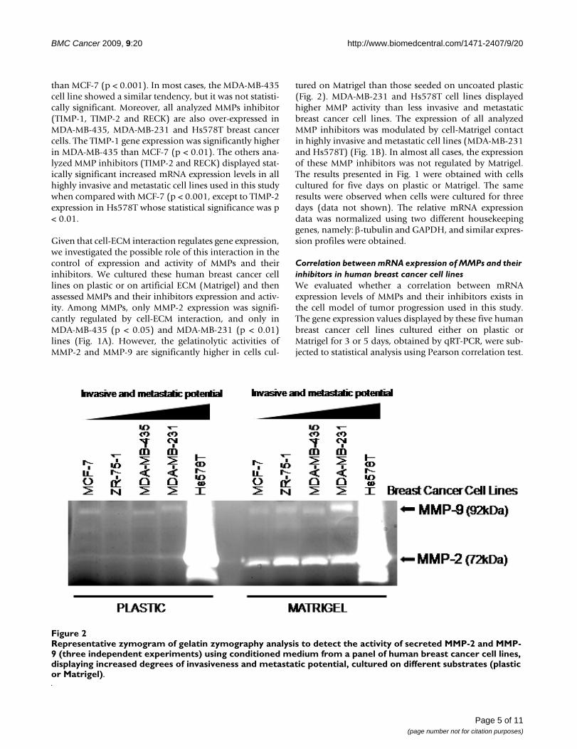
tured on Matrigel than those seeded on uncoated plastic(Fig. 2). MDA-MB-231 and Hs578T cell lines displayedhigher MMP activity than less invasive and metastaticbreast cancer cell lines. The expression of all analyzedMMP inhibitors was modulated by cell-Matrigel contactin highly invasive and metastatic cell lines (MDA-MB-231and Hs578T) (Fig. 1B). In almost all cases, the expressionof these MMP inhibitors was not regulated by Matrigel.The results presented in Fig. 1 were obtained with cellscultured for five days on plastic or Matrigel. The sameresults were observed when cells were cultured for threedays (data not shown). The relative mRNA expressiondata was normalized using two different housekeepinggenes, namely: β-tubulin and GAPDH, and similar expres-sion profiles were obtained.
Correlation between mRNA expression of MMPs and their inhibitors in human breast cancer cell linesWe evaluated whether a correlation between mRNAexpression levels of MMPs and their inhibitors exists inthe cell model of tumor progression used in this study.The gene expression values displayed by these five humanbreast cancer cell lines cultured either on plastic orMatrigel for 3 or 5 days, obtained by qRT-PCR, were sub-jected to statistical analysis using Pearson correlation test.
Representative zymogram of gelatin zymography analysis to detect the activity of secreted MMP-2 and MMP-9 (three independ-ent experiments) using conditioned medium from a panel of human breast cancer cell lines, displaying increased degrees of invasiveness and metastatic potential, cultured on different substrates (plastic or Matrigel)Figure 2Representative zymogram of gelatin zymography analysis to detect the activity of secreted MMP-2 and MMP-9 (three independent experiments) using conditioned medium from a panel of human breast cancer cell lines, displaying increased degrees of invasiveness and metastatic potential, cultured on different substrates (plastic or Matrigel).
Page 5 of 11(page number not for citation purposes)
BMC Cancer 2009, 9:20 http://www.biomedcentral.com/1471-2407/9/20
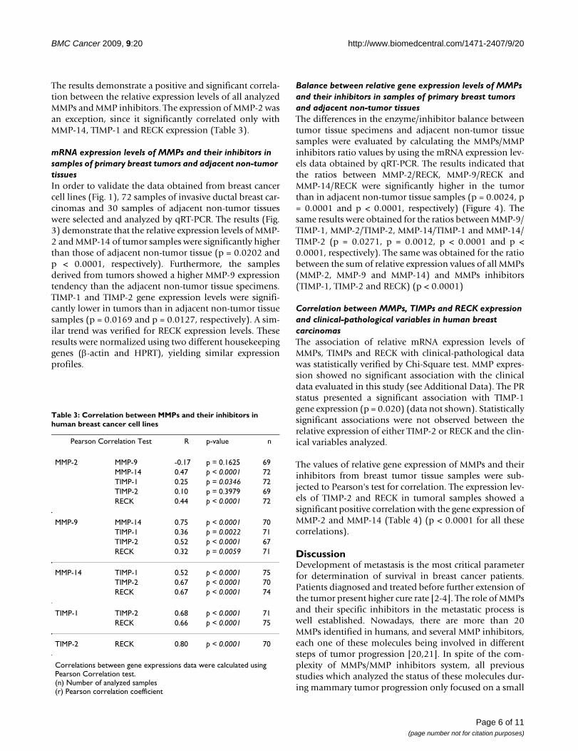
The results demonstrate a positive and significant correla-tion between the relative expression levels of all analyzedMMPs and MMP inhibitors. The expression of MMP-2 wasan exception, since it significantly correlated only withMMP-14, TIMP-1 and RECK expression (Table 3).
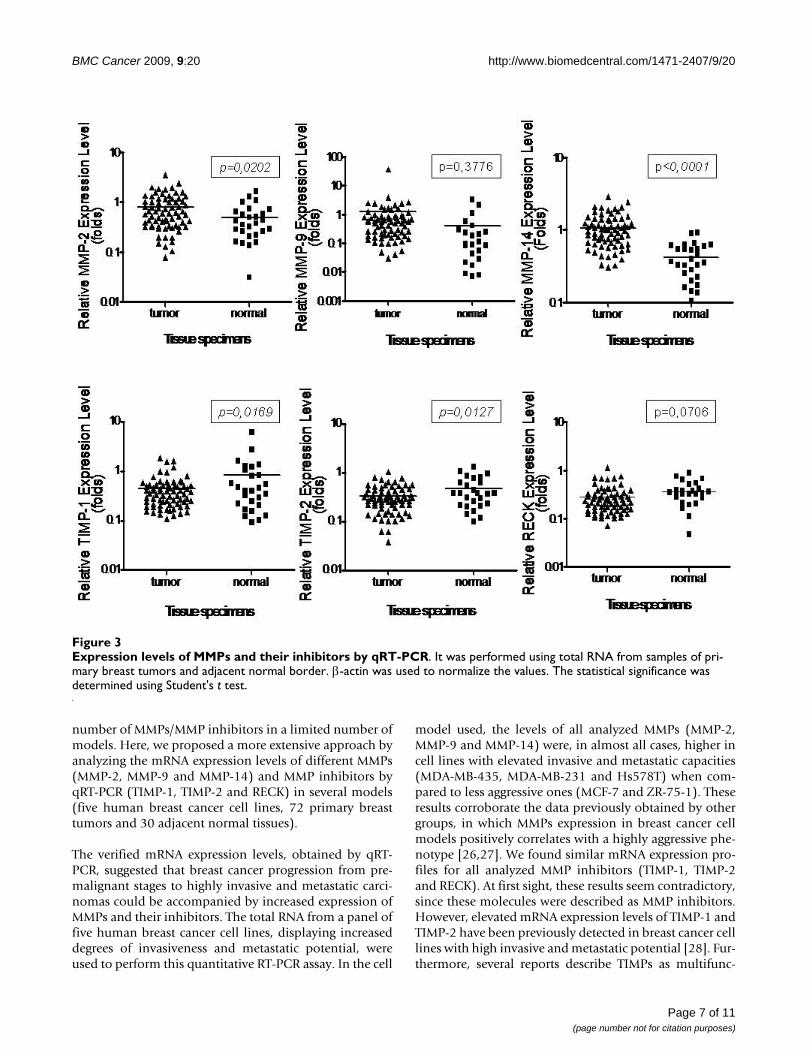
mRNA expression levels of MMPs and their inhibitors in samples of primary breast tumors and adjacent non-tumor tissuesIn order to validate the data obtained from breast cancercell lines (Fig. 1), 72 samples of invasive ductal breast car-cinomas and 30 samples of adjacent non-tumor tissueswere selected and analyzed by qRT-PCR. The results (Fig.3) demonstrate that the relative expression levels of MMP-2 and MMP-14 of tumor samples were significantly higherthan those of adjacent non-tumor tissue (p = 0.0202 andp < 0.0001, respectively). Furthermore, the samplesderived from tumors showed a higher MMP-9 expressiontendency than the adjacent non-tumor tissue specimens.TIMP-1 and TIMP-2 gene expression levels were signifi-cantly lower in tumors than in adjacent non-tumor tissuesamples (p = 0.0169 and p = 0.0127, respectively). A sim-ilar trend was verified for RECK expression levels. Theseresults were normalized using two different housekeepinggenes (β-actin and HPRT), yielding similar expressionprofiles.
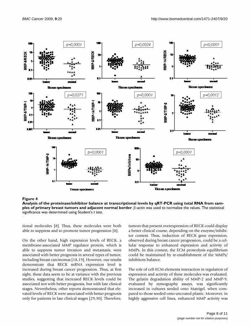
Balance between relative gene expression levels of MMPs and their inhibitors in samples of primary breast tumors and adjacent non-tumor tissuesThe differences in the enzyme/inhibitor balance betweentumor tissue specimens and adjacent non-tumor tissuesamples were evaluated by calculating the MMPs/MMPinhibitors ratio values by using the mRNA expression lev-els data obtained by qRT-PCR. The results indicated thatthe ratios between MMP-2/RECK, MMP-9/RECK andMMP-14/RECK were significantly higher in the tumorthan in adjacent non-tumor tissue samples (p = 0.0024, p= 0.0001 and p < 0.0001, respectively) (Figure 4). Thesame results were obtained for the ratios between MMP-9/TIMP-1, MMP-2/TIMP-2, MMP-14/TIMP-1 and MMP-14/TIMP-2 (p = 0.0271, p = 0.0012, p < 0.0001 and p <0.0001, respectively). The same was obtained for the ratiobetween the sum of relative expression values of all MMPs(MMP-2, MMP-9 and MMP-14) and MMPs inhibitors(TIMP-1, TIMP-2 and RECK) (p < 0.0001)
Correlation between MMPs, TIMPs and RECK expression and clinical-pathological variables in human breast carcinomasThe association of relative mRNA expression levels ofMMPs, TIMPs and RECK with clinical-pathological datawas statistically verified by Chi-Square test. MMP expres-sion showed no significant association with the clinicaldata evaluated in this study (see Additional Data). The PRstatus presented a significant association with TIMP-1gene expression (p = 0.020) (data not shown). Statisticallysignificant associations were not observed between therelative expression of either TIMP-2 or RECK and the clin-ical variables analyzed.
The values of relative gene expression of MMPs and theirinhibitors from breast tumor tissue samples were sub-jected to Pearson's test for correlation. The expression lev-els of TIMP-2 and RECK in tumoral samples showed asignificant positive correlation with the gene expression ofMMP-2 and MMP-14 (Table 4) (p < 0.0001 for all thesecorrelations).
DiscussionDevelopment of metastasis is the most critical parameterfor determination of survival in breast cancer patients.Patients diagnosed and treated before further extension ofthe tumor present higher cure rate [2-4]. The role of MMPsand their specific inhibitors in the metastatic process iswell established. Nowadays, there are more than 20MMPs identified in humans, and several MMP inhibitors,each one of these molecules being involved in differentsteps of tumor progression [20,21]. In spite of the com-plexity of MMPs/MMP inhibitors system, all previousstudies which analyzed the status of these molecules dur-ing mammary tumor progression only focused on a small
Table 3: Correlation between MMPs and their inhibitors in human breast cancer cell lines
Pearson Correlation Test R p-value n
MMP-2 MMP-9 -0.17 p = 0.1625 69MMP-14 0.47 p < 0.0001 72TIMP-1 0.25 p = 0.0346 72TIMP-2 0.10 p = 0.3979 69RECK 0.44 p < 0.0001 72
MMP-9 MMP-14 0.75 p < 0.0001 70TIMP-1 0.36 p = 0.0022 71TIMP-2 0.52 p < 0.0001 67RECK 0.32 p = 0.0059 71
MMP-14 TIMP-1 0.52 p < 0.0001 75TIMP-2 0.67 p < 0.0001 70RECK 0.67 p < 0.0001 74
TIMP-1 TIMP-2 0.68 p < 0.0001 71RECK 0.66 p < 0.0001 75
TIMP-2 RECK 0.80 p < 0.0001 70
Correlations between gene expressions data were calculated using Pearson Correlation test.(n) Number of analyzed samples(r) Pearson correlation coefficient
Page 6 of 11(page number not for citation purposes)
BMC Cancer 2009, 9:20 http://www.biomedcentral.com/1471-2407/9/20
number of MMPs/MMP inhibitors in a limited number ofmodels. Here, we proposed a more extensive approach byanalyzing the mRNA expression levels of different MMPs(MMP-2, MMP-9 and MMP-14) and MMP inhibitors byqRT-PCR (TIMP-1, TIMP-2 and RECK) in several models(five human breast cancer cell lines, 72 primary breasttumors and 30 adjacent normal tissues).
The verified mRNA expression levels, obtained by qRT-PCR, suggested that breast cancer progression from pre-malignant stages to highly invasive and metastatic carci-nomas could be accompanied by increased expression ofMMPs and their inhibitors. The total RNA from a panel offive human breast cancer cell lines, displaying increaseddegrees of invasiveness and metastatic potential, wereused to perform this quantitative RT-PCR assay. In the cell
model used, the levels of all analyzed MMPs (MMP-2,MMP-9 and MMP-14) were, in almost all cases, higher incell lines with elevated invasive and metastatic capacities(MDA-MB-435, MDA-MB-231 and Hs578T) when com-pared to less aggressive ones (MCF-7 and ZR-75-1). Theseresults corroborate the data previously obtained by othergroups, in which MMPs expression in breast cancer cellmodels positively correlates with a highly aggressive phe-notype [26,27]. We found similar mRNA expression pro-files for all analyzed MMP inhibitors (TIMP-1, TIMP-2and RECK). At first sight, these results seem contradictory,since these molecules were described as MMP inhibitors.However, elevated mRNA expression levels of TIMP-1 andTIMP-2 have been previously detected in breast cancer celllines with high invasive and metastatic potential [28]. Fur-thermore, several reports describe TIMPs as multifunc-
Expression levels of MMPs and their inhibitors by qRT-PCRFigure 3Expression levels of MMPs and their inhibitors by qRT-PCR. It was performed using total RNA from samples of pri-mary breast tumors and adjacent normal border. β-actin was used to normalize the values. The statistical significance was determined using Student's t test.
Page 7 of 11(page number not for citation purposes)
BMC Cancer 2009, 9:20 http://www.biomedcentral.com/1471-2407/9/20
tional molecules [8]. Thus, these molecules were bothable to suppress and to promote tumor progression [8].
On the other hand, high expression levels of RECK, amembrane-associated MMP regulator protein, which isable to suppress tumor invasion and metastasis, wereassociated with better prognosis in several types of tumor,including breast carcinomas [18,19]. However, our resultsdemonstrate that RECK mRNA expression level isincreased during breast cancer progression. Thus, at firstsight, these data seem to be at variance with the previousstudies, suggesting that increased RECK levels could beassociated not with better prognosis, but with late clinicalstages. Nevertheless, other reports demonstrated that ele-vated levels of RECK were associated with better prognosisonly for patients in late clinical stages [29,30]. Therefore,
tumors that present overexpression of RECK could displaya better clinical course, depending on the enzyme/inhibi-tor context. Thus, induction of RECK gene expression,observed during breast cancer progression, could be a cel-lular response to enhanced expression and activity ofMMPs. In this context, the ECM proteolysis equilibriumcould be maintained by re-establishment of the MMPs/inhibitors balance.
The role of cell-ECM elements interaction in regulation ofexpression and activity of these molecules was evaluated.The gelatin degradation ability of MMP-2 and MMP-9,evaluated by zymography assays, was significantlyincreased in cultures seeded onto Matrigel, when com-pared to those seeded onto uncoated plastic. Moreover, inhighly aggressive cell lines, enhanced MMP activity was
Analysis of the proteinase/inhibitor balance at transcriptional levels by qRT-PCR using total RNA from samples of primary breast tumors and adjacent normal borderFigure 4Analysis of the proteinase/inhibitor balance at transcriptional levels by qRT-PCR using total RNA from sam-ples of primary breast tumors and adjacent normal border. β-actin was used to normalize the values. The statistical significance was determined using Student's t test.
Page 8 of 11(page number not for citation purposes)

BMC Cancer 2009, 9:20 http://www.biomedcentral.com/1471-2407/9/20
observed in less invasive and metastatic breast cancercells, corroborating the data obtained in mRNA expres-sion analysis. However, among the analyzed proteases,only MMP-2 expression was significantly modulated bycell-ECM elements interaction. These results suggest thatincreased proteolytic activity is independent of transcrip-tional activation, corroborating previously published data[31]. Our results demonstrate that the transcript levels ofall studied MMP inhibitors are significantly modulated bythe reconstituted basal lamina. This regulation of TIMP-1,TIMP-2 and RECK mRNA levels by cell-Matrigel contactwas significant only in highly invasive and metastatic celllines. Therefore, the intense ECM degradation is corre-lated with increased MMP inhibitor expression. The ECMsupplies mechanical support to cells and structural integ-rity to tissues [6,32]. Furthermore, ECM performs aninstructive role, since it is a reservoir of cytokines, and ofgrowth and differentiation factors, which can be dis-charged and/or activated during proteolysis of ECM ele-ments [33] by MMPs, contributing to the crosstalkbetween tumor cells and the microenvironment [34].Therefore, the high mRNA expression levels of MMPinhibitors, found in highly aggressive cell lines, probablyreflects a cellular reaction mechanism to intense ECMremodeling, as previously discussed. Thus, during ECMdegradation, an important factor involved in induction ofTIMPs and RECK can be discharged and/or activated. Ourresults corroborate this hypothesis, since we demon-strated that ECM is important only in modulation of gene
expression of MMP inhibitors and, moreover, only inhighly invasive and metastatic cell lines. Therefore, wesuggest that this negative feedback mechanism of induc-tion of protease inhibitors, during breast cancer progres-sion, occurs for the re-establishment of the MMP/inhibitors balance and, consequently, for homeostasis ofECM proteolysis.
Cellular models are commonly used in Cell and Molecu-lar Biology studies due to the limited source of tissue sam-ples. However, enrichment for malignant cells andabsence of stroma may compromise comparisonsbetween cell lines and tumors [35]. Therefore, to discardpossible cell culture artifacts, we also analyzed the expres-sion of MMPs and their inhibitors in primary breast can-cer tumor samples by qRT-PCR. The results obtaineddemonstrate, in a more general scenario, that the mRNAexpression levels of MMPs are significantly higher intumor samples than in adjacent non-tumor tissues. Onthe other hand, TIMP-1, TIMP-2 and RECK gene expres-sion levels were significantly lower in tumors when com-pared with adjacent normal tissues. Furthermore, ourresults demonstrate a significant positive correlationbetween the gene expression levels of these proteases andtheir inhibitors in primary breast tumors from patients.Therefore, we suggest that increased expression of genesresponsible for ECM degradation is coordinately accom-panied by increased expression of their specific inhibitorsand that the levels of MMPs, TIMPs and RECK transcriptsmay be regulated by common factors and signaling path-ways.
Corroborating other studies in breast cancer [10,36], wefound no significant association between the mRNAexpression levels of the MMPs analyzed and clinical-path-ological data [10,36]. These results suggest that the MMPsmay supply independent prognostic information. How-ever, there are conflicting results on the prognostic valueof these proteases in this model. These differences can, atleast in part, be due to the different methodologies usedand to possible modulation of MMPs by adjuvant sys-temic treatment [37,38]. Furthermore, the TIMP-1, TIMP-2 and RECK transcript levels did not correlate with theclinical parameters from patients. The status of PR was anexception, which showed a significant correlation withTIMP-1 gene expression only. Likewise, we suggest thatexpression of the analyzed MMPs inhibitors may supplyindependent prognostic information.
ConclusionTaken together, the results obtained demonstrate thatMMPs and their specific inhibitors are overexpressed inmore aggressive breast cancer cell lines. However, it islikely that each one of these molecules plays distinct rolesin acquisition of the invasive and metastatic capacity. This
Table 4: Correlation between expression of MMPs and their inhibitors in samples of primary breast tumors
Pearson Correlation Test r p-value N
MMP-2 MMP-9 0.06 p = 0.6523 67MMP-14 0.68 p < 0.0001 70TIMP-1 -1.3 p = 0.2968 70TIMP-2 0.65 p < 0.0001 70RECK 0.64 p < 0.0001 70
MMP-9 MMP-14 0.17 p = 0.1586 67TIMP-1 0.07 p = 0.6005 67TIMP-2 0.21 p = 0.0925 67RECK 0.04 p = 0.7245 67
MMP-14 TIMP-1 -1.7 p = 0.1474 70TIMP-2 0.61 p < 0.0001 70RECK 0.49 p < 0.0001 70
TIMP-1 TIMP-2 0.06 p = 0.6375 70RECK -1.3 p = 0.2702 70
TIMP-2 RECK 0.42 p = 0.0003 70
Correlations between gene expressions data were calculated using Pearson Correlation test.(n) Number of analyzed samples(r) Pearson correlation coefficient
Page 9 of 11(page number not for citation purposes)

BMC Cancer 2009, 9:20 http://www.biomedcentral.com/1471-2407/9/20
complexity of the MMPs-inhibitors system is intensifiedby a strong correlation found between the expression lev-els of these key regulators of the metastatic process. Thus,we suggest that the expression analysis of only one ofthese molecules or the independent quantification ofthese molecular markers does not provide sufficient infor-mation to elucidate the pathological proteolytic mecha-nisms associated with the tumor. Therefore, simultaneousanalysis of the expression status of these molecules couldbe a better parameter to determine the most adequatetherapy for each patient.
AbbreviationsCM: conditioned medium; ECM: extracellular matrix; ER:estrogen receptor; MMP: matrix metalloproteinases; PR:progesterone receptor; qRT-PCR: quantitative reverse tran-scription polymerase chain reaction; RECK: reversion-inducing cysteine-rich protein with Kazal motifs; SD:standard deviation; TIMP: tissue inhibitors of matrix met-alloproteinases
Competing interestsThe authors declare that they have no competing interests.
Authors' contributionsRCSF was responsible for most of the experimental workand interpretation of some of the results. LRG was respon-sible for data discussion and interpretation, statisticalanalysis and manuscript preparation. JSN, FCS andIDCGS were responsible for collecting the tumor samplesused in this study. MCS participated in the study design,data discussion and supervision of the study. All authorsread and approved the final manuscript.
Additional material
AcknowledgementsThe excellent technical support of Zizi de Mendonça, Sandra Regina de Souza, Debora Cristina da Costa, Ricardo Krett de Oliveira, Patrícia Barros dos Santos and Guilherme Frutero is deeply appreciated. Thanks also to the critical reviewing of Letícia Labriola, María Giselle Peters and Wagner Mon-tor. This work was supported by Fundação de Amparo a Pesquisa do Estado de São Paulo (FAPESP), Conselho Nacional de Pesquisa (CNPq), Financiadora de Estudos e Projetos (FINEP) and Pró-Reitoria da Universi-dade de São Paulo (PRP-USP).
References1. Jemal A, Siegel R, Ward E, Hao Y, Xu J, Murray T, Thun MJ: Cancer
statistics, 2008. CA Cancer J Clin 2008, 58(2):71-96.2. Lee JH, Welch DR: Suppression of metastasis in human breast
carcinoma MDA-MB-435 cells after transfection with themetastasis suppressor gene, KiSS-1. Cancer Res 1997,57(12):2384-2387.
3. Welch DR, Steeg PS, Rinker-Schaeffer CW: Molecular biology ofbreast cancer metastasis. Genetic regulation of humanbreast carcinoma metastasis. Breast Cancer Res 2000,2(6):408-416.
4. Yang J, Mani SA, Donaher JL, Ramaswamy S, Itzykson RA, Come C,Savagner P, Gitelman I, Richardson A, Weinberg RA: Twist, a mas-ter regulator of morphogenesis, plays an essential role intumor metastasis. Cell 2004, 117(7):927-939.
5. Vernon AE, LaBonne C: Tumor metastasis: a new twist on epi-thelial-mesenchymal transitions. Curr Biol 2004,14(17):R719-721.
6. Itoh Y, Nagase H: Matrix metalloproteinases in cancer. EssaysBiochem 2002, 38:21-36.
7. Visse R, Nagase H: Matrix metalloproteinases and tissue inhib-itors of metalloproteinases: structure, function, and bio-chemistry. Circ Res 2003, 92(8):827-839.
8. Lambert E, Dasse E, Haye B, Petitfrere E: TIMPs as multifacial pro-teins. Crit Rev Oncol Hematol 2004, 49(3):187-198.
9. Talvensaari-Mattila A, Paakko P, Turpeenniemi-Hujanen T: Matrixmetalloproteinase-2 (MMP-2) is associated with survival inbreast carcinoma. Br J Cancer 2003, 89(7):1270-1275.
10. Jones JL, Glynn P, Walker RA: Expression of MMP-2 and MMP-9,their inhibitors, and the activator MT1-MMP in primarybreast carcinomas. J Pathol 1999, 189(2):161-168.
11. Iwata H, Kobayashi S, Iwase H, Masaoka A, Fujimoto N, Okada Y:Production of matrix metalloproteinases and tissue inhibi-tors of metalloproteinases in human breast carcinomas. JpnJ Cancer Res 1996, 87(6):602-611.
12. Schrohl AS, Christensen IJ, Pedersen AN, Jensen V, Mouridsen H,Murphy G, Foekens JA, Brunner N, Holten-Andersen MN: Tumortissue concentrations of the proteinase inhibitors tissueinhibitor of metalloproteinases-1 (TIMP-1) and plasminogenactivator inhibitor type 1 (PAI-1) are complementary indetermining prognosis in primary breast cancer. Mol Cell Pro-teomics 2003, 2(3):164-172.
13. Schrohl AS, Holten-Andersen MN, Peters HA, Look MP, Meijer-vanGelder ME, Klijn JG, Brunner N, Foekens JA: Tumor tissue levelsof tissue inhibitor of metalloproteinase-1 as a prognosticmarker in primary breast cancer. Clin Cancer Res 2004,10(7):2289-2298.
14. Remacle A, McCarthy K, Noel A, Maguire T, McDermott E, O'HigginsN, Foidart JM, Duffy MJ: High levels of TIMP-2 correlate withadverse prognosis in breast cancer. Int J Cancer 2000,89(2):118-121.
15. Takahashi C, Sheng Z, Horan TP, Kitayama H, Maki M, Hitomi K,Kitaura Y, Takai S, Sasahara RM, Horimoto A, et al.: Regulation ofmatrix metalloproteinase-9 and inhibition of tumor invasionby the membrane-anchored glycoprotein RECK. Proc NatlAcad Sci USA 1998, 95(22):13221-13226.
16. Oh J, Takahashi R, Kondo S, Mizoguchi A, Adachi E, Sasahara RM,Nishimura S, Imamura Y, Kitayama H, Alexander DB, et al.: Themembrane-anchored MMP inhibitor RECK is a key regulatorof extracellular matrix integrity and angiogenesis. Cell 2001,107(6):789-800.
17. Rhee JS, Coussens LM: RECKing MMP function: implications forcancer development. Trends Cell Biol 2002, 12(5):209-211.
18. Clark JC, Thomas DM, Choong PF, Dass CR: RECK – a newly dis-covered inhibitor of metastasis with prognostic significancein multiple forms of cancer. Cancer Metastasis Rev 2007, 26(3–4):675-683.
19. Span PN, Sweep CG, Manders P, Beex LV, Leppert D, Lindberg RL:Matrix metalloproteinase inhibitor reversion-inducingcysteine-rich protein with Kazal motifs: a prognostic markerfor good clinical outcome in human breast carcinoma. Cancer2003, 97(11):2710-2715.
20. Chang C, Werb Z: The many faces of metalloproteases: cellgrowth, invasion, angiogenesis and metastasis. Trends Cell Biol2001, 11(11):S37-43.
Additional file 1Clinic-pathological data. The data provided represent the clinical path-ological data from patients whose breast tumor or normal tissue samples were used in this study.Click here for file[http://www.biomedcentral.com/content/supplementary/1471-2407-9-20-S1.doc]
Page 10 of 11(page number not for citation purposes)
http://www.ncbi.nlm.nih.gov/entrez/query.fcgi?cmd=Retrieve&db=PubMed&dopt=Abstract&list_uids=9192814
http://www.ncbi.nlm.nih.gov/entrez/query.fcgi?cmd=Retrieve&db=PubMed&dopt=Abstract&list_uids=9192814
http://www.ncbi.nlm.nih.gov/entrez/query.fcgi?cmd=Retrieve&db=PubMed&dopt=Abstract&list_uids=9192814
http://www.ncbi.nlm.nih.gov/entrez/query.fcgi?cmd=Retrieve&db=PubMed&dopt=Abstract&list_uids=8766524
http://www.ncbi.nlm.nih.gov/entrez/query.fcgi?cmd=Retrieve&db=PubMed&dopt=Abstract&list_uids=8766524
http://www.ncbi.nlm.nih.gov/entrez/query.fcgi?cmd=Retrieve&db=PubMed&dopt=Abstract&list_uids=8766524
http://www.ncbi.nlm.nih.gov/entrez/query.fcgi?cmd=Retrieve&db=PubMed&dopt=Abstract&list_uids=9789069
http://www.ncbi.nlm.nih.gov/entrez/query.fcgi?cmd=Retrieve&db=PubMed&dopt=Abstract&list_uids=9789069

BMC Cancer 2009, 9:20 http://www.biomedcentral.com/1471-2407/9/20
Publish with BioMed Central and every scientist can read your work free of charge
"BioMed Central will be the most significant development for disseminating the results of biomedical research in our lifetime."
Sir Paul Nurse, Cancer Research UK
Your research papers will be:
available free of charge to the entire biomedical community
peer reviewed and published immediately upon acceptance
cited in PubMed and archived on PubMed Central
yours — you keep the copyright
Submit your manuscript here:http://www.biomedcentral.com/info/publishing_adv.asp
BioMedcentral
21. Sternlicht MD, Werb Z: How matrix metalloproteinases regu-late cell behavior. Annu Rev Cell Dev Biol 2001, 17:463-516.
22. Bachmeier BE, Albini A, Vene R, Benelli R, Noonan D, Weigert C,Weiler C, Lichtinghagen R, Jochum M, Nerlich AG: Cell density-dependent regulation of matrix metalloproteinase andTIMP expression in differently tumorigenic breast cancercell lines. Exp Cell Res 2005, 305(1):83-98.
23. Bachmeier BE, Nerlich AG, Lichtinghagen R, Sommerhoff CP: Matrixmetalloproteinases (MMPs) in breast cancer cell lines of dif-ferent tumorigenicity. Anticancer Res 2001, 21(6A):3821-3828.
24. Haupt LM, Thompson EW, Trezise AE, Irving RE, Irving MG, GriffithsLR: In vitro and in vivo MMP gene expression localisation byIn Situ-RT-PCR in cell culture and paraffin embedded humanbreast cancer cell line xenografts. BMC Cancer 2006, 6(1):18.
25. Pfaffl MW: A new mathematical model for relative quantifica-tion in real-time RT-PCR. Nucleic Acids Res 2001, 29(9):e45.
26. Balduyck M, Zerimech F, Gouyer V, Lemaire R, Hemon B, Grard G,Thiebaut C, Lemaire V, Dacquembronne E, Duhem T, et al.: Specificexpression of matrix metalloproteinases 1, 3, 9 and 13 asso-ciated with invasiveness of breast cancer cells in vitro. ClinExp Metastasis 2000, 18(2):171-178.
27. Kousidou OC, Roussidis AE, Theocharis AD, Karamanos NK:Expression of MMPs and TIMPs genes in human breast can-cer epithelial cells depends on cell culture conditions and isassociated with their invasive potential. Anticancer Res 2004,24(6):4025-4030.
28. Yoshiji H, Gomez DE, Thorgeirsson UP: Enhanced RNA expres-sion of tissue inhibitor of metalloproteinases-1 (TIMP-1) inhuman breast cancer. Int J Cancer 1996, 69(2):131-134.
29. Takenaka K, Ishikawa S, Kawano Y, Yanagihara K, Miyahara R, OtakeY, Morioka Y, Takahashi C, Noda M, Wada H, et al.: Expression ofa novel matrix metalloproteinase regulator, RECK, and itsclinical significance in resected non-small cell lung cancer.Eur J Cancer 2004, 40(10):1617-1623.
30. Takenaka K, Ishikawa S, Yanagihara K, Miyahara R, Hasegawa S, OtakeY, Morioka Y, Takahashi C, Noda M, Ito H, et al.: Prognostic signif-icance of reversion-inducing cysteine-rich protein with Kazalmotifs expression in resected pathologic stage IIIA N2 non-small-cell lung cancer. Ann Surg Oncol 2005, 12(10):817-824.
31. Azzam HS, Arand G, Lippman ME, Thompson EW: Association ofMMP-2 activation potential with metastatic progression inhuman breast cancer cell lines independent of MMP-2 pro-duction. J Natl Cancer Inst 1993, 85(21):1758-1764.
32. Ramirez F, Rifkin DB: Cell signaling events: a view from thematrix. Matrix Biol 2003, 22(2):101-107.
33. Aumailley M, Gayraud B: Structure and biological activity of theextracellular matrix. J Mol Med 1998, 76(3–4):253-265.
34. Mueller MM, Fusenig NE: Friends or foes – bipolar effects of thetumour stroma in cancer. Nat Rev Cancer 2004, 4(11):839-849.
35. Birgersdotter A, Sandberg R, Ernberg I: Gene expression pertur-bation in vitro – a growing case for three-dimensional (3D)culture systems. Semin Cancer Biol 2005, 15(5):405-412.
36. Remacle AG, Noel A, Duggan C, McDermott E, O'Higgins N, FoidartJM, Duffy MJ: Assay of matrix metalloproteinases types 1, 2, 3and 9 in breast cancer. Br J Cancer 1998, 77(6):926-931.
37. Lebeau A, Muller-Aufdemkamp C, Allmacher C, Sauer U, Nerlich A,Lichtinghagen R, Lohrs U: Cellular protein and mRNA expres-sion patterns of matrix metalloproteinases-2, -3 and -9 inhuman breast cancer: correlation with tumour growth. J MolHistol 2004, 35(5):443-455.
38. Li HC, Cao DC, Liu Y, Hou YF, Wu J, Lu JS, Di GH, Liu G, Li FM, OuZL, et al.: Prognostic value of matrix metalloproteinases(MMP-2 and MMP-9) in patients with lymph node-negativebreast carcinoma. Breast Cancer Res Treat 2004, 88(1):75-85.
Pre-publication historyThe pre-publication history for this paper can be accessedhere:
http://www.biomedcentral.com/1471-2407/9/20/prepub
Page 11 of 11(page number not for citation purposes)
http://www.ncbi.nlm.nih.gov/entrez/query.fcgi?cmd=Retrieve&db=PubMed&dopt=Abstract&list_uids=8608981
http://www.ncbi.nlm.nih.gov/entrez/query.fcgi?cmd=Retrieve&db=PubMed&dopt=Abstract&list_uids=8608981
http://www.ncbi.nlm.nih.gov/entrez/query.fcgi?cmd=Retrieve&db=PubMed&dopt=Abstract&list_uids=8608981
http://www.ncbi.nlm.nih.gov/entrez/query.fcgi?cmd=Retrieve&db=PubMed&dopt=Abstract&list_uids=8411260
http://www.ncbi.nlm.nih.gov/entrez/query.fcgi?cmd=Retrieve&db=PubMed&dopt=Abstract&list_uids=8411260
http://www.ncbi.nlm.nih.gov/entrez/query.fcgi?cmd=Retrieve&db=PubMed&dopt=Abstract&list_uids=8411260
http://www.ncbi.nlm.nih.gov/entrez/query.fcgi?cmd=Retrieve&db=PubMed&dopt=Abstract&list_uids=9535559
http://www.ncbi.nlm.nih.gov/entrez/query.fcgi?cmd=Retrieve&db=PubMed&dopt=Abstract&list_uids=9535559
http://www.ncbi.nlm.nih.gov/entrez/query.fcgi?cmd=Retrieve&db=PubMed&dopt=Abstract&list_uids=9528836