F:\PROJECT\20536- Catholic Charities Providence House Additions ...
Upload
khangminh22Category
view
0download
0
Phyton (Austria) Vol. 21 Fasc. 2 261-287 30. 9. 1981
Corrections and Additions to the Book'Morphology of Seed-Plants'
B y
Michel GrtiUDi&s*)
With 13 Figures . /
Received December 1, 1980
Key words: Spermatophyta. — Morphology
Summary
M. 1981. Corrections and additions to the book 'Morphology ofSeed-Plants'. — Phyton (Austria) 21 (2): 261-287. — English with Germansummary.
In an effort to correct mistakes in the author's "Morphology of Seed-Plants" (Cramer 1979, Vaduz) and to present further facts and interpretations,some notes are given here. Mainly roots, life form, axillary buds, unifacial leafportions, floral phyllotaxis and gynoecia are considered.
Zusammenfassung
GTTEDES M. 1981. Korrekturen und Ergänzungen zum Buch „Morphologyof Seed-Plants". - Phyton (Austria) 21 (2): 261-287. — Englisch mit deutscherZusammenfassung.
In dem Bestreben, in dem Buch „Morphology of Seed-Plants" (Cramer1979, Vaduz) enthaltene Fehler zu verbessern sowie weitere Fakten und Inter-pretationen darzustellen, bespricht der Autor eine Anzahl morphologischerThemen. Im besonderen werden Wurzeln, Lebensformen, Axillarknospen,unifaziale Blattabschnitte, florale Phyllotaxis, Fragen der Gynözeum-Morpho-logie u. a. behandelt.
The additions and corrections presented here are related to the author'sbook 'Morphology of Seed-Plants' (8°, 326 pages, 30 figures), which ispublished by J. Cramer (Vaduz 1979). References to the page numbersare given in relation to this book.
*) Michel GUEDES, Museum d'Histoire naturelle, 57, rue Cuvier, Paris,Seme, France. . .
©Verlag Ferdinand Berger & Söhne Ges.m.b.H., Horn, Austria, download unter www.biologiezentrum.at

Root branching (p. 21 and p. 24)
Lateral roots of most Monocots arise opposite xylem bundles, as withDicots (VAN TIEGHBM & DOTTLIOT 1888). They are initiated opposite phloembundles in the Gramineae and Gyperaceae, but in e. g. corn (Zea) they mayalso appear in front of xylem bundles. CLOWES 1978 recently confirmed theoccurrence of an endodermal pouch around rootlet primordia in corn. Thisis multilayered and indistinguishable from a root-cap, but is later replacedby a true cap from the dermatogen of the lateral root itself.
Regeneration of root apex (p. 30)
As early as 1933, GATJTHERET had shown that in Zea mays isolatedroot tips including the root-cap and at least part of the proximal meristemwith what is now called the quiescent center, could regenerate a wholeroot. When too little meristem remained with the cap, only anarchic growthoccurred. Distal portions of roots whose tip had been excised regenerateda cap and apparently a quiescent center from their remaining meristem.The latter fact had been known since PRANTL'S work in 1874.
Detailed experiments on root regeneration in Zea have recently beencarried out by FELDMANN 1979. He found that regeneration (of decapitatedroots) is possible from the meristem proximal to the quiescent center. Themeristem then regenerated a quiescent center and root-cap. Furthermore,roots could be regenerated directly, i. e. through direct elongation of theexplant along the same axis as in the intact root rather than through deve-lopment of an adventitious root from a callus, from cultivated meristematicportions excised just proximal to the quiescent center, and without thelatter. The meristem used in the latter experiments was 300—350 jxm inlength. When the quiescent center remained attached to it, regenerationwas also possible.
Since direct regeneration is possible from either the quiescent center,the adjacent upper meristem or both, it is all too clear that such experimentstell little or nothing about the actual initiating role of cells from these tworegions in the normal, intact root.
Root chimeras (p. 31)
I noticed that work should be done on root chimeras, if any, to clear upthe functioning of root apices. It must be mentioned that there already areat least two papers on such chimeras. BRTTMFIELD 1943, by irradiatingseedlings of Grepis capillaris (L.) WALLR. (Compositae) and Vicia faba L.(Papilionaceae), obtained sectorial chimeras, a sector being detectable bychromosomal abnormalities in many of its cells, and being apparentlyproduced by a single initial. There seemed to be three sectors, each of whichwas comprised of a third of the root-cap, the cortex and the stele. Themoment the roots were irradiated, they appear to have possessed three
©Verlag Ferdinand Berger & Söhne Ges.m.b.H., Horn, Austria, download unter www.biologiezentrum.at

263
initials for the whole of their tissues. RICKARD 1952, however, who alsoX-rayed Crepis, got three sectors in the cortex only of a single root, so hebelieved that there were three initials to the cortex, three to the cap andthree to the stele. I t is obvious that much further work is needed in this line.
Closed-type roots (p. 32)
The origin and functioning of cortical initials in the root of Cyperusfuscus L. (Cyperaceae) is described by JTJGTTET & VALLADE 1979.
Summer and winter annuals (p. 39)
Although it is not a morphologic one, the distinction may be mentionedbetween summer annuals which germinate, flower and die within the samecalendar years, and winter annuals, which germinate in fall, to flower anddie next year. As is well-known, the same species may behave both waysdepending on the strain. Wheat is normally a winter annual, but certaincultivars are summer annuals, because they need no vernalization.
Multi-articled sympodia within a season in woody plants (p. 45)
In temperate woody plants with sympodial development, only onearticle develops within a season, but there are exceptions especially in vinessuch as Wisteria sinensis (SIMS) SWEET (p. 47). Also in Citrus (Butaceae) twoor three sympodial articles generally develop during each season, the secondfrom the first and the third from the second. Sympodization here is throughapex death, not apex flowering (SCHROEDER 1951). In Gornus alba L.{Gornaceae) where sympodization occurs as a result of the development ofterminal inflorescences three articles at least may develop within a season(GUEDES, unpubl.).
Trees and shrubs (p. 48)
The acrotonous condition is not sufficient for a woody perennial tobecome a tree. A trunk must appear at least below. As a rule, the lowerportion of the trunk, from ground level up, is naked, which implies the lackor early disappearance of most lateral branches in the early life of the plant.On the other hand, in a basitonous woody perennial, if only one or a fewmain shoots arise from the lower zone of the previous year's shoot, and eachdevelops some sort of a trunk, this being mono- or sympodial, a polycormictree may result, i. e. a tree with several trunks diverging from near groundlevel. This may be seen in the elder {Sambucus nigra L., Caprifoliaceae) or inCotoneaster solidfolia FRANCH. (Bosaceae). In these plants, sprouts arisenin a basitonous way are themselves mesotonous, more or less plagiotropicand commonly sympodial in their development. Several oblique undulatetrunks may thus appear in old plants, and one of them may survive singlyas a small tortuous tree. .•> . . . • . -.. ••. <. ••>•. ... .-,..,. -.
©Verlag Ferdinand Berger & Söhne Ges.m.b.H., Horn, Austria, download unter www.biologiezentrum.at

264
I have prepared a morphologic system of tree and shrub architecture(GTJEDES, in press), suggesting to restrict the term shrub (true shrubs) tocolonial woody perennials with basitonous branching, other 'shrnbs'in effect being dwarf trees. The various constitutions of trunk and branchesof trees, respecting their sympodial or monopodial structure, the occurenceof short shoots etc. enabled me to define ten main morphological typesof architecture with several variants in most of them. It is hoped that thissystem is morphologically more consistent than HALLE, OLDEMAN &TOMLIKSON'S one (1978); perhaps I may also refer the reader to my reviewof the latter (GTJEDES 1980a).
Iterative innovation (p. 50)
An axillary shoot developed at ground level by a determinate shoot(and similar to the latter as in rhizomatous irises [p.43] or grasses) maybecalled an innovat ion . In perennial plants innovations may remainvegetative for one or more years before they flower (more years e. g. Aconi-tum sect. Lycoctonum, Ranunculaceae: SEREBRIAKOVA & POLYNTSEVA
1974). Their production is a kind of sympodization as explained. Wheninnovations are flowering they themselves develop innovations of the nexthigher order. I mentioned on p. 50 that young, not yet flowering Asparagusofficinalis L. plants give rise to several such 'generations' within the sameseason. This is also true of the adult flowering plant of this species. TROLL
1964: 325—331 spoke of iterative innovation in such instances, innovationthen meaning development of innovation shoots. Up to 10 flowering inno-vations develop within the same season in adult Asparagus officinalis,all or most of whose buds are formed in the previous fall. Iterative inno-vation according TROLL 1. C. also occurs in Canna indica L. (Gannaceae),Juncus subnodulosus SCHRANK (Juncaceae), Nardus stricta L. (Gramineae),Garex arenaria L. (Cyperaceae). In annual grasses, or annual shoots (inno-vations) of perennial grasses, severals orders of innovations commonlydevelop, the process being called tillering. Every tiller comes from a loweraxillary bud of the main shoot (first-order tillers) or of a previous-ordertiller. Tillering is different from iterative innovation insofar as, even inperennial grasses the ultimate generation of tillers will not give rise tonext year's main shoot(s). The latter will be originated by a lower axillarybud of the current year's shoot, as was the first generation of the currentyear's tillers. , .,,.
Apical growth of stem (p. 53)
I may have stated by mistake that Gymnosperms may have GW(green-white) chimeras. Only WG (white-green) chimeras seem to be known,although GW ones certainly are a possibility. Also the epidermis (TI =first tunica layer) rarely proliferates at the margins of leaves in Gymno-
©Verlag Ferdinand Berger & Söhne Ges.m.b.H., Horn, Austria, download unter www.biologiezentrum.at
265
sperms, so that my statement that there are white margins to leaves ofWG chimeras is erroneous.
Attention must be called to the work of STEFFENSEN 1968 (see alsoCOE & NEUTFER 1978) on the shoot apex of Zea mays L., where radiation-induced sectoring is very carefully related to a histogenetic study of theapex. The corn apex appears to have a few initials, probably often two, toeach of its histogens. TI-TIII initials make up two vertical series each ofwhich originates one half of the plant (one half means here one side of theplant divided by a plane cutting through stem and midribs of the leaves).
As the shoot is already well developed in the mature seed, to get acompletely half-mutated plant, one must irradiate a very young embryo,36—40 h after fertilization. It is known that at this time the first periclinaldivision occurs in the terminal cell of the filamentous embryo, establishingits bilateral symmetry. Half-mutated plants appear when one of the twocells then produced mutates. This cell must give rise to the three histogeninitials on one side and consequently these are all mutated. Later irradiationleads to more and more upwardly located sectors, limited to one histogen.If mature seeds are irradiated and one of the initials of an histogen is inducedto mutate, the mutation is found at tassel level, in the form or a y2 or %sector. If sectors occur below in such plants, they are narrower, becausethey come from mutated cells that were below the initials. In the matureseed, the initials have already produced the whole of the cells from whichleaves and bracts have been or will be initiated. So if there are two initialsfor each histogen, no irradiation at this stage can lead to a y2 sector at thevegetative level. The tassel, on the other hand, is still to be laid down bythe initials at the mature seed stage, so half mutated tassels may occurfrom irradiated seeds. As initials may be more than two in each histogen,or because they have already begun dividing to initiate the tassel, narrowersectors also are found in mutated tassels from irradiated seeds.
The midrib plane of corn is a boundary that is often trespassed on bycell progenies, i. e. one sector of a two-sectored plant may cross the midribof a leaf while not reaching that of the leaf below on the other orthostichy.Thus adjustments are constantly being made between cell progenies, someof which may become temporarily or progressively wider, while others areaccordingly restricted so that the specific form is maintained.
'•"•'• Half-rosette plants (p. 65)
Half-rosette plants may be annual, their rosette elongating into aflowering shoot within the year after germination. This is seen in e. g.Capsella bursapastoris L. (Cruciferae). TROLL 1937: 223—236 believed thata plant is still a rosette rather than a half-rosette one if the rosette apexelongates into an inflorescence stalk bearing no trve leaves. If this is acceptedit is hard, as TROLL acknowledged, to draw a line between such rosetteplants and those half-rosette ones with leaves only down their elongated
©Verlag Ferdinand Berger & Söhne Ges.m.b.H., Horn, Austria, download unter www.biologiezentrum.at
stem. This is why I prefer to restrict the use of the term rosette plant forplants which never elongate their rosette axis and develop inflorescencesor flowers laterally. Such plants belong to "rosette plants with indefinitemain axis" in TROLL'S terminology. In my view all plants with a rosettewhose axis elongates into a stalk, whether or not leafy, are half-rosetteplants.
Axillary buds: rarer locations. Con- and recaulescence, anaphysis (p. 66)
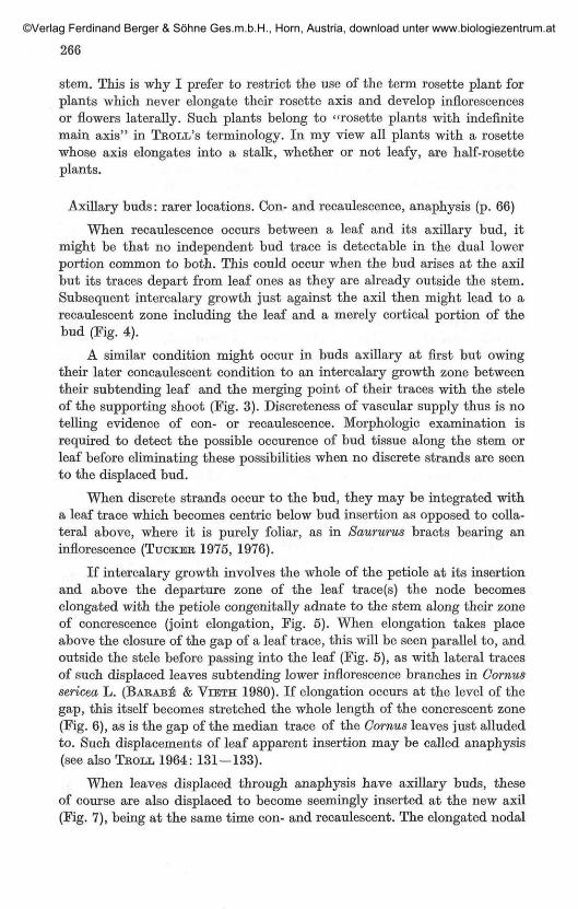
When recaulescence occurs between a leaf and its axillary bud, itmight be that no independent bud trace is detectable in the dual lowerportion common to both. This could occur when the bud arises at the axilbut its traces depart from leaf ones as they are already outside the stem.Subsequent intercalary growth just against the axil then might lead to arecaulescent zone including the leaf and a merely cortical portion of thebud (Fig. 4).
A similar condition might occur in buds axillary at first but owingtheir later concaulescent condition to an intercalary growth zone betweentheir subtending leaf and the merging point of their traces with the steleof the supporting shoot (Fig. 3). Discreteness of vascular supply thus is notelling evidence of con- or recaulescence. Morphologic examination isrequired to detect the possible occurence of bud tissue along the stem orleaf before eliminating these possibilities when no discrete strands are seento the displaced bud.
When discrete strands occur to the bud, they may be integrated witha leaf trace which becomes centric below bud insertion as opposed to colla-teral above, where it is purely foliar, as in Saururus bracts bearing aninflorescence (TUCKER 1975, 1976).
If intercalary growth involves the whole of the petiole at its insertionand above the departure zone of the leaf trace(s) the node becomeselongated with the petiole congenitally adnate to the stem along their zoneof concrescence (joint elongation, Fig. 5). When elongation takes placeabove the closure of the gap of a leaf trace, this will be seen parallel to, andoutside the stele before passing into the leaf (Fig. 5), as with lateral tracesof such displaced leaves subtending lower inflorescence branches in Cornussericea L. (BARABB & VIETH 1980). If elongation occurs at the level of thegap, this itself becomes stretched the whole length of the concrescent zone(Fig. 6), as is the gap of the median trace of the Cornus leaves just alludedto. Such displacements of leaf apparent insertion may be called anaphysis(see also TROLL 1964: 131 — 133).
When leaves displaced through anaphysis have axillary buds, theseof course are also displaced to become seemingly inserted at the new axil(Fig. 7), being at the same time con- and recaulescent. The elongated nodal
©Verlag Ferdinand Berger & Söhne Ges.m.b.H., Horn, Austria, download unter www.biologiezentrum.at
267
zone is made up of the stem and basal portions of the axillary bud and leafwhen intercalary growth occurs at the level of bud insertion involving thebasal portion of the leaf at the same time. Gornus sericea often has suchdisplaced leaves or bracts at the level of its two lowermost inflorescencebranches, the latter being involved in the displacement process (Fig. 7).Lower inflorescence branches in this Cornus may also be merely con- orrecaulescent. When bracts are very reduced their trace may not enter theappendage itself, remaining in the stele gap of the supporting axis, or theremay be a gap with no trace whatever. Upper inflorescence branches arealways concaulescent and lack bracts. Con- and recaulescence as well asanaphysis were studied in this plant by BARABE& VIETH 1980 in an illumi-nating paper.
Lateral and leaf-opposed serial buds (p. 68)
As indicated, serial buds may be collateral, i. e. in a tangential rowwithin each axil, in some Monocots. It may happen that only one bud in theseries occurs in an axil, without being the median one. In the palm Plecto-comiopsis corneri FURTADO the only bud in each leaf axil is 30—40° awayfrom the midrib zone of the leaf base (FISHEB & DRANSFIELD 1979). Whenleaves enwrap the stem, leaf axils in fact circle the latter, and collateralbuds might occur all around. In vegetative plagiotropic stems of Musa(Musaceae) the leaves have a single 'axillary' bud but it is inserted 180°away from the median leaf region i. e. above the meeting point of bothmargins of the leaf base on the side opposite the midrib (FISHER 1978). Thesame is seen in vegetative leaves or certain species of Daemonorops (Palmae),whereas in Korthalsia rigida BLTJME, another palm, the axillary bud is130° from the midrib (FISHER & DRANSFIELD 1979). Inflorescence budsare normal axillary buds in these palms, and in Daemonorops transitionalleaves between vegetative and fertile zones often subtend both a conven-tional inflorescence bud and a leaf-opposed vegetative bud. In my viewall these palm leaves potentially have a circling series of axillary buds,vegetative or sexualized, among which only two opposite or near-oppositeones are developed, singly or both at the same time. Bracts of Musa do notencircle the peduncle and subtend two superimposed collateral series offlower buds. Two species of Dracontium (Araceae) seem to have manycircling series of collateral buds in the axils of their enwrapping bulb scales(TROLL 1937: 538,539).
A lateral bud such as that of Korthalsia thus seems to be one of apotential circling series of serial collateral buds. It must be carefully dis-tinguished from extra-axillary lateral buds (p. 325), which are basicallyconventional axillary buds somewhat displaced laterally owing to theirdeveloping a bulky axillary bud to their own prophyll (Cucurbitaceae;TROLL 1939: 1976-1979).
©Verlag Ferdinand Berger & Söhne Ges.m.b.H., Horn, Austria, download unter www.biologiezentrum.at

Is dichotomy in seed plants a really true one ? (p. 72)
As initials appear to be present at the apex of stems of seed plants,one would expect that in the rare instances of true dichotomy, they areapportioned between the two daughter apices. Each half of them would goto each apex and regenerate the whole initial complement. Initials in factare hard to locate histogenetically, but at least one would see the apexproper become split into two. Now it seems that in dichotomizing apicesthe apex proper ceases growing and two new apices differentiate on itsflanks. Since these apices bear no relation to any leaf axil, they may becalled adventitious, and dichotomy would thus be subapical adventitiousbudding.
Lammas shoots (p. 131)
When the apical bud of a woody plant breaks several times within thesame season, the supplementary flushes besides the spring one give rise tolammas shoots, the term applying especially to the second flush, whichmay take place about the lammas (August 1). As a lammas shoot proper iselongating from the terminal bud, lateral lammas shoots may develop fromaxillary buds on the stem segment below, whether it is from the previousseason or the previous flush in the same season. I studied such a case inChoisya ternata KUNTH (Butaceae; GUEDES, 1980b).
Multinodal pines develop lammas shoots. Their winter bud may bemade up of the first flush only before its elongation, so the lammas shootsare not preformed. Other such pines have a multi-storied apical bud includ-ing the consecutive flushes of the season (see LANNBR 1976, 1978). Lammasshoots are apparently not preformed within apical buds of broad-leavedtrees.
Intermediary apical buds formed during the short resting periodsbetween the seasonal flushes may be imperfect, much in the way of incipientbuds of the avocado (p. 131). If they are lacking altogether, there is aprotracted growth period all through the season instead of several flusheswith intervening periods of rest. Flushing and continuously growing shootsoccur on the same individual. On conventional shoots of temperate woodyplants, elongation of course occurs during the sole spring flush.
Cauliflory
Flowers or inflorescences sometimes seem to appear on ripened shootsmore than a year old, with no very clear connection to former leaf axils,at first sight at least. It is often thought, and sometimes stated in print,that they are adventitious. This is unlikely in view of the rarity of adven-titious normal shoots above cotyledons. Cauliflory has rarely been studiedin enough detail. Some studies are quoted by HALLE & al. 1978. In theJudas-tree (Gerds süiquastrum L., Caesalpiniaceae) cauliflory is from serial
©Verlag Ferdinand Berger & Söhne Ges.m.b.H., Horn, Austria, download unter www.biologiezentrum.at
269
buds that develop on succeeding years in leaf axils. Most of the latterproduce inflorescences for some years only, after fall of the correspondingleaves. Some axils, however, may 'awake' after a many years' dormancy,and again send out flowers, their supporting shoot having become more orless bulky in the meantime. No inflorescence is adventitious (GUEDES,unpubl.).
In Gleditsia triacanthos L. (Caesalpiniaceae) long shoots bear axillaryshort determinate shoots with no elongated internodes, that live only oneyear. Commonly, after fall of its first determinate shoot, so two years afterfall of its subtending leaf, an axil develops new determinate shoots fromserial buds on succeeding years. Two or three serial shorts may appear inthe same axil within the same season. These shoots in effect are 'cauli-folious' vegetative short shoots. Inflorescences of Gleditsia arise in the leafaxils of one-year-old shoots, exactly as determinate vegetative shoots.Leaves are formed at the bottom of the inflorescent axis, and a steriledeterminate shoot is homologous with the sterile lower portion of aninflorescence axis. True cauliflory does not seem to occur in Gleditsiatriacanthos, i. e. axils more than a year old develop only vegetative deter-minate shoots, not inflorescence (GUEDES, unpubl.).
Locally multilayered epidermis (T I) and leaf initiation (p. 91)
Rather than occur in the young leaf primordium, periclinal divisionsof TI (dermatogen or first tunica layer) that lead to the invasion of Til(second tunica layer) by daughters of TI cells may occur laterally on thestem apex, above the youngest leaf primordium. TI then is locally twoor three cells thick, and when this region takes part in producing a leafprimordium, the latter may be entirely made up of cells that are geneticallyTI if the whole of its cell rows is produced by the multilayered TI. Incertain chimeras of Tradescantia fluminensis VELL. (Commelinaceae, THIELKE1957), whose leaves develop from the second and third layers, TI becomespartly two- or three-layered and leaf tissues normally derived from Til orTill are genetically TI in those leaf regions that come from apex zones withtwo- or three-layered TI.
Morphology and architecture of unifacial leaf portions (p. 105)
Although forerunner tips (p. 103) generally are epiunifacial, i. e. withtheir morphologically lower surface all around, some instances have beenquoted by VOGEL 1966 of orchid tepals with hypounifacial tips. The extre-mity of such phyllomes is then, outrolled, and they have their morpholo-gically upper (ventral) surface all around.
With respect to the morphology of the upper cross-zone of their petioles,leaves with unifacial petioles are of two kinds. (The word cross-zone meanshere the meeting zone of both right and left leaf margins, as well as the more
©Verlag Ferdinand Berger & Söhne Ges.m.b.H., Horn, Austria, download unter www.biologiezentrum.at

270
Fig. 1 — 13. — Schemes. — Fig. 1. Formation of a funnel-shaped structurethrough pinching of an inside proliferation of the cross-zone of the unifacialclaw of a tepal. Ventral view of the tepal. Fig. la. Cross-section of similarstructure, pinching perfect, so the inner tube is closed, broken lines indicateupper margins of unifacial claw. — Fig. 2. Formation of a funnel-shaped nectarythrough ventral proliferation (arrows) of a circular margin portion arisen throughmeeting of two loops from ventral margin on both sides of the cross-zone.Ventral view of petaloid stamen, with a ventral lobe on the left, bearing aventral pollen sac. Dorsal pollen sac on the same side. After LEINFELLNER'S(1959) data. Fig. 2a. Cross-section of such a nectary.
Fig. 3 — 7. Displacement of apparent insertion of leaves and axillary shootsthrough intercalary growth. — Fig. 3. Anaphysis. Leaf trace joins stele at levelof apparent insertion. — Fig. 4. Recaulescence with shoot trace inserted on leaftrace. — Fig. 5. Anaphysis, leaf trace joins the stele at the level of morphologicinsertion. — Fig. 6. Same, but the stele gap is elongated as a result of inter-calary growth at its level. — Fig. 7. Anaphysis of leaf with con- and recaule-scence of axillary bud. Stele gap elongated.
Fig. 8. Cellular architecture of a leaf with unifacial petiole. Between baseand petiole, cross-zone arises through bulging of ventral cell rows. Betweenpetiole and blade, cross zone either arises in the same way (left) or (right) ismarked by the subhorizontal courses of cell rows making up the lower blade onthis side. Representative cell rows from Til are marked M, 1, 2, 3, 1', 2', 3',their bulges at cross-zone level Ma, la, 2a, 3a, l'a, 2'a, 3'a. Ventral view withventral shanks of cell rows as solid lines, dorsal ones as broken lines. M, I, II,III, IV, V, VI; I', . . . VI', Vm denote bundles of the petiole and blade. Theseare not drawn but would differentiate at the levels of cell rows and their loops.Note that IV—VI do not occur in the blade on the left, whereas they do on theright. They would be ventral petiole bundles on both side. — Fig. 9. Ascidiateleaf with unifacial petiole and no base. Ventral view. Dorsal bundles as solidlines, ventral ones as broken lines. Bundles drawn only on the left. — Fig. 10.Leaf primordium, ventral view, with representative cell rows from Til. Arrowindicate level of proliferation to give rise to a cross-zone or ligule.
Fig. 11. From a leaf primordium (in longitudinal section, ventral side onthe right) drawn on the left, a dorsal spur may develop through proliferationon its back below apex (middle scheme) or an epiunifacial point may arise ifdorsal proliferation involves its apex (right scheme). Crosses denote leaf apex.
Fig. 12. Phyllotaxis of flower of Ranunculus repens. Redrawn from MEICEN-HEIMEE, 1979. Bracts white crescents outside, sepals hatched, petals dotted,stamens white circles. Contact parastichies are drawn, those appearing at thelevel of each floral whorl begin with a cross and se(sepals), p(petals), st(stamens).
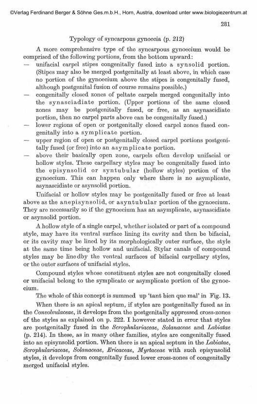
Fig. 13. Generalized scheme of a carpel constituent of a syncarpous gynoe-cium. Ventral view. Broken lines indicate zones of congenital fusion eitherwithin the carpel itself (ventral suture) or with its adjoining carpels in thegynoecium. Zones of the syncarpous gynoecium to which such a carpel wouldtake part are indicated as follows: ae anepisynsolid, As asymplicate, asyasyntubular, es episynsolid, sa synascidiate, sp symplicate, ss synsolid, stsyntubular; as apical septum.
©Verlag Ferdinand Berger & Söhne Ges.m.b.H., Horn, Austria, download unter www.biologiezentrum.at

271
©Verlag Ferdinand Berger & Söhne Ges.m.b.H., Horn, Austria, download unter www.biologiezentrum.at

272
or less horizontal margin segments just adjoining this meeting zone, orcross-zone proper, on the ventral side of the petiole). Either the ventral,inverted bundles of the petiole stop at the cross-zone and do not serve theblade (Fig. 8, left) or (same, right) they proceed into the lower zone of theblade. In the latter instance, the nearer a ventral bundle to the medianline of the petiole, the lower does it become located in the blade. If there is aventro-median, half of it becomes the lowest main blade bundle (VI' infig. 8). The first kind of leaf may be exemplified by those of some of theDioscoreaceae (GUEDES 1967) and the second kind by those of Pelargonium Xhortorum L. H. BAILEY.
When a leaf of the first kind becomes peltate-ascidiate, the upper cross-zone of its petiole proliferates in such a way that the ventral petiole bundlesnow proceed into a superadded ventral blade portion. On the contrary,when a leaf of the second kind becomes peltate or ascidiate, there is nofurther proliferation of the cross-zone, for this already develops the lowerblade portions on both sides of the normal leaf blade. To become peltate-ascidiate, the latter kind of leaf merely undergoes a rearrangement of theproducts of its upper petiole cross-zone: instead of being split into twolateral halves, one on each lower side of the blade, they are now continuousmedio-ventrally. In other words, both lateral halves of the blade are nowfolded ventrally and congenitally merged at their former lowermost margins.There is no superadded tissue in the peltate-ascidiate leaf when this iscompared to the normal one. It will perhaps be clear from Fig. 8 that on theleft (first kind) a mere ventral flange marks the cross-zone, while on theright (second kind) the cross-zone has originated the lower portion of theblade, giving rise to bundles IV, V, VI'. On the left bundles IV, V, VI ofthe petiole do not proceed beyond the cross-zone.
Architecturally, in a leaf built from TI-TIII layers of the apex, cellrows of the Til layer continue ventrally (Fig. 8, solid lines) from the petioleinto the blade, developing shallow bulges at the cross-zone (Fig. 8, on theleft) in leaves of the first kind, while in the second kind of leaf (Fig. 8, right),ventral Til rows, reaching the cross-zone, depart into the dorsal (lower)region of the lower blade zone, then back on the ventral region of the samezone, before they ascend again the ventral portion of the main blade. Thiscourse will be obvious for rows 1', 2', 3' whose cross-zone loops in lower bladeportion are numbered 3'a, 2'a, l'a. In both kinds of leaves, Til rows, havingreached the brim of the blade (1, 2, 3, 1', 2', 3') go down dorsally along theblade and petiole (broken lines). When ascidiate, both kinds of leaves aresimiliar (Fig. 9, with bundles IV, V, VI, IV, V, VI' either developed denovo in the superadded ventral blade portion, and as prolongations ofpetiole ventral bundles, or displaced through blade inrolling from theirformer lateral position in the blade).
The young leaf primordium (Fig. 10) has its Til rows vertical, goingfrom the stem along its ventral side (solid lines) till its upper brim, then
©Verlag Ferdinand Berger & Söhne Ges.m.b.H., Horn, Austria, download unter www.biologiezentrum.at
273
back along its dorsal surface (broken lines). During ontogeny of leaveswith unifacial petioles, a ventral proliferation takes place atop the latter(arrow level) to either originate a cross-zone bulge, or proliferate into thelower blade portion. If the leaf is to become peltate-ascidiate that zonedevelops the ventral portion of the ascidium or peltate blade.
Histogenetically the development of a shallow cross-zone playing nopart in forming the blade is similar to the development of a ventral ligule.Unifaciality of the petiole, however, becomes obvious in the first instance,even if it remains flat, through the orientation of its vascular bundles.
In the same fashion, the histogenetic development of an epiunifacialforerunner tip (Fig. 11) is similar to that of a dorsal spur. The only differenceis that the proliferation originating the forerunner tip occurs dorsally verynear the phyllome apex so as to involve it and displace it atop the for-erunner tip. Hypounifacial forerunner tips develop as ventral proliferationsagain involving the phyllome apex.
Between the bifacial leaf-base, if such occurs, and the unifacial petioleis a cross-zone (Fig. 8). As explained (p. 92, fig. 13/2) the most lateral cellrows in the leaf-base may not reach the petiole. Lateral rows of the petioleprolong outmost or near-outmost rows in the base (Fig. 8). As outmostbundles cross the base to merge into a ventro-median, or become verynear each other, they have to cross cell rows nearer to the median zone.Again architecture at the junction between base and petiole is the samewhether the leaf has a unifacial petiole or a conventional one with a ligule:in both instances cell rows emit transversal outgrowths ventrally (oftenlimited to the epidermis if a ligule is to be formed). Morphological signi-ficance of the structure becomes obvious as differentiation goes on.
Although most leaves with unifacial petioles have bifacial leaf-bases,the latter may be lacking, as in Ginkgo biloba (p. 109, fig. 16/9). It needsnot to be admitted that a virtual base occurs in such leaves. Cell rows ofthe stem apex may well develop a unifacial petiole as depicted in Fig. 9.
Equitant Iris leaves (p. 118)
When presenting BUGNON'S view on equitant Iris leaves I stressedthat it had been reached as a result of direct histogenetic studies, and asthese are very difficult it should be tested by means of chimeras. I have nowdone this myself by studying cvs. "variegata" of I. pallida and /. foetidis-sima (GTTEDES 1980 a). These are GWG chimeras where frequent periclinaldivisions send epidermal (TI) or Till cells, both genetically green, amongthe white cells of the Til layer. The progenies of so displaced cells are greenpatches among white tissue. Äs these patches may be elongated the wholelength of the leaf, I was unable to support BUGNON'S concept. Cell divisionsoriginating the Iris leaf give rise to longitudinal cell families whose orien-tation parallels that of main nerves, rather than crossing them. As in other
18 P h y t o n. Vol. 21, Fasc. 2, 1981. i8
©Verlag Ferdinand Berger & Söhne Ges.m.b.H., Horn, Austria, download unter www.biologiezentrum.at

274
Monocots (STEWART & DERMEN 1979) leaf architecture is closely reflectedby vascularization, and is as the classical view had long had it. Sinceequitant Iris leaves are merely grass-like leaves congenitally folded at bladelevel along their midrib, it becomes quite natural that Iris subgen. Scorpiris,in contradistinction to other irises, has non-equitant, conventional ribbonleaves.
Histogeny of leaves in the Gramineae (p. 118)
In the Gramineae it would seem that the leaf not rarely has a blade madeup of TI ('epidermal') cells while its sheath mostly comes from Til . Thiswas demonstrated in a bamboo by studying a chimera (THIELKE 1961) andconfirmed histogenetically by BUGNON 1979 in other grasses.
Leaflets and stipules (p. 121)
Stipules are the first and only basipetal leaflet pair in Ailanthus altis-sima. They are the lowermost pair of acropetal leaflets in other taxa (seeWEBERLING & LEENHOUTS 1965, they believed this is also true of Ailanthus,but their own figures seem to point to the view here suggested).
Leaflet development (p. 121)
Citation of Clematis as an example of a leaf with basipetal leaflets isa lapsus. Clematis is described by TROLL 1939: 1530 and TEPFER 1960 aswith acropetal leaflets to its leaves. Polemonium (Polemoniaceae) andLycopersicon (Solanaceae, COLEMAN & GREYSON 1976) afford true examplesof basipetal leaflets. Leaflets of the Papilionaceae are acropetal in theirdevelopment.
When all leaflets are acropetal, the whole of them correspond to thecompound terminal leaflet of those leaves whose first-developed leafletsare midway up the rachis.
Syncotyly and the origin of Monocots (p. 132)
HAINES & LYE 1979 have reviewed the subject of syncotyly and triedto support the interpretation of the single cotyledon of Monocots as orig-inated through fusion of the two in Dicots. In Impatiens glanduliferaROYLE (Balsaminaceae) and Daucus carota L. (Umbelliferae) there may beonly one cotyledon. In intermediate stages in Impatiens it is clear that thetwo cotyledons are more or less completely fused along their adjoiningedges, their axillary buds also becoming merged into one. In other wordsthe embryo may form the usual two cotyledons, a single lateral cotyledon,or a bivalent lateral cotyledon. Such monocotyledonous seedlings in Dicotsmay be induced experimentally in Eranthis hymemalis (L.)SALISB. (Ranun-culaceae, HACCIXJS & TROMPETER 1960). In these cases there is a switch inseedling phyllotaxy from the whorled to the alternate condition, in exactly
©Verlag Ferdinand Berger & Söhne Ges.m.b.H., Horn, Austria, download unter www.biologiezentrum.at

275
the same way as sometimes in vegetative stems of e. g. the Labiatae (p. 132)where intermediate stages with a bivalent leaf at every node are also known.If phyllotaxy is wholly modified the leaf or cotyledon is actually univalent,exhibiting no indication of duality. In some genera of the Umbelliferae(Bunium, Oonopodium) and in certain species of Peperomia (Piperuceae),Corydalis (Papaveraceae) and Ranunculus (R. fi,caria L.; Ranunculaceae)the seedling has a single univalent cotyledon and this no doubt must beconsidered as phylogenetically arisen from the usual pair, as seen in terato-logic Impatiens seedlings just alluded to. Such solitary cotyledons occurin genera or species of otherwise dicotyledonous taxa.
It then is a poss ib i l i ty that the sole cotyledon of Monocots, thoughat present univalent, also arose in this way from the two cotyledons ofdicotyledonous ancestors. As nothing very precise is known about originof Monocots, and no unquestionable reversion to any putative ancestraldicotyledonous state seems to have been described in seedlings of Monocots,this is a mere hypothesis. In any case the cotyledon of Monocots now isa single phyllome.
The petioles of the two cotyledons are united into a tube in manyUmbelliferae (HACCIUS 1952) and species of Rheum (Polygonaceae, Huss1980: 20—21) and of Anemone (Ranunculaceae). This parallels gamophyllyin the adult state in such plants as Dipsacus fullonum (p. 134; see alsoHACCIUS 1952: 501, TROLL 1937: 249).
Leafs with protracted apical growth (p. 137)
Often-quoted are pinnate leaves of several Meliaceae (Guarea, Ghisoch-eton, Dysoxylum, Gabralea) which are said to display a very protracted apicalactivity, behaving much as shoots, and to develop their pinnae continuouslyor sometimes (Chisocheton) in flushes of several leaflets with interveningresting periods. The apex of these leaves in fact has nothing to do with ashoot bud, for its meristematic activity is actually very short, as in allleaves of seed plants. As early as 1887, SONNTAG found that meristematicactivity soon ceases at the apex of Guarea leaves, and leaflets are initiatedacropetally. Then the leaflet and rachis grow to their full length in twoflushes. At the end of the first, apical leaflets remain in a primordial stateas a spurious bud atop this lower flush. Leaflet initiation, however, is per-fectly conventional, the whole of the acropetal series appearing after earlycessation of apical activity in the leaf primordium.
Inflorescences in woody plants (p. 146)
BKIGGS & JOHNSON 1979 published a very important paper on inflores-cences in the Myrtaceae. This will have to be used in future studies oninflorescences of woody plants. The authors recognize the importance ofproliferous inflorescences, which they call auxote l ic . Auxotelic inflore-
18*
©Verlag Ferdinand Berger & Söhne Ges.m.b.H., Horn, Austria, download unter www.biologiezentrum.at
276
scences are said to be conflorescences, being made up of unif lorescen-ces, the latter often reduced to single floweis. Contlorescences may beaggregated into auxotelic superconf lorescences . The authors also haveother useful terms such as m e t a x y p h y l l for Zwischenblatt. They do notseem to have met with the case of flower peduncles with one or two vege-tative shoots in the axils of their prophylls. They acknowledge that attemptsby English-speaking authors to account for inflorescences have often been'inadequate, and indeed vapid'.
Branching and inflorescences in Prunus subgen. Amygdalus (p. 149)
I stated in error that in Prunus subgen. Amygdalus there are axillaryinflorescences whose prophylls subtend vegetative buds. I unfortunatelywas led to this misstatement by the description in Flora Europaea 2:78 which reads 'each flower-bud flanked by 2 leaf-buds'. Actually, theaxillary bud is vegetative with one or both of its prophylls subtending asolitary flower. The situation is correctly stated by REHDER 1949 when hewrites of "buds three in each axil, the lateral ones flowers buds". In Prunustriloba LINDL., of the same subgenus, it seems that the axillary bud maysometimes be a flower whose prophylls are then 'sterile'. In P. subgen.Amygdalus, there are no conventional inflorescences, but the prophyllaryflower(s) may be considered a proliferous (auxotelic) inflorescence, oftenwith a single flower.
Acer negundo (p. 150)
It must be mentioned that my description refers to female inflores-cences, which were observed in the cv. 'variegatum'. Living male treeswere unavailable to me.
True and false umbels in Umbelliferae (p. 151)
In the Saniculoideae (FROEBE 1964), apparently simple umbellules arecompound ones, being themselves built of umbelkiles each of which basicallyhas a central female flower with peripheral müe flowers Abortion of oneor the other flower kind, reductions, condensations lead to the deceptivenormal condition. In e. g. Sanicula europaea L. the umbellules comprisefalse umbels in which some umbellules are inserted on the very shortenedpeduncles of the others, there being two orders of umbellules. Such falseumbels are then reminiscent of those of the Cornaceae.
In the Hydrocotyloideae, inflorescences are basically thyrses (compoundracemes of dichasia) that are often impoverished and condensed into umbelsof false umbellules, i. e. umbellules with flowers of two or more successiveorders. If only first order flowers develop, a true umbel is originated (FROEBE
1979). This has a terminal flower, topping the main axis of the originalthyrse. Umbels and umbellules in the Apioideae are true ones, generallywithout a terminal flower, although this occurs in more than 30 genera.
©Verlag Ferdinand Berger & Söhne Ges.m.b.H., Horn, Austria, download unter www.biologiezentrum.at

277
Apopetalous flowers (p. 162)
Apopetaly is generally a phylogenetic, and hypothetic, process. It mayhowever be postgenital, that is, petals are initiated, then very soon ceasegrowing, with the result that they are not to be found in the adult state.Although the genus Scleranthus (Garyophyllaceae) is believed to be apetalousby taxonomists, it has long been known that in S. annuus L. five petalprimordia arise, only to become immediately arrested in their development(PAYER 1857).
Floral phyllotaxis (p. 163)
The subject of flower phyllotaxis is further discussed in DUPTJY &GUEDES 1980 where attention is called to the possibility of floral phyllomesdeveloping, in many instances, each at the limit divergence angle from theprevious one in the phyllotactic spiral. This was suggested by HIRMEE andaccepted by HIEPKO in many cases in the Banales. Although it probablycannot be generalized it might be useful to keep this concept in mind whenstudying phyllotaxis of flower buds. Later rearrangements would lead tothe 2/5 or other phyllotaxis of the adult flower. Spiral sequence of initiationis probably more widespread in flowers than previously believed. Forinstance, perianth and androecium members appear in spiral sequence inHumulus Iwpulus L. (Gannabaceae, LEINS & ORTH 1979). As stamens areopposite tepals the spiral sequence is continuous and regular, and judgingfrom fig. 5 in LEINS & ORTH'S paper, it may well be that at initiation thedivergence angle is the limit angle, with the result that stamens at first arenot exactly opposite tepals. Continuous spiral sequence is also apparentduring perianth and androecium initiation of Cannabis (Gannabaceae) andLaportea (Urticaceae, SATTLER 1973). Remarkably stamens and carpels inSilene coeli-rosa (L.) GODEON (Garyophyllaceae) are initiated spirally. Whenthe plant is cultivated at lower temperatures, petals are also spiral in theirinitiation. Floral phyllomes in Silene coeli-rosa, however, appear in theiradult location, and hardly any limit divergence angle is to be measuredbetween any of them at any time (LYNDON, 1978a—b).
Contact parastichies may be detected in truly polymerous androeciaand gynoecia such as those of Ranunculus and other Banunculaceae. Istated on p. 169 that it is generally hard to connect the parastichies (ororthostichies) of androecium, gynoecium and tepals where they are spiral.Studying Ranunculus acris L., SATTLER 1973 wrote that 'the number ofvertical pistil rows is not necessarily the same as that of the stamens. Butoften the pistil primordia continue the vertical stamen rows'. EICHLER1878 on the other hand spoke in several instances of a continuous spiralpervading the perianth, androecium and gynoecium, often with phyllo-tactic changes between two kinds of phyllomes. Androecia and gynoeciain the Ranunculaceae might display ortho- or spirostichies. If the samephyllotaxis were retained between the two, ortho- or spirostichies were
©Verlag Ferdinand Berger & Söhne Ges.m.b.H., Horn, Austria, download unter www.biologiezentrum.at

278
continuous throughout the whole of the androecium and gynoecium.They might also include the petals. Phyllotaxis, however, often changedbetween the three sets, ortho- or spirostichies being added or deletedbetween two sets. According to EICHLER, among the Banunculaceae, inAdonis aestivalis L. there are at most 8 petals, whereas there are 13 stamenspirostichies. In Eranihus hymealis there are as a rule 6 petals and generally12 stamen spirostichies. The 3 carpels are seated atop three evenly spacedspirostichies among those of the androecium. In Ranunculus acris, R. bul-bosus L., EICHLER, found divergence angles of about 8/21 or 13/34 in theandroecium and gynoecium, also probably 5/13 in the latter. He stressedthat there is no discontinuity from the androecium to the gynoecium,meaning that 13 of the spirostichies in the former proceed into the latter.He also believed, out of comparison with Adonis, that the corolla was alsobasically spiral in Ranunculus. In these observations and reasonings, hewas following WYDLER 1859.
In a recent interesting paper, MEICENHEIMER 1979 indicated thatin Ranunculus repens L. sepals are arranged in a 3/8 phyllotaxis, which ischanged to 5/13 when petals are initiated, as a whorl. Then phyllotaxisshifts to 8/21 as stamens, then carpels appear spirally. Phyllotaxis is 2/5below the calyx, so one may draw a pair of contact parastichies among thebracts. On reaching the calyx, three sepals are found alcng these parastichies,and a further parastichy is needed to accommodate the remainingsepal, as their phyllotaxis is 3/8. The 3 now extant parastichies, then,reach 3 of the petals, and 2 further parastichies must be added for the tworemaining petals of the 5/13 corolla. The 5 parastichies now reach 5 of thestamens, and 3 further parastichies appear in the androecium. So phyllomesare initiated along 8 parastichies in the androecium, and these proceedinto the gynoecium.
I have redrawn MEICENHEIMER'S fig. 19 (Fig. 12) adding the para-stichies. I believe this way of presenting the data to be clearer to the rankand file morphologist than the author's so elaborate mathematical descrip-tion. In fact, all this merely means that the apex is producing relativelysmaller primordia nearer to one another in space and time as initiation offloral phyllomes proceeds. It is interesting that MEICENHEIMER found thecalyx and corolla with a 3/8 and 5/13 phyllotaxis respectively, though bothhave 5 members. This means that sepals and petals are initiated nearer tothe limit divergence angle than is supposed in the classical concept of a2/5 calyx and alternating corolla. HIRMER 1931 and SCHÖFFEL 1932 believedthat in the Ranunculus flower all phyllomes are initiated spirally with thelimit divergence angle between them.
Contorted corolla and phyllotactic sequence in the calyx (p. 167)Besides some of the Theaceae it must be mentioned that the Oxalidaceae,
Linaceae, Malvaceae normally have contorted corollas winding in the direc-
©Verlag Ferdinand Berger & Söhne Ges.m.b.H., Horn, Austria, download unter www.biologiezentrum.at
279
tion of the calyx spiral. Such corollas again are at the same time metatopicand apparently controlled by calyx aestivation in the arrangement of theirpetals. There are of course as many flowers with clockwise and anticlockwisecorollas in the same species, as with calyces.
In this connection, mention must be made of the curious resultsof GHOSH & DAVIS 1978. They found that as expected clockwise and anti-clockwise shoots predominantly produce axillary flowers with anticlockwiseand clockwise corollas respectively, flowers being heterodromous withrespect to their supporting axis. The rarer homodromous flowers, however,gave fruits with a greater proportion of normal seeds.
Sympetaly (p. 188)
The subject of petal fusion has been discussed in great detail byNISHINO 1978, SATTLER 1978 and DANIEL & SATTLER 1978. SATTLER
contemplated several possibilities for a continuous tube to arise belowfree lobes, and wondered what, if anything, could be called fusion whenthere is no true, postgenital fusion, as in such usual instances as the corollain the Solanaceae. He believed one might speak of meristematic fusion whenmarginal meristems are seen at primordium margins, then those of adjoin-ing margins come to meet each other between the lobes, with the issuingintervening meristem responsible for the development of the interlobularportions of the corolla tube. When a circular meristem develops below thelobes and in the interlobular zones to originate the floral tube, no fusioncould be contemplated.
In my view, even with these somewhat different ontogenies the tubeis anyway plurivalent, i. e. corresponds to the whole of the petal claws of achoripetalous corolla. In vegetative leaves, too, marginal meristems aremore or less obvious, leaves remaining homologous. When an interlobularportion seems to come from a meristem that did not originate from fusionof adjacent marginal meristems, the problem is that of knowing what itscells are building. If morphology reveals its product is petaline and partand parcel of neighboring sublobular regions, then it indeed belongs to bothpetals. Not all petals need arise in precisely the same way, no more than theyneed be vascularized in precisely the same fashion. In any case as a petalextends on both sides, more and more cells of the floral apex become involv-ed in its development, more and more cells of the apex flank giving rise tocell rows of the petal. When cell rows of neighboring petals of sympetalouscorollas come to meet each other, meristematic activity may or may not beobvious at the margins of the original lobes, i. e. first-elongated cell-rowsmaking up the median zones of the petals. Even when marginal meristemswere present in the beginning they may have faded away before or duringtangential extension of petals, i. e. formation of new cell rows from apicalcells adjacent to the young petal. Marginal meristems may also not yet be
©Verlag Ferdinand Berger & Söhne Ges.m.b.H., Horn, Austria, download unter www.biologiezentrum.at

280
differentiated during tangential extension of the lobes. When the floralapex is building the corolla tube, it may divide its cells below the lobes tofurther lengthen the cell rows that make them up, or the lower cells of thelobes may proliferate to the same effect. These are interesting growth moda-lities that detract nothing from the petaline and plurivalent character ofthe resulting corolla.
It may also be that in corollas, and especially in androecia and gynoe-cia, a continuous tore is developed by the apex, with the free lobes appearingonly later from its rim. The tore is plurivalent and its constitution is revealedby lobe development later, and further differentiation within the wholestructure. Fused compound stamens of some of the Malvaceae and gynoeciaof some of the Primulaceae were found to develop in this way.
Stamens of Gymnosperms (p. 202)
It was remarked by SPORNE 1980 that the word "stamen" used forGymnosperrn microsporophylls would shock Anglo-saxon morphologists.But are microsporophylls anything else than stamens, a word used byEnglish-speaking taxonomists of Gymnosperms (see e. g. DALLIMORE &JACKSON 1966) ?
A more disturbing problem is the interpretation of stamens in thePinaceae. When they are transformed into bracts as in hermaphroditecones, their blade becomes the bract one, with their pollen sacs vanishing.It is thus impossible to consider the main staminal blade as the ventral oneof a peltate phyllome, as I did. Were it so, the dorsal, polliniferous bladewould become the bract one rather than disappear (the bract is normallyoriented with respect to the cone axis). Since the stamen nonetheless ispeltate according to DLTJHOSH'S studies, it must be hypopeltate (p. 108)with a cross-zone on its back, developing the polliniferous blade, appressedon the main blade. The polliniferous blade no longer occurs in modifiedstamens which are left with their main blade only, now the bract blade. Asan example of studies on hermaphrodite cones, one may quote STENZEL'S1876 paper.
On reinvestigating the development of the glands in the Ginkgo stamen,AMEELE 1980 found that it differs widely from the development of a pollensac. He does not necessarily dismiss their homology with pollen sacs, how-ever, so my stand may be maintained for at least the time being.
Duality of cross-zone ovules (p. 209)
Attention is called to a paper by KSHETRAPAL & TIAGI 1970 wheredual vascularization of cross-zone ovules is demonstrated in certain Oleaceae,although the authors seem to be unaware of the peltate carpel concept.Such a duality, however, is far more common than suggested by them. It isseen also in Triosteum (Caprifoliaceae; WILKINSON 1948).
©Verlag Ferdinand Berger & Söhne Ges.m.b.H., Horn, Austria, download unter www.biologiezentrum.at
281
Typology of syncarpous gynoecia (p. 212)
A more comprehensive type of the syncarpous gynoecium would becomprised of the following portions, from the bottom upward:— unifacial carpel stipes congenitally fused into a synsolid portion.
(Stipes may also be merged postgenitally at least above, in which caseno portion of the gynoecium above the stipes is congenitally fused,although postgenital fusion of course remains possible.)
— congenitally closed zones of peltate carpels merged congenitally intothe synasciadiate portion. (Upper portions of the same closedzones may be postgenitally fused, or free, as an asynascidiateportion, then no carpel parts above can be congenitally fused.)
— lower regions of open or postgenitally closed carpel zones fused con-genitally into a symplicate portion.
— upper region of open or postgenitally closed carpel portions postgeni-tally fused (or free) into an asymplicate portion.
— above their basically open zone, carpels often develop unifacial orhollow styles. These carpellary styles may be congenitally fused intothe episynsolid or syntubular (hollow styles) portion of thegynoecium. This can happen only where there is no asymplicate,asynascidiate or asynsolid portion.Unifacial or hollow styles may be postgenitally fused or free at least
above as the anepisynsolid, or asyntubular portion of the gynoecium.They are necessarily so if the gynoecium has an asymplicate, asynascidiateor asynsolid portion.
A hollow style of a single carpel, whether isolated or part of a compoundstyle, may have its ventral surface lining its cavity and then be bifacial,or its cavity may be lined by its morphologically outer surface, the styleat the same time being hollow and unifacial. Stylar canals of compoundstyles may be linedby the ventral surfaces of bifacial carpellary styles,or the outer surfaces of unifacial styles.
Compound styles whose constituent styles are not congenitally closedor unifacial belong to the symplicate or asymplicate portion of the gynoe-cium.
The whole of this concept is summed up 'tant bien que mal' in Fig. 13.When there is an apical septum, if styles are postgenitally fused as in
the Convolvulaceae, it develops from the postgenitally appressed cross-zonesof the styles as explained on p. 222. I however stated in error that stylesare postgenitally fused in the Scrophulariaceae, Solanaceae and Labiatae(p. 214). In these, as in many other families, styles are congenitally fusedinto an episynsolid portion. When there is an apical septum in the Labiatae,Scrophulariaceae, Solanaceae, Ericaceae, Myrtaceae with such episynsolidstyles, it develops from congenitally fused lower cross-zones of congenitallymerged unifacial styles.
©Verlag Ferdinand Berger & Söhne Ges.m.b.H., Horn, Austria, download unter www.biologiezentrum.at

282
Ovule orientation (p. 213)
When cross-zone ovules are bent downward (their raphe above) theyare apotropous, whereas they are epitropous if bent upward, theirraphe below. In both cases, they may be erect or pendant. The sameorientations are seen in marginal ovules near the cross-zone on each side ofit. Two such ovules are epitropous in e. g. the Geraniaceae, while they areapotropous in the Labiatae. It may happen that a cross-zone ovule isapotropous and is flanked with two epitropous ovules as in Sphaeralceaangustifolia (CAV.) G. DON (Malvaceae).
Although as stated on p. 213, the common condition for marginalanatropous ovules on a placenta is to have their micropyles sideways andtoward the inner carpel surfaces, such ovules are sometimes apotropouswith their micropyles clearly downward (Hibiscus syriacus L., Malvaceae).It may be that instances occur of marginal rows of epitropous ovules.
Anatropous ovules inserted on the suture of the congenitally closedportion of peltate carpels may be lateral as on free margins, or sometimesepitropous (Oxalis, Oxalidaceae), or else apotropous (Impatiens, Bal-saviinaceae).
Chalazogamy (p. 232)
The description respecting chalazogamy must be replaced with thefollowing: It seems that in all cases of apparent chalazogamy, the pollentube travels within the ovule in such a way as to reach the embryo sacat its micropylar end, so that fertilization is always effected via the syner-gides.
Ovule interpretation (p. 237)
Attention may perhaps be called in this connection to teratologicleaflets in Caragana sinica (BUC'HOZ) REHD. (Papilionaceae, GTTEDES &DUPTJY 1980). Leaves of this shrub have 4 leaflets, and each of the uppertwo often bears a dorsal ascidium, or is itself turned into an ascidium inexactly the same way as foliarized ovules. From a plant teratologist's pointof view it seems highly interesting to point out this similarity. Caraganaleaflets behave as Oodiaeum leaves alluded to in the book.
Tubular nectaries in peltate petals (p. 242)
I had followed LEINFELLNER 1959 in stating that those Ranunculuspetals with a tubular nectary are basically normal Ranunculus petals witha very short unifacial claw whose ventral upper margin develops a nectari-ferous scale, the latter becoming the ventral wall of the nectariferous cup,whose closure occurs dorsally through the development of a ligular out-growth of the ventral surface of the main blade of the petal. In view of
©Verlag Ferdinand Berger & Söhne Ges.m.b.H., Horn, Austria, download unter www.biologiezentrum.at

283
LEINFELHNER'S own fig. IV/9 in his 1959 paper, concerning a Ranunculussceleratus L. phyllome. I now believe that the whole of the wall of the nectarycup comes from the ventral upper margin of the unifacial claw, as depictedon Fig. 2. That margin makes loops on each side, both loops meeting in themiddle. A circular margin zone is thus delimited and its proliferates asindicated by arrows on Fig. 2. If this tubular nectary were vascularized, itsbundles would be normally oriented (Fig. 2 a). Development of vascularbundles in such nectary tubes might probably be induced by means ofgrowth-promoting substances.
The ventral margins of unifacial petal or tepal stalks may also becomefolded medianly and inwardly as explained in Fig. 1. This is known asteratological occurences in Ranunculus petals and Narcissus tepals (seeDUPUY & GUEDES 1980). The resultant tubular structure then has itsbundles inversely oriented (Fig. la).
Homology, ontogeny, histogeny (p. 245)
Plant scientists are advised to ponder the zoological situation whichhas long revealed that nothing is to be made of the origin from one oranother germ-layer in deciphering morphologic meaning of structures. Atthe very same time that classical homologies were negated or at least que-stioned between floral phyllomes on the ground of their coming from dif-ferent apex layers, DE BEER 1947 was demonstrating that the ectodermal(today: mesectodermal) layer of the newt gave rise to cartilages of the jawsand visceral arches as well as dermal bones of the skull, all supposed to bealways derived from the mesoderm. In fact it had already long been knownthat during regeneration or asexual multiplication tissues are differentiatedby another layer than the one they come from in normal ontogeny. Now theregenerated organs are homologous with normal ones, and the newt skullis homologous with vertebrate skulls with less or no cells from the ecto-derm. And although not the same somites take part in forming limbs invarious vertebrates, vertebrates limbs are homologous.
As recalled by DE BEER 1971 it had been keenly stressed by WILSONas early as 1894 that "embryological development does not in itself affordat present any absolute criterion whatever for the determination of homo-logy .. . comparative anatomy, not comparative embryology, is the pri-mary standard for the study of homologies". For this problem see also theimportant paper of OSCHE 1973.
Evolution takes place in the genomes of gametes or asexual germs.Then all adult cells are potentially alike and may actualize any portion oftheir genome. Taxa or organisms use one or more of these cells from varioussources to build organs that are homologous when similar portions of thegenomes of their constituent cells are used, wherever these come from.
©Verlag Ferdinand Berger & Söhne Ges.m.b.H., Horn, Austria, download unter www.biologiezentrum.at

284
References
AMEELE R. J. 1980. Developmental anatomy of secretory cavities in the micro-sporophylls of Ginkgo biloba L. — Amer. <J. Bot. 67: 912 — 917.
BARABE D. & VIETH J. 1980. Contribution au probleme des fusions: la metato-pie dans rinflorescence de Gornus sericea. — Canad. J. Bot. 58: 918 — 935.
BEER G. R. DE 1947. The differentiation of neural crest cells into visceralcartilages and odontoblasts. — Proc. roy. Soc. B 134: 377.
— 1971. Homology, an unsolved problem. — Oxford (now Carolina)Biol. Read. 11.
BRIGGS B. G. & JOHNSON L. A. S. 1979. Evolution in the Myrtaceae. Evidencefrom inflorescence structure. — Proc. Linn. Soc. IST. S. Wales 102:157-256.
BRUMFIELD R. T. 1943. Cell-lineage studies in the root meristems by means ofchromosome rearrangements induced by X-rays. — Amer. J. Bot. 30:101-109.
BUGNON P. 1979. La feuille des Graminees; donnees nouvelles sur son organo-genese. - Bull. Soc. bot. Fr. 124: 153-166.
CLOWES. A. L. 1978. Chimeras and the origin of lateral root primordia inZea mays. — Ann. Bot. 42: 801 — 807.
COE E. H. & NETTFFER M. G. 1978. Embryo cells and their destinies in the cornplant. — In: SUBTELNY S. & SUSSEX I. M. (eds.), The clonal basis ofdevelopment, p. 113 —129. — New York, Academic Press.
COLEMAN W. K. & GREYSON R. I. 1976. The growth and development of the
leaf in tomato. II. Leaf ontogeny. — Canad. J. Bot. 54: 2704 — 2717.DALLIMORE W. & JACKSON A. B. 1966. A Handbook of Goniferae and Ginkgo-
aceae. Revised by S. G. HARRISON. — London, Arnold.DANIEL E. & SATTLER R. 1978. Development of perianth tube of Solanum
dulcamara. — Phytomorphology 28: 151 — 171.DLUHOSH H. 1936. Entwicklungsgeschichtliche Untersuchungen über die
Mikrosphorophyllgestaltung der Coniferen. — Bibl. bot. 28 (114/3):1-20.
DUPUY P. & GTTEDES M. 1980. Documents teratologiques pour servir ä l'etudemorphologique des Angiospermes. — Bull. Mus. nation. Hist. nat.Paris, 4e Ser. 2 (B/2): 83-144.
EICHLER A. W. 1878. Blüthendiagramme . . . 2. — Leipzig, Engelmann.
FELDMAN L. J. 1979. The proximal meristem in the root apex of Zea mays L. —
Ann. Bot. 43: 1-9.
FISHER J. B. 1978. Leaf-opposed buds in Musa. — Amer. J. Bot. 65: 784 — 791.— & DRANSFIELD 1979. Development of axillary and leaf-opposed buds in
rattan palms. — Ann. Bot. 44: 57 — 66.Flora Europaea = TTJTIN & al. 1968.FROEBE H. A. 1964. Die Blütenstände der Saniculoideen. — Beitr. Biol. Pflan-
zen 40: 325 — 388.— 1979. Die Infloreszenzen der Hydrocotyioideen. — Abhandl. Akad.
Wiss. Lit. Mainz math.-naturw. KL 29: 501 — 679.
GATTTHERET R. J. 1933. Culture de meristemes de racines de Zea mays. —C. R. Acad. Sc. Paris 197: 85.
©Verlag Ferdinand Berger & Söhne Ges.m.b.H., Horn, Austria, download unter www.biologiezentrum.at
285
GHOSH S. S. & DAVIS T. A. 1978. Foliar spirality and aestivation of flowersin Hibiscus cannabinus. — Experientia 34: 348 — 350.
GUEDES M. 1967. Sur la morphologie de la feuille de deux Dioscoreacees. —Phyton (Austria) 12: 216 — 227.
— 1980 a. Architecture de la feuille equitante d'Jris. — C R. Acad. Sc.Paris D 290: 131-134.
— 1980b. Endorhythmic development in Choisya ternata . . . — Bot. J .Linn. Soc. 80: 243-255.
— 1980c. (Review of F . HALLE, R. A. A. OLDEMAN & P. A. TOMLINSON,Tropical trees and forests . . . Berlin, Springer, 1978.) Adansonia 2dser. 20: 497-500.
— 1981. A simpler morphologic system of tree and shrub architecture. —Phytomorphology: in press.
— & DTJPTJY P. 1980. Mo phology of compound leaf in the Fabaceae: rachissubunifaciality, stipels and leaflet peltation. — Bot. Jahrb. 101:471-488.
HACCIUS B. 1952. Verbreitung und Ausbildung der Einkeimblättrigkeit beiden Umbelliferen. - Österr. bot. Z. 99: 483-505.
— & TROMPETER G. 1960. Experimentell induzierte Einkeimblättrigkeitbei Eranthis hyemalis. — Planta 51: 466 — 481.
HAINES, R. W. & LYE K. A. 1979. Monocotylar seedlings: a review of evidencesupporting an origin by fusion. — Bot. J . Linn. Soc. 78: 123—140.
HALLE F., OLDEMAN R. A. A. & TOMLINSON P. B. 1978. Tropical trees andforests . . . — Berlin, Springer.
HIEPKO P. 1965. Vergleichend-morphologische und entwicklungsgeschichtlicheUntersuchungen über das Perianth der Polycarpicae,. — Bot. Jahrb. 84:359-426.
HIRMER M. 1931. Zur Kenntnis der Schraubenstellungen im Pflanzenreich. —Planta 14: 132 — 206.
Huss H. 1980. Karyologische Studien an Samenpflanzen aus dem Wakhan undGroßen Pamir (NO-Afghanistan), I. — Preprint from: Phyton (Austria)21 (1): 1-24.
JTJGUET M. & VALLADE J. 1979. Origine, organisation et evolution du meristemeradiculaire dans l'embryon et la plantule d'une monocotyledone (Gyperus,juscus L.). — Bull. Soc. bot. Fr. Lettres bot. 126: 45 — 59.
KSHETRAPAL S. & TiAGi Y. D. 1970. Structure . . . of the gynoecium in familyOleaceae. — Acta bot. hungar. 16: 143—151.
LANNER R. M. 1976. Patterns of shoot development in Pinus and their relation-ship to growth development. — In: CANNELL M. G. R. & LAST F. T.(eds.), Tree physiology and yield improvement, p. 223 — 243. —New York,Academic Press.
— 1978. Development of the terminal bud and shoot of slash pine saplings. —Forest Sc. 24: 167-179.
LEINFELLNER W. 1959. Über die röhrenförmige Nektarschuppe an den Nektar-blättern verschiedener Ranunculus- und Batrachium-Avten. — Österr.bot. Z. 106: 8 8 - 1 0 3 .
LEINS P. & ORTH C. 1979. Zur Entwicklungsgeschichte männlicher Blüten vonHumulus Iwpulus (Cannabaceae). — Bot. Jahrb. 100: 372 — 378.
©Verlag Ferdinand Berger & Söhne Ges.m.b.H., Horn, Austria, download unter www.biologiezentrum.at
286
LYNDON R. F. 1978 a. Phyllotaxis and the initiation of primordia duringflower development in Silene. — Ann. Bot. 42: 1349 — 1360.
— 1978b. Flower development in Silene: morphology and sequence ofinitiation of primordia. — Ann. Bot. 42: 1343 — 1348.
MEICENHEIMER R. D. 1979. Relationships between shoot growth and changingphyllotaxy of Ranunculus. — Amer. J. Bot. 66: 557 — 569.
NISHINO E. 1978. Corolla tube formation in four species of Solanaceae. — Bot.Mag. Tokyo 91: 263-277.
OSCHE G. 1973. Das Homologisieren als eine grundlegende Methode der Phylo-genetik. — Aufsätze u. Reden d. senckenberg. naturf. Ges. 24: 155—165.
PAYER J.-B. 1857. Traite d'organogenie comparee de la fleur. 2 vol. — Paris.PRANTL K. 1874. Untersuchungen über die Regeneration des Vegetations-
punktes an Angiospermen-Wurzeln. — Arb. bot. Inst. Würzb. 1 :546-562.
REHDER A. 1949. Manual of cultivated trees and shrubs, 2d ed. — New York,MacMillan.
RICKARD W. H. 1952. X-ray induced chromosomal aberration and root histo-genesis in Crepis capillaris. — Thesis, Univ. of Colorado. — Quoted afterF. A. L. CLOWES 1961. Apical meristems. Oxford, Blackwell.
SATTLER R. 1973. Organogenesis of flowers. — Toronto, Univ. of TorontoPress.
— 1978. "Fusion" and continuity in floral morphology. — Notes roy. bot.Gard. Edinburgh 36: 397-405.
SCHÖFFEL K. 1932. Untersuchungen über den Blütenbau der Ranunculaceen. —Planta 17: 315-371.
SCHROEDER C. A. 1951. Shoot growth in Citrus. — California Citrograph 37:16, 18, 20.
SEREBRIAKOVA T. I. & POLYNTSEVA N. A. 1974. Ritm razvitija i evoljutsijazhiznennykh form v rode Aconitum. — Bjul. mosk. Obshsh. ispyt.Prir. Otdel. biol. 79: 78-97.
SONNTAG P. 1887. Über die Dauer des Scheitelwachstums und Entwicklungs-geschichte des Blattes. — Jahrb. wiss. Bot. 18: 236 —282.
SPORNE K. R. 1980. (Review of Guedes, Morphology of seed plants, Vaduz,Cramer, 1979). - New Phytol. 84: 418-419.
STEFFENSEN D. M. 1968. A reconstruction of cell development in the shootapex of maize. — Amer. J. Bot. 55: 354 — 369.
STENZEL G. 1876. Beobachtungen an durchwachsenen Fichtenzapfen . . . —Nova Acta Leop. Carol, deutsch. Akad. Naturf. 38: 289 — 340.
STEWART R. N. & DERMEN H. 1979. Ontogeny in Monocotyledons as revealedby studies of the developmental anatomy of periclinal chloroplastchimeras. — Amer. J. Bot. 66: 47 — 58.
TEPFER S. S. 1960. The shoot apex and early leaf development in Clematis. —J. Bot. 47: 655-664.
THIELKE C. 1957. Chimären mit periklinal spaltender Oberhaut am Scheitel. —Acta Soc. Bot. Pol. 26: 247-253.
— 1961. Die Panaschüre einiger Bambusoideen. — Planta 151: 428 — 454.
©Verlag Ferdinand Berger & Söhne Ges.m.b.H., Horn, Austria, download unter www.biologiezentrum.at
287
TIEGHEM P. VAN & DOTJLIOT H. 1888. Recherches comparatives sur l'originedes membres endogenes dans les plantes vasculaires. — Ann. Sc. nat.Bot. 7th ser. 8 : 1 - 660.
TROLL W. 1935—1939. Vergleichende Morphologie der höheren Pflanzen.Vegetationsorgane, 1 (1, 2). — Berlin, Bornträger.
— 1964. Die Infloreszenzen 1. — Stuttgart, Fischer.TUCKER S. C. 1975—1976. Floral development in Saururus cernuus I—II. —
Amer. J. Bot. 62: 997 — 1007.TUTIN T. G,. HEYWOOD V. H., BURGES N. A., MOORE D. M., VALENTINE D. EL,
WALTERS S. M. & WEBB D. A. (Eds.) 1968. Flora Europaea, 2. —Cambridge University Press.
VOGEL S. 1966. Scent organs of orchid flowers . . . — Proc. 5th World. OrchidConf. 1966: 253-259.
WEBERLING F. & LEENHOUTS P. W. 1965. Systematisch-morphologischeStudien an Terebinthales-Familien . . . — Abhandl. Akad. Wiss. Lit.Mainz, math.-naturw. Kl. 1965: 495 — 584.
WILKINSON A. M. 1948. Floral anatomy and morphology of some species ofthe tribe Lonicereae of the Caprifoliaceae. — Amer. J. Bot. 35: 261 — 271.
WYDLER H. 1859. Kleinere Beiträge zur Kenntniss einheimischer Gewächse. —Flora 42: 257-268.
©Verlag Ferdinand Berger & Söhne Ges.m.b.H., Horn, Austria, download unter www.biologiezentrum.at

ZOBODAT - www.zobodat.atZoologisch-Botanische Datenbank/Zoological-Botanical Database
Digitale Literatur/Digital Literature
Zeitschrift/Journal: Phyton, Annales Rei Botanicae, Horn
Jahr/Year: 1981
Band/Volume: 21_2
Autor(en)/Author(s): Guedes [Guédès] Michel
Artikel/Article: Corrections and Additions to the Book Morphology ofSeed-Plants. 261-287
Copyright © 2022 FDOKUMEN