Mainstream versus Special Needs Educators: Comparisons of ...
Upload
independentCategory
view
5download
0
ARTICLE
Copper(II)-selective chelation improves functionand antioxidant defences in cardiovascular tissues of ratsas a model of diabetes: comparisons between triethylenetetramineand three less copper-selective transition-metal-targeted treatments
J. Lu & D. Gong & S. Y. Choong & H. Xu & Y-K. Chan & X. Chen & S. Fitzpatrick &
S. Glyn-Jones & S. Zhang & T. Nakamura & K. Ruggiero & V. Obolonkin & S. D. Poppitt &A. R. J. Phillips & G. J. S. Cooper
Received: 24 November 2009 /Accepted: 22 January 2010# Springer-Verlag 2010
AbstractAims/hypothesis Treatment with the Cu(II)-selective chelatortriethylenetetramine (TETA) improves cardiovascular diseasein human patients, and cardiac and vascular/renal disease inrats used as a model of diabetes. Here we tested twohypotheses: first, that TETA elicits greater improvement inorgan function than less Cu-selective transition-metal-targetedtreatments; second, that the therapeutic actions of TETA areconsistent with mediation through suppression of oxidativestress.
Methods Rats were made diabetic with streptozotocin(55 mg/kg, i. v.) and treated from 8 weeks after diseaseinduction for the following 8 weeks with effective dosagesof oral TETA, or one of three less Cu-selective transition-metal-targeted treatments: D-penicillamine, deferiprone orZn acetate. Treatment effects were measured in ex vivocardiac and aortic tissues, plasma and urine.Results Diabetes damaged both cardiac and renal/vascularfunction by impairing the ability of cardiac output to respondphysiologically to rising afterload, and by significantly elevat-
Electronic supplementary material The online version of this article(doi:10.1007/s00125-010-1698-8) contains supplementary material,which is available to authorised users.
J. Lu :D. Gong : S. Y. Choong :Y.-K. Chan :X. Chen :S. Fitzpatrick : S. Glyn-Jones : S. Zhang :K. Ruggiero :V. Obolonkin : S. D. Poppitt :A. R. J. Phillips :G. J. S. Cooper (*)School of Biological Sciences, Faculty of Science,University of Auckland, Private Bag,92019 Auckland, New Zealande-mail: [email protected]
S. Zhang :A. R. J. Phillips :G. J. S. CooperThe Maurice Wilkins Centre for Molecular Biodiscovery,Faculty of Science,University of Auckland, Auckland, New Zealand
A. R. J. PhillipsDepartment of Surgery, Faculty of Medical and Health Sciences,University of Auckland, Auckland, New Zealand
S. D. PoppittDepartment of Medicine, Faculty of Medical and Health Sciences,University of Auckland, Auckland, New Zealand
Y.-K. Chan : S. D. PoppittHuman Nutrition Unit, University of Auckland,Auckland, New Zealand
J. LuNational Centre for Interprofessional Education and CollaborativePractice, Faculty of Health and Environmental Sciences,Auckland University of Technology,Auckland, New Zealand
H. XuCollege of Chemistry and Chemical Engineering,ShenZhen University, ShenZhen, People’s Republic of China
T. NakamuraDepartment of Pharmacology, Kansai Medical University,Osaka, Japan
G. J. S. CooperDepartment of Pharmacology, Medical Sciences Division,University of Oxford, Oxford, UK
DiabetologiaDOI 10.1007/s00125-010-1698-8
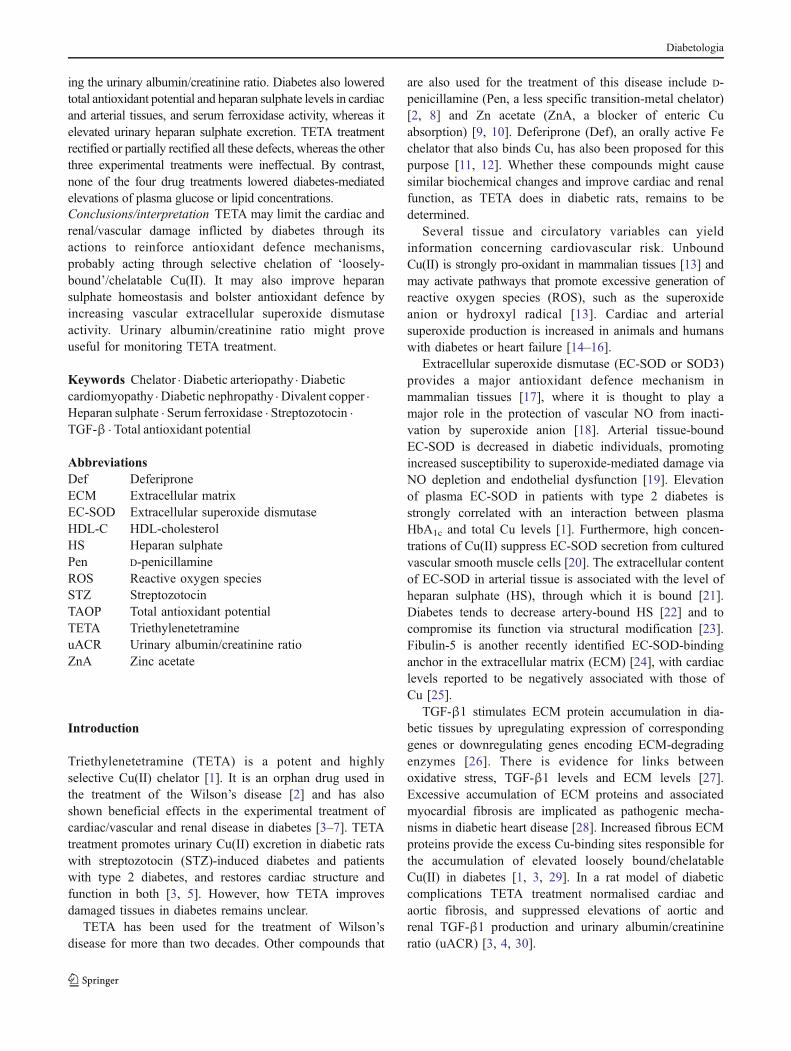
ing the urinary albumin/creatinine ratio. Diabetes also loweredtotal antioxidant potential and heparan sulphate levels in cardiacand arterial tissues, and serum ferroxidase activity, whereas itelevated urinary heparan sulphate excretion. TETA treatmentrectified or partially rectified all these defects, whereas the otherthree experimental treatments were ineffectual. By contrast,none of the four drug treatments lowered diabetes-mediatedelevations of plasma glucose or lipid concentrations.Conclusions/interpretation TETA may limit the cardiac andrenal/vascular damage inflicted by diabetes through itsactions to reinforce antioxidant defence mechanisms,probably acting through selective chelation of ‘loosely-bound’/chelatable Cu(II). It may also improve heparansulphate homeostasis and bolster antioxidant defence byincreasing vascular extracellular superoxide dismutaseactivity. Urinary albumin/creatinine ratio might proveuseful for monitoring TETA treatment.
Keywords Chelator . Diabetic arteriopathy . Diabeticcardiomyopathy . Diabetic nephropathy . Divalent copper .
Heparan sulphate . Serum ferroxidase . Streptozotocin .
TGF-β . Total antioxidant potential
AbbreviationsDef DeferiproneECM Extracellular matrixEC-SOD Extracellular superoxide dismutaseHDL-C HDL-cholesterolHS Heparan sulphatePen D-penicillamineROS Reactive oxygen speciesSTZ StreptozotocinTAOP Total antioxidant potentialTETA TriethylenetetramineuACR Urinary albumin/creatinine ratioZnA Zinc acetate
Introduction
Triethylenetetramine (TETA) is a potent and highlyselective Cu(II) chelator [1]. It is an orphan drug used inthe treatment of the Wilson’s disease [2] and has alsoshown beneficial effects in the experimental treatment ofcardiac/vascular and renal disease in diabetes [3–7]. TETAtreatment promotes urinary Cu(II) excretion in diabetic ratswith streptozotocin (STZ)-induced diabetes and patientswith type 2 diabetes, and restores cardiac structure andfunction in both [3, 5]. However, how TETA improvesdamaged tissues in diabetes remains unclear.
TETA has been used for the treatment of Wilson’sdisease for more than two decades. Other compounds that
are also used for the treatment of this disease include D-penicillamine (Pen, a less specific transition-metal chelator)[2, 8] and Zn acetate (ZnA, a blocker of enteric Cuabsorption) [9, 10]. Deferiprone (Def), an orally active Fechelator that also binds Cu, has also been proposed for thispurpose [11, 12]. Whether these compounds might causesimilar biochemical changes and improve cardiac and renalfunction, as TETA does in diabetic rats, remains to bedetermined.
Several tissue and circulatory variables can yieldinformation concerning cardiovascular risk. UnboundCu(II) is strongly pro-oxidant in mammalian tissues [13] andmay activate pathways that promote excessive generation ofreactive oxygen species (ROS), such as the superoxideanion or hydroxyl radical [13]. Cardiac and arterialsuperoxide production is increased in animals and humanswith diabetes or heart failure [14–16].
Extracellular superoxide dismutase (EC-SOD or SOD3)provides a major antioxidant defence mechanism inmammalian tissues [17], where it is thought to play amajor role in the protection of vascular NO from inacti-vation by superoxide anion [18]. Arterial tissue-boundEC-SOD is decreased in diabetic individuals, promotingincreased susceptibility to superoxide-mediated damage viaNO depletion and endothelial dysfunction [19]. Elevationof plasma EC-SOD in patients with type 2 diabetes isstrongly correlated with an interaction between plasmaHbA1c and total Cu levels [1]. Furthermore, high concen-trations of Cu(II) suppress EC-SOD secretion from culturedvascular smooth muscle cells [20]. The extracellular contentof EC-SOD in arterial tissue is associated with the level ofheparan sulphate (HS), through which it is bound [21].Diabetes tends to decrease artery-bound HS [22] and tocompromise its function via structural modification [23].Fibulin-5 is another recently identified EC-SOD-bindinganchor in the extracellular matrix (ECM) [24], with cardiaclevels reported to be negatively associated with those ofCu [25].
TGF-β1 stimulates ECM protein accumulation in dia-betic tissues by upregulating expression of correspondinggenes or downregulating genes encoding ECM-degradingenzymes [26]. There is evidence for links betweenoxidative stress, TGF-β1 levels and ECM levels [27].Excessive accumulation of ECM proteins and associatedmyocardial fibrosis are implicated as pathogenic mecha-nisms in diabetic heart disease [28]. Increased fibrous ECMproteins provide the excess Cu-binding sites responsible forthe accumulation of elevated loosely bound/chelatableCu(II) in diabetes [1, 3, 29]. In a rat model of diabeticcomplications TETA treatment normalised cardiac andaortic fibrosis, and suppressed elevations of aortic andrenal TGF-β1 production and urinary albumin/creatinineratio (uACR) [3, 4, 30].
Diabetologia
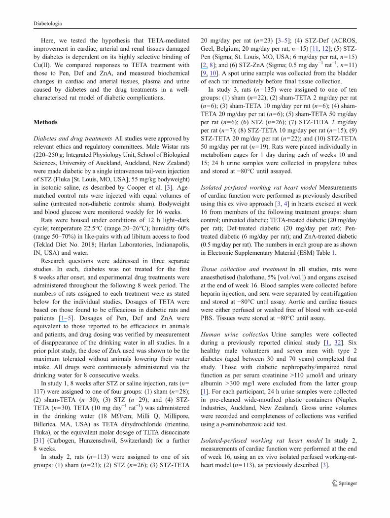
Here, we tested the hypothesis that TETA-mediatedimprovement in cardiac, arterial and renal tissues damagedby diabetes is dependent on its highly selective binding ofCu(II). We compared responses to TETA treatment withthose to Pen, Def and ZnA, and measured biochemicalchanges in cardiac and arterial tissues, plasma and urinecaused by diabetes and the drug treatments in a well-characterised rat model of diabetic complications.
Methods
Diabetes and drug treatments All studies were approved byrelevant ethics and regulatory committees. Male Wistar rats(220–250 g; Integrated Physiology Unit, School of BiologicalSciences, University of Auckland, Auckland, New Zealand)were made diabetic by a single intravenous tail-vein injectionof STZ (Fluka [St. Louis, MO, USA]; 55 mg/kg bodyweight)in isotonic saline, as described by Cooper et al. [3]. Age-matched control rats were injected with equal volumes ofsaline (untreated non-diabetic controls: sham). Bodyweightand blood glucose were monitored weekly for 16 weeks.
Rats were housed under conditions of 12 h light–darkcycle; temperature 22.5°C (range 20–26°C); humidity 60%(range 50–70%) in like-pairs with ad libitum access to food(Teklad Diet No. 2018; Harlan Laboratories, Indianapolis,IN, USA) and water.
Research questions were addressed in three separatestudies. In each, diabetes was not treated for the first8 weeks after onset, and experimental drug treatments wereadministered throughout the following 8 week period. Thenumbers of rats assigned to each treatment were as statedbelow for the individual studies. Dosages of TETA werebased on those found to be efficacious in diabetic rats andpatients [1–5]. Dosages of Pen, Def and ZnA wereequivalent to those reported to be efficacious in animalsand patients, and drug dosing was verified by measurementof disappearance of the drinking water in all studies. In aprior pilot study, the dose of ZnA used was shown to be themaximum tolerated without animals lowering their waterintake. All drugs were continuously administered via thedrinking water for 8 consecutive weeks.
In study 1, 8 weeks after STZ or saline injection, rats (n=117) were assigned to one of four groups: (1) sham (n=28);(2) sham-TETA (n=30); (3) STZ (n=29); and (4) STZ-TETA (n=30). TETA (10 mg day−1 rat−1) was administeredin the drinking water (18 MΩ/cm; Milli Q, Millipore,Billerica, MA, USA) as TETA dihydrochloride (trientine,Fluka), or the equivalent molar dosage of TETA disuccinate[31] (Carbogen, Hunzenschwil, Switzerland) for a further8 weeks.
In study 2, rats (n=113) were assigned to one of sixgroups: (1) sham (n=23); (2) STZ (n=26); (3) STZ-TETA
20 mg/day per rat (n=23) [3–5]; (4) STZ-Def (ACROS,Geel, Belgium; 20 mg/day per rat, n=15) [11, 12]; (5) STZ-Pen (Sigma; St. Louis, MO, USA; 6 mg/day per rat, n=15)[2, 8]; and (6) STZ-ZnA (Sigma; 0.5 mg day−1 rat−1, n=11)[9, 10]. A spot urine sample was collected from the bladderof each rat immediately before final tissue collection.
In study 3, rats (n=135) were assigned to one of tengroups: (1) sham (n=22); (2) sham-TETA 2 mg/day per rat(n=6); (3) sham-TETA 10 mg/day per rat (n=6); (4) sham-TETA 20 mg/day per rat (n=6); (5) sham-TETA 50 mg/dayper rat (n=6); (6) STZ (n=26); (7) STZ-TETA 2 mg/dayper rat (n=7); (8) STZ-TETA 10 mg/day per rat (n=15); (9)STZ-TETA 20 mg/day per rat (n=22); and (10) STZ-TETA50 mg/day per rat (n=19). Rats were placed individually inmetabolism cages for 1 day during each of weeks 10 and15; 24 h urine samples were collected in propylene tubesand stored at −80°C until assayed.
Isolated perfused working rat heart model Measurementsof cardiac function were performed as previously describedusing this ex vivo approach [3, 4] in hearts excised at week16 from members of the following treatment groups: shamcontrol; untreated diabetic; TETA-treated diabetic (20 mg/dayper rat); Def-treated diabetic (20 mg/day per rat); Pen-treated diabetic (6 mg/day per rat); and ZnA-treated diabetic(0.5 mg/day per rat). The numbers in each group are as shownin Electronic Supplementary Material (ESM) Table 1.
Tissue collection and treatment In all studies, rats wereanaesthetised (halothane, 5% [vol./vol.]) and organs excisedat the end of week 16. Blood samples were collected beforeheparin injection, and sera were separated by centrifugationand stored at −80°C until assay. Aortic and cardiac tissueswere either perfused or washed free of blood with ice-coldPBS. Tissues were stored at −80°C until assay.
Human urine collection Urine samples were collectedduring a previously reported clinical study [1, 32]. Sixhealthy male volunteers and seven men with type 2diabetes (aged between 30 and 70 years) completed thatstudy. Those with diabetic nephropathy/impaired renalfunction as per serum creatinine >110 µmol/l and urinaryalbumin >300 mg/l were excluded from the latter group[1]. For each participant, 24 h urine samples were collectedin pre-cleaned wide-mouthed plastic containers (NuplexIndustries, Auckland, New Zealand). Gross urine volumeswere recorded and completeness of collections was verifiedusing a p-aminobenzoic acid test.
Isolated-perfused working rat heart model In study 2,measurements of cardiac function were performed at the endof week 16, using an ex vivo isolated perfused working-rat-heart model (n=113), as previously described [3].
Diabetologia
Tab
le1
Dataacqu
ired
atweek16
from
untreatedcontrol(sham
)or
untreateddiabetic(STZ)rats,com
paredwith
diabeticratsthathadreceived
8weeks
oftreatm
entw
ithTETA
,Def,P
enor
ZnA
Measurement
Sham
STZ
TETA
Def
Pen
ZnA
Blood
glucose(m
mol/l)
10.6±0.3(45)
38.0±0.9(34)**
*39
.8±0.7(51)**
*41
.6±1.2(14)**
*40
.6±2.0(13)**
*35
.4±2.6(12)**
*
Bod
yweigh
t(g)
483±17
(20)
267±7(18)**
*28
2±14
(16)**
*26
9±11
(16)**
*24
5±11
(15)**
*25
7±12
(14)**
*
Heartweigh
t(g)
1.34
±0.05
(20)
0.99
±0.03
(18)**
*0.98
±0.04
(16)**
*0.97
±0.05
(16)**
*0.96
±0.05
(15)**
*0.89
±0.04
(14)**
*
HW/BW
ratio
(mg/g)
2.77
±0.08
(20)
3.72
±0.08
(18)**
*3.53
±0.15
(16)**
*3.61
±0.13
(16)**
*3.93
±0.10
(15)**
*3.54
±0.25
(14)**
*
uACRa(µg/mmol)
1.54
±0.26
(13)
5.99
±0.97
(10)
**
2.31
±0.28
(13)
†5.36
±0.83
(8)**
5.48
±2.08
(8)**
6.08
±1.54
(6)**
Serum
totalcholesterol(m
mol/l)
1.30
±0.03
(26)
2.52
±0.23
(18)**
*2.54
±0.19
(14)**
*2.86
±0.19
(14)**
*2.35
±0.22
(12)**
*2.74
±0.47
(6)***
Serum
triacylglycerol(m
mol/l)
1.3±0.1(26)
8.6±1.5(15)**
*5.0±0.8(11)**
*6.8±1.3(14)**
*5.9±1.3(12)**
*10
.5±3.2(6)***
Serum
HDL-C
(mmol/l)
1.09
±0.03
(26)
1.42
±0.07
(18)**
*1.42
±0.09
(13)**
*2.03
±0.17
(14)**
*1.51
±0.09
(12)**
*1.51
±0.12
(6)***
Serum
NEFA
(mmol/l)
0.94
±0.08
(26)
2.20
±0.38
(16)
2.05
±0.33
(14)
1.89
±0.32
(14)
1.66
±0.33
(12)
1.78
±0.53
(6)
Serum
ferrox
idase(U
/l)32
0±4(56)
289±5(39)**
336±8(30)
†††
298±18
(14)
302±19
(13)
308±19
(10)
Datawerederivedfrom
stud
ies1to
3,andaremeans
±SEM
The
numbers
ofanim
alsareshow
nin
parentheses
BW,bo
dyweigh
t;Def,STZ-diabetic
ratstreatedwith
Def
at20
mg/dayperrat;HDL-C,HDL-cho
lesterol;HW,heartweigh
t;Pen,STZ-diabetic
ratstreatedwith
Pen
at6mg/dayperrat;sham
,salin
e-injected
non-diabetic
controlratswith
outdrug
treatm
ent;STZ,ratswith
STZ-ind
uced
diabetes
with
outdrug
treatm
ent;TETA
,STZ-diabetic
ratstreatedwith
TETA
at20
mgday−
1rat−1;
ZnA
,STZ-diabetic
ratstreatedwith
zinc
acetateat
0.5mg/dayperrat
**p<0.01
vssham
;**
*p<0.00
1vs
sham
;†p<0.05
vsSTZ;†††p<0.00
1vs
STZ
auA
CR
values
werederivedfrom
measurementof
spot
urines
Diabetologia
ELISAs for HS, TGF-β1 and TNF-α Urinary HS levelsfrom study 3, and serum, cardiac and aortic tissue samplesfrom study 1 were assayed by ELISA (Seikagaku, Tokyo,Japan) as described [4]. Briefly, frozen cardiac or aortictissue was homogenised in ice-cold PBS and centrifuged at13,000×g for 20 min at 4°C; supernatant fractions wereisolated and protein concentrations determined. Then 10 µlof 20 mg/ml actinase E (Kaken Pharmaceuticals, Tokyo,Japan) was added to 100 µl supernatant fraction or serumand incubated at 55°C for 20 h to digest tissue and releasebound heparan sulphate from proteins. After incubation,samples were boiled for 5 min, cooled and centrifuged at3,000×g for 10 min. Processed serum, cardiac and aortictissue, and untreated urine samples were then assayedaccording to the manufacturer’s instructions in 96-wellplates (450 nm; SpectraMax 340, Molecular Devices,Sunnyvale, CA, USA). The HS content in tissue wasexpressed as μg/mg protein.
The levels of the active form of TGF-β1 in cardiac andaortic tissue from study 1 were determined using the TGF-β1 Emax ImmunoAssay System (Promega, Madison, WI,USA). The optical density at 450 nm was measured and theamount of active TGF-β1 was expressed as pg/mg protein.
TNF-α levels in serum, cardiac and aortic tissue fromstudy 1 were assayed using an Endogen rat TNF-α ELISAkit (Pierce Biotechnology, IL, USA) in 96-well plates(450 nm); TNF-α concentrations have been expressed aspg/ml in serum, and pg/mg protein in cardiac and aortictissues.
Biochemical analyses Cardiac and aortic tissues wereanalysed for TNF-α, TGF-β1, fibulin-5, total antioxidantpotential (TAOP) and HS in study 1. Urine samples wereanalysed for HS in study 3; for creatinine at weeks 10 and15 in study 2, and at week 16 in study 3; and for albumin atweek 15 in study 2, and at week 16 in study 3, and uACRvalues calculated. Serum from endpoint sampling (week16) was analysed for glucose, total cholesterol, HDL-cholesterol, triacylglycerol, NEFA and ferroxidase in allstudies; and for serum levels of Fe, total protein, alkalinephosphatase, aspartate aminotransferase, alanine amino-transferase, bilirubin, calcium and phosphate using standardbiochemical methods implemented with a Synchron CX5(Beckman, Brea, CA, USA).
Urinary albumin was determined using the rat AlbuminEnzyme Immunoassay kit (SPI-BIO, Québec, QC, Canada)and urinary creatinine by an enzymatic rate method on theSynchron CX5.
Tissue TAOP assay Supernatant fractions from cardiac andaortic tissue homogenates from study 1 were assayed fortotal antioxidant potential using an AOP-490 colorimetrickit (OxisResearch, Beverly Hills, CA, USA). This method
is based on the reduction of Cu(II) to Cu(I) by thecombined action of antioxidants present in the sample.The TAOP in each sample was expressed as μmol/l Cu-reducing equivalents/mg tissue protein.
Statistical analysis Data have been expressed as means ±SE. One- and two-way ANOVAs, followed by post-hocTukey’s tests, if appropriate, were performed to assesswhether differences between groups were statisticallysignificant. Survival curves describing the ability of isolatedperfused working hearts to sustain their outputs withincreasing afterload pressures were compared using thelogrank test (Prism, v4.02). General linear mixed modelswere fitted to the data from multi-factor experimentsinvolving repeated measurements over time (e.g. urinaryHS) using residual maximum likelihood (SAS v9.1 forWindows). Residual analyses were performed to verify thatthe underlying assumptions of these methods were met, andappropriate transformations were applied as necessary.Tukey–Kramer adjusted p values (for post-hoc pair-wisecomparisons) and Bonferroni–Holm adjusted p values (fora priori comparisons of interest) were calculated. In eithercase, p<0.05 was considered significant and the n valueindicates the number of replicates in corresponding assays.
Results
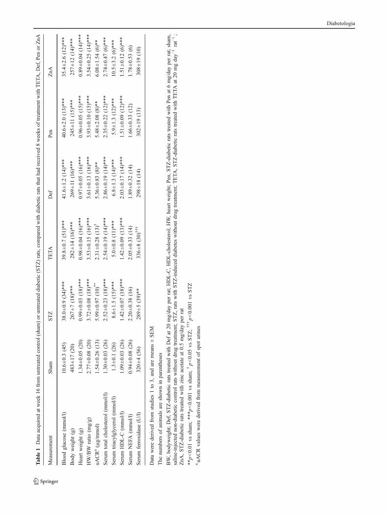
Diabetes and bodyweight Blood glucose concentrations didnot differ among groups at the time of STZ injection (datanot shown). Hyperglycaemia occurred within 2 days ofSTZ administration in all experiments and persistedequivalently in both untreated and drug-treated groups ofdiabetic rats for the duration of the experiment, indicatingthat none of the treatments decreased blood glucose levels.Endpoint blood glucose measurements were markedlyhigher than those in sham controls (p<0.0001, Table 1)and no differences were detected between diabetic ratsamong the various treatments (p>0.05, Table 1).
The mean bodyweights of different groups of rats werenot significantly different at the time of STZ injection (datanot shown), but at week 16, the bodyweights of all groupsof diabetic rats were significantly lower than those of shamrats (p<0.001, Table 1), and the various drug treatmentswere all without effect on this variable (p>0.05, Table 1).
Cardiac mass The heart weights of diabetic rats weresignificantly lower than those of sham rats (p<0.001, Table 1),and did not differ significantly among the treatment groups(p>0.05, Table 1). Heart weight/bodyweight ratios weresignificantly higher in diabetic groups than in sham controls(p<0.001, Table 1), and the experimental drug treatments didnot modify these ratios (p>0.05, Table 1).
Diabetologia

Response of cardiac output to increasing afterload in exvivo isolated working hearts All groups of diabetic ratshad impaired cardiac function compared with sham rats,as judged by their inability to maintain their cardiacoutput at physiological levels in the face of increasingafterload pressure (ESM Table 1). Ante mortem treatmentwith Pen, Def and ZnA did not significantly improvecardiac function in diabetic rats (all p>0.05) whereas, bycontrast, TETA treatment increased the resistance ofcardiac output to the effects of increasing afterloadpressure (p=0.021 compared with the untreated diabeticcontrol group). The responses to TETA are consistent withprevious reports [3, 4].
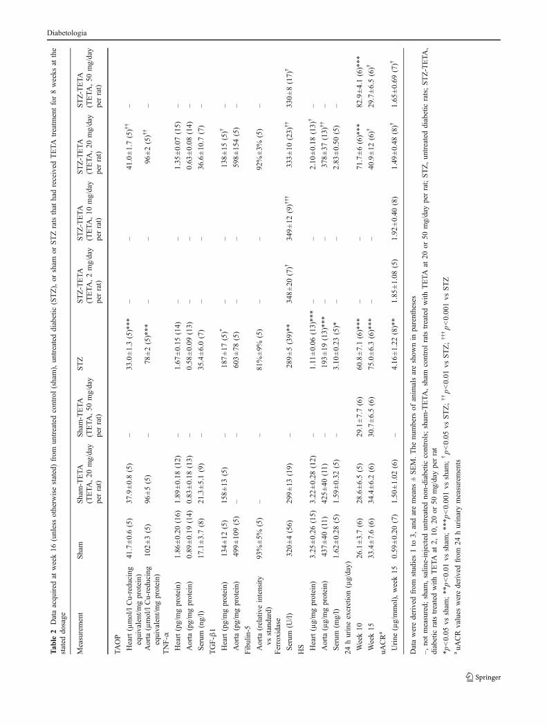
TAOP, HS, fibulin-5, TGF-β1 and TNF-α in cardiac andaortic tissues TAOP levels in cardiac and aortic tissueswere significantly lower in diabetic rats compared withsham rats (p<0.001 in both tissues, Table 2), and 8 weeks’TETA treatment significantly elevated TAOP levels (p<0.01 in both tissues, Table 2).
TNF-α levels were not significantly changed in eithercardiac or aortic tissue of diabetic rats compared with non-diabetic controls, nor were they significantly modified byTETA treatment (p>0.05, Table 2).
TGF-β1 levels were higher in cardiac tissue of diabeticthan sham rats (p<0.05, Table 2) and TETA treatmentrestored cardiac TGF-β1 protein production to normal (p<0.05, Table 2). There was no significant difference in TGF-β1 protein levels in aortic tissue between rats with orwithout diabetes or following drug treatment (data notshown).
Fibulin-5 levels in cardiac tissue were insufficient fordetection by western blotting, whereas in aortic tissue therewas no detectable difference between sham and diabeticrats, or between diabetic rats with and without TETAtreatment (Table 2), possibly due to low numbers ofreplicates.
HS levels in cardiac and aortic tissues were alsosignificantly lower in diabetic rats than in sham controls(p<0.001 in both tissues; Table 2). TETA treatmentsignificantly attenuated the diabetes-induced reduction ofHS levels in both tissues of diabetic rats (p<0.05 in heartand p<0.01 in aortic tissues, Table 2).
Urinary HS excretion in rats Urinary HS levels weremeasured in the 20 mg/day and 50 mg/day dosage groups.Diabetic rats had significantly greater 24 h urinary HSexcretion than matched controls at weeks 10 and 15 (bothp<0.0001, Table 2). Two weeks’ TETA treatment at thesedosages did not modify urinary HS excretion (week 10,both p>0.05, Table 2). By contrast, following 7 weeks’daily TETA treatment, both dosages did significantly lower24 h urinary HS excretion (week 15, p<0.05 at 20 mg/day
per rat; p<0.01, 50 mg/day per rat; Table 2), consistent witha dosage-dependent effect.
Urinary albumin/creatinine ratios Urinary ACR values inspot urine samples were higher in diabetic rats than shamcontrols (p<0.01, Table 1), as were those measured in 24 hurine samples (p<0.01, Table 2), and TETA treatmentsignificantly lowered the elevated uACR values in bothstudies (p<0.05 in each, Tables 1, 2). By contrast, Pen, Defand ZnA treatments were all without effect on uACR indiabetic rats (p>0.05, Table 1).
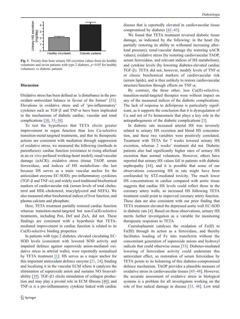
HS in human urine There was a significant elevation in24 h urinary HS excretion in patients with type 2 diabetescompared with healthy volunteers (p<0.05, Fig. 1).
Biochemical variables in serum Serum HS levels weresignificantly higher in diabetic rats than sham controls (p<0.05, Table 2) and were not significantly modified by TETAtreatment (p>0.05, Table 2).
Urinary HS excretion was positively correlated withserum HS levels (Pearson’s correlation analysis; r2=0.72, n=34; p<0.001). Mean TNF-α tended to be higherin both diabetic groups, but the difference was not significantand TETA treatment was without effect (both p>0.05,Table 2).
Cholesterol, HDL-cholesterol, triacylglycerol, NEFAand ferroxidase were measured in all three studies. Serumcholesterol, HDL-cholesterol and triacylglycerol levelswere higher in diabetic rats than in sham controls (all p<0.001, Table 1). None of the transition-metal-targetedtherapies, including TETA at various dosages, Pen, Def orZnA significantly altered the serum cholesterol, triacylglycerolor HDL-cholesterol levels in diabetic rats (all p>0.05, Table 1).NEFA levels did not differ significantly between sham anddiabetic rats, nor did levels change with TETA treatment (allp>0.05, Table 1).
Serum ferroxidase activity was significantly lower indiabetic rats than in sham controls (p<0.01, Tables 1, 2), andall dosages of TETA studied increased values in diabetic rats(p<0.05, 0.001, 0.01 and 0.05 for dosages of 2, 10, 20, and50 mg day−1 rat−1, respectively, Table 2). By contrast,treatment with Pen, Def or ZnA did not alter serum ferroxidaseactivity in diabetic rats (all p>0.05, Table 1), but in that studyTETA treatment (20 mg/day per rat) increased the serumferroxidase activity significantly (p<0.001, Table 1).
The serum values of total protein, alkaline phosphatase,aspartate aminotransferase, alanine aminotransferase, bilirubin,Fe, calcium and phosphate did not differ significantly betweensham and diabetic rats, nor did they show significantdifferences between diabetic rats with TETA treatment atdifferent dosages and untreated diabetic rats (all p>0.05, datanot shown).
Diabetologia
Tab
le2
Dataacqu
ired
atweek16
(unlessotherw
isestated)from
untreatedcontrol(sham),un
treateddiabetic
(STZ),or
sham
orSTZratsthat
hadreceived
TETA
treatm
entfor8weeks
atthe
stated
dosage
Measurement
Sham
Sham-TETA
(TETA
,20
mg/day
perrat)
Sham-TETA
(TETA
,50
mg/day
perrat)
STZ
STZ-TETA
(TETA
,2mg/day
perrat)
STZ-TETA
(TETA
,10
mg/day
perrat)
STZ-TETA
(TETA
,20
mg/day
perrat)
STZ-TETA
(TETA
,50
mg/day
perrat)
TAOP
Heart(µmol/lCu-redu
cing
equivalent/m
gprotein)
41.7±0.6(5)
37.9±0.8(5)
–33
.0±1.3(5)***
––
41.0±1.7(5)††
–
Aorta
(µmol/lCu-redu
cing
equivalent/m
gprotein)
102±3(5)
96±5(5)
–78
±2(5)***
––
96±2(5)††
–
TNF-α
Heart(pg/mgprotein)
1.86
±0.20
(16)
1.89
±0.18
(12)
–1.67
±0.15
(14)
––
1.35
±0.07
(15)
–
Aorta
(pg/mgprotein)
0.89
±0.19
(14)
0.83
±0.18
(13)
–0.58
±0.09
(13)
––
0.63
±0.08
(14)
–
Serum
(ng/l)
17.1±3.7(8)
21.3±5.1(9)
–35
.4±6.0(7)
––
36.6±10
.7(7)
–
TGF-β1
Heart(pg/mgprotein)
134±12
(5)
158±13
(5)
–18
7±17
(5)*
––
138±15
(5)†
–
Aorta
(pg/mgprotein)
499±10
9(5)
––
603±78
(5)
––
598±15
4(5)
–
Fibulin-5
Aorta
(relativeintensity
vsstandard)
93%±5%
(5)
––
81%±9%
(5)
––
92%±3%
(5)
–
Ferroxidase
Serum
(U/l)
320±4(56)
299±13
(19)
–28
9±5(39)**
348±20
(7)†
349±12
(9)†††
333±10
(23)
††
330±8(17)
†
HS Heart(µg/mgprotein)
3.25
±0.26
(15)
3.22
±0.28
(12)
–1.11
±0.06
(13)**
*–
–2.10
±0.18
(13)
†–
Aorta
(µg/mgprotein)
437±40
(11)
425±40
(11)
–19
3±19
(13)**
*–
–37
8±37
(13)
††
–
Serum
(mg/l)
1.62
±0.28
(5)
1.59
±0.32
(5)
–3.10
±0.23
(5)*
––
2.83
±0.50
(5)
–
24hurineexcretion(µg/day)
Week10
26.1±3.7(6)
28.6±6.5(5)
29.1±7.7(6)
60.8±7.1(6)***
––
71.7±6(6)***
82.9±4.1(6)***
Week15
33.4±7.6(6)
34.4±6.2(6)
30.7±6.5(6)
75.0±6.3(6)***
––
40.9±12
(6)†
29.7±6.5(6)†
uACRa
Urine
(µg/mmol),week15
0.59
±0.20
(7)
1.50
±1.02
(6)
–4.16
±1.22
(8)**
1.85
±1.08
(5)
1.92
±0.40
(8)
1.49
±0.48
(8)†
1.65
±0.69
(7)†
Datawerederivedfrom
stud
ies1to
3,andaremeans
±SEM.The
numbers
ofanim
alsareshow
nin
parentheses
–,no
tmeasured;
sham
,salin
e-injected
untreatedno
n-diabetic
controls;sham
-TETA
,sham
controlrats
treatedwith
TETA
at20
or50
mg/dayperrat;STZ,un
treateddiabetic
rats;STZ-TETA
,diabetic
ratstreatedwith
TETA
at2,
10,20
or50
mg/dayperrat
*p<0.05
vssham
;**p<0.01
vssham
;**
*p<0.00
1vs
sham
;†p<0.05
vsSTZ;††p<0.01
vsSTZ;†††p<0.00
1vs
STZ
auA
CRvalues
werederivedfrom
24hurinarymeasurements
Diabetologia
Discussion
Oxidative stress has been defined as ‘a disturbance in the pro-oxidant–antioxidant balance in favour of the former’ [33].Elevations in oxidative stress and of ‘pro-inflammatory’cytokines such as TGF-β and TNF-α have been implicatedin the mechanisms of diabetic cardiac, vascular and renalcomplications [30, 33–38].
To test the hypotheses that TETA elicits greaterimprovement in organ function than less Cu-selectivetransition-metal-targeted treatments, and that its therapeuticactions are consistent with mediation through suppressionof oxidative stress, we measured the following (methods inparentheses): cardiac function (resistance to rising afterloadin an ex vivo perfused working-heart model); renal/vasculardamage (uACR); oxidative stress (tissue TAOP, serumferroxidase, and indices of HS metabolism—the lastbecause HS serves as a main vascular anchor for theantioxidant enzyme EC-SOD); pro-inflammatory cytokines(TGF-β and TNF-α); and widely used traditional biochemicalmarkers of cardiovascular risk (serum levels of total choles-terol and HDL-cholesterol, triacylglycerol and NEFA). Wealso measured other biochemical indices of liver function, andplasma calcium and phosphate.
Here, TETA treatment partially restored cardiac functionwhereas transition-metal-targeted but non-Cu(II)-selectivetreatments, including Pen, Def and ZnA, did not. Thesefindings are consistent with a hypothesis that TETA-mediated improvement in cardiac function is related to itsCu(II)-selective binding properties.
In patients with type 2 diabetes, elevated circulating EC-SOD levels (consistent with lowered SOD activity andimpaired defence against superoxide anion-mediated oxi-dative stress in arterial walls), were reportedly normalisedby TETA treatment [1]. HS serves as a major anchor forthis important antioxidant defence enzyme [21, 24], bindingand localising it in the vascular ECM where it catalyses theelimination of superoxide anion and sustains NO bioavail-ability [39]. TGF-β1 elicits stimulation of collagen produc-tion and may play a pivotal role in ECM fibrosis [40]; andTNF-α is a pro-inflammatory cytokine linked with cardiac
disease that is reportedly elevated in cardiovascular tissuecompromised by diabetes [41–43].
We found that TETA treatment reversed diabetic tissuedamage, as indicated by the following: in the heart (bypartially restoring its ability to withstand increasing after-load pressure); renal/vascular damage (by restoring uACRvalues); oxidative stress (by restoring cardiovascular TAOP,serum ferroxidase, and relevant indices of HS metabolism);and cytokine levels (by lowering diabetes-elevated cardiacTGF-β). TETA did not, however, modify levels of TNF-αor classic biochemical markers of cardiovascular risk(serum lipids), and is thus unlikely to restore cardiovascularstructure/function through effects on TNF-α.
By contrast, the three other, less Cu(II)-selective,transition-metal-targeted therapies were without impact onany of the measured indices of the diabetic complications.The lack of response to deferiprone is particularly signif-icant, as it supports the conclusion that it is dysregulation ofCu and not of Fe homeostasis that plays a key role in theaetiopathogenesis of the diabetic complications [3].
In diabetic rats increased arterial HS was inverselyrelated to urinary HS excretion and blood HS concentra-tion, and these two variables were positively correlated.Treatment with TETA for 7 weeks lowered urinary HSexcretion, whereas 2 weeks’ treatment did not. Diabeticpatients also had significantly higher rates of urinary HSexcretion than normal volunteers. However, others havereported that urinary HS values fall in patients with diabeticnephropathy [44], and it is possible that some of ourobservations concerning HS in rats might have beenconfounded by STZ-mediated toxicity. The much lowerHS concentrations in cardiac compared with aortic tissuesuggests that cardiac HS levels could reflect those in thecoronary artery walls, so increased HS following TETAtreatment could point to improved coronary artery function.These data are also consistent with our prior finding thatTETA treatment elevated the depressed aortic wall EC-SODin diabetic rats [4]. Based on these observations, urinary HSmerits further investigation as a variable for monitoringtherapeutic responses to TETA.
Caeruloplasmin catalyses the oxidation of Fe(II) toFe(III) through its action as a ferroxidase, and therebyfacilitates loading of Fe into transferrin without theconcomitant generation of superoxide anions and hydroxylradicals that could otherwise ensue [33]. Diabetes-mediatedlowering of ferroxidase activity could undermine thisantioxidant effect, so restoration of serum ferroxidase byTETA points to its bolstering of this diabetes-compromiseddefence mechanism. TAOP provides a plausible measure ofoxidative stress in cardiovascular tissues [45–48]. However,the accurate assessment of oxidative stress in biologicalsystems is a problem for all investigators working on therole of free radical damage in disease [33, 48]. Low total
Fig. 1 Twenty-four hour urinary HS excretion values from six healthyvolunteers and seven patients with type 2 diabetes. p<0.05 for healthyvolunteers vs diabetic patients
Diabetologia
antioxidant capacity can be interpreted as indicative ofoxidative stress or increased susceptibility to oxidativedamage. However, measurements of TAOP, such as that weemployed here, have limitations [48]. Most assays tend tomeasure predominantly the antioxidants with lower molec-ular mass, such as urate, ascorbate, bilirubin, and thiols inthe aqueous phase, and α-tocopherol, carotenoids, andflavonoids in the lipid phase, excluding the contribution ofantioxidant enzymes and metal-binding proteins; differentapproaches to the measurement of TAOP correlate poorlywith one another [48]. In most biological fluids, the majorcontributor in such assays is urate, which frequentlyaccounts for more than 50% of the total antioxidantpotential measured. However, urate is of limited importanceas an antioxidant in vivo, and a preferable approach wouldbe to measure all potential antioxidants [48]. Such studieswill need to be performed in future, to more accuratelyjudge the antioxidant mechanisms by which Cu(II)-selectivechelation elevates TAOP.
We conclude that dysregulation of Cu, but not of Fe,homeostasis is a key pathogenic mechanism in the diabeticcomplications. These data are consistent with the hypothesisthat raised Cu(II)-evoked oxidative stress is causative of thecomplications of diabetes. TETA treatment may limit thecardiac and renal/vascular damage caused by diabetes throughits actions to bolster antioxidant defences, and its Cu(II)-selective binding ability probably plays a major role in itsactions. These findings are consistent with our previousdescription of pathogenic dysregulation of Cu homeostasisas a major contributor to the mechanisms of diabetes-evokedcardiac and renal/vascular damage, and their improvementfollowing TETA treatment [1]. Urinary ACR, whetherderived from 24 h or spot urinary measurements, could beuseful for monitoring TETA treatment. Further investigationof the mechanisms by which Cu(II)selective chelatorsabrogate tissue damage in diabetes is warranted.
Acknowledgements This study was funded by grants from theEndocore Research Foundation (G. J. S. Cooper), the Foundation forResearch, Science and Technology, New Zealand (G. J. S. Cooper);the Health Research Council of New Zealand (G. J. S. Cooper); theMaurice and Phyllis Paykel Trust (G. J. S. Cooper and J. Lu); theNational Heart Foundation of New Zealand (J. Lu); National NaturalScience Foundation of China (H. Xu, grant 30570421); AucklandMedical Research Foundation (J. Lu and D. Gong); the New ZealandLottery Grants Board (G. J. S. Cooper and D. Gong). We thank C. Tse(School of Biological Sciences, University of Auckland, Auckland,New Zealand) for her contributions to the management of thesestudies and the development of the manuscript, and V. Tintinger, R.Smith and S. Costa for their technical assistance.
Duality of interest J. Lu, D. Gong, Y. S. Choong, Y. K. Chan, X.Chen, S. Fitzpatrick, S. Glyn-Jones, S. Zhang, S. D. Poppitt, A. R. J.Phillips and G. J. S. Cooper declare past associations with ProtemixCorporation, Auckland, New Zealand. All other authors declare thatthere is no duality of interest associated with this work.
References
1. Cooper GJS, Chan YK, Dissanayake AM et al (2005) Demonstra-tion of a hyperglycemia-driven pathogenic abnormality of copperhomeostasis in diabetes and its reversibility by selective chelation:quantitative comparisons between the biology of copper and eightother nutritionally essential elements in normal and diabeticindividuals. Diabetes 54:1468–1476
2. Walshe JM (1973) Copper chelation in patients with Wilson’sdisease. A comparison of penicillamine and triethylene tetraminedihydrochloride. Q J Med 42:441–452
3. Cooper GJS, Phillips ARJ, Choong SY et al (2004) Regenerationof the heart in diabetes by selective copper chelation. Diabetes53:2501–2508
4. Gong D, Lu J, Chen X et al (2006) Molecular changes evoked bytriethylenetetramine (TETA) treatment in the extracellular matrix ofthe heart and aorta in diabetic rats. Mol Pharmacol 70:2045–2051
5. Cooper GJS, Young AA, Gamble GD et al (2009) A copper(II)-selective chelator ameliorates left-ventricular hypertrophy in type-2diabetic patients. Diabetologia 52:715–722
6. Baynes JW, Murray DB (2009) The metal chelators, trientine andcitrate, inhibit the development of cardiac pathology in the Zuckerdiabetic rat. Exp Diabetes Res 2009:696378
7. Gong D, Chen X, Middleditch M et al (2009) Quantitativeproteomic profiling identifies new renal targets of copper(II)-selective chelation in the reversal of diabetic nephropathy in rats.Proteomics 9:4309–4320
8. Joyce DA (1990) D-penicillamine. Baillières Clin Rheumatol4:553–574
9. Anderson LA, Hakojarvi SL, Boudreaux SK (1998) Zinc acetatetreatment in Wilson’s disease. Ann Pharmacother 32:78–87
10. Brewer GJ (2001) Zinc acetate for the treatment of Wilson’sdisease. Expert Opin Pharmacother 2:1473–1477
11. Kontoghiorghes GJ, Pattichis K, Neocleous K, Kolnagou A(2004) The design and development of deferiprone (L1) and otheriron chelators for clinical use: targeting methods and applicationprospects. Curr Med Chem 11:2161–2183
12. Piga A, Roggero S, Vinciguerra T, Sacchetti L, Gallo V, Longo F(2005) Deferiprone: new insight. Ann N Y Acad Sci 1054:169–174
13. Fraústo da Silva JJ, Williams RJ (2001) The biological chemistryof the elements: the inorganic chemistry of life. Clarendon,Oxford, pp 418–435
14. Kojda G, Harrison D (1999) Interactions between NO and reactiveoxygen species: pathophysiological importance in atherosclerosis,hypertension, diabetes and heart failure. Cardiovasc Res 43:562–571
15. Guzik T, Mussa S, Gastaldi D et al (2002) Mechanisms ofincreased vascular superoxide production in human diabetesmellitus: role of NAD(P)H oxidase and endothelial nitric oxidesynthase. Circulation 105:1656–1662
16. Creager MA, Luscher TF, Cosentino F, Beckman JA (2003) Diabetesand vascular disease: pathophysiology, clinical consequences, andmedical therapy: Part I. Circulation 108:1527–1532
17. Marklund SL (1984) Extracellular superoxide dismutase in humantissues and human cell lines. J Clin Invest 74:1398–1403
18. Oury TD, Day BJ, Crapo JD (1996) Extracellular superoxidedismutase: a regulator of nitric oxide bioavailability. Lab Invest75:617–636
19. Fattman CL, Schaefer LM, Oury TD (2003) Extracellularsuperoxide dismutase in biology and medicine. Free Radic BiolMed 35:236–256
20. Stralin P, Jacobsson H, Marklund SL (2003) Oxidative stress, NO•
and smooth muscle cell extracellular superoxide dismutaseexpression. Biochim Biophys Acta 1619:1–8
Diabetologia
21. Sandstrom J, Karlsson K, Edlund T, Marklund SL (1993)Heparin-affinity patterns and composition of extracellular super-oxide dismutase in human plasma and tissues. Biochem J294:853–857
22. Edwards IJ, Wagner JD, Vogl-Willis CA, Litwak KN, Cefalu WT(2004) Arterial heparan sulfate is negatively associated withhyperglycemia and atherosclerosis in diabetic monkeys. CardiovascDiabetol 3:6
23. Vogl-Willis CA, Edwards IJ (2004) High-glucose-induced struc-tural changes in the heparan sulfate proteoglycan, perlecan, ofcultured human aortic endothelial cells. Biochim Biophys Acta1672:36–45
24. Nguyen AD, Itoh S, Jeney V et al (2004) Fibulin-5 is a novelbinding protein for extracellular superoxide dismutase. Circ Res95:1067–1074
25. Zeng H, Saari JT, Dahlen GM (2006) Copper deficiency increasesfibulin-5 (DANCE/EVEC) but decreases cytochrome C oxidaseVIb subunit expression in rat heart. J Inorg Biochem 100:186–191
26. Roberts AB, McCune BK, Dunn SR (1992) TGF-β: regulation ofextracellular matrix. Kidney Int 41:557–559
27. Williams B (1998) Mechanical influences on vascular smoothmuscle cell function. J Hypertens 16:1921–1929
28. Marklund SL (1992) Regulation by cytokines of extracellularsuperoxide dismutase and other superoxide dismutase isoenzymesin fibroblasts. J Biol Chem 267:6696–6701
29. Qian M, Liu M, Eaton JW (1998) Transition metals bind toglycated proteins forming redox active “glycochelates”: implicationsfor the pathogenesis of certain diabetic complications. BiochemBiophys Res Commun 250:385–389
30. Gong D, Lu J, Chen X et al (2008) A copper(II)-selective chelatorameliorates diabetes-evoked renal fibrosis and albuminuria, andsuppresses pathogenic TGF-β activation in the kidneys of ratsused as a model of diabetes. Diabetologia 51:1741–1751
31. Wichmann KA, Boyd PDW, Söhnel T, Allen GR, Phillips ARJ,Cooper GJS (2007) Characterization of dicarboxylic salts ofprotonated triethylenetetramine useful for the treatment of copper-related pathologies. Cryst Growth Des 7:1844–1850
32. Lu J, Chan Y-K, Poppitt SD, Cooper GJS (2007) Determination oftriethylenetetramine (TETA) and its metabolites in human plasmaand urine by liquid chromatography–mass spectrometry (LC-MS).J Chromatogr B: Anal Technol Biomed Life Sci 859:62–68
33. Halliwell B, Gutteridge JM (1999) Free radicals in biology andmedicine. Oxford University Press, Oxford
34. Martin J, Kelly DJ, Mifsud SA et al (2005) Tranilast attenuatescardiac matrix deposition in experimental diabetes: role oftransforming growth factor-β. Cardiovasc Res 65:694–701
35. Jay D, Hitomi H, Griendling KK (2006) Oxidative stress anddiabetic cardiovascular complications. Free Radic Biol Med40:183–192
36. Haidara MA, Yassin HZ, Rateb M, Ammar H, Zorkani MA (2006)Role of oxidative stress in development of cardiovascular complica-tions in diabetes mellitus. Curr Vasc Pharmacol 4:215–227
37. Stephens JW, Khanolkar MP, Bain SC (2009) The biologicalrelevance and measurement of plasma markers of oxidative stressin diabetes and cardiovascular disease. Atherosclerosis 202:321–329
38. Friederich M, Hansell P, Palm F (2009) Diabetes, oxidative stress,nitric oxide and mitochondria function. Curr Diabetes Rev 5:120–144
39. Marklund SL (2002) Extracellular superoxide dismutase. MethodsEnzymol 349:74–80
40. Sharma K, Ziyadeh FN (1995) Hyperglycemia and diabetickidney disease: the case for transforming growth factor-beta as akey mediator. Diabetes 44:1139–1146
41. Mann DL (2002) Inflammatory mediators and the failing heart:past, present, and the foreseeable future. Circ Res 91:988–998
42. Gullestad L, Kjekshus J, Damas JK, Ueland T, Yndestad A,Aukrust P (2005) Agents targeting inflammation in heart failure.Expert Opin Investig Drugs 14:557–566
43. Kaur K, Sharma AK, Singal PK (2006) Significance of changes inTNF-alpha and IL-10 levels in the progression of heart failuresubsequent to myocardial infarction. Am J Physiol Heart CircPhysiol 291:H106–H113
44. Yokoyama H, Sato K, Okudaira M et al (1999) Serum and urinaryconcentrations of heparan sulfate in patients with diabeticnephropathy. Kidney Int 56:650–658
45. Ghiselli A, Serafini M, Natella F, Scaccini C (2000) Totalantioxidant capacity as a tool to assess redox status: critical viewand experimental data. Free Radic Biol Med 29:1106–1114
46. Prior RL, Cao G (1999) In vivo total antioxidant capacity:comparison of different analytical methods. Free Radic Biol Med27:1173–1181
47. Buffon A, Santini SA, Ramazzotti V et al (2000) Large, sustainedcardiac lipid peroxidation and reduced antioxidant capacity in thecoronary circulation after brief episodes of myocardial ischemia. JAm Coll Cardiol 35:633–639
48. Young IS (2001) Measurement of total antioxidant capacity. J ClinPathol 54:339
Diabetologia
Copyright © 2022 FDOKUMEN