contributions of the La - Archives-Ouvertes.fr
372
HAL Id: tel-03590277 https://tel.archives-ouvertes.fr/tel-03590277 Submitted on 27 Feb 2022 HAL is a multi-disciplinary open access archive for the deposit and dissemination of sci- entific research documents, whether they are pub- lished or not. The documents may come from teaching and research institutions in France or abroad, or from public or private research centers. L’archive ouverte pluridisciplinaire HAL, est destinée au dépôt et à la diffusion de documents scientifiques de niveau recherche, publiés ou non, émanant des établissements d’enseignement et de recherche français ou étrangers, des laboratoires publics ou privés. Phylogeny, paleobiogeography, and paleophysiology of the Triassic dicynodonts (Therapsida, Anomodontia) : contributions of the Laotian and Moroccan forms Chloe Olivier To cite this version: Chloe Olivier. Phylogeny, paleobiogeography, and paleophysiology of the Triassic dicynodonts (Ther- apsida, Anomodontia): contributions of the Laotian and Moroccan forms. Paleontology. Sorbonne Université, 2020. English. NNT : 2020SORUS399. tel-03590277
-
Upload
khangminh22 -
Category
Documents
-
view
1 -
download
0
Transcript of contributions of the La - Archives-Ouvertes.fr

HAL Id: tel-03590277https://tel.archives-ouvertes.fr/tel-03590277
Submitted on 27 Feb 2022
HAL is a multi-disciplinary open accessarchive for the deposit and dissemination of sci-entific research documents, whether they are pub-lished or not. The documents may come fromteaching and research institutions in France orabroad, or from public or private research centers.
L’archive ouverte pluridisciplinaire HAL, estdestinée au dépôt et à la diffusion de documentsscientifiques de niveau recherche, publiés ou non,émanant des établissements d’enseignement et derecherche français ou étrangers, des laboratoirespublics ou privés.
Phylogeny, paleobiogeography, and paleophysiology ofthe Triassic dicynodonts (Therapsida, Anomodontia) :
contributions of the Laotian and Moroccan formsChloe Olivier
To cite this version:Chloe Olivier. Phylogeny, paleobiogeography, and paleophysiology of the Triassic dicynodonts (Ther-apsida, Anomodontia) : contributions of the Laotian and Moroccan forms. Paleontology. SorbonneUniversité, 2020. English. �NNT : 2020SORUS399�. �tel-03590277�

Sorbonne Université
ED 398 - Géosciences, Ressources Naturelles et Environnement
Centre de Recherche en Paleontologie - Paris / Equipe Phylogénie et diversification
des métazoaires
Phylogeny, paleobiogeography, and paleophysiology of
the Triassic dicynodonts (Therapsida, Anomodontia)
Contributions of the Laotian and Moroccan forms
Par Chloé OLIVIER
Thèse de doctorat de Paléontologie
Dirigée par Jorge CUBO et Nour-Eddine JALIL
Présentée et soutenue publiquement le 25 février 2020
Devant un jury composé de :
Botha-Brink Jennifer, Professeure, Rapporteure
Kammerer Christian F., Research Curator of Paleontology, Rapporteur
Vignes-Lebbe Régine, Professeure, Examinatrice
Fröbisch Jörg, Professeur, Examinateur
Laurin Michel, Directeur de recherche, Examinateur

« 3R7R E7RiYtR 3Ts8 3E1 T1 T8 wRqR6 1Y wRsT5 3E5 1Y 7ReUiR= RyR5 3Y Usd 3R R2 tEhÌ wR 2E6a-
(“There are some things that it is better to begin than to refuse, even though the end may be dark.”) (J.R.R. Tolkien)
“Happiness is when what you think, what you say, and what you do are in harmony.”
(Mahatma Gandhi)
ACKNOWLEDGMENTS
Firslty, I extend my gratitude to my supervisors Prof. Jorge Cubo and Prof. Nour-
Eddine Jalil, without whom, my PhD would have not been proposed. I met Nour-Eddine
as an undergraduate when I was initiated to the vast world of paleontology and
discovered a strong passion for these seemingly strange animals that are the
dicynodonts. Jorge largely participated in my introduction to the complex and
fascinating field of bone histology. I thank them for their encouragement, support, and
driving me on to do my best. They enabled me to be self-reliant in my work pushing
me to always argue my opinion.
Funding was also a very important part to the success of this PhD, so I am very grateful
to my doctoral school for accepting to finance my PhD project, and my two laboratories
the Institut des Sciences de la Terre de Paris (ISTeP, Paris) and Centre de Recherche
Paléontologie – Paris (CR2P), which funded most of my travels to visit different
dicynodont collections and to participate to the congress IMERP (Lesvos, Greece,
2017). I also thank the GDRI PalBioDiv SEA project, the association Société des Amis
du Muséum, and the funded project SYNTHESYS for their supplementary financial
supports.
For helping me with the administrative complexity and the organisation of missions, I
thank A. Bastos, S. Colas, M.J. Queyroy, L. Pastor, D. Tristani, M. Lages, and E.
Tourneur.
Thanks to many curators who welcomed me in the different institutions: E. Butler
(National Museum, Bloemfontein, South Africa), J. D. Cundiff (Museum of Comparative
Zoology, Cambridge, Massachusetts, U.S.A.), S. Chapman (Natural History Museum,
London, United Kingdom), B. Zipfel (Evolutionary Studies Institute, Johannesburg,
South Africa), C. Mehling (American Museum of Natural History, New York City, New
York, U.S.A), J. Liu (Institute of Vertebrate Paleontology and Paleoanthropology,
Beijing, China), and the Ministry of Information and Culture of Laos P.D.R.
(Savannakhet Dinosaur Museum, Laos).
Submitting my papers would not be possible without support from many collaborators,
in addition to my supervisors. I thus would like to thank the geologist colleagues S.
Bourquin and C. Rossignol, histologist A. Houssaye, statisticians L. Legendre and Y.
Desdevises, and paleontologists M. Faure-Brac, J.-S. Steyer, and B. Battail.

For their technical support that allowed me to better study the fossils and to best figure
them despite a poor preservation, I am very grateful to C. Bouillet, P. Richir, and H.
Bourget for the preparation of fossils and moulds, H. Lamrous and S. Morel for the
histological sections, S. Fernandez for drawings, P. Loubry and L. Cazes for the
pictures, and M.A. Angel for guiding me in the paleontological library.
To improve my work and enrich the discussions by their relevant remarks, I give thanks
to J. Botha-Brink, K. Brink, V. Barriel, J. Camp, C. Kammerer, K.D. Angielczyck, J.
Bardin, A. Meade, J. Liu, A. Huttenlocker, R. Allain, B. Khalloufi, L. Villier, D. Germian,
M. Laurin, M. Pickford, K. Rey, K. Leverger, T. Arbez, O.Bethoux, G. Billet, G. Clément,
and anonymous reviewers.
Thanks to J. Botha-Brink and C. Kammerer to review my PhD thesis, and R. Vigne-
Lebbe, J. Fröbisch, and M. Laurin to be jury members of my thesis.
Staying enduring and positive during this adventure strewn with pitfalls would not be
achievable without the support of my supervisors and the “chatering” break shared with
friends T. Alloul, C. Bronnert, S. Toussaint, M. Tanrattana, L. Bento Da Costa, N.
Robin, M. Faure-Brac, M. Plasse, T. Arbez, L. Rozada, R. Allemand, C. Del Rio, F.
Clarac, and, in particular, V. Barriel.
Finally, I will always be grateful to my family and its encouragement to achieve my goal
and support me in the darkest moments of my PhD work.

TABLE OF CONTENTS
RÉSUMÉ DÉTAILLÉ DE LA THÈSE ......................................................................... 7
CHAPTER I – GENERAL INTRODUCTION ............................................................ 14
Dicynodontia ......................................................................................................... 15
Position of the Triassic forms within the phylogeny of Dicynodontia ..................... 17
Temporal evolution of the Triassic dicynodonts .................................................... 21
Spatial distribution of the Triassic dicynodonts ..................................................... 24
Histology and microanatomy of bone and the paleobiology of dicynodonts .......... 26
Objectives ............................................................................................................. 28
CHAPTER II – NEW LAOTIAN FORMS: ANATOMY, PHYLOGENY, AND
IMPLICATIONS ON PALEOBIOGEOGRAPHY AND POST-PERMIAN
DICYNODONT SURVIVORSHIP ............................................................................. 30
Laotian dicynodonts, a recent discovery that raises issues................................... 31
PAPER I “New dicynodonts (Therapsida, Anomodontia) from near the Permo-
Triassic boundary of Laos: implications for dicynodont survivorship across the
Permo-Triassic mass extinction and the paleobiogeography of Southeast Asian
blocks”, 2019, Journal of Vertebrate Paleontology ................................................ 33
CHAPTER III – TAXONOMIC REVISION OF THE MOROCCAN DICYNODONTS
WITH DESCRIPTION OF NEW DICYNODONT POSTCRANIAL REMAINS FROM
THE ARGANA BASIN (MOROCCO) ....................................................................... 56
Uncertain taxonomy of Moroccan dicynodonts ..................................................... 57
PAPER II - “First description of dicynodont postcranial material from the Argana
Basin (Late Triassic, Morocco) and taxonomic revision of the two Moroccan genera
Moghreberia and Azarifeneria”, unsubmitted ........................................................ 61
CHAPTER IV – PALEOPHYSIOLOGY OF THE DICYNODONTS AND MORE
SPECIFICALLY THE TRIASSIC FORMS .............................................................. 194
Quantitative inference of the metabolic rate in extinct synapsid fossils ............... 195
PAPER III - “First palaeohistological inference of resting metabolic rate in an
extinct synapsid, Moghreberia nmachouensis (Therapsida: Anomodontia)”, 2017,
Biological Journal of the Linnean Society ............................................................ 199
PAPER IV - “Palaeophysiological inference models using Phylogenetic
Generalised Least Squares applied to the evolution of endothermy in dicynodonts
(Synapsida, Therapsida)”, submitted in Palaeontology ....................................... 211
CHAPTER V – DISCUSSION ................................................................................. 246
Phylogenetic position still unconsensual ............................................................. 247
Datation of the Laotian dicynodont debated: discussion about the interpretation of
“ghost lineages” .................................................................................................. 249
Dicynodonts: large dominant herbivores facing to the end Triassic crisis ........... 252
CHAPTER VI – GENERAL CONCLUSIONS AND PERSPECTIVES .................... 256
BIBLIOGRAPHY .................................................................................................... 261
APPENDICES ........................................................................................................ 283
Appendix I: Supplementary data of PAPER I ...................................................... 284
Appendix II: Supplementary data of PAPER II (unpublished) ............................. 315
Appendix III: Supplementary data of PAPER III (unpublished) ........................... 352
Appendix IV: Supplementary data of PAPER IV ................................................. 355

DETAILED FRENCH SUMMARY
7
RÉSUMÉ DÉTAILLÉ DE LA THÈSE
Introduction
Les dicynodontes sont des thérapsides plantivores du Permo-Trias. Ils
costituent une composante majeure des faunes continentales des écosystèmes
terrestres, qu’ils ont dominés sur une période d’environ 70 millions d’années, du
Permien moyen au Trias supérieur. Ce travail de thèse s’intéresse tout
particulièrement aux formes triasiques. Ces dernières sont principalement
représentées par le grand clade des Kannemeyeriiformes qui inclut plus de 90% de
des formes triasiques. Les Lystrosauridae (avec le genre multipsécifique Lystrosaurus)
et les Emydopoidae (avec les genres Kombuisia et Myosaurus) forment les deux
autres clades contenant des dicynodontes du Trias. Alors que la position
phylogénétique de Myosaurus et Kombuisia au sein des Emydopoidae est
consensuelle, les relations phylogénétiques au sein des Kannemeyeriiformes et des
Lystrosauridae sont encore fortement débattues. De plus, la validité taxonomique de
nombreux genres kannemeyeriiformes et d’espèces de Lystrosaurus est remise en
cause.
Le clade des dicynodontes est connu pour avoir été l’un des rares groupes de
vertébrés terrestres à avoir survécu à la crise biologique majeure du Permo-Trias (P-
Tr). Les relations phylogénétiques au sein des dicynodontes montrent que plusieurs
groupes auraient franchi cette crise, comme c’est le cas des Lystrosauridae et des
Emydopoidae. L’âge Permien du groupe-frère des Kannemeyeriiformes, par ailleurs
absents au Trias inférieur, a conduit certains auteurs à supposer une lignée fantôme
pour ce clade, interprétée comme étant un biais stratigraphique. La rareté des restes
de dicynodontes a longtemps supposé une diversification post-crise tardive des
dicynodontes, repoussée au Trias moyen, du fait des rudes conditions
environnementales au Trias inférieur. Néanmoins, la découverte relativement récente
des genres Kombuisia, Myosaurus, et Sungeodon, s’ajoutant à Lystrosaurus au Trias
inférieur appuierait plutôt l’hypothèse d’une résilience post-crise des dicynodontes bien
plus rapide. Au Trias moyen, les dicynodontes connaissent une importante radiation
évolutive avec l’apparition d’une vingtaine de nouveaux genres. Malgré cette grande
diversification ils s’éteignent au cours du Trias supérieur. Les formes les plus tardives,
Lisowicia (Pologne) et Pentasaurus (Afrique du Sud), très récemment décrites, datent
de la fin Norien-début Rhétien. D’un point de vue paléogéographique, les dicynodontes
DETAILED FRENCH SUMMARY
8
sont principalement connus au Trias inférieur au sud du Gondwana (Inde, Afrique du
Sud, Zambie et Antarctique) avec également quelques formes en Eurasie (Russie et
Chine du Nord). Leurs occurrences se multiplient au Trias moyen pour s’étendre
jusqu’en Chine du Sud. Au Trias supérieur, les dicynodontes connaissent une
répartition cosmopolite. En plus du sud du Gondwana (Afrique du Sud, Namibie, Brésil
et Argentine) et de la Chine, leurs restes vont être retrouvés en Laurentia (Etats-Unis),
au nord du Gondwana (Maroc) et à l’ouest de l’Eurasie (Pologne).
L’organisation histologique osseuse, conservée au cours de la fossilisation,
peut être utilisée comme ‘proxy’ pour connaitre la physiologie d’organismes fossiles.
La description qualitative de l’histologie osseuse a seulement été renseignée chez cinq
genres de dicynodontes triasiques : Lystrosaurus, Placerias, Wadiasaurus,
Dinodontosaurus, et Kannemeyeria. Chez ces dicynodontes, le taux de croissance et
le mode de vie ont pu être inférés, bien que ce dernier soit encore débattu. Une
organisation osseuse de type fibro-lamellaire retrouvée chez ces formes a permis de
conclure à un fort taux de croissance de l’os. Une corrélation entre le taux de
croissance osseuse et le taux métabolique au repos a été proposée, suggérant un fort
métabolisme chez ces organismes. Néanmoins, de l’os fibro-lamellaire a été observé
chez des organismes ectothermes actuels, remettant alors en cause la précédente
corrélation. Récemment, une inférence quantitative du taux métabolique au repos,
issue de modèles statistiques paléohistologiques, a été effectuée chez des
archosauromorphes fossiles.
Par une approche multidisciplinaire, cette thèse vise à améliorer nos
connaissances sur l’anatomie, le métabolisme, l’écologie, la phylogénie, et l’histoire
biogéographique des dicynodontes au cours du Trias. Elle se fonde sur l’étude de
restes inédits de formes laotiennes et marocaines.
Apports des nouvelles formes laotiennes
Les premières découvertes de restes de dicynodontes au Laos remontent à la
fin du 19ème siècle, quand Counillon a récolté en 1896 un crâne partiel dans le Bassin
de Luang-Prabang. Etudié par Répelin en 1923, l’attribution taxonomique de ce crâne
a longtemps été débattu (Dicynodon ou Lystrosaurus). L’absence d’illustrations et de
descriptions anatomiques détaillées ne permet pas de clore ce débat et l’attribution
taxinomique de ce crâne aujourd’hui perdu demeure énigmatique. De nombreuses
missions franco-laotiennes (menées par P. Taquet) ont mis à jour un abondant matériel

DETAILED FRENCH SUMMARY
9
crânien et post-crânien de dicynodontes au sein la formation des argiles violettes du
Bassin de Luang-Prabang (Laos). Les trois crânes les mieux conservés, provenant
de ce matériel, ont été préparés et brièvement décrits par Battail (2009) et attribué au
genre Dicynodon. Néanmoins, aucune analyse phylogénétique n’a été effectuée pour
tester leurs relations au sein des dicynodontes. De plus, une large révision
taxonomique a été récemment réalisée pour le genre Dicynodon. En plus d’une
meilleure connaissance de leur anatomie et des questions taxonomique et
phylogénétique, l’étude des dicynodontes laotiens présente deux grands intérêts : (1)
l’ajout de nouvelles formes d’âge fin Permien-début Trias enrichit notre compréhension
de l’évolution du groupe autour de la crise P-Tr, et (2) la découverte de dicynodontes
sur le bloc Indochinois alimente le débat actuel sur la collision de ce bloc avec le reste
de la Pangée.
La description détaillée des trois crânes et la comparaison avec les autres
dicynodontes a permis de les attribuer à deux nouveaux genres monospécifiques de
dicynodontes : Counillonia superoculis et Repelinosaurus robustus. L’analyse
phylogénétique a mis en évidence une proche parentée entre Repelinosaurus et les
Kannemeyeriiformes, ainsi qu’entre Counillonia et les dicynodontidés permiens. Bien
que cela reste débattu, la formation des argiles violettes incluant les dicynodontes
laotiens serait plus probablement datée du Trias inférieur. La découverte de deux
nouvelles formes de dicynodontes juste après la crise P-Tr appuie l’hypothèse d’une
résilience post-crise plus rapide. De plus, la proximité phylogénétique entre Counillonia
et des dicynodontidés permiens ajouterait un autre groupe de dicynodontes connu de
par et d’autre de la limite P-Tr, en plus des Lystrosauridae et des Emydopoidae. Enfin,
la présence de dicynodontes presque exclusivement terrestres sur le bloc Indochinois
au Trias inférieur supposerait une collision du bloc avec le reste de la Pangée datée
au moins de cette époque.
Révision taxonomique des dicynodontes marocains et description de matériel
postcrânien inédit
En plus des restes crâniens du Trias supérieur (Carnien), décrits par J.M. Dutuit
dans les années 1980, un abondant matériel postcrânien de dicynodontes a été récolté
dans les mêmes sites au sein du Bassin d’Argana (Maroc). L’étue des restes crâniens
a permis à Dutuit (1988, 1989), de définir trois espèces: Moghreberia nmachouensis,
Azarifeneria barrati, et A. robustus. J.M. Dutuit distingue ces deux genres

DETAILED FRENCH SUMMARY
10
principalement par l’importante robustesse des os d’Azarifeneria. Cependant, la
validité taxonomique des genres marocains a été remise en question depuis par
certains auteurs, suggérant une synonymie entre Moghreberia et le genre nord-
américain Placerias, et entre Azarifeneria et Moghreberia ou le genre sud-américain
Ischigualastia. De ce fait, Azarifeneria, représenté par des restes incomplets, est
encore considéré nomen dubium et Moghreberia n’a été inclu dans une analyse
phylogénétique que très récemment (Kammerer et al. 2011), mais ce fut sur la base
d’un codage très partiel. Les objectifs de cette étude ont été : (1) de décrire une grande
partie du matériel postcrânien inédit retrouvé dans le Bassin d’Argana ; (2) de ré-
étudier les restes crâniens de Moghreberia et Azarifeneria ; (3) de discuter de la
validité taxonomique des deux genres et (4) de compléter la matrice phylogénétique
de Kammerer et al. (2011).
Moghreberia nmachouensis étant connu par la majorité des restes crâniens du
Bassin d’Argana, le morphotype le plus fréquent retrouvé dans les restes postcrâniens
lui a été attribué. Il est principalement caractérisé par une scapula avec une extrémité
dorsale relativement large et un processus acromial réduit, une crête deltopectorale
humérale bien développée formant un angle obtus entre son bord antérieur et distal,
une insertion du muscle latissimus dorsi sur l’humérus en forme de pavillon, et la
présence d’un processus supinateur sur l’ectépicondyle de l’humérus. L’étude des
restes crâniens et postcrâniens de Moghreberia a permis une meilleure connaissance
de son anatomie et, par conséquent, d’améliorer son codage dans la matrice
phylogénétique préexistante. Contrairement à ce qui est généralement admis, notre
analyse a montré que Moghreberia était phylogénétiquement plus proche du genre
polonais Lisowicia que de Placerias. Les restes crâniens très fragmentaires des deux
espèces d’Azarifeneria n’ont pas permis de les distinguer significativement de
Moghreberia ou des autres kannemeyeriiformes. De plus, bien qu’il n’y ait pas de
caractères diagnostiques significatifs, un deuxième morphotype pourrait être mis en
évidence dans le matériel postcrânien du fait de son importante robustesse, rappelant
celle du matériel crânien d’Azarifeneria.
Paléophysiologie des dicynodontes
L’endothermie correspond au maintien de la température corporelle par la
production de chaleur non-frissonnante. Elle se caractérise par un fort taux
métabolique au repos. On retrouve ce mécanisme physiologique chez les mammifères

DETAILED FRENCH SUMMARY
11
et les oiseaux actuels. Cette acquisition par convergence chez les deux groupes
constitue un évènement évolutif majeur puisqu’il modifie la relation énergétique de
l’organisme avec son environnement. De nombreux ‘proxy’ (comme la présence de
pelage ou d’os turbinaux chez les mammifères) sont utilisés pour inférer une stratégie
endotherme aux organismes fossiles. Comme mentionné précédemment,
l’organisation histologique osseuse peut également être utilisée. Néanmoins, la
relation entre le taux de croissance osseuse observable sur une coupe histologique et
le taux métabolique au repos présente certaines exceptions. Des modèles d’inférence
paléohistologiques ont été récemment proposés pour inférer quantitativement le taux
métabolique au repos chez des archosauromorphes fossiles. Cela a permis de mieux
contraindre temporellement et phylogénétiquement l’acquisition de l’endothermie chez
les sauropsides. L’étude de la paléophysiologie des dicynodontes s’est donc basée
sur la construction des premiers modèles paléohistologiques d’inférence incluant des
synapsides fossiles.
Premièrement, des modèles suivant la méthode de Guénard et al. (2013) et Legendre
et al. (2016) ont été construits en se servant de l’approche des Phylogenetic
Eigenvector Maps (PEMs) pour prendre en compte la phylogénie. La variable
expliquée était le taux métabolique au repos (RMR) et les variables explicatives, la
densité, l’aire, et la forme des lacunes ostéocytaires. Deux modèles distincts ont été
créés, l’un basé sur le fémur et l’autre sur l’humérus, pour inférer le RMR de trois
synapsides fossiles (dicynodontes permien Oudenodon baini, et triasiques
Lystrosaurus sp., et Moghreberia nmachouensis). La construction des modèles passe,
d’une part, par la sélection de la meilleur combinaison de vecteurs phylogénétiques
propres et, d’autre part, du choix d’une ou d’aucune des variables explicatives selon
le critère d’Akaike (AICc). Les modèles paléohistologiques ont inféré un fort RMR chez
les trois dicynodontes, corroborant l’observation qualitative d’un fort taux de croissance
osseuse (os fibro-lamellaire) chez ces trois taxons. Plus généralement, cette étude
montre une acquisition unique de l’endothermie mammalienne il y a au moins 260
millions d’années au nœud des Neotherapsida.
Du fait du faible échantillonnage de dicynodontes et de certaines limites
méthodologiques dans l’approche de Legendre et al. (2016), d’autres modèles
d’inférence ont ensuite été construits en utilisant un échantillonnage plus large et la
méthode des Phylogenetic Generalised Least Squares (PGLS). La nouvelle
méthodologie présente plusieurs avantages dont ceux de prendre en compte une
DETAILED FRENCH SUMMARY
12
combinaison de variables histologiques et de paramétrer un modèle d’évolution plus
explicite. Dans un but de comparaison, des modèles utilisant l’approche des PEMs ont
également été créés sur le nouvel échantillonnage. De même que pour la première
étude, les modèles basés sur le fémur et l’humérus ont été traités séparément. Les
variables histologiques ont porté sur la densité, l’aire, et la forme des lacunes
ostéocytaires, et sur la densité d’ostéones primaires. L’échantillonnage de
dicynodontes s’est élargi par rapport à la première étude : formes permiennes
Daptocephalus, Endothiodon, Oudenodon baini, et Tropidostoma, et triasiques
Kannemeyeria simocephalus, Lystrosaurus murrayi, Moghreberia nmachouensis, et
Myosaurus gracilis. Un haut taux métabolique au repos a été inféré pour l’ensemble
des dicynodontes étudiés avec les deux méthodes, excepté pour Myosaurus et
Endothiodon avec les PEMs (due à une variation méthodologique). Ces résultats
confirment les premières inférences quantitatives chez les dicynodontes et les
observations histologiques qualitatives documentées dans la littérature. Ils supportent
également une apparition unique de l’endothermie au moins au nœud des
Neotherapsida. Cependant, la découverte d’un fort taux métabolique chez des
dicynodontes permiens et triasiques tend à infirmer l’hypothèse de l’avantage sélectif
d’un taux élevé pour survivre à la crise P-Tr.
Conclusions et perspectives
Ce travail a mis en évidence l’apport des formes laotiennes et marocaines dans
notre compréhension de l’évolution des dicynodontes au cours du Trias. En effet, les
deux nouvelles formes laotiennes Counillonia superoculis et Repelinosaurus robustus,
très probablement du Trias inférieur, confirment, d’une part, une résilience post-crise
des dicynodontes plus rapide que précédemment supposée et, d’autre part, la survie
d’un plus grand nombre de clades à la crise P-Tr. De plus, la présence au Laos de ces
dicynodontes complètement adaptés à un mode de vie terrestre suppose un lien
continental entre le bloc Indochinois et le reste de la Pangée, dès le Trias inférieur.
Néanmoins, les relations phylogénétiques de ces dernières varient entre les
Kannemeyeriiformes, Lystrosauridae, et Dicynodontidae, très probablement à cause
de leur combinaison particulière de caractères. Une étude, après préparation, de
l’ensemble du matériel crânien et postcrânien disponible et inédit de dicynodontes
récoltés dans le Bassin de Luang Prabang permettrait de compléter nos
connaissances sur la faune de dicynodontes au Laos.

DETAILED FRENCH SUMMARY
13
La révision taxonomique du genre marocain Moghreberia a montré sa validité
au sein des dicynodontes, et de nombreux caractères le distinguent, notamment de
Placerias. L’étude du postcrânien inédit provenant du Bassin d’Argana (Maroc) a
permis d’en attribuer la majorité à Moghreberia, améliorant notre connaissance de
cette espèce et complétant ainsi sa diagnose. Cependant, le robuste Azarifeneria n’a
pas pu être significativement distingué des autres dicynodontes. Il est alors toujours
considéré nomen dubium. Bien qu’il n’ait pas de caractères diagnostiques, un
deuxième morphotype robuste a été mis en évidence au sein du matériel postcrânien.
La taille imposante de Moghreberia et de ce deuxième morphotype confirment
l’hypothèse de l’augmentation de la taille chez les dicynodontes au cours du Trias,
jusqu’aux plus grandes formes au Trias supérieur. Des fouilles supplémentaires,
notamment dans les niveaux équivalents du gisement XII encore peu prospectés,
pourrait apporter de nouvelles découvertes sur les dicynodontes nord-africains.
Contrairement à Lisowicia ou Pentasaurus, Moghreberia qui est d’âge Carnien,
n’apporte pas d’information sur les dernières formes de dicynodontes connus.
Cependant, un squelette complet inédit de dicynodonte (en cours de préparation) a
été retrouvé dans la formation des argiles rouges du Bassin de Luang-Prabang (Laos)
et daté du Norien. Son étude présente plusieurs intérêts : (1) la description du premier
dicynodonte complet (crâne et restes post-crâniens en connexion) connu au Trias
supérieur, (2) une meilleure compréhension de la distribution des dicynodontes au
cours du Trias supérieur, s’agit-il d'une nouvelle forme du Trias supérieur ou
l'extension stratigraphique d'une espèce déjà décrite ? et (3) l’apport des nouveaux
éléments à notre connaissance de la biodiversité au Trias supérieur et des conditions
d’extinction des dicynodontes.
Les modèles d’inférence paléophysiologiques basés sur l’histologie osseuse
ont permis d’inférer un métabolisme endotherme à tous les dicynodontes permiens et
triasiques étudiés. Plus généralement, cela suppose une apparition unique de
l’endothermie mammalienne au moins au nœud des Neotherapsida, il y a environ 260
millions d’années. L’utilisation en parallèle de deux méthodes, les PEMs et PGLS pour
construire les modèles, a mis en évidence certaines limites méthodologiques dans les
deux approches. Afin d’améliorer ces modèles, l’ajout de taxons actuels semble
primordial pour pouvoir augmenter l’échantillonnage fossile et couvrir une plus grande
échelle de l’évolution du thermométabolisme chez les Synapsides.

CHAPTER I – GENERAL INTRODUCTION
Triassic dicynodonts © WillemSvdMerwe (www.deviantart.com)
CHAPTER I
GENERAL INTRODUCTION

GENERAL INTRODUCTION
15
Dicynodontia
The first mention of dicynodont remains is due to Bain (1845) who reported near
Fort Beaufort town (Karoo basin in South Africa) the discovery of a “reptile” skull with
only two large teeth, which he named “Bidental”. The same year witnessed the first
description of a dicynodont by Owen (1845) (Fig. I.1), which he attributed to a new
genus Dicynodon within Reptilia. Dicynodontia (meaning “δις”, twice and “κυνόδους”,
canine-tooth) were first named by Owen (1860b) after the long maxillary tusk. Further
studies also presented dicynodont forms with vestigial tusks in Placerias (Camp and
Welles, 1956) even tuskless as in Myosaurus (Cluver, 1974), Kombuisia (Fröbisch,
2007), or Stahleckeria (Maisch, 2001). In some cases, the presence or absence of tusk
seems to be related to sexual dimorphism (e.g., Bandyopadhyay, 1988: Wadiasaurus).
Another remarkable character in the dicynodonts is the presence of an osseous
toothless beak that was probably encased in a keratinized sheath (i.e., ramphoteca
according to Benoit et al., 2018), identified as soon as the end of the 19th century by
Owen (1845) in Dicynodon testudiceps (Fig. I.1) (considered as nomen dubium since
the taxonomic revision of Kammerer et al., 2011).
Dicynodontia are included in Anomodontia (Owen, 1860b), within Therapsida
(Broom, 1905). They have colloquially been attributed to “mammal-like reptiles”, a
paraphyletic group comprising all non-mammalian synapsid taxa (including the
paraphyletic “pelycosaurs” and the non-mammalian therapsids) (e.g., Benton, 2005).
As indicated by their name, the “mammal-like reptiles” have long been considered as
Figure I.1. Lateral view of the skulls of Dicynodon lacerticeps (left) and “Dicynodon testudiceps” (right)
(modified from Owen, 1845). The illustrations are not in scale.
GENERAL INTRODUCTION
16
stem-mammals that arose from a hypothetical stem-reptile (e.g., Kemp, 1980), placing
them in this way at an intermediate position between the reptiles and mammals. With
the advent of cladistic analysis and the use of phylogenetic analysis, the term
“mammal-like reptiles” is outdated and inaccurate, and should be avoided. All non-
mammalian clades include extinct taxa, the only extant synapsids today are thus
mammals (Marsupialia, Placentalia and Monotremata). In term of taxonomic diversity,
abundance, and stratigraphic and geographic distributions, Dicynodontia represent the
most successful clade of synapsids after Mammalia. They comprise around well-
characterized 50 Permian and 30 Triassic genera (e.g., Fröbisch, 2009; Kammerer et
al., 2011, 2013; Cox and Angielczyk, 2015; Angielczyk and Kammerer, 2017;
Angielczyk et al., 2018; Kammerer et al., 2019; Sulej and Niedźwiedzki, 2019).
The oldest known dicynodonts Eodicynodon oosthuizeni (Barry, 1974) and
“Eodicynodon” oelofseni (Rubidge, 1990) are dated of Wordian (Middle Permian) and
come from the Eodicynodon Assemblage Zone (AZ) from South Africa. The youngest
definite dicynodont remains are those of Pentasaurus from the lower Elliot Formation
of South Africa (Kammerer, 2018) and of Lisowicia from the terrestrial sequence at
Lisowicie in Poland (Sulej and Niedźwiedzki, 2019). They extend the stratigraphic
range of the dicynodonts to the latest Norian–early Rhaetian (e.g., Knoll, 2004; Sulej
and Niedźwiedzki, 2019). This gives Dicynodontia a temporal range of about 60
millions years surviving to the Permo-Triassic mass extinction, the largest mass
extinction in Earth history that occurred about 250 million years ago (e.g., Fröbisch,
2007).
They were the dominant herbivores in their ecosystems and the major
component of primary consumers from late Permian (with the pareiasaurs and
captorhinids) (e.g., Bernardi et al., 2017) to Middle Triassic. They were widespread
with a cosmopolitan distribution. Their remains were found in Africa (South Africa,
Madagascar, Malawi, Morocco, Mozambique, Namibia, Tanzania, Zambia,
Zimbabwe), Antarctica, Asia (China, India, Laos), Europe (Poland, Russia, Scottland),
and North (U.S.A.) and South (Argentina, Brazil) America (e.g., Fröbisch, 2009; Sulej
and Niedźwiedzki, 2019). They represent an emblematic terrestrial vertebrate that play
an important role in assessing the impact of the end-Permian extinction on the
terrestrial fauna.
More than half of the known dicynodont specimens are originated from the
South African Karoo Basin (e.g., Fröbisch, 2009; Smith et al., 2012). In addition to the
GENERAL INTRODUCTION
17
fossil potential of this site, this probably highlights a clear geographic bias in studies of
dicynodont. Some geographic area are significantly more studied (South Africa) (e.g.,
Ward et al., 2005; Smith and Botha-Brink, 2014; Viglietti et al., 2018) than others (Laos,
Morocco, or Poland).
Position of the Triassic forms within the phylogeny of Dicynodontia
Kannemeyeriiformes
The kannemeyeriiform subclade includes the majority of the Triassic
dicynodonts with the following 27 valid genera: Angonisaurus (Cox and Li, 1983),
Dinodontosaurus (Romer, 1943), Dolichuranus (Keyser, 1973), Eubrachiosaurus
(Williston, 1904), Ischigualastia (Cox, 1962), Jachaleria (Bonaparte, 1970), Lisowicia
(Sulej and Niedźwiedzki, 2019), Kannemeyeria (Seeley, 1908), Moghreberia (Dutuit,
1980), Parakannemeyeria (Sun, 1960), Pentasaurus (Kammerer, 2018), Placerias
(Lucas, 1904), Rabidosaurus (Kalandadze, 1970), Rechnisaurus (Roy-Chowdhury,
1970), Rhadiodromus (Efremov, 1951), Rhinodicynodon (Kalandadze, 1970),
Sangusaurus (Cox, 1969), Shaanbeikannemeyeria (Cheng, 1980), Shansiodon (Yeh,
1959), Sinokannemeyeria (Young, 1937), Stahleckeria (Huene, 1935), Tetragonias
(Cruickshank, 1967), Ufudocyclops (Kammerer et al., 2019), Uralokannemeyeria
(Danilov, 1971), Vinceria (Bonaparte, 1969), Wadiasaurus (Roy-Chowdhury, 1970),
Xiyukannemeyeria (Liu and Li, 2003), and Zambiasaurus (Cox, 1969) (e.g., Kammerer
et al., 2011, 2013; Kammerer and Smith, 2017; Angielczyk et al., 2018; Kammerer,
2018; Kammerer et al., 2019). The inclusion of Middle and Late Triassic forms in a
same clade has been previously assumed by Huene (1948), who defined six families
within Anomodontia (Endothiodontidae, Dicynodontidae, Cistecephalidae, Geikiidae,
Lystrosauridae and Kannemeyeridae). However, while the monophyly of
Kannemeyeriiformes is consensual in recent literature, they have not always been
considered belonging to a distinct and unique clade. Within the Middle and Late
Triassic forms, Cox (1965) thus distinguished three distinct families: (1)
Kannemeyeriidae (Sinokannemeyeria, Parakannemeyeria, Kannemeyeria,
Ischigualastia, Placerias, and Barysoma a synonym of Stahleckeria (Lucas, 1993)), (2)
Shansiodontidae (Shansiodon and ‘Dicynodon’ njalilus a synonym of Tetragonias
(Kammerer et al., 2013)), and (3) Stahleckeriidae (Stahleckeria and Dinodontosaurus).
Romer (1956) placed Triassic forms such as Dinodontosaurus, Eubrachiosaurus,
Kannemeyeria, Placerias, Rhadiodromus, Sinokannemeyeria, and Stahleckeria within

GENERAL INTRODUCTION
18
Dicynodontidae including the Permian forms. The same year, Camp (1956) supported
that Stahleckeria, Kannemeyeria, and Placerias belong to the same family of
Kannemeyeriidae. Then, Keyser and Cruickshank (1979) revised the taxonomy of the
Triassic dicynodonts (excluding Lystrosauridae, and the genera Myosaurus and
Kombuisia) and grouped them in four subfamilies (Kannemeyerinae,
Dinodontosaurinae, Stahleckerinae, and Jachelerinae) within the Kannemeyeridae.
Keyser and Cruickshank (1979) also supposed that the Middle and Late Triassic
dicynodonts would be closely related to the Permian Rhachiocephalus,
Daptocephalus, and Dinamodon. It was not until the work of Maisch (2001) who
redefined Kannemeyeriiformes as the clade of the Triassic non-lystrosaurid
dicynodontoids, and clearly broke from studies considering this group as a
dicynodontid. As previously mentioned, the monophyly of Kannemeyeriiformes has not
been subjected to much doubt in all subsequent phylogenetic analyses (e.g., Damiani
et al., 2007; Kammerer et al., 2011; Boos et al., 2016; Angielczyk and Kammerer, 2017;
Angielczyk et al., 2018; Kammerer et al., 2019; Sulej and Niedźwiedzki, 2019). In
parallel to the “stratigraphic” definition of Maisch (2001), Kammerer et al. (2013)
proposed a more comprehensive taxonomic definition of Kannemeyeriiformes
comprising of Kannemeyeria simocephalus and all taxa more closely related to it than
to Lystrosaurus murrayi or Dicynodon lacerticeps.
Despite, the phylogenetic relationships within Kannemeyeriiformes and their
alpha taxonomy are raising issues, as shown by the absence of consensus in the
literature (e.g., Vega-Dias et al., 2004; Govender and Yates, 2009; Kammerer and
Angielczyk, 2009; Domnanovich and Marsicano, 2012; Maisch and Matzke, 2014;
Kammerer and Smith, 2017; Angielczyk et al., 2018; Kammerer et al., 2019). For
instance, the exhaustive taxonomic definitions of the kannemeyeriiform families by Cox
(1965) have been contested by most phylogenetic analyses in literature (e.g.,
Kammerer et al., 2011, 2013; Cox and Angielczyk, 2015; Boos et al., 2016; Angielczyk
and Kammerer, 2017; Kammerer and Smith, 2017; Angielczyk et al., 2018; Kammerer
et al., 2019; Sulej and Niedźwiedzki, 2019). Kammerer et al. (2013) then proposed a
comprehensive taxonomic definition for Stahleckeriidae including the last common
ancestor of Placerias hesternus and Stahleckeria potens, and all of its descendants,
excluding Shansiodon wangi or Kannemeyeria simocephalus. Uncertainties likewise
persisted about the validity of some kannemeyeriiform genera such as
Shaanbeikannemeyeria (synonym of Rechnisaurus), Rechnisaurus (junior synonym of
GENERAL INTRODUCTION
19
Kannemeyeria), Uralokannemeyeria (synonym of Kannemeyeria), or Moghreberia
(junior synonym of Placerias) (e.g., Keyser and Cruickshank, 1979; Cox, 1991; Hunt
and Lucas, 1991; Lucas and Wild, 1995). Some Middle and Late Triassic dicynodont
taxa may be considered nomen dubia due to their fragmentary associated material:
the Chinese Fukangolepis (Lucas and Hunt, 1993a) and Sungeodon (Maisch and
Matzke, 2014), the North American Brachybrachium (Williston, 1904), the North
African Azarifeneria (Dutuit, 1989a, 1989b), the South African Ptychocynodon (Seeley,
1904), and numerous Russian forms such as Calleonasus (Kalandadze and Sennikov,
1985), Cristonasus (Surkov, 1999a), Edaxosaurus (Kalandadze and Sennikov, 1985),
Elatosaurus (Kalandadze and Sennikov, 1985), Elephantosaurus (Vjuschkov, 1969),
Nasoplanites (Surkov, 1999a), Parvobestiola (Surkov, 1999a), Planirostris (Surkov,
1999b), Putillosaurus (Surkov, 2005), and Rhinocerocephalus (Vjuschkov, 1969) (e.g.,
Lucas and Hunt, 1993b; Lucas, 1995; Lucas and Wild, 1995; Fröbisch, 2009). Most of
these taxa have been closely related or attributed to Kannemeyeriiformes.
Lystrosauridae and the emydopoids Kombuisia and Myosaurus
Lystrosaurus (Cope, 1870) is the main representative genus of Lystrosauridae.
It is particularly prominent since it is one of the scarce terrestrial vertebrate to be known
from both sides of the Permo-Triassic boundary (e.g., Smith and Ward, 2001; Ray et
al., 2005; Botha and Smith, 2006; Fröbisch, 2009). Lystrosaurus is also a relevant
stratigraphic tool (Rubidge, 1990) by the abundance of its Triassic remains, its
Pangean repartition (South of Africa, India, Antarctica, Russia, China, and probably
Laos), and its short temporal distribution (Fröbisch, 2009; Jasinoski et al., 2014).
Lystrosauridae are the only family of dicynodonts whose monophyly has not been
questionned (e.g., Keyser and Cruickshank, 1979), particularly because the genus
Lystrosaurus is distinguished from all other dicynodonts by the peculiar and distinct
specialization of the skulls with a deep dorsoventrally-projected snout. Following the
comprehensive definition of Kammerer and Angielczyk (2009), Lystrosauridae are all
taxa more closely related to Lystrosaurus murrayi (Huxley, 1859), than to Dicynodon
lacerticeps (Owen, 1845), or to Kannemeyeria simocephala (Weithofer, 1888).
A proliferation of species were attributed to Lystrosaurus between 18th and 19th
centuries. However, most of these species were named on the basis of poorly
preserved fossils and thus questionable diagnostic criteria (Grine et al., 2006). A major
taxonomic revision of the South African species of Lystrosaurus has been conducted

GENERAL INTRODUCTION
20
by Grine et al. (2006) using multivariate allometry analyses. They reduced their number
from 27 to 4 valid species: L. maccaigi (Seeley, 1898), L. murrayi (Huxley, 1859), L.
declivis (Owen, 1860a), and L. curvatus (Owen, 1876). A recent morphological study
of the skulls attributed to L. murrayi and L. declivis by Thackeray (2018) supposed a
synonymy of these two species. The Indian dicynodont specimens, attributed to L.
murrayi (Huxley, 1859), L. platyceps (Seeley, 1898), L. maccaigi (Seeley, 1898), and
L. rajurkari (Tripathi and Satsangi, 1963) by Tripathi and Satsangi (1963), have been
re-evaluated by multivariate allometry analyses (Ray et al., 2005). They finally only
concluded to the presence of a single species L. murrayi (Huxley, 1859). Seven
Lystrosaurus species have been described in China: L. broomi (Young, 1939), L. hedini
(Young, 1935), L. robustus (Sun, 1973), L. shichanggouensis (Cheng, 1986), L.
weidenreichi (Young, 1939), and L. youngi (Sun, 1964). In addition, Battail (1997)
raised a further concern about the taxonomic validity of L. broomi (= L. murrayi?), L.
shichanggouensis (= L. maccaigi?), and L. youngi (= L. curvatus?). Few forms
attributed to Lystrosaurus were also discovered in Russia and Laos. The Laotian
specimen discovered by Counillon (1896) in the Luang Prabang Basin has originally
been assigned to a new species of Dicynodon, D. incisivum (Répelin, 1923), closely
related to D. orientalis (Huxley, 1865). It was then attributed by Das Gupta (1922) to
Lystrosaurus. The specimen is now lost, and we cannot rely on the illustrations from
the original description to confirm or invalidate the attribution of the Laotian form to
Lystrosaurus (e.g., Colbert, 1982; Kammerer et al., 2011). ‘Dicynodon incisivum’ is
then considered nomen dubium (e.g., Battail, 2009; Fröbisch, 2009; Kammerer et al.,
2011).
Lystrosauridae are mostly assumed as sister-group of Kannemeyeriiformes
within Dicynodontoidea (e.g., Angielczyk, 2007; Fröbisch and Reisz, 2008; Angielczyk
and Rubidge, 2010; Kammerer et al., 2011; Cox and Angielczyk, 2015; Boos et al.,
2016; Kammerer and Smith, 2017; Angielczyk et al., 2018; Kammerer, 2018;
Kammerer et al., 2019; Sulej and Niedźwiedzki, 2019). However, Angielczyk and
Kurkin (2003) found the “Dicynodon-like” taxa Peramodon and Vivaxosaurus more
closely related to Kannemeyeriiformes than to Lystrosaurus. The taxon Vivaxosaurus
was also the sister-taxa of Kannemeyeria in the phylogenetic analysis of Angielczyk
(2007). Moreover, in the analysis of Angielczyk and Kammerer (2017), Dicynodon, and
the “Dicynodon-like” taxa Delectosaurus, Vivaxosaurus, Jimusaria, Gordonia,
Sintocephalus, Euptychognathus, Daptocephalus, Peramodon, Dinanomodon, and

GENERAL INTRODUCTION
21
Turfanodon are more closely related to Kannemeyeriiformes than to Lystrosauridae.
Kammerer (2019a) and Kammerer et al. (2019) even assumed the Permian
“Dicynodon-like” taxa Gordonia and Jimusaria as kannemeyeriiforms (according the
comprehensive definition of Kammerer et al., 2013).
In addition to Kannemeyeriiformes and Lystrosauridae, the Triassic dicynodonts
also include Myosaurus and Kombuisia. These two are the only Triassic non-
dicynodontoid dicynodonts. In the phylogenetic analyses, Myosaurus and Kombuisia
are consistently related to the Permian cistecephalids within Emydopoidae (Cox and
Angielczyk, 2015; Boos et al., 2016; Kammerer and Smith, 2017; Angielczyk et al.,
2018; Kammerer, 2018, 2019b; Kammerer et al., 2019).
Temporal evolution of the Triassic dicynodonts
The dicynodonts have strongly been impacted by the Permian-Triassic crisis
(Fig. I.2; e.g., Fröbisch, 2007). As mentioned above, Lystrosauridae represent the most
emblematic group of dicynodonts to have survived this crisis (e.g., Fröbisch, 2007;
Botha-Brink et al., 2016). In addition, as suggested by the phylogenetic relationships
recovered herein, other clades would appear to have crossed the Permian-Triassic
boundary (Fig. I.2; e.g., Angielczyk, 2001; Fröbisch, 2007; Fröbisch et al., 2010;
Kammerer et al., 2011). The affiliation of the Triassic genera Kombuisia and
Myosaurus within Emydopoidae indicates the survival of the clade to the crisis. The
close relationships of Kannemeyeriiformes with the Permian dicynodontoids implies a
ghost lineage extending to the Permian (Kammerer et al., 2011). Many authors have
interpreted this ghost lineage as stratigraphic gaps explained by geographic biases
(e.g., Angielczyk, 2001; Fröbisch et al., 2010; Kammerer et al., 2011). This hypothesis
has notably been consolidated by the discovery of Early Triassic dicynodonts in poorly-
studied geographic areas: the emydopoid Kombuisia (previously known in Middle
Triassic in South Africa) in Antarctica (Fröbisch et al., 2010) and the kannemeyeriiform
Sungeodon in China (Maisch and Matzke, 2014).
Untill recently, Lystrosaurus, Myosaurus, and Kombuisia have been the only
known genera in the Early Triassic (e.g., Fröbisch, 2007, 2009). This led to the
hypothesis of a delayed recovery of the dicynodonts after the Permian-Triassic crisis
(e.g., Sahney and Benton, 2008; Chen and Benton, 2012; Irmis and Whiteside, 2012)
until the Middle Triassic that witnessed the radiation of Kannemeyeriiformes (e.g.,
Fröbisch, 2009). Sun et al. (2012) have indeed described an “equatorial tetrapod gap”

GENERAL INTRODUCTION
22
Figure I.2. Phylogeny of Dicynodontia modified from Kammerer (2018) with stratigraphic ranges
(modified from Fröbisch, 2009 after the international chronostratigraphic chart v2019/05 and the results
from Knoll, 2004; Fröbisch et al., 2010; Angielczyk et al., 2014; Ottone et al., 2014; Sidor et al., 2014;
Kammerer et al., 2015, 2019; Viglietti et al., 2016; Kammerer, 2018; Liu et al., 2018; Sulej and
Niedźwiedzki, 2019). The recent Ufudocyclops and Lisowicia were considered in stratigraphic ranges
(Kammerer et al., 2019; Sulej and Niedźwiedzki, 2019).

GENERAL INTRODUCTION
23
during Early Triassic, caused by excessive equatorial paleotemperatures. On one
hand, thee recent discovery of a Chinese Early Triassic dicynodont Sungeodon
(Maisch and Matzke, 2014) reduced the Kannemeyeriiformes stratigraphic gap
previously supposed (e.g., Kammerer et al., 2011). It also suggested an earlier
recovery of the dicynodont fauna from the end Permian event (Fig. I.3; e.g., Botha and
Smith, 2006; Maisch and Matzke, 2014). In addition, the work of Bernardi et al. (2018)
explained the “equatorial tetrapod gap” of Sun et al. (2012) by a massive migration of
tetrapod fauna to cooler region in higher latitudes, during the earliest Triassic.
However, the taxonomic validity of the Sungeodon is nevertheless still questioned
(Kammerer et al., 2019).
Figure I.3. Stratigraphic ranges of the Triassic dicynodont genera (modified from Fröbisch, 2009 after
the international chronostratigraphic chart v2019/05 and the results from Knoll, 2004; Fröbisch et al.,
2010; Angielczyk et al., 2014; Maisch and Matzke, 2014; Ottone et al., 2014; Sidor et al., 2014;
Kammerer et al., 2015, 2019; Viglietti et al., 2016; Kammerer, 2018; Liu et al., 2018; Sulej and
Niedźwiedzki, 2019).

GENERAL INTRODUCTION
24
Despite the great diversification of Kannemeyeriiformes during the Triassic with
more than forty new genera (Kammerer et al., 2013), they became extinct during Late
Norian-Early Rhaetian, with the latest forms from the Norian (Jachaleria colorata, Los
Colorados Formation of Argentina) (Fig. I.2; Kent et al., 2014), up to latest Norian–
earlier Rhaetian (Pentasaurus from the lower Elliot Formation of South Africa (Knoll,
2004; Kammerer, 2018), and Lisowicia from the terrestrial sequence at Lisowicie in
Poland (Sulej and Niedźwiedzki, 2019)) (Fig. I.3). Cranial remains have been
excavated in the Early Cretaceous Rolling Downs Group in Australia and attributed to
a dicynodont by Thulborn and Turner (2003). These interpretations raised many
questions, Lucas (2015) and Fröbisch (2009) thus attributed these remains to
Dicynodontia gen. et sp. indet. A recent study using X-ray synchrotron
microtomography reassesed the anatomy of the cranial remains that appear most
closely related to the late Cenozoic mammalian megafauna (Knutsen and Oerlemans,
2020).
Spatial distribution of the Triassic dicynodonts
In Early Triassic, the dicynodonts were known in the South of Gondwana and
Eurasia (North China and Russia), with the oldest kannemeyeriiforms Sungeodon in
China (Maisch and Matzke, 2014) (Fig. I.4). During the Middle Triassic, the geographic
distribution of the dicynodonts then further extends in South of Gondwana (Argentina,
Brazil, South of Africa, Namibia, Zambia, Tanzania, and India) and Eurasia (North and
South China, and Russia) (Fig. I.4). In Late Triassic, the dicynodonts are always found
in South of Gondawana (Argentina, Brazil, Namibia) and Eurasia (South China), but
also in Laurentia (United States of America) and North of Gondwana (Morocco) (Fig.
I.4). The youngest occurences of dicynodont representatives (Poland and South
Africa) have not been figured in Figure I.4, because of their late Norian-early Rhaetian
in age.
The Triassic was the theater of the first premises of fragmentation of the
supercontinent Pangea. Despite some intracontinental geographical barriers, in view
of the Pangean context, the continents would present similar fauna assemblages at a
given time during this period (e.g., Cracraft, 1974). Different paleobiogeographic
methods are used to study the correlation between the evolution of the biodiversity and
the paleogeographic areas. For instance, in dicynodonts, two strategies are mostly
used: (1) the “stratigraphic method” with correlation between the stratigraphic

GENERAL INTRODUCTION
25
Early Triassic (Onelekian) 232.9 My
Middle Triassic (Anisian) 241.5 My
Late Triassic (Carnian) 248.5 My
Figure I.4. Spatial distribution of dicynodonts during the Triassic (according the results from Fröbisch,
2009; Fröbisch et al., 2010; Abdala et al., 2013; Angielczyk et al., 2014; Maisch and Matzke, 2014;
Ottone et al., 2014; Sidor et al., 2014; Kammerer, 2018; Liu et al., 2018). Maps are modified from
Scotese (2014). The colored points indicate the locality of emydopoids (blue), lystrosaurids (yellow),
and kannemeyeriforms (red).

GENERAL INTRODUCTION
26
occurences of taxa and the position of the geographic areas, and (2) the
“phylogeography” (Cecca and Zaragüeta i Bagils, 2015) as a geographic interpretation
resulted from the phylogenetic history of the taxa or population (i.e., group of
individuals belonging to the same species). The paleobiogeography of the Triassic
dicynodonts using the “stratigraphic method” has been studied in a few taxa:
Ischigualastia (Elder, 2000) and Stahleckeria (Abdala et al., 2013). An extended
paleobiogeographic approach including paleogeographic, stratigraphic, and
phylogenetic data has been used by Hancox et al. (2013) for Angonisaurus and
Shansiodon. The global work on the Triassic tetrapods of Ezcurra (2010) based its
paleogeographic conclusions on “phylogeography” method. He briefly mentioned the
evolution of the spatial distribution of the Triassic dicynodonts Stahleckeria,
Dolichuranus, Kannemeyeria, Vinceria, Tetragonias, and Shansiodon. The tree
reconciliation analyses of Ezcurra (2010) identified a cosmopolitan distribution in some
tetrapod groups as the capitosaurid temnospondyles and the dicynodonts Vinceria,
Tetragonias, and Shansiodon in Triassic. Some studies highlighted the spatial
distribution of some tetrapod taxa highly impacted by the paleolatitudinal variations
(i.e., provicialism) (e.g., Shubin and Sues, 1991; Irmis et al., 2007; Bernardi et al.,
2018). In parallel, a paleolatitudinal distinction has been noticed between the plant
assemblages in Laurasia and Gondwanna (e.g., Artabe et al., 2003; Ezcurra, 2010;
Bernardi et al., 2018).
Histology and microanatomy of bone and the paleobiology of dicynodonts
The inner bone organization is preserved during the fossilisation and provide
information, by comparison with bone of extant organisms, on the growth strategies,
metabolism, and lifestyle of extinct organisms (e.g., Botha, 2003; Ray et al., 2005;
Green et al., 2010; Ray et al., 2010; Green, 2012). The study of the bone organisation
can be provided at two scales: a large one with the microanatomy (bone density,
proportion of the cortex related to the medulla…), and a limited scale with the histology
(organisation of the collagen fibers, osteocytes, bone remodelling…). The bone
histology and microanatomy were only known for five Triassic dicynodonts:
Lystrosaurus (e.g., Germain and Laurin, 2005; Ray et al., 2005), Placerias (Green et
al., 2010; Green, 2012), Wadiasaurus (Ray et al., 2010), Dinodontosaurus (Bueno,
2015), and Kannemeyeria (e.g., Botha-Brink and Angielczyk, 2010).

GENERAL INTRODUCTION
27
The microanatomy of the dicynodonts suggested a diversity of lifestyle with
terrestrial, semi-aquatic, and fossorial forms (e.g., Germain and Laurin, 2005; Ray et
al., 2005; Green et al., 2010; Ray et al., 2010, 2012). However, the interpretations of
the lifestyle of the dicynodonts based on the microanatomy raised issues. For instance,
the lifestyle of Lystrosaurus is supposed to be: (1) semi-aquatic based on the bone
microanatomy (Germain and Laurin, 2005: high bone density), morphoanatomie (Ray,
2006: forelimbs enlarged and flattened), and taphonomy (Germain and Laurin, 2005:
abundance of rests and well preservation), and (2) terrestrial based on bone
microanatomy (Botha-Brink and Angielczyk, 2010: an organization similar to the
terrestrial Kannemeyeria and Dicynodon), paleoenvironment (King and Cluver, 1990:
desertic environment), and the associated fauna (King and Cluver, 1990; Botha-Brink
and Angielczyk, 2010: the remains of the amphibious fauna are sparse in comparison
with those of Lystrosaurus). Indexes were used to quantify the bone thickness in the
cortex that is supposed to be correlated to the lifestyle (Chinsamy-Turan, 2005). Wall
(1983) proposed the RBT index (i.e., relative bone thickness), which indicates an
organism at least semi-aquatic if exceeding 30%. However, some authors questioned
the significance of the RBT when the bone thickness is medium or low developed, as
in semi-aquatic or terrestrial organisms (e.g., Magwene, 1993; Ray et al., 2012).
Another microanatomical index has been proposed, the cortico-diaphyseal index (i.e.,
mean cortical area relative to the external diameter of bone) measured by Bone Profiler
(Girondot and Laurin, 2003) to be included in a paleobiological inference model (e.g.,
Germain and Laurin, 2005; Kriloff et al., 2008; Canoville and Laurin, 2010).
Nonetheless, the software Bone profiler cannot measure the index without a clear
medullar cavity.
The histological studies of the Triassic dicynodonts highlighted the presence of
fibrolamellar bone ([FLB] primary osteons with lamellar bone within a woven bone
matrix) and parallel fibers layer in the periphery of the cortex in Lystrosaurus (Ray et
al., 2005), Placerias (Green et al., 2010), Wadiasaurus (Ray et al., 2010),
Dinodontosaurus (Bueno, 2015), and Kannemeyeria (Botha-Brink and Angielczyk,
2010). These features indicate a high and cyclic bone growth rate that may be subject
to environmental variations (e.g., Ricqlès et al., 1991; Chinsamy and Rubidge, 1993;
Ray and Chinsamy, 2004). An external fundamental system has been indentified in
Placerias (Green et al., 2010) and probably in Dinodontosaurus (Bueno, 2015),
suggesting a complete or partial end in growth. Montes et al. (2007) deduced a

GENERAL INTRODUCTION
28
correlation between the bone growth rate and the resting metabolic rate (i.e., amount
of energy used by an organism at rest per unit time). However, that correlation is
questioned by Padian et al. (2004) that highlighted the presence of FLB in ectotherm
organisms. The quantitative estimation of the resting metabolic rate fossil was first
performed by Legendre et al. (2016) using a paleophysiological inference model, but
only in extinct archosauromorphs.
Objectives
The large objective of this work is to better understand the evolution of the
dicynodonts during the Triassic. We more particularly focus on the Laotian and
Moroccan dicynodonts and their impact on paleobiogeographic, phylogenetic, and
paleophysiologic issues.
Three well-preserved Laotian skulls discovered in the Luang Prabang Basin are
described and added in a phylogenetic analysis to discuss about their taxonomic
attribution and phylogenetic relationships. They have also briefly been described by
Battail (2009) who attributed them to the genus Dicynodon. However, he never
included them in a phylogenetic analysis and a comprehensive taxonomic revision of
Dicynodon was recently made by Kammerer et al. (2011). The datation of the Purple
Claystone Fm (bearing the dicynodonts) to latest Permian-earlier Triassic first brought
interesting information about the still poorly-known evolution of the group around the
major biological P-Tr crisis. Secondly, the presence of dicynodonts of late Permian-
Early Triassic in age in the Indochina Block shed new light on the debate on the
paleogeography of the South East Asia.
Three Moroccan species are currently known in the Argana Basin (Morocco):
Moghreberia nmachouensis, Azarifeneria barrati, and A. robustus. However,
uncertainties remain on their taxonomic validity. The dicynodont postcranial specimens
discovered in the same basin are newly described to discuss about the presence of
one or multiple Moroccan morphotypes. As well as the Laotian dicynodonts, the study
of the Moroccan forms is also interested due to their occurrences in poorly-studied
geographic areas.
First paleobiological inference models based on histological variables are built
to infer thermometabolism in extinct synapsids: here, the dicynodonts. The inclusion of
the largest sample of dicynodonts provides quantitative data on their metabolism and
its evolution in this group during the Permian-Triassic. In a more broadly objective,
GENERAL INTRODUCTION
29
these inferences provides new evidence to better temporally and phylogenetically
constrain the acquisition of the mammalian endothermy.

CHAPTER II – NEW LAOTIAN FORMS: ANATOMY, PHYLOGENY,
AND IMPLICATIONS ON PALEOBIOGEOGRAPHY AND POST-
PERMIAN DICYNODONT SURVIVORSHIP
CHAPTER II
NEW LAOTIAN FORMS: ANATOMY, PHYLOGENY,
AND IMPLICATIONS ON PALEOBIOGEOGRAPHY
AND POST-PERMIAN DICYNODONT SURVIVORSHIP
Laotian dicynodont (M. Boulay & S. Lorrains © ADAGP – Paris 2006)

CHAPTER II - NEW LAOTIAN DICYNODONTS
31
Laotian dicynodonts, a recent discovery that raises issues
A fragmentary skull has been discovered by Counillon (1896) and then studied
by Répelin (1923) (Fig. II.1). As developed below, since its original description, its
taxonomic attribution to the genus Dicynodon or Lystrosaurus has always been
discussed (e.g., Das Gupta, 1922; Woodward, 1932; Yuan and Young, 1934; Piveteau,
1938; Battail, 2009; Kammerer et al., 2011). This question, unfortunately, remains
unanswered due to the lost of the Counillon’s specimen and the inaccuracy of the
original description and the associated illustrations (Fig. II.5; e.g., Colbert, 1982; Battail
et al., 1995; Kammerer et al., 2011).
Later, an abundant and relatively well-preserved dicynodont cranial and post-
cranian material was collected by Franco-Laotian expeditions (1993-2003) led by P.
Taquet (Muséum national d’Histoire naturelle [MNHN], Paris, France) in the Purple
Claystone Formation of the Luang Prabang Basin (Laos). Battail et al. (1995) first
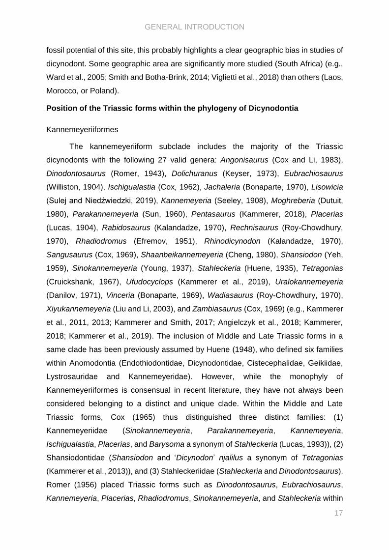
reported on the discovery of the Laotian dicynodont skulls on the basis of the two most-
prepared skulls (LPB 1993-2 and 1993-3, illustrated in PAPER I). The noticeable
characters highlighted by a preliminary description of the two skulls led them to
attribute the specimens to two distinct species of Dicynodon (Battail et al., 1995). Later,
Battail (2009) briefly redescribed LPB 1993-2 and 1993-3, plus a third specimen 1995-
9 from the same locality. On the basis of the definitions of Dicynodon by Cluver and
Hotton III (1981) and Cluver and King (1983), he confirmed the previous attribution to
Dicynodon (Battail et al., 1995; Battail, 2009). However, until the comprehensive
taxonomic revision of the genus Dicynodon by Kammerer et al. (2011), the validity of
many species of Dicynodon was problematic (e.g., Broom, 1911; Watson, 1917).
Figure II.1. Counillon’s specimen reconstructed by Piveteau (1938) in lateral (left) and dorsal (right)
view (modified from Piveteau, 1938). The illustrations are not in scale.

CHAPTER II - NEW LAOTIAN DICYNODONTS
32
Morphological variation were noticed between the three Laotian skulls, uncertainties
thus persisted about their attributions to different and distinct species in regards to
intraspecific (ontogeny, dimorphism) or taphonomic variation (Battail, 2009).
The revised study of the Laotian specimens presents several interests. First, the
taxonomic attribution of the Laotian specimens and their phylogenetic positions have
to be checked and settled. Then, while the age of the Purple Claystone Fm was largely
debated (e.g., Counillon, 1896; Répelin, 1923; Piveteau, 1938; Saurin, 1962; Battail,
2009; Rossignol et al., 2016), the precise relationships of dicynodont forms supposed
to be latest Permian-earlier Triassic in age would bring additional information about the
evolution of the group around the major biological P-Tr crisis. Finally, their occurrence
in Southeastern Asia (Laos) shed new light on the paleobiogeography of dicynodonts
during the early Triassic-latest Permian and consequently on the debate about the
timing of collision between the Indochina, the South China and the North China blocks.
The PAPER I dealed with these problematics and has been published in Journal
of Vertebrate Paleontology. The Laotian dicynodonts are currently conserved in the
Savannakhet Dinosaur Museum in Laos. Financial support granted by the GDRI
PalBioDiv SEA project allowed me a first-hand study of the three skulls LPB 1993-2,
1993-3, and 1995-9 in the Laotian museum, where I described and photographed
them. The morphological variation noticed between the Laotian forms has been
discussed based on observations collected during the visits of dicynodont collections
in the Evolutionary Studies Institute in Johannesburg (South Africa; financial support
from CR2P), the Museum of Comparative Zoology in Cambridge (Massachusetts,
U.S.A.; financial support from CR2P and ISTEP), and the Natural History Museum in
London (United Kingdom; financial support from SYNTHESYS), and on the literature.
In PAPER I, I wrote the majority of the article except the “Geological Setting” part that
has been provided by the geologist colleagues S. Bourquin (Université de Rennes,
France) and C. Rossignol (Universidade de São Paulo, Brazil). The discussion parts
“New Data Supporting the Survivorship of Multiple Lineages across the P-Tr
Boundary?” and “Paleobiogeographic Implications of the Two Laotian Dicynodonts”
resulted from joint discussions between all coauthors. I preformed the phylogenetic
analyses and produced all figures except the figure 1 and the reconstruction in figures
2 to 4 drawn by S. Fernandez (MNHN, France).
The results have been presented during the International Meeting of Early-stage
Researchers in Palaeontology (Lesvos, Greece, 2017).

PAPER I “New dicynodonts (Therapsida, Anomodontia) from near the Permo-
Triassic boundary of Laos: implications for dicynodont survivorship across the
Permo-Triassic mass extinction and the paleobiogeography of Southeast Asian
blocks”, 2019, Journal of Vertebrate Paleontology
New dicynodonts (Therapsida, Anomodontia) from near the Permo-
Triassic boundary of Laos: implications for dicynodont
survivorship across the Permo–Triassic mass extinction and the
paleobiogeography of Southeast Asian Blocks
Chloé Olivier, *,1, 2 Bernard Battail, 2 Sylvie Bourquin, 3 Camille Rossignol, 4 J. -
Sébastien Steyer 2, and Nour-Eddine Jalil 2
1CR2P (Centre de Recherche en Paléontologie - Paris), Museum National d’Histoire
Naturelle–CNRS–Sorbonne Université, 57 rue Cuvier, CP 38, F-75005, Paris, France,
[email protected]; [email protected]; [email protected];
2Univ Rennes, CNRS, Géosciences Rennes - UMR 6118, F-35000 Rennes, France,
3Instituto de Astronomia, Geofísica e Ciências Atmosféricas, Departamento de Geofísica,
Universidade de São Paulo, Rua do Matão, 1226, Cidade Universitária, Butantã, 05508-090
São Paulo, São Paulo, Brazil, [email protected];
4Laboratoire Biodiversité et Dynamique des Ecosystèmes, Faculté des Sciences Semlalia,
Université Cadi Ayyad, Boulevard Prince My Abdellah, 40000 Marrakech, Maroc
*Corresponding author
Published in Journal of Vertebrate Paleontology in 2019
PA
PE
R I

ARTICLE
NEW DICYNODONTS (THERAPSIDA, ANOMODONTIA) FROM NEAR THE PERMO-TRIASSICBOUNDARY OF LAOS: IMPLICATIONS FOR DICYNODONT SURVIVORSHIPACROSS THE
PERMO-TRIASSIC MASS EXTINCTION AND THE PALEOBIOGEOGRAPHY OF SOUTHEASTASIAN BLOCKS
CHLOE OLIVIER, *,1 BERNARD BATTAIL,1 SYLVIE BOURQUIN,2 CAMILLE ROSSIGNOL,3
J.-SEBASTIEN STEYER,1 and NOUR-EDDINE JALIL1,4
1CR2P (Centre de Recherche en Pale ontologie - Paris), Museum National d’Histoire Naturelle–CNRS–Sorbonne Université, 57rue Cuvier, CP 38, F-75005, Paris, France, [email protected]; [email protected]; [email protected];
[email protected];2Univ Rennes, CNRS, Ge osciences Rennes - UMR 6118, F-35000 Rennes, France, [email protected];
3Instituto de Astronomia, Geofísica e Ciências Atmosféricas, Departamento de Geofísica, Universidade de São Paulo, Rua do Matão,1226, Cidade Universitária, Butantã, 05508-090 São Paulo, São Paulo, Brazil, [email protected];
4Laboratoire Biodiversité et Dynamique des Ecosystèmes, Faculté des Sciences Semlalia, Université Cadi Ayyad, BoulevardPrince My Abdellah, 40000 Marrakech, Maroc
ABSTRACT—The dicynodonts are an emblematic group of herbivorous therapsids that survived the Permo-Triassic (P-Tr)crisis. Laotian dicynodonts from stratigraphically constrained beds, recently dated using the U-Pb zircon method, yield newinsights into terrestrial faunas of Southeast Asia during the latest Permian and earliest Triassic. Summarily described, theywere originally attributed to the genus Dicynodon. We provide a new phylogenetic analysis for Laotian dicynodonts, basedon three well-preserved skulls, indicating that they belong to two new taxa: Counillonia superoculis, gen. et sp. nov., andRepelinosaurus robustus, gen. et sp. nov. Our phylogenetic analysis of Dicynodontia indicates that (1) Counillonia is closelyrelated to some ‘Dicynodon’-grade taxa and (2) Repelinosaurus is a kannemeyeriiform. The phylogenetic affinities of thesenew Laotian dicynodonts allow discussion of the survivorship of multiple lineages (Kannemeyeriiformes and ‘Dicynodon’-grade dicynodontoids) across the P-Tr crisis. The Laotian dicynodonts also shed new light on the paleobiogeography ofSoutheast Asia from the late Paleozoic to the early Mesozoic, particularly the timing of collisions between the Indochina,the South China, and the North China blocks. The presence of dicynodonts in Laos, most likely in the Early Triassic, thusimplies that the connection between the Indochina Block and the South China Block occurred no later than the latestPermian or earliest Triassic (i.e., when the dicynodonts provide direct evidence for a connection).
http://zoobank.org/urn:lsid:zoobank.org:pub:FE310B53-41AA-4C13-B076-5D1CB91FC1BE
SUPPLEMENTAL DATA—Supplemental materials are available for this article for free at www.tandfonline.com/UJVP
Citation for this article: Olivier, C., B. Battail, S. Bourquin, C. Rossignol, J.-S. Steyer, and N.-E. Jalil. 2019. New dicynodonts(Therapsida, Anomodontia) from near the Permo-Triassic boundary of Laos: implications for dicynodont survivorship acrossthe Permo-Triassic mass extinction and the paleobiogeography of Southeast Asian blocks. Journal of Vertebrate Paleontology.DOI: 10.1080/02724634.2019.1584745.
INTRODUCTION
The dicynodonts are emblematic Permian and Triassic (P-Tr) therapsids. They constitute an important component ofthe terrestrial P-Tr fauna and were the dominant herbivoresin their ecosystems (Cluver and King, 1983). As such, dicyno-donts represent a key group for understanding the impact ofthe P-Tr crisis on terrestrial environments. Known Early Trias-sic dicynodont genera include the cosmopolitan and specioseLystrosaurus, the small-bodied emydopoids Myosaurus fromSouth Africa/Antarctica and Kombuisia from Antarctica, andthe Chinese kannemeyeriiform Sungeodon (Fröbisch et al.,2010; Maisch and Matzke, 2014).
In North China, Liu et al. (2013) used the U-Pb zircon method(based on zircon U-Pb sensitive high-resolution ion microprobe[SHRIMP] dating) within the Ermaying and Tongchuan dicyno-dont-bearing formations (with the kannemeyeriiform generaShansiodon and Sinokannemeyeria) and dated them to Early toMiddle Triassic. More recently, the higher-resolution chemicalabrasion–thermal ionization mass spectrometry (CA-TIMS)dated these formations as Middle Triassic (Anisian–Ladinian)(Liu et al., 2018). Thus, the main kannemeyeriiform radiationseems to have occurred after the beginning of the Triassic, withca. 40 species known by the Middle Triassic (Fröbisch, 2008).The first record of dicynodonts in Laos (Southeast Asia) dates
back to the 19th century: Counillon (1896) mentioned a poorlypreserved and incomplete skull found in the Purple ClaystoneFormation of the Luang Prabang Basin, northern Laos(Fig. 1A). This specimen was first studied by Repelin (1923),who assigned it to a new species of Dicynodon, D. incisivum,which he considered to be closely related toDicynodon orientalisfrom the Panchet Formation of India. Later, Das Gupta (1922)
*Corresponding author.Color versions of one or more of the figures in the article can be found
online at www.tandfonline.com/ujvp.
Journal of Vertebrate Paleontology e1584745 (22 pages)© by the Society of Vertebrate PaleontologyDOI: 10.1080/02724634.2019.1584745
Published online 23 Apr 2019

transferredD. orientalis to the genusLystrosaurus andWoodward(1932), followed by Yuan and Young (1934), attributed Counil-lon’s specimen to Lystrosaurus. Piveteau (1938) redescribed thespecimen and reassigned it to Dicynodon. Based on this study,Battail (2009) and Kammerer et al. (2011) also favored this taxo-nomic attribution. Nevertheless, this specimen continued to bementioned as Lystrosaurus without further comment (Keyserand Cruickshank, 1979; King, 1988). Counillon’s specimen hasunfortunately been lost, preventing further investigations. Theillustrations accompanying the original description cannot beinterpreted with confidence (Colbert, 1982; Kammerer et al.,2011). Accordingly, the taxon ‘Dicynodon incisivum’ should beconsidered a nomen dubium (as pointed out by many authors,e.g., Battail, 2009; Fröbisch, 2009; Kammerer et al., 2011).
Between 1993 and 2003, Franco-Laotian expeditions led byP. Taquet (Museum National d’Histoire Naturelle [MNHN],Paris, France) collected abundant dicynodont remains from the
Purple Claystone Formation of the Luang Prabang Basin.Among these fossils, three dicynodont skulls (LPB 1993-2, LPB1993-3, and LPB 1995-9) were tentatively ascribed to the genusDicynodon by Battail (2009). However, no phylogenetic analysishas been performed on these specimens, and their relationshipswith other dicynodonts remain equivocal.
The age of these specimens, collected in the Purple ClaystoneFormation, has long been a subject of debate. Indeed, this for-mation was first attributed to the Early Triassic (Counillon,1896; Repelin, 1923; Piveteau, 1938) but was later considered tobe Late Triassic to Middle Jurassic in age (Saurin, 1962). Basedon the dicynodont skulls and their supposed attribution to thegenus Dicynodon (Battail, 2009), this formation was consideredto be late Permian in age (Battail, 2009). Recent geochronologicalanalyses (based on U-Pb detrital zircons dated by laser ablationinductively coupled plasma mass spectroscopy [LA-ICP-MS])performed on volcaniclastic rocks from the Purple Claystone
FIGURE 1. Continental blocks of SoutheastAsia and geological map of the LuangPrabang Basin. A, tectonic subdivisions ofSoutheast Asia, after Metcalfe (2011). B, geo-logical map of the Luang Prabang Basin withthe emplacement of fossil sites and datedsamples. Modified after Blanchard et al.(2013). Sample LP03 was collected at the dicy-nodont site, sample LP04 in an unfossiliferoussite, and sample LP05 at the chroniosuchiansite (Arbez et al., 2018). Abbreviations: AL,Ailaoshan suture zone; CM, Changning Men-glian suture zone; JH, Jinghong suture zone;LP, emplacement of the Luang PrabangBasin; NU, Nan Uttaradit suture zone; SK,Sra Kaeo suture zone.
Olivier et al.—New dicynodonts from near the Permo-Triassic boundary of Laos (e1584745-2)

Formation suggest a maximum depositional age of 251.0 ± 1.4 Ma(Table 1) (Rossignol et al., 2016).This temporal framework thus allows us to document dicy-
nodont survivorship and post-extinction recovery in a P-Trbasin located outside the classic, extensively studied Russian(e.g., Benton et al., 2004) and South African (e.g., Wardet al., 2005; Smith and Botha-Brink, 2014; Viglietti et al.,2018) ones. The fauna preserved in the Luang Prabang Basinalso offers new evidence concerning the paleobiogeographyof dicynodonts.Here, we provide detailed description of the three Laotian
skulls (LPB 1993-2, LPB 1993-3, and LPB 1995-9), which rep-resent two new taxa. Phylogenetic analysis is then performed totest the relationships of these new taxa within Dicynodontia.Institutional Abbreviations—BP, Evolutionary Studies Insti-
tute (formerly the Bernard Price Institute for PalaeontologicalResearch and the Institute for Human Evolution), Johannesburg,South Africa; LPB, Laotian specimens found in the LuangPrabang Basin, currently in the Savannakhet DinosaurMuseum, Savannakhet, Laos; MCZ, Museum of ComparativeZoology, Harvard University, Cambridge, Massachusetts,U.S.A.; NHMUK, Natural History Museum, London, UnitedKingdom.Anatomical Abbreviations—aPt, anterior ramus of the ptery-
goid; Bo, basioccipital; Ch, choana; EcPt, ectopterygoid; Eo,exoccipital; EpiPt, epipterygoid; Fm, foramen magnum; Fr,frontal; Ip, interparietal; Ipv, interpterygoid vacuity; Ju, jugal;La, lacrimal; Laf, lacrimal foramen; Lbf, labial fossa; mPt,median plate of the pterygoid; Mx, maxilla; Na, nasal; Oct, occi-pital tuber; Opt, opisthotic; Pa, parietal; Pal, palatine; Pant, pilaantotica; Pbs, parabasisphenoid; Pi, pineal foramen; Pm, premax-illa; Po, postorbital; Pp, preparietal; PrF, prefrontal; Q, quadrate;Qj, quadratojugal; qPt, quadrate ramus of the pterygoid; Smx,septomaxilla; So, supraoccipital; Sq, squamosal; T, tusk; Tpf, post-temporal fenestra; Va, vagus nerve aperture; Vo, vomer.
GEOLOGICAL SETTING
The Luang Prabang Basin, located in the Indochina Block(Fig. 1A), was originally studied by Counillon (1896) and consistsof an asymmetric NE–SW (northeast–southwest) syncline withNE–SW thrusts separating the Purple Claystone Formation andthe Limestone and Sandstone Formation to the north fromyounger formations to the south (Fig. 1B; Blanchard et al., 2013).The Limestone and Sandstone Formation is made up of shallow
marine deposits, dated to the late Changhsingian on the basis ofits ammonoid remains (Blanchard et al., 2013). These marinedeposits are overlain by black claystone layers, containing atypical Cathaysian flora (Bercovici et al., 2012).The Limestone and Sandstone Formation is overlain by the
Purple Claystone Formation, from which various fossilremains have been excavated (dicynodonts and a chroniosu-chian; Steyer, 2009; Arbez et al., 2018). The Purple Claystone
Formation is mainly composed of homogeneous silty claystones,silts, and more rarely clays (Bercovici et al., 2012). The for-mation also comprises volcaniclastic siltstones and sandstones,with millimeter- to centimeter-sized rounded and highly weath-ered volcaniclasts (up to about 20 vol.%). These volcaniclastsexhibit a variety of volcanic textures (microlithic, trachytic, por-phyritic) and are sometimes embedded within lithic fragments,attesting to multiple reworking events for these volcaniclasts(Bercovici et al., 2012; Blanchard et al., 2013). The PurpleClaystone Formation also contains subordinate amounts ofcoarser deposits, including sandstone and conglomeratic facieswith three-dimensional megaripples typical of braided riverdeposits. The conglomeratic levels consist of rounded pebblesof highly fossiliferous limestones (foraminifers, corals, bryozo-ans), subangular to rounded pebbles of volcanic rocks, blackcherts, red quartzites, red sandstones, and siltstones. Paleosols,sometimes exhibiting vertical root traces, are developed withinthis formation (Bercovici et al., 2012). The sedimentary faciesassociation indicates braided river depositional environments,evolving vertically to alluvial plain environments, probablyincluding ponds (Bercovici et al., 2012).Three samples from the Purple Claystone Formation, includ-
ing one collected at the dicynodont site (Fig. 1C), were datedusing U-Pb geochronology on detrital zircon (Rossignol et al.,2016). The sample collected at the dicynodont fossil siteyielded a maximum depositional age of 252.0 ± 2.6 Ma,whereas the other volcaniclastic samples collected in the sameformation yielded maximum depositional ages of 251.0 ± 1.4and 300.5 ± 3.7 Ma (Table 1). The various volcaniclastic textures,their roundness, and the relatively low volcaniclast content(below 20%) implying an important and protracted mixingwith other detrital particles, as well as the fact that some ofthe volcaniclasts underwent at least two sedimentary cycles(Bercovici et al., 2012; Blanchard et al., 2013), suggest thatthese dates, obtained from zircon grains interpreted as beingdetrital in origin, represent maximum depositional ages. Theactual age of deposition of the Purple Claystone Formation istherefore likely to be younger. Both youngest maximum deposi-tional ages (i.e., 252.0 ± 2.6 and 251.0 ± 1.4 Ma) encompass theP-Tr boundary (251.902 ± 0.024 Ma; Burgess et al., 2014)within uncertainties. The consideration of a late Permian age,potentially plausible, would nonetheless imply that the rework-ing of the zircon grains took place within an unlikely brief timespan. Given the age of the overlying formation (224.9 ± 1.0 Ma;Blanchard et al., 2013), an age up to the Carnian could be pro-posed as the theoretical upper age limit for the Purple Clays-tone Formation. However, the occurrence of a regionalMiddle Triassic unconformity (e.g., Racey, 2009), probablysuperimposed onto the reverse fault separating the PurpleClaystone Formation from other sedimentary units to the south-east (Fig. 1B), reduces the likely time span for the deposition ofthe Purple Claystone Formation. Consequently, an Early Trias-sic age for the Purple Claystone Formation and its enclosedfossils is considered to be the most likely.
TABLE 1. Summaryof themaximumdepositional ages obtainedbyU-Pb laser ablation inductively coupledmass spectrometry (LA-ICP-MS)datingondetrital zircon grains from volcaniclastic rocks of the samples collected in the Purple Claystone Formation (LP03, LP04, LP05) (Rossignol et al., 2016).
Sample Na Nzr N
Maximum depositional age
Concordia age ± (2σ) n MSWD Probability
LP03 105 102 25 252.0 2.6 6 0.89 0.55LP04 41 36 7 300.5 3.7 4 0.73 0.65LP05 96 95 39 251.0 1.4 21 0.55 0.99
LP03 was collected at the dicynodont fossil site. TheMSWD and the probability given for the concordia ages are for both concordance and equivalence.Abbreviations: MSWD, mean square of weighted deviates; n, number of analyses used to calculate the maximum depositional age; N, number ofconcordant zircon grains; Na, number of analyses per sample; Nzr, number of zircon grains analyzed per sample.
Olivier et al.—New dicynodonts from near the Permo-Triassic boundary of Laos (e1584745-3)

METHODS
The interpretation of the cranial bone contacts is based eitheron direct observations of the scarce preserved sutures or onrelief differences. When possible, some sutures were deducedfrom the contacts between the surrounding bones.
SYSTEMATIC PALEONTOLOGY
THERAPSIDA Broom, 1905ANOMODONTIA Owen, 1860DICYNODONTIA Owen, 1860
DICYNODONTOIDEA Olson, 1944COUNILLONIA, gen. nov.
Type Species—Counillonia superoculis, gen. et sp. nov.,monotypic.
Etymology—In honor of the French geologist Jean-Baptiste-Henri Counillon, member of the Pavie Missions, who was thefirst to mention the occurrence of dicynodonts in the LuangPrabang Basin (Counillon, 1896; see Steyer, 2009, for abiography).
Diagnosis—See diagnosis of the type species.
COUNILLONIA SUPEROCULIS, gen. et sp. nov.(Fig. 2)
Holotype—LPB 1993-3, a skull without mandible (basallength: 16.02 cm; maximum width: 13.41 cm). The posterior sideof the left orbit, the quadrates, and the stapes are missing. Thedorsal surfaces of the premaxilla, the nasals, the prefrontals,and the frontals are partially eroded. The preparietal, theprootic, and the epipterygoid are poorly preserved.
Geographic Distribution and Stratigraphic Range—LPB 1993-3 (19°55′59″N, 102°07′41″E) was discovered in the Purple Clay-stone Formation, Luang Prabang Basin (Laos). This formationwas initially attributed to the Early Triassic by Counillon (1896)then to the late Permian by Battail (2009). Recent geochronolo-gical (U-Pb on detrital zircon; Rossignol et al., 2016) analysessuggest a maximum depositional age of 251.0 ± 1.4 Ma (see ‘Geo-logical Setting’ above).
Etymology—From the Latin ‘oculis’ (dative plural of oculus,eye) and ‘super’ (upward), referring to its largely dorsallyopening orbits due to an especially narrow interorbital bar.
Diagnosis—Medium-sized dicynodontoid characterized by theunique combination of the following character states: a reducedpremaxillary secondary palate; a nasofrontal suture with a distinctposterior process; a narrow intertemporal bar; a reduced temporalfenestra; a pineal foramen located in the posterior quarter of thedorsal skull length; an acute angle of the squamosal wings inlateral view; zygomatic squamosal rami posteriorly inserted atmid-height of the occiput; a relatively large median pterygoidplate; high-angled posterior pterygoid rami; anterior rami of thepterygoids ventrally highly expanded; a long interpterygoidvacuity; no intertuberal ridge; distinct exoccipital and basioccipitalcontributions to the occipital condyle; and a very sharp and poster-iorly directed lateral edge of the paroccipital process, which is dis-tinctly offset from the surface of the occipital plate. Thenasofrontal suture with a distinct posterior process distinguishesCounillonia from the Laotian Repelinosaurus and ‘Dicynodon’-grade taxa. Further distinguished from closely related ‘Dicyno-don’-grade taxa by a pineal foramen located far posteriorly anda large median pterygoid plate. Further distinguished from Repe-linosaurus by an anteriorly directed caniniform process, a narrowinterorbital bar, a triangular occiput, and zygomatic squamosalrami posteriorly inserted at mid-height of the occiput.
Description
The skull is slender and short, with a short preorbital regionand a narrow snout. In dorsal view, the skull is relatively broad,with laterally expanded zygomatic arches. The combination ofthe laterally bowed zygomatic arches and narrow interorbitalresults in notably dorsally directed orbits (Fig. 2A). The surfacesof the premaxilla, the maxillae, the nasals, the frontals, and thepostorbitals are weathered. Because of this, the mid-nasal, naso-premaxillary, preparieto-frontal, and postorbito-parietal suturesare not visible (Fig. 2A). The absence of visible sutural contactsin the weathered narial region precludes determination of thepossible presence of the septomaxillae (Fig. 2B). It is thus notclear whether the septomaxillae form part of the ventrolateralmargin of the nares, which seems to be formed only by the pre-maxilla anteriorly, the nasals dorsolaterally, and the maxillae ven-trolaterally. In addition, the bone contacts cannot be discerned onthe occiput, nor along the medial partition of the temporal andorbital fossae (Fig. 2B, D).
The short premaxilla is fused and forms the anterior portion ofthe snout that constricts and ends in a squared tip. The tip of thesnout shows weak ventral curvature in anterior view (not figuredhere) as in Tropidostoma and Aulacephalodon (Kammerer andSmith, 2017). However, this curvature and possible ridges or rug-osities cannot be confirmed due to the poor preservation of thedorsal surface of the premaxilla. The premaxilla contributes tothe external anterior edge of the naris. The large narial openingis situated near the anterior and ventral edges of the snout andcontributes to more than a third of the surface of the snout(Fig. 2B). The external ventral margin of the naris is visible indorsal view (Fig. 2A). The premaxilla contacts the nasals poster-odorsally and the maxilla posterolaterally at the level of theanterior third of the nares. In ventral view, the premaxilla bearstwo thin, parallel longitudinal ridges (the anterior palatalridges) that surround a wide, median longitudinal depressionand extend beyond the anterior third of the palatal surface (Fig.2C). This depression turns posteriorly into a sharp crest (themedian palatal ridge), which extends to the vomer posteriorlyand is flanked on either side by flat depressions. The height ofthis longitudinal ridge increases posteriorly. On the palatalsurface, the premaxilla contacts the maxillae laterally. Theabsence of visible sutures between the premaxilla and the pala-tines yields no information about a potential contact (Fig. 2C).The nasals bear a median, rugose, and well-developed boss,which extends onto the bones bordered by two wide elongateddepressions that broaden posteriorly and terminate on the fron-tals (Fig. 2A). However, as mentioned above, the poor preser-vation of the snout surface precludes firm conclusions about theoriginal external relief of the nasals. The nasals are surroundedby the frontals and the prefrontals posteriorly, the premaxillaanteriorly, and the maxillae and lacrimals laterally (Fig. 2A, B).
The prefrontals form the anterodorsal edge of the orbits. Onthe external surface of the skull, they contact the frontals medi-ally, the lacrimals laterally, and the nasals anteriorly (Fig. 2A).A single rugose boss is visible on each prefrontal. However, asalready noted for the nasals, there is no reliable informationabout the original relief of the bones due to the poor preservationof their external surfaces.
The frontals form the major part of the skull roof (Fig. 2A). Thesutures of the frontals can be discerned along their contact with thepostorbitals, the prefrontals, and the nasals, as well as at the inter-frontal contact (Fig. 2A). The external surface of the skull roof ispoorly preserved; the presence of postfrontals cannot be con-firmed in LPB 1993-3. The nasofrontal suture has a distinct pos-terior process. The dorsal margin of the orbits seems to belargely made up of the frontals. Apart from a small chip of bonemissing on the right dorsal margin of the orbit, the rest of itsmargin appears to be preserved, with a symmetry of the lateral
Olivier et al.—New dicynodonts from near the Permo-Triassic boundary of Laos (e1584745-4)
FIGURE 2. Counillonia superoculis, gen. et sp. nov., LPB 1993-3, holotype, photographs and interpretive drawings of skull inA, dorsal, B, right lateral,C, ventral, andD, occipital views. The thin gray lines represent sutures, and the bold black lines represent relief. The dotted line represents the authors’interpretation of sutures based on variation in bone texture. Scale bar equals 5 cm.
Olivier et al.—New dicynodonts from near the Permo-Triassic boundary of Laos (e1584745-5)

margins of the orbits and continuous lateral borders. The interor-bital region is broader than the intertemporal bar (Fig. 2A).
The preparietal is strongly weathered, and its anterior part ismissing. However, its posterior region displays a small bulgeanterior to the pineal foramen, the relief possibly being due toits poor preservation (Fig. 2A). The contact between thepreparietal and neighboring bones (postorbitals and frontals)cannot be determined.
The dorsal exposure of the parietals is limited to a midlinegroove between the posterior postorbital processes (Fig. 2A).The parietals then contact the postorbitals laterally and dorsally.External sutural contacts between the parietals and the frontalsare invisible due to the poor preservation of the preparietal andthe eroded external surface of the skull roof. The pineal foramenis surrounded by the parietals posteriorly and by the preparietalanteriorly. The oval pineal foramen, with a length of 1.36 cm anda width of 0.96 cm (Table 2), has its long axis perpendicular tothe axis of the intertemporal bar. It is located in the posteriorquarter of the skull roof (continuous character 7, Appendix 1).
The slender postorbitals have lateral and posterior processes.The posterior postorbital process merges along the intertemporalbar, posterior to the pineal foramen (Fig. 2A). The temporalportion of the postorbitals appears to be oblique, with a dorsolat-eral orientation. However, the poor preservation of the externalsurface of the postorbital does not allow confirmation of thisorientation with certainty. The posterior postorbital region isthinner than the preorbital one (Fig. 2A). The postorbitalsextend along almost the entire intertemporal bar, bordering thetemporal fossae medially and anteriorly (Fig. 2A). They thus con-stitute the posterior margin of the orbits. On their externalsurface, the posterior postorbital processes mainly contact thesquamosals and to a lesser degree the interparietal posteriorly,the parietals medially, and the frontals anteriorly. As mentionedabove, no reliable information can be provided on the contactbetween the postorbitals, the frontals, and the preparietal. Thelateral postorbital processes have a sutural contact with thejugals anteroventrally and the zygomatic squamosal processesposteroventrally (Fig. 2B).
The maxillae form the largest part of the lateral surface of thesnout. They contact the premaxilla anteriorly and the lacrimalsand the nasals dorsally (Fig. 2B). The maxillonasal suture lieson the dorsal edge of the nares. There is no sutural contactbetween the maxillae and the prefrontals due to the anteriorexpansion of the lacrimals that contact the nasals. The maxillaryzygomatic process is posteriorly pointed and contacts the squa-mosal posteriorly and the jugal dorsally (Fig. 2B). The maxillaebear short caniniform processes housing tusks. The dorsal edgeof the erupted portion of the tusk is anterior to the anterior
edge of the orbits. The caniniform processes and the tusks faceanteriorly. The tusks have a subcircular basal section (Fig. 2C;Table 2). On the ventral surface, no maxillary teeth are observedexcept the tusks (Fig. 2C). The maxillae contact the anterior ramiof the pterygoids and the ectopterygoids posteriorly and thejugals and the squamosals laterally. No boundaries are visible infront of the palatines, and nothing can be confirmed about theextension of medial maxillary processes posterior to the premax-illa. In Lystrosaurus (e.g., Cluver, 1971) and Kannemeyerialophorhinus (e.g., the holotype, BP/1/3638, and the specimen for-merly referred to Rechnisaurus cristarhynchus, NHMUK R11955[Renaut et al., 2003; C. Olivier, pers. observ., 2018]), these pro-cesses exclude the contact between the premaxilla and the pala-tines. A depression lies on the suture between the maxillae andthe pterygoids (Fig. 2B). A broad labial fossa opens posterior tothe tusk (Fig. 2C). Its boundaries cannot be discerned; however, itis usually surrounded by the maxillae ventrally, the jugals dorso-laterally, and the palatines medially (e.g., Cluver, 1971). It istherefore not certain whether Counillonia has a true labial fossa(circumscribed by the maxillae, the palatines, and the jugals) ora comparable structure to the foramen in dicynodonts such asDiictodon, Endothiodon, or Dicynodontoides (e.g., Angielczykand Kurkin, 2003).
The lacrimals are relatively triangular in lateral view (Fig. 2B).They contact the nasals and prefrontals dorsally, the maxillae ven-trally, and the jugals posteriorly. Their anterior, well-developedexpansions exclude a maxilloprefrontal sutural contact. Theirsutures within the orbits are not visible. The lacrimals constitutethe anterior margin of the orbits with the jugals anteroventrallyand the prefrontals anterodorsally (Fig. 2B). Within the orbit,each lacrimal is perforated by a single foramen (Fig. 2A).
The jugals are longitudinally elongated and form the ventraledge of the orbits (Fig. 2B). However, their expansion within theorbits and their natural limits cannot be discerned due tothe absence of visible sutures, as noted above. Visible sutures onthe lateral side of the skull show a long scarf joint with themaxillaeventrally, a jugal posterior process contacting the postorbitals pos-teriorly, and a small contact with the zygomatic squamosal pro-cesses ventroposteriorly and the lacrimals anterodorsally (Fig. 2B).
The squamosals are triradiate, with zygomatic, temporal, andquadrate rami. The zygomatic ramus shapes the posteriorregion of the zygomatic arch (Fig. 2B). In dorsal view (Fig. 2A),it widens posteriorly into a wing-shaped structure without afolded edge. It becomes narrow in the pointed anterior region,without dorsoventral expansion posterior to the postorbital bar(Fig. 2B). The squamosal zygomatic processes contact thejugals, the postorbitals, and the maxillae anteriorly (Fig. 2B).They circumscribe the temporal fossae laterally and partly
TABLE 2. Cranial measurements (in cm) of the three Laotian dicynodont skulls attributed to Counillonia superoculis and Repelinosaurus robustus,scaled with Image J 1.50i.
1. C. superoculis 2. R. robustus
Dimension LPB 1993-3 LPB 1995-9 LPB 1993-2
Basal length (from the tip of snout to the occipital condyle)a 16.02 15.72 19.00Maximum widthb 13.41 NA NAMaximum orbital heightc 4.82 4.19 5.79Maximum orbital lengthc 5.67 4.73 6.82Mediolateral diameter of the tusk roota 1.27 1.46 2.02Anteroposterior diameter of the tusk roota 1.56 0.90 1.49Pineal foramen lengthb 1.36 1.33 0.96Pineal foramen widthb 0.96 0.87 0.66
aIn ventral view.bIn dorsal view.cIn lateral view.
Olivier et al.—New dicynodonts from near the Permo-Triassic boundary of Laos (e1584745-6)

posteriorly. The zygomatic squamosal rami show a relativelyventral insertion on the back of the skull, at mid-height on theocciput (Fig. 2D). On the lateral side, they do not reach thedorsal region of the occiput. The temporal processes delimit thetemporal fossae posteromedially. On their external surface, theycontact the postorbitals dorsally. On their occipital side, nocontact between bones is visible (Fig. 2D). The absence of thelimits of the tabulars does not allow us to determine whetherthe squamosals are separated from the supraoccipitals by the tab-ulars. The steep angle between the temporal and zygomatic pro-cesses of the squamosal slightly exceeds 90° (Fig. 2D). The widelateral extensions of the squamosal make an angle of less than90° with the occipital side. A part of the lateral edge of theocciput is thus hidden. The squamosals extend posteriorly tothe occipital condyle (Fig. 2C).The lateral surface of the braincase is strongly eroded, but bone
structures can be noted. Although the sutural contacts of theprootics are not visible, the basal part of the left pila antotica(not figured here) is preserved. Only the dorsal region of thelong and narrow epipterygoids, which contacts the ventralprocess of the parietals, is preserved. A strong anteroventraldepression in the squamosals indicates the connection with themissing quadrates (Fig. 2B).The vomer displays a tuberosity turning into a vertical blade,
which narrows posteriorly and separates the two choanae (Fig.2C). The width of the median blade is constant along its length.The vomer contacts the premaxilla anteriorly and the pterygoidsposteriorly. It forms the anterior margin of an elongated andovoid interpterygoid vacuity (Fig. 2C).The palatines border the choanae anterolaterally. The anterior
palatine expansions form rugose and textured pads, indicating akeratinized covering (Fig. 2C). They narrow posteriorly to formrelatively smooth processes. The palatines contact the anteriorrami of the pterygoids along their entire lateral scarf joint andare anteriorly bordered by the maxillae. However, as mentionedabove, due to the uncertainties about their anterior suturalcontact, it is not clear whether they contact the premaxilla. Alateral palatine foramen is present alongside each anteriorexpanded pad (Fig. 2C).Each pterygoid has an anterior ramus and a posterior (or quad-
rate) ramus. A single median plate connects the two bones andlinks the four rami. The pterygoids contact the secondary palateand the basicranium (Fig. 2C). Their ventral projections extendstrongly anteroventrally, such that they form an anteriordepression with the maxillae (Fig. 2B). These anterior pterygoidkeels extend along most of the length of the anterior rami. Never-theless, this extension cannot be precisely measured because thesutures are not visible laterally (Fig. 2B). The anterior rami of thepterygoids merge from the median plate, posterior to the inter-pterygoid vacuity. The interpterygoid vacuity is thus borderedby the pterygoids posteriorly and the vomer anteriorly. The pos-terior margin of the vacuity rises flush with the median plate ofthe pterygoids. The ventral surface of the narrow median platebears a thin crista oesophagea (Fig. 2C). The median plate ofthe pterygoids contacts the parabasisphenoid posteriorly andthe vomer anteriorly. In addition, a possible contribution of theparabasisphenoid to the interpterygoid vacuity cannot beproved. Two posterior rami of the pterygoids contact the squamo-sal fossa posteriorly and probably the medial condyles of thequadrates laterally.Despite the absence of visible sutural contacts on the lateral
side of the skull, the presence of the ectopterygoids is indicatedby variation in bone texture on the lateral border of the anteriorrami of the pterygoids (Fig. 2B). The ectopterygoids have aslender leaf shape and expand laterally along the anterior ramiof the pterygoids. They do not expand posterior to the palatinesin palatal view (Fig. 2C).
The crista oesophagea continues onto the parabasisphenoidand then diverges posteriorly to form the ridges leading to thebasitubera (Fig. 2C). The contribution of the parabasisphenoidto the fenestra ovalis of the tubera is considerably restricted incomparison with the basioccipital. The stapedial facet is ventro-laterally directed, and its narrow margin mostly extends antero-posteriorly. The paired carotid canals, located in the anteriorregion of the parabasisphenoid in many dicynodonts (e.g.,Maisch, 2002; Surkov and Benton, 2004), are not visible here.No intertuberal ridge is visible. The basioccipital extends ontothe occipital plate and with the exoccipitals forms a tripartite occi-pital condyle (Fig. 2D). The exoccipitals and the basioccipitalappear not to be fused in the condyle. A circular centraldepression is located between the three occipital subcondyles.Medially, the exoccipitals border the aperture of the vagusnerves (Dutuit, 1988).The contacts between the interparietal and the other bones
forming the occipital side and the posterior region of the skullroof cannot be discerned (Fig. 2D). Despite the eroded surfaceof the posterior postorbital processes, the interparietal does notseem to contribute to the skull roof (Fig. 2A).The occiput is triangular in posterior view (Fig. 2D). Although
sutures are not clearly preserved, the overall similarity of the occi-puts of the Counillonia type specimens to those of other dicyno-donts (Lystrosaurus: Cluver, 1971; Dicynodon and Diictodon:Cluver and Hotton, 1981) suggests that they shared a similar con-struction of the occiput. The teardrop-shaped foramen magnummay be laterally bordered by the exoccipitals, which overhangthe basioccipital as in other dicynodonts. They would alsocontact the supraoccipital dorsally, the opisthotics laterally, andthe basioccipital ventrally. A central depression on the supraocci-pitals overhangs the foramen magnum and is dorsally borderedby a weak transverse nuchal crest, which extends upwardtoward the interparietal. The extent of the tabulars cannot bedetermined. The broad oval posttemporal fenestrae are locateddorsal to the level of the occipital condyle, at the transverselevel of the mid-height of the foramen magnum (Fig. 2D). Theyare oriented in the ventromedial-dorsolateral axis and would bedelimited by the squamosal laterally, the supraoccipital dorso-medially, and the opisthotic ventromedially as in most dicyno-donts. An oblique ridge on the supraoccipital extends over thefenestra. This ridge continues, below the fenestra, on the opistho-tics and terminates in a sharp tuberosity.
KANNEMEYERIIFORMES Maisch, 2001REPELINOSAURUS, gen. nov.
Type Species—Repelinosaurus robustus, gen. et sp. nov.,monotypic.Etymology—In honor of the French geologist Joseph Répelin,
member of the Pavie Missions, who described and named Counil-lon’s dicynodont skull ‘Dicynodon incisivum’ (Répelin, 1923; seeSteyer, 2009, for a biography). Also from the latinized Greek‘saurus’ (a lizard) often used for nonmammalian synapsids, collo-quially known as ‘mammal-like reptiles.’Diagnosis—See diagnosis of the type species.
REPELINOSAURUS ROBUSTUS, gen. et sp. nov.(Figs. 3–5)
Holotype—LPB 1993-2, a partial skull without mandible (basallength: 19 cm). The left postorbital bar, the zygomatic arch, thedorsolateral wing of the squamosal, the left quadratojugals, thequadrates, more than half of the left part of the occipital side,the external portion of the tusks, and the stapes are missing.The palatal surface is strongly eroded: most of the sutures
Olivier et al.—New dicynodonts from near the Permo-Triassic boundary of Laos (e1584745-7)

FIGURE 3.Repelinosaurus robustus, gen. et sp. nov., LPB 1993-2, holotype, photographs and interpretive drawings of skull inA, dorsal,B, right lateral,C, ventral, and D, occipital views. The thin gray lines represent the sutures, and the bold black lines represent relief. Scale bar equals 5 cm.
Olivier et al.—New dicynodonts from near the Permo-Triassic boundary of Laos (e1584745-8)

FIGURE 4. Repelinosaurus robustus, gen. et sp. nov., LPB 1995-9, referred specimen, photographs and interpretive drawings of skull in A, dorsal, B,right lateral, C, ventral, and D, occipital views. The thin gray lines represent the sutures, and the bold black lines represent relief. The dotted line rep-resents the authors’ interpretation of sutures based on variation in bone texture. Scale bar equals 5 cm.
Olivier et al.—New dicynodonts from near the Permo-Triassic boundary of Laos (e1584745-9)

cannot be made out and some bones are poorly preserved or aremissing.
Referred Material—LPB 1995-9 is a skull (basal length:15.72 cm) lacking the mandible, the right quadrate ramus of thepterygoid, the left stapes, the quadrates, and the left quadratoju-gal. The right stapes and the epipterygoids are poorly preserved.The specimen was subjected to lateral compression. However, theleft orbit seems to have maintained its original shape.
Geographic Distribution and Stratigraphic Range—Same asfor Counillonia (see above), LPB 1993-2 (19°55′59″N, 102°07′41″E) and LPB 1995-9 (19°55′16″N, 102°06′27″E) were collectedin the Purple Claystone Formation, Luang Prabang Basin (Laos).
Etymology—From the Latin ‘robustus’ (robust), referring to itsrobust cranial appearance.
Diagnosis—Medium-sized dicynodontoid characterized by theunique combinationof the following character states: a reducedpre-orbital region; anotchon thedorsal edgeof thenarial opening; nasalbosses present as a median swelling with a continuous posteriormargin; a straight frontonasal suture; parietals exposed in midlinegroove; a relatively flat temporal portion of the postorbital, sothatmost of the exterior surfaceof thebone faces dorsally; a verticalcaniniform process; and high insertion of the zygomatic squamosalrami on the occiput posteriorly. Distinguished from all dicynodon-toids by a very small preorbital region. Further distinguished fromall kannemeyeriiformsbyamedian nasal swellingwith a continuousposterior margin and a flat temporal portion of the postorbital.Further distinguished from the LaotianCounillonia by awide inter-orbital bar, a straight frontonasal suture, a vertical caniniformprocess, a rectangular occiput, and a high insertion of the zygomaticsquamosal rami on the occiput posteriorly.
Remarks—Some differences distinguish LPB 1993-2 and LPB1995-9, such as (1) the position of the pineal foramen on the skullroof that is more posterior in LPB 1993-2 (continuous character6, Appendix 1); (2) a higher angulation between the occiput andthe palate in LPB 1993-2 (continuous character 13, Appendix 1);(3) a relatively flat palatal surface of the premaxilla in LPB 1993-2 or with marked depressions on either side of the median crestin LPB 1995-9 (discrete character 29, Appendix 1); (4) a suturalcontact between the maxillae and the prefrontals present in LPB1995-9 and absent in LPB 1993-2 (discrete character 49, Appendix1); (5) a preparietal depressed in LPB 1993-2 or flushwith the skullroof inLPB1995-9 (discrete character 68,Appendix 1); (6) a biggerpineal foramen inLPB1995-9 (Table 2); (7) tusk basal section com-pressedmediolaterally inLPB1995-9andanteroposteriorly inLPB1993-2 (Table 2); and (8) more developed ornamentation on thefrontals in LPB 1993-2. However, each character-state variationnoted could be due to taphonomic deformation, ontogeny, sexual
dimorphism or other intraspecific variability (discussed below).Moreover, LPB 1995-9 and LPB 1993-2 were found in the samegeological formation, suggesting conspecificity.
Description
The skulls are narrow and robust, with a wide and short snoutthat narrows slightly anteriorly and terminates in a squared tip.Repelinosaurus has a wider interorbital region (Figs. 3A, 4A)than Counillonia (Fig. 2A). The orbits are thus open mainly later-ally. The dorsal surfaces of the premaxilla, the maxillae, and thenasals of LPB 1995-9 are weathered, but the preservation ofthese surfaces in LPB 1993-2 displays strong rugosities on thesnout (Figs. 3A, 4A). Nevertheless, the nasopremaxillary andthe mid-nasal sutures cannot be traced in LPB 1993-2. The septo-maxilla is well preserved in LPB 1995-9, unlike in LPB 1993-2where the poor preservation of the naris does not yield infor-mation about this bone (Figs. 3B, 4B, 5). Sutures are not visibleon the occiput of either specimen, except for the connection ofthe right quadrate in LPB 1995-9. The scarf joints also cannotbe discerned on the medial portion of the temporal and orbitalfossae, but the dorsal head of the epipterygoid and the basalregion of the pila antotica are preserved in LPB 1995-9 (Fig.4B). The palatal surface of LPB 1993-2 is poorly preserved; fewsutures are thus visible.
As mentioned above, lots of pits and strong rugosities mark thedorsal surface of the premaxilla in LPB 1993-2 and extend ontothe nasals before stopping abruptly at the nasoprefrontal andnasofrontal sutures (Fig. 3A). These rugosities are thought toindicate a keratinized covering (e.g., Angielczyk et al., 2018).The premaxilla contributes to the external anterior edge of thenares. In lateral view, the well-developed nares of both LPB1995-9 and LPB 1993-2 represent more than half of the lengthof the snout (Figs. 3B, 4B). Their ventral edges are close to theventral border of the snout. The premaxilla is toothless and ante-riorly bears two short parallel longitudinal ridges, which are sep-arated by a shallow depression and overlie the anterior quarter ofthe premaxilla (Figs. 3C, 4C). An anterior rounded pit of mediumsize in LPB 1993-2, visible posterior to the premaxillary longitudi-nal crest, could be of taphonomic origin (Fig. 3C). Posterior to thisdepression, a sharp median ridge extends onto two-thirds of thepremaxilla. In LPB 1993-2, the premaxilla is relatively flat.However, this median ridge is surrounded by two depressions,which deepen in the posterior region of the secondary palate inLPB 1995-9. These depressions may be caused by taphonomiclateral compression (discussed below). The premaxilla contactsthe nasals posterodorsally and the maxillae posterolaterally atthe level of the anterior third of the nares in LPB 1995-9 (Figs.3B, 4B). However, the sutures of the premaxilla with the nasalsand maxillae are not visible in LPB 1993-2. In both LPB 1995-9and LPB 1993-2, the premaxilla contacts the maxillae and theircaniniform processes laterally on the palatal surface. However,the suture between the premaxilla, the maxillae, and the palatinesare not clearly preserved (Figs. 3C, 4C).
The well-developed nasal bosses terminate laterally in a poster-odorsal notch at the dorsal edge of the nares and are separatedfrom the frontals and prefrontals by a shallow depression (Figs.3B, 4B). The notch on the posterodorsal edge of the naris isformed by a thick expansion of the nasal bosses, which hidesthe external ventral narial edge in dorsal view. The bosses forma median swelling that is more marked in LPB 1993-2 than inLPB 1995-9 (Figs. 3B, 4B). The nasals are bordered by the fron-tals and the prefrontals posteriorly, the premaxilla anteriorly,and the maxillae laterally. However, the nasals do not contactthe lacrimals in LPB 1995-9, in contrast to the condition in LPB1993-2 (Figs. 3B, 4B, 5).
The prefrontals form the edge of the orbits with the lacrimalsanteriorly, the jugals ventrally, the postorbitals posteriorly, and
FIGURE 5. Close-up photograph and interpretive drawing of the lateralsnout region of the referred skull LPB 1995-9, attributed to Repelino-saurus robustus, gen. et sp. nov. The thin gray lines represent sutures,and the bold black lines represent relief. Scale bar equals 5 cm.
Olivier et al.—New dicynodonts from near the Permo-Triassic boundary of Laos (e1584745-10)
the frontals dorsally (Figs. 3A, B, 4A, B). As for the nasals, the pre-frontal extension is broader in dorsal than in lateral view. The pre-frontals bear a weak boss, distinct from the nasals. The prefrontalscontact the maxillae ventrally in LPB 1995-9 (Figs. 4B, 5).The interorbital region of Repelinosaurus (Figs. 3A, 4A) is
clearly wider than that of Counillonia (Fig. 2A). In Repelino-saurus, it is mainly formed by the frontals constituting thedorsal margin of the orbits, which rise slightly laterally. Inaddition, as mentioned above, the prefrontals bear bosses inboth specimens. The interorbital region obscures the orbits indorsal view; their orientation is thus mainly lateral, whereas it isdorsolateral in Counillonia. In LPB 1993-2, the frontals are separ-ated by a sharp median ridge bordered by two deep depressions(Fig. 3A). In LPB 1995-9, the preparietal, the parietals, and theposterior region of the frontals show a depression. The frontalscontact the nasals anteromedially, the prefrontals laterally, thepostorbitals posterolaterally, and the preparietal posteromedially(Figs. 3A, 4A). The nasofrontal suture is relatively straight inRepelinosaurus (Figs. 3A, 4A), whereas it has a distinct posteriorprocess in Counillonia (Fig. 2A). No postfrontals are observed inLPB 1995-9 (Fig. 4A), but as in Counillonia, we cannot determinewhether they were absent or present in LPB 1993-2 because ofthe eroded external bone surface (Figs. 2A, 3A).In LPB 1993-2, the preparietal is depressed and bordered ante-
riorly by a ridge (Fig. 3A), which continues into the medianfrontal ridge. The surface of the preparietal is flush with thedepressed surface of the frontals in LPB 1995-9 (Fig. 4A). Thepreparietal contacts the postorbitals posterolaterally, the shortparietals posteriorly, and the frontals anteriorly (Figs. 3A, 4A).The parietal contribution of the skull roof in LPB 1993-2 is
limited to a midline groove between the posterior postorbital pro-cesses (Fig. 3A). These posterior parietal processes are thusslender. Noticeable lateral compression has modified the angula-tion of the postorbitals in LPB 1995-9. This taphonomic defor-mation may have resulted in artificial contact of the postorbitalsin the midline of the intertemporal bar (Fig. 4A). The posteriorexpansion of the parietals is dorsally hidden by the posterior pro-cesses of the postorbitals or cannot be determined due to the lackof a clear suture between the parietal and the interparietal.However, the anterior part of the parietals is preserved. In bothspecimens of Repelinosaurus, the anterolateral processes of theparietals, bordering the pineal foramen, are more anteroposter-iorly elongate than broad (Figs. 3A, 4A), but their posteriorend is not visible. The external dorsal surface of the parietals con-tacts the interparietal posteriorly, the postorbitals posterolater-ally, and the preparietal anteriorly. The parietals and thepreparietal surround the pineal foramen posteriorly and ante-riorly, respectively. The oval pineal foramen is clearly smaller inLPB 1993-2 (length: 0.96 cm; width: 0.66 cm) than in LPB 1995-9 (length: 1.33 cm; width: 0.87 cm) (Table 2). In both specimens,the foramen is perpendicular to the intertemporal bar and flushwith the skull roof. The pineal foramen is also more anterior inLPB 1995-9 than in LPB 1993-2 (continuous character 6, Appen-dix 1). In lateral view, the poor preservation of the specimens ofRepelinosaurus does not allow observation of the suturalcontact between the parietals and the prootic (Figs. 3B, 4B).The lateral postorbital bars possess tuberosities in LPB 1993-2,
in contrast to LPB 1995-9 (Figs. 3A, B, 4A, B). The posterior post-orbital processes of Repelinosaurus extend over the entire narrowintertemporal bar as in Counillonia (Figs. 2D, 3B, 4B). The tem-poral portion of the postorbitals is flat and dorsally directed inLPB 1993-2 (Fig. 3A). We consider that its slightly oblique direc-tion in LPB 1995-9 is due to lateral compression (Fig. 4A). Thishorizontal expansion of the posterior processes of the postorbitalsin Repelinosaurus is linked to a large fossa formed by the postor-bitals and the parietals below the intertemporal bar. Theposterior processes of the postorbitals, widened in both LPB1993-2 and LPB 1995-9, contact the squamosals and the
interparietal posteriorly. Anteriorly, they are separated fromthe preparietal and the parietals by a sharp ridge in LPB 1995-9(Fig. 4A). The postorbital surface has a triangular depressionbetween the posterior and lateral processes (Figs. 3A, 4A). Thelateral processes of the postorbitals constitute the posteriormargin of the orbits. These processes are wider in LPB 1993-2than in LPB 1995-9 (Figs. 3B, 4B). In LPB 1995-9, they have asutural contact with the jugals anteroventrally and the zygomaticsquamosal processes posteroventrally (Fig. 4A). The state ofpreservation of the zygomatic arches of LPB 1993-2 does notprovide information about the location of the scarf joints. Indorsal view, the lateral expansion of the postorbital bars issmaller in Repelinosaurus than in Counillonia, giving the skullof Repelinosaurus a narrower appearance (Figs. 2A, 3A, 4A).The lateral surface of the maxilla displays stronger rugosities in
LPB 1993-2 than in LPB 1995-9 (Figs. 3B, 4B), but it is difficult todetermine the degree to which these rugosities are a real feature.In both skulls, the maxillae contact the premaxilla anteriorly andthe lacrimals and nasals dorsally. However, in LPB 1993-2, themaxillae do not contact the prefrontals because the anteriorprocess of the lacrimals intervenes, in contrast to LPB 1995-9(Figs. 3B, 4B, 5). In LPB 1993-2, a notch on the palatal rim isvisible on the right side of the skull but not on its left, suggestinga taphonomic origin. The poor preservation of the lateral surfaceof LPB 1993-2 does not provide information about the sutures inthe zygomatic arch. Nevertheless, these contacts are visible inLPB 1995-9 (Fig. 4B). The zygomatic processes of the maxillaecomprise a bifid tip embedded in the squamosals and dorsally bor-dered by the jugals. The maxillae are relatively robust, especiallyin LPB 1993-2. In addition, the caniniform process is more devel-oped in LPB 1993-2 than in LPB 1995-9 (Figs. 3B, 4B). In Repe-linosaurus (Figs. 3B, 4B), the caniniform process is vertical,whereas it is anteriorly directed in Counillonia (Fig. 2B). Theexternal part of the tusks is missing in LPB 1993-2, but the tuskroots are anteroposteriorly flattened, whereas they are mediolat-erally compressed in LPB 1995-9 (Table 2). In LPB 1995-9, thetusks are posteriorly directed and turn medially, forming amedial concavity (Fig. 4B). The lack of fractures and the directionof the main taphonomic distortion (see above) suggest that thetusks have not been deformed, but the space left between thetusks would not be sufficient for inserting a jaw (J. Camp, pers.comm., 2017). Moreover, the distinct wear facet (formed as themandible slides, e.g., Cluver, 1971; K. Angielczyk, pers. comm.,2018) observed on the inner surface of the left tusk is backwardlydirected, indicating distortion. The distal part of the right tusk istoo eroded to reach any conclusion. On the ventral orbitaledge, a rounded labial fossa is visible posterior to the caniniformprocess (Figs. 3C, 4C). Nevertheless, as noted in Counillonia, thesutural contacts cannot be discerned; therefore, we cannot con-clude whether Repelinosaurus has a true labial fossa (circum-scribed by maxillae, palatines, and jugals) or just a labialforamen (e.g., Angielczyk and Kurkin, 2003).In LPB 1995-9, the well-preserved septomaxillae bear a sharp
ridge, which partly divides the nares, and form the posteroventralpart of the margin of the nares with the maxillae (Figs. 4B, 5). Theseptomaxillae also contact the nasal dorsally and the premaxillaeanteriorly.The lacrimals are limited by the nasals anteriorly, the prefron-
tals dorsally, the maxillae anteroventrally, and the jugals postero-laterally in LPB 1993-2 (Fig. 3B). Their anterior, well-developedexpansion does not allow a sutural contact between the maxillaeand the prefrontals. On the lateral surface of LPB 1995-9, thelacrimals are only limited by the maxillae anteriorly and slightlyby the jugals posterolaterally (Fig. 4B). A sutural contactbetween the maxillae and the prefrontals is also reported insome dicynodonts, such as Kombuisia (e.g., Fröbisch, 2007) orKannemeyeria (e.g., Renaut, 2000). Nevertheless, the antorbitalmargin formed by the prefrontals, the lacrimals, and the maxillae
Olivier et al.—New dicynodonts from near the Permo-Triassic boundary of Laos (e1584745-11)
(Figs. 4B, 5) seems to be unique to LPB 1995-9 within dicyno-donts. We therefore cannot rule out the possibility that thelimits of the lacrimals may be inaccurate due to taphonomicdeformation. Additionally, the left lateral side of the skull is tooeroded, and this particular bone contact cannot be confirmed.On both skulls of Repelinosaurus, the bone sutures of the orbitsare not visible (Figs. 3A, 4A). In LPB 1995-9, the lacrimalsshow a tuberosity on the antorbital rim, which forms the anteriorborder of the lacrimal foramen.
The jugal makes a small contribution to the lateral surface ofthe skull (Figs. 3B, 4B). In LPB 1993-2, the sutures with theother bones on the zygomatic arches (except the maxillae andlacrimals) cannot be determined because of poor preservation.The zygomatic arches of LPB 1995-9 show a sutural contactbetween the jugals and the postorbitals posteriorly, and the squa-mosals posteroventrally (Fig. 4B).
The zygomatic squamosal processes form the posterior regionsof the zygomatic arches and circumscribe the temporal fossaelaterally and partly posteriorly. They widen posteriorly into awing-shaped process and become narrow, without dorsoventralexpansion posterior to the postorbital bar (Fig. 3A, B). Theright zygomatic arch of LPB 1993-2 presents a ventral expansionat the intersection between the descending bar of the postorbitaland the zygomatic arch (Fig. 3B), somewhat reminiscent of theventrally directed convexity of the squamosal in Aulacephalodon(e.g., Tollman et al., 1980). However, the absence of the left zygo-matic arch and of visible sutures in the right zygomatic arch do notpermit us to conclude whether it is of natural or taphonomicorigin. Specimen LPB 1995-9 does not exhibit this type ofventral expansion on its zygomatic arches (Fig. 4B). The edgeof the zygomatic squamosal wing is flat and straight in LPB1993-2 (Fig. 3B). The lateral compression affecting LPB 1995-9has distorted the edge of the zygomatic arch. The squamosalzygomatic processes of LPB 1995-9 contact the jugals and thepostorbitals dorsally and the maxillae in the pointed anteriorregion (Fig. 4B). In contrast toCounillonia (Fig. 2D), the squamo-sal zygomatic processes of Repelinosaurus are inserted dorsallyon the back of the skull, well above the dorsal edge of theforamen magnum (Figs. 3D, 4D). They extend slightly onto thedorsal region of the occiput in LPB 1993-2 but not in LPB1995-9 (Figs. 3B, 4B) because of lateral compression. This com-pression also raised the intertemporal region and crushed thezygomatic squamosal processes in LPB 1995-9. The temporalsquamosal processes of Repelinosaurus, delimiting the temporalfossae posteromedially, are shorter than in Counillonia. Theycontact the postorbitals dorsally and the interparietal medially.As already noted, the bone contact on the occipital side cannotbe traced (Figs. 3D, 4D). No conclusion is thus possible concern-ing the limits of the tabulars and the potential separation of thesquamosals and supraoccipitals. The quadrate rami of the squa-mosals are laterally expanded in Repelinosaurus. Comparedwith Counillonia (Fig. 2D), they are in the same plane as theocciput surface in LPB 1993-2 (Fig. 3D). This structure cannotbe compared in LPB 1995-9 because of deformation andbecause the left quadrate ramus of the squamosal is missing.
The lateral surface of the skulls is eroded, but some structurescan be made out: a deep anteroventral depression in the squamo-sal may indicate their contact with the missing quadrate in LPB1993-2 and in the left side in LPB 1995-9 (Figs. 3B, 4B). Inaddition, the dorsal head of the epipterygoid contacting the par-ietals and the basal region of the pila antotica are preserved inLPB 1995-9 (Fig. 4B).
The vomer is missing in LPB 1993-2, but its poorly preservedanterior region can be observed (Fig. 3C). It displays a medianblade separating the choanae in LPB 1995-9 (Fig. 4C). Thewidth of this blade is constant throughout its length. Anteriorly,the vomer shows a tuberosity following the posteromedialridge of the premaxilla (Figs. 3C, 4C). The vomer is divided
posteriorly and delimits the anterior edge of theinterpterygoid vacuity, where it contacts the median plate ofthe pterygoids (Fig. 4C).
The palatines are bordered by the anterior rami of the ptery-goids laterally (Figs. 3C, 4C) and the maxillae anteriorly.Because of the poor preservation, the morphology of the pala-tine-premaxillary contact is uncertain. Only the wide anteriorregion of the palatines is preserved in LPB 1993-2 (Fig. 3C),but their poor preservation does not provide relevant informationabout their texture. In LPB 1995-9, the palatines widen anteriorly,forming a rugose textured pad (Fig. 4C). No foramen is observedwithin the palatines, and we cannot determine whether there is aforamen between these palatines and the anterior rami of thepterygoids because of lateral compression of the palate.
Due to the multiple breaks and the missing bone in some areas,nothing can be said about the presence of pterygoid keels in LPB1993-2 (Fig. 3B). The ventral projection of the anterior rami ofthe pterygoids does not extend strongly ventrally in LPB 1995-9(Fig. 4B), in contrast to Counillonia (Fig. 2B) where this extensionprojects strongly ventrally.However, as inCounillonia, the absenceof sutures in lateral view prevents measurements of the ventralexpansionof thepterygoidkeel frombeing taken.The thin anteriorrami of the pterygoids contact the ectopterygoids anterolaterally, inthe form of a slender leaf in LPB1995-9. The ectopterygoids do notexpand further posterior to the palatines in palatal view. In LPB1993-2, they arenot preservedbut a shallowdepressionon theante-rolateral edge of the pterygoidsmarks their presence. InLPB1995-9, the teardrop-shaped interpterygoid vacuity is bordered by themedian plate of thepterygoids posteriorly and thevomer anteriorly(Fig. 4C). The anterior margin of the interpterygoid vacuity is notdefined in LPB 1993-2 because the major portion of the vomer ismissing. As in Counillonia, the ventral surface of the medianplate bears a thin crista oesophagea in LPB 1995-9, which turnsinto two ridges posteriorly on the parabasisphenoid (Fig. 4C).However, the bone surface ofLPB1993-2 is eroded. Thepoor pres-ervation of the two skulls of Repelinosaurus excludes any com-ments about a potential contribution of the parabasisphenoid tothe interpterygoid vacuity. The posterior rami of the pterygoidsare poorly preserved in LPB 1993-2 and are missing in LPB1995-9 (Figs. 3C, 4C).
Two ridges extend onto the parabasisphenoid, from the cristaoesophagea, and widen posteriorly (Figs. 3C, 4C). They delimita broad triangular intertuberal depression. The parabasisphenoidis mainly vertical and makes less contribution to the fenestraovalis than the basioccipital. The stapedial facet in LPB 1993-2is exposed ventrolaterally, and its narrow margin extends antero-posteriorly (Fig. 3C). In LPB 1995-9, the structure of the paraba-sisphenoid-basioccipital tubera is distorted, probably because oftaphonomic deformation. As in Counillonia, the paired carotidcanals, located in the anterior region of the parabasisphenoid,are not visible in either of the specimens of Repelinosaurus. Nointertuberal ridge is visible. In LPB 1993-2, the basioccipitalextends onto the occipital plate and with the exoccipital forms atripartite occipital condyle (Fig. 3D). The occipital condyle istoo poorly preserved in LPB 1995-9 to be precisely described(Fig. 4D).
Only the right quadrate is preserved in LPB 1995-9 (Fig. 4C,D). It is ventrally bifid, with lateral and medial condyles separatedby a median groove. The lateral condyle is more anteroposter-iorly elongated than the medial one. However, the dorsal lobewas laterally crushed.
The contact between the interparietal and the other bonesforming the occiput and the posterior region of the skull roofcannot bediscerned (Figs. 3D, 4D).However, it seems that the inter-parietal does not contribute to the skull roof. In both LPB 1993-2andLPB1995-9, the interparietal seems to forma deep longitudinalnotch, which is mainly overhung by the postorbitals dorsally.However, the interparietal-parietal suture is not clearly visible.
Olivier et al.—New dicynodonts from near the Permo-Triassic boundary of Laos (e1584745-12)
The occiput is rectangular in LPB 1993-2 (Fig. 3D) but dis-torted in LPB 1995-9 (Fig. 4D). The insertion of the squamosalson the occiput makes an obtuse angle in LPB 1993-2 (Fig. 3D):most of the lateral squamosal expansion is thus visible in occipi-tal view, in contrast to the condition in Counillonia (Fig. 2D).The lateral compression of LPB 1995-9 may explain thesharper angle of the lateral squamosal expansions and theirasymmetry. As mentioned above, sutures are not clearly pre-served. Nevertheless, as in Counillonia, the overall similarityof the occiputs of Repelinosaurus (Figs. 3D, 4D) to Lystrosaurus(Cluver, 1971), Dicynodon, or Diictodon (Cluver and Hotton,1981) suggests that they shared a similar construction of theocciput. However, the dorsoventral expansion of the tabularcannot be determined. In LPB 1995-9, a short nuchal crest, ven-trally bordered by a depression, extends onto the most dorsalregion of the supraoccipital (Fig. 4D). No nuchal crest isvisible in LPB 1993-2. A very wide triangular depressionextends laterally to the foramen magnum and surrounds theposttemporal fenestrae (Figs. 3D, 4D). The anterior tip of thisdepression is located near the junction between the root ofthe squamosal wings and the supraoccipital. Its ventral baseextends between the basal tubera and the oblique opisthoticcrest, which turns into a sharp process.
PHYLOGENETIC ANALYSIS
Methodology
A phylogenetic analysis was conducted to test the systematicposition of the three Laotian dicynodont specimens. This analy-sis is based on an augmented version of the matrix of Angiel-czyk and Kammerer (2017), which is one of the most recentand comprehensive analyses of dicynodonts. Our final data set(Supplemental Data 1) thus includes 106 operational taxonomicunits and 194 characters: 171 discrete characters (treated as ofequal weight and as unordered, except for characters 81, 84,102, 163, 173, and 174, following Angielczyk and Kammerer,2017) and 23 continuous characters (Appendix S1 in Sup-plemental Data 2). All new measurements and codings, madefor the Laotian specimens studied (Appendix 1), are definedusing procedures mentioned in Kammerer et al. (2011) and per-sonal communications from C. F. Kammerer (2017) andK. D. Angielczyk (2017). The treatment of the continuous char-acters is additive (ordered), following Goloboff et al. (2006).Unknown and/or inapplicable states of discrete and continuouscharacters are coded as ‘?’ (Strong and Lipscomb, 1999). Inorder to treat the continuous characters with a continued andordered evolution permitted by Goloboff’s algorithm (Goloboffet al., 2006), we analyzed the data set using TNT 1.1 (December2013 version) (Goloboff et al., 2008). We performed two ana-lyses: a New Technology search that analyzes different partsof the tree separately (Goloboff, 1999) and a traditionalsearch. In the first case, we did a driven search with the initialsearch level set to check every three hits. One hundred replica-tions were chosen as the starting point for each hit, and thesearch was set to find the most parsimonious trees 20 times.We did the phylogenetic analysis using sectorial search(default settings) and tree-drifting (default settings but thenumber of cycles was three) to produce a nearly optimal tree,which could be used for tree-fusing (default settings, but aglobal fuse every three hits was input; Goloboff, 1999). In thesecond search, we used a traditional search of tree bisectionreconnection (TBR) branch swapping with 11,111 replicationsand nine trees saved per replication. Biarmosuchus was usedas the outgroup. We obtained the same most parsimonioustree with both methods (1,156.346 steps, consistency index =0.236, retention index = 0.712) (Fig. 6). We indicate theBremer values as a node support index (Fig. 6; Bremer,
1988). According to the recommendations of Goloboff et al.(2008), we performed successive traditional searches using themost parsimonious trees as a starting point. We increased thevalue of suboptimal trees each step to avoid overestimation ofthe value of the Bremer support. We saved successively largersets of suboptimal trees (‘stop when maxtrees hit’ selected).The resulting trees were checked to discard duplicate clado-grams each search. Once the optimal and suboptimal treesstored (99,999 unique cladograms), we tested the score differ-ences to lose each node using the ‘Bremer support’ function.
Results and Comparisons with Previous DicynodontPhylogenies
The most parsimonious tree is shown in Figure 6. The cladeDicynodontoidea is weakly supported. However, some cladeswithin Dicynodontoidea are well supported, such as Rhachioce-phalidae and Lystrosauridae (Fig. 6).We hereafter follow the comprehensive taxonomy of Kam-
merer and Angielczyk (2009). Our results are in accordancewith the strict consensus cladogram of Angielczyk and Kammerer(2017), except for some relationships within Dicynodontoidea.Our phylogenetic analysis indicates that Pylaecephalidae is dis-tinct from Emydopoidea, not recovered within Therochelonia,and placed in a comparable position to that proposed byAngielczyk and Kurkin (2003), Fröbisch (2007), Angielczyk andRubidge (2013), Boos et al. (2016), Angielczyk and Kammerer(2017), and Angielczyk et al. (2018). Additionally, the Kingorii-dae are here included in the Kistecephalia within Emydopoideaas previously proposed (e.g., Angielczyk and Kurkin, 2003;Angielczyk and Rubidge, 2013; Castanhinha et al., 2013; Coxand Angielczyk, 2015; Angielczyk et al., 2016, 2018; Angielczykand Kammerer, 2017; Kammerer and Smith, 2017). Within dicy-nodontoids, the relationships within Cryptodontia (here composi-tionally equivalent to Oudenodontidae), Rhachiocephalidae, andLystrosauridae are consistent with the results of Angielczyk andKammerer (2017). In contrast to previous studies (e.g., Angiel-czyk and Rubidge, 2013; Castanhinha et al., 2013; Kammereret al., 2013; Cox and Angielczyk, 2015; Kammerer and Smith,2017), Rhachiocephalidae and Geikiidae are not includedin Cryptodontia, but in Dicynodontoidea (as defined by Kam-merer and Angielczyk, 2009), as proposed by Boos et al.(2016), Angielczyk and Kammerer (2017), and Angielczyk et al.(2018).As mentioned by Kammerer and Angielczyk (2009), no con-
sensus exists on a taxonomic definition of Kannemeyeriiformes,probably because the alpha taxonomy of the Triassic forms isstill unresolved. If we consider the definition of Kannemeyerii-formes sensu Maisch (2001) to be the clade of Triassic non-lystro-saurid dicynodontoids, in our current results the Laotian Repeli-nosaurus may thus be considered to be a kannemeyeriiform.However, the other Laotian Triassic genus, Counillonia, is anon-kannemeyeriiform dicynodontoid, closely related toPermian ‘Dicynodon’-grade taxa (i.e., most taxa previously attrib-uted to Dicynodon before the taxonomic revision by Kammereret al., 2011): Daptocephalus, Peramodon, Dinanomodon, Turfa-nodon, Euptychognathus, Sintocephalus, Jimusaria, Gordonia,Delectosaurus, Vivaxosaurus, and the two valid species ofDicyno-don (Fig. 6; Kammerer et al., 2011). The stratigraphic definition ofKannemeyeriiformes sensu Maisch (2001) is thus challenged bythe phylogenetic position of Counillonia. We therefore followthe phylogenetic definition of Kannemeyeriiformes sensu Kam-merer et al. (2013) as the clade comprising Kannemeyeria simoce-phalus and all taxa more closely related to it than to Lystrosaurusmurrayi or Dicynodon lacerticeps. Under this definition, Repeli-nosaurus is recovered as a kannemeyeriiform. In contrast torecent studies, our phylogeny recovers a large clade of
Olivier et al.—New dicynodonts from near the Permo-Triassic boundary of Laos (e1584745-13)

FIGURE 6. The most parsimonious cladogram. See text for tree statistics. Numbers at nodes represent Bremer support values. Capital letters indicatethe following clades (Maisch, 2001; Kammerer and Angielczyk, 2009): A, Dicynodontia; B, Pylaecephalidae; C, Endothiodontia; D, Therochelonia; E,Emydopoidea; F, Emydopidae; G, Kistecephalia; H, Kingoriidae; I, Cistecephalidae; J, Bidentalia; K, Cryptodontia; L, Rhachiocephalidae; M, Geikii-dae; N, Geikiinae; O, Lystrosauridae; P, Kannemeyeriiformes; Q, Shansiodontidae; R, Stahleckeriidae; S, Kannemeyeriidae.
Olivier et al.—New dicynodonts from near the Permo-Triassic boundary of Laos (e1584745-14)

‘Dicynodon’-grade taxa uniting Daptocephalus, Peramodon,Dinanomodon, Turfanodon, Euptychognathus, Sintocephalus,Jimusaria, Gordonia, Delectosaurus, Vivaxosaurus, and the twovalid species ofDicynodon (Kammerer et al., 2011). These ‘Dicy-nodon’-grade taxa and Counillonia form a clade with Kanne-meyeriiformes characterized by two unambiguoussynapomorphies: (1) a lacrimal not in contact with the septomax-illa (60[0]) and (2) a dorsal process on the anterior end of theepipterygoid footplate (127[1]). The results of Angielczyk andKurkin (2003) also recovered Peramodon and Vivaxosaurus asmore closely related to Kannemeyeriiformes than to Lystro-saurus. Within Kannemeyerii formes, our phylogenetic resultsindicate that Repelinosaurus is sister to the rest of Kannemeyerii-formes, which is divided into three relatively well-supportedclades: Shansiodontidae, Kannemeyeriidae, and Stahleckeriidae.Shansiodontidae, as defined by Maisch (2001), includes Shansio-don, Vinceria, Rhinodicynodon, and Tetragonias. They are sistergroup to all other kannemeyeriiforms but Repelinosaurus, a pos-ition previously recovered by Castanhinha et al. (2013), Kam-merer et al. (2013), Cox and Angielczyk (2015), Boos et al.(2016), and Kammerer and Smith (2017). Despite the fact thatShansiodontidae sensu Maisch (2001) is paraphyletic in the ana-lyses of Kammerer and Angielczyk (2017) and Angielczyk et al.(2018), Shansiodon, Vinceria, Rhinodicynodon, and Tetragoniasare also more basal than all other kannemeyeriiforms in theirstudies. The compositions of the clades Kannemeyeriidae andStahleckeriidae are similar in the current analysis to those ofKammerer and Angielczyk (2017) and Angielczyk et al. (2018).Stahleckeriidae, as the last common ancestor of Placerias hester-nus and Stahleckeria potens, and all of its descendants, excludingShansiodon wangi or Kannemeyeria simocephalus (Kammereret al., 2013), are characterized by six unambiguous synapomor-phies: (1) a short interpterygoid vacuity (continuous character10); (2) a very reducedminimumwidth of the scapula (continuouscharacter 17); (3) a very long anterior iliac process (continuouscharacter 21); (4) a smooth and flat median pterygoid plate(115[1]); (5) the presence of six sacral vertebrae (165[3]); and(6) the M. latissimus dorsi inserted on a rugose tuberosity onthe posteroventral surface of the humerus (175[0]). Kannemeyer-iidae may therefore be defined here as the clade comprising Kan-nemeyeria simocephalus and all taxa more closely related to itthan to Stahleckeria potens, Placerias hesternus, or Shansiodonwangi. This clade is supported by six unambiguous synapomor-phies: (1) a high preorbital region (continuous character 1); (2)a high trochanteric crest on the femur (continuous character22); (3) a very narrow scapula (the narrowest in dicynodontoids;continuous character 23); (4) a dorsal edge of the erupted portionof the canine tusk anterior to the anterorbital margin (55[0]); (5) atemporal portion of the skull roof angled dorsally with a strongbreak in slope near its anterior end (67[1]); and (6) a lateraledge of the paroccipital process distinctly offset from thesurface of the occipital plate (135[1]).
Positions of the Three Laotian Specimens
Our results indicate that the Laotian specimens LPB 1993-2and LPB 1995-9 form a relatively well-supported clade, charac-terized by five unambiguous synapomorphies: (1) the mostreduced preorbital region in dicynodontoids (continuous charac-ter 1); (2) a notch on the dorsal edge of the narial opening (41[1]); (3) nasal bosses present as a median swelling with a con-tinuous posterior margin (57[1]); (4) parietals exposed in themidline groove (72[1]); and (5) a relatively flat temporalportion of the postorbitals, such that most of the externalsurface of the bone faces dorsally (74[0]). This supports ourattribution of these Laotian specimens to the single new taxonRepelinosaurus robustus, gen. et sp. nov., erected above. ThisLaotian clade is included in Kannemeyeriiformes, which are
thus defined by three derived character states: (1) the absenceof postfrontal (64[1]); (2) no converging ventral keels on theposterior portion of the anterior pterygoid rami (114[0]); and(3) the absence of the intertuberal ridge (126[0]).As mentioned above, the third Laotian specimen LPB 1993-3 is
close to some ‘Dicynodon’-grade taxa: Daptocephalus, Peramo-don, Dinanomodon, Turfanodon, Euptychognathus, Sintocepha-lus, Jimusaria, Gordonia, Delectosaurus, Vivaxosaurus, and thetwo valid species of Dicynodon (Kammerer et al., 2011). Allthese taxa form a clade supported by four unambiguous synapo-morphies: (1) the highest temporal fenestra within dicynodon-toids (continuous character 5); (2) a weakly developed distalend of the radius, in contrast to the condition in other dicynodon-toids (continuous character 19); (3) a rounded anterior tip of thesnout (35[0]); and (4) a raised circumorbital rim (62[1]). Withinthis clade, LPB 1993-3 is sister to the clade formed by the latePermian South Gondwanan Daptocephalus and Dinanomodon,Chinese Turfanodon, and Russian Peramodon (Fröbisch, 2009).Battail (2009) previously compared the three Laotian speci-
mens with Lystrosaurus and Dicynodon. Based on a morphologi-cal study, he attributed them to Dicynodon as previously definedby Cluver and Hotton (1981), before the taxonomic revision ofDicynodon by Kammerer et al. (2011). Our phylogenetic resultsdo not contradict this preliminary assignment because theLaotian specimens show closer affinities with ‘Dicynodon’-grade taxa and the two valid species of Dicynodon (Kammereret al., 2011) than with Lystrosaurus.
DISCUSSION
Morphological Variation in Repelinosaurus robustus
Specimens LPB 1995-9 and LPB 1993-2 are found in the sameclade as Repelinosaurus, which is an early kannemeyeriiform(Fig. 6). As noted above, a variety of features distinguishesthese two specimens. However, these differences could berelated to postmortem distortion, ontogeny, sexual dimorphism,or other intraspecific variation.Specimen LPB 1995-9 is distinguished by well-defined
depressions lateral to the median ridge of the premaxilla, in con-trast to the flat surface in LPB 1993-2 (discrete character 29,Appendix 1). Nevertheless, it is clear that LPB 1995-9 wasaffected by lateral compression, as indicated by the lack of sym-metry in ventral view, the tusks turned inward, the more anteriorposition of the left tusk with respect to the right one, and breaks inthe compressed zygomatic arches. In addition, the angulationbetween the occipital plate and the palate is less in LPB 1995-9than in LPB 1993-2 (continuous character 13, Appendix 1) andcould be linked to taphonomic distortion. The latter could alsoexplain other differences such as (1) the direction of the posteriorprocesses of the postorbitals (slightly oblique in LPB 1995-9 buthorizontal in LPB 1993-2); (2) the insertion of the squamosalwings in the occipital plate (reaching the dorsal margin of theocciput in LPB 1993-2 but not in LPB 1995-9); and (3) thedorsal expansion of the parietals in the intertemporal bar (as amidline groove in LPB 1993-2 but mostly overlapped by the post-orbitals in LPB 1995-9). In addition, although only two specimensofRepelinosaurus are currently known, it cannot be excluded thatthe other morphological differences between the two specimenscould be related to ontogenetic, dimorphic, or other intraspecificvariation.Indeed, the negative allometry measured in the length of the
pineal foramen versus skull size in Repelinosaurus (Table 2)could be interpreted as ontogenetic variability, as observed inColobodectes cluveri (Angielczyk and Rubidge, 2009). In thistaxon, Angielczyk and Rubidge (2009) also noted well-developedcaniniform processes, with a disappearance of the palatal rimnotch in the largest skull. Here, the large LPB 1993-2 shows a
Olivier et al.—New dicynodonts from near the Permo-Triassic boundary of Laos (e1584745-15)

deeper lateral convexity of the caniniform processes, yet nopalatal rim notch is present. A greater length of the intertemporalbar posterior to the pineal foramen is also noted in the smallerLPB 1995-9 (continuous character 6, Appendix 1), as is the casein the Middle Triassic Dolichuranus primaevus (C. Olivier, pers.observ., on BP/1/4570 vs. BP/1/4573). This could suggest thatthe position of the pineal foramen may be related to ontogeny.
Specimen LPB 1995-9 is less robust than LPB 1993-2, whichbears more-developed ornamentations: (1) its frontals bear asharp frontal ridge; (2) high rugosities are noted on its premaxilla,maxillae, lateral processes of the postorbitals, and the squamosalzygomatic arch; and (3) its nasal bosses are more laterally devel-oped. Variations in width, depth, and rugosity of skulls of theMiddle–Late Triassic Dinodontosaurus turpior appear to berelated to ontogeny (e.g., C. Olivier, pers. observ.; Lucas andHarris, 1996): the large specimen MCZ 1679 bears frontal, pre-frontal, and postorbital bosses, which contrasts with the smallerMCZ 1677 (C. Olivier, pers. observ.). The development ofcranial ridges and ornamentations is indeed related to skull sizein Lystrosaurus, but only up to a size threshold (Grine et al.,2006). In addition, the size variation of ridges and ornamentationdiffers according to the species of Lystrosaurus (Grine et al.,2006). More developed ornamentation is also observed in thelargest skulls of Lystrosaurus, but Ray (2005) proposed a sexuallydimorphic variation, with inferred male individuals more orna-mented than females. In addition, a more developed cranial orna-mentation in adult males has been evidenced in Diictodon(Sullivan et al., 2003) and in Pelanomodon, which may belinked to the ‘armament’ in the context of sexual selection (Kam-merer et al., 2016). The relative form and size of the nasal bossesalso appears to be related to sexual dimorphism in the PermianAulacephalodon (e.g., Keyser, 1969; Tollman et al., 1980).However, the quantitative analyses of Tollman et al. (1980) indi-cated a positive allometry in the width of the nasal bosses, insteadsuggesting an ontogenetic effect in Aulacephalodon. Moreover,as seen in LPB 1993-2, a ventrally directed convexity of the squa-mosal zygomatic arch in large specimens of Aulacephalodon areobserved only in males (e.g., Tollman et al., 1980).
The basal section of the tusk of LPB 1993-2 (anteroposteriorlycompressed) is different from that of LPB 1995-9 (mediolaterallycompressed) (Table 2). Angielczyk and Rubidge (2009) noted thefact, that the smallest specimen of Colobodectes cluveri has less-developed and newly erupted tusks, is related to ontogeny. InRepelinosaurus, the tusks of LPB 1995-9 are well erupted andin the same proportion as in the larger LPB 1993-2. This variationin the basal section of the tusk thus appears unlikely to be linkedto ontogeny. The mediolateral compression of the tusks in LPB1995-9 could not be explained by lateral postmortem compressionbecause of the excellent preservation of the tusks. Another intra-specific variation (i.e., related neither to sex nor to ontogeny) maythus explain the differences in tusk basal sections. This may alsobe the case for the preparietal that is depressed or flush withthe skull roof (discrete character 68, Appendix 1) and for themaxilloprefrontal suture, which is present only in some of thespecimens (discrete character 49, Appendix 1).
Most morphological variation within Repelinosaurus robustusmay therefore be related to ontogeny and/or sexual dimorphism,as demonstrated to occur in other dicynodonts (e.g., Keyser, 1969;Tollman et al., 1980; Ray, 2005; Angielczyk and Rubidge, 2009),taphonomic distortion, or other intraspecific variation. Neverthe-less, as mentioned above, the number of specimens of Repelino-saurus is too low to assess which kind of intraspecific variation(sexual dimorphism, ontogeny, etc.) is present.
Taxonomic Validity of Repelinosaurus and Counillonia
Phylogenetic affinities are found between the Laotian Counil-lonia and the late Permian Peramodon, Delectosaurus,
Vivaxosaurus, Turfanodon, Jimusaria, Gordonia, Euptychog-nathus, Daptocephalus, Dinanomodon, Sintocephalus, and thetwo valid species of Dicynodon (Fig. 6; Kammerer et al., 2011).Within the clade formed by Counillonia and these ‘Dicynodon’-grade taxa, the following autapomorphies distinguish theLaotian dicynodont: (1) a relatively large median pterygoidplate (continuous character 8, Appendix 1); (2) the absence ofan intertuberal ridge; and (3) opisthotics with distinct posteriorlydirected processes (Fig. 2C, D). As in Delectosaurus, the occipitalcondyle ofCounillonia is not fused, whereas the other cited ‘Dicy-nodon’-grade taxa have a co-ossified single unit. The nasofrontalsuture is either straight, as inDicynodon huenei and Jimusaria, orhas an anterior process as in the other cited ‘Dicynodon’-gradetaxa, unlike the clear posterior process in Counillonia (Fig. 2A).Overall, most morphological characters distinguish Counilloniafrom its closely related ‘Dicynodon’-grade taxa. If we focus onthe geographically close taxa such as the Russian Peramodon,Delectosaurus and Vivaxosaurus and the Chinese Turfanodonand Jimusaria, other differences can be highlighted. Peramodonand Turfanodon have a rounded dorsal margin of the squamosalwings in lateral view (Kammerer et al., 2011), whereas the dorsalmargin is more acute in Counillonia because of a lower lateralopening (Fig. 2B). The interorbital region in Turfanodon iswider than in Counillonia (Fig. 2A; Kammerer et al., 2011). Cou-nillonia also has an interpterygoid vacuity and temporal squamo-sal processes longer than in Jimusaria (Kammerer et al., 2011);the squamosals therefore do not reach the dorsal region of theocciput in this Laotian genus (Fig. 2B). In contrast to Vivaxo-saurus, the caniniform processes are less anteriorly projected inCounillonia and its maxillae do not bear a rounded boss anteriorto the tusks (Fig. 2B; Kammerer et al., 2011). In Counillonia, theanterior rami of the pterygoids are ventrally highly expanded andtherefore not in the same plane as the more dorsal palatines (Fig.2C), in contrast to the condition in Delectosaurus.
Repelinosaurus is recovered as a kannemeyeriiform. Only onegenus of kannemeyeriiform was previously known near thePermo-Triassic boundary: the Early Triassic Sungeodon (Maischand Matzke, 2014). Repelinosaurus differs from all kannemeyerii-forms by the strong reduction of the preorbital region (Figs. 3A,4A). This character state is shared to a lesser degree with Counil-lonia (Fig. 2A) and the kistecephalian Kombuisia (Fröbisch,2007). As in the Early Triassic Kombuisia and Myosaurus, thenasal bosses of Repelinosaurus form a single median swelling indorsal view (Figs. 3A, 4A), in contrast to currently known kanne-meyeriiforms, which have a pair of bosses. In Repelinosaurus, theparietals, weakly exposed on the skull roof, are inserted betweenthe two wide posterior processes of the postorbitals (Figs. 3A,4A), in contrast to the majority of kannemeyeriiforms except San-gusaurus, Uralokannemeyeria, and Rechnisaurus. Kannemeyerii-formes are known for their temporal crest, generally associatedwith laterally directed posterior processes of the postorbitals.This is not the case in Repelinosaurus (Figs. 3A, B, 4A, B),where the postorbitals mainly face dorsally.
New Data Supporting the Survivorship of Multiple Lineagesacross the P-Tr Boundary?
The latest Permian terrestrial biomes were dominated by herbi-vorous pareiasaurs and dicynodonts and carnivorous gorgonop-sians and therocephalians (e.g., Steyer, 2012; Benton andNewell, 2014). A recent study (Bernardi et al., 2017) evidenceda link between the distribution of herbivore tetrapods, phytopro-vinces, and latitudinal climatic zonation. More specifically, dicy-nodonts were predominant only in high paleolatitude biomes.The dicynodonts were strongly affected by the P-Tr crisis (e.g.,Fröbisch, 2007). The lystrosaurids are the emblematic clade tohave survived the P-Tr event (e.g., Fröbisch, 2007; Botha-Brinket al., 2016). However, as indicated by the dicynodont
Olivier et al.—New dicynodonts from near the Permo-Triassic boundary of Laos (e1584745-16)

phylogenetic relationships recovered herein (e.g., Angielczyk,2001; Fröbisch, 2007; Fröbisch et al., 2010; Kammerer et al.,2011), other lineages also appear to cross the end-Permianboundary. Fröbisch (2007) previously highlighted the interestingphylogenetic position of the Triassic Kombuisia, belonging tokingoriids and closely related to the Permian Dicynodontoides.Kombuisia is known from the probable Middle Triassic of SouthAfrica and also the Early Triassic of Antarctica (e.g., Fig. 6; Frö-bisch, 2007; Fröbisch et al., 2010). The stratigraphic positions ofthe two Kombuisia species imply lengthy ghost lineages, stretch-ing back into the Permian. The Early Triassic Myosaurus is alsoclosely related to Permian dicynodonts and is sister taxon to thecistecephalids (e.g., Fig. 6; Fröbisch, 2007). In addition, most pre-vious studies assumed a ghost lineage for Kannemeyeriiformes(e.g., Fröbisch, 2007; Fröbisch et al., 2010; Kammerer et al.,2011). Kammerer et al. (2011) indeed inferred Permian forms(such as lystrosaurids or ‘Dicynodon’-grade taxa) as sistergroups to the kannemeyeriiforms. They thus assumed a ghostlineage for the kannemeyeriiforms that spans at least part ofthe late Permian and the earliest Triassic.Most previous studies noted the impact of a potential geo-
graphic bias on the presence of ghost lineages in dicynodonts(e.g., Angielczyk, 2001; Fröbisch et al., 2010; Kammerer et al.,2011). Their hypothesis was supported by the discovery of speci-mens of Kombuisia (formerly known from the Karoo Basin inSouthAfrica, dated to theMiddle Triassic) from the Early TriassicFremouw Formation in Antarctica (Fröbisch et al., 2010). Inaddition, the assumption of Kammerer et al. (2013) regardingthe occurrence of kannemeyeriiforms in the Early Triassicgained support by the recent description of Sungeodon from theJunggar Basin in China (Maisch and Matzke, 2014). Despite rela-tively weak node support, the phylogenetic position of theLaotian Repelinosaurus also helps to shorten the ghost lineagebetween the kannemeyeriiforms and the other dicynodontoids,extending the first appearance of Kannemeyeriiformes to nearthe P-Tr boundary. In addition, the discovery of the earliest kan-nemeyeriiforms in an understudied geographic area such Laos,with Repelinosaurus, and China, with Sungeodon (Maisch andMatzke, 2014), strengthens these suggestions, underlining a geo-graphic bias in dicynodont sampling. The phylogenetic positionof Counillonia makes it the first known ‘Dicynodon’-grade dicy-nodontoid that could have survived the P-Tr extinction(maximum depositional age of 251.0 ± 1.4 Ma).This supports thesurvivorship of multiple dicynodont lineages across the P-Trevent, as previously suggested (e.g., Angielczyk, 2001; Fröbisch,2007; Fröbisch et al., 2010; Kammerer et al., 2011)The dicynodont postextinction recovery was thought to be have
been relatively delayed (e.g., Sahney and Benton, 2008; Chen andBenton, 2012), starting in the Middle Triassic when the kanne-meyeriiforms underwent a large adaptive radiation (Fröbisch,2009). Sun et al. (2012) described an ‘equatorial tetrapod gap’and attributed the delayed recovery to excessive paleotempera-tures during the Early Triassic, especially at the warmer equator-ial paleolatitudes. However, other studies have supported a rapidrecovery (e.g., Botha and Smith, 2006; Maisch and Matzke, 2014).The occurrence in the Early Triassic of the kannemeyeriiformSungeodon (Maisch and Matzke, 2014) and potentially of a newLaotian kannemeyeriiform Repelinosaurus (maximum deposi-tional age of 251.0 ± 1.4 Ma; Rossignol et al., 2016) wouldsupport a rapid recovery of the group after the P-Tr mass extinc-tion event. In addition, Bernardi et al. (2018) explained the ‘equa-torial tetrapod gap’ defined by Sun et al. (2012) by invoking anorthward tetrapod distribution shift during the Induan.Besides, as for Antarctica (Fröbisch et al., 2010), the presenceof dicynodonts (Repelinosaurus and Counillonia) and a chronio-suchian (Laosuchus naga, Arbez et al., 2018) in Laos near thePermo-Triassic boundary may also indicate a refuge zone wherethe dicynodont and chroniosuchian (and possibly other tetrapod
faunas) were not strongly affected by the P-Tr crisis. However,the available data on the Laotian fauna of the Permo-Triassicperiod are not yet sufficient to draw firm conclusions on this point.As mentioned above, Bernardi et al. (2017) demonstrated a sig-
nificant correlation between the distribution of the dicynodontsand phytoprovinces, indicating that the resilience and survivor-ship of dicynodonts after the crisis may be linked to plant diver-sity. Indeed, Gastaldo et al. (2017) described an uninterruptedplant cover of glossopterids and sphenophytes in theLystrosaurusAssemblage Zone, across the P-Tr boundary. A full recovery ofplants is also attested from the Middle Triassic (e.g., Bentonand Newell, 2014). An Early Triassic flora is well documentedin South China, combining the late Permian relict Gigantopterisand pioneer taxa dominated by the lycopsid Annalepsis (Yuet al., 2015). This Chinese paleoflora has been shown to bestable across the P-Tr boundary, with the highest turnover ratesoccurring during the Induan (Xiong and Wang, 2011). Even ifdocumented in a distinct and somewhat remote area from theLuang Prabang Basin at that time (Fig. 1), a rich and diversifiedpaleoflora (Bercovici et al., 2012) has been evidenced above thestrata correlated to the late Changhsingian (Blanchard et al.,2013) and below the Purple Claystone Formation (Rossignolet al., 2016). The occurrence of paleosols with root traces (Ber-covici et al., 2012) attests to the presence of plants during thedeposition of the Purple Claystone Formation.
Paleobiogeographic Implications of the Two LaotianDicynodonts
The occurrence of new dicynodonts in the Luang PrabangBasin (Laos), located in the Indochina Block (e.g., Fig. 1A;Cocks and Torsvik, 2013), provides interesting new insights intothe controversial paleogeography of Southeast Asia.Like all the other East and Southeast Asian continental blocks,
the Indochina Block originates from the eastern Gondwanamargin (e.g., Metcalfe, 2013; Burrett et al., 2014). The separationof this block from the Gondwana mainland, by the opening of thePaleotethyan Ocean, is dated to the Early Ordovician (e.g., Cocksand Torsvik, 2013) or the Devonian (e.g., Metcalfe, 2011, 2013;Thanh et al., 2011; Lai et al., 2014). The collision between theIndochina and South China blocks has been variously dated:Silurian to Devonian (e.g., Thanh et al., 2011), Carboniferous(e.g., Metcalfe, 2011; Vượng et al., 2013; Zhang et al., 2014),late Permian to Early Triassic (Halpin et al., 2016), Early Triassic(e.g., Lepvrier et al., 2004; Kamvong et al., 2014), Middle Triassic(e.g., Nakano et al., 2008; Zhang et al., 2013, 2014; Faure et al.,2014; Rossignol et al., 2018), or even Late Triassic (e.g., Liuet al., 2012).Microanatomical studies, analyzing the distribution of the bone
tissues and morphological and taphonomic evidence, support anessentially terrestrial lifestyle for most dicynodonts (e.g., Wall,1983; King and Cluver, 1990; Ray et al., 2005, 2010, 2012;Botha-Brink and Angielczyk, 2010). Lystrosaurus is one of therare dicynodonts supposed to be semiaquatic based on its micro-anatomy, morphology, and taphonomic preservation (e.g.,Germain and Laurin, 2005; Ray, 2006). However, these con-clusions have been questioned, and a terrestrial lifestyle hasbeen proposed for Lystrosaurus based on its microanatomy,associated faunas, and paleoenvironment (e.g., King andCluver, 1990; Botha-Brink and Angielczyk, 2010). The bonemicrostructure in Lystrosaurus is similar to that of Placerias,Wadiasaurus, and Kannemeyeria (Wall, 1983; Ray et al., 2005,2012). However, whereas a semiaquatic lifestyle based on micro-anatomy is proposed for Placerias (Green et al., 2010), Kanne-meyeria and Wadiasaurus were supposed to be terrestrial (Rayet al., 2010, 2012). Evidence for the dicynodont lifestyleremains equivocal, but even if some taxa did have a semiaquatic
Olivier et al.—New dicynodonts from near the Permo-Triassic boundary of Laos (e1584745-17)

but freshwater lifestyle, this would be unlikely to allow dispersalacross a wide oceanic domain.
The presence of dicynodonts in Laos highlights a connectionbetween the Indochina Block and the South China Block(SCB). U-Pb geochronology on detrital zircon suggested thatthe connection may occur not later than 251.0 ± 1.4 Ma, themaximum depositional age. Two different hypotheses can be pro-posed to account for such a connection.
Firstly, the Indochina Block could have been connected withthe North China Block (NCB), via the SCB. This hypothesisrequires that the contact between the NCB and the SCB waseffective at 251.0 ± 1.4 Ma, i.e., slightly before the Middle toLate Triassic age generally considered plausible for the col-lision between these blocks (Li, 1994; Weislogel et al., 2006;Chang and Zhao, 2012; Torsvik and Cocks, 2017). It alsoimplies a connection between the Indochina Block and theSCB before or during the latest Permian or earliest Triassic,as proposed by Lepvrier et al. (2004), Metcalfe (2011),Kamvong et al. (2014), Scotese (2014), and Halpin et al.(2016). However, other interpretations support a continentalconnection between the SCB and the Indochina Block laterthan the Early Triassic (see references above). A diachronouscontinental collision between the SCB and the IndochinaBlock (Halpin et al., 2016), beginning during the latePermian to the east (present day coordinates) and continuingtoward the west up to the Middle Triassic, has recently beenput forward. Such a hypothesis reconciles an Early to MiddleTriassic collision between the SCB and the Indochina Blockwith the paleobiogeographic distribution of dicynodonts. Thisis further corroborated with the discovery of a new chroniosu-chian in nonmarine rocks in the Purple Claystone Formation(Arbez et al., 2018). This form is inferred to have had anamphibious lifestyle (e.g., Buchwitz et al., 2012; Golubev,2015; Arbez et al., 2018) and supports a connection betweenEurasia and the Indochina Block at that time.
Secondly, another hypothesis to account for the presence ofdicynodont remains in the Indochina Block consists of anindirect connection with other landmasses via a string ofmicrocontinents. Indeed, a connection between Pangea andthe Indochina Block, involving the western Cimmerian conti-nental strip before or during the Early Triassic, was alsosuggested (Buffetaut, 1989; Metcalfe, 2006, 2011). Laos ischaracterized by a Cathaysian flora, also found in China,Korea, Japan, Thailand, Indonesia, and Malaysia (e.g., Ber-nardi et al., 2017). The strong affinities between Cathaysianand Cimmerian faunas and floras suggest geographic proximity(Wang and Sugiyama, 2002; Torsvik and Cocks, 2017). Never-theless, this proximity is based on plants and marine faunas(Wang and Sugiyama, 2002; Ueno, 2003; Shen et al., 2013;Torsvik and Cocks, 2017), which are less constrained for dis-persion by oceanic barriers than terrestrial faunas are. More-over, the presence of marine faunas and a majority oflimestone deposits during the late Permian in the SibumasuBlock (e.g., Ueno, 2003; Chaodumrong et al., 2007; Shenet al., 2013; Wang et al., 2013) indicates a largely submergedland (Metcalfe, 2011). Furthermore, the collision between theSibumasu or Simao block and the Indochina Block is con-sidered to have occurred after the Norian (e.g., Metcalfe,2011; Rossignol et al., 2016). The Cimmerian option also sup-poses proximity between the Cimmerian blocks and Pangea.The collision between Iran and Eurasia is latest Triassic–Juras-sic in age (e.g., Wilmsen et al., 2009; Zanchi et al., 2009).However, Zanchi et al. (2015) suggest an affinity and probableproximity between Eurasia and Central Iran as early as latePaleozoic. These uncertainties render difficult the involvementof the Cimmerian blocks during the Permian–Triassic in therole of an indirect connection and suggest the existence ofother microcontinents to explain this second hypothesis.
The aforementioned two hypotheses are based on amaximum depositional age of 251.0 ± 1.4 Ma for the Laotiandicynodonts. Such a maximum depositional age is also compati-ble with a deposition of the Purple Claystone Formation in theMiddle Triassic (Rossignol et al., 2016). This maximum deposi-tional age is therefore consistent with paleogeographic resultsindicating collisions between the NCB and the SCB (Li, 1994;Weislogel et al., 2006; Chang and Zhao, 2012; Torsvik andCocks, 2017) in the Middle Triassic and between the IndochinaBlock and the SCB in the Middle to Late Triassic (e.g., Liuet al., 2012; Zhang et al., 2013; Faure et al., 2014; Rossignolet al., 2018). This implies the survival of a ‘Dicynodon’-gradetaxon Counillonia superoculis through the P-Tr crisis. Thiswork brings new insights to ongoing debates about the paleo-biogeographic and geodynamic evolution of Southeast Asiafrom the late Paleozoic to the early Mesozoic. It warrantsfurther field expeditions in late Permian and Early Triassic for-mations in the former Indochina block to confirm or reject ourhypotheses.
ACKNOWLEDGMENTS
We thank the Ministry of Information and Culture of LaoP.D.R. and the Savannakhet Dinosaur Museum (Laos) for theirauthorization to study the material. We also thank C. Bouilletand P. Richir (CR2P, MNHN, Paris, France) for their helpduring the preparation of the material; S. Fernandez (MNHN)for the drawings; T. Arbez (CR2P, MNHN) for his commentson the Laotian chroniosuchian; B. Khalloufi (CR2P, MNHN)and R. Zaragüeta (ISYEB, MNHN) for their discussion on paleo-biogeographic issues; R. R. Allain, V. Barriel, O. Bethoux,G. Billet, and M. Laurin (CR2P, MNHN), A. K. Huttenlocker(University of Southern California, Los Angeles, California,U.S.A.), C. Kammerer (North Carolina Museum of NaturalSciences, Raleigh, North Carolina, U.S.A.), K. D. Angielczyk(Field Museum of Natural History, Chicago, Illinois, U.S.A.),J. Camp (University of California, Riverside, County California,U.S.A.), and an anonymous reviewer for their relevant and con-structive remarks; and M. Pickford (CR2P, MNHN) andM. Laurin (CR2P, MNHN) for improving the English. C.O. bene-fited from the financial support of the GDRI PalBioDiv SEAproject, which allowed her to consult the dicynodonts materialin Savanaketh (Laos). C.R. acknowledges the financial supportof the São Paulo Research Foundation (FAPESP [FundaçãoAmparo à Pesquisa do Estado de São Paulo]; processo 2018/02645-2 and processo 2015/16235-2).
ORCID
Chloe Olivier http://orcid.org/0000-0003-1466-6535
LITERATURE CITED
Angielczyk, K. D. 2001. Preliminary phylogenetic analysis and strati-graphic congruence of the dicynodont anomodonts (Synapsida:Therapsida). Palaeontologia africana 37:53–79.
Angielczyk, K. D., and C. F. Kammerer. 2017. The cranial morphology,phylogenetic position and biogeography of the upper Permian dicy-nodont Compsodon helmoedi van Hoepen (Therapsida,Anomodontia). Papers in Palaeontology 3:513–545.
Angielczyk, K. D., and A. A. Kurkin. 2003. Phylogenetic analysis ofRussian Permian dicynodonts (Therapsida: Anomodontia): impli-cations for Permian biostratigraphy and Pangaean biogeography.Zoological Journal of the Linnean Society 139:157–212.
Angielczyk, K. D., and B. S. Rubidge. 2009. The Permian dicynodontColobodectes cluveri (Therapsida, Anomodontia), with notes on itsontogeny and stratigraphic range in the Karoo Basin, SouthAfrica. Journal of Vertebrate Paleontology 29:1162–1173.
Olivier et al.—New dicynodonts from near the Permo-Triassic boundary of Laos (e1584745-18)

Angielczyk, K. D., and B. S. Rubidge. 2013. Skeletal morphology, phylo-genetic relationships and stratigraphic range of Eosimops newtoniBroom, 1921, a pylaecephalid dicynodont (Therapsida,Anomodontia) from the middle Permian of South Africa. Journalof Systematic Palaeontology 11:191–231.
Angielczyk, K. D., P. J. Hancox, and A. Nabavizadeh. 2018. A redescrip-tion of the Triassic kannemeyeriiform dicynodont Sangusaurus(Therapsida, Anomodontia), with an analysis of its feeding system;pp. 189–227 in C. A. Sidor and S. J. Nesbitt (eds.), Vertebrate andClimatic Evolution in the Triassic Rift Basins of Tanzania andZambia. Society of Vertebrate Paleontology Memoir 17. Journal ofVertebrate Paleontology 37(6, supplement).
Angielczyk, K. D., B. S. Rubidge, M. O. Day, and F. Lin. 2016. A reevalua-tion of Brachyprosopus broomi and Chelydontops altidentalis, dicy-nodonts (Therapsida, Anomodontia) from the middle PermianTapinocephalus Assemblage Zone of the Karoo Basin, SouthAfrica. Journal of Vertebrate Paleontology. doi: 10.1080/02724634.2016.1078342.
Arbez, T., C. A. Sidor, and J. S. Steyer. 2018. Laosuchus naga gen. et sp.nov., a new chroniosuchian from South-East Asia (Laos) withinternal structures revealed by micro-CT scan and discussion of itspalaeobiology. Journal of Systematic Palaeontology. doi: 10.1080/14772019.2018.1504827.
Battail, B. 2009. Late Permian dicynodont fauna from Laos. GeologicalSociety, London, Special Publications 315:33–40.
Bernardi, M., F. M. Petti, and M. J. Benton. 2018. Tetrapod distributionand temperature rise during the Permian–Triassic mass extinction.Proceedings of the Royal Society B: Biological Sciences285:20172331. doi: 10.1098/rspb.2017.2331.
Bernardi, M., F. M. Petti, E. Kustatscher, M. Franz, C. Hartkopf-Fröder,C. C. Labandeira, T. Wappler, J. H. A. van Konijnenburg-vanCittert, B. R. Peecook, and K. D. Angielczyk. 2017. Late Permian(Lopingian) terrestrial ecosystems: a global comparison with newdata from the low-latitude Bletterbach Biota. Earth-ScienceReviews 175:18–43.
Benton, M. J., and A. J. Newell. 2014. Impacts of global warming onPermo–Triassic terrestrial ecosystems. Gondwana Research25:1308–1337.
Benton, M. J., V. P. Tverdokhlebov, and M. V. Surkov. 2004. Ecosystemremodelling among vertebrates at the Permian–Triassic boundaryin Russia. Nature 432:97–100.
Bercovici, A., S. Bourquin, J. Broutin, J.-S. Steyer, B. Battail, M. Véran, R.Vacant, B. Khenthavong, and S. Vongphamany. 2012. Permian con-tinental paleoenvironments in Southeastern Asia: new insightsfrom the Luang Prabang Basin (Laos). Journal of Asian EarthSciences 60:197–211.
Blanchard, S., C. Rossignol, S. Bourquin, M.-P. Dabard, E. Hallot, T.Nalpas, M. Poujol, B. Battail, N.-E. Jalil, J.-S. Steyer, R. Vacant, M.Véran, A. Bercovici J. B. Diez, J.-L. Paquette, B. Khenthavong,and S. Vongphamany. 2013. Late Triassic volcanic activity inSouth-East Asia: new stratigraphical, geochronological and paleon-tological evidence from the Luang Prabang Basin (Laos). Journal ofAsian Earth Sciences 70–71:8–26.
Boos, A. D. S., C. F. Kammerer, C. L. Schultz, M. B. Soares, and A. L. R.Ilha. 2016. A new dicynodont (Therapsida: Anomodontia) from thePermian of southern Brazil and its implications for bidentalianorigins. PLoS ONE 11:e0155000.
Botha, J., and R. M. H. Smith. 2006. Rapid vertebrate recuperation in theKaroo Basin of South Africa following the End-Permian extinction.Journal of African Earth Sciences 45:502–514.
Botha-Brink, J., and K. D. Angielczyk. 2010. Do extraordinarily highgrowth rates in Permo–Triassic dicynodonts (Therapsida,Anomodontia) explain their success before and after the end-Permian extinction? Zoological Journal of the Linnean Society160:341–365.
Botha-Brink, J., D. Codron, A. K. Huttenlocker, K. D. Angielczyk, andM.Ruta. 2016. Breeding young as a survival strategy during Earth’sgreatest mass extinction. Scientific Reports 6:24053. doi: 10.1038/srep24053.
Bremer, K. 1988. The limits of amino acid sequence data in angiospermphylogenetic reconstruction. Evolution 42:795–803.
Broom, R. 1905. On the use of the term Anomodontia. Albany MuseumRecords 1:266–269.
Buchwitz, M., F. Witzmann, S. Voigt, and V. Golubev. 2012. Osteodermmicrostructure indicates the presence of a crocodylian-like trunk
bracing system in a group of armoured basal tetrapods. ActaZoologica 93:260–280.
Buffetaut, E. 1989. The contribution of vertebrate palaeontology to thegeodynamic history of South East Asia; pp. 645–653 in A. M. C.Sengör (ed.), Tectonic Evolution of the Tethyan Region. KluwerAcademic Publishers, Dordrecht, The Netherlands.
Burgess, S. D., S. A. Bowring, and S.-Z. Shen. 2014. High-precision time-line for Earth’s most severe extinction. Proceedings of the NationalAcademy of Sciences of the United States of America 111:3316–3321.
Burrett, C., K. Zaw, S. Meffre, C. K. Lai, S. Khositanont, P. Chaodumrong,M. Udchachon, S. Ekins, and J. Halpin. 2014. The configuration ofGreater Gondwana—evidence from LA ICPMS, U-Pb geochronol-ogy of detrital zircons from the Palaeozoic and Mesozoic ofSoutheast Asia and China. Gondwana Research 26:31–51.
Castanhinha, R., R. Araújo, L. C. Júnior, K. D. Angielczyk, G. G. Martins,R. M. S. Martins, C. Chaouiya, F. Beckmann, and F. Wilde. 2013.Bringing dicynodonts back to life: paleobiology and anatomy of anew emydopoid genus from the upper Permian of Mozambique.PLoS ONE 8:e80974.
Chang, K.-H., and X. Zhao. 2012. North and South China suturing in theeast end: what happened in Korean Peninsula? Gondwana Research22:493–506.
Chaodumrong, P., X.-D. Wang, and S.-Z. Shen. 2007. Permian lithostrati-graphy of the Shan–Thai Terrane in Thailand: revision of the KaengKrachan and Ratburi groups; pp. 229–236 in W. Tantiwanit (ed.)Prooceedings of the International Conference on Geology ofThailand (GEOTHAI’07): Towards Sustainable Development andSufficiency Economy, Bangkok, 21–22 November 2007.
Chen, Z.-Q., and M. J. Benton. 2012. The timing and pattern of bioticrecovery following the end-Permian mass extinction. NatureGeoscience 5:375–383.
Cluver, M. A. 1971. The cranial morphology of the dicynodont genusLystrosaurus. Annals of the South African Museum 56:155–274.
Cluver, M. A., and N. Hotton III. 1981. The genera Dicynodon andDiictodon and their bearing of classification of the Dicynodontia(Reptilia, Therapsida). Annals of the South African Museum83:99–146.
Cluver, M. A., and G. M. King. 1983. A reassessment of the relationships ofPermian Dicynodontia (Reptilia, Therapsida) and a new classificationof dicynodonts. Annals of the South African Museum 91:195–273.
Cocks, L. R. M., and T. H. Torsvik. 2013. The dynamic evolution of thePalaeozoic geography of eastern Asia. Earth-Science Reviews117:40–79.
Colbert, E. H. 1982. The distribution of Lystrosaurus in Pangaea and itsimplications. Geobios 15:375–383.
Counillon, H. 1896. Documents pour servir à l’étude géologique des envir-ons de Luang–Prabang (Cochinchine). Comptes rendus del’Académie des sciences 123:1330–1333.
Cox, C. B., and K. D. Angielczyk. 2015. A new endothiodont dicynodont(Therapsida, Anomodontia) from the Permian RuhuhuFormation (Songea Group) of Tanzania and its feeding system.Journal of Vertebrate Paleontology. doi: 10.1080/02724634.2014.935388.
Das Gupta, H. C. 1922. Notes on the Panchet reptile; pp. 237–241, SirAsutosh Mukherjee Silver Jubilee Volumes, Volume 2. UniversityPress, Calcutta, India. Das Gupta, 1922. Name of publishercorrect/complete? Or ‘[xxx] University Press’; ‘University [of xxx]Press’?
Dutuit, J. M. 1988. Ostéologie crânienne et ses enseignements, apportsgéologique et paléoécologique, de Moghreberia nmachouensis,Dicynodonte (Reptilia, Therapsida) du Trias supérieur marocain.Bulletin du Muséum national d’histoire naturelle, Section C,Sciences de la terre, paléontologie, géologie, minéralogie 10:227–285.
Faure, M., C. Lepvrier, V. V. Nguyen, T. V. Vu, W. Lin, and Z. Chen. 2014.The South China block-Indochina collision: where, when, and how?Journal of Asian Earth Sciences 79:260–274.
Fröbisch, J. 2007. The cranial anatomy of Kombuisia frerensis Hotton(Synapsida, Dicynodontia) and a new phylogeny of anomodont ther-apsids. Zoological Journal of the Linnean Society 150:117–144.
Fröbisch, J. 2008. Global taxonomic diversity of anomodonts (Tetrapoda,Therapsida) and the terrestrial rock record across the Permian-Triassic boundary. PLoS ONE 3:e3733.
Fröbisch, J. 2009. Composition and similarity of global anomodont-bearing tetrapod faunas. Earth-Science Reviews 95:119–157.
Olivier et al.—New dicynodonts from near the Permo-Triassic boundary of Laos (e1584745-19)
Fröbisch, J., K. D. Angielczyk, and C. A. Sidor. 2010. The Triassic dicyno-dont Kombuisia (Synapsida, Anomodontia) from Antarctica, arefuge from the terrestrial Permian-Triassic mass extinction.Naturwissenschaften 97:187–196.
Gastaldo, R. A., J. Neveling, C. V. Looy, M. K. Bamford, S. L. Kamo, andJ. W. Geissman. 2017. Paleontology of the Blaauwater 67 and 65farms, South Africa: testing the Daptocephalus/Lystrosaurusbiozone boundary in a stratigraphic framework. Palaios 32:349–366.
Germain, D., and M. Laurin. 2005. Microanatomy of the radius and life-style in amniotes (Vertebrata, Tetrapoda). Zoologica Scripta34:335–350.
Goloboff, P. A. 1999. Analyzing large data sets in reasonable times: sol-utions for composite optima. Cladistics 15:415–428.
Goloboff, P. A., J. S. Farris, and K. C. Nixon. 2008. TNT, a free program forphylogenetic analysis. Cladistics 24:774–786.
Goloboff, P. A., C. I. Mattoni, and A. S. Quinteros. 2006. Continuous char-acters analyzed as such. Cladistics 22:589–601.
Golubev, V. K. 2015. Dinocephalian stage in the history of the Permiantetrapod fauna of Eastern Europe. Paleontological Journal49:1346–1352.
Green, J. L., M. H. Schweitzer, and E.-T. Lamm. 2010. Limb bone his-tology and growth in Placerias hesternus (Therapsida:Anomodontia) from the Upper Triassic of North America.Palaeontology 53:347–364.
Grine, F. E., C. A. Forster, M. A. Cluver, and J. A. Georgi. 2006. Cranialvariability, ontogeny and taxonomy of Lystrosaurus from the KarooBasin of SouthAfrica; pp. 432–503 inM. T. Carrano, T. J., Gaudin, R.W. Blob, and J. R. Wible (eds.), Amniote Paleobiology: Perspectiveson the Evolution of Mammals, Birds, and Reptiles. University ofChicago Press, Chicago, Illinois.
Halpin, J. A., H. T. Tran, C.-K. Lai, S. Meffre, A. J. Crawford, and K. Zaw.2016. U-Pb zircon geochronology and geochemistry from NEVietnam: a “tectonically disputed” territory between the Indochinaand South China blocks. Gondwana Research 34:254–273.
Kammerer, C. F., and K. D. Angielczyk. 2009. A proposed higher taxon-omy of anomodont therapsids. Zootaxa 2018:1–24.
Kammerer, C. F., and R. M. H. Smith. 2017. An early geikiid dicynodontfrom the Tropidostoma Assemblage Zone (late Permian) of SouthAfrica. PeerJ 5:e2913.
Kammerer, C. F., K. D. Angielczyk, and J. Fröbisch. 2011. A comprehen-sive taxonomic revision of Dicynodon (Therapsida, Anomodontia)and its implications for dicynodont phylogeny, biogeography,and biostratigraphy. Journal of Vertebrate Paleontology 31(6,Supplement). Society of Vertebrate Paleontology Memoir 11:1–158.
Kammerer, C. F., K. D. Angielczyk, and J. Fröbisch. 2016. Redescriptionof the geikiid Pelanomodon (Therapsida, Dicynodontia), with areconsideration of ‘Propelanomodon.’ Journal of VertebratePaleontology. doi: 10.1080/02724634.2015.1030408.
Kammerer, C. F., J. Fröbisch, and K. D. Angielczyk. 2013. On the validityand phylogenetic position of Eubrachiosaurus browni, a kanne-meyeriiform dicynodont (Anomodontia) from Triassic NorthAmerica. PLoS ONE 8:e64203.
Kamvong, T., K. Zaw, S. Meffre, R. Maas, H. Stein, and C.-K. Lai. 2014.Adakites in the Truong Son and Loei fold belts, Thailand andLaos: genesis and implications for geodynamics and metallogeny.Gondwana Research 26:165–184.
Keyser, A. W. 1969. A re-evaluation of the systematics and morphology ofcertain anomodont Therapsida. Ph.D. dissertation, University of theWitwatersrand, Johannesburg, South Africa, 325 pp.
Keyser, A. W., and A. R. I. Cruickshank. 1979. The origins and classifi-cation of Triassic dicynodonts. Transactions of the GeologicalSociety of South Africa 82:81–108.
King, G. M. 1988. Anomodontia. Handbuch der Paläoherpetologie, 17C.Gustav Fischer Verlag, Stuttgart, Germany, 174 pp.
King, G.M., andM.A. Cluver. 1990. The aquaticLystrosaurus: an alterna-tive lifestyle. Historical Biology 4:323–341.
Lai, C.-K., S. Meffre, A. J. Crawford, K. Zaw, C.-D. Xue, and J. A. Halpin.2014. The Western Ailaoshan Volcanic Belts and their SE Asia con-nection: a new tectonic model for the Eastern Indochina Block.Gondwana Research 26:52–74.
Lepvrier, C., H. Maluski, V. Van Tich, A. Leyreloup, P. Truong Thi, and N.Van Vuong. 2004. The Early Triassic Indosinian orogeny inVietnam (Truong Son Belt and Kontum Massif); implicationsfor the geodynamic evolution of Indochina. Tectonophysics393:87–118.
Li, Z.-X. 1994. Collision between the North and South China blocks: acrustal-detachment model for suturing in the region east of theTanlu fault. Geology 22:739–742.
Liu, J., L. Li, and X.-W. Li. 2013. SHRIMP U-Pb zircon dating of theTriassic Ermaying and Tongchuan formations in Shanxi, China andits stratigraphic implications. Vertebrata PalAsiatica 51:162–168.
Liu, J., J. Ramezani, L. Li, Q.-H. Shang, G.-H. Xu, Y.-Y. Wang, and J.-S.Yang. 2018. High-precision temporal calibration of Middle Triassicvertebrate biostratigraphy: U-Pb zircon constraints for theSinokannemeyeria Fauna and Yonghesuchus. VertebrataPalAsiatica 56:16–24.
Liu, J., M.-D. Tran, Y. Tang, Q.-L. Nguyen, T.-H. Tran, W. Wu, J. Chen,Z. Zhang, and Z. Zhao. 2012. Permo-Triassic granitoids in thenorthern part of the Truong Son belt, NW Vietnam: geochronology,geochemistry and tectonic implications. Gondwana Research22:628–644.
Lucas, S. G., and S. K. Harris. 1996. Taxonomic and biochronological sig-nificance of specimens of the Triassic dicynodont DinodontosaurusRomer 1943 in the Tübingen collection. PaläontologischeZeitschrift 70:603–622.
Maisch, M. W. 2001. Observations on Karoo and Gondwana vertebrates.Part 2: a new skull-reconstruction of Stahleckeria potens von Huene,1935 (Dicynodon, Middle Triassic) and a reconsideration of kanne-meyeriiform phylogeny. Neues Jahrbuch für Geologie undPaläontologie, Abhandlungen, 220:127–152.
Maisch, M. W. 2002. A new basal lystrosaurid dicynodont from the UpperPermian of South Africa. Palaeontology 45:343–359.
Maisch, M. W., and A. T. Matzke. 2014. Sungeodon kimkraemeraen. gen. n. sp., the oldest kannemeyeriiform (Therapsida,Dicynodontia) and its implications for the early diversification oflarge herbivores after the P/T boundary. Neues Jahrbuch fürGeologie und Paläontologie, Abhandlungen 272:1–12.
Metcalfe, I. 2006. Palaeozoic andMesozoic tectonic evolution and palaeo-geography of East Asian crustal fragments: the Korean Peninsula incontext. Gondwana Research 9:24–46.
Metcalfe, I. 2011. Tectonic framework and Phanerozoic evolution ofSundaland. Gondwana Research 19:3–21.
Metcalfe, I. 2013. Gondwana dispersion and Asian accretion: tectonic andpalaeogeographic evolution of eastern Tethys. Journal of AsianEarth Sciences 66:1–33.
Nakano, N., Y. Osanai, N. T. Minh, T. Miyamoto, Y. Hayasaka, and M.Owada. 2008. Discovery of high-pressure granulite-facies meta-morphism in northern Vietnam: constraints on the Permo–TriassicIndochinese continental collision tectonics. Comptes RendusGeoscience 340:127–138.
Olson, E. C. 1944. Origin of mammals based upon cranial morphology ofthe therapsid suborders. Geological Society of America SpecialPapers 55:1–136.
Owen, R. 1860. On the orders of fossil and recent Reptilia, and their dis-tribution in time. Report of the Twenty-Ninth Meeting of the BritishAssociation for the Advancement of Science 1859:153–166.
Piveteau, J. 1938. Un therapside d’Indochine. Remarques sur la notion decontinent de Gondwana. Annales de Paléontologie 27:137–152.
Racey, A. 2009. Mesozoic red bed sequences from SEAsia and the signifi-cance of the Khorat Group of NE Thailand. Geological Society,London, Special Publications 315:41–67.
Ray, S. 2005. Lystrosaurus (Therapsida, Dicynodontia) from India: taxon-omy, relative growth and cranial dimorphism. Journal of SystematicPalaeontology 3:203–221.
Ray, S. 2006. Functional and evolutionary aspects of the postcranialanatomy of dicynodonts (Synapsida, Therapsida). Palaeontology49:1263–1286.
Ray, S., S. Bandyopadhyay, and R. Appana. 2010. Bone histology of a kan-nemeyeriid dicynodont Wadiasaurus: palaeobiological implications;pp. 73–89 in S. Bandyopadhyay (ed.), New Aspects of MesozoicBiodiversity. Springer, Heidelberg and Berlin, Germany.
Ray, S., J. Botha-Brink, and A. Chinsamy-Turan. 2012. Dicynodontgrowth dynamics and lifestyle adaptations; pp. 121–148 in A.Chinsamy-Turan (ed.), Forerunners of Mammals. IndianaUniversity Press, Bloomington, Indiana.
Ray, S., A. Chinsamy, and S. Bandyopadhyay. 2005. Lystrosaurus murrayi(Therapsida, Dicynodontia): bone histology, growth and lifestyleadaptations. Palaeontology 48:1169–1185.
Renaut, A. 2000. A re-evaluation of the cranial morphology and taxon-omy of the Triassic dicynodont genus Kannemeyeria. Ph.D.
Olivier et al.—New dicynodonts from near the Permo-Triassic boundary of Laos (e1584745-20)

dissertation, University of the Witwatersrand, Johannesburg, SouthAfrica, 214 pp.
Renaut, A. J., R. J. Damiani, A. M. Yates, and P. J. Hancox. 2003. A taxo-nomic note concerning a dicynodont (Synapsida: Anomodontia)from the Middle Triassic of East Africa. Palaeontologia africana39:93–94.
Repelin, J. 1923. Sur un fragment de crâne de Dicynodon, recueilli parH. Counillon dans les environs de Luang-Prabang (Haut-Laos).Bulletin du Service Géologique de l’Indochine 12:1–7.
Rossignol, C., S. Bourquin, M. Poujol, E. Hallot, M.-P. Dabard, and T.Nalpas. 2016. The volcaniclastic series from the Luang PrabangBasin, Laos: a witness of a triassic magmatic arc? Journal of AsianEarth Sciences 120:159–183.
Rossignol, C., S. Bourquin, E. Hallot, M. Poujol, M.-P. Dabard, R.Martini,M. Villeneuve, J.-J. Cornée, A. Brayard, and F. Roger. 2018. TheIndosinian orogeny: a perspective from sedimentary archives ofnorth Vietnam. Journal of Asian Earth Sciences 158:352–380.
Sahney, S., and M. J. Benton. 2008. Recovery from the most profoundmass extinction of all time. Proceedings of the Royal Society ofLondon B: Biological Sciences 275:759–765.
Saurin, E. 1962. Luang Prabang Est. Carte géologique du Vietnam,Cambodge, Laos 1:500,000. Service géographique national, Dalat,Vietnam.
Scotese, C. R. 2014. Atlas of middle and late Permian and Triassic paleo-geographic maps: Early Triassic. Paleomap Project, Department ofGeology, University of Texas at Arlington. Available at www.scotese.com. Accessed March 9, 2017.
Shen, S.-Z., H. Zhang, G. R. Shi, W. Li, J. Xie, L. Mu, and J. Fan. 2013.Early Permian (Cisuralian) global brachiopod palaeobiogeography.Gondwana Research 24:104–124.
Smith, R. M. H., and J. Botha-Brink. 2014. Anatomy of a mass extinction:sedimentological and taphonomic evidence for drought-induced die-offs at the Permo-Triassic boundary in the main Karoo Basin, SouthAfrica. Palaeogeography, Palaeoclimatology, Palaeoecology 396:99–118.
Steyer, J.-S. 2009. The geological and palaeontological exploration ofLaos; following in the footsteps of J. B. H. Counillon and A. Pavie.Geological Society, London, Special Publications 315:25–32.
Steyer, J.-S. (ed.). 2012. Earth Before the Dinosaurs. Indiana UniversityPress, Bloomington, Indiana, 182 pp.
Strong, E. E., and D. Lipscomb. 1999. Character coding and inapplicabledata. Cladistics 15:363–371.
Sullivan, C., R. R. Reisz, and R.M. H. Smith. 2003. The Permianmammal-like herbivore Diictodon, the oldest known example of sexuallydimorphic armament. Proceedings of the Royal Society of LondonB: Biological Sciences 270:173–178.
Sun, Y., M. M. Joachimski, P. B. Wignall, C. Yan, Y. Chen, H. Jiang, L.Wang, and X. Lai. 2012. Lethally hot temperatures during theEarly Triassic greenhouse. Science 338:366–370.
Surkov, M. V., and M. J. Benton. 2004. The basicranium of dicynodonts(Synapsida) and its use in phylogenetic analysis. Palaeontology47:619–638.
Thanh, N. X., M. T. Tu, T. Itaya, and S. Kwon. 2011. Chromian-spinel com-positions from the Bo Xinh ultramafics, Northern Vietnam: impli-cations on tectonic evolution of the Indochina block. Journal ofAsian Earth Sciences 42:258–267.
Tollman, S. M., F. E. Grine, and B. D. Hahn. 1980. Ontogeny and sexualdimorphism in Aulacephalodon (Reptilia, Anomodontia). Annalsof the South African Museum 8:159–186.
Torsvik, T. H., and J. R. M. Cocks (eds). 2017. Earth History andPalaeogeography. Cambridge University Press, Cambridge, U.K.,332 pp.
Ueno, K. 2003. The Permian fusulinoidean faunas of the Sibumasu andBaoshan blocks: their implications for the paleogeographic andpaleoclimatologic reconstruction of the Cimmerian Continent.Palaeogeography, Palaeoclimatology, Palaeoecology 193:1–24.
Viglietti, P. A., R. M. H. Smith, and B. S. Rubidge. 2018. Changingpalaeoenvironments and tetrapod populations in theDaptocephalus Assemblage Zone (Karoo Basin, South Africa) indi-cate early onset of the Permo-Triassic mass extinction. Journal ofAfrican Earth Sciences 138:102–111.
Vượng, N. V., B. T. Hansen, K. Wemmer, C. Lepvrier, V. Tích, and T. TrọngThắng. 2013. U/Pb and Sm/Nd dating on ophiolitic rocks of the SongMasuturezone(northernVietnam):evidence forupperPaleozoicpaleo-tethyan lithospheric remnants. Journal of Geodynamics 69:140–147.
Wall, W. P. 1983. The correlation between high limb-bone density andaquatic habits in recentmammals. Journal ofPaleontology57:197–207.
Wang, X.-D., and T. Sugiyama. 2002. Permian coral faunas of the easternCimmerian Continent and their biogeographical implications.Journal of Asian Earth Sciences 20:589–597.
Wang, X.-D., W. Lin, S.-Z. Shen, P. Chaodumrong, G. R. Shi, X.Wang, andQ.Wang. 2013. Early Permian rugose coralCyathaxonia faunas fromthe Sibumasu Terrane (Southeast Asia) and the southern SydneyBasin (Southeast Australia): paleontology and paleobiogeography.Gondwana Research 24:185–191.
Ward, P. D., J. Botha, R. Buick, M. O. De Kock, D. H. Erwin, G. H.Garrison, J. L. Kirschvink, and R. M. H. Smith. 2005. Abrupt andgradual extinction among late Permian land vertebrates in theKaroo Basin, South Africa. Science 307:709–714.
Weislogel, A. L., S. A. Graham, E. Z. Chang, J. L. Wooden, G. E. Gehrels,and H. Yang. 2006. Detrital zircon provenance of the Late TriassicSongpan-Ganzi complex: sedimentary record of collision of theNorth and South China blocks. Geology 34:97.
Wilmsen, M., F. T. Fürsich, K. Seyed-Emami, M. R. Majidifard, and J.Taheri. 2009. The Cimmerian Orogeny in northern Iran: tectono-stratigraphic evidence from the foreland. Terra Nova 21:211–218.
Woodward, A. S. 1932. Dicynodontidae; pp. 257–260 in K. A. von Zittel(ed.), Textbook of Palaeontology. Macmillan, London.
Xiong, C., and Q. Wang. 2011. Permian–Triassic land-plant diversity inSouth China: was there a mass extinction at the Permian/Triassicboundary? Paleobiology 37:157–167.
Yu, J. X., J. Broutin, Z.-Q Chen, X. Shi, H. Li, D. Chu, and Q. Huang.2015. Vegetation changeover across the Permian–TriassicBoundary in Southwest China: extinction, survival, recovery andpalaeoclimate: a critical review. Earth-Science Reviews 149:203–224.
Yuan, P. L., and C. C. Young. 1934. On the Occurrence of Lystrosaurus inSinkiang. Bulletin of the Geological Society of China 12:575–580.
Zanchi, A., S. Zanchetta, F. Berra, M. Mattei, E. Garzanti, S. Molyneux,A. Nawab, and J. Sabouri. 2009. The Eo-Cimmerian (Late?Triassic) orogeny in North Iran. Geological Society, SpecialPublications 312:31–55.
Zanchi, A., N. Malaspina, S. Zanchetta, F. Berra, L. Benciolini, M.Bergomi, A. Cavallo, H. R. Javadi, and M. Kouhpeyma. 2015. TheCimmerian accretionary wedge of Anarak, Central Iran. Journal ofAsian Earth Sciences 102:45–72.
Zhang, R. Y., C.-H. Lo, X.-H. Li, S.-L. Chung, T. T. Anh, and T. Van Tri.2014. U-Pb dating and tectonic implication of ophiolite and metaba-site from the Song Ma suture zone, northern Vietnam. AmericanJournal of Science 314:649–678.
Zhang, R. Y., C.-H. Lo, S.-L Chung, M. Grove, S. Omori, Y. Iizuka, J. G.Liou, and T. Van Tri. 2013. Origin and Tectonic Implication ofOphiolite and Eclogite in the Song Ma Suture Zone between theSouth China and Indochina Blocks. Journal of MetamorphicGeology 31:49–62.
Submitted June 1, 2017; revisions received January 24, 2019;accepted January 27, 2019.Handling editor: Adam Huttenlocker.
APPENDIX 1. Continuous and discrete codings in the Laotiandicynodonts used in the phylogenetic analysis; en dash (–)indicates missing values. The character-taxon matrix is availableas Supplemental Data 1.
Continuous codings (characters 1 to 23)
LPB 1993-3 (holotype of Counillonia superoculis)0.2975.068 – – 0.276 0.134–0.131 8.333 0.140–9.6160.876 – – – – – – – – – –
LPB 1993-2 (holotype of Repelinosaurus robustus)0.200–0.295–0.408 0.281 – – – – – 14.698 0.829 – – – –– – – – – –LPB 1995-9 (attributed to Repelinosaurus robustus)0.205–0.254 – – 0.316–0.124–0.101 – – 0.935 – – – – –– – – – –
Olivier et al.—New dicynodonts from near the Permo-Triassic boundary of Laos (e1584745-21)

Discrete codings (characters 24 to 194)
LPB 1993-3 (holotype of Counillonia superoculis)120022121010000???00022??1101000??2????????0???????0?0100211002011?0??11111210301??101210102110?022?120?????01011??????????????????????????????????????????????????????????
LPB 1993-2(holotype of Repelinosaurus robustus)1200221210?10000?100?22??1101001?100?10??100201110000010021100201??0??????1????????1012??10?11
??022?120?????01??1??????????????????????????????????????????????????????????
LPB 1995-9(attributed to Repelinosaurus robustus)12002012101?000??100022??0101001?10001001?0010?1100000100211?02??1?0??1111121030???1012101?2110?02???20????????1???????????????????????????????????????????????????????????
Olivier et al.—New dicynodonts from near the Permo-Triassic boundary of Laos (e1584745-22)

CHAPTER III – TAXONOMIC REVISION OF THE MOROCCAN
DICYNODONTS WITH DESCRIPTION OF NEW DICYNODONT
POSTCRANIAL REMAINS FROM THE ARGANA BASIN (MOROCCO)
CHAPTER III
TAXONOMIC REVISION OF THE MOROCCAN
DICYNODONTS WITH DESCRIPTION OF NEW
DICYNODONT POSTCRANIAL REMAINS FROM THE
ARGANA BASIN (MOROCCO)
Reconstructed skull of Moghreberia nmachouensis in anterior view by Dutuit (1988)

CHAPTER III – REVISION OF MOROCCAN DICYNODONTS
57
Uncertain taxonomy of Moroccan dicynodonts
The first mention of therapsids in the Argana Basin dated back to 1960 by
Arambourg (in Arambourg and Duffaud, 1960). He reported on the presence of a large
canine (probably dicynodont remain), that he recognised as belonging to a therapsid.
Later, Dutuit (1965) first mentioned the presence of dicynodonts in the Argana Basin
(Morocco). Dutuit (1976) briefly reported on the discoveries of the dicynodont remains
in the localities XI and XII. In the collection catalogue edited by J.M. Dutuit, he also
notified the presence of a unique dicynodont bone in the locality XIII. The localities XI,
XII, and XIII are located at the base of the Irohalene Member (lithostratigraphic unit t5)
in the Argana Basin or the lithostratigraphic level t5 according Tixeront’s nomenclature
(Fig. III.1).
The major part of the dicynodont remains have been excavated from the locality
XI comprising three levels: (1) lower level XIa (= “layer 1” of the locality XI) including a
big dicynodont form only known by postcranial remains, (2) middle level XIb (= “layer
2” of the locality XI) with remains describing an almost complete skeleton of a
dicynodont genus, associated with rests of the pseudosuchian Arganasuchus dutuiti,
and (3) upper level XIc (= “layer 1” of the locality XI or “XIp” for “fishes” level) including
actinopterygians, dipnoi, and the temnospondyl Almasaurus habbazi and
Figure III.1. Illustrations of the Irohalene Member t5 (according the nomenclature of M. Tixeront) and
stratigraphic log of the Argana Basin modified from Dutuit (1976). A, panorama of the Irohalen valley
oriented ESE-WNW; B, picture of the locality XI; and C, stratigraphic log of the North of the Argana
Basin provided by M. Tixeront. Abbreviations: d, unconformity; Jl, Late Jurassic; Jm, Middle Jurassic.
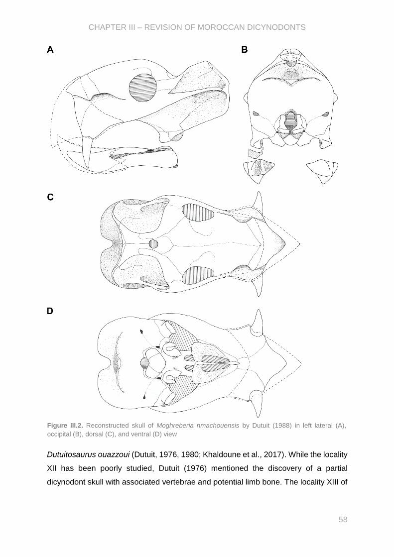
CHAPTER III – REVISION OF MOROCCAN DICYNODONTS
58
Dutuitosaurus ouazzoui (Dutuit, 1976, 1980; Khaldoune et al., 2017). While the locality
XII has been poorly studied, Dutuit (1976) mentioned the discovery of a partial
dicynodont skull with associated vertebrae and potential limb bone. The locality XIII of
Figure III.2. Reconstructed skull of Moghreberia nmachouensis by Dutuit (1988) in left lateral (A),
occipital (B), dorsal (C), and ventral (D) view

CHAPTER III – REVISION OF MOROCCAN DICYNODONTS
59
the Argana Basin is noticeable by the abundant and well-preserved rests of
Dutuitosaurus ouazzoui.
Moghreberia nmachouensis, recovered in the locality XIb, was first named by
Dutuit (1980) on the basis of the two skulls MNHN.F.ALM 280 and 281 that he briefly
described. Dutuit (1980) highlighted similarities between the Moroccan genus and the
kannemeyerids (sensu Huene, 1948) Placerias, Rabidosaurus, and
Parakannemeyeria. However, he did not establish the diagnosis of Moghreberia that
distinguishes the Moroccan genus from others. A more detailed description was later
provided by Dutuit (1988) (Fig. III.2), based on the two previous skulls and additional
partial cranial remains discovered in the same locality. Dutuit (1988) attributed the
Moroccan genus to Kannemeyeridae (sensu Huene, 1948). He considered
Moghreberia more closely related to the North American genus Placerias than to other
dicynodonts (Dutuit, 1988). However, the taxonomic validity of Moghreberia was
questioned by some authors (e.g., Cox, 1991; Hunt and Lucas, 1991; Lucas and Wild,
1995; Lucas, 2018). Kammerer et al. (2011) first introduced the Moroccan genus in a
phylogenetic analysis and placed it as a sister-taxa of Placerias, as the following
phylogenetic studies (e.g., Kammerer et al., 2013; Angielczyk and Kammerer, 2017;
Kammerer et al., 2019).
Two other Moroccan species were also added by Dutuit (1989a, 1989b) within
the new genus Azarifeneria. The first species, A. barrati, was defined based on a partial
dicynodont skull (MNHN.F.AZA 366.1-2-3) excavated from the locality XII (Dutuit,
1989a). A partial mandible (MNHN.F.ALM 167) from the locality XIb allowed Dutuit
(1989b) to define the second species A. robustus. However, the taxonomic validity of
these two species have been questioned by succeeding studies (e.g., Cox, 1991;
Lucas and Wild, 1995; Fröbisch, 2009; Kammerer et al., 2013) due to the lack of
relevant diagnostic features that would distinguish Azarifeneria from Moghreberia (e.g.,
Lucas and Wild, 1995; Lucas, 2018) or other dicynodonts such as Ischigualastia (Cox,
1991). No more study has been done done since Dutuit’s work (1989a, 1989b) and
Azarifeneria is still considered nomen dubium.
Dutuit (1980, 1988, 1989a, 1989b) focused his studies on cranial remains, but
abundant postcranial dicynodont specimens have been discovered in the same
localities XI, XII, and XIII, according to the collection catalogue edited by J.M. Dutuit.
As highlighted by Kammerer et al. (2011), the potential importance of missing data and
the lacks of post-cranial data shadow the phylogenetic position of Moghreberia. The

CHAPTER III – REVISION OF MOROCCAN DICYNODONTS
60
study of the post-cranial material would increase our knowledge of Moghreberia
nmachouensis known only by its cranial remains. It would lead to better define its
phylogenetic relationships and characterise it by comparison to the other dicynodonts
and particularly to Placerias. Moreover, it would provide information on the presence
of one or more distinct morphotypes morphotypes and consequently whether one or
more distinct dicynodont taxa are present in the Argana Basin. Finally, the abundance
of the rests would allow us to discuss on the potential morphological variation in the
postcranial skeleton in dicynodonts.
The study of the postcranial dicynodont remains from the Argana Basin and its
implications have been developed in PAPER II. All the cranial and postcranial material
is currently preserved in the collections of the Muséum national d’Histoire naturelle in
Paris (France). Due to the abundance of the postcranial material, the description
focused on the most preserved bones, while the study covers a large part of the
available material comprising around a hundred specimens (7 connected or isolated
vertebrae, 10 ribs, 22 scapulae, 8 interclavicles, 1 sternum, 13 humeri, 5 radii, 9 ulnae,
1 complete pelvic girdle, 1 ilium, 3 ischia, 1 pubis, 9 femora, 3 tibiae, and 1 fibula).
Even focused on the post-cranium, the cranial material (including holotypes), has been
briefly re-evaluated in order to discuss the validity of the Moroccan genera Moghreberia
and Azarifeneria, and check the coding of Moghreberia in the previous phylogenetic
matrices. The morphological variation noticed in the cranial and postcranial material
has been discussed based on first hand examination of dicynodont specimens during
my visits of collections in the American Museum of Natural History in New York (New
York City, U.S.A.; financial support from CR2P and ISTEP), the Evolutionary Studies
Institute in Johannesburg (South Africa; financial support from CR2P), the Institute of
Vertebrate Paleontology and Paleoanthropology in Beijing (China; financial supports
from CR2P and the Société des Amis du Muséum), the Museum of Comparative
Zoology in Cambridge (Massachusetts, U.S.A.; financial support from CR2P and
ISTEP), and the Natural History Museum in London (United Kingdom; financial support
from SYNTHESYS). I also relied on the literature and pictures provided by the
colleagues J. Liu (Institute of Vertebrate Paleontology and Paleoanthropology, Beijing,
China) and C.F. Kammerer (North Carolina Museum of Natural Sciences, Raleigh,
North Carolina, U.S.A.), In PAPER II, I wrote the wole of the article, produced all figures
except the pictures took by Lilian Cazes and Philippe Loubry (CR2P, MNHN, France),
and performed the phylogenetic analyses.

PAPER II - “First description of dicynodont postcranial material from the Argana
Basin (Late Triassic, Morocco) and taxonomic revision of the two Moroccan
genera Moghreberia and Azarifeneria”, unsubmitted
First description of dicynodont postcranial material from the
Argana Basin (Late Triassic, Morocco) and taxonomic revision of
the two Moroccan genera Moghreberia and Azarifeneria
Chloé Olivier 1* and Nour-Eddine Jalil 1,2
1Centre de Recherche en Paléontologie (CR2P, UMR 7207), Sorbonne Université, MNHN,
CNRS, Muséum national d’Histoire naturelle, Département Origines et Évolution, case
postale 38, 57 rue Cuvier, F-75231 Paris cedex 05 (France)
[email protected]; [email protected]
2Department of Geology, Faculté des Sciences Semlalia, Université Cadi Ayyad, Boulevard
Prince My Abdellah, 40000 Marrakech, Maroc
*Corresponding author
Considering submission to Geodiversitas
European Journal of Taxonomy
PA
PE
R I
I

CHAPTER III – REVISION OF MOROCCAN DICYNODONTS
62
First description of dicynodontian postcranial material from the Argana Basin (Late
Triassic, Morocco) and taxonomic revision of the two Moroccan genera Moghreberia
and Azarifeneria
Chloé Olivier 1* and Nour-Eddine Jalil 1,2
1Centre de Recherche en Paléontologie (CR2P, UMR 7207), Sorbonne Université, MNHN,
CNRS, Muséum national d’Histoire naturelle, Département Origines et Évolution, case
postale 38, 57 rue Cuvier, F-75231 Paris cedex 05 (France)
[email protected]; [email protected]
2Department of Geology, Faculté des Sciences Semlalia, Université Cadi Ayyad, Boulevard
Prince My Abdellah, 40000 Marrakech, Maroc
*Corresponding author

CHAPTER III – REVISION OF MOROCCAN DICYNODONTS
63
Abstract
Three dicynodont species have been described in the Triassic of the Argana
Basin (Carnian; base of the Irohalen Member, Morocco): Moghreberia nmachouensis,
Azarifeneria barrati, and A. robustus. While an abundant postcranial material have
been also collected in the same locality, only the cranial remains were studied and
used to diagnose the three species. The Moroccan species are then poorly known and
have always been questioned. The revised study of the cranial specimens attributed
to Azarifeneria did not emphasized significant differences distinguishing Azarifeneria
barrati and A. robustus from other Triassic dicynodonts, especially Moghreberia
nmachouensis. The present study provided the first description of the rich dicynodont
postcranial material from the Argana Basin. An almost complete skeleton attribuable
to M. nmachouensis enhanced our understanding of this species and completed the
gap in the phylogenetic matrix. Unlike previous studies that allied Moghreberia and the
North American Placerias, our phylogenetic analysis concluded that the Moroccan
species is more closely related to the Polish Lisowicia. Moreover, significant
morphological variations have been noticed in the postcranial material and discussed.
Despite a lack of diagnostic characters, at least a second more massive morphotype
can be likely interpreted in the postcranial material.

CHAPTER III – REVISION OF MOROCCAN DICYNODONTS
64
Introduction
Dicynodontia represent an emblematic taxa of the Permian-Triassic (P-Tr)
period to have survived the most important biological crisis. Considering the recent
phylogenetic analyses and their stratigraphic range implying ghost lineages, multiple
dicynodont clades would have crossed the P-Tr boundary: Lystrosauridae,
Emydopoidae, Dicynodontidae, and Kannemeyeriiformes (e.g., Angielczyk 2001;
Fröbisch 2007; Fröbisch et al. 2010; Kammerer et al. 2011; Olivier et al. 2019). They
highly diversified during the Middle Triassic whit the occurences of around twenty new
genera (Kammerer et al., 2013), to finally become extinct at the end of Triassic. During
this period, they were worldwide with a cosmopolitain distribution: Dinodontosaurus
(Chañares Formation [Fm] in Argentina, and Santa Maria Fm in Brazil), Stahleckeria
(Santa Maria Fm in Brazil), Eubrachiosaurus (Popo Agie Fm in USA), Ischigualastia
(Ischigualasta Fm in Argentina), Jachaleria (Chañares Fm and Los Colorados Fm in
Argentina, and Caturrita Fm in Brazil), Moghreberia (Argana Basin in Morocco),
Placerias (Blue Mesa Member of Petrified Forrest Fm, Bluewater Creek Fm, and Pekin
Fm in USA), Vinceria (Puesto Viejo Fm and Cerro de Las Cabras Fm in Argentina),
and the two youngest forms Lisowicia (Lipie Śląskie clay-pit at Lisowice village in
Poland) and Pentasaurus (Elliot Fm in South Africa) (e.g., Fröbisch, 2009; Kammerer,
2018; Sulej and Niedźwiedzki, 2019).
The Moroccan dicynodont Moghreberia nmachouensis has been originally and
briefly described by Dutuit (1980) based on two poorly-preserved skulls, collected in
the locality XIb (Dutuit’s nomenclature) of the Argana Basin and dated to the Late
Triassic. Later, a more detailed description of Moghreberia was published (Dutuit,
1988), based on the two above-cited skulls and isolated but well-preserved cranial
remains. Abundant postcranial remains have also been excavated but have never
been described. Dutuit (1988) highlighted anatomical similarities between the cranial
remains of Moghreberia and the skulls of Kannemeyeria and Stahleckeria, and
especially with the North American Placerias. He then attributed the Moroccan
dicynodont to Kannemeyeridae (Huene, 1948). Moghreberia has been distinguished
from other dicynodonts by (1) a relative low, dorsoventrally and laterally, expanded
occipital surface, (2) a lateral mandibular branches mainly horizontal, (3) a dorsal
margin of the erupted portion of the canine tusk anterior to the nasal cavity and, (4) a
very pointed triangular anterior tip of snout (Dutuit, 1988). The same year, King (1988)

CHAPTER III – REVISION OF MOROCCAN DICYNODONTS
65
attributed the Moroccan form to Kannemeyeriini (Lehman, 1961) that included the
genera Kannemeyeria, Shaanbeikannemeyeria, Dolichuranus, Wadiasaurus, and
Rabidosaurus. Dutuit (1988) has already alluded to a synonymy between Moghreberia
and the North American Placerias, while he accepted the validity of the Moroccan
genus. Later, several authors considered Moghreberia as a junior synonym of
Placerias (e.g., Cox, 1991; Hunt and Lucas, 1991; Lucas and Wild, 1995; Lucas, 2018),
delaying the inclusion of the Moroccan form up to the phylogenetic study of Kammerer
et al. (2011). Kammerer et al. (2011), followed by later analyses (e.g., Kammerer et
al., 2013, 2019; Angielczyk and Kammerer, 2017), placed Moghreberia as sister-group
of Placerias. They definitely distinguished these two genera by several cranial
morphological characters. Kammerer et al. (2011) also highlighted the potential impact
of missing data on the phylogenetic position of the Moroccan form and the need to
study its postcranial remains.
Two other endemic species were discovered in the Argana Basin: Azarifeneria
barrati and A. robustus (Dutuit, 1989a, 1989b). A. barrati has been described on the
basis of two cranial specimens from the same individual (the parietosquamosal
structure MNHN.F.AZA 366.1 and the posterior part of the basicranium MNHN.F.AZA
366.2), found in the locality XII (Dutuit’s nomenclature). Only a fragmentary mandible
(MNHN.F.ALM 167) from the locality XIb (Dutuit’s nomenclature) is known for A.
robustus. However, considerable uncertainties remain about the validity of the second
Moroccan genus, particularly because of an obvious lack of diagnostic character in the
available material (e.g., Cox, 1991; Lucas and Wild, 1995; Fröbisch, 2009; Kammerer
et al., 2013). Indeed, the cranial remains attributed to Azarifeneria, (Dutuit, 1989a,
1989b) mainly highlighted a big size, high robustness and similarities with the Brazilian
Stahleckeria. Azarifeneria was sometimes considered synonym of Moghreberia (e.g.,
Lucas and Wild, 1995; Lucas, 2018), Ischigualastia (Cox, 1991), or a nomen dubium
(e.g., Fröbisch, 2009).
The Moroccan dicynodonts represent the only North African therapsids. Our
main purpose was to describe the rich dicynodont postcranial remains collected in the
Argana Basin. On one hand, we completed our understanding about the anatomy of
Moghreberia nmahcouensis and checked its phylogenetic relationships and taxonomic
validity. On the other hand, due to the obvious lack of diagnostic character in

CHAPTER III – REVISION OF MOROCCAN DICYNODONTS
66
Azarifeneria, we revised the description of its cranial remains and discussed about its
validity.
Institutional Abbreviations—AMNH, American Museum of Natural History, New
York City, New York, U.S.A.; BP, Evolutionary Studies Institute (formerly the Bernard
Price Institute for Palaeontological Research and the Institute for Human Evolution),
Johannesburg, South Africa; BSPG, Bayerische Staatssammlung für Paläontologie
und Geologie, Munich, Germany; GSN, Geological Survey of Namibia, Namibia; GPIT,
Institut und Museum für Geologie und Paläontologie, Tübingen, Germany; IVPP,
Institute of Vertebrate Paleontology and Paleoanthropology, Beijing, China; MCZ,
Museum of Comparative Zoology, Harvard University, Cambridge, Massachusetts,
U.S.A.; MNHN, Muséum national d’Histoire naturelle, Paris, France; NHMUK, Natural
History Museum, London, United Kingdom; NHMW, Naturhistorisches Museum Wien,
Vienna, Austria ; UCMP, University of California Museum of Paleontology, Berkeley,
California, U.S.A.; USNM, National Museum of Natural History, Washington, District of
Columbia, U.S.A.; ZPAL, Institute of Paleobiology, Polish Academy of Sciences,
Warsaw, Poland.
Anatomical Abbreviations—A, angular; Acetf, actebular facet; Acr, acromion
process; Aip, anterior process of the ilium; Bo, basioccipital; Cnc, cnemial crest;
Corfor, coracoid foramen; Csp, caudal neural spine; Ct, centrum; D, dentary; Delt,
insertion of the deltoideus muscle; Dpc, deltopectoral crest; DRadf, distal radial facet;
Dt, dentary table; Ect, ectepicondyle; Ent, entepicondyle; Entefor, entepicondylar
foramen; Eo, exoccipital; Femf, femoral facet; Fh, hemoral head; Fibf, fibular facet;
Fr, frontal; Ft, insertion site of the flexores tibiales; Glef, glenoid facet; Hd, humeral
head; Hrb, head of rib; Il, ilium; IntF, intercondylar fossa; Ip, interparietal; Isch,
ischium; Itroch, insertion site of ischio-trochantericus muscle; LatdorDFK, insertion
for the latissimus dorsi muscle according to King (1981) and Defauw (1986); LatdorR,
insertion for the latissimus dorsi muscle according to Romer (1976); Ldr, lateral
dentary ridge; Lra, reflected angular lamina; Mdr, median dentary ridge; Mf, Meckel
fossa; Mfor, magnum foramen; Nc, neural canal; Obtfor, obturator foramen; OleF,
olecranon fossa; Olep, olecranon process; Opt, opisthotic; Pa, parietal; Par,
parapophyse; Pbs, parabasisphenoid; Pds, posterior dentary sulcus; Pfor, pineal
foramen; Pip, posterior process of the ilium; Po, postorbital; Pof, postorbital facet;
PopF, popliteal fossa; Poz, postzygapophyse; Pp, preparietal; PPt, posterior pterygoid

CHAPTER III – REVISION OF MOROCCAN DICYNODONTS
67
branch; PRadf, proximal radial facet; Prsf, prespinous fossa; Prz, prezygapophyse;
Pubt, pubic tubercle; Q, quadrate, Radc, radial condyle; Rpr, posterior ridge on the
rib; S, splenial; Sct, sacral centrum; Serasup, insertion of the serratus anterior
superficialis muscle; Sf, sacral facet; Sigf, sigmoidal facet; So, supraoccipital; Sp,
neural spine; Sq, squamosal; Srb, fused sacral rib; SSp, sacral neural spine; Subco,
insertion for the subcoracoscapularis muscle; Sup, supinator process; Supco,
insertion of the supracoracoideus muscle; T, tusk; Tcr, trochanteric crest; Tidf, tibial
facet; Tm, minor trochanter; Tri, insertion of the triceps muscle; Trif, triangular
flattened facet; Troch, ulnar trochlea; Trp, transverse process; Tth, third trochanter;
Tub, parabasisphenoid-basioccipital tuber; Vo, vomer.
Material and methods
Geological setting and palaeoenvironment
The Argana Basin is located in the western part of the High Atlas of Morocco
between Im’N’Tanout village in the North and Ameskroud in the South (Fig. 1). The
Basin comprises three formations subdivided in eight lithostratigraphic units (T1 to T8):
(1) the lower Ikakern Fm (Members T1 and T2), (2) the middle Timezgadiouine Fm
(Members T3 to T5), and (3) the upper Bigoudine Fm (Members T6 to T8) (e.g.,
Tixeront, 1973; Jalil, 1999). The cranial and postcranial dicynodont remains have been
excavated from the localities XI, XII, and XIII (Dutuit’s nomenclature), located at the
base of the Irohalene Member T5 in the Timezgadiouine Fm (Argana Basin, Morocco)
(Fig. 1; Tixeront, 1973; Dutuit, 1988, 1989a, 1989b).
In addition to the dicynodonts, the lower part of the Irohalene Member has
yielded the actinopterygians (Dipteronotus gibbosus, Mauritanichthys rugosus, cf.
Ischnolepis, cf. Procheirichthys, cf. Atopocephala), the dipnoi Arganodus, a probable
latimerioid, the amphibian temnospondyls Almasaurus and Dutuitosaurus, the
phytosaur Arganarhinus, the aetosaur Longosuchus, the archosauromorph
Azendohsaurus, and the silesaurid dinosauromorph Diodorus (e.g., Gauffre, 1993;
Lucas, 1998; Jalil, 1999; Kammerer et al., 2012; Khaldoune et al., 2017). The top of
the member present a more aquatic faunal assemblage with the amphibians
Metoposaurus and Arganasaurus, and the phytosaur Angistorhinus (e.g., Dutuit, 1976;
Jalil, 1999; Sulej, 2002; Khaldoune et al., 2017). Lucas (2018) correlated the faunal
assemblage of the Irohalene Member (T5) (phytosaurs ‘Parasuchus’ (=Arganarhinus;
Kammerer et al., 2016b) and Angistorhinus, the aetosaur Longosuchus, and the

CHAPTER III – REVISION OF MOROCCAN DICYNODONTS
68
dicynodont ‘Placerias’ (considering synonym of Moghreberia in his study)) to the
Otischalkian land-vertebrate faunachron, supposed to be Carnian in age (e.g., Lucas,
1998, 2018; Jalil, 1999). A similar faunal assemblage has also been described in North
America (Popo Agie Fm, Colorado City Fm, Salitral Fm, and Newark Supergroup),
Germany (Stuttgart Fm), and India (Maleri Fm) (e.g., Lucas, 1998, 2018).
Nevertheless, Kammerer et al. (2012) recommended to consider these biostratigraphic
interpretations with caution. Indeed, on one hand, the index taxa supporting the
Otischalkian land-vertebrate faunachron (Paleorhinus and Metoposaurus), as
traditionally defined, have been shown to represent non-monophyletic groups; on the
other hand, Azendohsaurus recovered in the Irohalene Member is also known in the
late Middle or early Late Triassic Makay Fm (Isalo Group, Madagascar) (e.g., Flynn et
al., 1999, 2000; Kammerer et al., 2010). Moreover, tetrapod footprints discovered in
the Irohalene Member have been attributed by Lagnaoui et al. (2012, 2016) to the
archosaur ichnogenera Apatopus (phytosaur), Atreipus-Grallator (dinosauromorph),
Brachychirotherium (crocodylian-stem archosaur), Eubrontes (theropod),
Parachirotherium (? dinosauriform), and Synaptichnium, and the non-archosaur
ichnogenus Rhynchosauroides (lepidosauromorph or archosauromorph). The
presence of Apatopus, Eubrontes, Atreipus-Grallator, and Brachychirotherium is
indicative of Late Triassic Brachychirotherium biochron (Klein and Lucas, 2010).
However, Parachirotherium has only been recorded in the Middle Triassic Benk Fm
(Germany). Its presence in the Irohalene Member would then suppose either the
youngest occurrence of this ichnotaxa (Lagnaoui et al., 2012), or support an earlier
age of the member as in the case of Azendohsaurus. A stratigraphic extention of the
Irohalene Member T5 in the late Middle Triassic does not contradict the previous
studies conluding to a Middle Triassic age of the underlying Aglegal Member T4 (e.g.,
Medina et al., 2001; Jalil et al., 2009). However, the occurrence of a Late Triassic
rhynchosaur in the basalmost Irohalene Member would indicate a later age for the
member (N.-E. Jalil, pers. comm., 2019). The magnetostratigraphy and radioisotopic
analyses of Irmis et al. (2010) suggest a longer Norian that would fully or partly include
the Otischalkian land-vertebrate faunachron. However, their conclusions are not
supported by the palynomorphic analyses of Tourani et al. (2000) that interpreted a
Late Carnian age for the Tadart Ouadou T6 and Sidi Mansour T7 Members, and an
early Norian age for the Hasseine Member T8. Also, the studies of Argana Basalt, a
CHAPTER III – REVISION OF MOROCCAN DICYNODONTS
69
volcanic sequence associated with the Central Atlantic Magmatic Province (CAMP)
underlying the Bigoudine Fm, suggested a Norian even early Jurassic age for the
upperpart of the formation (e.g., Olsen et al., 2000; Marzoli et al., 2004; Whiteside et
al., 2007; Deenen et al., 2011). A stratigraphic extention of the Irohalene Member T5
to the Norian appears less likely in regards to the palynology and geochronology.
Under the light of the previous results, a Late Triassic age for the Irohalene Member
appears most likely.
Most of the dicynodont remains have been discovered in the mid-level of the
locality XI (locality XIb according to Dutuit, 1976). The examination of the external
surface of the dicynodont bones (eroded, cracked, and peeling) related to the
taphonomic works of Behrensmeyer (1975), suggested to Dutuit and Heyler (1983) a
warm and relatively dry climate as a depositional environment of the Irohalene
Member. These latter is interpreted as an alluvial plain sandstones and mudstones
with a well-developed cyclical lacustrine sequence in the base of the member (e.g.,
Olsen et al., 2000). Furthermore, Olsen et al. (2000) assumed a relatively humid
environment in the Irohalene Member with shallow lacustrine water at the base and
even fluvial at the top. The faunal assemblage composed of aquatic, semi-aquatic, and
terrestrial forms would support a well-irrigated soil with lakes of fresh even brackish
water (e.g., Dutuit and Heyler, 1983).
New Moroccan dicynodont postcranial material
As mentioned above, an abundant dicynodont postcranial material has been
discovered in the localities XIa, XIb, and XIII of the Argana Basin. Almost every skeletal
element is represented within this material, only the atlas-axis complex, cleithrum,
clavicle, and autapodia have not been collected. The present study focused on the
well-preserved specimens (see Table 1S for more details about their preservation and
locality).
Vertebrae—Most of the specimens are discovered in the locality XIb. The locality of
MNHN.F.ALM 198, 274 and two unnumbered connected vertebrae (named ‘Vunknb 1
and 2’ from front to back) are unknown. Three specimens (MNHN.F.ALM 198, 288,
and Vunknb 1 and 2) are proably the last cervical or first dorsal vertebrae.
MNHN.F.ALM 120, 159, and 265 are probably the middorsal vertebrae. MNHN.F.ALM
120 consists of four vertebrae preserved in the same block and labelled ‘MNHN.F.ALM
120-1, -2, -3, and -4’ from front to back. MNHN.F.ALM 159 are two articulated
CHAPTER III – REVISION OF MOROCCAN DICYNODONTS
70
vertebrae named ‘MNHN.F.ALM 159-1 and -2’ (from front to back). MNHN.F.ALM 274
corresponds to the two pelves of a same individual, connected to sacral and first caudal
vertebrae, with two associated but disconnected posterior dorsal vertebrae (named
‘MNHN.F.ALM 274-1 and -2’). MNHN.F.ALM 136 composed of five vertebrae (labelled
‘MNHN.F.ALM 136-1, -2, -3, -4, and -5 from front to back) presents peculiar features
of sacral vertebrae.
Ribs—Seven ribs MNHN.F.ALM 86, 90, 91, 95, 96, 97, and 296, and three proximal
costal heads (MNHN.F.ALM 163, 272, and an unnumbered specimen) were stored in
collection. All have been excavated from the locality XIb, but the unknown locality of
the unnumbered costal head.
Scapulae, coracoids, and precoracoids—Twenty-one isolated dicynodont scapulae
(MNHN.F.ALM 17, 19, 29, 30, 83, 98, 108, 113, 114, 132, 153, 155, 259, 263, 283,
292, 299, and three unnumbered scapulae) have been discovered in the Argana Basin.
Most of scapulae have been excavated from the locality XIb, except MNHN.F.ALM 17,
113, 114, and 299 from the locality XIa. Two unnumbered scapulae (labelled ‘Sunknb
1 and 2’) are also found in the locality XI, but the level is unknown. The locality of the
third unnumbered scapula is unknown (labelled ‘Sunknb 3’). MNHN.F.AZA 365 is the
only dicynodont specimen from the locality XIII. Two scapulocoracoid complexes
(MNHN.F.ALM 92 and 298) were also found in collection and come from the locality
XIb.
Interclavicles—Eight interclavicles are known (MNHN.F.ALM 93, 94, 158, 260, 278,
295, and two unnumbered specimens), mainly in the locality XIb. The level of the
locality is unknown for MNHN.F.ALM 295 and the two unnumbered interclavicles
(labelled ‘Intunknb 1 and 2’).
Sternum—MNHN.F.ALM 134 is the only sternum of the collection. It comes from the
locality XIb.
Humeri—Eight complete (MNHN.F.ALM 24, 25, 101, 102, 105, 115, 275, and 297) and
five fragmentary (MNHN.F.ALM 121, 133, 147, and two unnumbered specimens
labelled ‘Humunknb 1 and 2’) dicynodont humeri are found in collection. Most of them
are found in the locality XIb of the basin. MNHN.F.ALM 115 and 275 have been located
in the lower level of XI (locality XIa). The level in the locality XI is unknown for
CHAPTER III – REVISION OF MOROCCAN DICYNODONTS
71
Humunknb 1. Humunknb 2 is supposed to be collected in the localities X, XI or XVI,
according to the collection catalogue edited by J.M. Dutuit.
Radii—Three nearly complete radii (MNHN.F.ALM 84, 131, 287, and 289) are found in
collection. They have been discovered in the locality XIb. An unnumbered specimen
contains the distal regions of a radius and ulna. Its locality is unknown.
Ulnae—Eight ulnae entirely or partly preserved (MNHN.F.ALM 18, 27, 28, 35, 87, 126,
154, and 261). All the specimens have been excavated from the locality XIb, except
MNHN.F.ALM 154 which is from the locality XIa. No supplementary information is
known about the stratigraphical level of MNHN.F.ALM 35 in the locality XI.
Pelves—One complete pelvic girdle MNHN.F.ALM 274 is preserved. It is associated
with a posterior dorsal, sacral, and caudal vertebrae (mentioned above). As noticed
above, the locality of MNHN.F.ALM 274 is unknown. In addition, an ilium
(MNHN.F.ALM 104), three ischia (MNHN.F.ALM 123, 125, and 138), and a pubis
(MNHN.F.ALM 122) are found isolated in the locality XIb.
Femora—Nine partly (MNHN.F.ALM 34 and 290) or entirely (MNHN.F.ALM 21, 22, 23,
26, 85, 100, and 284) preserved dicynodont femora are preserved. All come from the
locality XIb, except MNHN.F.ALM 284, which is from the locality XIa.
Tibiae—Three tibiae are found in collection. They have been discovered in the locality
XI, at the lowest stratigraphical level XIa (MNHN.F.ALM 291) and the middle one XIb
(MNHN.F.ALM 33 and unnumbered specimen).
Fibula—Only one dicynodont fibula is found in collection (detailed location is unknown).
Phylogenetic method
As a result of the description of the new postcranial material attributed to the
Moroccan dicynodont Moghreberia nmachouensis, a phylogenetic analysis was
conducted to test its phylogenetic position. This analysis was based on the matrix from
Kammerer (2018), the most recent and comprehensive matrix published for
dicynodonts. The final phylogenetic dataset thus included 111 operational taxonomical
units and 197 characters: 23 continuous characters and 174 discrete characters (of
equal weight and unordered, except for characters 81, 84, 102, 163, 173, and 174,
following Angielczyk and Kammerer, 2017). The Permian dicynodont Dicynodon
huenei is deleted and the two taxa Dicynodon angielczyki and Daptocephalus huenei

CHAPTER III – REVISION OF MOROCCAN DICYNODONTS
72
were added and coded, according to the recent taxonomic revision and phylogenetic
analysis of Kammerer (2019a). The coding of Compsodon and Sangusaurus was
modified according to the modifications suggested by Kammerer et al. (2019). The new
Polish dicynodont Lisowicia bojani (Sulej and Niedwźiedzki, 2019) has been added in
the phylogenetic analysis. The coding of this taxa followed the study of Sulej and
Niedwźiedzki (2019), but the characters 195 to 197 in the present study were newly
coded on the basis of the supplementary descriptions and figures from Sulej and
Niedwźiedzki (2019).
The coding of the cranial and mandibular characters of Moghreberia
nmachouensis has been revised using the cranial specimens MNHN.F.ALM 10, 12,
37, 168, 280, and 281, and the mandible MNHN.F.ALM 38 (see Supplementary data).
The attribution of the mandible MNHN.F.ALM 80 to Moghreberia is doubtful especially
on the basis of the particular form of the tip of the dentary symphysis (pointed tip of
snout; not figured here), while it appears more rounded or squared off in MNHNF.ALM
38. A more detailed comparison of this mandible and discussion about its phylogenetic
affinities were not the aim of the present analysis and will be provided in a future study.
Consequently, the specimen MNHN.F.ALM 80 has not been included in the analysis.
The description of the new postcranial remains filled gap in our knowledge of the
anatomy of Moghreberia and allowed a more comprehensive coding of its characters-
states than in Kammerer (2018). The attribution of the massive specimens
(MNHN.F.ALM 27, 96, 154, 275, 291, 299, and 365) to Moghreberia is still doubtful
and has been discussed below. As well for the femur MNHN.F.ALM 22, its peculiar
morphology (discussed below) significantly differs from all other femora attributed to
Moghreberia. These specimens have thus not been considered in the phylogenetic
analysis. As mentioned below, the revised study of the specimens attributed to
Azarifeneria barrati (MNHN.F.AZA 366.1 and 366.2) and A. robustus did not
emphasize significant differences between Azarifeneria and Moghreberia, even with
other kannemeyriiforms (discussed below). The two species previously attributed to
Azarifeneria by Dutuit (1989a, 1989b) have thus not been included in the analysis.
Following the phylogenetic method of Olivier et al. (2019) using TNT 1.1
(December 2013 version) (Goloboff et al., 2008), we performed a New Technology
search and a driven search with the initial search level set to check every three hits.
One hundred replications were chosen as the starting point for each hit, and the search

CHAPTER III – REVISION OF MOROCCAN DICYNODONTS
73
was set to find the most parsimonious trees 20 times. We did the phylogenetic analysis
using a sectorial search (default settings) and tree-drifting (default settings but the
number of cycles was three) to produce a nearly optimal tree, which was used for tree-
fusing (default settings, but a global fuse every three hits was input) (Goloboff, 1999).
In parallel, we did a traditional search of tree bisection reconnection (TBR) branch
swapping with 11,111 replications and nine trees saved per replication. The continuous
character were treated as ordered following the method from Goloboff et al. (2006).
Unknown and/or inapplicable states of discrete and continuous characters are coded
as ‘?’ (Strong and Lipscomb, 1999). Biarmosuchus was used as outgroup. We obtained
the same most parsimonious tree (1202.936 steps, consistency index= 0.229, retention
index= 0.712) with the two phylogenetic analyses. The Bremer indexes (BS) were
calculated to measure the node support according to the method detailed by Olivier et
al. (2019) following the recommendations of Goloboff et al. (2008).
Systematic palaeontology
THERAPSIDA Broom, 1905
ANOMODONTIA Owen, 1860
DIYCNODONTIA Owen, 1860
KANNEMEYERIIFORMES Maisch, 2001
MOGHREBERIA Dutuit, 1980
Type Species—Moghreberia nmachouensis (Dutuit, 1988)
Diagnosis (modified from (Dutuit, 1988)—Large-sized kannemeyeriiform with skull
over 40 cm but narrow with small lateral expansion of the squamosals. It can be
distinguished from all other adequately known dicynodonts by the following
combination of character-states: no posterior ridge of the palatal region of the
premaxilla, absenceof teeth except well-developed paired of tusks, preparietal deeply
depressed, small contribution of the posterior processes of the postorbitals to the
intertemporal bar, parietals flat and well exposed on the intertemporal bar with slender
and elongate posterior parietal processes, intertemporal bar highly angled in lateral
view with a strong break in slope between the pre- and post-pineal region, reduced
fossa on the ventral surface of the intertemporal bar, small contribution of the
interparietal bone to the intertemporal skull roof, no distinct notch below the zygomatic
process of the squamosal in posterior view, relatively large expansion of the dorsal

CHAPTER III – REVISION OF MOROCCAN DICYNODONTS
74
region of the scapula, acromion process low developed to absent, well-developed
deltopectoral crest, obtuse angle between the anterior and distal edges of the
deltopectoral crest, insertion of the m. latissimus dorsi onto a pinna-like process, and
supinator process present on the ectepicondyle.
MOGHREBERIA NMACHOUENSIS Dutuit, 1980
Holotype—Dutuit (1980) did not mention a holotype in the original publication of
Moghreberia nmachouensis. In disagreement with the International Code of Zoological
Nomenclature (ICZN) rules, Dutuit (1988) later designated MNHN.F.ALM 280 as the
holotype and MNHN.F.ALM 281 as the syntype for M. nmachouensis. In accordance
with ICZN rules, we here proposed the lectotype MNHN.F.ALM 280 (Fig. 2) and the
paralectotype MNHN.F.ALM 281 for M. nmachouensis.
Geographic Distribution and Stratigraphic range of the types—The locality XIb
(according Dutuit’s nomenclature) of the Argana Basin in Morocco. The locality XI
(GPS coordinates: 161°8’E, 456°9’N) is located at the basis of the Irohalene Member
T5, according to the nomenclature of Tixeront (1973). While the age of the Irohalene
Member is still debated, a Carnian age appears most likely (discussed above).
Referred material—An abundant material is available in the collection of MNHN. It
comes from the same locality as the cranial elements attributed to Moghreberia and
was collected at the same time as the latter. It contains almost all elements of a
complete postcranial skeleton, including:
eight connected or isolated vertebrae: MNHN.F.ALM 120, 136, 159, 198, 265, 288,
and two unnumbered specimens in connection named here ‘Vunknb 1 and 2’;
nine ribs: MNHN.F.ALM 86, 90, 91, 95, 97, 296, and the three costal heads
MNHN.F.ALM 163, 272, and an unnumbered specimen;
nineteen isolated scapulae: MNHN.F.ALM 17, 19, 29, 30, 83, 98, 108, 113, 114,
132, 153, 155, 259, 263, 283, 292, and three unnumbered specimens;
two scapulocoracoid complexes: MNHN.F.ALM 92 and 298;
seven intercalvicles: MNHN.F.ALM 93, 94, 158, 278, 295, and two unnumbered
specimens;
one sternum: MNHN.F.ALM 134;
twelve humeri: MNHN.F.ALM 24, 25, 101, 102, 105, 115, 121, 133, 147, 297, and
two unnumbered specimens;

CHAPTER III – REVISION OF MOROCCAN DICYNODONTS
75
four radii: MNHN.F.ALM 84, 131, 287, and 289;
six ulnae: MNHN.F.ALM 18, 28, 35, 87, 126, and 261;
one isolated ilium: MNHN.F.ALM 104;
three isolated ischia: MNHN.F.ALM 123, 125, and 138;
one isolated pubis: MNHN.F.ALM 122;
one complete pelvis: MNHN.F.ALM 274 connected with sacral and caudal
vertebrae;
seven femora: MNHN.F.ALM 21, 23, 26, 34, 85, 100, and 290;
two tibiae: MNHN.F.ALM 33 and an unnumbered specimen;
one fibula: MNHN.F.ALM 32.
Geographic Distribution and Stratigraphic range of the referred material—The
dicynodont postcranial remains were found in the localities XIa, XIb, and XIII (according
Dutuit’s nomenclature) of the Argana Basin in Morocco (see Table 1S for more details;
Dutuit 1976, 1980, 1988). The localities XI (GPS coordinates: 161°8’E, 456°9’N) and
XIII (GPS coordinates: 161°E, 458°N) are located on the basis of the Irohalene
Member T5, according the nomenclature of Tixeront (1973). While the age of the
Irohalene Member is still debated, a Carnian age appears most likely (discussed
above).
Diagnosis—See the generic diagnosis
THERAPSIDA Broom, 1905
ANOMODONTIA Owen, 1860
DIYCNODONTIA Owen, 1860
KANNEMEYERIIFORMES Maisch, 2001
AZARIFENERIA Dutuit, 1989b
Type Species—Azarifeneria barrati (Dutuit, 1989b)
Status—Nomen dubium (Dicynodontoidea indet.)
AZARIFENERIA BARRATI Dutuit, 1989b
Holotype—MNHN.F.AZA 366.1 and 366.3 in connection: the parieto-squamosal and
the right anterolateral region of skull, and MNHN.F.AZA 366.2: the occipital plate most
likely belonging to a same big skull, according to Dutuit (1989b) (Fig. 3A-C).

CHAPTER III – REVISION OF MOROCCAN DICYNODONTS
76
Geographic Distribution and Stratigraphic range—The locality XII (according Dutuit’s
nomenclature; GPS coordinates: 160°7’E, 458°6’N) of the Argana Basin in Morocco. It
is located at the basis of the Irohalene Member T5 (Dutuit, 1989b). While the age of
the Irohalene Member is still debated, a Carnian age appears most likely (discussed
above).
Status—Nomen dubium
Remarks—MNHN.F.AZA 366.1 and 366.2 present morphological similarities with the
Moghreberia nmachouensis cranial remains, like the postorbitals that do not extend on
the entire length of the intertemporal crest and the absence of an intertuberal crest.
Nevertheless, the holotype of Azarifeneria barrati from M. nmachouensis by: (1)
greater skull robustness, (2) longer intertemporal crest, (3) larger occipital condyle, and
(4) higher contribution of the postorbitals to the intertemporal bar. However, the
diagnostic features highlighted by Dutuit (1988) for Moghreberia could not be verified
in MNHN.F.AZA 366.1 and 366.2, and the noticeable differences are not sufficient to
firmly distinguish A. barrati from M. nmachouensis (discussed above).
AZARIFENERIA ROBUSTUS Dutuit, 1989a
Holotype—MNHN.F.ALM 167 (Fig. 4D-F), a fragmentary mandible with the anterior
extremity eroded and broken, almost the whole right hemi-mandible and only the two-
thirds of the left distorted hemi-mandible are preserved.
Geographic Distribution and Stratigraphic range—The locality XIb (according Dutuit’s
nomenclature) of the Argana Basin in Morocco. The locality XI (GPS coordinates:
161°8’E, 456°9’N) is located at the basis of the Irohalene Member T5 (Dutuit, 1989a).
While the age of the Irohalene Member is still debated, a Carnian age appears most
likely (discussed above).
Status—Nomen dubium
Remarks—The short anteroposterior expansion of the angular, abruptly curved
dorsally, distinguishes the mandible MNHN.F.ALM 167 from MNHN.F.ALM 38
attributed to Moghreberia. However, intraspecific variation of this feature has been
observed in Stahleckeria potens, thus supposing Azarifeneria robustus a synonym of
Moghreberia nmachouensis (discussed below). Nonetheless, MNHN.F.ALM 167
presents relevant similarities with the mandible of Stahleckeria potens as previously

CHAPTER III – REVISION OF MOROCCAN DICYNODONTS
77
assumed by Dutuit (1989a), but also with other kannemeyeriiforms as Angonisaurus
or Xiyukannemeyeria. As well as A. barrati, these only mandibular features are not
sufficient to firmly support the taxonomic validity of A. robustus (discussed below).
Restudy of the cranial material attributed to Azarifeneria in relation to
Moghreberia
Holotype of A. barrati: MNHN.F.AZA 366.1 and 366.2
According to Dutuit (1989b), there is no doubt that MNHN.F.AZA 366.1 and
366.2 belong to the same individual (Fig. 3A-C), while there is no bone contact between
the two specimens after preparation. They constitute a very massive skull in
comparison with Moghreberia (MNHN.F.ALM 280 (Fig. 2) and 281). A third specimen
MNHN.F.AZA 366.3 forms the right anterolateral region of the big skull, associated with
MNHN.F.ALM 366.1 (Fig. 3C). This specimen deserves more preparation and was not
described in the present study.
MNHN.F.AZA 366.1 constitutes the posterior region of the skull that it formed
with MNHN.F.AZA 366.3 (Fig. 3C). Only the dorsal region comprised of part of
squamosal wings and the highly eroded intertemporal crest is preserved (Fig. 3A-C).
The relief of the intertemporal crest cannot be interpreted due to the poor preservation,
while the bone contacts can be studied. As mentioned by Dutuit (1989b), the posterior
expansions of the postorbitals in MNHN.F.AZA 366.1 extend far beyond the pineal
foramen and constitute more than half of the intertemporal crest. The posterior
expansions of the postorbitals are more anteroposteriorly restricted in Moghreberia
(MNHN.F.ALM 280 and 281; Figs 2A, 3C; Dutuit, 1988). However, in MNHN.F.ALM 10
attributed to Moghreberia, the postorbitals also cover more than half of the
intertemporal crest as well as MNHN.F.AZA 366.1 (Figs 3C, E). As opposed to the
interpretations of Dutuit (1989b), the poor preservation of the posterior end of the
intertemporal crest in MNHN.F.AZA 366.1 does not provide firm conclusions about the
contribution of the interparietal to the crest as observed in Moghreberia (Dutuit, 1988).
MNHN.F.AZA 366.2 is a part of the lower half of the occiput and the posterior
region of the basicranium (Fig. 3A, C). Most of the supraoccipital and opisthotics, and
all the quadratojugals are missing, the interpretation of their structures thus remains
difficult. The large and rounded foramen magnum overhangs a highly distorted and
poorly preserved occipital condyle. The occipital condyle is therefore largely covered
by sediment. Despite the poor preservation of the condyle and a right exoccipital

CHAPTER III – REVISION OF MOROCCAN DICYNODONTS
78
condyle missing, the exoccipitals and basioccipital appear fused posteriorly to form a
slightly tripartite condyle as in Moghreberia (Fig. 3A, D; Dutuit, 1988). In MNHN.F.AZA
366.2, the exoccipitals form narrow dorsal expansions that run along the lateral margin
of the foramen magnum. They bear marked reliefs as in MNHN.F.ALM 268 and 280,
attributed to Moghreberia (Figs 2C, 3A, D). The right opisthotic condyle is well
preserved and presents a similar morphology to Moghreberia (Fig. 3A, D). On ventral
view, the basioccipital predominantly forms the parabasisphenoid-basioccipital tubera,
relatively narrow as in Moghreberia (MNHN.F.ALM 268 and 280; Figs 2B, C, F). The
parabasisphenoid, nearly vertical, only contributes to the anterior margin of the
stapedial facet of the tuber that is ventrolaterally exposed. The right opisthotic of
MNHN.F.AZA 366.2 (Fig. 2C) appears to border the stapedial fossa posterolaterally,
as in Moghreberia (MNHN.F.ALM 268 and 280; Fig. 3A, D), as opposed to the previous
interpretation of Dutuit (1988). No intertuberal ridge is observed in MNHN.F.AZA 366.2
(Fig. 3C). A deep and rounded depression on the parabasisphenoid of MNHN.F.AZA
366.2 (Fig. 3C) is observed in the intertuberal groove, close to the suture with the
basioccipital. A similar depression is noticed in MNHN.F.ALM 280 (Fig. 2B) as opposed
to MNHN.F.ALM 268 and 281 (Fig. 3F; C. Olivier pers. observ.). The two observed
depressions are partly filled by sediment. Nonetheless, these depressions cannot
correspond to the single carotid foramen described in Diictodon feliceps (Surkov and
Benton, 2004; previously D. testudirostris according the taxonomic revision of
Kammerer et al., 2011), this latter being located more anteriorly on the
parabasisphenoid and close to the suture with the pterygoid mid-plate. Most of the
pterygoids are missing with the proximal part of the posterior pterygoid rims and the
median plate highly eroded.
MNHN.F.ALM 167, holotype of A. robustus
MNHN.F.ALM 167 is a fragmentary mandible without its right rim and the third
posterior part of the left rim (Fig. 4D-F). As already mentioned, Dutuit (1989a) mainly
focused on the morphological similarities with the Brazilian Stahleckeria, without
clearly mentioned them and briefly described the anatomy of the mandible.
As noticed by Dutuit (1989a), the dentaries are fused and robust (Fig. 4D-F). In
dorsal view, the symphyseal region is deeply curved and bordered by two large
swellings as in Moghreberia (Fig. 4A, D). Nothing can be said about the presence of
dentary table (as defined by Angielczyk and Rubidge, 2013), because of the poor
CHAPTER III – REVISION OF MOROCCAN DICYNODONTS
79
preservation of these two reliefs. Despite sediment remaining, a distinct posterior
dentary sulcus (Fig. 4D), obviously narrow, is noticeable in dorsal view of the anterior
expansion of the left mandibular rim in MNHN.F.ALM 167. The anterior region of a
slight swelling is preserved and border the medial margin of the left dentary sulcus.
The sulcus runs on the dorsal surface of the dentary far beyond the anterior margin of
the Meckel fossa, as opposed to Moghreberia where the sulcus terminates at the level
of the anterior margin of the fossa (Fig. 4A, D). Thick but eroded lateral dentary ridges
are observed on both lateral sides of the dentary (Fig. 4F). Despite the lack of bone at
the anterior tip of the mandible, a median and thick ridge is noticeable on the dentary
in anterior view, indicating an anterior bone erosion relatively slight. As noticed by
Dutuit (1989a), the dentary appears more expanded lateromedially than
anteroposteriorly (Fig. 4D, F), in comparison with Moghreberia (Fig. 4A, C), giving it a
massive aspect. In addition, the tip of the lower jaw of MNHN.F.ALM 167 would most
likely be more rounded or squared-off (Fig. 4D). The lower jaw is highly constricted just
posterior to the lateral dentary ridges. In lateral view, the left posterodorsal expansion
of the dentary appears highly eroded. Most of the left surangular is missing. The
posterior expansion of the dentary delimits the anterior margin of the Meckel fossa.
The poor preservation of the lateral side of the left mandibular rim does not provide
information about the form of a lateral dentary shelf or a potential occlusion of the fossa
by a dentary sheet. However, if the Meckel fossa is laterally opened, its
anteroposteriorly expansion would appear significantly smaller than in Moghreberia
(Fig. 4C, F).
Posterior to the dentary symphysis, the fused splenial form a deep depression
on its posterior surface. An anterior triangular expansion of the splenial points on the
ventral surface of the dentary (Fig. 4E).
In dorsal view, the angle formed between the left mandibular rim and the long
axis of its symphyseal region appears more opened than in Moghreberia (Fig. 4A, D).
The posterior end of the angular and almost whole the reflected lamina are missing in
the left rim. Also, the surangular and the articular are not preserved. The angular mainly
forms the lateral border of the preserved region of the mandibular rim. Posteriorly to
the posterodorsal expansion of the dentary, the angular abruptly raises up to reach the
level of the dentary dorsally (Fig. 4F). In Moghreberia, the angular slightly points
dorsally and far beyond the posterior end of the dentary (Fig. 4C). As well as for the

CHAPTER III – REVISION OF MOROCCAN DICYNODONTS
80
ascending branch of the angular, the reflected angular lamina sharply curves ventrally
posterior to the short Meckel fossa (Fig. 4F). Potential ornementations could not be
studied on the external surface of the reflected lamina due to the poor preservation of
external bone surface and sediment remaining. Only a large median depression has
been interpreted between the ascending branch of the angular and the descending
one of the reflected lamina (Fig. 4F). In ventral view, the angular and the reflected
angular lamina split and diverge laterally and medially, respectively (Fig. 4E). The point
of this separation is more anterior in MNHN.F.ALM 167 than in Moghreberia (Fig. 4B).
Description of the new dicynodont postcranial material from Argana Basin
The orientation of the limb bone was based on an increasingly erected posture
of the dicynodonts as considered by the most recent studies (e.g., Ray, 2006; Sulej
and Niedźwiedzki, 2019). The interpretation of the muscular insertion sites followed
the studies of Camp and Welles (1956), Romer (1956a), Defauw (1986), and King
(1988).
Axial skeleton
VERTEBRAE
Because of gradual changing of characters along the vertebral column, the
attribution of the vertebrae to a specific region can be hard when they are isolated or
found in small connected groups. We thus described the groups of specimens that
present similar features.
Last cervical vertebrae and/or first dorsals MNHN.F.ALM 288, 198, Vunknb 1 and 2
(Fig. 5)—The two connected vertebrae Vunknb 1 and 2 (Fig. 5C-F) are identified to be
the last cervical vertebrae and/or first dorsals especially because of (1) parapophyses
clearly separated from the transverse processes in Vunknb 1, (2) neural spine of
Vunknb 2 nearly dorsally directed, (3) transverse processes large and triangular in both
vertebrae, and (4) obtuse angle between the prezygapophyses (e.g., Cox, 1959; King,
1981) (structures developed below). As previously mentioned, MNHN.F.ALM 288 is
highly anteroposteriorly compressed (Fig. 5A-B). The proportions of their different
structures may thus be altered. In addition, a sediment layer covers a part of the
external bone surface probably hiding specific characters. Like Vunknb 1 and 2,
MNHN.F.ALM 288 and 198 are supposed to be one of the last cervicals or first dorsals

CHAPTER III – REVISION OF MOROCCAN DICYNODONTS
81
because of the form of their transverse processes and the prezygapophyses largely
spaced.
The centra of the two connected vertebrae Vunknb 1 and 2, and MNHN.F.ALM
288 are concave in the anterior and posterior sides, indicating amphicoelous vertebrae
(Fig. 5A, B, C, E). While the sediment remains between the Vunknb 1 and 2, they have
been considered similar in their structures. The oval centra of Vunknb 1 and 2, and
MNHN.F.ALM 288 are higher than wide (Fig. 5A, B, C, E). Their lateral surfaces are
concave and limited anteriorly and posteriorly by a bone thickening, bordering the
anterior and posterior sides of the centra. The sediment and erosion prevent
observation of these structures in MNHN.F.ALM 288. The centra and neural arches
limit ovoid neural canals (almost or entirely filled by sediment). The ventral margin
formed by the centrum is well observed in MNHN.F.ALM 288. It is concave
anterodorsally and almost straight posterodorsally (Fig. 5A-B). The neural canal of
MNHN.F.ALM 288 appears larger than in Vunknb 1 and 2, as well as the size of
centrum that is higher in MNHN.F.ALM 288 (Fig. 5A, B, C, E). These differences are
most likely due to the post-mortem compression in MNHN.F.ALM 288, but they also
would define an individual with larger proportions.
Thick ovoid protuberances present in the lateral sides of the vertebrae Vunknb
1 and 2, in middle of the centra near to the anterior side, are interpreted as
parapophyses (Fig. 5D, F). The parapophyses of Vunknb 1 are rounded whereas that
of Vunknb 2 are ovoid and dorsoventrally elongated. The parapophyses of Vunknb 2
appear in continuity with transverse processes (=diapophyses) by a small ridge.
However, the thin sediment layer remaining preclude this interpretation with certainty.
However, the parapophyses of Vunknb 1 are clearly well separated from the transverse
processes, indicating double-heads associated ribs. The parapophyses distinct from
the transverse processes have also been observed in cervical vertebrae of most
dicynodonts like Dicynodontoides nowacki (previously “Kingoria” nowacki by Cox,
1959; then D. nowacki by Angielczyk et al., 2009), Dicynodon huenei (previously D.
“trigonocephalus” by King, 1981; then D. huenei by the taxonomic revision of
Kammerer et al., 2011), Placerias (Camp and Welles, 1956), Rhachiocephalus
(Maisch, 2004), and Wadiasaurus (Bandyopadhyay, 1988). However Cox (1959), King
(1981), and Bandyopadhyay (1988) also mentioned separated parapohyses and
transverse processes in the first two or three dorsals in Dicynodontoides nowacki,

CHAPTER III – REVISION OF MOROCCAN DICYNODONTS
82
Dicynodon huenei, and Wadiasaurus. The well-developed parapophyses in Vunknb 1
may indicate that the vertebra is one of the last cervicals or one of the first dorsals.
Sediment remaining in the lateral sides of the centrum of MNHN.F.ALM 288 makes
impossible the observation of parapophyses and their fusion with transverse
processes.
The suture between the centra and neural arches are not visible on Vunknb 1
and 2, and MNHN.F.ALM 288, suggesting adult individuals. In these three vertebrae
and MNHN.F.ALM 198, the triangular transverse processes are flattened
anteroposteriorly and extended dorsally or slightly dorsolaterally to form a dorsal
corner slightly posterior to the dorsal border of the prezygapophyses (Fig. 5A, B, C, E).
The dorsal margins of the transvere processes are thus nearly horizontal and not
clearly dorsolaterally oblique like in the dorsal vertebrae MNHN.F.ALM 120, 159, and
265 (Figs 6A, C, E, G, 7A, C). Moreover, the anterior and posterior surfaces of the
large transverse processes are slightly concave. These depressions would be
correlated to the insertion of a strong musculature supporting the head (King, 1981)
and the pectoral girdle that has no solid connection with the backbone (Romer, 1956a).
The lateral margins located at the dorsolateral corner of transverse processes are
anteroposteriorly extended.
The flattened prezygapophyses present large articular surfaces directed
mediodorsally (Fig. 5A, C). The angle between the prezygapophyses is obtuse
indicating high lateral movements (e.g., King, 1981). The short postzygapophyses,
located dorsally to the prezygapophyses, posteriorly arise from the basis of the neural
spine (Fig. 5B, E). They are separated by a deep and narrow groove, which forms a
“U”-shaped channel. Their articular surfaces are ventrolaterally directed. The neural
spine abruptly merges dorsally to the prezygapopyses. It is almost complete and nearly
vertical with a slight backward orientation in Vunknb 2(Fig. 5D), as opposed to the
neural spines of most dorsal vertebrae that are clearly posteriorly directed in
dicynodonts (e.g., Cox, 1959; Bandyopadhyay, 1988). In lateral view, the neural spine
is partly presvered in MNHN.F.ALM 288, but it would indicate a slight posterior
orientation of the process. The neural spine is missing in Vunknb 1.
Anterior dorsals MNHN.F.ALM 120, 159, and 265 (Figs 6, 7)—A dorsal position along
the vertebral column is supposed for MNHN.F.ALM 120, 159, and 265. This
interpretation of their position is based on the presence of (1) confluence of the
CHAPTER III – REVISION OF MOROCCAN DICYNODONTS
83
transverse processes and parapophyses, (2) narrow neural arches with the transverse
processes mainly dorsally expanded, (3) prezygapophyses and postzygapophyses
very closely spaced, and (4) neural spines strongly posteriorly directed.
All preserved centra of these vertebrae are higher than wide. Their anterior and
posterior surfaces are clearly concave indicating amphicoelous vertebrae. A well-
marked notochordal central pit is present on both anterior and posterior surfaces of
MNHN.F.ALM 265 and on the anterior surface of MNHN.F.ALM 159-1 (Figs 6C, G, 7A,
C). The lateral border of the centra is more concave than the ventral one, highlighting
a high lateral constriction of the centra, clearly visible in MNHN.F.ALM 159 and 265
(Figs 6D, H, 7B, D). In MNHN.F.ALM 159 and 120 (Fig. 6A, C, F, G), the ovoid neural
canal is ventrally limited by the centrum with a concave margin anteriorly and a straight
one posteriorly, as in MNHN.F.ALM 288, Vunknb 1 and 2 (Fig. 5A, B, C, E). The ventral
margin of the neural canal of MNHN.F.ALM 265 is almost straight in the posterior side,
but also in the anterior side that is most likely due to postmortem distortion (Fig. 7A,
C). The neural canal in the dorsal vertebrae MNHN.F.ALM 120, 159, and 265 (Figs 6A,
C, F, G, 7A, C) does not appear narrower and higher than in the last cervical and/or
first dorsal vertebrae Vunknb 1 and 2, and MNHN.F.ALM 288 (Fig. 5A, B, C, E); as
opposed to the observations of Cox (1959) in Dicynodontoides nowacki and Maisch
(2004) in Rhachiocephalus magnus.
No clear suture is observed between the centra and neural arches indicating
that MNHN.F.ALM 120, 159, and 265 are adult. As opposed to the last cervical and/or
first dorsal vertebrae Vunknb 1 and 2, and MNHN.F.ALM 288 (Fig. 5A, B, C, E), the
transverse processes of the dorsal vertebrae appear narrower with expansions more
dorsally projected than laterally (Figs 6A, C, F, G, 7A, C). The bone surface around the
neural canal and prezygapophyses is then more reduced. The contact between the
transverse processes and the neural spine extends far beyond the prezygapophyses
in dorsal vertebrae (Figs 6A, C, F, G, 7A, C), as opposed to Vunknb 1 and 2, and
MNHN.F.ALM 288 (Fig. 5A, B, C, E). In addition, the transverse processes are in
continuity with the parapophyses indicating associated single-head ribs (Figs 6B, D,
7B, D). The parapophyses are more ovoid and elongated than in the last cervical/first
dorsal vertebrae. They are also more dorsally located on the lateral side of the neural
arches, while they are on the lateral side of the centra in Vunknb 1 and 2, and
MNHN.F.ALM 288 (Fig. 5D, F).
CHAPTER III – REVISION OF MOROCCAN DICYNODONTS
84
The preserved prezygapophyses in MNHN.F.ALM 120-1, 120-4, and 159-1 (Fig.
6A, C, E) show thin layers that appear narrower and more dorsoventrally reduced than
in Vunknb 1 and 2, and MNHN.F.ALM 288 (Fig. 5A, C). The zygapophyses between
the dorsal vertebrae are set at a less obtuse angle to each other than in the last cervical
and/or first dorsal vertebrae described above. The articulation between the dorsal
vertebrae would then be less flexible permitting small lateral movements. In parallel,
the observation of the well-preserved postzygapophyses of MNHN.F.ALM 120-3 and
265 (Figs 6E, F, 7C) indicates that they are closer than in the last cervical or first dorsal
vertebrae Vunknb 2 and MNHN.F.ALM 288 (Fig. 5B, E).
The neural spine is well preserved in MNHN.F.ALM 265 but partly or entirely
missing in the other dorsal vertebrae. The neural spines are clearly posteriorly directed
(Figs 6B, D, H, 7B, D). The well-preserved neural spine in the last cervical or first dorsal
vertebra Vunknb 2 (Fig. 5D) appears more anteroposteriorly expanded than in
MNHN.F.ALM 120-1, 159-1, and 265 (Figs 6B, D, H, 7B, D). The neural spine of
MNHN.F.ALM 265 shows a thickening in its dorsal end (Fig. 7C-D), as previously
described in the mid-dorsal vertebrae of D. huenei (King, 1981). King (1981) explained
this thickening as a higher insertion site for interspinalis muscle to connect more firmly
the neural spines along the vertebral column.
Isolated posteriormost dorsals in MNHN.F.ALM 274 (a same block including a
complete pelvic girdle connected to sacral and caudal vertebrae, and associated with
two isolated vertebrae) (Fig. 8)—The structural similarity between these two isolated
vertebrae and the sacral vertebrae in MNHN.F.ALM 274 indicate that they probably
follow each other.
The centra of the two vertebrae are ovoid and amphicoelous with lateral
concavities (Fig. 8A, C, E, G). Judging by their association with a pelvic girdle and the
combination of characters (described below), the two vertebrae are supposed to be
posteriormost dorsal vertebrae. Nonetheless, the two disconnected vertebrae
MNHN.F.ALM 274 present similar features both with the last cervicals/first dorsals
MNHN.F.ALM 288, Vunknb 1 and 2; and with more anterior dorsals MNHN.F.ALM 120,
159, and 265.
The transverse processes are largely projected beyond the prezygapophyses
in the two isolated vertebrae MNHN.F.ALM 274 (Fig. 8A, C, E, G), a particular structure
that has been highlighted in the more anterior dorsals MNHN.F.ALM 120, 159, and 265

CHAPTER III – REVISION OF MOROCCAN DICYNODONTS
85
(Figs 6A, C, E, G, 7A, C). Also, the rib facets of the parapophyses and transverse
processes are continuous and the neural spines are largely directed posteriorly (Fig.
8B, D, F, H). The dorsal end of the neural spine is thick and roughened indicating
muscular insertion (Fig. 8A, E), probably for the interspinalis muscle (King, 1981), as
in MNHN.F.ALM 265 (Fig. 7C).
However, the large ovoid neural canal is laterally bordered by large lateral
expansions of neural arches that fill in the angle between the transverse processes
and the centra (Fig. 8A, C, E, G). These noticeable expansions have also been
described in the last cervicals/first dorsals MNHN.F.ALM 288, Vunknb 1 and 2 (Fig.
5A, B, C, E). King (1981) also mentioned these lateral expansions of the neural arches
in the posteriormost vertebrae of Dicynodon huenei. As in the last cervicals/first
dorsals, large transverse processes anteriorly covered by a depression would indicate
strong muscular insertions, probably due to the position close to the pelvic girdle. Also,
like in MNHN.F.ALM 288, Vunknb 1 and 2 (Fig. 5A, B, C, E), the angle between both
the prezygapophyses and postzygapophyses is largely open in the two isolated
vertebrae MNHN.F.ALM 274 (Fig. 8A, C, E, G). This could be indicative of flexibility
into the immediate presacral region (King, 1981), at the opposite of the relatively rigid
trunk region with pre- and postzygapophyses closer in the dorsals (MNHN.F.ALM 120,
159, and 265; Figs 6A, C, E, G, 7A, C).
Sacral vertebrae MNHN.F.ALM 136 and 274 (Figs 9, 24)—The centra of the sacral
vertebrae are fused together and their sutures are marked by small and roughened
thickenings in ventral view (Figs 9B, 24C). They appear anteroposteriorly longer than
the cervical and/or dorsal vertebrae. The lateral and ventral sides of the sacral
vertebrae are slightly concave compared to the studied cervicals and/or dorsals. A
sharp median crest can be observed on the ventral side of the second sacral centrum
of MNHN.F.ALM 274 (Fig. 24C). However, the specimen appears to be highly distorted
by a postmortem compression that crushed the centrum laterally and could generate
that crest. The first sacral vertebra of MNHN.F.ALM 136 is poorly preserved (Fig. 9 A).
Nonetheless, the centrum size appears to decrease from the first to the last sacral
vertebrae in both specimens (Figs 9B, 24C).
While the posterior side is hidden by the following vertebra, the anterior side of
the first sacral vertebra of MNHN.F.ALM 274 shows a large and dorsoventrally
expanded centrum with an anterior concavity likely suggesting an amphicoelous
CHAPTER III – REVISION OF MOROCCAN DICYNODONTS
86
vertebra. In spite of the high compression of the specimen, the neural canal appears
rounded and relatively reduced compared to the canals of cervical and/or dorsal
vertebrae. The prezygapophyses are largely spaced (Fig. 24A-B) like in the last
cervicals/first dorsals (Vunknb 1 and 2, and MNHN.F.ALM 288: Fig. 5A, C; the two
isolated posteriomost dorsals in MNHN.F.ALM 274: Fig. 8A, C). In the first sacral
vertebra of Dicynodon huenei (King, 1981) and Dicynodontoides nowacki (Cox, 1959),
the prezygapophyses seem larger than in the rest of the sacrum. Indeed, the first sacral
articulated with the last posterior dorsal vertebra that present a large angle between its
postzgapophyses (as observed in the two isolated posteriomost dorsals in
MNHN.F.ALM 274: Fig. 8E, G) suggesting high flexibility in this region. This flexibility
then strongly diminishes along the sacrum due to the fused centra and closely-spaced
zygapophyses as described in Dicynodon huenei (King, 1981). The quality of the
preservation and preparation of the studied material prevent the description of the
zygapophyses of the rest of the sacral vertebrae.
The sacral transverse processes are more robust but less laterally expanded,
compared to the cervical and/or dorsal vertebrae. They extend laterally, not dorsally
beyond the level of the dorsal margins of the prezygapophyses as in the first
cervicals/last dorsals (Vunknb 1 and 2, and MNHN.F.ALM 288: Fig. 5A, B, C, E).
However, as in the previous described last cervical/first dorsal and posteriormost
dorsal vertebrae (Figs 5A, B, C, E, 8A, C, E, G), the lateral expansions of neural arches
fill in the angle between the transverse processes and the centra. The lateral
expansions of both sacral vertebrae fuse with short sacral ribs that are directed
posteriorly in ventral view (Figs 9B, 24C). The suture between the sacral centra and
ribs are marked by a roughened swelling.
The sacral ribs are twisted and constricted at the middle of their long axis (Figs
9B, 24C). Their lateral ends are anteroposteriorly expanded and anteroventrally
oriented to contact the ilium. The lateral expansion of the first and second sacral ribs
of MNHN.F.ALM 136 and 274, has a “spatulate”-like form marked by anterior and
posterior swellings. The lateral expansions of the other preserved sacral ribs are also
thicker but more rounded in lateral view. A fragment of left fused ribs is visible
posteriorly to the fourth sacral ribs, indicating at least five sacral vertebrae in
MNHN.F.ALM 136 (Fig. 9A). Five sacral ribs could be distinguished in MNHN.F.ALM
274 supposing at least five sacral vertebrae (Fig. 24A). That corroborates the presence

CHAPTER III – REVISION OF MOROCCAN DICYNODONTS
87
of five sacral vertebrae in MNHN.F.ALM 136 (Fig. 9A). The poor preservation and high
sediment remaining in MNHN.F.ALM 274 make difficult to distinguish the boundary
between the sacral and caudal vertebrae. Nevertheless, a count of five sacral ribs
appears the most likely. The preserved part of the distal end of the first right sacral ribs
associated with four sacral articular facets in the ilium MNHN.F.ALM 104 (described
below; Fig. 25B) confirm the hypothesis of five sacral vertebrae in Moghreberia.
Only the neural spine of the first sacral vertebra of MNHN.F.ALM 136 can be is
preserved, while highly eroded. It is crushed and lies against the dorsal side of the
second sacral vertebrae. In MNHN.F.ALM 274, it is almost entirely preserved and
distinct from the sediment remaining (Fig. 24B). It is short and posteriorly directed. Its
distal end widens dorsally, probably for a strong epaxial muscular insertion.
Caudal vertebrae in MNHN.F.ALM 274 (Fig. 24B)—The caudal vertebrae reach the
posterior margin of the ischia. The unusual angle of the caudal vertebrae (Fig. 24B) is
probably du to a postmortem distortion. Most of caudal centra are still in the sediment
matrix. Judging by the number of the neural spines visible and the most posterior
visible centrum in lateral view, five caudal vertebrae can be observed. The centrum of
the first caudal vertebra is larger and longer than the fifth caudal one indicating that
size of caudal vertebrae strongly decreases posteriorly along the vertebral column.
The more posterior preserved caudal vertebra presents a small and rounded centrum
with a posterior concavity. The caudal ribs appear to be associated with vertebrae, but
their poor preservation does not provide more information.
RIBS
All the studied ribs are anteroposteriorly flattened. Judging by the angulation of
the transverse processes (Figs 6D, 7B, 8B, D), the anterior side of the ribs would be
anterodorsally faced. None ribs present an accentuated curvature. This feature, often
used to identify the position of the ribs could not be used in the present study. The
position has thus been concluded from the shape of the rib head and the distal end
when preserved, and the size of the crest located at the dorsal margin of the bone
close to the rib head.
MNHN.F.ALM 91 and the unnumbered rib head are attributed to an anterior
dorsal position along the vertebral column (Fig. 10A-B, I-J). As in the anterior dorsal
ribs of Jachaleria (Vega-Dias and Schultz, 2004) and Dicynodontoides nowacki (Cox,
1959), the two specimens present two well-developed articular facets (tuberculum and

CHAPTER III – REVISION OF MOROCCAN DICYNODONTS
88
capitelum) in proximal view of their heads, clearly separated by a marked incision (Fig.
10A-B, I-J). The dorsal turbeculum is dorsoventrally higher than the capitelum. Distinct
tuberculum and capitulum have also been described on cervicals ribs of most
dicynodonts like Dicynodon huenei (King, 1981), Dicynodontoides nowacki (Cox,
1959), and Jachaleria (Vega-Dias and Schultz, 2004). The rib heads of MNHN.F.ALM
91 and the unnumbered specimen being similar in size, they thus should have similar
size. These specimens appear too long to be cervical ribs. A large swelling merges
from the dorsal margin of the tuberculum on the anterior side of the ribs and extends
until the distal end of the rib. It dorsally protrudes to form a sharp crest that runs on the
dorsal margin of the one fifth third of MNHN.F.ALM 91 (Fig. 10B). This crest would
participate to the insertion site of the epaxial muscle ilio-costalis that extend until the
transverse processes (Romer, 1956a; King, 1981). A second narrower swelling forms
the ventral margin of the rib, parallel to the dorsal one. A groove separates the two
swellings and becomes deeper along the bone until the distal end (Fig. 10B). While the
distal end of MNHN.F.ALM 91 is highly eroded, a rounded and roughened extremity is
clearly visible. Except a small depression marked by the dorsal crest, the posterior side
of the rib is flat.
The head of MNHN.F.ALM 96 presents two well-developed tuberculum and
capitulum but only partly separated by a slight constriction (Fig. 10G, H). This continuity
would indicate an associated vertebra with fused transverse process and
parapophysis. This rib could thus be more posterior along the vertebral column than
MNHN.F.ALM 91 and the unnumbered rib head. While the rib head of MNHN.F.ALM
90 is poorly preserved, a marked crest protrudes on the proximal region of the dorsal
margin like in MNHN.F.ALM 96 (Fig. 10D, H). The dorsal margin of the other ribs
MNHN.F.ALM 86, 95, and 97 appears continuous without merging crest (Fig. 11B, D,
F). This would indicate that MNHN.F.ALM 90 and 96 are closer to MNHN.F.ALM 91
and the unnumbered rib head than the other ribs. The anterior surface of MNHN.F.ALM
90 is similar to MNHN.F.ALM 91 with parallel dorsal and ventral swellings, running
along the entire bone and becoming flattened close to the distal end (Fig. 10B, D).
MNHN.F.ALM 96 appears one of the most robust ribs (Fig. 10G, H). Compared to
MNHN.F.ALM 91 and the unnumbered rib head (Fig. 10A, B, I, J), MNHN.F.ALM 96
presents more developed tuberculum and capitulum (Fig. 10G, H). On the anterior
side, its dorsal crest is clearly thicker than in MNHN.F.ALM 90 and 91 (Fig. 10B, D, G).

CHAPTER III – REVISION OF MOROCCAN DICYNODONTS
89
In addition, the dorsal swelling becomes flattened before reaching the middle of the
long bone axis and no distinct ventral swelling is visible. The distal end of
MNHN.F.ALM 96 is dorsoventrally expanded, thick and rounded that could indicate an
articulation with the sternum, directly or by cartilaginous continuation (King, 1981).
The rib head MNHN.F.ALM 163 seems as robust and expanded as the head of
MNHN.F.ALM 96 (Fig. 10G, K). A slight constriction may be interpreted in proximal
view but the poor preservation of the articular facets does not allow firmly concluding.
A slight inflexion toward an anterodorsal direction of the dorsal margin of the bone
could suggest the initiation of the dorsal crest (Fig. 10L), as described in MNHN.F.ALM
90, 91, and 96 (Fig. 10A, C, H). These features would attribute MNHN.F.ALM 163 to
an anterior dorsal position along the vertebral column.
The head of MNHN.F.ALM 97 is as dorsoventrally expanded as in
MNHN.F.ALM 91, 96, and the unnumbered rib head (Figs 10B, G, J, 11E). No clear
incision is present between the tuberculum and capitulum. However, the posterior side
of the head is highly eroded, and because of the poor preservation it is not possible to
conclude with certainty about the presence of a constriction between the two proximal
articular facets as in MNHN.F.ALM 96. As well as the most previous described ribs,
two large swellings border the dorsal and ventral margins of the anterior side (Fig.
11E). While the dorsal swelling becomes thinner dorsally to form a crest, this latter
does not set off the dorsal margin of the rib as mentioned above in MNHN.F.ALM 90,
91, and 96 (Figs 10A, C, H, 11E). King (1981) suggested an insertion site of the muscle
ilio-costalis increasingly reduced more posteriorly in the vertebral column of Dicynodon
huenei. If we consider that the size of the dorsal crest is tied to that of the muscle ilio-
costalis, we could suggest the position of MNHN.F.ALM 97 more posterior than that of
MNHN.F.ALM 90, 91, 96, and the unnumbered rib head. The size and shape of the
head of MNHN.F.ALM 96 and 97 being mainly similar, another hypothesis would
explain the morphological differences (robustness, developed dorsal crest) by
dimorphism or polymorphism (discussed below).
MNHN.F.ALM 86 and 95 do not show dorsal crest in proximal region as in
MNHN.F.ALM 97 (Fig. 11B, D, E). The two swellings run along the dorsal and ventral
margins of the bones, but they seem less pronounced than in MNHN.F.ALM 97. In
addition, the groove separating the two ridges appears shallower than in most of the
other previously described ribs (Figs 10B, D, F, 11B, D, E). The heads of MNHN.F.ALM

CHAPTER III – REVISION OF MOROCCAN DICYNODONTS
90
86 and 95 are also less dorsoventrally expanded than in MNHN.F.ALM 91, 96, 97, and
the unnumbered rib head (Figs 10A, G, J, 11B, D, E). MNHN.F.ALM 86 and 95 are
poorly preserved, but it is clear that they are single-headed. The less developed
articular surfaces and reliefs, and the smaller size in MNHN.F.ALM 86 and 95 suggest
a more posterior position than the previous described ribs. These ribs were probably
associated to posterior dorsal vertebrae. A decrease of articular surfaces is also
described in posterior dorsal ribs in Jachaleria (Vega-Dias and Schultz, 2004) and
Dicynodon huenei (King, 1981), and from the mid-dorsal region in Diictodon (Ray and
Chinsamy, 2003). In addition, Ray and Chinsamy (2003) also noticed ribs with single
heads from the posterior dorsal region in dicynodonts.
The rib heads of MNHN.F.ALM 87 and 272 are as robust as in MNHN.F.ALM
96 and 163 (Figs 10G, K, 11G, 23E). Like in MNHN.F.ALM 86, 95, and 97 (Fig. 11B,
D, E), they do not present a pronounced crest in their dorsal margins (Fig. 11G, 23E).
In addition, the tuberculum and capitelum are continuous. These features would
suggest a posterior dorsal position along the vertebral column. However, most of
MNHN.F.ALM 87 and 27 are missing, except the more proximal region. Consequently,
their relative position along the column is still uncertain.
The head of MNHN.F.ALM 296 is missing (Fig. 10E, F). However, a distinct
crest sets off from the dorsal margin at proximal region of the bone as in MNHN.F.ALM
90, 91, and 96 (Fig. 10B, D, F, G). Also, two marked parallel swelling run along the
dorsal and ventral margins of the anterior side, with a median deep groove like in
MNHN.F.ALM 90, 91, and 97. A thick median ridge merges at the first third of posterior
side to flatten in the last third nearby the ventral margin of the rib (Fig. 10E). Judging
by its location on the ribs and its posteroventral orientation (supposing ribs directed
anterodorsally as mentioned above), the ridge could be the insertion site for intercostal
muscles. This feature distinguishes this specimen from all other. Similar ridges are also
observed in MNHN.F.ALM 90 and 97 but less developed (Figs 10C, 11F), the posterior
surface is almost flattened in MNHN.F.ALM 96 (Fig. 10H), and the posterior sides of
MNHN.F.ALM 86 and 96 are too poorly preserved to conclude. If we consider the
presence of a dorsal crest at the proximal side as a specific feature of the anterior
dorsal ribs in Moghreberia, MNHN.F.ALM 296 could be interpreted as such. The
median thick ridge on the posterior side could thus be attributed to variation of the size
of the insertion site for muscles along the vertebral column (like for the dorsal crest, a

CHAPTER III – REVISION OF MOROCCAN DICYNODONTS
91
probable insertion site for the iliocostalis muscle) and/or intraspecific variation
(ontogeny, dimorphism, and/or polymorphism) (discussed below).
Pectoral girdle
There is no trace of cleithrum nor clavicle attributed to dicynodont in postcranial
material found in the Argana Basin.
SCAPULOCORACOID
The scapula is an elongated bone with enlarged dorsal and ventral regions. The
ventral expansion is larger than the dorsal one. In posterior view, the scapulae are
more or less concave medially and convex laterally depending on the quality of the
preservation of specimens. MNHN.F.ALM 292 presents the highest medial concavity,
probably of taphonomic origin. This postmortem distortion is also evidenced by the
anterior blade of the ventral region highly medially directed. The dorsal expansion of
MNHN.F.ALM 17 and 30, and Sunknb 3 is mainly straight in posterior view. Marks of
crushing are observed on these scapulae with many cracks and fractures that may
reduce the original concavity, especially at the limit between the middle and dorsal
regions. The posterior margin of scapulae is much thicker than the anterior one forming
a crest.
The dorsal region of scapula is mediolaterally flattened to form a narrow and
elongated scapular blade (Figs 12-16). All well-preserved scapulae (MNHN.F.ALM 17,
29, 83, 153, 155, 259, and 298) present a distinct swelling that runs along the posterior
border of the dorsal expansion (Figs. 12A, E, 13C, 14C). This swelling borders the
large insertion site of the deltoidus muscle posteriorly and a small shallow fossa
anteriorly where the serratus anterior superficialis muscle inserts (Figs 12A, E, 13A, C;
e.g., King, 1981; Ray, 2006). On the medial side of MNHN.F.ALM 29, 83, and 298
(Figs 12B, F, 14D), a large and weak swelling anteriorly borders a rounded shallow
fossa, probably the insertion site for the subcorascapularis muscle (e.g., King, 1981;
Ray, 2006).
A flat surface extends in all specimens on the posterior margin at the middle
region of scapula. It is medially limited by a sharp ridge that is accentuated by a deep
medial fossa at the ventral region. The flat surface extends ventrally to be anteriorly
bordered by a pronounced posterior tuberosity in all well-preserved scapulae. This
CHAPTER III – REVISION OF MOROCCAN DICYNODONTS
92
corresponds to the site of the insertion of triceps muscle (Figs 12-16; e.g., Camp and
Welles, 1956).
The anterior margin of the scapulae is characterized by a weakly-developed and
hemispherical acromion process (Figs 12A, C, E, 14C, 15F, C), while it is reduced to
a flat roughened surface in MNHN.F.ALM 17 (Fig. 13C). In the other specimens, no
acromion process could be clearly observed, either due to a poor preservation of the
external bone surface or to the size of the process, extremely reduced even flat. The
acromion process laterally delimits a reduced prespinous fossa (e.g., Camp and
Welles, 1956; Cruickshank, 1975). In scapulae with well-preserved acromion and
triceps muscle tuberosities (MNHN.F.ALM 17, 29, 30, 83, 132, and 259), the first one
is usually more developed than the second one (Figs 12, 13C, D, 14C, D, 15C, D). The
acromion process overhangs a large expansion of the anterior margin of the ventral
region. This rectangular expansion dorsally projects (MNHN.F.ALM 29, 30, 83, 113,
132, 292, 298, 299, and Sunknb 3) (Figs 12, 13A, E, 14C, 15C, 16A) forming an ovoid
and roughened surface (MNHN.F.ALM 83, 113, 132, and Sunknb 3: Figs 12F, 13F,
15C, 16B). The broad expansion of the anteroventral region of the scapula may
indicate a large insertion of the supracoracoideus muscle on the lateral side. Together
with the deltoideus muscle, this muscle would perform the elevation and protraction of
the humerus, as suggested by Ray (2006) in Wadiasaurus indicus and Lystrosaurus
murrayi.
The ventral region of the scapula fuses with the coracoid and precoracoid
anteriorly, and articulates with the humerus posteriorly. In ventral view, the glenoid
facet is ovoid and mediolaterally expanded. It narrows anteriorly to be followed by the
thin co,tact zone with the coracoid. The well-preserved glenoid articular surfaces
(MNHN.F.ALM 29, 83, 92, 114, 132, 259, 283 and MNHN.F.AZA 365) are curved and
present two facets, the anterior one ventrally faced and the posterior one
posteroventrally faced (Figs 12B, F, 14B, 15D, F, I). The posterior articular facet of the
glenoid is more developed than the anterior one. The posterior margin of the glenoid
facet forms a rounded swelling that sets off from the rest of the posterior border of most
of the scapulae (MNHN.F.ALM 17, 29, 83, 92, 98, 113, 114, 132, 283, 298, and Sunknb
1 and 3: Figs 12A, E, 13C, E, 14A, C, 15A, C, F, 16A, C).
MNHN.F.ALM 92 and 298 are the only scapulae with preserved precoracoids
and coracoids (Fig. 14). These latter are better preserved in MNHN.F.ALM 298 than in
CHAPTER III – REVISION OF MOROCCAN DICYNODONTS
93
MNHN.F.ALM 92. However, only the medial side of the coracoid and precoracoid of
MNHN.F.ALM 298 can be observed. In ventral view, the coracoid appears thicker than
the precoracoid. The suture between the precoracoid and coracoid cannot be observed
in both specimens, the participation of the precoracoid to the glenoid cavity cannot thus
be determined. The suture between the scapula and precoracoid is clearly visible in
lateral view in MNHN.F.ALM 92, where it forms a weak and narrow swelling anteriorly
(Fig. 14A). This suture is also visible in the medial side of MNHN.F.ALM 298 but not
marked by a particular relief (Fig. 14D). The precoracoid and coracoid are poorly
preserved in MNHN.F.ALM 92 and sediment covers their external surface of the both
specimens MNHN.F.ALM 92 and 298. However, a coracoid foramen is noticeable and
entirely surrounded by the precoracoid (Fig. 14B, D). It is located on the posterodorsal
corner of the precoracoid. Below the coracoid foramen, the posteroventral region of
the precoracoid is concave. The dorsal margin of the coracoid is posterolaterally faced
and mainly constitutes the posteroventral margin of the glenoid fossa (Fig. 14B, D). As
for the scapula, the glenoid facet of the coracoid is surrounded by a ridge on the lateral
side of MNHN.F.ALM 92. This ridge runs posteriorly ending in a posteriorly pointed and
thick tip (Fig. 14B). It flattens posterodorsally to form a small facet facing
posterodorsally, which is surrounded by two sharp ridges projecting laterally and
medially (Fig. 14B). A posterior rounded process sets off from the posterior margin of
the coracoid and faces medially in MNHN.F.ALM 92. The poor preservation of the
glenoid facets, as well as, the posterior margin of the coracoid in MNHN.F.ALM 298
excludes any description.
INTERCLAVICLE
The interclavicle is a flattened T-shaped bone. It is divided in two regions
separated by a small constriction: a triangular anterior part extended laterally and an
elongated posterior one ending by a rounded extremity (Fig. 17). The posterior region
is longer than the anterior one, forming the two-thirds of the bone. In lateral view, the
anterior region turns dorsally to form an angle with the rest of the bone, marked by a
ventral median tuberosity. This angle is high in MNHN.F.ALM 93 and 158, as opposed
to MNHN.F.ALM 278 where it is almost plate. Intermediate states were observed in
MNHN.F.ALM 94, 260, and 295.
In ventral view, the triangular anterior region bears two shallow triangular fossae
separated from each other by a weak swelling, clearly visible in MNHN.F.ALM 260 and

CHAPTER III – REVISION OF MOROCCAN DICYNODONTS
94
295 (Fig. 17C, H). These two fossae would accomodate the two clavicles. Two thick
ridges run along the two lateral margins of the anterior region. They medially converged
to a pronounced median tuberosity (MNHN.F.ALM 93, 158, and 260: Fig. 17A, H). This
tuberosity is less developed in MNHN.F.ALM 94 and Intunknb 2 (Fig. 17G, J), even
almost absent in MNHN.F.ALM 278 and 295 (Fig. 17C, I). The size variation of the
interclavicular median tuberosity appears to be related to the angle between the
anterior and posterior regions of the interclavicle. Higher is the angle between the two
regions of interclavicle in lateral view, more pronounced is the interclavicular median
tuberosity. No lateral ridges are observed in ventral view of Intunknb 1, and the
interclavicular tuberosity is reduced to a roughened ovoid surface (Fig. 17E). The angle
between the two lateral ridges merging from the interclavicular median tuberosity
slightly varied from the different specimens, except in MNHN.F.ALM 260 where it is
significantly more obtuse (Fig. 17). As in ventral view, the lateral margins of the anterior
region of MNHN.F.ALM 93, 295 and Intunknb 1 are dorsally bordered by ridges
delimiting a shallow triangular fossa (Fig. 17B, D, F).
A constriction separates the anterior and posterior regions, especially in
Intunknb 1 (Fig. 17 E, F). The posterior region is flattened. It slightly widens posteriorly
and ends with a flat and rounded extremity, which is articulated with the sternum. In
ventral view, a large swelling arises from the interclavicular median tuberosity and
posteriorly runs along the long axis of the bone to flatten at the end. The relief of this
swelling seems to be related to the relief of the interclavicular process (Fig. 17C, G, H,
I, J). The lateral margins of the shallow fossae extend on either side of the swelling.
Nothing can be said about thus character on MNHN.F.ALM 93 because of the poor.
No noticeable character is present on the dorsal side of the posterior region of the
interclavicle except a short, deep, and median groove at the posterior end (Fig. 17B,
D).
STERNUM
The sternum presents anterior and posterior regions bordered by sternal bosses
on each lateral side (Fig. 11I). Despite the poor preservation of the posterior region,
the anterior margin of the sternum appears thicker. The anterior region extends on the
first third of the sternum and bears a large ovoid fossa posteriorly delimited by the
sternal bosses. In fact, the thickened anterior margin medially turns in both lateral sides
to form a unique pair of sternal bosses. These bosses present a rounded surface facing

CHAPTER III – REVISION OF MOROCCAN DICYNODONTS
95
anterodorsally. Two sharp crests arising from the sternal bosses to run to the posterior
region along the long axis of the bone would represent articulation surfaces with ribs
(e.g., Ray and Chinsamy, 2003). A large median swelling, formed by the two mentioned
narrow ridges, is also observed in the posterior region as in Placerias (Camp and
Welles, 1956) or Dinodontosaurus (MCZ 1692, C. Olivier pers. observ.).
Forelimb
HUMERUS
The humeri were mainly formed by two large triangular proximal and distal
regions joined by a twisted short shaft. The angle of twist of humeri is variable ranging
from 30° (MNHN.F.ALM 24 and 25) to 60° (MNHN.F.ALM 102, 101, and 115) with
intermediate values. We assumed that this character state is highly constrained by the
quality of the preservation of the bone and was affected by postmortem compression.
The anteroposterior expansion of the proximal region of the humerus varies between
the specimens. MNHN.F.ALM 24 presents the most expanded proximal region of the
sample, in comparison with the less expanded one in MNHN.F.ALM 102 (Figs 18B,
19D). Intermediate stages have been observed in the relatively well-preserved
MNHN.F.ALM 25, 101, and 115 (Figs 18E, 19E, F). Nothing can be said for
MNHN.F.ALM 275 due to the postmortem compression.
On the anterodorsal margin, the humeral head forms a prominent and thick
expansion onto the proximal border, clearly visible in MNHN.F.ALM 21, 25, 101, 102,
121, and 297 (Fig. 18F, K, 19G, H). The articular surface with the glenoid fossa
appears triangular in shape. While well-preserved, the proximal end is missing in
MNHN.F.ALM 24. The poor preservation of MNHN.F.ALM 275 does not provide
information about the shape of its humeral head. The proximal expansion of the humeri
is concave in posterior view (i.e., biccipital fossa) and convex in anterior one.
The dorsal margin of the proximal humeral region of most specimens
(MNHN.F.ALM 24, 25, 101, 102, 121, 297 and Humunknb 1) bears the insertion for the
muscle subcoracoscapularis, in a pinna-like process on the dorsomedial corner (Figs
18B, E, G, J, 19A, E). In posterior view, a thick tuberosity is located anterior to the
insertion of m. subcoracoscapularis in MNHN.F.ALM 25 and 102 (Figs 18F, 19G), while
there is no noticeable swelling in MNHN.F.ALM 24, 25, and Humunknb 1. As in
Dimetrodon (Romer 1956a), this tuberosity could be the insertion site of the muscle
latissimus dorsi. However, King (1981) and Defauw (1986) reconstructed the insertion

CHAPTER III – REVISION OF MOROCCAN DICYNODONTS
96
of this muscle in Robertia and Diictodon respectively, in a tuberosity close to the mid-
shaft on the dorsodistal margin of the proximal expansion. A pinna-like process is
observed at the same location in Kannemeyeria simocephalus (AMNH
5591/5592/5593, C. Olivier pers. observ.) and Parakannemeyeria (IVPP V979, C.
Olivier pers. observ.). A pinna-like process is also present on the dorsal margin of the
distal part of the proximal expansion in MNHN.F.ALM 24 and 102 (Figs 18B, 19D). A
distinct fractured zone on the posterior margin of MNHN.F.ALM 25 is also noticeable
(Fig. 18E) at the same location. According to the definition of King (1981) and Defauw
(1986), this pinna-like process could be the insertion site of m. latissimus dorsi.
The proximal expansion is anteroventrally bordered by a well-developed
deltopectoral crest, thinner in MNHN.F.ALM 24, 25, 115, and 297 than in MNHN.F.ALM
102 making it the most robust humerus. Sediment covers almost the whole posterior
surface of the proximal expansion of MNHN.F.ALM 101 that needs to be more
prepared to check this character. In all humeri where it is well-preserved, the
deltopectoral crest is longer than 50 % of the maximal length of humeri (Figs 18B, E,
19D, E, F). The distal angle of the deltopectoral crest is more curved in MNHN.F.ALM
102 and 197 (Fig. 19D) than in MNHN.F.ALM 24, 25, and 115 (Figs 18B, E, 19E). A
thick hemispherical tuberosity is observed close to the proximoventral margin of the
deltopectoral crest, in posterior view of MNHN.F.ALM 24, 102, and 115 (Figs 18B, 19D,
E). This bone tuberosity could participate to the insertion the m. pectoralis (e.g., King,
1981; Ray, 2006). The deltopectoral crest may also participate to the insertion of m.
scapulohumeralis anterior, m. supracoracoideus, and m. deltoidus, from the proximal
to distal end of the crest (e.g., Ray, 2006).
The distal expansion of humeri is formed by the entepicondyle anteriorly and
the ectepicondyle posteriorly. The ectepicondyle is dorsoventrally thicker than the
entepicondyle, while the entepicondyle is more anteroposteriorly elongated in ventral
view forming a rectangular process clearly visible in MNHN.F.ALM 24 and 102 (Figs
18C, 19G). This well-developed and rectangular process probably offers attach zone
to the flexor muscle of the forearm (Romer, 1956b). The ectepicondyle of
MNHN.F.ALM 102 and 101 bears a sharp and subvertical supinator process (Fig. 19A,
B) such as in Placerias (AMNH 4990, C. Olivier pers. observ.) and Lisowicia (Sulej and
Niedźwiedzki, 2019). A similar process is also present in MNHN.F.ALM 121 and
Humunknb 2 but more broadly elongated along the anterior border of the ectepicondyle

CHAPTER III – REVISION OF MOROCCAN DICYNODONTS
97
(Fig. 18H, K). Also, only a slight hemispherical supinator process, less marked than in
MNHN.F.ALM 121, is identified in MNHN.F.ALM 24 and 25 (Fig. 18A, D). The external
surfaces of these latter do not seem entirely eroded; but the ectepicondyle of
MNHN.F.ALM 24 appears crushed. The ectepicondyle and entepicondyle circumscribe
a deep triangular ventral fossa and a shallow triangular dorsal olecranon fossa. While
the ventral fossa is clearly visible in most specimens, the olecranon fossa is
unambiguously and only noticeable in MNHN.F.ALM 101 (Fig. 19H). Nothing can be
said about the presence of a marked olecranon fossa in the highly eroded
MNHN.F.ALM 297 and MNHN.F.ALM 24 that is affected by taphonomic compression.
Sediment remains and hides the dorsal side of the distal expansion of MNHN.F.ALM
102. The entepicondylar foramen crosses the shaft and is anterodistally oriented. This
foramen opens in the posterior margin of the mid-shaft and terminates in the ventral
fossa of the distal humeral expansion (Figs 18A, D, J, 19A). No ectepicondilar foramen
was observed. The distal expansion of the humeri bears two continuous articular
surfaces: the posterior trochlea (articulation with ulna) and the anterior radial condyle
(Figs 18A, D, 19A, B). This latter is entirely extended in ventral view. The articular
surface of the trochlea is more developed in dorsal view than in ventral one. On the
dorsal side of the distal expansion, the trochlea forms a small triangular surface marked
by a sharp and anteroproximally-directed ridge. It dorsally extends in a flat
hemispherical surface that borders the base of the ventral distal fossa.
RADIUS
The radii have a slender shaft and two proximal and distal regions,
anteroposteriorly and mediolaterally expanded. As most of dicynodonts, the proximal
expansion is slightly smaller than the distal one, clearly noticeable in MNHN.F.ALM
131 and 289 (Figs 20C, 21A). The posterior surface of the proximal and distal
expansions is slightly concave as opposed to the convexity of the anterior surface. In
dorsal view, the proximal articular surface is narrower laterally. The articular surfaces
of the specimens are poorly preserved, making difficult more detailed description.
The posterior surface of the shaft is flat as opposed to the anterior one that
presents a large swelling close to the medial border. This oblique swelling makes the
medial side of the shaft looking thicker than the lateral border. It is more developed in
MNHN.F.ALM 84, 287, and the unnumbered radius. On the posterior surface, a crest,
runs along the long axis of the shaft, on the opposite side of the described swelling. A

CHAPTER III – REVISION OF MOROCCAN DICYNODONTS
98
thin crest also extends along of the lateral margin on the posterior surface (Figs 20B,
D, F, 21B, D). The lateral margin of MNHN.F.ALM 84, 289, and the unnumbered radius
is more concave than the medial border (Figs 20A, 21A, C). The distal expansion of
these radii laterally projects to form a slightly squared off tubercle, clearly visible in
MNHN.F.ALM 289 and the unnumbered radius (Fig. 21 A, C). While the distal
expansion of MNHN.F.ALM 131 is highly crushed, the lateral margin of the long axis
of the bone appears more curved than the medial one (Fig. 20C).
On the posterior surface, a roughened surface is located on the medial border
of the proximal expansion of MNHN.F.ALM 131 (Fig. 20C) and could be interpreted as
the insertion area for the biceps radialis tendon as suggested by Camp and Welles
(1956). This localized insertion could not be observed in MNHN.F.ALM 289 due to a
large roughened surface extended on the whole posterior side of the proximal region.
The poor preservation of the proximal expansion of MNHN.F.ALM 287 and 84
precludes any dascription of this feature.
ULNA
The ulnae are mediolaterally flattened and anteroposteriorly expanded. In all
completely preserved ulnae, the distal end and shaft are reduced compared to the
great expansion of the proximal region. The olecranon process is well expanded
beyond the articulation for humerus.
On the medial side of the ulnae, a large ovoid fossa extends on over the half of
the long axis of bone, mainly in dorsal region. This fossa is deeper in MNHN.F.ALM
154 than in other specimens (Figs 22, 23). However, the deepness of this fossa may
depend onto the quality of the preservation of the surrounding swellings. These latter
appear highly eroded and/or fractured in most specimens. The dorsal expansion
extends anteroposteriorly with a thick posterior margin and a narrow anterior one that
forms a crest directed anteriorly. This crest starts on the dorsal part of the shaft and
dorsally forms the anterior margin of the radial facet (Figs 22A, 23A, D). The articular
surface with the radius represents a deep triangular fossa located below the
anterolateral margin of the sigmoidal facet. As its anterior border, the posterior border
of the radial facet is sharply demarcated by a lateral crest. This crest is more or less
anteriorly directed depending on the specimens and the quality of preservation (Figs
22A, 23A, D). A second fossa is observed posteriorly to this lateral crest (Figs 22A,
23A, D). It is thus more or less expanded depending on the projection angle of the

CHAPTER III – REVISION OF MOROCCAN DICYNODONTS
99
crest. According to Ray (2006), this depression would be the insertion for m. triceps.
The sigmoidal facet constitutes the articular surface of the olecranon process with the
humerus. It forms a triangular surface with a rounded posterior corner and two anterior
sharp corners: (1) the lateral corner formed by the crest limiting the radial facet
posteriorly and (2) the anterior one formed by the anterior proximal crest of ulna (Figs
22A, 23A, D). The sigmoidal facet is anterolaterally faced. There is no evidence of
cartilaginous suture between the olecranon process and the rest of ulna, as described
in Placerias hesternus (Camp and Welles, 1956). The structures are difficult to observe
on the lateral side of MNHN.F.ALM 126 and 154 due to their poor preservation.
The hemispherical distal articulation, relatively well preserved in MNHN.F.ALM
15, 18, 87, 126, 154, and 261 (Figs 22B, F, G, 23E), is more medially expanded than
laterally. Moreover, the articular process mainly appears posteriorly directed. In ventral
view, the articular surface is broader posteriorly and becomes narrower anteriorly to
form a ridge. This sharp ridge starts from the distal articular surface and vanishes along
the shaft in medial view. This relief is only visible in MNHN.F.ALM 15 and 87 (Fig. 23E).
The erosion of the distal epiphyses of MNHN 18, 126, 154, and 261 excludes any
description of these peculiar features. In anterior view, a triangular fossa is present
above the distal articular process. It is dorsally pointed and clearly visible in
MNHN.F.ALM 18, 154, and especially in MNHN.F.ALM 87 (Figs 22A, G, 23D). The
fossa is limited by a sharp ridge, previously mentioned, medially and a second one
laterally (clearly visible in MNHN.F.ALM 87). It is most likely the distal contact area with
the radius. The lateral ridge, shallower than the medial one, continues the anterior
proximal crest and runs along the anterior margin to posteriorly end on the distal
articular process (Fig. 23D).
Pelvic girdle
A complete pelvic girdle MNHN.F.ALM 274 and isolated pelvic bones (an ilium,
three ischia, and a pubis) are preserved. Distinct sutures between the three pelvic
bones can clearly be observed on the right side of MNHN.F.ALM 274 (Fig. 24D). Also,
the well-preserved articular surfaces of the isolated bones indicate that the three pelvic
bones are not fused in Moghreberia (if we consider the specimen adult). This feature
would indicate a flexibility of the pelvis during the life as mentioned by Cox (1959) and
King (1981).

CHAPTER III – REVISION OF MOROCCAN DICYNODONTS
100
ILIUM
The ilia MNHN.F.ALM 104 and 274 are constituted by a thin dorsal iliac blade
that narrows ventrally before widening to form a thick and stout articular process (Figs
24B, D, 25). The complete pelvis MNHN.F.ALM 274 shows that the ilium articulates
with the ischium posteriorly and the pubis anteriorly by its ventral process. Areas of
contact with the sacral ribs are visible on the medial side of the iliac plate (Fig. 25B).
The iliac blades expand anteroposteriorly to form two asymmetrical processes:
a longer anterior process and a reduced posterior one (Figs 24B, 25). The right ilium
of MNHN.F.ALM 274 is too poorly preserved to observe the anterior iliac process. The
posterior iliac process is dorsoventrally narrower and thus appears more pointed than
the anterior one. The anterior process of the left ilium of MNHN.F.ALM 274 appears
less expanded than in MNHN.F.ALM 104, likely due to the postmortem distortion of the
pelvis. The iliac blades are mainly concave laterally, with the iliac anterior and posterior
processes curving laterally in MNHN.F.ALM 104 and the left ilium of MNHN.F.ALM 274
(Figs 24B, 25). The lateral surface of MNHN.F.ALM 104 is well preserved unlike the
iliac surfaces of MNHN.F.ALM 274. While the lateral surface is mostly flat, a shallow
and large ovoid fossa can be observed on the whole anterior iliac process. This fossa
would correspond to the large insertion site of the anterior bundle of m. iliofemoralis
(King, 1981). A smaller shallow depression also covers the lateral side of the posterior
iliac process and would be the insertion site of the posterior bundle of m. iliofemoralis
(King, 1981). These two large bundles of m. iliofemoralis would extend to the trochanter
major of the femur (King, 1981). The medial side of the ilia of MNHN.F.ALM 274 is
hidden by the connected sacrum. In MNHN.F.ALM 104, the incomplete and expanded
distal end of the first sacral rib remains connected with the ilium (Fig. 25B). While highly
eroded, the traces of the articular facets of four succeeding sacral ribs are preserved
on the external surface of the medial side of the iliac blade. They are situated
ventroposteriorly to the first described sacral rib (Fig. 25). Each presumed facet is
marked by striations and ventrally limited by a bone thickening increasingly developed
from the second to the fourth facet. Low striations marked the fifth smallest facet. Its
presence is deduced from a small horizontal ridge located on the same oblique axis
connecting the other bone thickenings.
The iliac blade thickens ventrally and constricts anteroposteriorly to form a neck.
This latter slightly widens ventrally to constitute the dorsal region of the acetabulum
CHAPTER III – REVISION OF MOROCCAN DICYNODONTS
101
(Figs 24B, 25). Dorsally to the acetabulum facet, a roughened and thick swelling
vertically runs along the neck. A similar relief has been observed in Sangusaurus
paringtonii (Angielczyck et al., 2018). On the medial side of the neck in MNHN.F.ALM
104, the ventral limits of the fourth and fifth rib facets overhang a deep and rounded
fossa (Fig. 25B).
In lateral view, a thick swelling forms the well-developed supraacetabular
buttress that overhangs the acetabulum. This buttress is laterally and ventrally directed
on the lateral and medial sides, respectively. (Fig. 25). A small and pointed tuberosity
merges from the posterior margin of the supraacetabular buttress. The anterior part of
the buttress is not preserved in both studied ilia. A notch marks the posterior corner of
the supraacetabular buttress on the right ilium in MNHN.F.ALM 274. However it
appears to be of taphonomic origin and no noticeable feature is observed on the
buttress of MNHN.F.ALM 104 and on the left ilium in MNHN.F.ALM 274. The
acetabular facet formed by the ilia is ventrally oriented. The ilium connects the
puboischiatic plate ventrally to form acetabulum, a rounded and deep facet that
receives the femoral head (Fig. 24 B, D). The suture between the ilium and the
puboischiatic plate is oblique and anteroventrally oriented.
ISCHIUM
As well as the described ilia, the ischia MNHN.F.ALM 123, 125, 138, and 274
present three different successive regions from dorsal to ventral: an acetabular facet,
an anteroposteriorly constricted neck, and a ventral region anteroposteriorly expanded.
The ischium constitutes the posteroventral corner of the acetabulum. This latter is
dorsolaterally oriented and ovoid (Figs 24B, D, 26A, B). A thick swelling limits the
lateral margin of the acetabular facet to form a well-developed posteroventral buttress
of the acetabulum. Medially, a flat surface borders the anterior corner of the dorsal
articular process of the ischia (MNHN.F.ALM 123 and 125: Fig. 26B, D). This surface
extends along the two thirds of the medial margin of the acetabular facet. It is anteriorly
and slightly dorsally directed to articulate with the pubis. Medially, the anterior side of
the acetabular facet is separated from the dorsal articular facet by a sharp ridge (Fig.
26B, D). The suture between the ilium and ischium crosses the acetabulum at mid-
height (MNHN.F.ALM 274: Fig. 24B, D).
The stout dorsal articular process of the ischia only contributes to the first
quarter of the dorsoventral axis of the bone. It constricts ventrally to form the posterior

CHAPTER III – REVISION OF MOROCCAN DICYNODONTS
102
margin of the ovoid obturator foramen (Fig. 24B, D, 26A, C). The ischia then abruptly
widen to a large bone plate anteroposteriorly expanded. While the anterior and
posterior margins of the ischiatic expansions are fractured or eroded in MNHN.F.ALM
125 (Fig. 26C), the anterior expansion clearly appears to be more anteroposteriorly
expanded than the posterior one, clearly visible. The medial side of the ischia
MNHN.F.ALM 123, 125, and 138 are strongly concave with the anterior and posterior
expansions medially curved. The curvature of the ischia of MNHN.F.ALM 274 are less
marked than in the three other specimens, most likely due to the quality of
conservation. In ventral view, the bone is mediolaterally thick in its median axis and
becomes thin to form the flattened anterior and posterior expansions (Fig. 26A, C). The
posterior expansion is fan-shaped and seems thinner than the anterior one. In
MNHN.F.ALM 123, a hemispherical tuberosity is located ventrally to the median
swelling and would correspond to the insertion of m. flexores tibiales (Fig. 26A), as
supposed by Ray (2006) in Wadiasaurus. A shallow depression is present on the
lateral surface of the posterior ischiatic expansion. It is anteriorly limited by the thick
median swelling. A flat triangular surface, located on the posterior side of the lateral
region of the neck, overhangs this depression from which it is separated by a shallow
swelling (Fig. 26A, C). This surface would correspond to the lateral insertion site of m.
ischio-trochantericus (e.g., King, 1981; Ray, 2006). This muscle then turns medially to
insert on the medial side of the ischium (e.g., King, 1981; Ray, 2006). The lateral side
of the anterior plate expansion would contribute to the insertion of m. pubo-
ischiofemoralis externus that also extends on the pubis (e.g., King, 1981; Ray, 2006).
PUBIS
As well as the ischia, the pubes MNHN.F.ALM 122 and 274 present a short and
stout dorsal process that ventrally constricts in a neck that expands anteroposteriorly
(Figs 24D, 26E). The pubis constitutes the anteroventral corner of the acetabulum and
faces dorsolaterally. It is laterally bordered by a low swelling as opposed to the well-
developped lateral buttresses present in the ilia and ischia. A drop-shaped and flat
surface is observed close to the anteromedial corner of the acetabular facet
(MNHN.F.ALM 122: Fig. 26E). As described in Sangusaurus (Angielczyk, 2019), it
would dorsally connect the iliac supraacetabular. A second surface is present in
MNHN.F.ALM 122. It is ovoid, flat, and faces posteriorly bordering the posterior margin
CHAPTER III – REVISION OF MOROCCAN DICYNODONTS
103
of the acetabular facet. This small articular surface most likely connects the dorsal
process of the ischium (Fig. 26E).
Ventrally to the dorsal articular process of pubis, the bone constricts to form the
anterior margin of the obturator foramen. In all preserved pubes (MNHN.F.ALM 274
and 122), a thick process merges from the neck and extends anteroventrally to
constitute the pubic tubercle (Figs 24D, 26E). This tubercle bears an elongated
articular surface anteroventrally facing. Laterally, a roughened surface extends from
the anterodorsal corner of the lateral side to the anterior side of the pubic tubercle in
MNHN.F.ALM 122 (Fig. 26E). Angielczyk et al. (2019) described a similar pitting area
on the anterior edge of the pubic tubercle in Sangusaurus. This area has been
interpreted differently by other authors, as the insertion zone of m. ambiens (e.g., King,
1981; Ray, 2006) or a zone contributing to the pubic symphysis covered by cartilage
(Angielczyk et al., 2019). Posteriorly to the pubic tubercle, a thin bone expansion
posteriorly extends to connect the ischium and form the puboischiatic plate. This latter
laterally offers a large insertion zone for m. pubo-ischiofemoralis externus (e.g., King,
1981; Ray, 2006). The medial side of the pubes is hidden by sediment or too eroded
in MNHN.F.ALM 274 and 122 respectively, preventing the interpretation of muscular
insertion sites.
Hindlimb
FEMUR
The femurs are relatively robust and anteroposteriorly flat. Constricted at the
shaft, the femur widens both proximally and distally to form a proximal region more
developed than distal one (Figs 27, 28). The medial border of the femur is thick and
straight, while the lateral one bears a narrow crest in its proximal region, the
trochanteric crest. As opposed to the other preserved femurs, MNHN.F.ALM 22
presents a sigmoid border in medial view, probably caused by post-mortem
compression (Fig. 27C, F, I, 28D, H). This taphonomic distortion may also explain the
dorsoventral crushing of both proximal and distal ends of MNHN.F.ALM 22 (discussed
below).
The femoral head is a rounded swelling that dorsally offsets from the anterior
side of the proximal region (MNHN.F.ALM 21, 23, 26: Fig. 27). While the femoral heads
are more or less preserved depending on specimens, all of them are anteromedially
oriented. The dorsal margin is thus not continuous between the femoral head and the

CHAPTER III – REVISION OF MOROCCAN DICYNODONTS
104
great trochanter (or trochanter major), but in MNHN.F.ALM 23 most likely due to
postmortem erosion. Due to a bad state of preservation, the femoral head forms a
hemispherical tuberosity in MNHN.F.ALM 22 (preservation discussed above) and 284
(Fig. 28C, F), or even a weak swelling in MNHN.F.ALM 100 and 290 (Fig. 28A). Both
proximal and distal epiphyses of MNHN.F.ALM 100 and 290 (two of the smallest
specimens) are weakly preserved suggesting a juvenile ontogenetic stage (e.g., Gale,
1988) or because of taphonomic erosion (discussed below).
A broad and shallow ovoid fossa extends below the proximal femoral head (Figs
27B, E, H, 28B, D, G). It is dorsomedially limited by a weak swelling forming the
incipient minor trochanter, posteriorly located in MNHN.F.ALM 22, 23, and 284, and
more medially in MNHN.F.ALM 21 and 26. Moreover, this fossa is more or less marked
depending to the specimens: deeper in MNHN.F.ALM 23, 26, and 284 than in
MNHN.F.ALM 21, 22, 85, and 100. Nothing can be said about this fossa on
MNHN.F.ALM 290 besause of poor preservation. The great trochanter is present in the
lateral border of the femurs. It extends along more than a third of the femur, forming a
distinct trochanteric crest that is almost parallel to the long axis of the bone (Figs 27,
28). Nothing can be said on the morphology of the minor and great trochanters, and
the trochanteric crest of MNHN.F.ALM 85 and 290 because of the poor preservation
of their proximal region. A third trochanter is present in MNHN.F.ALM 22 (Fig. 28C, D,
E). It is clearly separated from the great trochanter forming a thick swelling, as in
Dolichuranus (BP/1/4578, C. Olivier pers. observ.), Tetragonias (GPIT 292, C. Olivier
pers. observ.) and Dinodontosaurus (MCZ VPRA-3105, C. Olivier pers. observ.) (Fig.
31I, J, K). However, the lateral shape of the trochanteric crest in MNHN.F.ALM 21, 23,
26, and 284 do not suggest a distinct third trochanter (Figs 27B, E, H, 28D, G). Nothing
can be said about the presence of a third trochanter in MNHN.F.ALM 100 due to the
poor preservation of the lateral margin of its trochanteric crest.
Two posteriorly-directed condyles constitute the distal region of the femur. The
articular surface of the medial condyle is smaller than the lateral one (Figs 27, 28F).
As opposed to the medial border that is continuous with the rest of the femur, the lateral
margin of the distal end offsets laterally by a rectangular (MNHN.F.ALM 21, 26, 290,
and 284: Figs 27A, D, 28F) tuberosity reaching the level of the lateral expansion of the
trochanteric crest. The pointed shape of the tuberosity of the distal region of
MNHN.F.ALM 22, 23, 100 (Figs 27G, 28A, C) appears most likely due to the erosion.

CHAPTER III – REVISION OF MOROCCAN DICYNODONTS
105
The articular condyles are distinctly separated by a shallow ventral groove that runs
on the posterior side to form the intercondylar fossa. It is more clearly noticeable in
MNHN.F.ALM 26 (Fig. 27E) than in MNHN.F.ALM 21, 23 and 284. The poor
conservation of the distal ends of MNHN.F.ALM 85, 100, and 22 prevents the
observation of this structure. Except in MNHN.F.ALM 85 and 100, all femurs present
the popliteal fossa that deepens from the distal part of the shaft to the intercondylar
fossa on posterior side (Figs 27B, E, H, 28D, G).
TIBIA
On both extremities of the almost rounded shaft, the proximal and distal ends
are expanded with the proximal region larger than the distal one (Fig. 29A, C, E). In
medial view, the tibia looks sigmoidal with a proximal and distal ends projecting
anteriorly and posteriorly, respectively.
In the unnumbered tibia, the proximal articular surface is triangular in dorsal
view with two concave facets for the medial and lateral condyles of the femur, and the
hemispherical dorsal expansion of the cnemial crest. The proximal regions of
MNHN.F.ALM 33 and 291 are too eroded to be described, in particular the shape of
the articular surface. The anterior side of the proximal end is characterized by a large,
median, and longitudinal cnemial crest that disappears before the mid-length of the
tibia (Fig. 29A, C, E). This crest is clearly visible in the unnumbered tibia, it is highly
eroded. Due to a taphonomic distortion, the cnemial crest of the unnumbered tibia (Fig.
29C) is constituted by the large proximal median tuberosity and the anteromedial thin
ridge separated from each other by a narrow groove. The anterior surface of the
proximal end of the cnemial crest is roughened indicating the insertion of m. tibialis
anterior (e.g., Ray, 2006). The cnemial crest is highly eroded in MNHN.F.ALM 33 and
291. Medial and lateral fossae circumscribe on both sides of the cnemial crest in the
three tibiae. In the unnumbered tibia, a lateral fossa extends proximally forming a notch
in dorsal view, between the facet for the lateral condyle of the femur posterolaterally
and the cnemial crest anteromedially (Fig. 29C). Medially to the cnemial crest, a
shallow and triangular fossa allows the posteromedially-directed site of contact with
the fibula (Fig. 29A, C).
In MNHN.F.ALM 291 (Fig. 29F), a wide crest extends on the posterior side of
the tibia and flattens to the distal expansion. The proximal and distal ends of this crest

CHAPTER III – REVISION OF MOROCCAN DICYNODONTS
106
are not preserved in the unnumbered tibia and MNHN.F.ALM 33 (Fig. 29B, D). This
crest runs along the long bone axis, close to the medial margin.
In anterior view, the medial margin of the distal expansion of the tibia is
straighter than the lateral margin, which is more concave with a lateral process (Fig.
29B, D). A small depression marks the lateral rim and is overhung by a triangular
flattened surface. The distal ends of the unnumbered tibia and MNHN.F.ALM 33 are
poorly preserved. While MNHN.F.ALM 291 appears to have ‘rolled’ with smoothed
reliefs, the round distal articulation with the femur looks a convex surface directed
ventrally (Fig. 29F).
FIBULA
The fibula MNHN.F.ALM 32 consists of a spherical shaft that flattens in two large
and flattened expansions (Fig. 29G). The proximal and distal articular surfaces face
anterodorsally and anteroventrally, respectively. A thin longitudinal crest extends along
the anterior margin of the middle of shaft. In medial view, the posterior margin is almost
entirely straight compared to the anterior one, highly concave.
A shallow and large groove extends on most of the lateral side of the proximal
expansion. Two flat articular surfaces, poorly preserved, extend on the proximal end:
(1) the anterior tibial condyle and (2) the posterior femoral one (Fig. 29H). A large
fossa, overhung by these condyles, extends on most of the proximal expansion. The
anterior border of this fossa is mediolaterally thinner than the posterior one.
As for the proximal expansion, a fossa extends on the distal expansion that is
anteriorly limited by a crest. While part of the distal articular condyle is missing, it
appears anteromedially faced (Fig. 29H).
Phylogenetic relationships within Dicynodontia and phylogenetic position of
Moghreberia
We hereafter follow the comprehensive taxonomic definitions of Angielczyk et
al. (2009), Kammerer et al. (2013), and Olivier et al. (2019) to identify the dicynodont
clades. Most of the clades in Dicynodontia are well supported, especially within
Dicynodontoidae, such as Geikiidae, Kannemeyeriiformes, or Lystrosauridae.
Most of the non-dicynodontoid dicynodonts interrelationships of the present
analysis are congruent with the recent phylogenetic studies (Fig. 30; e.g., Angielczyk
and Kammerer, 2017; Angielczyk et al., 2018; Kammerer et al., 2019; Olivier et al.,

CHAPTER III – REVISION OF MOROCCAN DICYNODONTS
107
2019), but Emydopoidae that are not included in Endothiodontia as in Kammerer
(2019b) and not the sister taxa of Phylaecephalidae as in Cox and Angielczyk (2015),
Kammerer and Smith (2017), and Kammerer et al. (2017). As opposed to Cox and
Angielczyk (2015), Kammerer et al. (2017, 2019), and Kammerer (2018), Cryptodontia
do not comprise Rhachiocephalidae and Geikiidae, that are both considered as
dicynodontoids (Fig. 30), as in Angielczyk and Kammerer (2017), Angielczyk et al.
(2018), and Kammerer (2019a, 2019b). The present phylogenetic analysis resulted in
Rhachiocephalidae as sister taxa of Geikiidae (Fig. 30), while they are more closely
related to Lystrosauridae and Kannemeyeriiformes than to Geikiidae in Angielczyk and
Kammerer (2017) and Angielczyk et al. (2018).
In all previous phylogenetic studies, the Late Permian Australobarbarus from
Russia is placed in Oudenotontidae (e.g., Cox and Angielczyk, 2015; Angielczyk and
Kammerer, 2017; Kammerer and Smith, 2017; Angielczyk et al., 2018; Kammerer,
2018, 2019b, 2019a; Kammerer et al., 2019; Olivier et al., 2019). However, our
phylogeny shows a position of Australobarbarus more closely related to some
“Dicynodon”-grade taxa (Basilodon, Sintocephalus, and Euptychognathus; i.e., taxa
previously attributed to Dicynodon before the taxonomic revision of Kammerer et al.,
2011), Kannemeyeriiformes, and Lystrosauridae than to Rhachiocephalus,
Oudenodon, or Geikia (Fig. 30). The well-supported clade uniting Australobarbarus
with some “Dicynodon”-grade taxa, Lystrosauridae, and Kannemeyeriiformes is
characterized by (1) a low-developed anterior pterygoid keel (continuous character 7),
(2) the posterior pterygoid rami weakly spread (continuous character 9), (3) a broadly
expanded scapula (continuous character 17), (4) a posterior iliac process highly
developed (continuous character 20), and (5) a contact between the pterygoids and
the maxilla anteriorly (113[1]).
The clade comprising Kannemeyeriiformes, Lystrosauridae, and most of
“Dicynodon”-grade taxa is well supported (Fig. 30) and is especially characterized by
the posterior edges of the interpterygoid vacuity that flush with the median pterygoid
plate (117[1]). All taxa of this clade present this character-state except the two species
of Dicynodon (D. angielczyki and D. lacerticeps), where the vacuity is dorsal to the
pterygoid mid-plate (117[0]) that constitutes a synapomorphy of the genus in the
present study. However, the monophyly of the genus “Daptocephalus” is not supported
here (Fig. 30), as opposed to Kammerer (2019a). Indeed, D. huenei appears more

CHAPTER III – REVISION OF MOROCCAN DICYNODONTS
108
closely related to Dinanomodon gilli than to Daptocephalus leoniceps (Fig. 30) based
on: (1) a relatively larger intertemporal bar (continuous character 4), (2) a relatively
longer temporal fenestra (continuous character 5), (3) a more obtuse angle between
the occipital plate and the palate (continuous character 13), and (4) a notch on the
dorsal edge of the naris (41[1]).
Multiple extensive definitions of the family Dicynodontidae have been proposed
(e.g., Van Hoepen, 1934; Toerien, 1953; Romer, 1956b), while they are not
consensual. We hereafter consider the family Dicynodontidae as the group including
all taxa more related to Dicynodon lacerticeps (Owen, 1845) than to Lystrosaurus
murrayi (Huxley, 1859) and to Kannemeyeria simocephalus (Weithofer, 1888).
Dicynodontidae thus here unit Dicynodon and some of “Dicynodon”-grade taxa:
Vivaxosaurus, Delectosaurus, “Daptocephalus”, Peramodon, and Dinanomodon (Fig.
30). They constitute a well-supported clade defined by (1) an interpterygoid vacuity
relatively long comparatively with other dicynodontoids like Kannemeyeriiformes and
Lystrosauridae (continuous character 10), (2) long deltopectoral crest in humerus
(continuous character 18), (3) a rounded snout (35[0]), (4) the presence of a
circumorbital rim (62[1]), and (5) no contribution of the splenial to the dentary
symphysis (154[1]). In the most phylogenetic studies (e.g., Kammerer et al., 2011;
Boos et al., 2016; Angielczyk and Kammerer, 2017; Angielczyk et al., 2018; Kammerer,
2018), Dicynodontidae only include Dicynodon lacerticeps and D. huenei (=
Daptocephalus huenei here, according to the recent taxonomic revision of Kammerer,
2019a). As well as the present study (Fig. 30), the analyses of Kammerer (2019a,
2019b) and Kammerer et al. (2019) showed Dicynodon, Vivaxosaurus, Delectosaurus,
“Daptocephalus”, Peramodon, Dinanomodon, and Turfanodon within Dicynodontidae.
Olivier et al. (2019) placed the same quoted genera in Dicynodontidae; but also
Gordonia, Jimusaria, Euptychognathus, Sintocephalus, and Counillonia.
Lystrosauridae only comprised the different species of the genus Lystrosaurus
in Angielczyk and Kammerer (2017), Kammerer (2019a, 2019b), Kammerer et al.
(2019), and Olivier et al. (2019). As well as the present study, Kammerer (2018) has
also placed Syops in Lystrosauridae (Fig. 30), in addition to Euptychognathus,
Sintocephalus, Gordonia, and Jimusaria. As opposed to Olivier et al. (2019) who
placed the Loatian taxa Counillonia in Dicynodontidae and Repelinosaurus in
Kannemeyeriiformes, the present analysis included them in Lystrosauridae (Fig. 30).

CHAPTER III – REVISION OF MOROCCAN DICYNODONTS
109
The two specimens attributed to Repelinosaurus form a clade supporting the
monophyly of the genus, as previously assumed by Olivier et al. (2019). Among the
synapomorphies supporting Lystrosauridae, the relatively straight suture between the
frontals and nasals (58[0]), the presence of rugosities and thickenings on the
postorbital bars (65[1]), and the distinct contributions of the basioccipital and
exoccipitals to the occipital condyle (132[0]), distinguish Counillonia and
Repelinosaurus from Kannemeyeriiformes and Dicynodontidae. The different
phylogenetic positions of the Laotian dicynodonts by the present study (Fig. 30) and
Olivier et al. (2019) remind the debate around the taxonomic attribution (Lystrosaurus
or Dicynodon) of the lost dicynodont Counillon’s (1896) specimen discovered in Laos
(e.g., Das Gupta, 1922; Woodward, 1932; Yuan and Young, 1934; Piveteau, 1938;
Battail, 2009; Kammerer et al., 2011).
The large clade of Kannemeyeriiformes is sister taxa of the clade uniting
Dicynodontidae, Lystrosauridae, and Turfanodon (Fig. 30). According to the
comprehensive definition of Kannemeyeriiformes proposed by Kammerer et al. (2013)
as all taxa more closely related to Kannemeyeria simocephalus (Weithofer, 1888) than
to Lystrosaurus murrayi (Huxley, 1859) or to Dicynodon lacerticeps (Owen, 1845), the
late Permian Gordonia and Jimusaria are here included in this clade (Fig. 30), as in
Kammerer (2019a, 2019b) and Kammerer et al. (2019). Kannemeyeriiformes are here
a well-supported clade, defined by: a raised circumorbital rim (62[1]) (while absent in
most kannemeyeriiforms) and a contact between the periotic and parietal (119[1])
(while uncoded in most kannemeyeriiforms). Other previous studies also placed some
Permian taxa in Kannemeyeriiformes, such as Vivaxosaurus and Peramodon
(Angielczyk and Kurkin, 2003; Angielczyk, 2007). Also, the analysis of Angielczyk and
Kammerer (2017) placed Dicynodon, and the “Dicynodon”-grade taxa Delectosaurus,
Vivaxosaurus, Jimusaria, Gordonia, Sintocephalus, Euptychognathus, Daptocephalus,
Peramodon, Dinanomodon, and Turfanodon more closely related to
Kannemeyeriiformes than to Lystrosauridae and to Dicynodon lacerticeps. The
phylogenetic interrelationships of Stahleckeridae and Kannemeyeridae are mainly
congruent with recent phylogenetic analyses (Fig. 30; e.g., Angielczyk and Kammerer,
2017; Kammerer et al., 2017, 2019; Angielczyk et al., 2018; Kammerer, 2018, 2019b,
2019a; Olivier et al., 2019). The two genera Tetragonias and Vinceria form a clade
(Fig. 30) as in Angielczyk and Kammerer (2017), Angielczyk et al. (2018), and
CHAPTER III – REVISION OF MOROCCAN DICYNODONTS
110
Kammerer (2018), but are not included in a larger clade with Rhinodicynodon and
Shansiodon, as in Olivier et al. (2019). As opposed to all recent phylogenetic studies,
the kannemeyerid Shaanbeikannemeyeria is placed more closely related to the
Chinese Parakannemeyeria, Sinokannemeyeria, and Xiyukannemeyeria, than to other
kannemeyerids (Fig. 30). The two recently described Pentasaurus (Kammerer, 2018)
and Ufudocyclops (Kammerer et al., 2019) are confirmed as members of
Stahleckeridae, as in Kammerer (2018, 2019a) and Kammerer et al. (2019). Also, Sulej
and Niedźwiedzki (2019) described the genus Lisowicia and placed it sister taxa of the
clade Moghreberia + Placerias. As these previous results, the present analysis
considered these three recent genera as stahleckerids (Fig. 30).
The Moroccan genus Moghreberia was firstly included in a computer-assisted
phylogenetic analysis by Kammerer et al. (2011) who have highlighted its close
relatives with the North American Placerias within Stahleckeridae. This phylogenetic
hypothesis was accepted by subsequent studies (e.g., Castanhinha et al., 2013;
Kammerer et al., 2013, 2017; Cox and Angielczyk, 2015; Boos et al., 2016; Angielczyk
and Kammerer, 2017; Kammerer and Smith, 2017; Angielczyk et al., 2018; Olivier et
al., 2019; Sulej and Niedźwiedzki, 2019). In the recent analyses of Kammerer (2018,
2019a, 2019b) and Kammerer et al. (2019), Moghreberia forms a clade with Placerias,
Pentasaurus, and Zambiasaurus, but the phylogenetic relationships between these
genera remain unresolved (i.e., polytomy). The Moroccan genus Moghreberia was
here placed within a well-supported clade formed by Placerias, Pentasaurus,
Lisowicia, and Zambiasaurus (Fig. 30), and characterized by four synapomorphies: (1)
the palatal surface of premaxilla not exposed in lateral view (44[0]), (2) absence of a
distinct lateral caniniform buttress (52[0]), (3) the insertion site of the triceps muscle
developed into a prominent posterior projection (169[1]), and (4) a tall and subvertical
supinator process close to the base of the humerus shaft (197[1]). As opposed to all
previous studies, the present result shows that the North American Placerias is more
closely relative to Pentasaurus than to Moghreberia. The clade Pentasaurus +
Placerias is supported by the absence of curved lateral ridge between the anterior and
lateral surfaces of the dentary (145[1]), as opposed to all other stahleckeriids.
Moghreberia is here closer to Lisowicia than to Placerias (Fig. 30) forming a clade
strongly supported by (1) the longest deltopectoral crest in Stahleckeridae but
CHAPTER III – REVISION OF MOROCCAN DICYNODONTS
111
Ischigualastia (continuous character 18), (2) a small contribution of the interparietal to
the intertemporal skull roof (80[1]), and (3) a low-developed acromion process (170[0]).
Morphological variations in the postcranial material
The great majority of the cranial remains have been recovered in the locality XIb
of the Argana Basin and attributed to Moghreberia nmachouensis. Also, most of the
postcranial remains described here have been excavated from the locality XIb. The
taphonomic data, the quality of preservation excluding any transport before
fossilisation, and the concordance in size between the cranial and postcranial remains
suggest that the most of postcranial remains could be attributed to the same taxon
Moghreberia nmachouensis (Table 1). Nonetheless, we also discussed about the
alternative morphotypes. Indeed, few morphological variations were highlighted in the
description of the postcranial remains. First, some postcranial bones clearly appear
more robust than their homologues. Then, variations in the development of bone
structures have also been observed, especially on the humeri (shape of the supinator
process) and femora (presence or not of a third trochanter and high erosion of articular
regions). Finally, variations are noticed in the humeral twist angle, the ventral curve of
the deltopectoral crest, and the shape of the proximal region of the interclavicle. These
variations could be explained either by (1) intraspecific variation such as dimorphism
or ontogeny, by (2) postmortem alteration, or (3) by the evidence of a distinct form from
Moghreberia in the postcranial material. These variations are discussed under the light
of the geographic locations of the sites, intraspecific variations known among
dicynodonts, cranial features of the two Moroccan dicynodonts, Moghreberia and
Azarifeneria, and postmortem deformation.
Dutuit (1976) mentioned the presence of a big dicynodont form, only known by
postcranial material, in the lowest level of the locality XI (the locality XIa), with the
scapula MNHN.F.ALM 299 (Fig. 13A-B), the humerus MNHN.F.ALM 275 (Fig. 18G),
the ulna MNHN.F.ALM 154 (Fig. 22G), the femur MNHN.F.ALM 284 (Fig. 28F-H), and
the tibia MNHN.F.ALM 291 (Fig. 29G-H). These specimens present big proportions
and a noticeable massive appearance in comparison with their homologous bones.
Nonetheless, the locality XIa also provided dicynodont postcranial remains comparable
in proportions and appearance to most of the postcranial material from the locality XIb
(the scapulae MNHN.F.ALM 17, 113, and 114: Fig. 13C-F; and the humerus
MNHN.F.ALM 115: Fig. 19C, F, I). In parallel, postcranial remains of massive

CHAPTER III – REVISION OF MOROCCAN DICYNODONTS
112
appearance have also been excavated from the locality XIb (the rib MNHN.F.ALM 96:
Fig. 10G-H; femur MNHN.F.ALM 22: Fig. 28C-E; and ulna MNHN.F.ALM 27: Fig. 23G-
I) and from the locality XIII (the scapula MNHN.F.AZA 365: Fig. 15H-J).
First, a massive appearance could be explained by intraspecific variation such as
ontogeny or dimorphism. Indeed, in some dicynodonts, bigger skulls appear more
robust than the smaller one, as in Repelinosaurus robustus (Olivier et al., 2019),
Dinodontosaurus turpior (e.g., C. Olivier pers. observ.; Lucas and Harris, 1996), and
Lystrosaurus (e.g., Grine et al., 2006). However, this more massive appearance of the
skull is frequently correlated to a more-developed ornamentations that is not the case
in the massive postcranial bones from Argana. Moreover, while the humerus
MNHN.F.ALM 275 (Fig. 18G), ulnae MNHN.F.ALM 27 and 154 (Figs 22G, 23G-I), and
femur MNHN.F.ALM 284 (Fig. 28F-H) are bigger than their homologues, the sizes of
the robust rib MNHN.F.ALM 96 (Fig. 10G-H), scapulae MNHN.F.ALM 299 (Fig. 13A-
B) and MNHN.F.AZA 365 (Fig. 13A-B), femur MNHN.F.ALM 22 (Fig. 28C-E), and tibia
MNHN.F.ALM 291 (Fig. 29G-H) are similar even lower than their more gracile
homologous bones (Figs 10-11, 13-15, 27-29). Variation in robustness in similar size
could thus be explained, on the one hand, by sexual dimorphism, as observed in
Lystrosaurus (Ray et al., 2005), Diictodon (Sullivan et al., 2003), or Pelanomodon
(Kammerer et al., 2016a). On the other hand, a distinct morphotype from Moghreberia,
more massive, could finally be supposed to explain the clear distinction of robustness.
The second Moroccan genus Azarifeneria is indeed distinguished from Moghreberia
by its relevant bigger and more massive appearance. The hypothesis suggesting the
association between the cranial remains of Azarifeneria and a massive postcranial
material appears likely and it can also be observed in the highly robust humerii of
Dinodontosaurus, be they are the smallest (e.g., MCZ 4230, C. Olivier pers. observ.)
or the biggest specimen (e.g., MCZ 1687, C. Olivier pers. observ.). Nonetheless, the
restudy of the cranial material of Azarifeneria did not emphasized significant
differences distinguishing it from other Triassic genera. In addition, the cranial remains
attributed to Azarifeneria have been excavated from the localities XIb and XII (Dutuit,
1976, 1989a, 1989b). The attribution of the massive second morphotype from the
localities XIa and XIII to Azarifeneria could thus not be firmly concluded. In addition,
except for the specimens MNHN.F.ALM 22 and 96, no diagnostic characters permit to
CHAPTER III – REVISION OF MOROCCAN DICYNODONTS
113
clearly distinguish the second morphotype from the postcranial material of
Moghreberia.
Within the femora sample, both proximal and distal epiphyses of MNHN.F.ALM
100 and 290 (two of the smallest specimens attributed to Moghreberia) are highly
eroded, suggesting a weak ossification and a probable juvenile ontogenetic stage (e.g.,
Gale, 1988). Their morphologies are similar to the femur attributed to Zambiasaurus
submersus (Fig. 31A, B, D) that was identified as a juvenile individual (e.g., Angielczyk
et al., 2014). Nevertheless, the microanatomical study of MNHN.F.ALM 100 (Olivier et
al., 2017) highlighted the presence of an intense remodeling without external
fundamental system (usually indicating a partially or complete arrest of growth), thus
suggesting a subadult individual (e.g., Ray et al., 2005, 2010). A taphonomic origin
may thus be proposed to explain the erosion of proximal and distal ends in
MNHN.F.ALM 100 and 290. Similarly, the hypothesis of a juvenile ontogenetic stage
of the femur NHMUK R9118 of Zambiasaurus submersus (e.g., Angielczyk et al., 2014)
should be checked by microanatomical analysis.
The femur MNHN.F.ALM 22 is different from other specimens. It is relatively
more robust (thicker shaft and larger proximal and distal regions) and presents a third
trochanter. Indeed, a rugose process clearly sets off from the lateral margin of the
trochanteric crest of MNHN.F.ALM 22 (Fig. 28C-D) compared to the continuous lateral
margin of the femora in Moghreberia nmachouensis (Fig. 27), Ischigualastia jenseni
(MCZ 378-58M, C. Olivier pers. observ.), Kannemeyeria simocephalus (NHMUK
R3740, C. Olivier pers. observ.), Parakannemeyeria (IVPP V985, C. Olivier pers.
observ.), and Shansiodon (IVPP V2415, C. Olivier pers. observ.) (Fig. 31). However,
the third trochanter is rectangular and more developed in Dolichuranus (BP/1/4578, C.
Olivier pers. observ.), Tetragonias (GPIT 292, C. Olivier pers. observ.),
Dinodontosaurus (MCZ 3116, C. Olivier pers. observ.), and Rhinodicynodon (Surkov,
1998) than in MNHN.F.ALM 22 (Figs 28C-D, 31). A similar weak but distinct process
is also present on the trochanteric crest of Placerias hesternus (Fig. 31; Camp and
Welles, 1956; Kammerer et al., 2013), while the third trochanter have been coded
‘absent’ by Kammerer (2018). The weak but distinct third trochanteric of MNHN.F.ALM
22 and the femur of Placerias hesternus could be considered to represent an
intermediate stage between the well-developed third trochanter of Dolichuranus,
Dinodontosaurus, Tetragonias, and Rhinodicynodon, and the continuous trochanteric

CHAPTER III – REVISION OF MOROCCAN DICYNODONTS
114
crest described in all other dicynodonts known by their femurs (Fig. 31). Taphonomic
erosion cannot be excluded to explain the absence of a clear third trochanter in the
femurs attributed to Moghreberia (Figs 27-28). MNHN.F.ALM 22 is part of the smallest
femurs with MNHN.F.ALM 100 and 290, the development of the third trochanter could
be thus related to ontogeny. However, the previous studies supported a positive
allometric relation between the development of the processes and the size of the
dicynodonts (e.g., Lucas and Harris, 1996; Grine et al., 2006), as opposed to the
previous assumption. In addition, while MNHN.F.ALM 22 present a similar size with
MNHN.F.ALM 100, the shaft of MNHN.F.ALM 22 appears relatively larger than
MNHN.F.ALM 100 and the other femora (Figs 27-28). At present, a distinct femoral
morphotype (with MNHN.F.ALM 22) from Moghreberia appears the most likely, also
supporting at least a second morphotype in the postcranial material more massive than
Moghreberia.
The description of the humeri attributed to Moghreberia show a variation in the
development and the shape of the supinator process. The distinct hemispherical and
tab-like process is largely extended on the anterior margin of the ectepicondyle of
MNHN.F.ALM 24, 25, 121, and Humunknb 2 (Fig. 18A, D, I, F), such as Kannemeyeria
simocephalus (NHMUK R3741 and AMNH 5591/5592/5593, C. Olivier pers. observ.).
Angonisaurus cruickshanki (NHMUK R9732, C. Olivier pers. observ.), Stahleckeria
potens (Kammerer, 2018), and Pentasaurus goggai (Kammerer, 2018) (Fig. 32).
However, the supinator process of MNHN.F.ALM 101 and 102 (Fig. 19A-B) is
significantly more anteriorly developed but reduced along the ectepicondyle, such as
Lisowicia bojani (Fig. 32). A similar variation of the shape of the supinator process can
be noticed in Placerias hesternus with elongated (Fig. 32E) or reduced (Fig. 32D; e.g.,
Lucas, 1904; Camp and Welles, 1956; Kammerer et al., 2013) processes along the
anterior margin of the ectepicondyle. Also, in Zambiasaurus submersus, the supinator
process is either short (NHMUK R9140), or more elongated on the anterior margin of
the ectepicondyle (NHMUK R9089) (Fig. 32I-J). This variation may be related to
intraspecific variation, while a postmortem alteration would most likely explain it.
Indeed, the supinator process is often altered during the fossilization in most of
dicynodonts. Also, difference in the twist angle and the ventral curve of the
deltopectoral crest in humeri may also be explained by intraspecific variation or
postmortem distortion. A clearly more open twist angle of the shaft is noticed in

CHAPTER III – REVISION OF MOROCCAN DICYNODONTS
115
MNHN.F.ALM 24 and 25 (Fig. 18A, D) in regards to MNHN.F.ALM 102 and especially
101 (Fig.19A-B).
In the same way, the angle between the two lateral branches of the interclavicles differs
depending on the specimen. The aperture angle is indeed clearly higher in
MNHN.F.ALM 260 than in all other interclavicles (Fig. 17). Unlike the humeri, this
variation is explained by intraspecific variation or by belonging to a distinct form instead
of being due to a postmortem preservation.
Taxonomic revision of the Moroccan species
Distinction between Moghreberia and Placerias
Dutuit (1988) distinguished Moghreberia from all other dicynodonts by (1) a
relatively narrow expansions of the occipital face, (2) lateral mandibular branches
mainly horizontal, (3) dorsal margin of the erupted portion of the tusk anterior to the
nasal cavity and, (4) a very pointed triangular anterior tip of snout. He noticed
significant similarities that consider the Moroccan genus closer to Placerias than other
forms, by (1) a similar organization of the snout, (2) premaxillae ending in a point, (3)
highly reduced postorbitals and interparietal on the skull roof, and (4) convex skull roof
with a high angulation of the occiput plate relative to the palate. However, Moghreberia
has been considered synonym of the North American Placerias on the basis of the
reconstruction of its skull by Dutuit (1988) (e.g., Cox, 1991; Hunt and Lucas, 1991;
Lucas and Wild, 1995; Lucas, 2018). Moghreberia nmachouensis has first been
included as a distinct species in the phylogenetic analyses of Kammerer et al. (2011).
It was placed sister taxon of Placerias as originally assumed by Dutuit (1988).
According to Kammerer et al. (2011), Moghreberia differs from Placerias by (1) large
and elongated postnarial excavation (42[2]), (2) no caniniform depression (51[0]), (3)
distinct lateral caniniform buttress with posteroventral furrow (52[2]), and (4) depressed
preparietal (68[2]). The phylogenetic matrix of Kammerer et al. (2011) serves as a
basis for the majority of the following phylogenetic studies in Dicynodontia. While it was
continually adjusted by the authors, the coding of Moghreberia remains unchanged
since the study of Kammerer et al. (2011) (e.g., Castanhinha et al., 2013; Kammerer
et al., 2013, 2016b, 2019; Cox and Angielczyk, 2015; Boos et al., 2016; Angielczyk
and Kammerer, 2017; Angielczyk et al., 2018; Kammerer, 2018). In addition, as well
as in Kammerer et al. (2011), almost all the phylogenetic analyses supported
Moghreberia closer related to Placerias (e.g., Castanhinha et al., 2013; Kammerer et

CHAPTER III – REVISION OF MOROCCAN DICYNODONTS
116
al., 2013, 2016b, 2019; Cox and Angielczyk, 2015; Boos et al., 2016; Angielczyk and
Kammerer, 2017; Angielczyk et al., 2018). The recent inclusion of the Late Triassic
Pentasaurus from the Elliot Fm (South Africa) in the phylogenetic analyses resulted in
a polytomy between Moghreberia, Placerias, Pentasaurus, and Zambiasaurus
(Kammerer, 2018, 2019a, 2019b; Kammerer et al., 2019) that Kammerer (2018)
explained by the importance of the missing data for Pentasaurus.
As in all previous phylogenetic studies including Moghreberia, the present study
placed the Moroccan genus in Stahleckeridae with Angonisaurus, Dinodontosaurus,
Eubrachiosaurus, Ischigualastia, Jachaleria, Lisowicia, Pentasaurus, Placerias,
Sanguasaurus, Stahleckeria, Ufudocyclops, and Zambiasaurus, especially on the
basis of its narrower scapula compared to the other non-staleckeriids
kannemeyeriiforms but Tetragonias (continuous character 17) (Fig. 30). As Placerias,
the Moroccan genus here presents (1) a reduced fossa on the ventral surface of the
intertemporal bar (76[1]), (2) stapedial facets of the parabasisphenoid-basioccipital
tubera exposed ventrolaterally and open distally (122[2]; Figs 2B, 3F; Camp and
Welles, 1956), (3) two sternal bosses (166[0]; Fig. 11I; Camp and Welles, 1956), (4)
proeminent posterior process to insert the triceps muscle (169[1]; Figs 12-16; Camp
and Welles, 1956), and (5) pubic plate anteroposteriorly and ventrodorsally shorter
than ischium (184[1] and 185 [1]; Figs 24D, 26; Camp and Welles, 1956). However, as
opposed to all previous studies, the Moroccan genus has been placed closer related
to the Polish Lisowicia than to Placerias (Fig. 30). Indeed, as in Kammerer et al. (2011),
Moghreberia differs here from Placerias by (1) large and elongated postnarial
excavation (42[2]), (2) no caniniform depression (51[0]), (3) distinct lateral caniniform
buttress with posteroventral furrow (52[2]), and (4) depressed preparietal (68[2]). The
coding of some cranial and postcranial characters was revised in the present
phylogenetic matrix in regards to the previous matrix of Kammerer (2018) (see
Supplementary data). This highlighted the characteristics of the Moroccan genus
compared to North American one by (1) low ventral expansion of the caniniform
process (38[1]; Fig. 2A), (2) short internasals suture (56[1], visible in MNHN.F.ALM 37
no figured here), (3) small contribution of the interparietal to intertemporal bar (80[1];
Fig. 2A, 3E), (4) curved lateral ridge on the dentary (145[1]; Fig. 4C), (5) absent or very
small acromion process (170[0]; Figs 12, 14-15), (6) insertion of the latissimus dorsi
muscle as a pinna-like process (175[1]; Fig. 18B, E), and (7) obtuse angle between the

CHAPTER III – REVISION OF MOROCCAN DICYNODONTS
117
anterior and distal edges of the deltopectoral crest (176[1]; Figs 18B, E, 19F). Most of
cranial and postcranial characters thus significantly distinguishe Moghreberia from
Placerias, and unvalidate the previous hypotheses of synonymy between these two
genera (e.g., Cox, 1991; Hunt and Lucas, 1991; Lucas and Wild, 1995; Lucas, 2018).
In addition, the genus Placerias was created and first diagnosed by Lucas
(1904) on the basis of a right humerus (USNM 2198, holotype of Placerias type-
species, P. hesternus). The second known species of Placerias, P. gigas, differs from
P. hesternus on the basis of the robustness of its humerus that is more massive with
broader distal condyles (Camp and Welles, 1956). The humerus of Moghreberia
significantly differs from those of Placerias by an obtuse angle between the anterior
and distal edges of the deltopectoral crest (Figs 18, 19, 32), as mentioned above.
Moreover, the distal margin of the humeri of Moghreberia appears continuous between
the trochlea and radial condyle (Fig. 18A, D, I, F), as opposed to Placerias where a
distinct groove separates the two distal condyles (Fig. 32D-E; e.g., Camp and Welles,
1956; Kammerer et al., 2013). Despite the variation of the shape of the supinator in
Moghreberia (discussed above), the supinator process of most of studied humeri
represents a hemispherical and tab-like process largely extended on the anterior
margin of the ectepicondyle (Fig. 18A, D, I, F). P. gigas and P. hesternus are
characterized by a tall and subvertical supinator process with the dorsal margin close
to the shaft (Fig. 32E; e.g., Camp and Welles, 1956; Kammerer et al., 2013). However,
while poorly preserved, the pinna-like supinator process also appears on the
ectepicondyle of the holotype of Placerias (USNM 2198: Fig. 32D; e.g., Lucas, 1904;
Kammerer et al., 2013), reminding the humerus of Moghreberia. Nonetheless, the
supinator process of USNM 2198 differs from Moghreberia because it is clearly more
reduced and closer to the distal margin of the humerus (Figs 18A, D, I, F, 32D; e.g.,
Lucas, 1904; Kammerer et al., 2013).
Azarifeneria barrati
In the diagnosis of Azarifeneria barrati, Dutuit (1989b) highlighted the similarities
of the skull roofs, occiputs, and basicrania of the Moroccan dicynodont and
Stahleckeria. He detailed some differences between Stahleckeria and A. barrati that
presents (1) a larger and longer intertemporal crest, (2) high contribution of the
postorbitals to the intertemporal crest, (3) large occipital condyle, and (4) more

CHAPTER III – REVISION OF MOROCCAN DICYNODONTS
118
diverged tubers (Dutuit, 1989b). Nonetheless, Cox (1991) noticed very similar
proportions between Azarifeneria and Ischigualastia.
The postorbitals have long posterior expansion in the intertemporal crest in
MNHN.F.AZA 366.1. Nevertheless, they do not reach the entire length of the crest (Fig.
3B), as in Angonisaurus (BP 1/5530, C. Olivier pers. observ.), Shansiodon (IVPP
V2416, C. Olivier pers. observ.), Dinodontosaurus (MCZ 3453, 1677, C. Olivier pers.
observ.), Ischigualastia (Cox, 1965), and Moghreberia (Figs 2A, 3E), as opposed to
Stahleckeria (e.g. Maisch, 2001; Vega-Dias et al., 2005). Moreover, like Ischigualastia
(Cox, 1965) and Moghreberia (Figs 2B, 3F), the basicranium of MNHN.F.AZA 366.2
does not bear a distinct intertuberal crest (Fig. 3C), as opposed to Stahleckeria (AMNH
3857, C. Olivier pers. observ.). The shape of the parabasisphenoid-basioccipital tubera
is distinctive in Ischigualastia and Jachaleria. They are thick, quadrangular and very
close to each other (e.g., Cox, 1965; Vega-Dias and Schultz, 2004), whereas they are
relatively narrower and more diverged in Azarifeneria, Angonisaurus (NHMUK R9732,
C. Olivier pers. observ.), Shansiodon (IVPP V2416, C. Olivier pers. observ.),
Dinodontosaurus (MCZ 1628 and 3454, C. Olivier pers. observ.), Stahleckeria (AMNH
FARB 3857 and GSN OM-10, C. Olivier pers. observ.), and Moghreberia (Figs 2B, 3F).
Despite the few data available on the anatomy of A. barrati, the morphologies of
MNHN.F.AZA 366.1 and 366.2 suggest more similarities with Moghreberia than with
Stahleckeria or Ischigualastia, as opposed to the previous conclusions of Dutuit
(1989b) and Cox (1991).
Nonetheless, the revised study of MNHN.F.AZA 366.1 and 366.2 and their
comparison with other kannemeyeriiforms emphasized only few even no significant
differences between A. barrati and Moghreberia: (1) robustness, (2) length of
intertemporal bar, (3) relative size of the occipital condyle, and (4) contribution of the
postorbitals to the intertemporal bar. The major contrast between Moghreberia and A.
barrati is the bone robustness. The variation of the bone thickness within dicynodont
species is known in most taxa, and is mainly related to ontogeny or sexual dimorphism
(e.g. Tollman et al., 1980; Lucas and Harris, 1996; Sullivan et al., 2003; Ray et al.,
2005; Grine et al., 2006; Kammerer et al., 2016a; Olivier et al., 2019). This character
is often associated to developed bone ornementations of dermal bones (bone
rugosities, bosses and/or ridges in the nasals, frontals, prefrontals and/or postorbitals)
that could not be evaluated in the present study due to the taphonomic erosion and

CHAPTER III – REVISION OF MOROCCAN DICYNODONTS
119
fragmentary skull in MNHN.F.AZA 366.1 and 366.2. The intertemporal bar of
MNHN.F.AZA 366.1 appears longer than in Moghreberia (Figs 2A, 3B). Variations of
the length of the intertemporal bar (i.e. measured from the posterior margin of the
pineal foramen to the angle between the bar and the occiput) have already been
noticed within some dicynodonts species. A relatively long crest has been observed in
the smaller skulls in Dolichuranus primaevus (BP 1/4573 vs. BP 1/4570, C. Olivier
pers. observ.), Repelinosaurus robustus (LPB 1995-9 vs. LPB 1993-2, (Olivier et al.,
2019), but also in the bigger skull Dinodontosaurus turpior (MCZ 1679 vs. MCZ 1678,
C. Olivier pers. observ.). In these cases, a negative or positive allometry may cause
the variation of the relative length of the intertemporal. However, differences between
skulls of almost equal size (measured from the tip of the snout to the angle between
the intertemporal bar and the occiput) have also been observed in Dinodontosaurus
turpior (AMNH 7806 and 7901; C. Olivier pers. observ.). A longer intertemporal crest
in MNHN.F.AZA 366.1 may thus be due to ontogeny, sexual dimorphism, and/or
intraspecific polymorphism. As noticed by Dutuit (1989b), the postorbitals of
MNHN.F.AZA 366.1 cover more than half of the intertemporal length (Fig. 3B).
Nevertheless, this feature can also be observed in the gracile MNHN.F.ALM 10
attributed to Moghreberia (Fig. 3E). The variation of the contribution of the posterior
postorbital expansions to the intertemporal crest may be explained by intraspecific
variation.
As mentioned above, the occipital condyle of MNHN.F.AZA 366.2 is partly filled by
sediment that hides its dorsal margin and its right part made by the exoccipital. Despite
its poor preservation, the relative size of the basioccipital condyle does not appear
significantly different between MNHN.F.AZA 366.2 and MNHN.F.ALM 268 that was
attributed to Moghreberia (Fig. 3A, D).
Most of the supposed differences distinguishing A. barrati from Moghreberia
may thus be due to taphonomy or related to ontogeny, dimorphism, and/or intraspecific
polymorphism. However, MNHN.F.AZA 366.1 and 366.2 could not be attributed with
confidence to Moghreberia nmachouensis. Indeed, none of the diagnostic features of
Moghreberia (Dutuit, 1988) can be checked in MNHN.F.AZA 366.1 and 366.2 due to
the absence or poor preservation of the cranial structures.
CHAPTER III – REVISION OF MOROCCAN DICYNODONTS
120
Azarifeneria robustus
Dutuit (1989a) presumed the proportions of the different parts of the incomplete
mandible of Azarifeneria robustus (MNHN.F.ALM 167) with reference to the mandible
of Stahleckeria. He differentiated A. robustus from Moghreberia on the basis of the
robustness and the inferred proportions of MNHN.F.ALM 167. Later, Cox (1991)
supposed these proportions more similar to those of Ischigualastia mandible, than to
those of Moghreberia.
As mentioned by Dutuit (1989a), similarities are noticeable between
MNHN.F.ALM 167 and Stahleckeria; but also with other Triassic genera. Indeed, a
massive lower jaw with a short anteroposterior expansion of the angular, abruptly
curved dorsally is also observed in Angonisaurus cruickshanki (NHMUK R9732 and
BP 1/452, C. Olivier pers. observ.), Xiyukannemeyeria brevirostris (IVPP V4457, C.
Olivier pers. observ.), and Stahleckeria potens (AMNH FARB 3857, C. Olivier pers.
observ.) (Fig. 33). A short and massive mandible has also been described in
Ischigualastia jenseni (Fig. 33; Cox, 1965). In comparison, the mandible of
Moghreberia is more gracile with a long Meckel fossa, and an angular more elongated
and slightly dorsally curved (Fig. 4), as in Kannemeyeria latirostris (BP 1/3636, C.
Olivier pers. observ.), K. lophorhinus (BP 1/3638, C. Olivier pers. observ.), K.
simocephalus (NHMUK R3602, BP 1/4524 and 5624, C. Olivier pers. observ.),
Shaanbeikannemeyeria xilougouensis (IVPP V11676, C. Olivier pers. observ.), and
Shansiodon sp. (BP 1/5532, C. Olivier pers. observ.) (Fig. 33). Nonetheless, a variation
of the anterodorsal expansion of the angular was noticed in Stahleckeria potens with
a short and highly dorsally curved angular in AMNH FARB 3857, while it is more
elongated and less curved in GSN OM-10 (Fig. 33C-D; Abdala et al., 2013).
The restudy of the mandible of Azarifeneria robustus highlighted three
hypotheses: (1) A. robustus synonym of Moghreberia nmachouensis as supposed by
Lucas and Wild (1995) and Lucas (2018), with intraspecific variation in angular as it is
the case in Stahleckeria potens; (2) A. robustus distinct from M. nmachouensis, but
synonym of another genus such as Angonisaurus, Xiyukannemeyeria, Stahleckeria, or
Ischigualastia as supposed by Cox (1991) for this latter, or (3) A. robustus is a valid
species. However, the poor preservation of the holotype and the only known specimen
MNHN.F.ALM 167 does not allow to firmly distinguish A. robustus from Moghreberia
or other kannemeyeriiforms such as Angonisaurus, Xiyukannemeyeria, Ischigualastia,
CHAPTER III – REVISION OF MOROCCAN DICYNODONTS
121
or Stahleckeria. In the absence of new data and material, Azarifeneria robustus should
be considered a nomen dubium.
Acknowledgements
We want to thank the many curators and collection teams who have provided
access to comparative materials: Jessica D. Cundiff (Museum of Comparative
Zoology, Harvard University, Cambridge, Massachusetts, U.S.A.), Sandra Chapman
(Natural History Museum, London, United Kingdom), Bernhard Zipfel (Evolutionary
Studies Institute (formerly the Bernard Price Institute for Palaeontological Research
and the Institute for Human Evolution), Johannesburg, South Africa), Jun Liu (Institute
of Vertebrate Paleontology and Paleoanthropology, Beijing, China), and Carl Mehling
(American Museum of Natural History, New York City, New York, U.S.A.). Kind thanks
are also given to Jun Liu (Institute of Vertebrate Paleontology and Paleoanthropology,
Beijing, China) and Christian F. Kammerer (North Carolina Museum of Natural
Sciences, Raleigh, North Carolina, U.S.A.) for the pictures of the specimens GSN OM
10 and GPIT 295, respectively; and Lilian Cazes and Philippe Loubry (Centre de
Recherche de Paléontologie - Paris, France) for the pictures of the Moroccan
dicynodont material.
References
Abdala, F., C. A. Marsicano, R. M. H. Smith, and R. Swart. 2013. Strengthening
Western Gondwanan correlations: A Brazilian Dicynodont (Synapsida,
Anomodontia) in the Middle Triassic of Namibia. Gondwana Research 23:1151–
1162.
Angielczyk, K. D. 2007. New specimens of the tanzanian dicynodont “Cryptocynodon”
parringtoni Von Huene, 1942 (Therapsida, Anomodontia), with an expanded
analysis of Permian dicynodont phylogeny. Journal of Vertebrate Paleontology
27:116–131.
Angielczyk, K. D., and A. A. Kurkin. 2003. Phylogenetic analysis of Russian Permian
dicynodonts (Therapsida: Anomodontia): implications for Permian
biostratigraphy and Pangaean biogeography. Zoological Journal of the Linnean
Society 139:157–212.
Angielczyk, K. D., and B. S. Rubidge. 2013. Skeletal morphology, phylogenetic
relationships and stratigraphic range of Eosimops newtoni Broom, 1921, a

CHAPTER III – REVISION OF MOROCCAN DICYNODONTS
122
pylaecephalid dicynodont (Therapsida, Anomodontia) from the Middle Permian
of South Africa. Journal of Systematic Palaeontology 11:191–231.
Angielczyk, K. D., and C. F. Kammerer. 2017. The cranial morphology, phylogenetic
position and biogeography of the upper Permian dicynodont Compsodon
helmoedi van Hoepen (Therapsida, Anomodontia). Papers in Palaeontology
3:513–545.
Angielczyk, K. D., P. J. Hancox, and A. Nabavizadeh. 2018. A redescription of the
Triassic kannemeyeriiform dicynodont Sangusaurus (Therapsida,
Anomodontia), with an analysis of its feeding system. Journal of Vertebrate
Paleontology 37:189–227.
Angielczyk, K. D., C. A. Sidor, S. J. Nesbitt, R. M. H. Smith, and L. A. Tsuji. 2009.
Taxonomic revision and new observations on the postcranial skeleton,
biogeography, and biostratigraphy of the dicynodont genus Dicynodontoides,
the senior subjective synonym of Kingoria (Therapsida, Anomodontia). Journal
of Vertebrate Paleontology 29:1174–1187.
Angielczyk, K. D., J.-S. Steyer, C. A. Sidor, R. M. H. Smith, R. L. Whatley, and S. Tolan.
2014. Permian and Triassic Dicynodont (Therapsida: Anomodontia) Faunas of
the Luangwa Basin, Zambia: Taxonomic Update and Implications for
Dicynodont Biogeography and Biostratigraphy; pp. 93–138 in Early Evolutionary
History of the Synapsida. Springer, Dordrecht.
Bandyopadhyay, S. 1988. A Kannemeyeriid Dicynodont from the Middle Triassic
Yerrapalli Formation. Philosophical Transactions of the Royal Society of London
B: Biological Sciences 320:185–233.
Battail, B. 2009. Late Permian dicynodont fauna from Laos. Geological Society,
London, Special Publications 315:33–40.
Behrensmeyer, A. K. 1975. The taphonomy and paleoecology of Plio-Pleistocene
vertebrate assemblages east of Lake Rodolf, Kenya. Bulletin of the Museum of
Comparative Zoology 146:473–573.
Boos, A. D. S., C. F. Kammerer, C. L. Schultz, M. B. Soares, and A. L. R. Ilha. 2016.
A New Dicynodont (Therapsida: Anomodontia) from the Permian of Southern
Brazil and Its Implications for Bidentalian Origins. PLoS ONE 11.

CHAPTER III – REVISION OF MOROCCAN DICYNODONTS
123
Camp, C. L., and S. P. Welles. 1956. Triassic dicynodont reptiles. Part I. The North
American genus Placerias. Memories of the University of California 13:255–
304.
Castanhinha, R., R. Araújo, L. C. Júnior, K. D. Angielczyk, G. G. Martins, R. M. S.
Martins, C. Chaouiya, F. Beckmann, and F. Wilde. 2013. Bringing Dicynodonts
Back to Life: Paleobiology and Anatomy of a New Emydopoid Genus from the
Upper Permian of Mozambique. PLoS ONE 8:e80974.
Counillon, H. 1896. Documents pour servir à l’étude géologique des environs de
Luang-Prabang (Cochinchine). Comptes Rendus de l’Académie Des Sciences,
Paris 123:1330–1333.
Cox, C. B. 1959. On the anatomy of a new dicynodont genus with evidence of the
position of the tympanum. Journal of Zoology 132:321–367.
Cox, C. B. 1965. New Triassic Dicynodonts from South America, Their Origins and
Relationships. Philosophical Transactions of the Royal Society of London B
248:457–514.
Cox, C. B. 1991. The Pangaea dicynodont Rechnisaurus and the comparative
biostratigraphy of Triassic dicynodont faunas. Palaeontology 34:767–784.
Cox, C. B., and K. D. Angielczyk. 2015. A New Endothiodont Dicynodont (Therapsida,
Anomodontia) from the Permian Ruhuhu Formation (Songea Group) of
Tanzania and Its Feeding System. Journal of Vertebrate Paleontology
35:e935388.
Cruickshank, A. R. 1975. The skeleton of the Triassic anomodont Kannemeyeria
wilsoni Broom. Palaeontologica Africana 18:137–142.
Das Gupta, H. C. 1922. Notes on the Panchet Reptile; pp. 237–241 in Sir Asutosh
Mukherjee Silver Jubilee Volumes, University Press. vol. 2. Calcutta.
Defauw, S. L. 1986. The Appendicular Skeleton of African Dicynodonts (therapsida,
Dicynodontia, Permian, Triassic). unpublished PhD dissertation, Wayne State
University, Detroit, USA, 248 pp.
Deenen, M., C. Langereis, W. Krijgsman, H. El Hachimi, and E. H. Chellai. 2011.
Palaeomagnetic results from Upper Triassic red-beds and CAMP lavas of the
Argana Basin, Morocco. Geological Society, London, Special Publications
357:195–209.

CHAPTER III – REVISION OF MOROCCAN DICYNODONTS
124
Dutuit, J. M. 1976. Introduction à l’étude paléontologique du Trias continental
marocain: description des premiers stegocephales recueillis dans le couloir
d’Argana (Atlas occidental). Éditions du Museum, Paris, 253 pp.
Dutuit, J. M. 1980. Principaux caractères d’un genre de Dicynodonte du Trias
marocain. Comptes Rendus de l’Académie Des Sciences, Paris 290:655–658.
Dutuit, J. M. 1988. Ostéologie crânienne et ses enseignements, apports géologique et
paléoécologique, de Moghreberia nmachouensis, Dicynodonte (Reptilia,
Therapsida) du Trias supérieur marocain. Bulletin Du Muséum National
d’histoire Naturelle. Section C, Sciences de La Terre, Paléontologie, Géologie,
Minéralogie 10:227–285.
Dutuit, J. M. 1989a. Confirmation des affinités entre Trias supérieurs marocain et sud-
américain: découverte d’un troisième Dicynodonte (Reptilia, Therapsida),
Azarifeneria robustus, n. sp., de la formation d’Argana (Atlas occidental).
Comptes Rendus de l’Académie Des Sciences. Série 2, Mécanique, Physique,
Chimie, Sciences de l’univers, Sciences de La Terre 309:1267–1270.
Dutuit, J. M. 1989b. Azarifeneria barrati, un deuxieme genre de Dicynodonte du Trias
supérieur marocain. Comptes Rendus de l’Académie Des Sciences. Série 2,
Mécanique, Physique, Chimie, Sciences de l’univers, Sciences de La Terre
309:303–306.
Dutuit, J. M., and D. Heyler. 1983. Taphonomie des gisements de Vertébrés triasiques
marocains (couloir d’Argana) et paléogéographie. Bulletin de La Societe
Geologique de France 25:623–633.
Flynn, J. J., J. M. Parrish, B. Rakotosamimanana, W. F. Simpson, R. L. Whatley, and
A. R. Wyss. 1999. A Triassic Fauna from Madagascar, Including Early
Dinosaurs. Science 286:763–765.
Flynn, J. J., J. M. Parrish, B. Rakotosamimanana, L. Ranivoharimanana, W. F.
Simpson, and A. R. Wyss. 2000. New Traversodontids (Synapsida:
Eucynodontia) from the Triassic of Madagascar. Journal of Vertebrate
Paleontology 20:422–427.
Fröbisch, J. 2009. Composition and similarity of global anomodont-bearing tetrapod
faunas. Earth-Science Reviews 95:119–157.
Gale, T. M. 1988. Comments on a “nest” of juvenile dicynodont reptiles. Modern
Geology 13:119–124.
CHAPTER III – REVISION OF MOROCCAN DICYNODONTS
125
Gauffre, F. 1993. The prosauropod dinosaur Azendohsaurus laaroussii from the Upper
Triassic of Morocco. Palaeontology 36:897–908.
Goloboff, P. A. 1999. Analyzing large data sets in reasonable times: solutions for
composite optima. Cladistics 15:415–428.
Goloboff, P. A., C. I. Mattoni, and A. S. Quinteros. 2006. Continuous characters
analyzed as such. Cladistics 22:589–601.
Goloboff, P. A., J. S. Farris, and K. C. Nixon. 2008. TNT, a free program for
phylogenetic analysis. Cladistics 24:774–786.
Grine, F. E., C. A. Forster, M. A. Cluver, and J. A. Georgi. 2006. Cranial variability,
ontogeny and taxonomy of Lystrosaurus from the Karoo Basin of South Africa;
pp. 432–503 in Amniote Paleobiology: Perspectives on the Evolution of
Mammals, Birds, and Reptiles, University of Chicago Press. Carrano M. T.,
Gaudin T. J., Blob R. W. & Wible J. R., Chicago.
Huene, F. 1948. Short review of the lower tetrapods; pp. 65–106 in Robert Broom
Commemorative Volume, Special Publications of theRoyal Society of South
Africa. A. L. Du Toit (ed.), Cape Town.
Hunt, A. P., and S. G. Lucas. 1991. The Paleorhinus biochron and the correlation of
the non-marine Upper Triassic of Pangaea. Palaeontology 34:487–501.
Huxley, T. H. 1859. On a new species of dicynodont (D. murrayi) from near Colesburg,
South Africa; and on the structure of the skull in dicynodonts. Quarterly Journal
of the Geological Society of London 15:649–659.
Irmis, R., J. Martz, W. Parker, and S. Nesbitt. 2010. Re-evaluating the correlation
between Late Triassic terrestrial vertebrate biostratigraphy and the GSSP-
defined marine stages. Albertiana 38:40–58.
Jalil, N. E. 1999. Continental Permian and Triassic vertebrate localities from Algeria
and Morocco and their stratigraphical correlations. Journal of African Earth
Sciences 29:219–226.
Jalil, N.-E., P. Janvier, and J. S. Steyer. 2009. A new cyclotosaurid (Amphibia,
Temnospondyli) from the Triassic of Argana Basin (High Atlas Mountains,
Morocco); biostratigraphic implications. 36–37.
Kammerer, C. F. 2018. The first skeletal evidence of a dicynodont from the lower Elliot
Formation of South Africa. Palaeontologia Africana 52:102–128.

CHAPTER III – REVISION OF MOROCCAN DICYNODONTS
126
Kammerer, C. F. 2019a. Revision of the Tanzanian dicynodont Dicynodon huenei
(Therapsida: Anomodontia) from the Permian Usili Formation. PeerJ 33.
Kammerer, C. F. 2019b. A new dicynodont (Anomodontia: Emydopoidea) from the
terminal Permian of KwaZulu-Natal, South Africa. Palaeontologia Africana.
Kammerer, C. F., and R. M. H. Smith. 2017. An early geikiid dicynodont from the
Tropidostoma Assemblage Zone (late Permian) of South Africa. PeerJ 5:e2913.
Kammerer, C. F., K. D. Angielczyk, and J. Fröbisch. 2011. A comprehensive taxonomic
revision of “Dicynodon” (Therapsida, Anomodontia) and its implications for
dicynodont phylogeny, biogeography, and biostratigraphy. Journal of Vertebrate
Paleontology 31:1–158.
Kammerer, C. F., S. J. Nesbitt, and N. H. Shubin. 2012. The First Silesaurid
Dinosauriform from the Late Triassic of Morocco. Acta Palaeontologica Polonica
57:277–284.
Kammerer, C. F., J. Fröbisch, and K. D. Angielczyk. 2013. On the Validity and
Phylogenetic Position of Eubrachiosaurus browni, a Kannemeyeriiform
Dicynodont (Anomodontia) from Triassic North America. PLoS ONE 8:e64203.
Kammerer, C. F., K. D. Angielczyk, and J. Fröbisch. 2016a. Redescription of the geikiid
Pelanomodon (Therapsida, Dicynodontia), with a reconsideration of
‘Propelanomodon.’ Journal of Vertebrate Paleontology 36:e1030408.
Kammerer, C. F., K. D. Angielczyk, and S. J. Nesbitt. 2017. Novel hind limb
morphology in a kannemeyeriiform dicynodont from the Manda Beds (Songea
Group, Ruhuhu Basin) of Tanzania. Journal of Vertebrate Paleontology 37:178–
188.
Kammerer, C. F., J. J. Flynn, L. Ranivoharimanana, and A. R. Wyss. 2010. The first
record of a probainognathian (Cynodontia: Chiniquodontidae) from the Triassic
of Madagascar. Journal of Vertebrate Paleontology 30:1889–1894.
Kammerer, C. F., R. J. Butler, S. Bandyopadhyay, and M. R. Stocker. 2016b.
Relationships of the Indian phytosaur Parasuchus hislopi Lydekker, 1885.
Papers in Palaeontology 2:1–23.
Kammerer, C. F., P. A. Viglietti, P. J. Hancox, R. J. Butler, and J. N. Choiniere. 2019.
A new kannemeyeriiform dicynodont (Ufudocyclops mukanelai , gen. et sp.
nov.) from Subzone C of the Cynognathus Assemblage Zone, Triassic of South
CHAPTER III – REVISION OF MOROCCAN DICYNODONTS
127
Africa, with implications for biostratigraphic correlation with other African
Triassic Faunas. Journal of Vertebrate Paleontology e1596921.
Khaldoune, F., N. E. Jalil, D. Germain, and J. S. Steyer. 2017. Les vertébrés du
Permien et du Trias du Maroc (Bassin d’Argana, Haut Atlas occidental) : fenêtre
ouverte sur l’évolution autour de la grande crise fin-paléozoïque. Mémoires de
La Société Géologique de France 64p.
King, G. M. 1981. The functional anatomy of a Permian dicynodont. Philosophical
Transactions of the Royal Society of London B: Biological Sciences 291:243–
322.
King, G. M. 1988. Anomodontia; pp. 1–174 in Handbuch der Paläoherpetologie, 17. C,
Fisher Verlag. G. Fisher, Stuttgart.
Klein, H., and S. Lucas. 2010. Tetrapod footprints – their use in biostratigraphy and
biochronology of the Triassic. Geological Society London Special Publications
334:419–446.
Lagnaoui, A., H. Klein, H. Saber, A. Fekkak, A. Belahmira, and J. W. Schneider. 2016.
New discoveries of archosaur and other tetrapod footprints from the
Timezgadiouine Formation (Irohalene Member, Upper Triassic) of the Argana
Basin, western High Atlas, Morocco – Ichnotaxonomic implications.
Palaeogeography, Palaeoclimatology, Palaeoecology 453:1–9.
Lagnaoui, A., H. Klein, S. Voigt, A. Hminna, H. Saber, J. W. Schneider, and R.
Werneburg. 2012. Late Triassic Tetrapod-Dominated Ichnoassemblages from
the Argana Basin (Western High Atlas, Morocco). Ichnos 19:238–253.
Lehman, J.-P. 1961. Dicynodontia. Traité de Paléontologie 4:287–351.
Lucas, S. G. 1904. A new batrachian and a new reptile from the Triassic of Arizona.
Proceeding of the U. S. National Museum 27:193–197.
Lucas, S. G. 1998. Global Triassic tetrapod biostratigraphy and biochronology.
Palaeogeography, Palaeoclimatology, Palaeoecology 143:347–384.
Lucas, S. G. 2018. Late Triassic Terrestrial Tetrapods: Biostratigraphy, Biochronology
and Biotic Events; pp. 351–405 in L. H. Tanner (ed.), The Late Triassic World.
vol. 46. Springer International Publishing, Cham.
Lucas, S. G., and R. Wild. 1995. A Middle Triassic dicynodont from Germany and the
biochronology of Triassic dicynodonts. Stuttgarter Beiträge Zur Naturkunde B
220:1–16.

CHAPTER III – REVISION OF MOROCCAN DICYNODONTS
128
Lucas, S. G., and S. K. Harris. 1996. Taxonomic and biochronological significance of
specimens of the Triassic dicynodont Dinodontosaurus Romer 1943 in the
Tübingen collection. Paläontologische Zeitschrift 70:603–622.
Maisch, M. W. 2001. Observations on Karoo and Gondwana vertebrates. Part 2: A new
skull-reconstruction of Stahleckeria potens Von Huene, 1935 (Dicynodontia,
Middle Triassic) and a reconsideration of kannemeyeriiform. Neues Jahrbuch
Für Geologie Und Paläontologie - Abhandlungen 220:127–152.
Maisch, M. W. 2004. Postcranial morphology of Rhachiocephalus Seeley, 1898
(Therapsida: Dicynodontia) from the Upper Permian of Tanzania and the status
of Platypodosaurus robustus Owen, 1880. Geologica et Palaeontologica
38:161–175.
Marzoli, A., H. Bertrand, K. B. Knight, S. Cirilli, N. Buratti, C. Vérati, S. Nomade, P. R.
Renne, N. Youbi, R. Martini, K. Allenbach, R. Neuwerth, C. Rapaille, L.
Zaninetti, and G. Bellieni. 2004. Synchrony of the Central Atlantic magmatic
province and the Triassic-Jurassic boundary climatic and biotic crisis. Geology
32:973–976.
Medina, F., D. Vachard, J.-P. Colin, D. Ouarhache, and M. Ahmamou. 2001.
Charophytes et ostracodes du niveau carbonaté de Taourirt Imzilen (Membre
d’Aglegal, Trias d’Argana) ; implications stratigraphiques. Bulletin de l’Institut
scientifique 23:21–26.
Olivier, C., A. Houssaye, N.-E. Jalil, and J. Cubo. 2017. First palaeohistological
inference of resting metabolic rate in an extinct synapsid, Moghreberia
nmachouensis (Therapsida: Anomodontia). Biological Journal of the Linnean
Society 121:409–419.
Olivier, C., B. Battail, S. Bourquin, C. Rossignol, J.-S. Steyer, and N.-E. Jalil. 2019.
New dicynodonts (Therapsida, Anomodontia) from near the Permo-Triassic
boundary of Laos: implications for dicynodont survivorship across the Permo-
Triassic mass extinction and the paleobiogeography of Southeast Asian blocks.
Journal of Vertebrate Paleontology 0:e1584745.
Olsen, P. E., D. V. Kent, S. J. Fowell, R. W. Schlische, M. O. Withjack, and P. M.
LeTourneau. 2000. Implications of a comparison of the stratigraphy and
depositional environments of the Argana (Morocco) and Fundy (Nova Scotia,
Canada) Permian-Jurassic basins. 165–183.

CHAPTER III – REVISION OF MOROCCAN DICYNODONTS
129
Owen, R. 1845. Report on the Reptilian Fossils of South Africa: PART I.-Description of
certain Fossil Crania, discovered by AG Bain, Esq., in Sandstone Rocks at the
South-eastern extremity of Africa, referable to different species of an Extinct
genus of Reptilia (Dicynodon), and indicative of a new Tribe or Sub-order of
Sauria. Proceedings of the Zoological Society of London 91:647–674.
Piveteau, J. 1938. Un Therapside d’Indochine. Remarques sur la notion de continent
de Gondwana. Annales de Paléontologie 27:137–152.
Ray, S. 2006. Functional and evolutionary aspects of the postcranial anatomy of
dicynodonts (Synapsida, Therapsida). Palaeontology 49:1263–1286.
Ray, S., and A. Chinsamy. 2003. Functional aspects of the postcranial anatomy of the
Permian dicynodont Diictodon and their ecological implications. Palaeontology
46:151–183.
Ray, S., A. Chinsamy, and S. Bandyopadhyay. 2005. Lystrosaurus murrayi
(Therapsida, Dicynodontia): bone histology, growth and lifestyle adaptations.
Palaeontology 48:1169–1185.
Ray, S., S. Bandyopadhyay, and R. Appana. 2010. Bone Histology of a Kannemeyeriid
Dicynodont Wadiasaurus: Palaeobiological Implications; pp. 73–89 in New
Aspects of Mesozoic Biodiversity. vol. 132. Springer Berlin Heidelberg, Berlin,
Heidelberg.
Romer, A. S. 1956a. The Vertebrate Body, Second edition. W.B. Saunders Company,
Philadelphia, USA, 644 pp.
Romer, A. S. 1956b. Osteology of the Reptiles, University of Chicago Press, Chicago,
Ill, 772 pp.
Strong, E. E., and D. Lipscomb. 1999. Character Coding and Inapplicable Data.
Cladistics 15:363–371.
Sulej, T. 2002. Species discrimination of the Late Triassic temnospondyl amphibian
Metoposaurus diagnosticus. Acta Palaeontologica Polonica 12.
Sulej, T., and G. Niedźwiedzki. 2019. An elephant-sized Late Triassic synapsid with
erect limbs. Science 363:78–80.
Sullivan, C., R. R. Reisz, and R. M. H. Smith. 2003. The Permian mammal-like
herbivore Diictodon, the oldest known example of sexually dimorphic armament.
Proceedings of the Royal Society of London B: Biological Sciences 270:173–
178.

CHAPTER III – REVISION OF MOROCCAN DICYNODONTS
130
Surkov, M. V. 1998. The postcranial skeleton of Rhinodicynodon gracile Kalandadze,
1970 (Dicynodontia). Paleontological Journal 32:402–409.
Surkov, M. V., and M. J. Benton. 2004. The basicranium of dicynodonts (Synapsida)
and its use in phylogenetic analysis. Palaeontology 47:619–638.
Tixeront, M. 1973. Lithostratigraphie et minéralisation cuprifères et uranifères
stratiformes syngénétiques et familières des formations detritiques permo-
triasiques du Couloir d’Argana Haut-Atlas occidental (Maroc). Notes Service
Geologique Maroc 33:147–177.
Toerien, M. J. 1953. The evolution of the palate in South African Anomodontia and its
classificatory significance. Paleontologica Africana 1:49–117.
Tollman, S. M., F. E. Grine, and B. D. Hahn. 1980. Ontogeny and sexual dimorphism
in Aulacephalodon (Reptilia, Anomodontia). Annals of the South African
Museum 8:159–186.
Tourani, A., J. J. Lund, N. Benaouiss, and R. Graupp. 2000. Stratigraphy of Triassic
syn-rift deposition in Western Morocco. Zentralblatt Für Geologie Und
Paläontologie 9–10:1193–1215.
Van Hoepen, E. C. N. 1934. Oor die indeling van die Dicynodontidae na aanleiding
van nuew vorme. Paleontologiese Navorsing van Die Nasionale Museum 2:67–
101.
Vega-Dias, C., and C. L. Schultz. 2004. Postcranial material of Jachaleria
candelariensis Araújo and Gonzaga 1980 (Therapsida, Dicynodontia), Upper
Triassic of Rio Grande do Sul, Brazil. PaleoBios 24:7–31.
Vega-Dias, C., M. W. Maisch, and C. Schwanke. 2005. The taxonomic status of
Stahleckeria impotens (Therapsida, Dicynodontia): redescription and
discussion of its phylogenetic position. Revista Brasileira de Paleontologia
8:221–228.
Weithofer, A. 1888. Ueber einen neuen Dicynodonten (Dicynodon simocephalus) aus
der Karrooformation Südafrikas. Annalen Des Naturhistorischen Museums in
Wien 3:1–6.
Whiteside, J. H., P. E. Olsen, D. V. Kent, S. J.Fowell, and M. Et-Touhami. 2007.
Synchrony between the Central Atlantic magmatic province and the Triassic–
Jurassic mass-extinction event? Palaeogeography, Palaeoclimatology,
Palaeoecology 244:345–367.

CHAPTER III – REVISION OF MOROCCAN DICYNODONTS
131
Woodward, A. S. 1932. Dicynodontidae; pp. 257–260 in Textbook of Palaeontology.
vol. 2. Von Zittel, K. A. (ed.).
Yuan, P. L., and C. C. Young. 1934. On the Occurrence of Lystrosaurus in Sinkiang.
Bulletin of the Geological Society of China 12:575–580.

CHAPTER III – REVISION OF MOROCCAN DICYNODONTS
132
Figure captions
Figure 1. Geographic and stratigraphic position of dicynodont remains localities XI,
XII, and XIII (Dutuit’s nomenclature). A-C, Position in Morocco and in the Argana Basin
of the two localities indicated by a star in C; D, Geological map with dicynodont
localities XI, XII, and XIII; E, Stratigraphic section of the Argana Basin showing the
level of the two localities indicated by a star. Geographic and geological maps, and
stratigraphic section modified from Lagnaoui et al. (2016).

CHAPTER III – REVISION OF MOROCCAN DICYNODONTS
133
Figure 2. Lectotype of Moghreberia nmachouensis (MNHN.F.ALM 280), skull figured
in dorsal (A), ventral (B), and posterior (C) views. The scale bar represents 5 cm.

CHAPTER III – REVISION OF MOROCCAN DICYNODONTS
134

CHAPTER III – REVISION OF MOROCCAN DICYNODONTS
135
Figure 3. Holotypes of Azarifeneria barrati and cranial material attributed to
Moghreberia nmachouensis, figured in posterior (A, D), dorsal (B, E), and ventral (C,
F) views. A, C, MNHN.F.AZA 366.2: occipital plate of A. barrati; B, MNHN.F.AZA 366.1
and 366.3 connected forming the parieto-squamosal region of A. barrati; D, F,
MNHN.F.ALM 268: occipital plate attributed to M. nmachouensis; E, MNHN.F.ALM 10:
posterior region of the skull roof attributed to M. nmachouensis. The scale bars
represent 5 cm.
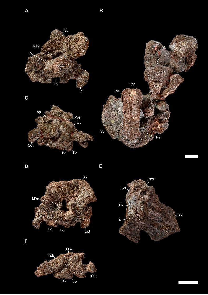
CHAPTER III – REVISION OF MOROCCAN DICYNODONTS
136

CHAPTER III – REVISION OF MOROCCAN DICYNODONTS
137
Figure 4. Holotypes of Azarifeneria robustus and madibular material attributed to
Moghreberia nmachouensis, figured in dorsal (A, D), ventral (B, E), and lateral (C, F)
views. A-C, MNHN.F.ALM 38: mandible attributed to M. nmachouensis; B-F,
MNHN.F.ALM 167: anterior portion of the mandible of A. robustus. The scale bar
represents 5 cm.
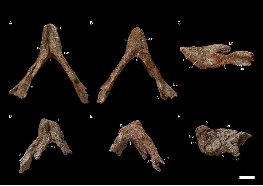
CHAPTER III – REVISION OF MOROCCAN DICYNODONTS
138

CHAPTER III – REVISION OF MOROCCAN DICYNODONTS
139
Figure 5. Vertebrae attributed to Moghreberia nmachouensis and supposed to be last
cervical and/or first dorsal vertebrae, figured in anterior (A, C), posterior (B, E), right
lateral (D), and left lateral (F) views. A-B, MNHN.F.ALM 288; C-F, two unnumbered
connected vertebrae called ‘Vunknb 1 and 2’, with the centra indicated by ‘Ct 1 and 2’
following the anteroposterior vertebral axis. The scale bar represents 5 cm.

CHAPTER III – REVISION OF MOROCCAN DICYNODONTS
140
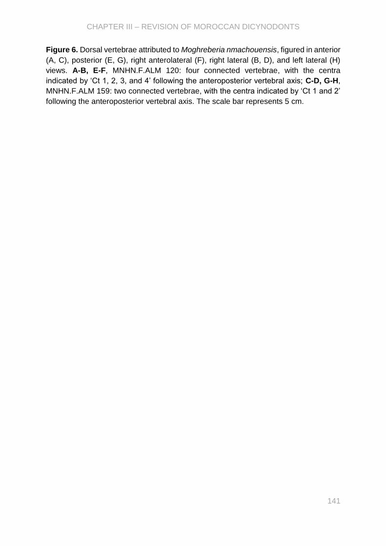
CHAPTER III – REVISION OF MOROCCAN DICYNODONTS
141
Figure 6. Dorsal vertebrae attributed to Moghreberia nmachouensis, figured in anterior
(A, C), posterior (E, G), right anterolateral (F), right lateral (B, D), and left lateral (H)
views. A-B, E-F, MNHN.F.ALM 120: four connected vertebrae, with the centra
indicated by ‘Ct 1, 2, 3, and 4’ following the anteroposterior vertebral axis; C-D, G-H,
MNHN.F.ALM 159: two connected vertebrae, with the centra indicated by ‘Ct 1 and 2’
following the anteroposterior vertebral axis. The scale bar represents 5 cm.

CHAPTER III – REVISION OF MOROCCAN DICYNODONTS
142

CHAPTER III – REVISION OF MOROCCAN DICYNODONTS
143
Figure 7. MNHN.F.ALM 265, dorsal vertebra attributed to Moghreberia
nmachouensis, figured in anterior (A), posterior (C), right lateral (B), and left lateral
(D) views. The scale bar represents 5 cm.

CHAPTER III – REVISION OF MOROCCAN DICYNODONTS
144
Figure 8. MNHN.F.ALM 274, two disconnected but associated posterior dorsal
vertebrae attributed to Moghreberia nmachouensis, figured in anterior (A, C), posterior
(E, G), right lateral (B, D), and left lateral (F, H) views. A-B, E-F, MNHN.F.ALM 274-1;
C-D, G-H, MNHN.F.ALM 274-2. These two vertebrae were discovered in association
with a complete pelvic girdle (with sacral and caudal vertebrae) represented in Fig. 24.
The scale bar represents 5 cm.
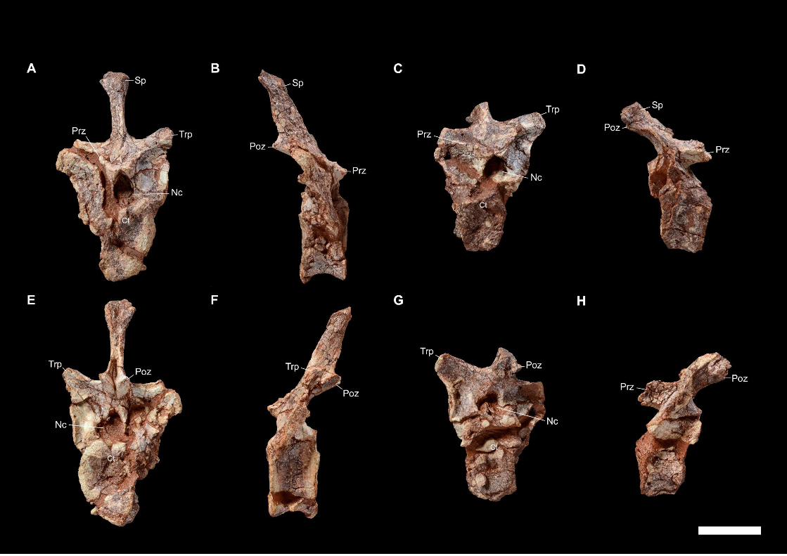
CHAPTER III – REVISION OF MOROCCAN DICYNODONTS
145

CHAPTER III – REVISION OF MOROCCAN DICYNODONTS
146
Figure 9. MNHN.F.ALM 136, incomplete sacrum attributed to Moghreberia
nmachouensis, figured in dorsal (A) and ventral (B) views. The sacral centra were
indicated by ‘Sct 2, 3, and 4’ following the anteroposterior vertebral axis. The scale
bar represents 5 cm.
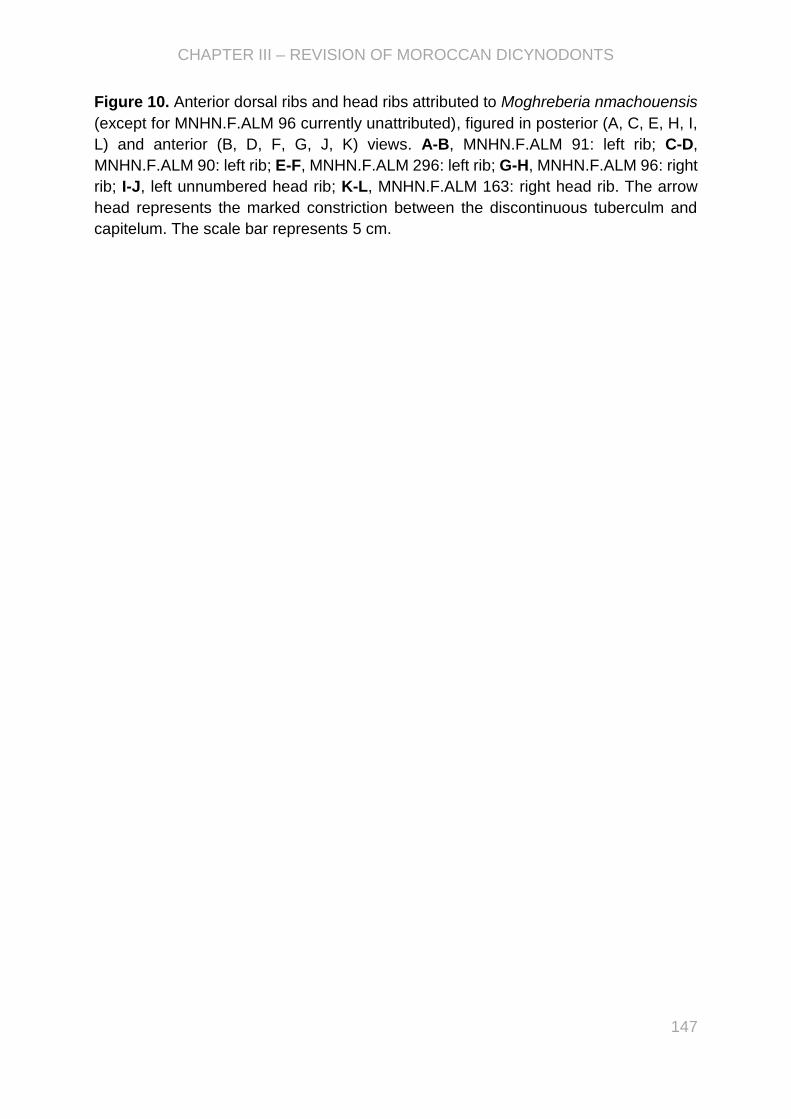
CHAPTER III – REVISION OF MOROCCAN DICYNODONTS
147
Figure 10. Anterior dorsal ribs and head ribs attributed to Moghreberia nmachouensis
(except for MNHN.F.ALM 96 currently unattributed), figured in posterior (A, C, E, H, I,
L) and anterior (B, D, F, G, J, K) views. A-B, MNHN.F.ALM 91: left rib; C-D,
MNHN.F.ALM 90: left rib; E-F, MNHN.F.ALM 296: left rib; G-H, MNHN.F.ALM 96: right
rib; I-J, left unnumbered head rib; K-L, MNHN.F.ALM 163: right head rib. The arrow
head represents the marked constriction between the discontinuous tuberculm and
capitelum. The scale bar represents 5 cm.


CHAPTER III – REVISION OF MOROCCAN DICYNODONTS
149
Figure 11. Sternum and posterior dorsal ribs and head rib attributed to Moghreberia
nmachouensis, figured in posterior (A, C, F, H), anterior (B, D, E, G), and ventral (I)
views. A-B, MNHN.F.ALM 86: left rib; C-D, MNHN.F.ALM 95: left rib; E-F,
MNHN.F.ALM 97: right rib; G-H, MNHN.F.ALM 272: right head rib; I, MNHN.F.ALM
134: sternum. The scale bar represents 5 cm.
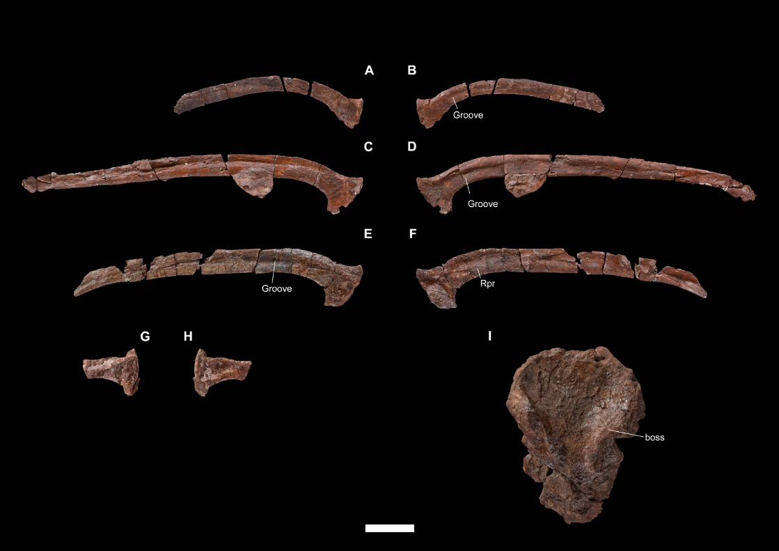

CHAPTER III – REVISION OF MOROCCAN DICYNODONTS
151
Figure 12. Scapulae attributed to Moghreberia nmachouensis, figured in lateral (A, C,
E), and medial (B, D, F) views. A-B, MNHN.F.ALM 29: right scapula; C-D,
MNHN.F.ALM 30: left scapula; E-F, MNHN.F.ALM 83: left scapula. The scale bar
represents 5 cm.

CHAPTER III – REVISION OF MOROCCAN DICYNODONTS
152

CHAPTER III – REVISION OF MOROCCAN DICYNODONTS
153
Figure 13. Scapulae attributed to Moghreberia nmachouensis (except for
MNHN.F.ALM 299 currently unattributed), figured in lateral (A, C, E), and medial (B, D,
F) views. A-B, MNHN.F.ALM 299: right scapula; C-D, MNHN.F.ALM 17: left scapula;
E-F, MNHN.F.ALM 113: left scapula. The scale bar represents 5 cm.


CHAPTER III – REVISION OF MOROCCAN DICYNODONTS
155
Figure 14. Scapulocoracoids attributed to Moghreberia nmachouensis, figured in
lateral (A, C), and medial (B, D) views. A-B, MNHN.F.ALM 92: left scapulocoracoid; C-
D, MNHN.F.ALM 298: left scapulocoracoid. The scale bar represents 5 cm.


CHAPTER III – REVISION OF MOROCCAN DICYNODONTS
157
Figure 15. Scapulae attributed to Moghreberia nmachouensis (except for
MNHN.F.AZA 365 currently unattributed), figured in lateral (A, C, F, H), medial (B, D,
G, I), and ventral (E, J) views. A-B, MNHN.F.ALM 98: left scapula; C-E, MNHN.F.ALM
132: left scapula; F-G, MNHN.F.ALM 283: right scapula; H-J, MNHN.F.AZA 365: right
scapula. The scale bar represents 5 cm.

CHAPTER III – REVISION OF MOROCCAN DICYNODONTS
158

CHAPTER III – REVISION OF MOROCCAN DICYNODONTS
159
Figure 16. Scapulae attributed to Moghreberia nmachouensis, figured in lateral (A, C,
D) and medial (B, E, F) views. A-B, right unnumbered scapula called ‘Sunknb 3’; C-F,
right unnumbered scapula called ‘Sunknb 1’; D-E, left unnumbered scapula called
‘Sunknb 2’. The scale bar represents 5 cm.

CHAPTER III – REVISION OF MOROCCAN DICYNODONTS
160

CHAPTER III – REVISION OF MOROCCAN DICYNODONTS
161
Figure 17. Interclavicles attributed to Moghreberia nmachouensis (except for
MNHN.F.ALM 260 currently unattributed), figured in ventral (A, C, E, G-J) and dorsal
(B, D) views. A-B, MNHN.F.ALM 93; C-D, MNHN.F.ALM 295; E-F, unnumbered
interclavicle called ‘Intunknb 1’; G, MNHN.F.ALM 94; H, MNHN.F.ALM 260; I,
MNHN.F.ALM 278; J, unnumbered interclavicle called ‘Intunknb 2’. The scale bar
represents 5 cm.
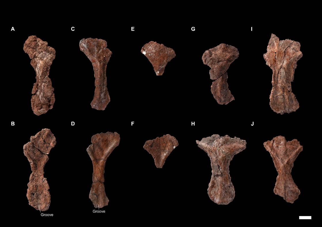

CHAPTER III – REVISION OF MOROCCAN DICYNODONTS
163
Figure 18. Humerii attributed to Moghreberia nmachouensis (except for MNHN.F.ALM
275 currently unattributed), figured in ventral (A, D, G, H, J), anteroventral (B, E), and
dorsal (C, F, I, K) views. A-C, MNHN.F.ALM 24: left humerus; D-F, MNHN.F.ALM 25:
left humerus; G, MNHN.F.ALM 275: left humerus; H-I, distal region of a right
unnumbered humerus called ‘Humunknb 2’; J-K, MNHN.F.ALM 121: left humerus. The
scale bar represents 5 cm.


CHAPTER III – REVISION OF MOROCCAN DICYNODONTS
165
Figure 19. Humerii attributed to Moghreberia nmachouensis, figured in ventral (A-C),
anteroventral (D-F), and dorsal (G-I) views. A, D, G, MNHN.F.ALM 102: left humerus;
B, E, H, MNHN.F.ALM 101: right humerus; C, F, I, MNHN.F.ALM 115: right humerus.
The scale bar represents 5 cm.


CHAPTER III – REVISION OF MOROCCAN DICYNODONTS
167
Figure 20. Radii attributed to Moghreberia nmachouensis, figured in anterior (A, C, E)
and posterior (B, D, F) views. A-B, MNHN.F.ALM 84: left radius; C-D, MNHN.F.ALM
131: left radius; E-F, MNHN.F.ALM 287: right radius. The scale bar represents 5 cm.

CHAPTER III – REVISION OF MOROCCAN DICYNODONTS
168

CHAPTER III – REVISION OF MOROCCAN DICYNODONTS
169
Figure 21. Radii attributed to Moghreberia nmachouensis, figured in anterior (A, C)
and posterior (B, D) views. A-B, MNHN.F.ALM 289: left radius; C-D, left unnumbered
radius. The scale bar represents 5 cm.

CHAPTER III – REVISION OF MOROCCAN DICYNODONTS
170
Figure 22. Ulnae attributed to Moghreberia nmachouensis (except for MNHN.F.ALM
154 currently unattributed), figured in lateral (A, C, E, G) and medial (B, D, F) views.
A-B, MNHN.F.ALM 18: right ulna; C-D, MNHN.F.ALM 28: left ulna; E-F, MNHN.F.ALM
261: left ulna; G, MNHN.F.ALM 154: left ulna. The scale bar represents 5 cm.

CHAPTER III – REVISION OF MOROCCAN DICYNODONTS
171

CHAPTER III – REVISION OF MOROCCAN DICYNODONTS
172
Figure 23. Ulnae attributed to Moghreberia nmachouensis (except for MNHN.F.ALM
27 currently unattributed), figured in lateral (A, D, G), medial (B, E, H), and posterior
(C, F, I) views. A-C, MNHN.F.ALM 35: right ulna; D-F, MNHN.F.ALM 87: right ulna
associated with a left head rib; G-I, MNHN.F.ALM 27: right ulna. The scale bar
represents 5 cm.
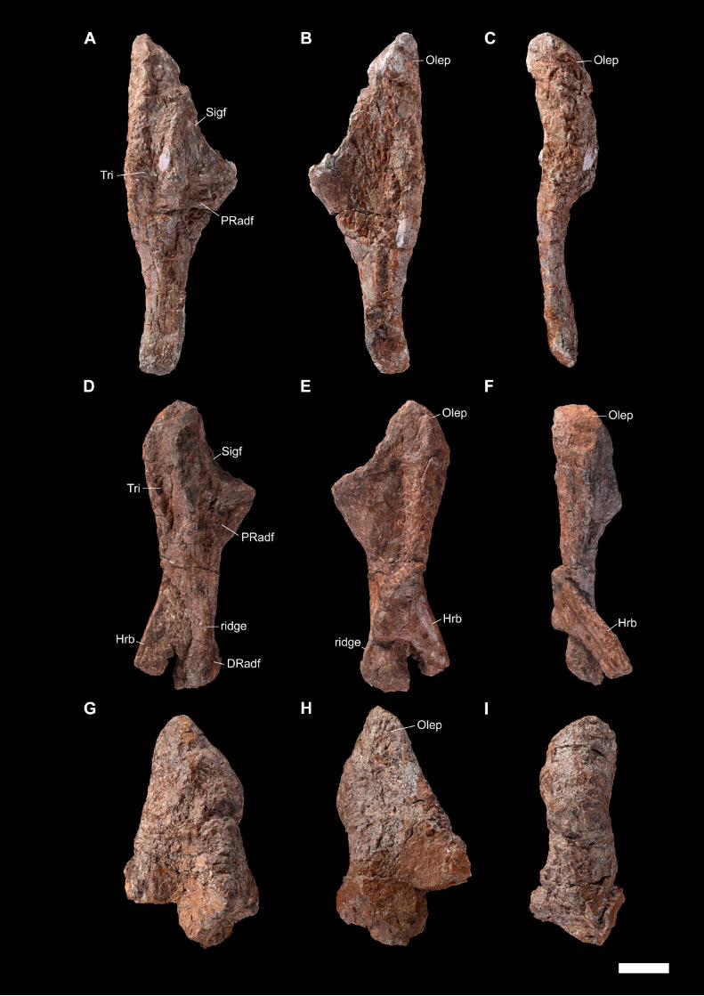

CHAPTER III – REVISION OF MOROCCAN DICYNODONTS
174
Figure 24. MNHN.F.ALM 274, complete pelvis attributed to Moghreberia
nmachouensis, figured in dorsal (A), left lateral (B), ventral (C) and right lateral (D)
views. The scale bar represents 5 cm.

CHAPTER III – REVISION OF MOROCCAN DICYNODONTS
175

CHAPTER III – REVISION OF MOROCCAN DICYNODONTS
176
Figure 25. MNHN.F.ALM 104, left ilium attributed to Moghreberia nmachouensis,
figured in lateral (A) and medial (B) views. The scale bar represents 5 cm.

CHAPTER III – REVISION OF MOROCCAN DICYNODONTS
177
Figure 26. Ischia and a pubis attributed to Moghreberia nmachouensis, figured in
lateral (A, C, E) and medial (B, D, F) views. A-B, MNHN.F.ALM 123: right ischium; C-
D, MNHN.F.ALM 125: right ischium; E-F, MNHN.F.ALM 122: left pubis. The scale bar
represents 5 cm.

CHAPTER III – REVISION OF MOROCCAN DICYNODONTS
178

CHAPTER III – REVISION OF MOROCCAN DICYNODONTS
179
Figure 27. Femora attributed to Moghreberia nmachouensis, figured in anterior (A, D,
G), posterior (B, E, H), and proximal (C, F, I) views. A-C, MNHN.F.ALM 21: left femur;
D-F, MNHN.F.ALM 26: left femur; G-I, MNHN.F.ALM 23: right femur. The scale bar
represents 5 cm.

CHAPTER III – REVISION OF MOROCCAN DICYNODONTS
180

CHAPTER III – REVISION OF MOROCCAN DICYNODONTS
181
Figure 28. Femora attributed to Moghreberia nmachouensis (except for MNHN.F.ALM
22 and 284 currently unattributed), figured in anterior (A, C, F), posterior (B, D, G), and
proximal (E, H) views. A-B, MNHN.F.ALM 100: left femur; C-E, MNHN.F.ALM 22: right
femur; F-H, MNHN.F.ALM 284: left femur. The scale bar represents 5 cm.


CHAPTER III – REVISION OF MOROCCAN DICYNODONTS
183
Figure 29. Tibiae and a fibula attributed to Moghreberia nmachouensis (except for
MNHN.F.ALM 291 currently unattributed), figured in anterior (A, C, E), posterior (B, D,
F), lateral (G), and medial (H) views. A-B, MNHN.F.ALM 33: right tibia; C-D, right
unnumbered tibia; E-F, MNHN.F.ALM 291: left tibia; G-H, MNHN.F.ALM 32: left fibula.
The scale bar represents 5 cm.

CHAPTER III – REVISION OF MOROCCAN DICYNODONTS
184

CHAPTER III – REVISION OF MOROCCAN DICYNODONTS
185
Figure 30. The most parsimonious cladogram (1202.936 steps, consistency
index=0.229, retention index=0.712), with BS indicated above the branches and PBS
(between the continuous [on right] and discrete [on left] characters) below. Capital
letters indicate the following clades (Kammerer and Angielczyk, 2009; Kemmerer et
al., 2013; Olivier et al., 2019): A, Dicynodontia; B, Pylaecephalidae; C,
Endothiodontia; D, Therochelonia; E, Emydopoidea; F, Kistecephalia; G,
Emydopidae; H, Kingoriidae; I, Cistecephalidae; J, Bidentalia; K, Cryptodontia; L,
Geikiidae; M, Geikiinae; N, Rhachiocephalidae; O, Dicynodontidae ; P,
Lystrosauridae; Q, Stahleckeriidae; R, Kannemeyeriidae.
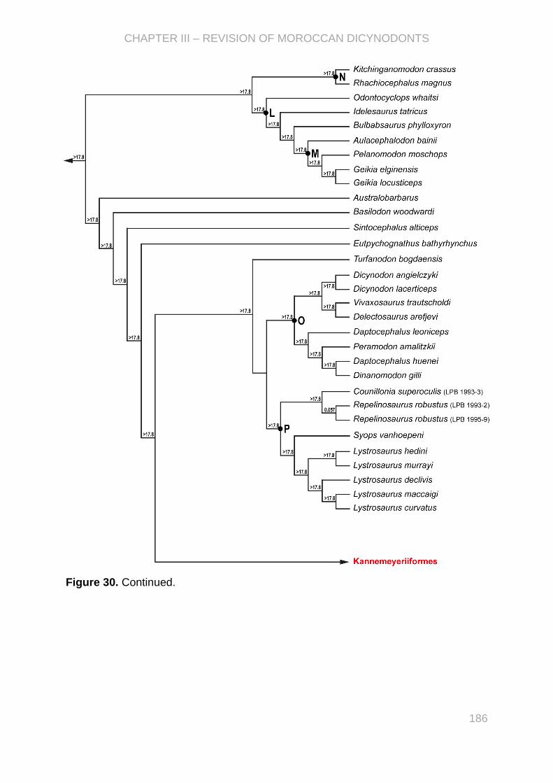
CHAPTER III – REVISION OF MOROCCAN DICYNODONTS
186
Figure 30. Continued.

CHAPTER III – REVISION OF MOROCCAN DICYNODONTS
187
Figure 30. Continued.

CHAPTER III – REVISION OF MOROCCAN DICYNODONTS
188
Figure 31. Kannemeyeriiform femora in anterior view. A-B, Moghreberia
nmachouensis: MNHN.F.ALM 21 and 100; C, MNHN.F.ALM 22, incertae sedis; D,
Zambiasaurus submersus: NHMUK R9118; E, Placerias hesternus: UCMP 32394
(figured by Kammerer et al., 2013); F, Parakannemeyeria dolichocephala: IVPP V985;
G, Ischigualastia jenseni: MCZ 378-58M; H, Kannemeyeria simocephalus: NHMUK
R3740; I, Tetragonias njalilus: GPIT 292; J, Dinodontosaurus turpior: MCZ 3116; K,
Dolichuranis primaevus: BP/1/4578. The specimens are left femora in A, B and E
(reversed for comparative purposes), and right femora in C-D and F-K. The scale bars
represent 5 cm.

CHAPTER III – REVISION OF MOROCCAN DICYNODONTS
189

CHAPTER III – REVISION OF MOROCCAN DICYNODONTS
190
Figure 32. Kannemeyeriiform humeri in dorsal (A-J) and ventral (K) view. A, G,
Moghreberia nmachouensis: MNHN.F.ALM 102 (in A) and MNHN.F.ALM 24 (in G); B,
Lisowicia bojani: ZPAL V.33/96 (modified from Sulej and Niedźwiedzki, 2019); C,
Angonisaurus cruickshanki: NHMUK R9732; D-E, Placerias hesternus: USNM 2198
(in D) and UCMP 25361 (in E) (modified from Kammerer et al., 2013); F, Pentasaurus
goggai: NHMW 1876-VII-B-123 (modified from Kammerer, 2018); H, Kannemeyeria
simocephalus: NHMUK R3741; I-J, Zambiasaurus submersus: NHMUK R9089 and
NHMUK R9140; K, Stahleckeria potens: BSPG AS-XXV-148 (modified from
Kammerer, 2018). All the specimens are left humeri, except the right ones in C-E and
K (reversed for comparative purposes). The scale bars represent 5 cm.

CHAPTER III – REVISION OF MOROCCAN DICYNODONTS
191

CHAPTER III – REVISION OF MOROCCAN DICYNODONTS
192
Figure 33. Kannemeyeriiform mandibles in lateral view. A, ‘Azarifeneria robustus’:
MNHN.F.ALM 167; B, Moghreberia nmachouensis: MNHN.F.ALM 38; C-D,
Stahleckeria potens: cast AMNH FARB3857 (in C) and GSN OM 10 (in D); E,
Angonisaurus cruickshanki: NHMUK R 9732; F, Shaanbeikannemeyeria
xilougouensis: IVPP V11676; G, Xiyukannemeyeria brevisrostris: IVPP V4457; H,
Kannemeyeria simocephalus: NHMUK R3602; I, Ischigualastia jenseni: MCZ 3120
figured by Cox (1965). All the specimens are in right lateral view (reversed for
comparative purposes), except the mandibles in left lateral view in A, B, D, I. The scale
bars represent 5 cm (specimen in I not in scale).

CHAPTER III – REVISION OF MOROCCAN DICYNODONTS
193

CHAPTER IV – PALEOPHYSIOLOGY OF THE DICYNODONTS AND
MORE SPECIFICALLY THE TRIASSIC FORMS
CHAPTER IV
PALEOPHYSIOLOGY OF THE DICYNODONTS AND
MORE SPECIFICALLY THE TRIASSIC FORMS
Dicynodont © M. Boulay

CHAPTER IV – PALEOPHYSIOLOGY OF DICYNODONTS
195
Quantitative inference of the metabolic rate in extinct synapsid fossils
The energy expenditure is often studied by the ecologists to evaluate the
adjustements made by the organisms to their environments. To be compared, we used
the standardized “resting” metabolic rate defined as “the rate (1) in the zone of
thermoneutrality when the individuals are (2) inactive, (3) postabsorptive, (4) adult
(thereby eliminating the cost of growth), (5) nonreproductive (eliminating the cost of of
pregnancy, lactation, egg formation, or incubation), and (6) regulating body
temperature, and it is (7) measured during the inactive period” (McNab, 1997).
Endothermy has evolved at least twice, in the mammals and birds. The acquisition of
the mammalian endothermy is a major event in vertebrate evolution since it modified
the energetic relationships between the organisms and their environment (Walter and
Seebacher, 2009). In the classical definitions, an endothermic organism produces itself
heat to control its body temperature. Clarke and Pörtner (2010) defined the endothermy
as “the controlled maintenance of a relatively high and more or less constant internal
body temperature, where the main source of heat is a high resting (basal) metabolic
rate. […] Body temperature may exhibit circadian variability, with the amplitude
typically inversely proportional to body size. Endothermy may also be suspended
temporarly in periods of torpor or hibernation”. The studies of the role of muscles in the
heat production led to propose a more detailed definition of endothermy as a non-
shivering thermogenesis (e.g., Rowland et al., 2015; Nowack et al., 2017; Cubo and
Jalil, 2019).
Many morphological proxies have been used to infer an endothermic
metabolism in the synapsid fossil record such as the insulative fur (e.g., Ruben and
Jones, 2000; Smith and Botha-Brink, 2011; Ruben et al., 2012; Benoit et al., 2018) and
respiratory turbinates (e.g., Hillenius, 1992, 1994; Ruben et al., 1996; Crompton et al.,
2015; Owerkowicz et al., 2015; Crompton et al., 2017). The inner bone organization is
preserved during the fossilisation and would be also correlated with the bone growth
rate and then the resting metabolic rate (e.g., Montes et al., 2007). A fibrolamellar bone
(i.e., [FLB] primary osteons with lamellar bone within a woven bone matrix) thus
indicates a high growth rate according to Francillon Vieillot et al. (1990) and Ricqlès et
al. (1991). The qualitative histology (description of the inner organization of bone) of
the synapsids was intensively studied and especially in dicynodonts. The presence of
FLB was demonstrated in (1) cynodonts such as Procynosuchus, Trucidocynodon and

CHAPTER IV – PALEOPHYSIOLOGY OF DICYNODONTS
196
Langbergia (Botha-Brink et al., 2012), Thrinaxodon (Ricqlès, 1969; Ray et al., 2004;
Chinsamy and Hurum, 2006; Botha-Brink et al., 2012), Cynognathus and Diademodon
(Botha and Chinsamy, 2000; Botha-Brink and Angielczyk, 2010), Trirachodon (Botha
and Chinsamy, 2004; Botha-Brink et al., 2012), and Tritylodon (Ricqlès, 1969; Ray et
al., 2004; Chinsamy and Hurum, 2006; Botha-Brink et al., 2012), (2) therocephalians
(Huttenlocker and Botha-Brink, 2014), (3) gorgonopsians (Chinsamy-Turan and Ray,
2012), (4) sphenacodontid Dimetrodon (Shelton et al., 2013), (5) ophiacodontid
Ophiacodon (Shelton and Sander, 2017), (5) dromasaur Galeops (Botha-Brink and
Angielczyk, 2010), and (6) dicynodonts Eodicynodon, Diictodon, Endothiodon,
Cistecephalus, Dicynodontoides, Rhachiocephalus, Aulacephalodon, Tropidostoma,
Oudenodon, Dicynodon, Kannemeyeria, Placerias, Lystrosaurus declivis, L. murrayi
and L. maccaigi (e.g., Ray et al., 2005; Botha-Brink and Angielczyk, 2010; Green et
al., 2010). These results would indicate an origin of FLB in Synapsida during the early
Permian (Shelton and Sander, 2017). However, some extant ectotherms such as the
alligators are able to produce FLB in captivity (Padian et al., 2004) and in wild
(Tumarkin-Deratzian, 2007), that was been considered as an inherited feature from the
endothermic ancestral state in archosaurs (Seymour et al., 2004).
Most of proxies used to infer metabolism of extinct oragnisms are qualitative.
The qualitative histology being insufficient to rather precisely infer resting metabolic
rates or physiological thermoregulation. A recent quantitative histological approach
were thus first proposed by Legendre et al. (2016) to perform quantitative inferences
of the resting metabolic rates on archosaur fossils using paleobiological models. These
authors built their models based on microanatomical features (density, shape, and
area of the osteocyte and vascular density) within a phylogenetic context (using the
Phylogenetic Eigenvector Maps). Their study allowed to constrain both temporally and
phylogenetically the acquisition of the avian endothermy assuming it at the
archosauriform node. This method was then re-used by Fleischle et al. (2018) who
inferred a high resting metabolic rate in plesiosaurs. However, these paleobiological
inference models have never been applied to Synapsida.
This chapter comprises two studies dealing with the application of
paleohistological inference models to infer the resting metabolic rate in synapsid fossils
using the Phylogenetic Eigenvector Maps (PEMs) (PAPER III) and Phylogenetic
Generalised Least Squares (PGLS) (PAPER IV) methods. They focused on a

CHAPTER IV – PALEOPHYSIOLOGY OF DICYNODONTS
197
dicynodont sample and highlighted on the implications on our understanding of the
evolution of the acquisition of the mammalian endothermy.
The PAPER III has been started during my Master internship where I studied the
qualitative histology of the humerus and femur of Moghreberia nmachouensis, and
performed preliminary paleobiological inference models (based on humerus and
femur) using PEMs. However, the extant sample of synapsids (including Microcebus
murinus, Cavia porcellus, and Mus musculus) was not sufficient compared to the
extinct sample (including the dicynodonts M. nmachouensis, Lystrosaurus sp., and
Oudenodon baini). The first work of my PhD was to include three more extant
synapsids (Lepus europaeus, Oryctolagus cuniculus, and Capreolus capreolus) and
build new models. The bone sections were all conserved in MNHN. In this paper, J.
Cubo, A. Houssaye, and I wrote the article. I made the description of the qualitative
histology and the discussion “Growth Rate and Stage”. I measured the histological
variables (density, shape, and area of the osteocyte) for both femur and humerus of
the three fossils and the three added extant synapsids (Fig. IV.1). To smooth variations
between the different regions of the bone, I took 30 measurements per region (anterior,
posterior, lateral, and medial) of each bones (femur and humerus) for each histological
variable (density, shape, and area of the osteocyte). Then, I performed the two
statistical models (based on femur and humerus) using the R package MPSEM
(Guénard et al., 2013), following the method of Legendre et al. (2016). I produced all
the figures of the paper.
PAPER IV used the same extant sample as PAPER III but comprised a larger sample
of dicynodonts with Daptocephalus (femur), Endothiodon (humerus), Kannemeyeria
simocephalus (femur), Lystrosaurus murrayi (femur and humerus), Moghreberia
nmachouensis (femur and humerus), Myosaurus gracilis (humerus), Oudenodon baini
(femur and humerus), and Tropidostoma (femur and humerus). I visited the histological
collection of the National Museum of Bloemfontein (South Africa; financial support from
CR2P) to consult and photograph the bone sections of all cited dicynodont except
those of Moghreberia nmachouensis and Oudenodon baini conserved in MNHN. As in
the previous paper, the paleobiological models based on femur and humerus are
distinct. As opposed to PAPER III, the main objective of this paper was to build models
using a combination of histological variables and PGLS method. For comparative
purposes, models using PEMs method have also been done. In PAPER IV, I wrote the
whole of the article that has been improved by comments of all coauthors. I produced

CHAPTER IV – PALEOPHYSIOLOGY OF DICYNODONTS
198
all the figures of the paper. Following the method of PAPER III and Fleischle et al.
(2018), I measured all histological variables (density, shape, and area of the osteocyte,
and primary osteon density) for femora and humeri of the fossil sample (except the
primary osteon density of the femur of Oudenodon quantified by M. Faure-Brac), and
the primary osteon density for humerus of the extant sample (Fig. IV.1). M. Faure-Brac
quantified the primary osteon density of the femora of the extant species. The statistical
methods have been discussed by Y. Desdevises, L. Legendre, and I. I built the models
using PGLS (with BayesTraits V3.0.1; Pagel and Meade, 2016) and PEMs (with the R
package MPSEM; Guénard et al., 2013) methods, and did the multiple tests of
correlations, AICs, BayesFactor, and coss-validations.
Figure IV.1. Measurements on the bone sections of the humerus NMQR 9960 of Tropidostoma. A, the
surface of the mid-diaphyseal cross section of the bone divided in four parts; B, primary osteon density
measured as the sum of areas of the osteons (yellow circled; i.e., primary osteons and the associated
lamellar bone) compared to the total bone area (red limited); C, osteocyte density measured as the
number of osteocytes (pink pointed) per bone area (yellow limited); D, osteocyte area (pink circled) and
shape that is measured as the ratio between the major and the minor axes of the lacuna.

PAPER III - “First palaeohistological inference of resting metabolic rate in an
extinct synapsid, Moghreberia nmachouensis (Therapsida: Anomodontia)”,
2017, Biological Journal of the Linnean Society
First palaeohistological inference of resting metabolic rate in an
extinct synapsid, Moghreberia nmachouensis (Therapsida:
Anomodontia)
Chloe Olivier1,2, Alexandra Houssaye3, Nour-Eddine Jalil2 and Jorge Cubo1*
1Sorbonne Universités, UPMC Univ Paris 06, CNRS, UMR 7193, Institut des Sciences de la
Terre Paris (iSTeP), 4 place Jussieu, BC 19, Paris 75005, France
2Sorbonne Universités -CR2P -MNHN, CNRS, UPMC-Paris6. Muséum national d’Histoire
naturelle, 57 rue Cuvier, CP38, Paris F-75005, France
3Département Écologie et Gestion de la Biodiversité, UMR 7179, CNRS/Muséum national
d’Histoire naturelle, 57 rue Cuvier, CP55, Paris 75005, France
*Corresponding author
Published in Biological Journal of Linnean Society in 2017
PA
PE
R I
II
409
Biological Journal of the Linnean Society, 2017, 121, 409–419. With 4 figures.
© 2017 The Linnean Society of London, Biological Journal of the Linnean Society, 2017, 121, 409–419
First palaeohistological inference of resting metabolic rate in an extinct synapsid, Moghreberia nmachouensis (Therapsida: Anomodontia)
CHLOE OLIVIER1,2, ALEXANDRA HOUSSAYE3, NOUR-EDDINE JALIL2 and JORGE CUBO1*
1Sorbonne Universités, UPMC Univ Paris 06, CNRS, UMR 7193, Institut des Sciences de la Terre Paris (iSTeP), 4 place Jussieu, BC 19, Paris 75005, France2Sorbonne Universités -CR2P -MNHN, CNRS, UPMC-Paris6. Muséum national d’Histoire naturelle, 57 rue Cuvier, CP38, Paris F-75005, France3Département Écologie et Gestion de la Biodiversité, UMR 7179, CNRS/Muséum national d’Histoire naturelle, 57 rue Cuvier, CP55, Paris 75005, France
Received 10 November 2016; revised 30 November 2016; accepted for publication 1 December 2016
The independent acquisition of endothermy in synapsids and diapsids are major events in vertebrate evolution since they were the driving force of a suite of correlated changes in anatomical, physiological, behavioural and ecological traits. While avian endothermy is assumed to have occurred at the archosauriform node, the acquisition of mamma-lian endothermy is poorly constrained both temporally and phylogenetically. Among the many unequivocal anatomi-cal correlates of endothermy in synapsids, the presence of insulative pelage or respiratory turbinates only allows discrete inferences of presence/absence of endothermy. The analysis of bone histology allows richer palaeobiologi-cal inferences. We described the osteohistology and growth patterns of Moghreberia nmachouensis and two related taxa (Lystrosaurus and Oudenodon) for comparative purposes. Our observations suggest increasing growth rates from Moghreberia [the presence of incipient fibrolamellar bone (FLB) in humerus and femur], to Lystrosaurus (the presence of well-developed FLB in the femur but the presence of incipient FLB in the humerus), to Oudenodon (the presence of well-developed FLB in humerus and femur). However, qualitative histology does not allow reliable infer-ences about the occurrence of endothermy. We performed the first quantitative inferences of resting metabolic rates on fossil synapsids (M. nmachouensis as a model and Lystrosaurus and Oudenodon for comparative purposes) using quantitative histology (size, shape and density of osteocyte lacunae) combined with phylogenetic eigenvector maps. Our inferences are consistent with our qualitative histological observations: the mass-independent resting metabolic rate inferred for M. nmachouensis (2.58 mLO2 h
−1 g−0.67) is lower than the value inferred for Lystrosaurus (3.80 mLO2 h−1 g−0.67), which is lower than that inferred for Oudenodon (4.58 mLO2 h
−1 g−0.67). Optimization of these inferences onto a phylogenetic tree of tetrapods using the parsimony method allowed us to better constrain the temporal (more than 260 Myr ago) and phylogenetic (Neotherapsida) frames of the acquisition of mammalian endothermy.
ADDITIONAL KEYWORDS: Dicynodontia – endothermy – fibrolamellar bone – palaeohistology – phylogenetic eigenvector maps.
INTRODUCTION
The acquisition of mammalian endothermy is a major event in vertebrate evolution since it modified the ener-getic relationships between organisms and their envi-ronment (Walter & Seebacher, 2009). To constrain the
temporal and phylogenetic frames of this event, we need to identify unequivocal anatomical correlates of endo-thermy (Ruben et al., 2012). Among them, the oldest evidence for an insulative pelage (through fossilized fur impressions) has been found in the Middle Jurassic non-mammalian therapsids (Castorocauda, Ji et al., 2006; Megaconus, Zhou et al., 2013; Agilodocodon, Meng et al., 2015), but it seems that the acquisition of endothermy *Corresponding author. E-mail: [email protected]
409

410 C. OLIVIER ET AL.
© 2017 The Linnean Society of London, Biological Journal of the Linnean Society, 2017, 121, 409–419
based on increased aerobic capacity is older (Ruben et al., 2012). The presence of respiratory turbinates is another anatomical feature linked to endothermy. Although these structures are rarely preserved in fossils, bone ridges of turbinate attachment are frequently preserved (Ruben et al., 2012). These structures are first recogniz-able in therocephalians and in cynodonts (Ruben et al., 2012), so they may have been acquired by the eutheri-odonts. Thermic modelling (Florides et al., 2001) and the isotopic composition of mineralized remains (Rey, 2016) have also been used to infer the physiological ther-moregulation of nonmammalian synapsids.
Bone palaeohistology is a useful method to infer the bone growth rates and resting metabolic rates of extinct vertebrates. The bone histology of nonmam-malian synapsids has been intensively studied in the last decade. Laurin & Buffrénil (2016) analysed the ophiacodont Clepsydrops collettii and concluded that the femur is made of woven-fibered tissue com-bined with parallel-fibered bone (PFB) in primary osteons. Shelton & Sander (2015) reported the pres-ence of fibrolamellar bone (FLB) in Ophiacodon ret-roversus, Ophiacodon uniformis and Ophiacodon mirus. Shelton et al. (2013) reported ‘incipient fibrola-mellar bone’ (iFLB) in the humerus and femur of Dimetrodon natalis. Botha-Brink & Angielczyk (2010) analysed a sample of Anomodontia and showed the presence of FLB in the dromasaur Galeops and the dicynodonts Eodicynodon, Diictodon, Endothiodon, Cistecephalus, Dicynodontoides, Rhachiocephalus, Aulacephalodon , Tropidostoma , Oudenodon , Dicynodon, Kannemeyeria, Lystrosaurus declivis, Lystrosaurus murrayi and Lystrosaurus maccaigi. Ray, Chinsamy & Bandyopadhyay (2005) analysed also the bone histology of L. murrayi and showed the presence of FLB in the humerus, femur, tibia and a proximal phalanx. Chinsamy & Ray (2012) reported FLB tis-sue in the long bones of two Gorgonopsia (Scylacops and Aelurognathus) and in the tibia and fibula of two undetermined Therocephalia. Huttenlocker & Botha-Brink (2014) concluded that basal therocephal-ians (e.g. Lycosuchus) are characterized by highly vascularized FLB, whereas Eutherocephalia (but Moschorhinus) lack this condition (e.g. Theriognathus). Some authors analysed the histological features of nonmammalian cynodonts and showed the pres-ence of FLB in Procynosuchus, Trucidocynodon and Langbergia (Botha-Brink, Abdala & Chinsamy, 2012); Thrinaxodon (Botha & Chinsamy, 2005; Botha-Brink et al., 2012; de Ricqlès, 1969); Cynognathus and Diademodon (Botha & Chinsamy, 2000; Botha-Brink et al., 2012); Trirachodon (Botha & Chinsamy, 2004; Botha-Brink et al., 2012); and Tritylodon (de Ricqlès, 1969; Ray, Botha & Chinsamy, 2004; Chinsamy & Hurum, 2006; Botha-Brink et al., 2012). All these stud-ies were carried out using classic qualitative histology.
Results obtained using this approach are useful to infer bone growth rates assuming Amprino’s rule (e.g. Montes, Castanet & Cubo, 2010), that is, considering that the organization of the collagenous wave and of the vascular network in the osseous tissue is strongly correlated with bone growth rate. However, they are not suitable to infer resting metabolic rates or physi-ological thermoregulation. Indeed, it has been shown that some extant ectotherms (e.g. the alligators) are able to form lamellar-zonal bone (de Ricqlès, Padian & Horner, 2003; Lee, 2004), but also FLB both in cap-tivity (Padian, Horner & de Ricqlès, 2004) and in the wild (Tumarkin-Deratzian, 2007), probably an atavis-tic characteristic of an endothermic ancestry among archosaurs (Seymour et al., 2004).
Recent developments in quantitative bone histology have facilitated the first direct inferences of resting met-abolic rates in extinct diapsids (Legendre et al., 2016). This study is aimed at performing the first quantitative inferences of resting metabolic rates on fossil synapsids, using three dicynodonts as models (Moghreberia nmacho-uensis Lystrosaurus and Oudenodon). Dicynodonts are among the most abundant terrestrial herbivores from the Middle Permian to Late Triassic. They were cosmopoli-tan and had a wide range of ecologies and morphologies represented by an abundant and diversified fossil record. Myosaurus and the species-rich genus Lystrosaurus sur-vived the Permian/Triassic crisis (Maisch & Matzke, 2014). Therefore, dicynodonts are good models to gain a better understanding of the impact of this biotic crisis on terrestrial ecosystems. This survey is focused on the Moroccan Upper Triassic M. nmachouensis but includes the Permian Oudenodon baini and Triassic Lystrosaurus for comparative purposes. Only the cranial material of the monospecific genus Moghreberia was shortly described by Dutuit (1988). Moghreberia is a kannemey-eriiform, the most frequent group in the Triassic, but its phylogenetic position is discussed. Certain authors considered it as a synonym of the Upper Triassic North-American dicynodont Placerias (Cox, 1991), while others supposed they are sister-groups (Kammerer, Fröbisch & Angielczyk, 2013). In addition to inferring the resting metabolic rates of M. nmachouensis, we will describe the osteohistology and growth patterns of this taxon, using two related taxa (Lystrosaurus and Oudenodon) for com-parative purposes.
MATERIAL AND METHODS
Materials
The materials used are humeral and femoral mid-diaphyseal transverse sections of the Moroccan Upper Triassic M. nmachouensis (femur MNHN.F.ALM 100 of 310 mm in length and humerus MNHN.F.ALM 297 of 375 mm in length; Fig. 1) and two additional

BONE HISTOLOGY OF MOGHREBERIA NMACHOUENSIS 411
© 2017 The Linnean Society of London, Biological Journal of the Linnean Society, 2017, 121, 409–419
dicynodonts from the personal collection of Armand de Ricqlès (research collection, Museum National d’Histoire Naturelle, Paris, France). On the one hand, Lystrosaurus sp. (humerus 24.1 of 131 mm in length and femur 11–15 of 90.7 mm in length without the proximal epiphysis) from the Triassic Lystrosaurus assemblage zone of the South African Karoo Basin (de Ricqlès, 1969), where Lystrosaurus curvatus, L. declivis and L. murrayi have been discovered (de Ricqlès, 1972; Smith & Botha, 2005; Botha & Smith, 2007; Fröbisch, 2009). On the other hand Oudenodon baini (humerus 292-1 of 119 mm in length and an unnumbered femur of 107 mm in length) from the Cynognathus assem-blage zone of the South African Karoo Basin (de Ricqlès, 1969). Humerus and femur came from differ-ent specimens in all three taxa.
Histology
Bones were moulded prior to sectioning. All sec-tions were made at the mid-diaphysis using stand-ard procedures (e.g. Padian & Lamm, 2013; only the central part of the diaphysis has been embedded in a polyester resin). Sections were observed micro-scopically using Leica DM2700 P and Zeiss Axiovert 35 (Jena, Germany) microscopes under natural and polarized light and photographed using a DXM 1200 Digital Eclipse Camera System (Nikon, Japan) and an Olympus Digital Camera (Japan). Sections were also scanned using a high-resolution Epson V740 PRO scanner. The histological terminology follows Francillon-Vieillot et al. (1990).
inference Models
We constructed two resting metabolic rate infer-ence models (for the humerus and for the femur)
using phylogenetic eigenvector maps, a new powerful approach developed by Guenard, Legendre & Peres-Neto (2013) and recently used by Legendre et al. (2016). The interest of this approach lies in the fact that it takes into account both phylogenetic and phenotypic infor-mation (here bone quantitative histology) to perform palaeobiological inferences. We used a sample of 17 extant tetrapods (Pleurodeles waltl, Microcebus muri-nus, Lepus europaeus, Oryctolagus cuniculus, Cavia porcellus, Mus musculus, Capreolus capreolus, Zootoca vivipara, Podarcis muralis, Varanus exanthematicus, Varanus niloticus, Chelodina oblonga, Pelodiscus sin-ensis, Trachemys scripta, Crocodylus niloticus, Gallus gallus and Anas platyrhynchos) for which we know both the variable to be inferred in extinct taxa (rest-ing metabolic rate) and the quantified histological fea-tures (size, shape and density of osteocyte lacunae in primary bone outside osteons). The bone histological data (size, shape and density of osteocyte lacunae in primary bone outside osteons) for all extant taxa but L. europaeus, O. cuniculus and C. capreolus were taken from Legendre et al. (2016). Bone histological data for the quoted three taxa were quantified in this study. Mass-independent resting metabolic rates (in mLO2 h−1 g−0.67) for all extant taxa but Lepus, Oryctolagus and Capreolus were experimentally quantified by Montes et al. (2007). Mass-independent resting metabolic rates for the quoted three taxa were taken respectively from Hackländer, Arnold & Ruf (2002), Seltmann, Ruf & Rödel (2009) and Mauget, Mauget & Sempéré (1999). We quantified the quoted bone histological variables in the humeri and femora of the taxa for which we aim to infer the resting metabolic rate (M. nmachouensis, Lystrosaurus and Oudenodon) using the procedure described by Legendre et al. (2016). Osteohistological data of extant growing tetrapod species taken from Legendre et al. (2016) were quantified in the outer
Figure 1. Moghreberia nmachouensis (Triassic, Argana basin, Morocco): Mid-diaphyseal cross sections of stylopodial bones. (A) Humerus MNHN.F.ALM 297; (B) femur MNHN.F.ALM 100. Abbreviations: ant., anterior side; dor., dorsal side; lat., lat-eral side; med., medial side; post., posterior side; vent., ventral side. Scale bars represent 5 mm.

412 C. OLIVIER ET AL.
© 2017 The Linnean Society of London, Biological Journal of the Linnean Society, 2017, 121, 409–419
cortex, whereas osteohistological measurements per-formed in this study for three adult extant mammals (L. europaeus, O. cuniculus and C. capreolus) and three subadult or adult extinct synapsids (M. nmachouensis, Lystrosaurus and Oudenodon) were made in the deep cortex. In addition to this histological information, the structure of a phylogenetic tree was taken into account, expressed as a set of eigenfunctions, termed phylogenetic eigenvector maps. Afterwards, a subset of phylogenetic eigenfunctions and bone histological variables were selected to infer resting metabolic rates for species in the tree for which these trait data are otherwise lacking (M. nmachouensis, Lystrosaurus and Oudenodon). For each model (one constructed using femora and another using humeri), we calculated the Akaike information criterion (AIC) values to select the osteohistological variable (density, size or shape of osteocyte lacunae) that best fits with the phylogeny transcribed as eigenvectors. We choose the variable corresponding to the smallest AIC value. The combi-nation of this selected variable and the eigenvectors (i.e. the phylogeny) allowed us to predict the depend-ent variable (metabolic rate) unknown in fossils. We performed the analyses using the R package MPSEM (Guénard et al., 2013) and constructed an inference model for each bone (humerus and femur). We selected the best set of variables among phylogenetic eigen-vector maps plus one of the three histological char-acters, using their AIC corrected for finite sample sizes (Burnham, Anderson & Huyvaert, 2011), and cross-validated using leave-one-out cross-validation. Indeed, including more than one histological copredic-tor might have been a potential source of power loss considering our rather small sample size (Legendre et al., 2016). In this way, we were able to infer the rest-ing metabolic rates of M. nmachouensis, Lystrosaurus and Oudenodon with their 95% confidence intervals (CI95%). We obtained two resting metabolic rate values for each taxon, one using the humerus and the other using the femur. We used the highest estimation in our comparative analysis because a given specimen with the resting metabolic rate of an endotherm (e.g. M. murinus, Montes et al., 2007) can grow slowly (M. murinus, Castanet et al., 2004), but the contrary is not true (e.g. ectotherms including active animals such as Varanus have low resting metabolic rates and grow slowly; Montes et al., 2007).
RESULTS
MicroanatoMical features
The humerus microstructure is relatively homogene-ous with thin osseous trabeculae and reduced intertra-becular spaces occupying most of the section (Fig. 1A).
The medullary area is difficult to distinguish from the cortex, although the tightness of the spongiosa becomes tighter (smaller intertrabecular spaces) away from the section centre.
The femur displays a thick compact cortex with a dis-tinct medullary zone occupied by thin osseous trabecu-lae (Fig. 1B). Unfortunately the state of preservation prevents us to conclude whether an open medullary cavity occurred in the core of the section or whether the whole medullary area was occupied by a spongiosa.
Histological features
Moghreberia nmachouensis humerus. Bone is highly vascularized. An intense remodelling (i.e. process of bone resorption and the subsequent deposition of new bone over the resorption surfaces) extends from the inner to the outer cortex. It is of two types: (1) cen-tripetal secondary bone platings on resorption lines (numerous cementing lines in various directions) with no formation of secondary osteons (Fig. 2A), on most of the section; and (2) Haversian remodelling, with sec-ondary osteons (Fig. 2B), on the posterior and dorsal borders. Remains of primary bone are rare. They nev-ertheless suggest that primary bone consists in iFLB (sensu Klein [2010]; Fig. 2C), that is, with primary bone consisting of both PFB and woven bone and the vas-cular canals not being all primary osteons. Avascular PFB is observed at the periphery of the outer cortex locally along the posteroventral and posterodorsal bor-ders. It is also observed as thin layers alternating with layers of iFLB, illustrating an alternation of zones (iFLB) and annuli (PFB; Fig. 2C). Important concen-trations of Sharpey’s fibres inserted rather perpendic-ularly to the collagen fibres are observed at the three pointed extremities of the section.
Moghreberia nmachouensis femur. As opposed to what is observed in the humerus, the femur is essen-tially composed of Haversian bone and remodelling extends up to the periphery of the outer cortex on the posteromedial and anterolateral borders, with only a few remains of primary bone on the periphery. The transition zone between the cortex and the medul-lary area is characterized by a loose spongiosa of sec-ondary bone with only a few secondary osteons that show a large lumen (Fig. 2D). Bone vascularity is high in most of the cortex, but the vascular canal density decreases in the periphery of the anteromedial and anterolateral borders. Primary bone also consists of incipient FLB with some osteons aligned in concentric layers, alternating with thin layers of avascular PFB. Vascular canals are essentially longitudinal with cir-cular and radial (even oblique) anastomoses. Sharpey’s fibres are concentrated on the posterior border (less on the anterolateral and anteromedial borders). On
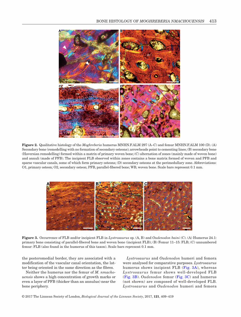
BONE HISTOLOGY OF MOGHREBERIA NMACHOUENSIS 413
© 2017 The Linnean Society of London, Biological Journal of the Linnean Society, 2017, 121, 409–419
the posteromedial border, they are associated with a modification of the vascular canal orientation, the lat-ter being oriented in the same direction as the fibres.
Neither the humerus nor the femur of M. nmacho-uensis shows a high concentration of growth marks or even a layer of PFB (thicker than an annulus) near the bone periphery.
Lystrosaurus and Oudenodon humeri and femora were analysed for comparative purposes. Lystrosaurus humerus shows incipient FLB (Fig. 3A), whereas Lystrosaurus femur shows well-developed FLB (Fig. 3B). Oudenodon femur (Fig. 3C) and humerus (not shown) are composed of well-developed FLB. Lystrosaurus and Oudenodon humeri and femora
Figure 2. Qualitative histology of the Moghreberia humerus MNHN.F.ALM 297 (A–C) and femur MNHN.F.ALM 100 (D). (A) Secondary bone (remodelling with no formation of secondary osteons); arrowheads point to cementing lines; (B) secondary bone (Haversian remodelling) formed within a matrix of primary woven bone; (C) alternation of zones (mainly made of woven bone) and annuli (made of PFB). The incipient FLB observed within zones contains a bone matrix formed of woven and PFB and sparse vascular canals, some of which form primary osteons; (D) secondary osteons at the perimedullary zone. Abbreviations: O1, primary osteon; O2, secondary osteon; PFB, parallel-fibered bone; WB, woven bone. Scale bars represent 0.1 mm.
Figure 3. Occurrence of FLB and/or incipient FLB in Lystrosaurus sp. (A, B) and Oudenodon baini (C). (A) Humerus 24.1: primary bone consisting of parallel-fibered bone and woven bone (incipient FLB); (B) Femur 11–15: FLB; (C) unnumbered femur: FLB (also found in the humerus of this taxon). Scale bars represent 0.1 mm.

414 C. OLIVIER ET AL.
© 2017 The Linnean Society of London, Biological Journal of the Linnean Society, 2017, 121, 409–419
show a lower quantity of secondary bone than the cor-responding Moghreberia bones.
resting Metabolic rate inference Models
For the humerus, the AIC suggests that the more accu-rate model includes the shape of osteocyte lacunae in addition to the selected phylogenetic eigenvector maps (R2 = 0.9901686; AIC score = 18.66448). The mass-inde-pendent resting metabolic rate values (in mLO2 h
−1 g−0.67) inferred for the three taxa are: Moghreberia: 1.76 (CI95% = [1.31, 2.37]); Lystrosaurus: 2.43 (CI95% = [1.95, 3.03]); and Oudenodon: 1.09 (CI95% = [0.88, 1.34]).
For the femur, the AIC suggests a model that includes the osteocyte lacunae density in addi-tion to the selected phylogenetic eigenvector maps (R2 = 0.993772; AIC score = 18.61558). The resting metabolic rate values (in mLO2 h
−1 g−0.67) inferred for the three taxa are: Moghreberia: 2.58 (CI95% = [1.94, 3.43]); Lystrosaurus: 3.80 (CI95% = [3.04, 4.76]); and Oudenodon: 4.58 (CI95% = [3.63, 5.82]).
As stated in the ‘Materials and methods’ section, of the two mass-independent resting metabolic rate val-ues inferred for each taxon (one using the model con-structed using femora and the other using the model constructed using humeri), we used the higher one in our comparative analysis. In all three taxa, the higher value was inferred using the femora. These values are represented in Fig. 4 together with values experimen-tally measured in extant taxa. Dataset is available as supplementary information.
DISCUSSION
The parsimony method suggests that avian and mam-malian endothermies are convergent (i.e. non-inher-ited from a common ancestor). These independent acquisitions of endothermy are major events in verte-brate evolution since they were the driving force of a suite of correlated changes in the respiratory and the circulatory systems, as well as in physiological, behav-ioural and ecological traits (Seymour et al., 2004).
Figure 4. Mass-independent resting metabolic rate (mLO2 h−1 g−0.67) for the species sampled in this study. For all extant
taxa but Lepus, Oryctolagus and Capreolus, mass-independent resting metabolic rates (in mLO2 h−1 g−0.67) were experimen-
tally quantified by Montes et al. (2007) and histological data are taken from Legendre et al. (2016). For Lepus, Oryctolagus and Capreolus, mass-independent resting metabolic rates were taken respectively from Hackländer et al. (2002), Seltmann et al. (2009) and Mauget et al. (1999) and histological data were quantified in this study. For extinct taxa, histological data were quantified in this study, and mass-independent resting metabolic rates were estimated using phylogenetic eigenvector maps. For extant species, red dots correspond to endotherms (mammals and birds) and blue dots to ectotherms (all other taxa). We show inferred values for the fossil taxa with the corresponding 95% confidence intervals. Arrowhead shows the acquisition of endothermy by synapsids.

BONE HISTOLOGY OF MOGHREBERIA NMACHOUENSIS 415
© 2017 The Linnean Society of London, Biological Journal of the Linnean Society, 2017, 121, 409–419
Bone palaeohistology is a reliable method that has been extensively used to infer the resting metabolic rates and physiological thermoregulation of extinct diapsids. Two approaches have been used. On the one hand, several authors performed indirect inferences of physiological thermoregulation assuming a relation-ship between bone growth rates and resting metabolic rate (Montes et al., 2007). Indeed, de Ricqlès et al. (2003, 2008) inferred bone growth rates from bone tis-sue types using a qualitative approach and Amprino’s rule (Montes et al., 2010). Other authors performed quantitative palaeohistological inferences of bone growth rates (Cubo et al., 2012; Legendre, Segalen & Cubo, 2013). On the other hand, some authors per-formed direct inferences of resting metabolic rates in extinct diapsids using bone palaeohistology and phylo-genetic eigenvector maps (Legendre et al., 2016). Here we used qualitative and quantitative histology to infer respectively growth patterns and resting metabolic rates of M. nmachouensis.
growtH rate and stage
(1) Ontogenetic stage. The Moghreberia humeral sec-tion shows a high remodelling activity and, in periph-ery, a cyclical growth with zones and annuli. Following the analysis of Ray et al. (2005) on Lystrosaurus, this would suggest, if Moghreberia grows in a similar way as Lystrosaurus, that the individual was at least sub-adult. Similarly, high remodelling has been tentatively interpreted as suggesting a nonjuvenile ontogenetic stage (Ray & Chinsamy, 2004; Ray et al., 2004). This is consistent with the anatomy that suggests a subadult ontogenetic stage.
The femoral section shows again a high remodelling activity and an alternation of zones and annuli associ-ated with a reduction in the degree of vascularization towards the bone periphery. Although the morphol-ogy suggests a juvenile ontogenetic stage (small size, loss of the epiphyses; Gale [1988]), the histology is rather in accordance with an at least subadult ontoge-netic stage (Ray et al., 2005; Ray, Bandyopadhyay & Appana, 2010).
(2) Growth rate. The occurrence of well-developed FLB and the very high vascularization observed in Lystrosaurus femur (Fig. 3B) and Oudenodon femur (Fig. 3C) and humerus (not shown) evoke previ-ous observations made on the Permian dicynodonts Endothiodon, Diictodon, Oudenodon, and Tropidostoma (Chinsamy & Rubidge, 1993; Botha, 2003; Botha & Angielczyck, 2007; Botha-Brink & Angielczyk, 2010) and the Triassic dicynodonts Placerias, Lystrosaurus, Wadiasaurus, and Kannemeyeria (Botha-Brink & Angielczyck, 2010; Green, Schweitzer & Lamm, 2010; Ray et al., 2005, 2010; Ray, Botha & Chinsamy, 2012; Green & Chinsamy-Turan, 2012). In contrast,
Moghreberia humerus and femur and Lystrosaurus humerus lack a well-developed FLB but show incipi-ent FLB. Fibrolamellar bone is generally observed in mammals and birds (e.g. Chinsamy & Elzanowski, 2001; Kolb et al., 2015) and associated with rapid oste-ogenesis in extant and extinct taxa (e.g. Chinsamy & Rubidge, 1993; Padian, de Ricqlès & Horner, 2001; Ray & Chinsamy, 2004; Chinsamy & Tumarkin-Deratzian, 2009). As bone growth rate is associated, though indi-rectly, to metabolic rate (Reid, 1987; Padian et al., 2001; Chinsamy-Turan, 2005; Montes et al., 2007), FLB is generally associated with endothermy. Please keep in mind that although FLB is also observed in some ectothermic Crocodylia (Tumarkin-Deratzian, 2007; Woodward, Horner & Farlow, 2014), this pres-ence is probably an atavistic characteristic inherited from the endothermic last common ancestor of archo-saurs (Seymour et al., 2004).
Incipient FLB, described by Klein (2010), has also been observed in temnospondyls (Konietzko-Meier & Schmitt, 2013), sauropterygians (pachypleurosaurs, nothosaurs; Klein, 2012; Klein et al., 2016), phytosaurs (de Ricqlès et al., 2003) and mosasaurs (Houssaye et al., 2013), but also in the pelycosaur Dimetrodon (Shelton et al., 2013). Considering that it has been shown that the PFB is formed at a lower growth rate than the woven bone, the presence of incipient FLB suggests a lower growth rate than the presence of well-developed FLB. Therefore, qualitative histo-logical observations suggest increasing growth rates from Moghreberia (the presence of incipient FLB in humerus and femur) to Oudenodon (the presence of well-developed FLB in humerus and femur) and inter-mediate values for Lystrosaurus (the presence of well-developed FLB in the femur but presence of incipient FLB in the humerus). However, qualitative histology does not allow reliable inferences about the occurrence of endothermy.
(3) Bone remodelling. The strong remodelling in the long bones of Moghreberia evokes that observed in Placerias and Kannemeyeria (Green et al., 2010; Ray et al., 2012). It is, however, much more limited in other dicynodonts, where it essentially occurs in the medul-lary spongiosa and only extends up to the perimedul-lary region in adult specimens (Ray & Chinsamy, 2004; Ray et al., 2005, 2012; Ray, Bandyopadhyay & Bhawal, 2009; Botha-Brink & Angielczyk, 2010) and where it is much scarcer in the rest of the cortex. Bone remodelling is interpreted as reflecting biomechanical constraints (Currey, 2003; Skedros et al., 2007) but is also consid-ered associated with an active metabolism (Currey, 2003), related to the blood flow of the bone vascular network (Seymour et al., 2012) or to the size of the skeletal elements (Padian, Werning & Horner, 2016). This would be consistent with the hypothesis of a high metabolism in Moghreberia. However, comparative

416 C. OLIVIER ET AL.
© 2017 The Linnean Society of London, Biological Journal of the Linnean Society, 2017, 121, 409–419
data remain too limited to discuss further the possible physiological significance of bone remodelling.
resting Metabolic rate inference Models
Palaeobiological inferences obtained using femoral quantitative histology (size, shape and density of osteocyte lacunae) and phylogenetic eigenvector maps suggest that the three taxa analysed – M. nmacho-uensis, Lystrosaurus and Oudenodon baini – had high mass-independent resting metabolic rates (mLO2 h
−1 g−0.67), similar to, or even higher than, those observed in some of the extant mammals from our sample (Fig. 4). These inferences are congruent with the conclu-sions obtained using qualitative histology (see above), according to which, assuming Amprino’s rule, bone growth rates may increase from Moghreberia (charac-terized by the presence of incipient FLB in humerus and femur), to Lystrosaurus (the presence of incipient FLB in the humerus but the presence of well-devel-oped FLB in the femur) to Oudenodon (the presence of well-developed FLB in both the humerus and the femur). Of the two mass-independent resting meta-bolic rate values inferred for each taxon (one using the femur and other using the humerus), we used the higher one (that obtained using the femur; cf. methods) in our comparative analysis. Of the three extinct taxa analysed, Oudenodon had the highest mass-independent resting metabolic rate. This result is the outcome of two facts: (1) the histological vari-able selected by the phylogenetic eigenvector maps for the femoral model was osteocyte lacunae density, and (2) Oudenodon shows the highest osteocyte lacu-nae density (0.00496 osteocyte lacunae/µm2), followed by Lystrosaurus (0.00428 osteocyte lacunae/µm2) and Moghreberia (0.00316 osteocyte lacunae/µm2). Thus palaeobiological inferences performed in this study are robust because results obtained using quantita-tive histology and phylogenetic eigenvector maps are consistent with those obtained using qualitative histology.
Rey (2016) analysed the isotopic composition of mineralized tissues and concluded that endothermy was acquired twice among synapsids, by the non-Dicynodon Dicynodontoidea and the Eucynodontia. The optimization of our results onto the phylogeny using the parsimony method suggests a different pattern (Fig. 4): endothermy was acquired only once among synapsids, by the last common ancestor of Neotherapsida (i.e. the last common ancestor of mam-mals and Oudenodon more than 260 Myr ago). Once endothermy was acquired by the last common ancestor of Neotherapsida, taxa within this clade are expected to show high mass-independent resting metabolic rate values. Natural selection may have promoted slight variations in the mass-independent resting metabolic
rate of Oudenodon, Lystrosaurus and Moghreberia, but the inferred values fall within the range of variation observed in extant mammals (Fig. 4).
In conclusion, quantitative histology and phyloge-netic eigenvector maps developed by Guénard et al. (2013) and recently used by Legendre et al. (2016) allowed us to better constrain the temporal (more than 260 Myr ago) and phylogenetic (Neotherapsida) frames of the acquisition of mammalian endothermy.
ACKNOWLEDGEMENTS
We would like to express our gratitude to L. Legendre (Bloemfontein National Museum, South Africa) for helping us with statistical analyses, to A. de Ricqlès (Université Pierre et Marie Curie, Paris, France) and V. de Buffrénil (Muséum National d’Histoire Naturelle, Paris, France) for allowing access to col-lection specimens and to H. Lamrous (UPMC) and S. Morel (MNHN) for the preparation of the thin sections. We also thank H. Bourget (MNHN) for the execution of the moulds and L. Zylberberg (UPMC) for access to her microscope. Many thanks also to Jennifer Botha-Brink, Kirstin Brink and an anonymous reviewer for their helpful comments.
REFERENCES
Botha J. 2003. Biological aspects of the Permian dicynodont Oudenodon (Therapsida, Dicynodontia), deduced from bone histology and cross-sectional geometry. Palaeontologia Africana 39: 37–44.
Botha J, Angielczyk KD. 2007. An integrative approach to distinguishing the Late Permian dicynodont spe-cies Oudenodon bainii and Tropidostoma microtrema (Therapsida: Anomodontia). Palaeontology 50: 1175–1209.
Botha J, Chinsamy A. 2000. Growth patterns deduced from the bone histology of the cynodonts Diademodon and Cynognathus. Journal of Vertebrate Paleontology 20: 705–711.
Botha J, Chinsamy A. 2004. Growth and life habits of the Triassic cynodont Trirachodon, inferred from bone histology. Acta Palaeontologica Polonica 49: 619–627.
Botha J, Chinsamy A. 2005. Growth patterns of Thrinaxodon liorhinus, a non-mammalian Cynodont from the Lower Triassic of South Africa. Palaeontology 48: 385–394.
Botha J, Smith RMH. 2007. Lystrosaurus species composi-tion across the Permo-Triassic boundary in the Karoo Basin of South Africa: Lystrosaurus across the PTB. Lethaia 40: 125–137.
Botha-Brink J, Angielczyk KD. 2010. Do extraordinar-ily high growth rates in Permo-Triassic dicynodonts (Therapsida, Anomodontia) explain their success before and after the end-Permian extinction? Zoological Journal of the Linnean Society 160: 341–365.

BONE HISTOLOGY OF MOGHREBERIA NMACHOUENSIS 417
© 2017 The Linnean Society of London, Biological Journal of the Linnean Society, 2017, 121, 409–419
Botha-Brink J, Abdala F, Chinsamy A. 2012. The radia-tion and osteohistology of nonmammaliaform cynodonts. In: Chinsamy-Turan A, ed. Forerunners of Mammals: radia-tion, histology and biology. Bloomington: Indiana University Press, 223–246.
Burnham KP, Anderson DR, Huyvaert KP. 2011. AIC model selection and multimodel inference in behavioral ecology: some background, observations, and comparisons. Behavioral Ecology and Sociobiology 65: 3–35.
Castanet J, Croci S, Aujard F, Perret M, Cubo J, de Margerie E. 2004. Lines of arrested growth in bone and age estimation in a small primate: Microcebus murinus. Journal of Zoology 263: 31–39.
Chinsamy A. 2012. The microstructure of bones and teeth of nonmammalian Therapsids. In: Chinsamy-Turan A, ed. Forerunners of mammals: radiation, histology and biology. Bloomington: Indiana University Press, 68–89.
Chinsamy A, Elzanowski A. 2001. Bone histology. Evolution of growth pattern in birds. Nature 412: 402–403.
Chinsamy A, Hurum JH. 2006. Bone microstructure and growth patterns of early mammals. Acta Palaeontologica Polonica 51: 325–338.
Chinsamy A, Ray S. 2012. Bone histology of some thero-cephalians and gorgonopsians, and evidence of bone degra-dation by fungi. In: Chinsamy-Turan A, ed. Forerunners of mammals: radiation, histology and biology. Bloomington: Indiana University Press, 199–221.
Chinsamy A, Rubidge BS. 1993. Dicynodont (Therapsida) bone histology: phylogenetic and physiological implications. Palaeontologia Africana 30: 97–102.
Chinsamy A, Tumarkin-Deratzian A. 2009. Pathologic bone tissues in a Turkey vulture and a nonavian dinosaur: impli-cations for interpreting endosteal bone and radial fibrola-mellar bone in fossil dinosaurs. Anatomical record (Hoboken, N.J.: 2007) 292: 1478–1484.
Chinsamy-Turan A. 2005. The microstructure of dino-saur bone: deciphering biology with fine-scale techniques. Baltimore: Johns Hopkins University Press.
Cox CB. 1991. The Pangaea dicynodont Rechnisaurus and the comparative biostratigraphy of Triassic dicynodont faunas. Palaeontology 34: 767–784.
Cubo J, Le Roy N, Martinez-Maza C, Montes L. 2012. Paleohistological estimation of bone growth rate in extinct archosaurs. Paleobiology 38: 335–349.
Currey JD. 2003. The many adaptations of bone. Journal of Biomechanics 36: 1487–1495.
de Ricqlès A. 1969. Recherches paléohistologiques sur les os longs des Tétrapodes: Quelques observations sur la structure des os longs des Thériodontes. Annales de Paléontologie 55: 1–52.
de Ricqlès A. 1972. Recherches paléohistologiques sur les os longs des Tétrapodes: Titanosuchiens, Dinocéphales et Dicynodontes. Annales de Paléontologie 58: 1–78.
de Ricqlès A, Meunier FJ, Castanet J, Francillon-Vieillot H. 1991. Comparative microstructure of bone. Bone 3: 1–78.
de Ricqlès A, Padian K, Horner JR. 2003. On the bone histology of some Triassic pseudosuchian archosaurs and related taxa. Annales de Paléontologie 89: 67–101.
de Ricqlès A, Padian K, Knoll F, Horner JR. 2008. On the origin of high growth rates in archosaurs and their ancient relatives: complementary histological studies on Triassic archosauriforms and the problem of a ‘phylogenetic signal’ in bone histology. Annales de Paléontologie 94: 57–76.
Dutuit JM. 1988. Ostéologie crânienne et ses enseignements, apports géologique et paléoécologique, de Moghreberia nma-chouensis, Dicynodonte (Reptilia, Therapsida) du Trias supé-rieur marocain. Bulletin du Muséum National d’Histoire Naturelle 3: 227–285.
Florides GA, Kalogirou SA, Tassou SA, Wrobel L. 2001. Natural environment and thermal behaviour of Dimetrodon limbatus. Journal of Thermal Biology 26: 15–20.
Francillon-Vieillot H, de Buffrénil V, Castanet J, Géraudie J, Meunier FJ, Sire JY, Zylbeberg L, de Ricqlès A. 1990. Microstructure and mineralization of ver-tebrate skeletal tissues. In: Carter JG, ed. Skeletal biomin-eralization: patterns, processes and evolutionary trends. New York: Van Nostrand Reinhold, 471–530.
Fröbisch J. 2007. The cranial anatomy of Kombuisia freren-sis Hotton (Synapsida, Dicynodontia) and a new phylogeny of anomodont therapsids. Zoological Journal of the Linnean Society 150: 117–144.
Fröbisch J. 2009. Composition and similarity of global anomo-dont-bearing tetrapod faunas. Earth-Science Reviews 95: 119–157.
Gale TM. 1988. Comments on a ‘nest’ of juvenile dicynodont reptiles. Modern Geology 13: 119–124.
Green J, Chinsamy-Turan A. 2012. Bone and dental histol-ogy of Late Triassic dicynodonts from North America. In: Chinsamy-Turan A, ed. Forerunners of mammals: radia-tion, histology and biology. Bloomington: Indiana University Press, 178–196.
Green JL, Schweitzer MH, Lamm ET. 2010. Limb bone histology and growth in Placerias hesternus (Therapsida: Anomodontia) from the Upper Triassic of North America. Palaeontology 53: 347–364.
Guenard G, Legendre P, Peres-Neto P. 2013. Phylogenetic eigenvector maps: a framework to model and predict species traits. Methods in Ecology and Evolution 4: 1120–1131.
Hackländer K, Arnold W, Ruf T. 2002. Postnatal develop-ment and thermoregulation in the precocial European hare (Lepus europaeus). Journal of Comparative Physiology. B, Biochemical, Systemic, and Environmental Physiology 172: 183–190.
Houssaye A, Lindgren J, Pellegrini R, Lee AH, Germain D, Polcyn MJ. 2013. Microanatomical and histological fea-tures in the long bones of Mosasaurine mosasaurs (Reptilia, Squamata)–implications for aquatic adaptation and growth rates. PLoS ONE 8: e76741.
Huttenlocker AK, Botha-Brink J. 2014. Bone microstruc-ture and the evolution of growth patterns in Permo-Triassic therocephalians (Amniota, Therapsida) of South Africa. PeerJ 2: e325.
Ji Q, Luo ZX, Yuan CX, Tabrum AR. 2006. A swimming mammaliaform from the Middle Jurassic and ecomorpho-logical diversification of early mammals. Science (New York, N.Y.) 311: 1123–1127.

418 C. OLIVIER ET AL.
© 2017 The Linnean Society of London, Biological Journal of the Linnean Society, 2017, 121, 409–419
Kammerer CF, Fröbisch J, Angielczyk KD. 2013. On the validity and phylogenetic position of Eubrachiosaurus browni, a kannemeyeriiform dicynodont (Anomodontia) from Triassic North America. PLoS One 8: e64203.
Klein N. 2010. Long bone histology of sauropterygia from the lower Muschelkalk of the Germanic basin provides unex-pected implications for phylogeny. PLoS One 5: e11613.
Klein N. 2012. Postcranial morphology and growth of the pachy-pleurosaur Anarosaurus heterodontus (Sauropterygia) from the Lower Muschelkalk of Winterswijk, The Netherlands. Paläontologische Zeitschrift 86: 389–408.
Klein N, Sander PM, Krahl A, Scheyer TM, Houssaye A. 2016. Diverse aquatic adaptations in Nothosaurus spp. (Sauropterygia)-inferences from humeral histology and microanatomy. PLoS One 11: e0158448.
Kolb C, Scheyer TM, Veitschegger K, Forasiepi AM, Amson E, Van der Geer AA, Van den Hoek Ostende LW, Hayashi S, Sánchez-Villagra MR. 2015. Mammalian bone palaeohistology: a survey and new data with emphasis on island forms. PeerJ 3: e1358.
Konietzko-Meier D, Sander PM. 2013. Long bone histol-ogy of Metoposaurus diagnosticus (Temnospondyli) from the Late Triassic of Krasiejów (Poland) and its paleobio-logical implications. Journal of Vertebrate Paleontology 33: 1003–1018.
Konietzko-Meier D, Schmitt A. 2013. A histological study of a femur of Plagiosuchus, a Middle Triassic temnospondyl amphibian from southern Germany, using thin sections and micro-CT scanning. Netherlands Journal of Geosciences 92: 97–108.
Laurin M, de Buffrenil V. 2016. Microstructural features of the femur in early ophiacodontids: a reappraisal of ances-tral habitat use and lifestyle of amniotes. Comptes Rendus Palevol 15: 115–127.
Lee AH. 2004. Histological organization and its relationship to function in the femur of Alligator mississippiensis. Journal of Anatomy 204: 197–207.
Legendre LJ, Guénard G, Botha-Brink J, Cubo J. 2016. Palaeohistological evidence for ancestral high metabolic rate in Archosaurs. Systematic Biology 65: 989–996.
Legendre LJ, Segalen L, Cubo J. 2013. Evidence for high bone growth rate in Euparkeria obtained using a new paleo-histological inference model for the humerus. Journal of Vertebrate Paleontology 33: 1343–1350.
Maisch MW, Matzke AT. 2014. Sungeodon kimkraemerae n. gen. n. sp., the oldest kannemeyeriiform (Therapsida, Dicynodontia) and its implications for the early diversifi-cation of large herbivores after the P/T boundary. Neues Jahrbuch für Geologie und Paläontologie 272: 1–12.
Mauget C, Mauget R, Sempéré A. 1999. Energy expenditure in European roe deer fawns during the suckling period and its relationship with maternal reproductive cost. Canadian Journal of Zoology-Revue Canadienne De Zoologie 77: 389–396.
Meng QJ, Ji Q, Zhang YG, Liu D, Grossnickle DM, Luo ZX. 2015. Mammalian evolution. An arboreal docodont from the Jurassic and mammaliaform ecological diversification. Science (New York, N.Y.) 347: 764–768.
Montes L, Castanet J, Cubo J. 2010. Relationship between bone growth rate and bone tissue organization in amniotes: first test of Amprino’s rule in a phylogenetic context. Animal Biology 60: 25–41.
Montes L, Le Roy N, Perret M, de Buffrenil V, Castanet J, Cubo J. 2007. Relationships between bone growth rate, body mass and resting metabolic rate in growing amniotes: a phylogenetic approach. Biological Journal of the Linnean Society 92: 63–76.
Padian K, de Ricqlès AJ, Horner JR. 2001. Dinosaurian growth rates and bird origins. Nature 412: 405–408.
Padian K, Horner JR, de Ricqlès A. 2004. Growth in small dinosaurs and pterosaurs: the evolution of archosaurian growth strategies. Journal of Vertebrate Paleontology 24: 555–571.
Padian K, Lamm ET. 2013. Bone histology of fossil tetrapods: advancing methods, analysis, and interpretation. Berkeley: University of California Press.
Padian K, Werning S, Horner JR. 2016. A hypothesis of dif-ferential secondary bone formation in dinosaurs. Comptes Rendus Palevol 15: 40–48.
Ray S, Bandyopadhyay S, Appana R. 2010. Bone histology of a kannemeyeriid dicynodont Wadiasaurus: palaeobiologi-cal implications. In: Bandyopadhyay S, ed. New aspects of mesozoic biodiversity. Berlin: Springer Berlin Heidelberg, 73–89.
Ray S, Bandyopadhyay S, Bhawal D. 2009. Growth pat-terns as deduced from bone microstructure of some selected neotherapsids with special emphasis on dicynodonts: phylo-genetic implications. Palaeoword 18: 53–66.
Ray S, Botha J, Chinsamy A. 2004. Bone histology and growth patterns of some nonmammalian therapsids. Journal of Vertebrate Paleontology 24: 634–648.
Ray S, Botha J, Chinsamy A. 2012. Dicynodont growth dynamics and lifestyle adaptations. In: Chinsamy-Turan A, ed. Forerunners of mammals: radiation, histology and biology. Bloomington: Indiana University Press, 121–148.
Ray S, Chinsamy A. 2004. Diictodon feliceps (Therapsida, Dicynodontia): bone histology, growth, and biomechanics. Journal of Vertebrate Paleontology 24: 180–194.
Ray S, Chinsamy A, Bandyopadhyay S. 2005. Lystrosaurus murrayi (Therapsida, Dicynodontia): bone histology, growth and lifestyle adaptations. Palaeontology 48: 1169–1185.
Reid REH. 1987. Bone and dinosaurian ‘endothermy’. Modern Geology 11: 133–154.
Rey K. 2016. Thermophysiologie des thérapsides et change-ments climatiques du Permien et du Trias (300-200 Ma). PhD thesis, Université de Lyon, France.
Ruben J, Hillenius W, Kemp T, Quick D. 2012. The evolu-tion of mammalian endothermy. In: Chinsamy-Turan A, ed. Forerunners of mammals: radiation, histology and biology. Bloomington: Indiana University Press, 273–288.
Rubidge BS, Sidor CA. 2001. Evolutionary patterns among Permo-Triassic therapsids. Annual Review of Ecology and Systematics 32: 449–480.
Seltmann MW, Ruf T, Röedel HG. 2009. Effects of body mass and huddling on resting metabolic rates of post-weaned

BONE HISTOLOGY OF MOGHREBERIA NMACHOUENSIS 419
© 2017 The Linnean Society of London, Biological Journal of the Linnean Society, 2017, 121, 409–419
European rabbits under different simulated weather condi-tions. Functional Ecology 23: 1070–1080.
Seymour RS, Bennett-Stamper CL, Johnston SD, Carrier DR, Grigg GC. 2004. Evidence for endothermic ancestors of crocodiles at the stem of archosaur evolution. Physiological and Biochemical Zoology: PBZ 77: 1051–1067.
Seymour RS, Smith SL, White CR, Henderson DM, Schwarz-Wings D. 2012. Blood flow to long bones indicates activity metabolism in mammals, reptiles and dinosaurs. Proceedings of the Royal Society B-Biological Sciences 279: 451–456.
Shelton C, Sander PM. 2015. Ophiacodon long bone histol-ogy: the earliest occurrence of FLB in the mammalian stem lineage. PeerJ PrePrints 3: e1262.
Shelton CD, Sander PM, Stein K, Winkelhorst H. 2013. Long bone histology indicates sympatric species of Dimetrodon (Lower Permian, Sphenacodontidae). Earth and Environmental Science Transactions of the Royal Society of Edinburgh 103: 217–236.
Skedros JG, Sorenson SM, Hunt KJ, Holyoak JD. 2007. Ontogenetic structural and material variations in ovine cal-canei: a model for interpreting bone adaptation. Anatomical record (Hoboken, N.J.: 2007) 290: 284–300.
Smith R, Botha J. 2005. The recovery of terrestrial vertebrate diversity in the South African Karoo Basin after the end-Per-mian extinction. Comptes Rendus Palevol 4: 623–636.
Tumarkin-Deratzian AR. 2007. Fibrolamellar bone in wild adult Alligator mississippiensis. Journal of Herpetology 41: 341–345.
Walter I, Seebacher F. 2009. Endothermy in birds: under-lying molecular mechanisms. The Journal of Experimental Biology 212: 2328–2336.
Woodward HN, Horner JR, Farlow JO. 2014. Quantification of intraskeletal histovariability in Alligator mississippiensis and implications for vertebrate osteohistology. PeerJ 2: e422.
Zhou CF, Wu S, Martin T, Luo ZX. 2013. A Jurassic mam-maliaform and the earliest mammalian evolutionary adapta-tions. Nature 500: 163–167.
PAPER IV - “Palaeophysiological inference models using Phylogenetic
Generalised Least Squares applied to the evolution of endothermy in
dicynodonts (Synapsida, Therapsida)”, submitted in Palaeontology
PA
PE
R I
V
Palaeophysiological inference models using Phylogenetic
Generalised Least Squares applied to the evolution of endothermy
in dicynodonts (Synapsida, Therapsida)
Chloé Olivier1*, Yves Desdevises2, Lucas Legendre3, Mathieu Faure-Brac1 and Jorge
Cubo1
1Sorbonne Université, Museum National d’Histoire Naturelle, CNRS, Centre de Recherche
en Paléontologie – Paris (CR2P), 4 pl Jussieu, CP 104, F-75005, Paris, France; e-mails:
[email protected], [email protected], [email protected]
2Sorbonne Universités, Sorbonne Université, CNRS, Biologie Intégrative des Organismes
Marins (BIOM), Observatoire Océanologique de Banyuls/Mer, F-66650, Banyuls/Mer,
France; e-mails: [email protected]
3Jackson School of Geosciences, The University of Texas at Austin, Austin, TX, USA; e-mail:
*Corresponding author
Reject with option to resubmit in Palaeontology in December 2019
CHAPTER IV – PALEOPHYSIOLOGY OF DICYNODONTS
212
Palaeophysiological Inference Models Using Phylogenetic Generalised Least
Squares Applied to the Evolution of Endothermy in Dicynodonts (Synapsida,
Therapsida)
CHLOÉ OLIVIER1*, YVES DESDEVISES2, LUCAS LEGENDRE3, MATHIEU FAURE-
BRAC1 and JORGE CUBO1
1Sorbonne Université, Museum National d’Histoire Naturelle, CNRS, Centre de
Recherche en Paléontologie – Paris (CR2P), 4 pl Jussieu, CP 104, F-75005, Paris,
France; e-mails: [email protected], [email protected],
2Sorbonne Universités, Sorbonne Université, CNRS, Biologie Intégrative des
Organismes Marins (BIOM), Observatoire Océanologique de Banyuls/Mer, F-66650,
Banyuls/Mer, France; e-mails: [email protected]
3Jackson School of Geosciences, The University of Texas at Austin, Austin, TX,
USA; e-mail: [email protected]
*Corresponding author

CHAPTER IV – PALEOPHYSIOLOGY OF DICYNODONTS
213
Abstract: Palaeobiological inference modelling is increasingly used to infer biological
or ecological traits in extinct taxa and understand their evolution. Recently, many
studies have used palaeohistological inference models to infer the resting metabolic
rate of sauropsids and synapsids and investigate the origin and evolution of
endothermy independently acquired in these two clades. The inclusion of the
phylogeny as predictive variables was recently performed using Phylogenetic
Eigenvector Maps. In the present study, we used the phylogenetic generalised least
squares in a Monte Carlo Markov Chain framework that enables the introduction of a
combination of histological variables and the modelling of a more accurate evolutionary
model. We built palaeohistological inference models based on Phylogenetic
Generalised Least Squares to understand the evolution of endothermy in synapsids.
For comparative purpose, inference models using Phylogenetic Eigenvector Maps
methods were also performed. We used these models to infer the mass-independent
resting metabolic rate in a broader sample of dicynodonts than in previous studies. A
high resting metabolic rate was inferred for all Permian and Triassic dicynodonts
included, corroborating previous qualitative and quantitative histological studies.
Optimisation of these inferred values onto a calibrated phylogeny recovered a unique
acquisition of endothermy in Synapsida at the Neotherapsida node, at least in the
middle Permian (around 260 My). Within Dicynodontia, the inferred values of metabolic
rate do not efficiently explain the differential survival rates across the Permo-Triassic
boundary, as inferred in the literature.
Key words: dicynodonts, endothermy, palaeohistology, Phylogenetic Generalised
Least Squares, Phylogenetic Eigenvector Maps

CHAPTER IV – PALEOPHYSIOLOGY OF DICYNODONTS
214
Dicynodonts survived to the Permo-Triassic crisis and were the most abundant
terrestrial herbivores from the middle Permian to the Late Triassic (e.g. Cluver & King
1983). Many studies have focused on qualitative histology to understand their
palaeophysiology (e.g. Ricqlès 1972; Chinsamy & Rubidge 1993; Ray & Chinsamy
2004; Botha & Angielczyk 2007; Green et al. 2010; Ray et al. 2010; Olivier et al. 2017).
Amprino’s rule (Amprino 1947) considers that the inner organisation of bone tissue
(orientation of collagen fibers and vascularisation) reflects its bone growth rate.
Therefore, the fibrolamellar bone (FLB, i.e., abundant primary osteons infilled with
lamellar bone included in a woven bone matrix) is traditionally considered to indicate a
high bone growth rate (e.g. Francillon Vieillot et al. 1990; Ricqlès et al. 1991). A high
growth rate has thus been inferred for dicynodonts due to FLB or ‘incipient’ FLB
described in all specimens studied to date: the middle Permian Eodicynodon (Botha-
Brink & Angielczyk 2010), the late Permian Aulacephalodon (Ricqlès 1972; Chinsamy
& Rubidge 1993; Botha-Brink and Angielczyk 2010), Cistecephalus (Chinsamy &
Rubidge 1993; Botha-Brink & Angielczyk 2010), Dicynodontoides (Botha-Brink &
Angielczyk 2010), Diictodon (Chinsamy & Rubidge 1993; Ray & Chinsamy 2004;
Botha-Brink & Angielczyk 2010), Endothiodon (Chinsamy & Rubidge 1993; Ray et al.
2009, 2012; Botha-Brink and Angielczyk 2010), Oudenodon (Ricqlès 1972; Chinsamy
& Rubidge 1993; Botha 2003; Botha & Angielczyk 2007; Botha-Brink & Angielczyk
2010; Ray et al. 2012), Rhachiocephalus (Botha-Brink & Angielczyk 2010), and
Tropidostoma (Botha & Angielczyk 2007; Botha-Brink & Angielczyk 2010), the late
Permian-Early Triassic Lystrosaurus (Chinsamy & Rubidge 1993; Ray et al. 2005,
2012; Botha-Brink & Angielczyk 2010), the Early Triassic Myosaurus (Botha-Brink et
al. 2016), the Middle Triassic Dinodontosaurus (Enlow & Brown 1957), Kannemeyeria
(Enlow & Brown 1957; Chinsamy & Rubidge 1993; Botha-Brink & Angielczyk 2010;
Ray et al. 2012), and Wadiasaurus (Ray et al. 2010), and the Late Triassic
Moghreberia (Olivier et al. 2017) and Placerias (Green et al. 2010; Green 2012). A
significant relationship between bone growth rate (BGR) and resting metabolic rate
(RMR) has been shown by Montes et al. (2007) in amniotes. However, a given bone
tissue type (i.e., FLB) used to deduce BGR does not always appear to be associated
with a given RMR. For instance, ectothermic alligators are able to form FLB in the wild
(Tumarkin-Deratzian 2007), a capacity which could result from a potentially ancestral
endothermy in archosaurs (e.g. Seymour et al. 2004; Legendre et al. 2016).

CHAPTER IV – PALEOPHYSIOLOGY OF DICYNODONTS
215
These relationships have been used to perform palaeobiological inferences.
Palaeobiological inference modelling using quantitative histological data in a
phylogenetic context is increasingly used to infer the palaeophysiology of extinct taxa
and provide elements to understand the evolution of endothermy in sauropsids and
synapsids (e.g. Legendre et al. 2016; Olivier et al. 2017; Fleischle et al. 2018; Cubo &
Jalil, 2019). A strong phylogenetic signal in bone microstructural features has been
described in many studies (e.g. Cubo et al. 2005; Germain & Laurin 2005; Botha-Brink
& Angielczyk 2010; Legendre et al. 2013). Cubo et al. (2012) and Legendre et al.
(2013) built statistical palaeobiological models using histological data to estimate the
bone growth rate of extinct archosauromorphs and reconstructed its evolution using a
calibrated phylogeny. Then, following the Phylogenetic Eigenvector Maps (PEMs)
approach of Guénard et al. (2013), Legendre et al. (2016) improved these
palaeobiological inference models based on histological variables by including
phylogeny as a predictive variable for the first time. Olivier et al. (2017) built two
inference models (humerus and femur), following the method of Legendre et al. (2016),
and inferred a high RMR in three dicynodonts (Moghreberia, Lystrosaurus and
Oudenodon). Except for the Permian Oudenodon, the geochemical analysis of Rey et
al. (2017) also recovered an endotherm-like thermoregulation for all studied Triassic
therapsids (e.g. Moghreberia and Lystrosaurus).
The Phylogenetic Generalised Least Squares approach (PGLS) (Grafen 1989;
Martins & Hansen 1997; Pagel 1999) with Monte Carlo Markov Chain (MCMC) method
(Gilks et al. 1995), using the software BayesTraits V3.0.1 (Pagel & Meade 2016), have
been used to predict morphological and ecological variables in extinct species (e.g.
Organ et al. 2007, 2011). On one hand, compared with PEMs, PGLS allow to apply an
explicit evolutionary model with multiple parameters to model phylogenetic
relationships (e.g. branch length scaling parameters: lambda, kappa, delta, and OU
[Ornstein-Uhlenbeck model] parameters) and the evolution of the independent
variables (e.g. distribution of coefficients of regression). On the other hand, the
selection of the best model in Legendre et al. (2016) is solely based on one histological
predictive variable, which may not reflect the whole histological information that could
be used to predict RMR. Using the PGLS method, Organ et al. (2007) performed
multiple regressions and included several independent variables to infer the unknown
value of the dependent variable.

CHAPTER IV – PALEOPHYSIOLOGY OF DICYNODONTS
216
Few comparative studies with a large phylogenetic bracket have investigated
evolutionary patterns of bone histological features (and resting metabolic rate) in
dicynodonts (Botha-Brink & Angielczyk 2010; Green 2012; Olivier et al. 2017). The
main objective of this study is to build palaeophysiological inference models using a
combination of histological variables and PGLS methods. Models using PEMs were
also built to be compared with the inferred RMR obtained with PGLS and the
estimations from previous studies. We used these palaeohistological models to infer
resting metabolic rate for a larger sample of dicynodonts and discussed the evolution
of RMR within Dicynodontia and more widely in Synapsida.
MATERIAL AND METHODS
Histological and phylogenetic data on the extant and extinct species
Taxa sample. The palaeobiological models were built using the femora and humeri of
16 extant species (including six synapsids, nine sauropsids, and a salamander) with
known values of mass-independent resting metabolic rate: Capreolus capreolus, Cavia
porcellus, Lepus europaeus, Microcebus murinus, Mus musculus, Oryctolagus
cuniculus, Anas platyrhynchos, Chelodina oblonga, Pelodiscus sinensis, Trachemys
scripta, Crocodylus niloticus, Podarcis muralis, Varanus exanthematicus, Varanus
niloticus, Zootoca vivpara, and Pleurodeles waltl (Tables 1–2; Appendix 1). Except for
Lepus, Oryctolagus, and Capreolus, RMR values were taken from Montes et al. (2007).
RMR values of Capreolus, Lepus, and Oryctolagus were respectively taken from
Hackländer et al. (2002), Seltmann et al. (2009), and Mauget et al. (1999). Our extinct
species sample includes Daptocephalus (femur), Endothiodon (humerus),
Kannemeyeria simocephalus (femur), Lystrosaurus murrayi (femur and humerus),
Moghreberia nmachouensis (femur and humerus), Myosaurus gracilis (humerus),
Oudenodon baini (femur and humerus), and Tropidostoma (femur and humerus)
(Tables 1–2; Appendix 1). This is a larger dicynodont sample than that used by Olivier
et al. (2017), which included only three dicynodonts (Lystrosaurus sp., Moghreberia
nmachouensis, and Oudenodon baini). Our dicynodont sample ranges from the late
Permian (Endothiodon, Daptocephalus, Tropidostoma, and Oudenodon baini), Early
Triassic (Lystrosaurus murrayi and Myosaurus gracilis), Middle Triassic
(Kannemeyeria simocephalus), to Late Triassic (Moghreberia nmachouensis) (Dutuit
1988; Fröbisch 2009; Botha-Brink & Angielczyk 2010; Kammerer et al. 2011; Viglietti
2016) (Appendix 1).

CHAPTER IV – PALEOPHYSIOLOGY OF DICYNODONTS
217
Histological variables. The effect of body mass on physiological variables, e.g. RMR,
has long been established (e.g. Schmidt-Nielsen 1984; Peters 1986; Glazier 2005).
The debate about an universal value of the allometric scaling exponent is ongoing,
mainly arguing about the limits and benefits of two exponents: a 3/4 (Kleiber’s law) or
2/3 (Rubner’s rule) power scaling (Schmidt-Nielsen 1984; Dodds et al. 2001).
Previously calculated RMR of birds and mammals were reviewed by Dodds et al.
(2001), who did not find evidence in favour of one or the other power scaling exponent.
Farrell-Gray & Gotelli (2005) used a likelihood analysis approach to compare the two
exponents for RMR of mammals, birds, reptiles, and insects. The 95% confidence
intervals of allometric exponent was from 0.72 to 0.73 and from 0.70 to 0.75 for birds
and mammals, respectively. However, the confidence interval was from 0.55 to 0.93
for reptiles. The authors thus supported an universal 3/4 power scaling exponent only
for endotherms. The meta-analysis of White et al. (2007) concluded significant
differences between the estimated scaling exponents and a lack of support of a single
power scaling exponent model. The results were later supported by the results in
mammals of Sieg et al. (2009) and Capellini et al. (2010). Recently, the results of White
et al. (2019) also confirmed the previous assumptions and showed an empirical
variation of allometric exponents ranging from around 0.55 to 0.76, 0.63 to 0.88, and
0.74 to 1 for birds, mammals and ectotherms (insects, fishes amphibians, reptiles),
respectively. Since the influence of mass on biological processes and the lack of
consensus on a single allometric exponent law, we used the 2/3-power scaling for the
RMR (in mLO2 h-1 g-0.67), for comparative purposes, as in Legendre et al. (2016), Olivier
et al. (2017), Fleischle et al. (2018), and Cubo & Jalil (2019).
The area, shape, and density of osteocyte lacunae in between primary osteons
were quantified by Olivier et al. (2017) for Capreolus, Lepus, and Oryctolagus, and by
Legendre et al. (2016) for all other extant taxa (Tables 1–2). These histological
variables were taken from Olivier et al. (2017) for the extinct taxa Mogreberia
nmachouensis and Odenodon baini, and were measured following the procedure
proposed by Cubo et al. (2012) for Daptocephalus, Endothiodon, Kannemeyeria
simocephalus, Lystrosaurus murrayi, Myosaurus gracilis, and Tropidostoma (Tables
1–2; Appendix 1). Fleischle et al. (2018) proposed a new histological variable that we
included in our analyses, primary osteon density (defined as the proportion of the areas
of primary osteons canals and the associated lamellar bone to the whole bone area),
CHAPTER IV – PALEOPHYSIOLOGY OF DICYNODONTS
218
to avoid underestimating vascular density in bones when the canals are mainly running
perpendicular to the cutting plane. Primary osteon density was measured in this study
for all extant and extinct taxa, following the procedure of Fleischle et al. (2018) (Tables
1–2; Appendix 1).
Phylogenetic context. For extant species, the topology and branch lengths were taken
from the previous analyses of Benton et al. (2015) and Legendre et al. (2016). The
authors in the literature agree on most phylogenetic relationships between the
dicynodont taxa used in the study except for the position of the late Permian dicynodont
Daptocephalus. Lystrosaurids (including Lystrosaurus in our study) are more closely
related to kannemeyeriiforms (including Moghreberia and Kannemeyeria in our study)
in the most recent studies, e.g. Cox & Angielczyk (2015), Boos et al. (2016), Angielczyk
& Kammerer (2017), Kammerer & Smith (2017), and Angielczyk et al. (2018). However,
Daptocephalus is more closely related to kannemeyeriiforms than lystrosaurids in the
recent analysis of Olivier et al. (2019). The dating of the extinct clades was based on
the oldest and youngest known fossils. The youngest appearance and disappearance
ages were preferred. Branch lengths were calculated using the studies of Dutuit (1988),
Fröbisch (2009), Kammerer et al. (2011), and Viglietti (2016). For comparative
purposes, we applied models, using PGLS and PEMs methods, within two reference
phylogenies: (1) considering the clade (((Moghreberia, Kannemeyeria),
Daptocephalus), Lystrosaurus) (Olivier et al. 2019); and (2) the clade (((Moghreberia,
Kannemeyeria), Lystrosaurus), Daptocephalus) (e.g. Cox & Angielczyk 2015; Boos et
al. 2016; Angielczyk & Kammerer 2017; Kammerer & Smith 2017; Angielczyk et al.
2018).
Inference models using PGLS
Correlation between RMR and histological variables. The palaeobiological models
based on the femur and the humerus were built upon the correlation between the
predicted variable RMR (unknown in fossils) and four predictive histological variables
(the area, shape, and density of osteocyte lacunae in between primary osteons and
the primary osteon density). The data were ln-transformed to meet normality and
homoscedasticity requirements.
The contribution of femoral and humeral histological data to predict RMR was
assessed using Bayes Factors (Gilks et al. 1995) comparing two hypotheses: (1) a
regression model where the regression coefficients β of the histological predictive

CHAPTER IV – PALEOPHYSIOLOGY OF DICYNODONTS
219
variables were estimated by the model; and (2) a regression model where the
regression coefficients β of the histological predictive variables were forced to be zero
(Organ et al. 2007). The histological predictive variables (taken one by one and/or in
combination with others) explained a significant part of the variance of the RMR
(Supplementary data), verified by a Bayes Factor superior to 2.
We also confirmed a significant correlation (Supplementary data) between the
humeral and femoral predictive variables (taken one by one and/or in combination with
others, for each bone) and RMR in a phylogenetic context using the R package caper
and the pgls function (Orme 2018). We fixed the evolutionary parameters lambda at
0.7, kappa at 2.5, and delta at 1.5 (values justified below).
Relative significance of the independent variables to the RMR. We analysed the
percentage of the variance in the RMR explained by the predictive variables.
Considering all four humeral and femoral predictive variables to explain the RMR, the
multiple regression, performed with the R package caper and the pgls function (Orme
2018), suggested that only the primary osteon density was significantly related to RMR.
We evaluated the best selection of variables to build the best multiple regression
models comparing their Akaike information criterion (AIC) (Sakamoto et al. 1986). The
lower AICs were obtained when excluding the osteocyte area and shape for the femur,
and the osteocyte area and density for the humerus (Supplementary data). However,
as mentioned above, considering histological predictive variables for the femur and
humerus one by one, all of them significantly explained a part of the variance of RMR
(Supplementary data). We thus built the predictive regression models with all
independent variables. Using BayesTraits V3.0.1 (Pagel & Meade 2016) and Bayes
Factors, we posteriorly compared models including different combinations of
histological variables using the selected model for the femur and the selected model
for the humerus (discussed below). Each test yielded a Bayes Factor lower than 2
(Supplementary data), indicating no significant differences between the different
combinations of histological variables and thus low risk of overparametrisation in the
selected models.
Predictive modelling and model checking with BayesTraits. We followed the method
used by Organ et al. (2007) to estimate unknown values for fossil taxa using a
combination of variables (where values were known for all taxa) within a phylogenetic
context. We constructed the inference models using multiple regressions with MCMC
CHAPTER IV – PALEOPHYSIOLOGY OF DICYNODONTS
220
(Gilks et al. 1995) and PGLS (Grafen 1989; Martins & Hansen 1997; Pagel 1999). We
used the software BayesTraits V3.0.1 (Pagel & Meade 2016) to build the two
palaeobiological inference models (respectively based on the femur and the humerus).
The number of iterations to run the converged chain was defined at 10,000,000 and
the burnin set at 2,000,000. To estimate the marginal likelihood, we used the stepping
stone sampler (Xie et al. 2011) using 100 stones with 10,000 iterations for each.
We did not want to only focus on the priors and branch length scaling
parameters (kappa, lambda, delta, and OU) automatically determined by BayesTraits
and performed numerous tests to estimate the best priors of the Bayesian analyses
and branch length scaling parameters. We evaluated the adequacy of the regression
models by a leave-one-out cross validation, removing all the extant taxa one by one
and inferring their RMR (Supplementary data). The posterior predictive modelling were
derived from the posterior distributions estimated from the regression models. Overall,
the best inferred RMR based on the femur and the humerus of extant taxa was
provided with a lambda of 0.7 (i.e., a strong phylogenetic signal in data), kappa of 2.5
(i.e., gradual evolution with more change occurring in longer branches), delta of 1.5
(i.e., longer branches that contribute more to trait evolution indicating an accelerating
evolution as time spans), and an exponential β distribution (useful when small
regression coefficients are more likely than large ones according to Pagel & Meade
(2016), with a mean of 5). In addition, the construction of the two inference models
based on the same branch length scaling parameters and exponential β distributions
indicated close evolutionary models of the femoral and humeral histological variables
and allowed comparison between the results of the two models built.
Inference models using PEMs
For comparative purposes, we built two inference models for the femur and the
humerus using PEMs, following the method of Guénard et al. (2013), then applied on
archosaurs by Legendre et al. (2016). We used the four described predictive
histological variables (osteocyte area, density, and shape, and primary osteon density)
to predict the RMR values of studied dicynodonts. We performed the analyses using
the R package MPSEM (Guénard et al. 2013).
In the method of PEMs used by Legendre et al. (2016), the best models have
been selected (using the AICc) relative to: (1) the best set of variables among PEMs;
and (2) one of the histological variables that best fit with the predicted variable. Using
CHAPTER IV – PALEOPHYSIOLOGY OF DICYNODONTS
221
the consensus phylogeny (see above; e.g. Cox & Angielczyk 2015; Boos et al. 2016;
Angielczyk & Kammerer 2017; Kammerer & Smith 2017; Angielczyk et al. 2018), the
AICc suggested that the more accurate models excluded all histological variables
(adjusted R2= 0.97; AIC score= 29.38) for the humerus and included the osteocyte
density (adjusted R2= 0.99; AIC score= 25.18) for the femur. Considering
Daptocephalus sister-group of kannemeyeriiformes (Olivier et al. 2019), the model for
the femur, selected by the AICc, included the osteocyte density (adjusted R2= 0.99;
AIC score= 25.18). In both models, the selected evolutionary model was close to a
strict Brownian one. In the same way as for the inference models constructed with
PGLS, we checked the predictive power of the selected models using leave-one-out
cross-validation.
RESULTS
For each statistical method (PGLS and PEMs), different results were obtained
in the models using femoral and humeral histological variables that may be attributed
to intra-individual variability (e.g. Castanet et al. 2004). We used the highest
estimations of RMR in each statistical method, because an ectotherm has a low growth
rate most of the time, as opposed to endotherm that can grow slowly or fast (e.g.
Castanet et al. 2004; Montes et al. 2007; Olivier et al. 2017). There is very little
difference between the highest inferred values of RMR (Tables 3–5) from models using
the two phylogenetic contexts: the clades (((Moghreberia, Kannemeyeria),
Daptocephalus), Lystrosaurus) (Olivier et al. 2019) and (((Moghreberia,
Kannemeyeria), Lystrosaurus), Daptocephalus) (e.g. Cox & Angielczyk 2015; Boos et
al. 2016; Angielczyk & Kammerer 2017; Kammerer & Smith 2017; Angielczyk et al.
2018).
Except for Endothiodon and Myosaurus, the analyses using PEMs and PGLS
both inferred high RMR values for fossils (i.e. values superior to the lowest RMR values
of the endotherm extant species, Tables 3–4; Figs 1–2), that we thus supposed
endotherms: Daptocephalus (��RMRFemur= 5.6081 mLO2 h-1 g-0.67 using PGLS and
��RMRFemur= 3.0039 mLO2 h-1 g-0.67 using PEMs), Kannemeyeria simocephalus
(��RMRFemur= 8.2487 mLO2 h-1 g-0.67 using PGLS and ��RMRFemur= 2.5697 mLO2 h-1 g-
0.67 using PEMs), Lystrosaurus murrayi (��RMRFemur= 6.4263 mLO2 h-1 g-0.67 using
PGLS and ��RMRFemur= 3.1099 mLO2 h-1 g-0.67 using PEMs), Moghreberia
nmachouensis (��RMRFemur= 5.4165 mLO2 h-1 g-0.67 using PGLS and ��RMRFemur=

CHAPTER IV – PALEOPHYSIOLOGY OF DICYNODONTS
222
2.3782 mLO2 h-1 g-0.67 using PEMs), Oudenodon baini (��RMRHumerus= 3.5929 mLO2 h-
1 g-0.67 using PGLS and ��RMRFemur= 3.5050 mLO2 h-1 g-0.67 using PEMs), and
Tropidostoma (��RMRHumerus= 6.1707 mLO2 h-1 g-0.67 using PGLS and ��RMRFemur=
2.4354 mLO2 h-1 g-0.67 using PEMs). High RMR values were predicted for Endothiodon
(��RMRHumerus= 5.4032 mLO2 h-1 g-0.67) and Myosaurus (��RMRHumerus= 4.3141 mLO2 h-
1 g-0.67) by PGLS methods, as opposed to PEMs methods that inferred intermediate
RMR values (i.e., variance comprised between the lowest RMR of included endotherm
extant taxa and the higher included ectotherm extant taxa) (Fig. 2; Table 4).
DISCUSSION
Selection of the best predictive models using PGLS and their predictive power
As mentioned in the ‘Material and Methods’ section, the inference models
(based on the femur and the humerus) were selected checking the adequacy between
the known values of RMR of the studied extant taxa and those predicted by the
regression models (i.e., test of the ‘predictive power’) (Supplementary data). The
coefficient of determination R2 is the most popular measure to assess the fit of a
regression model to the data. However, the R2 of the two ‘by hand’ selected models
(based on the femur and the humerus) was around 0.68 that is not the best value
compared to the other evaluated models (Supplementary data). The simultaneous
estimations of the branch length scaling parameters (lambda, kappa, delta, and/or OU)
by BayesTraits resulted in models with a higher R2. However, the models based on
these automatically estimated parameters more under- or overestimated RMR than
the ‘by hand’ selected models, especially in extant taxa with low RMR values
(Supplementary data). The R2 does not contain much valuable information on the
predictive power of a regression model, as already noticed in literature (e.g. Gelman
et al. 2018).
The ‘by hand’ selected humeral and femoral regression models using a lambda
of 0.7, kappa of 2.5, delta of 1.5, and an exponential β distribution (with a mean of 5)
presented the best ‘predictive power’. However, the RMR of several extant taxa were
strongly under- (Anas) or overestimated (Chelodina, Varanus niloticus, Microcebus) by
the ‘by hand’ selected predictive models (Supplementary data). A lambda fixed at 0.7
indicates a strong phylogenetic signal in predictive variables. Given that Anas was the
only extant avian, and hence the only endothermic sauropsid included in analysis, its

CHAPTER IV – PALEOPHYSIOLOGY OF DICYNODONTS
223
phylogenetic position may explain its underestimated RMR value (��RMRFemur= 0.798
mLO2 h-1 g-0.67 and ��RMRHumerus= 0.680 mLO2 h-1 g-0.67). While their known RMR values
are equal (RMRMicrocebus= 1.526 mLO2 h-1 g-0.67 and RMRMus= 1.696 mLO2 h-1 g-0.67),
the values of the histological variables of Microcebus are higher than those of Mus,
notably for the primary osteon density that appears to be strongly correlated to the
RMR (see above) (Tables 1–2). In addition, a kappa fixed at 2.5 and delta at 1.5 means
that longer branches more contribute to trait evolution that may explain why the
predicted RMR value of Microcebus is overestimated and higher than in Mus (Fig. I.1;
Supplementary data). As for Microcebus, the predicted RMR value of Chelodina was
overestimated and higher than in Pelodiscus, probably due to the value of kappa and
delta (Fig. I.1; Tables 1–2; Supplementary data). The predicted RMR value of Varanus
niloticus (��RMRFemur= 0.324 mLO2 h-1 g-0.67 and ��RMRHumerus= 0.328 mLO2 h-1 g-0.67)
was more overestimated than in Varanus exanthematicus (��RMRFemur= 0.197 mLO2 h-
1 g-0.67 and ��RMRHumerus=0.195 mLO2 h-1 g-0.67), while the known RMR of V. niloticus
(RMR= 0.157 mLO2 h-1 g-0.67) is lower than in V. exanthematicus (RMR= 0.173 mLO2
h-1 g-0.67). These two species of Varanus present the same branch lengths (Fig. I.1),
but the histological variables values are mainly higher in V. niloticus, which may explain
its higher RMR value than that of V. exanthematicus (Tables 1–2).
Inferred RMR values using PEMs or PGLS and comparison
Fibrolamellar or ‘incipient’ fibrolamellar bone, functionally associated with a
high bone growth rate, was described in Daptocephalus (Botha-Brink & Angielczyk
(2010): formerly Dicynodon until the taxonomic revision of Kammerer et al. (2011)),
Endothiodon (Chinsamy & Rubidge 1993; Ray et al. 2009, 2012; Botha-Brink &
Angielczyk 2010), Kannemeyeria (Ricqlès 1972; Chinsamy & Rubidge 1993; Botha-
Brink & Angielczyk 2010; Ray et al. 2012), Lystrosaurus (Ricqlès 1972; Chinsamy &
Rubidge 1993; Ray et al. 2005, 2005, 2012; Botha-Brink and Angielczyk 2010),
Moghreberia (Olivier et al. 2017), Myosaurus (Botha-Brink et al. 2016), Oudenodon
(Ricqlès 1972; Chinsamy & Rubidge 1993; Botha 2003; Botha & Angielczyk 2007;
Botha-Brink & Angielczyk 2010; Ray et al. 2012), and Tropidostoma (Botha &
Angielczyk 2007).
The palaeobiological inferences of Olivier et al. (2017) are congruent with the
results of the present study that suggest a high RMR for the three dicynodonts
Moghreberia, Lystrosaurus, and Oudenodon. Olivier et al. (2017) suggested a RMR

CHAPTER IV – PALEOPHYSIOLOGY OF DICYNODONTS
224
value lower in Moghreberia (2.58 mLO2 h-1 g-0.67) than in Oudenodon (4.58 mLO2 h-1 g-
0.67), with an intermediate state in Lystrosaurus (3.80 mLO2 h-1 g-0.67), as in the models
using PEMs in this study (2.3782 mLO2 h-1 g-0.67 for Moghreberia, 3.1099 mLO2 h-1 g-
0.67 for Lystrosaurus, and 3.505 mLO2 h-1 g-0.67 for Oudenodon) (Table 4). However,
some differences can be observed between the RMR values from the two studies.
First, as opposed to Moghreberia and Oudenodon, the studied specimens of
Lystrosaurus analysed here were different from those of Olivier et al. (2017), the inter-
individual variability may explain this difference. Then, keeping the highest estimations
of the RMR from the present study and Olivier et al. (2017), the predicted values using
the PEMs methods for Moghreberia, Oudenodon, and Lystrosaurus were both
dependent of the density of the osteocyte in femur that could explain the similar results.
The differences between the inferred RMR from the present study and Olivier et al.
(2017) may mainly be due to variation in branch lengths that were not exactly the same
in the two studies. As mentioned above, the inference models using PEMs were built
based solely on one of the histological variables that best fit with the predicted variable
or on none of these. Using PEMs methods, the present RMR of Myosaurus and
Endothiodon were predicted from a humeral model solely based on phylogeny (best
AIC score without histological variables). The qualitative histological pattern of
Myosaurus has never been published, but fibrolamellar bone has been described in
Endothiodon (Chinsamy & Rubidge 1993; Ray et al. 2009, 2012; Botha-Brink &
Angielczyk 2010). The RMR values of these dicynodonts just resulted in an
optimisation onto the phylogeny (only depending on the branch lengths and the
evolutionary model), explaining the close predicted RMR values for the humeral model
in all dicynodonts using PEMs (Table 4). Concerning Moghreberia, Oudenodon, and
Lystrosaurus, the predicted RMR from PEMs methods were closely related to the
density of the femur osteocytes, explaining a higher RMR in Oudenodon than in
Moghreberia, and an intermediate state in Lystrosaurus (Tables 1–3).
As opposed to Olivier et al. (2017) and the present RMR values resulting from
PEMs, the models using PGLS predicted a lower RMR for Oudenodon (3.593 mLO2 h-
1 g-0.67) than for Moghreberia (��RMRFemur= 5.417 mLO2 h-1 g-0.67) and Lystrosaurus
(��RMRFemur= 6.426 mLO2 h-1 g-0.67). Also, high differences can be observed between
the predicted RMR values resulting from PEMs and PGLS in the other studied
dicynodonts (Tables 3–4). Several hypotheses may be envisaged to explain the
CHAPTER IV – PALEOPHYSIOLOGY OF DICYNODONTS
225
difference between the results from PEMs and PGLS approaches. First, as previously
mentioned, the models using PGLS take into account a combination of predictive
histological variables, with low risk of overparametrisation. The RMR values predicted
from PEMs approaches in the present study and Olivier et al. (2017) are only based
on one predictive histological variable even none, depending on the AICc criterion.
Then, the PGLS and PEMs methods differently implement the phylogenetic
components. The PGLS directly incorporate the phylogenetic context in the error term
of the model, then implying non-independent data; while the phylogenetic eigenvector
regression (PVR), from which the PEMs is derived, adds the phylogeny as a predictive
variable thus supposing independently distributed error among taxa (e.g. Adams &
Church 2011; Freckleton et al. 2011). In addition, the selection of the best combination
of eigenvector maps to characterise the phylogenetic relationships (e.g. Legendre et
al. 2016) implies partial ignoring of them. While the PEMs improves the PVR in better
taking account the phylogenetic signal in trait evolution (e.g. Diniz Filho et al. 2015)
and adding a more complex evolutionary model, the evolutionary model resulted from
the PEMs and PGLS remain different with a more explicit parametrisation in PGLS.
These methodological differences between the two used statistical methods most likely
imply differences in the predicted RMR values.
Evolution of endothermy in dicynodonts
As already noted, except for Endothiodon and Myosaurus (discussed above),
the palaeobiological inference models suggested a high resting metabolic rate in all
studied dicynodonts (Figs. 1–2; Tables 3–4) and are consistent with the qualitative
histological studies. The results of the present study suggest a unique acquisition of
endothermy in Synapsida at the Neotherapsida node (Fig. 1), at least at the middle
Permian (around 260 My), as previously inferred by Olivier et al. (2017).
Rey et al. (2017) analysed, in the bone and teeth of Permo-Triassic therapsids,
the oxygen isotopic compositions (δ18O) related to the δ18O of body water (derived
from the ingested water that depends on the environmental conditions) and the body
temperature. They concluded that endothermy appeared either (1) twice in Synapsida
within Lystrosauridae + Kannemeyeriiformes and Eucynodontia clades; or (2) once at
the Dicynodontia node that may confirm our results. An endotherm-like metabolism
was inferred in the Triassic dicynodonts as Moghreberia, Kannemeyeria, and
Lystrosaurus, confirming our conclusions. However, an ectotherm-like metabolism was

CHAPTER IV – PALEOPHYSIOLOGY OF DICYNODONTS
226
predicted in the Permian dicynodonts Oudenodon and Tropidostoma, as opposed to
the present results from the PGLS and PEMs methods and the qualitative histology
studies (Ricqlès 1972; Chinsamy & Rubidge 1993; Botha 2003; Botha & Angielczyk
2007; Botha-Brink & Angielczyk 2010; Ray et al. 2012). The metabolic interpretations
of Rey et al. (2017) were based on the inferred body temperature. In the present study,
we assumed the endothermy as the result of any mechanism of non-shivering
thermogenesis (NST) (e.g. Lowell & Spiegelman 2000; Rowland et al. 2015; Nowack
et al. 2017) that increases both the body temperature and resting metabolic rate.
Regional endothermy was described in species of lamnid shark, billfishes, opah, and
tunas that were capable of conserving metabolic derived heat thus increasing regional
temperature tissues (e.g. Dickson & Graham 2004; Runcie et al. 2009). In addition, the
body temperature of the echidna, usually referred to as a ‘protoendotherm’, has a mode
of 32°C, but can range over 10°C in activity (Grigg et al. 2004). The body temperature
thus does not always reflect the resting metabolic rate of animals.
Some authors explained the resistance and resilience of some therapsids to
environmental perturbations during the Permo-Triassic boundary (e.g. Benton &
Newell 2014; Rey et al. 2016), by multiple biological and ecological characters, like the
presence of a high bone growth rate (e.g. Botha-Brink & Angielczyk 2010) and an
elevated thermometabolism (e.g. Rey et al. 2017). High RMR values, ranging from
4.31 to 8.25 mLO2 h-1 g-0.67 in studied dicynodonts (Fig. 1), supported the hypothesis
of a high growth strategy at least at the Dicynodontia node, as assumed by Botha-
Brink & Angielczyk (2010). However, high bone growth and metabolic rate are not the
sole explaining factor of differential success across the Permo-Triassic boundary for
survival within dicynodonts. In fact, except for Endothiodon and Myosaurus (discussed
above), high values of resting metabolic rate were found in all Permian and Triassic
dicynodonts using PGLS and PEM methods (Figs. 1–2).
Our sample of dicynodont taxa is still not large enough to describe accurately
the evolution of resting metabolic rate in this clade and more broadly in synapsids. All
included dicynodonts presented a relative high bone growth rate (Botha-Brink &
Angielczyk, 2010: supposed by a high mean channel density); the addition of genera
with low bone growth rate (e.g. Dicynodontoides, Cistecephalus) may confirm or not if
a fast growth rate is definitely related to the elevated metabolic capabilities of
endothermy in dicynodonts. Extending the inference palaeophysiological models to

CHAPTER IV – PALEOPHYSIOLOGY OF DICYNODONTS
227
other “mammal-like reptile” taxa could allow lead to a broader understanding of the
evolution of endothermy in synapsids. In addition, it would be noteworthy to investigate
if the high growth rate supposed in several ophiacodonts (e.g. Ophiacodon,
Clepsydrops) (Shelton & Sander 2015; Laurin & De Buffrénil 2016), sphenacodonts
(e.g. Dimetrodon) (Shelton et al. 2013), dromasaurs (e.g. Galeops) (Botha-Brink &
Angielczyk 2010), basal therocepalians (e.g. Lycosuchus) (Huttenlocker & Botha-Brink
2014), and gorgonopsians (e.g. Scylacops and Aelurognathus) (Chinsamy-Turan &
Ray 2012) is related to high inferred RMR values.
CONCLUSIONS
1. Keeping the highest values of RMR from each statistical method (PEMs and
PGLS), a high resting metabolic rate were inferred for the late Permian
Endothiodon, Daptocephalus, Tropidostoma, and Oudenodon baini, the Early
Triassic Lystrosaurus murrayi and Myosaurus gracilis, the Middle Triassic
Kannemeyeria simocephalus, and the Late Triassic Moghreberia nmachouensis.
These results confirmed the previous quantitative and qualitative histological
studies.
2. A high RMR value inferred in all studied Permian and Triassic dicynodonts, mainly
known for a high bone growth rate too, does not confirm an unique and important
role of the acquisition of the endothermy on the differential survival success across
the Permo-Triassic boundary within dicynodonts, as previously supposed in
literature. However, a high RMR value inferred in all studied synapsids suggests
an unique appearance of the acquisition of the endothermy in Synapsida, at the
Neotherapsida node, at least at the middle Permian. The increase of taxa sample
with dicynodonts known by their low bone growth rate and more basal synapsids
may confirm or not the present results.
3. The automatic selection of the priors and branch length scaling parameters (kappa,
lambda, delta, and OU) by BayesTraits V3.0.1 (Pagel & Meade 2016) is based on
the coefficient of determination R2 that would not efficiently inform about the
‘predicitve power’ of an inference model. The ‘by hand’ selection of the parameters
of the regression models have thus been preferred.
4. Differences of RMR values are noticed if we used the PEMs or PGLS approach.
They most be likely due to methodological variations between the two statistical
CHAPTER IV – PALEOPHYSIOLOGY OF DICYNODONTS
228
approaches, like taking into account the phylogenetic components or the
parametrisation of the evolutionary model.
Acknowledgments. We want to thank J. Bardin (CR2P, MNHN, France) and A. Meade
(University of Reading, Berkshire, UK) for discussions on PGLS methods. We thank J.
Botha-Brink (National Museum, Bloemfontein, South Africa) for her help in histological
data acquisition and her relevant comments. We thank D. Germain (CR2P, MNHN,
France) for his help in histological collections of MNHN and E. Butler of the National
Museum of Bloemfontein (South Africa) for the histological collections access.
REFERENCES
ADAMS, D. C. and CHURCH, J. O. 2011. The evolution of large-scale body size clines
in Plethodon salamanders: evidence of heat-balance or species-specific artifact?
Ecography, 34, 1067–1075.
AMPRINO, R. 1947. La structure du tissu osseux envisagée comme expression de
différences dans la vitesse de l’accroissement. Archives de biologie, 58, 315–330.
ANGIELCZYK, K. D. and KAMMERER, C. F. 2017. The cranial morphology,
phylogenetic position and biogeography of the upper Permian dicynodont Compsodon
helmoedi van Hoepen (Therapsida, Anomodontia). Papers in Palaeontology, 3, 513–
545.
———, HANCOX, P. J. and NABAVIZADEH, A. 2018. A redescription of the Triassic
kannemeyeriiform dicynodont Sangusaurus (Therapsida, Anomodontia), with an
analysis of its feeding system. Journal of Vertebrate Paleontology, 37, 189–227.
BENTON, M. J. and NEWELL, A. J. 2014. Impacts of global warming on Permo-
Triassic terrestrial ecosystems. Gondwana Research, 25, 1308–1337.
———, DONOGHUE, P. C. J., ASHER, R. J., FRIEDMAN, M., NEAR, T. J. and
VINTHER, J. 2015. Constraints on the timescale of animal evolutionary history.
Palaeontologia Electronica, 18, 1–106.
BOOS, A. D. S., KAMMERER, C. F., SCHULTZ, C. L., SOARES, M. B. and ILHA, A.
L. R. 2016. A New Dicynodont (Therapsida: Anomodontia) from the Permian of
Southern Brazil and Its Implications for Bidentalian Origins. PLoS ONE, 11.

CHAPTER IV – PALEOPHYSIOLOGY OF DICYNODONTS
229
BOTHA, J. 2003. Biological aspects of the Permian dicynodont Oudenodon
(Therapsida: Dicynodontia) deduced from bone histology and cross-sectional
geometry. Palaeontologica africana, 39, 37–44.
BOTHA, J. and ANGIELCZYK, K. D. 2007. An integrative approach to distinguishing
the Late Permian dicynodont species Oudenodon bainii and Tropidostoma microtrema
(Therapsida: Anomodontia). Palaeontology, 50, 1175–1209.
BOTHA-BRINK, J. and ANGIELCZYK, K. D. 2010. Do extraordinarily high growth rates
in Permo-Triassic dicynodonts (Therapsida, Anomodontia) explain their success
before and after the end-Permian extinction ? Zoological Journal of the Linnean
Society, 160, 341–365.
———, CODRON, D., HUTTENLOCKER, A. K., ANGIELCZYK, K. D. and RUTA, M.
2016. Breeding Young as a Survival Strategy during Earth’s Greatest Mass Extinction.
Scientific Reports, 6, 24053.
BOUVIER, M. 1977. Dinosaur Haversian Bone and Endothermy. Evolution, 31, 449–
450.
CAPELLINI, I., VENDITTI, C. and BARTON, R. A. 2010. Phylogeny and metabolic
scaling in mammals. Ecology, 91, 2783–2793.
CASTANET, J., CROCI, S., AUJARD, F., PERRET, M., CUBO, J. and DE MARGERIE,
E. 2004. Lines of arrested growth in bone and age estimation in a small primate:
Microcebus murinus. Journal of Zoology, 263, 31–39.
CHINSAMY, A. and RUBIDGE, B. S. 1993. Dicynodont (Therapsida) bone histology:
phylogenetic and physiological implications. Palaeontologica africana, 30, 97–102.
——— and HURUM, J. H. 2006. Bone microstructure and growth patterns of early
mammals. Acta Palaeontologica Polonica, 51, 325–338.
CHINSAMY-TURAN, A. and RAY, S. 2012. Bone histology of some Therocephalians
and Gorgonopsians, and evidence of bone degradation by fungi. In Forerunners of
Mammals, Chinsamy-Turan, A. (ed.), 199–222 pp.
CLUVER, M. A. and KING, G. M. 1983. A reassessment of the relationships of Permian
Dicynodontia (Reptilia, Therapsida) and a new classification of dicynodonts. Annals of
South African Museum, 91, 195–273.

CHAPTER IV – PALEOPHYSIOLOGY OF DICYNODONTS
230
COX, C. B. and ANGIELCZYK, K. D. 2015. A New Endothiodont Dicynodont
(Therapsida, Anomodontia) from the Permian Ruhuhu Formation (Songea Group) of
Tanzania and Its Feeding System. Journal of Vertebrate Paleontology, 35, e935388.
CUBO, J. and JALIL, N.-E. 2019. Bone histology of Azendohsaurus laaroussii:
Implications for the evolution of thermometabolism in Archosauromorpha.
Paleobiology, 45, 317–330.
———, LE ROY, N., MARTINEZ-MAZA, C. and MONTES, L. 2012. Paleohistological
estimation of bone growth rate in extinct archosaurs. Paleobiology, 38, 335–349.
CUBO, J., PONTON, F., LAURIN, M., MARGERIE, E. de and CASTANET, J. 2005.
Phylogenetic Signal in Bone Microstructure of Sauropsids. Systematic Biology, 54,
562–574.
DICKSON, K. A. and GRAHAM, J. B. 2004. Evolution and consequences of
endothermy in fishes. Physiological and biochemical zoology: PBZ, 77, 998–1018.
DINIZ FILHO, J. A. F., VILLALOBOS, F., BINI, L. M., DINIZ FILHO, J. A. F.,
VILLALOBOS, F. and BINI, L. M. 2015. The best of both worlds: Phylogenetic
eigenvector regression and mapping. Genetics and Molecular Biology, 38, 396–400.
DODDS, P. S., ROTHMAN, D. H. and WEITZ, J. S. 2001. Re-examination of the ‘3/4-
law’ of metabolism. Journal of Theoretical Biology, 209, 9–27.
DUTUIT, J. M. 1988. Ostéologie crânienne et ses enseignements, apports géologique
et paléoécologique, de Moghreberia nmachouensis, Dicynodonte (Reptilia,
Therapsida) du Trias supérieur marocain. Bulletin du Museum national d’histoire
naturelle. Section C, Sciences de la terre, paléontologie, géologie, minéralogie, 10,
227–285.
FARRELL-GRAY, C. C. and GOTELLI, N. J. 2005. Allometric exponents support 3/4-
power scaling law. Ecology, 86, 2083–2087.
FLEISCHLE, C. V., WINTRICH, T. and SANDER, P. M. 2018. Quantitative histological
models suggest endothermy in plesiosaurs. PeerJ, 6, e4955.
FRANCILLON VIEILLOT, H., DE BUFFRÉNIL, V., CASTANET, J., GÉRAUDIE, J.,
MEUNIER, F. J., SIRE, J. Y., ZYLBERBERG, L. and DE RICQLÈS, A. 1990.
Microstructure and mineralization of vertebrate skeletal tissues. In Skeletal

CHAPTER IV – PALEOPHYSIOLOGY OF DICYNODONTS
231
Biomineralization : Patterns, Process and Evolutionary Trends, Carter, J. G. (ed.),
471–530 pp.
FRECKLETON, R. P., COOPER, N. and JETZ, W. 2011. Comparative Methods as a
Statistical Fix: The Dangers of Ignoring an Evolutionary Model. The American
Naturalist, 178, E10–E17.
FRÖBISCH, J. 2009. Composition and similarity of global anomodont-bearing tetrapod
faunas. Earth-Science Reviews, 95, 119–157.
GELMAN, A., GOODRICH, B., GABRY, J. and VEHTARI, A. 2018. R-squared for
Bayesian regression models. The American Statistician, 73, 307–309.
GERMAIN, D. and LAURIN, M. 2005. Microanatomy of the radius and lifestyle in
amniotes (Vertebrata, Tetrapoda). Zoologica Scripta, 34, 335–350.
GILKS, W. R., RICHARDSON, S. and SPIEGELHALTER, D. J. 1995. Introducing
Markov chain Monte Carlo. In Markov Chain Monte Carlo in Practice, W. R. Gilks, S.
Richardson and D. J. Spiegelhalter, London, 1–19 pp.
GLAZIER, D. S. 2005. Beyond the ‘3/4-power law’: variation in the intra- and
interspecific scaling of metabolic rate in animals. Biological Reviews, 80, 611–662.
GRAFEN ALAN. 1989. The phylogenetic regression. Philosophical Transactions of the
Royal Society of London. B, Biological Sciences, 326, 119–157.
GREEN, J. L. 2012. Bone and Dental Histology of Late Triassic Dicynodonts from
North America. In Forerunners of Mammals, Chinsamy-Turan, A. (ed.), 179–198 pp.
———, SCHWEITZER, M. H. and LAMM, E.-T. 2010. Limb bone histology and growth
in Placerias hesternus (Therapsida: Anomodontia) from the Upper Triassic of North
America. Palaeontology, 53, 347–364.
GRIGG, G. C., BEARD, L. A. and AUGEE, M. L. 2004. The Evolution of Endothermy
and Its Diversity in Mammals and Birds. Physiological and Biochemical Zoology, 77,
982–997.
GUÉNARD, G., LEGENDRE, P. and PERES-NETO, P. 2013. Phylogenetic
eigenvector maps: a framework to model and predict species traits. Methods in
Ecology and Evolution, 4, 1120–1131.

CHAPTER IV – PALEOPHYSIOLOGY OF DICYNODONTS
232
HACKLÄNDER, K., ARNOLD, W. and RUF, T. 2002. Postnatal development and
thermoregulation in the precocial European hare (Lepus europaeus). Journal of
Comparative Physiology B, 172, 183–190.
HUTTENLOCKER, A. K. and BOTHA-BRINK, J. 2014. Bone microstructure and the
evolution of growth patterns in Permo-Triassic therocephalians (Amniota, Therapsida)
of South Africa. PeerJ, 2, e325.
KAMMERER, C. F. and SMITH, R. M. H. 2017. An early geikiid dicynodont from the
Tropidostoma Assemblage Zone (late Permian) of South Africa. PeerJ, 5, e2913.
———, ANGIELCZYK, K. D. and FRÖBISCH, J. 2011. A comprehensive taxonomic
revision of ‘Dicynodon’ (Therapsida, Anomodontia) and its implications for dicynodont
phylogeny, biogeography, and biostratigraphy. Journal of Vertebrate Paleontology, 31,
1–158.
LAURIN, M. and DE BUFFRÉNIL, V. 2016. Microstructural features of the femur in
early ophiacodontids: A reappraisal of ancestral habitat use and lifestyle of amniotes.
Comptes Rendus Palevol, 15, 115–127.
LEGENDRE, L. J., SEGALEN, L. and CUBO, J. 2013. Evidence for high bone growth
rate in Euparkeria obtained using a new paleohistological inference model for the
humerus. Journal of Vertebrate Paleontology, 33, 1343–1350.
———, GUÉNARD, G., BOTHA-BRINK, J. and CUBO, J. 2016. Palaeohistological
Evidence for Ancestral High Metabolic Rate in Archosaurs. Systematic biology, 65,
989–996.
LOWELL, B. B. and SPIEGELMAN, B. M. 2000. Towards a molecular understanding
of adaptive thermogenesis. Nature, 404, 652–660.
MARTINS, E. P. and HANSEN, T. F. 1997. Phylogenies and the Comparative Method:
A General Approach to Incorporating Phylogenetic Information into the Analysis of
Interspecific Data. The American Naturalist, 149, 646–667.
MAUGET, C., MAUGET, R. and SEMPÉRÉ, A. 1999. Energy expenditure in European
roe deer fawns during the suckling period and its relationship with maternal
reproductive cost. Canadian Journal of Zoology, 77, 389–396.
MONTES, L., LE ROY, N., PERRET, M., DE BUFFRENIL, V., CASTANET, J. and
CUBO, J. 2007. Relationships between bone growth rate, body mass and resting
CHAPTER IV – PALEOPHYSIOLOGY OF DICYNODONTS
233
metabolic rate in growing amniotes: a phylogenetic approach. Biological Journal of the
Linnean Society, 92, 63–76.
NOWACK, J., GIROUD, S., ARNOLD, W. and RUF, T. 2017. Muscle Non-shivering
Thermogenesis and Its Role in the Evolution of Endothermy. Frontiers in Physiology,
8, 889.
OLIVIER, C., HOUSSAYE, A., JALIL, N.-E. and CUBO, J. 2017. First
palaeohistological inference of resting metabolic rate in an extinct synapsid,
Moghreberia nmachouensis (Therapsida: Anomodontia). Biological Journal of the
Linnean Society, 121, 409–419.
———, BATTAIL, B., BOURQUIN, S., ROSSIGNOL, C., STEYER, J.-S. and JALIL,
N.-E. 2019. New dicynodonts (Therapsida, Anomodontia) from near the Permo-
Triassic boundary of Laos: implications for dicynodont survivorship across the Permo-
Triassic mass extinction and the paleobiogeography of Southeast Asian blocks.
Journal of Vertebrate Paleontology, 0, e1584745.
ORGAN, C., NUNN, C. L., MACHANDA, Z. and WRANGHAM, R. W. 2011.
Phylogenetic rate shifts in feeding time during the evolution of Homo. Proceedings of
the National Academy of Sciences, 108, 14555–14559.
ORGAN, C. L., SHEDLOCK, A. M., MEADE, A., PAGEL, M. and EDWARDS, S. V.
2007. Origin of avian genome size and structure in non-avian dinosaurs. Nature, 446,
180–184.
ORME, D. 2018. The caper package: comparative analysis of phylogenetics and
evolution in R. 36.
PAGEL, M. 1999. Inferring the historical patterns of biological evolution. Nature, 401,
877–884.
PAGEL, M. and MEADE, A. 2016. BayesTraits V3 Manual. .
PETERS, R. H. 1986. The ecological implications of body size. Cambridge,
Massachussetts.
RAY, S. and CHINSAMY, A. 2004. Diictodon feliceps (Therapsida, Dicynodontia):
bone histology, growth, and biomechanics. Journal of Vertebrate Paleontology, 24,
180–194.

CHAPTER IV – PALEOPHYSIOLOGY OF DICYNODONTS
234
———, ——— and BANDYOPADHYAY, S. 2005. Lystrosaurus murrayi (Therapsida,
Dicynodontia): bone histology, growth and lifestyle adaptations. Palaeontology, 48,
1169–1185.
———, BANDYOPADHYAY, S. and BHAWAL, D. 2009. Growth patterns as deduced
from bone microstructure of some selected neotherapsids with special emphasis on
dicynodonts: Phylogenetic implications. Palaeoworld, 18, 53–66.
———, ——— and APPANA, R. 2010. Bone Histology of a Kannemeyeriid Dicynodont
Wadiasaurus: Palaeobiological Implications. In New Aspects of Mesozoic Biodiversity,
Vol. 132. Springer Berlin Heidelberg, Berlin, Heidelberg, 73–89 pp.
———, BOTHA-BRINK, J. and CHINSAMY-TURAN, A. 2012. Dicynodont growth
dynamics and lifestyle adaptations. In Forerunners of Mammals, Chinsamy-Turan, A.
(ed.), 121–148 pp.
REVELL, L. J. 2012. Phytools: an R package for phylogenetic comparative biology
(and other things). Methods in Ecology and Evolution, 3, 217–223.
REY, K., AMIOT, R., FOUREL, F., ABDALA, F., FLUTEAU, F., JALIL, N.-E., LIU, J.,
RUBIDGE, B. S., SMITH, R., STEYER, J.-S., VIGLIETTI, P. A., WANG, X. and
LÉCUYER, C. 2017. Oxygen isotopes suggest elevated thermometabolism within
multiple Permo-Triassic therapsid clades. ELife, e28589.
———, ———, ———, RIGAUDIER, T., ABDALA, F., DAY, M. O., FERNANDEZ, V.,
FLUTEAU, F., FRANCE-LANORD, C., RUBIDGE, B. S., SMITH, R. M., VIGLIETTI, P.
A., ZIPFEL, B. and LÉCUYER, C. 2016. Global climate perturbations during the
Permo-Triassic mass extinctions recorded by continental tetrapods from South Africa.
Gondwana Research, 37, 384–396.
RICQLÈS, A. 1972. Recherches paléohistologiques sur les os longs des Tétrapodes.
Part III. Titanosuchiens, Dinocephales, Dicynodontes. Annales de Paléontologie
(Vertébrés), 58, 17–60.
———, MEUNIER, F. J., CASTANET, J. and FRANCILLON‐VIEILLOT, H. 1991.
Comparative microstructure of bone. Bone, 3, 1–78.
ROWLAND, L. A., BAL, N. C. and PERIASAMY, M. 2015. The role of skeletal-muscle-
based thermogenic mechanisms in vertebrate endothermy. Biological Reviews of the
Cambridge Philosophical Society, 90, 1279–1297.

CHAPTER IV – PALEOPHYSIOLOGY OF DICYNODONTS
235
RUNCIE, R. M., DEWAR, H., HAWN, D. R., FRANK, L. R. and DICKSON, K. A. 2009.
Evidence for cranial endothermy in the opah (Lampris guttatus). The Journal of
Experimental Biology, 212, 461–470.
SAKAMOTO, Y., ISHIGURO, M. and KITAGAWA, G. 1986. Akaike information
criterion statistics. Vol. 81. D. Reidel Publishing Company.
SCHMIDT-NIELSEN, K. 1984. Scaling: Why is animal size so important?. Cambridge
University Press, Cambridge, Massachussetts.
SELTMANN, M. W., RUF, T. and RÖDEL, H. G. 2009. Effects of body mass and
huddling on resting metabolic rates of post-weaned European rabbits under different
simulated weather conditions. Functional Ecology, 23, 1070–1080.
SEYMOUR, R. S., BENNETT‐STAMPER, C. L., JOHNSTON, S. D., CARRIER, D. R.
and GRIGG, G. C. 2004. Evidence for Endothermic Ancestors of Crocodiles at the
Stem of Archosaur Evolution. Physiological and Biochemical Zoology: Ecological and
Evolutionary Approaches, 77, 1051–1067.
SHELTON, C. and SANDER, P. M. 2015. Ophiacodon long bone histology: the earliest
occurrence of FLB in the mammalian stem lineage. PeerJ PrePrints.
SHELTON, C. D., SANDER, P. M., STEIN, K. and WINKELHORST, H. 2013. Long
bone histology indicates sympatric species of Dimetrodon (Lower Permian,
Sphenacodontidae). Earth and Environmental Science Transactions of The Royal
Society of Edinburgh, 103, 217–236.
SIEG, A. E., O’CONNOR, M. P., MCNAIR, J. N., GRANT, B. W., AGOSTA, S. J. and
DUNHAM, A. E. 2009. Mammalian Metabolic Allometry: Do Intraspecific Variation,
Phylogeny, and Regression Models Matter? The American Naturalist, 174, 720–733.
TUMARKIN-DERATZIAN, A. R. 2007. Fibrolamellar Bone in Wild Adult Alligator
Mississippiensis. Journal of Herpetology, 41, 341–345.
VIGLIETTI, P. A. 2016. Stratigraphy and sedimentary environments of the Late
Permian Dicynodon Assemblage Zone (Karoo Supergroup, South Africa) and
implications for basin development. Ph.D. dissertation, University of the
Witwatersrand, Johannesburg, South Africa, 272pp.
WHITE, C. R., CASSEY, P. and BLACKBURN, T. M. 2007. Allometric Exponents Do
Not Support a Universal Metabolic Allometry. Ecology, 88, 315–323.

CHAPTER IV – PALEOPHYSIOLOGY OF DICYNODONTS
236
———, MARSHALL, D. J., ALTON, L. A., ARNOLD, P. A., BEAMAN, J. E., BYWATER,
C. L., CONDON, C., CRISPIN, T. S., JANETZKI, A., PIRTLE, E., WINWOOD-SMITH,
H. S., ANGILLETTA, M. J., CHENOWETH, S. F., FRANKLIN, C. E., HALSEY, L. G.,
KEARNEY, M. R., PORTUGAL, S. J. and ORTIZ-BARRIENTOS, D. 2019. The origin
and maintenance of metabolic allometry in animals. Nature Ecology & Evolution, 3,
598.
XIE, W., LEWIS, P. O., FAN, Y., KUO, L. and CHEN, M.-H. 2011. Improving marginal
likelihood estimation for Bayesian phylogenetic model selection. Systematic Biology,
60, 150–160.

CHAPTER IV – PALEOPHYSIOLOGY OF DICYNODONTS
237
FIG. 1. Mass-independent resting metabolic rate (in mLO2 h-1 g-0.67) for the extant and
extinct species of the study using MCMC and PGLS methods, considering the
consensus clade (((Moghreberia, Kannemeyeria), Lystrosaurus), Daptocephalus) (e.g.
Cox & Angielczyk 2015; Boos et al. 2016; Angielczyk & Kammerer 2017; Kammerer &
Smith 2017; Angielczyk et al. 2018). The RMR values gradient extends from
ectotherms (indicated by blue dots) to endotherms (indicated by red dots). We show
highest inferred values for the fossil taxa using models. The corresponding 95%
confidence intervals (Table 3) could not be represented because of the scale.
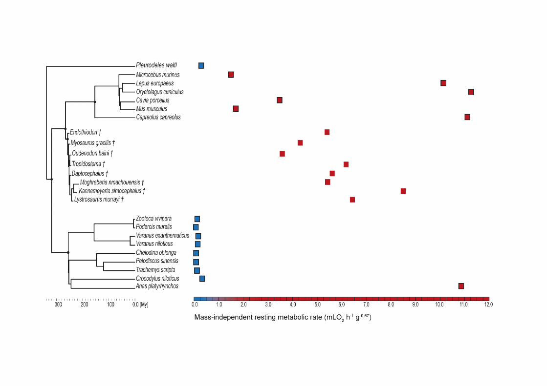

239
FIG. 2. Mass-independent resting metabolic rate (in mLO2 h-1 g-0.67) for the extant and
extinct species of study using PEMs, considering the consensus clade (((Moghreberia,
Kannemeyeria), Lystrosaurus), Daptocephalus) (e.g. Cox & Angielczyk 2015; Boos et
al. 2016; Angielczyk & Kammerer 2017; Kammerer & Smith 2017; Angielczyk et al.
2018). The RMR values gradient extends from ectotherms (indicated by blue dots) to
endotherms (indicated by red dots). We show highest inferred values for the fossil taxa
using models. The ambiguous inferred RMR values were represented by a question
mark. The corresponding 95% confidence intervals (Table 4) are represented for the
predicted value of extinct taxa.


TABLE 1. RMR and histological measurements in the femur in the extant and extinct taxa of the study taken from the literature and newly added.
Species RMR (mLO2 h-1 g-0.67) OstD (ost./µm2) OstA (µm2) OstS VascD (vas. cav./mm2)
Anas platyrhynchos 10.865a 0.002567298b 20.6440151b 0.496256631b 0.582682
Capreolus capreolus 11.111d 0.00503404c 66.3338c 0.682011681c 0.735128
Cavia porcellus 3.477a 0.0018753b 34.1550045b 0.497251828b 0.336093
Chelodina oblonga 0.085a 0.000972828b 28.1246416b 0.615366523b 0
Crocodylus niloticus 0.336a 0.000707445b 29.6935670b 0.474721005b 0.051708
Lepus europaeus 10.149e 0.00485708b 49.1568c 0.6433817c 0.809494
Microcebus murinus 1.526a 0.003900548b 30.0081130b 0.586549866b 0.241296
Mus musculus 1.696a 0.001222124b 29.4847303b 0.48045441b 0.076195
Oryctolagus cuniculus 11.263f 0.00403514b 73.277c 0.646803938c 0.42166
Pelodiscus sinensis 0.083a 0.000913358b 24.4859618b 0.52191737b 0
Pleurodeles waltl 0.297a 0.0005911427b 134.939471b 0.59478267b 0
Podarcis muralis 0.084a 0.001117739b 11.9915127b 0.438353062b 0
Trachemys scripta 0.117a 0.001403736b 28.9630975b 0.494580499b 0
Varanus exanthematicus 0.173a 0.001195398b 33.3619992b 0.462645883b 0.005003
Varanus niloticus 0.157a 0.001007931b 39.6995653b 0.478829164b 0.017692
Zootoca vivipara 0.124a 0.002442651b 13.6767645b 0.42653396b 0
Moghreberia nmachouensis NA 0.00316485c 82.1627c 0.6130401c 0.31360095
Oudenodon baini NA 0.00495632c 21.0048c 0.5732658c 0.369144
Kannemeyeria simocephala NA 0.003461466 90.37377 0.660984 0.5792235
Lystrosaurus murrayi NA 0.004316035 66.175304 0.619432 0.52530187
Daptocephalus NA 0.0041463 52.9261 0.65018 0.48533496
Tropidostoma NA 0.0032531 69.8459 0.63246 0.49969028 a from (Montes et al. 2007); b from (Legendre et al. 2016); c from (Olivier et al. 2017); d from (Mauget et al. 1999); e from (Hackländer et al. 2002); f from (Seltmann et al. 2009). Abbreviations: OstA, osteocyte area; OstD, osteocyte density; OstS, osteocyte shape; ost, osteocyte; RMR, mass-specific resting metabolic rate; vasc. cav., vascular cavities; VascD, primary osteon density.

TABLE 2. RMR and histological measurements in the humerus in the extant and extinct taxa of the study taken from the literature and newly added.
Species RMR (mLO2 h-1 g-0.67) OstD (ost./µm2) OstA (µm2) OstS VascD (vas. cav./mm2)
Anas platyrhynchos 10.865a 0.002283169b 24.2913684 0.509126729 0.22331333
Capreolus capreolus 11.111d 0.00469016c 64.6514c 0.701349738c 0.9137543
Cavia porcellus 3.477a 0.001966108b 34.98627114c 0.485471037c 0.29232459
Chelodina oblonga 0.085a 0.001208368c 24.27796702c 0.56336775c 0
Crocodylus niloticus 0.336a 0.000699242b 34.60152723b 0.485430779b 0.05218821
Lepus europaeus 10.149e 0.00517246c 49.7288c 0.668795497c 0.52979847
Microcebus murinus 1.526a 0.003801204b 28.5159804b 0.526748184b 0.24489796
Mus musculus 1.696a 0.001230761b 22.62412616b 0.526253882b 0.12684615
Oryctolagus cuniculus 11. 263f 0.00401476c 79.9143c 0.661216357c 0.28426461
Pelodiscus sinensis 0.083a 0.001185422b 20.92560266b 0.497140919b 0
Pleurodeles waltl 0.297a 0.000834283b 107.919445b 0.553076316b 0
Podarcis muralis 0.084a 0.000864162b 11.95338603b 0.404075493b 0
Trachemys scripta 0.117a 0.001196311b 27.13462106b 0.516404884b 0
Varanus exanthematicus 0.173a 0.001232928b 36.29314955b 0.43520731b 0.04525424
Varanus niloticus 0.157a 0.001394133b 38.16383708b 0.470869021b 0.03805003
Zootoca vivipara 0.124a 0.003264965b 10.45412684b 0.442348205b 0
Moghreberia nmachouensis NA 0.00361166 72.9888 0.5619222 0.4812
Oudenodon baini NA 0.00418836 92.5187 0.51583 0.15008925
Lystrosaurus murrayi NA 0.00404318 71.06834 0.603978 0.54666341
Tropidostoma NA 0.003743684 92.963724 0.614617 0.45762625
Myosaurus gracilis NA 0.005984664 46.205507 0.593428 0.359545
Endothiodon NA 0.0046199 53.9649 0.6408 0.60566135 a from (Montes et al. 2007); b from (Legendre et al. 2016); c from (Olivier et al. 2017); d from (Mauget et al. 1999); e from (Hackländer et al. 2002); f from (Seltmann et al. 2009). Abbreviations: OstA, osteocyte area; OstD, osteocyte density; OstS, osteocyte shape; ost, osteocyte; RMR, mass-specific resting
metabolic rate; vasc. cav., vascular cavities; VascD, primary osteon density.

TABLE 3. Predicted mass-independent resting metabolic rate (in mLO2 h-1 g-0.67) in dicynodont fossils, based on a continuous multiple regression model using MCMC and PGLS methods, considering the consensus clade (((Moghreberia, Kannemeyeria), Lystrosaurus), Daptocephalus) (e.g. Cox and Angielczyk 2015; Boos et al. 2016; Angielczyk and Kammerer 2017; Kammerer and Smith 2017; Angielczyk et al. 2018).
Taxa Femur Humerus
��RMRpred Min limit of CI95% Max limit of CI95% ��RMRpred Min limit of CI95% Max limit of CI95%
Kannemeyeria simocephalus † 8.2487 8.1417 8.3570 NA NA NA
Moghreberia nmachouensis † 5.4165 5.3506 5.4832 4.2684 4.2194 4.3179
Myosaurus gracilis † NA NA NA 4.3141 4.2650 4.3637
Lystrosaurus murrayi † 6.4263 6.3515 6.5020 5.3439 5.2847 5.4039
Oudenodon baini † 2.2269 2.1951 2.2592 3.5929 3.5443 3.6421
Tropidostoma † 5.9379 5.8704 6.0062 6.1707 6.0985 6.2437
Daptocephalus † 5.6081 5.5439 5.6730 NA NA NA
Endothiodon † NA NA NA 5.4032 5.3417 5.4655
Abbreviations: CI, 95% confidence interval; ��RMRpred, the predicted arithmetic mean of the mass-specific resting metabolic rate.

TABLE 4. Predicted mass-independent resting metabolic rate (in mLO2 h-1 g-0.67) in dicynodont fossils using PEMs (following the method of Guénard et al. (2013) and Legendre et al. (2016)), considering the consensus clade (((Moghreberia, Kannemeyeria), Lystrosaurus), Daptocephalus) (e.g. Cox and Angielczyk 2015; Boos et al. 2016; Angielczyk and Kammerer 2017; Kammerer and Smith 2017; Angielczyk et al. 2018).
Taxa Femur Humerus
��RMRpred Min limit of CI95% Max limit of CI95% ��RMRpred Min limit of CI95% Max limit of CI95%
Kannemeyeria simocephalus † 2.5697 1.8787 3.5148 NA NA NA
Moghreberia nmachouensis † 2.3782 1.6934 3.3399 1.6464 1.2530 2.1633
Myosaurus gracilis † NA NA NA 1.6464 1.2921 2.0979
Lystrosaurus murrayi † 3.1099 2.2891 4.2250 1.6464 1.2842 2.1108
Oudenodon baini † 3.5050 2.5597 4.7995 1.6464 1.2921 2.0979
Tropidostoma † 2.4354 1.9079 3.1088 1.6464 1.2921 2.0979
Daptocephalus † 3.0039 2.2679 3.9785 NA NA NA
Endothiodon † NA NA NA 1.6464 1.2921 2.0979
Abbreviations: CI, 95% confidence interval; ��RMRpred, the predicted arithmetic mean of the mass-specific resting metabolic rate.
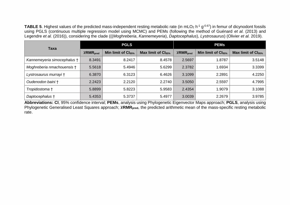
TABLE 5. Highest values of the predicted mass-independent resting metabolic rate (in mLO2 h-1 g-0.67) in femur of dicynodont fossils using PGLS (continuous multiple regression model using MCMC) and PEMs (following the method of Guénard et al. (2013) and Legendre et al. (2016)), considering the clade (((Moghreberia, Kannemeyeria), Daptocephalus), Lystrosaurus) (Olivier et al. 2019).
Taxa PGLS PEMs
��RMRpred Min limit of CI95% Max limit of CI95% ��RMRpred Min limit of CI95% Max limit of CI95%
Kannemeyeria simocephalus † 8.3491 8.2417 8.4578 2.5697 1.8787 3.5148
Moghreberia nmachouensis † 5.5618 5.4946 5.6299 2.3782 1.6934 3.3399
Lystrosaurus murrayi † 6.3870 6.3123 6.4626 3.1099 2.2891 4.2250
Oudenodon baini † 2.2423 2.2120 2.2740 3.5050 2.5597 4.7995
Tropidostoma † 5.8899 5.8223 5.9583 2.4354 1.9079 3.1088
Daptocephalus † 5.4353 5.3737 5.4977 3.0039 2.2679 3.9785
Abbreviations: CI, 95% confidence interval; PEMs, analysis using Phylogenetic Eigenvector Maps approach; PGLS, analysis using
Phylogenetic Generalised Least Squares approach; ��RMRpred, the predicted arithmetic mean of the mass-specific resting metabolic rate.

CHAPTER V – DISCUSSION
CHAPTER V
DISCUSSION
Reconstruction of dicynodonts Lisowicia in a Late Triassic paleoenvironment of Poland (figure modified from Romano and Manucci, 2019)
CHAPTER V – DISCUSSION
247
Phylogenetic position still unconsensual
As largely developed in PAPER I, the taxonomic attribution of the first
dicynodont specimen discovered by Counillon (1896) in the Luang Prabang Basin
(Laos) has been strongly debated. The Laotian dicynodont is supposed to belong to
Dicynodon or Lystrosaurus (e.g., Das Gupta, 1922; Woodward, 1932; Yuan and
Young, 1934; Piveteau, 1938; Battail, 2009; Kammerer et al., 2011). The different
analyses conducted in this work (PAPER I, PAPER II), as well as recent phylogenetic
study (J. Liu, submitted, 2019) demonstrated that no consensus can be reach to
position the Laotian skulls of Counillonia and Repelinosaurus. The phylogenetic
analysis performed in PAPER I placed the new Laotian species Counillonia superoculis
in Dicynodontidae with Daptocephalus, Peramodon, Dinanomodon, Turfanodon,
Euptychognathus, Sintocephalus, Jimusaria, Gordonia, Delectosaurus, Vivaxosaurus,
and Dicynodon. In the same analysis, Repelinosaurus robustus is sister taxa of all
Triassic kannemeyeriiforms and thus considered as such by the comprehensive
definition of Kannemeyeriiformes by Kammerer et al. (2013). In PAPER II, the
phylogenetic matrix has been modified from PAPER I by: (1) addition of three new
discrete characters (discrete 172-174) from Kammerer (2018), (2) revision of the
cranial and postcranial coding of Moghreberia nmachouensis, (3) deletion of the
species Dicynodon huenei and addition of Dicynodon angielczyki and Daptocephalus
huenei, according to the recent taxonomic revision of Kammerer (2019a), (4) addition
of Pentasaurus goggai (Kammerer, 2018), Ufudocyclops mukanelai (Kammerer et al.,
2019), and Lisowicia bojani (Sulej & Niedwźiedzki 2019), and (5) revision of the coding
of Compsodon and Sangusaurus according to the suggestions of Kammerer et al.
(2019). The phylogenetic analysis conducted with the modified matrix in PAPER II led
to a position of Counillonia and Repelinosaurus within Lystrosauridae with Syops and
Lystrosaurus. In a recent analysis, both Laotian dicynodonts are included in
Dicynodontidae (J. Liu, submitted, 2019). Repelinosaurus and Counillonia are placed
sister taxa in the two later analyses (PAPER II; J. Liu, submitted, 2019).
The matrices differed in PAPER I, PAPER II, and a recent phylogenetic study
(J. Liu, submitted, 2019). They thus resulted in different phylogenetic analyses and
thus potentially different phylogenetic relationships. The ambiguous phylogenetic
position of the Laotian dicynodonts may also be explained by their varied combination
of character-states, with particular features only defined in Kannemeyeriiformes,
CHAPTER V – DISCUSSION
248
Dicynodontidae, and/or Lystrosaurus. Indeed, Counillonia and Repelinosaurus present
an interparietal that does not contribute to the intertemporal skull roof (80[0]; PAPER
II) and an occipital condyle distinctly triradiate (133[1]; PAPER II) as in Dicynodon,
Vivaxosaurus, Delectosaurus, Daptocephalus, Peramodon, Syops, and Lystrosaurus.
The ectopterygoids do not extend further posteriorly than the palatines in the Laotian
dicynodonts (107[1]; PAPER II) as in Syops, Sintocephalus, and Jimusaria, while the
ectopterygoids are absent in the majority of Kannemeyeriiformes and Lystrosaurus. As
well as the majority of Kannemeyeriifromes, no intertuberal ridge is present between
the two parabasisphenoid-basioccipital tubera in the Laotian dicynodonts (126[0];
PAPER II). This is not the case for Dicynodon, Vivaxosaurus, Delectosaurus,
Daptocephalus, Peramodon, Turfanodon, Jimusaria, and Lystrosaurus. The anterior
tip of snout is rounded in Counillonia (35[0]; PAPER II) as it is in Dicynodon,
Vivaxosaurus, Dinanomodon, Delectosaurus, Daptocephalus, Peramodon,
Turfanodon, Gordonia, and Sintocephalus. The nasals of Repelinosaurus bear a
distinct median swelling (57[1]; PAPER II) as in Dicynodon, Vivaxosaurus,
Dinanomodon, Delectosaurus, Daptocephalus, Peramodon, Turfanodon, and
Jimusaria, while clear paired swellings are present in the majority of species of
Lystrosaurus and Kannemeyeriiformes. However, the postorbitals of Repelinosaurus
bear well-developed rugosities (65[1]; PAPER II) as in Lystrosaurus and the majority
of Kannemeyeriiformes. Finally, Repelinosaurus would be particularly related to
Kannemeyeriiformes by the absence of postfrontal bone (64[1]; PAPER II).
In addition to the three skulls LPB 1993-2, 1993-3, and 1995-9, abundant
dicynodont postcranial and fragmentary cranial remains have also been excavated
from the Purple Claystone Fm. The study of this new material should help to determine
more precisey the phylogenetic position of the Laotian dicynodonts and their more
closely relatives (Kannemeyeriiformes, Dicynodontidae, or Lystrosaurus). For
instance, the precoracoid participates to the glenoid in Lystrosaurus (discrete character
172; PAPER II) and not to Kannemeyeriiformes, Gordonia, and Daptocephalus
leoniceps, the only ‘Dicynodon’-grade taxa coded for this character. Also, the ilium is
notched on its dorsal edge in Lystrosaurus murrayi, Vivaxosaurus, and Peramodon, as
not in Kannemeyeriiformes (discrete character 183; PAPER II).

CHAPTER V – DISCUSSION
249
Datation of the Laotian dicynodont debated: discussion about the interpretation
of “ghost lineages”
As discussed in PAPER I, the Purple Claystone Fm (Laos) has been dated using
U-Pb geochronology on detrital zircon, which concluded to an age encompassing the
Permo-Triassic boundary (Rossignol et al., 2016). However, while a late Permian age
can not be excluded, an Early Triassic age appears the most likely. This hypothesis is
notably based on (1) zircon grains older than the real age of sedimentation of the
deposits bearing the dicynodonts and (2) volcaniclasts (likely comprising the youngest
zircon grains) extensively reworked (PAPER I; Bercovici et al., 2012; Blanchard et al.,
2013; Rossignol et al., 2016). Battail (2009) supposed a late Permian age based on
the attribution of the Laotian skulls LPB 1993-2, 1993-3, and 1995-9 to the Permian
genus “Dicynodon”. A similar age has also been suggested by a recent phylogenetic
study including the Laotian dicynodonts (J. Liu, submitted, 2019). The author
supported his hypothesis by the close relationships between Counillonia,
Repelinosaurus, and Permian dicynodontids, and the interpretation of the implied
“ghost lineage” by a stratigraphic bias. The shorter the ghost lineage, the more
parsimonious is the hypothesis to date the Laotian taxa. However, this latter
interpretation rests on only one of the multiple possible interpretations of the “ghost
lineage”.
According to Norell (1992), the ghost lineage is an unknown evolutionnary line
resulting from the combination of the stratigraphy and phylogeny. It is respresented by
the difference in age between the oldest occurences of two sister taxa. A clear
disctinction was made between the “ghost lineages” meaning “the corrected
extensions of groups”, and the “ghost taxa” meaning “the kinds of ghost lineages that
are embedded in the internal structure of trees” (Fig. V.1; Norell, 1993). In other words,
the ghost lineage represents a stratigraphic gap in fossil records implied by the
phylogenetic relationships. Following the Norell (1993)’s scheme, the “ghost taxa” are
groups that filled the stratigraphic gap. However, Norell (1993) practically defined the
“ghost taxa” as the ancestral group of the clade emerging from the ghost lineage (for
instance, see E, the ancestral group of the clade A+B; Fig. V.1). This assumption is
erroneous under the light of a pattern interpretation of the cladogram. As opposed to
some authors (e.g., Sidor and Hopson, 1998), I thus assume that distinguishing the
“ghost lineage” and “ghost taxa” is critical. Indeed, using the example given by Norell

CHAPTER V – DISCUSSION
250
(1993) (Fig. V.1), I consider the “ghost taxa” as all taxa more closely related to the
clade A+B, or to A, or to B than to the clade C+D, with occurences included in the
stratigraphic gap of the “ghost lineage”. According to the latter definition, the “ghost
taxa” could either correspond to (1) the occurences of unknown stem-taxa of the clade
A+B or (2) the unknown occurrence of the taxa A and/or B (Fig. V.1). In the first case,
the age of the clade A+B remains unchanged whereas in the second case, its age can
effectively be extended to the older occurrences of the “ghost taxa”. There is no reason
to choose one hypothesis over the others.
As mentioned above, the interpretation of a late Permian age based on the
phylogenetic relationships between the Laotian dicynodonts and Permian
dicynodontoids thus did not contemplate all the possible hypotheses of the implied
ghost lineage. Similarly, the hypothesis of unknown kannemeyeriiforms in late Permian
only supported by Permian close relatives (Dicynodontidae and/or Lystrosaurus) (e.g.,
Fröbisch, 2007; Fröbisch et al., 2010; Kammerer et al., 2011), must be considered with
caution. Considering the survivorship of multiple dicynodont lineages across the P-Tr
boundary (discussed in PAPER I), it is risky to assume that Kannemeyeriiformes
Figure V.1. Distinction between the “ghost lineages” and “ghost taxa”, figure provided from Norell
(1993)

CHAPTER V – DISCUSSION
251
survived the crisis. Based on their real stratigraphic occurences, it is more reasonable
to suggest that the group comprising Kannemeyeriiformes and their sister-taxa has
survived the crisis. Also, it is more accurate to assume that the emydopoids have
survived the P-Tr crisis, than to suggest hypothetical occurences of the Triassic
Myosaurus and Kombuisia in Permian.
To go even further, the relevance of the concept of the “ghost lineage” can also
be discussed. This concept implicitly recognises the evolutionnary process of
diversification as dichotomic implying a same age for two sister-taxa. In this case, the
cladogram is a direct image of the evolutionnary history of the groups. Still, multiple
evolutionnary scenarii can match a unique cladogram (Fig. V.2; e.g., Harper, 1976;
Platnick, 1977). As shown by Platnick (1977) (Fig. V.2), the topology A1 can result
either from a dichotomic evolutionnary process (“cladogenesis” according to Rensch,
1929, 1959; see A1 on Fig. V.2) or a linear and gradual evolutionnary process
(“anagenesis” according to Rensch, 1929, 1959; see B1 on Fig. V.2). Also, a
Figure V.2. Cladograms and their multiple possible evolutionnary scenarii associated, figure provided
by Platnick (1977)

CHAPTER V – DISCUSSION
252
combination of both evolutionnary processes (see C1 and D1 on Fig. V.2) can be
considered. Therefore, the evolutionnary history of the kannemeyeriiforms could be
interpreted either as an ancestral group splitting into the kannemeyeriiforms and their
sister-taxa representatives (“cladogenesis”, named ‘scenario 1’), or as an emergence
of the kannemeyeriiforms within their sister-taxa representatives (“anagenesis”, named
‘scenario 2’). Theoritically, the kannemeyeriiforms may also be the ancestral
population of their sister-taxa representatives. However, this last scenario is less likely
considering the stratigraphic records. Returning to the “ghost lineage” concept, in
scenario 1, the “ghost lineage” is an effective stratigraphic gap in the fossil record. The
“ghost taxa” can thus be unknown Permian kannemeyeriiforms and/or unknown
Permian stem-kannemeyeriiforms. In scenario 2, the “ghost lineage” may only be a
topologic construction implied by the cladogram, and the “ghost taxa” do not exist. A
stratigraphic extension of the fossil record of the kannemeyeriiforms and stem-
kannemeyeriiforms until Permian is thus not required in scenario 2. Nevertheless, a
stratigraphic overlap between the extensions of the two sister-taxa is necessary. In the
case of a stratigraphic lacuna between the sister-taxa, scenario 2 involves a
stratigraphic extension of one or both sister-taxa to reach the overlap. Only in this
particular situation, there is an effective stratigraphic gap in fossil record in scenario 2.
Nonetheless, filling this gap will always be more parsimonious than in the case of
scenario 1.
As it is the case with Kannemeyeriiformes, the scenario 2 proposes an origin of
the representatives of the Laotian dicynodonts Counillonia and Repelinosaurus within
the Permian dicynodontids. However, considering this scenario, a Permian age of the
Laotian dicynodonts is thus not required.
Dicynodonts: large dominant herbivores facing to the end Triassic crisis
After surviving the Permian-Triassic crisis, the dicynodonts greatly diversified
during the Middle Triassic with the apparition of around twenty new genera (Fig. I.3).
They constituted the dominant herbivores of their ecosystem, especially by their
imposing size. Indeed, the body size of the dicynodonts noticeably increased during
the Triassic. Among the few forms known in the Early Triassic, none of the skulls
exceed thirty centimeters (basal lengths [BLS] measured in ventral view from the end
of the premaxilla to the posterior border of the occipital condyle): Lystrosaurus (except
the Permian L. maccaigi), Kombuisia, Myosaurus, and the Laotian dicynodonts

CHAPTER V – DISCUSSION
253
Counillonia and Repelinosaurus (see discussion about their age in PAPER I) (C. Olivier
pers. observ.; e.g., Cluver, 1974; Hammer and Cosgriff, 1981; Botha and Smith, 2007;
Fröbisch, 2007; Fröbisch et al., 2010; Ray et al., 2012). Except few big forms (BLS≈
45 cm) such as Kannemeyeria, Angonisaurus, and Dolichuranus (C. Olivier pers. obs.),
most of Middle Triassic dicynodonts such as Rechnisaurus, Sangusaurus,
Shaanbeikannemeyeria, Shansiodon, Tetragonias, Uralokannemeyeria, and
Xiyukannemeyeria, are middle-sized forms with a skull not exceeding 35 cm (C. Olivier
pers. observ.; e.g., Cruickshank, 1967; Roy-Chowdhury, 1970; Bandyopadhyay, 1989;
Angielczyk et al., 2018). The biggest dicynodonts mainly appeared during the Late
Triassic with skulls ranging from 40 to 45 cm (Dinodontosaurus, Jachaleria,
Moghreberia, Parakannemeyeria, Placerias, and Stahleckeria), and superior to 60 cm
with Ischigualastia (C. Olivier pers. observ.; e.g., Camp and Welles, 1956; Vega-Dias
et al., 2004; Abdala et al., 2013). While exceptional, small-sized dicynodont skulls
around 20 cm, such as Vinceria and Sinokannemeyeria are also known in the Late
Triassic (C. Olivier pers. observ.; e.g., Domnanovich and Marsicano, 2012). No firm
conclusions resulted from the study of the dicynodont postcranial material discovered
in the Argana Basin (Morocco) about the presence of multiple morphotypes and the
taxonomic validity of the Moroccan genus Azarifeneria (PAPER II). However, a second
big morphotype, distinct from Moghreberia nmachouensis by its clear robustness, can
be highlighted (PAPER II). The femur MNHN.F.ALM 284 (maximum length [FL] of 47
cm) (Fig. 28F-H in PAPER II) is one of the biggest Late Triassic forms including
Ischigualastia (FL= 47 cm), Stahleckeria (FL= 55 cm), and Lisowicia (FL= 80 cm) (C.
Olivier pers. observ.; e.g., Sulej and Niedźwiedzki, 2019). In conclusion, both the skull
and femora sizes of dicynodonts increase during the Triassic.
Predator-prey interactions could be a hypothesis explaining the increase of the
body size in dicynodonts (Sinclair et al., 2003; Niedźwiedzki et al., 2011). Indeed, bite
marks on dicynodont bones indicated that predatory dinosaurs hunted the dicynodonts
in Late Triassic (Niedźwiedzki et al., 2011). An increase of the predator body size (i.e.,
dinosauromorphs and non-dinosaur archosauromorphs) might have lead to a bigger
dicynodont body size (Niedźwiedzki et al., 2011). By selection pressure, a large size
therefore constitutes a refuge from large predators. According to Sinclair et al. (2003),
increase in body size implies a higher energetic need including the maintenance of a
high metabolic rate in dicynodonts (PAPERS III and IV). The study of dicynodont
coprolites by Bajdek et al. (2014) provided elements about their diet and suggested an

CHAPTER V – DISCUSSION
254
efficient digestive process, because of the high degree of decomposition of the
ingested food. The retention time of the food bowl is related to the size of the digestive
system (Fritz et al., 2009). With herbivory diet, selection favors long gastrointestinal
tract and thus a large body size in mammals and reptiles to maximize the energetic
gain (Fritz et al., 2010, 2011; Müller et al., 2013).
The abundance of dicynodont remains and their diversity, with 10 early Late
Triassic genera including four newly appeared forms (Dinodontosaurus, Stahleckeria,
Sinokannemeyeria, Parakannemeyeria, Vinceria, Moghreberia, Eubrachiosaurus,
Placerias, Ischigualastia, and Jachaleria), suggest that they were well adapted to their
ecosystems. Nonetheless, only Jachaleria survived the end of the Carnian (Fig. I.3).
Newell (1963) indentified a mass extinction crisis in the end of Triassic (Tr-J crisis) that
impacted more than a third of the fauna families. This crisis was considered as one of
the big five major mass extinctions that include also: Late Ordovician, Late Devonian,
Late Permian, and Late Cretaceous (e.g., McGhee et al., 2004; Twitchett, 2006). In the
Eastern United States, catastrophic events in the Late Triassic have been suggested
by Olsen et al. (2002) in the non dinosaurian vertebrate and plant assemblages.
Nevertheless, a hard and abruptness decline of the biodiversity at the end of the
Triassic has been questioned by some authors (e.g., Hallam, 2002; Lucas and Tanner,
2004; Tanner et al., 2004). Richoz et al. (2007) explained that the impact of the Tr-J
crisis may be overestimated: it could have been multiple smaller crises taking place all
Late Triassic long. During the Carnian and the early Norian, high changes in
environment and biodiversity have been shown with a mean decreasing of 70% of the
specific diversity of molluscs (McRoberts and Newton, 1995; Richoz et al., 2007).
Benton (1991, 1994) supported that the environment perturbations in the late Carnian
impacted more the diversity of European terrestrial tetrapods than the Tr-J crisis. High
environmental changes have been noticed during the Carnian with negative excursions
of the isotopic carbon, oceanic transgressions, climatic changes (“Carnian Humid
Episode“). They have been correlated to perturbations in european plant assemblages
(Richoz et al., 2007; Roghi et al., 2010; Ruffell et al., 2015; Sun et al., 2016). Lucas
and Tanner (2004) and Tanner et al. (2004) explained the apparent abruptness of the
Tr-J crisis by the stratigraphic imprecision (Teichert, 1988) deriving from the
combination of multiple temporal distributions on a large scale (“Signor-Lipps effect”).
Lucas (1994) called this inaccuracy the “Compiled Correlation Effect” (CCE). The CCE
results in “artificial concentration of extinctions at stage/age boundaries; a complex
CHAPTER V – DISCUSSION
255
extinction of significant temporal duration during a stage/age is made to appear as a
mass extinction at the end of the stage/age”, as noted by Lucas and Tanner (2004).
The CCE suggested that the ammonits became abruptly exinct at the end of the
Rhaetian, while a more detailed study of the temporal distribution of the group
demonstrated multiple small extinction events during the Rhaetian, suggesting a more
gradual disappearance of the ammonits (Lucas and Tanner, 2004).
A significant faunal and floral turnover characterizes the Carnian (e.g.,
Behrensmeyer et al., 1992; Simms et al., 1994; Fraser and Sues, 2010). Fraser and
Sues (2010) therefore designated this period as “the Dawn of the Modern World”, with
a major change from ‘archaic’ forms to ‘modern’. Indeed, the terrestrial ecosystems
was formerly dominated by the dicynodont therapsids and their comtemporaries:
procolophonid parareptiles, chroniosuchian anthracosaurs, and some
archosauromorphs such as the rhynchosaurs (e.g., Behrensmeyer et al., 1992; Simms
et al., 1994; Fraser and Sues, 2010). The Late Triassic was marked by the appearance
of the prosauropod dinosaurs and a Mesozoic ecosystem dominated by crocodiliforms,
lepidosaurs, mammaliaforms, and dinosaurs. Behrensmeyer et al. (1992, p. 343)
added that “the late Carnian and Norian produced a major turnover, not only in the
groups of herbivorous tetrapods present but also in the nature of vertebrate herbivory”.
The domination of the ‘herbivores’ ecological niche by the dicynodont synapsids during
the Triassic has been strongly impacted by the major floral changes during the Carnian,
and then by the appearance of the larger and probably more competitive dinosaurs
(prosauropods, sauropods, and ornithischians).
As it happened with the ammonits (Lucas and Tanner, 2004), the recent
discovery of the kannemeyeriiforms Pentasaurus and Lisowicia dated to latest Norian–
earlier Rhaetian would support a more gradual extinction of the dicynodonts during the
Late Triassic (e.g., Knoll, 2004; Fraser and Sues, 2010; Kammerer, 2018; Sulej and
Niedźwiedzki, 2019). In addition, Sulej and Niedźwiedzki (2019) confronted the classic
picture of a Norian ecosystem dominated by dinosaurs by including the dicynodont
Lisowicia as a potential challenger in the ecosystem. However, the volumic body mass
of 9 tons of Lisowicia previously estimated (Sulej and Niedźwiedzki, 2019) was
reassessed by Romano and Manucci (2019) and reduced to 6 tons. Nevertheless,
Romano and Manucci (2019) confirmed the size of the big dicynodont reaching that of
extant elephants.

CHAPTER VI – GENERAL CONCLUSIONS AND PERSPECTIVES
CHAPTER VI
GENERAL CONCLUSIONS AND PERSPECTIVES
Artist’s reconstruction of the dicynodont Lisowicia © Karolina Suchan-Okulska

CHAPTER VI – GENERAL CONCLUSIONS AND PERSPECTIVES
257
Two new dicynodont species (Counillonia superoculis and Repelinosaurus
robustus) were described in the Luang Prabang Basin (Laos), a poorly-studied
geographic area. By the phylogenetic analysis, Counillonia and Repelinosaurus were
first placed in Dicynodontidae and Kannemeyeriiformes, respectively. However, two
more recent studies highlighted different phylogenetic position of these two taxa, both
of them assigned to Lystrosauridae or Dicynodontidae. Their particular combination of
character-states, specific of Lystrosaurus, dicynodontids, and/or kannemeyeriiforms,
may likely explain their non-consensual phylogenetic relationships.
The age of the Purple Claystone Fm (bearing the two Laotian dicynodonts) ranging
from the late Permian to Early Triassic shed new light on the paleogeography of the
Indochina Block and its collision with the rest of the Pangea. Indeed, judging by the
presence of the almost exclusively terrestrial Laotian dicynodonts on the Indochina
Block, a terrestrial connection between this block and Pangea can thus be interpreted
at least in late Permian-Early Triassic, most likely via the South and North China
blocks.
An Early Triassic age for the Purple Claystone Fm being the most likely, the addition
of two new dicynodont forms shortly after the P-Tr crisis supports, on one hand, the
hypothesis of a more rapid recovery of the dicynodonts; and, on the other hand, the
survivorship of multiple dicynodont lineages through the P-Tr boundary. However, the
interpretation of the “ghost lineages” must be considered with caution. The current
stratigraphic and phylogenetic results only allow to suppose the survivorship of
Emydopoidae (with the Triassic Kombuisia and Myosaurus), of Lystrosauridae (with
most of the Lystrosaurus species), and of the clade including Kannemeyeriiformes and
their Permian sister taxa. The close affinities between the Laotian dicynodonts and
Permian dicynodontids, resulted from some phylogenetic analyses, may also add
Dicynodontidae to the list of the dicynodont lineages to have survived the crisis.
In parallel, the taxonomic validity of the three Moroccan dicynodont species
Mogherberia nmachouensis, Azarifeneria barrati, and A. robustus, was reassessed.
An abundant postcranial material found in the Argana Basin (Morocco) have been
described and the major part was attributed to M. nmachouensis, the most abundantly
represented by cranial remains. These new descriptions and the revised study of the
cranial material clearly distinguished Moghreberia from the North American Placerias.
In addition, the phylogenetic analysis showed that Moghreberia is more closely related
CHAPTER VI – GENERAL CONCLUSIONS AND PERSPECTIVES
258
to Lisowicia than to Placerias in Stahleckeridae. The diagnosis of M. nmachouensis
has thus been revised and completed.
The revised study of the cranial material of Azarifeneria did not emphasized significant
differences between the genus and Moghreberia or other Triassic kannemeyeriiforms.
However, while the current absence of diagnostic characters, a second morphotype is
highlighted on the basis of the strong robustness of the postcranial material, which
reminds the cranial specimens of Azarifeneria. The large size of Moghreberia and
especially of the second big morphotype confirmed the increase of the body size in
dicynodonts during the Triassic, with the largest forms in Late Triassic.
Integrated in a large comparative morphological study of the cranial and
postcranial material of Triassic dicynodonts, the study of dicynodonts from Laos and
Morocco highlighted a significant morphological variation and especially an
intraspecific one (ontogeny, sexual dimorphism, and others). While most of this
variation is known in dicynodonts and does not interfer in species distinction, it remains
problematic when it concerns characters used in phylogeny. For instance, the
preparietals depressed or flushed with the skull roof were both present in
Repelinosaurus. Also, a distinct hemispherical and tab-like supinator process as well
as reduced and more anteriorly-developped processes have been described in humeri
of Moghreberia.
The paleophysiology of the dicynodonts has been investigated using statistical
inference models. Paleohistological statistical models have indeed been built to infer
the resting metabolic rate (RMR) of dicynodonts. A first statistical model using the
Phylogenetic Eigenvector Maps approach was used following the method of Guénard
et al. (2013) and Legendre et al. (2016). This first analysis dealed with three
dicynodonts (Moghreberia nmachouensis, Lystrosaurus sp., and Oudenodon baini)
and three histological variables (size, shape, and density of osteocyte lacunae). Later
on, other paleohistological inference models have been performed including a larger
dicynodont sample (Daptocephalus, Endothiodon, Kannemeyeria simocephalus,
Lystrosaurus murrayi, Moghreberia nmachouensis, Myosaurus gracilis, Oudenodon
baini, and Tropidostoma) and using two statistical approaches: PEMs and
Phylogenetic Generalised Least Squares (PGLS) methods. As opposed to PEMs, the
analyses using PGLS provided a more explicit evolutionnary model and the inclusion
of a combination of histological variables (size, shape, and density of osteocyte
lacunae, and primary osteon density). Except for Endothiodon and Myosaurus, a high

CHAPTER VI – GENERAL CONCLUSIONS AND PERSPECTIVES
259
RMR was inferred in all Permian and Triassic studied dicynodonts by PEMs and PGLS
methods.
The results first confirmed the previous qualitative histological studies highlighting
fibrolamellar or ‘incipient’ fibrolamellar bone in dicynodonts. The inference of
endothermy in all Permian and Triassic studied dicynodonts then confronted the
hypothesis supposing the endothermy as the major explanatory factor of differential
success across the P-Tr boundary. More broadly, the paleobiological inference models
suggested a unique acquisition of the mammalian endothermy in Synapsida at least
260 Myr ago at the Neotherapsida node.
However, methodological limits have been observed in both statistical methods (PGLS
and PEMs). The approach using PEMs proposed by Legendre et al (2016) implies the
selection of the best model based on the phylogeny and eventually one of the
histological predictive variables. Using the phylogeny only equal to a simple
optimization of the RMR. The selection of the best model using PGLS with BayesTraits
rests on the coefficient of determination R2, which is unsufficient to attest to the
predictive power of a statistical model. In addition, while a more explicit evolutionary
model is useful to make inferences, it results in the choice of parameters without clear
possible biological explanations (for instance, the distribution of the regression
coefficients β of the histological predictive variable).
This work dealed with the evolution of the dicynodonts during the Triassic and
especially highlighted the impact of the Laotian and Moroccan forms on
paleobiogeographic, phylogenetic, and paleobiologic issues. However, most of these
studies need to be completed and some problematics remain.
The non-consensual position of the Laotian dicynodonts could be more constrained by
the study of their undescribed cranial and postcranial remains (present in MNHN
collections). The use of field data (not possible for this sutdy) will enable to confidently
associate cranial with postcranial remains and therefore to better understand the bone
anatomy of the Laotian forms. Their study would also allow discussing on the presence
of multiple morphotypes in the Laotian material and on possible morphological
variation. If a clear taxonomic attribution of the Laotian postcrania is possible, the
associated stylopods (femora and humeri) could be included in a qualitative and
quantitative histological study to better understand the biology of dicynodonts living
close to the P-Tr crisis.

CHAPTER VI – GENERAL CONCLUSIONS AND PERSPECTIVES
260
Pending better preparation of the cranial remains of the Moroccan Azarifeneria and a
probable highlight of diagnostic features, this genus is considered a nomen dubium. In
addition, only the holotype (MNHN.F.AZA 366.1-.2-.3) of A. barrati has been
discovered in the locality XII of the Argana Basin, a still poorly-studied region according
to Dutuit (1976). Future field works in this region would provide supplementary carnial
and/or postcranial material.
The big size of Moghreberia and the potential second form characterized by the
robustness of its postcranium, confirmed the tendency to increase in size of the
dicynodonts in Late Traissic. However, as opposed to the genera Lisowicia, and
Pentasaurus, the Moroccan dicynodonts do not provide information about the features
of the last dicynodont forms. A complete skeleton in connection of a Laotian form was
discovered in the Red Claystone Fm, dated to Norian by U–Pb zircon geochronology,
and attributed to an undetermined kannemeyeriiform (e.g., Blanchard et al., 2013;
Rossignol et al., 2016). Its study would would lead to: (1) a description of the first
complete dicynodont in Late Triassic, (2) the occurence of a new latest Triassic form
or the stratigraphic extension of a known species, and (3) better understanding of the
dicynodont biodiversity during Late Triassic and the conditions of the extinction of the
dicynodonts.
Finally, despite the multiple methodological limits of PGLS, the most important
perspective to improve the paleobiological inference models should be to add more
extant synapsid taxa to later infer the RMR in a larger sample of extinct non-
mammalian synapsid taxa.

BIBLIOGRAPHY
BIBLIOGRAPHY
Dicynodont © tepuitrouble (www.deviantart.com)
BIBLIOGRAPHY
262
Abdala, F., C. A. Marsicano, R. M. H. Smith, and R. Swart. 2013. Strengthening
Western Gondwanan correlations: A Brazilian Dicynodont (Synapsida,
Anomodontia) in the Middle Triassic of Namibia. Gondwana Research 23:1151–
1162.
Angielczyk, K. D. 2001. Preliminary phylogenetic analysis and stratigraphic
congruence of the dicynodont anomodonts (Synapsida: Therapsida).
Palaeontologica Africana 37:53–79.
Angielczyk, K. D. 2007. New specimens of the tanzanian dicynodont “Cryptocynodon”
parringtoni Von Huene, 1942 (Therapsida, Anomodontia), with an expanded
analysis of Permian dicynodont phylogeny. Journal of Vertebrate Paleontology
27:116–131.
Angielczyk, K. D., and A. A. Kurkin. 2003. Phylogenetic analysis of Russian Permian
dicynodonts (Therapsida: Anomodontia): implications for Permian
biostratigraphy and Pangaean biogeography. Zoological Journal of the Linnean
Society 139:157–212.
Angielczyk, K. D., and B. S. Rubidge. 2010. A new pylaecephalid dicynodont
(Therapsida, Anomodontia) from the Tapinocephalus Assemblage Zone, Karoo
Basin, Middle Permian of South Africa. Journal of Vertebrate Paleontology
30:1396–1409.
Angielczyk, K. D., and C. F. Kammerer. 2017. The cranial morphology, phylogenetic
position and biogeography of the upper Permian dicynodont Compsodon
helmoedi van Hoepen (Therapsida, Anomodontia). Papers in Palaeontology
3:513–545.
Angielczyk, K. D., P. J. Hancox, and A. Nabavizadeh. 2018. A redescription of the
Triassic kannemeyeriiform dicynodont Sangusaurus (Therapsida,
Anomodontia), with an analysis of its feeding system. Journal of Vertebrate
Paleontology 37:189–227.
Arambourg, C. and Duffaud, F. 1960. Note sur la découverte d'un gisement de
vertébrés continentaux dans le Trias du Haut Atlas. Bulletin de la Société
Géologique de France, 2: 172–177.
Artabe, A. E., E. M. Morel, and L. A. Spalletti. 2003. Caracterización de las provincias
fitogeográficas triásicas del Gondwana extratropical. Ameghiniana 40:387–405.
Bain, A. G. 1845. On the Discovery of the Fossil Remains of Bidental and other
Reptiles in South Africa. Quarterly Journal of the Geological Society 1:317–318.

BIBLIOGRAPHY
263
Bajdek, P., K. Owocki, and G. Niedźwiedzki. 2014. Putative dicynodont coprolites from
the Upper Triassic of Poland. Palaeogeography, Palaeoclimatology,
Palaeoecology 411:1–17.
Bandyopadhyay, S. 1988. A Kannemeyeriid Dicynodont from the Middle Triassic
Yerrapalli Formation. Philosophical Transactions of the Royal Society of London
B: Biological Sciences 320:185–233.
Bandyopadhyay, S. 1989. The mammals-like reptile Rechnisaurus from the Triassic of
India. .
Barry, T. H. 1974. A new dicynodont ancestor from the Upper Ecca (lower Middle
Permian) of South Africa. Annals of the South African Museum 64:7–136.
Battail, B. 1997. Les genres Dicynodon et Lystrosaurus (Therapsida, Dicynodontia) en
Eurasie: Une mise au point. Geobios 30:39–48.
Battail, B. 2009. Late Permian dicynodont fauna from Laos. Geological Society,
London, Special Publications 315:33–40.
Battail, B., J. Dejax, P. Richir, P. Taquet, and M. Véran. 1995. New data on the
continental Upper Permian in the area of Luang-Prabang, Laos. Journal of
Geology 11–15.
Behrensmeyer, A. K., J. D. Damuth, W. A. DiMichele, R. Potts, H.-D. Sues, and S. L.
Wing. 1992. Terrestrial Ecosystems through Time: Evolutionnary Paleoecology
of Terretrial Plants and Animals, The University of Chicago Press.
Behrensmeyer, A. K., Damuth, J. D., DiMichele, W. A., Potts, R., Sues, H. -D.,
and Wing S. L., Chicago, 568 pp.
Benoit, J., P. Manger, V. Fernandez, and B. S. Rubidge. 2018. Evolution of facial
innervation in anomodont therapsids (Synapsida): Insights from X-ray
computerized microtomography. Journal of Morphology 1–29.
Benton, M. J. 1991. What really happened in the late Triassic? Historical Biology
5:263–278.
Benton, M. J. 1994. Late Triassic to Middle Jurassic extinctions among continental
tetrapods: testing pattern; pp. 445 in In the shadow of the dinosaurs, University
Press. N. C. Fraser & H. -D. Sues (eds), Cambridge, UK.
Benton, M. J. 2005. Vertebrate Palaeontology, 3rd ed. Blackwell Science, Malden, MA,
455 pp.
Bercovici, A., S. Bourquin, J. Broutin, J.-S. Steyer, B. Battail, M. Véran, R. Vacant, B.
Khenthavong, and S. Vongphamany. 2012. Permian continental

BIBLIOGRAPHY
264
paleoenvironments in Southeastern Asia: New insights from the Luang Prabang
Basin (Laos). Journal of Asian Earth Sciences 60:197–211.
Bernardi, M., F. M. Petti, and M. J. Benton. 2018. Tetrapod distribution and
temperature rise during the Permian–Triassic mass extinction. Proceedings of
the Royal Society B: Biological Sciences 285:20172331.
Bernardi, M., F. M. Petti, E. Kustatscher, M. Franz, C. Hartkopf-Fröder, C. C.
Labandeira, T. Wappler, J. H. A. van Konijnenburg-van Cittert, B. R. Peecook,
and K. D. Angielczyk. 2017. Late Permian (Lopingian) terrestrial ecosystems: A
global comparison with new data from the low-latitude Bletterbach Biota. Earth-
Science Reviews 175:18–43.
Blanchard, S., C. Rossignol, S. Bourquin, M.-P. Dabard, E. Hallot, T. Nalpas, M. Poujol,
B. Battail, N.-E. Jalil, J.-S. Steyer, R. Vacant, M. Véran, A. Bercovici, J. B. Diez,
J.-L. Paquette, B. Khenthavong, and S. Vongphamany. 2013. Late Triassic
volcanic activity in South-East Asia: New stratigraphical, geochronological and
paleontological evidence from the Luang Prabang Basin (Laos). Journal of
Asian Earth Sciences 70–71:8–26.
Bonaparte, J. F. 1969. Two new reptilian “faunas” of the Argentine Triassic. Gondwana
Stratigraphy 2:283–306.
Bonaparte, J. F. 1970. Annotated list of the South American Triassic tetrapods.
Proceedings and Papers 665–682.
Boos, A. D. S., C. F. Kammerer, C. L. Schultz, M. B. Soares, and A. L. R. Ilha. 2016.
A New Dicynodont (Therapsida: Anomodontia) from the Permian of Southern
Brazil and Its Implications for Bidentalian Origins. PLoS ONE 11.
Botha, J. 2003. Biological aspects of the Permian dicynodont Oudenodon (Therapsida:
Dicynodontia) deduced from bone histology and cross-sectional geometry.
Palaeontologica Africana 39:37–44.
Botha, J., and A. Chinsamy. 2000. Growth patterns deduced from the bone histology
of the cynodonts Diademodon and Cynognathus. Journal of Vertebrate
Paleontology 20:705–711.
Botha, J., and A. Chinsamy. 2004. Growth and life habits of the Triassic cynodont
Trirachodon, inferred from bone histology. Acta Palaeontologica Polonica
49:619–627.

BIBLIOGRAPHY
265
Botha, J., and R. M. H. Smith. 2006. Rapid vertebrate recuperation in the Karoo Basin
of South Africa following the End-Permian extinction. Journal of African Earth
Sciences 45:502–514.
Botha, J., and R. M. H. Smith. 2007. Lystrosaurus species composition across the
Permo-Triassic boundary in the Karoo Basin of South Africa: Lystrosaurus
across the PTB. Lethaia 40:125–137.
Botha-Brink, J., and K. D. Angielczyk. 2010. Do extraordinarily high growth rates in
Permo-Triassic dicynodonts (Therapsida, Anomodontia) explain their success
before and after the end-Permian extinction ? Zoological Journal of the Linnean
Society 160:341–365.
Botha-Brink, J., F. Abdala, and A. Chinsamy-Turan. 2012. The radiation and
osteohistology of Nonmammaliaform Cynodonts; pp. 223–248 in Forerunners
of Mammals, Indiana Universiy Press. Chinsamy-Turan, A. (ed.).
Botha-Brink, J., D. Codron, A. K. Huttenlocker, K. D. Angielczyk, and M. Ruta. 2016.
Breeding Young as a Survival Strategy during Earth’s Greatest Mass Extinction.
Scientific Reports 6:24053.
Broom, R. 1905. On the use of the term Anomodontia. Albany Museum Records
1:266–269.
Broom, R. 1911. On some new South African Permian reptiles. Proceedings of the
Zoological Society of London 1911:1073–1082.
Bueno, A. de O. 2015. Descrição osteo-histológica de elementos fósseis de
Dinodontosaurus turpior (Therapsida, Dicynodontia), Mesotriássico do Rio
Grande do Sul, BrasilD. .
Camp, C. L. 1956. Triassic dicynodont reptiles. Part II. Triassic dicynodont compared.
Memories of the University of California 13:305–337.
Camp, C. L., and S. P. Welles. 1956. Triassic dicynodont reptiles. Part I. The North
American genus Placerias. Memories of the University of California 13:255–
304.
Canoville, A., and M. Laurin. 2010. Evolution of humeral microanatomy and lifestyle in
amniotes, and some comments on palaeobiological inferences. Biological
Journal of the Linnean Society 100:384–406.
Cecca, F., and R. Zaragüeta i Bagils. 2015. Paléobiogéographie. Edp Sciences, Paris,
France, 193 pp.

BIBLIOGRAPHY
266
Chen, Z.-Q., and M. J. Benton. 2012. The timing and pattern of biotic recovery following
the end-Permian mass extinction. Nature Geoscience 5:375–383.
Cheng, Z. 1980. Cheng, Z. (1980). Mesozoic stratigraphy and paleontology of the
Shaanxi-Gansu-Ningxia basin. Vertebrate Fossils 2:115–188.
Cheng, Z. W. 1986. Vertebrate fossils; pp. 207–218 in Permian and Triassic Strata and
Fossil Assemblages in the Dalongkou Area of Jimsar, Xinjiang, Geological
Publishing House. Beijing.
Chinsamy, A., and B. S. Rubidge. 1993. Dicynodont (Therapsida) bone histology:
phylogenetic and physiological implications. Palaeontologica Africana 30:97–
102.
Chinsamy, A., and J. H. Hurum. 2006. Bone microstructure and growth patterns of
early mammals. Acta Palaeontologica Polonica 51:325–338.
Chinsamy-Turan, A. 2005. The Microstructure of Dinosaur Bone: Deciphering Biology
with Fine-Scale Techniques. Johns Hopkins University Press, Baltimore, 216
pp.
Chinsamy-Turan, A., and S. Ray. 2012. Bone histology of some Therocephalians and
Gorgonopsians, and evidence of bone degradation by fungi; pp. 199–222 in
Forerunners of Mammals, Indiana Universiy Press. Chinsamy-Turan, A. (ed.).
Clarke, A., and H.-O. Pörtner. 2010. Temperature, metabolic power and the evolution
of endothermy. Biological Reviews 85:703–727.
Cluver, M. A. 1974. The Cranial Morphology of the Lower Triassic Dicynodont
"Myosaurus Gracilis". Annals of The South African Museum 66:35–54.
Cluver, M. A., and N. Hotton III. 1981. The genera Dicynodon and Diictodon and their
bearing of classification of the Dicynodontia (Reptilia, Therapsida). Annales of
the South African Museum 83:99–146.
Cluver, M. A., and G. M. King. 1983. A reassessment of the relationships of Permian
Dicynodontia (Reptilia, Therapsida) and a new classification of dicynodonts.
Annals of South African Museum 91:195–273.
Colbert, E. H. 1982. The distribution of Lystrosaurus in pangaea and its implications.
Geobios 15:375–383.
Cope, E. D. 1870. On the skull of dicynodont Reptilia. Lystrosaurus frontosus from
Cape Colony. Proceedings of the American Philosophical Society 11:419.

BIBLIOGRAPHY
267
Counillon, H. 1896. Documents pour servir à l’étude géologique des environs de
Luang-Prabang (Cochinchine). Comptes Rendus de l’Académie Des Sciences,
Paris 123:1330–1333.
Cox, C. B. 1962. Preliminary diagnosis of Ischigualastia, a new genus of Dicynodont
from Argentina. Breviora 156:8–9.
Cox, C. B. 1965. New Triassic Dicynodonts from South America, Their Origins and
Relationships. Philosophical Transactions of the Royal Society of London B
248:457–514.
Cox, C. B. 1969. Two new dicynodonts from the Triassic Ntawere Formation, Zambia.
Bulletin of the British Museum (Natural History) Geology 17:257–294.
Cox, C. B. 1991. The Pangaea dicynodont Rechnisaurus and the comparative
biostratigraphy of Triassic dicynodont faunas. Palaeontology 34:767–784.
Cox, C. B., and J. Li. 1983. A new genus of Triassic Dicynodont from East-Africa and
its classification. Palaeontology 26:389–406.
Cox, C. B., and K. D. Angielczyk. 2015. A New Endothiodont Dicynodont (Therapsida,
Anomodontia) from the Permian Ruhuhu Formation (Songea Group) of
Tanzania and Its Feeding System. Journal of Vertebrate Paleontology
35:e935388.
Cracraft, J. 1974. Continental drift and vertebrate distribution. Annual Review of
Ecology and Systematics 5:215–261.
Crompton, A. W., C. Musinsky, and T. Owerkowicz. 2015. Evolution of the mammalian
nose; pp. 189–203 in The Great Transformations in Vertebrate Evolution,
University of Chicago Press. E. L. Brainerd, Kenneth Dial, and N. H. Shubin,
Chicago.
Crompton, A. W., T. Owerkowicz, B.-A. S. Bhullar, and C. Musinsky. 2017. Structure
of the nasal region of non-mammalian cynodonts and mammaliaforms:
Speculations on the evolution of mammalian endothermy. Journal of Vertebrate
Paleontology 37:e1269116.
Cruickshank, A. R. I. 1967. A new dicynodont genus from the Manda Formation of
Tanzania (Tanganyika). Journal of Zoology 153:163–208.
Cubo, J., and N.-E. Jalil. 2019. Bone histology of Azendohsaurus laaroussii:
Implications for the evolution of thermometabolism in Archosauromorpha.
Paleobiology 45:317–330.

BIBLIOGRAPHY
268
Damiani, R., C. Vasconcelos, A. Renaut, J. Hancox, and A. Yates. 2007. Dolichuranus
primaevus (Therapsida: Anomodontia) from the middle Triassic of Nambia and
its phylogenetic relationships. Palaeontology 50:1531–1546.
Danilov, A. I. 1971. A new dicynodont from the Middle Triassic of southern Cisuralia.
Paleontological Journal 5:265–268.
Das Gupta, H. C. 1922. Notes on the Panchet Reptile; pp. 237–241 in Sir Asutosh
Mukherjee Silver Jubilee Volumes, University Press. vol. 2. Calcutta.
Domnanovich, N. S., and C. A. Marsicano. 2012. The Triassic dicynodont Vinceria
(Therapsida, Anomodontia) from Argentina and a discussion on basal
Kannemeyeriiformes. Geobios 45:173–186.
Dutuit, J. M. 1965. Découverte de dicynodontes (reptiles therapsides) dans le Trias du
couloir d’Argana (Atlas occidental marocain). Comptes Rendus Hebdomadaires
Des Séances de l’Académie Des Sciences 260:3447.
Dutuit, J. M. 1976. Introduction à l’étude paléontologique du Trias continental
marocain. Description des premiers stégocéphales recueillis dans le couloir
d’Argana (Atlas occidental). Mémoires Du Muséum National d’Histoire Naturelle
36:1–333.
Dutuit, J. M. 1980. Principaux caractères d’un genre de Dicynodonte du Trias
marocain. Comptes Rendus de l’Académie Des Sciences, Paris 290:655–658.
Dutuit, J. M. 1988. Ostéologie crânienne et ses enseignements, apports géologique et
paléoécologique, de Moghreberia nmachouensis, Dicynodonte (Reptilia,
Therapsida) du Trias supérieur marocain. Bulletin Du Muséum National
d’histoire Naturelle. Section C, Sciences de La Terre, Paléontologie, Géologie,
Minéralogie 10:227–285.
Dutuit, J. M. 1989a. Azarifeneria barrati, un deuxieme genre de Dicynodonte du Trias
supérieur marocain. Comptes Rendus de l’Académie Des Sciences. Série 2,
Mécanique, Physique, Chimie, Sciences de l’univers, Sciences de La Terre
309:303–306.
Dutuit, J. M. 1989b. Confirmation des affinités entre Trias supérieurs marocain et sud-
américain: découverte d’un troisième Dicynodonte (Reptilia, Therapsida),
Azarifeneria robustus, n. sp., de la formation d’Argana (Atlas occidental).
Comptes Rendus de l’Académie Des Sciences. Série 2, Mécanique, Physique,
Chimie, Sciences de l’univers, Sciences de La Terre 309:1267–1270.

BIBLIOGRAPHY
269
Efremov, J. A. 1951. On the structure of the knee joint in the higher Dicynodontidae.
Doklady Akademii Nauk SSSR 7 77:483–485.
Elder, A. L. 2000. Late Triassic Dicynodonts: their anatomy, relationships, and
paleobiogeography. the Graduate Faculty of Texas Tech University, 114 pp.
Ezcurra, M. D. 2010. Biogeography of Triassic tetrapods: evidence for provincialism
and driven sympatric cladogenesis in the early evolution of modern tetrapod
lineages. Proceedings of the Royal Society B: Biological Sciences 277:2547–
2552.
Fleischle, C. V., T. Wintrich, and P. M. Sander. 2018. Quantitative histological models
suggest endothermy in plesiosaurs. PeerJ 6:e4955.
Francillon Vieillot, H., V. de Buffrénil, J. Castanet, J. Géraudie, F. J. Meunier, J. Y. Sire,
L. Zylberberg, and A. De Ricqlès. 1990. Microstructure and mineralization of
vertebrate skeletal tissues; pp. 471–530 in Skeletal biomineralization : patterns,
process and evolutionary trends. Carter, J. G. (ed.).
Fraser, N. C., and H.-D. Sues. 2010. The beginning of the ‘Age of Dinosaurs’: a brief
overview of terrestrial biotic changes during the Triassic. Earth and
Environmental Science Transactions of the Royal Society of Edinburgh
101:189–200.
Fritz, J., J. Hummel, E. Kienzle, W. J. Streich, and M. Clauss. 2010. To chew or not to
chew: fecal particle size in herbivorous reptiles and mammals. Journal of
Experimental Zoology Part A: Ecological Genetics and Physiology 313A:579–
586.
Fritz, J., J. Hummel, E. Kienzle, C. Arnold, C. Nunn, and M. Clauss. 2009. Comparative
chewing efficiency in mammalian herbivores. Oikos 118:1623–1632.
Fritz, J., J. Hummel, E. Kienzle, O. Wings, W. J. Streich, and M. Clauss. 2011. Gizzard
vs. teeth, it’s a tie: food-processing efficiency in herbivorous birds and mammals
and implications for dinosaur feeding strategies. Paleobiology 37:577–586.
Fröbisch, J. 2007. The cranial anatomy of Kombuisia frerensis Hotton (Synapsida,
Dicynodontia) and a new phylogeny of anomodont therapsids. Zoological
Journal of the Linnean Society 150:117–144.
Fröbisch, J. 2009. Composition and similarity of global anomodont-bearing tetrapod
faunas. Earth-Science Reviews 95:119–157.

BIBLIOGRAPHY
270
Fröbisch, J., and R. R. Reisz. 2008. A new species of Emydops (Synapsida,
Anomodontia) and a discussion of dental variability and pathology in
dicynodonts. Journal of Vertebrate Paleontology 28:770–787.
Fröbisch, J., K. D. Angielczyk, and C. A. Sidor. 2010. The Triassic dicynodont
Kombuisia (Synapsida, Anomodontia) from Antarctica, a refuge from the
terrestrial Permian-Triassic mass extinction. Naturwissenschaften 97:187–196.
Germain, D., and M. Laurin. 2005. Microanatomy of the radius and lifestyle in amniotes
(Vertebrata, Tetrapoda). Zoologica Scripta 34:335–350.
Girondot, M., and M. Laurin. 2003. Bone profiler: a tool to quantify, model, and
statistically compare bone-section compactness profiles. Journal of Vertebrate
Paleontology 23:458–461.
Govender, R., and A. Yates. 2009. Dicynodont postcrania from the Triassic of Namibia
and their implication for the systematics of Kannemeyeriiforme dicynodonts.
Palaeontologica Africana 44:41–57.
Green, J. L. 2012. Bone and Dental Histology of Late Triassic Dicynodonts from North
America; pp. 179–198 in Forerunners of Mammals, Indiana Universiy Press.
Chinsamy-Turan, A. (ed.).
Green, J. L., M. H. Schweitzer, and E.-T. Lamm. 2010. Limb bone histology and growth
in Placerias hesternus (Therapsida: Anomodontia) from the Upper Triassic of
North America. Palaeontology 53:347–364.
Grine, F. E., C. A. Forster, M. A. Cluver, and J. A. Georgi. 2006. Cranial variability,
ontogeny and taxonomy of Lystrosaurus from the Karoo Basin of South Africa;
pp. 432–503 in Amniote Paleobiology: Perspectives on the Evolution of
Mammals, Birds, and Reptiles, University of Chicago Press. Carrano M. T.,
Gaudin T. J., Blob R. W. & Wible J. R., Chicago.
Guénard, G., P. Legendre, and P. Peres-Neto. 2013. Phylogenetic eigenvector maps:
a framework to model and predict species traits. Methods in Ecology and
Evolution 4:1120–1131.
Hallam, A. 2002. How catastrophic was the end-Triassic mass extinction? Lethaia
35:147–157.
Hammer, W. R., and J. W. Cosgriff. 1981. Myosaurus gracilis, an anomodont reptile
from the Lower Triassic of Antarctica and South Africa. Journal of Paleontology
55:410–424.
BIBLIOGRAPHY
271
Hancox, P. J., K. D. Angielczyk, and B. S. Rubidge. 2013. Angonisaurus and
Shansiodon, dicynodonts (Therapsida, Anomodontia) from subzone C of the
Cynognathus Assemblage Zone (Middle Triassic) of South Africa. Journal of
Vertebrate Paleontology 33:655–676.
Harper, C. W. J. 1976. Phylogenetic inference in paleontology. Journal of Vertebrate
Paleontology 50:180–193.
Hillenius, W. J. 1992. The evolution of nasal turbinates and mammalian endothermy.
Paleobiology 18:17–29.
Hillenius, W. J. 1994. Turbinates in Therapsids: Evidence for Late Permian Origins of
Mammalian Endothermy. Evolution 48:207–229.
Huene, F. 1935. Die fossilen Reptilien des südamerikanischen Gondwanalandes; pp.
93–159 in Ergebnisse der Sauriergrabungen in Südbrasilien 1928–29.
Lieferung 1, Verlag Franz F. Heine. Tübingen.
Huene, F. 1948. Short review of the lower tetrapods; pp. 65–106 in Robert Broom
Commemorative Volume, Special Publications of theRoyal Society of South
Africa. A. L. Du Toit (ed.), Cape Town.
Hunt, A. P., and S. G. Lucas. 1991. The Paleorhinus biochron and the correlation of
the non-marine Upper Triassic of Pangaea. Palaeontology 34:487–501.
Huttenlocker, A. K., and J. Botha-Brink. 2014. Bone microstructure and the evolution
of growth patterns in Permo-Triassic therocephalians (Amniota, Therapsida) of
South Africa. PeerJ 2:e325.
Huxley, T. H. 1859. On a new species of dicynodont (D. murrayi) from near Colesburg,
South Africa; and on the structure of the skull in dicynodonts. Quarterly Journal
of the Geological Society of London 15:649–659.
Huxley, T. H. 1865. On a colle.ction of vertebrate fossils from the Panchet rocks,
Raniganj Coalfield. Memoirs of the Geological Survey of India, Palaeontologia
Indica 1:2–24.
Irmis, R. B., and J. H. Whiteside. 2012. Delayed recovery of non-marine tetrapods after
the end-Permian mass extinction tracks global carbon cycle. Proceedings of the
Royal Society of London B: Biological Sciences 279:1310–1318.
Irmis, R. B., S. J. Nesbitt, K. Padian, N. D. Smith, A. H. Turner, D. Woody, and A.
Downs. 2007. A Late Triassic Dinosauromorph Assemblage from New Mexico
and the Rise of Dinosaurs. Science 317:358–361.

BIBLIOGRAPHY
272
Jasinoski, S. C., M. A. Cluver, A. Chinsamy, and B. D. Reddy. 2014. Anatomical
Plasticity in the Snout of Lystrosaurus; pp. 139–149 in C. F. Kammerer, K. D.
Angielczyk, and J. Fröbisch (eds.), Early Evolutionary History of the Synapsida.
Springer Netherlands, Dordrecht.
Kalandadze, N. N. 1970. New Triassic kanemeyeriids from the South Ural region; pp.
51–57 in Materials on evolution of terrestrial vertebrates, K. K. Flerov (ed.).
Nauka Press, Moscow.
Kalandadze, N. N., and A. G. Sennikov. 1985. New reptiles from the Middle Triassic in
the southern Ural forelands. Paleontological Journal 19:73–80.
Kammerer, C. F. 2018. The first skeletal evidence of a dicynodont from the lower Elliot
Formation of South Africa. Palaeontologia Africana 52:102–128.
Kammerer, C. F. 2019a. Revision of the Tanzanian dicynodont Dicynodon huenei
(Therapsida: Anomodontia) from the Permian Usili Formation. PeerJ 33.
Kammerer, C. F. 2019b. A new dicynodont (Anomodontia: Emydopoidea) from the
terminal Permian of KwaZulu-Natal, South Africa. Palaeontologia Africana.
Kammerer, C. F., and K. D. Angielczyk. 2009. A proposed higher taxonomy of
anomodont therapsids. Zootaxa 2018:1–24.
Kammerer, C. F., and R. M. H. Smith. 2017. An early geikiid dicynodont from the
Tropidostoma Assemblage Zone (late Permian) of South Africa. PeerJ 5:e2913.
Kammerer, C. F., K. D. Angielczyk, and J. Fröbisch. 2011. A comprehensive taxonomic
revision of “Dicynodon” (Therapsida, Anomodontia) and its implications for
dicynodont phylogeny, biogeography, and biostratigraphy. Journal of Vertebrate
Paleontology 31:1–158.
Kammerer, C. F., J. Fröbisch, and K. D. Angielczyk. 2013. On the Validity and
Phylogenetic Position of Eubrachiosaurus browni, a Kannemeyeriiform
Dicynodont (Anomodontia) from Triassic North America. PLoS ONE 8:e64203.
Kammerer, C. F., P. A. Viglietti, P. J. Hancox, R. J. Butler, and J. N. Choiniere. 2019.
A new kannemeyeriiform dicynodont (Ufudocyclops mukanelai , gen. et sp.
nov.) from Subzone C of the Cynognathus Assemblage Zone, Triassic of South
Africa, with implications for biostratigraphic correlation with other African
Triassic Faunas. Journal of Vertebrate Paleontology e1596921.
Kemp, T. S. 1980. Origin of the mammal-like reptiles. Nature 283:378–380.
Kent, D. V., P. S. Malnis, C. E. Colombi, O. A. Alcober, and R. N. Martínez. 2014. Age
constraints on the dispersal of dinosaurs in the Late Triassic from

BIBLIOGRAPHY
273
magnetochronology of the Los Colorados Formation (Argentina). Proceedings
of the National Academy of Sciences 111:7958–7963.
Keyser, A. W. 1973. A new Triassic vertebrate fauna from South West Africa.
Paleontologica Africana 16:1–15.
Keyser, A. W., and A. R. I. Cruickshank. 1979. The origins and classification of Triassic
dicynodonts. Transactions of the Geological Society of South Africa 82:81–108.
Khaldoune, F., N. E. Jalil, D. Germain, and J. S. Steyer. 2017. Les vertébrés du
Permien et du Trias du Maroc (Bassin d’Argana, Haut Atlas occidental) : fenêtre
ouverte sur l’évolution autour de la grande crise fin-paléozoïque. Mémoires de
La Société Géologique de France 64p.
King, G. M., and M. A. Cluver. 1990. The aquatic Lystrosaurus: an alternative lifestyle.
Historical Biology 4:323–341.
Knoll, F. 2004. Review of the tetrapod fauna of the “Lower Stormberg Group” of the
main Karoo Basin (southern Africa): implication for the age of the Lower Elliot
Formation. Bulletin de La Societe Geologique de France 175:73–83.
Knutsen, E. M., and E. Oerlemans. 2020. The last dicynodont? Re-assessing the
taxonomic and temporal relationships of a contentious Australian fossil.
Gondwana Research 77:184–203.
Kriloff, A., D. Germain, A. Canoville, P. Vincent, M. Sache, and M. Laurin. 2008.
Evolution of bone microanatomy of the tetrapod tibia and its use in
palaeobiological inference. Journal of Evolutionary Biology 21:807–826.
Legendre, L. J., G. Guénard, J. Botha-Brink, and J. Cubo. 2016. Palaeohistological
Evidence for Ancestral High Metabolic Rate in Archosaurs. Systematic Biology
65:989–996.
Liu, J., and J. Li. 2003. A new material of kannemeyerid from Xinjiang and the restudy
of Parakannemeyeria brevirostris. Vertebrata Palasiatica 41:147–156.
Lucas, S. 2015. Age and correlation of Late Triassic tetrapods from southern Poland.
Annales Societatis Geologorum Poloniae.
Lucas, S. G. 1904. A new batrachian and a new reptile from the Triassic of Arizona.
Proceeding of the U. S. National Museum 27:193–197.
Lucas, S. G. 1993. Barysoma lenzii (Synapsida: Dicynodontia) from the Middle Triassic
of Brazil, a synonym of Stahleckeria potens. Journal of Paleontology 67:318–
321.

BIBLIOGRAPHY
274
Lucas, S. G. 1994. Triassic Tetrapod Extinctions and the Compiled Correlation Effect.
869–875.
Lucas, S. G. 1995. Triassic dicynodont biochronology. Albertiana 16:33–40.
Lucas, S. G. 2018. Late Triassic Terrestrial Tetrapods: Biostratigraphy, Biochronology
and Biotic Events; pp. 351–405 in L. H. Tanner (ed.), The Late Triassic World.
vol. 46. Springer International Publishing, Cham.
Lucas, S. G., and A. P. Hunt. 1993a. Fukangolepis Yang, 1978 from the Triassic of
China is not an aetosaur. Journal of Vertebrate Paleontology 13:145–147.
Lucas, S. G., and A. P. Hunt. 1993b. A dicynodont from ther Upper Triassic of New
Mexico and its biochronologic significance. New Mexico Museum Natural
History and Science Bulletin 3:321–325.
Lucas, S. G., and R. Wild. 1995. A Middle Triassic dicynodont from Germany and the
biochronology of Triassic dicynodonts. Stuttgarter Beiträge Zur Naturkunde B
220:1–16.
Lucas, S. G., and L. H. Tanner. 2004. Late Triassic extinction events. Albertiana 31:31–
40.
Magwene, P. M. 1993. What’s Bred in the Bone: Histology and Cross-Sectional
Geometry of Mammal-like Reptile Long Bones: Evidence of Changing
Physiological and Biomechanical Demands. Harvard University, Cambridge,
Massacuhssetts, pp.
Maisch, M. W. 2001. Observations on Karoo and Gondwana vertebrates. Part 2: A new
skull-reconstruction of Stahleckeria potens Von Huene, 1935 (Dicynodontia,
Middle Triassic) and a reconsideration of kannemeyeriiform. Neues Jahrbuch
Für Geologie Und Paläontologie - Abhandlungen 220:127–152.
Maisch, M. W., and A. T. Matzke. 2014. Sungeodon kimkraemerae n. gen. n. sp., the
oldest kannemeyeriiform (Therapsida, Dicynodontia) and its implications for the
early diversification of large herbivores after the P/T boundary. Neues Jahrbuch
Für Geologie Und Paläontologie - Abhandlungen 272:1–12.
McGhee, G. R., P. M. Sheehan, D. J. Bottjer, and M. L. Droser. 2004. Ecological
ranking of Phanerozoic biodiversity crises: ecological and taxonomic severities
are decoupled. Palaeogeography, Palaeoclimatology, Palaeoecology 211:289–
297.
McNab, B. K. 1997. On the Utility of Uniformity in the Definition of Basal Rate of
Metabolism. Physiological Zoology 70:718–720.

BIBLIOGRAPHY
275
McRoberts, C. A., and C. R. Newton. 1995. Selective extinction among end-Triassic
European bivalves. Geology 23:102.
Montes, L., N. Le Roy, M. Perret, V. De Buffrenil, J. Castanet, and J. Cubo. 2007.
Relationships between bone growth rate, body mass and resting metabolic rate
in growing amniotes: a phylogenetic approach. Biological Journal of the Linnean
Society 92:63–76.
Müller, D. W. H., D. Codron, C. Meloro, A. Munn, A. Schwarm, J. Hummel, and M.
Clauss. 2013. Assessing the Jarman–Bell Principle: Scaling of intake,
digestibility, retention time and gut fill with body mass in mammalian herbivores.
Comparative Biochemistry and Physiology Part A: Molecular & Integrative
Physiology 164:129–140.
Newell, A. J. 1963. Crises in the history of life. Scientific American 76–93.
Niedźwiedzki, G., P. Gorzelak, and T. Sulej. 2011. Bite traces on dicynodont bones
and the early evolution of large terrestrial predators. Lethaia 44:87–92.
Norell, M. A. 1992. The Effect of Phylogeny, in Extinction and Phylogeny; pp. 89–118
in Taxic Origin and Temporal Diversity, Columbia University Press. Novacek,
M.J. and Wheeler, Q.D. (eds.), New York.
Norell, M. A. 1993. Tree-based approaches to understanding history; comments on
ranks, rules and the quality of the fossil record. American Journal of Science
293:407–407.
Nowack, J., S. Giroud, W. Arnold, and T. Ruf. 2017. Muscle Non-shivering
Thermogenesis and Its Role in the Evolution of Endothermy. Frontiers in
Physiology 8:889.
Olivier, C., B. Battail, S. Bourquin, C. Rossignol, J.-S. Steyer, and N.-E. Jalil. 2019.
New dicynodonts (Therapsida, Anomodontia) from near the Permo-Triassic
boundary of Laos: implications for dicynodont survivorship across the Permo-
Triassic mass extinction and the paleobiogeography of Southeast Asian blocks.
Journal of Vertebrate Paleontology 0:e1584745.
Olsen, P. E., D. V. Kent, H.-D. Sues, C. Koeberl, H. Huber, A. Montanari, E. C.
Rainforth, S. J. Fowell, M. J. Szajna, and B. W. Hartline. 2002. Ascent of
dinosaurs linked to an Iridium anomaly at the Triassic-Jurassic Boundary.
Science 296:1305–1307.
Owen. 1860a. On some Reptilian Fossils from South Africa. Quarterly Journal of the
Geological Society 16:49–63.

BIBLIOGRAPHY
276
Owen, R. 1845. Report on the Reptilian Fossils of South Africa: PART I.-Description of
certain Fossil Crania, discovered by AG Bain, Esq., in Sandstone Rocks at the
South-eastern extremity of Africa, referable to different species of an Extinct
genus of Reptilia (Dicynodon), and indicative of a new Tribe or Sub-order of
Sauria. Proceedings of the Zoological Society of London 91:647–674.
Owen, R. 1860b. On the orders of fossil and recent Reptilia, and their distribution in
time. Report of the British Association for the Advancement of Science 29:e166.
Owen, R. 1876. Descriptive and illustrated catalogue of the fossil Reptilia of South
Africa in the collection of the British Museum. .
Owerkowicz, T., C. Musinsky, K. M. Middleton, and A. W. Crompton. 2015. Respiratory
Turbinates and the Evolution of Endothermy in Mammals and Birds; pp. 143–
165 in The Great Transformations in Vertebrate Evolution, University of Chicago
Press. E. L. Brainerd, Kenneth Dial, and N. H. Shubin, Chicago.
Padian, K., J. R. Horner, and A. De Ricqlès. 2004. Growth in small dinosaurs and
pterosaurs: the evolution of archosaurian growth strategies. Journal of
Vertebrate Paleontology 24:555–571.
Pagel, M., and A. Meade. 2016. BayesTraits V3 Manual.
Piveteau, J. 1938. Un Therapside d’Indochine. Remarques sur la notion de continent
de Gondwana. Annales de Paléontologie 27:137–152.
Platnick, N. I. 1977. Cladograms, Phylogenetic Trees, and Hypothesis Testing.
Systematic Biology 26:438–442.
Ray, S. 2006. Functional and evolutionary aspects of the postcranial anatomy of
dicynodonts (Synapsida, Therapsida). Palaeontology 49:1263–1286.
Ray, S., and A. Chinsamy. 2004. Diictodon feliceps (Therapsida, Dicynodontia): bone
histology, growth, and biomechanics. Journal of Vertebrate Paleontology
24:180–194.
Ray, S., J. Botha, and A. Chinsamy. 2004. Bone histology and growth patterns of some
nonmammalian therapsids. Journal of Vertebrate Paleontology 24:634–648.
Ray, S., A. Chinsamy, and S. Bandyopadhyay. 2005. Lystrosaurus murrayi
(Therapsida, Dicynodontia): bone histology, growth and lifestyle adaptations.
Palaeontology 48:1169–1185.
Ray, S., S. Bandyopadhyay, and R. Appana. 2010. Bone Histology of a Kannemeyeriid
Dicynodont Wadiasaurus: Palaeobiological Implications; pp. 73–89 in New

BIBLIOGRAPHY
277
Aspects of Mesozoic Biodiversity. vol. 132. Springer Berlin Heidelberg, Berlin,
Heidelberg.
Ray, S., J. Botha-Brink, and A. Chinsamy-Turan. 2012. Dicynodont growth dynamics
and lifestyle adaptations; pp. 121–148 in Forerunners of Mammals, Indiana
University Press. Chinsamy-Turan, A. (ed.).
Rensch, B. 1929. Das Prinzip Geographischer Rassenkreise Und Das Problem Der
Artbildung. Borntraeger, Berlin, pp.
Rensch, B. 1959. Evolution above the Species Level, Second edition. Columbia
University Press, New York, 426 pp.
Répelin, J. 1923. Sur un fragment de crâne de Dicynodon recueilli par H. Counillon
dans les environs de Luang- Prabang (Haut-Laos). , 12, 1–7. Bulletin Du Service
Géologique de l’Indochine 12:1–7.
Richoz, S., L. Krystyn, and C. Spötl. 2007. Towards a carbon isotope reference curve
of the Upper Triassic. New Mexico Museum of Natural History and Science
Bulletin 41:366–367.
Ricqlès, A., F. J. Meunier, J. Castanet, and H. Francillon‐Vieillot. 1991. Comparative
microstructure of bone. Bone 3:1–78.
Ricqlès, A. D. 1969. L’histologie osseuse envisagée comme un indicateur de la
physiologie thermique chez les tétrapodes fossiles. Comptes Rendus de
l’Académie Des Sciences, Paris 268:782–785.
Roghi, G., P. Gianolla, L. Minarelli, C. Pilati, and N. Preto. 2010. Palynological
correlation of Carnian humid pulses throughout western Tethys.
Palaeogeography, Palaeoclimatology, Palaeoecology 290:89–106.
Romano, M., and F. Manucci. 2019. Resizing Lisowicia bojani: volumetric body mass
estimate and 3D reconstruction of the giant Late Triassic dicynodont. Historical
Biology 0:1–6.
Romer, A. S. 1943. Recent mounts of fossil reptiles and amphibians in the Museum of
Comparative Zoölogy. Bulletin of the Museum of Comparative Zoölogy 92:331–
338.
Romer, A. S. 1956. The Vertebrate Body, Second edition. W.B. Saunders Company,
Philadelphia, USA, 644 pp.
Rossignol, C., S. Bourquin, M. Poujol, E. Hallot, M.-P. Dabard, and T. Nalpas. 2016.
The volcaniclastic series from the Luang Prabang Basin, Laos: A witness of a
Triassic magmatic arc? Journal of Asian Earth Sciences 120:159–183.

BIBLIOGRAPHY
278
Rowland, L. A., N. C. Bal, and M. Periasamy. 2015. The role of skeletal-muscle-based
thermogenic mechanisms in vertebrate endothermy. Biological Reviews of the
Cambridge Philosophical Society 90:1279–1297.
Roy-Chowdhury, T. 1970. Two new dicynodonts from the Triassic Yerrapalli Formation
of central India. Palaeontology 13:132–144.
Ruben, J. A., and T. D. Jones. 2000. Selective Factors Associated with the Origin of
Fur and Feathers. Integrative and Comparative Biology 40:585–596.
Ruben, J. A., W. J. Hillenius, T. S. Kemp, and D. E. Quick. 2012. The evolution of
mammalian endothermy; pp. 273–286 in Forerunners of Mammals, Indiana
Universiy Press. Chinsamy-Turan, A. (ed.).
Ruben, J. A., W. J. Hillenius, N. R. Geist, A. Leitch, T. D. Jones, P. J. Currie, J. R.
Horner, and G. Espe. 1996. The Metabolic Status of Some Late Cretaceous
Dinosaurs. Science 273:1204–1207.
Rubidge, B. S. 1990. The cranial morphology of a new species of the genus
Eodicynodon (Therapsida, Dicynodontia). Navorsinge van Die Nasionale
Museum Bloemfontein 7:29–41.
Ruffell, A., M. J. Simms, and P. B. Wignall. 2015. The Carnian Humid Episode of the
Late Triassic: a review. Geological Magazine 153:271–284.
Sahney, S., and M. J. Benton. 2008. Recovery from the most profound mass extinction
of all time. Proceedings of the Royal Society of London B: Biological Sciences
275:759–765.
Saurin, E. 1962. Luang Prabang Est. Carte géologique du Vietnam, Cambodge, Laos.
.
Seeley, H. G. 1898. On the skull of Mochlorhinus platyceps from Bethulie, Orange Free
State, preserved in the Albany Museum, Grahamstown. Annals and Magazine
of Natural History 1:164–176.
Seeley, H. G. 1904. On a new Type of Reptilian Tooth (Ptychocynodon) from the Upper
Karroo Beds near Burghersdorp, Cape Colony. Journal of Natural History
14:290–293.
Seeley, H. G. 1908. On a fossil reptile with a trunk from the Upper Karroo rocks of
Cape Colony. Report British Association 78:713.
Seymour, R. S., C. L. Bennett‐Stamper, S. D. Johnston, D. R. Carrier, and G. C. Grigg.
2004. Evidence for Endothermic Ancestors of Crocodiles at the Stem of
BIBLIOGRAPHY
279
Archosaur Evolution. Physiological and Biochemical Zoology: Ecological and
Evolutionary Approaches 77:1051–1067.
Shelton, C. D., and P. M. Sander. 2017. Long bone histology of Ophiacodon reveals
the geologically earliest occurrence of fibrolamellar bone in the mammalian
stem lineage. Comptes Rendus Palevol.
Shelton, C. D., P. M. Sander, K. Stein, and H. Winkelhorst. 2013. Long bone histology
indicates sympatric species of Dimetrodon (Lower Permian,
Sphenacodontidae). Earth and Environmental Science Transactions of The
Royal Society of Edinburgh 103:217–236.
Shubin, N. H., and H.-D. Sues. 1991. Biogeography of early Mesozoic continental
tetrapods: patterns and implications. Paleobiology 17:214–230.
Sidor, C. A., and J. A. Hopson. 1998. Ghost lineages and “mammalness”: assessing
the temporal pattern of character acquisition in the Synapsida. Paleobiology
24:254–273.
Simms, M. J., A. H. Ruffell, and A. L. A. Johnson. 1994. Biotic and climatic changes in
the Carnian (Triassic) of Europe and adjacent areas; pp. 445 in In the shadow
of the dinosaurs, University Press. N. C. Fraser & H. -D. Sues (eds), Cambridge,
UK.
Sinclair, A. R. E., S. Mduma, and J. S. Brashares. 2003. Patterns of predation in a
diverse predator–prey system. Nature 425:288–290.
Smith, R., B. S. Rubidge, and M. van der Walt. 2012. Therapsid biodiversity patterns
and paleoenvironments of the Karoo Basin, South Africa; pp. 31–64 in
Forerunners of Mammals, Indiana Universiy Press. Chinsamy-Turan, A. (ed.).
Smith, R. M., and P. D. Ward. 2001. Pattern of vertebrate extinctions across an event
bed at the Permian-Triassic boundary in the Karoo Basin of South Africa.
Geology 29:1147–1150.
Smith, R. M. H., and J. Botha-Brink. 2011. Morphology and composition of bone-
bearing coprolites from the Late Permian Beaufort Group, Karoo Basin, South
Africa. Palaeogeography, Palaeoclimatology, Palaeoecology 312:40–53.
Smith, R. M. H., and J. Botha-Brink. 2014. Anatomy of a mass extinction:
Sedimentological and taphonomic evidence for drought-induced die-offs at the
Permo-Triassic boundary in the main Karoo Basin, South Africa.
Palaeogeography, Palaeoclimatology, Palaeoecology 396:99–118.

BIBLIOGRAPHY
280
Sulej, T., and G. Niedźwiedzki. 2019. An elephant-sized Late Triassic synapsid with
erect limbs. Science 363:78–80.
Sun, A. 1960. On a new genus of kannemeyerids from Ningwu, Shansi. Vertebrata
Palasiatica 4:67–81.
Sun, A. 1964. Preliminary report of a new species of Lystrosaurus of Sinkiang.
Vertebrata PalAsiatica 8:216–217.
Sun, A. 1973. Permo-Triassic dicynodonts from Turfan, Sinkiang. Memoirs of the
Institute of Vertebrate Palaeontology and Paleoanthropology, Academia Sinica
10:53–68.
Sun, Y., M. M. Joachimski, P. B. Wignall, C. Yan, Y. Chen, H. Jiang, L. Wang, and X.
Lai. 2012. Lethally Hot Temperatures During the Early Triassic Greenhouse.
Science 338:366–370.
Sun, Y. D., P. B. Wignall, M. M. Joachimski, D. P. G. Bond, S. E. Grasby, X. L. Lai, L.
N. Wang, Z. T. Zhang, and S. Sun. 2016. Climate warming, euxinia and carbon
isotope perturbations during the Carnian (Triassic) Crisis in South China. Earth
and Planetary Science Letters 444:88–100.
Surkov, M. V. 1999a. New data on Middle Triassic anomodonts from the southern
Fore-Urals. Paleontological Journal 33:302–307.
Surkov, M. V. 1999b. A new middle Triassic Kannemeyeriin from the Pechora district.
Paleontological Journal 33:420–421.
Surkov, M. V. 2005. The first dicynodont from the terminal Lower Triassic of European
Russia, with special reference to the evolution of the masticatory apparatus of
these therapsids. Paleontological Journal 39:72–78.
Tanner, L. H., S. G. Lucas, and M. G. Chapman. 2004. Assessing the record and
causes of Late Triassic extinctions. Earth-Science Reviews 65:103–139.
Teichert, C. 1988. Crises in cephalopod evolution; pp. 7–64 in L’évolution dans sa
Réalité et ses Diverses Modalités, Fondation Singer-Polignac. Marois M. (ed.),
Paris, France.
Thackeray, J. F. 2018. Do specimens attributed to Lystrosaurus murrayi and L. declivis
(Triassic Therapsida) represent one species? South African Journal of Science
114.
Thulborn, T., and S. Turner. 2003. The last dicynodont: an Australian Cretaceous relict.
Proceedings of the Royal Society of London B: Biological Sciences 270:985–
993.

BIBLIOGRAPHY
281
Tripathi, C., and P. P. Satsangi. 1963. Lystrosaurus fauna of the Panchet series of the
Raniganj coalfield. Memoirs of the Geological Survey of India, Palaeontologia
Indica, New Series 37:1–65.
Tumarkin-Deratzian, A. R. 2007. Fibrolamellar Bone in Wild Adult Alligator
Mississippiensis. Journal of Herpetology 41:341–345.
Twitchett, R. J. 2006. The palaeoclimatology, palaeoecology and palaeoenvironmental
analysis of mass extinction events. Palaeogeography, Palaeoclimatology,
Palaeoecology 232:190–213.
Vega-Dias, C., M. W. Maisch, and C. L. Schultz. 2004. A new phylogenetic analysis of
Triassic dicynodonts (Therapsida) and the systematic position of Jachaleria
candelariensis from the Upper Triassic of Brazil. Neues Jahrbuch Fur Geologie
Und Palaontologie Abhandlungen 231:145–166.
Viglietti, P. A., R. M. H. Smith, and B. S. Rubidge. 2018. Changing palaeoenvironments
and tetrapod populations in the Daptocephalus Assemblage Zone (Karoo Basin,
South Africa) indicate early onset of the Permo-Triassic mass extinction. Journal
of African Earth Sciences 138:102–111.
Vjuschkov, V. P. 1969. New dicynodonts from the Triassic of Southern Cisuralia.
Paleontological Journal 3:237–242.
Wall, W. P. 1983. The correlation between high limb-bone density and aquatic habits
in recent mammals. Journal of Paleontology 57:197–207.
Walter, I., and F. Seebacher. 2009. Endothermy in birds: underlying molecular
mechanisms. Journal of Experimental Biology 212:2328–2336.
Ward, P. D., J. Botha, R. Buick, M. O. De Kock, D. H. Erwin, G. H. Garrison, J. L.
Kirschvink, and R. Smith. 2005. Abrupt and Gradual Extinction Among Late
Permian Land Vertebrates in the Karoo Basin, South Africa. Science 307:709–
714.
Watson, D. M. S. 1917. A sketch classification of the pre-Jurassic tetrapod vertebrates.
Proceedings of the Zoological Society of London 1917:167–186.
Weithofer, A. 1888. Ueber einen neuen Dicynodonten (Dicynodon simocephalus) aus
der Karrooformation Südafrikas. Annalen Des Naturhistorischen Museums in
Wien 3:1–6.
Williston, S. W. 1904. Notice of some new reptiles from the Upper Trias of Wyoming.
The Journal of Geology 12:688–697.
BIBLIOGRAPHY
282
Woodward, A. S. 1932. Dicynodontidae; pp. 257–260 in Textbook of Palaeontology.
vol. 2. Von Zittel, K. A. (ed.).
Yeh, H. K. 1959. New dicynodont from Sinokannemeyeria-fauna from Shansi.
Vertebrata Palasiatica 3:187–204.
Young, C. C. 1935. On Two Skeletons of Dicynodontia from Sinkiang*. Bulletin of the
Geological Society of China 14:483–518.
Young, C. C. 1937. On the Triassic Dicynodonts from Shansi*. Bulletin of the
Geological Society of China 17:393–412.
Young, C. C. 1939. Additional Dicynodontia remains from Sinkiang. Bulletin of the
Geological Society of China 19:111–139.
Yuan, P. L., and C. C. Young. 1934. On the Occurrence of Lystrosaurus in Sinkiang.
Bulletin of the Geological Society of China 12:575–580.

APPENDICES
APPENDICES

APPENDIX I: Supplementary data of PAPER I
284
Appendix I: Supplementary data of PAPER I
1. Discrete and continuous characters used in the phylogenetic analysis (modified
from Angielczyk and Kammerer, 2017) (p. 285–295).
2. Table 1S. Phylogenetic matrix used in the analysis (p. 296–312).

APPENDIX I: Supplementary data of PAPER I
285
Discrete and continuous characters used in the phylogenetic analysis (modified from
Angielczyk and Kammerer, 2017).
Continuous Characters (1) Length of preorbital region of skull relative to basal length of skull. (From
Kammerer et al., 2011: 1) (2) Relative length of premaxillary secondary palate. (From Angielczyk 2007: 63) (3) Minimum width of interorbital skull roof relative to basal length of skull. (From
Kammerer et al., 2011: 3) (4) Relative width of temporal bar at level of postorbital bar versus the relative width
at the junction of the intertemporal bar with the occipital plate. (From Kammerer et al., 2011: 4)
(5) Length of temporal fenestra relative to basal length of skull. (From Kammerer et al., 2011: 5)
(6) Relative position of pineal foramen, measured as the ratio of dorsal skull length posterior to the foramen versus dorsal skull length anterior to the foramen. (From Angielczyk and Kammerer, 2017: continuous 7)
(7) Height of anterior pterygoid keel in lateral view relative height of non-keel ramus. (From Angielczyk and Kammerer, 2017: continuous 8)
(8) Width of median pterygoid plate relative to basal skull length. (From Kammerer et al., 2011: 6)
(9) Angle formed by the posterior pterygoid rami. (From Kammerer et al., 2011: 7) (10) Length of interpterygoid vacuity relative to basal length of skull. (From
Kammerer et al., 2011: 8) (11) Relative area of the internal nares. (From Angielczyk, 2007: 64) (12) Angle between ascending and zygomatic processes of the squamosal. (From
Kammerer et al., 2011: 10) (13) Angulation of the occiput relative to the palate, expressed the ratio of dorsal and
basal lengths of the skull. (From Kammerer et al., 2011: 11) (14) Ratio of length to height of mandibular fenestra in lateral view. (From Kammerer
et al., 2011: 12) (15) Ratio of height of dentary ramus to height of dentary symphysis. (From
Kammerer et al., 2011: 13) (16) Ratio of maximum height of postdentary bones (excluding reflected lamina of
angular) to the height of the dentary ramus. (From Kammerer et al., 2011: 14) (17) Ratio of minimum width of the scapula to maximum width of dorsal end of
scapula. (From Angielczyk, 2007: 72) (18) Length of the deltopectoral crest relative to total length of the humerus. (From
Angielczyk, 2007: 68) (19) Maximum width of the distal end of the radius relative to the maximum length of
the radius. (From Angielczyk, 2007: 69) (20) Ratio of posterior iliac process length to acetabulum diameter. (From Sidor and
Hopson, 1998: 157) (21) Ratio of anterior iliac process length to acetabulum diameter. (From Sidor and
Hopson, 1998: 158) (22) Length of trochanteric crest on femur relative to length of femur. (From
Kammerer et al., 2011: 20)

APPENDIX I: Supplementary data of PAPER I
286
(23) Breadth of scapula measured as ration of maximal proximal width of scapula versus length of scapula (measured from dorsal edge of glenoid to proximal tip). (From Kammerer et al., 2013: 21)
Discrete Characters (24) Premaxillae unfused (0) or fused (1). (From Angielczyk and Kurkin, 2003: 3) (25) Paired anterior ridges on palatal surface of premaxilla absent (0), present and
converge posteriorly (1), or present and do not converge (2). (From Angielczyk and Kurkin, 2003: 7)
(26) Lateral anterior palatal ridges absent (0) or present (1). (From Angielczyk, 2007: 60)
(27) Rounded depression on anterior palatal surface of premaxilla: absent (0), present (1). (From Angielczyk and Kammerer, 2017: discrete 4)
(28) Posterior median ridge on palatal surface of premaxilla absent (0), present with a flattened, expanded anterior area (1), or present without a flattened, expanded anterior area (2). (From Angielczyk and Kurkin, 2003: 8)
(29) Palatal surface of premaxilla with well-defined depressions with curved sides lateral to median ridge (if present) (0), with distinct accessory ridges lateral to medial ridge (1), or relatively flat with poorly defined or no depressions present (2). (From Angielczyk and Kammerer, 2017: discrete 6)
(30) Palatal surface of premaxilla with antero-posterior vascular groove lateral to median ridge (if present) absent (0), present (1). (From Angielczyk and Kammerer, 2017: discrete 7)
(31) Location of premaxillary teeth lateral (0), medial (1) or absent (2). (From Angielczyk and Kurkin, 2003: 2)
(32) Posterior exposure of the premaxilla on the palate: absent (0), present (1). (From Hopson and Barghusen, 1986: 6.8)
(33) Posterior process of the premaxilla with a non-bifurcated posterior tip (0) or with a bifurcated posterior tip (1). (From Kammerer et al., 2011: 153)
(34) Palatine shelf ventral to internal naris: absent (0), present (1). (From Hopson and Barghusen, 1986: 20.3 and 21.4)
(35) Anterior tip of snout rounded (0), squared off (1), or with a deep central invagination, giving the snout a “hare-lip” appearance in anterior view (2). (From Angielczyk and Kammerer, 2017: discrete 12)
(36) Marked anterior expansion of preorbital region absent (0) or present (1). (From Kammerer et al., 2011: 30)
(37) Snout roughly parallel to long axis of skull (0) or strongly angled ventrally (1). (From Kammerer et al., 2011: 31)
(38) Height of canine-bearing portion of maxilla: relatively short (0), extremely deep, with long caniniform process, but with equally long premaxilla resulting in an overall tall snout (1), extremely long caniniform process offset from rest of snout (2). (From Angielczyk and Kammerer, 2017: discrete 15)
(39) Snout open to back of the skull (0) or anterior margin of orbit extended posteromedially to partly close off the snout from the rest of the skull (1). (From Angielczyk and Kurkin, 2003: 25)
(40) Septomaxilla posterodorsal spur present and widely separates nasal and maxilla (0), spur present but does not separate maxilla and nasal (i.e., nasal-maxilla suture present and well defined in this region) (1), septomaxilla spur absent (2). (From Kammerer et al., 2011: 33)
APPENDIX I: Supplementary data of PAPER I
287
(41) Notch on dorsal edge of narial opening absent (0) or present (1). (From Kammerer et al., 2011: 34)
(42) Postnarial excavation absent (0), present, relatively small, and rounded posteriorly (1), or present, very large, and elongate (2). (From Vega-Dias et al, 2004: 8)
(43) Maxillary alveolar region short, occupying less than 53% of the ventral length of the bone (0) or tooth bearing region long, occupying 72% or more of the ventral length of the bone (1). (From Modesto et al., 1999: 9)
(44) Palatal surface of premaxilla exposed in lateral view (1) or not exposed in lateral view (0). (From Kammerer et al., 2011: 37)
(45) Maxillary canine present as large member of tooth series (0), absent (1), or present as tusk (2). (From Modesto et al., 2003: 6)
(46) Maxillary non-caniniform teeth located near lateral margin of maxilla (0), located more medially, (1), or absent (2). (From Angielczyk and Kurkin, 2003: 4)
(47) Shelf-like area lateral to the maxillary non-caniniform teeth absent (0) or present (1). (From Angielczyk and Kurkin, 2003: 5)
(48) Fine serrations on maxillary teeth present (0), serrations absent (1), or coarse serrations present (2). (From Modesto et al., 1999: 3)
(49) Sutural contact of maxilla and prefrontal present (0) or absent (1). (From Modesto et al., 2003: 10)
(50) Caniniform process absent (0) or present (1). (From Kammerer et al., 2011: 43) (51) Caniniform depression: has the form of an embayment bounded by a ridge
medially of palatal rim anterior to caniniform process or tusk (1), has the form of a notch in palatal rim anterior to caninfiorm process (2), or absent (0). (From Angielczyk and Kammerer, 2017: discrete 28)
(52) Distinct lateral caniniform buttress absent (0), present (1), or present with posteroventral furrow (2). (From Damiani et al., 2007: 25)
(53) Keel-like extension of the palatal rim posterior to the caniniform process absent (0) or present (1). (From Angielczyk and Kurkin, 2003: 1)
(54) Postcaniniform crest absent (0) or present (1). (From Angielczyk and Kurkin, 2003: 28)
(55) Ventral edge of the caniniform process or dorsal edge of the erupted portion of the canine tusk anterior (0) to, or at the same level to slightly posterior to (1) the anterior orbital margin. (From Angielczyk and Kurkin, 2003: 44)
(56) Nasals with a long median suture that separates the premaxilla from the frontals (0) or with a short median suture and frontals and premaxilla in close proximity (1) (From Kammerer et al., 2011: 155)
(57) Nasal bosses absent (0), present as a median swelling with a continuous posterior margin (1), present as paired swellings near the dorsal or posterodorsal margin of external nares (2), present as paired swellings that meet in the midline to form a swollen anterodorsal surface on the snout (3). (From Kammerer et al., 2011: 48)
(58) Naso-frontal suture relatively straight, interdigitated, or gently bowed (0), with a distinct anterior process (1), or with a distinct posterior process (2). (From Kammerer et al., 2011: 154)
(59) Transverse crest approximately at level of naso-frontal suture absent (0); present and straightly transverse to curved with posterior convexity (1); present and strongly curved with posterior concavity (2). (From Angielczyk and Kammerer, 2017: discrete 36)

APPENDIX I: Supplementary data of PAPER I
288
(60) Lacrimal does not contact septomaxilla (0) or does contact septomaxilla (1). (From Vega-Dias et al., 2004: 9)
(61) Prefrontal bosses absent (0), present but separate from nasals (1), or present and confluent with nasal bosses (2). (From Kammerer et al., 2011: 50)
(62) Raised, sometimes rugose, circumorbital rim absent (0) or present (1). (From Kammerer et al., 2011: 51)
(63) Frontal contribution to the dorsal rim of the orbit: broad, frontal forms a major part of the orbital rim (0); thin or absent, if present a thin frontal process extends laterally between the prefrontal and postorbital to reach the orbital margin (1). (From Kammerer et al., 2013: 38)
(64) Postfrontal bone present on dorsal surface of skull (0) or absent (1). (From Maisch, 2002: 8)
(65) Postorbital bar without (0) or with thickenings and rugosities (1). (From Maisch and Gebauer, 2005: 5)
(66) Mediolateral flattening and anteroposterior expansion of postorbital bar for most or all of its length absent (0) or present (1). (From Kammerer et al., 2011: 143)
(67) Temporal portion of skull roof relatively straight, without a strong break in slope (0), or temporal portion of skull roof angled dorsally with a strong break in slope near its anterior end (1). (From Kammerer et al., 2011: 54)
(68) Preparietal bone absent (0), present and flush with skull roof (1), present and depressed (2). (From Angielczyk and Kammerer, 2017: discrete 45)
(69) Lateral ridges bounding preparietal absent (0) or present (1). (From Angielczyk and Kammerer, 2017: discrete 46)
(70) Parietals’ contribution to skull table transversely as broad as long (0), longer anteroposteriorly than broad (1), or shorter anteroposteriorly than broad (2). (From Modesto and Rybczynski, 2000: 16)
(71) Parietal posterolateral process slender and elongate (0), or short (1). (From Modesto and Rybczynski, 2000: 17)
(72) Parietals well exposed on the skull roof and relatively flat (0), parietals exposed in midline groove or channel (1), dorsal parietal exposure narrow and crest-like (2). (From Kammerer et al., 2013: 46)
(73) Parietals bulge outwards as ovoid swellings at posterior end of sagittal crest: no (0); yes (1). (From Kammerer et al., 2013: 47)
(74) Orientation of the temporal portion of the postorbital: relatively flat, so that most of the exterior surface of the bone faces dorsally (0), close to vertical, so that most of the exterior surface of the bone faces laterally (1), or bi-planar, with approximately equally-sized dorsal and lateral surfaces that are close to perpendicular (2). (From Kammerer et al., 2011: 59)
(75) Postorbitals extend the entire length of intertemporal bar (0) or do not extend the entire length of intertemporal bar, such that the posterior portion of the bar is formed only by the parietals (1). (From Kammerer et al., 2011: 146)
(76) Fossa on the ventral surface of the intertemporal bar formed by the postorbital and parietal large (0), reduced (1), or absent (2). (From Angielczyk and Kurkin, 2003: 53)
(77) Pineal foramen present (0); absent (1). (From Angielczyk and Kammerer, 2017: discrete 54)
(78) Circumpineal ornamentation: chimney-like boss (0); no boss, foramen flush with skull surface (1); dome- or collar-like boss, rugosity present (2); boss present with incomplete border, more strongly developed on lateral edges of pineal foramen (3). (From Angielczyk and Kammerer, 2017: discrete 55)

APPENDIX I: Supplementary data of PAPER I
289
(79) Orientation of pineal foramen: exits perpendicular to long axis of intertemporal bar (0); angled anterior to perpendicular relative to long axis of intertemporal bar (1). (From Angielczyk and Kammerer, 2017: discrete 56)
(80) Interparietal does not contribute to intertemporal skull roof (0), makes a small contribution to intertemporal skull roof (1), or makes a large contribution to intertemporal skull roof (2). (From Kammerer et al., 2011: 62)
(81) Squamosal without (0) or with (1) a small or (2) large lateral fossa for the origin of the lateral branch of the M. adductor mandibulae externus. ORDERED (From Angielczyk and Kammerer, 2017: discrete 58)
(82) Distinct dorsolateral notch in squamosal below zygomatic arch in posterior view absent (0) or present (1). (From Kammerer et al., 2011: 64)
(83) Squamosal posteroventral process short such that there is relatively extensive exposure of quadrate and quadratojugal in posterior view and the quadrate foramen (if present) is visible in posterior view) (0) or long such that nearly all of the quadrate and quadratojugal are covered by the squamosal in posterior view and the quadrate foramen (if present) is not visible in posterior view (1). (From Kammerer et al., 2011: 65)
(84) Zygomatic portion of the squamosal without folded edge (0), out-turned to downturned (1) (Oudenodon, Odontocylops, etc.), or folded-over (2) (Pelanomodon, Geikia). ORDERED (From Angielczyk and Kammerer, 2017: discrete 61)
(85) Dorsoventral expansion of squamosal posterior to postorbital bar: (0) absent (1) present. (From Angielczyk and Kammerer, 2017: discrete 62)
(86) Zygomatic process of squamosal parasagittally deep (0), narrow and rod-like (1), or transversely expanded (2). (From Modesto et al., 1999: 12)
(87) Oblique ridge on lateral side of zygomatic arch giving triangular cross-section and overhanging a weak groove present (1) or absent (0). (From Kammerer et al., 2011: 157)
(88) Squamosal zygomatic process narrowly based and in line with occipital condyle (0) or widely based and flares posteriorly beyond occipital condyle (1). (From Modesto et al., 2003: 15)
(89) Sutural contact of squamosal and maxilla absent (0) or present (1). (From Angielczyk and Kurkin, 2003: 34)
(90) Squamosal separated by tabular bone from supraoccipital (0) or contacts supraoccipital (1). (From Modesto et al., 1999: 20)
(91) Suborbital boss on jugal absent (0) or present (1). (From Angielczyk and Kammerer, 2017: discrete 68)
(92) Quadratojugal narrow and rod-like (0) or plate-like distally (1). (From Modesto et al., 1999: 17)
(93) Quadrate with a dorsal lobe that has a convex, rounded anterior edge that rests against quadrate ramus of pterygoid (0) or with a dorsal lobe that is developed into a distinct process that extends anteriorly along the quadrate ramus of the pterygoid and is triangular to sub-triangular in shape (1). (From Kammerer et al., 2011: 72)
(94) Vomers unfused (0) or fused (1). (From Angielczyk and Kurkin, 2003: 11) (95) Mid-ventral plate of vomers with an expanded, oval-shaped area posterior to
junction with premaxilla (0) or without a notable expanded area posterior to junction with premaxilla (1). (From Angielczyk and Kurkin, 2003: 12)
(96) Mid-ventral plate of vomers relatively wide in ventral view (0), more narrow and blade-like in ventral view (1). (From Kammerer et al., 2011: 75)

APPENDIX I: Supplementary data of PAPER I
290
(97) Trough on mid-ventral plate of vomers (i.e., ventral surface concave ventrally with raised edges): present (0) or absent (1). (From Kammerer et al., 2011: 76)
(98) Palatine dentition present (0) or absent (1). (From Modesto et al., 1999: 25) (99) Bone texture of the palatine: primarily smooth, without evidence of keratinized
covering (0), relatively smooth but with fine pitting and texturing suggestive of a keratinized covering (1), rugose and textured (2). (From Angielczyk and Kammerer, 2017: discrete 76)
(100) Position of palatine: raised, central palatine boss present (0); entire palatine flush with surrounding palatal elements (1), raised posterior section with anterior section that is flush with the secondary palate (2). (From Angielczyk and Kammerer, 2017: discrete 77)
(101) Paired fossae on palatine surface absent (0), present (1). (From Angielczyk and Kammerer, 2017: discrete 78)
(102) Palatine widest at its approximate midpoint of length (0), widens posteriorly (1), width relatively constant for entire length (2), widens anteriorly forming a palatine pad (3). ORDERED (from Angielczyk and Kammerer, 2017 : discrete character 79)
(103) Foramen on the palatal surface of the palatine absent (0) or present (1). (From Angielczyk and Kurkin, 2003: 24)
(104) Lateral palatal foramen absent (0), present at level of the anterior, expanded palatal exposure of the palatines (1), present posterior and dorsal to the level of the anterior, expanded palatal exposure of the palatines (2). (From Angielczyk and Kurkin, 2003: 35)
(105) Sutural contact of palatine and premaxilla absent (0) or present (1). (From Angielczyk and Kurkin, 2003: 27)
(106) Labial fossa surrounded by maxilla, jugal, and palatine absent (0) or present (1). (From Angielczyk, 2001: 19; Angielczyk and Kurkin 2003: 19)
(107) Ectopterygoid extends further posteriorly than palatine in palatal aspect (0), or does not extend further posteriorly than palatine in palatal aspect (1), or absent (2). (From Kammerer et al., 2011: 83)
(108) Ectopterygoid dentition absent (0) or present (1). (From Kammerer et al., 2013: 76)
(109) Pterygoids contact anteriorly (0) or separated by vomers (1). (From Kammerer et al., 2011: 84)
(110) Transverse flange of pterygoid projects laterally, free of posterior ramus (0), projects laterally, bound by posterior ramus (1) does not project laterally (2). (From Angielczyk and Kammerer, 2017: discrete 87)
(111) Anterior pterygoid keel: absent (0), present (1). (From Angielczyk and Kammerer, 2017: discrete 88)
(112) Anterior pterygoid keel extending for most of the length of anterior ramus of pterygoid (0), anterior pterygoid keel restricted to the anterior tip of the anterior ramus of the pterygoid (1). (From Angielczyk and Kammerer, 2017: discrete 89)
(113) Contact of pterygoid and maxilla absent (0) or present (1). (From Angielczyk and Kurkin, 2003: 46)
(114) Converging ventral keels on posterior portion of anterior pterygoid rami absent (0) or present (1). (From Kammerer et al., 2011: 150)
(115) Ventral surface of the median pterygoid plate depressed (0), smooth and flat (1), with a thin median ridge (2), with a wide, boss-like median ridge (3), or with a low rugose median swelling (4), or with a conical ventral projection (5), or with

APPENDIX I: Supplementary data of PAPER I
291
thin paried ridges that are contiguous with the edges of the interpterygoid vacuity (6). (From Angielczyk and Kammerer, 2017: discrete 92)
(116) Pterygoid dentition present, conical (0); absent (1); present, bucco-lingually expanded. (From Kammerer et al., 2011: 88)
(117) Posterior edges of the interpterygoid vacuity located dorsal to the median pterygoid plate (0) or extended ventrally such that they are flush with the median pterygoid plate (1). (From Kammerer et al., 2011: 89)
(118) Development of the pila antotica as a rod-like process on the anterior edge of the periotic with a corresponding notch for the trigeminal never posterior to it (0), or pronounced pila antotica absent and trigeminal notch is a horizontal hollow in the anterior edge of the periotic (1). (From Surkov and Benton 2004: 12)
(119) Contact between periotic and parietal absent (0) or present (1). (From: Surkov and Benton 2004: 15)
(120) Parasphenoid excluded from (0) or reaches (1) interpterygoid vacuity. (From Modesto et al., 1999: 32)
(121) Basisphenoid contribution to the basisphenoid-basioccpital tubera slopes anterodorsally at a shallow angle, forming elongate ridges on the basicranium that are close to the same height as the tubera for most of their length (0), slopes anterodorsally at a steeper angle such that the parabasisphenoid conribution is still somewhat ridge-like but the portion of the ridge on the anterior surface of the tuber is more vertically-oriented (1), or is nearly vertical, forming very weak ridges if any (2). (From Angielczyk and Rubidge, 2013: 77)
(122) Stapedial facet of basisphenoid-basioccipital tuber exposed laterally (0), exposed ventrolaterally (1), or exposed ventrolaterally and open distally (2). (From Kammerer et al., 2011: 94)
(123) Exposure of internal carotid between mid-pterygoid plate and parasphenoid: directed laterally (0), directed medially (1). (From Angielczyk and Kammerer, 2017: discrete 100)
(124) Shape of basal tubera: bifurcating and posteriorly directed (0), laterally directed anteroposteriorly elongate with relatively narrow edges (1), strongly rounded, such that anterior and posterior tips of tuber curve towards each other, nearly enclosing the stapedial facet; tuber inflated (2), elongate, nearly quadrangular, with tubera extremely close together (3). (From Angielczyk and Kammerer, 2017: discrete 101)
(125) Margin of fenestra ovalis formed predominantly by parabasisphenoid, with little or no contribution from basioccipital (0), formed by approximately equal portions of parabasisphenoid and basioccipital (1), or formed predominantly by basioccipital, with little or no contribution by parabasisphenoid (2). (From Angielczyk, 2007: 54)
(126) Intertuberal ridge absent (0) or present (1). (From Angielczyk and Kurkin, 2003: 49)
(127) Dorsal process on anterior end of epipterygoid footplate absent (0) or present (1). (From Angielczyk and Rubidge 2010: 73)
(128) Stapedial foramen present (0) or absent (1). (From Angielczyk and Kurkin, 2003: 29)
(129) Dorsal process of the stapes present (0) or absent (1). (From Fröbisch, 2007: 72)
(130) Tabular contacts opisthotic (0) or separated from opisthotic by squamosal (1). (From Modesto et al., 1999: 21)

APPENDIX I: Supplementary data of PAPER I
292
(131) Prootic bearing rectangular alar process that forms a plate raised above surface of temporal fenestra wall, in front of fossa: absent (0) present (1). (From Angielczyk and Kammerer, 2017: discrete 108)
(132) Exoccipital and basioccipital contributions to the occipital condyle distinct (0) or co-ossified into a single unit (1). (From Kammerer et al., 2011: 101)
(133) Occipital condyle round to subspherical in posterior view (0) or distinctly tri-radiate (1) in posterior view. (From Kammerer et al., 2011: 144)
(134) Circular central depression or fossa on the occipital condyle between the exoccipitals and basioccipital present (0) or absent (1). (From Kammerer et al., 2011: 147)
(135) Lateral edge of paroccipital process drawn into sharp posteriorly-directed process that is distinctly offset from the surface of the occipital plate: absent (0) present (1). (From Angielczyk and Kammerer, 2017: discrete 112)
(136) Floccular fossa present (0) or absent (1). (From Angielczyk and Kurkin, 2003: 41)
(137) Mandibular fenestra absent (0), present (1), or present but occluded by a thin sheet of the dentary (2). (From Kammerer et al., 2011: 103)
(138) Jaw ramus straight in dorsal view, without strong lateral bends (0), or bends strongly laterally (1) posterior to symphysis. (From Kammerer et al., 2011: 104)
(139) Dentaries sutured (0) or fused (1) at symphysis. (From Modesto et al., 1999: 33) (140) Teeth present on dorsal surface of dentaries (0), medially displaced, sometimes
on a swelling or shelf (1), or absent (2). (From Angielczyk and Kurkin, 2003: 10) (141) Fine serrations on dentary teeth present (0), serrations absent (1), or coarse
serrations present (2). (From Kammerer et al., 2011: 107) (142) Denticulated cingulum on dentary teeth absent (0) or present (1). (From
Kammerer et al., 2011: 108) (143) Anteriormost dentary tooth: not distinct from rest of tooth row (0); massively
enlarged and incisiform (1). (From Angielczyk and Kammerer, 2017: discrete 120)
(144) Jaw symphysis terminates in dorsal platform bearing the incisors and canine elevated above level of posterior dentary ramus (0); symphyseal region of lower jaw smoothly rounded and at same level as rest of dentary ramus in lateral view (1), with an upturned beak that is raised above the level of the dorsal surface of the jaw rami and has a scooped-out depression on its posterior surface (2), drawn into a sharp, spiky beak (3), or shovel-shaped beak with a rounded or squared-off edge and a weak depression on its posterior surface (4). (From Angielczyk and Kammerer, 2017: discrete 121)
(145) Curved ridge that follows the profile of the symphysis present on the edge between the anterior and lateral surfaces of the dentary absent (0) or present (1). (From Kammerer et al., 2011: 142)
(146) Boss present on ventral surface of anterior dentary ramus absent (0) present (1). (From Angielczyk and Kammerer, 2017: discrete 123)
(147) Dentary table absent (0) or present (1). (From Angielczyk and Rubidge, 2013: 15)
(148) Posterior dentary sulcus absent (0), present but does not extend past dentary teeth (if present) (1), present and extends past dentary teeth (if present), but is relatively wide and shallow (2), or present, extends past dentary teeth (if present) and is narrower and deeper (3). (From Angielczyk and Rubidge, 2013: 16)
APPENDIX I: Supplementary data of PAPER I
293
(149) Tall, dorsally-convex cutting blade on medial edge of dorsal surface of dentary absent (0) or present (1). (From Angielczyk and Rubidge, 2013: 78)
(150) Lateral dentary shelf absent (0), present but relatively small (1), present and well developed (2). (From Kammerer et al., 2011: 113)
(151) Anterodorsal edge of lateral dentary shelf relatively flat (0), with a groove (1), or developed into a rounded swelling (2). (From Kammerer et al., 2011: 114)
(152) Lateral dentary shelf relatively thick, with distinct dorsal and ventral surfaces above the mandibular fenestra (0) or a thin ventrolaterally-directed sheet that forms the dorsal margin of the mandibular fenestra (1). (From Kammerer et al., 2011: 148)
(153) Splenial symphysis unfused (0) or fused (1). (From Sidor, 2001: 26) (154) Spenial contribution to dentary symphysis: anterior process on splenial present
in ventral view (0) or absent (1). (From Kammerer et al., 2011: 116) (155) Exposed contribution of the angular to the symphysis: absent (0) present (1).
(From Angielczyk and Kammerer, 2017: discrete 132) (156) Coronoid bone present (0), or absent (1). (From Modesto et al., 1999: 38) (157) Angular with anterolateral trough for the posterior process of the dentary absent
(0) or present (1). (From Kammerer et al., 2011: 118) (158) Reflected lamina: (0) reflected lamina large, rounded, unornamented; (1) with
perpendicular ridges, (2) with reticulate ridges, (3) triradiate, with distinct groove-ridge-groove morphology dorsoventrally arrayed along lamina, (4) small, tab-like (more elongate than rounded), unornamented (5), large, rounded, but with only a central groove bisecting the lamina. (From Angielczyk and Kammerer, 2017: discrete 135)
(159) Reflected lamina of angular closely approaches or touches articular (0) or widely separated from articular (1). (From Maisch, 2001: 28)
(160) Prearticular with (0) or without (1) lateral exposure posteriorly. (From Modesto et al., 1999: 39)
(161) Articular distinct (0) or at least partially fused to prearticular (1). (From Sidor, 2001: 48)
(162) Surangular vertical lamina present and lateral to articular (0) or absent (1). (From Modesto et al., 1999: 37)
(163) Jaw joint allows strictly orthal closure (0); allows parasagittal movement with joint surfaces of quadrate and articular approximately equal (1), allows parasagittal movement with joint surfaces on articular large than that of quadrate (2). (From Angielczyk and Kammerer, 2017: discrete 135) ORDERED
(164) Enlarged dentary caniniform present (0) or absent (1). (From Kammerer et al., 2011: 123)
(165) Number of sacral vertebrae three (0), four (1), five (2), or six (3). (From Angielczyk and Kurkin, 2003: 36)
(166) Number of sternal bosses: 2 (0), 4(1). (From Vega-Dias et al., 2004: 32) (167) Cleithrum absent (0) or present (1). (From Angielczyk and Kurkin, 2003: 39) (168) Anterior edge of scapula extended laterally to form a strong crest (1) or not (0).
(From Kammerer et al., 2011: 159) (169) Origin of triceps on posterior surface of scapula relatively low (0) or developed
into a prominent posterior projection (1). (From Kammerer et al., 2011: 160) (170) Acromion process: absent or very small (0) or present and well defined (1).
(From Kammerer et al., 2011: 126)

APPENDIX I: Supplementary data of PAPER I
294
(171) Procoracoid foramen or notch entirely contained within the procoracoid (0) or formed by contributions of the procoracoid and scapula in lateral view (1). (From Angielczyk, 2007: 66)
(172) Procoracoid does not participate in formation of glenoid (0) or participates in formation of glenoid (1). (From Angielczyk, 2007: 67)
(173) Proximal articular surface of humerus formed by a slightly convex area on proximal surface of the bone without much expansion onto the dorsal surface (0), somewhat expanded with some encroachment onto the dorsal surface (1), or strongly developed and set off from rest of humerus by a weak neck (2). (From Angielczyk and Kurkin, 2003: 30) ORDERED Angielczyk and Kammerer, 2017
(174) Insertion of M. subcoracoscapularis on humerus a rounded, rugose area on proximal end of humerus (0), short, pinna-like process (1); large elongate process (2). (From Angielczyk and Kammerer, 2017: discrete 151) ORDERED
(175) Insertion of M. latissimus dorsi at rugose tuberosity on the posteroventral surface of humerus (0) or extended into a dorsoventrally flattened pinna-like process (1). (From Angielczyk and Kurkin, 2003: 50)
(176) Anterior and distal edges of deltopectoral crest close to perpendicular (0) or very obtuse (1). (From Kammerer et al., 2011: 149)
(177) Ectepicondylar foramen on humerus present (0) or absent (1). (From Angielczyk and Kurkin, 2003: 38)
(178) Radial and ulnar condyle continuous (0) or well ossified and separate (1) on ventral surface of humerus. (From Surkov et al., 2005: 12)
(179) Ulna with small olecranon process that does not extend far past the articular surface for the humerus (0), or with a large olecranon process that extends well past the articular surface for the humerus (1). (From Angielczyk, 2007: 61)
(180) Distal carpal 5: present as a distinct element (0), not present as a distinct element (1). (From Kammerer et al., 2011: 140)
(181) Manual digit III, shape of second phalanx: long (0), short (disc-like) (1), absent (2). (From Sidor and Hopson, 1998: 152)
(182) Manual digit IV, phalangeal number: 5 (0), or 3 (1). (From Angielczyk and Kammerer, 2017: discrete 159)
(183) Dorsal edge of ilium: unnotched (0) or notched (1). (From Kammerer et al., 2011: 134)
(184) Pubic plate is significantly expanded anteroposteriorly, such that its length is comparable to that of ischium (0) or anteroposteriorly short, so that it is much shorter than ischium (1). (From Kammerer et al., 2011: 136)
(185) Pubic plate is significantly expanded ventrally such that it is nearly the same height as ischium (0) or reduced ventrally such that it is shorter than ischium (1). (From Kammerer et al., 2011: 145)
(186) Distinct cranial process on anterior end of pubis absent (0) or present (1). (From Kammerer et al., 2011: 137)
(187) Femoral head continuous with the dorsal margin of femur (0) or offset dorsally from dorsal margin (1). (From Kammerer et al., 2011: 162)
(188) Proximal articular surface of the femur present as a weak swelling that is mostly limited to the proximal surface of the bone (0) or present as a more rounded, hemispherical swelling that has some encroachment on the anterior surface of the femur (1). (From Kammerer et al., 2011: 138)
(189) Insertion of M. iliofemoralis present as a low rugosity on the dorsolateral portion of the femur (0), developed into a distinct crest that extends down part of the

APPENDIX I: Supplementary data of PAPER I
295
lateral surface of the femur (1) or a lateral crest that is split into a distinct first trochanter and third trochanter (2). (From Kammerer et al., 2011: 139) ORDERED Angielczyk and Kammerer, 2017
(190) Pedal digit III, shape of second phalanx: long (0), short (disc-like) (1), absent (2). (From Sidor and Hopson, 1998: 178)
(191) Pedal digit IV, phalangeal number: 5 (0), 4 (1), or 3 (2). (From Sidor and Hopson, 1998: 179)
(192) Pedal digit IV, shape of second and third phalanges: long (0), short (1). (From Sidor and Hopson, 1998: 180)
(193) Pedal digit V, shape of second phalanx: short (0), absent (1). (From Angielczyk and Kammerer, 2017: discrete 170)
(194) Greatly enlarged vascular channels present (1) or absent (0). (From Angielczyk, 2007: 74)
Table 1S. Phylogenetic matrix used in the analysis →

1 2 3 4 5 6 7 8 9 10 11 12 13 14 15 16 17 18 19 20 21 22 23
Biarmosuchus 0.693 ? 0.261 ? 0.148 0.029 ? ? 5.8 ? ? ? 1.06 ? 0.502 1.87 ? 0.446 ? 0.553 0.426 0.0 ?
Hipposaurus boonstrai 0.606 ? 0.263 0.842 0.19 0.046 ? ? 3.5 ? ? ? 1.07 ? 0.539 2.63 0.667 0.367 0.125 ? ? 0.0 0.222
Archaeosyodon praeventor ? ? ? ? ? 0.104 ? ? 5.6 ? ? ? ? ? 0.92 ? ? ? ? ? ? ? ?
Titanophoneus potens 0.758 ? 0.244 0.7 0.227 0.051 ? ? 6.2 ? ? ? 1.16 ? 0.855 1.62 0.5 0.292 0.274 0.5 ? 0.0 0.343
Gorgonops torvus 0.504 ? 0.29 1.15 0.305 0.072 ? ? 8.5 ? ? ? 0.977 ? ? ? ? 0.329 ? ? ? ? ?
Lycosuchus vanderrieti 0.509 ? 0.261 0.333 0.352 0.303 ? ? 7.7 0.094 ? ? 1.06 ? 0.595 1.71 0.5 0.333 0.294 ? ? 0.0 0.4
Glanosuchus macrops 0.547 ? 0.145 0.25 0.367 0.218 ? ? 5.3 0.05 ? ? 1.02 ? 0.718 2.12 0.545 0.309 0.2 0.833 ? 0.0 0.342
Biseridens qilianicus ? ? ? 1.032 ? 0.094 ? ? ? ? ? 11.0 ? ? 0.956 ? ? ? ? ? ? ? ?
Anomocephalus africanus ? ? ? ? ? ? ? ? ? ? ? ? ? ? ? ? ? ? ? ? ? ? ?
Tiarajudens eccentricus ? ? ? ? ? ? ? ? ? ? ? ? ? ? ? ? ? 0.396 0.164 ? ? ? ?
Otsheria netzvetajevi 0.381 5.425 0.231 ? 0.512 0.108 ? 0.181 5.3 0.108 10.95 15.5 0.86 ? ? ? ? ? ? ? ? ? ?
Ulemica 0.47 5.364 0.125 1.06 0.407 0.102 ? 0.216 4.9 ? 10.91 15.4 1.098 0.294 0.698 1.135 ? ? ? ? ? ? ?
Suminia getmanovi 0.336 4.879 0.205 ? 0.23 0.105 ? 0.198 5.0 0.09 10.46 11.6 0.902 0.395 0.698 1.136 0.558 0.299 0.133 0.709 0.966 0.0 0.186
Patranomodon nyaphulii 0.29 ? 0.228 0.914 0.272 0.076 ? 0.202 6.0 0.043 11.03 13.7 0.744 0.476 1.25 1.059 ? ? ? ? ? 0.0 ?
Galeops whaitsi 0.339 5.325 0.18 ? 0.327 ? ? 0.136 4.1 ? 10.39 ? ? 0.489 0.831 0.968 0.646 0.356 0.181 ? ? ? 0.257
Galepus jouberti ? ? ? 0.855 ? ? ? ? ? ? ? ? ? ? ? ? ? 0.351 0.149 ? ? 0.0 ?
Galechirus scholtzi ? ? ? ? ? ? ? ? ? ? ? ? ? 0.355 0.865 0.7 ? 0.401 0.206 ? ? 0.0 ?
Eodicynodon oelofseni 0.351 ? ? 0.968 0.435 ? 2.47 ? ? ? ? 12.0 ? 0.259 0.708 ? ? ? ? ? ? ? ?
Eodicynodon oosthuizeni 0.322 5.555 0.234 0.908 0.518 0.111 2.501 0.14 8.2 0.182 10.46 9.0 0.847 0.253 0.698 1.061 0.446 0.509 0.244 0.3 0.5 0.0 0.475
Colobodectes cluveri 0.203 5.714 0.195 ? 0.52 0.269 2.07 0.142 6.6 0.206 10.18 ? 0.948 ? ? ? ? ? ? ? ? ? ?
Lanthanostegus mohoii ? ? ? 0.661 ? ? 2.434 ? ? ? ? ? ? ? ? ? ? ? ? ? ? ? ?
Eosimops newtoni 0.186 5.89 0.243 0.795 0.541 0.257 2.014 0.126 8.1 0.172 9.402 11.3 0.831 0.402 0.803 0.904 0.633 0.466 0.26 ? ? 0.0 0.444
Prosictodon dubei 0.238 5.807 0.171 0.864 0.548 0.228 2.469 0.098 8.1 0.131 9.78 9.3 0.881 ? 0.834 0.637 ? ? ? ? ? ? ?
Diictodon feliceps 0.247 5.925 0.223 0.64 0.534 0.273 1.702 0.134 8.0 0.214 9.418 11.9 0.881 0.257 0.742 0.901 0.443 0.486 0.297 0.67 1.388 0.0 0.366
Robertia broomiana 0.237 5.863 0.197 0.721 0.555 0.259 1.681 0.105 8.6 0.199 9.417 9.8 0.85 0.132 0.711 0.868 0.487 0.502 0.227 ? ? 0.0 0.309
Pristerodon mackayi 0.225 5.874 0.184 1.026 0.582 0.222 1.751 0.116 10.0 0.206 9.578 11.2 0.813 0.208 0.723 0.928 0.594 0.43 0.277 0.275 0.5 0.0 0.275
Brachyprosopus broomi 0.231 5.81 0.19 0.998 0.515 0.338 1.847 0.108 6.8 0.15 9.687 10.6 0.898 0.132 0.715 0.824 0.612 0.437 ? ? ? 0.0 0.315
Endothiodon bathystoma 0.335 5.396 0.378 0.268 0.599 0.4 1.457 0.111 8.1 0.147 9.694 7.1 0.988 0.373 0.798 0.73 0.554 0.491 0.406 0.301 0.964 0.0 0.414
Endothiodon tolani 0.328 ? ? 0.396 ? 0.025 1.505 0.109 10.6 ? ? ? 0.902 0.319 0.723 0.825 ? ? ? ? ? ? ?
Niassodon mfumukasi ? ? ? 0.867 ? ? 1.548 ? 8.4 ? ? 9.5 0.697 0.29 0.859 0.938 ? ? ? 0.214 1.145 ? ?
Digalodon rubidgei 0.354 6.155 0.294 0.899 0.473 0.139 1.565 0.134 ? 0.173 ? ? 0.871 0.293 ? ? ? ? ? ? ? ? ?
Emydops 0.22 5.872 0.204 0.906 0.537 0.166 1.646 0.118 9.8 0.176 9.667 9.3 0.84 0.398 0.692 0.955 0.668 0.428 0.23 0.454 1.283 0.0 0.353
Dicynodontoides 0.269 5.95 0.24 0.764 0.539 0.211 1.492 0.085 8.823 0.181 9.814 11.9 0.887 ? 0.726 0.924 0.304 0.497 ? 0.167 2.5 0.406 0.359
Kombuisia frerensis 0.193 5.976 0.202 0.4 0.555 ? 1.589 0.195 ? ? 9.645 ? 0.746 ? 0.684 ? ? ? ? ? ? ? ?
Myosaurus gracilis 0.26 5.902 0.275 1.032 0.483 0.108 1.59 0.085 9.6 0.208 9.775 9.6 0.853 0.348 0.958 0.942 0.586 ? ? ? ? ? 0.208
Sauroscaptor tharavati 0.281 ? 0.298 1.339 0.421 0.048 ? ? ? ? ? ? 0.912 0.564 0.73 0.714 ? ? ? ? ? ? ?
Cistecephalus microrhinus 0.279 6.056 0.289 1.139 0.509 0.088 1.49 0.117 9.55 0.0 9.965 10.2 0.872 0.38 0.687 0.789 0.485 0.403 0.317 0.754 1.857 0.0 0.324
Cistecephaloides boonstrai 0.311 6.047 0.425 1.319 0.372 0.04 ? 0.194 ? ? 10.31 12.5 0.632 0.346 0.796 0.914 ? ? ? ? ? ? ?
Kawingasaurus fossilis 0.308 5.933 0.33 1.404 0.34 ? 1.5 0.135 10.7 ? 10.12 10.0 0.836 0.286 ? 0.964 0.298 0.419 ? ? ? ? 0.446
Rhachiocephalus magnus 0.329 6.094 0.271 0.738 0.598 0.606 1.48 0.094 8.8 0.143 9.17 12.8 1.011 0.167 0.838 0.777 0.501 0.506 ? ? ? ? 0.372
Kitchinganomodon crassus 0.32 6.137 0.318 0.708 0.566 0.752 1.556 0.108 7.4 0.07 9.747 11.1 0.936 0.165 0.843 0.752 ? 0.466 ? ? ? ? ?
Oudenodon bainii 0.281 6.054 0.173 0.844 0.609 0.271 1.458 0.092 8.1 0.141 9.219 11.3 0.864 0.264 0.798 0.745 0.57 0.491 0.366 0.514 0.765 0.353 0.411
Tropidostoma dubium 0.284 5.989 0.197 0.899 0.565 0.277 1.436 0.098 8.8 0.14 9.106 12.4 0.875 0.237 0.758 0.766 0.531 0.503 ? 0.474 1.368 0.345 0.378
Australobarbarus 0.333 6.058 0.183 0.697 0.561 0.298 1.324 0.101 6.1 0.16 9.822 13.35 0.882 0.193 0.624 0.694 0.466 0.485 ? 0.848 1.033 0.333 0.391
Odontocyclops whaitsi 0.376 6.066 0.232 0.988 0.551 0.232 1.448 0.092 10.1 0.131 9.74 12.3 0.929 0.213 0.787 0.731 0.551 0.518 0.427 0.435 1.13 0.426 0.491
Idelesaurus tataricus 0.367 5.967 0.175 1.022 0.5 0.174 1.497 0.107 8.25 0.173 10.12 13.5 0.969 0.204 0.693 0.764 ? ? ? ? ? ? ?

1 2 3 4 5 6 7 8 9 10 11 12 13 14 15 16 17 18 19 20 21 22 23
Aulacephalodon bainii 0.3 6.089 0.323 0.892 0.583 0.204 1.425 0.117 6.8 0.122 9.48 13.5 0.822 0.172 0.76 0.785 0.55 0.532 0.348 0.714 1.067 0.395 0.491
Pelanomodon moschops 0.289 6.093 0.305 0.912 0.584 0.213 1.468 0.104 9.4 0.155 9.414 12.3 0.801 ? 0.856 0.742 ? ? ? ? ? ? ?
Geikia locusticeps 0.256 6.049 0.278 0.908 0.514 0.251 1.627 0.104 8.72 0.141 9.392 13.9 0.756 0.205 0.818 0.848 ? ? ? ? ? ? ?
Geikia elginensis 0.366 ? 0.521 0.846 0.529 ? 1.429 ? ? ? ? 13.1 0.829 0.212 0.9 0.987 ? ? ? ? ? ? ?
Elph borealis 0.279 5.927 0.186 0.64 0.544 0.29 1.515 0.157 8.9 ? ? 13.0 0.914 ? 0.781 ? ? ? ? ? ? ? ?
Interpresosaurus blomi ? ? ? ? ? ? ? ? ? ? ? ? ? ? ? ? ? ? ? ? ? ? ?
Katumbia parringtoni 0.196 5.868 0.22 0.527 0.541 0.347 1.489 0.112 9.3 0.132 9.928 14.4 ? ? 0.833 0.822 ? ? ? ? ? ? ?
Delectosaurus arefjevi 0.369 5.861 0.258 0.702 0.546 0.334 1.618 0.119 7.8 0.13 9.798 14.7 0.985 ? ? ? ? ? ? ? ? ? ?
Dicynodon lacerticeps 0.324 5.982 0.253 0.607 0.546 0.359 1.485 0.117 7.7 0.121 9.869 11.9 0.926 0.241 0.749 0.766 ? ? ? ? ? ? ?
Dicynodon huenei 0.317 5.952 0.243 0.526 0.566 0.353 1.317 0.111 8.2 0.115 9.889 11.8 0.961 0.206 0.756 0.818 0.538 0.554 0.317 0.625 1.313 0.374 0.522
Daptocephalus leoniceps 0.268 6.091 0.237 0.539 0.567 0.437 1.5 0.118 6.4 0.111 9.378 ? 0.858 0.238 0.763 0.75 ? 0.545 ? ? 1.667 ? ?
Daqingshanodon limbus 0.282 6.112 0.26 0.619 0.442 0.18 1.528 0.08 ? 0.189 9.34 10.3 0.808 0.212 0.677 0.819 ? ? ? ? ? ? ?
Dinanomodon gilli 0.337 6.17 0.205 0.48 0.577 0.522 1.347 0.116 7.8 0.107 9.751 9.9 1.047 0.186 ? 0.724 ? ? ? ? ? ? ?
Peramodon amalitzkii 0.272 ? 0.224 0.563 0.553 0.369 ? ? ? ? ? 9.5 0.839 0.167 0.676 0.68 0.463 ? ? ? ? ? 0.494
Vivaxosaurus trautscholdi 0.38 6.019 0.23 0.685 0.481 0.285 1.824 0.116 7.9 0.121 10.20 13.9 1.041 0.203 0.702 0.731 0.317 0.544 ? ? ? ? 0.641
Jimusaria sinkiangensis 0.307 ? 0.278 0.672 0.767 0.464 1.443 0.089 7.2 0.098 ? 8.9 0.992 0.251 ? 0.699 ? ? ? ? ? ? ?
Sintocephalus alticeps 0.354 5.969 0.244 0.516 0.522 0.294 1.338 0.124 6.5 0.132 9.399 9.6 1.022 ? ? ? ? ? ? ? ? ? ?
Basilodon woodwardi 0.315 5.994 0.212 0.722 0.497 0.225 1.305 0.128 5.7 0.139 9.599 12.6 0.814 ? ? ? ? ? ? ? ? ? ?
Turfanodon bogdaensis ? ? ? 0.588 ? 0.405 1.327 ? ? ? ? 10.3 0.895 ? ? ? ? ? ? ? ? ? ?
Keyseria benjamini 0.244 6.155 0.159 1.034 0.605 0.203 1.6 0.083 6.3 ? ? 9.0 0.831 ? ? ? ? ? ? ? ? ? ?
Gordonia traquairi 0.252 ? 0.139 0.65 0.604 0.309 ? ? ? ? ? ? 0.924 0.188 ? 0.773 0.543 0.49 0.144 ? ? ? 0.476
Syops vanhoepeni ? ? ? 0.484 ? 0.343 ? ? ? ? ? ? ? ? ? ? ? ? ? ? ? ? ?
Euptychognathus bathyrhynchus 0.336 6.028 0.13 0.562 0.56 0.238 1.559 0.091 6.5 0.084 9.663 10.25 0.792 ? 0.79 0.815 ? ? ? ? ? ? ?
Lystrosaurus hedini 0.32 6.111 0.414 0.705 0.413 0.146 1.471 0.174 9.2 0.079 10.20 11.4 0.673 0.294 0.691 0.781 ? ? ? ? ? ? ?
Lystrosaurus maccaigi 0.274 6.072 0.347 0.656 0.325 0.108 1.764 0.165 8.4 0.123 10.02 10.9 0.805 0.354 0.711 0.706 ? 0.526 0.54 ? ? 0.389 ?
Lystrosaurus curvatus 0.315 6.005 0.383 0.689 0.417 0.119 1.664 0.162 7.4 0.086 10.15 8.6 0.906 0.289 0.767 0.643 0.473 0.485 0.465 0.787 1.484 0.313 0.489
Lystrosaurus declivis 0.347 5.954 0.424 0.705 0.415 0.112 2.141 0.157 8.8 0.097 10.31 9.7 0.874 0.315 0.757 0.711 0.418 0.458 0.478 0.75 1.3 0.347 0.545
Lystrosaurus murrayi 0.288 6.003 0.427 0.726 0.466 0.143 2.002 0.175 8.4 0.101 10.36 11.7 0.816 0.316 0.793 0.754 0.478 0.46 0.441 0.843 1.455 0.392 0.605
Angonisaurus cruickshanki 0.339 6.301 0.514 0.667 0.572 0.6 1.491 0.19 5.9 0.06 10.21 10.0 1.012 0.143 0.785 0.722 ? 0.524 ? 0.647 2.541 ? ?
Tetragonias njalilus 0.378 5.912 0.354 0.201 0.574 0.446 ? 0.125 6.8 0.088 10.01 10.4 1.004 0.205 0.858 0.796 0.447 0.481 0.3 0.675 1.825 0.304 0.596
Shansiodon 0.265 6.064 0.327 0.163 0.538 0.633 1.314 0.13 7.2 0.1 9.78 11.2 1.129 0.286 0.821 0.84 0.575 0.479 ? 0.758 1.363 0.363 0.675
Vinceria andina 0.267 6.099 0.352 0.201 0.49 0.466 ? 0.137 7.0 0.153 10.53 11.3 0.983 ? ? ? ? ? ? ? ? ? ?
Rhinodicynodon gracile 0.304 6.213 0.253 0.5 0.523 0.435 1.568 0.1 ? ? 9.174 10.1 0.879 0.24 0.875 0.734 0.49 0.354 0.361 ? ? 0.352 0.383
Dinodontosaurus 0.398 6.104 0.347 0.24 0.461 0.328 1.414 0.135 7.3 0.074 10.16 10.9 0.926 0.202 0.814 0.653 0.398 0.507 0.423 0.652 1.896 0.31 0.752
Kannemeyeria simocephalus 0.421 6.121 0.409 0.289 0.531 0.479 1.461 0.147 9.8 0.074 10.48 12.1 1.147 0.179 0.8 0.686 0.551 0.558 0.338 ? 1.381 0.372 0.411
Kannemeyeria lophorhinus 0.361 6.286 ? 0.663 ? 0.318 2.051 0.149 10.8 0.086 10.43 ? ? 0.077 0.786 0.542 ? ? ? ? ? ? ?
Dolichuranus primaevus 0.356 6.079 0.392 0.293 0.484 0.4 1.339 0.143 8.7 0.085 10.67 9.7 1.018 0.192 0.756 0.542 ? 0.519 ? ? ? 0.425 ?
Wadiasaurus indicus 0.423 6.105 0.337 0.539 0.506 0.523 ? 0.218 ? ? ? 10.6 1.052 0.344 0.9 0.81 0.607 0.563 0.45 0.562 1.239 0.417 0.355
Rabidosaurus cristatus 0.464 ? ? 0.268 0.607 ? ? ? ? ? ? ? ? ? ? ? ? ? ? ? ? ? ?
Rhadiodromus 0.509 6.144 0.403 0.285 0.574 0.254 ? 0.122 6.9 0.088 10.02 10.8 1.085 ? ? ? ? ? 0.604 ? ? ? ?
Rechnisaurus cristarhynchus 0.51 5.971 0.436 0.154 ? 0.211 ? 0.14 9.9 ? ? ? ? ? ? ? ? ? ? ? ? ? ?
Uralokannemeyeria vjuschkovi 0.49 ? 0.407 0.526 0.454 0.233 ? 0.147 ? 0.091 ? 11.9 1.154 ? ? ? ? ? ? 0.819 1.193 ? ?
Shaanbeikannemeyeria 0.527 6.069 0.513 0.886 0.5 0.414 ? 0.157 7.8 0.176 10.64 11.3 1.432 ? 0.734 ? ? 0.58 ? ? ? 0.416 ?
Xiyukannemeyeria brevirostris 0.286 6.203 0.413 0.46 0.433 0.23 ? 0.169 8.5 0.086 10.03 8.9 0.795 0.32 ? 0.668 ? ? ? ? ? ? ?
Parakannemeyeria 0.455 6.28 0.377 0.359 0.354 0.195 1.468 0.122 7.5 0.102 9.688 12.3 0.92 0.166 0.79 0.515 0.467 0.52 0.478 0.443 1.465 0.411 0.294
Sinokannemeyeria 0.42 6.14 0.478 0.543 0.382 0.175 1.505 0.161 5.9 0.109 10.47 ? 0.918 0.243 0.549 0.385 0.494 0.489 0.556 0.458 1.187 0.408 0.405

1 2 3 4 5 6 7 8 9 10 11 12 13 14 15 16 17 18 19 20 21 22 23
Stahleckeria potens 0.438 5.992 0.445 0.726 0.491 0.288 ? 0.183 11.1 0.052 10.28 8.7 0.953 0.237 0.856 0.897 0.442 0.53 ? 0.391 2.217 0.434 0.565
Eubrachiosaurus browni ? ? ? ? ? ? ? ? ? ? ? ? ? ? ? ? ? 0.538 ? ? 1.789 ? ?
Sangusaurus parringtonii 0.411 6.177 0.461 0.618 ? 0.368 1.129 0.144 ? 0.091 ? 9.2 1.061 ? 0.832 ? ? ? ? 0.592 1.944 0.433 ?
Ischigualastia jenseni 0.482 6.139 0.423 0.331 0.479 0.528 1.39 0.151 5.7 0.064 10.02 ? 1.032 ? 0.912 0.841 0.419 0.577 0.561 ? 0.818 0.412 0.526
Jachaleria 0.392 6.078 0.393 0.199 0.379 0.499 1.431 0.161 4.0 0.084 10.27 ? 0.766 ? 0.822 0.766 0.427 ? 0.529 ? 0.808 0.434 0.667
Zambiasaurus submersus ? ? ? ? ? ? ? ? ? ? ? ? ? ? ? ? ? 0.498 ? ? ? 0.401 ?
Placerias hesternus ? ? ? ? ? ? ? ? ? ? ? ? ? ? ? ? ? 0.49 0.499 ? ? ? ?
Moghreberia nmachouensis ? ? ? ? ? ? ? ? ? ? ? ? ? ? ? ? ? ? ? ? ? ? ?
Rastodon procurvidens 0.221 ? 0.209 0.444 0.57 0.258 ? 0.128 6.3 0.105 ? 12.0 0.907 0.1 0.824 0.786 ? ? ? ? ? ? ?
Bulbasaurus phylloxyron 0.284 6.003 0.25 0.562 0.534 0.233 1.362 0.112 10.33 0.052 9.851 14.78 0.887 ? 0.643 ? ? ? ? ? ? ? ?
Compsodon helmoedi 0.196 5.874 0.207 1.066 0.431 0.187 1.71 0.082 10.3 0.22 9.858 11.0 0.817 ? ? ? ? ? ? ? ? ? ?
LPB 1993 3 0.297 5.068 ? ? 0.276 0.134 ? 0.131 8.333 0.140 ? 9.616 0.876 ? ? ? ? ? ? ? ? ? ?
LPB 1993 2 0.200 ? 0.295 ? 0.408 0.281 ? ? ? ? ? 14.70 0.829 ? ? ? ? ? ? ? ? ? ?
LPB 1995 9 0.205 ? 0.254 ? ? 0.316 ? 0.124 ? 0.101 ? ? 0.935 ? ? ? ? ? ? ? ? ? ?

24 25 26 27 28 29 30 31 32 33 34 35 36 37 38 39 40 41 42 43 44 45 46 47 48 49 50 51 52 53 54
Biarmosuchus 0 0 0 0 0 2 0 0 0 0 0 0 0 0 0 0 0 0 0 1 0 0 0 0 0 0 0 ? ? ? ?
Hipposaurus boonstrai 0 0 0 0 0 2 0 0 0 0 0 0 0 0 0 0 0 0 0 1 0 0 0 0 0 0 0 ? ? ? ?
Archaeosyodon praeventor 0 0 0 0 0 2 0 0 0 0 0 0 0 0 0 0 0 0 0 1 0 0 0 0 1 0 0 ? ? ? ?
Titanophoneus potens 0 0 0 0 0 2 0 0 0 0 0 0 0 0 0 0 1 0 0 0 0 0 0 0 0 0 0 ? ? ? ?
Gorgonops torvus 0 0 0 0 0 2 0 0 0 0 0 0 0 0 0 0 0 0 0 0 0 0 0 0 0 0 0 ? ? ? ?
Lycosuchus vanderrieti 0 0 0 0 0 2 0 0 0 0 0 0 0 0 0 0 0 0 0 0 0 0 0 1 0 0 0 ? ? ? ?
Glanosuchus macrops 0 0 0 0 0 2 0 0 0 0 0 0 0 0 0 0 0 0 0 1 0 0 0 0 0 0 0 ? ? ? ?
Biseridens qilianicus 0 0 0 0 0 2 1 0 0 0 0 0 0 0 0 0 1 0 0 1 0 0 0 0 1 0 0 ? ? ? ?
Anomocephalus africanus ? ? ? ? ? ? ? 0 ? ? ? ? ? 0 0 ? ? 0 ? 0 ? 1 0 0 ? ? 0 ? ? ? ?
Tiarajudens eccentricus 0 ? ? ? ? ? ? 0 ? 0 ? ? 0 0 0 0 1 0 0 0 0 0 0 0 1 0 0 ? ? ? ?
Otsheria netzvetajevi 0 0 0 0 0 2 1 0 1 0 0 0 0 0 0 ? 1 0 0 1 ? 1 0 0 ? 0 0 ? ? ? ?
Ulemica 0 0 0 0 0 2 1 0 1 0 1 0 0 0 0 ? 1 0 0 1 ? 0 0 0 1 0 0 ? ? ? ?
Suminia getmanovi 0 0 0 0 0 2 ? 0 1 0 1 0 0 0 0 0 1 0 0 1 ? 1 0 0 2 0 0 ? ? ? ?
Patranomodon nyaphulii 0 0 0 ? 0 ? 1 0 1 0 1 ? 0 0 0 0 2 0 0 0 ? 1 0 0 1 0 0 ? ? ? ?
Galeops whaitsi 0 0 0 0 0 2 1 0 1 0 1 0 0 0 0 0 2 0 0 0 ? 1 0 0 1 0 0 ? ? ? ?
Galepus jouberti ? 0 0 ? 0 2 ? ? 1 ? ? ? ? 0 0 ? ? 0 ? ? ? ? ? ? ? ? 0 ? ? ? ?
Galechirus scholtzi 0 0 ? ? 0 ? ? 0 1 0 ? 0 ? 0 0 ? ? 0 0 0 ? 1 0 0 1 ? 0 ? ? ? ?
Eodicynodon oelofseni ? ? ? ? ? ? ? 0 1 ? 1 0 0 0 0 0 2 0 0 0 0 1 0 0 1 ? 0 ? ? ? ?
Eodicynodon oosthuizeni 0 0 0 0 1 0 1 2 1 0 1 0 0 0 0 0 2 0 0 0 0 2 0 0 1 0 1 0 0 0 0
Colobodectes cluveri 0 1 0 0 1 0 1 2 1 0 1 0 0 0 0 0 2 0 0 0 0 2 0 0 1 0 1 0 0 0 0
Lanthanostegus mohoii 0 ? ? ? ? ? ? ? ? ? 1 ? ? ? ? 0 ? ? ? ? ? 2 0 0 ? 1 ? ? ? ? ?
Eosimops newtoni 1 1 1 0 1 0 1 2 1 1 1 0 0 0 0 0 2 0 0 0 0 2 1 0 1 1 1 2 0 0 0
Prosictodon dubei 1 1 1 0 1 0 1 2 1 1 1 0 0 0 0 0 2 0 0 0 0 2 1 0 1 1 1 2 0 0 0
Diictodon feliceps 1 1 1 0 1 0 1 2 1 1 1 0 0 0 0 0 2 0 0 0 0 2 2 ? ? 1 1 2 0 0 0
Robertia broomiana 1 1 1 0 1 0 1 2 1 1 1 0 0 0 0 0 2 0 0 0 0 2 1 0 1 1 1 2 0 0 0
Pristerodon mackayi 1 1 0 0 1 2 1 2 1 0 1 0 0 0 0 0 2 0 0 0 0 2 1 0 1 1 1 0 0 1 0
Brachyprosopus broomi 1 0 1 1 1 2 1 2 1 0 1 1 0 0 0 1 2 0 0 0 0 2 1 1 1 1 1 0 0 1 0
Endothiodon bathystoma 1 0 0 1 [01] 2 1 1 1 0 1 2 0 0 0 0 2 0 1 0 0 1 1 1 2 0 0 ? ? ? ?
Endothiodon tolani 1 0 0 1 0 2 1 1 1 0 1 2 0 0 0 1 2 0 1 0 0 2 1 1 2 0 0 ? ? ? ?
Niassodon mfumukasi 1 ? ? ? 2 2 ? 1 1 ? 1 ? 0 0 0 1 ? ? ? 0 ? 1 1 0 1 1 1 0 0 1 0
Digalodon rubidgei 1 2 1 0 2 0 1 2 1 0 1 0 0 0 0 1 2 0 0 0 0 2 2 ? ? 0 1 1 0 1 0
Emydops 1 0 1 1 1 0 1 2 1 0 1 0 0 0 0 0 2 0 0 0 0 2 0 0 1 1 1 1 0 1 0
Dicynodontoides 1 0 1 1 2 0 1 2 1 0 1 0 0 0 0 1 2 0 0 0 0 2 2 ? ? 1 1 1 0 1 0
Kombuisia frerensis 1 0 1 0 2 0 1 2 1 0 1 0 0 0 0 1 2 ? 0 0 0 1 2 ? ? 0 1 1 0 1 0
Myosaurus gracilis 1 0 0 0 2 1 1 2 1 0 1 0 0 0 0 1 2 0 0 0 0 1 2 ? ? 1 1 1 0 1 0
Sauroscaptor tharavati 1 ? ? ? 2 ? ? 2 1 ? 1 0 0 0 0 1 ? 0 0 0 0 1 2 ? ? ? 1 1 0 1 0
Cistecephalus microrhinus 1 0 1 0 2 0 1 2 1 0 1 0 0 0 0 1 2 0 1 0 0 1 2 ? ? 1 1 1 0 1 0
Cistecephaloides boonstrai 1 0 0 0 2 0 1 2 1 0 1 0 0 0 0 1 2 0 1 0 0 1 2 ? ? 0 1 1 0 1 0
Kawingasaurus fossilis 1 0 0 0 2 0 1 2 1 0 1 0 0 0 0 1 2 0 1 0 0 1 2 ? ? 0 1 1 0 1 0
Rhachiocephalus magnus 1 2 0 0 2 2 1 2 1 0 1 1 0 0 0 0 2 0 0 0 0 1 2 ? ? 1 1 0 0 0 1
Kitchinganomodon crassus 1 2 0 0 2 2 1 2 1 0 1 1 0 0 0 0 2 0 0 0 0 1 2 ? ? 1 1 0 0 0 1
Oudenodon bainii 1 2 0 0 2 2 1 2 1 0 1 1 0 0 0 0 2 0 0 0 0 1 2 ? ? 1 1 0 1 0 1
Tropidostoma dubium 1 2 0 0 2 2 1 2 1 0 1 1 0 0 0 0 2 0 0 0 0 2 1 0 1 1 1 0 1 0 1
Australobarbarus 1 2 0 0 2 2 1 2 1 0 1 1 0 0 0 0 2 0 0 0 0 2 1 0 ? 1 1 0 1 0 1
Odontocyclops whaitsi 1 2 0 0 2 2 1 2 1 0 1 1 0 0 0 0 2 0 0 0 0 2 2 ? ? 1 1 0 1 0 1
Idelesaurus tataricus 1 2 0 0 2 2 1 2 1 0 1 1 0 0 0 0 2 0 0 0 0 2 2 ? ? 1 1 0 1 0 1

55 56 57 58 59 60 61 62 63 64 65 66 67 68 69 70 71 72 73 74 75 76 77 78 79 80 81 82 83 84 85
Biarmosuchus 0 0 0 0 0 0 0 0 0 0 0 0 0 0 ? 2 0 0 0 2 0 0 0 0 0 0 0 0 0 0 0
Hipposaurus boonstrai 0 0 0 0 0 0 0 0 0 0 0 0 0 1 0 2 0 0 0 0 0 0 0 0 0 0 0 0 0 0 0
Archaeosyodon praeventor 0 0 0 0 0 0 0 1 0 0 0 0 0 0 ? 2 0 0 0 1 0 0 0 0 0 0 0 0 0 0 0
Titanophoneus potens 0 0 0 0 0 0 0 1 0 0 1 0 0 0 ? 1 0 0 0 0 0 0 0 [02] 0 0 0 0 0 0 0
Gorgonops torvus 0 0 0 0 0 0 0 0 1 0 0 0 0 1 0 2 0 0 0 2 0 0 0 2 0 0 0 0 0 0 0
Lycosuchus vanderrieti 0 0 0 2 0 0 0 0 0 0 0 0 0 0 ? 1 0 2 0 1 1 0 0 1 0 0 0 0 0 0 0
Glanosuchus macrops 0 0 0 0 0 0 0 0 0 0 0 0 1 0 ? 1 0 2 0 1 1 0 0 1 0 0 0 0 0 0 0
Biseridens qilianicus 0 0 0 0 0 0 0 1 0 0 0 1 0 0 ? 2 0 0 0 1 0 0 0 0 0 0 0 0 0 0 0
Anomocephalus africanus ? ? 0 ? ? ? 0 ? ? ? 0 0 ? ? ? ? ? ? ? ? ? ? ? ? ? ? 0 ? ? 0 0
Tiarajudens eccentricus 0 0 0 ? ? 0 0 ? 0 0 0 0 ? ? ? ? ? ? ? ? ? ? ? ? ? ? 0 0 ? 0 0
Otsheria netzvetajevi ? 0 0 0 0 0 0 0 0 0 0 0 0 0 ? 2 ? 0 0 1 0 ? 0 0 0 0 0 0 0 0 0
Ulemica 0 0 0 0 0 0 0 1 0 0 0 0 0 0 ? 2 0 0 0 1 0 ? 0 0 0 0 1 0 0 0 0
Suminia getmanovi ? 0 0 1 0 0 0 0 0 0 0 0 0 0 ? 2 1 0 0 1 0 ? 0 0 0 0 2 0 0 0 0
Patranomodon nyaphulii ? 0 0 0 0 0 0 0 0 0 0 0 0 1 0 0 1 0 0 0 0 0 0 1 0 0 0 0 0 0 0
Galeops whaitsi ? ? 1 ? 0 0 0 0 0 0 0 0 0 ? ? ? 1 0 0 0 ? 0 0 ? 0 ? 2 0 0 0 0
Galepus jouberti ? ? ? ? ? ? ? ? ? ? 0 0 ? ? ? ? ? ? ? ? ? ? ? ? ? ? ? ? ? ? 0
Galechirus scholtzi ? ? 1 ? ? 0 0 ? ? ? ? ? ? ? ? ? ? ? ? ? ? ? ? ? ? ? ? ? ? ? ?
Eodicynodon oelofseni ? ? ? ? 0 ? 0 0 0 0 0 0 0 1 0 ? ? 0 0 1 0 0 0 1 0 0 2 0 1 0 ?
Eodicynodon oosthuizeni 0 0 1 1 0 0 0 0 0 0 0 0 0 1 0 0 1 0 0 1 0 0 0 1 0 0 2 1 1 0 0
Colobodectes cluveri 0 0 1 1 0 0 0 0 0 1 0 0 0 1 0 1 1 0 0 0 0 0 0 1 0 ? 2 1 1 0 0
Lanthanostegus mohoii 0 ? ? ? 0 ? 0 1 0 0 0 1 0 1 0 1 ? 0 0 1 0 0 0 2 0 0 ? ? ? 0 0
Eosimops newtoni 1 0 1 0 0 0 0 1 0 0 0 1 0 1 0 1 1 0 0 1 0 0 0 2 0 0 2 1 1 0 0
Prosictodon dubei 1 0 1 1 0 0 0 1 0 0 0 0 0 1 0 1 1 2 0 0 0 0 0 1 0 ? 2 1 1 0 0
Diictodon feliceps 1 [01] 1 [01] 0 0 0 1 0 0 0 0 0 1 0 1 1 2 0 0 0 0 0 [12] 0 0 2 1 1 0 0
Robertia broomiana 1 0 1 0 0 0 0 1 0 0 0 0 0 1 0 1 1 0 0 1 0 0 0 2 0 0 2 1 1 0 0
Pristerodon mackayi 0 0 1 0 0 0 0 1 0 0 0 ? 0 1 0 1 1 0 0 1 0 0 0 1 0 1 2 1 1 0 0
Brachyprosopus broomi 0 0 1 0 0 0 0 1 0 0 0 0 0 1 0 1 1 0 0 1 0 0 0 2 0 0 2 1 1 0 0
Endothiodon bathystoma ? 0 [12] ? 0 0 1 0 0 [01] 1 0 1 1 1 1 1 1 0 1 0 1 0 2 0 0 2 1 1 0 0
Endothiodon tolani ? ? [01] 0 0 0 0 0 0 1 ? ? 0 1 0 1 1 1 0 1 0 1 0 1 0 0 2 ? 1 ? ?
Niassodon mfumukasi 1 0 1 0 0 0 0 0 0 0 0 0 0 2 1 2 1 0 0 2 0 0 0 1 0 0 2 0 1 0 0
Digalodon rubidgei 0 0 1 0 0 0 0 0 0 1 0 0 0 1 0 1 1 0 0 2 0 0 0 3 0 1 2 1 1 0 0
Emydops 1 0 1 0 0 0 0 0 0 0 0 0 0 1 0 1 1 0 0 1 0 0 0 1 0 0 2 0 1 0 0
Dicynodontoides 0 0 1 0 0 0 0 0 0 1 0 0 0 2 0 1 1 2 0 0 0 0 0 1 0 0 2 1 1 0 0
Kombuisia frerensis 1 0 1 0 0 0 0 0 0 1 0 0 0 2 0 1 0 2 0 1 1 ? 1 ? ? 0 2 ? 1 0 0
Myosaurus gracilis 1 0 1 2 0 0 0 0 0 1 0 0 0 1 0 2 0 0 0 2 0 0 0 1 0 0 2 0 1 0 0
Sauroscaptor tharavati 0 ? 1 ? 0 ? 0 0 ? 1 0 0 0 0 ? ? ? 0 0 2 0 ? 0 1 0 0 2 0 1 0 0
Cistecephalus microrhinus 1 0 1 0 0 0 [01] 1 0 1 [01] 0 0 0 ? 2 1 0 0 2 0 0 0 1 0 1 2 0 1 0 0
Cistecephaloides boonstrai 0 0 1 0 0 0 0 0 1 1 0 0 0 0 ? 2 1 0 0 2 0 0 0 1 0 1 2 0 1 0 0
Kawingasaurus fossilis 0 0 0 2 0 0 0 0 0 1 0 0 0 0 ? 2 1 0 0 2 0 0 1 ? ? ? 2 0 1 0 0
Rhachiocephalus magnus 0 0 2 0 0 1 1 0 0 0 1 0 0 1 0 1 1 2 0 0 0 0 0 2 1 0 2 1 1 0 0
Kitchinganomodon crassus 0 0 2 0 0 1 1 0 0 0 1 0 0 1 0 1 1 2 0 0 0 0 0 2 1 1 2 1 1 1 0
Oudenodon bainii 0 0 2 1 0 [01] 0 1 0 0 0 0 0 1 0 1 1 1 0 0 0 0 0 1 0 1 2 1 1 1 0
Tropidostoma dubium 0 0 2 1 0 1 0 1 0 0 0 0 0 1 0 1 1 1 0 0 0 0 0 1 0 1 2 1 1 1 0
Australobarbarus 0 0 2 ? 0 ? 0 1 0 0 0 0 0 1 0 1 1 1 0 0 0 0 0 1 0 1 2 1 1 1 0
Odontocyclops whaitsi 0 0 2 0 0 1 2 0 0 0 0 0 0 2 1 1 1 1 0 0 0 0 0 2 0 1 2 1 1 1 0
Idelesaurus tataricus 0 0 2 1 0 1 1 0 0 0 0 0 0 2 1 1 1 1 0 0 0 0 0 1 0 0 2 1 1 1 0

86 87 88 89 90 91 92 93 94 95 96 97 98 99 100 101 102 103 104 105 106 107 108 109 110 111 112 113 114 115 116
Biarmosuchus 0 0 1 0 0 0 0 0 1 0 0 1 0 0 0 0 0 0 0 0 0 0 0 0 0 0 ? 0 1 2 0
Hipposaurus boonstrai 0 0 1 0 0 0 0 0 1 0 1 1 0 0 0 0 0 0 0 0 0 0 0 0 0 0 ? 0 1 0 0
Archaeosyodon praeventor 0 0 1 0 0 0 ? ? 0 1 0 0 0 0 0 0 1 0 0 0 0 0 0 0 0 0 ? 0 1 2 0
Titanophoneus potens 0 0 1 0 0 0 0 0 0 1 0 0 0 0 0 0 1 0 0 0 0 0 0 0 0 0 ? 0 1 2 0
Gorgonops torvus 0 0 1 0 0 0 0 0 1 0 0 0 0 0 0 0 0 0 0 0 0 0 0 0 0 0 ? 0 1 2 0
Lycosuchus vanderrieti 0 0 1 0 0 0 0 0 0 1 0 0 1 0 0 0 1 1 0 0 0 0 0 0 0 0 ? 0 1 2 0
Glanosuchus macrops 0 0 1 0 0 0 0 0 0 1 0 0 1 0 0 0 1 1 0 0 0 0 0 0 0 0 ? 0 1 2 0
Biseridens qilianicus 0 0 1 0 0 1 ? ? 0 0 0 0 0 0 0 0 1 0 0 0 0 0 0 0 0 0 ? 0 0 ? 0
Anomocephalus africanus 0 0 ? 0 ? 0 1 ? ? ? ? ? ? ? ? ? ? ? ? ? ? ? 1 ? ? ? ? ? ? ? 2
Tiarajudens eccentricus 0 0 ? 0 ? 0 1 ? ? ? ? ? ? ? ? ? ? ? ? ? 0 1 1 ? ? ? ? 0 ? ? 2
Otsheria netzvetajevi 1 0 1 0 1 0 ? ? 1 1 ? ? 1 0 1 0 0 0 0 1 0 1 0 0 0 0 ? 0 0 0 1
Ulemica 1 0 1 0 ? 0 0 0 1 1 0 0 1 1 1 0 2 0 0 1 0 1 0 0 1 0 ? 0 0 0 1
Suminia getmanovi 1 0 ? 0 1 0 0 0 1 1 0 0 ? 1 1 0 2 0 2 1 0 1 0 0 1 0 ? 0 1 0 1
Patranomodon nyaphulii 1 0 0 0 0 0 0 0 0 0 0 0 1 0 0 0 1 0 ? 0 1 1 0 0 0 0 ? 0 1 0 1
Galeops whaitsi 1 0 1 0 ? 0 0 1 0 0 0 0 1 ? 0 0 1 0 ? 0 ? 1 0 0 0 0 ? 0 ? 0 1
Galepus jouberti ? ? ? ? ? 0 ? ? ? ? ? ? ? ? ? ? ? ? ? ? ? ? ? ? ? ? ? ? ? ? ?
Galechirus scholtzi ? ? ? 0 ? 0 ? ? ? ? ? ? ? ? ? ? ? ? ? ? ? ? ? ? ? ? ? ? ? ? ?
Eodicynodon oelofseni 2 0 0 ? ? 0 ? ? 0 0 0 0 1 1 1 0 3 0 ? ? ? ? 0 1 2 ? ? ? 1 3 1
Eodicynodon oosthuizeni 2 0 1 0 1 0 1 0 0 0 0 0 1 1 1 0 3 0 1 0 0 1 0 1 2 1 0 0 1 3 1
Colobodectes cluveri 2 0 1 0 1 0 ? ? 0 0 0 1 1 1 1 0 3 0 1 0 0 1 0 1 2 1 0 0 1 3 1
Lanthanostegus mohoii 0 0 ? 0 ? 0 ? ? 0 ? ? ? 1 1 1 0 3 0 1 ? 0 1 0 1 2 1 1 0 ? 3 1
Eosimops newtoni 2 0 1 1 1 0 1 0 1 0 [01] [01] 1 1 1 0 3 0 1 0 0 1 0 1 2 1 0 0 1 3 1
Prosictodon dubei 2 0 1 1 1 0 1 0 1 0 0 0 1 1 1 0 3 0 1 0 0 1 0 1 2 1 0 0 1 2 1
Diictodon feliceps 2 0 1 1 1 0 1 0 1 1 1 1 1 1 1 0 3 0 1 0 0 1 0 1 2 1 0 0 1 2 1
Robertia broomiana 2 0 1 1 1 0 1 0 1 0 0 0 1 1 1 0 3 0 1 0 0 1 0 1 2 1 0 0 1 2 1
Pristerodon mackayi 2 0 1 1 1 0 1 0 1 0 0 0 1 1 1 0 3 0 2 1 0 1 0 1 2 1 0 0 1 2 1
Brachyprosopus broomi 2 0 1 0 1 0 1 0 0 0 0 0 1 1 1 0 3 0 2 0 0 1 0 1 2 1 0 0 1 2 1
Endothiodon bathystoma 2 0 1 0 1 1 1 0 1 0 0 0 1 1 1 1 3 0 2 1 0 1 0 1 2 0 ? 0 1 3 1
Endothiodon tolani 2 ? ? 0 ? 1 1 0 1 1 0 0 1 1 1 1 3 0 2 1 0 1 0 1 2 1 0 0 1 3 1
Niassodon mfumukasi 2 0 1 0 1 0 1 0 1 0 0 0 1 1 1 1 3 1 1 1 0 ? 0 1 2 1 1 0 1 2 1
Digalodon rubidgei 2 0 1 1 1 0 1 0 1 1 1 1 1 1 1 0 3 0 1 1 0 1 0 1 2 1 0 0 1 2 1
Emydops 2 0 1 1 1 0 1 0 1 1 1 1 1 1 1 0 3 0 1 1 0 1 0 1 2 1 1 0 0 2 1
Dicynodontoides 2 0 1 0 1 0 1 0 1 0 0 0 1 1 1 0 3 0 1 1 0 1 0 1 2 1 1 0 0 2 1
Kombuisia frerensis 2 0 ? 0 1 0 1 ? 1 ? ? ? 1 1 1 0 3 0 1 1 0 1 0 ? 2 1 1 0 0 ? 1
Myosaurus gracilis 2 0 1 0 1 0 1 0 1 1 1 1 1 0 1 0 3 1 1 1 0 1 0 1 2 1 1 0 1 2 1
Sauroscaptor tharavati 2 0 0 1 1 0 1 0 1 1 ? ? 1 1 1 0 3 ? ? 1 0 ? 0 1 2 1 1 ? 1 3 1
Cistecephalus microrhinus 2 0 0 1 1 0 1 0 1 1 0 1 1 1 1 0 3 1 1 1 0 1 0 1 2 1 1 0 0 2 1
Cistecephaloides boonstrai 2 0 0 0 1 0 1 0 1 1 0 1 1 1 1 0 3 ? 1 1 0 1 0 1 2 0 ? ? 0 ? 1
Kawingasaurus fossilis 2 0 0 1 ? 0 1 0 1 1 0 1 1 1 1 0 3 1 1 1 0 1 0 1 2 0 ? 0 0 1 1
Rhachiocephalus magnus 2 0 1 1 1 0 1 1 1 1 1 1 1 2 2 0 3 0 1 1 0 1 0 1 2 1 0 0 1 2 1
Kitchinganomodon crassus 2 0 1 1 1 0 1 1 1 1 0 1 1 2 2 0 3 0 1 1 0 1 0 1 2 1 0 0 1 3 1
Oudenodon bainii 2 0 1 1 1 0 1 0 1 1 1 1 1 2 2 0 3 0 1 1 0 1 0 1 2 1 0 0 1 2 1
Tropidostoma dubium 2 0 1 1 1 0 1 0 1 1 0 0 1 2 2 0 3 0 1 1 0 1 0 1 2 1 0 0 1 2 1
Australobarbarus 2 0 1 1 1 0 1 0 1 1 1 1 1 2 2 0 3 0 1 0 0 1 0 1 2 1 0 1 1 2 1
Odontocyclops whaitsi 2 0 1 1 1 0 1 0 1 1 1 1 1 2 2 0 3 0 1 1 0 1 0 1 2 1 0 0 1 2 1
Idelesaurus tataricus 2 0 1 1 1 0 1 0 1 1 0 0 1 2 2 0 3 0 1 1 1 1 0 1 2 1 0 0 1 2 1
117 118 119 120 121 122 123 124 125 126 127 128 129 130 131 132 133 134 135 136 137 138 139 140 141 142 143 144 145 146 147
Biarmosuchus 1 ? ? 0 ? ? 0 0 ? 0 ? ? 0 0 0 0 ? ? 0 0 0 0 0 0 0 0 0 0 0 0 0
Hipposaurus boonstrai 1 ? ? 0 ? 0 0 0 ? 0 ? ? ? 0 0 0 0 0 0 ? 0 0 0 0 0 0 0 0 0 0 0
Archaeosyodon praeventor 1 ? ? 0 ? ? ? 0 ? ? ? ? ? ? ? ? ? ? ? ? 0 0 0 0 1 1 0 0 0 0 0
Titanophoneus potens 1 0 0 0 0 0 0 0 ? 0 0 1 0 0 0 0 1 0 0 0 0 0 0 0 0 0 0 0 0 0 0
Gorgonops torvus 1 0 0 0 0 0 0 0 ? 0 0 0 0 0 0 0 0 0 0 0 0 0 0 0 0 0 0 0 0 0 0
Lycosuchus vanderrieti 1 0 0 1 0 0 0 0 1 0 0 1 ? 0 0 0 0 0 0 0 0 0 0 0 0 0 0 0 0 0 0
Glanosuchus macrops 1 0 0 0 0 0 0 0 1 0 0 1 0 0 0 0 0 0 0 0 0 0 0 0 0 0 0 0 0 0 0
Biseridens qilianicus 1 ? ? ? ? ? ? 0 ? 0 ? ? ? 0 0 0 1 0 0 ? 0 0 1 0 1 0 0 1 0 0 0
Anomocephalus africanus ? ? ? ? ? ? ? ? ? ? ? ? ? ? ? ? ? ? ? ? 1 ? ? 0 1 0 ? ? ? ? ?
Tiarajudens eccentricus ? ? ? ? ? ? ? ? ? ? ? ? ? ? ? ? ? ? ? ? ? ? ? ? ? ? ? ? ? ? ?
Otsheria netzvetajevi 1 ? ? ? 0 0 ? 0 0 0 ? ? ? 1 ? 1 ? ? 0 ? ? ? ? ? ? ? ? ? ? ? ?
Ulemica 1 ? ? 1 0 0 0 0 1 0 ? 1 0 ? ? 1 ? 0 0 0 1 0 1 0 1 1 1 1 0 1 0
Suminia getmanovi 1 0 ? 1 0 0 ? 0 0 0 0 1 0 1 0 0 1 ? 0 ? 1 0 0 0 2 1 1 1 0 0 0
Patranomodon nyaphulii 1 ? ? 0 0 0 1 1 0 0 ? 1 0 1 0 0 1 0 0 ? 1 0 ? 0 1 0 0 1 0 0 0
Galeops whaitsi 1 0 ? 1 ? 1 ? 1 ? ? ? ? ? ? ? ? ? ? ? ? 1 0 0 0 ? ? 0 1 0 0 0
Galepus jouberti ? ? ? ? ? ? ? ? ? ? ? ? ? ? ? ? ? ? ? ? ? 0 ? 0 1 0 0 1 ? 0 0
Galechirus scholtzi ? ? ? ? ? ? ? ? ? ? ? ? ? ? ? ? ? ? ? ? 1 0 ? 0 1 0 0 1 0 0 ?
Eodicynodon oelofseni 0 ? ? 0 0 0 ? ? ? 0 ? ? ? ? 0 ? 1 0 ? 0 1 0 1 1 1 0 0 2 0 0 0
Eodicynodon oosthuizeni 0 0 1 0 0 0 0 1 0 0 0 0 1 1 0 1 1 0 0 0 1 0 1 0 1 0 0 2 0 0 1
Colobodectes cluveri 0 0 1 0 0 0 0 1 1 0 0 ? ? 1 0 ? ? ? 0 ? ? ? ? ? ? ? ? ? ? ? ?
Lanthanostegus mohoii 0 ? 1 0 0 ? 0 1 1 0 ? ? ? ? ? ? ? ? ? ? ? ? ? ? ? ? ? ? ? ? ?
Eosimops newtoni 0 0 ? 0 0 0 0 1 1 0 0 1 ? 1 ? 1 1 0 0 ? 1 0 1 1 ? ? ? 4 0 0 0
Prosictodon dubei 0 ? ? 0 0 0 1 1 1 0 0 1 1 1 ? 1 1 0 0 ? 1 0 1 1 ? ? ? 2 0 ? 1
Diictodon feliceps 0 0 0 0 0 0 1 1 2 0 0 1 1 1 0 1 1 0 0 0 1 0 1 2 ? ? ? 2 0 0 1
Robertia broomiana 0 0 ? 0 0 0 1 1 1 0 0 1 ? 1 0 1 1 0 0 0 1 0 1 1 1 0 ? 2 0 0 1
Pristerodon mackayi 0 0 1 0 0 0 1 1 2 0 0 1 1 1 0 1 1 0 0 0 1 0 1 1 2 0 0 2 ? 0 1
Brachyprosopus broomi 1 0 1 0 0 0 1 1 2 0 0 1 ? 1 1 1 1 0 0 0 1 0 1 1 2 0 0 2 0 0 1
Endothiodon bathystoma 0 0 0 0 0 0 1 1 2 0 0 1 ? 1 ? 1 1 0 0 0 1 0 1 1 2 0 0 3 0 1 1
Endothiodon tolani 0 0 0 0 0 0 ? 1 2 0 0 ? ? 1 1 1 1 0 0 0 1 0 1 1 2 0 0 3 0 1 1
Niassodon mfumukasi 0 0 0 0 0 0 1 1 1 0 0 1 1 1 1 1 1 0 0 0 1 0 1 1 ? ? 0 4 0 0 ?
Digalodon rubidgei 0 0 0 0 0 1 1 1 ? 0 0 1 ? 1 1 1 1 0 0 ? 1 0 1 2 ? ? ? 4 0 0 0
Emydops 0 0 0 0 0 1 1 1 2 0 0 1 0 1 1 1 1 0 1 0 1 0 1 1 [12] 0 0 4 0 0 0
Dicynodontoides 0 0 0 0 0 1 1 1 2 0 0 1 0 1 1 1 1 0 0 0 2 0 1 2 ? ? ? 4 1 0 0
Kombuisia frerensis ? ? 1 0 0 1 1 1 2 0 0 1 0 1 1 0 1 0 0 ? 2 0 1 2 ? ? ? 4 1 0 1
Myosaurus gracilis 0 0 1 0 0 1 1 1 2 0 0 1 1 1 1 1 1 0 0 0 1 0 1 2 ? ? ? 4 1 0 0
Sauroscaptor tharavati ? ? ? ? 0 1 ? 1 ? 0 0 ? ? ? ? 1 1 0 0 ? 1 1 1 ? ? ? ? 4 0 0 0
Cistecephalus microrhinus ? 0 0 ? 0 1 1 1 2 0 0 0 1 ? 1 1 1 0 0 ? 1 1 1 2 ? ? ? 4 0 0 0
Cistecephaloides boonstrai ? ? ? ? 0 1 1 1 1 0 ? 1 ? ? 1 1 1 0 0 ? 1 1 1 2 ? ? ? 4 0 0 0
Kawingasaurus fossilis ? ? ? ? 0 1 1 1 1 0 ? 0 ? ? 0 1 1 0 0 0 1 0 1 2 ? ? ? ? ? 0 0
Rhachiocephalus magnus 0 0 0 0 1 2 1 1 2 0 0 1 1 1 0 1 1 0 0 0 1 1 1 2 ? ? ? 2 0 0 1
Kitchinganomodon crassus ? 0 ? 0 1 2 1 1 2 0 1 1 ? 1 0 1 1 0 0 ? 1 1 1 2 ? ? ? 2 0 0 1
Oudenodon bainii 0 0 0 0 1 2 1 1 2 0 0 1 1 1 0 1 1 0 0 0 1 1 1 2 ? ? ? 2 0 0 1
Tropidostoma dubium 0 0 ? 0 1 2 1 1 2 0 0 1 1 1 0 1 1 0 0 0 1 1 1 1 ? ? ? 2 0 0 1
Australobarbarus 0 ? ? 0 1 2 1 1 2 0 1 1 1 1 0 1 1 0 0 ? 1 1 1 1 2 0 ? 2 0 0 1
Odontocyclops whaitsi 0 0 ? 0 1 2 1 1 2 0 1 1 ? 1 0 1 1 0 0 ? 1 1 1 2 ? ? ? 2 0 0 1
Idelesaurus tataricus 0 0 0 0 0 2 1 1 2 0 1 ? ? 1 0 0 1 0 0 ? 1 1 1 2 ? ? ? 2 1 0 1

148 149 150 151 152 153 154 155 156 157 158 159 160 161 162 163 164 165 166 167 168 169 170 171 172 173 174 175 176 177 178
Biarmosuchus 0 0 0 ? ? 0 0 0 0 0 0 0 0 0 0 0 0 0 ? 0 0 0 0 ? 1 0 0 0 0 1 1
Hipposaurus boonstrai 0 0 0 ? ? 0 0 0 0 0 1 0 0 0 0 0 0 0 ? 1 0 0 0 0 1 0 0 0 1 1 1
Archaeosyodon praeventor 0 0 0 ? ? 0 0 0 ? ? ? ? ? ? ? ? 0 ? ? ? ? ? ? ? ? ? ? ? ? ? ?
Titanophoneus potens 0 0 0 ? ? 0 0 0 1 0 0 0 0 0 0 0 0 0 ? 0 0 0 0 0 ? 0 0 0 0 0 0
Gorgonops torvus 0 0 0 ? ? 0 0 0 0 0 1 1 0 0 0 0 0 0 ? 0 0 0 0 0 1 0 0 0 1 0 0
Lycosuchus vanderrieti 0 0 0 ? ? 0 1 0 0 0 2 0 1 0 0 0 0 0 ? 0 0 0 0 0 1 0 0 0 1 0 0
Glanosuchus macrops 0 0 0 ? ? 0 1 0 0 0 2 0 1 0 0 0 0 0 ? 0 0 0 0 0 1 0 0 0 1 0 0
Biseridens qilianicus 0 0 0 ? ? 0 0 0 0 0 0 ? 1 ? ? ? 0 ? ? ? ? ? ? ? ? ? ? ? ? ? ?
Anomocephalus africanus ? ? 0 ? ? ? ? ? ? ? ? ? ? ? ? ? ? ? ? ? ? ? ? ? ? ? ? ? ? ? ?
Tiarajudens eccentricus ? ? ? ? ? ? ? ? ? ? ? ? ? ? ? ? ? ? ? ? 0 0 ? ? ? 0 0 0 1 0 1
Otsheria netzvetajevi ? ? ? ? ? ? ? ? ? ? ? ? ? ? ? ? ? ? ? ? ? ? ? ? ? ? ? ? ? ? ?
Ulemica 0 0 1 0 0 [01] 0 0 0 0 0 0 1 0 0 1 1 ? ? ? ? ? ? ? ? ? ? ? ? ? ?
Suminia getmanovi 0 0 1 0 0 0 0 0 0 0 0 0 0 0 0 2 1 0 ? 0 0 0 0 0 0 0 0 0 0 0 0
Patranomodon nyaphulii 0 0 0 ? ? ? ? 0 0 0 3 1 0 0 0 0 1 0 ? ? ? ? ? ? ? ? ? ? ? ? ?
Galeops whaitsi 0 0 1 0 0 ? 1 0 1 1 3 1 0 0 0 1 1 ? ? 0 0 1 0 0 0 0 0 0 0 1 0
Galepus jouberti 0 0 ? ? ? ? ? ? ? ? ? ? ? ? ? ? 1 ? ? ? ? ? ? 1 ? 0 0 0 1 0 ?
Galechirus scholtzi ? 0 ? ? ? ? ? ? ? 0 3 1 ? ? ? ? 1 ? ? ? 0 0 0 0 ? ? 0 ? 1 0 0
Eodicynodon oelofseni 0 0 1 0 0 1 ? 0 1 ? ? 1 ? ? 1 2 1 ? ? ? ? ? ? ? ? ? ? ? ? ? ?
Eodicynodon oosthuizeni 0 0 2 0 0 1 0 0 1 1 3 1 1 1 1 2 1 0 ? 0 0 0 1 0 0 0 0 0 0 1 1
Colobodectes cluveri ? ? ? ? ? ? ? ? ? ? ? ? ? ? ? ? ? ? ? ? ? ? ? ? ? ? ? ? ? ? ?
Lanthanostegus mohoii ? ? ? ? ? ? ? ? ? ? ? ? ? ? ? ? ? ? ? ? ? ? ? ? ? ? ? ? ? ? ?
Eosimops newtoni 1 1 1 0 0 0 0 0 1 1 3 1 ? 1 1 2 1 0 ? 1 0 0 1 1 ? 0 0 0 0 0 1
Prosictodon dubei 1 1 1 0 0 ? 0 ? 1 ? ? 1 ? ? 1 2 1 ? ? ? ? ? ? ? ? ? ? ? ? ? ?
Diictodon feliceps 1 1 1 0 0 1 0 0 1 1 3 1 1 1 1 2 1 1 ? 1 0 0 1 1 0 0 0 0 0 0 0
Robertia broomiana 1 1 1 0 0 ? 0 0 1 1 3 1 1 1 1 2 1 0 ? 1 0 0 1 ? 0 0 0 0 0 0 0
Pristerodon mackayi 2 0 2 0 0 1 0 0 1 1 3 1 1 1 1 2 1 2 ? 1 1 0 1 0 0 0 0 0 0 1 0
Brachyprosopus broomi 2 0 1 0 0 1 0 0 1 1 3 1 1 1 1 2 1 ? ? 0 0 0 1 ? ? 0 0 0 0 1 0
Endothiodon bathystoma 2 0 2 0 0 1 0 0 1 1 4 1 1 1 1 2 1 2 ? 0 0 0 1 0 ? 0 0 0 0 1 0
Endothiodon tolani 2 0 2 0 0 1 0 0 1 1 4 1 1 1 1 2 1 ? ? ? ? ? ? ? ? ? ? ? ? ? ?
Niassodon mfumukasi 2 0 2 0 0 1 0 0 1 1 4 ? 1 1 1 2 1 1 ? ? ? ? ? ? ? ? ? ? ? ? ?
Digalodon rubidgei 2 ? ? ? ? 1 0 0 ? ? ? ? ? ? ? ? ? ? ? ? ? ? ? ? ? ? ? ? ? ? ?
Emydops 2 0 2 1 0 1 0 0 1 1 3 1 1 1 1 2 1 0 ? 1 ? 0 1 0 ? 0 0 0 0 0 0
Dicynodontoides 0 0 2 1 0 1 0 0 1 1 3 1 1 1 1 2 1 1 0 0 1 0 1 1 0 1 1 0 0 0 1
Kombuisia frerensis 0 0 2 0 0 1 ? 0 1 1 ? 1 1 0 1 2 1 ? ? ? ? ? ? ? ? ? ? ? ? ? ?
Myosaurus gracilis 0 0 2 1 0 ? ? 0 1 1 3 1 ? ? 1 2 1 0 ? 1 1 0 1 1 0 1 0 1 1 0 ?
Sauroscaptor tharavati ? 0 2 1 0 1 0 ? ? 1 ? 1 ? ? ? ? 1 ? ? ? ? ? ? ? ? ? ? ? ? ? ?
Cistecephalus microrhinus 2 0 2 1 0 1 0 1 1 1 3 1 1 1 1 2 1 0 ? 1 1 0 0 0 1 2 2 1 0 0 1
Cistecephaloides boonstrai 2 0 2 1 0 1 ? 1 1 1 ? 1 1 1 1 2 1 ? ? ? ? ? ? ? ? ? ? ? ? ? ?
Kawingasaurus fossilis 0 0 1 0 0 1 1 1 1 ? ? ? 1 1 1 2 1 ? ? 1 1 0 0 0 ? 2 2 0 0 0 1
Rhachiocephalus magnus 3 0 1 0 0 1 0 0 1 1 4 1 1 1 1 2 1 ? ? 0 0 1 1 [01] 1 1 0 1 0 1 0
Kitchinganomodon crassus ? 0 1 2 1 ? ? 0 ? 1 4 1 1 ? 1 2 1 ? ? ? 0 0 ? ? ? 1 0 ? 0 1 0
Oudenodon bainii 3 0 1 2 1 1 0 0 1 1 4 1 1 1 1 2 1 2 0 0 0 0 1 0 0 1 0 1 0 1 0
Tropidostoma dubium 3 0 1 2 1 1 ? 0 1 1 4 1 1 1 1 2 1 2 ? 0 0 0 1 0 0 1 0 1 0 1 0
Australobarbarus 3 0 1 2 1 1 ? 0 1 1 4 1 ? ? 1 2 1 ? ? ? ? ? ? ? ? ? ? ? ? ? ?
Odontocyclops whaitsi 3 0 1 2 1 ? ? 0 1 1 4 1 1 ? 1 2 1 2 0 0 0 0 1 ? ? 1 0 1 0 1 0
Idelesaurus tataricus 3 0 1 2 1 1 ? 0 1 1 4 1 1 1 1 2 1 ? ? ? ? ? ? ? ? ? ? ? ? ? ?
179 180 181 182 183 184 185 186 187 188 189 190 191 192 193 194
Biarmosuchus 0 1 ? 0 0 0 1 1 0 0 0 0 1 1 0 ?
Hipposaurus boonstrai 0 1 ? ? ? ? 0 ? 0 0 0 0 1 0 ? ?
Archaeosyodon praeventor ? ? ? ? ? ? ? ? ? ? ? ? ? ? ? ?
Titanophoneus potens 0 0 1 1 0 0 1 1 0 0 0 1 2 ? 1 ?
Gorgonops torvus 0 1 0 0 0 1 1 0 1 1 1 0 0 1 1 ?
Lycosuchus vanderrieti 0 1 1 1 0 ? ? ? 0 0 0 ? ? ? ? ?
Glanosuchus macrops 0 1 1 1 0 0 1 0 0 0 0 1 2 1 1 ?
Biseridens qilianicus ? ? ? ? ? ? ? ? ? ? ? ? ? ? ? ?
Anomocephalus africanus ? ? ? ? ? ? ? ? ? ? ? ? ? ? ? ?
Tiarajudens eccentricus 0 ? ? ? ? ? ? ? ? ? ? 1 2 ? 1 ?
Otsheria netzvetajevi ? ? ? ? ? ? ? ? ? ? ? ? ? ? ? ?
Ulemica ? ? ? ? ? ? ? ? ? ? ? ? ? ? ? ?
Suminia getmanovi 0 1 0 0 0 0 0 0 0 0 0 0 0 1 0 ?
Patranomodon nyaphulii ? 0 1 1 0 ? ? ? 0 0 0 ? ? ? ? ?
Galeops whaitsi 0 ? ? ? ? ? ? ? ? ? ? ? ? ? ? 0
Galepus jouberti 0 ? 1 1 ? 0 0 ? 0 0 0 ? 2 ? 1 ?
Galechirus scholtzi 0 0 1 1 ? 0 0 ? 0 0 0 1 2 1 1 ?
Eodicynodon oelofseni ? ? ? ? ? ? ? ? ? ? ? ? ? ? ? ?
Eodicynodon oosthuizeni 0 ? 1 1 0 1 0 0 0 0 0 1 ? ? ? 1
Colobodectes cluveri ? ? ? ? ? ? ? ? ? ? ? ? ? ? ? ?
Lanthanostegus mohoii ? ? ? ? ? ? ? ? ? ? ? ? ? ? ? ?
Eosimops newtoni 0 ? 1 1 0 1 0 0 0 0 0 1 ? ? ? ?
Prosictodon dubei ? ? ? ? ? ? ? ? ? ? ? ? ? ? ? ?
Diictodon feliceps 0 ? 1 1 0 1 0 0 0 0 0 1 2 1 1 0
Robertia broomiana 0 ? 1 1 ? ? ? 0 0 0 0 1 2 1 1 ?
Pristerodon mackayi 0 ? 1 1 0 1 ? 0 0 0 0 1 2 1 1 ?
Brachyprosopus broomi ? ? ? ? ? ? ? ? 0 1 ? ? ? ? ? ?
Endothiodon bathystoma 0 ? 1 1 0 1 1 ? 0 0 0 1 2 1 1 0
Endothiodon tolani ? ? ? ? ? ? ? ? ? ? ? ? ? ? ? ?
Niassodon mfumukasi ? ? ? ? 0 1 ? ? ? 1 ? ? ? ? ? ?
Digalodon rubidgei ? ? ? ? ? ? ? ? ? ? ? ? ? ? ? ?
Emydops 0 1 1 1 0 ? ? ? 0 0 0 1 2 1 1 ?
Dicynodontoides ? 1 ? ? 0 1 1 0 0 1 1 1 2 1 1 0
Kombuisia frerensis ? ? ? ? ? ? ? ? ? ? ? ? ? ? ? ?
Myosaurus gracilis ? ? ? ? 0 ? ? ? ? ? ? ? ? ? ? 0
Sauroscaptor tharavati ? ? ? ? ? ? ? ? ? ? ? ? ? ? ? ?
Cistecephalus microrhinus 1 1 1 1 0 1 0 0 1 1 0 1 2 1 1 0
Cistecephaloides boonstrai ? ? ? ? ? ? ? ? ? ? ? ? ? ? ? ?
Kawingasaurus fossilis 1 ? 1 1 ? ? ? ? ? ? ? 1 2 1 1 ?
Rhachiocephalus magnus 0 ? ? ? ? ? ? ? 0 1 1 ? ? ? ? 1
Kitchinganomodon crassus ? ? ? ? ? ? ? ? ? ? ? ? ? ? ? ?
Oudenodon bainii 0 ? 1 1 0 1 1 1 0 1 1 1 2 1 1 0
Tropidostoma dubium 0 ? ? ? 0 ? ? ? 0 1 1 ? ? ? ? 0
Australobarbarus ? ? ? ? ? ? ? ? ? ? ? ? ? ? ? ?
Odontocyclops whaitsi 0 ? ? ? 0 1 1 1 0 1 1 ? ? ? ? ?
Idelesaurus tataricus ? ? ? ? ? ? ? ? ? ? ? ? ? ? ? ?

24 25 26 27 28 29 30 31 32 33 34 35 36 37 38 39 40 41 42 43 44 45 46 47 48 49 50 51 52 53 54
Aulacephalodon bainii 1 2 0 0 2 2 1 2 1 0 1 1 1 0 0 0 2 0 0 0 0 2 2 ? ? 1 1 0 1 0 [01]
Pelanomodon moschops 1 2 0 0 2 2 1 2 1 0 1 1 1 0 0 0 2 0 0 0 0 1 2 ? ? 1 1 0 1 0 1
Geikia locusticeps 1 2 0 0 2 2 1 2 1 0 1 1 1 0 0 0 2 0 0 0 0 1 2 ? ? 1 1 0 1 0 1
Geikia elginensis 1 2 0 0 2 2 ? 2 1 0 1 1 1 0 0 0 2 0 0 0 0 1 2 ? ? 1 1 0 1 0 1
Elph borealis 1 ? ? 0 2 ? ? 2 1 0 1 0 0 0 0 0 2 0 1 0 0 2 2 ? ? 1 1 0 1 0 0
Interpresosaurus blomi 1 2 0 0 ? ? 1 2 1 0 1 0 0 0 0 0 2 0 1 0 0 2 2 ? ? 1 1 0 1 0 0
Katumbia parringtoni 1 2 0 0 2 2 1 2 1 0 1 ? 0 0 0 0 ? ? 1 0 0 2 2 ? ? 1 1 0 0 0 0
Delectosaurus arefjevi 1 2 0 0 2 2 1 2 1 0 1 0 0 0 0 0 2 1 0 0 0 2 2 ? ? 1 1 0 1 0 0
Dicynodon lacerticeps 1 2 0 0 2 2 1 2 1 0 1 0 0 0 0 0 2 0 0 0 0 2 2 ? ? 1 1 0 1 0 0
Dicynodon huenei 1 2 0 0 2 2 ? 2 1 0 1 0 0 0 0 0 2 0 0 0 0 2 2 ? ? 1 1 0 1 0 0
Daptocephalus leoniceps 1 2 0 0 2 2 ? 2 1 0 1 0 0 0 0 0 2 0 0 0 0 2 2 ? ? 1 1 0 1 0 0
Daqingshanodon limbus 1 2 0 0 2 2 1 2 1 0 1 0 0 0 0 0 2 0 0 0 0 2 2 ? ? 1 1 0 1 0 1
Dinanomodon gilli 1 2 0 0 2 2 1 2 1 0 1 0 0 0 0 0 2 1 1 0 1 2 2 ? ? 1 1 0 1 0 0
Peramodon amalitzkii 1 ? ? 0 ? ? ? ? ? 0 ? 0 0 0 0 0 2 0 1 0 0 2 ? ? ? 1 1 ? 1 0 0
Vivaxosaurus trautscholdi 1 2 0 0 2 2 ? 2 1 0 1 0 0 0 0 0 2 0 1 0 0 2 2 ? ? 1 1 0 1 0 0
Jimusaria sinkiangensis 1 2 0 0 ? ? ? 2 1 0 1 1 0 0 0 0 2 0 0 0 0 2 ? ? ? 1 1 0 2 0 0
Sintocephalus alticeps 1 2 0 0 2 2 ? 2 1 0 1 0 0 0 0 ? 2 0 0 0 0 2 2 ? ? 1 1 0 0 0 0
Basilodon woodwardi 1 2 0 0 2 2 ? 2 1 0 1 1 0 0 0 0 2 0 0 0 0 2 2 ? ? 1 1 0 0 0 0
Turfanodon bogdaensis 1 2 ? 0 ? ? ? 2 1 0 1 ? 0 0 0 0 ? ? ? 0 ? 2 2 ? ? 1 1 0 1 0 0
Keyseria benjamini 1 2 0 0 2 2 ? 2 1 0 1 0 0 0 0 ? 2 0 0 0 0 2 2 ? ? 1 1 0 1 0 1
Gordonia traquairi 1 2 0 0 2 2 ? 2 1 ? 1 0 0 0 0 ? ? 0 0 0 0 2 2 ? ? 1 1 0 1 0 0
Syops vanhoepeni 1 ? ? 0 1 2 ? 2 1 0 1 1 0 0 0 ? 2 0 0 0 0 2 2 ? ? 1 1 0 1 0 1
Euptychognathus bathyrhynchus 1 2 0 0 2 2 ? 2 1 0 1 1 0 1 1 0 2 0 0 0 0 2 2 ? ? 1 1 0 1 0 0
Lystrosaurus hedini 1 2 0 0 2 2 1 2 1 0 1 1 1 1 1 0 2 0 0 0 0 2 2 ? ? 1 1 0 1 0 0
Lystrosaurus maccaigi 1 2 0 0 2 2 ? 2 1 0 1 1 1 1 1 ? 2 0 0 0 0 2 2 ? ? 1 1 0 1 0 0
Lystrosaurus curvatus 1 2 0 0 2 2 ? 2 1 0 1 1 1 1 1 0 2 0 0 0 0 2 2 ? ? 1 1 0 1 0 0
Lystrosaurus declivis 1 2 0 0 2 2 1 2 1 0 1 1 1 1 1 0 2 0 0 0 0 2 2 ? ? 1 1 0 1 0 0
Lystrosaurus murrayi 1 2 0 0 2 2 1 2 1 0 1 1 1 1 1 0 2 0 0 0 0 2 2 ? ? 1 1 0 1 0 0
Angonisaurus cruickshanki 1 2 0 0 2 2 ? 2 1 ? 1 1 0 0 0 0 2 ? 0 0 1 1 2 ? ? 1 1 0 1 0 0
Tetragonias njalilus 1 2 0 0 2 2 1 2 1 0 1 1 0 0 0 0 2 0 0 0 0 2 2 ? ? 1 1 0 [12] 0 0
Shansiodon 1 2 0 0 2 2 1 2 1 0 1 1 0 0 0 0 2 0 0 0 0 2 2 ? ? 1 1 0 1 0 0
Vinceria andina 1 2 0 0 2 2 ? 2 1 0 1 1 0 0 0 ? 2 0 0 0 0 2 2 ? ? 1 1 0 2 0 0
Rhinodicynodon gracile 1 2 0 0 2 2 ? 2 1 0 1 1 0 0 0 0 2 0 0 0 0 2 2 ? ? 1 1 0 1 0 0
Dinodontosaurus 1 2 0 0 2 2 ? 2 1 0 1 1 0 0 0 0 2 0 1 0 0 2 2 ? ? 1 1 0 1 0 0
Kannemeyeria simocephalus 1 2 0 0 2 2 ? 2 1 0 1 0 0 0 0 0 2 1 1 0 0 2 2 ? ? 0 1 0 2 0 0
Kannemeyeria lophorhinus 1 2 0 0 2 2 ? 2 1 0 1 0 0 0 2 0 2 1 1 0 1 2 2 ? ? 0 1 0 1 0 0
Dolichuranus primaevus 1 2 0 0 2 2 ? 2 1 0 1 1 0 0 0 0 2 1 1 0 0 2 2 ? ? 1 1 0 2 0 0
Wadiasaurus indicus 1 2 0 0 2 2 ? 2 1 0 1 0 0 0 0 ? 2 1 1 0 0 [12] 2 ? ? 1 1 0 ? 0 0
Rabidosaurus cristatus 1 2 0 ? 2 2 ? 2 1 0 1 0 0 0 0 0 2 1 1 0 0 2 2 ? ? 1 1 0 ? 0 0
Rhadiodromus 1 2 0 0 2 2 ? 2 1 0 1 1 0 0 0 0 2 0 1 0 0 2 2 ? ? 1 1 0 2 0 0
Rechnisaurus cristarhynchus 1 2 0 0 2 2 ? 2 1 0 1 1 0 0 2 ? 2 0 1 0 1 2 2 ? ? 1 1 0 ? 0 0
Uralokannemeyeria vjuschkovi 1 2 0 0 2 2 ? 2 1 ? 1 1 0 0 2 ? 2 ? ? 0 1 2 2 ? ? ? 1 0 ? 0 0
Shaanbeikannemeyeria 1 2 0 0 2 2 ? 2 1 0 1 1 0 0 0 0 2 0 1 0 0 2 2 ? ? 1 1 0 1 0 0
Xiyukannemeyeria brevirostris 1 2 0 ? ? ? ? 2 1 0 1 1 0 0 2 ? 2 0 2 0 0 2 2 ? ? 1 1 0 2 0 0
Parakannemeyeria 1 2 0 0 2 2 ? 2 1 0 1 1 0 0 2 0 2 0 2 0 1 2 2 ? ? 1 1 0 2 0 0
Sinokannemeyeria 1 2 0 0 2 2 ? 2 1 0 1 0 0 0 2 0 2 1 2 0 0 2 2 ? ? 1 1 0 1 0 0
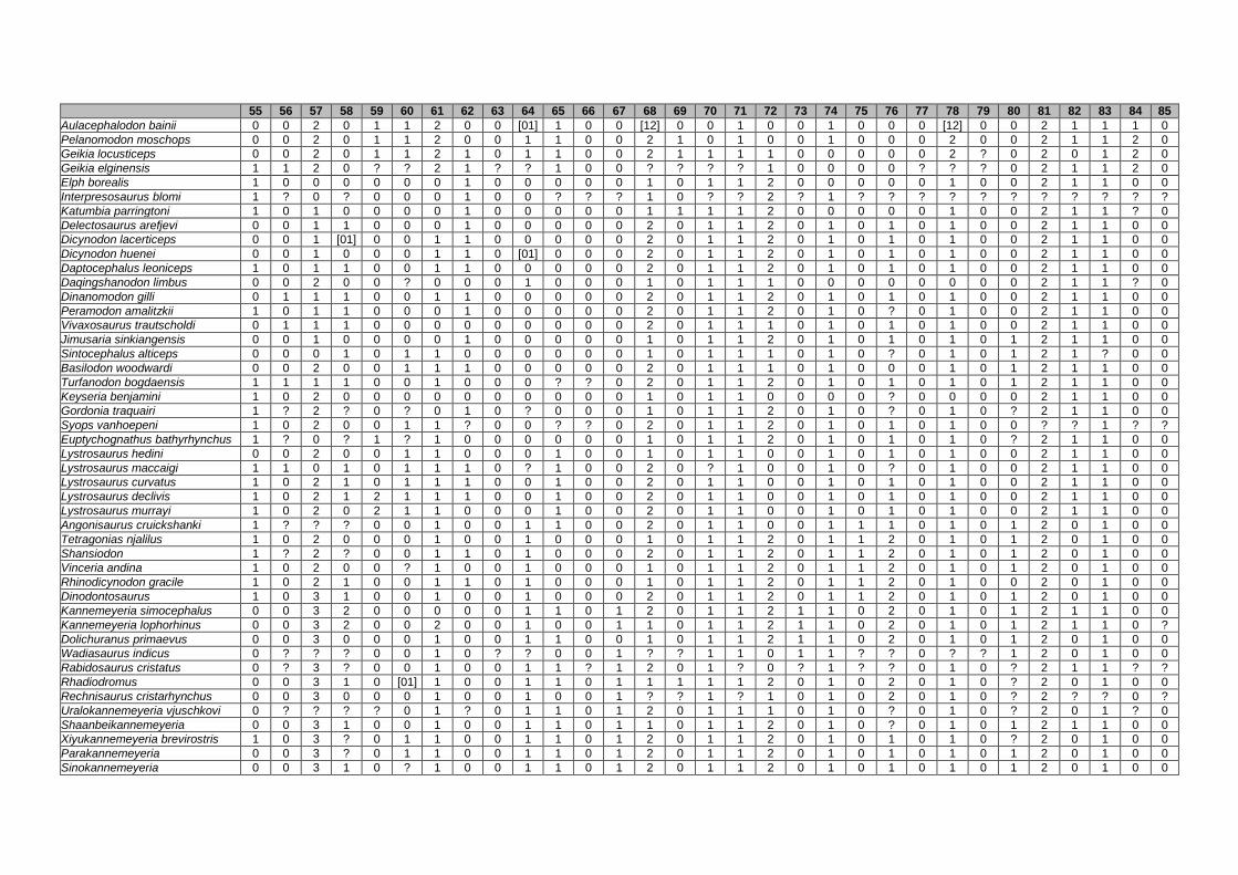
55 56 57 58 59 60 61 62 63 64 65 66 67 68 69 70 71 72 73 74 75 76 77 78 79 80 81 82 83 84 85
Aulacephalodon bainii 0 0 2 0 1 1 2 0 0 [01] 1 0 0 [12] 0 0 1 0 0 1 0 0 0 [12] 0 0 2 1 1 1 0
Pelanomodon moschops 0 0 2 0 1 1 2 0 0 1 1 0 0 2 1 0 1 0 0 1 0 0 0 2 0 0 2 1 1 2 0
Geikia locusticeps 0 0 2 0 1 1 2 1 0 1 1 0 0 2 1 1 1 1 0 0 0 0 0 2 ? 0 2 0 1 2 0
Geikia elginensis 1 1 2 0 ? ? 2 1 ? ? 1 0 0 ? ? ? ? 1 0 0 0 0 ? ? ? 0 2 1 1 2 0
Elph borealis 1 0 0 0 0 0 0 1 0 0 0 0 0 1 0 1 1 2 0 0 0 0 0 1 0 0 2 1 1 0 0
Interpresosaurus blomi 1 ? 0 ? 0 0 0 1 0 0 ? ? ? 1 0 ? ? 2 ? 1 ? ? ? ? ? ? ? ? ? ? ?
Katumbia parringtoni 1 0 1 0 0 0 0 1 0 0 0 0 0 1 1 1 1 2 0 0 0 0 0 1 0 0 2 1 1 ? 0
Delectosaurus arefjevi 0 0 1 1 0 0 0 1 0 0 0 0 0 2 0 1 1 2 0 1 0 1 0 1 0 0 2 1 1 0 0
Dicynodon lacerticeps 0 0 1 [01] 0 0 1 1 0 0 0 0 0 2 0 1 1 2 0 1 0 1 0 1 0 0 2 1 1 0 0
Dicynodon huenei 0 0 1 0 0 0 1 1 0 [01] 0 0 0 2 0 1 1 2 0 1 0 1 0 1 0 0 2 1 1 0 0
Daptocephalus leoniceps 1 0 1 1 0 0 1 1 0 0 0 0 0 2 0 1 1 2 0 1 0 1 0 1 0 0 2 1 1 0 0
Daqingshanodon limbus 0 0 2 0 0 ? 0 0 0 1 0 0 0 1 0 1 1 1 0 0 0 0 0 0 0 0 2 1 1 ? 0
Dinanomodon gilli 0 1 1 1 0 0 1 1 0 0 0 0 0 2 0 1 1 2 0 1 0 1 0 1 0 0 2 1 1 0 0
Peramodon amalitzkii 1 0 1 1 0 0 0 1 0 0 0 0 0 2 0 1 1 2 0 1 0 ? 0 1 0 0 2 1 1 0 0
Vivaxosaurus trautscholdi 0 1 1 1 0 0 0 0 0 0 0 0 0 2 0 1 1 1 0 1 0 1 0 1 0 0 2 1 1 0 0
Jimusaria sinkiangensis 0 0 1 0 0 0 0 1 0 0 0 0 0 1 0 1 1 2 0 1 0 1 0 1 0 1 2 1 1 0 0
Sintocephalus alticeps 0 0 0 1 0 1 1 0 0 0 0 0 0 1 0 1 1 1 0 1 0 ? 0 1 0 1 2 1 ? 0 0
Basilodon woodwardi 0 0 2 0 0 1 1 1 0 0 0 0 0 2 0 1 1 1 0 1 0 0 0 1 0 1 2 1 1 0 0
Turfanodon bogdaensis 1 1 1 1 0 0 1 0 0 0 ? ? 0 2 0 1 1 2 0 1 0 1 0 1 0 1 2 1 1 0 0
Keyseria benjamini 1 0 2 0 0 0 0 0 0 0 0 0 0 1 0 1 1 0 0 0 0 ? 0 0 0 0 2 1 1 0 0
Gordonia traquairi 1 ? 2 ? 0 ? 0 1 0 ? 0 0 0 1 0 1 1 2 0 1 0 ? 0 1 0 ? 2 1 1 0 0
Syops vanhoepeni 1 0 2 0 0 1 1 ? 0 0 ? ? 0 2 0 1 1 2 0 1 0 1 0 1 0 0 ? ? 1 ? ?
Euptychognathus bathyrhynchus 1 ? 0 ? 1 ? 1 0 0 0 0 0 0 1 0 1 1 2 0 1 0 1 0 1 0 ? 2 1 1 0 0
Lystrosaurus hedini 0 0 2 0 0 1 1 0 0 0 1 0 0 1 0 1 1 0 0 1 0 1 0 1 0 0 2 1 1 0 0
Lystrosaurus maccaigi 1 1 0 1 0 1 1 1 0 ? 1 0 0 2 0 ? 1 0 0 1 0 ? 0 1 0 0 2 1 1 0 0
Lystrosaurus curvatus 1 0 2 1 0 1 1 1 0 0 1 0 0 2 0 1 1 0 0 1 0 1 0 1 0 0 2 1 1 0 0
Lystrosaurus declivis 1 0 2 1 2 1 1 1 0 0 1 0 0 2 0 1 1 0 0 1 0 1 0 1 0 0 2 1 1 0 0
Lystrosaurus murrayi 1 0 2 0 2 1 1 0 0 0 1 0 0 2 0 1 1 0 0 1 0 1 0 1 0 0 2 1 1 0 0
Angonisaurus cruickshanki 1 ? ? ? 0 0 1 0 0 1 1 0 0 2 0 1 1 0 0 1 1 1 0 1 0 1 2 0 1 0 0
Tetragonias njalilus 1 0 2 0 0 0 1 0 0 1 0 0 0 1 0 1 1 2 0 1 1 2 0 1 0 1 2 0 1 0 0
Shansiodon 1 ? 2 ? 0 0 1 1 0 1 0 0 0 2 0 1 1 2 0 1 1 2 0 1 0 1 2 0 1 0 0
Vinceria andina 1 0 2 0 0 ? 1 0 0 1 0 0 0 1 0 1 1 2 0 1 1 2 0 1 0 1 2 0 1 0 0
Rhinodicynodon gracile 1 0 2 1 0 0 1 1 0 1 0 0 0 1 0 1 1 2 0 1 1 2 0 1 0 0 2 0 1 0 0
Dinodontosaurus 1 0 3 1 0 0 1 0 0 1 0 0 0 2 0 1 1 2 0 1 1 2 0 1 0 1 2 0 1 0 0
Kannemeyeria simocephalus 0 0 3 2 0 0 0 0 0 1 1 0 1 2 0 1 1 2 1 1 0 2 0 1 0 1 2 1 1 0 0
Kannemeyeria lophorhinus 0 0 3 2 0 0 2 0 0 1 0 0 1 1 0 1 1 2 1 1 0 2 0 1 0 1 2 1 1 0 ?
Dolichuranus primaevus 0 0 3 0 0 0 1 0 0 1 1 0 0 1 0 1 1 2 1 1 0 2 0 1 0 1 2 0 1 0 0
Wadiasaurus indicus 0 ? ? ? 0 0 1 0 ? ? 0 0 1 ? ? 1 1 0 1 1 ? ? 0 ? ? 1 2 0 1 0 0
Rabidosaurus cristatus 0 ? 3 ? 0 0 1 0 0 1 1 ? 1 2 0 1 ? 0 ? 1 ? ? 0 1 0 ? 2 1 1 ? ?
Rhadiodromus 0 0 3 1 0 [01] 1 0 0 1 1 0 1 1 1 1 1 2 0 1 0 2 0 1 0 ? 2 0 1 0 0
Rechnisaurus cristarhynchus 0 0 3 0 0 0 1 0 0 1 0 0 1 ? ? 1 ? 1 0 1 0 2 0 1 0 ? 2 ? ? 0 ?
Uralokannemeyeria vjuschkovi 0 ? ? ? ? 0 1 ? 0 1 1 0 1 2 0 1 1 1 0 1 0 ? 0 1 0 ? 2 0 1 ? 0
Shaanbeikannemeyeria 0 0 3 1 0 0 1 0 0 1 1 0 1 1 0 1 1 2 0 1 0 ? 0 1 0 1 2 1 1 0 0
Xiyukannemeyeria brevirostris 1 0 3 ? 0 1 1 0 0 1 1 0 1 2 0 1 1 2 0 1 0 1 0 1 0 ? 2 0 1 0 0
Parakannemeyeria 0 0 3 ? 0 1 1 0 0 1 1 0 1 2 0 1 1 2 0 1 0 1 0 1 0 1 2 0 1 0 0
Sinokannemeyeria 0 0 3 1 0 ? 1 0 0 1 1 0 1 2 0 1 1 2 0 1 0 1 0 1 0 1 2 0 1 0 0

86 87 88 89 90 91 92 93 94 95 96 97 98 99 100 101 102 103 104 105 106 107 108 109 110 111 112 113 114 115 116
Aulacephalodon bainii 2 0 1 1 1 0 1 0 1 1 0 1 1 2 2 0 3 0 1 1 1 1 0 1 2 1 0 0 1 2 1
Pelanomodon moschops 2 0 1 1 1 0 1 0 1 1 1 1 1 2 2 0 3 0 1 1 1 1 0 1 2 1 0 [01] 1 2 1
Geikia locusticeps 2 0 1 1 1 0 1 0 1 1 1 1 1 2 2 0 3 0 1 1 1 1 0 1 2 1 0 1 1 2 1
Geikia elginensis 2 0 1 ? ? 0 1 ? 1 1 1 1 1 ? 2 0 3 0 ? 1 ? ? 0 1 2 1 0 ? ? 2 1
Elph borealis 2 0 1 1 1 0 1 0 ? ? ? ? 1 2 2 0 3 0 1 1 0 1 0 ? 2 1 0 0 ? 1 1
Interpresosaurus blomi ? ? ? ? ? ? ? ? 1 ? ? ? 1 ? ? ? 3 0 ? ? 0 ? ? ? ? ? ? ? ? ? ?
Katumbia parringtoni 2 0 1 1 ? 0 1 0 1 1 1 1 1 2 2 0 3 0 1 1 0 1 0 1 2 1 0 1 0 2 1
Delectosaurus arefjevi 2 0 1 1 1 0 1 0 1 1 1 1 1 2 1 0 3 0 1 1 1 [12] 0 1 2 1 0 1 1 2 1
Dicynodon lacerticeps 2 0 1 1 1 0 1 0 1 1 1 1 1 2 1 0 3 0 1 1 1 1 0 1 2 1 0 0 1 2 1
Dicynodon huenei 2 0 1 1 1 0 1 0 1 1 1 1 1 2 1 0 3 0 1 1 1 1 0 1 2 1 0 1 1 2 1
Daptocephalus leoniceps 2 0 1 1 1 0 1 0 1 1 1 1 1 2 1 0 3 0 1 1 1 1 0 1 2 1 0 1 0 1 1
Daqingshanodon limbus 2 0 1 1 1 0 1 0 1 1 ? ? 1 2 2 0 3 0 1 1 0 1 0 1 2 1 0 0 1 2 1
Dinanomodon gilli 2 0 1 1 1 0 1 0 1 1 1 1 1 2 1 0 3 0 1 1 1 1 0 1 2 1 1 1 0 2 1
Peramodon amalitzkii 2 0 1 1 ? 0 1 0 ? ? ? ? ? ? ? ? ? ? ? ? ? ? ? 1 2 ? ? ? ? 2 1
Vivaxosaurus trautscholdi 2 0 1 1 1 0 1 0 1 1 0 0 1 2 1 0 3 0 1 1 1 [12] 0 1 2 1 0 1 1 2 1
Jimusaria sinkiangensis 2 0 1 1 1 0 1 0 ? ? ? ? 1 2 1 0 3 0 1 ? 1 1 0 1 2 1 0 0 ? 2 1
Sintocephalus alticeps 2 0 ? 1 ? 0 ? ? ? ? ? ? 1 2 1 0 3 0 1 1 ? 1 0 1 2 1 0 1 ? 2 1
Basilodon woodwardi 2 0 1 1 1 0 1 0 1 1 1 1 1 2 2 0 3 0 1 1 1 1 0 1 2 1 0 1 ? 2 1
Turfanodon bogdaensis 2 0 1 ? 1 0 ? ? ? ? ? ? 1 2 1 0 3 0 1 ? ? 2 0 1 2 1 1 1 1 2 1
Keyseria benjamini 2 0 1 1 1 0 1 0 1 1 ? ? 1 2 2 0 3 0 ? 1 ? 1 0 1 2 1 0 0 0 2 1
Gordonia traquairi 2 0 1 1 ? 0 1 0 ? 1 ? ? 1 ? 1 0 3 ? ? ? ? ? ? 1 2 1 0 1 ? ? 1
Syops vanhoepeni ? ? ? 1 1 ? ? 0 1 1 1 1 1 2 2 0 3 0 1 1 1 1 ? 1 2 1 ? 1 ? ? 1
Euptychognathus bathyrhynchus 2 0 1 1 1 0 1 0 1 1 1 1 1 2 1 0 3 0 ? 1 ? ? ? 1 2 1 0 ? 1 2 1
Lystrosaurus hedini 2 0 1 1 1 0 1 0 1 1 1 1 1 2 1 0 3 0 1 1 1 2 ? 1 2 1 0 1 0 4 1
Lystrosaurus maccaigi 2 0 1 1 1 0 1 ? 1 1 1 1 1 2 1 0 3 0 1 0 1 2 ? 1 2 1 0 1 0 4 1
Lystrosaurus curvatus 2 0 1 1 1 0 1 0 1 1 1 1 1 2 1 0 3 0 1 0 1 2 ? 1 2 1 0 1 1 2 1
Lystrosaurus declivis 2 0 1 1 1 0 1 0 1 1 1 1 1 2 1 0 3 0 1 0 1 2 ? 1 2 1 0 1 1 2 1
Lystrosaurus murrayi 2 0 1 1 1 0 1 0 1 1 1 1 1 2 1 0 3 0 1 0 1 2 ? 1 2 1 0 1 1 2 1
Angonisaurus cruickshanki 2 0 1 1 1 0 ? ? 1 1 1 1 1 ? ? ? 3 0 ? 1 ? ? ? 1 2 1 0 1 ? ? 1
Tetragonias njalilus 2 1 1 1 1 0 1 0 1 1 1 1 1 2 1 0 3 0 1 0 1 2 ? 1 2 1 0 1 0 2 1
Shansiodon 2 1 1 1 1 0 1 ? ? ? ? ? 1 2 1 0 3 0 1 ? 1 2 ? 1 2 1 0 1 ? ? 1
Vinceria andina 2 0 1 1 1 0 ? ? 1 1 1 1 1 ? ? ? 3 0 ? ? 1 2 ? 1 2 1 0 1 0 2 1
Rhinodicynodon gracile 2 1 1 1 1 0 1 0 ? ? ? ? 1 2 1 0 3 0 ? 1 1 2 ? 1 2 1 0 1 ? ? 1
Dinodontosaurus 2 1 1 1 1 0 1 0 1 1 1 1 1 2 1 0 3 0 0 0 1 1 0 1 2 1 0 1 0 1 1
Kannemeyeria simocephalus 2 0 1 1 1 0 1 0 1 1 1 1 1 2 1 0 3 0 1 1 1 2 ? 1 2 1 0 1 0 1 1
Kannemeyeria lophorhinus 2 0 1 1 1 0 1 ? 1 1 1 1 1 2 1 0 3 0 1 ? 1 2 ? 1 2 1 0 1 1 1 1
Dolichuranus primaevus 2 0 1 1 1 0 1 0 1 1 1 1 1 2 1 0 3 0 1 0 1 1 0 1 2 1 0 1 1 6 1
Wadiasaurus indicus 2 0 1 ? 1 0 1 0 ? ? ? ? 1 ? ? ? 3 0 ? ? 1 2 ? 1 2 1 0 1 ? ? 1
Rabidosaurus cristatus 2 ? 1 1 1 0 ? ? ? ? ? ? 1 2 ? ? 3 0 1 0 1 ? ? 1 2 1 ? 1 ? 1 1
Rhadiodromus 2 0 1 1 1 0 ? ? 1 1 1 1 1 2 1 0 3 0 ? 1 ? 1 0 1 2 ? ? 1 ? ? 1
Rechnisaurus cristarhynchus 2 ? ? ? ? ? ? ? ? ? ? ? 1 2 1 0 3 0 ? 0 1 1 0 1 2 1 0 1 ? ? 1
Uralokannemeyeria vjuschkovi 2 0 1 1 1 0 1 0 1 1 1 1 1 ? ? ? 3 0 ? ? ? ? ? 1 2 ? ? ? 1 2 1
Shaanbeikannemeyeria 2 1 1 1 1 0 1 ? 1 1 1 1 1 2 1 0 3 0 1 1 1 ? 0 1 2 1 0 1 ? 2 1
Xiyukannemeyeria brevirostris 2 1 1 1 1 0 1 0 1 1 1 1 1 2 ? ? 3 0 1 ? 1 2 ? 1 2 ? ? 1 0 2 1
Parakannemeyeria 2 1 1 1 1 0 1 0 1 1 1 1 1 2 1 0 3 0 1 ? 1 2 ? 1 2 1 0 1 0 1 1
Sinokannemeyeria 2 1 1 1 1 0 1 1 1 1 1 1 1 2 1 0 3 0 1 ? ? 2 ? 1 2 1 0 1 ? 2 1

117 118 119 120 121 122 123 124 125 126 127 128 129 130 131 132 133 134 135 136 137 138 139 140 141 142 143 144 145 146 147
Aulacephalodon bainii 0 0 0 0 1 2 1 1 2 [01] 0 1 1 1 0 1 1 0 0 0 1 1 1 2 ? ? ? 2 0 0 1
Pelanomodon moschops 0 0 ? 0 1 2 1 1 2 1 0 1 1 1 0 1 1 0 0 ? ? ? ? ? ? ? ? ? ? ? ?
Geikia locusticeps 0 0 0 0 2 2 1 1 2 1 0 1 1 1 0 1 1 0 0 ? 1 1 1 2 ? ? ? 2 1 0 1
Geikia elginensis 0 ? 1 0 ? ? ? ? ? ? ? ? ? ? ? ? ? ? 0 ? 1 0 1 2 ? ? ? 2 1 0 1
Elph borealis ? 0 0 0 1 2 1 1 2 0 ? ? ? 1 0 1 1 0 0 ? 1 1 1 2 ? ? ? 2 0 ? 1
Interpresosaurus blomi ? ? ? ? ? ? ? ? ? ? ? ? ? ? ? ? ? ? ? ? ? ? ? ? ? ? ? ? ? ? ?
Katumbia parringtoni 0 ? ? 0 1 2 1 1 2 1 1 1 1 ? 0 0 1 0 0 ? 1 1 1 2 ? ? ? 2 1 0 1
Delectosaurus arefjevi 1 0 ? 0 1 2 1 1 2 1 ? ? ? 1 0 0 1 0 0 0 ? ? ? ? ? ? ? ? ? ? ?
Dicynodon lacerticeps 0 0 0 0 2 2 1 1 2 1 1 1 1 1 0 1 1 0 0 1 1 1 1 2 ? ? ? 2 1 0 1
Dicynodon huenei 0 0 0 0 2 2 1 1 2 1 1 1 1 1 0 1 1 0 0 1 1 1 1 2 ? ? ? 2 ? 0 1
Daptocephalus leoniceps 1 0 1 0 1 2 1 1 2 1 1 1 1 1 0 1 1 1 0 1 1 1 1 2 ? ? ? 2 1 0 1
Daqingshanodon limbus 0 ? 0 0 ? ? 1 1 ? ? ? ? ? 1 0 1 1 ? 0 ? 1 1 1 2 ? ? ? 2 1 0 1
Dinanomodon gilli 1 ? ? 0 1 2 1 1 2 1 1 1 1 1 0 1 1 ? 0 1 1 ? 1 ? ? ? ? ? 1 0 ?
Peramodon amalitzkii ? 0 ? 0 ? 2 ? 1 2 1 ? 1 1 ? 0 ? 1 ? ? ? 1 1 1 ? ? ? ? 2 1 0 ?
Vivaxosaurus trautscholdi 1 0 ? 0 2 2 1 1 2 [01] ? 1 ? 1 0 1 1 0 0 1 1 1 1 2 ? ? ? 2 1 0 1
Jimusaria sinkiangensis 1 0 1 0 2 2 1 1 2 1 1 1 ? 1 0 ? ? ? 0 ? 1 1 1 2 ? ? ? 2 1 0 1
Sintocephalus alticeps 0 ? ? 0 ? ? ? ? ? ? ? ? ? ? ? ? ? ? ? ? ? ? ? ? ? ? ? ? ? ? ?
Basilodon woodwardi 0 ? ? 0 1 2 1 1 2 1 ? 1 ? 1 ? 1 1 0 1 ? 1 ? 1 ? ? ? ? ? 1 ? ?
Turfanodon bogdaensis 1 ? ? 0 1 2 1 1 2 1 ? ? ? 1 0 1 1 0 0 1 ? ? ? ? ? ? ? ? ? ? ?
Keyseria benjamini 0 ? ? 0 1 ? 1 1 2 1 0 1 1 1 ? 1 1 0 0 ? ? ? ? ? ? ? ? ? ? ? ?
Gordonia traquairi ? ? ? 0 ? ? ? 1 2 ? ? 1 1 ? ? ? 1 ? 0 ? 1 1 1 2 ? ? ? 2 ? 0 ?
Syops vanhoepeni ? ? ? ? ? 2 ? ? ? ? 0 ? ? 1 ? 0 1 0 ? ? 1 ? 1 ? ? ? ? 2 0 0 ?
Euptychognathus bathyrhynchus 0 ? ? 0 2 2 1 1 ? 1 1 ? ? 1 0 1 1 0 0 ? 1 1 1 2 ? ? ? 2 1 0 1
Lystrosaurus hedini 1 ? ? 0 2 2 1 1 2 1 0 ? ? 1 0 1 1 0 0 1 1 1 1 2 ? ? ? 2 1 0 1
Lystrosaurus maccaigi 1 ? ? 0 2 2 1 1 2 1 0 ? ? 1 ? 1 1 0 0 ? 1 1 1 2 ? ? ? 2 1 0 1
Lystrosaurus curvatus 1 0 0 0 2 2 1 1 2 1 ? ? ? 1 0 0 1 0 0 1 1 1 1 ? ? ? ? ? 1 0 ?
Lystrosaurus declivis 1 0 0 0 2 2 1 1 2 1 0 1 1 1 0 0 1 0 0 1 1 1 1 2 ? ? ? 2 1 0 1
Lystrosaurus murrayi 1 ? 0 0 2 2 1 1 2 1 1 1 1 1 0 1 1 0 0 0 1 1 1 2 ? ? ? 2 1 0 1
Angonisaurus cruickshanki 1 0 ? 0 2 2 1 1 2 0 ? ? ? 1 ? 1 1 0 ? 1 1 1 1 2 ? ? ? 2 1 0 1
Tetragonias njalilus 1 0 1 0 ? 2 1 1 2 1 1 1 1 1 0 1 1 0 0 1 1 1 1 2 ? ? ? 2 1 0 1
Shansiodon ? 0 ? 0 2 2 1 1 2 0 ? 1 1 1 ? 1 1 0 0 1 1 1 1 2 ? ? ? 2 0 0 1
Vinceria andina 1 ? ? 0 2 2 ? 1 ? 0 ? 1 1 1 ? 1 1 ? ? ? 1 ? ? 2 ? ? ? ? ? ? ?
Rhinodicynodon gracile ? 0 ? 0 2 2 ? 1 ? ? ? ? ? 1 ? 0 1 0 0 ? 1 1 1 2 ? ? ? 2 1 0 1
Dinodontosaurus 1 ? 1 0 2 2 ? 1 2 1 1 ? ? ? 0 1 0 0 0 1 1 1 1 2 ? ? ? 2 1 0 1
Kannemeyeria simocephalus 1 1 1 0 2 2 1 2 2 0 1 ? ? 1 0 1 0 0 1 1 1 1 1 2 ? ? ? 2 1 0 1
Kannemeyeria lophorhinus 1 0 1 0 2 2 ? ? 2 0 1 ? ? 1 ? 0 0 0 ? ? 1 1 1 2 ? ? ? 2 1 0 1
Dolichuranus primaevus 1 0 ? 0 2 2 1 2 2 0 0 ? ? 1 ? 1 0 0 1 ? 1 1 1 2 ? ? ? 2 1 0 1
Wadiasaurus indicus ? ? ? 0 2 2 ? ? 2 0 ? ? ? 1 ? 1 1 ? 1 ? 1 1 1 2 ? ? ? 2 ? ? 1
Rabidosaurus cristatus ? ? ? 0 ? 2 ? ? ? ? 1 ? ? 1 ? ? 0 0 ? 1 ? ? ? ? ? ? ? ? ? ? ?
Rhadiodromus ? 1 1 0 2 2 ? 2 2 0 ? ? ? 1 0 1 1 0 ? ? ? ? ? ? ? ? ? ? ? ? ?
Rechnisaurus cristarhynchus ? ? ? 0 2 2 1 2 2 0 ? ? ? ? ? ? ? 0 ? ? ? ? ? ? ? ? ? ? ? ? ?
Uralokannemeyeria vjuschkovi 1 ? ? 0 2 2 ? 2 2 0 ? ? ? 1 ? 1 0 0 ? ? ? ? ? ? ? ? ? ? ? ? ?
Shaanbeikannemeyeria 1 ? ? 0 2 2 1 2 2 0 ? ? ? ? ? 1 0 0 ? ? 1 1 1 2 ? ? ? 2 0 ? 1
Xiyukannemeyeria brevirostris 1 ? ? 0 2 2 ? ? 2 0 ? 1 1 1 ? 0 0 0 ? ? 1 1 1 2 ? ? ? 2 1 0 ?
Parakannemeyeria 1 0 ? 0 2 2 ? ? 2 0 1 1 1 1 0 1 0 0 1 1 1 1 1 2 ? ? ? 2 1 0 1
Sinokannemeyeria 1 ? ? 0 2 2 1 ? 2 0 ? 1 1 1 0 1 0 0 1 1 1 1 1 2 ? ? ? 2 1 0 1

148 149 150 151 152 153 154 155 156 157 158 159 160 161 162 163 164 165 166 167 168 169 170 171 172 173 174 175 176 177 178
Aulacephalodon bainii 3 0 1 2 1 1 1 0 1 1 4 1 1 1 1 2 1 2 1 0 0 0 1 0 0 1 0 1 0 1 0
Pelanomodon moschops ? ? ? ? ? ? ? ? ? ? ? ? ? ? ? 2 ? ? ? ? ? ? ? ? ? ? ? ? ? ? ?
Geikia locusticeps 3 0 1 0 1 1 0 0 1 1 4 1 1 1 1 2 1 ? ? 0 0 ? 1 0 ? ? ? ? ? ? ?
Geikia elginensis 3 0 1 2 1 ? ? ? ? ? 4 1 ? ? 1 2 1 ? ? ? ? ? ? ? ? ? 0 1 0 1 ?
Elph borealis 3 0 1 2 1 1 1 0 ? ? ? ? ? ? ? 2 1 ? ? ? ? ? ? ? ? ? ? ? ? ? ?
Interpresosaurus blomi ? ? ? ? ? ? ? ? ? ? ? ? ? ? ? ? ? ? ? ? ? ? ? ? ? ? ? ? ? ? ?
Katumbia parringtoni 3 0 1 2 1 1 1 0 ? ? ? ? ? ? ? 2 1 ? ? ? ? ? ? ? ? ? ? ? ? ? ?
Delectosaurus arefjevi ? ? ? ? ? ? ? ? ? ? ? ? ? ? ? ? ? ? ? ? ? ? ? ? ? ? ? ? ? ? ?
Dicynodon lacerticeps 3 0 2 2 1 1 1 1 1 1 5 1 1 1 1 2 1 ? ? ? ? ? ? ? ? ? ? ? ? ? ?
Dicynodon huenei 3 0 2 2 1 1 1 ? 1 1 5 1 1 1 1 2 1 2 ? 0 0 0 1 0 1 1 0 1 0 1 0
Daptocephalus leoniceps 3 0 2 2 1 1 1 1 1 1 5 1 1 1 1 2 1 ? ? ? ? ? ? ? ? 1 0 1 1 1 0
Daqingshanodon limbus 3 0 1 2 1 1 ? 0 1 1 4 1 1 1 1 2 1 ? ? ? ? ? ? ? ? ? ? ? ? ? ?
Dinanomodon gilli 3 ? 1 2 1 ? ? 1 ? 1 5 1 1 ? 1 2 ? ? ? ? ? ? ? ? ? ? ? ? ? ? ?
Peramodon amalitzkii ? ? 2 2 1 1 1 1 ? 1 5 1 1 ? 1 2 ? 3 ? 1 0 0 1 0 1 ? ? ? ? ? ?
Vivaxosaurus trautscholdi 3 0 2 2 1 ? ? 1 1 1 5 1 1 0 1 2 1 ? ? 0 0 0 ? ? 1 1 0 1 1 1 0
Jimusaria sinkiangensis 3 0 2 2 1 1 0 1 1 1 5 1 1 ? 1 2 1 ? ? ? ? ? ? ? ? ? ? ? ? ? ?
Sintocephalus alticeps ? ? ? ? ? ? ? ? ? ? ? ? ? ? ? 2 ? ? ? ? ? ? ? ? ? ? ? ? ? ? ?
Basilodon woodwardi ? ? 2 2 1 ? ? ? ? ? ? 1 ? ? ? 2 ? ? ? ? ? ? ? ? ? ? ? ? ? ? ?
Turfanodon bogdaensis ? ? ? ? ? ? ? ? ? ? ? ? ? ? ? ? ? ? ? ? ? ? ? ? ? ? ? ? ? ? ?
Keyseria benjamini ? ? ? ? ? ? ? ? ? ? ? ? ? ? ? 2 ? ? ? ? ? ? ? ? ? ? ? ? ? ? ?
Gordonia traquairi 3 0 2 2 1 ? ? ? ? 1 ? 1 ? ? 1 2 1 ? ? ? 0 0 1 ? ? 1 0 ? 0 1 0
Syops vanhoepeni ? ? ? ? ? ? ? 0 ? ? ? 1 1 ? 1 2 ? ? ? ? ? ? ? ? ? ? ? ? ? ? ?
Euptychognathus bathyrhynchus 3 0 2 2 1 1 ? 1 ? ? ? 1 ? ? ? 2 1 ? ? ? ? ? ? ? ? ? ? ? ? ? ?
Lystrosaurus hedini 3 0 2 2 1 1 0 1 1 1 5 1 1 0 1 2 1 ? ? ? ? ? 1 ? ? ? ? ? ? ? ?
Lystrosaurus maccaigi 3 0 2 2 1 ? ? 1 1 1 5 1 ? ? 1 2 1 3 ? ? ? ? ? ? ? ? 0 1 0 1 0
Lystrosaurus curvatus ? ? 2 2 1 1 0 1 ? 1 5 1 1 ? 1 2 ? ? ? 0 0 0 1 ? ? 1 0 1 0 1 ?
Lystrosaurus declivis 3 0 2 2 1 1 0 1 1 1 5 1 1 0 1 2 1 3 ? 0 ? ? 1 ? ? 1 0 1 1 1 0
Lystrosaurus murrayi 3 0 2 2 1 1 0 1 1 1 5 1 1 0 1 2 1 ? ? 0 0 0 1 1 1 1 0 1 1 1 0
Angonisaurus cruickshanki 3 0 1 2 1 1 0 1 1 0 0 1 1 ? 1 2 1 3 ? ? 0 0 1 0 0 ? 1 0 ? 1 0
Tetragonias njalilus 3 0 1 2 1 1 0 1 1 0 5 1 1 1 1 2 1 2 0 0 0 0 1 0 0 1 1 1 0 1 0
Shansiodon 3 0 2 2 1 1 0 ? ? 0 ? 1 ? ? 1 2 1 2 ? 0 0 0 1 ? ? 1 1 1 0 1 1
Vinceria andina ? ? ? ? ? ? ? ? ? ? ? ? ? ? 1 2 1 ? ? ? ? ? ? ? ? ? ? ? ? ? ?
Rhinodicynodon gracile 3 0 2 2 1 ? ? 1 1 ? ? 1 ? ? 1 2 1 ? ? 0 0 0 1 0 0 1 0 1 ? 1 0
Dinodontosaurus 3 0 1 2 1 1 0 0 1 0 ? 1 1 ? 1 2 1 3 0 0 0 0 1 0 0 1 1 0 0 1 0
Kannemeyeria simocephalus 3 0 2 2 1 1 0 1 1 1 5 1 1 1 1 2 1 3 0 0 0 1 1 0 0 1 0 1 0 1 0
Kannemeyeria lophorhinus 3 0 1 2 1 ? ? 1 1 0 ? 0 1 0 1 2 1 ? ? ? 0 0 ? ? ? 1 1 ? 1 1 0
Dolichuranus primaevus 3 0 1 2 1 1 ? 1 1 0 ? 1 1 ? 1 2 1 ? ? 0 0 0 0 ? ? 1 1 0 0 1 0
Wadiasaurus indicus 3 0 ? ? ? ? ? ? ? ? ? 1 1 ? 1 2 1 2 0 0 1 0 1 ? 0 1 1 1 1 1 0
Rabidosaurus cristatus ? ? ? ? ? ? ? ? ? ? ? ? ? ? ? ? ? ? ? ? ? ? ? ? ? ? 1 ? ? ? ?
Rhadiodromus ? ? ? ? ? ? ? ? ? ? ? ? ? ? ? ? ? ? ? ? ? ? ? ? ? ? ? ? ? ? ?
Rechnisaurus cristarhynchus ? ? ? ? ? ? ? ? ? ? ? ? ? ? ? ? ? ? ? ? ? ? ? ? ? ? ? ? ? ? ?
Uralokannemeyeria vjuschkovi ? ? ? ? ? ? ? ? ? ? ? ? ? ? ? 2 ? 2 ? ? ? ? ? ? ? ? ? ? ? ? ?
Shaanbeikannemeyeria ? ? 1 2 1 ? ? ? ? ? ? ? ? 1 ? 2 1 ? ? ? 0 0 1 ? ? 1 ? ? 1 1 0
Xiyukannemeyeria brevirostris ? ? 1 2 1 1 ? ? 1 ? ? 1 1 ? 1 2 1 ? ? ? 0 ? 1 ? ? 1 1 1 0 ? 0
Parakannemeyeria ? 0 1 2 1 ? ? ? 1 ? ? 1 1 ? 1 2 1 3 ? 0 0 0 1 0 0 1 0 1 0 1 0
Sinokannemeyeria 3 0 1 2 1 1 ? ? ? 0 ? 1 1 ? 1 2 1 2 0 0 0 0 1 0 ? 1 1 1 0 1 0

179 180 181 182 183 184 185 186 187 188 189 190 191 192 193 194
Aulacephalodon bainii 0 ? 1 1 0 1 0 1 0 1 1 1 2 1 1 1
Pelanomodon moschops ? ? ? ? ? ? ? ? ? ? ? ? ? ? ? ?
Geikia locusticeps ? ? ? ? ? ? ? ? ? ? ? ? ? ? ? ?
Geikia elginensis ? ? ? ? ? ? ? ? ? ? ? ? ? ? ? ?
Elph borealis ? ? ? ? ? ? ? ? ? ? ? ? ? ? ? ?
Interpresosaurus blomi ? ? ? ? ? ? ? ? ? ? ? ? ? ? ? ?
Katumbia parringtoni ? ? ? ? ? ? ? ? ? ? ? ? ? ? ? ?
Delectosaurus arefjevi ? ? ? ? ? ? ? ? ? ? ? ? ? ? ? ?
Dicynodon lacerticeps ? ? ? ? ? ? ? ? ? ? ? ? ? ? ? ?
Dicynodon huenei 0 ? 1 1 0 1 1 1 0 1 1 ? ? ? ? ?
Daptocephalus leoniceps ? ? ? ? 0 1 1 1 ? ? ? ? ? ? ? ?
Daqingshanodon limbus ? ? ? ? ? ? ? ? ? ? ? ? ? ? ? ?
Dinanomodon gilli ? ? ? ? ? ? ? ? ? ? ? ? ? ? ? ?
Peramodon amalitzkii ? ? ? ? ? ? ? ? ? ? ? ? ? ? ? ?
Vivaxosaurus trautscholdi ? ? ? ? ? ? ? ? ? ? ? ? ? ? ? ?
Jimusaria sinkiangensis ? ? ? ? ? ? ? ? ? ? ? ? ? ? ? ?
Sintocephalus alticeps ? ? ? ? ? ? ? ? ? ? ? ? ? ? ? ?
Basilodon woodwardi ? ? ? ? ? ? ? ? ? ? ? ? ? ? ? ?
Turfanodon bogdaensis ? ? ? ? ? ? ? ? ? ? ? ? ? ? ? ?
Keyseria benjamini ? ? ? ? ? ? ? ? ? ? ? ? ? ? ? ?
Gordonia traquairi 0 ? ? ? 0 ? ? ? ? ? ? ? ? ? ? ?
Syops vanhoepeni ? ? ? ? ? ? ? ? ? ? ? ? ? ? ? ?
Euptychognathus bathyrhynchus ? ? ? ? ? ? ? ? ? ? ? ? ? ? ? ?
Lystrosaurus hedini ? ? ? ? ? ? ? ? ? ? ? ? ? ? ? ?
Lystrosaurus maccaigi 0 ? 1 1 ? 1 1 ? 0 0 1 1 2 1 1 1
Lystrosaurus curvatus 0 ? ? ? 1 ? ? ? 0 0 1 ? ? ? ? 1
Lystrosaurus declivis 0 ? 1 1 1 1 1 1 0 1 1 1 2 1 1 1
Lystrosaurus murrayi 0 ? 1 1 1 ? ? 1 0 1 1 1 2 1 1 1
Angonisaurus cruickshanki ? ? ? ? 0 1 1 1 ? ? ? ? ? ? ? ?
Tetragonias njalilus 0 ? 1 1 0 1 1 1 1 1 2 1 2 1 1 ?
Shansiodon 1 ? 1 1 0 ? ? ? 1 1 1 1 2 1 1 ?
Vinceria andina ? ? ? ? ? ? ? ? ? ? ? ? ? ? ? ?
Rhinodicynodon gracile 0 ? ? ? 0 1 1 1 1 1 2 ? ? ? ? ?
Dinodontosaurus 1 ? 1 1 0 1 1 1 0 1 2 1 2 1 1 ?
Kannemeyeria simocephalus 0 ? 1 1 0 1 1 1 0 1 1 1 2 1 1 1
Kannemeyeria lophorhinus ? ? ? ? 0 ? ? ? ? ? ? ? ? ? ? ?
Dolichuranus primaevus 1 ? ? ? ? ? ? ? 0 1 2 ? ? ? ? ?
Wadiasaurus indicus 1 ? ? ? 0 1 1 1 1 1 1 ? ? ? ? ?
Rabidosaurus cristatus ? ? ? ? ? ? ? ? ? ? ? ? ? ? ? ?
Rhadiodromus ? ? ? ? ? ? ? ? ? ? ? ? ? ? ? ?
Rechnisaurus cristarhynchus ? ? ? ? ? ? ? ? ? ? ? ? ? ? ? ?
Uralokannemeyeria vjuschkovi ? ? ? ? 0 ? ? ? ? ? ? ? ? ? ? ?
Shaanbeikannemeyeria 0 ? ? ? ? ? ? ? 0 1 1 ? ? ? ? ?
Xiyukannemeyeria brevirostris 0 ? 1 1 0 ? ? ? 0 1 1 1 2 1 1 ?
Parakannemeyeria 0 ? 1 1 0 1 1 1 [01] 1 1 1 2 1 1 ?
Sinokannemeyeria 1 ? 1 1 0 ? ? ? 0 1 1 1 2 1 1 ?

24 25 26 27 28 29 30 31 32 33 34 35 36 37 38 39 40 41 42 43 44 45 46 47 48 49 50 51 52 53 54
Stahleckeria potens 1 2 0 0 2 0 ? 2 1 0 1 1 0 0 0 0 2 1 2 0 1 1 2 ? ? 1 1 0 1 0 0
Eubrachiosaurus browni ? ? ? ? ? ? ? ? ? ? ? ? ? ? ? ? ? ? ? ? ? ? ? ? ? ? ? ? ? ? ?
Sangusaurus parringtonii 1 2 0 0 2 2 ? 2 1 0 1 1 0 0 0 0 0 0 1 0 1 1 2 ? ? 0 1 0 1 0 0
Ischigualastia jenseni 1 2 0 0 2 2 ? 2 1 0 1 1 0 0 0 0 2 1 2 0 1 1 2 ? ? 1 1 0 1 0 0
Jachaleria 1 2 0 0 2 2 1 2 1 0 1 1 0 0 0 0 2 1 1 0 1 1 2 ? ? 1 1 0 1 0 0
Zambiasaurus submersus 1 2 0 0 2 2 ? 2 1 ? ? 1 ? ? ? ? ? ? ? 0 0 1 2 ? ? ? 1 ? 0 ? ?
Placerias hesternus 1 2 0 0 0 2 ? 2 1 ? ? 0 0 0 2 0 2 0 0 0 0 2 2 ? ? 1 1 1 0 0 0
Moghreberia nmachouensis 1 ? 0 0 0 2 ? 2 1 0 1 0 0 0 2 ? ? 0 2 0 0 2 2 ? ? ? 1 0 2 0 0
Rastodon procurvidens 1 ? ? ? ? ? ? 2 1 0 1 0 0 0 0 0 2 0 0 0 0 2 ? ? ? 1 1 0 0 0 0
Bulbasaurus phylloxyron 1 2 0 0 2 2 1 2 1 0 1 1 0 0 0 0 2 0 0 0 0 2 2 ? ? 1 1 0 1 0 0
Compsodon helmoedi 1 0 1 1 1 0 1 2 1 0 1 0 0 0 0 1 2 0 1 0 0 2 0 0 1 1 1 1 0 1 0
LPB 1993 3 1 2 0 0 2 2 1 2 1 0 1 0 0 0 0 ? ? ? 0 0 0 2 2 ? ? 1 1 0 1 0 0
LPB 1993 2 1 2 0 0 2 2 1 2 1 0 ? 1 0 0 0 0 ? 1 0 0 ? 2 2 ? ? 1 1 0 1 0 0
LPB 1995 9 1 2 0 0 2 0 1 2 1 0 1 ? 0 0 0 ? ? 1 0 0 0 2 2 ? ? 0 1 0 1 0 0
55 56 57 58 59 60 61 62 63 64 65 66 67 68 69 70 71 72 73 74 75 76 77 78 79 80 81 82 83 84 85
Stahleckeria potens 0 0 3 1 0 0 1 0 1 1 1 0 1 0 ? 1 1 0 0 1 0 1 0 1 0 2 2 0 1 0 0
Eubrachiosaurus browni ? ? ? ? ? ? ? ? ? ? ? ? ? ? ? ? ? ? ? ? ? ? ? ? ? ? ? ? ? ? ?
Sangusaurus parringtonii 0 0 3 1 0 0 2 0 1 1 1 0 0 1 0 1 1 1 0 1 0 2 0 1 0 2 2 0 1 0 0
Ischigualastia jenseni 0 0 3 1 0 0 1 0 1 1 1 0 1 1 0 1 1 2 0 1 1 2 0 1 0 2 2 0 1 0 0
Jachaleria 1 0 3 1 0 1 1 0 1 1 1 0 0 2 0 1 1 2 0 1 1 2 0 1 0 2 2 0 1 0 0
Zambiasaurus submersus ? ? ? ? ? ? ? 0 0 ? ? ? 1 0 ? 1 1 0 0 ? ? ? 0 1 0 2 2 0 ? ? ?
Placerias hesternus 0 0 3 1 0 ? 1 0 0 1 1 0 1 1 0 1 0 0 0 1 1 1 0 1 0 2 2 0 1 0 1
Moghreberia nmachouensis 0 ? 3 ? 0 ? ? 0 0 1 1 ? 1 2 0 1 0 0 0 1 1 1 0 1 0 2 2 0 ? 0 1
Rastodon procurvidens 0 0 1 0 0 0 0 0 0 0 0 0 0 1 0 1 1 1 0 0 0 0 0 0 0 0 2 1 1 0 0
Bulbasaurus phylloxyron 0 [01] 2 [01] 1 0 0 0 0 1 1 0 0 2 0 1 1 [12] 0 1 0 0 0 [12] 0 0 2 1 1 1 0
Compsodon helmoedi 1 0 1 0 0 0 0 1 0 0 0 0 0 2 1 1 0 1 0 0 0 0 0 3 0 0 2 1 1 0 0
LPB 1993 3 0 ? ? 2 ? ? ? ? ? ? ? ? 0 ? ? ? ? ? ? ? 0 ? 0 1 0 0 2 1 1 0 0
LPB 1993 2 1 ? 1 0 0 ? 1 0 ? ? 1 0 0 2 0 1 1 1 0 0 0 0 0 1 0 0 2 1 1 0 0
LPB 1995 9 1 ? 1 0 0 0 1 0 0 1 ? 0 0 1 0 ? 1 1 0 0 0 0 0 1 0 0 2 1 1 ? 0
86 87 88 89 90 91 92 93 94 95 96 97 98 99 100 101 102 103 104 105 106 107 108 109 110 111 112 113 114 115 116
Stahleckeria potens 2 0 1 1 1 0 1 0 1 1 1 1 1 2 1 0 3 0 1 1 1 1 0 1 2 1 1 1 0 1 1
Eubrachiosaurus browni ? ? ? ? ? ? ? ? ? ? ? ? ? ? ? ? ? ? ? ? ? ? ? ? ? ? ? ? ? ? ?
Sangusaurus parringtonii 2 0 0 1 1 0 1 0 1 ? ? ? 1 2 1 0 3 0 1 1 1 2 ? 1 2 1 0 1 0 1 1
Ischigualastia jenseni 2 0 0 1 1 0 1 1 1 1 1 1 1 2 1 0 3 0 ? ? 1 2 ? 1 2 1 0 1 0 5 1
Jachaleria 2 0 0 1 1 0 1 1 1 1 1 1 1 2 1 0 3 0 1 0 ? 2 ? 1 2 1 0 1 0 1 1
Zambiasaurus submersus 2 ? 1 ? ? ? ? 0 ? ? ? ? ? ? ? ? ? ? ? ? ? ? ? ? ? ? ? ? ? ? ?
Placerias hesternus 2 0 1 1 1 0 1 0 ? ? ? ? ? ? ? ? ? ? ? ? ? ? ? 1 ? ? ? ? 0 5 1
Moghreberia nmachouensis 2 0 1 ? ? 0 ? 0 1 1 1 1 1 2 1 0 3 0 1 1 ? ? ? ? 2 1 0 ? ? ? 1
Rastodon procurvidens 2 0 1 1 1 0 1 0 ? 1 1 0 ? ? ? ? ? ? ? ? 0 ? ? 1 2 1 0 ? 1 2 1
Bulbasaurus phylloxyron 2 0 1 1 1 0 1 0 1 1 0 1 1 2 2 0 3 0 1 1 0 1 0 1 2 1 0 0 1 2 1
Compsodon helmoedi 2 0 1 1 ? 0 1 0 1 1 1 0 1 1 1 0 3 1 1 1 0 1 0 1 2 1 1 0 0 2 0
LPB 1993 3 2 0 1 1 ? 0 ? ? 1 1 1 1 1 2 1 0 3 0 1 ? ? 1 0 1 2 1 0 1 0 2 1
LPB 1993 2 2 0 1 ? ? 0 ? ? ? ? ? ? 1 ? ? ? ? ? ? ? ? 1 0 1 2 ? ? 1 0 ? 1
LPB 1995 9 2 ? ? 1 ? 0 ? ? 1 1 1 1 1 2 1 0 3 0 ? ? ? 1 0 1 2 1 0 1 ? 2 1

117 118 119 120 121 122 123 124 125 126 127 128 129 130 131 132 133 134 135 136 137 138 139 140 141 142 143 144 145 146 147
Stahleckeria potens 1 1 1 0 2 2 1 1 2 1 1 1 ? 1 0 1 0 0 0 1 1 1 1 2 ? ? ? 2 1 0 1
Eubrachiosaurus browni ? ? ? ? ? ? ? ? ? ? ? ? ? ? ? ? ? ? ? ? ? ? ? ? ? ? ? ? ? ? ?
Sangusaurus parringtonii 1 1 1 0 2 2 1 1 2 1 ? ? ? 1 1 1 0 0 0 1 1 1 1 2 ? ? ? ? 1 0 1
Ischigualastia jenseni 1 ? 1 0 2 2 1 3 2 0 ? 1 1 1 0 1 0 0 0 ? 1 1 1 2 ? ? ? 2 1 0 1
Jachaleria 1 ? 1 0 2 2 1 3 2 0 0 ? ? 1 0 1 0 0 0 ? ? 1 1 2 ? ? ? 2 ? 0 1
Zambiasaurus submersus ? ? ? ? ? ? ? ? ? ? ? ? ? ? ? 1 1 0 ? ? ? ? 1 2 ? ? ? 2 ? ? 1
Placerias hesternus 1 1 0 0 2 2 ? ? 2 0 ? 1 1 1 ? 1 0 1 ? 1 1 1 1 2 ? ? ? 2 0 0 1
Moghreberia nmachouensis ? ? 0 ? 2 2 1 ? 2 0 ? ? ? ? ? 1 0 1 ? ? 1 1 1 2 ? ? ? 2 ? 0 1
Rastodon procurvidens 0 0 ? 0 0 ? ? 1 ? 0 0 ? ? 1 0 1 1 0 0 ? 1 0 1 ? ? ? ? 2 0 0 ?
Bulbasaurus phylloxyron 0 0 0 0 1 2 1 1 2 0 ? ? ? 1 0 1 1 0 0 ? 1 1 1 ? ? ? ? 2 1 0 1
Compsodon helmoedi 0 0 ? 0 1 1 1 1 2 0 0 1 1 1 1 0 1 0 1 ? 1 ? ? ? ? ? ? ? ? ? ?
LPB 1993 3 1 0 ? 0 2 2 ? 1 2 0 ? ? ? ? ? 0 1 0 1 1 ? ? ? ? ? ? ? ? ? ? ?
LPB 1993 2 1 ? ? 0 2 2 ? 1 2 0 ? ? ? ? ? 0 1 ? ? 1 ? ? ? ? ? ? ? ? ? ? ?
LPB 1995 9 1 0 ? 0 2 ? ? ? 2 0 ? ? ? ? ? ? ? ? 1 ? ? ? ? ? ? ? ? ? ? ? ?
148 149 150 151 152 153 154 155 156 157 158 159 160 161 162 163 164 165 166 167 168 169 170 171 172 173 174 175 176 177 178
Stahleckeria potens 3 0 1 2 1 1 0 1 1 1 0 0 1 1 1 2 1 3 1 0 1 0 1 0 0 1 1 1 0 1 0
Eubrachiosaurus browni ? ? ? ? ? ? ? ? ? ? ? ? ? ? ? ? ? ? ? ? 1 0 1 ? ? 1 ? 0 0 1 0
Sangusaurus parringtonii 3 0 1 2 1 1 1 1 ? 1 0 0 1 1 1 2 1 3 ? ? ? ? ? ? ? ? ? ? ? ? ?
Ischigualastia jenseni 3 0 1 ? ? ? ? 1 ? ? 0 0 1 ? 1 2 1 2 1 0 1 0 0 1 0 1 1 0 0 1 0
Jachaleria ? 0 ? ? ? ? ? ? ? ? ? ? ? ? 1 2 1 2 1 0 0 1 0 ? ? ? 1 ? ? ? ?
Zambiasaurus submersus ? ? ? ? ? ? 0 ? ? ? ? ? ? ? ? ? 1 ? 0 ? 1 1 1 0 0 1 ? 0 1 1 0
Placerias hesternus 3 0 ? ? ? 1 0 ? ? 1 ? 1 ? ? 1 2 1 ? 0 0 1 1 1 0 0 1 ? 0 0 1 0
Moghreberia nmachouensis 3 0 1 2 1 1 0 ? ? ? ? 0 ? ? 1 2 1 ? ? ? ? ? ? ? ? ? ? ? ? ? ?
Rastodon procurvidens ? 0 2 0 0 1 0 0 ? 1 ? 1 ? ? ? ? 1 ? ? ? ? ? ? ? ? ? ? ? ? ? ?
Bulbasaurus phylloxyron ? 0 1 2 1 1 ? ? ? ? ? ? ? ? ? ? 1 ? ? ? ? ? ? ? ? ? ? ? 0 ? ?
Compsodon helmoedi ? ? ? ? ? ? ? ? ? ? ? ? ? ? ? ? ? ? ? ? ? ? ? ? ? ? ? ? ? ? ?
LPB 1993 3 ? ? ? ? ? ? ? ? ? ? ? ? ? ? ? ? ? ? ? ? ? ? ? ? ? ? ? ? ? ? ?
LPB 1993 2 ? ? ? ? ? ? ? ? ? ? ? ? ? ? ? ? ? ? ? ? ? ? ? ? ? ? ? ? ? ? ?
LPB 1995 9 ? ? ? ? ? ? ? ? ? ? ? ? ? ? ? ? ? ? ? ? ? ? ? ? ? ? ? ? ? ? ?
179 180 181 182 183 184 185 186 187 188 189 190 191 192 193 194
Stahleckeria potens 1 ? 1 1 0 1 1 1 1 1 1 1 2 1 1 ?
Eubrachiosaurus browni ? ? ? ? 0 ? ? ? ? ? ? ? ? ? ? ?
Sangusaurus parringtonii ? ? ? ? 0 1 1 1 1 1 1 ? ? ? ? ?
Ischigualastia jenseni 1 ? 1 1 0 1 1 1 1 1 1 1 2 1 1 ?
Jachaleria 1 ? ? ? 0 1 1 1 1 1 1 ? ? ? ? ?
Zambiasaurus submersus 0 ? ? ? 0 1 1 1 1 1 1 ? ? ? ? ?
Placerias hesternus ? ? ? ? 0 1 1 1 1 1 1 ? ? ? ? 1
Moghreberia nmachouensis ? ? ? ? ? ? ? ? ? ? ? ? ? ? ? ?
Rastodon procurvidens ? ? ? ? ? ? ? ? ? ? ? ? ? ? ? ?
Bulbasaurus phylloxyron ? ? ? ? ? ? ? ? ? ? ? ? ? ? ? ?
Compsodon helmoedi ? ? ? ? ? ? ? ? ? ? ? ? ? ? ? ?
LPB 1993 3 ? ? ? ? ? ? ? ? ? ? ? ? ? ? ? ?
LPB 1993 2 ? ? ? ? ? ? ? ? ? ? ? ? ? ? ? ?
LPB 1995 9 ? ? ? ? ? ? ? ? ? ? ? ? ? ? ? ?

APPENDIX I: Supplementary data of PAPER I
313
Bibliography Angielczyk, K. D. 2001. Preliminary phylogenetic analysis and stratigraphic
congruence of the dicynodont anomodonts (Synapsida: Therapsida). Palaeontologica Africana 37:53–79.
Angielczyk, K. D. 2004. Phylogenetic evidence for and implications of a dual origin of propaliny in anomodont therapsids. Paleobiology 30:268–296.
Angielczyk, K. D. 2007. New specimens of the Tanzanian dicynodont “Cryptocynodon” parringtoni von Huene, 1943 (Therapsida, Anomodontia), with an expanded analysis of Permian dicynodont phylogeny. Journal of Vertebrate Paleontology 27: 116–131.
Angielczyk, K. D., and C. F. Kammerer. 2017. The cranial morphology, phylogenetic position and biogeography of the upper Permian dicynodont Compsodon helmoedi van Hoepen (Therapsida, Anomodontia). Papers in Palaeontology 1–33.
Angielczyk, K. D., and A. A. Kurkin. 2003. Phylogenetic analysis of Russian Permian dicynodonts (Therapsida: Anomodontia): implications for Permian biostratigraphy and Pangaean biogeography. Zoological Journal of the Linnean Society 139:157–212.
Angielczyk, K. D., and B. S. Rubidge. 2010. A new pylaecephalid dicynodont (Therapsida, Anomodontia) from the Tapinocephalus Assemblage Zone, Karoo Basin, Middle Permian of South Africa. Journal of Vertebrate Paleontology 30:1396–1409.
Angielczyk, K. D., and B. S. Rubidge. 2013. Skeletal morphology, phylogenetic relationships, and stratigraphic range of Eosimops newtoni Broom, 1921, a pylaecephalid dicynodont (Therapsida, Anomodontia) from the Middle Permian of South Africa. Journal of Systematic Palaeontology 11:191–231.
Damiani, R., C. Vasconcelos, A. Renaut, J. Hancox, and A. Yates. 2007. Dolichuranus primaevus (Therapsida: Anomodontia) from the Middle Triassic of Namibia and its phylogenetic relationships. Palaeontology 50:1531–1546.
Fröbisch, J. 2007. The cranial anatomy of Kombuisia frerensis Hotton (Synapsida, Dicynodontia) and a new phylogeny of anomodont therapsids. Zoological Journal of the Linnean Society 150:117–144.
Kammerer, C. F., K. D. Angielczyk, and J. Fröbisch. 2011. A comprehensive taxonomic revision of “Dicynodon” (Therapsida, Anomodontia) and its implications for dicynodont phylogeny, biogeography, and biostratigraphy. Journal of Vertebrate Paleontology 31:1–158.
Kammerer, C. F., J. Fröbisch, and K. D. Angielczyk. 2013. On the Validity and Phylogenetic Position of Eubrachiosaurus browni, a Kannemeyeriiform Dicynodont (Anomodontia) from Triassic North America. PLoS ONE 8:e64203.
Maisch, M. W. 2001. Observations on Karoo and Gondwana vertebrates. Part 2: A new skull-reconstruction of Stahleckeria potens von Huene, 1935 (Dicynodontia, Middle Triassic) and a reconsideration of kannemeyeriiform phylogeny. Neues Jahrbuch für Geologie und Paläontologie – Abhandlungen 220:127–152.
Maisch, M. W. 2002. A new basal lystrosaurid dicynodont from the Upper Permian of South Africa. Palaeontology 45:343–359.
Maisch, M. W., and E.V.I. Gebauer. 2005. Reappraisal of Geikia locusticeps (Therapsida: Dicynodontia) from the Upper Permian of Tanzania. Palaeontology 48:309–324.
Modesto, S. P., and N. Rybczynski. 2000. The amniote faunas of the Russian Permian: implications for Late Permian terrestrial vertebrate biogeography; pp. 17–34 in M.J.
APPENDIX I: Supplementary data of PAPER I
314
Benton, M.A. Shishkin, D.M. Unwin, and E.N. Kurochkin (eds.), The age of dinosaurs in Russia and Mongolia. Cambridge University Press, Cambridge.
Modesto, S. P., B. Rubidge, and J. Welman. 1999. The most basal anomodont therapsid and the primacy of Gondwana in the evolution of the anomodonts. Proceedings of the Royal Society of London B: Biological Sciences 266:331–337.
Modesto, S. P., B. Rubidge, I. Visser, and J. Welman. 2003. A new basal dicynodont from the Upper Permian of South Africa. Palaeontology 46:211–223.
Sidor, C. A. 2001. Simplification as a trend in synapsid cranial evolution. Evolution 55:1419–1442.
Sidor, C. A., and J. A. Hopson. 1998. Ghost lineages and ‘mammal-ness’: assessing the temporal pattern of character acquisition in the Synapsida. Paleobiology 24:254–273.
Surkov, M. V., and M. J. Benton. 2004. The basicranium of dicynodonts (Synapsida) and its use in phylogenetic analysis. Palaeontology 47:619–638.
Surkov, M. V., N. N. Kalandadze, and M. J. Benton. 2005. Lystrosaurus georgi, a dicynodont from the Lower Triassic of Russia. Journal of Vertebrate Paleontology 25:402–413.
Vega-Dias, C., M. W. Maisch, and C. L. Schultz. 2004. A new phylogenetic analysis of Triassic dicynodonts (Therapsida) and the systematic position of Jachaleria candelariensis from the Upper Triassic of Brazil. Neues Jahrbuch für Geologie und Paläontologie – Abhandlungen 231:145–166.
APPENDIX II: Supplementary data of PAPER II
315
Appendix II: Supplementary data of PAPER II (unpublished)
1. Table 1S. Postcranial remains found in the Argana Basin and attributed to
Dicynodontia. (p. 316–327)
2. Discrete and continuous characters used in the phylogenetic analysis (modified
from Kamerer, 2018) (p. 328)
3. Table 2S. Measurements of the continuous characters in Moghreberia
nmachouensis. (p. 329)
4. New or changed coding from the phylogenetic matrix of Kammerer (2018). (p. 331
–334)
5. Table 3S. Phylogenetic matrix used in the analysis. (p. 334–351)

Table 1S. Postcranial remains found in the Argana Basin and attributed to Dicynodontia.
Specimen number
Original bone identification
Current bone identification
Preservation Original
attribution Current
attributionc Locality
MNHN.F.ALM 120 Four connected vertebraeb
Three connected dorsal vertebrae (‘MNHN.F.ALM 120-1, -2, and -3’ following the anteroposterior axis) and a fourth dorsal vertebra (‘MNHN.F.ALM 120-4’) associated with MNHN.F.ALM 120-3c
Vertebrae connected, except MNHN.F.ALM 120-4 originally associated with MNHN.F.ALM 120-3. Bones slightly compressed. Details of connections and bone structures hidden by remaining sediment. Anterior surface of MNHN.F.ALM 120-1 partly eroded with left processes (transverse and prezygapophysis) and the right border of the centrum missing. MNHN.F.ALM 120-3 partially disconnected from MNHN.F.ALM 120-2 and shifting to the right. Left prezygapophysis of MNHN.F.ALM 120-3 eroded. Neural spine partially or completely missing in MNHN.F.ALM 120-3 and -4, respectively. Postzygapophyses and distal end of the left transverse process missing in MNHN.F.ALM 120-4.
M. nmachouensisb Unchanged
Locality XIb in Argana Basin (Morocco)a
MNHN.F.ALM 136 Sacral vertebraea
Five sacral vertebrae (‘MNHN.F.ALM 136-1, -2, -3, -4, and -5’)c
Ventral and dorsal sides of the vertebrae only observable, all the rest included in sediment. Part of the neural spine and fragment of the left associated ribs only preserved in MHNH.F.ALM 136-1. MNHN.F.ALM 136-5 only preserved by its left associated sacral rib and the small anterior part of its centrum highly eroded. Neural spine and associated right sacral ribs of the other vertebrae missing. External ventral surface of MNHN.F.ALM 136-2 eroded.
Dicynodontiaa M. nmachouensis Locality XIb in Argana Basin (Morocco)a
MNHN.F.ALM 159 Two vertebraea
Two connected dorsal vertebrae (‘MNHN.F.ALM 159-1 and 2’)c
Connections (posterior side of MNHN.F.ALM 159-1 and anterior side of MNHN.F.ALM 159-2) and bone structures hidden by remaining sediment. MNHN.F.ALM 159-2 transversely crushed. Neural canals of both vertebrae filled by sediment. Neural spine of both vertebrae and left transverse process
Dicynodontiaa M. nmachouensis Locality XIb in Argana Basin (Morocco)a

of MNHN.F.ALM 159-1 broken. Transverse processes and prezygapophyses of MNHN.F.ALM 159-1, as well as postzygapophyses of MNHN.F.ALM 159-2 highly eroded.
MNHN.F.ALM 198 Vertebraa
Posterior part of neural arch of a last cervical or first dorsal vertebrac
Posterior part of the neural arch of the vertebra only preserved. Neural spine missing. Pre- and postzygapophyses highly eroded. Right transverse process broken.
Unattributeda M. nmachouensis Argana Basin (Morocco)a
MNHN.F.ALM 265 Dorsal vertebrab Unchanged
Vertebra with distorted neural canal due to a post-mortem compression. Left anterior margin of the centrum, left transverse process, and prezygapophyses broken. Anterior surface highly eroded.
Dicynodontiaa M. nmachouensis Locality XIb in Argana Basin (Morocco)a
MNHN.F.ALM 288 Vertebraa
Vertebra (last cervical or first dorsal)c
Highly anteroposteriorly compressed vertebrae with transverse processes flattened. Sediment layers covering the bone surface, and especially the lateral surface of the centrum, in the neural canal, and around the neural spine.
? M. nmachouensisb M. nmachouensis
Locality XIb in Argana Basin (Morocco)a
Unnumbered Two connected vertebraeb
Two connected vertebrae (‘Vunknb 1 and 2’) identified as last cervical vertebrae and/or first dorsalsc
Sediment covering some connections and bone structures. Transverse processes, pre- and postzygapophyses of vertebrae highly eroded. Neural spine completely and partly missing in Vunknb 1 and 2, respectively. Neural canal completely or partly filled by sediment in Vunknb 1 and 2, respectively.
Unattributedb M. nmachouensis Unknownb
MNHN.F.ALM 86 Riba Right dorsal rib in middle or posterior positionc
Well-preserved but fractured bone with a distal part broken and highly eroded.
M. nmachouensisb
Unchanged Locality XIb in Argana Basin (Morocco)a
MNHN.F.ALM 90 Limb bonea
Left rib (associated with last cervical or first dorsal vertebrae?)c
Proximal and distal ends of rib missing. Sediment covering the posterior side of the head and distal part of the rib. Posterior side of rib highly eroded.
Unattributeda M. nmachouensis Locality XIb in Argana Basin (Morocco)a

MNHN.F.ALM 91 Ribb
Left rib associated with last cervical or first dorsal vertebraec
Well-preserved bone but fractured, with a distal part broken and highly eroded. Sediment covering the posterior side of the distal end.
Dicynodontiab M. nmachouensis Locality XIb in Argana Basin (Morocco)a
MNHN.F.ALM 95 Riba Left dorsal rib in middle or posterior positionc
Well-preserved but fractured with a distal part broken and highly eroded.
Dicynodontiab M. nmachouensis Locality XIb in Argana Basin (Morocco)a
MNHN.F.ALM 96 Riba
Right anterior dorsal rib (with a single head)c
Fractured rib entirely preserved. Dicynodontiab Dicynodontoidae indet.
Locality XIb in Argana Basin (Morocco)a
MNHN.F.ALM 97 Riba
Right dorsal rib in middle or posterior positionc
Well-preserved but fractured with a distal part broken and highly eroded. Sediment covering the posterior side of the proximal head highly eroded.
Dicynodontiab M. nmachouensis Locality XIb in Argana Basin (Morocco)a
MNHN.F.ALM 163 Distal part of a riba
Proximal head of a right rib, probably anterior dorsalc
Articular surfaces highly eroded with sediment remaining.
? M. nmachouensisb M. nmachouensis
Locality XIb in Argana Basin (Morocco)a
MNHN.F.ALM 272 Fragmentary riba
Proximal head of a right ribc
Sediment covering the dorsal side of the costal head of rib. Proximal part highly eroded and broken.
Dicynodontiab M. nmachouensis Locality XIb in Argana Basin (Morocco)a
MNHN.F.ALM 296 Riba Left ribc
Well-preserved bone but fractured rib with proximal end missing. Distal part broken and highly eroded.
Dicynodontiab M. nmachouensis Locality XIb in Argana Basin (Morocco)a
Unnumbered Unidentifiedb
Proximal head of a left rib (associated with last cervical or first dorsal vertebrae?)c
Relatively well-preserved proximal costal head with eroded articular surfaces.
Unattributedb M. nmachouensis ? Argana Basin (Morocco)b
MNHN.F.ALM 17 Scapulab Left scapulac
Dorsal expansion and anterior blade of the ventral part broken. Lateral surface of the ventral part crushed with a lack of bone. Sediment covering the medial part of the dorsal and ventral ends.
M. nmachouensisb Unchanged
Locality XIa in Argana Basin (Morocco)a

MNHN.F.ALM 19 Left scapulab Unchanged Nearly complete scapula. Glenoid process missing. Dorsal part poorly preserved with sediment remaining.
Dicynodontiab M. nmachouensis Locality XIb in Argana Basin (Morocco)a
MNHN.F.ALM 29 Scapulab Right scapulac
Complete scapula crushed at the ventral region. Sediment covering the medial surface of the anterior blade of the ventral part.
Dicynodontiab M. nmachouensis Locality XIb in Argana Basin (Morocco)a
MNHN.F.ALM 30 Left scapulab Unchanged
Nearly complete scapula. Glenoid facet and dorsal end poorly preserved. Sediment covering the lateral surface of the dorsal part and slightly the medial side of most bone surface.
M. nmachouensisb Unchanged
Locality XIb in Argana Basin (Morocco)a
MNHN.F.ALM 83 Scapulaa Left scapulab Well-preserved bone without any particular taphonomic tampering.
Dicynodontiab M. nmachouensis Locality XIb in Argana Basin (Morocco)a
MNHN.F.ALM 92 Scapulaa
Left scapula in connection with precoracoid and coracoidc
Dorsal expansion and anterior blade of the ventral part not preserved. External surface of the precoracoid and coracoid eroded with fractured outlines.
Dicynodontia ? M. nmachouensisb
M. nmachouensis Locality XIb in Argana Basin (Morocco)a
MNHN.F.ALM 98 Right scapulab Left scapulac Complete scapula highly fragmented. Anteroventral corner of the anterior blade missing
M. nmachouensisb Unchanged
Locality XIb in Argana Basin (Morocco)a
MNHN.F.ALM 113 Left scapulab Unchanged Well-preserved bone, but dorsal region missing. M. nmachouensisb Unchanged
Locality XIa in Argana Basin (Morocco)a
MNHN.F.ALM 114 Fragment of scapulaa Left scapulac
Dorsal part missing and ventral part crushed. Anterior blade of the ventral part missing. Lateral side of the ventral part highly eroded. Sediment covering the bone external surface.
M. nmachouensisb Unchanged
Locality XIa in Argana Basin (Morocco)a
MNHN.F.ALM 132 Scapulaa Left scapulac Well-preserved bone, but dorsal region missing. Dicynodontiaa M. nmachouensis Locality XIb in Argana Basin (Morocco)a
MNHN.F.ALM 153 Scapulaa Right scapulac
Nearly complete scapula preserved in three parts. Sediment covering the medial side of the middle and ventral fragments, and at the glenoid facet. Anterior blade of the ventral part and posterior margin of the dorsal expansion missing.
M. nmachouensisb Unchanged
Locality XIb in Argana Basin (Morocco)a
MNHN.F.ALM 155 Scapulaa Left scapulac Nearly complete scapula. Ventral sutural zone poorly preserved and anterior blade of the ventral
Dicynodontiaa M. nmachouensis Locality XIb in Argana Basin (Morocco)a

part broken. Sediment covering the whole medial region.
MNHN.F.ALM 259 Scapulaa Left scapulac
Nearly complete scapula. Dorsal part and anterior blade of the ventral region missing. Sediment covering the medial side, above the sutural zone with the coracoid.
M. nmachouensisb Unchanged
Locality XIb in Argana Basin (Morocco)a
MNHN.F.ALM 263 Fragmentary scapulaa Right scapulac Fractured and crushed bone. Dorsal expansion
without the anterior crest only preserved. ? M. nmachouensisb M. nmachouensis
Locality XIb in Argana Basin (Morocco)a
MNHN.F.ALM 283 Scapulaa Right scapulac
Well-preserved and nearly complete scapula. Proximal end missing. Part of the anterior blade of the ventral region broken.
Dicynodontiaa M. nmachouensis Locality XIb in Argana Basin (Morocco)a
MNHN.F.ALM 292 Scapulaa Right scapulac
Highly curved scapula in anterior view. Glenoid and sutural facets, and anterior blade of the ventral region poorly preserved.
Dicynodontiaa M. nmachouensis Locality XIb in Argana Basin (Morocco)a
MNHN.F.ALM 298 Scapulaa
Left scapula in connection with precoracoid and coracoidc
Well-preserved scapulocoracoid complex. Posterior margin of the dorsal expansion and anterior margin of the ventral region eroded. Sediment covering the lateral side of the dorsal region, anterior expansion of the ventral part, precoracoid, and coracoid. Glenoid facet, acromion and triceps muscle processes poorly preserved.
Dicynodontia ? M. nmachouensisb
M. nmachouensis Locality XIb in Argana Basin (Morocco)a
MNHN.F.ALM 299 Scapulaa Right scapulac Highly fragmented but complete scapula. Glenoid and coracoid facets poorly preserved.
Dicynodontiaa Dicynodontoidae indet.
Locality XIa in Argana Basin (Morocco)a
MNHN.F.AZA 365 Scapulaa Right scapulac
The more robust scapula of the collection. Middle and ventral regions preserved. Anterior blade of the ventral part missing.
Dicynodontiaa Dicynodontoidae indet.
Locality XIII in Argana Basin (Morocco)a (the only dicynodont bone found associated to remains mainly attributed to Metoposaurus ouazzaoui)

Unnumbered Right scapulab Unchanged (‘Sunknb 1’)
Poorly-preserved and fragmented scapula. Almost the whole ventral region and anterior margin of the dorsal part missing.
? M. nmachouensisb M. nmachouensis
Locality XI in Argana Basin (Morocco)b
Unnumbered Scapulab Left scapula (‘Sunknb 2’)c
Poorly-preserved bone with highly eroded lateral external bone surface. Sediment covering the medial side of the dorsal and ventral regions.
Unattributedb M. nmachouensis Locality XI in Argana Basin (Morocco)b
Unnumbered Unidentifiedb Right scapula (‘Sunknb 3’)a
Nearly complete but crushed bone. Sutural facet with the coracoid, anterior blade of the dorsal and ventral regions highly altered.
Unattributedb M. nmachouensis ? Argana Basin (Morocco)b
MNHN.F.ALM 93 Interclavicleb Unchanged Highly fractured bone. Bone missing particularly in the anterior part. Sediment covering the external surface of bone.
Dicynodontiab M. nmachouensis Locality XIb in Argana Basin (Morocco)a
MNHN.F.ALM 94 Interclavicleb Unchanged Interclavicle fractured in two parts. External bone surface eroded. Anterior and posterior ends broken. Sediment covering the whole dorsal side.
? Dicynodontiab M. nmachouensis
Locality XIb in Argana Basin (Morocco)a
MNHN.F.ALM 158 ? Interclavicleb Interclaviclec
Relatively well-preserved interclavicle with anterior and posterior regions broken. Sediment covering on the whole dorsal side.
M. nmachouensisb Unchanged
Locality XIb in Argana Basin (Morocco)a
MNHN.F.ALM 260 Interclavicleb Unchanged Relatively well-preserved posterior region, as opposed to the anterior one where bone missing. Sediment covering the whole dorsal side.
M. nmachouensisb
Dicynodontoidae indet.
Locality XIb in Argana Basin (Morocco)a
MNHN.F.ALM 278 Bone from pectoral girdlea
Interclaviclec Anterior part missing and lateral margins eroded. Sediment covering the whole dorsal side.
Dicynodontiaa M. nmachouensis Locality XIb in Argana Basin (Morocco)a
MNHN.F.ALM 295 Interclavicleb Unchanged Well-preserved external bone surface. Anterior and posterior ends missing.
M. nmachouensisb Unchanged
Locality XI in Argana Basin (Morocco)b
Unnumbered Incompleted interclavicleb
Anterior part of interclavicle (‘Intunknb 1’)c
Anterior part of the bone only preserved, with eroded anterior margin. Anterior end missing.
Dicynodontiab M. nmachouensis Locality XI in Argana Basin (Morocco)b
Unnumbered ? Interclavicleb Interclavicle (‘Intunknb 2’)c
Highly eroded and fragmented interclavicle. Distorted bone with poorly-preserved posterior and anterior parts not aligned and slightly twisted
M. nmachouensisb Unchanged
Locality XI in Argana Basin (Morocco)b

at the middle constriction. Anterior part laterally compressed.
MNHN.F.ALM 134 Bone from girdle?a Sternumc Posterior region poorly preserved. Ventral side
entirely covered by sediment Dicynodontiaa M. nmachouensis
Locality XIb in Argana Basin (Morocco)a
MNHN.F.ALM 24 Left humerusb Unchanged Well-preserved complete humerus. Proximal end missing. Dorsal surface of distal part highly eroded.
M. nmachouensisb Unchanged
Locality XIb in Argana Basin (Morocco)a
MNHN.F.ALM 25 Left humerusb Unchanged Well-preserved complete humerus. Proximal end missing. Dorsal surface of the distal part highly eroded.
M. nmachouensisb
Unchanged Locality XIb in Argana Basin (Morocco)a
MNHN.F.ALM 101 Humerusa Right humerusc
Relatively well-preserved complete humerus. Sediment covering the posterior part of the proximal expansion. Distal epiphysis eroded.
Dicynodontiab M. nmachouensis Locality XIb in Argana Basin (Morocco)a
MNHN.F.ALM 102 Left humerusb Unchanged Well-preserved complete humerus. Sediment covering the dorsal side of the distal expansion.
M. nmachouensisb Unchanged
Locality XIb in Argana Basin (Morocco)a
MNHN.F.ALM 105 Partial humerusa Left humerusc
Highly fractured humerus with epiphyses and part of metaphyses of the proximal and distal expansions missing. Sediment covering the ventral margin of the deltopectoral crest and most of the distal expansion.
Unattributeda M. nmachouensis Locality XIb in Argana Basin (Morocco)a
MNHN.F.ALM 115 Humerusa Right humerusc
Fractured humerus with epiphyses and part of metaphyses of the proximal and distal expansion missing. Sediment covering the proximal and distal expansions. Two third of the distal part of the deltopectoral crest well preserved.
Unattributeda M. nmachouensis Locality XIa in Argana Basin (Morocco)a
MNHN.F.ALM 121-1-2-3
Humerusa Fragmentary parts of a left humerusc
Highly fractured and eroded bone preserved in three parts: the shaft and the distal expansion (MNHN.F.ALM 121-1), the posterior corner of the proximal end with the humeral head (MNHN.F.ALM 121-2), and a fragmentary part of the deltopectoral crest (MNHN.F.ALM 121-3). Humeral head, external surface of the shaft, and entepicondyle foramen well preserved.
Dicynodontiaa M. nmachouensis Locality XIb in Argana Basin (Morocco)a

MNHN.F.ALM 133 Framentary part of humerusa
Fragmentary part of a left humerusc
Shaft and proximal part of the distal expansion (with visible entepicondyle foramen) only preserved. Sediment covering most of bone.
Dicynodontiaa M. nmachouensis Locality XIb in Argana Basin (Morocco)a
MNHN.F.ALM 147 Limb bonea Distal part of a right humerusc
Highly eroded distal part of humerus with preserved ventral surface of the trochlea and capitelum.
Dicynodontiaa M. nmachouensis Locality XIb in Argana Basin (Morocco)a
MNHN.F.ALM 275 Humerusa Left humerusc
The biggest humerus of the collection, but highly fractured. Proximal end and entepicondyle missing. Deltopectoral crest highly altered.
Dicynodontiaa Dicynodontoidae indet.
Locality XIa in Argana Basin (Morocco)a
MNHN.F.ALM 297 Humerusa Left humerusc Highly eroded complete humerus. Proximal corner of deltopectoral crest missing.
Dicynodontiab M. nmachouensis Locality XIb in Argana Basin (Morocco)a
Unnumbered Distal part of humerusb
Proximal part of a right humerus (‘Humunknb 1’)c
Posterior part of the proximal expansion of humerus only preserved. Sediment covering the humeral head and posterior surface of bone. Deltopectoral crest not preserved.
Dicynodontiab M. nmachouensis Locality XI in Argana Basin (Morocco)b
Unnumbered Distal part of humerusb
Distal part of a right humerus (‘Humunknb 2’)c
Shaft and ectepicondyle well preserved. Distal part of the entepicondyle missing. Sediment covering most of bone.
Dicynodontiab M. nmachouensis Locality X, XI or XVI? in Argana Basin (Morocco)b
MNHN.F.ALM 84 Radiusb Left radiusc Complete radius with eroded proximal and distal ends. Sediment covering its posterior side.
M. nmachouensisb Unchanged
Locality XIb in Argana Basin (Morocco)a
MNHN.F.ALM 131 Limb bonea Left radiusc
Broken at the middle of the shaft. Distal expansion crushed and poorly preserved. Fractured and eroded proximal end.
Dicynodontiaa M. nmachouensis Locality XIb in Argana Basin (Morocco)a
MNHN.F.ALM 287 Radiusb Right radiusc Well-preserved complete radius with eroded proximal and distal ends
M. nmachouensisb Unchanged
Locality XIb in Argana Basin (Morocco)a
MNHN.F.ALM 289 Radiusb Left radiusc Highly crushed with some cracks. Sediment covering its proximal and distal ends.
M. nmachouensisb
Unchanged Locality XIb in Argana Basin (Morocco)a
Unnumbered Distal parts of a radius and an ulnab
Distal parts of left radius and ulnac
Radius highly fractured and compressed, with distal end eroded. No diagnostic features present on the ulnar fragment except the anterior crest bordering the radial facet anteriorly.
M. nmachouensisb Unchanged Unknownb

MNHN.F.ALM 18 Ulnab Right ulnac Lack of bone and medial part of the olecranon process crushed.
M. nmachouensisb Unchanged
Locality XIb in Argana Basin (Morocco)a
MNHN.F.ALM 27 Dorsal part of ulnab
Dorsal part of the olecranon process of a right ulnac
Dorsal part of the olecranon process only preserved. Highly eroded and fractured bone. One of the two biggest ulnae.
Dicynodontiab Dicynodontoidae indet.
Locality XIb in Argana Basin (Morocco)a
MNHN.F.ALM 28 Ulnab Left ulnac Fractured ulna with lack of bone at the olecranon process and ventral epiphysis.
Dicynodontiab M. nmachouensis Locality XIb in Argana Basin (Morocco)a
MNHN.F.ALM 35 Ulnab Right ulnac Sediment covering the olecranon process. Ventral epiphysis missing.
M. nmachouensisb Unchanged
Locality XI in Argana Basin (Morocco)b
MNHN.F.ALM 87 Ulna and the proximal part of a ribb
Right ulna and the proximal part of a right ribc
Ulna fragmented in two parts. Highly eroded and fractured proximal part. Lack of bone and sediment covering the medial side of the ventral region. Ventral epiphysis and external bone surface highly eroded.
M. nmachouensisb
Unchanged Locality XIb in Argana Basin (Morocco)a
MNHN.F.ALM 126-1, -2
Fragmentary ulnaa Left ulnac Fragmented and highly eroded bone. One of the
two biggest ulnae. M. nmachouensisb Unchanged
Locality XIb in Argana Basin (Morocco)a
MNHN.F.ALM 154 Ulnaa Left ulnac
Well-preserved ulna. Dorsal end of the olecranon process missing. Lack of bone at the ventral epiphysis.
Dicynodontiaa Dicynodontoidae indet.
Locality XIa in Argana Basin (Morocco)a
MNHN.F.ALM 261 Ulnaa Left ulnac Lack of bone and medial part of the olecranon process crushed.
? M. nmachouensisb M. nmachouensis
Locality XIb in Argana Basin (Morocco)a
MNHN.F.ALM 274 ? Vertebrae and pelvisa
Pelvic girdle connected to sacral and caudal vertebrae, and two associated disconnected vertebrae (‘MNHN.F.ALM 274-1 and -2’) that are interpreted as posterior dorsal vertebraec
Pelvis laterally compressed. Two puboischiatic plates not in natural position, with posterior margins of the ischia very closely spaced. Pubes connected by their ventral margins most likely due to the lateral compression. Left ilium almost entirely preserved, while distorted, as opposed to the right one (iliac blade highly altered). Left puboischiatic plate entirely preserved, while ischium particularly crushed. Posterior flattened expansion of this latter highly fractured. Right puboischiatic plate more altered, with posterior part of the right ischium missing. Obturator foramens filled by sediment.
Unattributeda M. nmachouensis Unknowna

Vertebrae MNHN.F.ALM 274-1 and -2 relatively well preserved, except some cracks and a lack of bone structures. Neural spine, distal part of the right transverse process, and postzygapophyses of MNHN.F.ALM 274-2 missing. Centra of both MNHN.F.ALM 274-1 and -2 altered, while a well preservation of the rest of MNHN.F.ALM 274-1. Ventral side of sacral vertebrae well visible as opposed to the dorsal side, mainly included in sediment. Left rib of the first sacral vertebra cracked and almost entirely missing. Caudal vertebrae connected to the last sacral vertebra, but most entirely included in sediment. Neural spines only observable, as well as most of the first and last centra. Neural spine of the most posterior caudal vertebrae missing.
MNHN.F.ALM 104 Left iliumb Unchanged
Well-preserved ilium. Posterodorsal margin of its iliac blade missing. Right expanded transverse process of the first sacral vertebra connected to the medial part of the iliac blade dorsally. Sediment covering the acetabulum fossa and the medial side of the anterior tubercle of the ilium.
? M. nmachouensisb M. nmachouensis
Locality XIb in Argana Basin (Morocco)a
MNHN.F.ALM 123 Bone from pelvic girdle ?a Right ischiumc
Relatively well-preserved ischium with sligh break on anterior and posterior margins of the ventral expansion.
Dicynodontiaa M. nmachouensis Locality XIb in Argana Basin (Morocco)a
MNHN.F.ALM 125 Bone from girdle ?a
Left ischiumc
Whole lateral side and acetabular region hidden by sediment. Posteroventral margin of the obturator foramen broken, as well as most of its posterior flattened expansion.
Dicynodontiaa M. nmachouensis Locality XIb in Argana Basin (Morocco)a
MNHN.F.ALM 138 Scapulaa Right ischiumc
Relatively well-preserved ischium with sligh break on anterior and posterior margins of the ventral expansion. Whole medial side covered by sediment.
Dicynodontiaa M. nmachouensis Locality XIb in Argana Basin (Morocco)a
MNHN.F.ALM 122 Humerusa Left pubisc Well-preserved bone with ventral and posterior margins slightly eroded and fractured.
Dicynodontiaa M. nmachouensis Locality XIb in Argana Basin (Morocco)a

MNHN.F.ALM 21 Left femurb Unchanged
Well-preserved complete femur with sediment covering the femoral head and ventral condyles. Lateral margin of trochanteric crest and proximal part of shaft partially altered.
M. nmachouensisb Unchanged
Locality XIb in Argana Basin (Morocco)a
MNHN.F.ALM 22 ? femurb Right femurc Complete femur with taphonomic compression. Epiphyses appearing missing.
Dicynodontiab Dicynodontoidae indet.
Locality XIb in Argana Basin (Morocco)a
MHN.F.ALM 23 Femurb Right femurc
Well-preserved complete femur with sediment covering the whole bone. Slight taphonomic distortion along the long axis of bone.
M. nmachouensisb Unchanged
Locality XIb in Argana Basin (Morocco)a
MNHN.F.ALM 26 Femurb Left femurc
Well-preserved complete femur with sediment covering the femoral head and ventral condyles. Lateral margin of trochanteric crest slightly altered.
Dicynodontiab M. nmachouensis Locality XIb in Argana Basin (Morocco)a
MNHN.F.ALM 34 Unidentifieda Right femurc Fragmentary femur, with shaft and part of the ventral expansion only preserved.
Dicynodontiab M. nmachouensis Unknowna
MNHN.F.ALM 85 Femurb Right femurc Complete but fractured femur, with highly eroded surface especially at epiphyses.
Dicynodontiab M. nmachouensis Locality XIb in Argana Basin (Morocco)a
MNHN.F.ALM 100 Femurb Left femurc
Complete femur with highly eroded surface especially at epiphyses. Lateral margin of trochanteric crest altered.
Dicynodontiab M. nmachouensis Locality XIb in Argana Basin (Morocco)a
MNHN.F.ALM 290 Femurb Left femurc Highly eroded and fractured femur. Proximal end missing. Distal condyles highly eroded.
Dicynodontiaa M. nmachouensis Locality XIb in Argana Basin (Morocco)a
MNHN.F.ALM 284 Femura Left femurc
The biggest complete femur of the collection. Proximal part and distal condyles eroded. Femoral head and lateral margin of trochanteric crest slightly altered.
Dicynodontiaa Dicynodontoidae indet.
Locality XIa in Argana Basin (Morocco)a
MNHN.F.ALM 33 Unidentifieda Right tibiac Poorly preserved bone with metaphyses and epiphyses highly eroded.
Dicynodontiab M. nmachouensis Locality XI in Argana Basin (Morocco)b
MNHN.F.ALM 291 Limb bonea Left tibiac The biggest tibia of the collection. Reliefs smoothed and most likely eroded.
? Dicynodontiaa
Dicynodontoidae indet.
Locality XIa in Argana Basin (Morocco)a
Unnumbered Limb boneb Right tibiac
Complete tibia with eroded proximal and distal ends. Lack of bone at the cnemial crest. Sediment covering the epiphyses.
Dicynodontiab M. nmachouensis Locality XI in Argana Basin (Morocco)b

MNHN.F.ALM 32 Unidentifieda Left fibulac Complete fibula with lack of bone at proximal and distal ends.
Unattributeda M. nmachouensis Argana Basin (Morocco)b
afrom the collection catalogue edited by J.M. Dutuit badditional information or modifications by the research curator N.-E. Jalil cadditional information or modifications by C. Olivier

APPENDIX II: Supplementary data of PAPER II
328
Discrete and continuous characters used in the phylogenetic analysis (modified from
Kamerer, 2018)
The characters from 1 to 194 were provided by the phylogenetic matrix of PAPER I
(Olivier et al., 2019, modified from Angielczyk and Kammerer, 2017). The following
characters are provided by Kammerer (2018):
(195) Anterior face of dentary symphysis: 0, unornamented; 1, with median ridge.
(From Kammerer 2018: discrete 172)
(196) Supinator process above ectepicondyle of humerus: 0, absent; 1, present. (From
Kammerer 2018: discrete 173)
(197) Morphology of supinator process: 0, low, broadly separated from the shaft; 1, tall
and subvertical, with dorsal margin close to base of shaft; 2, discrete, tab-like process
occupying restricted portion of anterior face of distal humerus. (From Kammerer 2018:
discrete 174)

APPENDIX II: Supplementary data of PAPER II
329
Table 2S. Measurements of the continuous characters in Moghreberia nmachouensis
Phylogenetic characters Specimens Ratio
continuous 4 MNHN.F.ALM 280 0.863
continuous 17 MNHN.F.ALM 29 0.476
MNHN.F.ALM 298 0.493
continuous 18
MNHN.F.ALM 25 0.517
MNHN.F.ALM 101 0.588
MNHN.F.ALM 102 0.572
continuous 19 MNHN.F.ALM 287 0.402
MNHN.F.ALM 289 0.429
continuous 22 MNHN.F.ALM 21 0.359
MNHN.F.ALM 26 0.369
continuous 23 MNHN.F.ALM 29 0.280
MNHN.F.ALM 298 0.299

APPENDIX II: Supplementary data of PAPER II
330
New or changed coding from the phylogenetic matrix of Kammerer (2018)
aThe poor preservation of the premaxilla in MNHN.F.ALM 37, 280, and 281 does not
permit concluding about their bone contacts and the form of the snout.
bThe pterygoids are not preserved in MNHN.F.ALM 37 and 281, and almost entirely
missing in MNHN.F.ALM 280. Interpretations about their forms and bone contacts are
impossible.
Cranial characters
Character 32: Moghreberia changed from 1→?a
Character 33: Moghreberia changed from 0→?a
Character 35: Moghreberia changed from 0→?a
Character 38: Moghreberia changed from 2→0 (height of canine-bearing portion of
maxilla relatively short). As opposed to the previous coding, the study of the
caniniform of MNHN.F.ALM 280 revealed low ventral expansions of the maxilla as
in Angonisaurus cruickshanki (NHMUK R9732, C. Olivier pers. observ.),
Dinodontosaurus (MCZ 1679 and 3453, C. Olivier pers. observ.), Ischigualastia
(MCZ 3118, C. Olivier pers. observ.), or Kannemeyeria simocephalus (BP/1/1168,
C. Olivier pers. observ.).
Character 44: Moghreberia changed from 0→?a
Character 56: Moghreberia changed from ?→1 (nasals with a short median suture
and frontals and premaxilla in close proximity). As opposed to the interpretation of
the bone contacts figured by (Dutuit 1988) in MNHN.F.ALM 37, we suggested a
marked and triangular posterior process of the premaxilla that contact the nasals
posteriorly. The median suture between the nasals is short. They posteriorly
contact the frontals with a suture mainly straight, however the preservation of the
external bone surface does not permit to conclude about a potential anterior or
posterior process of the suture.
Character 62: Moghreberia changed from 0→?. None orbital margin is sufficiently
well preserved to conclude about a potential circumorbital rim in Moghreberia.
Character 63: Moghreberia changed from 0→?. The frontals of MNHN.F.ALM 280
are the best preserved in Moghreberia, however, their lateral expansion are
missing, the interpretation about the contributions of these bones in the orbital
margin is thus impossible.
Character 64: Moghreberia changed from 1→?. In MNHN.F.ALM 280, the erosion
of the bone surface around the postorbito-frontal contact does not permit to firmly
conclude about a potential postfrontal.
Character 80: Moghreberia changed from 2→1 (interparietal makes a small
contribution to intertemporal skull roof). The interpretation of the contribution of the
interparietal to the skull roof depending on the definition of the skull roof.

APPENDIX II: Supplementary data of PAPER II
331
MNHN.F.ALM 280, the holotype of Moghreberia shows an occipital crests highly
developed, as also described by (Dutuit, 1988), with an obtuse angle between the
occipital plate and the palate. If we define the skull roof of MNHN.F.ALM 280 until
the occipital crest, we could conclude to a large contribution of the interparietal to
the skull roof. We choose to define posterior margin of the skull roof of
MNHN.F.ALM 280 at the origin of the lateral expansion of the squamosals, the
contribution of the interparietal can thus be considered as short. In addition, the
occipital crest of MNHN.F.ALM 281 is not as developed as in MNHN.F.ALM 280
and not forms a high angulation between the interparietal and the rest of the
occipital plate. A small contribution of the interparietal to the skull roof of
MNHN.F.ALM 281 can also be interpreted.
Character 105: Moghreberia changed from 1→?a
Character 109: Moghreberia changed from ?→1. While the pterygoid are poorly
preserved in MNHN.F.ALM 280, the developed anterior expansion of the fused
vomer most likely suggests pterygoids separated by the vomer.
Character 110: Moghreberia changed from 2→?b
Character 111: Moghreberia changed from 1→?b
Character 112: Moghreberia changed from 0→?b
Character 119: Moghreberia changed from 0→?. The poor preservation of the
medial side of the temporal fossa does not allow distinguishing the periotic.
Character 123: Moghreberia changed from 1→?. A connection between the
parasphenoid and the median pterygoid plate is only preserved in MNHN.F.ALM
280. A clear exposure of the internal carotid cannot be observed due to the highly
eroded bone surface.
Character 124: Moghreberia changed from ?→1 (basal tubera laterally directed and
anteroposteriorly elongate with relatively narrow edges). The basal tubera have
been well preserved in MNHN.F.ALM 268 and 280. They appear relatively narrow
and anteroposteriorly straight as in Stahleckeria potens (AMNH 3857, C. Olivier
pers. observ.) or Dinodontosaurus turpior (MCZ 1628, C. Olivier pers. observ.), as
opposed to Shaanbeikannemeyeria xilougouensis (IVPP V11674 and V11677, C.
Olivier pers. observ.), Dolichuranus (BP/1/4569 and BP/1/4573, C. Olivier pers.
observ.), and Kannemeyeria simocephalus (BP/1/4523, C. Olivier pers. observ.) in
which the basal tubera are thicker and crescent-shaped.
Character 134: Moghreberia changed from 1→?. The preservation of the external
bone surface of the occipital condyle in MNHN.F.ALM 268, 280, and 281 does not
allow firm conclusion about the presence of a central depression on the condyle
between the exoccipitals and basioccipital.
Mandibular characters
Character 145: Moghreberia changed from ?→1 (curved ridge that follows the
profile of the symphysis present on the edge between the anterior and lateral

APPENDIX II: Supplementary data of PAPER II
332
surfaces of the dentary present). A lateral ridge have been observed on each lateral
side of MNHN.F.ALM 38 around the posterior margin of the dentary symphysis.
Character 159: Moghreberia changed from 0→?. The attribution of the specimen
MNHN.F.ALM 80 to Moghreberia is doubtful due to the particular form of the tip of
the dentary compared to MNHN.F.ALM 38. The posterior part of the mandibular rim
and especially the articulars are not preserved in MNHN.F.ALM 38.
Character 162: Moghreberia changed from 1→?. The surangulars are poorly
preserved even missing in MNHN.F.ALM 38 that we considered the only mandible
firmly attributed to Moghreberia.
Character 163: Moghreberia changed from 2→?. None cranial remain have
quadrate in original position and the articulars are not preserved in MNHN.F.ALM
38.
Postcranial characters
Concerning Moghreberia, the description of the new postcranial material permitted to
code some phylogenetic associated characters.
Character 166: Moghreberia changed from ?→0 (2 sternal bosses)
Character 168: Moghreberia changed from ?→1 (anterior edge of scapula extended
laterally to form a strong crest)
Character 169: Moghreberia changed from ?→1 (origin of triceps on posterior
surface of scapula developed into a prominent posterior projection)
Character 170: Moghreberia changed from ?→0 (acromion process absent or very
small)
Character 171: Moghreberia changed from ?→0 (procoracoid foramen entirely
contained within the procoracoid)
Character 173: Moghreberia changed from ?→1 (proximal articular surface of
humerus somewhat expanded with some encroachment onto the dorsal surface)
Character 174: Moghreberia changed from ?→1 (insertion of M.
subcoracoscapularis on humerus as a pinna-like process)
Character 175: Moghreberia changed from ?→1, (insertion of M. latissimus dorsi
extended into a dorsoventrally flattened pinna-like process)
Character 176: Moghreberia changed from ?→1 (anterior and distal edges of
deltopectoral crest very obtuse)
Character 177: Moghreberia changed from ?→1 (ectepicondylar foramen on
humerus absent)
Character 178: Moghreberia changed from ?→0 (radial and ulnar condyle
continuous on ventral surface of humerus)

APPENDIX II: Supplementary data of PAPER II
333
Character 179: Moghreberia changed from ?→1 (ulna with a large olecranon
process that extends well past the articular surface for the humerus)
Character 183: Moghreberia changed from ?→0 (dorsal edge of ilium unnotched)
Character 184: Moghreberia changed from ?→1 (pubic plate significantly expanded
anteroposteriorly so that it is much shorter than ischium)
Character 185: Moghreberia changed from ?→1 (pubic plate is significantly
expanded ventrally such that it is shorter than ischium)
Character 186: Moghreberia changed from ?→1 (distinct cranial process present
on anterior end of pubis)
Character 187: Moghreberia changed from ?→1 (femoral head offset dorsally from
dorsal margin)
Character 188: Moghreberia changed from ?→1 (proximal articular surface of the
femur present as a more rounded, hemispherical swelling that has some
encroachment on the anterior surface of the femur)
Character 189: Moghreberia changed from ?→1 (insertion of M. iliofemoralis
developed into a distinct crest that extends down part of the lateral surface of the
femur)
Character 194: Moghreberia changed from ?→1 (greatly enlarged vascular
channels present). This histological characters has been coded according the
histological study of (Olivier et al., 2017)
Character 195: Lisowicia coded “?” because only the right posterior part of a
mandible is known for the mandible.
Character 196: Moghreberia changed from ?→1 (presence of a supinator process
above ectepicondyle of humerus) based on condition in MNHN.F.ALM 24, 25, 101,
102, and 121. Lisowicia coded “1” based on condition in ZPAL V.33/96, in which
distinct process is also clearly present above the ectepicondyle.
Character 197: Lisowicia coded “1” (tall and subvertical supinator process, with
dorsal margin close to base of shaft) based on condition in ZPAL V.33/96.
Table 3S. Phylogenetic matrix used in the analysis →

1 2 3 4 5 6 7 8 9 10 11 12 13 14 15 16 17 18 19 20 21 22 23
Biarmosuchus 0.693 ? 0.261 ? 0.148 0.029 ? ? 5.8 ? ? ? 1.06 ? 0.502 1.87 ? 0.446 ? 0.553 0.426 0.0 ?
Hipposaurus boonstrai 0.606 ? 0.263 0.842 0.19 0.046 ? ? 3.5 ? ? ? 1.07 ? 0.539 2.63 0.667 0.367 0.125 ? ? 0.0 0.222
Archaeosyodon praeventor ? ? ? ? ? 0.104 ? ? 5.6 ? ? ? ? ? 0.92 ? ? ? ? ? ? ? ?
Titanophoneus potens 0.758 ? 0.244 0.7 0.227 0.051 ? ? 6.2 ? ? ? 1.16 ? 0.855 1.62 0.5 0.292 0.274 0.5 ? 0.0 0.343
Gorgonops torvus 0.504 ? 0.29 1.15 0.305 0.072 ? ? 8.5 ? ? ? 0.977 ? ? ? ? 0.329 ? ? ? ? ?
Lycosuchus vanderrieti 0.509 ? 0.261 0.333 0.352 0.303 ? ? 7.7 0.094 ? ? 1.06 ? 0.595 1.71 0.5 0.333 0.294 ? ? 0.0 0.4
Glanosuchus macrops 0.547 ? 0.145 0.25 0.367 0.218 ? ? 5.3 0.05 ? ? 1.02 ? 0.718 2.12 0.545 0.309 0.2 0.833 ? 0.0 0.342
Biseridens qilianicus ? ? ? 1.032 ? 0.094 ? ? ? ? ? 11.0 ? ? 0.956 ? ? ? ? ? ? ? ?
Anomocephalus africanus ? ? ? ? ? ? ? ? ? ? ? ? ? ? ? ? ? ? ? ? ? ? ?
Tiarajudens eccentricus ? ? ? ? ? ? ? ? ? ? ? ? ? ? ? ? ? 0.396 0.164 ? ? ? ?
Otsheria netzvetajevi 0.381 5.425 0.231 ? 0.512 0.108 ? 0.181 5.3 0.108 10.95 15.5 0.86 ? ? ? ? ? ? ? ? ? ?
Ulemica 0.47 5.364 0.125 1.06 0.407 0.102 ? 0.216 4.9 ? 10.91 15.4 1.098 0.294 0.698 1.135 ? ? ? ? ? ? ?
Suminia getmanovi 0.336 4.879 0.205 ? 0.23 0.105 ? 0.198 5.0 0.09 10.46 11.6 0.902 0.395 0.698 1.136 0.558 0.299 0.133 0.709 0.966 0.0 0.186
Patranomodon nyaphulii 0.29 ? 0.228 0.914 0.272 0.076 ? 0.202 6.0 0.043 11.03 13.7 0.744 0.476 1.25 1.059 ? ? ? ? ? 0.0 ?
Galeops whaitsi 0.339 5.325 0.18 ? 0.327 ? ? 0.136 4.1 ? 10.39 ? ? 0.489 0.831 0.968 0.646 0.356 0.181 ? ? ? 0.257
Galepus jouberti ? ? ? 0.855 ? ? ? ? ? ? ? ? ? ? ? ? ? 0.351 0.149 ? ? 0.0 ?
Galechirus scholtzi ? ? ? ? ? ? ? ? ? ? ? ? ? 0.355 0.865 0.7 ? 0.401 0.206 ? ? 0.0 ?
Eodicynodon oelofseni 0.351 ? ? 0.968 0.435 ? 2.47 ? ? ? ? 12.0 ? 0.259 0.708 ? ? ? ? ? ? ? ?
Eodicynodon oosthuizeni 0.322 5.555 0.234 0.908 0.518 0.111 2.501 0.14 8.2 0.182 10.46 9.0 0.847 0.253 0.698 1.061 0.446 0.509 0.244 0.3 0.5 0.0 0.475
Colobodectes cluveri 0.203 5.714 0.195 ? 0.52 0.269 2.07 0.142 6.6 0.206 10.18 ? 0.948 ? ? ? ? ? ? ? ? ? ?
Lanthanostegus mohoii ? ? ? 0.661 ? ? 2.434 ? ? ? ? ? ? ? ? ? ? ? ? ? ? ? ?
Eosimops newtoni 0.186 5.89 0.243 0.795 0.541 0.257 2.014 0.126 8.1 0.172 9.402 11.3 0.831 0.402 0.803 0.904 0.633 0.466 0.26 ? ? 0.0 0.444
Prosictodon dubei 0.238 5.807 0.171 0.864 0.548 0.228 2.469 0.098 8.1 0.131 9.78 9.3 0.881 ? 0.834 0.637 ? ? ? ? ? ? ?
Diictodon feliceps 0.247 5.925 0.223 0.64 0.534 0.273 1.702 0.134 8.0 0.214 9.418 11.9 0.881 0.257 0.742 0.901 0.443 0.486 0.297 0.67 1.388 0.0 0.366
Robertia broomiana 0.237 5.863 0.197 0.721 0.555 0.259 1.681 0.105 8.6 0.199 9.417 9.8 0.85 0.132 0.711 0.868 0.487 0.502 0.227 ? ? 0.0 0.309
Pristerodon mackayi 0.225 5.874 0.184 1.026 0.582 0.222 1.751 0.116 10.0 0.206 9.578 11.2 0.813 0.208 0.723 0.928 0.594 0.43 0.277 0.275 0.5 0.0 0.275
Brachyprosopus broomi 0.231 5.81 0.19 0.998 0.515 0.338 1.847 0.108 6.8 0.15 9.687 10.6 0.898 0.132 0.715 0.824 0.612 0.437 ? ? ? 0.0 0.315
Endothiodon bathystoma 0.335 5.396 0.378 0.268 0.599 0.4 1.457 0.111 8.1 0.147 9.694 7.1 0.988 0.373 0.798 0.73 0.554 0.491 0.406 0.301 0.964 0.0 0.414
Endothiodon tolani 0.328 ? ? 0.396 ? 0.025 1.505 0.109 10.6 ? ? ? 0.902 0.319 0.723 0.825 ? ? ? ? ? ? ?
Niassodon mfumukasi ? ? ? 0.867 ? ? 1.548 ? 8.4 ? ? 9.5 0.697 0.29 0.859 0.938 ? ? ? 0.214 1.145 ? ?
Digalodon rubidgei 0.354 6.155 0.294 0.899 0.473 0.139 1.565 0.134 ? 0.173 ? ? 0.871 0.293 ? ? ? ? ? ? ? ? ?
Emydops 0.22 5.872 0.204 0.906 0.537 0.166 1.646 0.118 9.8 0.176 9.667 9.3 0.84 0.398 0.692 0.955 0.668 0.428 0.23 0.454 1.283 0.0 0.353
Dicynodontoides 0.269 5.95 0.24 0.764 0.539 0.211 1.492 0.085 8.823 0.181 9.814 11.9 0.887 ? 0.726 0.924 0.304 0.497 ? 0.167 2.5 0.406 0.359
Kombuisia frerensis 0.193 5.976 0.202 0.4 0.555 ? 1.589 0.195 ? ? 9.645 ? 0.746 ? 0.684 ? ? ? ? ? ? ? ?
Myosaurus gracilis 0.26 5.902 0.275 1.032 0.483 0.108 1.59 0.085 9.6 0.208 9.775 9.6 0.853 0.348 0.958 0.942 0.586 ? ? ? ? ? 0.208
Sauroscaptor tharavati 0.281 ? 0.298 1.339 0.421 0.048 ? ? ? ? ? ? 0.912 0.564 0.73 0.714 ? ? ? ? ? ? ?
Cistecephalus microrhinus 0.279 6.056 0.289 1.139 0.509 0.088 1.49 0.117 9.55 0.0 9.965 10.2 0.872 0.38 0.687 0.789 0.485 0.403 0.317 0.754 1.857 0.0 0.324
Cistecephaloides boonstrai 0.311 6.047 0.425 1.319 0.372 0.04 ? 0.194 ? ? 10.31 12.5 0.632 0.346 0.796 0.914 ? ? ? ? ? ? ?
Kawingasaurus fossilis 0.308 5.933 0.33 1.404 0.34 ? 1.5 0.135 10.7 ? 10.12 10.0 0.836 0.286 ? 0.964 0.298 0.419 ? ? ? ? 0.446
Rhachiocephalus magnus 0.329 6.094 0.271 0.738 0.598 0.606 1.48 0.094 8.8 0.143 9.17 12.8 1.011 0.167 0.838 0.777 0.501 0.506 ? ? ? ? 0.372
Kitchinganomodon crassus 0.32 6.137 0.318 0.708 0.566 0.752 1.556 0.108 7.4 0.07 9.747 11.1 0.936 0.165 0.843 0.752 ? 0.466 ? ? ? ? ?
Oudenodon bainii 0.281 6.054 0.173 0.844 0.609 0.271 1.458 0.092 8.1 0.141 9.219 11.3 0.864 0.264 0.798 0.745 0.57 0.491 0.366 0.514 0.765 0.353 0.411
Tropidostoma dubium 0.284 5.989 0.197 0.899 0.565 0.277 1.436 0.098 8.8 0.14 9.106 12.4 0.875 0.237 0.758 0.766 0.531 0.503 ? 0.474 1.368 0.345 0.378
Australobarbarus 0.333 6.058 0.183 0.697 0.561 0.298 1.324 0.101 6.1 0.16 9.822 13.35 0.882 0.193 0.624 0.694 0.466 0.485 ? 0.848 1.033 0.333 0.391
Odontocyclops whaitsi 0.376 6.066 0.232 0.988 0.551 0.232 1.448 0.092 10.1 0.131 9.74 12.3 0.929 0.213 0.787 0.731 0.551 0.518 0.427 0.435 1.13 0.426 0.491
Idelesaurus tataricus 0.367 5.967 0.175 1.022 0.5 0.174 1.497 0.107 8.25 0.173 10.12 13.5 0.969 0.204 0.693 0.764 ? ? ? ? ? ? ?

1 2 3 4 5 6 7 8 9 10 11 12 13 14 15 16 17 18 19 20 21 22 23
Aulacephalodon bainii 0.3 6.089 0.323 0.892 0.583 0.204 1.425 0.117 6.8 0.122 9.48 13.5 0.822 0.172 0.76 0.785 0.55 0.532 0.348 0.714 1.067 0.395 0.491
Pelanomodon moschops 0.289 6.093 0.305 0.912 0.584 0.213 1.468 0.104 9.4 0.155 9.414 12.3 0.801 ? 0.856 0.742 ? ? ? ? ? ? ?
Geikia locusticeps 0.256 6.049 0.278 0.908 0.514 0.251 1.627 0.104 8.72 0.141 9.392 13.9 0.756 0.205 0.818 0.848 ? ? ? ? ? ? ?
Geikia elginensis 0.366 ? 0.521 0.846 0.529 ? 1.429 ? ? ? ? 13.1 0.829 0.212 0.9 0.987 ? ? ? ? ? ? ?
Elph borealis 0.279 5.927 0.186 0.64 0.544 0.29 1.515 0.157 8.9 ? ? 13.0 0.914 ? 0.781 ? ? ? ? ? ? ? ?
Interpresosaurus blomi ? ? ? ? ? ? ? ? ? ? ? ? ? ? ? ? ? ? ? ? ? ? ?
Katumbia parringtoni 0.196 5.868 0.22 0.527 0.541 0.347 1.489 0.112 9.3 0.132 9.928 14.4 ? ? 0.833 0.822 ? ? ? ? ? ? ?
Delectosaurus arefjevi 0.369 5.861 0.258 0.702 0.546 0.334 1.618 0.119 7.8 0.13 9.798 14.7 0.985 ? ? ? ? ? ? ? ? ? ?
Dicynodon lacerticeps 0.324 5.982 0.253 0.607 0.546 0.359 1.485 0.117 7.7 0.121 9.869 11.9 0.926 0.241 0.749 0.766 ? ? ? ? ? ? ?
Dicynodon angielczyki 0.308 5.937 0.253 0.524 0.569 0.359 1.351 0.105 7.56 0.114 9.95 12.0 0.965 0.219 0.808 ? 0.476 ? ? ? ? ? ?
Daptocephalus leoniceps 0.268 6.091 0.237 0.539 0.567 0.437 1.5 0.118 6.4 0.111 9.378 ? 0.858 0.238 0.763 0.75 ? 0.545 ? ? 1.667 ? ?
Daptocephalus huenei 0.292 6.091 0.249 0.450 0.574 ? 1.25 0.123 ? 0.117 9.68 ? 1.024 ? ? ? ? 0.573 ? ? ? ? ?
Daqingshanodon limbus 0.282 6.112 0.26 0.619 0.442 0.18 1.528 0.08 ? 0.189 9.34 10.3 0.808 0.212 0.677 0.819 ? ? ? ? ? ? ?
Dinanomodon gilli 0.337 6.17 0.205 0.48 0.577 0.522 1.347 0.116 7.8 0.107 9.751 9.9 1.047 0.186 ? 0.724 ? ? ? ? ? ? ?
Peramodon amalitzkii 0.272 ? 0.224 0.563 0.553 0.369 ? ? ? ? ? 9.5 0.839 0.167 0.676 0.68 0.463 ? ? ? ? ? 0.494
Vivaxosaurus trautscholdi 0.38 6.019 0.23 0.685 0.481 0.285 1.824 0.116 7.9 0.121 10.20 13.9 1.041 0.203 0.702 0.731 0.317 0.544 ? ? ? ? 0.641
Jimusaria sinkiangensis 0.307 ? 0.278 0.672 0.767 0.464 1.443 0.089 7.2 0.098 ? 8.9 0.992 0.251 ? 0.699 ? ? ? ? ? ? ?
Sintocephalus alticeps 0.354 5.969 0.244 0.516 0.522 0.294 1.338 0.124 6.5 0.132 9.399 9.6 1.022 ? ? ? ? ? ? ? ? ? ?
Basilodon woodwardi 0.315 5.994 0.212 0.722 0.497 0.225 1.305 0.128 5.7 0.139 9.599 12.6 0.814 ? ? ? ? ? ? ? ? ? ?
Turfanodon bogdaensis ? ? ? 0.588 ? 0.405 1.327 ? ? ? ? 10.3 0.895 ? ? ? ? ? ? ? ? ? ?
Keyseria benjamini 0.244 6.155 0.159 1.034 0.605 0.203 1.6 0.083 6.3 ? ? 9.0 0.831 ? ? ? ? ? ? ? ? ? ?
Gordonia traquairi 0.252 ? 0.139 0.65 0.604 0.309 ? ? ? ? ? ? 0.924 0.188 ? 0.773 0.543 0.49 0.144 ? ? ? 0.476
Syops vanhoepeni ? ? ? 0.484 ? 0.343 ? ? ? ? ? ? ? ? ? ? ? ? ? ? ? ? ?
Euptychognathus bathyrhynchus 0.336 6.028 0.13 0.562 0.56 0.238 1.559 0.091 6.5 0.084 9.663 10.25 0.792 ? 0.79 0.815 ? ? ? ? ? ? ?
Lystrosaurus hedini 0.32 6.111 0.414 0.705 0.413 0.146 1.471 0.174 9.2 0.079 10.20 11.4 0.673 0.294 0.691 0.781 ? ? ? ? ? ? ?
Lystrosaurus maccaigi 0.274 6.072 0.347 0.656 0.325 0.108 1.764 0.165 8.4 0.123 10.02 10.9 0.805 0.354 0.711 0.706 ? 0.526 0.54 ? ? 0.389 ?
Lystrosaurus curvatus 0.315 6.005 0.383 0.689 0.417 0.119 1.664 0.162 7.4 0.086 10.15 8.6 0.906 0.289 0.767 0.643 0.473 0.485 0.465 0.787 1.484 0.313 0.489
Lystrosaurus declivis 0.347 5.954 0.424 0.705 0.415 0.112 2.141 0.157 8.8 0.097 10.31 9.7 0.874 0.315 0.757 0.711 0.418 0.458 0.478 0.75 1.3 0.347 0.545
Lystrosaurus murrayi 0.288 6.003 0.427 0.726 0.466 0.143 2.002 0.175 8.4 0.101 10.36 11.7 0.816 0.316 0.793 0.754 0.478 0.46 0.441 0.843 1.455 0.392 0.605
Angonisaurus cruickshanki 0.339 6.301 0.514 0.667 0.572 0.6 1.491 0.19 5.9 0.06 10.21 10.0 1.012 0.143 0.785 0.722 ? 0.524 ? 0.647 2.541 ? ?
Tetragonias njalilus 0.378 5.912 0.354 0.201 0.574 0.446 ? 0.125 6.8 0.088 10.01 10.4 1.004 0.205 0.858 0.796 0.447 0.481 0.3 0.675 1.825 0.304 0.596
Shansiodon 0.265 6.064 0.327 0.163 0.538 0.633 1.314 0.13 7.2 0.1 9.78 11.2 1.129 0.286 0.821 0.84 0.575 0.479 ? 0.758 1.363 0.363 0.675
Vinceria andina 0.267 6.099 0.352 0.201 0.49 0.466 ? 0.137 7.0 0.153 10.53 11.3 0.983 ? ? ? ? ? ? ? ? ? ?
Rhinodicynodon gracile 0.304 6.213 0.253 0.5 0.523 0.435 1.568 0.1 ? ? 9.174 10.1 0.879 0.24 0.875 0.734 0.49 0.354 0.361 ? ? 0.352 0.383
Dinodontosaurus 0.398 6.104 0.347 0.24 0.461 0.328 1.414 0.135 7.3 0.074 10.16 10.9 0.926 0.202 0.814 0.653 0.398 0.507 0.423 0.652 1.896 0.31 0.752
Kannemeyeria simocephalus 0.421 6.121 0.409 0.289 0.531 0.479 1.461 0.147 9.8 0.074 10.48 12.1 1.147 0.179 0.8 0.686 0.551 0.558 0.338 ? 1.381 0.372 0.411
Kannemeyeria lophorhinus 0.361 6.286 ? 0.663 ? 0.318 2.051 0.149 10.8 0.086 10.43 ? ? 0.077 0.786 0.542 ? ? ? ? ? ? ?
Dolichuranus primaevus 0.356 6.079 0.392 0.293 0.484 0.4 1.339 0.143 8.7 0.085 10.67 9.7 1.018 0.192 0.756 0.542 ? 0.519 ? ? ? 0.425 ?
Wadiasaurus indicus 0.423 6.105 0.337 0.539 0.506 0.523 ? 0.218 ? ? ? 10.6 1.052 0.344 0.9 0.81 0.607 0.563 0.45 0.562 1.239 0.417 0.355
Rabidosaurus cristatus 0.464 ? ? 0.268 0.607 ? ? ? ? ? ? ? ? ? ? ? ? ? ? ? ? ? ?
Rhadiodromus 0.509 6.144 0.403 0.285 0.574 0.254 ? 0.122 6.9 0.088 10.02 10.8 1.085 ? ? ? ? ? 0.604 ? ? ? ?
Rechnisaurus cristarhynchus 0.51 5.971 0.436 0.154 ? 0.211 ? 0.14 9.9 ? ? ? ? ? ? ? ? ? ? ? ? ? ?
Uralokannemeyeria vjuschkovi 0.49 ? 0.407 0.526 0.454 0.233 ? 0.147 ? 0.091 ? 11.9 1.154 ? ? ? ? ? ? 0.819 1.193 ? ?
Shaanbeikannemeyeria 0.527 6.069 0.513 0.886 0.5 0.414 ? 0.157 7.8 0.176 10.64 11.3 1.432 ? 0.734 ? ? 0.58 ? ? ? 0.416 ?
Xiyukannemeyeria brevirostris 0.286 6.203 0.413 0.46 0.433 0.23 ? 0.169 8.5 0.086 10.03 8.9 0.795 0.32 ? 0.668 ? ? ? ? ? ? ?
Parakannemeyeria 0.455 6.28 0.377 0.359 0.354 0.195 1.468 0.122 7.5 0.102 9.688 12.3 0.92 0.166 0.79 0.515 0.467 0.52 0.478 0.443 1.465 0.411 0.294

1 2 3 4 5 6 7 8 9 10 11 12 13 14 15 16 17 18 19 20 21 22 23
Sinokannemeyeria 0.42 6.14 0.478 0.543 0.382 0.175 1.505 0.161 5.9 0.109 10.47 ? 0.918 0.243 0.549 0.385 0.494 0.489 0.556 0.458 1.187 0.408 0.405
Stahleckeria potens 0.438 5.992 0.445 0.726 0.491 0.288 ? 0.183 11.1 0.052 10.28 8.7 0.953 0.237 0.856 0.897 0.442 0.53 ? 0.391 2.217 0.434 0.565
Eubrachiosaurus browni ? ? ? ? ? ? ? ? ? ? ? ? ? ? ? ? ? 0.538 ? ? 1.789 ? ?
Sangusaurus parringtonii 0.411 6.177 0.461 0.618 ? 0.368 1.129 0.144 ? 0.091 ? 9.2 1.061 ? 0.832 ? ? ? ? 0.592 1.944 0.433 ?
Ischigualastia jenseni 0.482 6.139 0.423 0.331 0.479 0.528 1.39 0.151 5.7 0.064 10.02 ? 1.032 ? 0.912 0.841 0.419 0.577 0.561 ? 0.818 0.412 0.526
Jachaleria 0.392 6.078 0.393 0.199 0.379 0.499 1.431 0.161 4.0 0.084 10.27 ? 0.766 ? 0.822 0.766 0.427 ? 0.529 ? 0.808 0.434 0.667
Zambiasaurus submersus ? ? ? ? ? ? ? ? ? ? ? ? ? ? ? ? ? 0.498 ? ? ? 0.401 ?
Placerias hesternus ? ? ? ? ? ? ? ? ? ? ? ? ? ? ? ? ? 0.49 0.499 ? ? ? ?
Moghreberia nmachouensis ? ? ? 0.863 ? ? ? ? ? ? ? ? ? ? ? ? 0.486 0.559 0.416 ? ? 0.364 0.290
Rastodon procurvidens 0.221 ? 0.209 0.444 0.57 0.258 ? 0.128 6.3 0.105 ? 12.0 0.907 0.1 0.824 0.786 ? ? ? ? ? ? ?
Bulbasaurus phylloxyron 0.284 6.003 0.25 0.562 0.534 0.233 1.362 0.112 10.33 0.052 9.851 14.78 0.887 ? 0.643 ? ? ? ? ? ? ? ?
Compsodon helmoedi 0.196 5.874 0.207 1.066 0.431 0.187 1.71 0.082 10.3 0.22 9.858 11.0 0.817 ? ? ? ? ? ? ? ? ? ?
LPB 1993 3 0.297 5.068 ? ? 0.276 0.134 ? 0.131 8.333 0.140 ? 9.616 0.876 ? ? ? ? ? ? ? ? ? ?
LPB 1993 2 0.200 ? 0.295 ? 0.408 0.281 ? ? ? ? ? 14.70 0.829 ? ? ? ? ? ? ? ? ? ?
LPB 1995 9 0.205 ? 0.254 ? ? 0.316 ? 0.124 ? 0.101 ? ? 0.935 ? ? ? ? ? ? ? ? ? ?
Pentasaurus goggai ? ? ? ? ? ? ? ? ? ? ? ? ? ? ? ? ? ? ? ? ? ? ?
Ufudocyclops mukanelai 0.376 ? 0.468 0.45 0.515 0.363 1.409 0.156 9.31 ? ? 10.9 0.983 ? ? ? ? ? ? ? ? ? ?
Lisowicia bojani ? ? ? ? ? ? ? ? ? ? ? ? ? ? ? ? 0.517 0.576 0.522 0.764 0.963 0.438 0.457
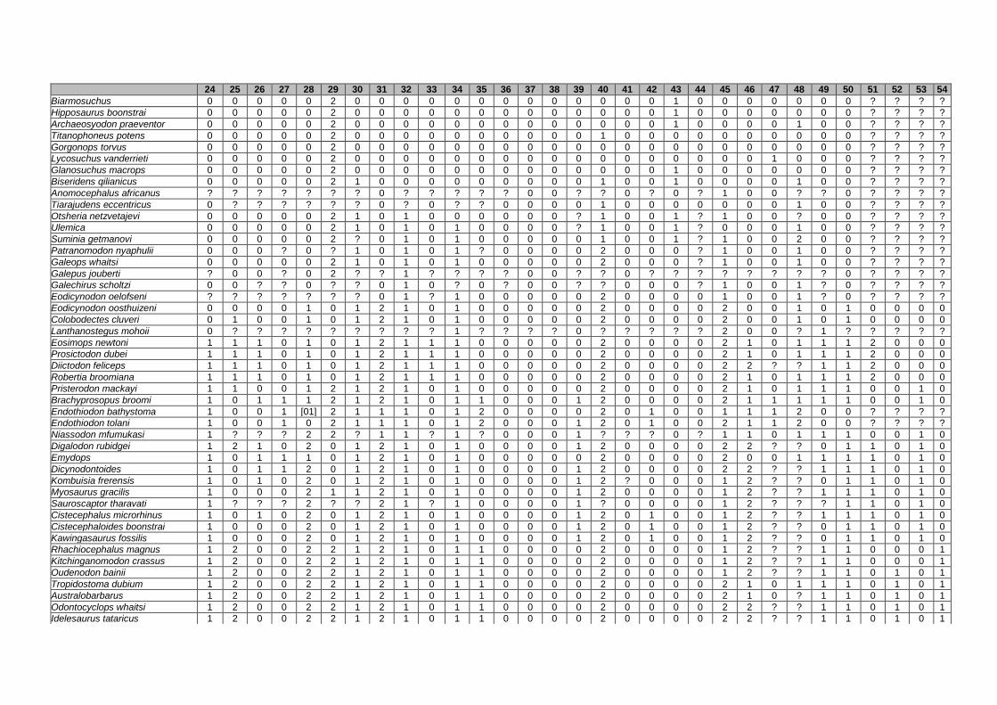
24 25 26 27 28 29 30 31 32 33 34 35 36 37 38 39 40 41 42 43 44 45 46 47 48 49 50 51 52 53 54
Biarmosuchus 0 0 0 0 0 2 0 0 0 0 0 0 0 0 0 0 0 0 0 1 0 0 0 0 0 0 0 ? ? ? ?
Hipposaurus boonstrai 0 0 0 0 0 2 0 0 0 0 0 0 0 0 0 0 0 0 0 1 0 0 0 0 0 0 0 ? ? ? ?
Archaeosyodon praeventor 0 0 0 0 0 2 0 0 0 0 0 0 0 0 0 0 0 0 0 1 0 0 0 0 1 0 0 ? ? ? ?
Titanophoneus potens 0 0 0 0 0 2 0 0 0 0 0 0 0 0 0 0 1 0 0 0 0 0 0 0 0 0 0 ? ? ? ?
Gorgonops torvus 0 0 0 0 0 2 0 0 0 0 0 0 0 0 0 0 0 0 0 0 0 0 0 0 0 0 0 ? ? ? ?
Lycosuchus vanderrieti 0 0 0 0 0 2 0 0 0 0 0 0 0 0 0 0 0 0 0 0 0 0 0 1 0 0 0 ? ? ? ?
Glanosuchus macrops 0 0 0 0 0 2 0 0 0 0 0 0 0 0 0 0 0 0 0 1 0 0 0 0 0 0 0 ? ? ? ?
Biseridens qilianicus 0 0 0 0 0 2 1 0 0 0 0 0 0 0 0 0 1 0 0 1 0 0 0 0 1 0 0 ? ? ? ?
Anomocephalus africanus ? ? ? ? ? ? ? 0 ? ? ? ? ? 0 0 ? ? 0 ? 0 ? 1 0 0 ? ? 0 ? ? ? ?
Tiarajudens eccentricus 0 ? ? ? ? ? ? 0 ? 0 ? ? 0 0 0 0 1 0 0 0 0 0 0 0 1 0 0 ? ? ? ?
Otsheria netzvetajevi 0 0 0 0 0 2 1 0 1 0 0 0 0 0 0 ? 1 0 0 1 ? 1 0 0 ? 0 0 ? ? ? ?
Ulemica 0 0 0 0 0 2 1 0 1 0 1 0 0 0 0 ? 1 0 0 1 ? 0 0 0 1 0 0 ? ? ? ?
Suminia getmanovi 0 0 0 0 0 2 ? 0 1 0 1 0 0 0 0 0 1 0 0 1 ? 1 0 0 2 0 0 ? ? ? ?
Patranomodon nyaphulii 0 0 0 ? 0 ? 1 0 1 0 1 ? 0 0 0 0 2 0 0 0 ? 1 0 0 1 0 0 ? ? ? ?
Galeops whaitsi 0 0 0 0 0 2 1 0 1 0 1 0 0 0 0 0 2 0 0 0 ? 1 0 0 1 0 0 ? ? ? ?
Galepus jouberti ? 0 0 ? 0 2 ? ? 1 ? ? ? ? 0 0 ? ? 0 ? ? ? ? ? ? ? ? 0 ? ? ? ?
Galechirus scholtzi 0 0 ? ? 0 ? ? 0 1 0 ? 0 ? 0 0 ? ? 0 0 0 ? 1 0 0 1 ? 0 ? ? ? ?
Eodicynodon oelofseni ? ? ? ? ? ? ? 0 1 ? 1 0 0 0 0 0 2 0 0 0 0 1 0 0 1 ? 0 ? ? ? ?
Eodicynodon oosthuizeni 0 0 0 0 1 0 1 2 1 0 1 0 0 0 0 0 2 0 0 0 0 2 0 0 1 0 1 0 0 0 0
Colobodectes cluveri 0 1 0 0 1 0 1 2 1 0 1 0 0 0 0 0 2 0 0 0 0 2 0 0 1 0 1 0 0 0 0
Lanthanostegus mohoii 0 ? ? ? ? ? ? ? ? ? 1 ? ? ? ? 0 ? ? ? ? ? 2 0 0 ? 1 ? ? ? ? ?
Eosimops newtoni 1 1 1 0 1 0 1 2 1 1 1 0 0 0 0 0 2 0 0 0 0 2 1 0 1 1 1 2 0 0 0
Prosictodon dubei 1 1 1 0 1 0 1 2 1 1 1 0 0 0 0 0 2 0 0 0 0 2 1 0 1 1 1 2 0 0 0
Diictodon feliceps 1 1 1 0 1 0 1 2 1 1 1 0 0 0 0 0 2 0 0 0 0 2 2 ? ? 1 1 2 0 0 0
Robertia broomiana 1 1 1 0 1 0 1 2 1 1 1 0 0 0 0 0 2 0 0 0 0 2 1 0 1 1 1 2 0 0 0
Pristerodon mackayi 1 1 0 0 1 2 1 2 1 0 1 0 0 0 0 0 2 0 0 0 0 2 1 0 1 1 1 0 0 1 0
Brachyprosopus broomi 1 0 1 1 1 2 1 2 1 0 1 1 0 0 0 1 2 0 0 0 0 2 1 1 1 1 1 0 0 1 0
Endothiodon bathystoma 1 0 0 1 [01] 2 1 1 1 0 1 2 0 0 0 0 2 0 1 0 0 1 1 1 2 0 0 ? ? ? ?
Endothiodon tolani 1 0 0 1 0 2 1 1 1 0 1 2 0 0 0 1 2 0 1 0 0 2 1 1 2 0 0 ? ? ? ?
Niassodon mfumukasi 1 ? ? ? 2 2 ? 1 1 ? 1 ? 0 0 0 1 ? ? ? 0 ? 1 1 0 1 1 1 0 0 1 0
Digalodon rubidgei 1 2 1 0 2 0 1 2 1 0 1 0 0 0 0 1 2 0 0 0 0 2 2 ? ? 0 1 1 0 1 0
Emydops 1 0 1 1 1 0 1 2 1 0 1 0 0 0 0 0 2 0 0 0 0 2 0 0 1 1 1 1 0 1 0
Dicynodontoides 1 0 1 1 2 0 1 2 1 0 1 0 0 0 0 1 2 0 0 0 0 2 2 ? ? 1 1 1 0 1 0
Kombuisia frerensis 1 0 1 0 2 0 1 2 1 0 1 0 0 0 0 1 2 ? 0 0 0 1 2 ? ? 0 1 1 0 1 0
Myosaurus gracilis 1 0 0 0 2 1 1 2 1 0 1 0 0 0 0 1 2 0 0 0 0 1 2 ? ? 1 1 1 0 1 0
Sauroscaptor tharavati 1 ? ? ? 2 ? ? 2 1 ? 1 0 0 0 0 1 ? 0 0 0 0 1 2 ? ? ? 1 1 0 1 0
Cistecephalus microrhinus 1 0 1 0 2 0 1 2 1 0 1 0 0 0 0 1 2 0 1 0 0 1 2 ? ? 1 1 1 0 1 0
Cistecephaloides boonstrai 1 0 0 0 2 0 1 2 1 0 1 0 0 0 0 1 2 0 1 0 0 1 2 ? ? 0 1 1 0 1 0
Kawingasaurus fossilis 1 0 0 0 2 0 1 2 1 0 1 0 0 0 0 1 2 0 1 0 0 1 2 ? ? 0 1 1 0 1 0
Rhachiocephalus magnus 1 2 0 0 2 2 1 2 1 0 1 1 0 0 0 0 2 0 0 0 0 1 2 ? ? 1 1 0 0 0 1
Kitchinganomodon crassus 1 2 0 0 2 2 1 2 1 0 1 1 0 0 0 0 2 0 0 0 0 1 2 ? ? 1 1 0 0 0 1
Oudenodon bainii 1 2 0 0 2 2 1 2 1 0 1 1 0 0 0 0 2 0 0 0 0 1 2 ? ? 1 1 0 1 0 1
Tropidostoma dubium 1 2 0 0 2 2 1 2 1 0 1 1 0 0 0 0 2 0 0 0 0 2 1 0 1 1 1 0 1 0 1
Australobarbarus 1 2 0 0 2 2 1 2 1 0 1 1 0 0 0 0 2 0 0 0 0 2 1 0 ? 1 1 0 1 0 1
Odontocyclops whaitsi 1 2 0 0 2 2 1 2 1 0 1 1 0 0 0 0 2 0 0 0 0 2 2 ? ? 1 1 0 1 0 1
Idelesaurus tataricus 1 2 0 0 2 2 1 2 1 0 1 1 0 0 0 0 2 0 0 0 0 2 2 ? ? 1 1 0 1 0 1

55 56 57 58 59 60 61 62 63 64 65 66 67 68 69 70 71 72 73 74 75 76 77 78 79 80 81 82 83 84 85
Biarmosuchus 0 0 0 0 0 0 0 0 0 0 0 0 0 0 ? 2 0 0 0 2 0 0 0 0 0 0 0 0 0 0 0
Hipposaurus boonstrai 0 0 0 0 0 0 0 0 0 0 0 0 0 1 0 2 0 0 0 0 0 0 0 0 0 0 0 0 0 0 0
Archaeosyodon praeventor 0 0 0 0 0 0 0 1 0 0 0 0 0 0 ? 2 0 0 0 1 0 0 0 0 0 0 0 0 0 0 0
Titanophoneus potens 0 0 0 0 0 0 0 1 0 0 1 0 0 0 ? 1 0 0 0 0 0 0 0 [02] 0 0 0 0 0 0 0
Gorgonops torvus 0 0 0 0 0 0 0 0 1 0 0 0 0 1 0 2 0 0 0 2 0 0 0 2 0 0 0 0 0 0 0
Lycosuchus vanderrieti 0 0 0 2 0 0 0 0 0 0 0 0 0 0 ? 1 0 2 0 1 1 0 0 1 0 0 0 0 0 0 0
Glanosuchus macrops 0 0 0 0 0 0 0 0 0 0 0 0 1 0 ? 1 0 2 0 1 1 0 0 1 0 0 0 0 0 0 0
Biseridens qilianicus 0 0 0 0 0 0 0 1 0 0 0 1 0 0 ? 2 0 0 0 1 0 0 0 0 0 0 0 0 0 0 0
Anomocephalus africanus ? ? 0 ? ? ? 0 ? ? ? 0 0 ? ? ? ? ? ? ? ? ? ? ? ? ? ? 0 ? ? 0 0
Tiarajudens eccentricus 0 0 0 ? ? 0 0 ? 0 0 0 0 ? ? ? ? ? ? ? ? ? ? ? ? ? ? 0 0 ? 0 0
Otsheria netzvetajevi ? 0 0 0 0 0 0 0 0 0 0 0 0 0 ? 2 ? 0 0 1 0 ? 0 0 0 0 0 0 0 0 0
Ulemica 0 0 0 0 0 0 0 1 0 0 0 0 0 0 ? 2 0 0 0 1 0 ? 0 0 0 0 1 0 0 0 0
Suminia getmanovi ? 0 0 1 0 0 0 0 0 0 0 0 0 0 ? 2 1 0 0 1 0 ? 0 0 0 0 2 0 0 0 0
Patranomodon nyaphulii ? 0 0 0 0 0 0 0 0 0 0 0 0 1 0 0 1 0 0 0 0 0 0 1 0 0 0 0 0 0 0
Galeops whaitsi ? ? 1 ? 0 0 0 0 0 0 0 0 0 ? ? ? 1 0 0 0 ? 0 0 ? 0 ? 2 0 0 0 0
Galepus jouberti ? ? ? ? ? ? ? ? ? ? 0 0 ? ? ? ? ? ? ? ? ? ? ? ? ? ? ? ? ? ? 0
Galechirus scholtzi ? ? 1 ? ? 0 0 ? ? ? ? ? ? ? ? ? ? ? ? ? ? ? ? ? ? ? ? ? ? ? ?
Eodicynodon oelofseni ? ? ? ? 0 ? 0 0 0 0 0 0 0 1 0 ? ? 0 0 1 0 0 0 1 0 0 2 0 1 0 ?
Eodicynodon oosthuizeni 0 0 1 1 0 0 0 0 0 0 0 0 0 1 0 0 1 0 0 1 0 0 0 1 0 0 2 1 1 0 0
Colobodectes cluveri 0 0 1 1 0 0 0 0 0 1 0 0 0 1 0 1 1 0 0 0 0 0 0 1 0 ? 2 1 1 0 0
Lanthanostegus mohoii 0 ? ? ? 0 ? 0 1 0 0 0 1 0 1 0 1 ? 0 0 1 0 0 0 2 0 0 ? ? ? 0 0
Eosimops newtoni 1 0 1 0 0 0 0 1 0 0 0 1 0 1 0 1 1 0 0 1 0 0 0 2 0 0 2 1 1 0 0
Prosictodon dubei 1 0 1 1 0 0 0 1 0 0 0 0 0 1 0 1 1 2 0 0 0 0 0 1 0 ? 2 1 1 0 0
Diictodon feliceps 1 [01] 1 [01] 0 0 0 1 0 0 0 0 0 1 0 1 1 2 0 0 0 0 0 [12] 0 0 2 1 1 0 0
Robertia broomiana 1 0 1 0 0 0 0 1 0 0 0 0 0 1 0 1 1 0 0 1 0 0 0 2 0 0 2 1 1 0 0
Pristerodon mackayi 0 0 1 0 0 0 0 1 0 0 0 ? 0 1 0 1 1 0 0 1 0 0 0 1 0 1 2 1 1 0 0
Brachyprosopus broomi 0 0 1 0 0 0 0 1 0 0 0 0 0 1 0 1 1 0 0 1 0 0 0 2 0 0 2 1 1 0 0
Endothiodon bathystoma ? 0 [12] ? 0 0 1 0 0 [01] 1 0 1 1 1 1 1 1 0 1 0 1 0 2 0 0 2 1 1 0 0
Endothiodon tolani ? ? [01] 0 0 0 0 0 0 1 ? ? 0 1 0 1 1 1 0 1 0 1 0 1 0 0 2 ? 1 ? ?
Niassodon mfumukasi 1 0 1 0 0 0 0 0 0 0 0 0 0 2 1 2 1 0 0 2 0 0 0 1 0 0 2 0 1 0 0
Digalodon rubidgei 0 0 1 0 0 0 0 0 0 1 0 0 0 1 0 1 1 0 0 2 0 0 0 3 0 1 2 1 1 0 0
Emydops 1 0 1 0 0 0 0 0 0 0 0 0 0 1 0 1 1 0 0 1 0 0 0 1 0 0 2 0 1 0 0
Dicynodontoides 0 0 1 0 0 0 0 0 0 1 0 0 0 2 0 1 1 2 0 0 0 0 0 1 0 0 2 1 1 0 0
Kombuisia frerensis 1 0 1 0 0 0 0 0 0 1 0 0 0 2 0 1 0 2 0 1 1 ? 1 ? ? 0 2 ? 1 0 0
Myosaurus gracilis 1 0 1 2 0 0 0 0 0 1 0 0 0 1 0 2 0 0 0 2 0 0 0 1 0 0 2 0 1 0 0
Sauroscaptor tharavati 0 ? 1 ? 0 ? 0 0 ? 1 0 0 0 0 ? ? ? 0 0 2 0 ? 0 1 0 0 2 0 1 0 0
Cistecephalus microrhinus 1 0 1 0 0 0 [01] 1 0 1 [01] 0 0 0 ? 2 1 0 0 2 0 0 0 1 0 1 2 0 1 0 0
Cistecephaloides boonstrai 0 0 1 0 0 0 0 0 1 1 0 0 0 0 ? 2 1 0 0 2 0 0 0 1 0 1 2 0 1 0 0
Kawingasaurus fossilis 0 0 0 2 0 0 0 0 0 1 0 0 0 0 ? 2 1 0 0 2 0 0 1 ? ? ? 2 0 1 0 0
Rhachiocephalus magnus 0 0 2 0 0 1 1 0 0 0 1 0 0 1 0 1 1 2 0 0 0 0 0 2 1 0 2 1 1 0 0
Kitchinganomodon crassus 0 0 2 0 0 1 1 0 0 0 1 0 0 1 0 1 1 2 0 0 0 0 0 2 1 1 2 1 1 1 0
Oudenodon bainii 0 0 2 1 0 [01] 0 1 0 0 0 0 0 1 0 1 1 1 0 0 0 0 0 1 0 1 2 1 1 1 0
Tropidostoma dubium 0 0 2 1 0 1 0 1 0 0 0 0 0 1 0 1 1 1 0 0 0 0 0 1 0 1 2 1 1 1 0
Australobarbarus 0 0 2 ? 0 ? 0 1 0 0 0 0 0 1 0 1 1 1 0 0 0 0 0 1 0 1 2 1 1 1 0
Odontocyclops whaitsi 0 0 2 0 0 1 2 0 0 0 0 0 0 2 1 1 1 1 0 0 0 0 0 2 0 1 2 1 1 1 0
Idelesaurus tataricus 0 0 2 1 0 1 1 0 0 0 0 0 0 2 1 1 1 1 0 0 0 0 0 1 0 0 2 1 1 1 0
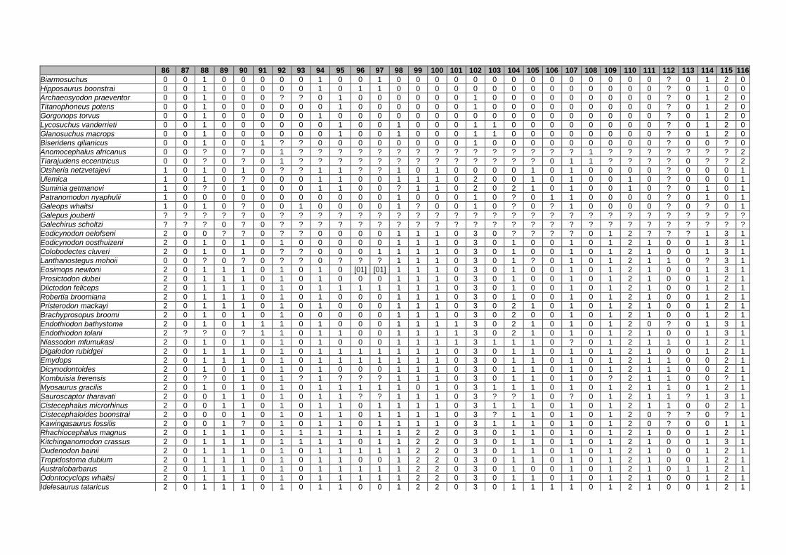
86 87 88 89 90 91 92 93 94 95 96 97 98 99 100 101 102 103 104 105 106 107 108 109 110 111 112 113 114 115 116
Biarmosuchus 0 0 1 0 0 0 0 0 1 0 0 1 0 0 0 0 0 0 0 0 0 0 0 0 0 0 ? 0 1 2 0
Hipposaurus boonstrai 0 0 1 0 0 0 0 0 1 0 1 1 0 0 0 0 0 0 0 0 0 0 0 0 0 0 ? 0 1 0 0
Archaeosyodon praeventor 0 0 1 0 0 0 ? ? 0 1 0 0 0 0 0 0 1 0 0 0 0 0 0 0 0 0 ? 0 1 2 0
Titanophoneus potens 0 0 1 0 0 0 0 0 0 1 0 0 0 0 0 0 1 0 0 0 0 0 0 0 0 0 ? 0 1 2 0
Gorgonops torvus 0 0 1 0 0 0 0 0 1 0 0 0 0 0 0 0 0 0 0 0 0 0 0 0 0 0 ? 0 1 2 0
Lycosuchus vanderrieti 0 0 1 0 0 0 0 0 0 1 0 0 1 0 0 0 1 1 0 0 0 0 0 0 0 0 ? 0 1 2 0
Glanosuchus macrops 0 0 1 0 0 0 0 0 0 1 0 0 1 0 0 0 1 1 0 0 0 0 0 0 0 0 ? 0 1 2 0
Biseridens qilianicus 0 0 1 0 0 1 ? ? 0 0 0 0 0 0 0 0 1 0 0 0 0 0 0 0 0 0 ? 0 0 ? 0
Anomocephalus africanus 0 0 ? 0 ? 0 1 ? ? ? ? ? ? ? ? ? ? ? ? ? ? ? 1 ? ? ? ? ? ? ? 2
Tiarajudens eccentricus 0 0 ? 0 ? 0 1 ? ? ? ? ? ? ? ? ? ? ? ? ? 0 1 1 ? ? ? ? 0 ? ? 2
Otsheria netzvetajevi 1 0 1 0 1 0 ? ? 1 1 ? ? 1 0 1 0 0 0 0 1 0 1 0 0 0 0 ? 0 0 0 1
Ulemica 1 0 1 0 ? 0 0 0 1 1 0 0 1 1 1 0 2 0 0 1 0 1 0 0 1 0 ? 0 0 0 1
Suminia getmanovi 1 0 ? 0 1 0 0 0 1 1 0 0 ? 1 1 0 2 0 2 1 0 1 0 0 1 0 ? 0 1 0 1
Patranomodon nyaphulii 1 0 0 0 0 0 0 0 0 0 0 0 1 0 0 0 1 0 ? 0 1 1 0 0 0 0 ? 0 1 0 1
Galeops whaitsi 1 0 1 0 ? 0 0 1 0 0 0 0 1 ? 0 0 1 0 ? 0 ? 1 0 0 0 0 ? 0 ? 0 1
Galepus jouberti ? ? ? ? ? 0 ? ? ? ? ? ? ? ? ? ? ? ? ? ? ? ? ? ? ? ? ? ? ? ? ?
Galechirus scholtzi ? ? ? 0 ? 0 ? ? ? ? ? ? ? ? ? ? ? ? ? ? ? ? ? ? ? ? ? ? ? ? ?
Eodicynodon oelofseni 2 0 0 ? ? 0 ? ? 0 0 0 0 1 1 1 0 3 0 ? ? ? ? 0 1 2 ? ? ? 1 3 1
Eodicynodon oosthuizeni 2 0 1 0 1 0 1 0 0 0 0 0 1 1 1 0 3 0 1 0 0 1 0 1 2 1 0 0 1 3 1
Colobodectes cluveri 2 0 1 0 1 0 ? ? 0 0 0 1 1 1 1 0 3 0 1 0 0 1 0 1 2 1 0 0 1 3 1
Lanthanostegus mohoii 0 0 ? 0 ? 0 ? ? 0 ? ? ? 1 1 1 0 3 0 1 ? 0 1 0 1 2 1 1 0 ? 3 1
Eosimops newtoni 2 0 1 1 1 0 1 0 1 0 [01] [01] 1 1 1 0 3 0 1 0 0 1 0 1 2 1 0 0 1 3 1
Prosictodon dubei 2 0 1 1 1 0 1 0 1 0 0 0 1 1 1 0 3 0 1 0 0 1 0 1 2 1 0 0 1 2 1
Diictodon feliceps 2 0 1 1 1 0 1 0 1 1 1 1 1 1 1 0 3 0 1 0 0 1 0 1 2 1 0 0 1 2 1
Robertia broomiana 2 0 1 1 1 0 1 0 1 0 0 0 1 1 1 0 3 0 1 0 0 1 0 1 2 1 0 0 1 2 1
Pristerodon mackayi 2 0 1 1 1 0 1 0 1 0 0 0 1 1 1 0 3 0 2 1 0 1 0 1 2 1 0 0 1 2 1
Brachyprosopus broomi 2 0 1 0 1 0 1 0 0 0 0 0 1 1 1 0 3 0 2 0 0 1 0 1 2 1 0 0 1 2 1
Endothiodon bathystoma 2 0 1 0 1 1 1 0 1 0 0 0 1 1 1 1 3 0 2 1 0 1 0 1 2 0 ? 0 1 3 1
Endothiodon tolani 2 ? ? 0 ? 1 1 0 1 1 0 0 1 1 1 1 3 0 2 1 0 1 0 1 2 1 0 0 1 3 1
Niassodon mfumukasi 2 0 1 0 1 0 1 0 1 0 0 0 1 1 1 1 3 1 1 1 0 ? 0 1 2 1 1 0 1 2 1
Digalodon rubidgei 2 0 1 1 1 0 1 0 1 1 1 1 1 1 1 0 3 0 1 1 0 1 0 1 2 1 0 0 1 2 1
Emydops 2 0 1 1 1 0 1 0 1 1 1 1 1 1 1 0 3 0 1 1 0 1 0 1 2 1 1 0 0 2 1
Dicynodontoides 2 0 1 0 1 0 1 0 1 0 0 0 1 1 1 0 3 0 1 1 0 1 0 1 2 1 1 0 0 2 1
Kombuisia frerensis 2 0 ? 0 1 0 1 ? 1 ? ? ? 1 1 1 0 3 0 1 1 0 1 0 ? 2 1 1 0 0 ? 1
Myosaurus gracilis 2 0 1 0 1 0 1 0 1 1 1 1 1 0 1 0 3 1 1 1 0 1 0 1 2 1 1 0 1 2 1
Sauroscaptor tharavati 2 0 0 1 1 0 1 0 1 1 ? ? 1 1 1 0 3 ? ? 1 0 ? 0 1 2 1 1 ? 1 3 1
Cistecephalus microrhinus 2 0 0 1 1 0 1 0 1 1 0 1 1 1 1 0 3 1 1 1 0 1 0 1 2 1 1 0 0 2 1
Cistecephaloides boonstrai 2 0 0 0 1 0 1 0 1 1 0 1 1 1 1 0 3 ? 1 1 0 1 0 1 2 0 ? ? 0 ? 1
Kawingasaurus fossilis 2 0 0 1 ? 0 1 0 1 1 0 1 1 1 1 0 3 1 1 1 0 1 0 1 2 0 ? 0 0 1 1
Rhachiocephalus magnus 2 0 1 1 1 0 1 1 1 1 1 1 1 2 2 0 3 0 1 1 0 1 0 1 2 1 0 0 1 2 1
Kitchinganomodon crassus 2 0 1 1 1 0 1 1 1 1 0 1 1 2 2 0 3 0 1 1 0 1 0 1 2 1 0 0 1 3 1
Oudenodon bainii 2 0 1 1 1 0 1 0 1 1 1 1 1 2 2 0 3 0 1 1 0 1 0 1 2 1 0 0 1 2 1
Tropidostoma dubium 2 0 1 1 1 0 1 0 1 1 0 0 1 2 2 0 3 0 1 1 0 1 0 1 2 1 0 0 1 2 1
Australobarbarus 2 0 1 1 1 0 1 0 1 1 1 1 1 2 2 0 3 0 1 0 0 1 0 1 2 1 0 1 1 2 1
Odontocyclops whaitsi 2 0 1 1 1 0 1 0 1 1 1 1 1 2 2 0 3 0 1 1 0 1 0 1 2 1 0 0 1 2 1
Idelesaurus tataricus 2 0 1 1 1 0 1 0 1 1 0 0 1 2 2 0 3 0 1 1 1 1 0 1 2 1 0 0 1 2 1
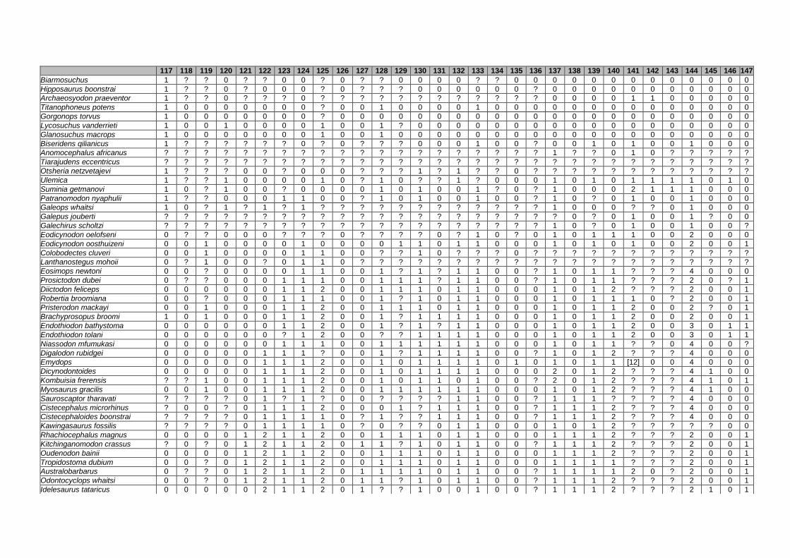
117 118 119 120 121 122 123 124 125 126 127 128 129 130 131 132 133 134 135 136 137 138 139 140 141 142 143 144 145 146 147
Biarmosuchus 1 ? ? 0 ? ? 0 0 ? 0 ? ? 0 0 0 0 ? ? 0 0 0 0 0 0 0 0 0 0 0 0 0
Hipposaurus boonstrai 1 ? ? 0 ? 0 0 0 ? 0 ? ? ? 0 0 0 0 0 0 ? 0 0 0 0 0 0 0 0 0 0 0
Archaeosyodon praeventor 1 ? ? 0 ? ? ? 0 ? ? ? ? ? ? ? ? ? ? ? ? 0 0 0 0 1 1 0 0 0 0 0
Titanophoneus potens 1 0 0 0 0 0 0 0 ? 0 0 1 0 0 0 0 1 0 0 0 0 0 0 0 0 0 0 0 0 0 0
Gorgonops torvus 1 0 0 0 0 0 0 0 ? 0 0 0 0 0 0 0 0 0 0 0 0 0 0 0 0 0 0 0 0 0 0
Lycosuchus vanderrieti 1 0 0 1 0 0 0 0 1 0 0 1 ? 0 0 0 0 0 0 0 0 0 0 0 0 0 0 0 0 0 0
Glanosuchus macrops 1 0 0 0 0 0 0 0 1 0 0 1 0 0 0 0 0 0 0 0 0 0 0 0 0 0 0 0 0 0 0
Biseridens qilianicus 1 ? ? ? ? ? ? 0 ? 0 ? ? ? 0 0 0 1 0 0 ? 0 0 1 0 1 0 0 1 0 0 0
Anomocephalus africanus ? ? ? ? ? ? ? ? ? ? ? ? ? ? ? ? ? ? ? ? 1 ? ? 0 1 0 ? ? ? ? ?
Tiarajudens eccentricus ? ? ? ? ? ? ? ? ? ? ? ? ? ? ? ? ? ? ? ? ? ? ? ? ? ? ? ? ? ? ?
Otsheria netzvetajevi 1 ? ? ? 0 0 ? 0 0 0 ? ? ? 1 ? 1 ? ? 0 ? ? ? ? ? ? ? ? ? ? ? ?
Ulemica 1 ? ? 1 0 0 0 0 1 0 ? 1 0 ? ? 1 ? 0 0 0 1 0 1 0 1 1 1 1 0 1 0
Suminia getmanovi 1 0 ? 1 0 0 ? 0 0 0 0 1 0 1 0 0 1 ? 0 ? 1 0 0 0 2 1 1 1 0 0 0
Patranomodon nyaphulii 1 ? ? 0 0 0 1 1 0 0 ? 1 0 1 0 0 1 0 0 ? 1 0 ? 0 1 0 0 1 0 0 0
Galeops whaitsi 1 0 ? 1 ? 1 ? 1 ? ? ? ? ? ? ? ? ? ? ? ? 1 0 0 0 ? ? 0 1 0 0 0
Galepus jouberti ? ? ? ? ? ? ? ? ? ? ? ? ? ? ? ? ? ? ? ? ? 0 ? 0 1 0 0 1 ? 0 0
Galechirus scholtzi ? ? ? ? ? ? ? ? ? ? ? ? ? ? ? ? ? ? ? ? 1 0 ? 0 1 0 0 1 0 0 ?
Eodicynodon oelofseni 0 ? ? 0 0 0 ? ? ? 0 ? ? ? ? 0 ? 1 0 ? 0 1 0 1 1 1 0 0 2 0 0 0
Eodicynodon oosthuizeni 0 0 1 0 0 0 0 1 0 0 0 0 1 1 0 1 1 0 0 0 1 0 1 0 1 0 0 2 0 0 1
Colobodectes cluveri 0 0 1 0 0 0 0 1 1 0 0 ? ? 1 0 ? ? ? 0 ? ? ? ? ? ? ? ? ? ? ? ?
Lanthanostegus mohoii 0 ? 1 0 0 ? 0 1 1 0 ? ? ? ? ? ? ? ? ? ? ? ? ? ? ? ? ? ? ? ? ?
Eosimops newtoni 0 0 ? 0 0 0 0 1 1 0 0 1 ? 1 ? 1 1 0 0 ? 1 0 1 1 ? ? ? 4 0 0 0
Prosictodon dubei 0 ? ? 0 0 0 1 1 1 0 0 1 1 1 ? 1 1 0 0 ? 1 0 1 1 ? ? ? 2 0 ? 1
Diictodon feliceps 0 0 0 0 0 0 1 1 2 0 0 1 1 1 0 1 1 0 0 0 1 0 1 2 ? ? ? 2 0 0 1
Robertia broomiana 0 0 ? 0 0 0 1 1 1 0 0 1 ? 1 0 1 1 0 0 0 1 0 1 1 1 0 ? 2 0 0 1
Pristerodon mackayi 0 0 1 0 0 0 1 1 2 0 0 1 1 1 0 1 1 0 0 0 1 0 1 1 2 0 0 2 ? 0 1
Brachyprosopus broomi 1 0 1 0 0 0 1 1 2 0 0 1 ? 1 1 1 1 0 0 0 1 0 1 1 2 0 0 2 0 0 1
Endothiodon bathystoma 0 0 0 0 0 0 1 1 2 0 0 1 ? 1 ? 1 1 0 0 0 1 0 1 1 2 0 0 3 0 1 1
Endothiodon tolani 0 0 0 0 0 0 ? 1 2 0 0 ? ? 1 1 1 1 0 0 0 1 0 1 1 2 0 0 3 0 1 1
Niassodon mfumukasi 0 0 0 0 0 0 1 1 1 0 0 1 1 1 1 1 1 0 0 0 1 0 1 1 ? ? 0 4 0 0 ?
Digalodon rubidgei 0 0 0 0 0 1 1 1 ? 0 0 1 ? 1 1 1 1 0 0 ? 1 0 1 2 ? ? ? 4 0 0 0
Emydops 0 0 0 0 0 1 1 1 2 0 0 1 0 1 1 1 1 0 1 0 1 0 1 1 [12] 0 0 4 0 0 0
Dicynodontoides 0 0 0 0 0 1 1 1 2 0 0 1 0 1 1 1 1 0 0 0 2 0 1 2 ? ? ? 4 1 0 0
Kombuisia frerensis ? ? 1 0 0 1 1 1 2 0 0 1 0 1 1 0 1 0 0 ? 2 0 1 2 ? ? ? 4 1 0 1
Myosaurus gracilis 0 0 1 0 0 1 1 1 2 0 0 1 1 1 1 1 1 0 0 0 1 0 1 2 ? ? ? 4 1 0 0
Sauroscaptor tharavati ? ? ? ? 0 1 ? 1 ? 0 0 ? ? ? ? 1 1 0 0 ? 1 1 1 ? ? ? ? 4 0 0 0
Cistecephalus microrhinus ? 0 0 ? 0 1 1 1 2 0 0 0 1 ? 1 1 1 0 0 ? 1 1 1 2 ? ? ? 4 0 0 0
Cistecephaloides boonstrai ? ? ? ? 0 1 1 1 1 0 ? 1 ? ? 1 1 1 0 0 ? 1 1 1 2 ? ? ? 4 0 0 0
Kawingasaurus fossilis ? ? ? ? 0 1 1 1 1 0 ? 0 ? ? 0 1 1 0 0 0 1 0 1 2 ? ? ? ? ? 0 0
Rhachiocephalus magnus 0 0 0 0 1 2 1 1 2 0 0 1 1 1 0 1 1 0 0 0 1 1 1 2 ? ? ? 2 0 0 1
Kitchinganomodon crassus ? 0 ? 0 1 2 1 1 2 0 1 1 ? 1 0 1 1 0 0 ? 1 1 1 2 ? ? ? 2 0 0 1
Oudenodon bainii 0 0 0 0 1 2 1 1 2 0 0 1 1 1 0 1 1 0 0 0 1 1 1 2 ? ? ? 2 0 0 1
Tropidostoma dubium 0 0 ? 0 1 2 1 1 2 0 0 1 1 1 0 1 1 0 0 0 1 1 1 1 ? ? ? 2 0 0 1
Australobarbarus 0 ? ? 0 1 2 1 1 2 0 1 1 1 1 0 1 1 0 0 ? 1 1 1 1 2 0 ? 2 0 0 1
Odontocyclops whaitsi 0 0 ? 0 1 2 1 1 2 0 1 1 ? 1 0 1 1 0 0 ? 1 1 1 2 ? ? ? 2 0 0 1
Idelesaurus tataricus 0 0 0 0 0 2 1 1 2 0 1 ? ? 1 0 0 1 0 0 ? 1 1 1 2 ? ? ? 2 1 0 1

148 149 150 151 152 153 154 155 156 157 158 159 160 161 162 163 164 165 166 167 168 169 170 171 172 173 174 175 176 177 178
Biarmosuchus 0 0 0 ? ? 0 0 0 0 0 0 0 0 0 0 0 0 0 ? 0 0 0 0 ? 1 0 0 0 0 1 1
Hipposaurus boonstrai 0 0 0 ? ? 0 0 0 0 0 1 0 0 0 0 0 0 0 ? 1 0 0 0 0 1 0 0 0 1 1 1
Archaeosyodon praeventor 0 0 0 ? ? 0 0 0 ? ? ? ? ? ? ? ? 0 ? ? ? ? ? ? ? ? ? ? ? ? ? ?
Titanophoneus potens 0 0 0 ? ? 0 0 0 1 0 0 0 0 0 0 0 0 0 ? 0 0 0 0 0 ? 0 0 0 0 0 0
Gorgonops torvus 0 0 0 ? ? 0 0 0 0 0 1 1 0 0 0 0 0 0 ? 0 0 0 0 0 1 0 0 0 1 0 0
Lycosuchus vanderrieti 0 0 0 ? ? 0 1 0 0 0 2 0 1 0 0 0 0 0 ? 0 0 0 0 0 1 0 0 0 1 0 0
Glanosuchus macrops 0 0 0 ? ? 0 1 0 0 0 2 0 1 0 0 0 0 0 ? 0 0 0 0 0 1 0 0 0 1 0 0
Biseridens qilianicus 0 0 0 ? ? 0 0 0 0 0 0 ? 1 ? ? ? 0 ? ? ? ? ? ? ? ? ? ? ? ? ? ?
Anomocephalus africanus ? ? 0 ? ? ? ? ? ? ? ? ? ? ? ? ? ? ? ? ? ? ? ? ? ? ? ? ? ? ? ?
Tiarajudens eccentricus ? ? ? ? ? ? ? ? ? ? ? ? ? ? ? ? ? ? ? ? 0 0 ? ? ? 0 0 0 1 0 1
Otsheria netzvetajevi ? ? ? ? ? ? ? ? ? ? ? ? ? ? ? ? ? ? ? ? ? ? ? ? ? ? ? ? ? ? ?
Ulemica 0 0 1 0 0 [01] 0 0 0 0 0 0 1 0 0 1 1 ? ? ? ? ? ? ? ? ? ? ? ? ? ?
Suminia getmanovi 0 0 1 0 0 0 0 0 0 0 0 0 0 0 0 2 1 0 ? 0 0 0 0 0 0 0 0 0 0 0 0
Patranomodon nyaphulii 0 0 0 ? ? ? ? 0 0 0 3 1 0 0 0 0 1 0 ? ? ? ? ? ? ? ? ? ? ? ? ?
Galeops whaitsi 0 0 1 0 0 ? 1 0 1 1 3 1 0 0 0 1 1 ? ? 0 0 1 0 0 0 0 0 0 0 1 0
Galepus jouberti 0 0 ? ? ? ? ? ? ? ? ? ? ? ? ? ? 1 ? ? ? ? ? ? 1 ? 0 0 0 1 0 ?
Galechirus scholtzi ? 0 ? ? ? ? ? ? ? 0 3 1 ? ? ? ? 1 ? ? ? 0 0 0 0 ? ? 0 ? 1 0 0
Eodicynodon oelofseni 0 0 1 0 0 1 ? 0 1 ? ? 1 ? ? 1 2 1 ? ? ? ? ? ? ? ? ? ? ? ? ? ?
Eodicynodon oosthuizeni 0 0 2 0 0 1 0 0 1 1 3 1 1 1 1 2 1 0 ? 0 0 0 1 0 0 0 0 0 0 1 1
Colobodectes cluveri ? ? ? ? ? ? ? ? ? ? ? ? ? ? ? ? ? ? ? ? ? ? ? ? ? ? ? ? ? ? ?
Lanthanostegus mohoii ? ? ? ? ? ? ? ? ? ? ? ? ? ? ? ? ? ? ? ? ? ? ? ? ? ? ? ? ? ? ?
Eosimops newtoni 1 1 1 0 0 0 0 0 1 1 3 1 ? 1 1 2 1 0 ? 1 0 0 1 1 ? 0 0 0 0 0 1
Prosictodon dubei 1 1 1 0 0 ? 0 ? 1 ? ? 1 ? ? 1 2 1 ? ? ? ? ? ? ? ? ? ? ? ? ? ?
Diictodon feliceps 1 1 1 0 0 1 0 0 1 1 3 1 1 1 1 2 1 1 ? 1 0 0 1 1 0 0 0 0 0 0 0
Robertia broomiana 1 1 1 0 0 ? 0 0 1 1 3 1 1 1 1 2 1 0 ? 1 0 0 1 ? 0 0 0 0 0 0 0
Pristerodon mackayi 2 0 2 0 0 1 0 0 1 1 3 1 1 1 1 2 1 2 ? 1 1 0 1 0 0 0 0 0 0 1 0
Brachyprosopus broomi 2 0 1 0 0 1 0 0 1 1 3 1 1 1 1 2 1 ? ? 0 0 0 1 ? ? 0 0 0 0 1 0
Endothiodon bathystoma 2 0 2 0 0 1 0 0 1 1 4 1 1 1 1 2 1 2 ? 0 0 0 1 0 ? 0 0 0 0 1 0
Endothiodon tolani 2 0 2 0 0 1 0 0 1 1 4 1 1 1 1 2 1 ? ? ? ? ? ? ? ? ? ? ? ? ? ?
Niassodon mfumukasi 2 0 2 0 0 1 0 0 1 1 4 ? 1 1 1 2 1 1 ? ? ? ? ? ? ? ? ? ? ? ? ?
Digalodon rubidgei 2 ? ? ? ? 1 0 0 ? ? ? ? ? ? ? ? ? ? ? ? ? ? ? ? ? ? ? ? ? ? ?
Emydops 2 0 2 1 0 1 0 0 1 1 3 1 1 1 1 2 1 0 ? 1 ? 0 1 0 ? 0 0 0 0 0 0
Dicynodontoides 0 0 2 1 0 1 0 0 1 1 3 1 1 1 1 2 1 1 0 0 1 0 1 1 0 1 1 0 0 0 1
Kombuisia frerensis 0 0 2 0 0 1 ? 0 1 1 ? 1 1 0 1 2 1 ? ? ? ? ? ? ? ? ? ? ? ? ? ?
Myosaurus gracilis 0 0 2 1 0 ? ? 0 1 1 3 1 ? ? 1 2 1 0 ? 1 1 0 1 1 0 1 0 1 1 0 ?
Sauroscaptor tharavati ? 0 2 1 0 1 0 ? ? 1 ? 1 ? ? ? ? 1 ? ? ? ? ? ? ? ? ? ? ? ? ? ?
Cistecephalus microrhinus 2 0 2 1 0 1 0 1 1 1 3 1 1 1 1 2 1 0 ? 1 1 0 0 0 1 2 2 1 0 0 1
Cistecephaloides boonstrai 2 0 2 1 0 1 ? 1 1 1 ? 1 1 1 1 2 1 ? ? ? ? ? ? ? ? ? ? ? ? ? ?
Kawingasaurus fossilis 0 0 1 0 0 1 1 1 1 ? ? ? 1 1 1 2 1 ? ? 1 1 0 0 0 ? 2 2 0 0 0 1
Rhachiocephalus magnus 3 0 1 0 0 1 0 0 1 1 4 1 1 1 1 2 1 ? ? 0 0 1 1 [01] 1 1 0 1 0 1 0
Kitchinganomodon crassus ? 0 1 2 1 ? ? 0 ? 1 4 1 1 ? 1 2 1 ? ? ? 0 0 ? ? ? 1 0 ? 0 1 0
Oudenodon bainii 3 0 1 2 1 1 0 0 1 1 4 1 1 1 1 2 1 2 0 0 0 0 1 0 0 1 0 1 0 1 0
Tropidostoma dubium 3 0 1 2 1 1 ? 0 1 1 4 1 1 1 1 2 1 2 ? 0 0 0 1 0 0 1 0 1 0 1 0
Australobarbarus 3 0 1 2 1 1 ? 0 1 1 4 1 ? ? 1 2 1 ? ? ? ? ? ? ? ? ? ? ? ? ? ?
Odontocyclops whaitsi 3 0 1 2 1 ? ? 0 1 1 4 1 1 ? 1 2 1 2 0 0 0 0 1 ? ? 1 0 1 0 1 0
Idelesaurus tataricus 3 0 1 2 1 1 ? 0 1 1 4 1 1 1 1 2 1 ? ? ? ? ? ? ? ? ? ? ? ? ? ?
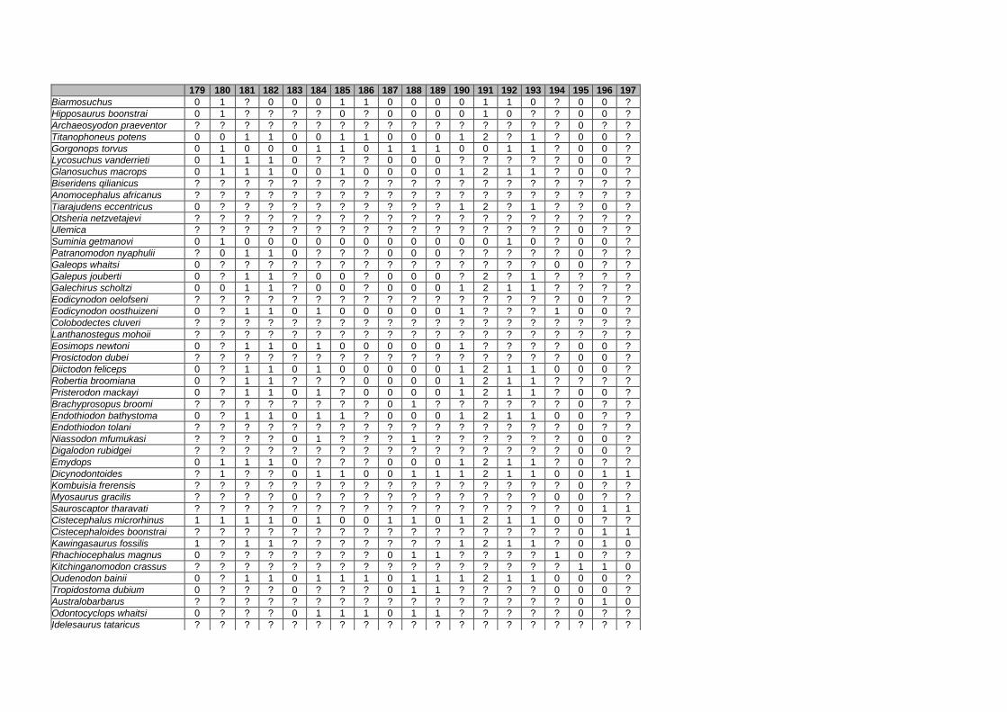
179 180 181 182 183 184 185 186 187 188 189 190 191 192 193 194 195 196 197
Biarmosuchus 0 1 ? 0 0 0 1 1 0 0 0 0 1 1 0 ? 0 0 ?
Hipposaurus boonstrai 0 1 ? ? ? ? 0 ? 0 0 0 0 1 0 ? ? 0 0 ?
Archaeosyodon praeventor ? ? ? ? ? ? ? ? ? ? ? ? ? ? ? ? 0 ? ?
Titanophoneus potens 0 0 1 1 0 0 1 1 0 0 0 1 2 ? 1 ? 0 0 ?
Gorgonops torvus 0 1 0 0 0 1 1 0 1 1 1 0 0 1 1 ? 0 0 ?
Lycosuchus vanderrieti 0 1 1 1 0 ? ? ? 0 0 0 ? ? ? ? ? 0 0 ?
Glanosuchus macrops 0 1 1 1 0 0 1 0 0 0 0 1 2 1 1 ? 0 0 ?
Biseridens qilianicus ? ? ? ? ? ? ? ? ? ? ? ? ? ? ? ? ? ? ?
Anomocephalus africanus ? ? ? ? ? ? ? ? ? ? ? ? ? ? ? ? ? ? ?
Tiarajudens eccentricus 0 ? ? ? ? ? ? ? ? ? ? 1 2 ? 1 ? ? 0 ?
Otsheria netzvetajevi ? ? ? ? ? ? ? ? ? ? ? ? ? ? ? ? ? ? ?
Ulemica ? ? ? ? ? ? ? ? ? ? ? ? ? ? ? ? 0 ? ?
Suminia getmanovi 0 1 0 0 0 0 0 0 0 0 0 0 0 1 0 ? 0 0 ?
Patranomodon nyaphulii ? 0 1 1 0 ? ? ? 0 0 0 ? ? ? ? ? 0 ? ?
Galeops whaitsi 0 ? ? ? ? ? ? ? ? ? ? ? ? ? ? 0 0 ? ?
Galepus jouberti 0 ? 1 1 ? 0 0 ? 0 0 0 ? 2 ? 1 ? ? ? ?
Galechirus scholtzi 0 0 1 1 ? 0 0 ? 0 0 0 1 2 1 1 ? ? ? ?
Eodicynodon oelofseni ? ? ? ? ? ? ? ? ? ? ? ? ? ? ? ? 0 ? ?
Eodicynodon oosthuizeni 0 ? 1 1 0 1 0 0 0 0 0 1 ? ? ? 1 0 0 ?
Colobodectes cluveri ? ? ? ? ? ? ? ? ? ? ? ? ? ? ? ? ? ? ?
Lanthanostegus mohoii ? ? ? ? ? ? ? ? ? ? ? ? ? ? ? ? ? ? ?
Eosimops newtoni 0 ? 1 1 0 1 0 0 0 0 0 1 ? ? ? ? 0 0 ?
Prosictodon dubei ? ? ? ? ? ? ? ? ? ? ? ? ? ? ? ? 0 0 ?
Diictodon feliceps 0 ? 1 1 0 1 0 0 0 0 0 1 2 1 1 0 0 0 ?
Robertia broomiana 0 ? 1 1 ? ? ? 0 0 0 0 1 2 1 1 ? ? ? ?
Pristerodon mackayi 0 ? 1 1 0 1 ? 0 0 0 0 1 2 1 1 ? 0 0 ?
Brachyprosopus broomi ? ? ? ? ? ? ? ? 0 1 ? ? ? ? ? ? 0 ? ?
Endothiodon bathystoma 0 ? 1 1 0 1 1 ? 0 0 0 1 2 1 1 0 0 ? ?
Endothiodon tolani ? ? ? ? ? ? ? ? ? ? ? ? ? ? ? ? 0 ? ?
Niassodon mfumukasi ? ? ? ? 0 1 ? ? ? 1 ? ? ? ? ? ? 0 0 ?
Digalodon rubidgei ? ? ? ? ? ? ? ? ? ? ? ? ? ? ? ? 0 0 ?
Emydops 0 1 1 1 0 ? ? ? 0 0 0 1 2 1 1 ? 0 ? ?
Dicynodontoides ? 1 ? ? 0 1 1 0 0 1 1 1 2 1 1 0 0 1 1
Kombuisia frerensis ? ? ? ? ? ? ? ? ? ? ? ? ? ? ? ? 0 ? ?
Myosaurus gracilis ? ? ? ? 0 ? ? ? ? ? ? ? ? ? ? 0 0 ? ?
Sauroscaptor tharavati ? ? ? ? ? ? ? ? ? ? ? ? ? ? ? ? 0 1 1
Cistecephalus microrhinus 1 1 1 1 0 1 0 0 1 1 0 1 2 1 1 0 0 ? ?
Cistecephaloides boonstrai ? ? ? ? ? ? ? ? ? ? ? ? ? ? ? ? 0 1 1
Kawingasaurus fossilis 1 ? 1 1 ? ? ? ? ? ? ? 1 2 1 1 ? 0 1 0
Rhachiocephalus magnus 0 ? ? ? ? ? ? ? 0 1 1 ? ? ? ? 1 0 ? ?
Kitchinganomodon crassus ? ? ? ? ? ? ? ? ? ? ? ? ? ? ? ? 1 1 0
Oudenodon bainii 0 ? 1 1 0 1 1 1 0 1 1 1 2 1 1 0 0 0 ?
Tropidostoma dubium 0 ? ? ? 0 ? ? ? 0 1 1 ? ? ? ? 0 0 0 ?
Australobarbarus ? ? ? ? ? ? ? ? ? ? ? ? ? ? ? ? 0 1 0
Odontocyclops whaitsi 0 ? ? ? 0 1 1 1 0 1 1 ? ? ? ? ? 0 ? ?
Idelesaurus tataricus ? ? ? ? ? ? ? ? ? ? ? ? ? ? ? ? ? ? ?

24 25 26 27 28 29 30 31 32 33 34 35 36 37 38 39 40 41 42 43 44 45 46 47 48 49 50 51 52 53 54
Aulacephalodon bainii 1 2 0 0 2 2 1 2 1 0 1 1 1 0 0 0 2 0 0 0 0 2 2 ? ? 1 1 0 1 0 [01]
Pelanomodon moschops 1 2 0 0 2 2 1 2 1 0 1 1 1 0 0 0 2 0 0 0 0 1 2 ? ? 1 1 0 1 0 1
Geikia locusticeps 1 2 0 0 2 2 1 2 1 0 1 1 1 0 0 0 2 0 0 0 0 1 2 ? ? 1 1 0 1 0 1
Geikia elginensis 1 2 0 0 2 2 ? 2 1 0 1 1 1 0 0 0 2 0 0 0 0 1 2 ? ? 1 1 0 1 0 1
Elph borealis 1 ? ? 0 2 ? ? 2 1 0 1 0 0 0 0 0 2 0 1 0 0 2 2 ? ? 1 1 0 1 0 0
Interpresosaurus blomi 1 2 0 0 ? ? 1 2 1 0 1 0 0 0 0 0 2 0 1 0 0 2 2 ? ? 1 1 0 1 0 0
Katumbia parringtoni 1 2 0 0 2 2 1 2 1 0 1 ? 0 0 0 0 ? ? 1 0 0 2 2 ? ? 1 1 0 0 0 0
Delectosaurus arefjevi 1 2 0 0 2 2 1 2 1 0 1 0 0 0 0 0 2 1 0 0 0 2 2 ? ? 1 1 0 1 0 0
Dicynodon lacerticeps 1 2 0 0 2 2 1 2 1 0 1 0 0 0 0 0 2 0 0 0 0 2 2 ? ? 1 1 0 1 0 0
Dicynodon angielczyki 1 2 0 0 2 2 1 2 1 0 1 0 0 0 0 0 2 0 0 0 0 2 2 ? ? 1 1 0 1 0 0
Daptocephalus leoniceps 1 2 0 0 2 2 ? 2 1 0 1 0 0 0 0 0 2 0 0 0 0 2 2 ? ? 1 1 0 1 0 0
Daptocephalus huenei 1 2 0 0 2 2 ? 2 1 0 1 0 0 0 0 0 2 1 1 0 0 2 2 ? ? 1 1 0 1 0 0
Daqingshanodon limbus 1 2 0 0 2 2 1 2 1 0 1 0 0 0 0 0 2 0 0 0 0 2 2 ? ? 1 1 0 1 0 1
Dinanomodon gilli 1 2 0 0 2 2 1 2 1 0 1 0 0 0 0 0 2 1 1 0 1 2 2 ? ? 1 1 0 1 0 0
Peramodon amalitzkii 1 ? ? 0 ? ? ? ? ? 0 ? 0 0 0 0 0 2 0 1 0 0 2 ? ? ? 1 1 ? 1 0 0
Vivaxosaurus trautscholdi 1 2 0 0 2 2 ? 2 1 0 1 0 0 0 0 0 2 0 1 0 0 2 2 ? ? 1 1 0 1 0 0
Jimusaria sinkiangensis 1 2 0 0 ? ? ? 2 1 0 1 1 0 0 0 0 2 0 0 0 0 2 ? ? ? 1 1 0 2 0 0
Sintocephalus alticeps 1 2 0 0 2 2 ? 2 1 0 1 0 0 0 0 ? 2 0 0 0 0 2 2 ? ? 1 1 0 0 0 0
Basilodon woodwardi 1 2 0 0 2 2 ? 2 1 0 1 1 0 0 0 0 2 0 0 0 0 2 2 ? ? 1 1 0 0 0 0
Turfanodon bogdaensis 1 2 ? 0 ? ? ? 2 1 0 1 ? 0 0 0 0 ? ? ? 0 ? 2 2 ? ? 1 1 0 1 0 0
Keyseria benjamini 1 2 0 0 2 2 ? 2 1 0 1 0 0 0 0 ? 2 0 0 0 0 2 2 ? ? 1 1 0 1 0 1
Gordonia traquairi 1 2 0 0 2 2 ? 2 1 ? 1 0 0 0 0 ? ? 0 0 0 0 2 2 ? ? 1 1 0 1 0 0
Syops vanhoepeni 1 ? ? 0 1 2 ? 2 1 0 1 1 0 0 0 ? 2 0 0 0 0 2 2 ? ? 1 1 0 1 0 1
Euptychognathus bathyrhynchus 1 2 0 0 2 2 ? 2 1 0 1 1 0 1 1 0 2 0 0 0 0 2 2 ? ? 1 1 0 1 0 0
Lystrosaurus hedini 1 2 0 0 2 2 1 2 1 0 1 1 1 1 1 0 2 0 0 0 0 2 2 ? ? 1 1 0 1 0 0
Lystrosaurus maccaigi 1 2 0 0 2 2 ? 2 1 0 1 1 1 1 1 ? 2 0 0 0 0 2 2 ? ? 1 1 0 1 0 0
Lystrosaurus curvatus 1 2 0 0 2 2 ? 2 1 0 1 1 1 1 1 0 2 0 0 0 0 2 2 ? ? 1 1 0 1 0 0
Lystrosaurus declivis 1 2 0 0 2 2 1 2 1 0 1 1 1 1 1 0 2 0 0 0 0 2 2 ? ? 1 1 0 1 0 0
Lystrosaurus murrayi 1 2 0 0 2 2 1 2 1 0 1 1 1 1 1 0 2 0 0 0 0 2 2 ? ? 1 1 0 1 0 0
Angonisaurus cruickshanki 1 2 0 0 2 2 ? 2 1 ? 1 1 0 0 0 0 2 ? 0 0 1 1 2 ? ? 1 1 0 1 0 0
Tetragonias njalilus 1 2 0 0 2 2 1 2 1 0 1 1 0 0 0 0 2 0 0 0 0 2 2 ? ? 1 1 0 [12] 0 0
Shansiodon 1 2 0 0 2 2 1 2 1 0 1 1 0 0 0 0 2 0 0 0 0 2 2 ? ? 1 1 0 1 0 0
Vinceria andina 1 2 0 0 2 2 ? 2 1 0 1 1 0 0 0 ? 2 0 0 0 0 2 2 ? ? 1 1 0 2 0 0
Rhinodicynodon gracile 1 2 0 0 2 2 ? 2 1 0 1 1 0 0 0 0 2 0 0 0 0 2 2 ? ? 1 1 0 1 0 0
Dinodontosaurus 1 2 0 0 2 2 ? 2 1 0 1 1 0 0 0 0 2 0 1 0 0 2 2 ? ? 1 1 0 1 0 0
Kannemeyeria simocephalus 1 2 0 0 2 2 ? 2 1 0 1 0 0 0 0 0 2 1 1 0 0 2 2 ? ? 0 1 0 2 0 0
Kannemeyeria lophorhinus 1 2 0 0 2 2 ? 2 1 0 1 0 0 0 2 0 2 1 1 0 1 2 2 ? ? 0 1 0 1 0 0
Dolichuranus primaevus 1 2 0 0 2 2 ? 2 1 0 1 1 0 0 0 0 2 1 1 0 0 2 2 ? ? 1 1 0 2 0 0
Wadiasaurus indicus 1 2 0 0 2 2 ? 2 1 0 1 0 0 0 0 ? 2 1 1 0 0 [12] 2 ? ? 1 1 0 ? 0 0
Rabidosaurus cristatus 1 2 0 ? 2 2 ? 2 1 0 1 0 0 0 0 0 2 1 1 0 0 2 2 ? ? 1 1 0 ? 0 0
Rhadiodromus 1 2 0 0 2 2 ? 2 1 0 1 1 0 0 0 0 2 0 1 0 0 2 2 ? ? 1 1 0 2 0 0
Rechnisaurus cristarhynchus 1 2 0 0 2 2 ? 2 1 0 1 1 0 0 2 ? 2 0 1 0 1 2 2 ? ? 1 1 0 ? 0 0
Uralokannemeyeria vjuschkovi 1 2 0 0 2 2 ? 2 1 ? 1 1 0 0 2 ? 2 ? ? 0 1 2 2 ? ? ? 1 0 ? 0 0
Shaanbeikannemeyeria 1 2 0 0 2 2 ? 2 1 0 1 1 0 0 0 0 2 0 1 0 0 2 2 ? ? 1 1 0 1 0 0
Xiyukannemeyeria brevirostris 1 2 0 ? ? ? ? 2 1 0 1 1 0 0 2 ? 2 0 2 0 0 2 2 ? ? 1 1 0 2 0 0
Parakannemeyeria 1 2 0 0 2 2 ? 2 1 0 1 1 0 0 2 0 2 0 2 0 1 2 2 ? ? 1 1 0 2 0 0

55 56 57 58 59 60 61 62 63 64 65 66 67 68 69 70 71 72 73 74 75 76 77 78 79 80 81 82 83 84 85
Aulacephalodon bainii 0 0 2 0 1 1 2 0 0 [01] 1 0 0 [12] 0 0 1 0 0 1 0 0 0 [12] 0 0 2 1 1 1 0
Pelanomodon moschops 0 0 2 0 1 1 2 0 0 1 1 0 0 2 1 0 1 0 0 1 0 0 0 2 0 0 2 1 1 2 0
Geikia locusticeps 0 0 2 0 1 1 2 1 0 1 1 0 0 2 1 1 1 1 0 0 0 0 0 2 ? 0 2 0 1 2 0
Geikia elginensis 1 1 2 0 ? ? 2 1 ? ? 1 0 0 ? ? ? ? 1 0 0 0 0 ? ? ? 0 2 1 1 2 0
Elph borealis 1 0 0 0 0 0 0 1 0 0 0 0 0 1 0 1 1 2 0 0 0 0 0 1 0 0 2 1 1 0 0
Interpresosaurus blomi 1 ? 0 ? 0 0 0 1 0 0 ? ? ? 1 0 ? ? 2 ? 1 ? ? ? ? ? ? ? ? ? ? ?
Katumbia parringtoni 1 0 1 0 0 0 0 1 0 0 0 0 0 1 1 1 1 2 0 0 0 0 0 1 0 0 2 1 1 ? 0
Delectosaurus arefjevi 0 0 1 1 0 0 0 1 0 0 0 0 0 2 0 1 1 2 0 1 0 1 0 1 0 0 2 1 1 0 0
Dicynodon lacerticeps 0 0 1 [01] 0 0 1 1 0 0 0 0 0 2 0 1 1 2 0 1 0 1 0 1 0 0 2 1 1 0 0
Dicynodon angielczyki 0 0 1 0 0 0 1 1 0 0 0 0 0 2 0 1 1 2 0 0 0 1 0 1 0 0 2 1 1 0 0
Daptocephalus leoniceps 1 0 1 1 0 0 1 1 0 0 0 0 0 2 0 1 1 2 0 1 0 1 0 1 0 0 2 1 1 0 0
Daptocephalus huenei 1 0 1 1 0 0 1 1 0 0 0 0 0 2 0 1 1 2 0 1 0 1 0 1 0 0 2 1 1 0 0
Daqingshanodon limbus 0 0 2 0 0 ? 0 0 0 1 0 0 0 1 0 1 1 1 0 0 0 0 0 0 0 0 2 1 1 ? 0
Dinanomodon gilli 0 1 1 1 0 0 1 1 0 0 0 0 0 2 0 1 1 2 0 1 0 1 0 1 0 0 2 1 1 0 0
Peramodon amalitzkii 1 0 1 1 0 0 0 1 0 0 0 0 0 2 0 1 1 2 0 1 0 ? 0 1 0 0 2 1 1 0 0
Vivaxosaurus trautscholdi 0 1 1 1 0 0 0 0 0 0 0 0 0 2 0 1 1 1 0 1 0 1 0 1 0 0 2 1 1 0 0
Jimusaria sinkiangensis 0 0 1 0 0 0 0 1 0 0 0 0 0 1 0 1 1 2 0 1 0 1 0 1 0 1 2 1 1 0 0
Sintocephalus alticeps 0 0 0 1 0 1 1 0 0 0 0 0 0 1 0 1 1 1 0 1 0 ? 0 1 0 1 2 1 ? 0 0
Basilodon woodwardi 0 0 2 0 0 1 1 1 0 0 0 0 0 2 0 1 1 1 0 1 0 0 0 1 0 1 2 1 1 0 0
Turfanodon bogdaensis 1 1 1 1 0 0 1 0 0 0 ? ? 0 2 0 1 1 2 0 1 0 1 0 1 0 1 2 1 1 0 0
Keyseria benjamini 1 0 2 0 0 0 0 0 0 0 0 0 0 1 0 1 1 0 0 0 0 ? 0 0 0 0 2 1 1 0 0
Gordonia traquairi 1 ? 2 ? 0 ? 0 1 0 ? 0 0 0 1 0 1 1 2 0 1 0 ? 0 1 0 ? 2 1 1 0 0
Syops vanhoepeni 1 0 2 0 0 1 1 ? 0 0 ? ? 0 2 0 1 1 2 0 1 0 1 0 1 0 0 ? ? 1 ? ?
Euptychognathus bathyrhynchus 1 ? 0 ? 1 ? 1 0 0 0 0 0 0 1 0 1 1 2 0 1 0 1 0 1 0 ? 2 1 1 0 0
Lystrosaurus hedini 0 0 2 0 0 1 1 0 0 0 1 0 0 1 0 1 1 0 0 1 0 1 0 1 0 0 2 1 1 0 0
Lystrosaurus maccaigi 1 1 0 1 0 1 1 1 0 ? 1 0 0 2 0 ? 1 0 0 1 0 ? 0 1 0 0 2 1 1 0 0
Lystrosaurus curvatus 1 0 2 1 0 1 1 1 0 0 1 0 0 2 0 1 1 0 0 1 0 1 0 1 0 0 2 1 1 0 0
Lystrosaurus declivis 1 0 2 1 2 1 1 1 0 0 1 0 0 2 0 1 1 0 0 1 0 1 0 1 0 0 2 1 1 0 0
Lystrosaurus murrayi 1 0 2 0 2 1 1 0 0 0 1 0 0 2 0 1 1 0 0 1 0 1 0 1 0 0 2 1 1 0 0
Angonisaurus cruickshanki 1 ? ? ? 0 0 1 0 0 1 1 0 0 2 0 1 1 0 0 1 1 1 0 1 0 1 2 0 1 0 0
Tetragonias njalilus 1 0 2 0 0 0 1 0 0 1 0 0 0 1 0 1 1 2 0 1 1 2 0 1 0 1 2 0 1 0 0
Shansiodon 1 ? 2 ? 0 0 1 1 0 1 0 0 0 2 0 1 1 2 0 1 1 2 0 1 0 1 2 0 1 0 0
Vinceria andina 1 0 2 0 0 ? 1 0 0 1 0 0 0 1 0 1 1 2 0 1 1 2 0 1 0 1 2 0 1 0 0
Rhinodicynodon gracile 1 0 2 1 0 0 1 1 0 1 0 0 0 1 0 1 1 2 0 1 1 2 0 1 0 0 2 0 1 0 0
Dinodontosaurus 1 0 3 1 0 0 1 0 0 1 0 0 0 2 0 1 1 2 0 1 1 2 0 1 0 1 2 0 1 0 0
Kannemeyeria simocephalus 0 0 3 2 0 0 0 0 0 1 1 0 1 2 0 1 1 2 1 1 0 2 0 1 0 1 2 1 1 0 0
Kannemeyeria lophorhinus 0 0 3 2 0 0 2 0 0 1 0 0 1 1 0 1 1 2 1 1 0 2 0 1 0 1 2 1 1 0 ?
Dolichuranus primaevus 0 0 3 0 0 0 1 0 0 1 1 0 0 1 0 1 1 2 1 1 0 2 0 1 0 1 2 0 1 0 0
Wadiasaurus indicus 0 ? ? ? 0 0 1 0 ? ? 0 0 1 ? ? 1 1 0 1 1 ? ? 0 ? ? 1 2 0 1 0 0
Rabidosaurus cristatus 0 ? 3 ? 0 0 1 0 0 1 1 ? 1 2 0 1 ? 0 ? 1 ? ? 0 1 0 ? 2 1 1 ? ?
Rhadiodromus 0 0 3 1 0 [01] 1 0 0 1 1 0 1 1 1 1 1 2 0 1 0 2 0 1 0 ? 2 0 1 0 0
Rechnisaurus cristarhynchus 0 0 3 0 0 0 1 0 0 1 0 0 1 ? ? 1 ? 1 0 1 0 2 0 1 0 ? 2 ? ? 0 ?
Uralokannemeyeria vjuschkovi 0 ? ? ? ? 0 1 ? 0 1 1 0 1 2 0 1 1 1 0 1 0 ? 0 1 0 ? 2 0 1 ? 0
Shaanbeikannemeyeria 0 0 3 1 0 0 1 0 0 1 1 0 1 1 0 1 1 2 0 1 0 ? 0 1 0 1 2 1 1 0 0
Xiyukannemeyeria brevirostris 1 0 3 ? 0 1 1 0 0 1 1 0 1 2 0 1 1 2 0 1 0 1 0 1 0 ? 2 0 1 0 0
Parakannemeyeria 0 0 3 ? 0 1 1 0 0 1 1 0 1 2 0 1 1 2 0 1 0 1 0 1 0 1 2 0 1 0 0

86 87 88 89 90 91 92 93 94 95 96 97 98 99 100 101 102 103 104 105 106 107 108 109 110 111 112 113 114 115 116
Aulacephalodon bainii 2 0 1 1 1 0 1 0 1 1 0 1 1 2 2 0 3 0 1 1 1 1 0 1 2 1 0 0 1 2 1
Pelanomodon moschops 2 0 1 1 1 0 1 0 1 1 1 1 1 2 2 0 3 0 1 1 1 1 0 1 2 1 0 [01] 1 2 1
Geikia locusticeps 2 0 1 1 1 0 1 0 1 1 1 1 1 2 2 0 3 0 1 1 1 1 0 1 2 1 0 1 1 2 1
Geikia elginensis 2 0 1 ? ? 0 1 ? 1 1 1 1 1 ? 2 0 3 0 ? 1 ? ? 0 1 2 1 0 ? ? 2 1
Elph borealis 2 0 1 1 1 0 1 0 ? ? ? ? 1 2 2 0 3 0 1 1 0 1 0 ? 2 1 0 0 ? 1 1
Interpresosaurus blomi ? ? ? ? ? ? ? ? 1 ? ? ? 1 ? ? ? 3 0 ? ? 0 ? ? ? ? ? ? ? ? ? ?
Katumbia parringtoni 2 0 1 1 ? 0 1 0 1 1 1 1 1 2 2 0 3 0 1 1 0 1 0 1 2 1 0 1 0 2 1
Delectosaurus arefjevi 2 0 1 1 1 0 1 0 1 1 1 1 1 2 1 0 3 0 1 1 1 [12] 0 1 2 1 0 1 1 2 1
Dicynodon lacerticeps 2 0 1 1 1 0 1 0 1 1 1 1 1 2 1 0 3 0 1 1 1 1 0 1 2 1 0 0 1 2 1
Dicynodon angielczyki 2 0 1 1 1 0 1 0 1 1 1 1 1 2 1 0 3 0 1 1 1 1 0 1 2 1 0 1 1 2 1
Daptocephalus leoniceps 2 0 1 1 1 0 1 0 1 1 1 1 1 2 1 0 3 0 1 1 1 1 0 1 2 1 0 1 0 1 1
Daptocephalus huenei 2 0 1 1 1 0 1 0 1 1 1 1 1 2 1 0 3 0 1 1 1 1 0 1 2 1 0 1 0 1 1
Daqingshanodon limbus 2 0 1 1 1 0 1 0 1 1 ? ? 1 2 2 0 3 0 1 1 0 1 0 1 2 1 0 0 1 2 1
Dinanomodon gilli 2 0 1 1 1 0 1 0 1 1 1 1 1 2 1 0 3 0 1 1 1 1 0 1 2 1 1 1 0 2 1
Peramodon amalitzkii 2 0 1 1 ? 0 1 0 ? ? ? ? ? ? ? ? ? ? ? ? ? ? ? 1 2 ? ? ? ? 2 1
Vivaxosaurus trautscholdi 2 0 1 1 1 0 1 0 1 1 0 0 1 2 1 0 3 0 1 1 1 [12] 0 1 2 1 0 1 1 2 1
Jimusaria sinkiangensis 2 0 1 1 1 0 1 0 ? ? ? ? 1 2 1 0 3 0 1 ? 1 1 0 1 2 1 0 0 ? 2 1
Sintocephalus alticeps 2 0 ? 1 ? 0 ? ? ? ? ? ? 1 2 1 0 3 0 1 1 ? 1 0 1 2 1 0 1 ? 2 1
Basilodon woodwardi 2 0 1 1 1 0 1 0 1 1 1 1 1 2 2 0 3 0 1 1 1 1 0 1 2 1 0 1 ? 2 1
Turfanodon bogdaensis 2 0 1 ? 1 0 ? ? ? ? ? ? 1 2 1 0 3 0 1 ? ? 2 0 1 2 1 1 1 1 2 1
Keyseria benjamini 2 0 1 1 1 0 1 0 1 1 ? ? 1 2 2 0 3 0 ? 1 ? 1 0 1 2 1 0 0 0 2 1
Gordonia traquairi 2 0 1 1 ? 0 1 0 ? 1 ? ? 1 ? 1 0 3 ? ? ? ? ? ? 1 2 1 0 1 ? ? 1
Syops vanhoepeni ? ? ? 1 1 ? ? 0 1 1 1 1 1 2 2 0 3 0 1 1 1 1 ? 1 2 1 ? 1 ? ? 1
Euptychognathus bathyrhynchus 2 0 1 1 1 0 1 0 1 1 1 1 1 2 1 0 3 0 ? 1 ? ? ? 1 2 1 0 ? 1 2 1
Lystrosaurus hedini 2 0 1 1 1 0 1 0 1 1 1 1 1 2 1 0 3 0 1 1 1 2 ? 1 2 1 0 1 0 4 1
Lystrosaurus maccaigi 2 0 1 1 1 0 1 ? 1 1 1 1 1 2 1 0 3 0 1 0 1 2 ? 1 2 1 0 1 0 4 1
Lystrosaurus curvatus 2 0 1 1 1 0 1 0 1 1 1 1 1 2 1 0 3 0 1 0 1 2 ? 1 2 1 0 1 1 2 1
Lystrosaurus declivis 2 0 1 1 1 0 1 0 1 1 1 1 1 2 1 0 3 0 1 0 1 2 ? 1 2 1 0 1 1 2 1
Lystrosaurus murrayi 2 0 1 1 1 0 1 0 1 1 1 1 1 2 1 0 3 0 1 0 1 2 ? 1 2 1 0 1 1 2 1
Angonisaurus cruickshanki 2 0 1 1 1 0 ? ? 1 1 1 1 1 ? ? ? 3 0 ? 1 ? ? ? 1 2 1 0 1 ? ? 1
Tetragonias njalilus 2 1 1 1 1 0 1 0 1 1 1 1 1 2 1 0 3 0 1 0 1 2 ? 1 2 1 0 1 0 2 1
Shansiodon 2 1 1 1 1 0 1 ? ? ? ? ? 1 2 1 0 3 0 1 ? 1 2 ? 1 2 1 0 1 ? ? 1
Vinceria andina 2 0 1 1 1 0 ? ? 1 1 1 1 1 ? ? ? 3 0 ? ? 1 2 ? 1 2 1 0 1 0 2 1
Rhinodicynodon gracile 2 1 1 1 1 0 1 0 ? ? ? ? 1 2 1 0 3 0 ? 1 1 2 ? 1 2 1 0 1 ? ? 1
Dinodontosaurus 2 1 1 1 1 0 1 0 1 1 1 1 1 2 1 0 3 0 0 0 1 1 0 1 2 1 0 1 0 1 1
Kannemeyeria simocephalus 2 0 1 1 1 0 1 0 1 1 1 1 1 2 1 0 3 0 1 1 1 2 ? 1 2 1 0 1 0 1 1
Kannemeyeria lophorhinus 2 0 1 1 1 0 1 ? 1 1 1 1 1 2 1 0 3 0 1 ? 1 2 ? 1 2 1 0 1 1 1 1
Dolichuranus primaevus 2 0 1 1 1 0 1 0 1 1 1 1 1 2 1 0 3 0 1 0 1 1 0 1 2 1 0 1 1 6 1
Wadiasaurus indicus 2 0 1 ? 1 0 1 0 ? ? ? ? 1 ? ? ? 3 0 ? ? 1 2 ? 1 2 1 0 1 ? ? 1
Rabidosaurus cristatus 2 ? 1 1 1 0 ? ? ? ? ? ? 1 2 ? ? 3 0 1 0 1 ? ? 1 2 1 ? 1 ? 1 1
Rhadiodromus 2 0 1 1 1 0 ? ? 1 1 1 1 1 2 1 0 3 0 ? 1 ? 1 0 1 2 ? ? 1 ? ? 1
Rechnisaurus cristarhynchus 2 ? ? ? ? ? ? ? ? ? ? ? 1 2 1 0 3 0 ? 0 1 1 0 1 2 1 0 1 ? ? 1
Uralokannemeyeria vjuschkovi 2 0 1 1 1 0 1 0 1 1 1 1 1 ? ? ? 3 0 ? ? ? ? ? 1 2 ? ? ? 1 2 1
Shaanbeikannemeyeria 2 1 1 1 1 0 1 ? 1 1 1 1 1 2 1 0 3 0 1 1 1 ? 0 1 2 1 0 1 ? 2 1
Xiyukannemeyeria brevirostris 2 1 1 1 1 0 1 0 1 1 1 1 1 2 ? ? 3 0 1 ? 1 2 ? 1 2 ? ? 1 0 2 1
Parakannemeyeria 2 1 1 1 1 0 1 0 1 1 1 1 1 2 1 0 3 0 1 ? 1 2 ? 1 2 1 0 1 0 1 1

117 118 119 120 121 122 123 124 125 126 127 128 129 130 131 132 133 134 135 136 137 138 139 140 141 142 143 144 145 146 147
Aulacephalodon bainii 0 0 0 0 1 2 1 1 2 [01] 0 1 1 1 0 1 1 0 0 0 1 1 1 2 ? ? ? 2 0 0 1
Pelanomodon moschops 0 0 ? 0 1 2 1 1 2 1 0 1 1 1 0 1 1 0 0 ? ? ? ? ? ? ? ? ? ? ? ?
Geikia locusticeps 0 0 0 0 2 2 1 1 2 1 0 1 1 1 0 1 1 0 0 ? 1 1 1 2 ? ? ? 2 1 0 1
Geikia elginensis 0 ? 1 0 ? ? ? ? ? ? ? ? ? ? ? ? ? ? 0 ? 1 0 1 2 ? ? ? 2 1 0 1
Elph borealis ? 0 0 0 1 2 1 1 2 0 ? ? ? 1 0 1 1 0 0 ? 1 1 1 2 ? ? ? 2 0 ? 1
Interpresosaurus blomi ? ? ? ? ? ? ? ? ? ? ? ? ? ? ? ? ? ? ? ? ? ? ? ? ? ? ? ? ? ? ?
Katumbia parringtoni 0 ? ? 0 1 2 1 1 2 1 1 1 1 ? 0 0 1 0 0 ? 1 1 1 2 ? ? ? 2 1 0 1
Delectosaurus arefjevi 1 0 ? 0 1 2 1 1 2 1 ? ? ? 1 0 0 1 0 0 0 ? ? ? ? ? ? ? ? ? ? ?
Dicynodon lacerticeps 0 0 0 0 2 2 1 1 2 1 1 1 1 1 0 1 1 0 0 1 1 1 1 2 ? ? ? 2 1 0 1
Dicynodon angielczyki 0 0 0 0 2 2 1 1 2 1 1 1 1 1 0 1 1 0 0 1 1 1 1 2 ? ? ? 2 1 0 1
Daptocephalus leoniceps 1 0 1 0 1 2 1 1 2 1 1 1 1 1 0 1 1 1 0 1 1 1 1 2 ? ? ? 2 1 0 1
Daptocephalus huenei 1 0 1 0 1 2 1 1 2 1 1 1 1 1 0 1 1 1 0 1 ? ? ? ? ? ? ? ? ? ? ?
Daqingshanodon limbus 0 ? 0 0 ? ? 1 1 ? ? ? ? ? 1 0 1 1 ? 0 ? 1 1 1 2 ? ? ? 2 1 0 1
Dinanomodon gilli 1 ? ? 0 1 2 1 1 2 1 1 1 1 1 0 1 1 ? 0 1 1 ? 1 ? ? ? ? ? 1 0 ?
Peramodon amalitzkii ? 0 ? 0 ? 2 ? 1 2 1 ? 1 1 ? 0 ? 1 ? ? ? 1 1 1 ? ? ? ? 2 1 0 ?
Vivaxosaurus trautscholdi 1 0 ? 0 2 2 1 1 2 [01] ? 1 ? 1 0 1 1 0 0 1 1 1 1 2 ? ? ? 2 1 0 1
Jimusaria sinkiangensis 1 0 1 0 2 2 1 1 2 1 1 1 ? 1 0 ? ? ? 0 ? 1 1 1 2 ? ? ? 2 1 0 1
Sintocephalus alticeps 0 ? ? 0 ? ? ? ? ? ? ? ? ? ? ? ? ? ? ? ? ? ? ? ? ? ? ? ? ? ? ?
Basilodon woodwardi 0 ? ? 0 1 2 1 1 2 1 ? 1 ? 1 ? 1 1 0 1 ? 1 ? 1 ? ? ? ? ? 1 ? ?
Turfanodon bogdaensis 1 ? ? 0 1 2 1 1 2 1 ? ? ? 1 0 1 1 0 0 1 ? ? ? ? ? ? ? ? ? ? ?
Keyseria benjamini 0 ? ? 0 1 ? 1 1 2 1 0 1 1 1 ? 1 1 0 0 ? ? ? ? ? ? ? ? ? ? ? ?
Gordonia traquairi ? ? ? 0 ? ? ? 1 2 ? ? 1 1 ? ? ? 1 ? 0 ? 1 1 1 2 ? ? ? 2 ? 0 ?
Syops vanhoepeni ? ? ? ? ? 2 ? ? ? ? 0 ? ? 1 ? 0 1 0 ? ? 1 ? 1 ? ? ? ? 2 0 0 ?
Euptychognathus bathyrhynchus 0 ? ? 0 2 2 1 1 ? 1 1 ? ? 1 0 1 1 0 0 ? 1 1 1 2 ? ? ? 2 1 0 1
Lystrosaurus hedini 1 ? ? 0 2 2 1 1 2 1 0 ? ? 1 0 1 1 0 0 1 1 1 1 2 ? ? ? 2 1 0 1
Lystrosaurus maccaigi 1 ? ? 0 2 2 1 1 2 1 0 ? ? 1 ? 1 1 0 0 ? 1 1 1 2 ? ? ? 2 1 0 1
Lystrosaurus curvatus 1 0 0 0 2 2 1 1 2 1 ? ? ? 1 0 0 1 0 0 1 1 1 1 ? ? ? ? ? 1 0 ?
Lystrosaurus declivis 1 0 0 0 2 2 1 1 2 1 0 1 1 1 0 0 1 0 0 1 1 1 1 2 ? ? ? 2 1 0 1
Lystrosaurus murrayi 1 ? 0 0 2 2 1 1 2 1 1 1 1 1 0 1 1 0 0 0 1 1 1 2 ? ? ? 2 1 0 1
Angonisaurus cruickshanki 1 0 ? 0 2 2 1 1 2 0 ? ? ? 1 ? 1 1 0 ? 1 1 1 1 2 ? ? ? 2 1 0 1
Tetragonias njalilus 1 0 1 0 ? 2 1 1 2 1 1 1 1 1 0 1 1 0 0 1 1 1 1 2 ? ? ? 2 1 0 1
Shansiodon ? 0 ? 0 2 2 1 1 2 0 ? 1 1 1 ? 1 1 0 0 1 1 1 1 2 ? ? ? 2 0 0 1
Vinceria andina 1 ? ? 0 2 2 ? 1 ? 0 ? 1 1 1 ? 1 1 ? ? ? 1 ? ? 2 ? ? ? ? ? ? ?
Rhinodicynodon gracile ? 0 ? 0 2 2 ? 1 ? ? ? ? ? 1 ? 0 1 0 0 ? 1 1 1 2 ? ? ? 2 1 0 1
Dinodontosaurus 1 ? 1 0 2 2 ? 1 2 1 1 ? ? ? 0 1 0 0 0 1 1 1 1 2 ? ? ? 2 1 0 1
Kannemeyeria simocephalus 1 1 1 0 2 2 1 2 2 0 1 ? ? 1 0 1 0 0 1 1 1 1 1 2 ? ? ? 2 1 0 1
Kannemeyeria lophorhinus 1 0 1 0 2 2 ? ? 2 0 1 ? ? 1 ? 0 0 0 ? ? 1 1 1 2 ? ? ? 2 1 0 1
Dolichuranus primaevus 1 0 ? 0 2 2 1 2 2 0 0 ? ? 1 ? 1 0 0 1 ? 1 1 1 2 ? ? ? 2 1 0 1
Wadiasaurus indicus ? ? ? 0 2 2 ? ? 2 0 ? ? ? 1 ? 1 1 ? 1 ? 1 1 1 2 ? ? ? 2 ? ? 1
Rabidosaurus cristatus ? ? ? 0 ? 2 ? ? ? ? 1 ? ? 1 ? ? 0 0 ? 1 ? ? ? ? ? ? ? ? ? ? ?
Rhadiodromus ? 1 1 0 2 2 ? 2 2 0 ? ? ? 1 0 1 1 0 ? ? ? ? ? ? ? ? ? ? ? ? ?
Rechnisaurus cristarhynchus ? ? ? 0 2 2 1 2 2 0 ? ? ? ? ? ? ? 0 ? ? ? ? ? ? ? ? ? ? ? ? ?
Uralokannemeyeria vjuschkovi 1 ? ? 0 2 2 ? 2 2 0 ? ? ? 1 ? 1 0 0 ? ? ? ? ? ? ? ? ? ? ? ? ?
Shaanbeikannemeyeria 1 ? ? 0 2 2 1 2 2 0 ? ? ? ? ? 1 0 0 ? ? 1 1 1 2 ? ? ? 2 0 ? 1
Xiyukannemeyeria brevirostris 1 ? ? 0 2 2 ? ? 2 0 ? 1 1 1 ? 0 0 0 ? ? 1 1 1 2 ? ? ? 2 1 0 ?
Parakannemeyeria 1 0 ? 0 2 2 ? ? 2 0 1 1 1 1 0 1 0 0 1 1 1 1 1 2 ? ? ? 2 1 0 1

148 149 150 151 152 153 154 155 156 157 158 159 160 161 162 163 164 165 166 167 168 169 170 171 172 173 174 175 176 177 178
Aulacephalodon bainii 3 0 1 2 1 1 1 0 1 1 4 1 1 1 1 2 1 2 1 0 0 0 1 0 0 1 0 1 0 1 0
Pelanomodon moschops ? ? ? ? ? ? ? ? ? ? ? ? ? ? ? 2 ? ? ? ? ? ? ? ? ? ? ? ? ? ? ?
Geikia locusticeps 3 0 1 0 1 1 0 0 1 1 4 1 1 1 1 2 1 ? ? 0 0 ? 1 0 ? ? ? ? ? ? ?
Geikia elginensis 3 0 1 2 1 ? ? ? ? ? 4 1 ? ? 1 2 1 ? ? ? ? ? ? ? ? ? 0 1 0 1 ?
Elph borealis 3 0 1 2 1 1 1 0 ? ? ? ? ? ? ? 2 1 ? ? ? ? ? ? ? ? ? ? ? ? ? ?
Interpresosaurus blomi ? ? ? ? ? ? ? ? ? ? ? ? ? ? ? ? ? ? ? ? ? ? ? ? ? ? ? ? ? ? ?
Katumbia parringtoni 3 0 1 2 1 1 1 0 ? ? ? ? ? ? ? 2 1 ? ? ? ? ? ? ? ? ? ? ? ? ? ?
Delectosaurus arefjevi ? ? ? ? ? ? ? ? ? ? ? ? ? ? ? ? ? ? ? ? ? ? ? ? ? ? ? ? ? ? ?
Dicynodon lacerticeps 3 0 2 2 1 1 1 1 1 1 5 1 1 1 1 2 1 ? ? ? ? ? ? ? ? ? ? ? ? ? ?
Dicynodon angielczyki 3 0 2 0 1 1 1 1 1 1 5 1 1 1 1 2 1 ? ? ? ? ? ? ? ? ? ? ? ? ? ?
Daptocephalus leoniceps 3 0 2 2 1 1 1 1 1 1 5 1 1 1 1 2 1 ? ? ? ? ? ? ? ? 1 0 1 1 1 0
Daptocephalus huenei ? ? ? ? ? ? ? ? ? ? ? ? ? ? ? ? ? ? ? ? ? ? ? ? ? ? ? ? ? ? ?
Daqingshanodon limbus 3 0 1 2 1 1 ? 0 1 1 4 1 1 1 1 2 1 ? ? ? ? ? ? ? ? ? ? ? ? ? ?
Dinanomodon gilli 3 ? 1 2 1 ? ? 1 ? 1 5 1 1 ? 1 2 ? ? ? ? ? ? ? ? ? ? ? ? ? ? ?
Peramodon amalitzkii ? ? 2 2 1 1 1 1 ? 1 5 1 1 ? 1 2 ? 3 ? 1 0 0 1 0 1 ? ? ? ? ? ?
Vivaxosaurus trautscholdi 3 0 2 2 1 ? ? 1 1 1 5 1 1 0 1 2 1 ? ? 0 0 0 ? ? 1 1 0 1 1 1 0
Jimusaria sinkiangensis 3 0 2 2 1 1 0 1 1 1 5 1 1 ? 1 2 1 ? ? ? ? ? ? ? ? ? ? ? ? ? ?
Sintocephalus alticeps ? ? ? ? ? ? ? ? ? ? ? ? ? ? ? 2 ? ? ? ? ? ? ? ? ? ? ? ? ? ? ?
Basilodon woodwardi ? ? 2 2 1 ? ? ? ? ? ? 1 ? ? ? 2 ? ? ? ? ? ? ? ? ? ? ? ? ? ? ?
Turfanodon bogdaensis ? ? ? ? ? ? ? ? ? ? ? ? ? ? ? ? ? ? ? ? ? ? ? ? ? ? ? ? ? ? ?
Keyseria benjamini ? ? ? ? ? ? ? ? ? ? ? ? ? ? ? 2 ? ? ? ? ? ? ? ? ? ? ? ? ? ? ?
Gordonia traquairi 3 0 2 2 1 ? ? ? ? 1 ? 1 ? ? 1 2 1 ? ? ? 0 0 1 ? ? 1 0 ? 0 1 0
Syops vanhoepeni ? ? ? ? ? ? ? 0 ? ? ? 1 1 ? 1 2 ? ? ? ? ? ? ? ? ? ? ? ? ? ? ?
Euptychognathus bathyrhynchus 3 0 2 2 1 1 ? 1 ? ? ? 1 ? ? ? 2 1 ? ? ? ? ? ? ? ? ? ? ? ? ? ?
Lystrosaurus hedini 3 0 2 2 1 1 0 1 1 1 5 1 1 0 1 2 1 ? ? ? ? ? 1 ? ? ? ? ? ? ? ?
Lystrosaurus maccaigi 3 0 2 2 1 ? ? 1 1 1 5 1 ? ? 1 2 1 3 ? ? ? ? ? ? ? ? 0 1 0 1 0
Lystrosaurus curvatus ? ? 2 2 1 1 0 1 ? 1 5 1 1 ? 1 2 ? ? ? 0 0 0 1 ? ? 1 0 1 0 1 ?
Lystrosaurus declivis 3 0 2 2 1 1 0 1 1 1 5 1 1 0 1 2 1 3 ? 0 ? ? 1 ? ? 1 0 1 1 1 0
Lystrosaurus murrayi 3 0 2 2 1 1 0 1 1 1 5 1 1 0 1 2 1 ? ? 0 0 0 1 1 1 1 0 1 1 1 0
Angonisaurus cruickshanki 3 0 1 2 1 1 0 1 1 0 0 1 1 ? 1 2 1 3 ? ? 0 0 1 0 0 ? 1 0 ? 1 0
Tetragonias njalilus 3 0 1 2 1 1 0 1 1 0 5 1 1 1 1 2 1 2 0 0 0 0 1 0 0 1 1 1 0 1 0
Shansiodon 3 0 2 2 1 1 0 ? ? 0 ? 1 ? ? 1 2 1 2 ? 0 0 0 1 ? ? 1 1 1 0 1 1
Vinceria andina ? ? ? ? ? ? ? ? ? ? ? ? ? ? 1 2 1 ? ? ? ? ? ? ? ? ? ? ? ? ? ?
Rhinodicynodon gracile 3 0 2 2 1 ? ? 1 1 ? ? 1 ? ? 1 2 1 ? ? 0 0 0 1 0 0 1 0 1 ? 1 0
Dinodontosaurus 3 0 1 2 1 1 0 0 1 0 ? 1 1 ? 1 2 1 3 0 0 0 0 1 0 0 1 1 0 0 1 0
Kannemeyeria simocephalus 3 0 2 2 1 1 0 1 1 1 5 1 1 1 1 2 1 3 0 0 0 1 1 0 0 1 0 1 0 1 0
Kannemeyeria lophorhinus 3 0 1 2 1 ? ? 1 1 0 ? 0 1 0 1 2 1 ? ? ? 0 0 ? ? ? 1 1 ? 1 1 0
Dolichuranus primaevus 3 0 1 2 1 1 ? 1 1 0 ? 1 1 ? 1 2 1 ? ? 0 0 0 0 ? ? 1 1 0 0 1 0
Wadiasaurus indicus 3 0 ? ? ? ? ? ? ? ? ? 1 1 ? 1 2 1 2 0 0 1 0 1 ? 0 1 1 1 1 1 0
Rabidosaurus cristatus ? ? ? ? ? ? ? ? ? ? ? ? ? ? ? ? ? ? ? ? ? ? ? ? ? ? 1 ? ? ? ?
Rhadiodromus ? ? ? ? ? ? ? ? ? ? ? ? ? ? ? ? ? ? ? ? ? ? ? ? ? ? ? ? ? ? ?
Rechnisaurus cristarhynchus ? ? ? ? ? ? ? ? ? ? ? ? ? ? ? ? ? ? ? ? ? ? ? ? ? ? ? ? ? ? ?
Uralokannemeyeria vjuschkovi ? ? ? ? ? ? ? ? ? ? ? ? ? ? ? 2 ? 2 ? ? ? ? ? ? ? ? ? ? ? ? ?
Shaanbeikannemeyeria ? ? 1 2 1 ? ? ? ? ? ? ? ? 1 ? 2 1 ? ? ? 0 0 1 ? ? 1 ? ? 1 1 0
Xiyukannemeyeria brevirostris ? ? 1 2 1 1 ? ? 1 ? ? 1 1 ? 1 2 1 ? ? ? 0 ? 1 ? ? 1 1 1 0 ? 0
Parakannemeyeria ? 0 1 2 1 ? ? ? 1 ? ? 1 1 ? 1 2 1 3 ? 0 0 0 1 0 0 1 0 1 0 1 0

179 180 181 182 183 184 185 186 187 188 189 190 191 192 193 194 195 196 197
Aulacephalodon bainii 0 ? 1 1 0 1 0 1 0 1 1 1 2 1 1 1 1 1 0
Pelanomodon moschops ? ? ? ? ? ? ? ? ? ? ? ? ? ? ? ? 1 ? ?
Geikia locusticeps ? ? ? ? ? ? ? ? ? ? ? ? ? ? ? ? 1 ? ?
Geikia elginensis ? ? ? ? ? ? ? ? ? ? ? ? ? ? ? ? ? ? ?
Elph borealis ? ? ? ? ? ? ? ? ? ? ? ? ? ? ? ? 0 ? ?
Interpresosaurus blomi ? ? ? ? ? ? ? ? ? ? ? ? ? ? ? ? ? ? ?
Katumbia parringtoni ? ? ? ? ? ? ? ? ? ? ? ? ? ? ? ? 0 ? ?
Delectosaurus arefjevi ? ? ? ? ? ? ? ? ? ? ? ? ? ? ? ? ? ? ?
Dicynodon lacerticeps ? ? ? ? ? ? ? ? ? ? ? ? ? ? ? ? 0 0 ?
Dicynodon angielczyki ? ? ? ? ? ? ? ? ? ? ? ? ? ? ? ? ? ? ?
Daptocephalus leoniceps ? ? ? ? 0 1 1 1 ? ? ? ? ? ? ? ? 1 0 ?
Daptocephalus huenei ? ? ? ? ? ? ? ? ? ? ? ? ? ? ? ? ? ? ?
Daqingshanodon limbus ? ? ? ? ? ? ? ? ? ? ? ? ? ? ? ? 0 ? ?
Dinanomodon gilli ? ? ? ? ? ? ? ? ? ? ? ? ? ? ? ? 1 ? ?
Peramodon amalitzkii ? ? ? ? ? ? ? ? ? ? ? ? ? ? ? ? 1 ? ?
Vivaxosaurus trautscholdi ? ? ? ? ? ? ? ? ? ? ? ? ? ? ? ? 0 ? ?
Jimusaria sinkiangensis ? ? ? ? ? ? ? ? ? ? ? ? ? ? ? ? 0 ? ?
Sintocephalus alticeps ? ? ? ? ? ? ? ? ? ? ? ? ? ? ? ? ? ? ?
Basilodon woodwardi ? ? ? ? ? ? ? ? ? ? ? ? ? ? ? ? ? ? ?
Turfanodon bogdaensis ? ? ? ? ? ? ? ? ? ? ? ? ? ? ? ? ? ? ?
Keyseria benjamini ? ? ? ? ? ? ? ? ? ? ? ? ? ? ? ? ? ? ?
Gordonia traquairi 0 ? ? ? 0 ? ? ? ? ? ? ? ? ? ? ? 0 0 ?
Syops vanhoepeni ? ? ? ? ? ? ? ? ? ? ? ? ? ? ? ? 1 ? ?
Euptychognathus bathyrhynchus ? ? ? ? ? ? ? ? ? ? ? ? ? ? ? ? 1 ? ?
Lystrosaurus hedini ? ? ? ? ? ? ? ? ? ? ? ? ? ? ? ? 1 ? ?
Lystrosaurus maccaigi 0 ? 1 1 ? 1 1 ? 0 0 1 1 2 1 1 1 1 0 ?
Lystrosaurus curvatus 0 ? ? ? 1 ? ? ? 0 0 1 ? ? ? ? 1 1 0 ?
Lystrosaurus declivis 0 ? 1 1 1 1 1 1 0 1 1 1 2 1 1 1 1 0 ?
Lystrosaurus murrayi 0 ? 1 1 1 ? ? 1 0 1 1 1 2 1 1 1 1 0 ?
Angonisaurus cruickshanki ? ? ? ? 0 1 1 1 ? ? ? ? ? ? ? ? 1 1 1
Tetragonias njalilus 0 ? 1 1 0 1 1 1 1 1 2 1 2 1 1 ? 1 1 0
Shansiodon 1 ? 1 1 0 ? ? ? 1 1 1 1 2 1 1 ? 1 1 0
Vinceria andina ? ? ? ? ? ? ? ? ? ? ? ? ? ? ? ? ? ? ?
Rhinodicynodon gracile 0 ? ? ? 0 1 1 1 1 1 2 ? ? ? ? ? 1 1 0
Dinodontosaurus 1 ? 1 1 0 1 1 1 0 1 2 1 2 1 1 ? 1 0 ?
Kannemeyeria simocephalus 0 ? 1 1 0 1 1 1 0 1 1 1 2 1 1 1 1 1 0
Kannemeyeria lophorhinus ? ? ? ? 0 ? ? ? ? ? ? ? ? ? ? ? ? ? ?
Dolichuranus primaevus 1 ? ? ? ? ? ? ? 0 1 2 ? ? ? ? ? 1 1 0
Wadiasaurus indicus 1 ? ? ? 0 1 1 1 1 1 1 ? ? ? ? ? 1 1 0
Rabidosaurus cristatus ? ? ? ? ? ? ? ? ? ? ? ? ? ? ? ? ? ? ?
Rhadiodromus ? ? ? ? ? ? ? ? ? ? ? ? ? ? ? ? ? ? ?
Rechnisaurus cristarhynchus ? ? ? ? ? ? ? ? ? ? ? ? ? ? ? ? ? ? ?
Uralokannemeyeria vjuschkovi ? ? ? ? 0 ? ? ? ? ? ? ? ? ? ? ? ? ? ?
Shaanbeikannemeyeria 0 ? ? ? ? ? ? ? 0 1 1 ? ? ? ? ? ? ? ?
Xiyukannemeyeria brevirostris 0 ? 1 1 0 ? ? ? 0 1 1 1 2 1 1 ? 1 1 0
Parakannemeyeria 0 ? 1 1 0 1 1 1 [01] 1 1 1 2 1 1 ? ? 1 0
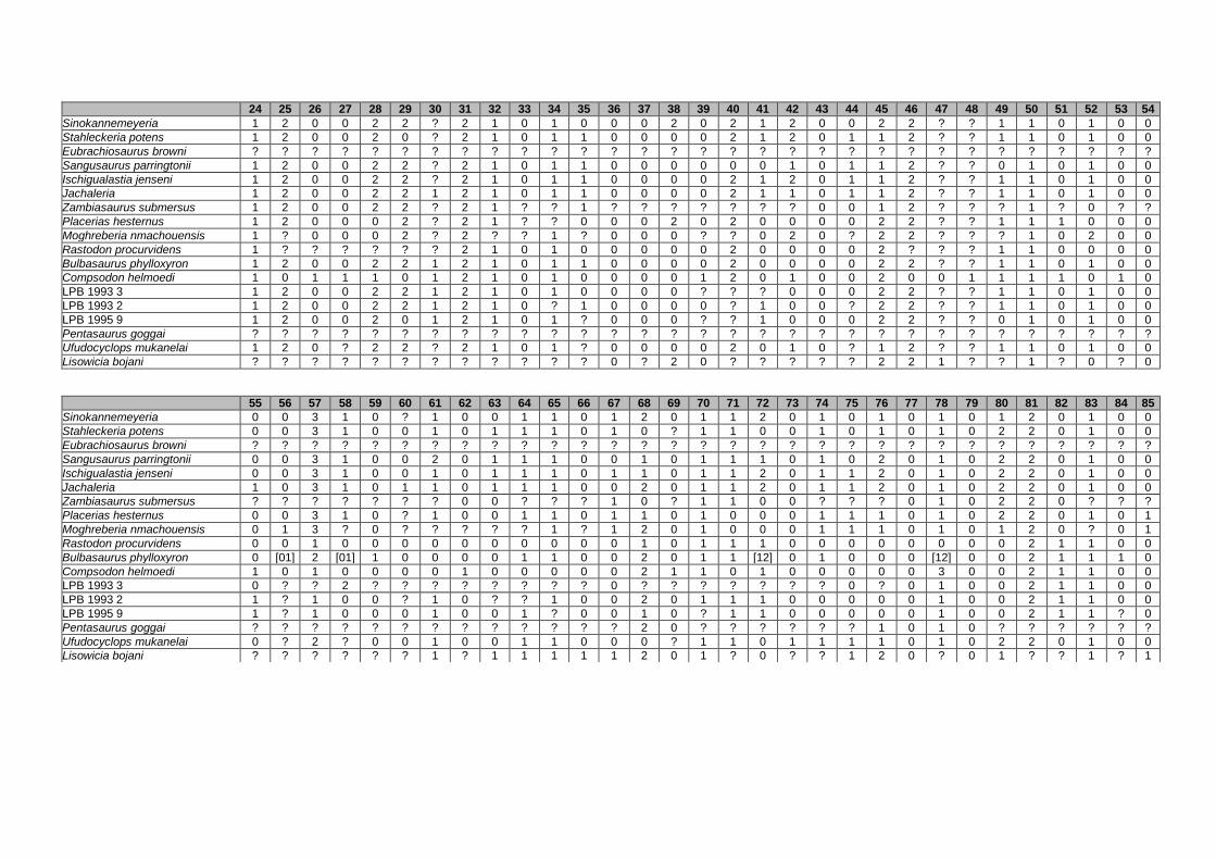
24 25 26 27 28 29 30 31 32 33 34 35 36 37 38 39 40 41 42 43 44 45 46 47 48 49 50 51 52 53 54
Sinokannemeyeria 1 2 0 0 2 2 ? 2 1 0 1 0 0 0 2 0 2 1 2 0 0 2 2 ? ? 1 1 0 1 0 0
Stahleckeria potens 1 2 0 0 2 0 ? 2 1 0 1 1 0 0 0 0 2 1 2 0 1 1 2 ? ? 1 1 0 1 0 0
Eubrachiosaurus browni ? ? ? ? ? ? ? ? ? ? ? ? ? ? ? ? ? ? ? ? ? ? ? ? ? ? ? ? ? ? ?
Sangusaurus parringtonii 1 2 0 0 2 2 ? 2 1 0 1 1 0 0 0 0 0 0 1 0 1 1 2 ? ? 0 1 0 1 0 0
Ischigualastia jenseni 1 2 0 0 2 2 ? 2 1 0 1 1 0 0 0 0 2 1 2 0 1 1 2 ? ? 1 1 0 1 0 0
Jachaleria 1 2 0 0 2 2 1 2 1 0 1 1 0 0 0 0 2 1 1 0 1 1 2 ? ? 1 1 0 1 0 0
Zambiasaurus submersus 1 2 0 0 2 2 ? 2 1 ? ? 1 ? ? ? ? ? ? ? 0 0 1 2 ? ? ? 1 ? 0 ? ?
Placerias hesternus 1 2 0 0 0 2 ? 2 1 ? ? 0 0 0 2 0 2 0 0 0 0 2 2 ? ? 1 1 1 0 0 0
Moghreberia nmachouensis 1 ? 0 0 0 2 ? 2 ? ? 1 ? 0 0 0 ? ? 0 2 0 ? 2 2 ? ? ? 1 0 2 0 0
Rastodon procurvidens 1 ? ? ? ? ? ? 2 1 0 1 0 0 0 0 0 2 0 0 0 0 2 ? ? ? 1 1 0 0 0 0
Bulbasaurus phylloxyron 1 2 0 0 2 2 1 2 1 0 1 1 0 0 0 0 2 0 0 0 0 2 2 ? ? 1 1 0 1 0 0
Compsodon helmoedi 1 0 1 1 1 0 1 2 1 0 1 0 0 0 0 1 2 0 1 0 0 2 0 0 1 1 1 1 0 1 0
LPB 1993 3 1 2 0 0 2 2 1 2 1 0 1 0 0 0 0 ? ? ? 0 0 0 2 2 ? ? 1 1 0 1 0 0
LPB 1993 2 1 2 0 0 2 2 1 2 1 0 ? 1 0 0 0 0 ? 1 0 0 ? 2 2 ? ? 1 1 0 1 0 0
LPB 1995 9 1 2 0 0 2 0 1 2 1 0 1 ? 0 0 0 ? ? 1 0 0 0 2 2 ? ? 0 1 0 1 0 0
Pentasaurus goggai ? ? ? ? ? ? ? ? ? ? ? ? ? ? ? ? ? ? ? ? ? ? ? ? ? ? ? ? ? ? ?
Ufudocyclops mukanelai 1 2 0 ? 2 2 ? 2 1 0 1 ? 0 0 0 0 2 0 1 0 ? 1 2 ? ? 1 1 0 1 0 0
Lisowicia bojani ? ? ? ? ? ? ? ? ? ? ? ? 0 ? 2 0 ? ? ? ? ? 2 2 1 ? ? 1 ? 0 ? 0
55 56 57 58 59 60 61 62 63 64 65 66 67 68 69 70 71 72 73 74 75 76 77 78 79 80 81 82 83 84 85
Sinokannemeyeria 0 0 3 1 0 ? 1 0 0 1 1 0 1 2 0 1 1 2 0 1 0 1 0 1 0 1 2 0 1 0 0
Stahleckeria potens 0 0 3 1 0 0 1 0 1 1 1 0 1 0 ? 1 1 0 0 1 0 1 0 1 0 2 2 0 1 0 0
Eubrachiosaurus browni ? ? ? ? ? ? ? ? ? ? ? ? ? ? ? ? ? ? ? ? ? ? ? ? ? ? ? ? ? ? ?
Sangusaurus parringtonii 0 0 3 1 0 0 2 0 1 1 1 0 0 1 0 1 1 1 0 1 0 2 0 1 0 2 2 0 1 0 0
Ischigualastia jenseni 0 0 3 1 0 0 1 0 1 1 1 0 1 1 0 1 1 2 0 1 1 2 0 1 0 2 2 0 1 0 0
Jachaleria 1 0 3 1 0 1 1 0 1 1 1 0 0 2 0 1 1 2 0 1 1 2 0 1 0 2 2 0 1 0 0
Zambiasaurus submersus ? ? ? ? ? ? ? 0 0 ? ? ? 1 0 ? 1 1 0 0 ? ? ? 0 1 0 2 2 0 ? ? ?
Placerias hesternus 0 0 3 1 0 ? 1 0 0 1 1 0 1 1 0 1 0 0 0 1 1 1 0 1 0 2 2 0 1 0 1
Moghreberia nmachouensis 0 1 3 ? 0 ? ? ? ? ? 1 ? 1 2 0 1 0 0 0 1 1 1 0 1 0 1 2 0 ? 0 1
Rastodon procurvidens 0 0 1 0 0 0 0 0 0 0 0 0 0 1 0 1 1 1 0 0 0 0 0 0 0 0 2 1 1 0 0
Bulbasaurus phylloxyron 0 [01] 2 [01] 1 0 0 0 0 1 1 0 0 2 0 1 1 [12] 0 1 0 0 0 [12] 0 0 2 1 1 1 0
Compsodon helmoedi 1 0 1 0 0 0 0 1 0 0 0 0 0 2 1 1 0 1 0 0 0 0 0 3 0 0 2 1 1 0 0
LPB 1993 3 0 ? ? 2 ? ? ? ? ? ? ? ? 0 ? ? ? ? ? ? ? 0 ? 0 1 0 0 2 1 1 0 0
LPB 1993 2 1 ? 1 0 0 ? 1 0 ? ? 1 0 0 2 0 1 1 1 0 0 0 0 0 1 0 0 2 1 1 0 0
LPB 1995 9 1 ? 1 0 0 0 1 0 0 1 ? 0 0 1 0 ? 1 1 0 0 0 0 0 1 0 0 2 1 1 ? 0
Pentasaurus goggai ? ? ? ? ? ? ? ? ? ? ? ? ? 2 0 ? ? ? ? ? ? 1 0 1 0 ? ? ? ? ? ?
Ufudocyclops mukanelai 0 ? 2 ? 0 0 1 0 0 1 1 0 0 0 ? 1 1 0 1 1 1 1 0 1 0 2 2 0 1 0 0
Lisowicia bojani ? ? ? ? ? ? 1 ? 1 1 1 1 1 2 0 1 ? 0 ? ? 1 2 0 ? 0 1 ? ? 1 ? 1

86 87 88 89 90 91 92 93 94 95 96 97 98 99 100 101 102 103 104 105 106 107 108 109 110 111 112 113 114 115 116
Sinokannemeyeria 2 1 1 1 1 0 1 1 1 1 1 1 1 2 1 0 3 0 1 ? ? 2 ? 1 2 1 0 1 ? 2 1
Stahleckeria potens 2 0 1 1 1 0 1 0 1 1 1 1 1 2 1 0 3 0 1 1 1 1 0 1 2 1 1 1 0 1 1
Eubrachiosaurus browni ? ? ? ? ? ? ? ? ? ? ? ? ? ? ? ? ? ? ? ? ? ? ? ? ? ? ? ? ? ? ?
Sangusaurus parringtonii 2 0 0 1 1 0 1 ? 1 ? ? ? 1 2 1 0 3 0 1 1 1 2 ? 1 2 1 0 1 0 1 1
Ischigualastia jenseni 2 0 0 1 1 0 1 1 1 1 1 1 1 2 1 0 3 0 ? ? 1 2 ? 1 2 1 0 1 0 5 1
Jachaleria 2 0 0 1 1 0 1 1 1 1 1 1 1 2 1 0 3 0 1 0 ? 2 ? 1 2 1 0 1 0 1 1
Zambiasaurus submersus 2 ? 1 ? ? ? ? 0 ? ? ? ? ? ? ? ? ? ? ? ? ? ? ? ? ? ? ? ? ? ? ?
Placerias hesternus 2 0 1 1 1 0 1 0 ? ? ? ? ? ? ? ? ? ? ? ? ? ? ? 1 ? ? ? ? 0 5 1
Moghreberia nmachouensis 2 0 1 ? ? 0 ? 0 1 1 1 1 1 2 1 0 3 0 1 ? ? ? ? 1 ? ? ? ? ? ? 1
Rastodon procurvidens 2 0 1 1 1 0 1 0 ? 1 1 0 ? ? ? ? ? ? ? ? 0 ? ? 1 2 1 0 ? 1 2 1
Bulbasaurus phylloxyron 2 0 1 1 1 0 1 0 1 1 0 1 1 2 2 0 3 0 1 1 0 1 0 1 2 1 0 0 1 2 1
Compsodon helmoedi 2 0 1 1 ? 0 1 0 1 1 1 0 1 1 1 0 3 1 1 1 0 1 0 1 2 1 1 0 0 2 0
LPB 1993 3 2 0 1 1 ? 0 ? ? 1 1 1 1 1 2 1 0 3 0 1 ? ? 1 0 1 2 1 0 1 0 2 1
LPB 1993 2 2 0 1 ? ? 0 ? ? ? ? ? ? 1 ? ? ? ? ? ? ? ? 1 0 1 2 ? ? 1 0 ? 1
LPB 1995 9 2 ? ? 1 ? 0 ? ? 1 1 1 1 1 2 1 0 3 0 ? ? ? 1 0 1 2 1 0 1 ? 2 1
Pentasaurus goggai ? ? ? ? ? ? ? ? ? ? ? ? ? ? ? ? ? ? ? ? ? ? ? ? ? ? ? ? ? ? ?
Ufudocyclops mukanelai 2 0 1 1 1 0 1 0 1 1 1 1 1 2 1 0 3 0 2 1 1 2 ? 1 2 1 1 1 0 1 1
Lisowicia bojani ? ? ? ? ? ? 1 ? ? ? ? ? ? ? ? ? ? ? ? ? ? ? ? ? ? ? ? ? ? ? 1
117 118 119 120 121 122 123 124 125 126 127 128 129 130 131 132 133 134 135 136 137 138 139 140 141 142 143 144 145 146 147
Sinokannemeyeria 1 ? ? 0 2 2 1 ? 2 0 ? 1 1 1 0 1 0 0 1 1 1 1 1 2 ? ? ? 2 1 0 1
Stahleckeria potens 1 1 1 0 2 2 1 1 2 1 1 1 ? 1 0 1 0 0 0 1 1 1 1 2 ? ? ? 2 1 0 1
Eubrachiosaurus browni ? ? ? ? ? ? ? ? ? ? ? ? ? ? ? ? ? ? ? ? ? ? ? ? ? ? ? ? ? ? ?
Sangusaurus parringtonii 1 1 1 0 2 2 1 1 2 1 ? ? ? 1 1 1 0 0 0 1 1 1 1 2 ? ? ? ? 1 0 1
Ischigualastia jenseni 1 ? 1 0 2 2 1 3 2 0 ? 1 1 1 0 1 0 0 0 ? 1 1 1 2 ? ? ? 2 1 0 1
Jachaleria 1 ? 1 0 2 2 1 3 2 0 0 ? ? 1 0 1 0 0 0 ? ? 1 1 2 ? ? ? 2 ? 0 1
Zambiasaurus submersus ? ? ? ? ? ? ? ? ? ? ? ? ? ? ? 1 1 0 ? ? ? ? 1 2 ? ? ? 2 ? ? 1
Placerias hesternus 1 1 0 0 2 2 ? ? 2 0 ? 1 1 1 ? 1 0 1 ? 1 1 1 1 2 ? ? ? 2 0 0 1
Moghreberia nmachouensis ? ? ? ? 2 2 ? 1 2 0 ? ? ? ? ? 1 0 ? ? ? 1 1 1 2 ? ? ? 2 1 0 1
Rastodon procurvidens 0 0 ? 0 0 ? ? 1 ? 0 0 ? ? 1 0 1 1 0 0 ? 1 0 1 ? ? ? ? 2 0 0 ?
Bulbasaurus phylloxyron 0 0 0 0 1 2 1 1 2 0 ? ? ? 1 0 1 1 0 0 ? 1 1 1 ? ? ? ? 2 1 0 1
Compsodon helmoedi 0 0 ? 0 1 1 1 1 2 0 0 1 1 1 1 0 1 0 1 ? 1 ? ? ? ? ? ? ? ? ? ?
LPB 1993 3 1 0 ? 0 2 2 ? 1 2 0 ? ? ? ? ? 0 1 0 1 1 ? ? ? ? ? ? ? ? ? ? ?
LPB 1993 2 1 ? ? 0 2 2 ? 1 2 0 ? ? ? ? ? 0 1 ? ? 1 ? ? ? ? ? ? ? ? ? ? ?
LPB 1995 9 1 0 ? 0 2 ? ? ? 2 0 ? ? ? ? ? ? ? ? 1 ? ? ? ? ? ? ? ? ? ? ? ?
Pentasaurus goggai ? ? ? ? ? ? ? ? ? ? ? ? ? ? ? ? ? ? ? ? ? ? 1 2 ? ? ? 2 0 0 ?
Ufudocyclops mukanelai 1 ? 1 0 ? 2 1 1 ? 0 ? ? ? 1 ? 1 1 0 0 ? 1 1 1 2 ? ? ? ? 1 0 1
Lisowicia bojani ? ? ? ? ? 1 ? 2 2 0 ? ? ? ? ? 1 0 1 1 ? ? ? ? ? ? ? ? ? ? ? ?

148 149 150 151 152 153 154 155 156 157 158 159 160 161 162 163 164 165 166 167 168 169 170 171 172 173 174 175 176 177 178
Sinokannemeyeria 3 0 1 2 1 1 ? ? ? 0 ? 1 1 ? 1 2 1 2 0 0 0 0 1 0 ? 1 1 1 0 1 0
Stahleckeria potens 3 0 1 2 1 1 0 1 1 1 0 0 1 1 1 2 1 3 1 0 1 0 1 0 0 1 1 1 0 1 0
Eubrachiosaurus browni ? ? ? ? ? ? ? ? ? ? ? ? ? ? ? ? ? ? ? ? 1 0 1 ? ? 1 ? 0 0 1 0
Sangusaurus parringtonii 3 0 1 2 1 1 1 1 ? 1 0 0 1 1 1 2 1 3 ? ? ? ? ? ? ? ? ? ? ? ? ?
Ischigualastia jenseni 3 0 1 ? ? ? ? 1 ? ? 0 0 1 ? 1 2 1 2 1 0 1 0 0 1 0 1 1 0 0 1 0
Jachaleria ? 0 ? ? ? ? ? ? ? ? ? ? ? ? 1 2 1 2 1 0 0 1 0 ? ? ? 1 ? ? ? ?
Zambiasaurus submersus ? ? ? ? ? ? 0 ? ? ? ? ? ? ? ? ? 1 ? 0 ? 1 1 1 0 0 1 ? 0 1 1 0
Placerias hesternus 3 0 ? ? ? 1 0 ? ? 1 ? 1 ? ? 1 2 1 ? 0 0 1 1 1 0 0 1 ? 0 0 1 0
Moghreberia nmachouensis 3 0 1 2 1 1 0 ? ? ? ? ? ? ? ? ? 1 ? 0 ? 1 1 0 0 ? 1 1 1 1 1 0
Rastodon procurvidens ? 0 2 0 0 1 0 0 ? 1 ? 1 ? ? ? ? 1 ? ? ? ? ? ? ? ? ? ? ? ? ? ?
Bulbasaurus phylloxyron ? 0 1 2 1 1 ? ? ? ? ? ? ? ? ? ? 1 ? ? ? ? ? ? ? ? ? ? ? 0 ? ?
Compsodon helmoedi ? ? ? ? ? ? ? ? ? ? ? ? ? ? ? ? ? ? ? ? ? ? ? ? ? ? ? ? ? ? ?
LPB 1993 3 ? ? ? ? ? ? ? ? ? ? ? ? ? ? ? ? ? ? ? ? ? ? ? ? ? ? ? ? ? ? ?
LPB 1993 2 ? ? ? ? ? ? ? ? ? ? ? ? ? ? ? ? ? ? ? ? ? ? ? ? ? ? ? ? ? ? ?
LPB 1995 9 ? ? ? ? ? ? ? ? ? ? ? ? ? ? ? ? ? ? ? ? ? ? ? ? ? ? ? ? ? ? ?
Pentasaurus goggai ? ? 1 0 ? 1 0 1 ? ? ? ? ? ? ? ? ? ? ? ? ? ? ? 0 0 ? ? ? ? 1 0
Ufudocyclops mukanelai 3 ? 1 2 1 1 0 ? ? ? ? ? ? 1 ? 2 1 ? ? ? ? ? ? ? ? ? ? ? ? ? ?
Lisowicia bojani ? ? ? ? ? ? ? ? ? 0 0 1 1 1 1 1 ? 1 1 ? 1 0 0 0 0 1 2 0 0 1 0
179 180 181 182 183 184 185 186 187 188 189 190 191 192 193 194 195 196 197
Sinokannemeyeria 1 ? 1 1 0 ? ? ? 0 1 1 1 2 1 1 ? 1 1 0
Stahleckeria potens 1 ? 1 1 0 1 1 1 1 1 1 1 2 1 1 ? 0 1 0
Eubrachiosaurus browni ? ? ? ? 0 ? ? ? ? ? ? ? ? ? ? ? ? 1 0
Sangusaurus parringtonii ? ? ? ? 0 1 1 1 1 1 1 ? ? ? ? ? 0 ? ?
Ischigualastia jenseni 1 ? 1 1 0 1 1 1 1 1 1 1 2 1 1 ? 0 1 0
Jachaleria 1 ? ? ? 0 1 1 1 1 1 1 ? ? ? ? ? 0 ? ?
Zambiasaurus submersus 0 ? ? ? 0 1 1 1 1 1 1 ? ? ? ? ? 1 1 1
Placerias hesternus ? ? ? ? 0 1 1 1 1 1 1 ? ? ? ? 1 1 1 1
Moghreberia nmachouensis 1 ? ? ? 0 1 1 1 1 1 1 ? ? ? ? 1 1 1 ?
Rastodon procurvidens ? ? ? ? ? ? ? ? ? ? ? ? ? ? ? ? 0 ? ?
Bulbasaurus phylloxyron ? ? ? ? ? ? ? ? ? ? ? ? ? ? ? ? 1 ? ?
Compsodon helmoedi ? ? ? ? ? ? ? ? ? ? ? ? ? ? ? ? ? ? ?
LPB 1993 3 ? ? ? ? ? ? ? ? ? ? ? ? ? ? ? ? ? ? ?
LPB 1993 2 ? ? ? ? ? ? ? ? ? ? ? ? ? ? ? ? ? ? ?
LPB 1995 9 ? ? ? ? ? ? ? ? ? ? ? ? ? ? ? ? ? ? ?
Pentasaurus goggai 1 ? ? ? ? ? ? ? ? ? ? ? ? ? ? ? 1 1 1
Ufudocyclops mukanelai ? ? ? ? ? ? ? ? ? ? ? ? ? ? ? ? ? ? ?
Lisowicia bojani 1 ? ? ? 0 0 0 1 1 1 1 ? ? ? ? ? ? 1 1
APPENDIX III: Supplementary data of PAPER III
352
Appendix III: Supplementary data of PAPER III (unpublished)
1. Table 1S. Resting metabolic rate values of extant species. (p. 353)
2. Table 2S. Initial histological data. (p. 354)
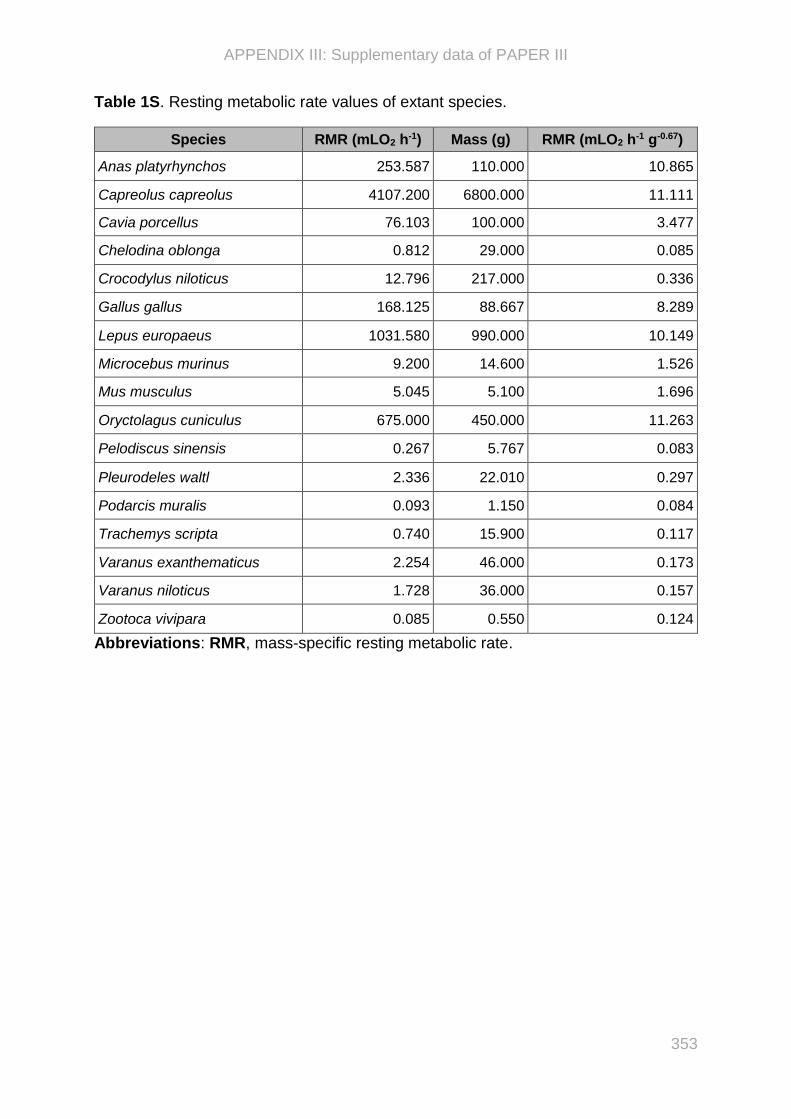
APPENDIX III: Supplementary data of PAPER III
353
Table 1S. Resting metabolic rate values of extant species.
Species RMR (mLO2 h-1) Mass (g) RMR (mLO2 h-1 g-0.67)
Anas platyrhynchos 253.587 110.000 10.865
Capreolus capreolus 4107.200 6800.000 11.111
Cavia porcellus 76.103 100.000 3.477
Chelodina oblonga 0.812 29.000 0.085
Crocodylus niloticus 12.796 217.000 0.336
Gallus gallus 168.125 88.667 8.289
Lepus europaeus 1031.580 990.000 10.149
Microcebus murinus 9.200 14.600 1.526
Mus musculus 5.045 5.100 1.696
Oryctolagus cuniculus 675.000 450.000 11.263
Pelodiscus sinensis 0.267 5.767 0.083
Pleurodeles waltl 2.336 22.010 0.297
Podarcis muralis 0.093 1.150 0.084
Trachemys scripta 0.740 15.900 0.117
Varanus exanthematicus 2.254 46.000 0.173
Varanus niloticus 1.728 36.000 0.157
Zootoca vivipara 0.085 0.550 0.124
Abbreviations: RMR, mass-specific resting metabolic rate.
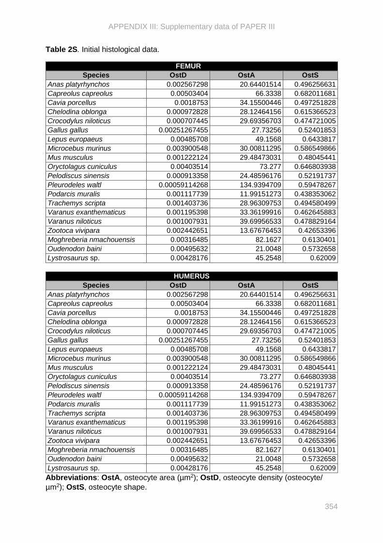
APPENDIX III: Supplementary data of PAPER III
354
Table 2S. Initial histological data.
FEMUR
Species OstD OstA OstS
Anas platyrhynchos 0.002567298 20.64401514 0.496256631
Capreolus capreolus 0.00503404 66.3338 0.682011681
Cavia porcellus 0.0018753 34.15500446 0.497251828
Chelodina oblonga 0.000972828 28.12464156 0.615366523
Crocodylus niloticus 0.000707445 29.69356703 0.474721005
Gallus gallus 0.00251267455 27.73256 0.52401853
Lepus europaeus 0.00485708 49.1568 0.6433817
Microcebus murinus 0.003900548 30.00811295 0.586549866
Mus musculus 0.001222124 29.48473031 0.48045441
Oryctolagus cuniculus 0.00403514 73.277 0.646803938
Pelodiscus sinensis 0.000913358 24.48596176 0.52191737
Pleurodeles waltl 0.00059114268 134.9394709 0.59478267
Podarcis muralis 0.001117739 11.99151273 0.438353062
Trachemys scripta 0.001403736 28.96309753 0.494580499
Varanus exanthematicus 0.001195398 33.36199916 0.462645883
Varanus niloticus 0.001007931 39.69956533 0.478829164
Zootoca vivipara 0.002442651 13.67676453 0.42653396
Moghreberia nmachouensis 0.00316485 82.1627 0.6130401
Oudenodon baini 0.00495632 21.0048 0.5732658
Lystrosaurus sp. 0.00428176 45.2548 0.62009
HUMERUS
Species OstD OstA OstS
Anas platyrhynchos 0.002567298 20.64401514 0.496256631
Capreolus capreolus 0.00503404 66.3338 0.682011681
Cavia porcellus 0.0018753 34.15500446 0.497251828
Chelodina oblonga 0.000972828 28.12464156 0.615366523
Crocodylus niloticus 0.000707445 29.69356703 0.474721005
Gallus gallus 0.00251267455 27.73256 0.52401853
Lepus europaeus 0.00485708 49.1568 0.6433817
Microcebus murinus 0.003900548 30.00811295 0.586549866
Mus musculus 0.001222124 29.48473031 0.48045441
Oryctolagus cuniculus 0.00403514 73.277 0.646803938
Pelodiscus sinensis 0.000913358 24.48596176 0.52191737
Pleurodeles waltl 0.00059114268 134.9394709 0.59478267
Podarcis muralis 0.001117739 11.99151273 0.438353062
Trachemys scripta 0.001403736 28.96309753 0.494580499
Varanus exanthematicus 0.001195398 33.36199916 0.462645883
Varanus niloticus 0.001007931 39.69956533 0.478829164
Zootoca vivipara 0.002442651 13.67676453 0.42653396
Moghreberia nmachouensis 0.00316485 82.1627 0.6130401
Oudenodon baini 0.00495632 21.0048 0.5732658
Lystrosaurus sp. 0.00428176 45.2548 0.62009
Abbreviations: OstA, osteocyte area (µm2); OstD, osteocyte density (osteocyte/
µm2); OstS, osteocyte shape.

APPENDIX VI: Supplementary data of PAPER IV
355
Appendix IV: Supplementary data of PAPER IV
1. Table 1S. Bone and specimen numbers added in the study. (p. 356–357)
2. Table 2S. Tests of the correlation between the humeral and femoral predictive
(taken one by one and/or in combination with others, for each bone) and RMR in a
phylogenetic context, using the R package caper and the pgls function (Orme
2018). (p. 358)
3. Table 3S. Comparison between inference models (built using the R package caper
and the pgls function) including different combinations of histological variables.
Hypothesis testing for the regression model based on the femur and humerus the
R package caper and the pgls function (Orme 2018) and the AIC function
(Sakamoto et al. 1986). (p. 359)
4. Table 4S. Comparison between inference models (built using BayesTraits V3.0.1
(Pagel and Meade 2016)) including different combinations of histological variables
using the selected model “28” for the femur and the selected model “5” for the
humerus (see Tables 4S-5S). The difference between the models were assessed
by Bayes Factor. (p. 360)
5. Table 5S. Model checking of the multiple regressions based on femur using MCMC
and PGLS methods, using BayesTraits V3.0.1 (Pagel and Meade 2016). The
branch length scaling parameters were automatically determined simultaneously in
models “10”, “23”, and “32”. The model “28” has been selected in this study for the
femur. The RMR values were expressed in mLO2 h-1 g-0.67. (p. 361–366)
6. Table 6S. Model checking of the multiple regressions based on humerus using
MCMC and PGLS methods, using BayesTraits V3.0.1 (Pagel and Meade 2016).
The branch length scaling parameters were automatically determined
simultaneously in models “1”, “2”, and “3”. The model “5” has been selected in this
study for the femur. The RMR values were expressed in mLO2 h-1 g-0.67. (p. 367–
369)

APPENDIX VI: Supplementary data of PAPER IV
356
Table 1S. Bone and specimen numbers added in the study.
Taxa Higher taxon Bones and specimen number
Capreolus capreolus Synapsida, Mammalia Humerus MNHN-Histos 2681 Femur MNHN-Histos 2674, 2675
Cavia porcellus Synapsida, Mammalia Humerus MNHN-Histos 2508, 2509, 2511, 2516, 2517 Femur MNHN-Histos 2500, 2502, 2504
Lepus europaeus Synapsida, Mammalia Humerus MNHN-Histos 2687 Femur MNHN-Histos 2684
Microcebus murinus Synapsida, Mammalia Humerus MNHN-Histos 2665 Femur MNHN-Histos 2664
Mus musculus Synapsida, Mammalia
Humerus MNHN-Histos 2598, 2599, 2600, 2603, 2608, 2609 Femur MNHN-Histos 2588, 2590, 2592, 2594, 2596
Oryctolagus cuniculus Synapsida, Mammalia Humerus MNHN-Histos 2693 Femur MNHN-Histos 2690
Anas platyrhynchos Saurospida,
Dinosauromorpha
Humerus MNHN-Histos 2538, 2543, 2544 Femur MNHN-Histos 2527, 2529, 2531, 2533
Chelodina oblonga Saurospida, Testudines Humerus MNHN-Histos 2399, 2400, 2402, 2403 Femur MNHN-Histos 2393, 2395, 2397
Pelodiscus sinensis Saurospida, Testudines
Humerus MNHN-Histos 2568, 2572, 2577, 2578 Femur MNHN-Histos 2557, 2559, 2561, 2563, 2565
Trachemys scripta Saurospida, Testudines
Humerus MNHN-Histos 2630, 2631, 2632, 2635, 2636, 2638, 2640, 2646, 2647 Femur MNHN-Histos 2619, 2621, 2623, 2625, 2627
Crocodylus niloticus Saurospida, Crurotarsia Humerus MNHN-Histos 2471, 2474, 2478, 2479 Femur MNHN-Histos 2463, 2465, 2467
Podarcis muralis Sauropsida, Squamata
Humerus MNHN-Histos 2453, 2455, 2457, 2459 Femur MNHN-Histos 2452, 2454, 2456, 2458
Varanus exanthematicus Sauropsida, Squamata Humerus MNHN-Histos 2433, 2434, 2436, 2437, 2442, 2443 Femur MNHN-Histos 2426, 2428, 2430
Varanus niloticus Sauropsida, Squamata Humerus MNHN-Histos 2419, 2420 Femur MNHN-Histos 2410, 2412
Zootoca vivipara Sauropsida, Squamata Humerus MNHN-Histos 2492, 2493 Femur MNHN-Histos 2483, 2485, 2487, 2489
Pleurodeles waltl Urodela, Amphibia
Humerus MNHN-Histos 2364, 2369, 2370, 2376, 2381, 2382, 2386 Femur MNHN-Histos 2363, 2368, 2375, 2379, 2380, 2385

APPENDIX VI: Supplementary data of PAPER IV
357
Kannemeyeria simocephalus †
Synapsida, Dicynodontia Femur NMQR 2674
Moghreberia nmachouensis †
Synapsida, Dicynodontia Humerus MNHN-Histos 2717, 2718 Femur MNHN-Histos 2714, 2715, 2716
Myosaurus gracilis † Synapsida, Dicynodontia Humerus BP1/4269
Lystrosaurus murrayi † Synapsida, Dicynodontia Humerus NMQR 3840 Femur NMQR 835
Oudenodon baini † Synapsida, Dicynodontia Humerus MNHN-Histos 2695, 2697, 2700 Femur MNHN-Histos 2696, 2699
Tropidostoma † Synapsida, Dicynodontia Humerus NMQR 9960 Femur NMQR 9960
Daptocephalus † Synapsida, Dicynodontia Femur NMQR 3633
Endothiodon † Synapsida, Dicynodontia Humerus SAM-PK-5605

APPENDIX VI: Supplementary data of PAPER IV
358
Table 2S. Tests of the correlation between the humeral and femoral predictive (taken one by one and/or in combination with others, for each bone) and RMR in a phylogenetic context, using the R package caper and the pgls function (Orme 2018).
Femur Humerus
adjusted R2 p-value adjusted R2 p-value
All 0.6697 0.002 0.6484 0.003
with OstD - 0.060 - 0.386
with OstA - 0.474 - 0.986
with OstS - 0.854 - 0.149
with VascD - 0.022 - 0.019
only OstD 0.3979 0.005 0.2968 0.017
only OstA 0.3209 0.013 0.3643 0.008
only OstS 0.2069 0.044 0.3659 0.008
only VascD 0.4973 0.001 0.4119 0.004
Abbreviations: All, all predictive variables included; OstA, osteocyte area; OstD, osteocyte density; OstS, osteocyte shape; VascD, primary osteon density.

APPENDIX VI: Supplementary data of PAPER IV
359
Table 3S. Comparison between inference models (built using the R package caper and the pgls function) including different combinations of histological variables. Hypothesis testing for the regression model based on the femur and humerus the R package caper and the pgls function (Orme 2018) and the AIC function (Sakamoto et al. 1986).
Femur Humerus
R2 AIC criterion R2 AIC criterion
All 0.6697 44.18096 0.6484 45.17998
OstD ex 0.5768 47.53806 0.6777 44.32515
OstA ex 0.6821 42.96247 0.6073 43.18044
OstS ex 0.6962 42.23243 0.6538 46.33979
VascD ex 0.5014 50.16157 0.4563 51.54730
OstS/VascD ex 0.526 48.63139 0.4906 49.78413
OstA/VascD ex 0.3787 52.96215 0.3866 52.75836
OstS/OstA ex 0.6851 42.09183 0.5749 46.89040
OstD/VascD ex 0.2894 55.11007 0.4071 52.21470
OstS/OstD ex 0.5466 47.92034 0.4945 49.66351
OstA/OstD ex 0.6069 45.63932 0.6789 42.40167
only OstD 0.3979 51.64720 0.2968 54.13040
only OstA 0.3209 53.57087 0.3643 52.51468
only OstS 0.2069 56.05439 0.3659 52.47496
only VascD 0.4973 48.75843 0.4119 51.27064
Abbreviations: All, all predictive variables included; “X” ex, predictive variable “X”
excluded; OstA, osteocyte area; OstD, osteocyte density; OstS, osteocyte shape;
VascD, primary osteon density.

APPENDIX VI: Supplementary data of PAPER IV
360
Table 4S. Comparison between inference models (built using BayesTraits V3.0.1 (Pagel and Meade 2016)) including different combinations of histological variables using the selected model “28” for the femur and the selected model “5” for the humerus (see Tables 4S-5S). The difference between the models were assessed by Bayes Factor.
Femur Humerus
R2 log marginal
likelihood log BF R2
log marginal likelihood
log BF
All 0.697 -31.1367 0 0.684 -31.8359 0
OstD ex 0.631 -31.1101 -0.0533 0.704 -30.5482 -2.5754
OstA ex 0.697 -30.1441 -1.9852 0.724 -29.9779 -3.7159
OstS ex 0.713 -30.3999 -1.4736 0.649 -31.8436 0.0155
VascD ex 0.510 -30.7481 -0.7772 0.556 -30.6533 -2.3651
OstS/VascD ex 0.578 -29.6947 -2.8842 0.565 -30.1063 -3.4591
OstA/VascD ex 0.442 -30.9802 -0.3131 0.386 -30.5547 -2.5623
OstS/OstA ex 0.693 -29.6738 -2.9258 0.599 -31.5610 -0.5498
OstD/VascD ex 0.410 -30.9277 -0.4182 0.511 -29.8244 -4.0230
OstS/OstD ex 0.591 -30.9280 -0.4176 0.571 -31.8268 -0.0181
OstA/OstD ex 0.556 -29.9633 -2.3469 0.715 -28.6957 -6.2803
only OstD 0.418 -30.5123 -1.2489 0.324 -31.6722 -0.3273
only OstA 0.397 -30.6610 -0.9516 0.437 -30.2425 -3.1868
only OstS 0.273 -31.3274 0.3812 0.424 -29.9325 -3.8066
only VascD 0.496 -31.4446 0.6158 0.415 -32.5315 1.3912
Abbreviations: BF, Bayes Factor (log BF= 2 (log marginal likelihood of Model 1 - log
marginal likelihood of Model 2)).
Table 5S. Model checking of the multiple regressions based on femur using MCMC and PGLS methods, using BayesTraits V3.0.1 (Pagel and Meade 2016). The branch length scaling parameters were automatically determined simultaneously in models “10”, “23”, and “32”. The model “28” has been selected in this study for the femur. The RMR values were expressed in mLO2 h-1 g-0.67. Abbreviations: abs diff, absolute difference; CI, 95% confidence interval; RMRobs,
the known mass-specific resting metabolic rate in literature; ��RMRpred, the predicted arithmetic mean of the mass-specific resting metabolic rate. 0 →

N° Priors Evolution parameters
R2
��RMRpred
Capreolus (RMRobs= 11.11)
Min limit of CI95%
Max limit of CI95%
% abs diff between RMRobs and ��RMRpred Beta 2 Beta 3 Beta 4 Beta 5 lambda kappa delta OU
1 exp 5 exp 5 exp 5 exp 5 0.10000 1.60000 5.00000 0.00000 0.84539 11.95706 11.58300 12.34319 7.61459
2 exp 5 exp 5 exp 5 exp 5 0.55000 1.30000 1.30000 0.30000 0.82509 15.44918 15.05822 15.85030 39.04405
3 exp 5 exp 5 exp 5 exp 5 0.70000 1.30000 5.00000 0.10000 0.82475 14.93292 14.55427 15.32143 34.39766
4 exp 5 exp 5 exp 5 exp 5 0.55000 1.30000 1.30000 0.50000 0.82434 15.65315 15.26546 16.05069 40.87978
5 exp 5 exp 5 exp 5 exp 5 0.55000 1.30000 1.30000 0.70000 0.82399 14.70981 14.34074 15.08838 32.38961
6 exp 5 exp 5 exp 5 exp 5 0.55000 1.00000 1.00000 0.70000 0.82362 15.53826 15.14441 15.94235 39.84571
7 exp 5 exp 5 exp 5 exp 5 0.70000 2.50000 5.00000 0.10000 0.82360 14.68005 14.31023 15.05942 32.12173
8 exp 5 exp 5 exp 5 exp 5 0.80000 1.30000 1.30000 0.70000 0.82324 15.19379 14.81941 15.57763 36.74549
9 exp 5 exp 5 exp 5 exp 5 0.70000 1.30000 1.30000 0.01000 0.82260 15.35673 14.98282 15.73997 38.21193
10 exp 5 exp 5 exp 5 exp 5 0.51941 1.43945 1.46348 0.77172 0.81665 15.31757 14.92442 15.72107 37.85949
11 exp 5 exp 5 exp 5 exp 5 0.70000 1.30000 5.00000 0.00010 0.79574 10.40275 10.12277 10.69047 6.37432
12 exp 5 exp 5 exp 5 exp 5 0.50000 2.50000 1.50000 0.00001 0.79476 13.26735 12.92923 13.61431 19.40734
13 exp 5 exp 5 exp 5 exp 5 0.60000 1.60000 5.00000 0.00000 0.79258 8.80004 8.52968 9.07898 20.79882
14 exp 5 exp 5 exp 5 exp 5 0.70000 1.30000 5.00000 0.00001 0.79179 10.45167 10.16601 10.74535 5.93407
15 exp 5 exp 5 exp 5 exp 5 0.60000 1.00000 5.00000 0.00000 0.78789 12.78469 12.46232 13.11540 15.06336
16 exp 5 exp 5 exp 5 exp 5 0.80000 1.00000 5.00000 0.00000 0.77773 12.40117 12.09207 12.71818 11.61169
17 exp 5 exp 5 exp 5 exp 5 0.30000 1.60000 1.50000 0.00000 0.77098 12.20882 11.90221 12.52333 9.88052
18 exp 5 exp 5 exp 5 exp 5 0.50000 2.30000 5.00000 0.00000 0.76501 7.30891 6.99311 7.63898 34.21914
19 exp 5 exp 5 exp 5 exp 5 0.70000 1.30000 1.30000 0.00100 0.75451 12.59148 12.27314 12.91808 13.32450
20 exp 5 exp 5 exp 5 exp 5 0.40000 4.00000 1.50000 0.00000 0.73962 9.84011 9.56849 10.11944 11.43812
21 exp 5 exp 5 exp 5 exp 5 0.50000 2.50000 1.50000 0.00000 0.73164 10.29287 10.02603 10.56682 7.36321
22 exp 5 exp 5 exp 5 exp 5 0.50000 1.30000 1.30000 0.00010 0.73022 12.25403 11.95527 12.56026 10.28741
23 exp 5 exp 5 exp 5 exp 5 0.63107 1.63673 1.55861 0.00000 0.72030 10.67887 10.41094 10.95369 3.88923
24 exp 5 exp 5 exp 5 exp 5 0.60000 1.60000 1.50000 0.00000 0.71828 10.81190 10.54566 11.08487 2.69190
25 exp 5 exp 5 exp 5 exp 5 0.75000 2.50000 5.00000 0.00000 0.71590 5.99340 5.69693 6.30531 46.05883
26 exp 5 exp 5 exp 5 exp 5 0.75000 2.50000 2.50000 0.00000 0.70998 8.23497 7.97327 8.50525 25.88456
27 exp 5 exp 5 exp 5 exp 5 0.70000 1.30000 1.30000 0.00010 0.70107 11.07630 10.80796 11.35129 0.31234
28 exp 5 exp 5 exp 5 exp 5 0.70000 2.50000 1.50000 0.00000 0.69692 9.60442 9.35240 9.86324 13.55932
29 exp 5 exp 5 exp 5 exp 5 0.75000 3.00000 3.00000 0.00000 0.69251 7.21431 6.93585 7.50394 35.07060
30 exp 5 exp 5 exp 5 exp 5 0.80000 1.60000 1.50000 0.00000 0.69023 10.11545 9.85734 10.38032 8.96007
31 exp 5 exp 5 exp 5 exp 5 0.75000 2.00000 1.30000 0.00000 0.68632 9.98316 9.73240 10.24039 10.15064
32 unif -100 100 unif -100 100 unif -100 100 unif -100 100 0.61970 1.63535 1.50507 0.00000 0.68554 8.85243 8.61040 9.10126 20.32731
33 exp 5 exp 5 exp 5 exp 5 0.75000 2.50000 1.30000 0.00000 0.68192 9.96669 9.71005 10.23012 10.29885
34 exp 5 exp 5 exp 5 exp 5 0.70000 4.00000 1.50000 0.00000 0.67697 9.63627 9.36478 9.91563 13.27269
35 unif -100 100 unif -100 100 unif -100 100 unif -100 100 1.00000 1.00000 1.00000 0.00000 0.60349 7.48741 7.27523 7.70578 32.61260
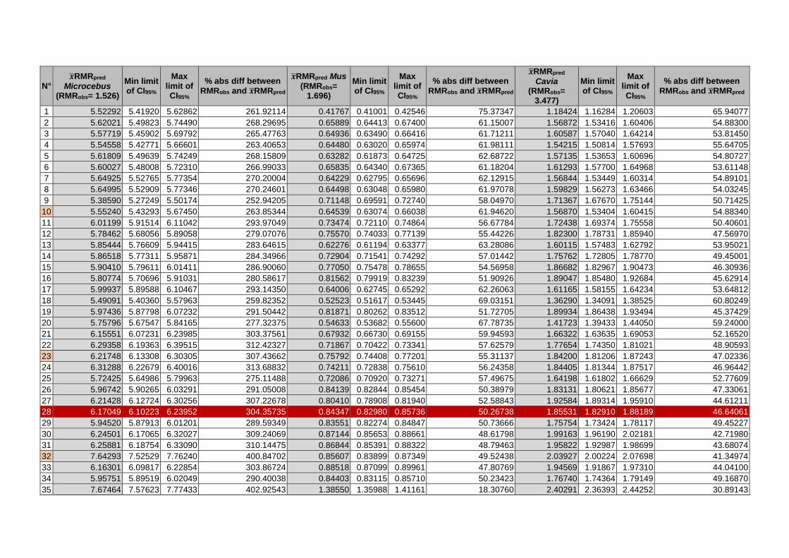
N° ��RMRpred
Microcebus (RMRobs= 1.526)
Min limit of CI95%
Max limit of CI95%
% abs diff between RMRobs and ��RMRpred
��RMRpred Mus (RMRobs=
1.696)
Min limit of CI95%
Max limit of CI95%
% abs diff between RMRobs and ��RMRpred
��RMRpred
Cavia (RMRobs=
3.477)
Min limit of CI95%
Max limit of CI95%
% abs diff between RMRobs and ��RMRpred
1 5.52292 5.41920 5.62862 261.92114 0.41767 0.41001 0.42546 75.37347 1.18424 1.16284 1.20603 65.94077
2 5.62021 5.49823 5.74490 268.29695 0.65889 0.64413 0.67400 61.15007 1.56872 1.53416 1.60406 54.88300
3 5.57719 5.45902 5.69792 265.47763 0.64936 0.63490 0.66416 61.71211 1.60587 1.57040 1.64214 53.81450
4 5.54558 5.42771 5.66601 263.40653 0.64480 0.63020 0.65974 61.98111 1.54215 1.50814 1.57693 55.64705
5 5.61809 5.49639 5.74249 268.15809 0.63282 0.61873 0.64725 62.68722 1.57135 1.53653 1.60696 54.80727
6 5.60027 5.48008 5.72310 266.99033 0.65835 0.64340 0.67365 61.18204 1.61293 1.57700 1.64968 53.61148
7 5.64925 5.52765 5.77354 270.20004 0.64229 0.62795 0.65696 62.12915 1.56844 1.53449 1.60314 54.89101
8 5.64995 5.52909 5.77346 270.24601 0.64498 0.63048 0.65980 61.97078 1.59829 1.56273 1.63466 54.03245
9 5.38590 5.27249 5.50174 252.94205 0.71148 0.69591 0.72740 58.04970 1.71367 1.67670 1.75144 50.71425
10 5.55240 5.43293 5.67450 263.85344 0.64539 0.63074 0.66038 61.94620 1.56870 1.53404 1.60415 54.88340
11 6.01199 5.91514 6.11042 293.97049 0.73474 0.72110 0.74864 56.67784 1.72438 1.69374 1.75558 50.40601
12 5.78462 5.68056 5.89058 279.07076 0.75570 0.74033 0.77139 55.44226 1.82300 1.78731 1.85940 47.56970
13 5.85444 5.76609 5.94415 283.64615 0.62276 0.61194 0.63377 63.28086 1.60115 1.57483 1.62792 53.95021
14 5.86518 5.77311 5.95871 284.34966 0.72904 0.71541 0.74292 57.01442 1.75762 1.72805 1.78770 49.45001
15 5.90410 5.79611 6.01411 286.90060 0.77050 0.75478 0.78655 54.56958 1.86682 1.82967 1.90473 46.30936
16 5.80774 5.70696 5.91031 280.58617 0.81562 0.79919 0.83239 51.90926 1.89047 1.85480 1.92684 45.62914
17 5.99937 5.89588 6.10467 293.14350 0.64006 0.62745 0.65292 62.26063 1.61165 1.58155 1.64234 53.64812
18 5.49091 5.40360 5.57963 259.82352 0.52523 0.51617 0.53445 69.03151 1.36290 1.34091 1.38525 60.80249
19 5.97436 5.87798 6.07232 291.50442 0.81871 0.80262 0.83512 51.72705 1.89934 1.86438 1.93494 45.37429
20 5.75796 5.67547 5.84165 277.32375 0.54633 0.53682 0.55600 67.78735 1.41723 1.39433 1.44050 59.24000
21 6.15551 6.07231 6.23985 303.37561 0.67932 0.66730 0.69155 59.94593 1.66322 1.63635 1.69053 52.16520
22 6.29358 6.19363 6.39515 312.42327 0.71867 0.70422 0.73341 57.62579 1.77654 1.74350 1.81021 48.90593
23 6.21748 6.13308 6.30305 307.43662 0.75792 0.74408 0.77201 55.31137 1.84200 1.81206 1.87243 47.02336
24 6.31288 6.22679 6.40016 313.68832 0.74211 0.72838 0.75610 56.24358 1.84405 1.81344 1.87517 46.96442
25 5.72425 5.64986 5.79963 275.11488 0.72086 0.70920 0.73271 57.49675 1.64198 1.61802 1.66629 52.77609
26 5.96742 5.90265 6.03291 291.05008 0.84139 0.82844 0.85454 50.38979 1.83131 1.80621 1.85677 47.33061
27 6.21428 6.12724 6.30256 307.22678 0.80410 0.78908 0.81940 52.58843 1.92584 1.89314 1.95910 44.61211
28 6.17049 6.10223 6.23952 304.35735 0.84347 0.82980 0.85736 50.26738 1.85531 1.82910 1.88189 46.64061
29 5.94520 5.87913 6.01201 289.59349 0.83551 0.82274 0.84847 50.73666 1.75754 1.73424 1.78117 49.45227
30 6.24501 6.17065 6.32027 309.24069 0.87144 0.85653 0.88661 48.61798 1.99163 1.96190 2.02181 42.71980
31 6.25881 6.18754 6.33090 310.14475 0.86844 0.85391 0.88322 48.79463 1.95822 1.92987 1.98699 43.68074
32 7.64293 7.52529 7.76240 400.84702 0.85607 0.83899 0.87349 49.52438 2.03927 2.00224 2.07698 41.34974
33 6.16301 6.09817 6.22854 303.86724 0.88518 0.87099 0.89961 47.80769 1.94569 1.91867 1.97310 44.04100
34 5.95751 5.89519 6.02049 290.40038 0.84403 0.83115 0.85710 50.23423 1.76740 1.74364 1.79149 49.16870
35 7.67464 7.57623 7.77433 402.92543 1.38550 1.35988 1.41161 18.30760 2.40291 2.36393 2.44252 30.89143

N° ��RMRpred Lepus
(RMRobs= 10.149)
Min limit of CI95%
Max limit of CI95%
% abs diff between RMRobs and ��RMRpred
��RMRpred
Oryctolagus (RMRobs= 11.263)
Min limit of CI95%
Max limit of CI95%
% abs diff between RMRobs and ��RMRpred
��RMRpred
Podarcis (RMRobs=
0.084)
Min limit of CI95%
Max limit of CI95%
% abs diff between RMRobs and ��RMRpred
1 9.54865 9.35152 9.74993 5.91535 7.39334 7.24623 7.54342 34.35732 0.03912 0.03837 0.03989 53.42578
2 10.87676 10.6098 11.1505 7.17074 9.30914 9.09052 9.53302 17.34759 0.04292 0.04185 0.04401 48.90737
3 11.02199 10.7538 11.2969 8.60177 9.27562 9.05315 9.50354 17.64525 0.04258 0.04155 0.04363 49.31389
4 10.37189 10.1223 10.6276 2.19618 9.43676 9.20902 9.67014 16.21447 0.04251 0.04149 0.04355 49.39595
5 10.99852 10.7234 11.2801 8.37043 9.06474 8.84531 9.28961 19.51756 0.04220 0.04117 0.04325 49.76519
6 10.33938 10.0834 10.6019 1.87585 9.27030 9.04885 9.49717 17.69244 0.04395 0.04289 0.04505 47.67443
7 10.65705 10.4015 10.9189 5.00595 9.07342 8.85448 9.29777 19.44047 0.04299 0.04193 0.04407 48.82426
8 10.55260 10.3034 10.8078 3.97680 9.56475 9.33452 9.80065 15.07816 0.04270 0.04167 0.04377 49.16277
9 10.62122 10.3719 10.8765 4.65284 9.21315 8.99673 9.43476 18.19990 0.05047 0.04955 0.05141 39.91323
10 10.42732 10.1730 10.688 2.74236 9.53821 9.30572 9.77650 15.31378 0.04259 0.04155 0.04366 49.29214
11 9.93136 9.75751 10.1083 2.14447 8.63074 8.47464 8.78971 23.37089 0.05152 0.05076 0.05230 38.66073
12 10.77485 10.5555 10.9987 6.16662 9.47102 9.27753 9.66854 15.91032 0.05135 0.05038 0.05233 38.87400
13 9.20080 9.04495 9.35933 9.34283 8.15425 8.01505 8.29587 27.60140 0.04809 0.04738 0.04882 42.74555
14 9.53187 9.36432 9.70243 6.08066 8.43984 8.29171 8.59061 25.06581 0.05209 0.05133 0.05287 37.98402
15 10.27298 10.0607 10.4898 1.22157 9.54494 9.34686 9.74721 15.25404 0.05064 0.04971 0.05158 39.71691
16 10.41840 10.2124 10.6285 2.65440 9.19168 9.01374 9.37314 18.39047 0.05385 0.05302 0.05469 35.89030
17 9.75594 9.56236 9.95343 3.87292 8.54616 8.37781 8.71789 24.12180 0.04308 0.04221 0.04396 48.71960
18 8.21846 8.07325 8.36628 19.02199 7.67723 7.54407 7.81274 31.83671 0.04542 0.04474 0.04611 45.93107
19 10.01979 9.83073 10.2125 1.27315 9.06735 8.89831 9.23960 19.49439 0.05305 0.05218 0.05394 36.83933
20 8.03194 7.88957 8.17688 20.85977 7.57859 7.44454 7.71504 32.71255 0.04516 0.04443 0.04590 46.24060
21 8.84816 8.69983 8.99901 12.81746 7.97994 7.84338 8.11887 29.14909 0.04696 0.04615 0.04778 44.09451
22 9.91230 9.72195 10.1064 2.33225 8.72095 8.55698 8.88805 22.56995 0.04589 0.04500 0.04679 45.37495
23 9.30404 9.14886 9.46185 8.32556 8.20554 8.06910 8.34428 27.14608 0.04971 0.04888 0.05057 40.81636
24 9.24114 9.08579 9.39916 8.94527 8.31136 8.17013 8.45503 26.20653 0.04898 0.04812 0.04984 41.69557
25 7.86579 7.75143 7.98184 22.49690 7.99715 7.87364 8.12260 28.99626 0.05294 0.05232 0.05357 36.97602
26 8.04536 7.93455 8.15772 20.72755 8.43539 8.31117 8.56147 25.10527 0.05613 0.05539 0.05688 33.18074
27 9.54149 9.37992 9.70584 5.98592 8.50411 8.35818 8.65258 24.49517 0.05122 0.05033 0.05212 39.02508
28 8.29871 8.17749 8.42172 18.23128 8.12083 7.99671 8.24688 27.89818 0.05379 0.05299 0.05460 35.96585
29 7.59783 7.49399 7.70311 25.13717 8.20627 8.08569 8.32864 27.13959 0.05615 0.05545 0.05685 33.15537
30 8.88871 8.75859 9.02077 12.41783 8.30966 8.18293 8.43835 26.22162 0.05630 0.05549 0.05711 32.97770
31 8.65624 8.52750 8.78692 14.70842 8.20959 8.08573 8.33535 27.11009 0.05435 0.05353 0.05519 35.29566
32 8.10008 7.94746 8.25564 20.18835 6.69085 6.56092 6.82335 40.59444 0.04386 0.04301 0.04473 47.78255
33 8.12422 8.01000 8.24006 19.95058 8.25280 8.13233 8.37507 26.72641 0.05605 0.05523 0.05688 33.27419
34 7.50442 7.40011 7.61020 26.05756 8.17130 8.04837 8.29611 27.45008 0.05658 0.05580 0.05736 32.64667
35 7.78393 7.65987 7.91000 23.30349 6.41859 6.31599 6.52285 43.01175 0.03807 0.03755 0.03859 54.68293

N° ��RMRpred
Zootoca (RMRobs= 0.124)
Min limit of CI95%
Max limit of CI95%
% abs diff between RMRobs and ��RMRpred
��RMRpred V.
exanthemicus (RMRobs= 0.173)
Min limit of CI95%
Max limit of CI95%
% abs diff between RMRobs and ��RMRpred
��RMRpred V.
niloticus (RMRobs= 0.157)
Min limit of CI95%
Max limit of CI95%
% abs diff between RMRobs and ��RMRpred
1 0.13899 0.13585 0.14220 12.09132 0.41240 0.40607 0.41883 138.38289 0.55032 0.54186 0.55891 250.52007
2 0.11889 0.11564 0.12222 4.12467 0.59806 0.58571 0.61067 245.70004 0.77127 0.75564 0.78723 391.25658
3 0.12246 0.11902 0.12599 1.24488 0.58429 0.57221 0.59662 237.73781 0.75777 0.74232 0.77354 382.65451
4 0.12076 0.11742 0.12420 2.61256 0.58988 0.57751 0.60252 240.97377 0.78529 0.76906 0.80187 400.18566
5 0.12165 0.11831 0.12508 1.89877 0.59693 0.58441 0.60972 245.04451 0.75464 0.73926 0.77035 380.66404
6 0.11861 0.11535 0.12195 4.34857 0.58469 0.57247 0.59717 237.97147 0.77338 0.75763 0.78945 392.59802
7 0.11278 0.10967 0.11599 9.04590 0.59858 0.58617 0.61126 246.00104 0.78501 0.76910 0.80125 400.00739
8 0.12223 0.11887 0.12569 1.42415 0.59499 0.58249 0.60775 243.92348 0.78252 0.76654 0.79884 398.42308
9 0.15488 0.15174 0.15810 24.90536 0.40464 0.39656 0.41289 133.89699 0.57255 0.56167 0.58364 264.67945
10 0.11368 0.11059 0.11685 8.32391 0.60238 0.58991 0.61512 248.19833 0.76961 0.75372 0.78584 390.19976
11 0.15161 0.14906 0.15420 22.26576 0.23487 0.23153 0.23827 35.76457 0.35496 0.34999 0.36000 126.09001
12 0.13914 0.13623 0.14210 12.20625 0.28639 0.28118 0.29169 65.54219 0.42918 0.42133 0.43717 173.36150
13 0.14383 0.14136 0.14635 15.99284 0.23222 0.22915 0.23533 34.23128 0.35208 0.34754 0.35667 124.25271
14 0.15238 0.14989 0.15491 22.88409 0.23065 0.22743 0.23391 33.32257 0.35566 0.35078 0.36060 126.53186
15 0.14614 0.14316 0.14918 17.85274 0.29813 0.29270 0.30365 72.32682 0.44234 0.43428 0.45055 181.74650
16 0.15405 0.15144 0.15672 24.23788 0.23567 0.23175 0.23965 36.22471 0.36416 0.35830 0.37013 131.95158
17 0.12225 0.11948 0.12508 1.41491 0.38226 0.37550 0.38915 120.96001 0.52422 0.51485 0.53376 233.89539
18 0.14554 0.14310 0.14801 17.36856 0.21868 0.21626 0.22113 26.40654 0.31473 0.31114 0.31836 100.46339
19 0.14477 0.14218 0.14740 16.74700 0.23438 0.23050 0.23833 35.47994 0.36554 0.35969 0.37149 132.82954
20 0.13290 0.13045 0.13541 7.18138 0.25149 0.24823 0.25480 45.37058 0.36732 0.36253 0.37216 133.95966
21 0.12162 0.11934 0.12394 1.92215 0.25635 0.25248 0.26028 48.17957 0.39025 0.38453 0.39607 148.57000
22 0.12473 0.12202 0.12750 0.58587 0.31257 0.30694 0.31831 80.67796 0.45549 0.44731 0.46382 190.12332
23 0.12872 0.12632 0.13115 3.80257 0.24392 0.24010 0.24779 40.99255 0.37407 0.36826 0.37997 138.26033
24 0.12914 0.12661 0.13172 4.14469 0.25929 0.25517 0.26347 49.87797 0.39314 0.38688 0.39951 150.40951
25 0.14962 0.14763 0.15164 20.66052 0.16428 0.16292 0.16565 5.04245 0.25928 0.25709 0.26150 65.14764
26 0.13180 0.12994 0.13369 6.29233 0.17011 0.16829 0.17196 1.66946 0.29228 0.28919 0.29540 86.16453
27 0.12967 0.12725 0.13214 4.57548 0.23476 0.23099 0.23860 35.70195 0.36862 0.36266 0.37469 134.79174
28 0.12193 0.11992 0.12397 1.67104 0.19748 0.19489 0.20011 14.15250 0.32427 0.32002 0.32857 106.53911
29 0.12968 0.12791 0.13148 4.58380 0.16351 0.16199 0.16505 5.48380 0.27025 0.26775 0.27277 72.13122
30 0.13287 0.13080 0.13497 7.15156 0.18694 0.18441 0.18951 8.05823 0.31553 0.31132 0.31981 100.97756
31 0.12259 0.12055 0.12466 1.13753 0.19575 0.19308 0.19845 13.14842 0.32276 0.31839 0.32720 105.58263
32 0.18600 0.18120 0.19093 50.00345 0.26302 0.25850 0.26761 52.03231 0.36634 0.36009 0.37270 133.33913
33 0.12301 0.12101 0.12504 0.80101 0.18724 0.18481 0.18971 8.23163 0.31536 0.31131 0.31945 100.86361
34 0.12305 0.12119 0.12494 0.76732 0.17623 0.17430 0.17818 1.86704 0.29127 0.28808 0.29450 85.52332
35 0.27657 0.27191 0.28131 123.03971 0.14548 0.14399 0.14699 15.90574 0.25086 0.24828 0.25347 59.78246
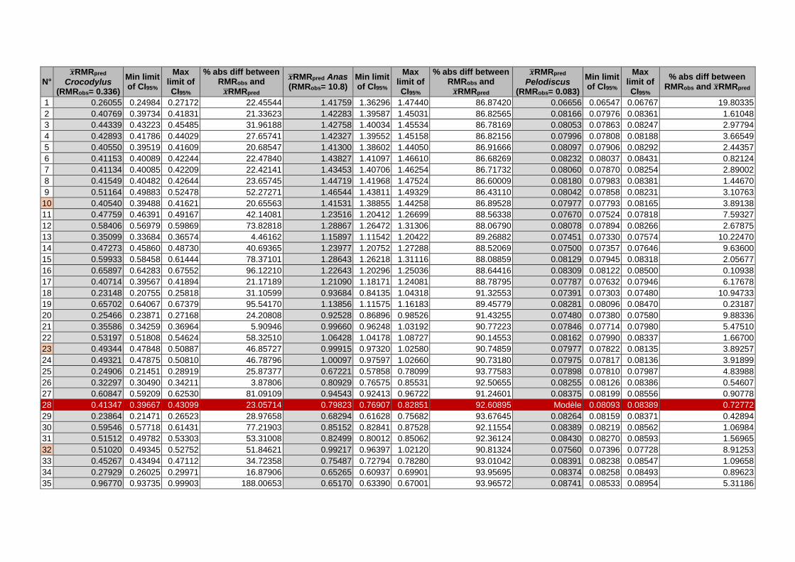
N° ��RMRpred
Crocodylus (RMRobs= 0.336)
Min limit of CI95%
Max limit of CI95%
% abs diff between RMRobs and ��RMRpred
��RMRpred Anas (RMRobs= 10.8)
Min limit of CI95%
Max limit of CI95%
% abs diff between RMRobs and ��RMRpred
��RMRpred
Pelodiscus (RMRobs= 0.083)
Min limit of CI95%
Max limit of CI95%
% abs diff between RMRobs and ��RMRpred
1 0.26055 0.24984 0.27172 22.45544 1.41759 1.36296 1.47440 86.87420 0.06656 0.06547 0.06767 19.80335
2 0.40769 0.39734 0.41831 21.33623 1.42283 1.39587 1.45031 86.82565 0.08166 0.07976 0.08361 1.61048
3 0.44339 0.43223 0.45485 31.96188 1.42758 1.40034 1.45534 86.78169 0.08053 0.07863 0.08247 2.97794
4 0.42893 0.41786 0.44029 27.65741 1.42327 1.39552 1.45158 86.82156 0.07996 0.07808 0.08188 3.66549
5 0.40550 0.39519 0.41609 20.68547 1.41300 1.38602 1.44050 86.91666 0.08097 0.07906 0.08292 2.44357
6 0.41153 0.40089 0.42244 22.47840 1.43827 1.41097 1.46610 86.68269 0.08232 0.08037 0.08431 0.82124
7 0.41134 0.40085 0.42209 22.42141 1.43453 1.40706 1.46254 86.71732 0.08060 0.07870 0.08254 2.89002
8 0.41549 0.40482 0.42644 23.65745 1.44719 1.41968 1.47524 86.60009 0.08180 0.07983 0.08381 1.44670
9 0.51164 0.49883 0.52478 52.27271 1.46544 1.43811 1.49329 86.43110 0.08042 0.07858 0.08231 3.10763
10 0.40540 0.39488 0.41621 20.65563 1.41531 1.38855 1.44258 86.89528 0.07977 0.07793 0.08165 3.89138
11 0.47759 0.46391 0.49167 42.14081 1.23516 1.20412 1.26699 88.56338 0.07670 0.07524 0.07818 7.59327
12 0.58406 0.56979 0.59869 73.82818 1.28867 1.26472 1.31306 88.06790 0.08078 0.07894 0.08266 2.67875
13 0.35099 0.33684 0.36574 4.46162 1.15897 1.11542 1.20422 89.26882 0.07451 0.07330 0.07574 10.22470
14 0.47273 0.45860 0.48730 40.69365 1.23977 1.20752 1.27288 88.52069 0.07500 0.07357 0.07646 9.63600
15 0.59933 0.58458 0.61444 78.37101 1.28643 1.26218 1.31116 88.08859 0.08129 0.07945 0.08318 2.05677
16 0.65897 0.64283 0.67552 96.12210 1.22643 1.20296 1.25036 88.64416 0.08309 0.08122 0.08500 0.10938
17 0.40714 0.39567 0.41894 21.17189 1.21090 1.18171 1.24081 88.78795 0.07787 0.07632 0.07946 6.17678
18 0.23148 0.20755 0.25818 31.10599 0.93684 0.84135 1.04318 91.32553 0.07391 0.07303 0.07480 10.94733
19 0.65702 0.64067 0.67379 95.54170 1.13856 1.11575 1.16183 89.45779 0.08281 0.08096 0.08470 0.23187
20 0.25466 0.23871 0.27168 24.20808 0.92528 0.86896 0.98526 91.43255 0.07480 0.07380 0.07580 9.88336
21 0.35586 0.34259 0.36964 5.90946 0.99660 0.96248 1.03192 90.77223 0.07846 0.07714 0.07980 5.47510
22 0.53197 0.51808 0.54624 58.32510 1.06428 1.04178 1.08727 90.14553 0.08162 0.07990 0.08337 1.66700
23 0.49344 0.47848 0.50887 46.85727 0.99915 0.97320 1.02580 90.74859 0.07977 0.07822 0.08135 3.89257
24 0.49321 0.47875 0.50810 46.78796 1.00097 0.97597 1.02660 90.73180 0.07975 0.07817 0.08136 3.91899
25 0.24906 0.21451 0.28919 25.87377 0.67221 0.57858 0.78099 93.77583 0.07898 0.07810 0.07987 4.83988
26 0.32297 0.30490 0.34211 3.87806 0.80929 0.76575 0.85531 92.50655 0.08255 0.08126 0.08386 0.54607
27 0.60847 0.59209 0.62530 81.09109 0.94543 0.92413 0.96722 91.24601 0.08375 0.08199 0.08556 0.90778
28 0.41347 0.39667 0.43099 23.05714 0.79823 0.76907 0.82851 92.60895 Modèle 0.08093 0.08389 0.72772
29 0.23864 0.21471 0.26523 28.97658 0.68294 0.61628 0.75682 93.67645 0.08264 0.08159 0.08371 0.42894
30 0.59546 0.57718 0.61431 77.21903 0.85152 0.82841 0.87528 92.11554 0.08389 0.08219 0.08562 1.06984
31 0.51512 0.49782 0.53303 53.31008 0.82499 0.80012 0.85062 92.36124 0.08430 0.08270 0.08593 1.56965
32 0.51020 0.49345 0.52752 51.84621 0.99217 0.96397 1.02120 90.81324 0.07560 0.07396 0.07728 8.91253
33 0.45267 0.43494 0.47112 34.72358 0.75487 0.72794 0.78280 93.01042 0.08391 0.08238 0.08547 1.09658
34 0.27929 0.26025 0.29971 16.87906 0.65265 0.60937 0.69901 93.95695 0.08374 0.08258 0.08493 0.89623
35 0.96770 0.93735 0.99903 188.00653 0.65170 0.63390 0.67001 93.96572 0.08741 0.08533 0.08954 5.31186
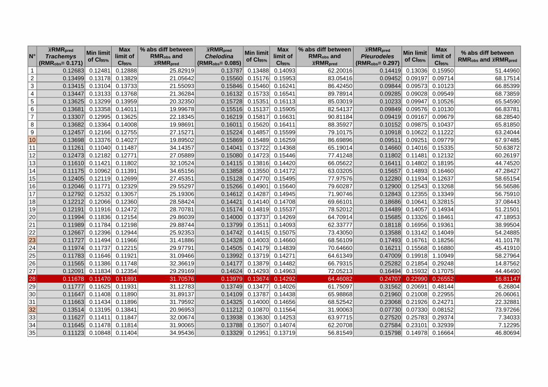
N° ��RMRpred
Trachemys (RMRobs= 0.171)
Min limit of CI95%
Max limit of CI95%
% abs diff between RMRobs and ��RMRpred
��RMRpred
Chelodina (RMRobs= 0.085)
Min limit of CI95%
Max limit of CI95%
% abs diff between RMRobs and ��RMRpred
��RMRpred
Pleurodeles (RMRobs= 0.297)
Min limit of CI95%
Max limit of CI95%
% abs diff between RMRobs and ��RMRpred
1 0.12683 0.12481 0.12888 25.82919 0.13787 0.13488 0.14093 62.20016 0.14419 0.13036 0.15950 51.44960
2 0.13499 0.13178 0.13829 21.05642 0.15560 0.15176 0.15953 83.05416 0.09452 0.09197 0.09714 68.17514
3 0.13415 0.13104 0.13733 21.55093 0.15846 0.15460 0.16241 86.42450 0.09844 0.09573 0.10123 66.85399
4 0.13447 0.13133 0.13768 21.36284 0.16132 0.15733 0.16541 89.78914 0.09285 0.09028 0.09549 68.73859
5 0.13625 0.13299 0.13959 20.32350 0.15728 0.15351 0.16113 85.03019 0.10233 0.09947 0.10526 65.54590
6 0.13681 0.13358 0.14011 19.99678 0.15516 0.15137 0.15905 82.54137 0.09849 0.09576 0.10130 66.83781
7 0.13307 0.12995 0.13625 22.18345 0.16219 0.15817 0.16631 90.81184 0.09419 0.09167 0.09679 68.28540
8 0.13682 0.13364 0.14008 19.98691 0.16011 0.15620 0.16411 88.35927 0.10152 0.09875 0.10437 65.81850
9 0.12457 0.12166 0.12755 27.15271 0.15224 0.14857 0.15599 79.10175 0.10918 0.10622 0.11222 63.24044
10 0.13698 0.13376 0.14027 19.89502 0.15869 0.15489 0.16259 86.69896 0.09511 0.09251 0.09779 67.97485
11 0.11261 0.11040 0.11487 34.14357 0.14041 0.13722 0.14368 65.19014 0.14660 0.14016 0.15335 50.63872
12 0.12473 0.12182 0.12771 27.05889 0.15080 0.14723 0.15446 77.41248 0.11802 0.11481 0.12132 60.26197
13 0.11610 0.11421 0.11802 32.10524 0.14115 0.13816 0.14420 66.05622 0.16411 0.14802 0.18195 44.74520
14 0.11175 0.10962 0.11391 34.65156 0.13858 0.13550 0.14172 63.03205 0.15657 0.14893 0.16460 47.28427
15 0.12405 0.12119 0.12699 27.45351 0.15128 0.14770 0.15495 77.97576 0.12280 0.11934 0.12637 58.65154
16 0.12046 0.11771 0.12329 29.55297 0.15266 0.14901 0.15640 79.60287 0.12900 0.12543 0.13268 56.56586
17 0.12792 0.12532 0.13057 25.19306 0.14612 0.14287 0.14945 71.90746 0.12843 0.12355 0.13349 56.75910
18 0.12212 0.12066 0.12360 28.58424 0.14421 0.14140 0.14708 69.66101 0.18686 0.10641 0.32815 37.08443
19 0.12191 0.11916 0.12472 28.70781 0.15174 0.14819 0.15537 78.52012 0.14489 0.14057 0.14934 51.21501
20 0.11994 0.11836 0.12154 29.86039 0.14000 0.13737 0.14269 64.70914 0.15685 0.13326 0.18461 47.18953
21 0.11989 0.11784 0.12198 29.88744 0.13799 0.13511 0.14093 62.33777 0.18118 0.16956 0.19361 38.99504
22 0.12667 0.12396 0.12944 25.92353 0.14742 0.14415 0.15075 73.43050 0.13588 0.13142 0.14049 54.24885
23 0.11727 0.11494 0.11966 31.41886 0.14328 0.14003 0.14660 68.56109 0.17493 0.16761 0.18256 41.10178
24 0.11974 0.11737 0.12215 29.97791 0.14505 0.14179 0.14839 70.64660 0.16211 0.15568 0.16880 45.41910
25 0.11783 0.11646 0.11921 31.09466 0.13992 0.13719 0.14271 64.61349 0.47009 0.19918 1.10949 58.27964
26 0.11565 0.11386 0.11748 32.36619 0.14177 0.13879 0.14482 66.79315 0.25282 0.21854 0.29248 14.87562
27 0.12091 0.11834 0.12354 29.29169 0.14624 0.14293 0.14963 72.05213 0.16494 0.15932 0.17075 44.46490
28 0.11678 0.11470 0.11891 31.70576 0.13979 0.13674 0.14292 64.46082 0.24707 0.22990 0.26552 16.81147
29 0.11777 0.11625 0.11931 31.12783 0.13749 0.13477 0.14026 61.75097 0.31562 0.20691 0.48144 6.26804
30 0.11647 0.11408 0.11890 31.89137 0.14109 0.13787 0.14438 65.98868 0.21960 0.21008 0.22955 26.06061
31 0.11663 0.11434 0.11896 31.79592 0.14325 0.14000 0.14656 68.52542 0.23068 0.21926 0.24271 22.32881
32 0.13514 0.13195 0.13841 20.96953 0.11212 0.10870 0.11564 31.90063 0.07730 0.07330 0.08152 73.97266
33 0.11627 0.11411 0.11847 32.00674 0.13938 0.13630 0.14253 63.97715 0.27520 0.25783 0.29374 7.34033
34 0.11645 0.11478 0.11814 31.90065 0.13788 0.13507 0.14074 62.20708 0.27584 0.23101 0.32939 7.12295
35 0.11123 0.10848 0.11404 34.95436 0.13329 0.12951 0.13719 56.81549 0.15798 0.14978 0.16664 46.80694
APPENDIX III: Supplementary data of PAPER III
367
Table 6S. Model checking of the multiple regressions based on humerus using MCMC and PGLS methods, using BayesTraits V3.0.1 (Pagel and Meade 2016). The branch length scaling parameters were automatically determined simultaneously in models “1”, “2”, and “3”. The model “5” has been selected in this study for the femur. The RMR values were expressed in mLO2 h-1 g-0.67.
Abbreviations: abs diff, absolute difference; CI, 95% confidence interval; RMRobs,
the known mass-specific resting metabolic rate in literature; ��RMRpred, the predicted
arithmetic mean of the mass-specific resting metabolic rate.

N° Priors Evolution parameters
R2 ��RMRpred Capreolus (RMRobs= 11.11)
Min limit of CI95%
Max limit of CI95%
% abs diff between
RMRobs and ��RMRpred Beta 2 Beta 3 Beta 4 Beta 5 lambda kappa delta OU
1 exp 5 exp 5 exp 5 exp 5 0.52152 1.43549 1.45507 0.76811 0.77599 15.58945 15.16712 16.02353 40.30641
2 exp 5 exp 5 exp 5 exp 5 0.71366 1.79858 1.51148 0.00000 0.68626 10.29899 10.02991 10.57528 7.30821
3 unif -100 100 unif -100 100 unif -100 100 unif -100 100 0.63447 1.76616 1.48912 0.00000 0.66198 10.93141 10.61082 11.26169 1.61631
4 unif -100 100 unif -100 100 unif -100 100 unif -100 100 1.00000 1.00000 1.00000 0.00000 0.57683 8.46276 8.20870 8.72469 23.83436
5 exp 5 exp 5 exp 5 exp 5 0.70000 2.50000 1.50000 0.00000 0.68378 9.60531 9.34754 9.87019 13.55134
6 exp 5 exp 5 exp 5 exp 5 0.70000 2.50000 1.50000 0.000100 0.78481 13.31175 12.96113 13.67185 19.80692
N° ��RMRpred
Microcebus (RMRobs= 1.526)
Min limit of CI95%
Max limit of CI95%
% abs diff between RMRobs and ��RMRpred
��RMRpred Mus (RMRobs= 1.696)
Min limit of CI95%
Max limit of CI95%
% abs diff between RMRobs and ��RMRpred
��RMRpred Cavia (RMRobs= 3.477)
Min limit of CI95%
Max limit of CI95%
% abs diff between RMRobs and ��RMRpred
1 3.61005 3.51961 3.70281 136.56919 0.69385 0.67579 0.71240 59.08875 1.25543 1.22507 1.28655 63.89316
2 4.02812 3.96643 4.09077 163.96612 1.15125 1.13031 1.17257 32.11979 1.59918 1.57344 1.62535 54.00676
3 4.39812 4.31569 4.48212 188.21227 1.85373 1.80145 1.90753 9.30019 1.12186 1.10020 1.14396 67.73475
4 3.94651 3.88214 4.01195 158.61813 2.42230 2.37091 2.47480 42.82432 1.67688 1.64626 1.70807 51.77216
5 3.90026 3.84578 3.95552 155.58732 1.09978 1.08089 1.11900 35.15443 1.53118 1.50864 1.55405 55.96267
6 3.30856 3.22905 3.39002 116.81240 0.97313 0.94913 0.99772 42.62228 1.50235 1.46652 1.53906 56.79173
N° ��RMRpred Lepus (RMRobs= 10.149)
Min limit of CI95%
Max limit of CI95%
% abs diff between RMRobs and ��RMRpred
��RMRpred
Oryctolagus (RMRobs= 11.263)
Min limit of CI95%
Max limit of CI95%
% abs diff between RMRobs and ��RMRpred
��RMRpred
Podarcis (RMRobs= 0.084)
Min limit of CI95%
Max limit of CI95%
% abs diff between RMRobs and ��RMRpred
1 11.32565 11.02489 11.63461 11.59372 9.06347 8.82102 9.31259 19.52883 0.02406 0.02343 0.02470 71.36008
2 9.17806 9.02858 9.33003 9.56681 8.82833 8.67877 8.98046 21.61656 0.03590 0.03530 0.03651 57.26435
3 9.90364 9.70713 10.10413 2.41757 5.95086 5.82744 6.07688 47.16456 0.03255 0.03192 0.03320 61.24417
4 8.10968 7.97884 8.24265 20.09384 7.30217 7.16942 7.43738 35.16677 0.04563 0.04499 0.04629 45.67609
5 8.82466 8.69201 8.95934 13.04893 8.86189 8.72206 9.00397 21.31854 0.03747 0.03686 0.03809 55.39320
6 9.61105 9.36247 9.86622 5.30056 9.15122 8.91663 9.39198 18.74972 0.02958 0.02903 0.03014 64.78565

N° ��RMRpred
Zootoca (RMRobs= 0.124)
Min limit of CI95%
Max limit of CI95%
% abs diff between RMRobs and
��RMRpred
��RMRpred V.
exanthemicus (RMRobs= 0.173)
Min limit of CI95%
Max limit of CI95%
% abs diff between RMRobs and
��RMRpred
��RMRpred V.
niloticus (RMRobs= 0.157)
Min limit of CI95%
Max limit of CI95%
% abs diff between
RMRobs and ��RMRpred
1 0.15288 0.14814 0.15777 23.29124 0.72522 0.70799 0.74288 319.20440 0.99609 0.97446 1.01819 534.44916
2 0.16501 0.16166 0.16842 33.06971 0.21323 0.20993 0.21657 23.25282 0.35196 0.34704 0.35695 124.18015
3 0.18495 0.17960 0.19045 49.14951 0.21686 0.21279 0.22100 25.35005 0.39808 0.39163 0.40463 153.55196
4 0.20328 0.19956 0.20708 63.93899 0.14085 0.13926 0.14245 18.58369 0.26028 0.25759 0.26301 65.78596
5 0.15707 0.15413 0.16006 26.66543 0.19545 0.19271 0.19823 12.97817 0.32791 0.32383 0.33203 108.85745
6 0.28181 0.27561 0.28814 127.26286 0.31441 0.30804 0.32091 81.73999 0.49400 0.48492 0.50326 214.65114
N° ��RMRpred
Crocodylus (RMRobs= 0.336)
Min limit of CI95%
Max limit of CI95%
% abs diff between RMRobs and ��RMRpred
��RMRpred Anas (RMRobs= 10.8)
Min limit of CI95%
Max limit of CI95%
% abs diff between RMRobs and ��RMRpred
��RMRpred
Pelodiscus (RMRobs= 0.083)
Min limit of CI95%
Max limit of CI95%
% abs diff between RMRobs and ��RMRpred
1 0.46026 0.44773 0.47315 36.98360 1.20458 1.17984 1.22984 88.84650 0.10385 0.10117 0.10660 25.11815
2 0.55820 0.53953 0.57751 66.13029 0.72167 0.70175 0.74215 93.31790 0.09570 0.09381 0.09763 15.30081
3 0.63348 0.61159 0.65615 88.53447 0.84609 0.82150 0.87141 92.16586 0.09912 0.09700 0.10129 19.42378
4 1.21502 1.17753 1.25371 261.61435 0.52059 0.50735 0.53418 95.17972 0.09346 0.09112 0.09586 12.60083
5 0.46747 0.44896 0.48673 39.12650 0.67963 0.65491 0.70528 93.70712 0.09507 0.09340 0.09676 14.53893
6 0.78200 0.76226 0.80226 132.73822 1.28312 1.25813 1.30859 88.11930 0.09666 0.09424 0.09913 16.45610
N° ��RMRpred
Trachemys (RMRobs= 0.171)
Min limit of CI95%
Max limit of CI95%
% abs diff between RMRobs and
��RMRpred
��RMRpred
Chelodina (RMRobs= 0.085)
Min limit of CI95%
Max limit of CI95%
% abs diff between RMRobs and
��RMRpred
��RMRpred
Pleurodeles (RMRobs= 0.297)
Min limit of CI95%
Max limit of CI95%
% abs diff between RMRobs and ��RMRpred
1 0.13033 0.12699 0.13375 23.78638 0.18232 0.17760 0.18716 114.49143 0.17196 0.16676 0.17731 42.10204
2 0.11541 0.11311 0.11777 32.50627 0.15580 0.15234 0.15933 83.29280 0.26743 0.25493 0.28054 9.95593
3 0.10828 0.10591 0.11070 36.67897 0.20393 0.19889 0.20910 139.91819 0.09282 0.08774 0.09818 68.74877
4 0.10655 0.10391 0.10926 37.68949 0.16593 0.16131 0.17067 95.20757 0.35216 0.33540 0.36975 18.57090
5 0.11666 0.11455 0.11881 31.77673 0.15555 0.15226 0.15890 82.99617 0.28472 0.26534 0.30552 4.13358
6 0.11872 0.11578 0.12174 30.57294 0.16842 0.16416 0.17280 98.14312 0.20794 0.20169 0.21438 29.98751
APPENDIX IV: Supplementary data of PAPER IV
370
Bibliography
Orme D. 2018. The caper package: comparative analysis of phylogenetics and evolution in R.
Pagel M., Meade A. 2016. BayesTraits V3 Manual. Sakamoto Y., Ishiguro M., Kitagawa G. 1986. Akaike information criterion statistics. D.
Reidel Publishing Company.

Chloé OLIVIER – Thèse de doctorat - 2020
Dicynodonts represent an emblematic Permian-Triassic (P-Tr) taxa to survive the big crisis at the end of Permian. While they were strongly impacted, they largely diversified during the Middle Triassic. They occupied an important part of the herbivore fauna in their ecosystem. However, the major environmental changing occurring in Late Triassic, and more specifically in Carnian, sharply affected the populations of dicynodont, which finally became extinct at the end of Triassic. The evolution of Triassic dicynodonts and their recovery and extinction conditions are still unclear. This work focused on the Laotian and Moroccan forms and their input to our knowledge on the phylogenetic relationships, paleobiogeography, and paleophysiology of the Triassic dicynodonts. Previously supposed to be delayed until the Middle Triassic, the post-crisis recovery of dicynodonts would appear to be earlier as supported by the description of two new Laotian species (Counillonia superoculis and Repelinosaurus robustus) in Early Triassic. Despite their phylogenetic relationships remaining non-consensual, they support the survivorship of multiple lineages across the P-Tr boundary. A high termometabolism was proposed to mainly explain the survivorship of populations beyond the crisis. Paleophysiological models built with histological features to infer metabolism in Permian and Triassic dicynodonts concluded all studied dicynodonts endotherm, and refuted the previous hypothesis. They also highlighted a unique acquisition of the mammalian endothermy in Synapsida occurring at least in middle Permian at the Neotherapsida node. The geographic situation of the Laotian dicynodonts in the Indochina blocks provide new data on the debate about the collision of the block with the rest of Pangea. Indeed, cconsidering the age and the almost entirely terrestrial lifestyle of dicynodonts, a terrestrial connection between the Indochina Block and Pangea could thus be interpreted at least in late Permian-Early Triassic, most likely via the South and North China blocks. A new dicynodont postcranial material discovered in Morocco was studied and a taxonomic revision of the Moroccan dicynodonts was made. The species Moghreberia nmachouensis, known by most of the remains recovered in the Argana Basin, was considered as valid and clearly distinguished from the North American Placerias that was previously supposed to be synonym by many studies. The restudy of the cranial material of Azarifeneria did not emphasized significant differences distinguishing it from other Triassic genera. However, a second morphotype could at least be noticed in the postcranial material by its strong robustness. The large size of Moghreberia and especially the second big morphotype confirmed an increase of the body size in dicynodonts during the Triassic.
Key words: Dicynodontia, Morocco, Laos, taxonomy, phylogeny, paleobiogeography, endothermy, paleohistology Les dicynodontes sont des organismes emblématiques de la période du Permien-Trias (P-Tr), du fait de leur survie à la grande crise ayant eu lieu à la fin du Permien. Bien que fortement impactés par cette crise, ils se sont largement diversifiés durant le Trias moyen. Ils représentaient les herbivores dominants de leur écosystème. Cependant, les changements environnementaux majeurs survenus au Trias supérieur, et plus particulièrement au Carnien, ont fortement affecté les populations de dicynodontes, qui ont finalement disparus à la fin du Trias. L'évolution des dicynodontes triasiques ainsi que les conditions de leur résilience et de leur extinction restent peu connues. Ce travail s'est concentré sur les formes laotiennes et marocaines et leur apport à notre connaissance des relations phylogénétiques, de la paléobiogéographie, et de la paléophysiologie des dicynodontes du Trias. Précédemment supposée retardée au Trias moyen, la résilience post-crise des dicynodontes semble avoir été plus précoce, comme l'atteste l’ajout de deux nouvelles espèces laotiennes (Counillonia superoculis et Repelinosaurus robustus) datée du Trias inférieur. Bien que leurs relations phylogénétiques restent non consensuelles, elles confirment la survie de plusieurs lignées de dicynodontes à la crise P-Tr. Un thermométabolisme élevé aurait été l’élément déterminant pour expliquer la survie des populations à la crise. Réfutant cette hypothèse, les modèles paléophysiologiques construits à partir de variables histologiques pour inférer le métabolisme de dicynodontes permiens et triasiques ont conclu à une endothermie chez tous les dicynodontes étudiés. Ils ont également mis en évidence une acquisition unique de l'endothermie mammalienne chez les Synapsida au moins au Permien moyen, au niveau du nœud des Neotherapsida. La situation géographique des dicynodontes laotiens au sein du bloc indochinois apporte de nouveaux éléments sur le débat concernant la collision de ce dernier avec le reste de la Pangée. En effet, du fait de l'âge et du mode de vie presque essentiellement terrestre des dicynodontes, une connexion terrestre entre le bloc indochinois et la Pangée a donc été supposée au moins dès la fin Permien-début Trias, très probablement via les blocs sud- et nord-chinois. Du matériel post-crânien marocain inédit de dicynodonte a été étudié et une révision taxonomique des dicynodontes marocains a été effectuée. L'espèce Moghreberia nmachouensis, connue par la plupart des restes retrouvés au sein du bassin d'Argana, a été considérée valide et clairement distincte du genre nord-américain Placerias, auparavant supposé synonyme par plusieurs études. L’étude du matériel crânien d'Azarifeneria n'a pas montré de différences significatives le distinguant des autres genres triasiques. Cependant, un second morphotype, au moins, a pu être remarqué dans le matériel postcrânien par sa forte robustesse. La grande taille de Moghreberia et surtout celle de ce deuxième grand morphotype ont confirmé une augmentation de la taille corporelle des dicynodontes au cours du Trias.
Mots clés: Dicynodontia, Maroc, Laos, taxonomie, phylogénie, paléobiogéographie, endothermie, paléohistologie.