Domain Organization, Catalysis and Regulation of Eukaryotic Cystathionine Beta-Synthases
Upload
independentCategory
view
0download
0
ORIGINAL RESEARCH COMMUNICATION
Contributions of Nitric Oxide Synthases,Dietary Nitrite/Nitrate, and Other Sources
to the Formation of NO Signaling Products
Alexandra B. Milsom,1 Bernadette O. Fernandez,1,2 Maria F. Garcia-Saura,1
Juan Rodriguez,3* and Martin Feelisch1,2*
Abstract
Mice lacking all three nitric oxide synthase (NOS) genes remain viable even though deletion of the majordownstream target of NO, soluble guanylyl cyclase, is associated with a dramatically shortened life expectancy.Moreover, findings of relatively normal flow responses in eNOS knockouts are generally attributed to com-pensatory mechanisms including upregulation of remaining NOS isoforms, but the alternative possibility thatdietary nitrite/nitrate (NOx) may contribute to basal levels of NO signaling has never been investigated. Aim:The aim of the present study was to examine how NO signaling products (nitrosated and nitrosylated proteins)and NO metabolites (nitrite, nitrate) are affected by single NOS deletions and whether dietary NOx plays acompensatory role in any deficiency. Specifically, we sought to ascertain whether profound alterations of theseproducts arise upon genetic deletion of either NOS isoform, inhibition of all NOS activity, NOx restriction, or allof the above. Results: Our results indicate that while some significant changes do indeed occur, they aresurprisingly moderate and compartmentalized to specific tissues. Unexpectedly, even after pharmacologicalinhibition of all NOSs and restriction of dietary NOx intake in eNOS knockout mice significant levels of NO-related products remain. Innovation/Conclusion: These findings suggest that a yet unidentified source of NO,unrelated to NOSs or dietary NOx, may be sustaining basal NO signaling in tissues. Given the significance ofNO for redox regulation in health and disease, it would seem to be important to identify the nature of thisadditional source of NO products as it may offer new therapeutic avenues for correcting NO deficiencies.Antioxid. Redox Signal. 00, 000–000.
Introduction
Nitric oxide (NO) is a critical molecule in numerouscell signaling pathways that is essential to maintaining
health, including host defense, neuronal communication, andthe control of vascular tone (22); in addition, it is involved inpathophysiological mechanisms (13). Its production from L-arginine is controlled by three distinct isoforms of NO syn-thase (NOS), coded for by three distinct genes with discretecell/tissue localization, catalytic properties, regulation, andinhibitor sensitivities (24). More recently, the role of a fourthNO signaling source, nitrite, has become increasingly appar-ent (3, 18). This source is of particularly interest due to itschemical stability in vivo and because it can be derived, eitherdirectly or via reduction of nitrate, from a normal diet. Inaddition to locally produced NO itself, a circulating pool of
longer-lived NO metabolites, including nitrite, nitrate, S-nitroso and N-nitroso species (27), can give rise to NOfollowing reductive bioactivation and cooperates with tissue-bound storage forms of NO undergoing redox-activation (2, 6,29) to contribute to overall NO availability. Although there isample evidence for cross-talk between these NO species, aunifying concept of regulation and the factors that governtheir concentrations in different biological compartments islacking. The current view of how these different sources ofNO are related to NO signaling is represented diagrammati-cally in Figure 1.
The two principal pathways through which NO signalsinclude the binding of NO to heme-containing proteins(chemically, a nitrosylation reaction), exemplified by activa-tion of the enzyme soluble guanylyl cyclase with productionof the second messenger cyclic GMP, and the nitrosation of
1Whitaker Cardiovascular Institute, Boston University School of Medicine, Boston, Massachusetts.2Division of Metabolic & Vascular Health, University of Warwick Medical School, Coventry, United Kingdom.3Department of Physics, Centenary College of Louisiana, Shreveport, Louisiana.*These authors contributed equally to this article.
ANTIOXIDANTS & REDOX SIGNALINGVolume 00, Number 00, 2012ª Mary Ann Liebert, Inc.DOI: 10.1089/ars.2011.4156
1

critical sulfhydryl groups in proteins, a reaction coined ‘‘S-nitrosylation’’. The former has long been thought to mediatethe majority of NO’s effects in physiology (22), while the latteris believed to represent the prototypic redox-based signalingmechanism in biology (31). In fact, NO can be considered as aredox array of species (13), including nitrosonium (NO + )equivalents and nitroxyl anions (NO-) in addition to the freeradical, each with a distinct biological chemistry (21, 32). NOis also a potent antioxidant (36) as well as a regulator of cel-lular redox status (26) and redox-based gene expression.
Many studies to date have sought to investigate the role ofNO through NOS gene-deficient (knockout) animals, includingsingle, double, and triple knockout mice (30). In cardiovascularphysiology, the production of NO from eNOS is critical notonly for the maintenance of vascular tone but also for its anti-thrombotic, antiproliferative, and anti-inflammatory actions
(35). Interestingly, transgenic mice lacking a functional eNOSgene are viable and, although mildly hypertensive, show nor-mal flow responses in various vascular beds. These findingshave most commonly been attributed to compensatory path-ways, including upregulation of remaining NOS isoforms,soluble guanylate cyclase activity, and endothelial-derivedhyperpolarizing factor (12). Of particular interest was the de-velopment of triple NOS knockouts (33). The realization thatthese animals are viable is surprising given that total deletion ofthe major downstream target of NO, soluble guanylyl cyclase,is known to be associated with dramatically reduced life ex-pectancy (19). Taken together, these findings invite questionson the compensatory role of extra-NOS sources in maintainingbasal levels of NO signaling that ultimately may help keep totalNOS-knockouts viable.
Earlier studies have demonstrated that dietary restrictionof nitrite and nitrate (NOx) intake is associated with changesin NO signaling products in a direction opposite to that of anincrease in nitrite and/or NO availability (3). Conversely,blood levels of nitrosyl hemoglobin increase when mice arefed a nitrate-rich diet (5). The aim of the current study was toinvestigate whether compensatory reactions take place inNOS-deficient animals fed a low-NOx diet. Rather thanaddressing this question from the perspective of physiologicalor cell signaling responses, as has been followed in the past,our present study focuses on the alterations in NO signalingproducts (nitrosated, nitrosylated proteins) and NO metabo-lites (nitrite, nitrate) found in these animals, species known tobe associated with NO bioactivity (and elevated following, forexample, genetic eNOS overexpression (8), iNOS upregula-tion in inflammation (7), nitrite elevation (3), and NO inha-lation (23)). Towards that goal our aim was to ascertainwhether significant alterations of these products take placeupon: i) genetic deletion of each of the three NOS isoforms; ii)inhibition of all NOS activity; iii) restriction of dietary NOxintake; or iv) all of the above. Surprisingly, we find that noneof these interventions eliminates NO signaling, leading us toconclude that a new source of NO is at play. This new sourceof NO signaling products may offer an explanation as to whytriple knockout mice remain viable by suggesting the in-volvement of additional mechanisms to maintain basal NOlevels.
Results
Effect of eNOS, nNOS, and iNOS gene deletionon protein-bound NO and NOx levels
Basal levels of protein-bound NO products found in nNOS,iNOS, and eNOS knockout mice, here collectively expressedas ‘‘total nitrosylation’’ products’’, are shown in Figure 2 (leftpanels). Individual steady-state concentrations of S-nitrosothiol, N-nitrosamines and NO-heme concentrations intissues can be found in Supplementary Figure 1 online (sup-plementary data are available online at www.liebertonline.com/ars). Compared to WT, total nitroso and nitrosyl levelschanged little with specific removal of a single NOS isoform,with the exception of nNOS - / - animals displaying lowerbrain nitros(yl)ation products ( p < 0.05). If one calculates globalplasma and tissue NO metabolite concentrations, adjusted forwet weight (see Fig. 2, right panels), total body protein-NOlevels do not change significantly following deletion of any ofthe NOS isoforms.
FIG. 1. Schematic representation of the sources contrib-uting to the bodily NO pool. eNOS, endothelial NOS; iNOS,inducible NOS; nNOS, neuronal NOS; NO, bodily pool ofnitric oxide related signaling products; NO2
- , nitrite; NO3- ,
nitrate; protein-NO, protein-bound NO storage forms.
Innovation
Adequate production of the ubiquitous signaling andeffector molecule nitric oxide (NO) is essential to mam-malian health; it is also involved in pathophysiologicalprocesses. Three distinct nitric oxide synthases (NOS) havebeen identified to account for the majority of NO-relatedcell signaling, and dietary nitrite/nitrate intake and redoxcycling of the circulating NO metabolite pool are the onlyother sources known to contribute to bodily NO avail-ability. Using established NOS knockout models in com-bination with pharmacological and dietary manipulation,we here provide evidence for the existence of one or moreunrecognized source(s) of NO production in vivo. Our re-sults suggest that this unidentified source may be linked tomitochondrial activity. Given the significance of NO forredox regulation in health and disease, it would seem to beimportant to identify the nature of this additional source ofNO production and characterize its role and significancefor cell regulation and redox homeostasis. Moreover, per-turbation of NO formation via this new pathway may alsobe linked to disease; thus, its identification could offer newtherapeutic avenues for correction of any deficiencies thatare distinct from the current use of classical NO donors.Our results may be of importance for redox signaling andcell regulation as well as translational research.
2 MILSOM ET AL.
Basal nitrate, the largest contributor to the NO metabolitepool, showed essentially no alteration in steady-state con-centration with the removal of individual NOS isoforms. Twoexceptions to this general finding were liver nitrate, whichwas significantly reduced in all NOS knockouts comparedwith the control group ( p < 0.05), and a significant reductionin plasma and brain nitrate levels in nNOS-deficient animals( p < 0.05). However, none of the NOS genetic deletions had asignificant effect on total body nitrate.
In terms of impact on nitrite, the genetic deletion of eithernNOS, iNOS, or eNOS resulted in a significant reduction in
plasma and tissue nitrite levels as compared to WT controls(Fig. 2, top panels). The reduction is particularly evident in thegraphs depicting whole body levels of nitrite, with a highlysignificant reduction seen in eNOS knockouts.
Effect of low NOx diet on protein-bound NOand NOx levels
To investigate the contribution of dietary NOx to the stea-dy-state concentrations of NO metabolites across bodilycompartments, WT animals were placed on a low-NOx diet
FIG. 2. Contribution of different NOS isoforms to basal NO metabolite concentrations in plasma and tissues of mice.All animals were maintained on a standard rodent chow; wild-type controls (C57BL/6J) are represented by white bars (n = 6–8) and isoform-specific knockouts by shaded bars (n = 4–9). For illustrative purposes, all protein-bound NO moieties (includingS- and N-nitroso and NO-heme species) are displayed as total nitrosylation products (the higher-resolution picture detailingindividual changes in either NO species can be found in Supplementary Fig. 1 online). Left-hand panels show steady-stateconcentrations in individual compartments, right-hand panels display a theoretical total body concentration for each NOmetabolite pool as a weighted average. Data are mean – SEM, *p < 0.05, ** or #p < 0.01.
SOURCES OF NO SIGNALING PRODUCTS 3

for 7 days prior to the analysis of tissues and blood. The re-sults obtained after restriction of dietary NOx intake areshown in Figure 3. Compared to a normal diet, animals placedon a low-NOx diet showed a trend towards reduction, but nostatistically significant changes in protein-NO or NOx levels,in plasma or in either of the tissue compartments studied.Although red blood cells were not included in our studies inmice, these results are consistent with data from another in-vestigation demonstrating that changes in nitrosyl hemoglo-bin levels in the blood of WT mice fed a low-nitrate diet arerather modest when compared to a normal diet (5).
Combined effect of NOS gene deletion or NOSinhibition, and low NOx diet, on protein-bound NOand NOx levels
To study the combined effect of dietary NOx restriction andNOS inhibition, we investigated a further subset of WT ani-mals on a low NOx diet additionally receiving the pan-NOSinhibitor L-NIO to block endogenous NO production. Theresults of these experiments are also shown in Figure 3. Totalbody nitrite was significantly reduced, but did not vanishentirely. Nitrate and protein-bound NO levels remained lar-gely unchanged.
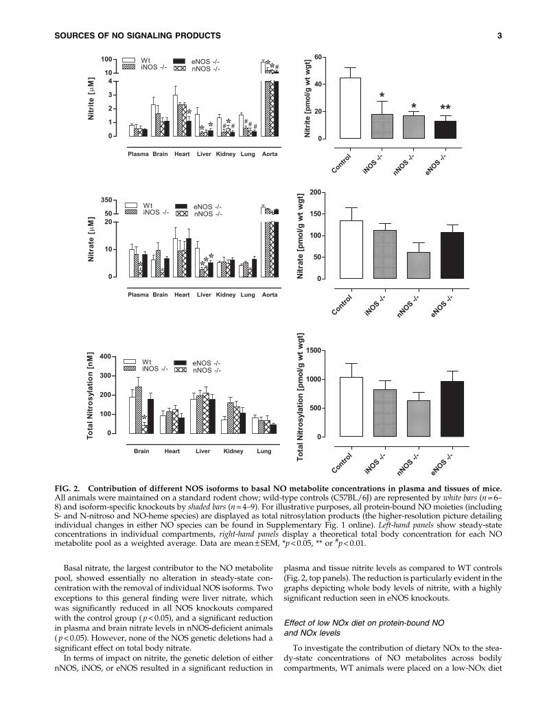
The finding that NO metabolite levels were not drasticallyreduced when animals are maintained on a low NOx diet andsystemic NOS activity is inhibited was unexpected since, ac-cording to the scheme in Figure 1, these actions should haveblocked all known sources of NO-related substances. To fur-ther investigate the resilience of NO products, the experi-ments were repeated in eNOS - / - animals subjected todietary NOx restriction, with and without additional phar-macological inhibition of the remaining NOS isoforms. Theseresults are depicted in Figure 4 (additional information aboutindividual changes to S-nitrosothiol, N-nitrosamines and NO-heme levels is presented in Supplementary Fig. 2). A generaltrend towards lower tissue protein-bound NO was observedin every organ and at the whole body level, although nonesufficiently large to reach statistical significance.
Dietary restriction of nitrite and nitrate intake on top ofeNOS deletion reduced plasma nitrite, whilst tissue nitritelevels remained essentially unchanged. This pattern waspreserved on further NOS inhibition. Dietary depletion alsohad little effect on nitrate levels, with the lung being the onlyorgan showing reduced nitrate concentrations ( p < 0.05).Further pharmacological inhibition of remaining nNOS andiNOS in this knockout model reduced nitrate levels in theheart and kidney, in addition to a further reduction in the lung( p < 0.05). However, whole body levels of nitrate remainedstatistically unchanged (Fig. 4, lower right).
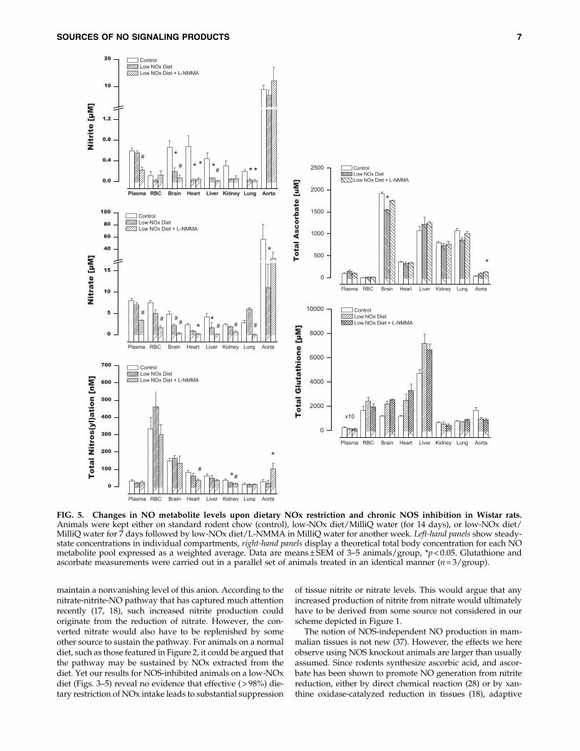
Effects of combined dietary NOx restrictionand chronic NOS inhibition on NO productsin blood and tissues of Wistar rats
In order to exclude the possibility of a species-specificphenomenon, additional experiments were carried out inmale Wistar rats. In addition, these experiments extended theperiod of dietary NOx restriction and NOS inhibition andinvestigated the existence of a compensatory upregulation oflow-molecular-weight antioxidants as a result of dietary ma-nipulation and NOS inhibition. These animals were main-tained on either regular chow/tap water, low-NOx diet/
MilliQ water, or on a regimen combining restricted dietaryNOx intake and NOS inhibition. As shown in Figure 5, thesestudies produced qualitatively identical results, confirmingour original observations in mice. Furthermore, they extendthose findings to a more chronic setting. Quantitative differ-ences in NO product concentrations between species and in-dividual compartments notwithstanding, these data showthat blood and tissue nitros(yl)ation levels overall did notdrop much further after 2 weeks of dietary NOx restriction orwith 1 week of NOS inhibition compared to what was ob-served acutely (3 h) in mice. These effects occurred in the ab-sence of marked changes in circulating or tissue antioxidantcontent (Fig. 5, right panels). Interestingly, combined dietaryNOx restriction and chronic NOS inhibition revealed an in-triguing additional feature that appears to be specific to thevasculature: a paradoxical upregulation of NO signalingproducts and metabolites in the aorta (Fig. 5), which was ac-companied by a 3-fold increase in ascorbate concentration inthis compartment.
Discussion
Heme nitrosylation (16) and S-nitrosation (14) representtwo protein modifications that account for much of thesignaling action that NO exerts in cells. Both types of post-translational modifications are effected through NO or NO-derived substances (20, 21), including nitrite (3). In all cases,the sources of these substances are associated with endoge-nous NO synthesis and metabolism (2, 7, 8) or dietary NOxintake (5, 17). Consequently, our finding that protein-boundNO modifications persist in the absence of active NOsynthesis and dietary NOx intake (e.g., in eNOS - / - animalson a low-NOx diet and under the acute action of the NOS-inhibitor, L-NIO, or in Wistar rats on low-NOx diet withchronic administration of L-NMMA) is surprising since itsuggests the existence of an unrecognized, and significant,source that contributes to the formation of NO signalingproducts. Our results may also explain why nitrosyl hemo-globin levels only dropped by half following dietary NOxrestriction and complete NOS inhibition in another study (5).Although our results cannot pinpoint the precise nature of thisnew source of NO products, our results hint at the interme-diacy of nitrite in the process, as we shall discuss below.
Our finding that protein-bound NO levels remain essen-tially unchanged upon deletion of each NOS isoform (Fig. 2,lower panels) likely reflects the action of a compensatorymechanism that maintains basal levels of NO metabolites intissues. One obvious compensatory mechanism that wouldkeep tissue NO levels constant is upregulation of remainingNOSs in an attempt to restore basal levels. If upregulation ofalternative NOS isoforms alone were to take place, then therewould be no physical reason why nitrite levels should change,according to the chemical network depicted in Figure 1. Yet,our results shown in Figure 2 clearly indicate that nitrite isconsistently depleted, regardless of the nature of the specificNOS isoform deleted. These consistent nitrite reductionssuggest that increased utilization of nitrite may be a signifi-cant component of the regulatory mechanism associated withdeficient NOS expression/NOS deletion.
If inhibition of any one NOS isoform leads to increasednitrite consumption, then it must also be accompanied byincreased production of nitrite elsewhere if tissues are to
4 MILSOM ET AL.

FIG. 3. Contribution of diet and other NOS isoforms to the depletion of NO metabolites in control mice. Data are fromwild-type controls (C57BL/6J) (n = 8) kept on a standard rodent chow, and wild-type animals kept on a low NOx diet without(n = 5) and with additional L-NIO treatment (n = 3). Left-hand panels show steady-state concentrations in individual com-partments, right-hand panels display a theoretical total body concentration for each NO metabolite pool expressed as aweighted average. Data are mean – SEM, *p < 0.05.
SOURCES OF NO SIGNALING PRODUCTS 5
FIG. 4. Contribution of diet and other NOS isoforms to the depletion of NO metabolites in eNOS2/2 mice. Data are fromeNOS - / - animals (n = 9) kept on a standard rodent chow, and eNOS - / - kept on a low NOx diet without (n = 5) and withadditional L-NIO treatment (n = 3). Left-hand panels show steady-state concentrations in individual compartments (individualchanges in nitrosothiol, nitrosamine and NO-heme concentrations can be found in Supplementary Fig. 2 online), right-handpanels display a theoretical total body concentration for each NO metabolite pool, expressed as a weighted average. Data aremean – SEM, *p < 0.05.
6 MILSOM ET AL.
maintain a nonvanishing level of this anion. According to thenitrate-nitrite-NO pathway that has captured much attentionrecently (17, 18), such increased nitrite production couldoriginate from the reduction of nitrate. However, the con-verted nitrate would also have to be replenished by someother source to sustain the pathway. For animals on a normaldiet, such as those featured in Figure 2, it could be argued thatthe pathway may be sustained by NOx extracted from thediet. Yet our results for NOS-inhibited animals on a low-NOxdiet (Figs. 3–5) reveal no evidence that effective ( > 98%) die-tary restriction of NOx intake leads to substantial suppression
of tissue nitrite or nitrate levels. This would argue that anyincreased production of nitrite from nitrate would ultimatelyhave to be derived from some source not considered in ourscheme depicted in Figure 1.
The notion of NOS-independent NO production in mam-malian tissues is not new (37). However, the effects we hereobserve using NOS knockout animals are larger than usuallyassumed. Since rodents synthesize ascorbic acid, and ascor-bate has been shown to promote NO generation from nitritereduction, either by direct chemical reaction (28) or by xan-thine oxidase-catalyzed reduction in tissues (18), adaptive
FIG. 5. Changes in NO metabolite levels upon dietary NOx restriction and chronic NOS inhibition in Wistar rats.Animals were kept either on standard rodent chow (control), low-NOx diet/MilliQ water (for 14 days), or low-NOx diet/MilliQ water for 7 days followed by low-NOx diet/L-NMMA in MilliQ water for another week. Left-hand panels show steady-state concentrations in individual compartments, right-hand panels display a theoretical total body concentration for each NOmetabolite pool expressed as a weighted average. Data are means – SEM of 3–5 animals/group, *p < 0.05. Glutathione andascorbate measurements were carried out in a parallel set of animals treated in an identical manner (n = 3/group).
SOURCES OF NO SIGNALING PRODUCTS 7

changes in ascorbate levels may have taken place to attenuatethe drop of NOS-dependent NO production. This possibilitywas investigated in rats subjected to chronic NOS inhibitionrather than by comparing wild-type with NOS - / - mice. Inmost compartments, changes in ascorbate content in responseto restriction of NOx intake and chronic NOS inhibition weremoderate. Only in the aorta, ascorbate increased significantlywhile glutathione levels dropped, and even these changes arenot accompanied by nitrite depletion. Thus, the data does notsupport the notion that tissue nitrite reduction is enhanceddue to increased ascorbate content and/or recycling. More-over, administration of the xanthine oxidase inhibitor allo-purinol did not alter nitrite levels significantly (not shown).Taken together, these results suggest that neither ascorbatenor xanthine oxidase make a major contribution to total NOproduct formation in vivo.
Whether nitrite plays a central role in sustaining NO sig-naling in the absence of NOS activity, an inescapable con-clusion from our results is that NO-related substancescontinue to be synthesized in mice even after dietary NOx ishighly reduced and NOS is inhibited. Our study in mice islimited inasmuch as we did not extend the period of NOSinhibition beyond 3 hours (in order to avoid stress as a pos-sible confounder). Nevertheless, this period should have beenmore than enough to see substantial changes in nitrite andprotein-bound NO levels as it corresponds to twice the plas-ma half-life of nitrate in mice (34), the NO metabolite with thelongest half-life in vivo and the largest pool of NO products inthe body. In the absence of other sources of intake or pro-duction, whole-body nitrate levels should have dropped by75% within that time if NOS was completely inhibited. Yet,
such level of reduction is not observed. Considering that re-versible enzyme inhibitors were used, one could argue thatNOS inhibition may have been incomplete. However, ourmore chronic NOS inhibitor studies in rats yielded similarresults. Had the degree of enzyme inhibition in the acutestudies in mice and in the chronic studies in rats not both beenclose to maximal, such similarity in outcome would have beenhighly unlikely.
An intriguing additional feature emerging from the ratstudies was the paradoxical upregulation of NO productsduring chronic combined dietary NOx restriction and phar-macological NOS inhibition in aortic tissue. The observedincrease of NO products in the aorta following NOS inhibitionmay be secondary to enhanced NOx uptake from the circu-lation, indicative of an enhanced need for such products in thevasculature when NO production from L-arginine is com-promised.
However surprising these findings may be, they can onlybe interpreted as evidence that a source other than those de-picted in Figure 1 may be sustaining the nitros(yl)ation/NOxcycle at basal levels when NOS is inhibited. This source, al-though unidentified at this time, may be a key to under-standing why even triple NOS knockout mice remain viable.
At this stage, we can only speculate about the origin of thiselusive source of NO products. Future studies under specificpathogen-free conditions may provide insight as to whether itoriginates from microbial or mammalian activities. Furtherinvestigations, requiring the use of tracer techniques such as15N-labeling in combination with isotope-specific analyticalmethods, may reveal that the unknown pool of NO metabo-lites is derived from the oxidation of ammonia, metabolism ofhydroxylamine, uptake/metabolism of a nitrogen species inair, or perhaps ‘‘recycling’’ of urea. Other possibilities mayinclude reductive biotransformation of nitrated species. Al-ternatively, it may be a by-product of protein catabolism/turnover and derived from the a-amino group of amino acids.
Finally, we wish to point out an interesting observation thatmay bear on the role of nitrite in NOx-restricted, NOS-deletedanimals. As stated earlier, these animals exhibit significantlyreduced levels of whole-body nitrite compared to those of WTanimals. However, when one examines the concentrationchanges relative to WT for each organ, the level of reductionvaries widely from one organ to another (Fig. 6). In fact, thelevel of reduction appears to vary systematically with tissuemitochondrial inner surface area (a proxy for maximal oxi-dative phosphorylative capacity of that tissue; data takenfrom 15) as suggested in that figure. This observation confirmsand extends earlier in vitro findings (10) related to the nitritereductase activity of the same tissues to the in vivo situation.This link suggests that the nitrite depletion we observe intissues may be related to mitochondrial activity and perhaps itholds a key to unlocking the mystery of how basal NO me-tabolite concentrations (and, presumably, related down-stream signaling) is sustained in the absence of NOS anddietary NOx pathways.
Materials and Methods
Materials
All chemicals were of the highest purity available andpurchased from Sigma-Aldrich (St. Louis, MO) unless other-wise specified.
FIG. 6. Fractional change in steady-state concentrations ofnitrite in organs from eNOS2/2 mice maintained on a low-NOx diet, with and without additional NOS inhibition. Allvalues are expressed relative to those found in the same or-gans of wild-type (WT) animals fed a standard rodent chow,and plotted against total tissue mitochondrial inner surfacearea (data from Reference 15) for each organ; closed symbols:eNOS - / - on low NOX diet; open symbols: eNOS - / - on lowNOX diet plus L-NIO. Inset: Correlation between mitochon-drial inner surface area and nitrite depletion, expressed aspercent of control levels in wild-type mice, following re-moval of all currently known endogenous and exogenoussupplies of nitrite (r2 = 0.989, excluding the kidney).
8 MILSOM ET AL.

Animals
Ten-week-old male mice of the iNOS - / - and eNOS - / -
strains ( Jackson Laboratory, Ben Harbor, ME; stock # 002684and 002609) were compared with age-matched C57BL/6Jwild-type mice (WT; Jackson Laboratory), and nNOS - / -
mice ( Jackson; #002633) were compared with their corre-sponding genetic background strain, B6129SF2/J ( Jackson;#101045). No significant differences in basal levels of NOmetabolites were observed between the two WT strains (datanot shown). Male Wistar rats (10–12 weeks of age) werepurchased from Harlan (South Easton, MA). Animals weremaintained on a standard rodent chow (2018; Harlan Teklad,South Easton, MA), with food and tap water ad libitum, andkept on a normal 12/12h light cycle. A minimum of 10 dayswas allowed for local vivarium acclimatization prior to ex-perimental use. All experiments complied with federal andstate regulations in accordance with the Guide for the Careand Use of Laboratory Animals (Institute for LaboratoryAnimal Research, National Research Council) and were ap-proved by the Institutional Animal Care and Use Committeeof the Boston University School of Medicine.
Dietary depletion of nitrite and nitrate
To deplete animals of their dietary source of nitrite andnitrate, a subset of acclimatized animals was switched toreceive an amino acid diet (TD99366; Harlan Teklad) withmatched L-arginine content (1.2% vs. 1% in the standardrodent diet) and MilliQ water for 1 week prior to experi-mental analysis. This particular diet has considerably lowerlevels of nitrite/nitrate (1.5/16.3 nmol g - 1) when comparedwith the standard chow (5.4/118 nmol g - 1) (3). Drinkingwater contributes significantly to total daily intake as NOxconcentrations approach maximal contaminant levels ofnitrite/nitrate. Based on strain-specific information aboutfood and water consumption (1) and measured NOx con-tent of the water provided the daily intake of food andwater-derived nitrite/nitrate on the standard diet/tap wa-ter regimen was calculated to be 0.46/4.81 lmoles nitrite/nitrate compared with the distilled water/amino acid diet(referred to hereafter as the ‘‘low-NOx’’ diet) of 6/67nmoles for a 25 g mouse, corresponding to a > 98.5% re-duction in total NOx intake on the latter compared to theregular diet regimen.
NOS inhibitor treatment
To investigate possible contributions from other NOSisoforms the pan-NOS inhibitor, L-N-iminoethyl-ornithine(L-NIO hydrochloride; A.G. Scientific, Inc., San Diego, CA)was administered for 3 h using a dose regimen confirmed toensure maximal NOS inhibition (100 mg/kg s.c., every45 min) (2) prior to measurement in both wt and eNOS - / -
animals fed a low NOx diet. In separate experiments in rats,L-N-monomethyl-arginine (L-NMMA acetate, 1 mg/mLdissolved in MilliQ water; 11) was administered for 7 dayswith the drinking water while animals were kept on a low-NOx diet, with controls receiving either normal rodentchow/tap water or low-NOx diet/MilliQ water. We optedto employ L-NMMA instead of L-NG-nitroarginine methy-lester (L-NAME, 0.3 mg/mL; 25) as NOS inhibitor fortwo reasons: i) L-NAME, but not L-NMMA, exerts NOS-
independent effects by virtue of muscarinic receptor an-tagonistic activity (4); ii) earlier pilot experiments with thiscompound revealed that it is metabolized to nitrite andnitrate (particularly in the liver from which these anionsappear to leak into the circulation), confounding the anal-ysis of NO metabolite status.
Organ harvest and tissue homogenization
Heparinized (0.07 units/g body weight, i.p.) mice or ratswere anaesthetized with diethylether (2 min) and eutha-nized by cervical dislocation. Blood was collected by car-diac puncture, and plasma and red blood cells wereobtained by centrifugation. After thoracotomy, a catheterwas inserted into the infrarenal part of the abdominal aorta,and organs were flushed free of blood by retrograde in situperfusion with air-equilibrated phosphate-buffered salinesupplemented with N-ethylmaleimide/ethylenediaminetetraacetic acid (10/2.5 mM) at a rate of 10 ml/min, andorgans were harvested as detailed previously (2). To pro-vide sufficient sample for comprehensive analysis of NOmetabolite levels in animals lacking individual NOS iso-form, samples were pooled from three animals; in all sub-sequent experiments on eNOS - / - groups, analysis wasperformed on individual tissue and blood samples. Nosignificant differences in levels were observed betweenpooled and individual measurements. Despite pooling,volumes of murine plasma and aorta samples were insuf-ficient for an accurate determination of nitrosation levels,allowing us to only analyze nitrite and nitrate concentra-tions in these compartments.
Determination of tissue nitroso/nitrosyl, nitrite/nitrate,ascorbate, and glutathione content
Tissue levels of nitroso and nitrosyl compounds werequantified using group-specific reductive denitrosation byiodine-iodide with subsequent detection of liberated NO bygas-phase chemiluminescence (9). ‘‘RSNO’’ signifies mercury-labile S-nitroso species, whereas ‘‘RNNO’’ signifies mercury-resistant N-nitroso adducts and may include nitrosaminesand metal nitrosyls other than NO-heme species. NO-heme, aproduct of heme nitrosylation, was determined by injection ofreplicate aliquots of tissue homogenates into a solution of 0.05M ferricyanide in PBS at pH 7.5 and 37�C (2). This methodemploys one-electron oxidation rather than reduction toachieve denitrosation and NO release. Nitrate and nitrite inplasma and tissues were quantified by ion chromatographywith on-line reduction of nitrate to nitrite and post-columnGriess diazotization (ENO-20 Analyzer; Eicom, Kyoto, Japan)(2). Total ascorbate and glutathione concentrations in bloodand tissues were analyzed as described previously (2). Insome animals, allopurinol (100 mg/kg ip) was administered30 min prior to organ harvest.
Statistical analysis
Data are means – SEM from n individual experiments.Statistical analysis was performed using Graph Pad Prism 4.0.Comparison between groups was achieved by one-wayANOVA following Bonferroni correction, or Dunnett’s post-hoc test where appropriate. Statistical significance was de-termined by p < 0.05.
SOURCES OF NO SIGNALING PRODUCTS 9

Acknowledgments
We wish to thank S.M. Bauer and N. S. Bryan for skillfultechnical assistance during the initial parts of the study. Thiswork was supported by grants from the National Institute ofHealth (HL 69029 and DA020644 to M.F., P20 RR16456 fromthe BRIN/INBRE Program of the NCRR to JR) and the Stra-tegic Appointment Scheme of the Medical Research Council,UK (to MF).
Author Disclosure Statement
No competing financial interests exist for any of the au-thors.
References
1. Bachmanov AA, Reed DR, Beauchamp GK, and TordoffMG. Food intake, water intake, and drinking spout sidepreference of 28 mouse strains. Behav Genet 32: 435–443,2002.
2. Bryan NS, Rassaf T, Maloney RE, Rodriguez CM, Saijo F,Rodriguez JR, and Feelisch M. Cellular targets and mecha-nisms of nitros(yl)ation: An insight into their nature andkinetics in vivo. Proc Natl Acad Sci USA 101: 4308–4313, 2004.
3. Bryan NS, Fernandez BO, Bauer SM, Garcia-Saura MF,Milsom AB, Rassaf T, Maloney RE, Bharti A, Rodriguez J,and Feelisch M. Nitrite is a signaling molecule and regulatorof gene expression in mammalian tissues. Nat Chem Biol 1:290–297, 2005.
4. Buxton IL, Cheek DJ, Eckman D, Westfall DP, Sanders KM,and Keef KD. NG-nitro L-arginine methyl ester and otheralkyl esters of arginine are muscarinic receptor antagonists.Circ Res. 72: 387–395, 1993.
5. Dikalov S and Fink B. ESR techniques for the detection ofnitric oxide in vivo and in tissues. Methods Enzymol 396: 597–610, 2005.
6. Donzelli S, Switzer CH, Thomas DD, Ridnour LA, EspeyMG, Isenberg JS, Tocchetti CG, King SB, Lazzarino G, Mi-randa KM, Roberts DD, Feelisch M, and Wink DA. The ac-tivation of metabolites of nitric oxide synthase by metals isboth redox and oxygen dependent: A new feature of nitro-gen oxide signaling. Antioxid Redox Signal 8: 1363–1371,2006.
7. Dyson A, Bryan NS, Fernandez BO, Garcia-Saura MF, SaijoF, Mongardon N, Rodriguez J, Singer M, and Feelisch M. Anintegrated approach to assessing nitroso-redox balance insystemic inflammation. Free Radic Biol Med 51: 1137–1145,2011.
8. Elrod JW, Greer JJ, Bryan NS, Langston W, Szot JF, Geb-regzlabher H, Janssens S, Feelisch M, and Lefer DJ. Cardio-myocyte-specific overexpression of NO synthase-3 protectsagainst myocardial ischemia-reperfusion injury. ArteriosclerThromb Vasc Biol 26: 1517–1523, 2006.
9. Feelisch M, Rassaf T, Mnaimneh S, Singh N, Bryan NS,Jourd’Heuil D, and Kelm M. Concomitant S-, N-, and heme-nitros(yl)ation in biological tissues and fluids: Implicationsfor the fate of NO in vivo. FASEB J 16: 1775–1785, 2002.
10. Feelisch M, Fernandez BO, Bryan NS, Garcia-Saura MF,Bauer S, Whitlock DR, Ford PC, Janero DR, Rodriguez J, andAshrafian H. Tissue processing of nitrite in hypoxia: Anintricate interplay of nitric oxide-generating and -scavengingsystems. J Biol Chem 283: 33927–33934, 2008.
11. Gardiner SM, Kemp PA, and Bennett T. Effects of chronictreatment with nitric oxide synthase inhibitors on regional
haemodynamic responses to vasodilators in consciousBrattleboro rats. Br J Pharmacol 109: 222–228, 1993.
12. Godecke A and Schrader J. Adaptive mechanisms of thecardiovascular system in transgenic mice—Lessons fromeNOS and myoglobin knockout mice. Basic Res Cardiol 95:492–498, 2000.
13. Gross SS and Wolin MS. Nitric oxide: Pathophysiologicalmechanisms. Annu Rev Physiol 57: 737–769, 1995.
14. Hess DT, Matsumoto A, Kim SO, Marshall HE, and StamlerJS. Protein S-nitrosylation: Purview and parameters. Nat RevMol Cell Biol 6: 150–166, 2005.
15. Hulbert AJ and Else PL. Evolution of mammalian endo-thermic metabolism: Mitochondrial activity and cell com-position. Am J Physiol 256: R63–R69, 1989.
16. Ignarro LJ. Signal transduction mechanisms involving nitricoxide. Biochem Pharmacol 41: 485–490, 1991.
17. Lundberg JO, Weitzberg E, and Gladwin MT. The nitrate-nitrite-nitric oxide pathway in physiology and therapeutics.Nat Rev Drug Discov 7: 156–167, 2008.
18. Lundberg JO, Gladwin MT, Ahluwalia A, Benjamin N,Bryan NS, Butler A, Cabrales P, Fago A, Feelisch M,Ford PC, Freeman BA, Frenneaux M, Friedman J, Kelm M,Kevil CG, Kim-Shapiro DB, Kozlov AV, Lancaster JR Jr,Lefer DJ, McColl K, McCurry K, Patel RP, Petersson J, RassafT, Reutov VP, Richter-Addo GB, Schechter A, Shiva S,Tsuchiya K, van Faassen EE, Webb AJ, Zuckerbraun BS,Zweier JL, and Weitzberg E. Nitrate and nitrite in bi-ology, nutrition and therapeutics. Nat Chem Biol 5: 865–869,2009.
19. Mergia E, Koesling D, and Friebe A. Genetic mouse modelsof the NO receptor ’soluble’ guanylyl cyclases. Handb ExpPharmacol 191: 33–46, 2009.
20. Miersch S and Mutus B. Protein S-nitrosation: Biochemistryand characterization of protein thiol-NO interactions ascellular signals. Clin Biochem 38: 777–791, 2005.
21. Miranda KM, Nims RW, Thomas DD, Espey MG, Citrin D,Bartberger MD, Paolocci N, Fukuto JM, Feelisch M, andWink DA. Comparison of the reactivity of nitric oxide andnitroxyl with heme proteins. A chemical discussion of thedifferential biological effects of these redox related productsof NOS. J Inorg Biochem 93: 52–60, 2003.
22. Moncada S, Palmer RM, and Higgs EA. Nitric oxide: Phy-siology, pathophysiology, and pharmacology. Pharmacol Rev43: 109–142, 1991.
23. Nagasaka Y, Fernandez BO, Garcia-Saura MF, Petersen B,Ichinose F, Bloch KD, Feelisch M, and Zapol WM. Briefperiods of nitric oxide inhalation protect against myocardialischemia-reperfusion injury. Anesthesiology 109: 675–682,2008.
24. Nathan C and Xie QW. Nitric oxide synthases: Roles, tolls,and controls. Cell 78: 915–918, 1994.
25. Navarro J, Sanchez A, Saiz J, Ruilope LM, Garcıa-Estan J,Romero JC, Moncada S, and Lahera V. Hormonal, renal, andmetabolic alterations during hypertension induced bychronic inhibition of NO in rats. Am J Physiol 267: R1516–R1521, 1994.
26. Padgett CM and Whorton AR. Regulation of cellular thiolredox status by nitric oxide. Cell Biochem Biophys 27: 157–177,1995.
27. Rassaf T, Feelisch M, and Kelm M. Circulating NO pool:Assessment of nitrite and nitroso species in blood and tis-sues. Free Radic Biol Med 36: 413–422, 2004.
28. Rocha BS, Gago B, Barbosa RM, and Laranjinha J. Diffusionof nitric oxide through the gastric wall upon reduction of
10 MILSOM ET AL.

nitrite by red wine: Physiological impact. Nitric Oxide 22:235–241, 2010.
29. Rodriguez J, Maloney RE, Rassaf T, Bryan NS, and FeelischM. Chemical nature of nitric oxide storage forms in ratvascular tissue. Proc Natl Acad Sci USA 100: 336–341, 2003.
30. Shimokawa H and Tsutsui M. Nitric oxide synthases in thepathogenesis of cardiovascular disease: Lessons from ge-netically modified mice. Pflugers Arch 459: 959–967, 2010.
31. Stamler JS, Lamas S, and Fang FC. Nitrosylation. The pro-totypic redox-based signaling mechanism. Cell 106: 675–683,2001.
32. Thomas DD, Ridnour LA, Isenberg JS, Flores-Santana W,Switzer CH, Donzelli S, Hussain P, Vecoli C, Paolocci N,Ambs S, Colton CA, Harris CC, Roberts DD, and Wink DA.The chemical biology of nitric oxide: Implications in cellularsignaling. Free Radic Biol Med 45: 18–31, 2008.
33. Tsutsui M, Shimokawa H, Morishita T, Nakashima Y, andYanagihara N. Development of genetically engineered micelacking all three nitric oxide synthases. J Pharmacol Sci 102:147–154, 2006.
34. Veszelovsky E, Holford NH, Thomsen LL, Knowles RG, andBaguley BC. Plasma nitrate clearance in mice: Modeling ofthe systemic production of nitrate following the induction ofnitric oxide synthesis. Cancer Chemother Pharmacol 36: 155–159, 1995.
35. Walford G and Loscalzo J. Nitric oxide in vascular biology. JThromb Haemost 1: 2112–2118, 2003.
36. Wink DA, Miranda KM, Espey MG, Pluta RM, Hewett SJ,Colton C, Vitek M, Feelisch M, and Grisham MB. Mechan-isms of the antioxidant effects of nitric oxide. Antioxid RedoxSignal 3: 203–213, 2001.
37. Zweier JL, Samouilov A, and Kuppusamy P. Non-enzymaticnitric oxide synthesis in biological systems. Biochim BiophysActa 1411: 250–262, 1999.
Address correspondence to:Prof. Martin Feelisch
Experimental Medicine and Integrative BiologyUniversity of Warwick Medical School
Gibbet Hill RoadCoventry CV4 7AL
United Kingdom
E-mail: [email protected]
Date of first submission to ARS Central, July 12, 2011; date offinal revised submission, December 1, 2011; date of accep-tance, December 1, 2011.
Abbreviations Used
eNOS¼ endothelial nitric oxide synthaseiNOS¼ inducible nitric oxide synthase
L-NIO¼L-N-iminoethyl-ornithineL-NMMA¼L-N-monomethyl-arginine
nNOS¼neuronal nitric oxide synthaseNO¼nitric oxide
NOS¼nitric oxide synthaseNOx¼ sum of nitrite and nitrate
WT¼wild-type
SOURCES OF NO SIGNALING PRODUCTS 11
Supplementary Data
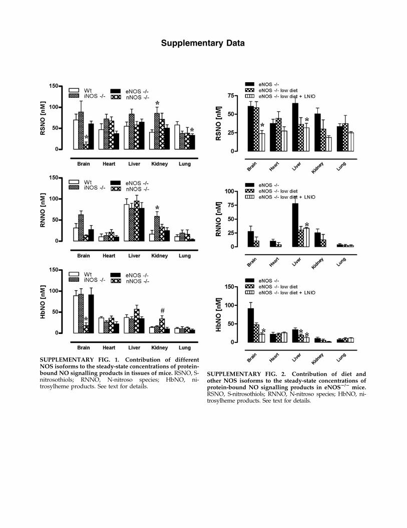
SUPPLEMENTARY FIG. 1. Contribution of differentNOS isoforms to the steady-state concentrations of protein-bound NO signalling products in tissues of mice. RSNO, S-nitrosothiols; RNNO, N-nitroso species; HbNO, ni-trosylheme products. See text for details.
SUPPLEMENTARY FIG. 2. Contribution of diet andother NOS isoforms to the steady-state concentrations ofprotein-bound NO signalling products in eNOS2/2 mice.RSNO, S-nitrosothiols; RNNO, N-nitroso species; HbNO, ni-trosylheme products. See text for details.
Copyright © 2022 FDOKUMEN