Content Based Retrieval Systems in a Clinical Context
20
Chapter 1 Content Based Retrieval Systems in a Clinical Context Frederico Valente, Carlos Costa and Augusto Silva Additional information is available at the end of the chapter http://dx.doi.org/10.5772/53027 1. Introduction Nowadays, digital images and protocols stand as a cornerstone of most modern health-care systems where they are used to provide important data and insights into the inner workings and ailments of the human body. The recent appearance of new modalities, the devices re‐ sponsible for data acquisition, such as the fMRI 1 and the MDCT 2 , produce copious amounts of data [1]. This, coupled with recent advances in storage technology has had as conse‐ quence an explosion in the amount of data produced at medical imaging institutions. For in‐ stance, the Geneva Hospital alone produced, in 2006, over 50000 images per day and such numbers are steadily rising [2]. Given the prevalence of digital images and protocols in the medical arena, we are, nonetheless, still a long way from fully taking advantage of the po‐ tential brought up by this digital revolution. The current data explosion makes it trouble‐ some to a practitioner to sift through the imaging repositories while searching for data relevant for his context. This means we have the data, but not the information, which should be readily available to the experts in the area. In fact, data overload has been reported as a problem by practitioners from medium to large imaging institutions [3]. The current methods of data search, such as the ones provided by the standard query mechanisms present in Digital Imaging and Communication in Medicine (DICOM) are sub-optimal, relying on template matching over a limited number of textual fields [4] (which fields are available depend on the specific software backend), and can conse‐ quently be improved upon. It is expected that, by providing more refined and robust methods of searching the large image repositories that currently exist, diagnostic accura‐ cy and efficiency can be improved and more accurate and useful Computer Aided Diag‐ nosis (CAD) tools be devised. 1 Functional Magnetic Ressonance Imaging 2 Multi-detector computed tomography © 2013 Valente et al.; licensee InTech. This is an open access article distributed under the terms of the Creative Commons Attribution License (http://creativecommons.org/licenses/by/3.0), which permits unrestricted use, distribution, and reproduction in any medium, provided the original work is properly cited.
-
Upload
independent -
Category
Documents
-
view
4 -
download
0
Transcript of Content Based Retrieval Systems in a Clinical Context
Chapter 1
Content Based Retrieval Systems in a Clinical Context
Frederico Valente, Carlos Costa and Augusto Silva
Additional information is available at the end of the chapter
http://dx.doi.org/10.5772/53027
1. Introduction
Nowadays, digital images and protocols stand as a cornerstone of most modern health-caresystems where they are used to provide important data and insights into the inner workingsand ailments of the human body. The recent appearance of new modalities, the devices re‐sponsible for data acquisition, such as the fMRI1 and the MDCT2, produce copious amountsof data [1]. This, coupled with recent advances in storage technology has had as conse‐quence an explosion in the amount of data produced at medical imaging institutions. For in‐stance, the Geneva Hospital alone produced, in 2006, over 50000 images per day and suchnumbers are steadily rising [2]. Given the prevalence of digital images and protocols in themedical arena, we are, nonetheless, still a long way from fully taking advantage of the po‐tential brought up by this digital revolution. The current data explosion makes it trouble‐some to a practitioner to sift through the imaging repositories while searching for datarelevant for his context. This means we have the data, but not the information, which shouldbe readily available to the experts in the area. In fact, data overload has been reported as aproblem by practitioners from medium to large imaging institutions [3].
The current methods of data search, such as the ones provided by the standard querymechanisms present in Digital Imaging and Communication in Medicine (DICOM) aresub-optimal, relying on template matching over a limited number of textual fields [4](which fields are available depend on the specific software backend), and can conse‐quently be improved upon. It is expected that, by providing more refined and robustmethods of searching the large image repositories that currently exist, diagnostic accura‐cy and efficiency can be improved and more accurate and useful Computer Aided Diag‐nosis (CAD) tools be devised.
1 Functional Magnetic Ressonance Imaging2 Multi-detector computed tomography
© 2013 Valente et al.; licensee InTech. This is an open access article distributed under the terms of theCreative Commons Attribution License (http://creativecommons.org/licenses/by/3.0), which permitsunrestricted use, distribution, and reproduction in any medium, provided the original work is properly cited.

A promising approach to solve the data explosion problem is to integrate computer-basedassistance into the image querying and storage processes. This brings us into the topic ofContent-Based Image Retrieval (CBIR). At its core, CBIR are a set of techniques to extract rel‐evant pieces of information directly from an image or multimedia object itself with mini‐mum (ideally none) intervention from a human.
The overarching goals are to improve the efficiency, accuracy, usability and reliability ofmedical imaging services within healthcare enterprises by analyzing content extracted di‐rectly from raw image data.
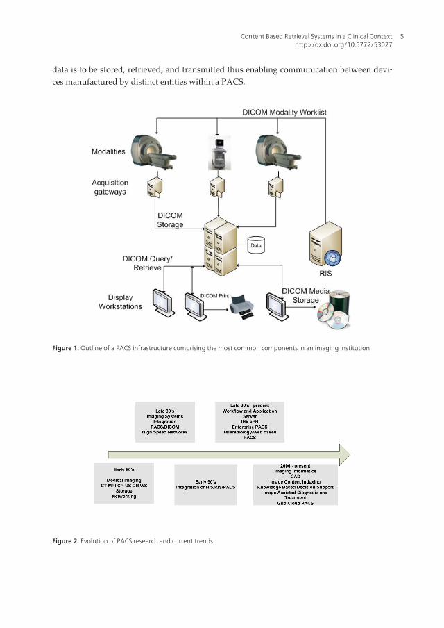
2. Picture archive and communication systems
In a medical imaging institution, such as a hospital or a clinic, the set of technologies employedthrough the processes of archiving, visualizing, acquiring and distributing medical imagesover a computer network (see Figure 1) is commonly referred to as a Picture Archive and Com‐munication System (PACS). PACS have evolved tremendously since when, as early as 1972,Dr. Richard J. Steckel implemented a minimal imaging system, comprising not much morethan a scanner next to a film developer for digitalization of radiographs, a communication pro‐tocol to transmit those images and a video monitor to receive and display them [5]. No morethan a proof of concept back then, but fast forward to current days, however, and a properly in‐tegrated hospital, or an enterprise PACS implementation is now a major undertaking that re‐quires careful planning and several million dollars of investment [6]. Such investment is oftenrequired since large PACS commonly have to handle more than 20000 radiological proceduresper year each procedure comprising potentially hundreds of distinct images. This meansaround 10 Terabytes of imaging information are stored per year [7]. It is in such situations thatContent Based Image Retrieval systems are expected to provide the largest benefits.
PACS are still a very active field of research where the ever-changing requirements and thedesire to provide more efficient services coupled with new ideas. Figure 2 shows a chrono‐logical view of the different challenges that arose and of some of the problems in which theresearch community is currently focusing. The push for CBIR enabled PACS has gained mo‐mentum since the late 90’s up to the present day, however, even nowadays there are veryfew such systems currently powering medical institutions.
2.1. Digital imaging and communication in medicine
A major step in the direction of modern PACS was given circa 1985 with the creation of anearlier form of what would become the current DICOM standard. This protocol stands asone of the key protocols involved in medical imaging systems. We can consider it as the gluethat holds the equipment and software developed by multiple companies together. It is anexpansible, object oriented protocol with support for multiple imaging modalities and re‐spective structured reports that also allows for private data to be embedded in its objects. Ofgreat importance is the fact that it defines how medical image data and correspondent meta-
Medical Imaging in Clinical Practice4
data is to be stored, retrieved, and transmitted thus enabling communication between devi‐ces manufactured by distinct entities within a PACS.
Figure 1. Outline of a PACS infrastructure comprising the most common components in an imaging institution
Figure 2. Evolution of PACS research and current trends
Content Based Retrieval Systems in a Clinical Contexthttp://dx.doi.org/10.5772/53027
5
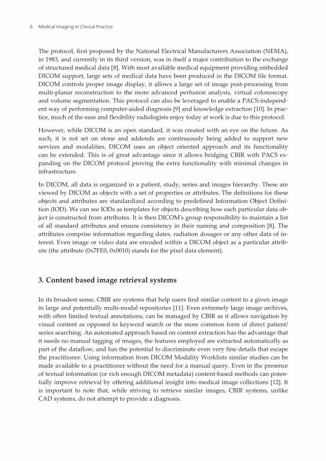
The protocol, first proposed by the National Electrical Manufacturers Association (NEMA),in 1983, and currently in its third version, was in itself a major contribution to the exchangeof structured medical data [8]. With most available medical equipment providing embeddedDICOM support, large sets of medical data have been produced in the DICOM file format.DICOM controls proper image display, it allows a large set of image post-processing frommulti-planar reconstruction to the more advanced perfusion analysis, virtual colonoscopyand volume segmentation. This protocol can also be leveraged to enable a PACS-independ‐ent way of performing computer-aided diagnosis [9] and knowledge extraction [10]. In prac‐tice, much of the ease and flexibility radiologists enjoy today at work is due to this protocol.
However, while DICOM is an open standard, it was created with an eye on the future. Assuch, it is not set on stone and addends are continuously being added to support newservices and modalities. DICOM uses an object oriented approach and its functionalitycan be extended. This is of great advantage since it allows bridging CBIR with PACS ex‐panding on the DICOM protocol proving the extra functionality with minimal changes ininfrastructure.
In DICOM, all data is organized in a patient, study, series and images hierarchy. These areviewed by DICOM as objects with a set of properties or attributes. The definitions for theseobjects and attributes are standardized according to predefined Information Object Defini‐tion (IOD). We can see IODs as templates for objects describing how each particular data ob‐ject is constructed from attributes. It is then DICOM's group responsibility to maintain a listof all standard attributes and ensure consistency in their naming and composition [8]. Theattributes comprise information regarding dates, radiation dosages or any other data of in‐terest. Even image or video data are encoded within a DICOM object as a particular attrib‐ute (the attribute (0x7FE0, 0x0010) stands for the pixel data element).
3. Content based image retrieval systems
In its broadest sense, CBIR are systems that help users find similar content to a given imagein large and potentially multi-modal repositories [11]. Even extremely large image archives,with often limited textual annotations, can be managed by CBIR as it allows navigation byvisual content as opposed to keyword search or the more common form of direct patient/series searching. An automated approach based on content extraction has the advantage thatit needs no manual tagging of images, the features employed are extracted automatically aspart of the dataflow, and has the potential to discriminate even very fine details that escapethe practitioner. Using information from DICOM Modality Worklists similar studies can bemade available to a practitioner without the need for a manual query. Even in the presenceof textual information (or rich enough DICOM metadata) content-based methods can poten‐tially improve retrieval by offering additional insight into medical image collections [12]. Itis important to note that, while striving to retrieve similar images, CBIR systems, unlikeCAD systems, do not attempt to provide a diagnosis.
Medical Imaging in Clinical Practice6
3.1. Searching information in content based image retrieval systems
Searching relevant data is a fundamental operation in CBIR. In relational databases thesearch procedure is applied to structured data, that is, numerical or alphabetical informationthat is searched for exactly. More sophisticated searches such as range queries on numericalkeys or prefix searching on strings still rely on the concept that two keys are, or are not,equal. In order to guarantee query performance, traditional databases assume that there ex‐ists a total linear order on the keys which is used to establish indexes over the tables. Thattotal order is something that does not arise naturally when dealing with unstructured high-dimensional spaces [13]. However, content-based retrieval relies heavily on similarity quer‐ies performed over them [14], hence a similarity function can be defined that establishes thatordering in relation to the source image.
When a query is performed with a source image, every element matches the input with asimilarity value. If performed naively, without resorting to advanced indexing techniques,the outcome of similarity query is a permutation of all database content. That is, the ele‐ments are rearranged from the highest similarity value to the lowest [13]. This is a behaviorthat is not desirable. Assuming the similarity is properly defined there are two canonicaltypes of queries that are of interest3:
• Range Query: Where we want to retrieve all elements that are closer than a given distanceto the query content.
• Nearest Neighbor Query: Where we want to retrieve a certain number of the elementsmost similar to the query.
In order for a practitioner to perform a search he must provide input to the CBIR. Unlike intraditional query systems text is not used. Several approaches have been explored:
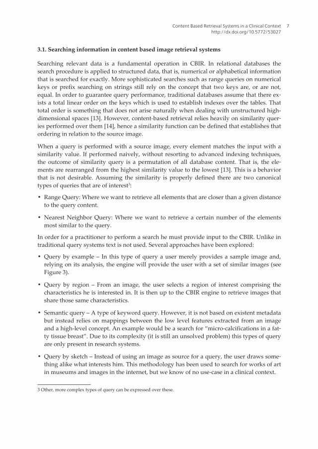
• Query by example – In this type of query a user merely provides a sample image and,relying on its analysis, the engine will provide the user with a set of similar images (seeFigure 3).
• Query by region – From an image, the user selects a region of interest comprising thecharacteristics he is interested in. It is then up to the CBIR engine to retrieve images thatshare those same characteristics.
• Semantic query – A type of keyword query. However, it is not based on existent metadatabut instead relies on mappings between the low level features extracted from an imageand a high-level concept. An example would be a search for “micro-calcifications in a fat‐ty tissue breast”. Due to its complexity (it is still an unsolved problem) this types of queryare only present in research systems.
• Query by sketch – Instead of using an image as source for a query, the user draws some‐thing alike what interests him. This methodology has been used to search for works of artin museums and images in the internet, but we know of no use-case in a clinical context.
3 Other, more complex types of query can be expressed over these.
Content Based Retrieval Systems in a Clinical Contexthttp://dx.doi.org/10.5772/53027
7
Figure 3. Query by example
3.2. Content based image retrieval systems in a clinical context
The push for the usage of CBIR of systems in a clinical context comes from their success inother areas where they have been successfully applied to handle large quantities of data. Arecent example is Google’s “search by image”4 functionality that operates according to thequery-by-example paradigm.
Several scenarios exist where medical practitioners can benefit from the use of these types ofsystem. A key functionality that is of value to radiologists assessing medical images is theability to provide them with a set of similar images, already diagnosed, thereby aiding themin their process of interpretation by quickly providing them with a second opinion. Thisproves to be orders of magnitude faster than the current mechanisms provided to manuallybrowse the archives. The potential for this type of assisted interpretation is motivated notonly by time constrains, but also by the recognition that variations in interpretation betweenpractitioners, commonly based on perceptual errors, lack of training, or fatigue, do exist[11]. Significant inter-observer variation has been documented in numerous studies [15, 16].Besides being a useful clinical tool, it is conceivable its use in an academic context where stu‐dents can benefit from access to similar, diagnosed, data.
Selecting studies by similarity has another benefit. Considering a large repository, built overtime, some of the retrieved images are bound to of some age. If a medical institution has kepttrack of a patient, performing more recent examinations, this data can be very useful to pre‐dict possible outcomes to an ailment's evolution. Furthermore, DICOM headers may containa fairly high rate of errors, for example for the field anatomical region, error rates of 16% havebeen reported [17]. This hinders the correct retrieval of wanted images via textual search.
4 http://images.google.com/
Medical Imaging in Clinical Practice8
Yet another important and useful outcome of CBIR is the possibility to bridge the semanticgap, allowing users to search an image repository for high-level image features. For instancea researcher may be interested in all studies containing a particular type or disposition oflymph nodes, or query only for images containing a particular feature. This concept expandson CBIR systems and requires that we establish a relation between the low level featuresemployed and the high level concepts of a semantic interpretation.
3.3. Features and feature extraction
At the core of each CBIR there is a matching algorithm analyzing the similarity between thequery content and the content stored in the database. However, except for the most trivialCBIR engines operating on simple content, it is not the actual content that is compared. Asbriefly mentioned, features extracted from the source content, are used instead. In the caseof images, pixel by pixel comparisons are not commonly performed due to, not only to thecomputational effort involved, but also because such comparisons lacks any type of seman‐tic meaning, are dependent on resolution and often are very sensitive to small changes. Fur‐thermore, it is not clear which pixels from the one image correspond to pixels in anotherimage. That said, a feature is simply a relevant piece of information, a synonym for an inputvariable or an attribute of an image [18], usually much smaller in size than the original data.
Thus, when there is a need to cope with large datasets, such as the ones present in medicalrepositories, or to deal with large inputs where most information is redundant or irrelevant,as is the case with some images, the analysis is commonly preceded by a pre-processingstage that provides a reduced representation of the original data. This step is called featureextraction and is of crucial importance for any CBIR currently deployed, as content match‐ing operates by comparing features and only the features are indexed.
Using a feature based approach to image analysis brings several advantages to CBIR sys‐tems. Besides reducing the size of the input data, thus providing great performance im‐provements to the matching algorithms, its reduced representation also translates directly ina smaller storage footprint. Of great importance is that, by discarding redundant or uselessinformation, some features can generalize a concept and allow predictive models to becomeboth more general and accurate. Some features also map well into high level concepts (cir‐cles, nodes, shapes, nodes) and help bridge the semantic gap.
Generally a feature is represented by a set of values that can be organized into a vector. Theglobal entropy of an image is a single real value but, on the other hand, a normalized inten‐sity histogram or a texture descriptor can be understood as a n-dimensional vector whereeach index contains the probability of a pixel having an intensity value equal to that index.
In most CBIR systems, a single feature is very often not enough to fully represent the imagein a way that makes possible to perform relevant queries. The usual approach is then to ex‐tract multiple features from the image and merge them into a single vector, canonicallycalled the feature vector. The set of all possible feature vectors constitutes a feature space.Depending on the features, this can be a space with a very high dimensionality.
Content Based Retrieval Systems in a Clinical Contexthttp://dx.doi.org/10.5772/53027
9
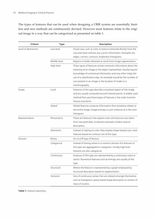
The types of features that can be used when designing a CBIR system are essentially limit‐less and new methods are continuously devised. However most features relate to the origi‐nal image in a way that can be categorized as presented on table 1.
Criteria Type Description
Level of abstraction Low level Visual cues, such as color or texture extracted directly from the
raw pixel data without any a priori information. Examples are
edges, corners, contours, brightness histograms.
Middle level Regions or blobs obtained as result from image segmentation.
High level These types of features contain semantic information about the
meaning of an image or the object represented. Usually require
knowledge of contextual information and very often imply the
use of a classification step. An example would be the number of
cars present in an image or the location of nodes in a
mammography.
Scope Local Features of this type describe a localized region of the image
and are usually computed around interest points. A widely used
method that uses these types of features is the scale-invariant
feature transform.
Global Global features comprise information that somehow relates to
the entire image. Image entropy is such a feature as is the color
histogram.
Representation Photometric These are features that explore color and textural cues taken
from raw pixel data. A relevant example is Gabor texture
descriptors.
Geometric Instead of relying on color they employ shape-based cues, most
features based on contours are of this type.
Domain Binary An on/off type of feature
Categorical Instead of having values in a numeric domain this features of
this type are aggregated in categories. Usually high level
features are also categorical.
Continuous Features of this type are represented by a continuous value or
vector. Numerical features such as entropy are usually of this
type.
Structural Where the feature is represented by a graph employed by
structural descriptors based on segmentation.
Vectorial Sets of continuous values that are related amongst themselves
such as histograms, space-based shape descriptors or centers of
mass of clusters.
Table 1. Feature taxonomy
Medical Imaging in Clinical Practice10
The relevancy of a feature is, however, highly dependent on the domain of the problem.This brings the problem of what features should be selected or are relevant in a given con‐text. Namely, what features should be used to perform efficient CBIR in a medical environ‐ment where multiple modalities are in place? This is a topic of great interest nowadays andthe subject of intensive research.
3.4. Similarity
Due to the unstructured nature of content in images, CBIR systems eschew exact matchingand rely instead in nearest neighbor or range queries based on a similarity function. Hence,one of the most important tasks in both the research and development of CBIR systems is toproperly define that similarity. Implicitly, one person has a clear notion of whether any twoobjects or images are similar. Even so, humans are also very much subject to subjective opin‐ions and those can vary wildly. Nonetheless, when searching for reasonable similarity meas‐ures, the most obvious place to look is at the human similarity assessment. After all, when auser searches for something similar, he already has in mind his own concept of similarity,whose form is doubtlessly quite different from the metric spaces (such as the Euclidean) typ‐ically used for feature vector comparison. The similarity used by the CBIR systems shouldthen be as similar as possible to the human concept of similarity if the results of the searchare to be satisfactory [19]. Algorithmically modeling that behavior thus requires that the in‐ternal image representations closely reflect the ways in which users interpret, understand,and encode visual data. Finding suitable image representations, based on the types of fea‐tures described previously is an important step towards the development of effective simi‐larity models [14]. However, creating such algorithmic functions is complicated due to thefact that there is no single model of human similarity. Furthermore a user may have in minda very specific type of similarity or criteria he is interested in. For instance, in a radiologysetting, a practitioner may wish to place more emphasis in finding mammographs sharing acertain disposition of micro-calcifications rather than those containing the same tissue typeor having a similar breast size.
Combining multiple representation models can partially resolve this problem. If a retrievalsystem allows parameterized or multiple similarity functions, the user should be able to se‐lect those that most closely model his or her perception [14]. This is not a trivial problem tosolve by any means and similarity selection functionality is hardly present in current medi‐cal CBIR. In fact such feature is lacking in even most CBIR systems. However, within a med‐ical institution often exist multiple modalities and the DICOM protocol offers support for allthose types of distinct imagery.
3.4.1. Similarity measures
Of crucial importance in a CBIR system is the design of the similarity metrics used tomatch a query to the database feature vectors. Mathematically we can define these metricsas a function f (x,x’) that takes as arguments the set of features belonging to two distinctimages and returns a value from an ordered set (such as the set of real numbers). Thissorting embodies the idea that some images look more like the query than others and al‐
Content Based Retrieval Systems in a Clinical Contexthttp://dx.doi.org/10.5772/53027
11
low a content engine to return, not only the closest match, but a bundle of images ar‐ranged by similarity thus increasing the probability that the user has of finding what he islooking for. Typically, smaller values correspond to higher similarity although that de‐pends, of course, on the specific function being used. The similarity measures employedin CBIR systems are deeply tied with the representation of the features extracted by thesystem. We now present some of the most applied functions.
• Vector distance – One of the most common similarity measures. A function of two featurevectors is defined over the feature space. These are often applied due to their conceptualsimplicity. Simpler distances, such as the Euclidean, are also quick to compute, however,that is not always the case, other measures such as the EMD5 or statistical distances canhave significant complexity.
• Shape based – These are used when features consist of points delineating a shape boun‐dary. The similarity between shapes is defined in terms of the transformations required totransform a shape into another.
• Structural/Graph matching - A class of similarity measurements that apply when the ex‐tracted features are represented by a graph. The similarity can be computed by an attrib‐uted graph-matching scheme such as relaxation schemes or combinatorial algorithms.
• Classifier-based - These classifiers employ machine learning techniques to classify theimage as pertaining to a set of predetermined labels. This scheme does not follow the con‐cept of a similarity function, however, in most systems the label obtained is merged withthe existing feature set and a vector distance metric is subsequently applied.
In table 2 we find a list containing some of the methodologies employed for similarity meas‐urements in various CBIR projects.
3.4.2. Relevance feedback
While CBIR systems should operate in a transparent manner, in order to increase theiroverall accuracy it can be desirable to allow a user to relate back to the system which re‐sults are actually relevant. Relevance feedback is the process of automatically adjusting animage query using the information provided from the expert on previously executed quer‐ies [20]. A way to achieve this goal is to expose to the user an interface that allows him toprovide feedback on the relevancy of the results on a per-image basis. A new query canthen be executed in order to replace non-relevant results and the feedback loop is repeat‐ed many times until the user is satisfied. A key issue is in how to effectively utilize thefeedback information to improve the retrieval performance. This aspect depends on theparticular implementation of the CBIR given it modifies the way the similarity computa‐tion is performed and several methodologies have been explored. An overview of thesemechanisms can be found in [21].
5 Earth Mover’s Distance
Medical Imaging in Clinical Practice12
3.5. Indexing and performance
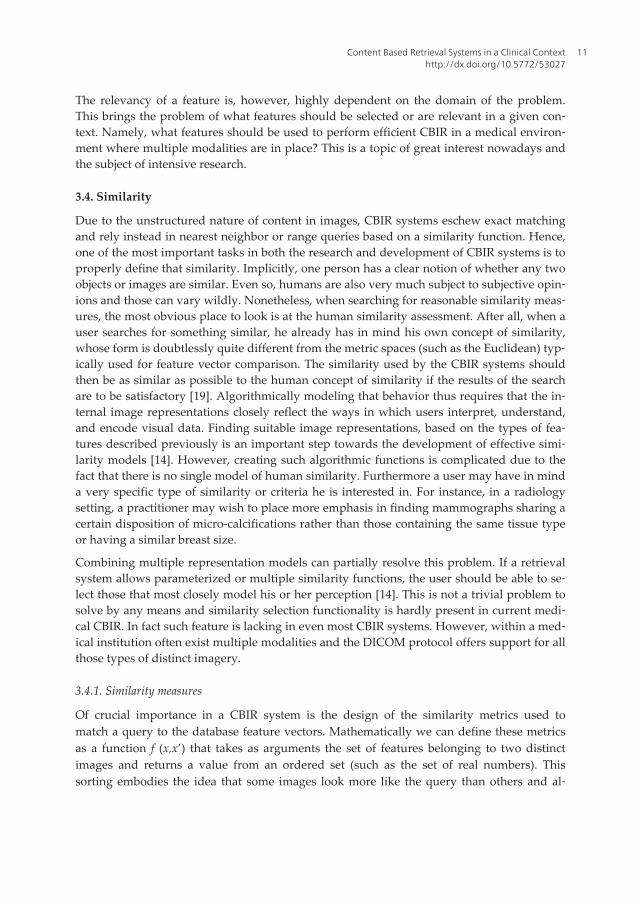
When the number of images in the database is small, as is often the case with researchsystems, a sequential linear search across all elements can provide an acceptable perform‐ance. However, with large-scale image databases, such as the ones present in medical sys‐tems, more efficient query mechanisms become a necessity. The search task can besignificantly improved by relying on multidimensional indexing structures. Like tradition‐al databases, the indexing of an image database should support an efficient search basedon the extracted features.
The basic idea behind any indexing procedure (figure 4) is a hierarchical division of spacethat increases the lookup speed by removing the need to sift the entire feature space o ob‐tain the desired resuls. Due to the nature of CBIR queries, which require quick lookup ofthe nearest neighbors to a data point in the feature space, the indexing structure must pre‐serve locality.
Figure 4. Indexing a feature space
The most popular class of indexing techniques in traditional databases is the B-tree familywhich provides very efficient searches when the key is a scalar. However, they are notsuitable to index the content of images represented by high-dimensional features. None‐theless, multidimensional indexing techniques exist. There are a large variety of multidi‐mensional indexing methods, which differ in the type of queries they support and thedimensionality of the space where they are advantageous. The R-tree [22] and its varia‐
Content Based Retrieval Systems in a Clinical Contexthttp://dx.doi.org/10.5772/53027
13
tions are probably the best-known multidimensional indexing techniques in general pur‐pose content retrieval engines. Other approaches are the k-d tree and variants such as theR+-tree and the R*-tree [23].
If a similarity function is at the same time a distance, and thereby a metric to the featurespace, a distinct set of methods that operate in a metric space are available. These meth‐ods rely only on the definition of the distance function and make no other assumptions.Hence they prove to be very general indexing structures. A study on such methods isavailable in [13] and [24]. One reason these types of data-structures are not more perva‐sive in medical CBIR is that research is still being conducted on how to provide mecha‐nisms in Database Management System (DBMS) that allow users to easily incorporatethem into search engines.
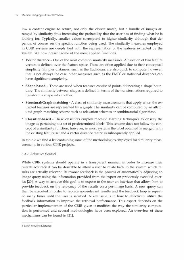
3.6. Architectural overview of content based retrieval engines
Taking into account the presented requirements and operations for CBIR systems, in figure5 we show how a generic architecture to a PACS-aware CBIR architecture can be designed.In this architecture the CBIR engine operates outside the PACS repository. This guaranteesthe integrity of the imaging repository and allows clinical operations to proceed should theCBIR engine fail.
Figure 5. General architecture of a PACS enabled CBIR system
The frontend is the component in charge of receiving similarity requests. Such requests canbe triggered manually, by a practitioner, or by analyzing DICOM’s Modality Worklist. It re‐plies to the requests with a list of DICOM files to be retrieved which are similar to the sourceimage of the request.
Medical Imaging in Clinical Practice14
The Feature Extractor component’s, main responsibility is, like the name indicates, to extractthe relevant features from an image. This behavior is triggered during an initialization pro‐cedure, when analyzing images from the PACS repository, or upon the creation of new im‐ages by the modalities. The extracted features are then passed to the Feature Database whichindexes them and allows for fast nearest neighbor queries. The last major component, thesimilarity engine comprises the set of metrics and similarities that can be applied.
3.7. Review of CBIR applications
Most radiological CBIR applications are still in a conceptual or research stage. In table 2 wepresent a brief listing of such systems together with the most important aspects present in aCBIR. This table is based on a similar, more complete table presented in [11].
The features column is arranged in three categories: General, Mixed and Specialized. Gener‐al features are low and middle level features, extracted from the image with no a priori do‐main specific knowledge and typically extracted with no user input. Mixed featurescomprise both general features and extra annotations, whether provided from a practitioneror extracted from other sources. Specialized features rely on specific knowledge about thenature and type of the dataset and are typically not automated requiring an expert to pro‐vide extra information such as regions of interest.
Ref Features Similarity measures Relevance feedback PACS Integration
[25] General Classifier-based Yes No
[26] General Classifier No No
[27] Mixed Classifier No No
[28] Mixed Vector distance No No
[29] Specialized Classifier No No
[30] Specialized Structural No No
[31] General Vector distance No No
[32] General Classifier No No
[33] Mixed Vector distance No No
[34] Mixed - No Yes
Table 2. Overview of medical CBIR systems
Of the presented systems, only [34] focuses specifically on PACS level integration, however,only the concepts and methodology are discussed.
4. Dicoogle
We have developed Dicoogle6, an open-source PACS with support for data indexing, peer-to-peer communication and CBIR functionality. This tool complements, and may even re‐
Content Based Retrieval Systems in a Clinical Contexthttp://dx.doi.org/10.5772/53027
15
place a traditional PACS server and enhance it with a more agile indexing and retrievalmechanism [35]. Besides providing basic DICOM services such as Storage and Query/Retrieval, Dicoogle can automatically extract, index and store all metadata present in a DI‐COM header (including data present in private data elements). The indexed data can thenbe queried using free text. A more advanced search mechanism is also provided using a richquery language based on Lucene’s syntax. This syntax has support for element selection, nu‐merical and range-based search, wildcard expansion and Boolean operators such as AND,OR and NOT. As a data-extraction tool, Dicoogle has been used in several small to mediumimaging institutions. For instance, in [36] Dicoogle was used to demonstrate several incon‐sistencies in the handling of some DICOM attributes by the modalities and to perform astudy on the radiation dosage of the patients handled at the site.
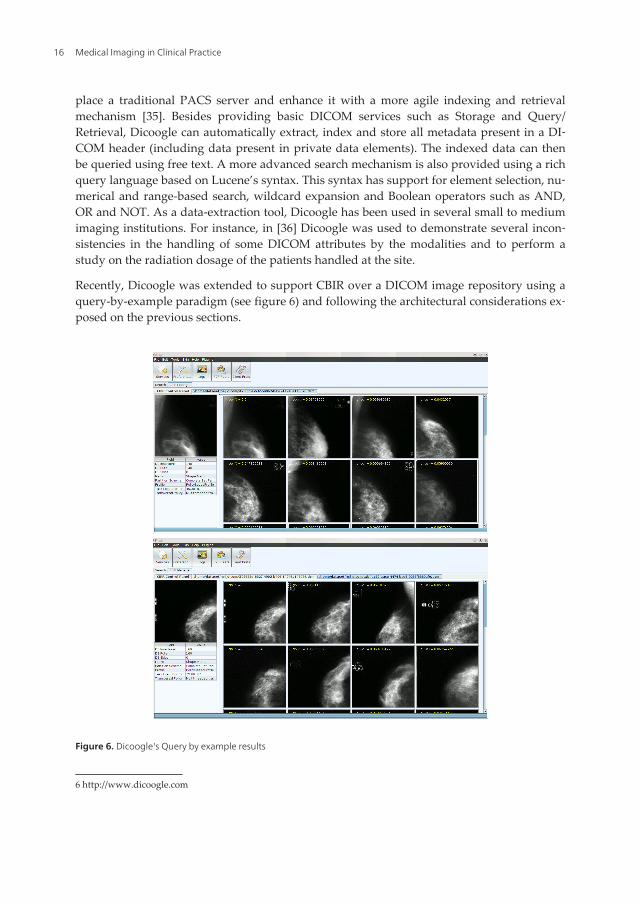
Recently, Dicoogle was extended to support CBIR over a DICOM image repository using aquery-by-example paradigm (see figure 6) and following the architectural considerations ex‐posed on the previous sections.
Figure 6. Dicoogle's Query by example results
6 http://www.dicoogle.com
Medical Imaging in Clinical Practice16
4.1. A profile-based approach to CBIR in a medical context
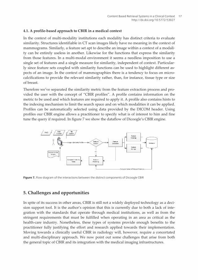
In the context of multi-modality institutions each modality has distinct criteria to evaluatesimilarity. Structures identifiable in CT scan images likely have no meaning in the context ofmammograms. Similarly, a feature set apt to describe an image within a context of a modali‐ty can be entirely useless in another. Likewise for the functions that express the similarityfrom those features. In a multi-modal environment it seems a needless imposition to use asingle set of features and a single measure for similarity, independent of context. Particular‐ly since feature sets coupled with similarity functions can be used to highlight different as‐pects of an image. In the context of mammographies there is a tendency to focus on micro-calcifications to provide the relevant similarity rather, than, for instance, tissue type or sizeof breast.
Therefore we’ve separated the similarity metric from the feature extraction process and pro‐vided the user with the concept of “CBIR profiles”. A profile contains information on themetric to be used and which features are required to apply it. A profile also contains hints tothe indexing mechanism to limit the search space and on which modalities it can be applied.Profiles can be automatically selected using data provided by the DICOM header. Usingprofiles our CBIR engine allows a practitioner to specify what is of interest to him and finetune the query if required. In figure 7 we show the dataflow of Dicoogle’s CBIR engine.
Figure 7. Flow diagram of the interactions between the distinct components of Dicoogle CBIR
5. Challenges and opportunities
In spite of its success in other areas, CBIR is still not a widely deployed technology as a deci‐sion support tool. It is the author’s opinion that this is currently due to both a lack of inte‐gration with the standards that operate through medical institutions, as well as from thestringent requirements that must be fulfilled when operating in an area as critical as thehealth-care industry. Nonetheless, these types of systems provide enough benefits to thepractitioner fully justifying the effort and research applied towards their implementation.Moving towards a clinically useful CBIR in radiology will, however, require a concertatedand multi-disciplinary approach. We now point out some challenges that arise from boththe general topic of CBIR and its integration with the medical imaging infrastructures.
Content Based Retrieval Systems in a Clinical Contexthttp://dx.doi.org/10.5772/53027
17

• PACS and DICOM integration. We proposed an approach that complements a PACS byexternalizing the CBIR and interacting through the DICOM protocol. However, in this ap‐proach, third party tools are limited to images pushed to them through the DICOM C-Move operation. This needlessly hampers the possibility of cooperation betweenapplications as third party tools are either passive, must conform to a private API or mustimplement themselves an indexing and similarity mechanism.
• Enabling multi-dimensional database systems. Several studies exist on how to performmulti-dimensional indexing. However, databases that natively provide support for index‐ing multi-dimensional data-points according to an arbitrary similarity function and ableto cope with large, dynamic volumes of information are, to the best of our knowledge, in‐existent. This leads researchers and application developers resorting to either implementindexing mechanisms atop relational databases or to completely ignore the problem andfocusing on the other aspects of a CBIR.
• Multi-modal data integration. To rely exclusively in pixel data imposes some limits toCBIR systems, not only in the medical arena. Gathering information from multiple sour‐ces, such as demography data of a patient from the Radiology Information System, andcombining it with the extracted data has the potential to further improve clinical CBIR. Afurther step forward will be to move from single image analysis and retrieval and mergeinformation from the multitudes of sources that may be present in a DICOM study. Com‐bining this information is not trivial due to missing information, the heterogenetic natureof the data and the problems involved in relating a set of images to another in a meaning‐ful way. This will allow CBIR engines to move to a study-based paradigm, the most com‐mon unit of search for practitioners in most modalities. Tackling this problem is likely tohave the biggest impact in a clinical environment [37].
• Lack of a Gold Standard for CBIR. It is currently not possible to compare the perform‐ance across different medical CBIR systems. Not only their respective application domainand specific goals differ, but there is a lack of a common Gold Standard. The Image‐CLEFmed7 is one of the few platforms to evaluate and compare different systems. Otherpublic datasets exist with annotated data, however, the fact they are scattered and the an‐notations provided do not follow any particular structure makes them cumbersome touse. To access the research effectively, task-related standardized databases on which dis‐tinct groups can apply their algorithms are needed. Cooperation with clinical experts is anecessity to provide the necessary relevancy assessments.
• Towards semantic search. Much emphasis is being placed on automatic and assisted con‐cept extraction. This effort is directed towards bridging the semantic gap and further in‐crease the accuracy of CBIR systems [38]. An advantage of a CBIR operating in themedical field is that semantics in the medical domain are much better defined and there isa vast accumulation of formal knowledge representations that could be exploited to sup‐port semantic search for any specialty areas in medicine [39].
7 http://www.imageclef.org/
Medical Imaging in Clinical Practice18

This idea expands on CBIR systems and requires that we establish a relation between thelow level features employed and the high level concepts of a semantic interpretation. Sever‐al strategies currently under study are the following [38]:
• Usage of object ontologies to define high-level concepts
• Employ machine learning concepts to perform the association between features andconcepts
• Rely on users and perform relevance feedback for continuous learning
• Generate semantic templates (profiles) to support the association
• Rely on the meta-data or other textual information provided by the user
The major functionality enabled by semantic search is the advanced textual queries that canbe provided to a practitioner. For example, queries such as “show me blood smears that includepolymorphonuclear neutrophils”8 should become a possibility.
6. Conclusion
In this chapter we’ve exposed the some of the most common methodologies employed inContent-based Image Retrieval and provided an overview of the state of such systems in aclinical context. We pointed out how CBIR, being a query mechanism more adapted to theworkflow of a radiologist than the traditional string matching present in DICOM, can helpimprove diagnosis speed and accuracy in a clinical context.
It was shown how the creation of accurate and performing CBIR systems in a clinical contextis a task hard to tackle, ripe with non-trivial challenges that arise from a multitude of factorssuch as the need for integration with currently deployed PACS, the need to handle multiplemodalities and cope with stringent performance requirements.
Furthermore, we presented Dicoogle, our approach to bring CBIR to DICOM enabled PACSsystems relying on a profile-based approach.
The goal of CBIR systems is not to replace the practitioner or, unlike CAD tools, provide au‐tomated diagnosis, but to empower the expert with tools that allow for faster and more ac‐curate diagnosis in a workflow adapted to his needs.
Acknowledgements
This work has received funding from “Fundação para a Ciência e Tecnologia“ (FCT) undergrant agreement PTDC/EIA-EIA/104428/2008
8 An example from the ImageCLEFmed competition as shown in [2]
Content Based Retrieval Systems in a Clinical Contexthttp://dx.doi.org/10.5772/53027
19

Author details
Frederico Valente, Carlos Costa and Augusto Silva
Universidade de Aveiro, IEETA, Portugal
References
[1] Rubin GD. Data explosion: the challenge of multidetector-row. European Journal ofRadiology. 2000;: p. 74-80.
[2] Henning Muller XZADMPJsIaAG. Medical Visual Information Retrieval: State of theArt and Challenges Ahead. 2007 IEEE International Conference on Multimedia andExpo. 2007;: p. 683-686.
[3] Andriole K. Addressing the coming radiology crisis - the society for computer appli‐cations in radiology transforming the radiological interpretation process (trip) initia‐tive. Journal of Digital Imaging. 2004;(17): p. 235-243.
[4] National Electrical Manufacturers Association. DICOM Part 4: Service Class Specifi‐cations. 2009..
[5] Steckel J. Daily x-ray rounds in a large teaching hospital using high-resolutionclosed-circuit television. Radiology. 1972; 105(2).
[6] Huang HK. PACS and Imaging Informatics: Basic Principles and Applications: JohnWiley & Sons; 2010.
[7] Oliveira MC, Cirne W, Marques PM. Towards applying content-based image retriev‐al in the clinical routine. Future Generation Computer Systems. 2007 March; 23(3): p.466-474.
[8] Oosterwijk H, Gihring P. DICOM basics.: Tech; 2002.
[9] CAD–PACS integration tool kit based on DICOM secondary capture, structured re‐port and IHE workflow profiles. Computerized medical imaging and graphics : theofficial journal of the Computerized Medical Imaging Society. 2007 June; 31(4).
[10] Lehmann TM, Güld O, Deselaers , Keysers , Schubert , Spitzer , et al. Automatic cate‐gorization of medical images for content-based retrieval and data mining. Computer‐ized Medical Imaging and Graphics. 2005 March; 29(2).
[11] Akgül B, Rubin DL, Napel , Beaulieu CF, Greenspan , Acar. Content-Based Image Re‐trieval in Radiology: Current Status and Future Directions. Journal of Digital Imag‐ing. 2011 April; 24(2).
Medical Imaging in Clinical Practice20

[12] Lew MS, Sebe , Djeraba , Jain. Content-based multimedia information retrieval: Stateof the art and challenges. ACM Transactions on Multimedia Computing, Communi‐cations, and Applications. 2006 February; 2(1).
[13] Chavez E, Navarro , Baeza-Yate , Marroquin JL. Searching in metric spaces. ACMComputer Survey. 2001; 33.
[14] Castelli V, Bergman LD. Image databases: search and retrieval of digital imagery:Wiley; 2002.
[15] Siegle RL, Baram EM, Reut SR. Rates of disagreement in imaging interpretation in agroup of community hospitals. Academic Radiology. 1998; 5(3).
[16] Soffa D, Lewis R, Sunshine J, Bhargavan M. Disagreement in interpretation: a meth‐od for the development of benchmarks for quality assurance in imaging. Journal ofthe American College of Radiology. 2004; 1(3).
[17] Guld MO, Kohnen , Keysers , Schubert. Quality of dicom header information for im‐age categorization. 2002..
[18] Guyon I. Feature extraction: foundations and applications (Studies in fuzziness andsoft computing): Springer-Verlag; 2006.
[19] Santini , Jain. Similarity queries in image databases. In IEEE Computer Society Con‐ference on Computer Vision and Pattern Recognition; 1996.
[20] Pinjarkar L, Sharma , Mehta. Comparative Evaluation of Image Retrieval Algorithmsusing Relevance Feedback and it’s Applications. International Journal of ComputerApplications. 2012 June; 48(18).
[21] Pinjarkar , Sharma , Mehta. Comparison and Analysis of Content Based Image Re‐trieval Systems Based On Relevance Feedback. Journal of Emerging Trends in Com‐puting and Information Sciences. 2012 July; 3(6).
[22] Qian , Tagare. Optimal embedding for shape indexing in medical image databases.In Medical Image Computing and Computer-Assisted Intervention - MICCAI 2005;2005.
[23] Beckmann , Kriege HP, Schneid. The r*-tree: an efficient and robust access methodfor points and rectangles. In SIGMOD; 1990. p. 322-331.
[24] Zezula , Amato , Dohnal , Batko. Similarity Search: The Metric Space Approach:Springer; 2006.
[25] Bhattacharya M, Desai B. A framework for medical image retrieval using machinelearning and statistical similarity matching techniques with relevance feedback. IEEETransactions in Information Technologie in Biomedicine. 2007; 11(1).
[26] Greenspan H, Pinhas A. Medical image categorization and retrieval for PACS usingthe GMM-KL framework. IEEE Transaction in Information Technologies in Biomedi‐cine. 2007; 11(2).
Content Based Retrieval Systems in a Clinical Contexthttp://dx.doi.org/10.5772/53027
21
[27] Lim J, Chevallet J. Vismed: A visual vocabulary approach for medical image index‐ing and retrieval. In Second Asia Information Retrieval Symposium; 2005; Jeju Island.
[28] Müller H ea. The Use of MedGIFT and EasyIR forImageCLEF 2005. in AccessingMultilingual Information Repositories. In Accessing Multilingual Information Repo‐sitories.: Springer p. 724-732.
[29] Petrakis E, Faloutsos C, Lin K. ImageMap: an image indexing method based on spa‐tial similarity. IEEE Transactions in Knowledge Data Engineering. 2002; 15(5).
[30] Amores J, Radeva P. Registration and retrieval of highly elastic bodies using contex‐tual information. Pattern Recognition Letters. 2005; 26(11).
[31] Mohammad-Reza et al. Content-based image database system for epilepsy. Comput‐er Methods and Programs in Biomedicine. 2005; 79(3).
[32] Alto H, Rangayyan R, Desautels J. Content-based retrieval and analysis of mammo‐graphic masses. Journal of Electronic Imaging. 2005; 14(2).
[33] Kim J et al. A new way for multidimensional medical data management: Volume ofinterest (VOI)-based retrieval of medical images with visual and functional features.IEEE Transactions in Information Technologies in Biomedicine. 2006; 10(3).
[34] Benedikt Fischer et al. Integration of a Research CBIR System with RIS and PACS forRadiological Routine. In Proceedings of the SPIE; 2008.
[35] Costa CaFCaBLaRLaSAaOJ. Dicoogle - an Open Source Peer-to-Peer PACS. Journalof Digital Imaging. 2011; 24(5): p. 848-856.
[36] Santos , Bastião L, Costa C, Silva A, Rocha N. DICOM and Clinical Data Mining in aSmall Hospital PACS: A Pilot Study. In Communications in Computer and Informa‐tion Science; 2011: Springer Berlin Heidelberg. p. 254-263.
[37] Medical Visual Information Retrieval: State of the Art and Challenges Ahead. InIEEE International Conference on Multimedia and Expo; 2007; Geneva. p. 683 - 686.
[38] Ying , Zhang , Lu , Ma WY. A survey of content-based image retrieval with high lev‐el semantics. Pattern Recognition. 2007 January; 40(1).
[39] Zhou XS, Zillner S, Moeller M, Sintek M, Zhan Y, Krishnan A, et al. Semantics andCBIR: a medical imaging perspective. In CIVR '08 Proceedings of the 2008 interna‐tional conference on Content-based image and video retrieval; 2008. p. 571-580.
Medical Imaging in Clinical Practice22