Connecticut State Dental Commission - CT.gov
305
AGENDA CONNECTICUT STATE DENTAL COMMISSION Wednesday, June 10, 2020 at 1:00 PM Department of Public Health 410 Capitol Avenue, Hartford Connecticut Third Floor Hearing Room CALL TO ORDER I. MINUTES April 8, 2020 II. NEW BUSINESS A. Provisional License Applications • Foteini Touloumi, DDS Presented by Judith Bailey, License and Applications Analyst, DPH B. Proposed Amend Memorandum of Decision Ammar Idlibi, DMD – Petition No. 2016-640 C. Declaratory Ruling Proceeding - Treatment of Sleep Apnea with Oral Appliance Therapy Status Requests III. OLD BUSINESS Non-patient based clinical licensure examinations ADJOURN This meeting will be held by video conference. Connecticut State Dental Commission Learn more about Teams | Meeting options
-
Upload
khangminh22 -
Category
Documents
-
view
0 -
download
0
Transcript of Connecticut State Dental Commission - CT.gov
AGENDA CONNECTICUT STATE DENTAL COMMISSION
Wednesday, June 10, 2020 at 1:00 PM
Department of Public Health 410 Capitol Avenue, Hartford Connecticut
Third Floor Hearing Room
CALL TO ORDER
I. MINUTESApril 8, 2020
II. NEW BUSINESSA. Provisional License Applications
• Foteini Touloumi, DDSPresented by Judith Bailey, License and Applications Analyst, DPH
B. Proposed Amend Memorandum of DecisionAmmar Idlibi, DMD – Petition No. 2016-640
C. Declaratory Ruling Proceeding - Treatment of Sleep Apnea with Oral Appliance TherapyStatus Requests
III. OLD BUSINESSNon-patient based clinical licensure examinations
ADJOURN
This meeting will be held by video conference.
Connecticut State Dental Commission
Learn more about Teams | Meeting options

The following minutes are draft minutes which are subject to revision and which have not yet been adopted by the Board.
CONNECTICUT STATE DENTAL COMMISSION
MINUTES OF MEETING April 8, 2020
The Connecticut State Dental Commission held a meeting on April 8, 2020, at the Department of Public Health Complex, 470 Capitol Avenue, Hartford, Connecticut, in the Room 470-A/B. COMMISSION MEMBERS PRESENT: Peter Katz, DMD, Chairman – via telephone Sarita Arteaga, DMD - via telephone Monica Cipes, DMD – via telephone Deborah Dodenhoff, RN – via telephone Mark Longobardi, DMD – via telephone Anatoliy Ravin, DDS – via telephone
Steven Reiss, DDS – via telephone Barbara Ulrich – via telephone Robert Zager – via telephone COMMISSION MEMBERS ABSENT: None Dr. Katz called the meeting to order at 1:00 p.m. All participants were present by telephone conference. Dr. Arteaga was welcomed to her first meeting as a member of the Commission. I. MINUTES The minutes from the January 8, 2020 meeting were reviewed and approved on a motion by Dr.
Longobardi, seconded by Ms. Ulrich. Dr. Arteaga abstained. II. NEW BUSINESS
A. Provisional License Application – Rawan Sarsour, DDS Judith Bailey, License and Applications Analyst, Department of Public Health presented a provisional license application for Rawan Sarsour, DDS, to allow for practice at the University of Connecticut, School of Dental Medicine. Following review of the application Mr. Zager made a motion, seconded by Dr. Ravin, to recommend provisional licensure for Dr. Sarsour. The motion passed unanimously. B. American Academy of Dental Sleep Medicine Request for Declaratory Ruling Treatment of Sleep Apnea with Oral Appliance Therapy Assistant Attorney General Kerry Colson was present for this discussion and to provide counsel to the Commission. The Commission reviewed a request from Nancy Abby, DDS, President, American Academy of Dental Sleep Medicine. asking the following:
1. Is it within a dentist's scope of practice to dispense portable monitors when ordered by physicians for patients at risk for sleep apnea? The test results are provided to a physician for interpretation and diagnosis. 2. Is it within a dentist's scope of practice to order portable monitors for patients identified by the dentist as being at risk for sleep apnea? The test results are provided to a physician for interpretation and diagnosis. 3. Is it within a dentist's scope of practice to use a portable monitor to help determine the optimal effective position of a patient's oral appliance? 4. If a dentist does not use a portable monitor to determine the optimal effective position, is it within a dentist's scope of practice to order a portable monitor to verify the effectiveness of an oral appliance? The test results are provided to physicians for interpretation and therapeutic effectiveness is determined by physicians.
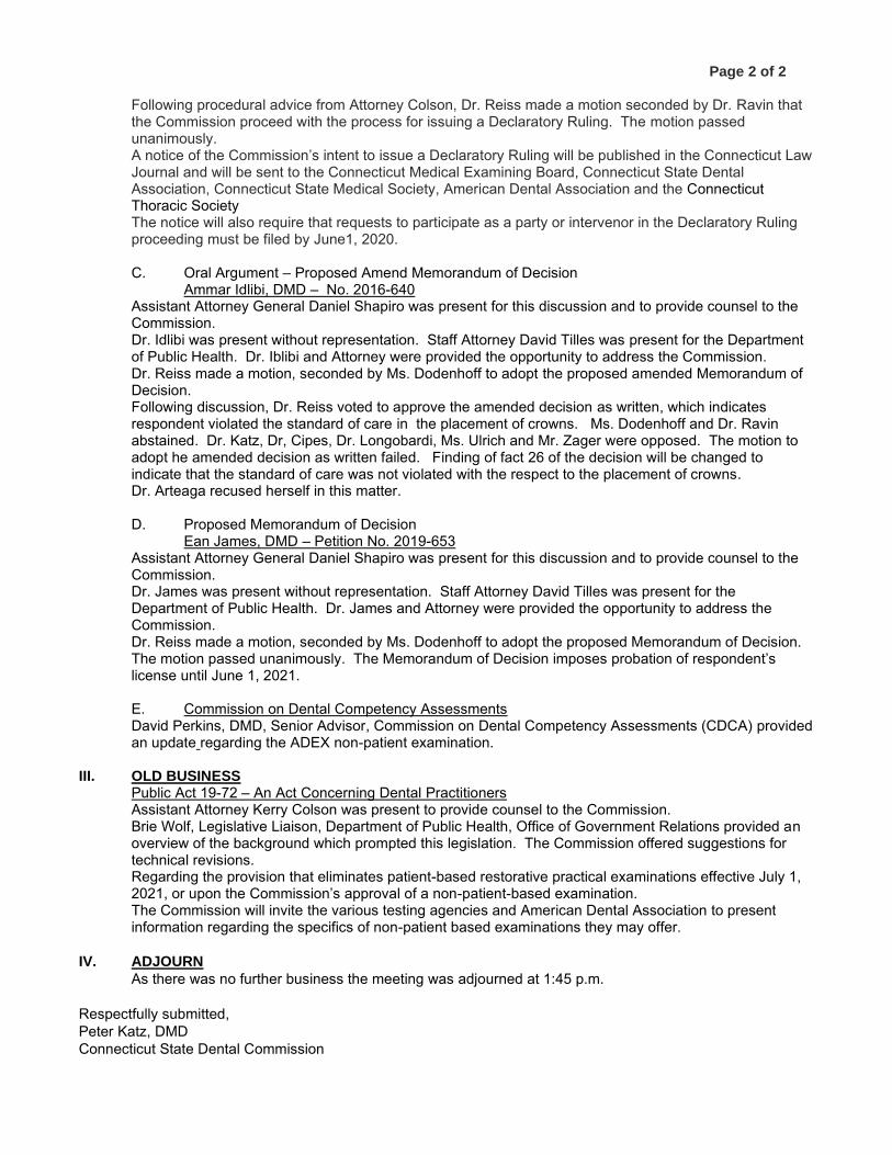
Page 2 of 2 Following procedural advice from Attorney Colson, Dr. Reiss made a motion seconded by Dr. Ravin that the Commission proceed with the process for issuing a Declaratory Ruling. The motion passed unanimously. A notice of the Commission’s intent to issue a Declaratory Ruling will be published in the Connecticut Law Journal and will be sent to the Connecticut Medical Examining Board, Connecticut State Dental Association, Connecticut State Medical Society, American Dental Association and the Connecticut Thoracic Society The notice will also require that requests to participate as a party or intervenor in the Declaratory Ruling proceeding must be filed by June1, 2020. C. Oral Argument – Proposed Amend Memorandum of Decision
Ammar Idlibi, DMD – No. 2016-640 Assistant Attorney General Daniel Shapiro was present for this discussion and to provide counsel to the Commission. Dr. Idlibi was present without representation. Staff Attorney David Tilles was present for the Department of Public Health. Dr. Iblibi and Attorney were provided the opportunity to address the Commission. Dr. Reiss made a motion, seconded by Ms. Dodenhoff to adopt the proposed amended Memorandum of Decision. Following discussion, Dr. Reiss voted to approve the amended decision as written, which indicates respondent violated the standard of care in the placement of crowns. Ms. Dodenhoff and Dr. Ravin abstained. Dr. Katz, Dr, Cipes, Dr. Longobardi, Ms. Ulrich and Mr. Zager were opposed. The motion to adopt he amended decision as written failed. Finding of fact 26 of the decision will be changed to indicate that the standard of care was not violated with the respect to the placement of crowns. Dr. Arteaga recused herself in this matter. D. Proposed Memorandum of Decision
Ean James, DMD – Petition No. 2019-653 Assistant Attorney General Daniel Shapiro was present for this discussion and to provide counsel to the Commission. Dr. James was present without representation. Staff Attorney David Tilles was present for the Department of Public Health. Dr. James and Attorney were provided the opportunity to address the Commission. Dr. Reiss made a motion, seconded by Ms. Dodenhoff to adopt the proposed Memorandum of Decision. The motion passed unanimously. The Memorandum of Decision imposes probation of respondent’s license until June 1, 2021. E. Commission on Dental Competency Assessments David Perkins, DMD, Senior Advisor, Commission on Dental Competency Assessments (CDCA) provided an update regarding the ADEX non-patient examination.
III. OLD BUSINESS Public Act 19-72 – An Act Concerning Dental Practitioners Assistant Attorney Kerry Colson was present to provide counsel to the Commission. Brie Wolf, Legislative Liaison, Department of Public Health, Office of Government Relations provided an overview of the background which prompted this legislation. The Commission offered suggestions for technical revisions. Regarding the provision that eliminates patient-based restorative practical examinations effective July 1, 2021, or upon the Commission’s approval of a non-patient-based examination. The Commission will invite the various testing agencies and American Dental Association to present information regarding the specifics of non-patient based examinations they may offer.
IV. ADJOURN
As there was no further business the meeting was adjourned at 1:45 p.m. Respectfully submitted, Peter Katz, DMD Connecticut State Dental Commission


Report Date: 05/27/2020
DENTPIN : 11194071
National Board Dental Examinations (NBDE)
Name DENTPIN® Graduation† School
TOULOUMI, FOTEINI 11194071 2008
Non-Accredited Dental School
Current Status
Test Date Exam Type Score ‡ Status
01/11/2014 NBDE II Pass
02/20/2009 NBDE I 88 Pass
National Board Dental Examination Part II
Test Date Exam Type Score ‡ Status
01/11/2014 NBDE II Pass
National Board Dental Examination Part I
Test Date Exam Type Score ‡ Status
02/20/2009 NBDE I 88 Pass
† The number listed is the candidate’s self reported year of graduation.‡ Numerical score is reported only for candidates who tested prior to January 1, 2012.
Print this page Close
Page 1 of 1NBDE Score History
5/27/2020https://dts.ada.org/HUB/Scores/ADAT/NBDEHistoryReport.aspx


Z�^h>d^�K&�^h���^^&h>���Ed�>��y�D/E�d/KE
&ŽƚĞŝŶŝ��dŽƵůŽƵŵŝϴϵϯ�&ĂƌŵŝŶŐƚŽŶ��ǀĞ͘���Ɖƚ�ϱ&tĞƐƚ�,ĂƌƚĨŽƌĚ͕��d�Ϭϲϭϭϵ
W�^^
/ŵƉŽƌƚĂŶƚ��ŽĐƵŵĞŶƚ�Ͳ�DĂŝŶƚĂŝŶ�ĨŽƌ�LJŽƵƌ�ƌĞĐŽƌĚƐ
KW�Z�d/s��^�KZ�W�^^
�E�K�KEd/�^�^�KZ�W�^^
W�Z/K�KEd/�^�^�KZ�W�^^
WZK^d,K�KEd/�^�^�KZ�W�^^
W�dW�^�KZ�W�^^
�ŽŶŐƌĂƚƵůĂƚŝŽŶƐ͊��^ƵĐĐĞƐƐĨƵů�ĐŽŵƉůĞƚŝŽŶ�ŽĨ�ƚŚĞ�ĞdžĂŵ�ƌĞƋƵŝƌĞƐ�ƉĂƐƐŝŶŐ�ĞĂĐŚ�ŽĨ�ƚŚĞ�ĨŝǀĞ�ƐĞĐƚŝŽŶƐ͘����ƉĂƐƐŝŶŐ�ƐĐŽƌĞ�ŝƐ�ĂŶ�ĂǀĞƌĂŐĞ�ŽĨ�ϯ͘Ϭ�Žƌ�ŚŝŐŚĞƌ�Žƌ�Ă�ƉĞƌĐĞŶƚĂŐĞ�ŽĨ�ϳϱй�Žƌ�ŚŝŐŚĞƌ͘��dŚĞƐĞ�ƌĞƐƵůƚƐ�ĚŽ�ŶŽƚ�ĐŽŶƐƚŝƚƵƚĞ�ůŝĐĞŶƐƵƌĞ͘�WůĞĂƐĞ�ĐŽŶƚĂĐƚ�LJŽƵƌ�ƐƚĂƚĞ�ďŽĂƌĚ�ŝŶ�ƚŚĞ�ƐƚĂƚĞ�ǁŚĞƌĞ�LJŽƵ�ƉůĂŶ�ƚŽ�ƐĞĞŬ�ůŝĐĞŶƐƵƌĞ͘
�y�D�Z�^h>d^
dƵĨƚƐ�hŶŝǀĞƌƐŝƚLJ�Ύ
dƵĨƚƐ�hŶŝǀĞƌƐŝƚLJ�Ύ
dƵĨƚƐ�hŶŝǀĞƌƐŝƚLJ�Ύ
dƵĨƚƐ�hŶŝǀĞƌƐŝƚLJ�Ύ
dƵĨƚƐ�hŶŝǀĞƌƐŝƚLJ�Ύ
WůĞĂƐĞ�ŶŽƚĞ�ƚŚĂƚ�tZ���ƐĞŶĚƐ�ŐƌŽƵƉ�ƉĞƌĨŽƌŵĂŶĐĞ�ƌĞƉŽƌƚƐ�ƚŽ�tZ���ŵĞŵďĞƌ�ƐƚĂƚĞ�ďŽĂƌĚƐ�ŽŶůLJ͘��/Ĩ�LJŽƵ�ĂƌĞ�ĂƉƉůLJŝŶŐ�ĨŽƌ�ůŝĐĞŶƐƵƌĞ�ŝŶ�Ă�ŶŽŶͲŵĞŵďĞƌ�ƐƚĂƚĞ�ƚŚĂƚ�ƌĞƋƵŝƌĞƐ�ƐĐŽƌĞƐ�ďĞ�ƐĞŶƚ�ĚŝƌĞĐƚůLJ�ĨƌŽŵ�tZ��͕�LJŽƵ�ǁŝůů�ŶĞĞĚ�ƚŽ�ĐŽŵƉůĞƚĞ�Ă�ƐĐŽƌĞ�ƌĞƋƵĞƐƚ�ĨŽƌŵ�ĨŽƵŶĚ�ŽŶ�ŽƵƌ�ǁĞďƐŝƚĞ�Ăƚ�ǁǁǁ͘ǁƌĞď͘ŽƌŐ͘
&Žƌ�Ă�ůŝƐƚŝŶŐ�ŽĨ�tZ���ŵĞŵďĞƌ�ƐƚĂƚĞƐ�ĂŶĚ�ƐƚĂƚĞƐ�ĂĐĐĞƉƚŝŶŐ�tZ��͕�ǀŝƐŝƚ�ŽƵƌ�ǁĞďƐŝƚĞ�Ăƚ�ǁǁǁ͘ǁƌĞď͘ŽƌŐ�ĂŶĚ�ĐůŝĐŬ�ΗtZ���/ŶĨŽƌŵĂƚŝŽŶΗ͘��&Žƌ�ŵŽƌĞ�ŝŶĨŽƌŵĂƚŝŽŶ�ŽŶ�ůŝĐĞŶƐƵƌĞ͕�LJŽƵ�ŵĂLJ�ǁĂŶƚ�ƚŽ�ǀŝƐŝƚ�ƚŚĞ������ǁĞďƐŝƚĞ�Ăƚ�ǁǁǁ͘ĂĂĚĞdžĂŵ͘ŽƌŐ͘
tZ���ŝƐ�Ă�ƚĞƐƚŝŶŐ�ĂŐĞŶĐLJ�ŽŶůLJ͕�ĂŶĚ�ƚŚĞƌĞĨŽƌĞ͕�ŝƚƐ�ĞŵƉůŽLJĞĞƐ�ĐĂŶŶŽƚ�ĂŶƐǁĞƌ�LJŽƵƌ�ƋƵĞƐƚŝŽŶƐ�ƌĞŐĂƌĚŝŶŐ�ůŝĐĞŶƐƵƌĞ͘
ϲͬϲͬϮϬϭϰ
ϲͬϲͬϮϬϭϰ
ϲͬϲͬϮϬϭϰ

1
STATE OF CONNECTICUT
CONNECTICUT STATE DENTAL COMMISSION
Ammar Idlibi, D.D.S. License No: 007893 Petition No. 2016-640
FINAL MEMORANDUM OF DECISION
Procedural Background
The Department of Public Health (“Department”) presented the Connecticut State
Dental Commission (“Commission”) with a Statement of Charges (“Charges”) against
dental license number 007893 held by Ammar Idlibi, D.D.S. (“Respondent”), dated
September 7, 2017. Commission (“Comm.”) Ex. 1. The Charges allege that Respondent’s
license is subject to disciplinary action pursuant to § 20-114(a) of the Connecticut General
Statutes (“the Statutes”). Comm. Ex. 1.
A Statement of Charges and a Notice of Hearing was sent to the Respondent by
certified mail, return receipt requested, and via email on October 13, 2017. Comm. Ex. 1.
The Department scheduled a hearing for December 14, 2017, and if necessary January 11,
2018. Comm. Ex. 1. On October 13, 2017, the parties were notified that the hearings
would be held before a duly authorized panel of Commissioners comprised of Steven G.
Reiss, D.D.S., Deborah Dodenhoff, RN, and Anatoliy Ravin, D.D.S. (“panel”). Comm.
Ex. 1.
On October 16, 2017, the Department filed a Motion for Continuance, which was
granted, and the December 14, 2017, hearing was rescheduled for January 11, 2018.
Comm. Ex. 4. On October 18, 2017, Respondent filed an Answer. Comm. Ex. 3. On
November 16, 2017, the parties were provided a revised hearing schedule with hearings
scheduled for January 11, 2018 and January 16, 2018. Comm. Ex. 5.
On January 8, 2018, the Department filed a Motion for its witness to make
testimony by telephone or other electronic means, which was granted. Comm. Ex. 6. On
January 11, 2018 and January 16, 2018, the panel held an administrative hearing to

2
adjudicate Respondent’s case. Respondent appeared and represented himself. Transcript
(“Tr.”) 1-11-2018, p. 3. Attorney David Tilles represented the Department. Id.
The panel conducted the hearing in accordance with the Statutes § 4-166 et seq.,
and the Regulations of Connecticut State Agencies (“Regulations”) § 19a-9a-1 et seq. Both
the Department and Respondent presented evidence, conducted cross-examination, and
provided argument on all issues.
All panel members involved in this decision attest that they have either heard the
case or read the record in its entirety. The Commission reviewed the panel’s proposed
final decision in accordance with the provisions of § 4-179 of the Statutes. This decision is
based entirely on the record and the specialized professional knowledge of the
Commission in evaluating the evidence. The Commission relied on the training and
experience of its members in making its findings of fact and conclusions of law. Pet v.
Department of Health Services, 228 Conn. 651, 670 (1994).
After the Commission issued its final memorandum of decision (“MOD”), plaintiff
appealed the decision to the Connecticut Superior Court. On January 7, 2020, the Court
(Cohn, J.) issued a Memorandum of Decision which remanded the case back to the
Commission to “elaborate on Finding #26” and ordered the Commission to issue a revised
final decision answering the question of whether plaintiff’s treatment justified a finding of a
violation of the AAPD standards.
On remand, the case was first heard by the panel who subsequently issued a new
proposed final decision which found, among other things, that “the use of stainless steel
crowns was not justified, and respondent practiced below the standard of care in using
eight stainless stel crowns.”
On April 8, 2020, the full Commission considered the panel’s new proposed final
decision. The Commission rejected the new proposed decision with respect to the new
findings in Paragraph #26. The Commission voted to change the new proposed decision
and the findings in Paragraph #26.
After determining that it was not a violation of the standard of care to place the
eight stainless steel crowns, the Commission carefully considered whether to change its
remedy in light of the new findings. The Commission voted to keep the period of
probation and other terms of the Order the same. The Commission found that the remedy
3
contained in the initial Final Memorandum of Decision was appropriate based upon the
other findings regarding the allegations in Count One of the Statement of Charges.
Allegations
1. In paragraph 1 of the Charges, the Department alleges that Ammar Idlibi, D.D.S., of Bristol, Connecticut, is and has been at all times referenced in the Charges, the holder of Connecticut dentist license number 007893.
2. In paragraph 2 of the Charges, the Department alleges that Respondent provided care to
three-year old Patient 1 on or about April 26, 2016. At that time, Respondent took x-rays and placed stainless steel crowns on eight teeth, all done under general anesthesia. Respondent’s care for Patient 1 failed to meet the standard of care in one or more of the following ways:
a. He failed to obtain adequate informed consent for eight crowns; b. He placed one or more crowns without adequate justification, or without
adequately documented justification; c. He failed to make adequate attempts at treatment without general anesthesia, or
failed to adequately document such attempts; d. He failed to adequately chart findings of cervical de-calcification;1 e. He failed to attempt treatment of cervical de-calcification other than placement of
crowns; and/or f. He failed to adequately chart caries or other dental disease for one or more of the
teeth that he crowned.
3. In paragraph 3 of the Charges, the Department alleges that the above facts constitute grounds for disciplinary action pursuant to § 20-114(a)(2) of the Statutes.
Findings of Fact
1. Respondent of Bristol, Connecticut, is and has been at all times referenced in this Charges, the holder of Connecticut dentist license number 007893.
2. On or about January 11, 2016, Joseph Guzzardi, D.D.S. performed an oral examination
on Patient 1, a three-year-old female. Dr. Guzzardi informed Patient 1’s mother that Patient 1 needed a stainless steel crown on tooth S. Tr. 1-11-2018, p. 118. He also indicated that teeth K and T appeared to have small cavities and that, absent the presence of interproximal cavities upon a more intense examination, those two teeth would only require treatment with fillings. Id.
1 The Charges originally had a typographical error, instead of stating the word “de-calcification,” it erroneously stated “calcification.” The Department orally requested a correction of the word, to which Respondent did not object. Tr. 1-11-2018, p. 29.

4
3. On or about January 11, 2016, Dr. Guzzardi was unable to take x-rays and perform a full examination that could lead to an adequate diagnosis and treatment without using general anesthesia on Patient 1 because she would not cooperate. Tr. 1-11-2018, p. 118.
4. On or about January 11, 2016, Dr. Guzzardi prepared a proposed treatment plan. Tr. 1-
11-2018, p. 119. 5. On January 16, 2016, Dr. Guzzardi’s noted that Patient 1 only brushed with fluoride
paste once per day independently, Patient 1 was timid and would not cooperate with the dental examination, and that she probably required stainless steel crowns. Tr. 1-11-2018, p. 127. Consequently, Dr. Guzzardi identified Patient 1 as a high risk patient. Id.
6. On or about January 21, 2016, Dr. Guzzardi held a telephonic consultation with Patient
1’s mother, and informed her that tooth S required a stainless steel crown under general anesthesia because it had multi-surface cavities. He also informed her that Patient 1 had a high sugar diet, and that she should obtain second and third consultations before agreeing to the proposed treatment plan. Patient 1’s mom informed Dr. Guzzardi that she did not wish to place a stainless steel crown on Patient 1. Tr. 1-11-2018, pp. 119-120.
7. On or about January 21, 2016, Dr. Guzzardi informed Patient 1’s mother that Patient 1
may need multiple stainless steel crowns depending on what a more comprehensive examination and x-rays performed under general anesthesia revealed. Tr. 1-11-2018, p. 121.
8. In January 2016, Dr. Guzzardi determined that Patient 1 required dental treatment
under general anesthesia because her tooth S exhibited symptoms of reversible pulpitis with multiple surface cavities2, and Patient 1 was uncooperative. Thus, Dr. Guzzardi was unable to use a temporary filling and take radiographs without placing the patient under general anesthesia. Tr. 1-11-2018, pp. 128-129.
9. Proposed treatment plans often change for patients after taking radiographs under
general anesthesia. Tr. 1-11-2018, p. 129. 10. On or around March 28, 2016, Patient 1’s mother gave her consent for Dr. Guzzardi or
Respondent to treat the patient, depending on which doctor was available at the scheduled date. Rec. Ex. 1, p. 2. Dr. Guzzardi and Respondent worked in the same practice at the time. Tr. 1-11-2018, p. 25.
2 Decay or cavities in teeth is a bacterial infection of the tooth. Tr. 1-11-2018, p. 200. It can be diagnosed with an x-ray or by clinical examination, such as poking the tooth with a pointed instrument. Id. If the cavity is deep enough that touches the root, the dentist will need to perform a root canal (go into the root of the tooth), or a pulpotomy (removal of the pulp or heart of the tooth). Id. at p. 201.

5
11. Dr. Guzzardi provided dental care to Patient 1 until April 8, 2016. On April 8, 2016,
Dr. Guzzardi attempted to treat Patient 1 under general anesthesia in his office, but was unsuccessful. Tr. 1-11-2018, p. 122.
12. At all relevant times in the course of Dr. Guzzardi’s treatment of Patient 1, Patient 1’s
mother only agreed to a stainless steel crown on tooth S, but she understood that more may be needed. Tr. 1-11-2018, pp. 122-123.
13. Patient 1’s mother requested and Dr. Guzzardi agreed that he would consult with her
after he had performed a full set of x-rays and clinical diagnosis under general anesthesia, and before he placed the stainless steel crowns on the patient. Tr. 1-11-2018, p. 123.
14. At all relevant times, Dr. Guzzardi did not have any discussion with Respondent
regarding the scope of his discussions with Patient 1’s mother. Tr. 1-11-2018, pp. 123-124.
15. Patient 1 was scheduled to be treated by Respondent on April 26, 2016, because Dr.
Guzzardi was not available to be in the operating room on that date. Rec. Ex. 1, p. 2.
16. Respondent provided care to Patient 1 on or about April 26, 2016. At that time, Respondent took x-rays and placed stainless steel crowns on eight teeth, all done under general anesthesia. Dept. Ex. 2. Tr. 1-11-2018, pp. 64-65.
17. Respondent’s care for Patient 1 failed to meet the standard of care in that he failed to
obtain adequate informed consent for eight stainless steel crowns. Dept. Ex. 7. Tr. 1-11-2018 pp. 66,136-137, 210-211, 216. Tr. 1-16-2018 pp. 53, 85-86, 91, 102, 107, 116-117, 140, 160, 161, 192-193.
18. Respondent placed one or more crowns without adequate justification, or without
adequately documented justification. Dept. Ex. 9, Tr. 1-11-2018, pp. 193-195, 199-200, 205, Tr. 1-16-2018 pp. 56-57, 58, 107.
19. The evidence is insufficient to establish that Respondent failed to make adequate
attempts at treatment without general anesthesia, or failed to adequately document such attempts. Resp. Ex. 1 pp. 1-2, 492,381. Tr. 1-16-2018, p. 33 -34, 128.
20. Respondent failed to adequately chart findings of cervical decalcification. Resp. Ex. 1
p. 10. Dept. Ex. 9.
21. Respondent failed to attempt treatment of cervical de-calcification other than by placement of crowns. Resp. Ex. 1 p. 10, 376.

6
22. Respondent failed to adequately chart caries or other dental disease for one or more of the teeth that he crowned. Resp. Ex. 1 p. 10.
23. Patient 1 is classified as a high risk patient because of the amount of caries found in her
teeth, the plaque score, and her high sugar intake. Tr. 1-16-2018, pp. 32-33. A high risk three-year-old patient is one who drinks mostly juice, eats a lot of candy, and does not have good oral hygiene. Tr. 1-11-2018, pp. 220-221.
24. Children’s primary teeth have very thin enamel coatings. Thus, cavities will easily
affect the inner surfaces of the teeth. Tr. 1-16-2018, p. 46. 25. Cavities found during clinical examination are usually deeper and more extensive than
the same cavities diagnosed on x-rays. Tr. 1-16-2018, p. 46. 26. In accordance with the American Academy of Pediatric Dentistry (“AAPD”)
Guidelines, stainless steel crowns are an appropriate treatment for interproximal multi-surface caries in primary teeth. Tr. 1-16-2018, pp. 47-48. The AAPD published a Guideline on Pediatric Restorative Dentistry ("Guideline"). Record, Volume III, pp. 68-76. The Guideline provides "recommendations" when caring for children. Id. at 68 (last sentence). The Guideline expressly stated that there would be "exceptions to the recommendations based upon individual clinical findings". Id. The AAPD Guideline also recommends glass ionomers for children. Id at 70. "Glass ionomers have several properties that make them favorable to use in children:" Id. With respect to stainless steel crowns, the Guidelines indicate that they can be useful if certain conditions are met. Id. at 72. The AAPD Guideline does not establish the standard of care. It makes recommendations if certain circumstances are present based upon clinical presentation. Id. at 68. The Guideline can be used to help determine whether a practitioner practiced within the standard of care based on the clinical presentation of the patient. In this case, based upon the Commission's review of all of the evidence, including the x-rays, and including the testimony of Dr. Federman, the Commission concludes that respondent did not practice below the standard of care with respect to the placement of the stainless steel crowns.
27. Decalcification of teeth is part of the cavities process and the initial lesion of teeth
decay or infection of the tooth. It is a clinical sign of tooth decay. Tr. 1-16-2018, p. 56.
28. Glass ionomer filling is a recaldent (recalcifying agent) that contains fluoride and
glass beads used to treat teeth with cavities. It sticks to decay and helps form secondary dentine, making the affected tooth stronger and healthier. Tr. 1-11-2018, pp. 177, 178. Glass ionomer treatment for children under three years of age, with primary teeth cavities can be used instead of using stainless steel crowns because it is efficient and less traumatic. Id. at pp. 178-179.

7
29. MI paste is a recaldent paste used for children in order to treat very small cavities and to re-calcify white lines on teeth (hypo-calcification and a precursor to decay). Tr. 1-11-2018, pp. 186-187.
Discussion and Conclusions of Law
Section 20-114 of the Statutes provides, in pertinent part, that:
(a) The Dental Commission may take any of the actions set forth in section 19a-17 for any of the following causes . . . (2) proof that a practitioner has become unfit or incompetent or has been guilty of cruelty, incompetence, negligence or indecent conduct toward patients; . . . . The Department is alleging that on or about April 26, 2016, Respondent provided
care to three-year old Patient 1 that failed to meet the standard of care. Specifically, the
Department alleges that the Respondent: failed to obtain adequate informed consent for
eight crowns; placed one or more crowns without adequate justification, or without
adequately documented justification; failed to make adequate attempts at treatment without
general anesthesia, or failed to adequately document such attempts; failed to adequately
chart findings of cervical decalcification; failed to attempt treatment of cervical de-
calcification other than by placement of crowns; and lastly failed to adequately chart caries
or other dental disease for one or more of the teeth that he crowned. The Department bears
the burden of proof by a preponderance of the evidence. Jones v. Connecticut Medical
Examining Board, 309 Conn. 227 (2013).
Respondent admitted to the allegation contained in paragraphs 1 of the Charges,
which states that the Respondent, of Bristol, Connecticut, is and has been at all times
referenced in this Charges, the holder of Connecticut dentist license number 007893.
Findings of Fact (“F.F.”) 1; Comm. Ex. 3. Therefore, these allegations are not in dispute.
See, Jones Destruction, Inc. v. Upjohn, 161 Conn. 191, 199 (1971); Commissioner of
Public Works v. Middletown, 53 Conn. App. 438, 444 (1999) cert. denied, 250 Conn. 923
(1999).
With regard to the allegations in paragraph 2a of the Charges, that Respondent’s
care for Patient 1 failed to meet the standard of care when he failed to obtain adequate
informed consent for eight crowns, the Department sustained its burden of proof.

8
Informed consent in pediatric dentistry is defined as the process of providing the
parent of a minor child with relevant information regarding diagnosis and treatment needs
so that the parent can make an educated decision regarding treatment. Dept. Ex. 7. The
AAPD also provides that “dentists are required to provide information to patients/parents
about the dental health problems that the dentist observes, the nature of any proposed
treatment, the potential benefits and risks associated with that treatment, any alternatives to
the treatment proposed, and the potential risks and benefits of alternative treatments,
including no treatment.” Id.
To ensure compliance with the requirement of informed consent, informed consent
is seen not from the practitioner’s point of view but rather the patient’s point of view. Tr.
1-16-2018 p. 102. The AAPD also provides that consent for sedation or general anesthesia
should be obtained separately from consent for other procedures. Dept. Ex. 7. The AAPD
further provides that consent may need to be updated or changed accordingly as changes to
the treatment plan occur. Id.
The standard of care places the authority to make decisions about the patient’s
treatment needs squarely in the hands of the patient or their representatives. Id.
Accordingly, the standard of care requires that a dentist, who is treating a child, must allow
the child’s parent to make a decision about the type of preventive care the child will
receive. It is the parent’s choice to decide whether the child will get treated by a
composite, glass ionomer, or a stainless steel crown. Tr. 1-11-2018, pp. 187, 212.
In this case, Patient 1’s mother testimony was reliable and credible. She testified
that when she signed consent for treatment and the administration of anesthesia, she told
Respondent to come out and talk to her about the treatment plan once Respondent had
finished taking x-rays, performed his clinical evaluation, and determined a treatment plan.
Dept. Ex. 1 pp. 18, 20. Tr. 1-11-2018 p. 66.
Patient 1’s mother’s testimony is corroborated by Dr. Guzzardi’s testimony. Dr.
Guzzardi's testimony was reliable and credible. He testified that in the April 8th visit he
agreed to come out and tell the patient’s mother what he found on the x-rays because the
patient’s mother told him that she would feel more comfortable if he discussed with her a
definitive treatment plan prior to actually doing it, especially if the treatment plan required
the placement of stainless steel crowns. Tr. 1-11-2018 pp. 123,137. Based on the
9
testimony of Patient 1’s mother and Dr. Guzzardi, it is evident that the April 8th consent
had within it a condition that Dr. Guzzardi would come out and let the patient’s mother
know what he found on the x-ray before doing anything else. According to Patient 1’s
mother, this was the same request she made of the Respondent when she signed the
consent forms for her daughter’s treatment on April 26, 2016. Dept. Ex. 1 pp. 18, 20; Tr. 1-
11-2018, p. 66.
Respondent in his testimony asserts that he obtained adequate informed consent to
treat the patient because he specifically told the patient’s mother that her daughter was
likely to get eight crowns and that the mother consented that she was okay with that. Tr. 1-
16-2018 p. 160. Respondent also testified that there was no condition that he come out and
talk to the patient’s mother because he spent 15 to 20 minutes talking about the procedure
and crowns and that the patient’s mother did not ask him a single question or interact with
him to the point that he was wondering if she was getting what he was saying or whether
there was some kind of a barrier where she’s not understanding. Tr. 1-16-2018 pp. 159-
160. In his support, Respondent showed a standardized form signed by Patient 1’s mother
that indicates that “[s]he acknowledge[s] and consent[s] to the use of stainless steel
crowns. . . .” Resp . Ex. 1, p. 9.
The Board finds that the standardized consent form is insufficient consent in the
present case (Pet, 228 Conn. at 670), and finds that Respondent’s testimony is not credible
in light of Patient 1’s mother’s corroborated testimony to the contrary. Tr. 1-11-2018 p.
66, 123, 127. Moreover, the Board agrees with pediatric dentist and Department’s expert
witness Dr. Jenny T. Federman’s testimony. She testified that Respondent should still
have come out of the operating room for ten to fifteen minutes and explain to Patient 1’s
mother his finding and obtain her authorization to place the eight crowns, as requested. Tr.
1-11-2018, pp. 213-214; see Pet, 228 Conn. at 670.
When Respondent realized that he would be placing eight crowns, as opposed to
the one that had been agreed upon, the treatment plan changed significantly. Tr. 1-16-2018
p. 107. Thus, Respondent should have come out and talked to the patient’s mother, or
called the mother from the operatory room. See Pet, 228 Conn. at 670. The testimony by
Patient 1’s mother that the Respondent failed to come and talk to her about the change in
treatment plan demonstrates that the Respondent violated the standard of care. Thus, the

10
Department sustained its burden of proof with regard to the allegations contained in
paragraph 2a of the Charges.
With regard to the allegations contained in paragraph 2b of the Charges that
Respondent placed one or more crowns without adequate justification, or without
adequately documented justification, the Department sustained its burden of proof.
With regard to the allegations in paragraph 2c of the Charges, that Respondent’s
care for Patient 1 failed to meet the standard of care in that he failed to make adequate
attempts at treatment without general anesthesia, or failed to document such attempts
adequately, the Department failed to sustain its burden of proof.
The AAPD recognizes that there exists a pediatric population for whom routine
behavior management is not a viable option, where deep sedation and general anesthesia is
necessary to provide optimum care. Resp. Ex. 1 p. 381. The AAPD Guidelines further
provide that patients who cannot cooperate due to a lack of psychological or emotional
maturity, for whom local anesthesia is ineffective, may be treated under general anesthesia.
Resp. Ex 1 p. 492. Dr. Federman’s testimony that she does not place children under
general anesthesia because she wants them to have a good experience and learn how to be
good dental patients, while noble, does not establish the standard of care. Tr. 1-11-2018,
pp. 148-149. Accordingly, Respondent’s actions in not following Dr. Federman’s
approach do not constitute a violation of the standard of care.
The standard of care, established in part by the AAPD provides that in situations
where a patient is uncooperative, general anesthesia may be administered in order to
provide optimum treatment. Resp. Ex. 1 pp. 381, 492. Dr. Kohn testified that Respondent
followed the AAPD Guidelines on the indication for the use of general anesthesia. Tr. 1-
16-2018, p. 33. Dr. Kohn also opined that Respondent was justified to treat Patient 1
under general anesthesia. Id. Specifically, because Patient 1 had several visits with
multiple dentists, showed signs of frank (soft cavities) cavities that had not yet been fully
diagnosed and treated, and the fact that Patient 1 could not sit for radiographs made the use
of general anesthesia justified. Id. at p. 34. Furthermore, Patient 1’s mother authorized the
general anesthesia. Id. The Department failed to sustain its burden of proof because it
failed to provide credible evidence that the use of general anesthesia on Patient 1 was a
deviation from the standard of care.

11
With regard to the allegations contained in paragraph 2d of the Charges, the
Department sustained its burden of proof that Respondent failed to adequately chart
findings of cervical decalcification.
Dr. Guzzardi testified that based on his examination of the patient, he reasoned that
tooth S would need a stainless steel crown and that tooth K and T appeared to have small
cavities or interproximal cavities between the teeth but could not see any cavities on the
other teeth or make a determinations on whether they needed any treatment because of the
patient’s behavior. Resp. Ex. 1 p. 1. Tr. 1-11-2018, p. 118. Dr. Guzzardi also testified that
based on the patient’s behavior, he was unable to give a definitive treatment plan for the
patient because he was unable to get radiographs. Tr. 1-11-2018, p. 118. According to Dr.
Guzzardi, without radiographs his treatment plan was just guessing. Tr. 1-11-2018, p. 121.
Based on Dr. Guzzardi’s testimony, it is evident that there was no definitive treatment plan
for the patient at the time the patient presented to the Respondent on April 26. Id.
In Respondent’s operative report, Respondent reports that tooth S had advanced
caries and was restored with a stainless steel crown cemented with a glass ionomer. Resp.
Ex. 1 p. 10. Respondent’s operative note also reports that teeth K, L, and T had multi-
surface interproximal caries and cervical decalcification, and was restored with a stainless
steel crown cemented with a glass ionomer cement. Id. Lastly, Respondent’s operative
notes report that teeth A, B, I and J had interproximal caries and general cervical
decalcifications and were restored with a stainless steel crown cemented with a glass
ionomer cement. Id. These notes fail to adequately chart findings of cervical
decalcification. F.F. 6. Reviewing the Respondent’s x-rays, submitted into evidence as
Dept. Ex. 9, and the Respondent’s operative notes, the Board finds that there is insufficient
evidence for the Respondent’s findings of cervical decalcification.
Dr. Kohn testified that cervical decalcification of teeth is part of the cavities
process and the initial lesion of tooth decay or infection of the tooth. F.F. 13. It has a
chalky white appearance and is the first sign of clinical tooth decay. Tr. 1-16-2018, p. 56.
Dr. Kohn also testified that, when an operative note makes a notation for multi-surface
caries, it could mean decalcification, part of a continuum of tooth decay. It can include
decalcified lesions that are really soft and chalky, which can be just scraped away. It can
also include a decalcification that is not soft, and which amounts to an actual cavity. Id.

12
Lastly, Dr. Kohn testified that based on the quality of the x-ray images he could not
discern any interproximal decay on the teeth except, possibly, on the distal side of tooth L
and the distal side of tooth S. Tr. 1-16-2018, pp. 76-79. Dr. Federman testified that she
did not see any decay on the x-rays provided that warranted a crown. Tr. 1-11-2018, p.
199.
The Board agrees with Dr. Federman’s testimony that the x-rays fail to show
cervical decalcifications on K, L, T, A, B, I and J that require crowns. The Board also finds
that the Respondent’s operative note fails to adequately describe the cervical
decalcifications that the Respondent found in his examination. The Respondent’s operative
note does not describe whether the cervical decalcification was at the initial chalky white
stage that could be scrapped away or whether it amounted to a cavity and therefore
warranted more aggressive treatment. See Pet, 228 Conn. at 670.
Respondent concedes that if you show his x-rays to any general dentist, the dentist
will tell you that he was not justified in placing the eight crowns and it does not make
sense to do so. Tr. 1-11-2018, p. 169. Knowing that his x-rays do not provide justification
for placement of eight crowns, Respondent should have provided greater detail about his
clinical findings in his operative notes to justify his aggressive treatment. Respondent’s
operative note fails to provide such justification. Resp. Ex. 1 p. 10. The operative note
fails to specify, which sides of the teeth have cervical decalcification, the depth of the
decalcification, and the type of disease that may result if left untreated. Resp. Ex. 1 p. 10.
Based on its own training and experience, the Board also fails to see a justification for 8
crowns. See Pet, 228 Conn. at 670. Thus, the Board finds that the Department has
sufficiently established by a preponderance of evidence that the Respondent failed to
adequately chart findings of cervical decalcification in violation of the standard of care.
With regard to the allegations contained in paragraph 2e of the Charges, that
Respondent failed to attempt treatment of cervical de-calcification other than by placement
of crowns, the Department did not sustain its burden of proof.
With regard to the allegations contained in paragraph 2f of the Charges that
Respondent failed to adequately chart caries or other dental disease for one or more of the
teeth that he crowned, the Department sustained its burden of proof. The preponderance of
the evidence establishes that Patient 1’s x-rays only showed two small cavities on the

13
occlusal side of tooth S, but no cavities on the remaining teeth. Respondent contends that
he placed stainless steel crowns on all the molars, including tooth S because he found that
all of those teeth had multiple surface cavities. However, as discussed above, the chart is
devoid of any such clinical finding. Therefore, the Department sustained its burden of
proof with regard to the allegations contained in paragraph 2f of the Charges.
Conclusion
After considering the facts as proven by the Department as well as Respondent’s
defenses and testimony, the Commission finds that Respondent practice of dentistry fell
below the standard of care and merits disciplinary action for the conduct alleged and
proven in the Charges.
Order
Based upon the record in this case, the above findings of fact and the conclusions of
law, and pursuant to the authority vested in it by §§ 19a-17 and 20-114 of the Statutes, the
Commission orders the following in the case of Connecticut dental license number 007893
held by Ammar Idlibi, D.D.S., Petition No. 2016-640, for the conduct alleged and proven
in the Charges, which warrants the disciplinary action imposed by this Order:
1. Respondent shall pay a civil penalty of ten thousand dollars ($10,000.00) by
certified or cashier’s check payable to “Treasurer, State of Connecticut.” The check
shall reference the Petition Number of the face of the check, and shall be payable
within thirty days of the effective date of this Memorandum of Decision
("Decision").
2. Respondent’s license number 007893 to practice as a dentist in the State of
Connecticut is hereby reprimanded.
3. Based on the allegations proven in the Charges, Respondent’s license number
007893 to practice as a dentist in the State of Connecticut is hereby placed on
probation for three (3) years, effective on the date of this Decision.

14
4. The terms and conditions of the probation are as follows:
a. Within six (6) months of the effective date of this Decision, Respondent shall
successfully complete courses, pre-approved by the Department, in ethics,
medical record documentation, and informed consent. Respondent shall
provide the Department with proof of course completion, in a form
satisfactory to the Department, within thirty (30) days of completing the
course.
b. Respondent shall obtain, at his own expense, the services of a dentist,
preapproved by the Department (“supervisor”) to conduct quarterly random
review of twenty percent (20%) or twenty (20) of Respondent’s patient
records, created or updated during the term of this Decision, whichever is the
larger. In the event Respondent has twenty (20) or fewer patients, the
supervisor shall review all of Respondent’s patients’ records.
(1) Respondent shall provide a copy of this Decision to his supervisor. (2) Respondent’s supervisor shall furnish written confirmation to the Department
of his or her engagement in that capacity and acknowledge receipt of a copy of this Decision within fifteen (15) days of the effective date of this Decision.
(3) Respondent’s supervisor shall conduct such review and meet with him not less
than once each quarter during the probationary period. (4) The supervisor shall have the right to monitor Respondent’s practice by any
other reasonable means which she or he deems appropriate. Respondent shall fully cooperate with the supervisor.
(5) Respondent’s patients’ records shall include digital imaging of teeth. (6) Respondent shall be responsible for providing written quarterly monitoring
reports directly to the Department for the entire probationary period. Such monitor reports shall include documentation of the date and duration of meetings with Respondent, number and a general description of the patients’ records, additional monitoring techniques utilized, and statement regarding whether Respondent is practicing with reasonable skill and safety.

15
5. All correspondence related to this Decision and Order must be delivered to:
Lavita Sookram, Nurse Consultant Department of Public Health
Division of Health Systems Regulation 410 Capitol Avenue, MS #12HSR
P.O. Box 340308 Hartford, CT 06134-0308
6. All reports required by the terms of this Decision shall be due according to a
schedule to be established by the Department.
7. Respondent shall comply with all state and federal statutes and regulations
applicable to his licensure.
8. Respondent shall pay all costs necessary to comply with this Decision.
9. In the event Respondent is not employed as a dentist for periods of thirty (30)
consecutive days or longer, or is employed as a dentist for less than twenty (20)
hours per week, or is employed outside of the State of Connecticut, Respondent
shall notify the Department in writing. Such periods of time shall not be counted in
reducing the probationary period covered by this Decision.
10. Legal notice shall be sufficient if sent to Respondent’s last known address of record
reported to the Office of Practitioner Licensing and Investigations of the
Department.
11. This Decision has no bearing on any criminal liability without the written consent
of the Director of the Medicaid Fraud Control Unit or the Bureau Chief of the
Division of Criminal Justice’s Statewide Prosecution Bureau.
12. This Decision is effective on the date it is signed by the Commission.

16
Dated at Hartford, Connecticut this _________ day of June, 2020.
Connecticut State Dental Commission __________________________________ Peter Katz, DMD
Chairman

STATE OF CONNECTICUT Connecticut State Dental Commission
In Re Declaratory Ruling Proceeding June 1, 2020 Regarding the Dispensing and Use of Unattended Cardiorespiratory Portable Monitors by Dentists
PETITION OF CONNECTICUT STATE DENTAL ASSOCIATION, INCORPORATED TO INTERVENE Pursuant to Conn. Gen. Stat. §§ 4-176(d), 4-177a (b) and Section 19a-9-27 of the
Regulations of Connecticut State Agencies, the Connecticut State Dental Association,
Incorporated (the “CSDA”) petitions the Connecticut State Dental Commission to be granted
status as an intervenor in this proceeding. Allowing CSDA to participate in this declaratory ruling
proceeding will serve the interests of justice and will not impair the orderly conduct of the
proceeding (Conn. Gen. Stat. § 4-177a(b)). In support of this petition, the CSDA states as follows:
1. CSDA contact information: Carol Dingeldey is the Executive Director of the CSDA, 835
West Queen Street, Southington, Connecticut 06489, telephone (860) 378-1800, email:
[email protected]. CSDA is a membership organization exempt from federal income taxation
under section 501(c)(6) of the Internal Revenue Code. CSDA’s authorized legal representative,
Wiggin and Dana LLP, Aaron Bayer, Esq. and Melinda A. Agsten, Esq., 20 Church Street, Suite
1610, Hartford, CT 06103, (860) 297-3700, is filing this petition on behalf of the CSDA.
2. CSDA’s interests affected by the proceeding: The CSDA is a statewide, not-for-profit
professional membership organization. The CSDA represents over 1,900 licensed Connecticut
dentists and has been the trusted leader and voice for oral healthcare in Connecticut since 1864.

The CSDA and its members have a direct and substantial interest in assuring that patients in
Connecticut receive the proper care and in maintaining the quality of dental services provided in
the State. Dentists play an essential role in the multidisciplinary care of patients with certain
sleep related breathing disorders. The American Dental Association’s The Role of Dentistry in the
Treatment of Sleep Related Breathing Disorders encourages dentists to screen and refer patients
to appropriate physicians for diagnosis, and indicates that the use of unattended portable
monitoring devices is helpful in adjusting oral appliances for treatment efficacy. Moreover, the
Commission itself has recognized that the CSDA is an interested party by providing CSDA with
notice of the proceeding.
3. CSDA’s proposed participation in the proceeding: As an intervenor in this proceeding,
the CSDA will address whether ordering and dispensing unattended cardiorespiratory portable
monitors, as well as utilizing data from these devices, is within the scope of practice of dentistry
as defined by Chapter 379, Sec 20-123 of the Connecticut General Statutes. It will also address
how the rulings on those practices will affect dental patients. CSDA proposes to address these
issues through the submission of relevant pre-file testimony and documentary evidence as well
as testimony and argument at the hearing. CSDA respectfully also requests the right to inspect
and copy records, documents, and other evidence, and to examine and cross-examine witnesses,
as may be appropriate based on the nature and scope of evidence submitted by the petitioner
and other parties and intervenors. CSDA’s proposed participation will not impair the orderly
conduct of the proceeding.

4. CSDA’s participation will assist the Dental Commission: The CSDA is uniquely positioned
to represent the interests of dentists in the state and to provide the Commission with information
directly related to dentistry. In particular, CSDA will provide information on why unattended
cardiorespiratory portable monitors are an integral part of dentists’ safe and effective treatment
of patients with sleep related breathing disorders and are within dentists’ training, expertise, and
scope of practice. The CSDA’s participation in this proceeding is in the interest of justice because
it will help the Commission evaluate the patient safety and dental practice issues raised by the
Petition and because a ruling will directly affect CSDA’s members and their ability to effectively
treat patients.
5. Summary of evidence CSDA proposes to offer: The CSDA plans to submit evidence and
argument to support the position of the American Dental Association (the “ADA”) that dentists
can and do play an essential role in the multidisciplinary care of patients with certain sleep
related breathing disorders (SRBD), and are well positioned to identify patients at greater risk of
SRBD. According to the American Dental Association, “SRBD can be caused by a number of
multifactorial medical issues and are therefore best treated through a collaborative model.
Working in conjunction with our colleagues in medicine, dentists have various methods of
mitigating these disorders... Oral appliance therapy (OAT) can improve OSA [obstructive sleep
apnea] in adult patients…Dentists are the only health care provider with the knowledge and
expertise to provide OAT.” Furthermore, the ADA states dentists who provide OAT to patients
should monitor and adjust the oral appliance (OA) for treatment efficacy as needed, or at least
annually. “As titration of OAs has been shown to affect the final treatment outcome and overall

OA success…a dentist trained in the use of these portable monitoring devices may assess the
objective interim results for the purposes of OA titration”.
Conclusion. For the foregoing reasons, Petitioner CSDA respectfully requests that it be
granted intervenor status in this proceeding.
Respectfully submitted, THE CONNECTICUT STATE DENTAL ASSOCIATION, INCORPORATED By: ___/s/ Aaron S. Bayer________________ Aaron S. Bayer
Melinda A. Agsten Wiggin and Dana, LLP 20 Church Street Hartford, CT 06103 [email protected] 860-297-3759 Counsel for Connecticut State Dental Association

Certification
This is to certify that a true and correct copy of the foregoing Petition to Intervene was sent via electronic mail on June 1, 2020, to: Nancy L. Addy, DDS American Academy of Dental Sleep Medicine 1001 Warrenville Road, Suite 175 Lisle, IL 60532 [email protected] ___/s/ Aaron S. Bayer_________________ Aaron S. Bayer Wiggin and Dana, LLP 20 Church Street Hartford, CT 06103 Counsel for Connecticut State Dental Association 20620\1\4841-5485-4845.v6

From: Anthony Dioguardi, D.M.D, Diplomate ABDSMTo: Kardys, JeffreySubject: Status to participate in proceeding regarding portable sleep monitors.Date: Wednesday, April 29, 2020 10:04:23 AMAttachments: Sleep Review- Dioguardi HST article.pdf
Otolaryng. clinics- Dioguardi.pdfAnthony Dioguardi CV.doc
Dear Dr. Kardys,
I would like to request standing to participate in the hearing pursuant to Conn. Gen Stat 4-176 as it relates to portable sleep monitors.
I believe my publications, lectures, and participation in the Yale University Sleep Fellowshipprogram and Yale- New Haven Hospital Department of Dentistry support my status as anexpert in the field.
Attached are my CV and two publications on the subject.
Thank you for your time and consideration.
Anthony T. Dioguardi, D.M.D.Diplomate of the American Board of Dental Sleep MedicineGeneral Dentistry and Dental Sleep MedicineEditorial Advisory Board Member, Sleep Review MagazineLecturing Faculty- Dental Sleep MedicineYale New Haven Hospital, Department of DentistrySleep Apnea and Snoring Dental Therapy of CT- an AADSM Accredited Facility.123 York St, Suite 2J, New Haven, CT 06511203.777.2513 Office203-776.1714 Faxdental123york.com
CONFIDENTIALITY NOTICE: This e-mail message, including any attachments, is for the sole use of theintended recipient(s) and may contain confidential, proprietary, and/or privileged information protected bylaw. If you are not the intended recipient, you may not use, copy, or distribute this e-mail message or itsattachments. If you believe you have received this e-mail message in error, please contact the sender byreply e-mail and destroy all copies of the original message.

8/9/2016 page 1
Anthony T. Dioguardi, DMD Diplomate of the American Board of Dental Sleep Medicine 123 York Street Suite 2J New Haven, CT 06511 (203) 777-2513 [email protected] Professional Private Practice, General Dentistry 1984-Present Experience Private clinical practice encompassing all phases of general dentistry
Hospital West Haven Veterans Administration 1984-1999 Affiliations Clinical instructor West Haven, CT Yale-New Haven Hospital – Department of Dentistry New Haven, CT Clinical Instructor 1990-2006
Program Director- Dental Sleep Medicine 2016- present
Academic Yale University New Haven, CT Affiliations School of Medicine 1994-2006 Assistant Clinical Professor of Surgery (Dental)
Yale University New Haven, CT 2010-present School of Medicine Department of Sleep Medicine Clinical Instructor- Responsible for training of medical fellows in the area of oral
appliance therapy in the management of sleep disorders.
Norwalk Hospital Norwalk, CT 2017-present Department of Sleep Medicine Clinical Instructor- Responsible for training of medical fellows in the area of oral
appliance therapy in the management of sleep disorders.
Education General Practice Residency Program- West Haven Veterans Administration Hospital and Hospital of Saint Raphael, 1984
Doctorate of Dental Medicine - University of Connecticut, 1983 Bachelors of Arts - Biology - S.U.N.Y at Buffalo, 1979 Honors Magna cum Laude, S.U.N.Y at Buffalo, 1979 Oral Pathology Award, University of Connecticut, 1983
Connecticut Magazine- Named one of top dentists in Connecticut , August 2008, August 2018
New Haven Living Magazine- Named one of top dentists in New Haven County 2014, 2015, 2016, 2017 Diplomate, American Board of Dental Sleep Medicine- May 2014- Present AADSM Dental Sleep Medicine Site Accreditation- 2015-present

8/9/2016 page 2
Director, Sleep Apnea and Snoring Dental Therapy of CT Member- Editorial Advisory Board- Sleep Review Journal
(current) Lectures Special Problems in Crown and Bridge Therapy To New Haven Dental Association, 1986 Overdenture Therapy To New Haven Dental Association, 1989 Treatment Planning Seminar Series Bi-Monthly seminar to Yale-New Haven Hospital Dental Residents,
1994 – 2006 Medical Emergency in Dental Practice Continuing education course given at University of Bridgeport
School of Dental Hygiene, 1991 CPR Instructor American Red Cross, 1989-90 Oral Mandibular Advancement Therapy in Sleep Apnea 2011 Update in Dental Sleep Medicine 2012 Update in Dental Sleep Medicine 2013
Yale New Haven Hospital Department of Pulmonology/Sleep Disorders to Attending Physicians, Residents, and Fellows
Update in Dental Sleep Medicine 2014 Connecticut State Sleep Conference Update in Dental Sleep Medicine 2015 Connecticut State Sleep Conference CT Valley Dental Association New Haven Dental Hygiene Association Update in Dental Sleep Medicine 2016 Connecticut State Sleep Conference, Norwalk Hospital Department of Sleep Medicine Shoreline Dental Association New Haven Dental Association Update in Dental Sleep Medicine 2017 Norwalk Hospital Department of Sleep Medicine Connecticut State Sleep Conference Clinical Workshop in Dental Sleep Medicine- Yale Sleep
Medicine Fellowship- 2015-2019 Clinical Workshop in Dental Sleep Medicine- Norwalk Hospital
Sleep Medicine Fellowship- 2017-2019 Yale Sleep Symposium “Treatment for Sleep Apnea: A
Personalized Approach” 2017

8/9/2016 page 3
Publications “Incorporating Home Sleep Testing into Oral Applaince Therapy” / “HST in the Dental Setting: Survey Results” Sleep Review Magazne (July 2016)
“Oral Appliances in Obstructive Sleep Apnea” Otolaryngology Clinics of North America (December 2016) “Demystifying Intraoral Scanning ind Dental Sleep Medicine
Sleep Review Magazne (September 2019) Affiliations American Dental Association 1984-Present Connecticut State Dental Association 1984-Present New Haven Dental Association 1984-Present American Academy of Dental Sleep Medicine 2005-Present National Sleep Foundation 2014-Present

8/9/2016 page 1
Anthony T. Dioguardi, DMD Diplomate of the American Board of Dental Sleep Medicine 123 York Street Suite 2J New Haven, CT 06511 (203) 777-2513 [email protected] Professional Private Practice, General Dentistry 1984-Present Experience Private clinical practice encompassing all phases of general dentistry Hospital West Haven Veterans Administration 1984-1999 Affiliations Clinical instructor West Haven, CT Yale-New Haven Hospital – Department of Dentistry New Haven, CT Clinical Instructor 1990-2006 Program Director- Dental Sleep Medicine 2016- present
Academic Yale University New Haven, CT Affiliations School of Medicine 1994-2006 Assistant Clinical Professor of Surgery (Dental) Yale University New Haven, CT 2010-present School of Medicine Department of Sleep Medicine Clinical Instructor- Responsible for training of medical fellows in the area of oral
appliance therapy in the management of sleep disorders. Norwalk Hospital Norwalk, CT 2017-present Department of Sleep Medicine Clinical Instructor- Responsible for training of medical fellows in the area of oral
appliance therapy in the management of sleep disorders. Education General Practice Residency Program- West Haven Veterans
Administration Hospital and Hospital of Saint Raphael, 1984 Doctorate of Dental Medicine - University of Connecticut, 1983 Bachelors of Arts - Biology - S.U.N.Y at Buffalo, 1979 Honors Magna cum Laude, S.U.N.Y at Buffalo, 1979 Oral Pathology Award, University of Connecticut, 1983
Connecticut Magazine- Named one of top dentists in Connecticut , August 2008, August 2018
New Haven Living Magazine- Named one of top dentists in New Haven County 2014, 2015, 2016, 2017 Diplomate, American Board of Dental Sleep Medicine- May 2014- Present AADSM Dental Sleep Medicine Site Accreditation- 2015-present

8/9/2016 page 2
Director, Sleep Apnea and Snoring Dental Therapy of CT Member- Editorial Advisory Board- Sleep Review Journal
(current) Lectures Special Problems in Crown and Bridge Therapy To New Haven Dental Association, 1986 Overdenture Therapy To New Haven Dental Association, 1989 Treatment Planning Seminar Series Bi-Monthly seminar to Yale-New Haven Hospital Dental Residents,
1994 – 2006 Medical Emergency in Dental Practice Continuing education course given at University of Bridgeport
School of Dental Hygiene, 1991 CPR Instructor American Red Cross, 1989-90 Oral Mandibular Advancement Therapy in Sleep Apnea 2011 Update in Dental Sleep Medicine 2012 Update in Dental Sleep Medicine 2013
Yale New Haven Hospital Department of Pulmonology/Sleep Disorders to Attending Physicians, Residents, and Fellows
Update in Dental Sleep Medicine 2014 Connecticut State Sleep Conference Update in Dental Sleep Medicine 2015 Connecticut State Sleep Conference CT Valley Dental Association New Haven Dental Hygiene Association Update in Dental Sleep Medicine 2016 Connecticut State Sleep Conference, Norwalk Hospital Department of Sleep Medicine Shoreline Dental Association New Haven Dental Association Update in Dental Sleep Medicine 2017 Norwalk Hospital Department of Sleep Medicine Connecticut State Sleep Conference Clinical Workshop in Dental Sleep Medicine- Yale Sleep
Medicine Fellowship- 2015-2019 Clinical Workshop in Dental Sleep Medicine- Norwalk Hospital
Sleep Medicine Fellowship- 2017-2019 Yale Sleep Symposium “Treatment for Sleep Apnea: A
Personalized Approach” 2017

8/9/2016 page 3
Publications “Incorporating Home Sleep Testing into Oral Applaince Therapy” / “HST in the Dental Setting: Survey Results” Sleep Review Magazne (July 2016)
“Oral Appliances in Obstructive Sleep Apnea” Otolaryngology Clinics of North America (December 2016) “Demystifying Intraoral Scanning ind Dental Sleep Medicine
Sleep Review Magazne (September 2019) Affiliations American Dental Association 1984-Present Connecticut State Dental Association 1984-Present New Haven Dental Association 1984-Present American Academy of Dental Sleep Medicine 2005-Present National Sleep Foundation 2014-Present

Oral Appliances inObstructive Sleep Apnea
Anthony Dioguardi, DMDa,*, Moh’d Al-Halawani, MDb
KEYWORDS
� Obstructive sleep apnea � Snoring � Oral appliance therapy� Mandibular advancement device � Temporomandibular joint � Home sleep testing� Bruxism � Dental sleep medicine
KEY POINTS
� Oral appliance therapy (OAT) should be considered for appropriate patients who requesttreatment of primary snoring or obstructive sleep apnea and express a preference for OATrather than alternative treatment.
� Patients who are considered appropriate for OAT should ideally have a minimum of 10healthy, well-supported and distributed teeth of sufficient size and contour in eacharch; and have a stable temporomandibular joint system without pain or restriction duringlateral or protrusive excursions.
� Dentists who treat patients with sleep disorders require advanced training in dental sleepmedicine, which is not commonly provided in dental school or residency programs.
� There are many types of oral appliances available, and these should be selected from pa-tient anatomy, physiology, sleep behavior, and preferences.
� Patients who have been treated with OAT should maintain long-term follow-up care withboth dentists and physicians beyond the initial adjustment period.
BACKGROUND
Oral appliances for the treatment of airway obstruction were first addressed in 1923 inthe literature by French pediatrician, Pierre Robin,1 who described the fall of the baseof the tongue as the cause of nasopharyngeal impairment and proposed a prostheticdevice to correct “the dysmorphic atresia of the mandible.” However, these appli-ances were not commonly used for the treatment of sleep disordered breathing untilthe early 1980s, when a tongue-retaining device for the treatment of snoring and sleep
Conflicts of Interest: The authors have no commercial conflicts of interest.a Sleep Apnea and Snoring Dental Therapy of Connecticut, 123 York Street, Suite 2J, NewHaven, CT 06511, USA; b Sleep Medicine Fellowship Program, Section of Pulmonary, CriticalCare and Sleep Medicine, Yale University School of Medicine, 20 York Street, New Haven, CT06510, USA* Corresponding author.E-mail address: [email protected]
Otolaryngol Clin N Am - (2016) -–-http://dx.doi.org/10.1016/j.otc.2016.07.005 oto.theclinics.com0030-6665/16/ª 2016 Elsevier Inc. All rights reserved.
Downloaded from ClinicalKey.com at SUNY Downstate Medical Center October 17, 2016.For personal use only. No other uses without permission. Copyright ©2016. Elsevier Inc. All rights reserved.

Dioguardi & Al-Halawani2
apnea was described by Cartwright and Samelson.2 This device was followed byrenewed interest in mandibular advancement devices (MADs) that reposition themandible in a protrusive position in order to help maintain the patency of the upperairway during sleep.The ensuing popular demand for these appliances led to a plethora of appliance de-
signs being targeted to both the dental professional and directly to the general popu-lation seeking relief from snoring. The US Food and Drug Administration (FDA) hasclassified over-the-counter antisnoring mouth guards as class II medical devices,which places restrictions on their sale without prescription by a physician. Althoughthis classification was challenged and upheld in United States v Snoring Relief LabsOf America (manufacturer of SnorBan an OTC mouthpiece), these devices continueto be readily available over the Internet, taking advantage of the FDA exemptionsfrom adequate directions for use, which require consumers to appropriately answera questionnaire before fulfilling an order. The variety of available devices also led tomuch confusion among practitioners and third-party payers as to which features ofappliances were fundamental to treatment success.3
In 2014, the American Academy of Dental Sleep Medicine (AADSM) released a po-sition paper designed to address these issues and define the characteristics of aneffective MAD.4
Mechanism of Oral Appliance Therapy Action
A mandibular advancement device functions by protruding and stabilizing themandible in order to maintain a patent upper airway during sleep.4 The precise phys-iologic and anatomic changes that result from mandibular advancement remainelusive.Tsuiki and colleagues5 reported that the protruded mandible results in changes in
the anteroposterior width of the upper airway, and positions of the hyoid bone andthe third cervical vertebra. However, Ryan and colleagues6 reported that MAD useresulted in an increase in the lateral dimension of the velopharynx greater than the in-crease in the anteroposterior dimension (Fig. 1).Various clinical attributes have been associated with successful treatment outcome.
These attributes include younger age, female sex, less severe obstructive sleep apnea(OSA), supine-dependent OSA, lower body mass index, and smaller neckcircumference.7,8
Analysis of lateral cephalometric images have shown an association betweencertain characteristics, such as retrognathic mandible, lower hyoid position, andgreater angle between the cranial base and mandibular plane, with favorable MADoutcomes. However, none of the cephalometric associations are considered strongenough to have any clinically significant predictive value.9
In short, there is currently no reliable way to predict who will respond positively toMAD based on observable clinical features. In some patients, mandibular advance-ment results in improvement in the airway obstruction, whereas in others it resultsin increased obstruction.10 However, Remmers and colleagues11 reported predictingMAD therapeutic success using a remotely controlled mandibular positioning deviceduring polysomnography.
Definition of an Effective Oral Appliance
The abundance of trademarked custom MAD appliances available on the market allshare the common characteristic of protrusively repositioning the mandible. Differ-ences in materials, weight, size, range, placement of protrusive element, and a host
Downloaded from ClinicalKey.com at SUNY Downstate Medical Center October 17, 2016.For personal use only. No other uses without permission. Copyright ©2016. Elsevier Inc. All rights reserved.

Fig. 1. Three-dimensional reconstruction of the velopharynx before and after MAD use.Red: tongue; white: mandible; blue: airway. (Courtesy of Alan A. Lowe, DMD, PhD, FRCD,Vancouver, British Columbia.)
Oral Appliances in Obstructive Sleep Apnea 3
of other factors provide dentists with a wide variety of appliance choices to accommo-date patients’ physiologies and preferences (Table 1). Studies support that custom-made, adjustable MADs are superior in efficacy to prefabricated and nonadjustablealternatives.12,13 The tongue-retaining device has similar efficacy but lower compli-ance than the MAD, but remains an option for significantly or partially edentulouspatients.14
In 2014, the AADSM published a position report defining what constituted an effec-tive oral appliance for the treatment of OSA in an effort set the standard of care andprovide scientific rationale for the inclusion or exclusion of various deviceparameters.4
This article defined an effective oral appliance as one that:
� Has a dual arch design� Is adjustable in a way that permits gradual protrusive advancement over a rangeof at least 5 mm
� Has an expected lifespan of at least 3 years� Has a mechanism of protrusion that is verifiable and reversible� Is custom fabricated for optimum fit and comfort
Downloaded from ClinicalKey.com at SUNY Downstate Medical Center October 17, 2016.For personal use only. No other uses without permission. Copyright ©2016. Elsevier Inc. All rights reserved.
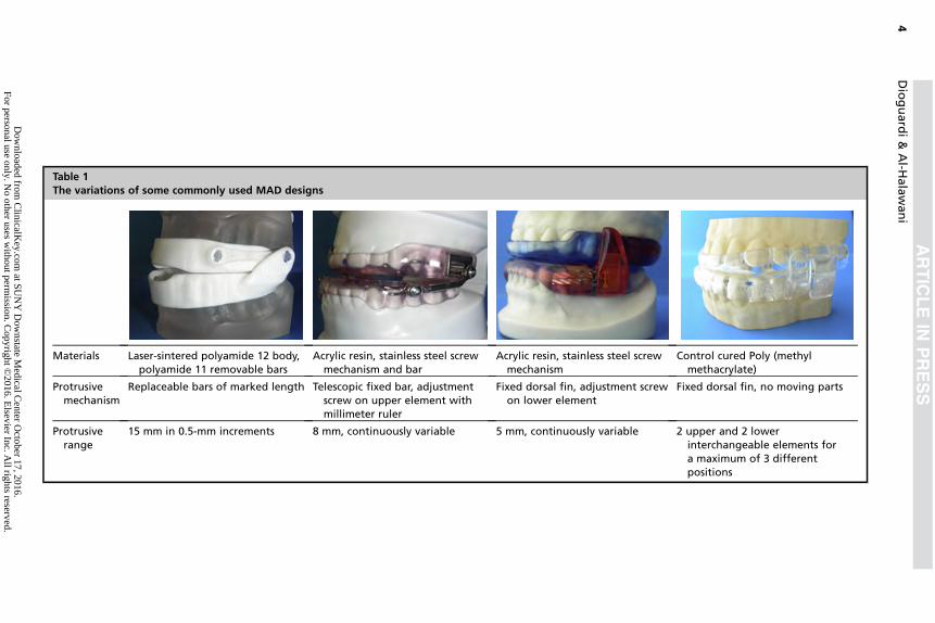
Table 1The variations of some commonly used MAD designs
Materials Laser-sintered polyamide 12 body,polyamide 11 removable bars
Acrylic resin, stainless steel screwmechanism and bar
Acrylic resin, stainless steel screwmechanism
Control cured Poly (methylmethacrylate)
Protrusivemechanism
Replaceable bars of marked length Telescopic fixed bar, adjustmentscrew on upper element withmillimeter ruler
Fixed dorsal fin, adjustment screwon lower element
Fixed dorsal fin, no moving parts
Protrusiverange
15 mm in 0.5-mm increments 8 mm, continuously variable 5 mm, continuously variable 2 upper and 2 lowerinterchangeable elements fora maximum of 3 differentpositions
Dioguardi&
Al-H
alawani
4
Dow
nloaded from C
linicalKey.com
at SUN
Y D
ownstate M
edical Center O
ctober 17, 2016.For personal use only. N
o other uses without perm
ission. Copyright ©
2016. Elsevier Inc. A
ll rights reserved.

Oral Appliances in Obstructive Sleep Apnea 5
Efficacy
Several studies provide evidence for the efficacy of MADs in reducing the overallapnea-hypopnea index [AHI], but with lower effectiveness compared with continuouspositive airway pressure (CPAP). Treatment success across all levels of OSA severityusing MADs is around 50%, with an overall average reduction in baseline AHI of 55%.MADs were also shown to have a positive effect on snoring and daytime symptoms,decreasing excessive daytime sleepiness and improving quality of life comparedwith placebo.8,12,13,15 These results were more evident when the MADs were custommade compared with prefabricated ones.16
In a recent meta-analysis, Sutherland and colleagues17 showed that 37% of pa-tients using MADs achieved an AHI less than 5/h, 52% achieved AHI less than 10/h,and 64% reduced AHI by greater than or equal to 50%. Response rates were lowerin patients with severe OSA; however, 70% of those showed a reduction in AHI greaterthan or equal to 50%, and 23% had complete resolution of OSA.
Compliance
MADs have higher compliance rates than CPAP with a median use of 77% of nightsduring the first year.15,18 A short-term study by Philips and colleagues18 showedthat subjective reports of nightly compliance were less for CPAP compared with MAD.
Side Effects of Mandibular Advancement Devices
MAD use is usually associated with mild and transient side effects that tend to resolvewithin several days or weeks, given that the device has a good fit and is used by thepatient regularly.19
Commonly reported side effects include:
� Temporomandibular joint (TMJ) discomfort or pain� Myofascial pain� Tooth tenderness� Excessive salivation� Gum irritation and bleeding� Dry mouth
Occasionally, side effects negatively affect treatment compliance, but significantand persistent side effects are rare.13,15
Long-term MAD use may lead to dental and skeletal side effects that include:
� Decrease in overjet and overbite� Retroclination of the maxillary incisors� Proclination of the mandibular incisors� Increases in the mandibular plane angle� Increases in the anterior facial height� Decrease in the number of occlusal contact points� Anteroposterior change in occlusion20,21
Morning jaw exercises following MAD use have been shown to:
� Improve compliance� Reduce side effects� Improve quality of life� Reduce sleep symptoms� Alleviate muscle stiffness� Aid in the mandible returning to its normal position22,23
Downloaded from ClinicalKey.com at SUNY Downstate Medical Center October 17, 2016.For personal use only. No other uses without permission. Copyright ©2016. Elsevier Inc. All rights reserved.

Dioguardi & Al-Halawani6
Hybrid Therapy
The use of a hybrid therapy combining nasal CPAP with MAD therapy for patients withOSA has been reported in the literature. Thornton24 reported the first case in 2002 ofcombined MAD and CPAP therapy in a patient with severe OSA who initially could nottolerate treatment with CPAP because of increased pressures and leakage, and laterfailed treatment with MAD because of TMJ symptoms at maximum protrusion. Thecombination therapy was better tolerated by the patient with fewer side effects,because the combination therapy allowed for the use of a lower CPAP pressure andless advancement of the MAD.This treatment strategy leads to reduced CPAP pressure, better fit, less leakage,
and greater compliance.24
Another case report was published by Denbar25; both reports agree that MADs in-crease the upper airway size, decreasing the need for high CPAP pressures to main-tain airway patency with combination therapy, and leading to better tolerance thanwith CPAP or MAD alone.A study by El-Solh and colleagues26 in 2010 included 10 patients who were using
MAD therapy for OSA after they could not tolerate CPAP and still had incompleteresponse to treatment. This study showed that combination therapy was well toleratedby all patients and resulted in a reduction of CPAP pressure and AHI by 29% and 86%respectively from baseline.26
This finding suggests that combination therapy may be effective in patients whocannot tolerate treatment with either CPAP or MAD alone.
Patient Selection
Clinical examination of oral appliance candidatesAn oral examination preceding a referral to a dentist for oral appliance therapy (OAT)should include evaluation of the overall condition of the existing dentition, their sup-porting structures, the TMJ, and health of the soft tissue (Box 1).
Dental Caries and Oral Appliance Therapy
The teeth that support an MAD should be free from active dental caries, periodontallyhealthy, and structurally sound in order to withstand the forces resisting the displace-ment of the arch over the long term.27–29
Evaluation of potential MAD candidates should include a complete intraoral exam-ination that includes visual inspection of the teeth for caries, structural compromise,and their supporting tissues. Advanced dental decay can result in the devitalizationof the pulp chamber, which can in turn lead to pain that is exacerbated by tapping
Box 1
Characteristics of an ideal oral appliance candidate
� No active dental decay or periodontitis.
� A stable dentition with at least 10 well-supported teeth well distributed in each arch.
� A healthy TMJ complex with pain-free and unrestricted protrusive, lateral, and verticalexcursive movements.
� Has been diagnosed with OSA and expresses a desire for a nonsurgical alternative to positiveairway pressure treatment.
� Expresses a desire for a nonsurgical treatment of primary snoring.
Downloaded from ClinicalKey.com at SUNY Downstate Medical Center October 17, 2016.For personal use only. No other uses without permission. Copyright ©2016. Elsevier Inc. All rights reserved.

Oral Appliances in Obstructive Sleep Apnea 7
the affected tooth with a mirror handle or similar instrument. These necrotic chambersoften drain to the buccal and lingual surfaces, resulting in a draining fistula adjacent tothe affected root (Fig. 2).30
The use of a removable oral appliance can lead to increased tooth decay by actingas a new retentive surface for the colonization of Streptococcus mutans and othercariogenic bacteria.31 In addition, any appliance that covers the surface of the teethhas the potential to compromise the caries-protective cleansing effect of saliva, whichcan lead to an increased rate of dental caries.32 Likewise, xerostomia (dry mouth), im-mune suppression, or any other condition that reduces the ability to resist dentaldecay or periodontal inflammation should be addressed before referral for an oralappliance.33,34
Can the Existing Teeth Support a Dental Appliance?
MADs reposition the mandibular arch to a protrusive position, and place a protrusiveforce on the lower teeth and an equal and opposite retrusive force on the upper teeth.In addition, oral appliances that have the upper element connected to the lowerelement by a hinge, bar, or elastic can act to dislodge the appliance when the mouthis opened. Accordingly, the teeth retaining the appliance should be large enough,adequately secured in bone, and possess the physical undercuts necessary to resistthe various forces placed on the appliance.Although a minimum of 10 healthy teeth per arch is traditionally considered the gen-
eral requirement to retain most types of tooth-borne MADs, it must be considered thatnot all teeth provide equal resistance to unwanted tooth movement, also known asanchorage.35 This anchorage is generally related to the size and root area of the teeththat are secured to bone, with the canines and molars providing significantly greateranchorage than the smaller rooted incisors and bicuspids.36
In situations in which natural undercuts are inadequate to retain an MAD, dentistscan alter the contour of the teeth either by enameloplasty or the addition of compositeresin (Fig. 3), creating undercuts that enable satisfactory retention of the appliance. Inaddition, if teeth are not well distributed throughout the arch, the resulting forces willbe disproportionality distributed as well.Anchorage can be compromised by periodontitis or occlusal trauma, which can
lead to the loss of the tooth’s bony support (Fig. 4). The root of a tooth is anchoredto the supporting bone by a periodontal ligament, which is attached to both the tooth
Fig. 2. Dental caries with fistula.
Downloaded from ClinicalKey.com at SUNY Downstate Medical Center October 17, 2016.For personal use only. No other uses without permission. Copyright ©2016. Elsevier Inc. All rights reserved.

Fig. 3. Short teeth and bonded undercuts; enameloplasty shown in pink and addition ofcomposite resin in blue.
Dioguardi & Al-Halawani8
and supporting bone. In many cases, a tooth becomes visibly mobile because of sec-ondary occlusal forces overcoming the ability of the tooth to resist those forces.37 Ex-ceptions to the paradigm that only teeth can retain an MAD are discussed in reports ofMAD designs that secure their retention by means of dental implants, extraoral softtissue, and the edentulous arch.38–40
Periodontitis can often appear as red, swollen gingiva adjacent to the teeth, andreadily bleeds on probing. In other cases, the disease exists without signs of obvioussurface inflammation, with the inflammatory process existing beneath the surface ofthe gingiva in the space adjacent to the tooth, known as the periodontal pocket(Fig. 5). Tooth mobility can easily be observed by applying gentle pressure with amirror handle. Active periodontitis and/or mobility in 1 or more of the teeth can be
Fig. 4. Normal bone level without periodontitis (left). Reduced bone level and anchoragesecondary to periodontitis (right).
Downloaded from ClinicalKey.com at SUNY Downstate Medical Center October 17, 2016.For personal use only. No other uses without permission. Copyright ©2016. Elsevier Inc. All rights reserved.

Fig. 5. Periodontal probe revealing loss of bone mesial of tooth 9. Note draining fistulabuccal to base of the periodontal pocket.
Oral Appliances in Obstructive Sleep Apnea 9
exacerbated by the use of an oral appliance and must be addressed before fabrica-tion of the device is considered. Clearly, patients presenting with a high caries rateand/or active periodontal disease, coupled with a history of only emergency-related dental visits, are better served by alternative therapies for their sleep disor-dered breathing.
Bruxism and Oral Appliance Therapy
The presence of teeth with flattened occlusal surfaces suggests a history of or activetooth grinding or bruxism, a condition that has been associated with sleep apnea.41
The precise nature of this association, or whether or not there is a causal relationshipbetween bruxism and OSA, remains unclear.42 Bruxism (Fig. 6) is commonly evalu-ated during polysomnography by electrodes placed on the masseter muscle, but isnot typically evaluated during type 3 home sleep testing.43,44 Nocturnal bruxismcommonly results in breakage of acrylic oral appliances (Fig. 7) and its presence sug-gests that the device should be fabricated from one of the newer, more durablematerials.45
Temporomandibular Joint Considerations in Oral Appliance Therapy
A healthy TMJ complex that enables the patient to have pain-free and unrestrictedprotrusive, lateral, and vertical movement is a prerequisite for any OAT. It is
Fig. 6. Bruxism; note the characteristic wear pattern on the lower incisors (left); and loss ofvertical dimension resulting from destruction of the incisal portion of the dentition (right).
Downloaded from ClinicalKey.com at SUNY Downstate Medical Center October 17, 2016.For personal use only. No other uses without permission. Copyright ©2016. Elsevier Inc. All rights reserved.

Fig. 7. Appliance failure secondary to nocturnal bruxism.
Dioguardi & Al-Halawani10
unreasonable to expect a patient to wear an MAD if it is uncomfortable. Therefore,careful consideration must be given before attempting fabrication of an MAD for a pa-tient with significant pain or restrictions when entering protrusive, lateral, or verticalmandibular excursions. Clinical evaluation of the TMJ complex should include obser-vation of anything suggestive of disorder during these excursions (clicks, pops, orpain). In addition, the muscles of mastication, including the temporalis, masseter,and internal pterygoid, should be palpated and any tenderness noted.46
Which Patients with Sleep Disorder Should Be Referred for Oral Appliance Therapy?
The July 2015 American Academy of Sleep Medicine (AASM)/AADSM guidelines47
recommend that oral appliances should be offered to adult patients:
� Who request treatment of primary snoring (without sleep apnea)
F
� The guideline recommends that patients who have failed traditional conserva-tive measures such as positional therapy, weight loss, and alcohol avoidancebe offered OAT.
� Because loud snoring is often a warning sign of underlying OSA,48 the diag-nosis of primary snoring should be made by a sleep physician rather than adentist. This notion has recently been challenged by the Texas Board of DentalExaminers, which passed Rule 108.12 referring to sleep studies as a screeningtool that dentists may use to differentiate OSA from primary snoring and left itto the dentists’ discretion to determine which patients require physicianassessment before the initiation of treatment. In response, the Texas MedicalAssociation and the AASM are currently challenging the ruling and requestedthe court to void the rule. This challenge has been supported by the AADSM,which maintains that treatment by a dentist may proceed only after a diagnosisof OSA or snoring has been made by a physician.28
� Are intolerant or prefer an alternative treatment to CPAP therapy� This new guideline no longer differentiates between different levels of OSA(mild, moderate, and severe). This recommendation was based on a meta-analysis of past studies that showed no statistically significant difference inthe mean reduction in AHI before and after treatment in patients using oral ap-pliances versus CPAP across all levels of OSA severity. However, the proba-bility of achieving a target AHI in patients with moderate to severe OSA issignificantly greater with CPAP than OAT.47
Downloaded from ClinicalKey.com at SUNY Downstate Medical Center October 17, 2016.or personal use only. No other uses without permission. Copyright ©2016. Elsevier Inc. All rights reserved.

Oral Appliances in Obstructive Sleep Apnea 11
Treatment Protocol and the Dental Referral
OAT should be undertaken by a qualified dentist only after a referral from a qualifiedphysician trained in sleep medicine who has performed a face-to-face evaluation ofthe patient.49
Dentists who treat patients with sleep disorders require advanced training in dentalsleep medicine, which is not commonly provided in dental school or residency pro-grams. The AASM/AADSM defines a qualified dentist as one who has completed25 hours of continuing education in dental sleep medicine within the past 2 yearsfrom a nonprofit organization, been designated a dental director of an accrediteddental sleep medicine facility by a nonprofit organization, or has certification in dentalsleep medicine from a nonprofit organization.47
The dental evaluation for OAT should include examination of the oral cavityincluding the teeth and their supporting structures, soft tissue, temporomandibularcomplex, and review of a recent complete dental radiographic survey. A medicaland sleep history should be taken and sleep studies reviewed. A thorough reviewof informed consent that includes the benefits, alternatives, potential side effects,and risks of the proposed therapy, along with the risks inherent in not treating the con-dition, should be reviewed in detail and signed by the patient in the presence of awitness.Various options in appliances should be discussed, as should the dentist’s recom-
mendation of the appliance that would be ideally suited for that patient based on theclinical examination and the patient’s preferences. It is highly advantageous for thepatient to have the opportunity to physically hold and examine demonstration modelsof the appliances. Although CPAP has been consistently proved to be more predict-ably effective in treating moderate to severe OSA than OAT,50 it must be remem-bered that the primary reason most patients seek out OAT is because they expectto be able to tolerate it better than they would a CPAP device. It is intuitively obviousthat patients are even more likely to be compliant with a device that they have had apart in choosing.The postinsertion adjustment period typically involves at least several visits over
approximately 3months before referral back to the sleep physician to confirm efficacy.During this time, the protrusive settings are gradually increased to what the dentist de-termines (by both subjective and sometimes objective means) to be the ideal thera-peutic position.The current clinical guidelines stress open communication between dentist and
physician, OAT efficacy confirmation by physician-ordered sleep testing, and short-term and long-term medical and dental follow-up once efficacy has been established.Once the physician has verified that the OAT is effectively treating the patient’s con-dition, the AADSM recommends that patients be seen a minimum of once every6 months for the first 2 years and then on a once-a-year basis.51 During these visits,the dentist can:
� Monitor the physical integrity of the appliance� Reassess the patient’s subjective symptoms, such as snoring and sleepiness asmeasured by the Epworth Sleepiness Scale
� Evaluate potential side effects of the device, such as bite changes (by comparingwith preliminary models, radiographs and photographs), caries, and temporo-mandibular dysfunction
� Refer the patient back to the sleep physician if there are reasons to think that thecurrent treatment is no longer effective
Downloaded from ClinicalKey.com at SUNY Downstate Medical Center October 17, 2016.For personal use only. No other uses without permission. Copyright ©2016. Elsevier Inc. All rights reserved.

Dioguardi & Al-Halawani12
Home Sleep Testing in the Dental Setting
The AADSM Protocol for Oral Appliance Therapy for Sleep Disordered Breathing inAdults: An Update for 2012,52 states that, “After this initial fitting, the dentist mayobtain objective data during an initial trial period using a portable monitor to verifythat the oral appliance effectively improves upper airway patency during sleep byenlarging the upper airway and/or decreasing upper airway collapsibility. If necessary,the dentist makes further adjustments to the device during a final fitting to ensure thatoptimal fit and positioning have been attained.”Type 3 or type 4 home sleep tests (HSTs) are commonly given to patients by qual-
ified dentists seeking to adjust the appliances to achieve the optimal desired effect ofupper airway patency. Various protrusive and vertical positions can be assessed in or-der to adjust the appliance to result in optimal sleep scores. This objective informationbecomes particularly useful in cases in which the patient does not experience subjec-tive symptoms such as snoring or daytime sleepiness. These tests are commonlyscored by computer algorithm, and although not diagnostic, can provide valuablecomparative information with respect to appliance settings.It must be stressed to the patient that dentist-administered HSTs are performed
solely to aid the dentist in the adjustment of the appliance and are not diagnostic,because the diagnosis of medical conditions is not within the ethical or legal scopeof the practice of dentistry. It is the opinion of this author that, before a dentist givesan HST to a patient, the patient must agree in writing that the patient understandsthat the results of the test will not be shared directly with the patient, but may beshared with the patient’s sleep physician.
SUMMARY
OAT use has become an increasingly popular option in the treatment of primary snor-ing and OSA in recent years. Although less consistently effective than CPAP, it re-mains an attractive nonsurgical treatment option because of its high levels ofcompliance, convenience, and stealth. A focused oral examination can help determinewhether or not a patient is a candidate for an MAD. Recent studies have suggestedthat CPAP and OAT used together can provide a higher level of success than eitherused alone.
REFERENCES
1. Robin P. A fall of the base of the tongue considered as a new cause of nasopha-ryngeal respiratory impairment: Pierre Robin sequence, a translation. 1923. PlastReconstr Surg 1994;93(6):1301–3.
2. Cartwright RD, Samelson CF. The effects of a nonsurgical treatment for obstruc-tive sleep apnea. The tongue-retaining device. JAMA 1982;248(6):705–9.
3. Food and Drug Administration. Food and Drug Administration, Department ofHealth and Human Services. Code of Federal Regulations Title 21, Sec.801.109 Prescription devices. 2015. Available at: http://www.accessdata.fda.gov/scripts/cdrh/cfdocs/cfcfr/cfrsearch.cfm?fr5801.109. Accessed March 13,2016.
4. Scherr SC, Dort LC, Almeida FR, et al. Definition of an effective oral appliance forthe treatment of obstructive sleep apnea and snoring: a report of the AmericanAcademy of Dental Sleep Medicine. J Dent Sleep Med 2014;1(1):51.
5. Tsuiki S, Hiyama S, Ono T, et al. Effects of a titratable oral appliance on supineairway size in awake non-apneic individuals. Sleep 2001;24(5):554–60.
Downloaded from ClinicalKey.com at SUNY Downstate Medical Center October 17, 2016.For personal use only. No other uses without permission. Copyright ©2016. Elsevier Inc. All rights reserved.

Oral Appliances in Obstructive Sleep Apnea 13
6. Ryan CF, Love LL, Peat D, et al. Mandibular advancement oral appliance therapyfor obstructive sleep apnoea: effect on awake calibre of the velopharynx. Thorax1999;54(11):972–7.
7. Blanco J, Zamarron C, Abeleira Pazos MT, et al. Prospective evaluation of an oralappliance in the treatment of obstructive sleep apnea syndrome. Sleep Breath2005;9(1):20–5.
8. Marklund M, Stenlund H, Franklin KA. Mandibular advancement devices in 630men and women with obstructive sleep apnea and snoring: tolerability and pre-dictors of treatment success. Chest 2004;125(4):1270–8.
9. Ng AT, Darendeliler MA, Petocz P, et al. Cephalometry and prediction of oralappliance treatment outcome. Sleep Breath 2012;16(1):47–58.
10. Ferguson KA, Ono T, Lowe AA, et al. A short-term controlled trial of an adjustableoral appliance for the treatment of mild to moderate obstructive sleep apnoea.Thorax 1997;52(4):362–8.
11. Remmers J, Charkhandeh S, Grosse J, et al. Remotely controlled mandibular pro-trusion during sleep predicts therapeutic success with oral appliances in patientswith obstructive sleep apnea. Sleep 2013;36(10):1517–25, 1525A.
12. Ahrens A, McGrath C, Hagg U. Subjective efficacy of oral appliance design fea-tures in the management of obstructive sleep apnea: a systematic review. Am JOrthod Dentofacial Orthop 2010;138(5):559–76.
13. Marklund M, Verbraecken J, Randerath W. Non-CPAP therapies in obstructivesleep apnoea: mandibular advancement device therapy. Eur Respir J 2012;39(5):1241–7.
14. Deane SA, Cistulli PA, Ng AT, et al. Comparison of mandibular advancementsplint and tongue stabilizing device in obstructive sleep apnea: a randomizedcontrolled trial. Sleep 2009;32(5):648–53.
15. Ferguson KA, Cartwright R, Rogers R, et al. Oral appliances for snoring andobstructive sleep apnea: a review. Sleep 2006;29(2):244–62.
16. Serra-Torres S, Bellot-Arcis C, Montiel-Company JM, et al. Effectiveness ofmandibular advancement appliances in treating obstructive sleep apnea syn-drome: a systematic review. Laryngoscope 2016;126(2):507–14.
17. Sutherland K, Takaya H, Qian J, et al. Oral appliance treatment response and pol-ysomnographic phenotypes of obstructive sleep apnea. J Clin Sleep Med 2015;11(8):861–8.
18. Phillips CL, Grunstein RR, Darendeliler MA, et al. Health outcomes of continuouspositive airway pressure versus oral appliance treatment for obstructive sleep ap-nea: a randomized controlled trial. Am J Respir Crit Care Med 2013;187(8):879–87.
19. Aarab G, Lobbezoo F, Hamburger HL, et al. Effects of an oral appliance withdifferent mandibular protrusion positions at a constant vertical dimension onobstructive sleep apnea. Clin Oral Investig 2010;14(3):339–45.
20. Doff MH, Finnema KJ, Hoekema A, et al. Long-term oral appliance therapy inobstructive sleep apnea syndrome: a controlled study on dental side effects.Clin Oral Investig 2013;17(2):475–82.
21. Wang X, Gong X, Yu Z, et al. Follow-up study of dental and skeletal changes inpatients with obstructive sleep apnea and hypopnea syndrome with long-termtreatment with the Silensor appliance. Am J Orthod Dentofacial Orthop 2015;147(5):559–65.
22. Cunali PA, Almeida FR, Santos CD, et al. Mandibular exercises improve mandib-ular advancement device therapy for obstructive sleep apnea. Sleep Breath2011;15(4):717–27.
Downloaded from ClinicalKey.com at SUNY Downstate Medical Center October 17, 2016.For personal use only. No other uses without permission. Copyright ©2016. Elsevier Inc. All rights reserved.

Dioguardi & Al-Halawani14
23. Ueda H, Almeida FR, Chen H, et al. Effect of 2 jaw exercises on occlusal functionin patients with obstructive sleep apnea during oral appliance therapy: a random-ized controlled trial. Am J Orthod Dentofacial Orthop 2009;135(4):430.e1-7 [dis-cussion: 430–1].
24. Thornton WK. Combined CPAP-oral appliance therapy: a case report. Sleep Re-view 2002. Available at: http://www.sleepreviewmag.com/2002/01/news-story-29/. Accessed August 6, 2016.
25. Denbar MA. A case study involving the combination treatment of an oral appli-ance and auto-titrating CPAP unit. Sleep Breath 2002;6(3):125–8.
26. El-Solh AA, Moitheennazima B, Akinnusi ME, et al. Combined oral appliance andpositive airway pressure therapy for obstructive sleep apnea: a pilot study. SleepBreath 2011;15(2):203–8.
27. Zlataric DK, Celebic A, Valentic-Peruzovic M. The effect of removable partial den-tures on periodontal health of abutment and non-abutment teeth. J Periodontol2002;73(2):137–44.
28. American Academy of Dental Sleep Medicine. President’s message: dentistry’srole in treating snoring and sleep apnea. 2014. Available at: http://www.aadsm.org/articles.aspx?id55224. Accessed March 13, 2016.
29. Petit FX, Pepin JL, Bettega G, et al. Mandibular advancement devices: rate ofcontraindications in 100 consecutive obstructive sleep apnea patients. Am J Re-spir Crit Care Med 2002;166(3):274–8.
30. Johnson BR, Remeikis NA, Van Cura JE. Diagnosis and treatment of cutaneousfacial sinus tracts of dental origin. J Am Dent Assoc 1999;130(6):832–6.
31. Batoni G, Pardini M, Giannotti A, et al. Effect of removable orthodontic applianceson oral colonisation by mutans streptococci in children. Eur J Oral Sci 2001;109(6):388–92.
32. Dowd FJ. Saliva and dental caries. Dent Clin North Am 1999;43(4):579–97.33. Dreizen S, Brown LR, Daly TE, et al. Prevention of xerostomia-related dental
caries in irradiated cancer patients. J Dent Res 1977;56(2):99–104.34. Stanford TW, Rees TD. Acquired immune suppression and other risk factors/indi-
cators for periodontal disease progression. Periodontol 2000 2003;32:118–35.35. Kushida CA, Morgenthaler TI, Littner MR, et al. Practice parameters for the treat-
ment of snoring and obstructive sleep apnea with oral appliances: an update for2005. Sleep 2006;29(2):240–3.
36. Roberts-Harry D, Sandy J. Orthodontics. Part 9: anchorage control and distalmovement. Br Dent J 2004;196(5):255–63.
37. Williams RC. Periodontal disease. N Engl J Med 1990;322(6):373–82.38. Tripathi A, Gupta A, Tripathi S, et al. A novel use of complete denture prosthesis
as mandibular advancement device in the treatment of obstructive sleep apneain edentulous subjects. J Dent Sleep Med 2014;1(3):115–9.
39. Guimaraes TM, Colen S, Cunali PA, et al. Treatment of obstructive sleep apneawith mandibular advancement appliance over prostheses: a case report. SleepSci 2015;8(2):103–6.
40. Hoekema A, de Vries F, Heydenrijk K, et al. Implant-retained oral appliances: anovel treatment for edentulous patients with obstructive sleep apnea-hypopneasyndrome. Clin Oral Implants Res 2007;18(3):383–7.
41. Knight DJ, Leroux BG, Zhu C, et al. A longitudinal study of tooth wear in orthodon-tically treated patients. Am J Orthod Dentofacial Orthop 1997;112(2):194–202.
42. Balasubramaniam R, Klasser GD, Cistulli PA, et al. The link between sleepbruxism, sleep disordered breathing and temporomandibular disorders: anevidence-based review. J Dent Sleep Med 2014;1(1):27–37.
Downloaded from ClinicalKey.com at SUNY Downstate Medical Center October 17, 2016.For personal use only. No other uses without permission. Copyright ©2016. Elsevier Inc. All rights reserved.

Oral Appliances in Obstructive Sleep Apnea 15
43. Saito M, Yamaguchi T, Mikami S, et al. Temporal association between sleep ap-nea–hypopnea and sleep bruxism events. J Sleep Res 2014;23:196–203. http://dx.doi.org/10.1111/jsr.12099.
44. Collop NA, Anderson WM, Boehlecke B, et al. Clinical guidelines for the use ofunattended portable monitors in the diagnosis of obstructive sleep apnea in adultpatients. Portable Monitoring Task Force of the American Academy of Sleep Med-icine. J Clin Sleep Med 2007;3(7):737–47.
45. Landry-Schonbeck A, de Grandmont P, Rompre PH, et al. Effect of an adjustablemandibular advancement appliance on sleep bruxism: a crossover sleep labora-tory study. Int J Prosthodont 2009;22(3):251–9.
46. Meyer RA. The temporomandibular joint examination. In: Walker HK, Hall WD,Hurst JW, editors. Clinical methods: the history, physical, and laboratory examina-tions. 3rd edition. Boston: Butterworths; 1990. p. 763–4. Available at: http://www.ncbi.nlm.nih.gov/books/NBK271/
47. Ramar K, Dort LC, Katz SG, et al. Clinical practice guideline for the treatment ofobstructive sleep apnea and snoring with oral appliance therapy: an update for2015. J Clin Sleep Med 2015;11(7):773–827.
48. Maimon N, Hanly PJ. Does snoring intensity correlate with the severity of obstruc-tive sleep apnea? J Clin Sleep Med 2010;6(5):475–8.
49. American Academy of Sleep Medicine/American Academy of Dental SleepMedicine. Policy statement on the diagnosis and treatment of obstructive sleepapnea. 2012. Available at: http://www.aasmnet.org/resources/pdf/AADSMJointOSApolicy.pdf. Accessed March 13, 2016.
50. Hoffstein V. Review of oral appliances for treatment of sleep-disordered breath-ing. Sleep Breath 2007;11(1):1–22.
51. American Academy of Dental Sleep Medicine. AADSM treatment protocol: oralappliance therapy for sleep disordered breathing: an update for 2013. 2013.Available at: http://www.aadsm.org/treatmentprotocol.aspx. Accessed March14, 2016.
52. American Academy of Dental Sleep Medicine. AADSM protocol for oral appliancetherapy for sleep disordered breathing in adults: an update for 2012. 2012.Available at: http://www.aadsm.org/PDFs/TreatmentProtocolOAT.pdf. AccessedMarch 14, 2016.
Downloaded from ClinicalKey.com at SUNY Downstate Medical Center October 17, 2016.For personal use only. No other uses without permission. Copyright ©2016. Elsevier Inc. All rights reserved.


For Immediate Release
Non-Patient-Based Dental Restorative Exam Option Using New Manikin Tooth Technology Approved by ADEX (Linthicum Heights, MD| April 3, 2020) The American Board of Dental Examiners (ADEX) will allow candidates for dental licensure to choose a non-patient-based restorative examination option to demonstrate readiness for practice. This format utilizes the CompeDont™ DTX, a new manikin tooth technology developed by the CDCA and Acadental, Inc. The CompeDont™ DTX is a first of its kind manufactured tooth that presents a high-fidelity opportunity for licensure candidates to diagnose and treat Class II and Class III caries. The CompeDont™ is patent pending with product development initiated by the CDCA in 2017 and was first made public last fall as part of pilot testing at 6 dental schools; University of Mississippi Medical Center School of Dentistry, University of Buffalo (SUNY), Indiana University School of Dentistry, University of Detroit Mercy School of Dentistry, Midwestern University College of Dental Medicine – Illinois, and Midwestern University College of Dental Medicine – Arizona. These schools hosted high stakes pilot examinations using the Class III lesions between September and December 2019. When compared to that of ADEX patient-based exams, independent psychometrician analyzed pilot data showed the simulated tooth identified the same critical deficiencies in skill typically revealed by the treatment of natural teeth. “Candidates encountered realistic and variable caries unlike other simulated teeth currently available,” says Dr. Guy Shampaine, CompeDont™ Development Team Leader. CDCA Director of Examinations Dr. Ellis Hall says it is the CompeDont™’s ability to accurately represent infected, affected and sclerotic dentin that is unique. “Both examiners and students reported that the tooth mimics decay, stickiness and tug-back and can be restored as if it were a natural tooth in this way.” The dental board representatives to ADEX voted 29-1 to approve the use of the CompeDont™ for both Class II and Class III restorative procedures at a special, virtual meeting on April 2, 2020. The CompeDont™ will be permitted in place of the patient portion of the restorative examinations in the same framework and processes of the existing psychometrically valid examination as soon as possible. The acceptance of a non-patient-based examination for licensure falls to each jurisdiction. “The CompeDont™’s new technology provides an option to many state dental boards seeking to address public health concerns in the face of the COVID-19 outbreak without reducing important existing licensure standards,” says Dr. Harvey Weingarten, CDCA Chair. Based on this immediate ADEX approval, the new non-patient-based administration option will be incorporated into CDCA licensure exams for the Class of 2020 candidates. “We recognize each state will determine independently whether

they will accept this new non-patient option for restorative procedures. To help address these differences, the ADEX licensure examination reporting system will clearly identify if a Class II or Class III restoration are manikin or patient-based,” reports Dr. Bill Pappas, ADEX President. ADEX dental licensure is a five-part examination. It includes a computerized written Objective Structured Clinical Examination (OSCE) measuring clinical judgments and treatment planning decision making, and four clinical portions including an anterior restorative, a posterior restorative, endodontic and prosthodontic sections. An optional periodontal scaling examination is also available. Since 2015, two administrative pathways have existed using the identical ADEX content and criteria, traditional patient-based and Patient-Centered Curriculum Integrated Format (PC-CIF, available at participating schools). The CDCA partnered with Acadental, Inc. for the development and production of the CompeDont™ DTX. The tooth will be made available for all ADEX examinations but will remain the intellectual, protected property of the CDCA. Based in the Kansas City metropolitan area, Acadental, Inc. currently holds at least 5 patents and distributes dental educational products worldwide. The Commission on Dental Competency Assessments, founded in 1969, is the largest nonprofit, third-party administrator of dental and dental hygiene assessments in the US. For more information about the CDCA CompeDont™, contact Stephanie Beeler, Multimedia, Communications, and Strategic Projects Leader at [email protected].


The Southern Regional Testing Agency (SRTA) is a candidate-focused, innovative testing agency committed to bringing the most modern and innovative thoughts and practices in dentistry to dentists and dental hygienists seeking licensure assessments.
SRTA Mission and Goal

The Southern Regional Testing Agency has a history of being at the forefront of current dental research, offering the most up-to-date content and setting for its clinical skills examinations.
SRTA Experience

SRTA is user friendly and innovative
SRTA is committed to bringing the most up-to-date clinical skills assessment, and to properly preparing them to excel in the dental profession.
Goal for Candidates

SRTA tests are valid with a commitment Quality Assurance
SRTA ensures State Boards the highest level of clinical skills competence in the certification of dental professionals.
Goal for Members and State Boards


Dental Examination
• Endodontics
• Fixed Prosthodontics
• Anterior Prep & Restoration
• Posterior Prep & Restoration
• Optional periodontal section
• Pass/Fail
SRTA Process and Testing Manual

Dental Examination Continued:
• Endodontic Procedure*: • Anterior endodontic procedure including access opening,
instrumentation and obturation, and
• Posterior endodontic procedure includes access opening and canal location, also
• Fixed Prosthodontic Procedure*:• An all-ceramic anterior crown preparation, and
• Three-unit fixed bridge, which includes a zirconium/gold posterior crown preparation and a porcelain fused to metal crown preparation.
SRTA Process and Testing Manual
*Please reference candidate manual for specific details on criteria tested

Dental Examination Continued:
•Restorative sections* consists of: • Anterior Restorative Class III: composite preparation
and restoration; • Posterior Restorative Class II: composite or amalgam
preparation and restoration; and • Periodontal Scaling Treatment (Optional):
demonstrating calculus detection and removal.
SRTA Process and Testing Manual
*Please reference candidate manual for specific details on criteria tested

Dental Hygiene Examination*
• Candidates screen typodont for 12 surfaces of heavy to moderate calculus
• Periodontal Pocket Depth Detection
• Calculus Detection
• Calculus Removal
• Intra/Extra Oral Exam (optional)
SRTA Process and Testing Manual
Copyright 2019 Southern Regional Testing Agency, Inc.
All rights reserved. No Part of this manual may be used or reproduced in any form without prior express written permission of Southern Regional Testing Agency, Inc.
ADDRESS
4698 HONEYGROVE RD, SUITE 2
VIRGINIA BEACH, VA 23455
PHONE & FAX
P: 757 318 9082
F: 757 318 9085
EMAILS
APPLICATION QUESTIONS?
DENTAL HYGIENE QUESTIONS?
SOUTHERN REGIONAL TESTING AGENCY, INC.
LICENSING EXAMINATION 2020 DENTAL HYGIENE
CANDIDATE MANUAL
*Please reference candidate manual for specific details on criteria tested

Dental Hygiene Examination Continued:
• Candidates will screen typodont for 12 surfaces of heavy to moderate calculus
• 2 teeth to measure Periodontal Pocket Depth Detection
• 3 teeth for Calculus Detection
• Calculus Removal for case selection (1 quadrant and any additional teeth)
• Optional Intra/Extra Oral Exam has 50 multiple choice questions testing areas such as disease of the oral cavity and healthy tissue treatment management
SRTA Process and Testing Manual

• Current and past SRTA state board members
• Educators from the universities where the SRTA exams are held, although they cannot examine within the same state they teach.
• Exchange examiners: examiners who currently examine for other testing agencies.
SRTA Examiners

• SRTA is experienced and innovative.
• SRTA is committed to its clinical skills assessment and its passage by our candidates.
• SRTA is able to adapt quickly and appropriately to modern clinical dental practice.
• SRTA is committed to the cutting edge of dental practice and examination technology.
SRTA Is….

SRTA Board and Members

SRTA Promise and Commitment
• Focusing on a positive, collaborative environment, SRTA welcomes evidence-based research to ensure that our candidates are well-prepared for the evolving practice of dentistry.

Addendum Slides

Candidate Qualification
• Graduate of an American or Canadian Dental College accredited by the American Dental Association Commission on Dental Accreditation
• International Candidates considered for the “state only” status

Psychometrically Valid Scoring
• Item analyses and calibration
• Psychometric equating
• Multiple-examiner review
• Analytic scoring

Candidate Anonymity
Anonymity is preserved between the scoring examiners and candidates.

ADDRESS
4698 HONEYGROVE RD, SUITE 2
VIRGINIA BEACH, VA 23455
PHONE & FAX
P: 757 318 9082
F: 757 318 9085
EMAILS
APPLICATION QUESTIONS?
DENTAL QUESTIONS?
SOUTHERN REGIONAL TESTING AGENCY, INC.
EXAMINATION 2020 DENTAL LICENSING
CANDIDATE MANUAL NON-PATIENT-BASED
RESTORATIVE, PERIODONTAL, ENDODONTIC,
PROSTHODONTICS

Copyright 2019 Southern Regional Testing Agency, Inc.
All rights reserved. No Part of this manual may be used or reproduced in any form without prior express written permission of Southern Regional Testing Agency, Inc.
SOUTHERN REGIONAL TESTING AGENCY, INC. Southern Regional Testing Agency, Inc. (SRTA) is a nonprofit corporation committed to being a
leader at the national level in examination development and administration by providing the
following –
• Uniformly administered examinations and confidential results that are consistently
reliable for use by the dental licensing boards or other agencies
• Protection for the public
• Appropriate care in the examination process
• Providing the most technologically advanced examination for its member states and
participating examination sites
• Providing valid examinations in the most candidate focused environment possible, for
the next generation of our colleagues in the Dental and Dental Hygiene Professions
MISSION STATEMENT SRTA will continue to provide valid, reliable, legally defensible examinations and results while
striving to implement new testing methodologies in a candidate focused environment for the
next generation of dental and dental hygiene professionals.
EXAMINATION PURPOSE This year’s SRTA Dental Examination has been developed, administered and reviewed in
accordance with guidelines from the American Dental Association (ADA), the American
Association of Dental Boards (AADB), the American Psychological Association (APA), the
American Educational Research Association and the National Council on Measurement in
Education. SRTA collects input from practicing dentists nationwide every five years through a
Task Analysis Survey, which is the basis for all decisions regarding content. The SRTA
Examination was developed to provide a reliable clinical assessment for use by state boards in
making valid licensing decisions. Prior to registering for the examination, candidates are
strongly encouraged to verify the examination is accepted in the state in which they seek
immediate licensure. After actively practicing two to five years, many states will accept
licensure by criteria (or reciprocity). Again, candidates should check with state boards on
licensure requirements.
ANONYMITY The SRTA Dental Examination is conducted anonymously. All examination materials are
identified by the candidate’s SRTA number. The candidate’s name and school information
should not appear on any testing materials. All examiners are vetted current and past State
Dental Board members with diverse backgrounds. We also utilize faculty examiners, although
they cannot examine in their respective state, the knowledge they gain through their
experience is imparted to the students. Examiners are trained and standardized prior to each
examination and are evaluated to ensure they are grading to established criteria. The
examiners are separated from the candidates and will remain in a separate area of the clinic.
Candidates must observe all signs and follow instructions so as not to breach anonymity.
Anonymity is preserved between the scoring examiners and the candidates, but not among
the examiners themselves. Examiners may consult with the SRTA Clinic Floor Coordinator (CFC)
or Scoring Area Coordinator (SAC) whenever necessary.
TABLE OF CONTENTS
SOUTHERN REGIONAL TESTING AGENCY, INC. ................................................................................. 2 MISSION STATEMENT ............................................................................................................................ 2 EXAMINATION PURPOSE ..................................................................................................................... 2 ANONYMITY ......................................................................................................................................... 2
TABLE OF CONTENTS ................................................................................................................................... 1
I. EXAMINATION OVERVIEW .................................................................................................................. 1
EXAMINATION SECTIONS & FORMATS ............................................................................................... 1 SCORING PROCESS ............................................................................................................................ 1
II. POLICIES & PROCEDURES ................................................................................................................... 3
PROFESSIONAL STANDARDS & COMPETENCY ................................................................................. 3 CANDIDATE ACCESSIBILITY ................................................................................................................ 3 CANDIDATE ELIGIBILITY ....................................................................................................................... 3 CANDIDATE INELIGIBILITY ................................................................................................................... 4 CANDIDATE RECOURSE – APPEALS PROCESS .................................................................................. 4 CHAIRSIDE ASSISTANT ......................................................................................................................... 4 UNETHICAL CONDUCT........................................................................................................................ 4 OTHER DISMISSAL REASONS ............................................................................................................... 4 RESTORATIVE MANIKIN-BASED SUBMISSIONS .................................................................................... 5 REFUNDS .............................................................................................................................................. 6 REMEDIATION ...................................................................................................................................... 6 RE-EXAMINATION REQUIREMENTS ..................................................................................................... 6 SUPPLIES PROVIDED BY EXAMINATION SITE ....................................................................................... 7 STATE BOARD OF DENTISTRY & LICENSURE INFORMATION .............................................................. 7 SCORING CERTIFICATION .................................................................................................................. 8 QUESTIONS........................................................................................................................................... 8
III. MANIKIN-BASED EXAMINATION ........................................................................................................ 9
ENDODONTICS SECTION .................................................................................................................. 10 FIXED PROSTHODONTIC SECTION .................................................................................................... 19 RESTORATIVE SECTION ...................................................................................................................... 33
IV. PERIODONTAL EXAM (OPTIONAL) ................................................................................................... 55
SELECTION & CHECK-IN .................................................................................................................... 55 INSTRUMENTS ..................................................................................................................................... 55 TREATMENT SELECTION ..................................................................................................................... 56 PROCEDURE & GUIDELINES .............................................................................................................. 56 PERIODONTAL CRITERIA ................................................................................................................... 59
V. EXAMINATION SCHEDULE ................................................................................................................ 61
TRADITIONAL EXAMINATION SCHEDULE ......................................................................................... 61 SECTIONAL EXAMINATION SCHEDULE............................................................................................. 63 CHECKLIST ......................................................................................................................................... 64
VI. EXAMINATION FORMS ...................................................................................................................... 65
PERIODONTAL TREATMENT SELECTION WORKSHEET ...................................................................... 65 PROGRESS FORMS ............................................................................................................................ 66 MODIFICATION REQUEST FORM ...................................................................................................... 67 INSTRUCTION TO CANDIDATE FORM (ITC) ...................................................................................... 68

VII. FAQS & HELPFUL HINTS ...................................................................................................................... 69
FAQS ................................................................................................................................................... 69 HELPFUL HINTS .................................................................................................................................... 71
VIII. GLOSSARY OF WORDS, TERMS & PHRASES ...................................................................................... 72
IX. CRITERIA REFERENCE CHART ............................................................................................................ 77

1
I. EXAMINATION OVERVIEW
EXAMINATION SECTIONS & FORMATS The Southern Regional Testing Agency (SRTA) Non-Patient Dental Examination consists of four
required manikin-based sections – Endodontics, Fixed Prosthodontics, Anterior Restorative, and
Posterior Restorative. SRTA also offers an optional manikin-based Periodontal section. Sections
will be broken down as follows –
A. Candidates will have one 7-hour day to complete both the manikin Endodontic and
Fixed Prosthodontic sections of the examination. If only one of these sections need to
be taken, then Candidates will get 3 hours to complete the Endodontics section or 4
hours to complete the Fixed Prosthodontic section.
B. Candidate will have one 7-hour day to complete the manikin Anterior Restorative and
manikin Posterior Restorative sections of the examination. If a candidate would like to
take the optional manikin-based Periodontal section along with his/her manikin
Restorative sections, then he/she will have a total of 9-hours to complete all three
sections.
The SRTA Non-Patient Dental Examination is available as a Traditional Examination. A
Traditional examination is where the manikin Endodontic/Fixed Prosthodontic sections and
manikin Restorative section with the optional Periodontal manikin-based section will be
administered in its entirety over the course of two consecutive days
SCORING PROCESS For SRTA’s Clinical Dental Skills Examinations, candidates are required to demonstrate job-
related skills in simulated settings (manikin-based) and an optional manikin-based Periodontal
section. To score these complex performance tasks, SRTA has developed scoring criteria for
each of these examinations to define important characteristics of minimally competent
performance. Using these scoring criteria, SRTA then trains and calibrates examiners to
independently evaluate candidates’ performance on the range of tasks and related
procedures that comprise each examination. All examiners are currently licensed dentists who
are current or past members of a state board of dentistry and who are familiar with the
content of the examination, the expectations of minimally competent performance, and the
characteristics of the target population of candidates.
To ensure that examiners interpret the scoring criteria consistently, SRTA relies on an industry-
standard practice of having two or more examiners independently review a candidate’s
work. Examiners use analytic scoring methods where candidate performances are defined as
a series of criteria that will influence the acceptability of the characteristics of the product that
the candidate produces. Each examiner specifies any observed major errors (i.e., domain
critical errors) using electronic data entry. For an error to influence a candidate's score, it must
be independently confirmed by at least one other examiner. This process helps to ensure that
any decision about the pass/fail status for a candidate is based on the independent
evaluations of at least two of three examiners.
Because of the efforts to train and calibrate examiners, decisions about errors will generally be
made based on the judgment of the first two examiners. However, as a measure of internal

2
quality control and in instances where there is a disagreement about whether the
performance constituted an overall pass or fail decision, a third examiner will also make
independent judgments about the candidate's performance. Because the third examiner will
not know whether his/her judgments are part of internal data collection for feedback or as an
adjudication judgment, there is no reason for an examiner to think that his/her judgment
carries more weight than any other examiner in the process.
SRTA uses analytic judgment (i.e., judgment based on a series of multiple steps and evaluation
points rather than on an overall impression) rather than holistic judgment for three purposes.
First, because a task comprises a number of skills, analytic judgment allows the examiner to
separately evaluate the different phases of process and product that occur for a given task.
Because each examination is unique, a slightly different number of skills have been defined
and are scored in each section. Second, analytic judgment enables some limited feedback
to the candidate about areas of strength or weakness and how these factors contributed to
the overall pass/fail decision. Third, analytic judgment requires that examiners justify their
ratings, given the specificity required for the judgments.
Overall, pass/fail decisions are conjunctive across examinations. This means that for a
candidate to successfully pass the examination as a whole, he/she must pass each individual
examination. This policy decision is based on empirical evidence suggesting that skills from one
section of the examination are not sufficiently related to skills in another section such that
someone would be able to compensate in practice. This decision also reflects the desire to be
able to use examination results to make a decision about a candidate’s minimum
competency within each of the important sub-domains of the dental profession. A score of 75
and above is reported as a passing grade.

3
II. POLICIES & PROCEDURES
PROFESSIONAL STANDARDS & COMPETENCY The purpose of this examination is to assess professional competency. The candidate is
expected to maintain professional standards in the following areas –
• Suitable operating attire, inclusive of the full barrier technique.
• Candidates must follow OSHA and CDC Guidelines
• Cooperation with examiners, examination site personnel and other candidates.
• Aseptic techniques and general cleanliness of the operatory during all procedures.
• Candidates must maintain proper infection control throughout the entire examination.
• Protection of and concern for tooth structure and supporting tissue during treatment.
Violation of these standards and guidelines is ground for immediate dismissal (failure) from the
examination, and the candidate may be denied reexamination for 12 months.
CANDIDATE ACCESSIBILITY Any candidate with a documented physical and/or learning disability that impairs sensory,
manual or speaking skills and that requires a reasonable deviation from the normal
administration of the examination may be accommodated. A written statement from a
qualified physician must be provided at the time of application. The limitation(s) must be
clearly defined, and the assistance required to ensure appropriate accommodations must be
detailed. Requests will be evaluated on a case-by-case basis. Accommodations/deviations
will not be allowed for components and skills the examination must measure.
Information received regarding the physical/learning challenges of a candidate will remain
confidential except in the case of disabilities that may require emergency treatment. In this
case, onsite safety personnel will be advised.
CANDIDATE ELIGIBILITY Candidates for the examination must be graduates of an American or Canadian dental
college accredited by the American Dental Association Commission on Dental Accreditation.
A candidate who has not formally graduated from his/her university is required to secure
certification from the dean of his/her program stating that:
1. The candidate is eligible and qualifies for the DDS or DMD degree requirements.
2. The candidate will complete the DDS or DMD degree requirements within 36 months of
the examination date.
This certification must be in the form of a letter from the dean submitted with the application
or provided to SRTA by the dean prior to the receipt of the candidate’s application.
Candidates who graduated from a school outside of the United States and Canada may
apply and be considered for the “state only” status, pending receipt of the appropriate state
authorization. The candidate must furnish a letter from the State Board of Dentistry that
accepts the results of this examination. This letter should indicate that the candidate is eligible
4
for licensure in that state only, upon successful completion of the examination. In addition, a
copy of the candidate’s diploma with an English translation must be provided.
CANDIDATE INELIGIBILITY If a candidate becomes ineligible to take the examination, they must notify the SRTA office, in
writing, two weeks prior to the scheduled examination. A letter from the dean of the
candidate’s institution will be required as proof of ineligibility. SRTA will retain the complete
application fee for any candidate declared ineligible by his/her dean. Candidates declared
ineligible will be allowed to examine at a future site within a 12-month period upon payment
of facility fees and a $200 administrative processing fee. A diploma or letter from the dean
stating the candidate’s eligibility is required for a rescheduled exam.
CANDIDATE RECOURSE – APPEALS PROCESS Refer to www.srta.org for information regarding the appeals process.
CHAIRSIDE ASSISTANT Chairside assistants are not permitted for the manikin-based examination.
UNETHICAL CONDUCT Professional behavior is a critical quality in the practice of dentistry. If a candidate is
suspected of unethical conduct as defined by SRTA guidelines, they will fail the examination.
Examples of unethical conduct include, but are not limited to:
• Using unauthorized equipment at any time during the exam
• Using assistants
• Altering teeth used in the manikin procedures
• Engaging in dishonesty
• Altering candidate progress forms
• Any other behavior that compromises the standards of professional behavior
When SRTA charges a candidate with unethical conduct, it is SRTA’s policy to notify all
participating state boards of the situation. Many state statutes have criteria that include
“good moral character” as a requirement for licensure. If a state board finds a candidate
guilty of the alleged unethical conduct, the candidate may be ineligible for licensure in that
state at any time in the future. While SRTA allows candidates to retake the SRTA Examination,
they may be unable to obtain licensure in any participating state. Candidates are
encouraged to address these matters with the state in which they desire licensure prior to
retaking the examination.
OTHER DISMISSAL REASONS This list is not all-inclusive. Listed below are the reasons for which a candidate may receive a
failing evaluation or dismissal. Some procedures may be deemed unsatisfactory for other
reasons. Additionally, a combination of several unsatisfactory evaluations may result in failure.
Reexamination will be denied for one year (12 months) from the date of dismissal from the
examination. Infractions that may lead to dismissal or failure include –

5
• Lack of protection and concern for tooth structure and supporting tissue during manikin
treatment.
• Lack of professional judgment.
• Evidence of dishonesty or misrepresentation during the application process, including
false or misleading statements or false documentation presented by the candidate or
on the candidate’s behalf.
• Evidence of dishonesty or misrepresentation during candidate registration or during the
examination.
• Rude, abusive, or uncooperative behavior exhibited by the candidate.
• Continuing to work after published cutoff time.
• Working on a manikin model in a manner that does not simulate actual patient
conditions.
• Working on Fixed Prosthodontics, Endodontics sextants, Restorative sextants or
Periodontal sextants not provided by SRTA. Any evidence of tampering with or
attempting to remove the screws from the sextants will result in failure of the entire
examination and will be grounds for dismissal from the exam.
• Failure to complete the examination within the allotted time (No make-up time, grace
period or second effort will be allowed for any part of this examination.)
• Receiving assistance from a dentist, another candidate, university representative(s),
etc., during the course of the examination.
• Preparing a tooth other than the one approved/assigned by the examiners. This is
considered major hard tissue damage.
• Thievery during the examination
• Performance of any unauthorized work outside of the examination site designated
areas.
• Noncompliance with anonymity requirements for approval and/or examiner scoring.
Candidates are not allowed in or near the area designated for scoring.
• Noncompliance with established guidelines for asepsis and infectious disease control
• Use of cellular telephones, pagers, cameras, or other electronic equipment by the
candidate while in the clinic or scoring areas
RESTORATIVE MANIKIN-BASED SUBMISSIONS If the candidate is scheduled to perform the Restorative Section as the first procedure of the
day, they may begin setting up as soon as the clinic opens at 6:00AM. Between 7:00AM and
8:00AM candidates will obtain their typodonts from a SRTA Dental Administrator (DA) or Clinic
Floor Coordinator (CFC) and secure it to the manikin head. A Clinic Floor Coordinator (CFC)
will need to be called over to document that the typodont is secure and provide a start
check. Restorative treatment begins at 8:00AM. Candidates will complete the preparation for
a Class III Composite restoration for tooth #9 and a Class II Amalgam or Conventional
Composite restoration for tooth #14. Candidates will begin with the Anterior procedure. Once
the Anterior procedure is complete, the candidate will remove the upper arch from the
typodont, place it in the poly bag or typodont box provided with his/her candidate number
label on it and take it to the Evaluation area for preparation grading. Once grading is
complete, the upper arch will be returned to the candidate. The candidate will then secure
the arch in the typodont and complete the finish section of the Anterior Restorative Exam.
When the finished Anterior restoration is ready for grading, the candidate will again remove
the tray/upper arch for final evaluation. The Posterior procedure will be the second procedure
of the examination. With the preparation check, again the tray/upper arch will be removed
and submitted for the prep check. Upon completing the posterior restoration, the candidate

6
will remove the entire typodont for final evaluation. The typodont will not be returned after
final evaluation of the last restorative procedure has been completed. Only one restorative
procedure can be done at a time. Both the Anterior and Posterior portions must be finished in
the allotted time.
REFUNDS Candidates who fail to appear for a scheduled examination will lose their entire examination
fees unless SRTA has received written notification at least 48 hours prior to the exam start date.
Refunds will not be given for non-acceptability of any section of the examination. Candidates
requesting a refund will have a $200 administrative processing fee deducted from the refund.
If you are requesting a refund, please email [email protected].
Any refunds requested prior to three weeks of the scheduled examination will result in:
75% Exam Fee minus $200 Administrative Processing Fee
Any refunds requested within three weeks prior to the scheduled examination will result in:
50% Exam Fee minus $200 Administrative Processing Fee
For candidates with a medical deferment, SRTA will retain the original fee and permit
examination within 12 months. A physician’s statement must substantiate the deferment.
REMEDIATION If the candidate has not passed all sections of the examination after three attempts, they must
contact the State Board of Dentistry where they plan to seek licensure to discuss remediation
requirements. An original letter of approval/permission from the State Board(s) is required for a
fourth and any subsequent examination effort. This letter must be submitted with the SRTA
application for examination.
RE-EXAMINATION REQUIREMENTS All sections (four or five, as required by state regulations) of the SRTA Examination Series must
be completed successfully within 18 months after the first section of the Traditional
examination. Candidates may retake each section up to three times within the 18-month
exam period. If not successful after 3 attempted retakes of any one section, the entire
examination must be taken/repeated. If a candidate needs to retake one or more sections of
the exam, all sections must be taken at the same examination site.
Time allowed for Endodontics procedure is three (3) hours; Fixed Prosthodontics procedure is
four (4) hours; One Restorative procedure is four (4) hours; Two Restorative procedures is seven
(7) hours; Two Restorative procedures with Periodontal is nine (9) hours.
SRTA will assign the candidate a day and time for sectional reexaminations. This information
will be emailed to the candidate. Candidates who do not attend registration and orientation
must register with the Clinic Floor Coordinator (CFC) between 7:00 AM and 8:00 Am in the
appropriate clinic on the day of the examination.

7
SUPPLIES PROVIDED BY EXAMINATION SITE Alcohol torches Impression material
Amalgam capsules Instrument trays (disposable or metal)
Articulating paper Isopropyl alcohol
Autoclave tape Mask
Cement Matches
Chair covers Needles, short and long
Cotton pellets Operator eyewear
Cotton rolls Operator gowns
2” x 2” cotton squares Paper towels
Cotton swabs Polishing materials
Deck paper Prophy paste
Disinfectant Red rope wax
Disposable irrigation syringe for sodium hypochlorite RC prep (EDTA or other appropriate material)
Drinking cups Rubber dam
Evacuator tips Rubber dam napkins
Facemasks Saliva ejectors
Facial tissue Soap
Floss Sodium hypochlorite
Gloves Trash bags
Hemodent Tray covers *Disclaimer: listed items may or may not all be supplied by the examination site. Please refer to the University Letter for available supplies provided.
Candidates are responsible for supplying all materials, equipment and supplies not listed
above for whichever techniques they choose to use. Candidates should download the
University Instruction Letter published by the examination site for any exceptions to this list. The
University Instruction Letter can be found under the “Documents” tab on your SRTA BrightTrac
Profile.
When the Periodontal and Restorative typodont is sent to the evaluation area, the following
items in an instrument box (enclosed) or tray must include: Progress form, white cubicle card,
a sharp explorer, a probe with standard 1.0 mm incremented markings, an abrasive-free
mirror, dental floss, and gauze.
STATE BOARD OF DENTISTRY & LICENSURE INFORMATION Candidates taking the SRTA Examination must also file applications with those states in which
they desire licensure, in addition to meeting the states’ individual licensure requirements.
Candidates should apply directly to the State Boards in which licensure is sought.
Licensure application forms for the participating State Boards of Dentistry are not available
through SRTA and must be obtained from the various State Boards.
Individual state laws regarding remedial training may vary. Candidates should contact the
states in which licensure is sought for their requirements on remedial education.
The Southern Regional Testing Agency’s policy allows score certification of the most recent
examination attempt for a period of five years. The individual State Boards of Dentistry
determine acceptance of scores. The State Boards of Dentistry listed in the following chart
automatically receive your examination results.

8
SRTA MEMBER STATES
Alabama
Alabama Board of Dental Examiners
2229 Rocky Ridge Rd
Birmingham, AL 35216
T: 205 985 7267
www.dentalboard.org
Arkansas
Arkansas State Board of Dental Examiners
101 East Capitol Avenue, Suite 111
Little Rock, AR 72201
T: 501 682 2085
www.asbde.org
South Carolina
South Carolina Board of Dentistry
Department of Labor, Licensing & Regulation
Synergy Business Park, Kingstree Building
110 Centerview Drive
Columbia, SC 29210
T: 803 896 4599
www.llr.state.sc.us
Tennessee
Tennessee Board of Dentistry
Bureau of Health, Licensure & Regulation
Division of Health-Related Boards
227 French Landing, Suite 300
Heritage Place Metro Center
Nashville, TN 37243
T: 800 778 4123 or 615 532 3202
http://health.state.tn.us/boards/dentistry/index.htm
Virginia
Virginia Board of Dentistry
9960 Mayland Drive, Suite 300
Henrico, VA 23233
T: 804 367 4538
www.dhp.virginia.gov/dentistry
West Virginia
West Virginia Board of Dental Examiners
1319 Robert C. Byrd Drive, PO Box 1447
Crab Orchard, WV 25327
T: 877 914 8266 or 304 252 8266
www.wvdentalboard.org
SCORING CERTIFICATION If you would like to request examination scores to be sent to your home or to a non-
participating State Board, you may do so for a nominal fee. Some State Boards may require a
notarized copy of the final report, which SRTA will also provide for a minimal fee. Please visit
www.srta.org and fill out a Score Card Request Form.
QUESTIONS Questions concerning jurisprudence, licensing, reciprocity and licensure by credentials should
be directed to the appropriate State Board of Dentistry where licensure is sought.
Questions concerning examination facilities and equipment should be directed to the
appropriate examination site. A University Instruction Letter with detailed site information can
be downloaded under the “Documents” tab on your SRTA BrightTrac Profile.
If necessary, please contact the university/examination site after you have thoroughly read
the letter. The addresses and telephone numbers for each examination site are listed under
Examination Sites, Dates and Fees.
All questions concerning examination procedures, content, applications and test dates should
be directed to Southern Regional Testing Agency. See the front cover of this manual for
address and telephone information.
If you prefer to email your questions, use [email protected] for dental examination
questions,[email protected] for application questions, and [email protected] for general
questions. Be sure to include your contact information. Once an application has been
processed for a particular site, any questions must be initiated by the candidate only.

9
III. MANIKIN-BASED EXAMINATION
The Endodontic, Fixed Prosthodontics and Restorative Sections are administered on a
typodont manikin head. All sections will be performed as if the manikin were a live patient.
The manikin head and facial shroud must be maintained in an acceptable operating position,
and the candidate must follow all appropriate infection control procedures.
When unpacking the typodont, all packing materials should be saved and used in repacking
the typodont when finished. If there are any problems with the typodont during the
examination, notify a Clinic Floor Coordinator (CFC) immediately.
Manikin heads may be mounted in simulation labs as part of a simulated patient work area, or
they may be chair mounted in a clinic setting. In either scenario, the manikin head may not be
disassembled or removed from the dental chair for any reason without prior permission of a
CFC.
Candidates will have three hours to complete the Endodontic Section and four hours to
complete the Fixed Prosthodontics Section. Candidates will have seven hours to complete
both Restorative Sections and if taking the Periodontal Section will have a combined total of
nine hours.
The Endodontics Section is followed by the Fixed Prosthodontics Section. After finishing the
Endodontics Section, a Clinic Floor Coordinator (CFC) must be called to check the
completion. If a candidate finishes the Endodontics Section early, they may proceed to the
Fixed Prosthodontics Section without waiting but will only be allowed the standard four hours
for this section.
The Restorative Section will begin with preparation of the Class III, followed by the Class III
lesion. The Periodontal Section will be the last procedure of the day if the candidate has
elected to take that section.
Air/Water spray: The Candidate should use only air but may use both air and water spray
when preparing the teeth. If water spray is utilized, a mechanism to collect and remove the
water must be in place during the use of the water spray.
Assigned teeth: Only the assigned teeth may be treated. If the candidate begins a procedure
on the wrong tooth, they must notify the CFC. Candidates may mark the teeth to be treated
(on the facial surface) but only after the actual examination has started and while employing
all infection control guidelines.
Assistants: Auxiliary personnel are not permitted to assist at chairside or in a laboratory during
the manikin-based Endodontic, Prosthodontic, Restorative and Periodontic examination
sections. Candidates may not assist each other or critique or discuss one another’s work.
Security requirements: No written materials may be in the operating area other than a copy of
the Candidate Manual or parts thereof, notes written in the manual and the examination
forms.
Note: Any validated unacceptable criteria recorded in either endodontics, fixed
prosthodontics, or restorative will result in a failure of that procedure.

10
ENDODONTICS SECTION The Endodontics Section consists of two procedures:
1. Anterior Endodontics – Access opening, canal instrumentation and obturation on an
anterior tooth (#8). Tooth #8 is considered to have a normal size pulp chamber for a 21-
year-old. The access opening must be triangular in shape, in the middle third of the
tooth both inciso-gingivally and mesio-distally and otherwise appropriate for a young
adult.
2. Posterior Endodontics – Access opening on a posterior tooth (#14). Candidate must
achieve direct access to all three canals.
Filling material: No temporary filling material, cotton pellet or restorative material should be
placed in the pulp chamber.
Instruments: Other than the instruments and materials provided by the examination site, the
candidate is responsible for providing the instruments, files and materials of their choice. Rotary
instruments are permissible during the Endodontics Section.
Isolation dam: The use of an isolation dam is required for the endodontic procedures. A single
dam may be used to isolate both teeth simultaneously or separate dams for each tooth. An
isolation dam clamp should not be placed on tooth #8 and #14. Doing so may cause the crown
to separate from the root of the manikin tooth. Clamping of adjacent teeth or ligation is
acceptable. All treatment must be done with the dam in place.
Caution: The use of warm gutta-percha or carrier-based, thermoplasticized gutta-percha
techniques is not recommended, as they may cause damage to the plastic endodontic tooth.
Radiographs: Since the tooth/canal length of tooth #8 is directly measured prior to the
procedure, no radiographs are permitted at any point during this section.
Reference point: The cemento-enamel junction (CEJ) on the facial surface should be used as the
reference point to determine the fill depth in the pulp chamber.
Tooth Fractures: If the anterior endodontic tooth fractures during filling, contact the Clinic Floor
Coordinator (CFC) before the treatment is continued/completed. If the crown fractures during
treatment, contact the CFC immediately.

11
ANTERIOR ENDODONTICS: TREATMENT MANAGEMENT CRITERIA
TREATMENT GOALS
1. The adjacent teeth and/or restorations are free from damage.
2. The simulated gingiva and/or typodont is/are free from damage.
3. Correct tooth related.
SCORING CRITERIA
Acceptable
1. Damage to adjacent tooth/teeth can be removed by polishing without adversely
altering the shape of the contour and/or contact.
2. There is slight damage to the simulated gingiva and/or typodont consistent with the
procedure.
3. Correct tooth treated.
Unacceptable
1. There is gross damage to adjacent tooth/teeth, requiring a restoration.
2. There is gross iatrogenic damage to the simulated gingiva and/or typodont
inconsistent with the procedure.
3. Wrong tooth treated.

12
ANTERIOR ENDODONTICS: ACCESS OPENING CRITERIA
TREATMENT GOALS
1. The placement of the access opening is on the lingual surface over the pulp chamber
and allows for:
• Pulp horns to be removed
• Debridement of the pulp chamber
• Provides straight line access to the root canal system.
2. The size of the access opening:
• Allows for complete removal of the pulp horns
• The incisal aspect of the access opening is 3.0 mm from the incisal edge which
provides for a fully supported incisal edge
• The cervical aspect of the access opening is 4.0 mm from the lingual CEJ which
provides for a fully supported cingulum
• The widest portion of the preparation mesio-distally provides for fully supported
2.0 mm marginal ridges.
3. From the lingual surface to the cervical portion, the internal form smoothly tapers to the
canal opening.
SCORING CRITERIA
Acceptable
1. The placement of the access opening is on the lingual surface over the pulp
chamber and allows for:
• Debridement of the pulp chamber
• Provides straight line access to the root canal system.
2. The size of the access opening:
• Allows for complete removal of the pulp horns
• The incisal aspect of the access opening is not less than 2.0 mm from the
incisal edge which provides for a fully supported incisal edge
• The cervical aspect of the access opening is not less than 3.0 mm from the
lingual CEJ which provides for a fully supported cingulum
• The widest portion of the preparation provides for fully supported marginal
ridges.
3. From the lingual surface to the cervical portion, the internal form tapers to the canal
opening with only slight irregularities.
ACCEPTABLE OPENING – ANTERIOR TOOTH
3.0 mm
2.0 mm 2.0 mm
4.0mm

13
Unacceptable
1. The placement of the access opening is NOT over the pulp chamber and/or does
NOT allow for:
• Complete debridement of the pulp chamber OR
• Access to debride the root canal system
2. The size of the access opening:
• Does NOT allow removal of the pulp horns
• The incisal aspect of the access opening is less than 2.0 mm from the incisal
edge which compromises incisal edge
• The cervical aspect of the access opening is less than 3.0 mm from the lingual
CEJ which compromises the cingulum
• The preparation compromises the mesial and/or distal marginal ridge(s) (less
than or equal to 1.0 mm measured from the external surface)
3. The internal form exhibits excessive gouges, which compromises the integrity of the
tooth.

14
ANTERIOR ENDODONTICS: CANAL INSTRUMENTATION CRITERIA
TREATMENT GOALS
1. The canal is shaped to a continuous taper to allow adequate debridement and
obturation.
2. The cervical portion of the canal is of appropriate location and size to allow access to
the apical root canal system.
3. The mid root portion of the canal blends smoothly with the cervical portion.
4. The apical portion of the canal is prepared to the anatomical apex of the tooth.
5. The mid root and apical preparations are not transported and are congruent with the
anatomical apex.
6. No portion of the tooth is perforated.
7. No portion of the tooth is fractured.
SCORING CRITERIA
Acceptable
1. The canal is shaped to a continuous taper to allow adequate debridement and
obturation.
2. The cervical portion of the canal is of appropriate location and size to allow access
to the apical root canal system.
3. The mid root portion of the canal blends smoothly with the cervical portion. If canal
irregularities are present, they will not prevent canal obturation.
4. The mid root or apical portion of the canal may be transported during preparation,
but the apical portion of the preparation is still congruent with the anatomical apex.
5. No portion of the tooth is perforated.
6. No portion of the tooth is fractured.
7. Apical portion of the canal is prepared to the anatomical apex of the tooth or < or =
3.0mm short of the anatomical apex.
Unacceptable
1. The shape of the canal preparation does not allow adequate debridement and
obturation.
2. The cervical portion of the canal is grossly over prepared affecting the integrity of the
tooth structure.
3. The mid root portion of the canal has significant instrumentation irregularities that will
compromise obturation.
4. The apical portion of the canal preparation is transported to the extent that the
apical portion of the canal is not instrumented.
5. Any portion of the tooth is perforated.
6. Any portion of the tooth is fractured.
7. Apical portion is under-prepared > 3.0 mm short of the anatomical apex.

15
ANTERIOR ENDODONTICS: CANAL OBTURATION CRITERIA
TREATMENT GOALS
1. The root canal is obturated with gutta percha and sealer 1.0 mm short of the
anatomical root apex.
2. The obturation in the root canal is dense without voids.
3. The coronal extent of the gutta percha in the root canal is removed to the level of the
CEJ when measured from the facial.
4. The pulp chamber is clean without remnants of gutta percha or sealer.
5. No file separation.
6. No restorative material present in pulp chamber.
SCORING CRITERIA
Acceptable
1. The root canal is obturated with gutta percha at the anatomical apex or up to 2.0
mm short of the root apex.
2. The apical third of the obturation in the root canal is dense but may contain minor
voids.
3. The gutta percha in the root canal is up to 3.0 mm apical to the CEJ when measured
from the facial.
4. Gutta percha and/or sealer is/are evident in the pulp chamber extending up to 2.0
mm coronal to the CEJ when measured from the facial.
5. A file is separated in the root canal but does not affect the obturation of the root
canal.
6. There is no restorative material present in the pulp chamber.
Unacceptable
1. The root canal is obturated with gutta percha more than 2.0 mm short of the
anatomical apex or beyond the anatomical apex.
2. There are significant voids throughout the obturation of the root canal or there is no
gutta percha present in the root canal or a material other than gutta percha was
used to obturate the root canal.
3. The gutta percha in the root canal is more than 3.0 mm apical to the CEJ when
measured from the facial.
4. Gutta percha and/or sealer is/are evident in the pulp chamber extending more than
2.0 mm coronal to the CEJ when measured from the facial.
5. A file is separated in the root canal and either prevents obturation or allows
obturation at a critically deficient level.
6. There is restorative material present in the pulp chamber.

16
POSTERIOR ENDODONTICS: TREATMENT MANAGEMENT CRITERIA
TREATMENT GOALS
1. The adjacent teeth and/or restorations are free from damage.
2. The simulated gingiva and/or typodont is/are free from damage.
3. Correct tooth treated.
SCORING CRITERIA
Acceptable
1. Damage to adjacent tooth/teeth can be removed by polishing without adversely
altering the shape of the contour and/or contact.
2. There is slight damage to simulated gingiva and/or typodont consistent with the
procedure.
3. Correct tooth treated.
Unacceptable
1. There is gross damage to adjacent tooth/teeth, requiring a restoration.
2. There is gross iatrogenic damage to the simulated gingiva and/or typodont
inconsistent with the procedure.
3. Wrong tooth treated.
17
POSTERIOR ENDODONTICS: ACCESS OPENING CRITERIA
TREATMENT GOALS
1. The placement of the access opening is over the pulp chamber allowing debridement
of the pulp chamber and straight-line access to the three root canals located in the
tooth. The access opening allows access to the three root canals to the extent that
instruments can be placed to the apex of the roots.
2. The access opening is in the mesial triangular pit and central fossa of the tooth and:
• The mesial extent of the access preparation is 3.0 mm from the external surface
of the mesial marginal ridge of the tooth.
• The buccal extent of the access preparation is 2.0 mm from the line bisecting the
mesiobuccal and distobuccal cusp tips.
• The distal extent of the access preparation is 1.5 mm from the distal oblique
groove.
• The palatal extent of the access preparation is 2.0 mm from the palatal cusp tip.
3. The depth of the access preparation removes the entire roof of the pulp chamber and
all three canals can be accessed. The depth of the access preparation is 8.0 mm when
measured from the buccal cavosurface margin of the access preparation.
4. The internal form of the access preparation leaves 2.0 mm of supported lateral tooth
structure at any point of the preparation and tapers to the canal orifices with no
gouges.
5. No portion of the tooth is perforated.
6. No portion of the tooth is fractured.
SCORING CRITERIA
Acceptable
1. The placement of the access opening is ideally over the pulp chamber allowing
debridement of the pulp chamber and straight-line access to the three root canals
located in the tooth. The placement of the access opening may not be directly over the
pulp chamber and may hinder, but will allow, complete debridement of the pulp
chamber. The access opening must allow access to the three root canals to the extent
that instruments can be placed to the apex of the roots.
2. The access opening is in the mesial triangular pit and central fossa of the tooth and:
• The mesial extent of the access preparation is not less than 2.0 mm from the
external surface of the mesial marginal ridge of the tooth.
• The buccal extent of the access preparation is not less than 1.0 mm from the line
bisecting the mesiobuccal and distobuccal cusp tips.
• The distal extent of the access preparation is not less than 1.0 mm from the distal
oblique groove.
• The palatal extent of the access preparation is not less than 1.0 mm from the
palatal cusp tip.
3. The depth and size of the access preparation removes the entire roof of the pulp
chamber and all three canals can be straight-line accessed. The depth of the access
preparation is a maximum of 10.0 mm when measured from the buccal cavosurface
margin of the access preparation.
4. The internal form of the access preparation leaves at least 1.0 mm of supported lateral
tooth structure at any point of the preparation and tapers to the canal orifices with no or
slight gouges.
5. No portion of the tooth is perforated.
6. No portion of the tooth is fractured.

18
ACCEPTABLE OPENING – POSTERIOR TOOTH
Unacceptable
1. The placement of the access opening is not over the pulp chamber and does not
allow complete debridement of the pulp chamber or access to the three root canals
to the extent that instruments can be placed to the apex of the roots.
2. The access opening is either grossly under-or-over-extended in one or more of the
following categories:
• The mesial extent of the access preparation is less than 2.0 mm distal to the
external surface of the mesial marginal ridge.
• The buccal extent of the access preparation is less than 1.0 mm to the line
bisecting the mesiobuccal and distobuccal cusp tips.
• The distal extent of the access preparation is less than 1.0 mm from the distal
oblique groove.
• The palatal extent of the access preparation is less than 1.0 mm from the
palatal cusp tip.
3. The depth and size of the access preparation does not remove the roof of the pulp
chamber to the extent that all pulp tissue can be removed or all 3 canals cannot be
straight-line accessed or the depth of the access preparation is more than 10.0 mm
deep when measured from the buccal cavosurface margin of the access
preparation.
4. The internal form of the access preparation leaves less than 1.0 mm of lateral
supported tooth structure at any point of the preparation and/or tapers to the canal
orifices with gross ledges that will inhibit access to the root canal orifices.
5. Any portion of the tooth is perforated.
6. Any portion of the tooth is fractured.
1.0mm
1.0mm
1.0mm
2.0mm

19
FIXED PROSTHODONTIC SECTION The Fixed Prosthodontics Section consists of three procedures:
1. Porcelain-fused-to-metal crown preparation as an anterior abutment for the 3-unit
bridge, plus an evaluation of the line of draw for the bridge abutment preparations
(tooth #5)
2. Cast metal / All-Zirconia crown preparation as a posterior abutment for the 3-unit
bridge (tooth #3)
3. All-ceramic crown preparation on an anterior central incisor (tooth #9)
Equilibration prohibited: No equilibration will be permitted on the typodont prior to or
subsequent to any crown preparation.
Isolation dam: No isolation dam is required for the crown preparations.
Reduction guide: A reduction guide/stent can be fabricated during the set-up time. This can be
done without the use of gloves prior to typodont mounting. Other impressions can be taken
during the exam but can only be made using appropriate infection control procedures. All
impressions, casts or models must be turned in at the end of the exam.
Reiteration: Stents and Reduction Guides can be fabricated during set up time. Upon
completion of the exam, candidates must write their candidate number using a black
permanent marker (indelible ink) on all sections of the stent. These are placed in a plastic bag
with a candidate label adhered to the bag. This bag is then turned in when the typodont is
submitted for scoring. If the candidate incorrectly fabricates stents, the ability to appeal is
forfeited.
Prohibited materials: Prefabricated impressions, registrations, overlays, clear plastic shells,
models or prefabricated preparations are not permitted to be brought to the examination site.
Failure to follow these requirements will result in confiscation of the materials as well as
dismissal from and failure of the examination.
Note: Before the typodont is submitted at the end of the examination, you must be sure it is clear
of all dust and debris. At the discretion of the examiner, the stents may be used to aid in grading
the typodont.

20
STENT FABRICATION
*Note: The fabrication of stents is required. Stents should be made during set-up time after
receiving the fixed prosthodontic sextants.
For the Fixed Prosthodontic Section, candidates may form stents for three assigned teeth (#3,
#5, and #9) using heavy-bodied putty PVS (poly vinyl siloxane). The stent for #3 and #5 can
be made with one piece of putty. The stent should cover #2 and extend to #7, extending
down past the facial and lingual surfaces of the teeth to be prepped and their adjacent
teeth.
Teeth #3 & #5
Form the stent to cover entirely from #2 to #7. Be certain to smear a small amount of the putty
into the central grooves immediately prior to placing the bulk of the putty over the sextant. This
will ensure the central groove area is captured in the putty stent.
Tooth #3
With a scalpel/knife (Bard Parker blade works well) make a cut connecting the mesial buccal
and the mesial lingual cusp tips. Then make a cut through the distal buccal and distal lingual
cusp tips. The resulting three sections should be easily reassembled over the teeth to ensure
that the stent is well adapted to all the contours of the tooth and supporting gingival area.
The mesial portion of the stent will include tooth #5, which is addressed on the next page.

21
Tooth #5
The mesial section of #3 contains your tooth #5. With a scalpel/knife make a cut connecting
the buccal and the lingual cusps tips. This will result in a total of four sections for the two
posterior teeth. These should be easily reassembled over the teeth #3 & #5 to ensure that the
stent is well adapted to all the contours of the tooth, adjacent teeth, and supporting gingival
area.
Tooth #9
Mark the mesial-distal center of the incisal edge of tooth #9. Using a scalpel/knife, make a cut
entirely through the putty stent, perpendicular to the incisal edge. The two resulting sections of
the stent should be easily reassembled over the teeth to ensure that the stent is well adapted
to all the contours of the tooth and supporting gingival area.

22
Fit of the stents
The stents should fit intimately to the teeth and adjacent soft tissue.
Remember: Write your candidate number using a black permanent marker (indelible ink) on
all sections of the stent. Place the stents in a plastic bag with a candidate label affixed to the
bag. Turn in the bag when the typodont is submitted for scoring. If stents are fabricated
incorrectly or are not submitted, you will forfeit the ability to pursue an appeal based on
reduction.

23
PORCELAIN-FUSED-TO-METAL CROWN PREPARATION: TREATMENT MANAGEMENT
CRITERIA
TREATMENT GOALS
1. The adjacent teeth and/or restorations are free from damage.
2. The simulated gingiva and/or typodont is/are free from damage.
3. Correct tooth treated
SCORING CRITERIA
Acceptable
1. Damage to adjacent tooth/teeth can be removed by polishing without adversely
altering the shape of the contour and/or contact.
2. There is slight damage to simulated gingiva and/or typodont consistent with the
procedure.
3. Correct tooth treated
Unacceptable
1. There is gross damage to adjacent tooth/teeth, requiring a restoration.
2. There is gross iatrogenic damage to the simulated gingiva and/or typodont
inconsistent with the procedure.
3. Wrong tooth treated

24
PORCELAIN-FUSED-TO-METAL CROWN PREPARATION: CERVICAL MARGIN CRITERIA
TREATMENT GOALS
1. The cervical margins should be 1.0 mm occlusal to the simulated free gingival margin.
2. The cervical margin is smooth, continuous and well defined.
3. The margin design is a chamfer/rounded shoulder.
4. The facial margin is 1.5 mm in width.
5. The lingual margin is 1.0 mm.
SCORING CRITERIA
Acceptable
1. The cervical margin is less than 0.5 mm below or no greater than 1.5 mm above the
simulated free gingival margin. If greater than 0.5 mm there is no visual damage.
2. The cervical margin is continuous but may be slightly rough and lacks some definition.
3. The margin design is a chamfer/rounded shoulder.
4. The facial margin is greater than 0.5 mm to 2.5 mm in width.
5. The lingual margin is 0.5 mm to 2.0 mm in width.
Unacceptable
1. The cervical margin is greater than 0.5 mm below with visual damage or greater than
1.5 mm above the simulated free gingival margin.
2. The cervical margin has no continuity and/or definition and will prevent fabrication of
an adequate restoration.
3. The cervical margin is cupped or j-shaped resulting in unsupported enamel that will
prevent fabrication of an adequate restoration.
4. The facial margin is less than 0.5 mm or greater than 2.5 mm in width.
5. The lingual margin is feathered and/or not explorer detectable or more than 2.0 mm
in width.

25
PORCELAIN-FUSED-TO-METAL CROWN PREPARATION: WALLS & TAPER CRITERIA
TREATMENT GOALS
1. The facial axial tissue removal is 1.5 mm to be sufficient for convenience, retention and
resistance form.
2. The lingual axial tissue removal is 1.0 mm to be sufficient for convenience, retention and
resistance form.
3. The walls are smooth and well defined and/or internal line angles and/or cusp tip areas
are rounded.
4. Taper, total occlusal convergence (TOC) is 10°– 16°.
5. There are no undercuts.
6. Occlusal reduction is 2.0 mm.
7. The appropriate path of insertion varies less than 10° from parallel to the long axis of the
tooth on all axial surfaces, and a line of draw is established.
SCORING CRITERIA
Acceptable
1. The facial axial tissue removal is 0.5 mm to 2.5 mm.
2. The walls may be slightly rough and lack some definition and/ or internal line angles
and/or cusp tip areas are rounded and have a slight tendency of being sharp.
3. Taper, total occlusal convergence (TOC) is 16° or less.
4. Slight undercut(s) exists, but an adequate restoration can be fabricated.
5. Occlusal reduction is 1.0 mm to 3.0 mm.
6. The path of insertion or line of draw deviates 10° to less 30° from the long axis of the
tooth.
Unacceptable
1. The facial axial tissue removal is less than 0.5 mm or greater than 2.5 mm.
2. The walls are grossly rough and lack definition and/or internal line angles and/or cusp
tip areas are sharp with no evidence of rounding.
3. Taper, total occlusal convergence (TOC) is greater than 16° TOC.
4. Undercut(s) exists greater than 0.5 mm and an adequate restoration cannot be
fabricated.
5. Occlusal reduction is less than 1.0 mm; more than 3.0 mm.
6. The path of insertion or line of draw is unacceptable, deviating 30° or more from the
long axis of the tooth.
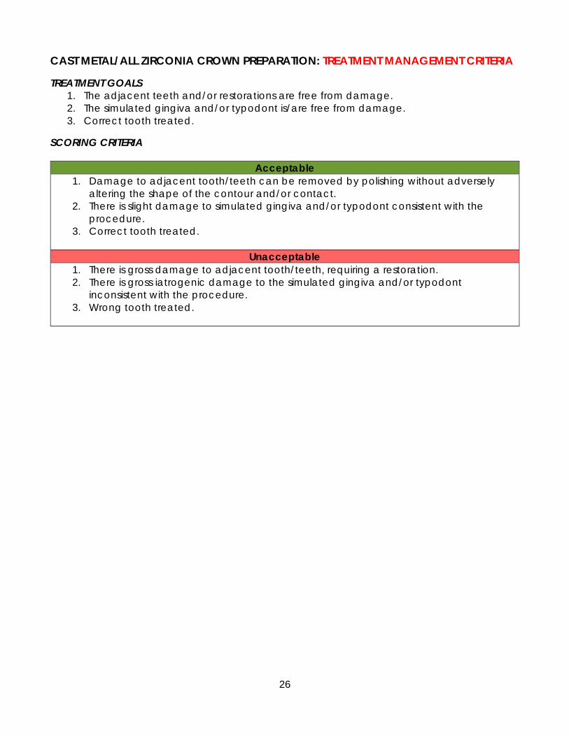
26
CAST METAL/ALL ZIRCONIA CROWN PREPARATION: TREATMENT MANAGEMENT CRITERIA
TREATMENT GOALS
1. The adjacent teeth and/or restorations are free from damage.
2. The simulated gingiva and/or typodont is/are free from damage.
3. Correct tooth treated.
SCORING CRITERIA
Acceptable
1. Damage to adjacent tooth/teeth can be removed by polishing without adversely
altering the shape of the contour and/or contact.
2. There is slight damage to simulated gingiva and/or typodont consistent with the
procedure.
3. Correct tooth treated.
Unacceptable
1. There is gross damage to adjacent tooth/teeth, requiring a restoration.
2. There is gross iatrogenic damage to the simulated gingiva and/or typodont
inconsistent with the procedure.
3. Wrong tooth treated.

27
CAST METAL/ALL ZIRCONIA CROWN PREPARATION: CERVICAL MARGIN CRITERIA
TREATMENT GOALS
1. The cervical margins should be 1.0 mm occlusal to the simulated free gingival margin.
2. The cervical margin is smooth, continuous and well defined.
3. The cervical margin meets the external surface of the tooth at approximately a right
angle.
4. The cervical margin is 1.0 mm in width.
SCORING CRITERIA
Acceptable
1. The cervical margin is less than 0.5 mm below or no more than 1.5 mm above the
simulated free gingival margin. If greater than 0.5 mm there is no visual damage.
2. The cervical margin is continuous but may be slightly rough and lacks some definition.
3. The cervical margin meets the external surface of the tooth at approximately a right
angle.
4. The cervical margin is a chamfer and varies slightly in width, is detectable visually or
with an explorer, and is less than or equal to 2.0 mm in width.
Unacceptable
1. The cervical margin is greater than 0.5 mm below causing visual damage or greater
than 1.5 mm above the simulated free gingival margin.
2. The cervical margin has no continuity and/or definition and/or will prevent fabrication
of an adequate restoration.
3. The cervical margin meets the external surface of the tooth at an angle greater than
120°. The cervical margin is cupped or j-shaped resulting in unsupported enamel that
will prevent fabrication of an adequate restoration.
4. The cervical margin is not a chamfer, is not detectable, and/or is greater than 2.0 mm
in width.

28
CAST METAL/ALL ZIRCONIA CROWN PREPARATION: WALLS & TAPER CRITERIA
TREATMENT GOALS
1. Axial tissue removal is optimally 1.0 mm to be sufficient for convenience, retention and
resistance form.
2. The walls are smooth and well defined and/or internal line angles and/or cusp tip areas
are rounded.
3. Taper, total occlusal convergence (TOC) is 10°– 16°.
4. There are no undercuts.
5. Occlusal reduction is 1.5 mm.
6. The appropriate path of insertion varies less than 10° from parallel to the long axis of the
tooth on all axial surfaces, and a line of draw is established.
SCORING CRITERIA
Acceptable
1. The axial tissue removal is greater than 0.5 mm but less than 2.5 mm.
2. The walls are slightly rough and lack some definition and/or internal line angles and/or
cusp tip areas are rounded and have a slight tendency of being sharp.
3. Taper, total occlusal convergence (TOC) is 16° or less.
4. Slight undercut(s) exists, but it will not interfere with fabrication of an adequate
restoration.
5. Occlusal reduction is greater than 1.0 mm or less than or equal to 2.5 mm.
6. The path of insertion or line of draw deviates 10° to less than 30° from the long axis of
the tooth.
Unacceptable
1. The axial tissue removal is less than 0.5 mm or greater than 2.5 mm.
2. The walls are rough and lack definition and/or internal line angles and/or cusp tip
areas are sharp with no evidence of rounding.
3. Taper, total occlusal convergence (TOC) is greater than 16°.
4. Undercut(s) exists greater than 0.5 mm and an adequate restoration cannot be
fabricated.
5. The occlusal reduction is less than 1.0 mm or more than 2.5 mm.
6. The path of insertion or line of draw is unacceptable, deviating 30° or more from the
long axis of the tooth.

29
BRIDGE FACTOR -PATH OF INSERTION/LINE OF DRAW CRITERIA
TREATMENT GOALS
1. The line of draw or path of insertion would allow for the full seating of a fixed prosthesis
in a direct vertical plane without rotation.
SCORING CRITERIA
Acceptable
1. A line of draw exists whereby an adequate prosthesis may be fabricated.
Unacceptable
1. An adequate prosthesis may not be fabricated without removal of additional tooth
structure.

30
ALL-CERAMIC CROWN PREPARATION: TREATMENT MANAGEMENT CRITERIA
TREATMENT GOALS
1. The adjacent teeth and/or restorations are free from damage.
2. The simulated gingiva and/or typodont is/are free from damage.
3. Correct tooth treated.
SCORING CRITERIA
Acceptable
1. Damage to adjacent tooth/teeth can be removed by polishing without adversely
altering the shape of the contour and/or contact.
2. There is slight damage to simulated gingiva and/or typodont consistent with the
procedure.
3. Correct tooth treated.
Unacceptable
1. There is gross damage to adjacent tooth/teeth, requiring a restoration.
2. There is gross iatrogenic damage to the simulated gingiva and/or typodont
inconsistent with the procedure.
3. Wrong tooth treated.

31
ALL-CERAMIC CROWN PREPARATION: CERVICAL MARGIN CRITERIA
TREATMENT GOALS
1. The cervical margins should be 1.0 mm occlusal to the simulated free gingival margin.
2. The cervical margin is smooth, continuous and well defined.
3. The cervical margin meets the external surface of the tooth at approximately a right
angle.
4. The cervical margin is 1.25 mm in width.
SCORING CRITERIA
Acceptable
1. The cervical margin is less than 0.5 mm below or no more than 1.5 mm above the
simulated free gingival margin. If greater than 0.5mm there is no visual damage.
2. The cervical margin is continuous but may be slightly rough and lacks some definition.
3. The cervical margin meets the external surface of the tooth at approximately a right
angle.
4. The cervical margin is 0.5 mm to 2.0 mm in width.
Unacceptable
1. The cervical margin is greater than 0.5 mm below with visual damage or greater than
1.5 mm above the simulated free gingival margin.
2. The cervical margin has no continuity and/or definition and/or will prevent fabrication
of an adequate restoration.
3. The cervical margin meets the external surface of the tooth at an angle greater than
120°. The cervical margin is cupped or j-shaped resulting in unsupported enamel that
will prevent fabrication of an adequate restoration.
4. The cervical margin is less than 0.5 mm or greater than 2.0 mm in width.
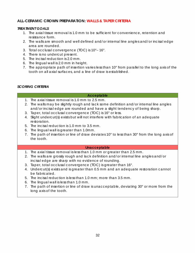
32
ALL-CERAMIC CROWN PREPARATION: WALLS & TAPER CRITERIA
TREATMENT GOALS
1. The axial tissue removal is 1.0 mm to be sufficient for convenience, retention and
resistance form.
2. The walls are smooth and well defined and/or internal line angles and/or incisal edge
area are rounded.
3. Total occlusal convergence (TOC) is 10°– 16°.
4. There is no undercut present.
5. The incisal reduction is 2.0 mm.
6. The lingual wall is 2.0 mm in height.
7. The appropriate path of insertion varies less than 10° from parallel to the long axis of the
tooth on all axial surfaces, and a line of draw is established.
SCORING CRITERIA
Acceptable
1. The axial tissue removal is 1.0 mm to 2.5 mm.
2. The walls may be slightly rough and lack some definition and/or internal line angles
and/or incisal edge are rounded and have a slight tendency of being sharp.
3. Taper, total occlusal convergence (TOC) is 16° or less.
4. Slight undercut(s) exists but will not interfere with fabrication of an adequate
restoration.
5. The incisal reduction is 1.0 mm to 3.5 mm.
6. The lingual wall is greater than 1.0mm.
7. The path of insertion or line of draw deviates 10° to less than 30° from the long axis of
the tooth.
Unacceptable
1. The axial tissue removal is less than 1.0 mm or greater than 2.5 mm.
2. The walls are grossly rough and lack definition and/or internal line angles and/or
incisal edge are sharp with no evidence of rounding.
3. Taper, total occlusal convergence (TOC) is greater than 16°.
4. Undercut(s) exists and is greater than 0.5 mm and an adequate restoration cannot
be fabricated.
5. The incisal reduction is less than 1.0 mm; more than 3.5 mm.
6. The lingual wall is less than 1.0 mm.
7. The path of insertion or line of draw is unacceptable, deviating 30° or more from the
long axis of the tooth.
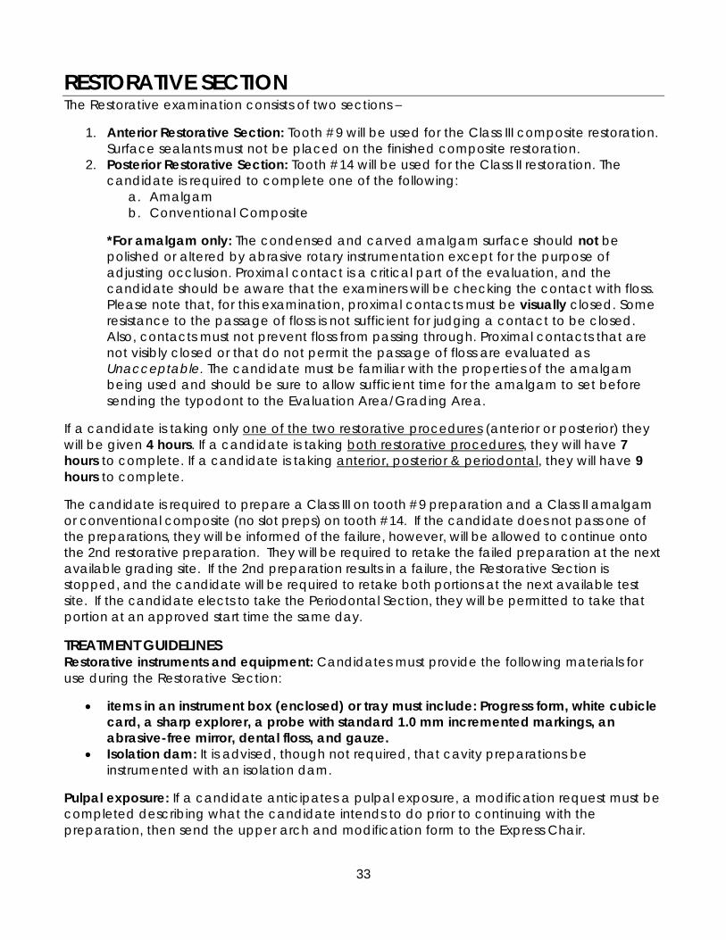
33
RESTORATIVE SECTION The Restorative examination consists of two sections –
1. Anterior Restorative Section: Tooth #9 will be used for the Class III composite restoration.
Surface sealants must not be placed on the finished composite restoration.
2. Posterior Restorative Section: Tooth #14 will be used for the Class II restoration. The
candidate is required to complete one of the following:
a. Amalgam
b. Conventional Composite
*For amalgam only: The condensed and carved amalgam surface should not be
polished or altered by abrasive rotary instrumentation except for the purpose of
adjusting occlusion. Proximal contact is a critical part of the evaluation, and the
candidate should be aware that the examiners will be checking the contact with floss.
Please note that, for this examination, proximal contacts must be visually closed. Some
resistance to the passage of floss is not sufficient for judging a contact to be closed.
Also, contacts must not prevent floss from passing through. Proximal contacts that are
not visibly closed or that do not permit the passage of floss are evaluated as
Unacceptable. The candidate must be familiar with the properties of the amalgam
being used and should be sure to allow sufficient time for the amalgam to set before
sending the typodont to the Evaluation Area/Grading Area.
If a candidate is taking only one of the two restorative procedures (anterior or posterior) they
will be given 4 hours. If a candidate is taking both restorative procedures, they will have 7
hours to complete. If a candidate is taking anterior, posterior & periodontal, they will have 9
hours to complete.
The candidate is required to prepare a Class III on tooth #9 preparation and a Class II amalgam
or conventional composite (no slot preps) on tooth #14. If the candidate does not pass one of
the preparations, they will be informed of the failure, however, will be allowed to continue onto
the 2nd restorative preparation. They will be required to retake the failed preparation at the next
available grading site. If the 2nd preparation results in a failure, the Restorative Section is
stopped, and the candidate will be required to retake both portions at the next available test
site. If the candidate elects to take the Periodontal Section, they will be permitted to take that
portion at an approved start time the same day.
TREATMENT GUIDELINES Restorative instruments and equipment: Candidates must provide the following materials for
use during the Restorative Section:
• items in an instrument box (enclosed) or tray must include: Progress form, white cubicle
card, a sharp explorer, a probe with standard 1.0 mm incremented markings, an
abrasive-free mirror, dental floss, and gauze.
• Isolation dam: It is advised, though not required, that cavity preparations be
instrumented with an isolation dam.
Pulpal exposure: If a candidate anticipates a pulpal exposure, a modification request must be
completed describing what the candidate intends to do prior to continuing with the
preparation, then send the upper arch and modification form to the Express Chair.
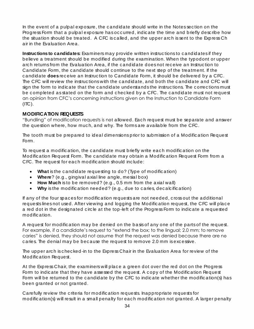
34
In the event of a pulpal exposure, the candidate should write in the Notes section on the
Progress Form that a pulpal exposure has occurred, indicate the time and briefly describe how
the situation should be treated. A CFC is called, and the upper arch is sent to the Express Ch
air in the Evaluation Area.
Instructions to candidates: Examiners may provide written instructions to candidates if they
believe a treatment should be modified during the examination. When the typodont or upper
arch returns from the Evaluation Area, if the candidate does not receive an Instruction to
Candidate Form, the candidate should continue to the next step of the treatment. If the
candidate does receive an Instruction to Candidate Form, it should be delivered by a CFC.
The CFC will review the instructions with the candidate, and both the candidate and CFC will
sign the form to indicate that the candidate understands the instructions. The corrections must
be completed as stated on the form and checked by a CFC. The candidate must not request
an opinion from CFC’s concerning instructions given on the Instruction to Candidate Form
(ITC).
MODIFICATION REQUESTS “Bundling” of modification requests is not allowed. Each request must be separate and answer
the question where, how much, and why. The forms are available from the CFC.
The tooth must be prepared to ideal dimensions prior to submission of a Modification Request
Form.
To request a modification, the candidate must briefly write each modification on the
Modification Request Form. The candidate may obtain a Modification Request Form from a
CFC. The request for each modification should include:
• What is the candidate requesting to do? (Type of modification)
• Where? (e.g., gingival axial line angle, mesial box)
• How Much is to be removed? (e.g., 0.5 mm from the axial wall)
• Why is the modification needed? (e.g., due to caries, decalcification)
If any of the four spaces for modification requests are not needed, cross out the additional
requests lines not used. After viewing and logging the Modification request, the CFC will place
a red dot in the designated circle at the top-left of the Progress Form to indicate a requested
modification.
A request for modification may be denied on the basis of any one of the parts of the request.
For example, if a candidate’s request to “extend the box; to the lingual; 2.0 mm; to remove
caries” is denied, they should not assume that the request was denied because there are no
caries. The denial may be because the request to remove 2.0 mm is excessive.
The upper arch is checked-in to the Express Chair in the Evaluation Area for review of the
Modification Request.
At the Express Chair, the examiners will place a green dot over the red dot on the Progress
Form to indicate that they have assessed the request. A copy of the Modification Request
Form will be returned to the candidate by the CFC to indicate whether the modification(s) has
been granted or not granted.
Carefully review the criteria for modification requests. Inappropriate requests for
modification(s) will result in a small penalty for each modification not granted. A larger penalty

35
will be assigned for requests for a modification for removal of caries or decalcification when
no caries or decalcification exists or for repeated modification requests in an apparent
attempt to have the examiners confirm when all caries is removed. Modifications that have
been approved and appropriately accomplished will not result in any penalties.
Regardless if the modification is granted or not granted, the candidate must complete the
preparation and send the upper arch back to the Evaluation Area for evaluation of the final
completed preparation.
If the candidate subsequently has additional requests for modification on the same
preparation, a new red dot is placed over the green dot on the Progress Form, and the same
procedure is followed. If more than one modification is anticipated at any time, it is to the
candidate’s advantage to submit them at the same time, as no additional time is provided for
evaluation of modification requests, and multiple submissions may significantly decrease
treatment time.
Once all approved modifications are completed, the upper arch and all required papers and
instruments are submitted to the Evaluation Area for evaluation of the final preparation.
Terminology to be used when requesting modifications

36
PROCEDURE MANAGEMENT GUIDELINES Restorative Procedures: Candidates will begin with the Anterior restorative procedure first,
followed by the Posterior restorative procedure. Candidates must have the CFC approve the
mounting of the typodont before starting the Preparation.
Final evaluation of the preparation: Three independent examiners evaluate the prepared
tooth/cavity. Once the Anterior procedure’s preparation is complete, the candidate will get
approval from the CFC to remove the upper (maxillary) arch and place it in the bag or
typodont box provided. Place a candidate label on the bag or box and send it to the
Evaluation area along with the required instruments, Progress form, and cubicle card for
grading. Once grading is complete for the preparation, the upper (maxillary) arch will be
returned to the candidate. The candidate will then secure the upper (maxillary) arch back
within the typodont and complete the finish section of the Anterior restorative Exam. The same
process will be conducted for the Posterior restorative examination.
Final evaluation of the restoration: Once the Anterior restoration have been restored, the
candidate will get approval from the CFC to remove the upper (maxillary) arch, and send it to
the evaluation area along with the required instruments, cubicle card, and Progress Form.
Note that for the Anterior restoration, you will only be submitting the upper arch, not the entire
typodont.
For the Posterior restoration, you will be submitting the entire typodont upon finishing the
finished restoration. A CFC will sign off on your Progress form to dismount the entire typodont
for submission. Affix the manila tag with a candidate ID label to the typodont. After the last
restoration has been submitted for grading, the typodont will not be returned. Ensure that all
packing materials from the typodont box are included.
For the Class II amalgam restoration, the amalgam must be sufficiently set to allow a check of
the occlusion. Any of the composite restorations must be presented without any surface
glaze/sealer on the restoration.
If adjustments to the restoration are required, an Instruction to Candidate Form (ITC) will be
issued. Candidates must perform the corrections as instructed and verified by a CFC. Only one
restorative procedure can be done at a time. Both the Anterior and Posterior portions must be
finished in the allotted time and turned into the Evaluation area for Finish grading.

37
CLASS III ANTERIOR COMPOSITE PREPARATION: TREATMENT MANAGEMENT CRITERIA
TREATMENT GOALS
1. The adjacent teeth and/or restorations are free from damage.
2. The soft tissue is free from damage, or there is tissue damage that is consistent with the
procedure.
3. Correct tooth treated.
SCORING CRITERIA
Acceptable
1. Any damage to adjacent tooth/teeth that can be removed by polishing or may
require recontouring that does not adversely change the shape or contact.
2. There may be slight iatrogenic trauma to the soft tissue inconsistent with the
procedure.
3. Correct tooth treated.
Unacceptable
1. There is evidence of gross damage and/or alteration to adjacent and/or opposing
hard tissue inconsistent with the procedure which may require additional evaluation,
intervention or treatment as a result of the damage.
2. There is gross iatrogenic damage to the soft tissue inconsistent with the procedure.
3. Wrong tooth treated.

38
CLASS III ANTERIOR COMPOSITE PREPARATION: EXTERNAL OUTLINE FORM CRITERIA
TREATMENT GOALS
1. The outline form is sufficient in size to have access to remove caries and to manipulate
and finish the restorative material.
2. The outline form is not overextended beyond what is necessary for complete removal
of caries.
3. The incisal cavosurface margin does not compromise the incisal angle.
4. The wall opposite the access, if broken, does not extend more than 0.5 mm beyond the
contact area.
5. There are no caries remaining.
6. The cavosurface margins are not irregular and there is no explorer-penetrable
decalcification remaining on the cavosurface margin.
7. There is no unsupported enamel.
8. Enamel cavosurface margin may be beveled.
SCORING CRITERIA
Acceptable
1. The outline form is sufficient in size to have access to remove caries and to manipulate
and finish the restorative material.
2. The outline form may be extended mesiodistally up to 1.5 mm beyond what is necessary.
3. The outline form may be extended incisogingivally up to 5.0 mm
4. The incisal cavosurface margin does not compromise the incisal angle.
5. The wall opposite the access, if broken, may extend no more than 2.0 mm beyond the
contact area.
6. There is no caries remaining.
7. The cavosurface margins may be slightly irregular but there is no explorer-penetrable
decalcification remaining on the cavosurface margin.
8. There may be a small area of unsupported enamel, which is necessary to preserve facial
esthetics.
9. Enamel cavosurface margin bevels, if present; do not exceed 1.0 mm in width.
10. The gingival clearance is open up to 2.0 mm.
Unacceptable
1. The outline form is under-extended, making it impossible to remove caries or to
manipulate and finish the restorative material.
2. The outline form is extended mesiodistally up by more than 1.5 mm beyond what is
necessary.
3. The outline form is extended incisogingivally by more than 5.0 mm
4. The incisal cavosurface margin is over-extended so that the incisal angle is compromised,
removed or fractured. A Class IV restoration is now necessary without prior justification.
5. The wall opposite the access opening extends more than 2.0 mm beyond the contact
area.
6. There is caries remaining.
7. The cavosurface margin does not terminate in sound natural tooth structure. There is
explorer–penetrable decalcification at the cavosurface margin.
8. There are large or multiple areas of unsupported enamel that are not necessary to
preserve facial esthetics.
9. Enamel cavosurface margin bevels, if present, exceed 1.0 mm in width, are not uniform or
are inappropriate for the size of the restoration
10. The gingival clearance is open greater than 2.0 mm.

39
CLASS III ANTERIOR COMPOSITE PREPARATION: INTERNAL FORM CRITERIA
TREATMENT GOALS
1. The preparation is free of caries.
2. The depth of the axial wall is just inside the DEJ.
3. No pulp exposure.
SCORING CRITERIA
Acceptable
1. The preparation is free of caries.
2. The depth of the axial wall is no more than 2.5 mm beyond the DEJ.
3. Properly managed justified pulp exposure.
Unacceptable
1. The preparation has remaining caries.
2. The axial wall is greater than 2.5 mm beyond the DEJ.
3. Any pulp exposure that is not properly managed or unjustified.

40
CLASS III ANTERIOR COMPOSITE FINISHED RESTORATION: TREATMENT MANAGEMENT
CRITERIA
TREATMENT GOALS
1. The adjacent and/or opposing hard tissue is free from evidence of damage and/or
alteration.
2. The soft tissue is free from damage, or there is soft tissue damage consistent with the
procedure.
3. Correct tooth treated.
SCORING CRITERIA
Acceptable
1. Any hard tissue damage to adjacent or opposing tooth/teeth that can be removed
by polishing or may require recontouring that does not adversely change the shape
or contact.
2. There may be slight iatrogenic trauma to the soft tissue inconsistent with the
procedure.
3. Correct tooth treated.
Unacceptable
1. There is evidence of gross damage and/or alteration to adjacent and/or opposing
hard tissue inconsistent with the procedure which may require additional evaluation,
intervention or treatment as a result of the damage.
2. There is gross iatrogenic damage to the soft tissue inconsistent with the procedure.
3. Wrong tooth treated.

41
CLASS III ANTERIOR COMPOSITE FINISHED RESTORATION: CONTOUR, CONTACT &
OCCLUSION CRITERIA
TREATMENT GOALS
1. Interproximal contact is present. The contact is visually closed and properly shaped and
positioned. There is definite, but not excessive, resistance to dental floss when passed
through the interproximal contact area.
2. The restoration reproduces the normal physiological proximal contours of the tooth,
occlusal anatomy and marginal ridge anatomy.
3. When checked with articulating paper, all centric and excursive contacts on the
restoration are consistent in size, shape and intensity with such contacts on other teeth
in that quadrant.
SCORING CRITERIA
Acceptable
1. Interproximal contact is visually closed, and the contact is adequate in size, shape or
position but may demonstrate little resistance to dental floss.
2. The restoration may not reproduce the normal lingual anatomy, proximal contours of
the tooth or marginal ridge anatomy but would not be expected to adversely affect
the tissue health.
3. When checked with articulating paper, the restoration is in hyperocclusion. The
restoration only requires minor occlusal adjusting.
Unacceptable
1. Interproximal contact is visually open or will not allow floss to pass through the
contact area.
2. The restoration does not reproduce the normal lingual anatomy, proximal contour of
the tooth or marginal ridge anatomy and as such it would be expected to adversely
affect the health of the surrounding soft tissue.
3. There is gross hyperocclusion so that the restoration is the only point of occlusion in
that quadrant.
42
CLASS III ANTERIOR COMPOSITE FINISHED RESTORATION: MARGIN INTEGRITY & SURFACE
FINISH CRITERIA
TREATMENT GOALS
1. There is no marginal deficiency.
2. There is no marginal excess.
3. The restoration is not fractured and is bonded to the prepared tooth structure.
4. There is no evidence of unwarranted or unnecessary removal, or recontouring of tooth
structure adjacent to the restoration, without a previous modification approval.
5. The shade of the restoration blends with the surrounding tooth structure.
SCORING CRITERIA
Acceptable
1. There is no open margin.
2. Marginal deficiency may be absent or detectable (either visually or with the tine of
an explorer) at the restoration-tooth interface, but is no greater than 0.5 mm.
3. Marginal excess may be absent or detectable at the restoration-tooth interface, but
it is no greater than 1.0 mm at the gingival margin and may extend greater than 0.5
mm in other areas.
4. The restoration is not fractured, debonded and/or movable in the preparation.
5. There is no or minimal evidence of unwarranted or unnecessary removal,
modification or recontouring of tooth structure adjacent to the restoration
(enameloplasty).
Unacceptable
1. An open margin is detectable (either visually or with the tine of an explorer) at the
restoration tooth interface.
2. Marginal deficiency is detectable (either visually or with the tine of an explorer) at the
restoration-tooth interface, and is greater than 0.5 mm.
3. Marginal excess is greater than 1.0 mm at the gingival margin which impinges on the
gingival tissue and will be detrimental to the gingival health.
4. The restoration is fractured, debonded and/or movable in the preparation.
5. There is evidence of unwarranted or unnecessary removal, modification or
recontouring of tooth structure adjacent to the restoration (enameloplasty).

43
CLASS II POSTERIOR AMALGAM PREPARATION: TREATMENT MANAGEMENT CRITERIA
TREATMENT GOALS
1. The adjacent and/or opposing hard tissue is free from evidence of damage and/or
alteration.
2. The soft tissue is free from damage, or there is soft tissue damage consistent with the
procedure.
3. Correct tooth treated.
SCORING CRITERIA
Acceptable
1. Any hard tissue damage to adjacent or opposing tooth/teeth that can be removed
by polishing or may require recontouring that does not adversely change the shape
or contact.
2. There may be slight iatrogenic trauma to the soft tissue inconsistent with the
procedure.
3. Correct tooth treated.
Unacceptable
1. There is evidence of gross damage and/or alteration to adjacent and/or opposing
hard tissue inconsistent with the procedure which may require additional evaluation,
intervention or treatment as a result of the damage.
2. There is gross iatrogenic damage to the soft tissue inconsistent with the procedure.
3. Wrong tooth treated.

44
CLASS II POSTERIOR AMALGAM PREPARATION: EXTERNAL OUTLINE FORM CRITERIA
TREATMENT GOALS
1. The proximal clearance at the height of contour is visibly open.
2. The proximal cavosurface margin is 900 to the external surface of the tooth.
3. There are no areas of unsupported enamel.
4. The gingival clearance is visibly open.
5. The isthmus is a minimum of 1.0 mm wide to no more than one-third the intercuspal
width.
6. The cavosurface margins terminates in sound tooth structure, are not irregular and there
is no explorer-penetrable decalcification remaining.
7. The outline form includes all carious and non-coalesced fissures and is smooth, rounded
and flowing.
SCORING CRITERIA
Acceptable
1. The proximal clearance at the height of contour is visibly open or extends no more
than 3.0 mm on either one or both proximal walls.
2. The proximal cavosurface margin may deviate from 900 but is unlikely to jeopardize
the longevity of the tooth or restoration; includes small areas of unsupported enamel.
3. The gingival clearance is visibly open or not greater than 3.0 mm.
4. The isthmus is from 1.0 mm wide to no more than one-half the intercuspal width.
5. The cavosurface margins should terminate in sound tooth structure, and may be
slightly irregular, but there is no explorer-penetrable decalcification remaining.
6. The outline form does not compromise the marginal ridge.
Unacceptable
1. The proximal clearance at the height of contour is not visibly open or extends beyond
3.0 mm on either one or both proximal walls.
2. The proximal cavosurface margin deviates excessively from 900 and will jeopardize
the longevity of the tooth or restoration; includes unsupported enamel.
3. The gingival clearance is not visibly open or is greater than 3.0 mm.
4. The isthmus is less than 1.0 mm or greater than one-half the intercuspal width.
5. The cavosurface margins do not terminate in sound tooth structure and there is
explorer- penetrable decalcification.
6. The marginal ridge is undermined and/or less than 1.0 mm in width.

45
CLASS II POSTERIOR AMALGAM PREPARATION: INTERNAL FORM CRITERIA
TREATMENT GOALS
1. The pulpal floor depth is 2.0 mm beyond the cavosurface margin.
2. The depth of the axial wall is just inside the DEJ.
3. The walls of the proximal box are convergent and appropriate internal retention is
present.
4. There is no evidence of caries.
5. Retention, when used, does not undermine the enamel.
6. Prepared surfaces are smooth.
7. No pulp exposure
SCORING CRITERIA
Acceptable
1. The pulpal floor depth is 1.5 mm to 2.5 mm beyond the cavosurface margin.
2. The depth of the axial wall is no more than 2.5 mm beyond the DEJ.
3. The walls of the proximal box should be convergent but may be parallel, but
appropriate internal retention is present.
4. There is no evidence of caries.
5. Retention, when used, may not undermine the enamel, and is acceptable and
therefore not likely to jeopardize the longevity of the tooth or restoration.
6. Prepared surfaces may be slightly rough, irregular, or sharp.
7. Properly managed justified pulp exposure.
Unacceptable
1. The pulpal floor depth is less than 1.5 mm or greater than 2.5 mm from the
cavosurface margin.
2. The axial wall is greater than 2.5 mm beyond the DEJ or is still in the enamel and does
not include the DEJ.
3. The walls of the proximal box diverge occlusally, offering no retention and
jeopardizing the longevity of the tooth or restoration.
4. The preparation has remaining caries.
5. Retention excessively undermines the enamel and is likely to jeopardize the longevity
of the tooth or restoration.
6. A prepared surface of the tooth is excessively rough, irregular or sharp and is likely to
jeopardize the longevity of the tooth restoration.
7. Any pulp exposure that is not properly managed and/or unjustified.
46
CLASS II POSTERIOR AMALGAM FINISHED RESTORATION: TREATMENT MANAGEMENT
CRITERIA
TREATMENT GOALS
1. The adjacent and/or opposing hard tissue is free from evidence of damage and/or
alteration.
2. The soft tissue is free from damage, or there is soft tissue damage consistent with the
procedure.
3. Correct tooth treated.
SCORING CRITERIA
Acceptable
1. Any hard tissue damage to adjacent or opposing tooth/teeth that can be removed
by polishing or may require recontouring that does not adversely change the shape
or contact.
2. There may be slight iatrogenic trauma to the soft tissue inconsistent with the
procedure.
3. Correct tooth treated.
Unacceptable
1. There is evidence of gross damage and/or alteration to adjacent and/or opposing
hard tissue inconsistent with the procedure which may require additional evaluation,
intervention or treatment as a result of the damage.
2. There is gross iatrogenic damage to the soft tissue inconsistent with the procedure.
3. Wrong tooth treated.

47
CLASS II POSTERIOR AMALGAM FINISHED RESTORATION: CONTOUR, CONTACT &
OCCLUSION CRITERIA
TREATMENT GOALS
1. Interproximal contact is present. The contact is visually closed and properly shaped and
positioned. There is definite, but not excessive, resistance to dental floss when passed
through the interproximal contact area.
2. When checked with articulating paper, all centric and excursive contacts on the
restoration are consistent in size, shape and intensity with such contacts on other teeth
in that quadrant.
3. The restoration reproduces the normal physiological proximal contours of the tooth,
occlusal anatomy and marginal ridge anatomy.
SCORING CRITERIA
Acceptable
1. Interproximal contact is visually closed, and the contact may be deficient in size,
shape or position but may demonstrate little resistance to dental floss or shreds the
floss.
2. When checked with articulating paper, the restoration may be in slight
hyperocclusion inconsistent in size, shape and intensity with the occlusal contacts on
surrounding teeth and the restoration may require adjustment.
3. The restoration may not reproduce the normal occlusal anatomy, proximal contours
of the tooth or marginal ridge anatomy, but would not be expected to adversely
affect the tissue health.
Unacceptable
1. Interproximal contact is visually open or will not allow floss to pass through the
contact area.
2. There is gross hyperocclusion so that the restoration is the only point of occlusion in
that quadrant.
3. The restoration does not reproduce the normal occlusal anatomy, proximal contours
of the tooth or marginal ridge anatomy and will adversely affect the tissue health.
48
CLASS II POSTERIOR AMALGAM FINISHED RESTORATION: MARGIN INTEGRITY & SURFACE
FINISH CRITERIA
TREATMENT GOALS
1. There is no marginal deficiency.
2. There is no marginal excess detectable, either visually or with the tine of an explorer, at
the restoration-tooth interface.
3. The surface of the restoration is free of pits and voids.
4. Restoration is not fractured.
5. There is no evidence of unwarranted or unnecessary removal, modification or
recontouring of tooth structure adjacent to the restoration (enameloplasty).
6. There is no evidence of open margins.
SCORING CRITERIA
Acceptable
1. Marginal deficiency may be absent or detectable (either visually or with the tine of
an explorer) at the restoration-tooth interface, but it is no greater than 0.5 mm.
2. Marginal excess may be absent or detectable at the restoration-tooth interface, but
it is no greater than 1.0 mm at the gingival margin and does not extend greater than
2.0 mm in other areas.
3. The surface of the restoration may be slightly grainy or rough, but it is free of pits and
voids.
4. Restoration is not fractured.
5. There is no or minimal evidence of unwarranted or unnecessary removal,
modification or recontouring of tooth structure adjacent to the restoration
(enameloplasty).
6. There is no evidence of open margins.
Unacceptable
1. Marginal deficiency is detectable (either visually or with the tine of an explorer) at the
restoration-tooth interface, and is greater than 0.5 mm.
2. Marginal excess is greater than 1.0 mm at the gingival margin which impinges on the
gingival tissue and will be detrimental to the gingival health and/or extends greater
than 2.0 mm in other areas.
3. The surface of the restoration is rough and exhibits significant surface irregularities, pits
or voids.
4. Restoration is fractured.
5. There is evidence of unwarranted or unnecessary removal, modification or
recontouring of tooth structure adjacent to the tooth being restored (enameloplasty).
6. There is an open margin detectable with the tine of an explorer.

49
CLASS II POSTERIOR CONVENTIONAL COMPOSITE PREPARATION: TREATMENT
MANAGEMENT CRITERIA
TREATMENT GOALS
1. There is no hard tissue damage to adjacent or opposing tooth/teeth.
2. There is no iatrogenic trauma to the soft tissue inconsistent with the procedure
3. Correct tooth treated.
SCORING CRITERIA
Acceptable
1. Any hard tissue damage to adjacent or opposing tooth/teeth that can be removed
by polishing or may require recontouring that does not adversely change the shape
or contact.
2. There may be slight iatrogenic trauma to the soft tissue inconsistent with the
procedure.
3. Correct tooth treated.
Unacceptable
1. There is evidence of gross damage and/or alteration to adjacent and/or opposing
hard tissue inconsistent with the procedure which may require additional evaluation,
intervention or treatment as a result of the damage.
2. There is gross iatrogenic damage to the soft tissue inconsistent with the procedure.
3. Wrong tooth treated.

50
CLASS II POSTERIOR CONVENTIONAL COMPOSITE PREPARATION: EXTERNAL OUTLINE
FORM CRITERIA
TREATMENT GOALS
1. The proximal contact is either closed, or visibly open.
2. The gingival clearance is visibly open.
3. The outline form is not sharp and irregular. The outline form is not over extended so that
it compromises the remaining marginal ridge and/or cusp(s).
4. The isthmus is at least 1.0 mm and may not exceed one-half the intercuspal width.
5. The cavosurface margin is 90° to the external surface of the tooth. There is no
unsupported enamel.
6. The cavosurface margins terminates in sound tooth structure, are not irregular and there
is no explorer-penetrable decalcification remaining.
7. There is no remaining non-coalesced fissure(s) that extend the DEJ and are contiguous
with the outline form.
SCORING CRITERIA
Acceptable
1. The proximal contact is either closed, or visibly open and proximal clearance at the
height of contour does not extend more than 2.5 mm on either one or both proximal
walls.
2. The gingival clearance may be closed, or is less than or equal to 2.0 mm.
3. The outline form may be sharp and irregular. The outline form may be inappropriately
over extended so that it compromises the remaining marginal ridge and/or cusp(s).
4. The isthmus must be at least 1.0 mm and may not exceed one-half the intercuspal
width.
5. The cavosurface margin may deviate from 90° but is unlikely to jeopardize the
longevity of the tooth or restoration; Includes small areas of unsupported enamel
and/or excessive bevel(s).
6. The cavosurface margins should terminate in sound tooth structure and may be
slightly irregular but there is no explorer-penetrable decalcification remaining.
7. There are no remaining non-coalesced fissure(s) that extend the DEJ and are
contiguous with the outline form.
Unacceptable
1. The proximal clearance at the height of contour extends beyond 2.5 mm on either
one or both proximal walls.
2. The gingival clearance is greater than 2.0 mm.
3. The outline form is grossly over-extended so that it compromises or undermines the
remaining marginal ridge to the extent that the cavosurface margin is unsupported
by dentin or the width of the marginal ridge is 0.5 mm or less.
4. The isthmus is less than 1.0 mm or greater than one-half the intercuspal distance.
5. The cavosurface margin deviates from 90° and is likely to jeopardize the longevity of
the tooth or restoration. This includes unsupported enamel and/or excessive bevel(s).
6. The cavosurface margins do not terminate in sound tooth structure, are grossly
irregular and there is explorer–penetrable decalcification.
7. There are remaining non-coalesced fissure(s) that extend to the DEJ and are
contiguous with the outline form.

51
CLASS II POSTERIOR CONVENTIONAL COMPOSITE PREPARATION: INTERNAL FORM
CRITERIA
TREATMENT GOALS
1. The pulpal floor depth is equal to 2.0 mm from the cavosurface margin and there is no
remaining enamel.
2. The depth of the axial wall is just inside the DEJ.
3. The walls of the proximal box may be divergent or convergent but there is no
undermined enamel.
4. There is no evidence of caries.
5. Retention, when used, does not undermine the enamel.
6. Prepared surfaces are smooth.
7. No pulp exposure.
SCORING CRITERIA
Acceptable
1. The pulpal floor depth is equal to or greater than 0.5 mm from the cavosurface
margin, and the pulpal floor depth is no more than 4.0 mm from the cavosurface
margin; there may be remaining enamel.
2. The depth of the axial wall is no more than 2.5 mm beyond the DEJ.
3. The walls of the proximal box may be divergent or convergent, and which may result
in some undermined enamel.
4. There is no evidence of caries.
5. Retention, when used, may not undermine the enamel which is not likely to
jeopardize the longevity of the tooth or restoration.
6. Prepared surfaces may be rough, irregular or sharp.
7. Properly managed justified pulp exposure.
Unacceptable
1. The pulpal floor depth is 4.0 mm or greater from the cavosurface margin or is less than
0.5 mm.
2. The axial wall is more than 2.5 mm beyond the DEJ or is entirely in enamel.
3. The walls of the proximal box are excessively divergent or convergent, resulting in
excessively undermined enamel, that is likely to jeopardize the longevity of the tooth
or restoration.
4. The preparation has remaining caries.
5. Retention excessively undermines the enamel and is likely to jeopardize the longevity
of the tooth or restoration.
6. Prepared surfaces are excessively rough, irregular or sharp and likely to jeopardize the
longevity of the tooth restoration.
7. Any pulp exposure that is not properly managed or unjustified.

52
CLASS II POSTERIOR CONVENTIONAL COMPOSITE FINISHED RESTORATION: TREATMENT
MANAGEMENT CRITERIA
TREATMENT GOALS
1. The adjacent and/or opposing hard tissue is free from evidence of damage and/or
alteration.
2. The soft tissue is free from damage, or there is soft tissue damage consistent with the
procedure.
3. Correct tooth treated.
SCORING CRITERIA
Acceptable
1. Any hard tissue damage to adjacent or opposing tooth/teeth that can be removed
by polishing or may require recontouring that does not adversely change the shape
or contact.
2. There may be slight iatrogenic trauma to the soft tissue inconsistent with the
procedure.
3. Correct tooth treated.
Unacceptable
1. There is evidence of gross damage and/or alteration to adjacent and/or opposing
hard tissue inconsistent with the procedure which may require additional evaluation,
intervention or treatment as a result of the damage.
2. There is gross iatrogenic damage to the soft tissue inconsistent with the procedure.
3. Wrong tooth treated.

53
CLASS II POSTERIOR CONVENTIONAL COMPOSITE FINISHED RESTORATION: CONTOUR,
CONTACT & OCCLUSION CRITERIA
TREATMENT GOALS
1. Interproximal contact is present. The contact is visually closed and is properly shaped
and positioned. There is definite, but not excessive, resistance to dental floss when
passed through the interproximal contact area.
2. When checked with articulating paper, all centric and excursive contacts on the
restoration are consistent in size, shape and intensity with such contacts on other teeth
in that quadrant.
3. The restoration reproduces the normal physiological proximal contours of the tooth,
occlusal anatomy and marginal ridge anatomy.
SCORING CRITERIA
Acceptable
1. Interproximal contact is visually closed, but the contact may be deficient in size,
shape or position, and may demonstrate little resistance to dental floss or shreds the
floss.
2. When checked with articulating paper, the restoration may be in hyperocclusion
inconsistent in size, shape and intensity with the occlusal contacts on surrounding
teeth. The restoration requires adjustment.
3. The restoration may not reproduce the normal occlusal anatomy, proximal contours
of the tooth, or marginal ridge anatomy, but would not be expected to adversely
affect the tissue health.
Unacceptable
1. Interproximal contact is visually open or will not allow floss to pass through the
contact area.
2. There is gross hyperocclusion, such that the restoration is the only point of occlusion in
that quadrant.
3. The restoration may not reproduce the normal occlusal anatomy, proximal contours
of the tooth, or marginal ridge anatomy, and adversely affects tissue health.

54
CLASS II POSTERIOR CONVENTIONAL COMPOSITE FINISHED RESTORATION: MARGIN
INTEGRITY & SURFACE FINISH CRITERIA
TREATMENT GOALS
1. There is no marginal deficiency.
2. There is no marginal excess detectable, either visually or with the tine of an explorer, at
the restoration-tooth interface.
3. The restoration is bonded to the prepared tooth structure.
4. There is no evidence of unwarranted or unnecessary removal, modification or
recontouring of tooth structure adjacent to the restoration.
5. Shade selection matches surrounding tooth structure.
6. There is no evidence of open margins.
SCORING CRITERIA
Acceptable
1. Marginal deficiency may be absent or detectable (either visually or with the tine of
an explorer) at the restoration-tooth interface, but it is no greater than 0.5 mm.
2. Marginal excess may be absent or detectable at the restoration-tooth interface, but
it is no greater than 1.0 mm at the gingival margin and may extend greater than 0.5
mm in other areas
3. The restoration is not fractured, debonded and/or movable in the preparation.
4. There is no or minimal evidence of unwarranted or unnecessary removal, or
recontouring of tooth structure adjacent to the restoration (enameloplasty).
5. There is no evidence of open margins.
Unacceptable
1. Marginal deficiency is detectable (either visually or with the tine of an explorer) at the
restoration-tooth interface, and is greater than 0.5 mm.
2. Marginal excess is greater than 1.0 mm at the gingival margin which impinges on the
gingival tissue and will be detrimental to the gingival health.
3. The restoration is fractured, debonded and/or movable in the preparation.
4. There is evidence of unwarranted or unnecessary removal, modification or
recontouring of tooth structure adjacent to the tooth being restored (enameloplasty).
5. There is an open margin detectable with the tine of an explorer.

55
IV. PERIODONTAL EXAM (OPTIONAL)
The Periodontal is the only optional section of this examination. Candidates should contact the
appropriate state board of dentistry directly to determine state-specific requirements before
deciding whether to take the Periodontal section.
If a candidate is taking anterior, posterior & periodontal, they will have 9 hours to complete
the examination. If taking only the Periodontal section, 3 hours total is allotted with a 1.5-hour
clinical treatment time included.
Assistants: Use of chairside assistants are not permitted during the periodontal section.
Time Management: The candidate should be aware that the time allowed for the examination
includes the time that the typodont will be at the Evaluation Area. The average time in the
Evaluation Area is 30 minutes and may vary depending on the time of the day and number of
candidates at this examination.
SELECTION & CHECK-IN Teeth selection for the Periodontal section is important. Candidates have a maximum of two
opportunities to present a valid treatment selection. If a second treatment selection is rejected
or is not presented after the first treatment selection is rejected, a candidate will not be
allowed to continue with the procedure and will result in a failed attempt.
Check-in: The candidates may begin setting up their operatories when the clinic opens.
Candidates will receive their typodont from the CFC. They must mount the manikin and get
the CFC’s approval before proceeding. The candidate will record their teeth selection using
the Treatment Selection Worksheet while the typodont is mounted in a manikin head. They will
then dismount and send the typodont to the Evaluation Area for approval along with cubicle
card, Periodontal Progress Form, Treatment Selection Worksheet and the required instruments.
INSTRUMENTS The candidate’s performance will not be evaluated without the proper instruments.
Sonic/ultrasonic instruments are permissible for scaling, but they may not be available at the
examination site (check with the examination site). If the candidate elects to provide their own
unit, they must check with the examination site about appropriate connection mechanisms. Air-
abrasive polishers are not permissible.
An instrument box or tray containing the following items must be sent with the typodont:
• Periodontal Progress Form
• Cubicle card
• Instruments
o 11/12 explorer
o Clear mirror (unscratched, not tinted, non-disposable)
56
TREATMENT SELECTION The candidate’s treatment selection must include the proper number of teeth, adequate
deposits of calculus and appropriate pocket depths as defined below:
• Teeth: There must be at least six and not more than eight permanent teeth selected, at
least three of which are molars or premolars, including at least one molar. All posterior
teeth must have at least one approximating tooth surface within 2.0 mm distance. Each of
the selected teeth must have at least one surface of subgingival calculus selected for
removal.
• Calculus: There must be exactly 12 surfaces of explorer-detectable subgingival calculus
identified on the selected teeth, and no more than four surfaces may be on the incisors.
Three of the 12 identified surfaces of calculus must be on interproximal surfaces of
posterior teeth, i.e., molars and/or premolars.
o Explorer detectable subgingival calculus is defined as a distinct deposit of calculus
that can be felt with an explorer as it passes over the calculus. Qualified deposits
may exhibit such characteristics as:
▪ A definite “jump” or “bump” felt by the explorer, with the rough surface
characteristic of calculus
▪ Ledges or ring formations
▪ Spiny or nodular formations
o Qualified deposits must be apical to the gingival margin and may occur with or
without associated supragingival deposits.
• If during the approval evaluation, the examiners confirm that four or more of the twelve
surfaces of explorer detectable subgingival calculus are not present; this section of the
examination is stopped as the candidate cannot successfully complete the examination.
Candidates may, however, submit a second submission for approval. A maximum of two
submissions for approval is allowed.
Candidates must use the Treatment Selection Worksheet (available at www.srta.org) to identify
and document a selection of the typodont’s teeth that meet these criteria. Please refer to
Examination Forms, Periodontal Treatment Selection Worksheet for more information.
Scaling: After the candidate performs the periodontal procedure, the subgingival surfaces of the
assigned teeth must be smooth, with no deposits detectable with an explorer. Air may be used to
deflect the tissue to locate areas for tactile confirmation. (All subgingival surfaces on an assigned
tooth must be scaled, but only the selected surfaces will be evaluated.)
Supragingival deposits (polishing): All supragingival calculus must be removed from all coronal
surfaces of the assigned teeth so that all surfaces are visually clean when air-dried and tactilely
smooth upon examination with an explorer. The use of disclosing solution is not permitted.
PROCEDURE & GUIDELINES 1. A maximum of two treatment selections may be presented for the Periodontal Section.
If the first treatment selection submitted is found unacceptable due to examination
protocols, guidelines or requirements, a second treatment selection may be presented.
If the selection is otherwise acceptable but there has been a correctable paperwork
error, the candidate may be allowed to correct those errors and re-submit that
selection for approval. In all circumstances the candidate must have their typodont
presented and approved for treatment BEFORE proceeding further with the

57
examination. Treatment on a typodont without documented approval by a Clinic Floor
Coordinator (CFC) is a violation of examination protocol and may subject the
candidate to dismissal from the examination.
2. The Periodontal Progress Form will be provided at the examination site. When the
candidate receives the Progress Form, they should place a candidate identification
label on the form, enter his/her cubicle number and typodont number.
3. The procedures, instruments and materials used are the choice of the candidate, as
long as they are currently accepted and taught by accredited dental schools and the
candidate has been trained in their use. It is the responsibility of the candidate to
provide the instruments used in this examination and listed in this Candidate Manual,
unless such instruments are furnished by the school.
4. The Periodontal section will be the last procedure of the day unless only taking the one
section. Candidates will retrieve the Periodontal typodont from the CFC and may
mount the typodont. A CFC will sign off to ensure the typodont is properly mounted
prior to beginning the screening process.
5. If any problems arise during the examination, the candidate should immediately notify
a CFC. The CFC is also present to aid in any emergencies that may occur.
6. When a typodont is sent to the Evaluation Area for check-in, they will be signed in with
the Dental Administrator. Typodonts will be evaluated for case acceptance in the order
in which they are signed in. The required forms, cubicle card and instruments are sent to
the Evaluation Area with the typodont.
7. The Dental Administrator will indicate a Finish Treatment Time on the Periodontal
Progress Form. The approximate total time for the Periodontal Section is about 3 hours.
The clinical treatment time is a maximum of 1 ½ hours and a minimum of 45 minutes.
Candidates must check their typodont in for pre-treatment approval by 3:45pm and be
checked out of the evaluation area by 4:15pm to receive a treatment time of 45
minutes prior to the end of the examination day. When the typodont is returned from
the Evaluation Area, treatment should begin. Treatment continues until it is completed
or until the Finish Time, as noted on the Periodontal Progress Form. If candidates finish
the treatment before their assigned Finish Time, they may check in the typodont with
the Dental Administrator for evaluation. The candidate must scale all subgingival
surfaces on the six to eight selected teeth, but only the 12 selected surfaces chosen by
the candidate will be evaluated. Supragingival calculus must be removed from all
surfaces of the selected teeth. No other teeth may be scaled or polished during the
examination, and once the examination is completed, the candidate will turn in their
typodont.

58
8. The typodont must be checked in with the Dental Administrator for evaluation at the
Evaluation Area by the recorded Finish Time.
9. For the treatment evaluation, the candidate must get approval from the CFC before
dismounting the typodont. They will then send the typodont in the box with candidate
ID label on it, the Progress form and cubicle card to the Evaluation Area.
10. The examiners will evaluate tissue management and subgingival calculus removal from
the selected tooth surfaces and evaluate supragingival calculus removal from all surfaces
on the selected teeth.
11. When the evaluation is completed, the Progress Form, cubicle card and instruments will
be returned to the candidate. The candidate must clean the clinic area following
accepted infection control procedures.

59
PERIODONTAL CRITERIA PERIODONTAL EXAMINATION: SELECTION CRITERIA
TREATMENT GOALS
1. The Progress Form is complete, accurate and current.
2. The Calculus Detection portion of the Treatment Worksheet is properly completed,
indicating:
a. Six to eight teeth selected, each with at least one surface of calculus charted
b. At least three posteriors (molars, premolars), including at least one molar, in the
selection. All posterior teeth must have at least one approximating tooth within
2.0 mm distance.
c. Exactly 12 surfaces of subgingival calculus charted, including at least three
surfaces of interproximal calculus on molars/premolars
d. At least eight of the surfaces on canines, premolars or molars (no more than four
surfaces on incisors)

60
PERIODONTAL EXAMINATION: TISSUE & TREATMENT MANAGEMENT CRITERIA
TREATMENT GOALS
1. Instruments, polishing cups or brushes and dental floss are effectively utilized so that no
unwarranted soft or hard tissue trauma occurs as a result of the scaling and polishing
procedures.
2. All calculus has been removed from the candidate’s 12 selected surfaces. No Plaque
and/or stain remain on those 12 surfaces.
SCORING CRITERIA
Acceptable
1. There is minor soft tissue trauma that is inconsistent with the procedure. Soft tissue
trauma may include, but is not limited to, abrasions, lacerations or ultrasonic burns.
2. There is minor hard tissue trauma that is inconsistent with the procedure. Hard tissue
trauma may include root surface abrasions that do not require additional definitive
treatment.
3. Calculus remaining on 3 or less surfaces with no plaque and/or stain remaining on
those selected surfaces.
Unacceptable
1. There is major damage to the soft and/or hard tissue that is inconsistent with the
procedure which may require additional evaluation, intervention or definitive
treatment as a result of the damage. This damage may include, but is not limited to,
such trauma as:
• Amputated papillae
• Exposure of the alveolar process
• A laceration or damage that requires suturing and/or periodontal packing
• One or more ultrasonic burns
• A broken instrument tip in the sulcus or soft tissue
2. There is major hard tissue trauma that is inconsistent with the procedure such as:
• Root surface abrasion that requires additional treatment
• Exposure of bone that requires further treatment
3. Calculus remaining on 4 or more surfaces and plaque and/or stain is remaining on
those selected surfaces.

61
V. EXAMINATION SCHEDULE
TRADITIONAL EXAMINATION SCHEDULE
All clinics will be open at 6:00AM so setup may begin.
DAY ONE Registration and Orientation
3:30PM or time designated by host examination site
DAY TWO &
THREE
MANIKIN ENDODONTIC/PROSTHODONTIC SCHEDULE
START FINISH PROCEDURE
7:00AM 8:00AM
Check-in, distribution of typodonts, set up
cubicle, call for CFC to measure endo
tooth.
8:00AM 11:00AM Candidates doing Endodontic Procedures
have 3 hours.
11:15AM 3:15PM Candidates doing Fixed Prosthodontic
Procedures have 4 hours.
MANIKIN RESTORATIVE AND PERIODONTAL SCHEDULE
START FINISH PROCEDURE
7:00AM 8:00AM
Candidates will begin with the Anterior
restorative procedure followed by the
Posterior restorative procedure.
Candidates can retrieve their typodonts
from the CFC and mount them in their
units. Candidates can then receive
approval from a CFC for set up and 8:00
AM start time.
Candidates only doing the Periodontal
Section, have 1.5** hours from the time
their teeth selection is approved.
*8:00AM 3:00PM Candidates doing only the Restorative
Section have 7 hours.
*8:00AM 5:00PM
Candidates doing both the Restorative
and Periodontal Section have 9 hours. For
periodontal, candidates have 1.5** hours
from the time their teeth selection is
approved. *Candidates will be assigned 7 hours on Day Two or Day Three for the Restoration section. If taking periodontal, candidates will have 9 hours.
**Maximum treatment time of 1 hour 30 minutes and minimum 45 minutes.

62
SRTA reserves the right to amend the schedule for the Traditional Exam as needed. All
candidates should remain on site during the examination. All scheduled times as listed could
be moved earlier if conditions exist to do so and all candidates permit this by means of vote.
DAY ONE – REGISTRATION & ORIENTATION Candidates will receive instructions on the location and time of registration and orientation.
During this time, candidates will register for the Traditional Examination and then proceed to
the orientation.
For registration, candidates must present one form of government-issued photo identification
(e.g.: Military ID, Driver’s License, State-Issued ID, or School ID). Candidates will receive a white
envelope which has peel-off ID labels, two (2) cubicle cards, badge, and progress forms.
Candidates must keep the white envelopes and turn them in at the end of the examination.
Orientation will begin after registration and will last approximately 45 minutes. Orientation will
deal with manikin-based procedures. It also will cover the following information:
• Examination schedule
• Equipment troubleshooting
• Scoring and forms
• Helpful examination hints
• How to avoid the most common examination errors
DAY TWO/THREE – TRADITIONAL EXAMINATION Any treatment procedure that is approved and/or started must be completed on the same
day before the designated cutoff time. Time management is the candidate’s responsibility.
All typodonts must be submitted by the designated cutoff time on the day the procedure was
initiated. Any procedure submitted to the Evaluation Area after the designated cutoff time
will not be scored, and the candidate will fail that section of the examination.
Candidates found treating typodonts that are not properly mounted and/or in the clinic after
the designated cutoff time will fail that section of the examination.
63
CHECK OUT PROCESS Upon completion of all manikin-based examinations, candidates must personally submit all
examination packets to the CFC. The following items must be submitted in the provided white
envelope and accounted for prior to dismissal from the examination site:
• Completed Progress Forms
• Candidate ID Badge
• Cubicle cards (2)
Note: Any one validated unacceptable criteria recorded in either anterior restorative,
posterior restorative, or periodontal will result in a failure of that section only.
SECTIONAL EXAMINATION SCHEDULE Candidates will be scheduled according to which sections they are taking, the number of
sections and the availability of operatory space.
Candidates scheduled for re-examination(s) must register with the Clinic Floor Coordinator the
day of their exam if they did not attend orientation.

64
CHECKLIST
Read the entire Candidate Manual for the SRTA Dental Examination Series.
REGISTRATION
Complete the online registration by following the instructions in Application Process
Section of this manual.
TAKE TO THE CLINICAL EXAMINATION SITE AND REGISTRATION/ORIENTATION
One form of identification, with your signature and photograph. Acceptable forms of ID include valid current driver’s license, passport, military ID, school and employee ID. An out-of-
date driver’s license is not considered a valid ID for this purpose.
Assigned examination site, time and candidate number, available for printing from your
SRTA online profile under the Apply tab.
A ballpoint pen to be used on the Progress Forms
Two #2 lead pencils
All necessary materials and instruments
SRTA Candidate Manual

65
VI. EXAMINATION FORMS
Forms to be completed at the examination: Progress Forms, Modification Form, and Instructions
to Candidate Form (ITC)
These forms will be distributed to candidates at the examination site. These forms may not be
removed from the examining area and may not be reviewed by unauthorized
PERIODONTAL TREATMENT SELECTION WORKSHEET The Periodontal Treatment Selection Worksheet is a form that candidates use to identify the
teeth they will treat during the Periodontal Clinical Examination Section.
The Periodontal Treatment Selection Worksheet needs to be with the typodont when
submitting to the Evaluation Area for approval.
To earn an Acceptable rating for teeth selection on the Periodontal Section, the candidate
must identify a selection of teeth that meet these criteria:
• Six to eight teeth selected, each with at
least one surface of calculus charted
• At least three posteriors (molars, premolars),
including at least one molar, in the selection
• All posterior teeth must have at least one
approximating tooth within 2 mm distance
• Exactly 12 surfaces of subgingival calculus
charted, including at least three surfaces of
interproximal calculus on molars/premolars
• At least eight of the surfaces on canines,
premolars or molars (no more than four
surfaces on incisors)
Candidates must enter their teeth selection prior to
the start of the periodontal examination with the
Dental Administrator on the day of the
examination.
Candidates may use the paper Periodontal
Treatment Selection Worksheet to identify and list
the selected teeth.
S A M P L E

66
PROGRESS FORMS Color-coded Progress Forms are utilized to track the candidate’s progress through each
procedure, and treatment provided, collect examiner pins for all completed portions of the
examination and provide appropriate progress notes from the candidate to examiners during
the course of treatment.
Candidates will be provided with identification labels to place on each procedure’s Progress
Form, as indicated on the form.
The appropriate Progress Forms must be presented to the examiners at the time of check-in.
The Endodontic Section Progress Form and Fixed Prosthodontics will be filled out at the
beginning of the examination and turned in upon completion of the manikin section of the
examination.
S A M P L E

67
MODIFICATION REQUEST FORM Modification Request Forms are utilized to request permission to deviate from a Satisfactory-
level restorative preparation.
Candidates who need to request a modification
should place an identification label on the
Modification Request Form and indicate their
cubicle number, procedure, day and time.
For more information refer to Treatment
Guidelines in the Restorative Section.
S A M P L E S A M P L E

68
INSTRUCTION TO CANDIDATE FORM (ITC) Candidates may receive written instructions
from examiners on an Instruction to Candidate
Form if the examiners believe the treatment
should be modified. The Instruction to
Candidate Form is generated electronically by
the examiners and printed out in the
Evaluation Area. Then it is delivered to the
candidate by a Clinic Floor Coordinator, in
order to preserve anonymity. The candidate
must initial on the Instruction to Candidate
Form that they understand the instructions.
S A M P L E

69
VII. FAQs & HELPFUL HINTS
FAQs 1. How long do I have before the start of the examination?
Clinics open at 6:00AM, Typodont distribution begins at 7:00AM and clinic time begins
at 8:00AM.
2. Can I use a dental chairside assistant?
No, chairside assistants are not permitted during the manikin examination.
3. What is the cutoff time to have my teeth selection for Periodontal approved?
Pre-treatment approval for Periodontal is 1 hour 15 minutes prior to the end of the day.
4. What is the cutoff time for my restorative or periodontal typodont to be sent to the
Evaluation Area at the end of the day?
5:00PM is the cutoff time for candidates doing the Restorative and Periodontics
sections. And 3:00PM is the cutoff time for candidates doing Restorative only, (This
applies to all candidates who are taking the complete examination. Reexamination
candidates should refer to the schedule of times emailed to them.)
5. For the Class II Amalgam Procedure, does there have to be contact on the amalgam?
Yes, the tooth must be in contact with a sound enamel surface.
6. Do I have to use an isolation dam?
During the Restorative Section, cavity preparations may be instrumented with or without
an isolation dam.
During the Endodontic section, an isolation dam is required.
7. Where do I obtain new Progress Forms in the event, I need to submit a different selection
for the Periodontal Section?
Additional copies of these forms are available through the Clinic Floor Coordinator
(CFC) and his/her assistant.
8. If a Periodontal selection is not approved, what do I do with the forms? Will I need a
new Procedure Form? Will they be available?
Submit the forms for the non-approval to the Clinic Floor Coordinator. The CFC will
provide you with new forms.
9. For the Amalgam Procedure, I have cut an ideal preparation, but caries is still present.
Do I need to send my typodont to the scoring area for observation of the condition and
then remove the caries and send the typodont for a second check?
Yes, you would obtain a Modification Form from the CFC and complete the form
stating the reason for deviating from ideal. You will then send your typodont to the
Evaluation Area. When you have finished the prep, send the typodont to the Evaluation
Area for the prep to be scored.
70
10. I have an exposure. How do I proceed?
Write in the Notes section on the Progress Form that a pulp exposure has occurred,
indicate the time and briefly describe how the situation should be treated. Then call a
CFC, who will consult with other examiners to determine the appropriate course of
treatment. The CFC may instruct you to send the typodont to the Evaluation Area.
11. What do I do if my manikin head is damaged?
Notify the CFC to observe the condition prior to beginning any work.
12. What is the purpose of the Instruction To Candidate Form?
An Instruction To Candidate Form is used by the scoring examiners to convey
information to the candidate. This form is used only for certain errors that can be
adjusted/corrected/removed at the examination site.
13. If the equipment provided by the examination site malfunctions, what do I do?
Notify the CFC immediately so repairs or appropriate arrangements can be made.
14. How do I know that all of the scoring examiners are grading to the same set of
standards?
All of the scoring examiners participate in a very detailed standardization program prior
to each examination. This training ensures that all examiners are grading reliably to the
same criteria.
15. If I think my attempt at the examination was unsuccessful and apply for reexamination,
and then receive my scores indicating that I passed, how do I obtain a refund?
You will not be eligible to receive a refund. We strongly recommend that candidates
not apply for reexamination until they check their scores online or receive their final
report.
16. I sent my application four days prior to the deadline, but I wasn’t placed in my preferred
examination site. Why?
Typically, sites may be filled before the published application deadline. There is no
guarantee of placement at any site even though the application is submitted prior to
the published date. Plan ahead, collect all required items and submit your application
as soon as you determine the need for the examination.
71
HELPFUL HINTS • Time management is the candidate’s responsibility. Be familiar with the time schedule
each day and plan accordingly.
• All procedures must be completed/scored in sufficient time to submit the typodont to
the Evaluation Area no later than the published cutoff time.
• Proper tooth selection for the Periodontal Section is a key to success in this examination.
Ensure that you select teeth that meet the criteria. Do not put yourself in jeopardy with
a difficult treatment selection. It is always recommended to have an alternate
selection.
• Verify that all required instruments, forms and supplies are on your tray when you send
your typodont to the Evaluation Area to be checked or scored.
• You must have a rubber dam in place for the Endodontic Procedures
• Do not use a rubber dam for final evaluation of the Restorative Restorations
• If a pulp exposure occurs during the preparation, write in the Notes section on the
Progress Form that a pulp exposure has occurred, indicate the time and briefly describe
how the situation should be treated. Then call a CFC, who will consult with other
examiners to determine the appropriate course of treatment.
• Exercise caution if using new burs when preparing the typodont teeth.
• To avoid adjacent damage, use an interproximal wedge and/or shim.
• Allow time for the amalgam to set before sending your typodont to the Evaluation Area
in order to prevent open contact created by flossing, either by the candidate or
examiners.
• When you have finished your preparation, get up and stretch or get a drink of water;
then, return and take a fresh look at your finished product.
• Be sure to look at your preparations and finished restorations from more than one
direction: facial, lingual and occlusal.
• Review your Progress Form to assure your tooth/teeth selection has been approved.
• Each time your typodont returns from the Evaluation Area, review the Progress Form to
confirm that it has been stamped. If the “SRTA” stamp is missing, contact the CFC.
• Work on your Endodontic, Fixed Prosthodontic, Restorative and Periodontal models as if
they were a patient, using the proper position in the operatory chair, rubber dam for
Endodontics and Restorative procedures, and a shroud for all manikin procedures.
• Use a marker to make an “X” on the teeth to be treated for the Restorative, Endodontic
and Fixed Prosthodontic procedures.
• Read this manual, keep it in your operatory and refer to it throughout the examination.

72
VIII. GLOSSARY OF WORDS, TERMS &
PHRASES
Abrasion Abnormal wearing of tooth substance or restoration by mechanical factors other than tooth
contact.
Abutment A tooth used to provide support or anchorage for a fixed removable prosthesis.
Adjustment Selective grinding of teeth or restorations to alter shape or contour and establish stable occlusion.
Angle A corner.
• Cavosurface angle: An angle formed between the cavity wall and surface of the tooth.
• Line angle: The angle formed between two cavity walls or tooth surfaces.
Apical The tip or apex of a root of a tooth and its immediate surroundings.
Attached gingiva The portion of the gingiva that extends apically from the base of the sulcus to the mucogingival
junction.
Axial wall An internal cavity surface parallel to the long axis of the tooth.
Base A replacement material for missing dentinal tooth structure, used for bulk build-up and/or for
blocking out undercuts. Examples include ZOIB&T, IRM and zinc-phosphate cement.
Bevel A plane sloping from the horizontal or vertical wall that creates a cavosurface angle greater than
90°.
Bonding agent A component of a bonded resin restorative system, which is applied to an etched tooth surface
and to which, after it is cured, the restorative material is applied and cured. A bonding agent may
also be used to seal the surface of a cured composite resin restoration.
Bridge A permanent restoration that replaces one or more missing natural teeth.
Build-up A restoration associated with a cast restoration that replaces some, but not all, of the missing
tooth structure coronal to the cementoenamel junction. The buildup provides resistance and
retention form for the subsequent cast restoration. Also called Pin Amalgam Build Up (PABU) or
Foundation.
Calculus A hard deposit attached to the teeth, usually consisting of mineralized bacterial plaque.
Caries An infectious microbiological disease that results in localized dissolution and destruction of the
calcified tissues of the teeth. The diagnosis of dentinal caries is made by tactile sensation with light
pressure on an explorer, described as 1) a defect with a soft, sticky base or 2) a defect that can
be penetrated and exhibits definite resistance upon withdrawal of the explorer.
Cavity preparation Removal and shaping of diseased or weakened tooth tissue to allow placement of a restoration.
Cavosurface margin The line angle formed by the prepared cavity wall with the unprepared tooth surface. The margin
is a continuous entity enclosing the entire external outline of the prepared cavity. Also called the
cavosurface line angle.
Cementoenamel junction Line formed by the junction of the enamel and cementum of a tooth.
Chamfer A finish line design for tooth preparation in which the gingival aspect meets the external axial
surface at an obtuse angle.
Contact area The area where two adjacent teeth approximate.

73
Crown Cast-metal restoration or porcelain restoration covering most of the surfaces of an anatomical
crown.
Cusp, functional Cusps of teeth that provide vertical stops that interdigitate with fossae or marginal ridges of an
opposing tooth/teeth when the teeth are occluded.
Cusp, non-functional Cusps of teeth, which by their present occlusion, do not provide a centric stop that interdigitates
with a fossa or marginal ridge of an opposing tooth/teeth.
Debonded
restoration
A restoration that exhibits immediate marginal leakage as a result of adhesive failure, which may
include, but is not limited to, marginal discoloration, movement of the restoration or foreign
substance between the restoration and tooth interface.
Debris Scattered or fragmented remains of the cavity preparation procedure. All debris should be
thoroughly removed from the preparation before the restoration is placed.
Defective restoration Any dental restoration that is judged to be causing or is likely to cause damage to the remaining
tooth structure if not modified or replaced.
Dentin Calcified tissue surrounding the pulp and forming the bulk of the tooth.
Deposits, subgingival Deposits that are apical to the gingival margin.
Deposits, supragingival Deposits that are coronal to the gingival margin.
Divergence The angle of opposing cavity walls that, when projected in an occlusal to gingival direction, would
meet at a point some distance gingival to the crown of the tooth.
Enameloplasty The selective reshaping of the enamel surfaces of teeth to improve their form.
Fissure A developmental linear fault in the occlusal, buccal or lingual surface of a tooth, commonly the
result of the imperfect fusion of adjoining enamel lobes.
Flash Excess restorative material extruded from the cavity preparation extending onto the unprepared
surface of the tooth.
Gingival recession The visible apical migration of the gingival margin, which exposes the CEJ and root surface.
Gingival wall An internal cavity surface perpendicular to the long axis of the tooth near the apical or cervical
end of the crown of the tooth or cavity preparation, which in a Class II preparation, is the floor of
the proximal box.
Gingivitis Inflammation of the gingiva.
Grainy The rough, perhaps porous, poorly detailed surface of a material.
Interproximal contact The area of contact between two adjacent teeth. Also called proximal contact.
Isthmus A narrow connection between two areas or parts of a cavity preparation.
Line angle The angle formed by the junction of two surfaces. In cavity preparations there can be internal
and external line angles, which are formed at the junction of two cavity walls.
Line of draw The path or direction of withdrawal or seating of a removable or cast restoration.
Liner Resin or cement coating of minimal thickness (usually less than 0.5 mm) to achieve a physical
barrier and/or therapeutic effect (a chemical effect that in some way benefits the health of the
tooth pulp). Examples include Dycal, Life, Cavitec, Hydroxyline, Vitrebond and Fuji Lining LC.
Liner, treatment An appropriate dental material placed in deep portions of a cavity preparation to produce
desired effects on the pulp, such as insulation, sedation, stimulation of odontoblasts, bacterial
reduction, etc. Also called therapeutic liner.
74
Long axis An imaginary straight line passing through the center of the whole tooth occlusoapically.
Marginal deficiencies Failure of the restorative material to meet the cut surface of the cavity preparation properly and
completely; the marginal discrepancy does not exceed 0.5 mm, and the margin is sealed.
Marginal deficiencies may include voids or under-contour.
Marginal excess Restorative material that extends beyond the cavosurface margin of the cavity walls. Marginal
excess may or may not extend onto the unprepared surface(s) of the tooth. See also:
over-contoured, flash, over-extension.
Mobility The degree of looseness of a tooth.
Occlusion As used in this manual, occlusion refers to the closed bite relationship of the teeth in which the
cusps are maximally interdigitated, i.e., “centric occlusion,” also known as CO, maximal
intercuspal position (MI/MIP), habitual occlusion or acquired occlusion).
Open margin A cavity margin or section of margin at which the restorative material is not tightly adapted to the
cavity preparation wall(s). Margins are generally determined to be open when they can be
penetrated by the tine of a sharp dental explorer.
Outline form, external The external boundary or perimeter of the finished cavity preparation.
Outline form, internal The internal details and dimensions of the finished cavity preparation.
Over-contouring Excessive shaping of the surface of a restoration so as to cause it to extend beyond the normal
physiologic contours of the tooth when in health.
Over-extension of
preparation
The placement of final cavity preparation walls beyond the position required to restore the tooth
properly as determined by the factors that necessitated the treatment.
Over-extension of
restoration
Restorative material that extends beyond the cavosurface margin of the cavity walls. Marginal
excess may or may not extend onto the unprepared surface(s) of the tooth. See also
over-contoured, flash, and marginal excess.
Overhang, restoration The projection of restorative material beyond the cavosurface margin of the cavity preparation
but not extending onto the unprepared surface of the tooth. Also refers to the projection of a
restoration outward from the nominal tooth surface. See also flash.
Path of insertion The path or direction of withdrawal or seating of a removable or cast restoration. See also line of
draw.
Periapical Area around the root end of a tooth.
Periodontitis Inflammation of the supporting tissues of the teeth. Usually a progressively destructive change
leading to loss of bone and periodontal ligament. An extension of inflammation from gingiva into
the adjacent bone and ligament.
Pits, surface Small voids on the polished surface (but not at the margins) of a restoration.
Polishing, restoration The act or procedure of imparting a smooth, lustrous and shiny character to the surface of the
restoration.
Porous, restoration Describes the surface of a restoration with minute orifices or openings that allow fluids or light to
pass through.
Previous restorative
material
Any preexisting restorative material present on a tooth, including pit and fissure sealants, liners,
bases, composites, resin-based materials, alloys or cements.
Provisional restoration Any restoration that, by intent, is placed for a limited period of time or until some event occurs.
Any restorative material can be placed as a provisional restoration. The intent in placing the
restoration and not the material determines the provisional status.
75
Pulp cap, direct The technique of placing a liner (composed of an appropriate protective material) over the
exposed pulp to promote reparative dentin formation and the formation of a dentinal bridge
across the exposure. Usually a base is placed over the liner to provide structural support. The
decision to perform a pulp cap or endodontics and the success of the procedure is determined
by the conditions under which the pulp was exposed.
Pulp cap, indirect The technique of deliberate incomplete caries removal in deep excavation to prevent frank pulp
exposure, followed by basing of the area with an appropriate pulpal protection material to
promote reparative dentin formation. The tooth may or may not be re-entered in six to eight
weeks to remove the remaining dentinal caries.
Pulp exposure, carious The frank exposure of the pulp through clinically carious dentin.
Pulp exposure, general The exposure of the pulp chamber or former pulp chamber of a tooth with or without evidence of
pulp hemorrhage.
Pulp exposure, irreparable Generally, a pulp exposure in which most or all of the following conditions apply:
• The exposure is greater than 0.5 mm.
• The tooth had been symptomatic.
• The hemorrhage is not easily controlled.
• The exposure occurred in a contaminated field.
• The exposure was relatively traumatic.
Pulp exposure,
mechanical/ unwarranted
The frank exposure of the pulp through non-carious dentin caused by operator error, misjudgment,
pulp chamber aberration, etc.
Pulp exposure, reparable Generally, a pulp exposure in which most or all of the following conditions apply:
• The exposure is less than 0.5 mm.
• The tooth had been asymptomatic.
• The pulp hemorrhage is easily controlled.
• The exposure occurred in a clean, uncontaminated field.
• The exposure was relatively atraumatic.
Pulpal wall An internal cavity surface perpendicular to the long axis of the tooth, which is the floor of the
occlusal portion of the cavity preparation. Also referred to as the pulpal floor.
Pulpoaxial line angle The line angle formed by the junction of the pulpal wall and axial wall of a prepared cavity.
Reduction of the crown, in
endodontics
Reduction of the occlusal surface of a posterior tooth or lingual and/or incisal surfaces of an
anterior tooth to take the tooth out of occlusion purposely.
Resistance form The feature of a tooth preparation that resists dislodgment of a restoration in a vertical direction or
along the path of placement.
Retention form The feature of a tooth preparation that resists dislodgment of a crown in a vertical direction or
along the path of placement.
Scaling Instrumentation of the crown and root surfaces of the teeth to remove plaque, calculus and stains
from these surfaces.
Surface sealant, composite
resin restoration coating
The application and curing of an unfilled resin to the surface of a composite restoration to fill
porosities or voids or to provide a smooth surface after polishing the restoration.
Sealers Cavity sealers provide a protective coating for freshly cut tooth structure of the prepared cavity.
• Varnish: A natural gum, such as copal rosin or a synthetic resin dissolved in an organic
solvent, such as acetone, chloroform or ether. Examples include Copalite, Plastodent,
Varnish, and Barrier.
• Resin bonding agents: Include the primers and adhesives of dentinal and all-purpose
bonding agents. Examples include All-Bond 2, Scotchbond MP+, Optibond, ProBond,
Amalgambond, etc.

76
Shade, restoration The color of a restoration as defined by hue, value and chroma, which is selected to match as
closely as possible the natural color of the tooth being restored.
Sound tooth structure Enamel that has not been demineralized or eroded; it may include proximal decalcification that
does not exceed one-half the thickness of the enamel and cannot be penetrated by an explorer.
Previous restorative material or calcareous deposits do not qualify as sound tooth structure.
Stain, extrinsic Stain that forms on and can become incorporated into the surface of a tooth after development
and eruption. These stains can be caused by a number of developmental and environmental
factors.
Stain, intrinsic Stain that becomes incorporated into the internal surfaces of the developing tooth. These stains
can be caused by a number of developmental and environmental factors.
Taper The convergence of two opposing external walls of a tooth preparation as viewed in a given
plane. The extension of those average lines within that plane form an angle describe as the total
angle of convergence. Also known as Total Occlusal Convergence.
Temporary restoration See provisional restoration.
Tissue trauma Unwarranted iatrogenic damage to extra/intraoral tissues resulting in significant injury to the
patient, such as lacerations greater than 3 mm, burns, amputated papillae or large tissue tags.
Total Occlusal
Convergence
The convergence of two opposing external walls of a tooth preparation as viewed in a given
plane. The extension of those average lines within that plane form an angle describe as the total
angle of convergence. Also known as taper.
Ultrasonic scaler An instrument tip attached to a transducer through which high frequency current causes
ultrasonic vibrations (approximately 30,000 cps). These vibrations, usually accompanied by the
use of a stream of water, produce a turbulence, which in turn removes adherent deposits from the
teeth.
Under-contouring Excessive removal of the surface of a restoration so as to cause it to be reduced beyond the
normal physiologic contours of the tooth when in health.
Undercut a. Feature of tooth preparation that retains the intracoronal restorative material. An undesirable
feature of tooth preparation for an extracoronal restoration.
Undermined enamel During cavity preparation procedures, an enamel tooth surface (particularly enamel rods) that
lacks dentinal support. Also called unsupported enamel.
Varnish See sealers.
Void(s) An unfilled space within the body of a restoration or at the restoration margin, which may or may
not be present at the external surface and therefore may or may not be visible to the naked eye.

77
IX. CRITERIA REFERENCE CHART
Posterior Endodontics
Treatment Management ACCEPTABLE UNACCEPTABLE
1
Damage to
adjacent/opposing
teeth
Damage to the adjacent tooth/teeth and/or
opposing teeth can be removed by polishing
without adversely altering the shape of the contour
and/or contact.
There is damage to adjacent tooth/teeth and/or
opposing teeth requiring a restoration.
2
Damage to simulated
gingiva and/or
typodont
There is slight damage to simulated gingiva and/or
typodont consistent with the procedure.
There is gross iatrogenic damage to the simulated
gingiva and/or typodont inconsistent with the
procedure.
3 Correct Tooth Treated Correct tooth treated. Wrong tooth treated
Access Opening ACCEPTABLE UNACCEPTABLE
1 Access Placement
The placement of the access opening is ideally
over the pulp chamber allowing debridement of
the pulp chamber and straight-line access to the
three root canals located in the tooth. The
placement of the access opening may not be
directly over the pulp chamber and may hinder,
but will allow, complete debridement of the pulp
chamber. The access opening must allow access
to the three root canals to the extent that
instruments can be placed to the apex of the roots.
The placement of the access opening is not over the
pulp chamber and does not allow complete
debridement of the pulp chamber or access to the
three root canals to the extent that instruments can
be placed to the apex of the roots.
2 Access Opening
The access opening is in the mesial triangular pit
and central fossa of the tooth and:
• The mesial extent of the access preparation is not
less than 2.0 mm from the external surface of the
mesial marginal ridge of the tooth.
• The buccal extent of the access preparation is
not less than 1.0 mm from the line bisecting the
mesiobuccal and distobuccal cusp tips.
• The distal extent of the access preparation is not
less than 1.0 mm from the distal oblique groove.
• The palatal extent of the access preparation is
not less than 1.0 mm from the palatal cusp tip.
The access opening is either grossly under-or-over-
extended in one or more of the following categories:
• The mesial extent of the access preparation is less
than 2.0 mm distal to the external surface of the
mesial marginal ridge.
• The buccal extent of the access preparation is less
than 1.0 mm to the line bisecting the mesiobuccal
and distobuccal cusp tips.
• The distal extent of the access preparation is less
than 1.0 mm from the distal oblique groove.
• The palatal extent of the access preparation is less
than 1.0 mm from the palatal cusp tip.
3 Access Depth
The depth and size of the access preparation
removes the entire roof of the pulp chamber and
all three canals can be straight-line accessed. The
depth of the access preparation is a maximum of
10.0 mm when measured from the buccal
cavosurface margin of the access preparation.
The depth and size of the access preparation does
not remove the roof of the pulp chamber to the
extent that all pulp tissue can be removed and all 3
canals cannot be straight-line accessed or the depth
of the access preparation is more than 10.0 mm
deep when measured from the buccal cavosurface
margin of the access preparation.
4 Internal Form
The internal form of the access preparation leaves
at least 1.0 mm of supported lateral tooth structure
at any point of the preparation and tapers to the
canal orifices with no or slight gouges.
The internal form of the access preparation leaves
less than 1.0 mm of lateral supported tooth structure
at any point of the preparation and/or tapers to the
canal orifices with gross ledges that will inhibit access
to the root canal orifices.
5 Perforation No portion of the tooth is perforated. Any portion of the tooth is perforated.
6 Tooth Fracture No portion of the tooth is fractured. Any portion of the tooth is fractured.

78
Fixed Prosthodontics: PFM Crown # 5 Treatment Management ACCEPTABLE UNACCEPTABLE
1
Damage to
adjacent/opposing
teeth
Damage to the adjacent tooth/teeth and/or
opposing teeth can be removed by polishing
without adversely altering the shape of the contour
and/or contact.
There is damage to adjacent tooth/teeth and/or
opposing teeth requiring a restoration.
2
Damage to simulated
gingiva and/or
typodont
There is slight damage to simulated gingiva and/or
typodont consistent with the procedure.
There is gross iatrogenic damage to the simulated
gingiva and/or typodont inconsistent with the
procedure.
3 Correct Tooth Treated Correct tooth treated. Wrong tooth treated
Cervical Margin ACCEPTABLE UNACCEPTABLE
1
Location
(mm from CEJ or Crest
of Free Gingival
Margin)
The cervical margin is less than 0.5 mm below or no
greater than 1.5 mm above the simulated free
gingival margin. If greater than 0.5mm there is no
visual damage.
The cervical margin is greater than 0.5 mm below
with visual damage or greater than 1.5 mm above
the simulated free gingival margin.
2 Margin Refinement The cervical margin is continuous but may be
slightly rough and lacks some definition.
The cervical margin has no continuity and/or
definition and will prevent fabrication of an
adequate restoration.
3 Margin Design The margin is a chamfer/rounded shoulder.
The cervical margin is cupped or j-shaped resulting in
unsupported enamel that will prevent fabrication of
an adequate restoration.
4 Facial Cervical Margin
(width mm)
The facial margin is greater than 0.5 mm to 2.5 mm
in width.
The facial margin is less than 0.5 mm or greater than
2.5 mm in width.
5 Lingual Cervical
Margin (width mm) The lingual margin is 0.5 mm to 2.0 mm in width.
The lingual margin is less than 0.5 mm, is feathered
and/or not explorer detectable or more than 2.0 mm
in width.
Walls, Taper, & Shoulder ACCEPTABLE UNACCEPTABLE
1 Axial Reduction -
Facial (mm) The facial axial tissue removal is 0.5 mm to 2.5 mm. Less than 0.5 mm or greater than 2.5 mm.
2 Walls / Axial
Refinement
The walls may be slightly rough and lack some
definition and/ or internal line angles and/or cusp
tip areas are rounded and have a slight tendency
of being sharp.
The walls are grossly rough and lack definition and/or
internal line angles and/or cusp tip areas are sharp
with no evidence of rounding.
3 Taper (Degrees TOC) Taper, total occlusal convergence (TOC) is 16° or
less. Greater than 16° TOC.
4 Undercuts Slight undercut(s) exists, but an adequate
restoration can be fabricated.
Undercut(s) exists and an adequate restoration
cannot be fabricated.
5 Occlusal Reduction
(mm) 1.0 mm to 3.0 mm Less than 1.0 mm; more than 3.0 mm.
6
Crown Path of
Insertion
(Degrees from Long
Axis)
The path of insertion or line of draw deviates 10° to
less than 30° from the long axis of the tooth.
The path of insertion or line of draw is unacceptable,
deviating 30° or more from the long axis of the tooth.
Bridge Factor Addressed on the Cast Metal-All Zirconia Crown Preparation Criteria Page

79
Fixed Prosthodontics: Cast Metal / All Zirconia Crown # 3 Treatment Management ACCEPTABLE UNACCEPTABLE
1 Damage to adjacent/
opposing teeth
Damage to the adjacent tooth/teeth and/or
opposing teeth can be removed by polishing
without adversely altering the shape of the contour
and/or contact.
There is damage to adjacent tooth/teeth and/or
opposing teeth requiring a restoration.
2
Damage to simulated
gingiva and/or
typodont
There is slight damage to simulated gingiva and/or
typodont consistent with the procedure.
There is gross iatrogenic damage to the simulated
gingiva and/or typodont inconsistent with the
procedure.
3 Correct Tooth Treated Correct tooth treated. Wrong tooth treated
Cervical Margin ACCEPTABLE UNACCEPTABLE
1
Location (mm from
CEJ or Crest of Free
Gingival Margin)
Less than 0.5 mm below or no more than 1.5 mm
above the simulated free gingival margin.
Greater than 0.5 mm below or greater than 1.5 mm
above the simulated free gingival margin.
2 Margin Refinement The cervical margin is continuous but may be
slightly rough and lacks some definition.
The cervical margin has no continuity and/or
definition and will prevent fabrication of an
adequate restoration.
3 Margin Design The cervical margin meets the external surface of
the tooth at approximately a right angle.
The cervical margin is cupped or j-shaped resulting in
unsupported enamel that will prevent fabrication of
an adequate restoration.
4 Cervical Margin
(width mm)
Is a chamfer/rounded shoulder and varies slightly in
width and is less than 1.5 mm in width.
Is not a chamfer/rounded shoulder and/or is greater
than 1.5 mm in width.
Walls, Taper, & Shoulder ACCEPTABLE UNACCEPTABLE
1 Axial Tissue Reduction
(mm) Is greater than 0.5 mm but less than 1.5 mm. Less than 0.5 mm or greater than 1.5 mm.
2 Walls / Axial
Refinement
The walls are slightly rough and lack some definition
and/or internal line angles and/or cusp tip areas
are rounded and have a slight tendency of being
sharp.
The walls are rough and lack definition and/or
internal line angles and/or cusp tip areas are sharp
with no evidence of rounding.
3 Taper (Degrees TOC) (TOC) is 16° or less. (TOC) is greater than 16°.
4 Undercuts Slight undercut exists, but it will not interfere with
fabrication of an adequate restoration.
Undercut(s) exists and an adequate restoration
cannot be fabricated
5 Occlusal Reduction
(mm) Greater than 0.5 mm or less than 2.5 mm. Less than 0.5 mm or more than 2.5 mm.
6
Crown Path of
Insertion
(Degrees from Long
Axis)
Path of insertion or line of draw deviates 10° to less
than 20° from the long axis of the tooth.
Path of insertion or line of draw is unacceptable,
deviating 20° or more from the long axis of the tooth.
Bridge Factor A line of draw exists whereby an adequate
prosthesis may be fabricated.
An adequate prosthesis may not be fabricated
without removal of additional tooth structure.

80
Fixed Prosthodontics: Ceramic Crown # 9
Treatment Management ACCEPTABLE UNACCEPTABLE
1 Damage to adjacent/
opposing teeth
Damage to the adjacent tooth/teeth and/or
opposing teeth can be removed by polishing
without adversely altering the shape of the contour
and/or contact.
There is damage to adjacent tooth/teeth and/or
opposing teeth requiring a restoration.
2
Damage to simulated
gingiva and/or
typodont
There is slight damage to simulated gingiva and/or
typodont consistent with the procedure.
There is gross iatrogenic damage to the simulated
gingiva and/or typodont inconsistent with the
procedure.
3 Correct Tooth Treated Correct tooth treated. Wrong tooth treated
Cervical Margin & Draw ACCEPTABLE UNACCEPTABLE
1
Location
(mm from CEJ or Crest
of Free Gingival
Margin)
Less than 0.5 mm below or no more than 1.5 mm
above the simulated free gingival margin. If greater
than 0.5 mm below, there is no visual damage.
Greater than 0.5 mm below with visual damage or
greater than 1.5 mm above the simulated free
gingival margin.
2 Margin Refinement The cervical margin is continuous but may be
slightly rough and lacks some definition.
The cervical margin has no continuity and/or
definition and will prevent fabrication of an
adequate restoration.
3 Margin Design The cervical margin meets the external surface of
the tooth at approximately a right angle.
The cervical margin meets the external surface of
the tooth at an angle greater than 120°. The cervical
margin is cupped or j-shaped resulting in
unsupported enamel that will prevent fabrication of
an adequate restoration.
4 Cervical Margin (width
mm) 0.5 mm to 2.0 mm in width. Less than 0.5 mm or greater than 2.0 mm in width.
Walls, Taper, & Shoulder ACCEPTABLE UNACCEPTABLE
1 Axial Reduction (mm) 1.0 mm to 2.5 mm Less than 1.0 mm or greater than 2.5 mm
2 Walls / Axial
Refinement
The walls may be slightly rough and lack some
definition and/or internal line angles and/or incisal
edge are rounded and have a slight tendency of
being sharp.
The walls are grossly rough and lack definition and/or
internal line angles and/or incisal edge are sharp
with no evidence of rounding.
3 Taper (Degrees TOC) (TOC) is 16° or less. (TOC) is greater than 16°.
4 Undercuts
There may be a slight undercut, but it will not
interfere with fabrication of an adequate
restoration.
Undercut(s) exists and an adequate restoration
cannot be fabricated.
5 Incisal Reduction
(mm) 1.0 mm to 3.5 mm. Less than 1.0 mm; more than 3.5 mm.
6 Lingual Wall Equal to or greater than1.0 mm in height. Less than 1.0 mm in height.
7
Crown Path of
Insertion
(Degrees from Long
Axis)
The path of insertion or line of draw deviates 10° to
less than 30° from the long axis of the tooth.
The path of insertion or line of draw is unacceptable,
deviating 30° or more from the long axis of the tooth.

81
Class III Anterior Composite Preparation TREATMENT MANAGEMENT ACCEPTABLE UNACCEPTABLE
1 Adjacent and/or
Opposing Tooth
Any damage to adjacent tooth/teeth that can be
removed by polishing or may require recontouring
that does not adversely change the shape or
contact.
There is evidence of gross damage and/or alteration
to adjacent and/or opposing hard tissue inconsistent
with the procedure which may require additional
evaluation, intervention or treatment as a result of
the damage.
2 Soft Tissue There may be slight iatrogenic trauma to the soft
tissue inconsistent with the procedure.
There is gross iatrogenic damage to the soft tissue
inconsistent with the procedure.
3 Correct Tooth Treated Correct tooth treated. Wrong tooth treated
EXTERNAL FORM ACCEPTABLE UNACCEPTABLE
1 Outline Form
(Access Size)
The outline form is sufficient in size to have access
to remove caries or previous restorative material
and to manipulate and finish the restorative
material.
The outline form is under-extended, making it
impossible to remove caries or previous restorative
material or to manipulate and finish the restorative
material.
2 Outline Form (Mesial-
Distal) May be extended mesiodistally up to 1.5 mm Extended mesiodistally by more than 1.5 mm
3 Outline Form (Incisal-
Gingival) May be extended incisogingivally up to 5.0 mm
Outline form is extended incisogingivally by more
than 5.0 mm
4 Incisal Cavosurface
Margin
The incisal cavosurface margin does not
compromise the incisal angle.
The incisal cavosurface margin is over-extended so
that the incisal angle is compromised, removed or
fractured. A Class IV restoration is now necessary
without prior justification.
5 Wall opposite the
access
If broken, may extend no more than 2.0 mm
beyond the contact area.
The access opening extends more than 2.0 mm
beyond the contact area.
6 Caries or previous
restorative material There is no caries remaining. There is caries remaining.
7 Cavosurface Margin
The cavosurface margins may be slightly irregular
but there is no explorer-penetrable decalcification
remaining on the cavosurface margin.
The cavosurface margin does not terminate in sound
natural tooth structure. There is explorer–penetrable
decalcification at the cavosurface margin.
8 Unsupported Enamel
May be small area of unsupported enamel which is
not necessary to
preserve facial esthetics
Large or multiple areas of unsupported enamel that
are not necessary to preserve facial aesthetics
9 Enamel Cavosurface
Margin Bevel
Enamel cavosurface margin bevels, if present; do
not exceed 1.0 mm in width.
Enamel cavosurface margin bevels, if present,
exceed 1mm in width, are not uniform or are
inappropriate for the size of the restoration
10 Gingival Clearance The gingival clearance is open up to 2.0 mm. The gingival clearance is open greater than 2.0 mm.
INTERNAL FORM ACCEPTABLE UNACCEPTABLE
1 Caries Preparation is free of caries. Preparation has remaining caries.
2
Axial Wall Depth
(mm) (Beyond the
DEJ)
The depth of the axial wall is no more than 2.5 mm
beyond the DEJ. The axial wall is greater than 2.5 mm beyond the DEJ.
3 Pulp Exposure Properly managed justified pulp exposure. Any pulp exposure that is not properly managed or
unjustified.

82
Class III Anterior Composite Restoration
TREATMENT MANAGEMENT ACCEPTABLE UNACCEPTABLE
1 Adjacent and/or
Opposing Tooth
Any damage to adjacent tooth/teeth that can be
removed by polishing or may require recontouring
that does not adversely change the shape or
contact.
There is evidence of gross damage and/or alteration
to adjacent and/or opposing hard tissue inconsistent
with the procedure which may require additional
evaluation, intervention or treatment as a result of
the damage.
2 Soft Tissue There may be slight iatrogenic trauma to the soft
tissue inconsistent with the procedure.
There is gross iatrogenic damage to the soft tissue
inconsistent with the procedure.
3 Correct Tooth Treated Correct tooth treated. Wrong tooth treated
CONTOUR, CONTACT,
OCCLUSION ACCEPTABLE UNACCEPTABLE
1 Interproximal
Contacts
Interproximal contact is visually closed. Adequate
in size, shape or position but may demonstrate little
resistance to dental floss.
Interproximal contact is visually open or will not allow
floss to pass through the contact area.
2 Anatomy
The restoration may not reproduce the normal
lingual anatomy, proximal contours of the tooth or
marginal ridge anatomy but would not be
expected to adversely affect the tissue health.
The restoration does not reproduce the normal
lingual anatomy, proximal contour of the tooth or
marginal ridge anatomy and as such it would be
expected to adversely affect the health of the
surrounding soft tissue.
3 Occlusion
When checked with articulating paper, the
restoration is in hyperocclusion. The restoration only
requires minor occlusal adjusting.
There is gross hyperocclusion so that the restoration is
the only point of occlusion in that quadrant.
MARGIN INTEGRITY-
SURFACE FINISH ACCEPTABLE UNACCEPTABLE
1 Open Margin There is no open margin.
An open margin is detectable (either visually or with
the tine of an explorer) at the restoration tooth
interface
2 Marginal Deficiency No marginal deficiency or if present is no greater
than 0.5 mm.
Marginal deficiency is detectable at the restorative-
tooth interface and is greater than 0.5 mm.
3 Marginal Excess
Marginal excess may be absent or detectable at
the restoration-tooth interface, but it is ≤ 1.0 mm at
the gingival margin and may extend greater than
0.5 mm in other areas.
Marginal excess is > 1.0 mm at the gingival margin
which impinges on the gingival tissue and will be
detrimental to the gingival health.
4 Integrity of
Restoration
Restoration is not fractured, debonded and/or
movable in the preparation.
Restoration is fractured, debonded and/or movable
in the preparation.
5 Enameloplasty
There is no or minimal evidence of unwarranted or
unnecessary removal, modification or recontouring
of tooth structure adjacent to the restoration
(enameloplasty).
There is evidence of unwarranted or unnecessary
removal, modification or recontouring of tooth
structure adjacent to the tooth being restored
(enameloplasty).

83
Class II Posterior Amalgam Preparation
TREATMENT MANAGEMENT ACCEPTABLE UNACCEPTABLE
1 Adjacent and/or
Opposing Tooth
Any damage to adjacent tooth/teeth that can
be removed by polishing or may require
recontouring that does not adversely change
the shape or contact.
There is evidence of gross damage and/or alteration
to adjacent and/or opposing hard tissue inconsistent
with the procedure which may require additional
evaluation, intervention or treatment as a result of the
damage.
2 Soft Tissue There may be slight iatrogenic trauma to the soft
tissue inconsistent with the procedure.
There is gross iatrogenic damage to the soft tissue
inconsistent with the procedure.
3 Correct Tooth Treated Correct tooth treated. Wrong tooth treated
EXTERNAL FORM ACCEPTABLE UNACCEPTABLE
1
Proximal Contact
Clearance at the
Height of Contour
(mm)
Visibly open or extends no more than 3.0 mm on
either one or both proximal walls.
Not visibly open or extends beyond 3.0 mm on either
one or both proximal walls.
2 Proximal
Cavosurface Margin
May deviate from 90° but is unlikely to jeopardize
the longevity of the tooth or restoration; Includes
small areas of unsupported enamel
Deviates from 90°. Will jeopardize the longevity of the
tooth or restoration; Includes unsupported enamel
3 Gingival Contact
Clearance Visibly open or not greater than 3.0 mm. Not visibly open or is greater than 3.0 mm.
4 Isthmus (mm) ≥ 1.0
≤ 1/2 Intercuspal Width
< 1.0
> 1/2 Intercuspal Width
5 Cavosurface Margin
Termination
The cavosurface margins should terminate in
sound natural tooth structure, and may be
slightly irregular, but there is no explorer-
penetrable decalcification remaining.
The cavosurface margins do not terminate in sound
natural tooth structure and there is explorer-
penetrable decalcification.
6 Outline Form The outline form does not compromise the
marginal ridge.
The marginal ridge is undermined and/or less than 1.0
mm in width.
INTERNAL FORM ACCEPTABLE UNACCEPTABLE
1
Pulpal Floor (mm)
(From the
cavosurface margin)
1.5 mm to 2.5 mm beyond the cavosurface
margin.
Less than 1.5 mm or greater than 2.5 mm from the
cavosurface margin.
2
Axial Wall Depth
(mm) (Beyond the
DEJ)
The depth of the axial wall is no more than 2.5
mm beyond the DEJ.
The axial wall is greater than 2.5 mm beyond the DEJ
or is still in the enamel and does not include the DEJ.
3 Wall Angulation
The walls of the proximal box should be
convergent but may be parallel. Internal
retention is present.
The walls of the proximal box diverge occlusally,
offering no retention and jeopardizing the longevity of
the tooth or restoration.
4 Caries No evidence of caries. Remaining caries.
5 Retention
Retention, when used, may not undermine the
enamel, which is not likely to jeopardize the
longevity of the tooth or restoration.
Retention excessively undermines the enamel and is
likely to jeopardize the longevity of the tooth or
restoration.
6 Refinement Prepared surfaces may be slightly rough,
irregular, or sharp.
A prepared surface of the tooth is excessively rough,
irregular or sharp and is likely to jeopardize the
longevity of the tooth restoration.
7 Pulp Exposure Properly managed justified pulp exposure. Any pulp exposure that is not properly managed or
unjustified.

84
Class II Posterior Amalgam Restoration
TREATMENT MANAGEMENT ACCEPTABLE UNACCEPTABLE
1 Adjacent and/or
Opposing Tooth
Any damage to adjacent tooth/teeth that can be
removed by polishing or may require recontouring
that does not adversely change the shape or
contact.
There is evidence of gross damage and/or alteration
to adjacent and/or opposing hard tissue inconsistent
with the procedure which may require additional
evaluation, intervention or treatment as a result of
the damage.
2 Soft Tissue There may be slight iatrogenic trauma to the soft
tissue inconsistent with the procedure.
There is gross iatrogenic damage to the soft tissue
inconsistent with the procedure.
3 Correct Tooth Treated Correct tooth treated. Wrong tooth treated
CONTOUR, CONTACT,
OCCLUSION ACCEPTABLE UNACCEPTABLE
1 Interproximal
Contacts
Interproximal contact is visually closed, and the
contact may be deficient in size, shape or position
but may demonstrate little resistance to dental floss
or shreds the floss.
Interproximal contact is visually open or will not allow
floss to pass through the contact area.
2 Occlusion
When checked with articulating paper, the
restoration may be in slight hyperocclusion
inconsistent in size, shape and intensity with the
occlusal contacts on surrounding teeth and the
restoration may require adjustment.
Gross hyperocclusion so that the restoration is the
only point of occlusion in that quadrant.
3 Anatomy
Restoration may not reproduce the normal occlusal
anatomy, proximal contours of the tooth or
marginal ridge anatomy, but would not be
expected to adversely affect the tissue health.
Restoration does not reproduce the normal occlusal
anatomy, proximal contours of the tooth or marginal
ridge anatomy and will adversely affect the tissue
health.
MARGIN INTEGRITY-
SURFACE FINISH ACCEPTABLE UNACCEPTABLE
1 Marginal Deficiency
Marginal deficiency may be absent or detectable
(either visually or with the tine of an explorer) at the
restoration-tooth interface, but it is no greater than
0.5 mm.
Marginal deficiency is detectable (either visually or
with the tine of an explorer) at the restoration-tooth
interface, and is greater than 0.5 mm.
2 Marginal Excess
Marginal excess may be absent or detectable at
the restoration-tooth interface, but it is no greater
than 1.0 mm at the gingival margin and does not
extend greater than 2.0 mm in other areas.
Marginal excess is greater than 1.0 mm at the
gingival margin which impinges on the gingival tissue
and will be detrimental to the gingival health and/or
extends greater than 2.0 mm in other areas.
3 Surface of Restoration The surface of the restoration may be slightly grainy
or rough, but it is free of pits and voids.
The surface of the restoration is rough and exhibits
significant surface irregularities, pits or voids.
4 Integrity of Restoration Restoration is not fractured Restoration is fractured
5 Enameloplasty
There is no or minimal evidence of unwarranted or
unnecessary removal, modification or recontouring
of tooth structure adjacent to the restoration
(enameloplasty).
Evidence of unwarranted or unnecessary removal,
modification or recontouring of tooth structure
adjacent to the tooth being restored
(enameloplasty).
6 Margins No evidence of open margins. Open margin detectable with the tine of an explorer.

85
Class II Posterior Conventional Composite Preparation TREATMENT MANAGEMENT ACCEPTABLE UNACCEPTABLE
1 Adjacent and/or
Opposing Tooth
Any damage to adjacent tooth/teeth that can be
removed by polishing or may require recontouring
that does not adversely change the shape or
contact.
There is evidence of gross damage and/or alteration
to adjacent and/or opposing hard tissue inconsistent
with the procedure which may require additional
evaluation, intervention or treatment as a result of
the damage.
2 Soft Tissue There may be slight iatrogenic trauma to the soft
tissue inconsistent with the procedure.
There is gross iatrogenic damage to the soft tissue
inconsistent with the procedure.
3 Correct Tooth Treated Correct tooth treated. Wrong tooth treated
EXTERNAL FORM ACCEPTABLE UNACCEPTABLE
1
Proximal Contact
Clearance at the
Height of Contour(mm)
Closed, or visibly open and proximal clearance at
the height of contour does not extend more than
2.5 mm on either one or both proximal walls.
Height of contour extends beyond 2.5 mm on either
one or both proximal walls.
2 Gingival Contact
Clearance (mm) Closed, or is less than or equal to 2.0 mm. Greater than 2.0 mm.
3 Outline Form
The outline form may be sharp and irregular. The
outline form may be inappropriately over
extended so that it compromises the remaining
marginal ridge and/or cusp(s).
Outline form is grossly over-extended so that it
compromises or undermines the remaining marginal
ridge to the extent that the cavosurface margin is
unsupported by dentin or the width of the marginal
ridge is 0.5 mm or less.
4 Isthmus (mm) At least 1.0 mm and may not exceed one-half the
intercuspal width.
Less than 1.0 mm or greater than one-half the
intercuspal distance.
5 Proximal Cavosurface
Margin
The cavosurface margin may deviate from 90° but
is unlikely to jeopardize the longevity of the tooth
or restoration; Includes small areas of unsupported
enamel. This includes unsupported enamel and/or
excessive bevel(s).
The cavosurface margin deviates from 90° and is
likely to jeopardize the longevity of the tooth or
restoration. This includes unsupported enamel
and/or excessive bevel(s).
6 Cavosurface Margin
Termination
The cavosurface margin should terminate in sound
natural tooth structure and may be slightly
irregular but there is no explorer-penetrable
decalcification remaining.
The cavosurface margins do not terminate in sound
natural tooth structure, are grossly irregular and
there is explorer–penetrable decalcification.
7 Outline Form
There are no remaining non-coalesced fissure(s)
that extend the DEJ and are contiguous with the
outline form.
There are remaining non-coalesced fissure(s) that
extend to the DEJ and are contiguous with the
outline form.
INTERNAL FORM ACCEPTABLE UNACCEPTABLE
1
Pulpal Floor (mm) (From
the cavosurface
margin)
Equal to or greater than 0.5 mm from the
cavosurface margin, and the pulpal floor depth is
no more than 4.0 mm from the cavosurface
margin; there may be remaining enamel.
Pulpal floor depth is 4.0 mm or greater from the
cavosurface margin or is less than 0.5 mm.
2 Axial Wall Depth (mm)
(Beyond the DEJ)
The depth of the axial wall is no more than 2.5 mm
beyond the DEJ.
The axial wall is greater than 2.5 mm beyond the
DEJ.
3 Proximal Walls
Walls of the proximal box may be divergent or
convergent, and which may result in some
undermined enamel.
Walls of the proximal box are excessively divergent
or convergent, resulting in excessively undermined
enamel, that is likely to jeopardize the longevity of
the tooth or restoration.
4 Caries No evidence of caries. Remaining caries.
5 Retention
Retention, when used, may not undermine the
enamel resulting in undermined enamel, which is
not likely to jeopardize the longevity of the tooth
or restoration.
Retention excessively undermines the enamel and is
likely to jeopardize the longevity of the tooth or
restoration.
6 Refinement Prepared surfaces may be rough, sharp, or
irregular
Prepared surfaces of the tooth are excessively
rough, irregular or sharp to jeopardize the longevity
of the tooth restoration.
7 Pulp Exposure Properly managed justified pulp exposure. Any pulp exposure that is not properly managed or
unjustified.

86
Class II Posterior Conventional Composite Restoration TREATMENT MANAGEMENT ACCEPTABLE UNACCEPTABLE
1 Adjacent and/or
Opposing Tooth
Any damage to adjacent tooth/teeth that can be
removed by polishing or may require recontouring
that does not adversely change the shape or
contact.
There is evidence of gross damage and/or alteration
to adjacent and/or opposing hard tissue inconsistent
with the procedure which may require additional
evaluation, intervention or treatment as a result of
the damage.
2 Soft Tissue There may be slight iatrogenic trauma to the soft
tissue inconsistent with the procedure.
There is gross iatrogenic damage to the soft tissue
inconsistent with the procedure.
3 Correct Tooth Treated Correct tooth treated. Wrong tooth treated
CONTOUR, CONTACT,
OCCLUSION ACCEPTABLE UNACCEPTABLE
1 Interproximal Contacts
Interproximal contact is visually closed, but the
contact may be deficient in size, shape or
position, and may demonstrate little resistance to
dental floss or shreds the floss.
Interproximal contact is visually open or will not allow
floss to pass through the contact area.
2 Occlusion
When checked with articulating paper, the
restoration may be in hyperocclusion inconsistent
in size, shape and intensity with the occlusal
contacts on surrounding teeth. The restoration
requires adjustment.
Restoration is in gross hyperocclusion, such that the
restoration is the only point of occlusion in that
quadrant.
3 Anatomy
Restoration may not reproduce the normal
occlusal anatomy, proximal contours of the tooth,
or marginal ridge anatomy, but would not be
expected to adversely affect the tissue health.
Restoration may not reproduce the normal occlusal
anatomy, proximal contours of the tooth, or
marginal ridge anatomy, and adversely affects
tissue health.
MARGIN INTEGRITY-SURFACE
FINISH ACCEPTABLE UNACCEPTABLE
1 Marginal Deficiency
Marginal deficiency may be absent or detectable
(either visually or with the tine of an explorer) at
the restoration-tooth interface, but it is no greater
than 0.5 mm.
Marginal deficiency is detectable (either visually or
with the tine of an explorer) at the restoration-tooth
interface, and is greater than 0.5 mm.
2 Marginal Excess
Marginal excess may be absent or detectable at
the restoration-tooth interface, but it is no greater
than 1.0 mm at the gingival margin and may
extend greater than 0.5 mm in other areas
Marginal excess is greater than 1.0 mm at the
gingival margin which impinges on the gingival
tissue and will be detrimental to the gingival health.
3 Integrity of Restoration Restoration is not fractured, debonded and/or
movable in the preparation.
Restoration is fractured, debonded and/or movable
in the preparation.
4 Enameloplasty
There is no or minimal evidence of unwarranted or
unnecessary removal, or recontouring of tooth
structure adjacent to the restoration
(enameloplasty).
There is evidence of unwarranted or unnecessary
removal, modification or recontouring of tooth
structure adjacent to the tooth being restored
(enameloplasty).
5 Margins No evidence of open margins. Open margin detectable with the tine of an
explorer.

87
Periodontal TREATMENT GOALS
1 Forms Progress Form is complete, accurate and current.
2 Calculus Requirements
The Calculus Detection portion of the Periodontal Evaluation Form is properly completed, indicating:
• Six to eight teeth selected, each with at least one surface of calculus charted
• At least three posteriors (molars, premolars), including at least one molar, in the selection. All posterior
teeth must have at least one approximating tooth within 2.0 mm distance.
• Exactly 12 surfaces of subgingival calculus charted, including at least three surfaces of interproximal
calculus on molars/premolars
• At least eight of the surfaces on canines, premolars or molars (no more than four surfaces on incisors)
TREATMENT MANAGEMENT TREATMENT GOALS
1 Hard Tissue Instruments, polishing cups or brushes and dental floss are effectively utilized so that no unwarranted soft or
hard tissue trauma occurs as a result of the scaling and polishing procedures.
2 Calculus Removal All calculus has been removed from the candidate’s 12 selected surfaces. No Plaque and/or stain remain
on those selected 12 surfaces.
TREATMENT MANAGEMENT ACCEPTABLE UNACCEPTABLE
1 Soft Tissue
There is minor soft tissue trauma that is inconsistent with
the procedure. Soft tissue trauma may include, but is
not limited to, abrasions, lacerations or ultrasonic burns.
There is major damage to the soft and/or hard
tissue that is inconsistent with the procedure
which may require additional evaluation,
intervention or definitive treatment as a result of
the damage. This damage may include, but is
not limited to, such trauma as:
• Exposure of the alveolar process
• A laceration or damage
• One or more ultrasonic burns
• A broken instrument tip in the sulcus or soft
tissue
2 Hard Tissue
There is minor hard tissue trauma that is inconsistent
with the procedure. Hard tissue trauma may include
root surface abrasions that do not require additional
definitive treatment.
There is major hard tissue trauma that is
inconsistent with the procedure such as:
• Root surface abrasion that requires additional
treatment
• Exposure of bone that requires further
treatment
3 Calculus Removal
Calculus remaining on 3 or less surfaces with no plaque
and/or stain remaining on those selected surfaces.
Calculus remaining on 4 or more surfaces and
plaque and/or stain is remaining on those
selected surfaces.

ADDRESS
4698 HONEYGROVE RD, SUITE 2
VIRGINIA BEACH, VA 23455
PHONE & FAX
P: 757 318 9082
F: 757 318 9085
EMAILS
APPLICATION QUESTIONS?
DENTAL HYGIENE QUESTIONS?
SOUTHERN REGIONAL TESTING AGENCY, INC.
LICENSING EXAMINATION 2020 DENTAL HYGIENE
CANDIDATE MANUAL (NON-PATIENT EDITION)

Copyright 2020 Southern Regional Testing Agency, Inc.
All rights reserved. No Part of this manual may be used or reproduced in any form without prior express written permission of Southern Regional Testing Agency, Inc.
SOUTHERN REGIONAL TESTING AGENCY, INC. Southern Regional Testing Agency, Inc. (SRTA) is a nonprofit corporation committed to being a
leader at the national level in examination development and administration by providing the
following –
• Uniformly administered examinations and confidential results that are consistently
reliable for use by the dental licensing boards or other agencies
• Protection for the public
• Appropriate care in the examination process
• Providing the most technologically advanced examination for its member states and
participating examination sites
• Providing valid examinations in the most candidate focused environment possible, for
the next generation of our colleagues in the Dental and Dental Hygiene Professions
MISSION STATEMENT SRTA will continue to provide valid, reliable, legally defensible examinations and results while
striving to implement new testing methodologies in a candidate focused environment for the
next generation of dental and dental hygiene professionals.
EXAMINATION PURPOSE This year’s SRTA Dental Hygiene Examination has been developed, administered and reviewed
in accordance with guidelines from the American Dental Association (ADA), the American
Association of Dental Boards (AADB), the American Psychological Association (APA), the
American Educational Research Association and the National Council on Measurement in
Education. SRTA collects input from practicing dental hygienists nationwide every five years
through a Task Analysis Survey, which is the basis for all decisions regarding content. The SRTA
Examination was developed to provide a reliable clinical assessment for use by state boards in
making valid licensing decisions. Prior to registering for the examination, candidates are
strongly encouraged to verify the examination is accepted in the state in which they seek
immediate licensure. After actively practicing two to five years, many states will accept
licensure by criteria (or reciprocity). Again, candidates should check with state boards on
licensure requirements.)
ANONYMITY The SRTA Dental and Dental Hygiene Examination is conducted anonymously. All examination
materials are identified by the candidate’s SRTA number. The candidate’s name and school
information should not appear on any testing materials. All examiners are vetted current and
past State Dental Board members with diverse backgrounds. We also utilize faculty examiners,
although they cannot examine in their respective state, the knowledge they gain through their
experience is imparted to the students. Examiners are trained and standardized prior to each
examination and are evaluated to ensure they are grading to established criteria. The
examiners are separated from the candidates and will remain in a separate area of the clinic.
Candidates must observe all signs and follow instructions so as not to breach anonymity.
Anonymity is preserved between the scoring examiners and the candidates, but not among
the examiners themselves. Examiners may consult with the SRTA Clinic Floor Manager (CFM) or
Dental Hygiene Administrator (DHA) whenever necessary.

Copyright 2020 Southern Regional Testing Agency, Inc.
All rights reserved. No Part of this manual may be used or reproduced in any form without prior express written permission of Southern Regional Testing Agency, Inc.
TABLE OF CONTENTS I. INTRODUCTION .................................................................................................................................... 1
PURPOSE ............................................................................................................................................... 1 APPLICATION ....................................................................................................................................... 1 SRTA PERSONNEL ................................................................................................................................. 1
II. CONTENT AND SCORING .................................................................................................................... 2
CLINICAL SKILLS EVALUATED .............................................................................................................. 2 POINTS .................................................................................................................................................. 2 OPTIONAL INTRA/EXTRA ORAL COMPUTERIZED EXAM ..................................................................... 3 SKILLS NOT EVALUATED ....................................................................................................................... 3
III. EXAMINATION PREPARATION............................................................................................................. 4
SELECTING A CASE .............................................................................................................................. 4 CALCULUS REQUIREMENTS (3/5/8) .................................................................................................... 4 SCORED SECTIONS .............................................................................................................................. 5 INSTRUMENTS ...................................................................................................................................... 11
IV. CLINIC SCHEDULE EXAMINATION DAY ........................................................................................... 12
GENERAL SESSION AND REGISTRATION ........................................................................................... 12
V. FORMS ................................................................................................................................................ 14
FORMS FOR REGISTRATION ............................................................................................................... 15 FORMS FOR SETUP .............................................................................................................................. 16
VI. INFECTION CONTROL ........................................................................................................................ 17 VII. CANDIDATE POLICIES ........................................................................................................................ 17
CANDIDATE ACCESSIBILITY ............................................................................................................... 17 DISMISSAL FROM EXAMINATION ...................................................................................................... 17 ELECTRONIC EQUIPMENT .................................................................................................................. 18 EXAMINATION PLACEMENT & LIMITATIONS ..................................................................................... 18 EXAMINATION RESULTS ...................................................................................................................... 18 EQUIPMENT ........................................................................................................................................ 19 INELIGIBLE CANDIDATES ................................................................................................................... 19 INFECTION CONTROL ........................................................................................................................ 20 INSTRUMENTS ...................................................................................................................................... 20 JURISPRUDENCE ................................................................................................................................ 21 MALPRACTICE INSURANCE............................................................................................................... 21 PROFESSIONAL STANDARDS ............................................................................................................. 21 QUESTIONS ......................................................................................................................................... 22 REEXAMINATION/REMEDIATION ...................................................................................................... 22 REFUNDS ............................................................................................................................................. 23 RESTRICTIONS ..................................................................................................................................... 23 SCHEDULING CONFLICTS .................................................................................................................. 23 SHARING EQUIPMENT ........................................................................................................................ 23 UNETHICAL CONDUCT ...................................................................................................................... 23
VIII. CHECKLIST .......................................................................................................................................... 24

1
I. INTRODUCTION
PURPOSE The purpose of this manual is to provide candidates with information regarding the criteria and
procedures for the SRTA Dental Hygiene Examination. The manual covers examination content
and scoring criteria for the Non-Patient-Based examination (hereby referred to as manikin or
typodont). Bring this manual to the examination and keep it available for easy reference.
Please visit www.srta.org for information regarding application, testing sites, dates, deadlines,
fees, scheduling examinations, results, appeals, and state board contact information.
APPLICATION To apply to the SRTA Dental Hygiene Examination, visit www.srta.org. The following items are
necessary when applying –
• Recent headshot photograph (.jpg, .gif, .png, etc.)
• Valid CPR certification (BLS or higher, no online courses will be accepted)
• Diploma or eligibility letter from your program director
• Visa or Mastercard for payments
SRTA PERSONNEL The following SRTA representatives are in the clinic throughout the day to assist candidates,
monitor infection control procedures, and answer questions –
• Clinic Floor Manager (CFM): A dentist who works with candidates and manages
activities in the clinic
• Dental Hygiene Administrator (DHA): A dental hygienist or SRTA staff member, who
conducts the registration and examination general session, and assists candidates in
the clinic. The DHA will serve as a liaison between the candidates and the examiners.
Clinical Examiners are made up of dentists and/or dental hygienists. Candidates will not have
any interaction with them. These examiners will be calibrated and trained by SRTA at and
before each clinical examination.
SRTA uses a triple-blind scoring system. The system requires three examiners to perform
independent evaluations of each phase of the candidate’s performance. The term “validate”
and it’s variants used in this manual means at least two of the three examiners independently
agree that the candidate’s work either met or did not meet the published criteria.. Points are
awarded on a 100-point scale, and candidates must earn 75 or more points to pass.
2
II. CONTENT AND SCORING
CLINICAL SKILLS EVALUATED During the two-hour clinical treatment portion of the examination, candidates must
demonstrate the clinical skills listed below –
• Calculus detection
• Periodontal pocket depth measurement
• Calculus removal
• Tissue management
• Calculus remaining on unassigned surfaces and stain removal
In addition to these scored criteria, candidates must follow standard infection control
precautions and demonstrate a thorough understanding of all requirements set forth in this
manual.
POINTS Points are awarded on a 100-point scale. Candidates must earn 75 or more points to pass. All
candidates will start the SRTA examination with zero points and earn them as examiners
validate that the criteria are met based on the following system below –
CATEGORY POINTS
Calculus requirements 6
Periodontal requirements 6
Detection of calculus 12
Removal of calculus 72
Removal of calculus on unassigned surfaces, stain and tissue
management 4
TOTAL POINTS 100

3
If the three examiners do not validate twelve surfaces of moderate to heavy calculus in the
candidate’s selection while evaluating both the primary and secondary quadrant submissions,
points will be withheld as follows –
11 validated surfaces -6 points
10 validated surfaces -12 points
9 validated surfaces -18 points
8 validated surfaces -24 points
7 or fewer validated surfaces
OR
8 or fewer validated surfaces
and do not meeting the 3/5/8
criteria
Candidate cannot earn enough points to
pass the exam.
-30 points
OPTIONAL INTRA/EXTRA ORAL COMPUTERIZED EXAM Candidates have the option to take the computerized portion of the SRTA examination within
one year of the initial start of the clinical portion of the exam. Candidates can take the
computerized portion at any conveniently located PSI Testing Center. Candidates are to
schedule an appointment with a PSI Testing Center directly. If special accommodations are
needed, contact the SRTA office.
The computerized portion is a one-hour examination that consists of 50 multiple choice
questions with emphasizes on oral manifestations and diseases and healthy tissue
management.
The fee to take the computerized portion is $150. Candidates that miss their appointment and
need to reregister for a new date are subject to a $50.00 seating fee. Check the SRTA website
for further details on the computerized examination and how to schedule an appointment
with a PSI Testing Center.
SKILLS NOT EVALUATED The skills listed below have been sufficiently covered by the National Board of Dental Hygiene
Examination, thus, SRTA does not examine these skills in the SRTA Dental Hygiene Examination.
• Radiography
• Medical assessment
• Emergency management
• Pharmacology

4
III. EXAMINATION PREPARATION
SELECTING A CASE The case selected must include the following elements –
• A primary quadrant with at least six teeth, one must be a molar
• A secondary quadrant with at least six teeth, one must be a molar
• At least one molar in the primary quadrant or one molar in the secondary quadrant
must have at least one proximal contact
Third molars: Candidates will choose whether to include the third molar as part of the primary
and/or secondary quadrants. If you choose not to include the third molar, those will not be
assigned for any part of the evaluation process.
SRTA does not prohibit candidates from seeking advice from faculty, peers, or others
regarding the selection of the patient verses the non-patient exam. However, this decision and
the primary and secondary case selection is the sole responsibility of the candidate. The
opinions of anyone other than the three calibrated SRTA examiners who evaluate the manikin
on the day of the examination will not be considered in any scoring decisions or appeals.
The SRTA Dental Hygiene Examination requires that the candidate select a primary and
secondary quadrant to present for his/her case selection. Examiners will evaluate the primary
quadrant for moderate to heavy explorer-detectable subgingival calculus. If twelve surfaces
of moderate to heavy calculus cannot be found within the primary quadrant, the secondary
quadrant will be utilized to find additional teeth that help meet calculus requirement. During
the two-hour clinical treatment time, the candidate must remove all calculus from the entire
quadrant selected, and any teeth that may be selected from the secondary quadrant. The
selections will be noted on the Procedure Form in Section IV.
CALCULUS REQUIREMENTS (3/5/8) To earn the maximum number of points, the case must include at least twelve surfaces of
moderate to heavy, explorer-detectable calculus, distributed as follows –
• Three of five surfaces must be located on mesial or distal surfaces on molars, and must
be subgingival
PRIMARY QUADRANT • At least six teeth
• At least one molar
• At least one molar with a proximal
contact
SECONDARY QUADRANT • At least six teeth
• At least one molar
• At least one molar proximal
contact (only if one is not found in
primary quadrant)

5
o Five of eight surfaces must be located on mesial or distal premolars and molars,
and must be subgingival
▪ Eight of the twelve surfaces must be on premolars and molars, and must
be subgingival
• The remaining four of the twelve surfaces of required calculus may be on any surface of
any teeth in the selected quadrants
• Calculus on anterior teeth may be subgingival or supragingival limited to a maximum
of four surfaces.
To earn enough points to pass, the case selection must have more than surfaces of moderate
to heavy calculus and meet the 3/5/8 criteria. Candidate will be dismissed if two of the three
examiners are unable to validate more than 8 surfaces with moderate to heavy calculus, or
there are 7 or fewer validated surfaces and it does not meet the 3/5/8 criteria. In cases where
the candidate is dismissed by the CFM, a verbal explanation is given to the candidate for the
reasons of dismissal and a written document is provided stating those reasons.
CHARACTERISTICS OF REQUIRED CALCULUS
• Moderate to heavy
• Easily explorer-detectable
• Subgingival on posteriors; may be sub- or supra-gingival on anterior teeth
• Distinct and easily detected with an 11/12 explorer
• A definite jump or bump detected by the explorer
• Binds the explorer or causes a definite catch
• Ledges or ring formation
• Spiny or nodular formations
SCORED SECTIONS
CALCULUS REQUIREMENTS 3/5/8 (6 POINTS)
Two points will be rewarded if examiners can validate three proximal surfaces of subgingival
moderate to heavy, explorer detectable calculus on molars, five on the proximal surfaces of
molars and premolars and eight surfaces on posterior teeth.
PERIODONTAL MEASUREMENTS (6 POINTS)
During check-in, examiners will assign one anterior and one posterior tooth for the candidate
to measure periodontal pocket depths. Three examiners independently measure and record
Two quadrants with at least 12 surfaces with mod-heavy
calculus and is easily explorer
detectable
3on M or D of
molars
5on M or D surfaces of posteriors (molars and/or
premolars)
8on
posteriors (molars and/or
premolars)
Remaining 4 from any
where
•Maximum of 4 on
anteriors

6
periodontal pocket depths on the two assigned teeth using a UNC probe, marked with 1.0 mm
increments, and document their findings in the computer scoring system.
During clinical treatment time, the candidate will measure and record pocket depths for the
same two assigned teeth on the mesio-lingual (ML), lingual (L), and disto-lingual (DL) surfaces.
SRTA’s computer scoring system compares a candidate’s measurements with the examiners’
measurements. Candidates earn one point for each measurement that is no more than +/- 1.0
mm from the examiners’ average measurement. Six points (one point per surface) can be
earned.
The examiners record their pocket depth measurements before removing any calculus.
Candidates must complete periodontal measurements before removing any calculus since
pocket depth measurement could change after removal.
Candidates are to record each measurement in the appropriate spaces on the Procedure
Form. For example, the measurement for the mesio-lingual surfaces of the assigned tooth must
be recorded in the space labeled “ML.” Errors are assessed for any space left blank.
After clinical treatment time ends, the DHA will assist the candidate in entering measurements
into the computer scoring system. Do not use any copies or reference materials for this section.
Candidates found using previously recorded and/or copied periodontal charts or found
copying other candidates’ periodontal measurements will be dismissed for unprofessional
conduct and will automatically fail the examination.
Procedure Form, Section 4: Periodontal Assessment
*COMPLETE THE PERIODONTAL MEASUREMENTS AND CALCULUS DETECTION EXERCISE ON PROCEDURE FORM BEFORE BEGINNING
CALCULUS REMOVAL
CALCULUS DETECTION (12 POINTS)
During check-in, examiners will assign candidates with three teeth to evaluate for the
presence or absence of calculus. Three examiners will evaluate the mesial (M), distal (D),
lingual (L), and facial (F) surfaces of those three teeth and document their findings in the
computer scoring system. Examiners will use the 11/12 explorer and compressed air for
calculus detection.
At the start of clinical treatment time and prior to removal of any calculus, evaluate the four
surfaces of the three assigned teeth. If any supra-or subgingival calculus-whether light,
moderate or heavy-is present on a surface, indicate “Yes” on the Procedure Form. If no
calculus is found on a surface, enter “No” on the form. For the purposes of the detection
exercise, any calculus present on the surface should be marked “Yes”. It does not have to be

7
moderate to heavy. Use the explorer and compressed air to determine the presence or
absence of calculus on each surface.
Complete the detection exercise prior to removing any calculus. If calculus is removed prior
to completing the detection exercise, candidates will unlikely be able to make an accurate
evaluation of the presence or absence of calculus. Remember that the examiners complete
their own calculus detection exercise during check-in and prior to any calculus removal.
After clinical treatment time ends, the DHA will assist candidates in entering their twelve
detection answers into the computer scoring system. One point can be earned for each
surface where the candidate’s findings match at least two of the three examiners’ findings for
a total of twelve points. If two of the three examiners find calculus on a surface and a
candidate finds calculus on the same surface, one point is earned. If examiners find no
calculus on a surface and a candidate finds no calculus on that surface, one point is also
earned. No points are earned if you do not select an answer at all or if you select both “Yes”
and “No”.
Procedure Form, Section 5: Calculus Detection
CALCULUS REMOVAL (72 POINTS)
This is the most important portion of the SRTA Dental Hygiene Examination. Candidates can
earn up to 72 points for complete removal of moderate to heavy, explorer-detectable
calculus.
Choose two quadrants where at least 12 surfaces of moderate to heavy, explorer-detectable
calculus is located and will meet the 3/5/8 calculus requirements discussed earlier in this
section. Document these on the Procedure Form.
1. Primary quadrant selection and whether a candidate wishes to include the third molar
2. Secondary quadrant selection and whether a candidate wishes to include the third
molar.
During check-in, examiners will evaluate both the primary and secondary quadrant
submissions, for moderate to heavy explorer-detectable calculus. Examiners will attempt to
validate twelve surfaces with moderate to heavy, explorer-detectable calculus in the primary
quadrant. If twelve surfaces of moderate to heavy calculus can be found within the primary
quadrant, then no additional teeth from the secondary quadrant will be assigned for

8
treatment. However, if examiners are unable to verify at least twelve surfaces of moderate to
heavy calculus in the primary quadrant, they will attempt to find additional surfaces in the
secondary quadrant to give you the highest chance to earn the maximum number of points.
If teeth from the secondary quadrant are not assigned, the teeth in that quadrant do not
have to be cleaned. However, if any additional teeth from the secondary quadrant are listed
on the Procedure Form, they must be cleaned and will be evaluated for remaining calculus.
Upon completion of check-in, one of the two submitted quadrants and any additional teeth
for treatment will be listed on the Procedure Form. All calculus, plaque, and stain must be
removed from all surfaces of the teeth in the assigned quadrant and any additional teeth
listed in Section 3 of the Procedure Form.
After completing periodontal measurements and calculus detection, clean all surfaces of all
teeth in the selection assigned. All surfaces of all teeth in the assigned selection will be
evaluated for remaining calculus, both subgingival and supragingival. Remaining subgingival
and supragingival calculus will be scored equally.

9
Example 1: Procedure Form, Section 2 & 3
Primary quadrant is assigned plus teeth #29 and #30 from the secondary quadrant, to help
the candidate earn the maximum number of points.
Example 2: Procedure Form, Section 2 & 3
Examiners validated twelve surfaces in the primary quadrant (LR). No additional teeth are
assigned, only the LR quad is to be treated.
UR #29 & #30
LR None

10
REMAINING CALCULUS ON UNASSIGNED SURFACES, STAIN & TISSUE MANAGEMENT (4
POINTS)
After treatment by the candidate, examiners will return to evaluate the assigned quadrant
and any additional teeth assigned for the presence of remaining stain and calculus on
surfaces that were not the twelve assigned surfaces. Tissue management is evaluated for
irreversible tissue trauma.
AUTOMATIC FAILURE (-100 POINTS)
A 100-point deduction will be assigned for major critical errors.
Major Infection Control Violation
• Although you will be working with a manikin, all infection control procedures will be
evaluated and monitored as if working with a patient.
• Examples of major infection control violations include, but are not limited to: forms,
gauze, and/or barriers visibly contaminated at check-in or final evaluation, use of non-
sterile instruments, and other violations that would put a patient, candidate, examiners
or staff members at risk for injury or exposure.
• Examiners will make an assessment at the start of check-in and the start of final
evaluation. The CFM, DHA and faculty personnel will be monitoring and evaluating that
candidates follow the CDC recommended procedures for infection control.
• Major infection control violations noted by the CFM or DHA during clinical treatment will
be validated, photographed, and witnessed by the two SRTA officials, and when
possible, a testing site staff member/educator.
Irreversible Tissue Trauma Caused by Candidate
• Although you will be working on a manikin, all tissue will be evaluated as patient tissue.
• This includes any injury that is inconsistent with the procedure that will not heal on its
own without professional treatment by a dentist or physician. Four or more validated
areas of reversible tissue trauma results in automatic failure. “Reversible tissue trauma” is
damage caused by the candidate that could have been avoidable but can be
expected to heal on its own.
• Examples of irreversible tissue trauma are, but not limited to, amputated papilla,
severely lacerated soft tissue, exposure of the alveolar process, broken instrument tip
evident in the sulcus or soft tissue, and root surface abrasion that requires professional
treatment.
• Must be independently validated during final evaluation by two examiners.

11
INSTRUMENTS Candidates may choose any instruments for calculus removal. However, for the calculus
detection and periodontal measurements exercises, all candidates and examiners must use
the same instruments. This ensures that the examination is standardized for all candidates at
all testing sites. The required instruments are listed below:
• Explorer: 11/12 explorer (i.e., the ODU or EXD 11/12) is used by candidates and
examiners for calculus detection. No other type of explorer will be used for detection of
calculus.
• Probe: A probe marked with 1 mm increments (i.e., the UNC probe) is used for the
probing exercise. SRTA prefers probes that have alternating colored markings such as
yellow/black, yellow/bare metal, yellow/white plastic, or any other combination of
colored markings. This improves accuracy of measurements by both the candidates
and examiners.
• Mirror: Can be single or double sided
• Pencils: Provide two pencils covered with a barrier.
Candidates are required to provide their own hand instruments and sonic/ultrasonic scalers.
Some material and equipment may be available at the testing site. A site letter will be
available under the “Documents” tab on your profile that explains what materials will and will
not be available at that testing site. Contact the testing site directly to determine whether the
equipment available onsite is compatible with your personal items. Prophy jets or air polishers
are not allowed.
For check in and final evaluation, have a clean mirror, explorer,
2X2 gauze and air/water tip set out.
If using double ended probe cover the unused side with
autoclavable tape.

12
IV. CLINIC SCHEDULE EXAMINATION DAY
GROUPS A (D) GROUPS B (E) GROUPS C (F)
Registration Examination Discussion 6:15 AM 8:10 AM 12:15 PM
Examination start-time (set up) 6:45 AM 8:40 AM 12:45 PM
Check in begins 8:00 AM 9:55 AM 2:00 PM
*SRTA reserves the right to amend the schedule. Candidates should be present onsite prior to the examination start-
time. All scheduled times as listed could be moved earlier if conditions exist to do so and if all candidates, and
examiners agree to an earlier start time.
GENERAL SESSION AND REGISTRATION At the group published registration/examination discussion time, the CFM and DHA will review
the procedures for the day and answer questions. This informal discussion is optional, but
attendance is highly recommended. After the discussion, the CFM and DHA will check
identifications, distribute the SRTA candidate badges & manikin, and collect paperwork/forms.
*DO NOT OPEN the typodont box until entering the clinic for set-up time. Opening the typodont
box prior to entering the clinic will result in a failure.
SET-UP & SCREENING
At the examination start time for each group, shown in the table above, candidates may set
up their manikin and instruments to prepare to evaluate primary and secondary quadrants
when instructed.
During this time, candidates will need to check that they have received the correct typodont
by verifying the label on the typodont box matches their assigned candidate ID. Once
ACADENTAL
CANDIDATE ID: 100
EXAM SITE: SRTA
EXAM DATE: 01/01/2020
TYPODONT #
1234
A ut ho r i z ed Mo d uPR O
Ex am i na t io n m od e l

13
candidate has confirmed that they have received the correct typodont, they will need to
write the typodont number on their procedure form. Candidates will then screen/evaluate
typodont for quadrants and/or additional teeth to meet the 3/5/8 rule and submit the case
selection to the DHA. The DHA will confirm quadrant selections in the computer scoring system
and assist with cubicle preparation.
CHECK-IN
The manikin check-in is the procedure during which three examiners complete the following
tasks:
• Evaluate the primary and secondary quadrants for the required criteria
• Assign teeth for the calculus detection and periodontal assessment exercises
• Document if any calculus is found on the four surfaces of the three teeth assigned for
detection
• Measure and record the periodontal pocket depths on the teeth assigned for the
periodontal assessment
• Validate up to twelve surfaces with moderate to heavy, explorer-detectable calculus
for evaluation of calculus removal skills
This process may take up to 60 minutes. Candidates may not enter the clinic during check-in.
Failure to leave or attempting to re-enter the clinic without permission from the CFM may result
in dismissal and automatic failure.
Preparing for Typodont Check-in
• Place mirror, 11/12 explorer and required probe on tray; no other instruments are
allowed on tray
• Place Procedure Form, and covered pencils on tray.
CLINICAL TREATMENT
When check-in is completed, candidates may begin clinical treatment on the quadrant and
any additional teeth assigned in Section 3 of the Procedure Form. The CFM will announce the
treatment start time.
Candidates are allowed two hours to complete all clinical treatment. During this time,
candidates must complete the following procedures:
• Measure periodontal pocket depths on the two assigned teeth
o Record measurements on the Procedure Form in the designated area
• Complete the calculus detection exercise
o Assess the assigned teeth for the presence or absence of any calculus on the
mesial, distal, facial and lingual surfaces of the three assigned teeth
o Circle “Yes” or “No” in the appropriate area of the Procedure Form
• Thoroughly clean all surfaces of all teeth in the final selection assigned, all surfaces of all
teeth in the final selection assignment will be evaluated for remaining calculus.

14
• Prior to the start of final evaluation, you will be evaluated on your infection control
procedures. Replace contaminated barriers, saliva ejector and air-water syringe tips
with clean ones.
FINAL EVALUATION
During final evaluation, three examiners independently assess the assigned quadrant and all
additional teeth for remaining calculus, stain, and tissue conditions.
To prepare for final evaluation, replace the manikin barriers with clean ones. Remove all
instruments from the tray except for one clean mirror and one 11/12 explorer. The mirror and
explorer should be free of visible debris but does not need to be sterile. A probe is not needed
for final evaluation.
When the examiners start final evaluation on the manikins, the DHA will collect the Procedure
Forms from the cubicles and enter into the computer scoring system the candidate’s
detection findings and periodontal probe measurements.
CLEAN UP
After final evaluation of the manikin, candidates have 20 minutes to clean and disinfect the
cubicle, return the manikin as directed, gather personal belongings and exit the clinic.
V. FORMS
Download and print forms from the SRTA website at www.srta.org
ACCESS & PRINT ALL
FORMS FROM HERE

15
FORMS FOR REGISTRATION
CANDIDATE IDENTIFICATION
Each candidate must provide one form of government or school-issued photo ID during
registration. A SRTA badge will be provided and must be worn at all times during the
examination.
ONLINE ORIENTATION NOTICE FORM (T1)
The signed form must be turned in at
registration. This form must be completed
and signed prior to registration. The on-line
presentation provides details on the
requirements for registration and orientation.
For your benefit, we strongly suggest you
review this presentation prior to the
examination date.
INCIDENT DISCLAIMER FORM (T2)
The candidate must sign and date, in ink, prior to
registration.

16
FORMS FOR SETUP
PROCEDURE FORM (T3)
Complete the Procedure Form, Section 1 prior to the date of examination. During set-up, the
candidate will complete section 2. Upon completion of check-in, Section 3 of the form will
indicate which quadrant has been assigned and any additional teeth assigned to treat.
Section 4 and 5 will indicate the teeth for both the periodontal and detection exercises,
respectively.
SAMPLE PROCEDURE FORM PRESENTED AT SET-UP
SECTION 2: Select a
primary and
secondary quadrant.
Indicate whether to
include the 3rd molar.
SECTION 4 & 5:
Complete after check-
in, but prior to any
calculus removal.
SECTION 1:
• Candidate #
• Cubicle #
• Typodont #
• Exam Site
• Exam date
DO NOT WRITE IN SECTION 3
100 10 4032 5/18/19
x x
UT
17
VI. INFECTION CONTROL
Candidates must follow the infection control procedures recommended by the Centers for
Disease Control and Prevention. Failure to follow standard precautions may result in dismissal
from and failure of the examination. For this examination the manikins will be considered as
real patients.
VII. CANDIDATE POLICIES
CANDIDATE ACCESSIBILITY Any candidate with a documented physical and/or learning disability that impairs sensory,
manual or speaking skills and that requires a reasonable deviation from the normal
administration of the examination may be accommodated. A written statement from a
qualified physician must be provided at the time of application. The limitation(s) must be
clearly defined, and the assistance required to ensure appropriate accommodations must be
detailed. Requests will be evaluated on a case-by-case basis. Accommodations/deviations
will not be allowed for components and skills the examination must measure.
Information received regarding the physical/learning challenges of a candidate will remain
confidential except in the case of disabilities that may require emergency treatment. In this
case, onsite safety personnel will be advised.
DISMISSAL FROM EXAMINATION This list is not all-inclusive. Listed below are the reasons for which a candidate may receive a
failing evaluation or dismissal. Some procedures may be deemed unsatisfactory for other
reasons. Additionally, a combination of several unsatisfactory evaluations may result in failure.
Reexamination will be denied for one year (12 months) from the date of dismissal from the
examination. Infractions that may lead to dismissal or failure include –
• Evidence of dishonesty or misrepresentation during the application process, including
false or misleading statements or false documentation presented by the candidate or
on the candidate’s behalf
• Evidence of dishonesty or misrepresentation during candidate registration or during the
examination
• Rude, abusive, or uncooperative behavior exhibited by the candidate and/or those
accompanying the candidate to the examination site
• Failure to vacate the clinic for manikin check-in or continuing to work after published
cut-off time
• Failure to complete the examination within the allotted time (No make-up time, grace
period or second effort is allowed for any part of this examination.)

18
• Thievery during the examination
• Performance of any unauthorized work outside of designated areas at the test site
• Noncompliance with anonymity requirements for patient check-in and/or examiner
scoring. Candidates must not enter the area during check-in or scoring.
• Noncompliance with established guidelines for asepsis and infectious disease control
• Use of previously recorded and/or copied periodontal charting forms, calculus
detection lists/charts or other references for the periodontal assessment or calculus
detection exercises
• Use of cellular telephones, pagers, cameras, or other electronic equipment by the
candidate while in the clinic.
ELECTRONIC EQUIPMENT SRTA prohibits the use of cellular phones, cameras, or other electronic equipment by
candidates within the clinic. Violation of this policy is a reason for dismissal from the
examination.
EXAMINATION PLACEMENT & LIMITATIONS When the application is processed, SRTA assigns a group and cubicle for each candidate
after the examination published registration deadline. SRTA policy does not allow transfer to
another testing date or location once an examination site assignment has been made.
However, in cases of a medical emergency, SRTA may consider transfers on a case-by-case
basis. The candidate must fully document the nature of the emergency in writing, including
contact information of a medical professional included for verification. The SRTA office must
receive notification prior to the examination, or the request will not be considered, and the
candidate will be deemed a “no-show.”
Priority seating for the examination is given for the exam site’s current students and then on a
first come, first serve basis for all other candidates. An exam site may become full prior to the
application deadline; therefore, SRTA cannot guarantee placement at any exam site.
Applying early may increase the probability of placement in the preferred site.
SRTA requires a minimum of 12 candidates at any testing site and reserves the right to cancel
an exam and reassign candidates to other testing sites in the event there are fewer than 12
candidates scheduled for any examination.
EXAMINATION RESULTS Candidates must pass the clinical and/or computerized examination with a score of at least
75 points out of 100.
Notification will be sent to the candidate via email when scores are available for viewing
online. Clinical examination results will be available online in the afternoon of the next
business day after the examination. The computerized examination results will be available

19
within ten days of taking the examination. Scores can be viewed by logging in to
https://srta.brighttrac.com by using the username and password that were created during the
online registration process. Candidates will also be able to view the details of their evaluation
on their profile.
To maintain confidentiality, SRTA staff and examiners will not discuss candidate concerns and
questions with a spouse, parent, friend, faculty member or any other family member.
The examination results of each candidate will automatically be sent to the secretaries of the
Board of Dentistry of Alabama, Arkansas, Kentucky, South Carolina, Tennessee, Virginia and
West Virginia. The examination results may also be sent to each current graduate’s university.
Candidates should contact the State Board of Dentistry where they are applying for licensure
to verify acceptance of the SRTA scores and to learn of other state-specific requirements.
Southern Regional Testing Agency does not analyze or interpret the results and makes no
recommendations on the way the scores are used by the state. Acceptance of the regional
scores is determined by the individual State Boards.
EQUIPMENT Providing the necessary equipment is the responsibility of each candidate. Each testing site
charges an additional fee for the use of facilities and incidental materials. This fee is
combined with the examination fee, which is listed by site on SRTA’s website. SRTA strongly
advises candidates to visit the examination site prior to examination to familiarize themselves
with the facilities and available equipment and to ensure that their hand-pieces and
ultrasonic/sonic equipment can be adapted to the unit available at the testing site. These
arrangements must be made directly with the school. The use of ultrasonic/sonic instruments is
permitted. However, it is the candidate's responsibility to provide equipment that is
compatible with testing site attachments. Some additional equipment may be available from
certain testing sites if candidates arrange in advance with the school. The testing site provides
the operating chair and unit. Candidates must furnish all necessary materials and required
instruments.
SRTA is not responsible for the malfunction of the facility’s or the candidate’s equipment and
will not allot additional time due to the malfunction of any equipment. Equipment
maintenance personnel are onsite during each examination to ensure the equipment and the
water are in working order. At the site, should an equipment malfunction occur prior to or
during the examination, the candidate must notify the CFM or DHA immediately, so the
appropriate personnel may be contacted.
INELIGIBLE CANDIDATES If a candidate becomes ineligible to take the examination, they must notify the SRTA office, in
writing, two weeks prior to the scheduled examination. A letter from the dean of the
candidate’s institution will be required as proof of ineligibility. SRTA will retain the complete
20
application fee for any candidate declared ineligible by his/her dean. Candidates declared
ineligible will be allowed to examine at a future site within a 12-month period upon payment
of facility fees and a $100 administrative processing fee. A diploma or letter from the dean
stating the candidate’s eligibility is required for a rescheduled exam.
INFECTION CONTROL SRTA requires candidate compliance with the Centers for Disease Control and Prevention:
Recommended Infection Control - U.S. Department of Health and Human Services - Public
Health Service, Centers for Disease Control and Prevention Guidelines for Infection Control in
Dental Health-Care Settings - 2003 as reprinted from Morbidity and Mortality Weekly Report,
Recommendations and Reports December 19, 2003, Vol. 52, No. RR-17. Refer to the
aforementioned publication for a complete listing of recommended practices.
INFECTION CONTROL PROCEDURES AND CATEGORIES OF PATIENT CARE
During the examination, candidates must follow the current recommended infection control
procedures as published by the CDC, beginning with the initial set-up of the unit, continuing
throughout the clinical examination, and including the final cleanup of the cubicle. Dental
professionals must prevent the spread of infectious diseases. Because many infectious
patients are asymptomatic, all manikins shall be treated as if they are, in fact, contagious. It is
the candidate's responsibility to ensure that he/she complies fully with these procedures.
Major violations of these standards and guidelines—defined as violations that put patients,
candidates, school staff, or examiners at risk—may be grounds for immediate dismissal, and
reexamination may be denied for one year (12 months) from the date of dismissal from the
examination.
Post-exposure management: Should a needle-stick injury or other exposure to blood borne
pathogens occur during the clinical module of the examination, follow these protocols:
• Contact the CFM immediately.
• Follow all guidelines and directions required by the facility.
• If time allows, the candidate may return to the clinic and complete the examination. If
the candidate cannot complete the examination, the reexamination fees will apply.
INSTRUMENTS Candidates must provide these instruments for the examiners during check-in and final
evaluation:
• A probe with markings of 1-2-3-4-5-6-7-8-9-10 mm (UNC probe) only. SRTA prefers color-
coded probes with yellow bands alternating with any other color, including bare metal
or plastic. The probe may be single ended or double-ended. However, if the
candidate provides a double-ended probe, the unused end must be covered using
autoclave tape. Candidates may use the brand or manufacturer of their choice.
21
• An 11/12 explorer for calculus detection at check-in and final evaluation of calculus
removal
• A reflective front surface mouth mirror, which may be one- or two-sided
All other instruments are the choice of the candidate.
If the candidate does not provide the appropriate instruments, examiners cannot evaluate
the manikin at check-in, and the candidate will lose the time necessary to provide the missing
item(s). If the candidate cannot obtain the required instruments, he/she will be unable to take
the examination and will, therefore, fail.
Candidates are encouraged to secure and provide additional instruments for the
examination. Although you will be working on a manikin you will be evaluated as if working on
a patient. Candidates will not be allowed additional time if an instrument is dropped or
requires autoclaving. The candidate should provide an additional sterile mirror, 11/12
explorer, and correct color-coded periodontal probe in case an instrument is dropped.
JURISPRUDENCE SRTA does not administer the jurisprudence examination for the participating boards of
dentistry. The respective boards of dentistry develop, administer, and score their own
jurisprudence examinations. SRTA does not have access to, nor can we provide, jurisprudence
study materials. Candidates should contact the board(s) of dentistry in the state(s) in which
licensure is sought to arrange to take the jurisprudence examination.
MALPRACTICE INSURANCE SRTA’s professional liability insurance company provides malpractice insurance for all
candidates at no additional charge. CNA Insurance Company extends SRTA’s professional
liability coverage to candidates with the limit of $1,000,000/$3,000,000 for the patient-based
portion of the SRTA clinical examination in dental hygiene.
PROFESSIONAL STANDARDS The purpose of this examination is to assess professional competency. SRTA expects the
candidates to maintain professional standards in the following areas:
• Suitable operating attire, inclusive of the Personal Protective Equipment. Candidates
must follow OSHA and CDC Guidelines.
• Consideration and cooperation with examiners, test site personnel, and other
candidates.
• Aseptic techniques and general cleanliness of the cubicle during all procedures.
Candidates must maintain proper infection control throughout the entire examination.
Major violations of these standards and guidelines are grounds for immediate dismissal
and possible failure. SRTA may deny reexamination for one year (12 months) from the
date of dismissal from the examination.

22
• Protection of and concern for tooth structure and supporting tissue during manikin
treatment. The unwarranted occurrence of major tissue trauma will result in automatic
failure of the entire examination.
Violation of any of these standards is grounds for immediate dismissal from the examination.
SRTA may deny reexamination for 12 months.
QUESTIONS Direct all questions concerning jurisprudence, licensing, reciprocity, and licensure by
credentials to the appropriate state board where licensure is sought. A listing of the addresses
and telephone numbers of the SRTA participating boards can be found on SRTA’s website.
Direct any questions concerning testing facilities, equipment, and facility fees to the
appropriate test site. The examination site instruction letter, available on the SRTA website in
the downloadable forms section, may address most questions. If necessary, please contact
the testing site after thoroughly reading this letter.
Direct all questions concerning examination procedures, content, applications, and
examination dates to the Southern Regional Testing Agency:
4698 Honeygrove Road, Suite 2
Virginia Beach, VA 23455-5934
(757) 318-9082
Email general questions and questions relating to the dental hygiene examination to
[email protected]. Be sure to include your contact information. Once an application
has been processed for a particular site, all questions for both pre-examination and post-
examination must be initiated by the candidate only. To preserve candidate confidentiality,
the SRTA staff and examiners will not discuss candidate concerns and questions with a
candidate’s spouse, parent, faculty member, family member, or friend.
REEXAMINATION/REMEDIATION After three unsuccessful examination attempts, the candidate must contact the state in which
licensure is sought to obtain a letter of approval/permission for a fourth examination attempt.
Some states may require remedial training after three unsuccessful attempts. Passing the
examination after four or more attempts does not negate the required remedial training. This
letter from the state dental board must be submitted with the SRTA application for
examination. Follow the same procedure for all subsequent examination attempts.

23
REFUNDS Candidates who fail to appear for a scheduled examination will lose their entire examination
fees unless SRTA has received written notification. Candidates requesting a dental hygiene
refund will have a $100 administrative processing fee deducted from the refund. If you are
requesting a refund, please email [email protected].
Any refunds requested prior to three weeks of the scheduled examination will result in:
75% Exam Fee minus $100 Administrative Processing Fee
Any refunds requested within three weeks prior to the scheduled examination will result in:
50% Exam Fee minus $100 Administrative Processing Fee
For candidates with a medical deferment, SRTA will retain the original fee and permit
examination within 12 months. A physician’s statement must substantiate the deferment.
RESTRICTIONS Candidates may not use:
• Nitrous oxide
• Air-abrasive instruments
• Assistants
SCHEDULING CONFLICTS Please contact the SRTA office for any special requirements, including religious exemptions.
SHARING EQUIPMENT SRTA discourages sharing sonic and ultrasonic scalers, hand-pieces, and other equipment
because it is possible that candidates who are sharing equipment could be placed in the
same testing group and would need to use the shared equipment simultaneously.
UNETHICAL CONDUCT Professional behavior is a critical quality in the practice of dental hygiene. Candidates
exhibiting unethical conduct are subject to examination termination and failure.
Examples of unethical conduct include, but are not limited to:
• Using unauthorized equipment at any time during the exam
• Receiving assistance from another practitioner during clinical treatment time
• Engaging in dishonesty
• Altering candidate worksheet or treatment notes
• Any other behavior that compromises the standards of professional behavior

24
When SRTA charges a candidate with unethical conduct, it is SRTA’s policy to notify all
participating state boards of the situation. Many state statutes have criteria that include
“good moral character” as a requirement for licensure. If a state board finds a candidate
guilty of the alleged unethical conduct, the candidate may be ineligible for licensure in that
state at any time in the future. While SRTA allows candidates to retake the SRTA Examination,
they may be unable to obtain licensure in any participating state. Candidates are
encouraged to address these matters with the state in which they desire licensure prior to
retaking the examination.
VIII. CHECKLIST
PRIOR TO THE DAY OF EXAMINATION Complete application and submit all required materials online
Watch the online orientation slide presentation
Sign the form attesting that you watched the slide presentation
Complete all pre-examination forms
FORMS FOR REGISTRATION & DISCUSSION SESSION Government- or school-issued photo ID
Completed and signed affidavit attesting that you watched the on-line orientation slides
CUBICLE SETUP Check equipment, air, water, light, and chair to ensure proper functioning; contact the CFM or DHA if any
problems are found.
Verify the accuracy of case selection, if entered electronically prior to the examination date. The DHA and
CFM can assist with any last-minute changes that need to be made in your case selection.
Place clean mirror, 11/12 explorer, and probe on tray (no other instruments).
Put all other forms and paperwork out of sight of examiners and away from the clinical treatment area.
Have a clipboard and covered pencils easily available. No mechanical pencils, please.
CLINICAL TREATMENT TIME Complete periodontal measurements and recording. Blanks are assessed as errors.
Complete detection exercise. Blanks are assessed as errors.
Remove all calculus, and stain from all teeth assigned in final case selection.
PREPARING FOR FINAL EVALUATION Ensure that all teeth assigned are free of calculus, stain, and prophy paste. Use air and 11/12 explorer.
Place clean mirror, a few clean gauze squares, and 11/12 explorer on tray (no other instruments or supplies on
tray).
Clear area of contaminated gauze, instruments, floor hazards, etc.
Remove ultrasonic/sonic inserts and contaminated prophy angles.
Attach a clean air/water syringe tip and saliva ejector.
Verify that all periodontal assessment measurements and detection findings are recorded on Procedure Form.
Replace contaminated barriers and tray covers.
Recline the manikin with light on.
DURING FINAL EVALUATION With assistance from the DHA and/or CFM, enter your detection findings, periodontal probe measurements into
the computer-scoring program.

Brief Overview of the SRTA DH Non-Patient Examination
Southern Regional Testing Agency, Inc. (SRTA) is a nonprofit corporation committed to being a leader at the national level in examination development and administration by providing the following –
• Uniformly administered examinations and confidential results that are consistently reliable for use by the dental licensing boards or other agencies
• Protection for the public • Appropriate care in the examination process • Providing the most technologically advanced examination for its member states and
participating examination sites • Providing valid examinations in the most candidate focused environment possible, for
the next generation of our colleagues in the Dental and Dental Hygiene Professions
SRTA will continue to provide valid, reliable, legally defensible examinations and results while striving to implement new testing methodologies in a candidate focused environment for the next generation of dental and dental hygiene professionals.
This nonpatient based clinical examination is an alternative method of testing that evaluates criteria that meets the requirements of state boards for licensure.
Each candidate will be given a typodont that has the required calculus meeting the SRTA criteria. The candidate will evaluate the typodont and select a primary and secondary quadrant to submit as a case selection during the designated set up period.
The submitted case selection will then be evaluated by three examiners, independently. These examiners will assign 12 areas of calculus that the candidate is expected to remove during treatment time.
The candidate will be assigned a primary quadrant and any additional teeth to fulfill the calculus requirements. If the primary quadrant meets all the calculus requirements, then the candidate will only have to perform treatment on that quadrant. If additional surfaces are required to meet the criteria, they will be assigned teeth numbers within a secondary quadrant and will have to treat the primary quadrant and those additional assigned teeth.
During the two-hour patient treatment portion of the examination, candidates must demonstrate the clinical skills listed below –
• Calculus detection • Periodontal pocket depth measurement • Calculus removal • Tissue management • Calculus remaining on unassigned surfaces and stain removal
In addition to these scored criteria, candidates must follow standard infection control precautions.
SRTA also offers an intra/extra oral computerized component to the examination as an optional section for those states that require a computerized portion for licensure. SRTA’s IEO allows candidates can take the computerized portion at any conveniently located PSI Testing Center.

Candidates are to schedule an appointment with a PSI Testing Center directly. If special accommodations are needed, contact the SRTA office.
The computerized portion is a one-hour examination that consists of 50 multiple choice questions with emphasizes on oral manifestations and diseases and healthy tissue management.
The skills listed below have been sufficiently covered by the National Board of Dental Hygiene Examination, thus, SRTA does not examine these skills in the SRTA Dental Hygiene Examination.
• Radiography • Medical assessment • Emergency management • Pharmacology
How are points calculated? (3/5/8)
To earn the maximum number of points, the case must include at least twelve surfaces of moderate to heavy, explorer-detectable calculus, distributed as follows –
• Three of five surfaces must be located on mesial or distal surfaces on molars, and must be subgingival
• Five of eight surfaces must be located on mesial or distal premolars and molars, and must be subgingival
• ▪ Eight of the twelve surfaces must be on premolars and molars, and must be subgingival
• The remaining four of the twelve surfaces of required calculus may be on any surface of any teeth in the selected quadrants
• Calculus on anterior teeth may be subgingival or supragingival limited to a maximum of four surfaces
Examination Process:
During check in the first examiner will assign on posterior and anterior tooth for periodontal measurement. The candidate will measure and record the ML, M and DL surfaces pocket depths.
In addition to assigning teeth for perio pocket measurements, the first examiner will assign three teeth for calculus detection for the presence or absence of any type of calculus on the mesial, distal, lingual, and buccal surface.
Once check in is complete, students will enter the clinic and begin treatment. They have two hours to complete the calculus detection, perio pocket depth measurements and calculus removal.
After treatment time is completed, the candidates exit the clinic and the examiners will return to evaluate the assigned quadrant and any additional teeth for the presence of remaining stain and calculus. Surfaces that were not the twelve assigned surfaces will also be evaluated to ensure

the entire quadrant was thoroughly treated. Tissue management is evaluated for irreversible tissue trauma.
Infection control standards will be in place and monitored even while working with a typodont. Any major infection control violation evaluated during set up, treatment time and final evaluation will be a reason for an automatic failure.

1
PURPOSE The Central Regional Dental Testing Service (CRDTS) is an organization of State Boards of Dentistry who have joined forces to develop and conduct examinations to measure the level of applied knowledge and skills for clinical competency in dentistry and dental hygiene. Each State Board has equal authority and responsibility to participate in the development and administration of the examination program. CRDTS' exams have been developed and administered on a national basis within the framework of its regional governing structure. HISTORY
CRDTS history began in the late 1960's through the cooperative efforts of three states who began administering simultaneous Board exams. CRDTS began in 1972 and was incorporated in 1974 as a non-profit organization. From these beginnings, CRDTS' membership has expanded for over 40 years to include 21 states with acceptance for licensure in 42 states. RELATIONSHIP WITH REGIONAL SCHOOLS As reported above, regional educators are involved in examination development as members and special consultants of the Examination Review Committees. One of CRDTS' greatest assets is the fine rapport that exists with the schools. Dental Deans and Dental Hygiene Program Directors are annually invited to CRDTS' Annual Meeting and Workshop. DENTAL EXAMINATION FORMAT
The exam is administered in 2 different formats, both examinations are identical in content, criteria and scoring.
Curriculum Integrated Format – This format is administered in segments over the course of eight months to eligible dental students during their senior year or graduate program in dental school. The time-sequencing of this format provides for the opportunity to treat patients of record within the dental school curriculum and allows for targeted remediation as well as for timely issuance of licenses upon graduation.
Traditional Format – This format is administered in the spring, usually within a few weeks of local graduation dates. Contingent upon the availability of testing sites, additional exams are usually conducted at one or two regional sites later in the year.
DENTAL EXAMINATION CONTENT
Examination Overview: The examination consists of individual, skill-specific parts. Each examination Part is listed below:
CONTENT Manikin-Based Examination Part II: Endodontics Access opening & Obturation Part III: Prosthodontics Ceramic, Cast Gold, PFM Patient-Based Examination Part IV: Periodontics Calculus detection/removal, oral assessment, supragingival deposit removal, tissue & treatment management Part V: Restorative Class II and III Preparation/Restoration

2
NON-PATIENT DENTAL EXAMINATION ALTERNATIVE – CRDTS SIMULATED PATIENT EXAMINATION This examination was initially developed as a remediation and relicensure tool for State Boards. It is identical in content, criteria and scoring to the current patient-based examination with the exception of several patient management aspects. It was developed over a 2 year period and utlilizes typodonts for each exam section above, including the Periodontal Section and procedures outlined. The typodonts for the Periodontal section have removable calculus and the typodonts for the Restorative section include 3 preparations on teeth with simulated decay and 3 pre-prepared teeth requiring restoration placement as outlined below: PARTS II – V CLINICAL SIMULATED PATIENT DENTAL EXAMINATION – 100 POINTS
CONTENT Manikin-Based Examination PART II: Endodontics Access opening & Obturation PART III: Prosthodontics Ceramic, Cast Gold, PFM PART IV: Periodontics Calculus detection/removal, probing depths, supragingival deposit removal, tissue & treatment management PART V: Restorative Class II Preparation/Restoration (2) Class III Preparation/Restoration (1)
DENTAL HYGIENE EXAMINATION There is one comprehensive total score, based on 100 points, reported for the Dental Hygiene Examination. CRDTS utilizes a criterion-based grading system to differentiate between acceptable and unacceptable performance. Criteria have been established for each clinical procedure.
CONTENT
Treatment Selection – Patient selection & Calculus detection Oral Assessment Probing Depth Measurements Subgingival Calculus Removal Supragingival Deposit Removal Tissue and Treatment Management
DENTAL THERAPIST EXAMINATION
CONTENT
Primary Molar Pulpotomy Procedure Primary Molar Stainless Steel Crown Placement Primary Molar Stainless Steel Crown Preparation Primary Molar Class II Amalgam Restoration Restorative Procedures – Class II and III Preparation/Restoration

3
LOCAL ANESTHESIA EXAMINATION
CONTENT
Written Examination 5 cases with 8 multiple choice questions/case
20 multiple choice questions
Clinical Examination One posterior superior alveolar (PSA) injection
One inferior alveolar (IA) injection
RESTORATIVE AUXILIARY EXAMINATION CONTENT
Class II Amalgam – Restoration
OR Class II Composite – Restoration
AND Class III Composite – Restoration
CONTACT INFORMATION Kimber Cobb, Executive Director
Central Regional Dental Testing Service, Inc. 1725 SW Gage Blvd.
Topeka, KS 66604 Phone: (785) 273-0380 Email: [email protected]
website: www.crdts.org

CENTRAL REGIONAL DENTAL
TESTING SERVICE, INC.
ALL MANIKIN
DENTAL EXAMINATION

Clinical Licensure Exams:
Examination Development
Reference book:Standards for Educational & Psychological Testing(2014)
AADB used to develop:Guidance for Clinical Licensure Examinations in Dentistry (2003)

Examination
Development
Content
•Knowledge, Skills, Abilities and Judgments to be Evaluated
Performance Criteria
Scoring System
Administrative Format
Examiner Calibration

Occupational AnalysisConducted every 8-10 years – determines content
» 2018 Dental &
Dental Hygiene
Occupational
Analysis
» Joint project
with Western
Regional
Examining
Board (WREB)

Technical Report 3rd Party Evaluation of Examinations, CRDTS publishes online

Examiners
State Board Members from Member States
Deputy Examiners referred to CRDTS by Member State Boards
Exchange Examiners from other Regional Testing Agencies
Selection Criteria
•Experienced practitioners and/or educators w/ acceptable credentials
•Available and willing to participate in 2-3 exams/year
•Demonstrate the ability to be calibrated
•Understand and apply CRDTS criteria appropriately
•Accept critique feedback and adjust accordingly
3 examiners independently evaluate all candidate performance
Observers often present

Electronic Scoring Devices (ESD’s) capture and record every mark made by each and every examiner during an examination.
Every examiner’s performance is analyzed and profiled each year to assess their reliability.
Examiner Assignment and Evaluation Committee reviews each individual profile every year before assigning examiners.
• May remediate, reassign or terminate an examiner.
Examiners receive their individual profiles at CRDTS’ annual meeting and use them as a self-assessment.

Comprehensive
Statistical
Analysis
• Evaluates each section of the exam
• Screens for Construct Irrelevant Variance
ERC ANALYSIS
• Failure Rates, Average Scores, Frequency of Specific Errors, Penalties
ANNUAL SCHOOLS’ REPORT
• Hygiene, Perio, Restorative, Manikin
• Pass/Fail Agreement, Error Detection, Peer Evaluations
EXAMINER PROFILES
TECHNICAL & OCCUPATIONAL REPORTS

CRDTS
ALL MANIKIN
EXAMINATION
History &
Development
Initially developed as a remediation/re-licensure resource for State Boards
Content, criteria and scoring are identical to current dental examination components
Psychometric data dating back to 2006
Procedures supported by data from current Occupational Analysis

CRDTS
ALL MANIKIN
EXAMINATION
SECTIONS
PART II - ENDODONTIC PROCEDURES
PART III - PROSTHODONTIC PROCEDURES*
PART IV - PERIODONTAL PROCEDURES
PART V - RESTORATVE PROCEDURES
*Moving to all zirconia materials for 2021

Scoring
System
Criterion based
Conjunctive Scoring System
4 Levels for Rating Restorative Competency
Periodontal – dichotomous scoring (Yes/No)
3 independent scorers

Competency Levels & Criteria
Satisfactory Minimally Acceptable
Marginally Substandard
Critically Deficient
Objective, measurable criteria developed for each rating by a
panel of experts consisting of examiners, practitioners, and
educators

SCORING
• Continual review of these items is conducted independently as well
Scoring methodologies were developed with consultation from various measurement specialists such
as the Rand Corporation and with input from studies completed by testing specialists from the University of
Chicago
3 examiners conduct separate, independent evaluations & assign a score
for each criteria rating
•Median score is assigned in the absence of corroboration for Restorative Procedures
Corroboration by at least 2 of the 3 examiners before points deducted or
zero/failing score assigned

CRDTS ALL
MANIKIN
EXAMINATION:
PART II -
ENDODONTIC
SECTION
ANTERIOR ENDONDOTIC PROCEDURE
TOOTH # 8
ACCESS OPENING
INSTRUMENTATION
OBTURATION
POSTERIOR ENDODOTIC PROCEDURE
TOOTH #14
ACCESS OPENING ONLY
TREATMENT MANAGEMENT FOR ALL PROCEDURES
PENALTY ONLY
ENDODONTIC MODULES
TOOTH # 8 OPENING
AND OBTURATION OF
CANAL GRADED
TOOTH # 3 OPENING
AND DEBRIDMENT OF THE
PULP CHAMBER GRADED
ENDODONTIC TYPODONT

ANTERIOR
ENDODONTIC
CRITERIA
CATEGORIES
ACCESS OPENING
PLACEMENT OF OPENING
SIZE OF OPENING
INTERNAL FORM
PULP HORN REMOVAL
CANAL INSTRUMENTATION
CERVICAL PORTION
MID-ROOT PORTION
APICAL PORTION
ROOT CANAL OBTURATION
OVERFIL/UNDERFIL
EXTRUDED SEALER
VOIDS IN GUTTA PERCHA
CORONAL FILL/APICAL TO CEJ
SEPARATED FILE

ANTERIOR ENDODONTIC
MODULES TOOTH # 8
\
POSTERIOR
ENDODONTIC
CRITERIA
CATEGORIES
ACCESS OPENING ONLY
PLACEMENT
SIZE
INTEGRITY OF OCCLUSAL ANATONY
INTERNAL FROM
PULP HORN REMOVAL

POSTERIOR ENDODONTIC
MODULES TOOTH # 14
)

CRDTS ALL
MANIKIN
EXAMINATION:
PART III -
PROSTHODONTIC
SECTION*
CERAMIC CROWN PREPARATION
TOOTH # 9
PORCELAIN FUSED TO METAL PREPARATION
TOOTH # 5
CAST CROWN PREPARATION
TOOTH # 3
BRIDGE DRAW FACTOR
TEETH # 3 - # 5
TREATMENT MANAGEMENT FOR ALL PROCEDURES
PENALTY ONLY
*Moving to all zirconia materials in 2021

CRDTS ALL MANIKIN
EXAMINATION:
PART III - PROSTHODONTIC SECTION MODULES

CERAMIC
CROWN # 9
CRITERIA
CATEGORIES
MARGIN EXTENTION
MARGIN DEFINITION
LINE OF DRAW
AXIAL WALLS – SMOOTHNESS/UNDERCUTS
TAPER
CERVICAL MARGIN WIDTH
INCISAL REDUCTION
LINGUAL FOSSA REDUCTION
LINGUAL WALL HEIGHT
FACIAL AXIAL REDUCTION
EXTERNAL/INTERNAL LINE ANGLES

ALL CERAMIC CROWN #9

PORCELAIN
FUSED TO
METAL
CRITERIA
CATEGORIES
MARGIN EXTENTION
MARGIN DEFINITION
LINE OF DRAW
AXIAL WALLS – SMOOTHNESS/UNDERCUT
TAPER
FACIAL SHOULDERWIDTH
FACIAL AXIAL REDUCTION
OCCULSAL AXIAL REDUCTION
INTERNAL LINE ANGLES
OCCLUSAL ANATOMY
MARGIN EXTENTION
MARGINDEFINITION
LINE OF DRAW

PORCELAIN FUSED TO METAL PREPARATION
TOOTH # 5
BEFOREAFTER
AFTER

CAST GOLD
CROWN # 3
CRITERIA
CATEGORIES
MARGIN EXTENTION
MARGIN DEFINITION
LINE OF DRAW
AXIAL WALLS – SMOOTHNESS/UNDERCUT
TAPER
CERVICAL FINISH LINE
OCCLUSAL/AXIAL REDUCTION
INTERNAL LINE ANGLES
OCCLUSAL ANATOMY

CAST GOLD CROWN
TOOTH # 3
BEFOREAFTER

BRIDGE
FRACTOR
CRITERIA
CATEGORIES
BRIDGE DRAW FACTOR
BRIDGE WILL DRAW
BRIDGE WILL DRAW WITH ALTERED PATH
OF INSERTION
BRIDGE WILL NOT DRAW DUE TO UNDERCUTS
OR ANY ALTERED PATH OF INSERTION -100 Point Deduction

BRIDGE FACTOR
TEETH # 3 - # 5

CRDTS ALL MANIKIN EXAMINATION
PART IV - PERIODONTAL SECTION
PART IV -
PERIODONTAL
SECTION
CONTENT
CALCULUS DETECTION
PROBING DEPTHS
CALCULUS REMOVAL
SUPRAGINGIVAL DEPOSIT REMOVAL
TISSUE AND TREATMENT MANAGEMENT

CRDTS
ALL MANIKIN
EXAMINATION:
PART V -
RESTORATIVE
SECTION
CLASS II PREPARATION DO #4 WITH SIMULATED DECAY
CLASS II PREPARATION MO # 14 WITH SIMULATED DECAY
CLASS III PREPARATION DL # 9 TOOTH WITH SIMULATED DECAY
CLASS II RESTORATION MO #13 STANDARDIZED PREPARATION
CLASS II RESTORATION DO # 28 STANDARDIZED PREPARATION
CLASS III RESTORATION DL # 23 STANDARDIZED PREPARATION
TREATMENT MANAGEMENT FOR ALL PROCEDURES
PENALTY ONLY

CRDTS RESTORATIVE ACADENTAL
ModuPro TYPODONT
14 MO
9
DL
4 DO
18 MO
23 DL
29 DO
PREPS TO BE GRADED
STANARDIZED PREPS TO BE RESTORED
WITH AMALGAM OR COMPOSITE AND
GRADEDUNPREPARED MAXILLARY TYPODONT
9
4
14

RESTORATIVE MANIKIN
PREPARATIONS
TOOTH # 4 MESIAL OCCUSAL TOOTH # 9 DISTAL LINGUAL
UNPREPARED TOOTH PREPARED TOOTH UNPREPARED TOOTH PREPARED TOOTH
TOOTH #14 MESIAL OCCUSAL
UNPREPAREED TOOTH PREPARED TOOTH
Candidates are informed that decay extends to or beyond DEJ radiographically

CLASS II
PREPARATION
CRITERIA
CATEGORIES
PROXIMAL CLEARANCE
GINGIVAL CLEARENCE
OUTLINE SHAPE/CONTINUITY/EXTENTION
ISTHMUS
CAVOSURFACE MARGIN
SOUND MARGINAL TOOTH STRUCTURE
INTERPROXIMAL CONTACT
ANATOMY/CONTOUR
AXIAL WALLS
PULPAL FLOOR
CARIES REMAINING
PROXIMAL BOX WALLS
PREPARED SURFACES

CLASS III
PREPARATION
CRITERIA
CATEGORIES
OUTLINE EXTENTION
GINGIVAL CONTACT
MARGIN SMOOTHNESS/CONTINUITY/BEVELS
SOUND MARGINAL TOOTH STRUCTURE
AXIAL WALLS
INTERNAL RESISTANCE
CARIES REMAINING

MANDIBULAR MANIKIN RESTORATIONS
PLACED ON PRE-PREPARED TEETH
TOOTH # 18
MESIAL OCCUSAL
AMALGAM OR COMPOSITE
TOOTH # 29
DISTAL OCCUSAL
AMALGAM OR COMPOSITE
TOOTH # 23
DISTAL LINGUAL
COMPOSITE
18
29 23

CLASS II
RESTORATION
CRITERIA
CATEGORIES
MARGIN DEFICIENCY
MARGIN EXCESS
GINGIVAL OVERHANG
SURFACE FINISH
CONTIGUOUS TOOTH STRUCTURE
INTERPROXIMAL CONTACT
CENTRIC/EXCURSIVE CONTACTS
ANATOMY/CONTOUR

CLASS III
RESTORATION
CRITERIA
CATEGORIES
MARGIN DEFICIENCY
MARGIN EXCESS
GINGIVAL OVERHANG
SURFACE FINISH
CONTIGIOUS TOOTH STRUCTURE
SHADE SELECTION
INTERPROXIMAL CONTACT
CENTRIC/EXCURSIVE CONTACTS
ANATOMY/CONTOUR

ALL MANIKIN
EXAMINATION
SCHEDULE
PART II – ENDODONTIC PROCEDURES
3 HOURS
PART III – PROSTHODONTIC PROCEDURES
4 HOURS
PART IV – PERIODONTAL PROCEDURES
PART V – RESTORATIVE PROCEDURES
Open Schedule Format 8-5 PM
PERIODONTAL ONLY – 3 HOURS
RESTORATIVE ONLY – 6 HOURS

CENTRAL REGIONAL DENTAL TESTING SERVICE
ALL MANIKIN DENTAL
EXAMINATION
QUESTIONS?
Kimber Cobb, Executive Director
www.crdts.org
1
WREB Dental and Dental Hygiene Licensing Examination COVID-19 Options for 2020
WREB is an independent testing agency that develops, administers, and reports the outcome of practical clinical examinations administered to candidates for licensing in dentistry and dental hygiene. While aware of the needs of students and dental education programs, WREB’s sole purpose is to provide state boards with examinations that have high reliability and are supported by a strong validity argument—examinations state boards can rely on to inform licensing decisions. For this reason, WREB is highly responsive to the needs and wishes of state boards that recognize its examinations.
WREB Dental Examination options are described below (pp. 1-4). WREB Dental Hygiene Examination options are described on pp. 5-6.
WREB Dental Licensing Examination COVID-19 Options for 2020
Following are options state boards could consider in response to COVID-19:
Dental Examination without Change
WREB’s standard dental examination which includes two simulations (Endodontics and Prosthodontics) and two patient-based sections (Operative Dentistry and Periodontics) in addition to the Comprehensive Treatment Planning (CTP) section will continue to be offered as soon as test sites again are able to schedule this type of examination. This option may not address the needs of state boards attempting to respond to the concerns of dental candidates and schools who wish to complete the licensure process within the next several months. Even when re-established, examination administration may be subject to interim restrictions. States that specifically require two patient-based restorative procedures and wish to reduce the burden on licensure candidates imposed by COVID-19 could safely accept WREB’s Operative Section as it is scored and validated, which has demonstrated that candidate competency can be reliably assessed with more than 40% fewer patient-based procedures.i
CTP Only
WREB’s CTP (Comprehensive Treatment Planning) Section ii is an ASCE (Authentic Simulated Clinical Examination) which requires the candidate to construct responses (as opposed to an OSCE in which the candidate selects responses from options, locations, or choices provided). The CTP ASCE is open-ended and graded by independent, anonymous examiners. It reveals candidate thinking and requires candidates to perform tasks that dentists perform and to make decisions that dentists make, all without choices they can select or cues of any kind. If acceptance of only an OSCE examination is being considered, then acceptance of WREB’s CTP ASCE which is an even more authentic demonstration of relevant candidate knowledge, skill, and ability, should be considered.

2
COVID-19 Alternative Performance-based Simulation Patient-based assessment has high fidelity. WREB is not abandoning patient-based assessment but continues to evaluate the validity and viability of assessment alternatives, including simulation. WREB has been developing simulations that soon may be able to replace patient-based assessment for Operative Dentistry and Periodontics, the last two patient-based sections of its current dental examination. These simulations are in development and undergoing review. In the meantime, the advent of COVID-19 has placed students and their education programs in a difficult and frustrating position. Students need to graduate, move on, obtain employment, or begin their advanced dental education residencies; their education programs need them to graduate and move on in order accept a new entering class and appropriately advance the classes below them. COVID-19 associated risk and social distancing currently completely obstruct student ability to challenge the traditional, patient-based examination. While WREB understands that COVID-19 is creating a crisis for students, for dental education programs, and even for the profession, its singular purpose is to support the needs of state boards in their regulatory role and charge to protect the public. Students and program directors recently have appealed to state boards and, not knowing exactly how long COVID-19 risk and need for social distancing might continue, state boards in a few states now have appealed to WREB for potential solutions they might consider along with suggestions they’ve received that include waiving clinical examination requirements altogether, waiving the patient-based sections of the clinical examination, granting a provisional license until the applicant is able to complete the full examination, acceptance of the DLOSCE in lieu of a practical demonstration of clinical skills, and variations of these. In response and in addition, WREB has field-tested an alternative, performance-based simulation that could be required in lieu of its traditional patient-based Operative Section. This alternative included the field-testing of social distancing for both candidates and examiners. In the simulation, each candidate is required to successfully perform both preparation and finish of a conventional Class II restoration on a molar and a Class III restoration on a central incisor. All procedures are performed, like they are for the Endodontics and Prosthodontics sections, in full simulation and with rubber-dam isolation. Results are assessed using established Operative Section criteria. Certain critical errors are preserved, and the passing cut-point remains unchanged. The simulation involves social distancing for both candidates and examiners and uses materials (simulation teeth and arches) which are readily available and with which candidates and their programs already are familiar. This alternative for the Operative Section is intended to be a provisional solution for 2020 (COVID-19) only and is intended neither to replace WREB's patient-based Operative Section in 2020 for states that continue to require it nor to be the simulation WREB intends to offer in the future

3
when social distancing is not a concern and the validity of a more realistic and involved simulation can be demonstrated.
The second patient-based section of the current WREB dental examination is the Periodontics Section. This section assesses a candidate’s understanding of periodontal diagnosis and ability to physically perform initial periodontal therapy (periodontal scaling and root-planing). However, this section already is elective, is not required for licensing in some states, and tests a physical skill that, increasingly, dentists do not themselves perform.iii The Periodontics Section, while valued by many states, is, by far, the least discriminating section of the entire examination. iv Also, important aspects of periodontal diagnosis and treatment decision-making (things dentists do and are expected to know how to do) already are well covered in the unique CTP Section of WREB’s dental examination. State boards may decide to waive or postpone the patient-based Periodontics section until such time as it again may become available to applicants. These are dental examination options that WREB currently is making available for state board consideration in this highly unusual year. It is assumed that any waiver or exception a state grants due to COVID-19 might be restricted to matriculated students of CODA accredited dental education programs graduating in the spring of 2020 and would not necessarily set a precedent for future years or apply to any other group of applicants. WREB recognizes that all these and related decisions reside with the state and depend on the Board or on the Board’s advice to the state authority empowered to grant a variance due to current, emergent COVID-19 circumstances. Logistic detail regarding the implementation of WREB’s dental examination or any of the described alternatives depends on the capacity, limitations, and COVID-19 restrictions imposed by or on any host site where an examination is conducted. WREB’s standard dental examination which includes the fidelity associated with two simulations (Endodontics and Prosthodontics) and two patient-based sections (Operative Dentistry and Periodontics) in addition to CTP will continue to be offered as soon as test sites again are able to host this type of examination. ______________________________________________________________________________ i Fewer patient-based procedures were required to determine 4,457 candidate pass/fail outcomes for the Operative Section in 2018 (42.0% fewer) and 2019 (41.1% fewer). No significant difference was found between first and second procedure performance for candidates who scored at or above the cut-score on the first procedure. The second procedure added no significant contribution to the assessment of these candidates. Only four of these candidates failed the section despite demonstrating competence on the first procedure; all four scored close to the cut-score and three have already passed upon retake.

4
ii The CTP Section is the most comprehensive section of the WREB Dental Examination. It tests candidate knowledge, skills and abilities that cannot be readily sampled in other ways and includes assessment of meaningful aspects of every other section of the Examination. The CTP Section is designed to integrate the disciplines of dentistry in a practical, clinical way. The construction of appropriately sequenced treatment plans and item responses requires broad understanding of diagnostic, preventive and restorative dentistry, of endodontics, periodontics, and prosthodontics, as well as oral surgical, radiological, pediatric dentistry, and patient-management procedures, and understanding of the relationships between these procedures and their clinical application under various patient conditions. The CTP Section is open-ended; it’s an authentic simulated clinical examination (ASCE)—a practical, performance-based examination. It requires candidates to construct their responses unaided by cues, choices, or locations they can select. In many instances it requires candidates to perform the very tasks dentists perform and, for this reason, has extraordinary fidelity for a computer-based examination. Rigorous examiner training and calibration contributes to high outcome reliability for the CTP examination. And the large reservoir of examination cases, frequent case modification, and the permutation of cases in the forms used every year significantly enhance test security for the CTP examination. All combine to create a strong validity argument for using results of WREB’s CTP examination to inform licensing decisions. iii In 2013 74.6% of general practitioners in solo practice employed one or more dental hygienists. For general practitioners in nonsolo practice (including various forms of group practice, "corporate" practice, etc.) 92.2% work in situations where dental hygienists perform scaling and root-planing services. -ADA, Science and Research – Health Policy Institute, Data Center, Dental Practice. Authors Thomas Wall, M.A., M.B.A.; Albert H. Guay, D.M.D. in their article Very Large Dental Practices Seeing Significant Growth in Market Share. Health Policy Institute – Research Brief. August 2015. Point out that:
From 2002 to 2012, market share increased for dental firms with 20 employees or more, while dental firms with fewer than five employees experienced a decline in market share.
During the same period, very large dental firms – those with 500 employees or more – also saw increases in number of establishments, number of employees and annual receipts.
The national 2018 Dental Practice Analysis conducted jointly by WREB and CRDTS suggests that dentists, themselves, now are performing very few scaling and root-planing procedures compared to dental hygienists. The 2017 Dental Hygiene Practice Analysis survey specifically asked how often certain procedures were performed by the dentist and 84.6% of respondents said the dentist performed these tasks Rarely or Never. The average of all general dentists employing dental hygienists in 2013 was 77.2%. From 1990 to 2013 the average number of dental hygienists per dentist in the primary practice (among dentists employing dental hygienists) steadily increased. This trend has been continuing. More and more dentists are having dental hygienists perform basic periodontal services and are using more dental hygienists per capita to do this. Dentists, themselves, are doing fewer and fewer of these tasks. Assessing these skills for dentists, now, may not be supported by the practice (task) analyses that underpin the design of a valid dental licensing examination. iv Evidence in favor of non-requirement includes exceptionally high proportions of candidates performing extremely well on the Periodontics section. Most of the candidates who do fail the Periodontics section multiple times have also failed at least one other section multiple times. Only four (4) out of almost 13,000 (i.e., 0.03%) candidates from 2011 to 2016 remained unsuccessful due to Periodontics Section failure.

5
WREB Dental Hygiene Licensing Examination COVID-19 Options for 2020
The following are options state boards could consider in response to COVID-19: Dental Hygiene Clinical Examination (patient-based) WREB’s standard dental hygiene examination includes the following components:
• Patient Qualification • Extraoral/Intraoral Examination • Calculus detection and removal • Tissue Management • Periodontal Assessment • Professional judgment
Many Candidates are still faced with completing educational requirements and CODA has approved alternative methods to have students complete their didactic and clinical requirements. The COVID-19 pandemic has touched everyone; however, some dental hygiene programs are seeing more restrictive state policies being implemented than similar programs in other states. Because of these inconsistencies, the time period for completion of dental hygiene requirements will vary by state; some programs are being postponed for several weeks and others for several months. In the interim, and at the request of educators, WREB has rescheduled all Dental Hygiene, Local Anesthesia, and Restorative examinations. Taking a clinical examination is still a viable option, as WREB anticipates Candidates will still want an examination that allows them greater portability than licensure in a single state. WREB is acutely aware of the risks associated with COVID-19 but is well prepared and capable of adjusting our exam protocol to adhere to national and state regulations without risking the integrity of the exam or the safety of the candidates, patient, and examiners. Comprehensive Written Dental Hygiene OSCE Component WREB understands that for many states, the current patient-based clinical examination may not fit the current needs of state boards seeking alternative pathways for dental hygiene licensure. COVID-19 associated risks along with social distancing, impede a student’s ability to challenge the traditional, patient-based examination. WREB understands that COVID-19 is creating a crisis for students, for dental hygiene education programs, and even for the profession, and is prepared to serve as a resource for our member state boards and committees during this crisis and provide alternative testing methods while still maintaining the fidelity of our examinations. WREB is developing a dental hygiene written OSCE that includes dental hygiene components that are essential for safe practice while testing a candidate’s knowledge about dental hygiene care. This examination is an accumulation of beta-tested dental hygiene items that have been used in

6
other WREB examinations and are psychometrically sound. The examination may serve as an alternative to a patient-based examination for licensure. WREB is prepared to administer this examination on site at each school with our own equipment utilizing social distancing protocols Utilizing testing centers will not be necessary. The process of treating a patient’s oral health not only requires good instrumentation skills, but also possessing an aptitude for making correct treatment decisions. Critical thinking skills are important in the assessment of the patient’s needs and to accurately develop a care plan that reflects a patient’s individualized care. These steps form the foundation for dental hygiene treatment which ultimately leads to healthy outcomes and improvement in health. The WREB Dental Hygiene OSCE is a multiple-choice written component that assesses these multi-faceted components of dental hygiene care. This is a comprehensive overview of dental hygiene knowledge, radiographic interpretation, AAP staging and grading, extra and intra oral assessment and risk assessment, care plan development, and assessment and treatment of the periodontium. The exam is an avenue to test the skills of an entry-level student, either replacing the current clinical examination or in conjunction with a clinical licensure exam should a state board want an additional assessment examination.

Option Exam Type Description Availability
WREB Comprehensive Treatment Planning
Exam
Written Authentic Simulated Clinical
Examination(ASCE)
Constructed response exam requiring students to perform tasks and
make decisions with high fidelity to dental practice. For states
considering an OSCE examination only as a pathway to licensure WREB's
CTP ASCE is a more authentic demonstration of relevant candidate
knowledge.
Most candidates completed this
exam in the Fall of 2019. For
those that have not, they can
complete it as soon as Prometric
Testing Centers open again.
Projected to be May 1, 2020.
Traditional WREB Patient Based Examination
Traditional exam requiring demonstration of
skills on a mannikin for Endodontic and
Prosthodontics and on a patient for Periodontics
and Operative and the written CTP (ASCE) exam.
Although many states require completing two procedures for the
Operative section WREB has demonstrated that candidate competency
can reliably assessed with 1 patient. For states that require 2 procedures
currently they could relax the requirement to require only one
procedure.
Depends on the event line of
COVID-19; circumstances will
vary widely across sites and
require willing patients and
available volunteers, freedom of
air travel, available lodging, etc.
COVID-19 Alternative Performance Based
Simulation
Written Authentic Simulated Clinical
Examination(ASCE) exam and mannikin based
Operative, Endodontics and Prosthodontics
sections
Candidate is required to successfully perform both preparation and finish
of a conventional Class II restoration on a molar and a Class III restoration
on a central incisor. All procedures are performed, like they are for the
Endodontics and Prosthodontics sections, in full simulation and with
rubber-dam isolation. Results are assessed using established Operative
Section criteria. Certain critical errors are preserved, and the passing cut-
point remains unchanged.
Can begin as soon as June
depending on CDC
recommendations, local
conditions, etc. Will be
administered utilizing
appropriate social distancing
protocols
Option Exam Type Description Availability
Dental Hygiene Clinical Examination Patient Based Examination
WREB’s standard dental hygiene examination includes the following
components: Patient Qualification; Extraoral/Intraoral examination,
Calculus detection and removal, Tissue Management, Periodontal
Assessment and Professional Judgment.
Depends on the event line of
COVID-19; circumstances will
vary widely across sites and
require willing patients and
available volunteers, freedom of
air travel, available lodging, etc.
Comprehensive Dental Hygiene OSCE Written Exam
The WREB Dental Hygiene OSCE is a multiple-choice written component
that assesses these multi-faceted components of dental hygiene care.
This is a comprehensive overview of dental hygiene knowledge,
radiographic interpretation, AAP staging and grading, extra and intra oral
assessment and risk assessment, care plan development, and assessment
and treatment of the periodontium. The exam is an avenue to test the
skills of an entry-level student, either replacing either replacing the
current clinical examination or to be administered in conjunction with a
clinical licensure exam should a state board want an additional
assessment examination.
Can be administered beginning in
June of 2020.
WREB Dental Examination Options Under COVID-19
WREB Dental Hygiene Examination Options Under COVID-19

Interim Dental Hygiene Comprehensive OSCE for COVID-19 Hello Dental Hygiene Directors and Educators, Many of you have reached out to WREB requesting information about the WREB Dental Hygiene Objective Structured Clinical Examination (OSCE), and specific content of the examination. WREB is working on finalizing a Candidate Guide that will be available for educators and students. The WREB OSCE has been developed to address the need for an alternative to the patient-based clinical examination, in response to the COVID-19 pandemic. A Candidate should confirm that the OSCE is a pathway for licensure in the state where they are seeking employment. This multiple-choice written examination will be administered onsite by WREB personnel at designated dental hygiene schools. Proctoring the examination at a school will allow Candidates to take the examination earlier and also eliminate the burden of having to register and travel to a testing center. Social distancing and infection prevention protocols will be followed in the exam’s administration. The WREB base fee for the examination is $450.00. In addition to the base fee, each school may also assess a school use fee, which may be different site to site. Candidates already registered for the patient-based exam will receive a refund of the difference in fees. If not registered, Candidates will need to email the WREB office ([email protected]) to assist them with registration. WREB staff will send notifications via email with details regarding their schools schedule and testing session information. The exam will be administered in sessions, with the actual examination time scheduled for two hours. Initially, results will not be available onsite. Candidates will generally have access to their results within a few days after completing the examination. However, the timing for receiving results may be 4-8 weeks longer in the earliest part of the examination season, until a sufficient quantity of data has been collected to confirm the adequacy of equating, which ensures fairness across multiple test forms. Candidates will receive an email notification that results have been posted to their confidential profile. CONTENT The OSCE is comprised of multiple-choice items that include dental hygiene components that are essential for safe practice. The topics tested are based on the protocols and concepts required as educational and performance standards by the American Dental Association, the American Dental Hygiene Association and the Council on Dental Accreditation. A Candidate should be familiar with these principles and be able to demonstrate entry-level competency in identifying common intraoral conditions, as well as the extent and severity of bone loss. Treating a patient’s oral health not only requires good instrumentation skills, but also possessing an aptitude for making correct treatment decisions. Critical thinking skills are important in the assessment of the patient’s needs and to accurately develop a care plan that reflects a patient’s individualized care. The following categories (including an overview of topics within the categories) reflect the components of dental hygiene care that are important and tested on the examination.
Category
Medical History
• Interpretation of medical history and systemic conditions
• Systemic conditions (i.e., diabetes, heart)
Risk Assessment
• ASA Classifications
• Caries
• Risk factors
Extraoral and Intraoral Assessment (Images)
• Recognition of oral conditions
Periodontal Assessment Periodontal Evaluation
• AAP (staging and grading)
• Classification of furcation
• Clinical attachment loss
• Types of gingival diseases Dentition Evaluation
• Abscesses
• Occlusal trauma Radiographic Interpretation (Images)
• Recognition of types of bone loss
• Extent of bone loss
Dental Hygiene Care Plan Dental Hygiene Diagnosis
• Documentation
• Patient recare
• Dental hygiene aids
• Non-surgical periodontal treatment
• Outcomes
• Fluoride, fluoride varnish
• Local anesthesia
• Teeth whitening
Instrumentation
• Instrumentation technique
• Ultrasonic instrumentation
• Implants
• Air and rubber cup polishing
Tissue Management

WREB Interim Clinical Dental Examination:
COVID-19 Performance-Based Simulation Examination
Psychometric Overview
May 6, 2020

ii
WREB Interim Clinical Dental Examination:
COVID-19 Performance-Based Simulation Examination
Psychometric Overview
TABLE OF CONTENTS
Page
List of Tables ..................................................................................................................... iii
List of Figures .................................................................................................................... iv
Introduction ………........................................................................................................... 1
Background and Overview of the Interim Examination ............................................... 1
Existing Examination Sections ........................................................................................ 2
Comprehensive Treatment Planning (CTP) Section ............................................... 2
Endodontics Simulation Section ............................................................................. 3
Prosthodontics Simulation Section ......................................................................... 3
Periodontics Patient-based Section ......................................................................... 4
Operative Simulation Section: Development and Field Testing ................................... 5
Interim Social Distancing and Infection Prevention Protocol ....................................... 6
Administration and Security ............................................................................................. 7
Operative Simulation Test Specifications and Grading Criteria .................................. 9
Scoring and Results Reporting ......................................................................................... 13
Examiner Training and Calibration ................................................................................ 14
Field Testing of the Operative Simulation Section Overview ........................................ 16
Initial Field Test Results: Faculty-graded ....................................................................... 16
Treatment Times ................................................................................................................ 17
Field-Test Candidate Survey Results ............................................................................... 18
Field-Test Grading Session Overview .............................................................................. 23
Field-Test Examiner Performance ................................................................................... 23
Field-Test Examiner Survey Results ................................................................................ 25
Field-Test Results: Candidate Performance and Test Quality ...................................... 29
References ........................................................................................................................... 34

iii
LIST OF TABLES
Page
Table 1 Simulated Operative Section Procedure Options with CDT Codes ................. 10
Table 2a Operative Simulation Scoring Criteria and Weighting: Preparation ................ 13
Table 2b Operative Simulation Scoring Criteria and Weighting: Finish ......................... 13
Table 3 Operative Simulation Treatment Times in Minutes by Field Test Site............. 18
Table 4 Percentages of Examiner Agreement, Harshness, and Lenience:
Standard Operative Section and Operative Simulation Field Test ................... 24
Table 5 Many-Faceted Rasch Model Examiner Severity Analysis Indicators
in Logits: Standard Operative Section and Operative Simulation
Field Test .......................................................................................................... 25
Table 6 Operative Simulation Grading Session Field-Test Examiner
Survey Questions 1 to 5 with Responses.......................................................... 26
Table 7 Grading Criteria and Section Scores for Standard Operative Section
and Operative Simulation Field Test: Means and Standard Deviations
of Raw Unweighted Class II Median Criterion Scores, Raw and Final
Scores, with t-Tests........................................................................................... 30
Table 8 Standard Operative Section and Operative Simulation Field Test:
Many-Faceted Rasch Model Criterion Analysis Indicators in Logits .............. 31
Table 9 Standard Operative Section and Operative Simulation Field Test:
Overall Test Summary Statistics ...................................................................... 32
Table 10 Standard Operative Section and Operative Simulation Field Test:
Passing Percentages .......................................................................................... 33

iv
LIST OF FIGURES
Page
Figure 1 Scoring criteria definitions for the
Simulation Class II Composite Preparation, 2020 ........................................ 11
Figure 2 Scoring criteria definitions for the
Simulation Class II Amalgam Preparation, 2020 .......................................... 11
Figure 3 Scoring criteria definitions for the
Simulation Class III (Composite) Preparation, 2020 .................................... 12
Figure 4 Scoring criteria definitions for the
Simulation Class II and Class III Finishes, 2020 .......................................... 12
Figure 5a Proportion of Yes or No responses to Field-Test Question 1 ....................... 19
Figure 5b Proportion of Yes or No responses to Field-Test Question 2 ....................... 19
Figure 5c Proportion of Yes or No responses to Field-Test Question 3 ....................... 19
Figure 6 Proportion of different responses to Field-Test Survey Question 4 ............. 20
Figure 7a Proportion of different responses to Field-Test Survey Question 5 ............. 21
Figure 7b Proportion of different responses to Field-Test Survey Question 6 ............. 21
Figure 8 Proportion of different responses to Field-Test Survey Question 7 ............. 22
Figure 9 Proportion of different responses to Examiner Survey Question 6 ............. 27
Figure 10 Proportion of different responses to Examiner Survey Question 7 ............. 27
Figure 11 Proportion of different responses to Examiner Survey Question 8 ............. 28

1
WREB Interim Clinical Dental Examination:
COVID-19 Performance-Based Simulation Examination
Psychometric Overview
Introduction
Results from standardized assessments are one source of evidence used by licensing bodies
to make decisions about a candidate's readiness for practice. Licensing examinations must be
developed and administered in a valid, reliable, and legally defensible manner. The purpose of this
report is to provide test users with an overview of descriptive and technical documentation
regarding the nature and quality of the WREB Interim Clinical Dental Examination to support
inferences based on examination results.
WREB examinations are developed, administered, and scored in accordance with the
Standards for Educational and Psychological Testing (AERA, APA, NCME; 2014) and Guidance
for Clinical Licensure Examinations in Dentistry (AADB, 2005). An overview and description of
activities conducted to evaluate the technical quality of the WREB Interim Clinical Dental
Examination, with a focus on the new Operative Simulation Section, are provided, including
psychometric and statistical results of field-testing. Details of additional activities and research
studies relevant to the Interim Clinical Dental Examination are also maintained and available for
review by test users, test takers, and other stakeholders.
Background and Overview of the Interim Examination
WREB has been researching and evaluating the validity and viability of alternatives to
patient-based assessment for several years. For example, simulations that could substitute for
Operative Dentistry and Periodontics, the two patient-based sections of WREB’s standard dental
examination, are currently in development and undergoing review. WREB had not planned to
implement any of these assessment alternatives during the 2020 dental examination season.
The advent of health risks due to the COVID-19 (SARS-CoV-2) virus and the social-
distancing directives that have been in place since March of 2020 has put pressure on many state
licensing boards to consider temporary alternatives to the traditional patient-based dental

2
examination. Several state licensing boards have requested that WREB propose temporary
examination alternatives that could be administered during the COVID-19 crisis.
WREB has developed an interim alternative examination that includes existing simulation
sections (i.e., Comprehensive Treatment Planning [CTP], Endodontics, and Prosthodontics) and a
new, field-tested, restorative dentistry simulation that can serve as a temporary replacement for the
patient-based Operative Section while the challenges posed by COVID-19 limit patient-based
options. A brief overview of temporary changes to existing examination sections will be provided,
followed by a more detailed description of the development and collection of validity evidence for
the new Operative Simulation Section.
Existing Examination Sections
Comprehensive Treatment Planning (CTP) Section. WREB’s existing Comprehensive
Treatment Planning (CTP) Section is a performance-based ASCE (Authentic Simulated Clinical
Examination) which requires the candidate to construct responses (as opposed to an OSCE in
which the candidate selects responses from options, locations, or choices provided). The CTP
Section is open-ended and graded by independent, anonymous examiners. It reveals candidate
thinking and requires candidates to perform tasks that dentists perform and to make decisions that
dentists make, all without choices they can select or cues of any kind. The construction of
appropriately sequenced treatment plans and item responses requires broad understanding of
diagnostic, preventive, restorative, endodontic, periodontal, prosthodontic, oral surgical,
radiological, pediatric dentistry, and patient-management procedures, as well as the relationships
between these procedures and their clinical application under various patient conditions. The CTP
examination can result in failure if a candidate commits a critical error, i.e., constructs a response
that could result in life-threatening harm, such as administering more than the upper limit of a safe
dose of local anesthetic for the weight of a pediatric patient. The CTP Section has been
administered to dental licensure candidates since 2014 and will be a required, unchanged section
on the WREB Interim Clinical Dental Examination. Details and results of technical analyses and
candidate results for the CTP Section have been documented in annual technical reports (e.g.,
WREB, 2019a).
Over 2,000 dental candidates have already completed the CTP examination for the 2020
season, including 179 from dental schools in states that border Connecticut (i.e., Massachusetts

3
and New York). For any candidates who have not yet challenged the CTP Section, Prometric
testing centers are opening for testing in May 2020 and have established guidelines for social
distancing and safety (https://www.prometric.com/corona-virus-update).
Endodontics Simulation Section. WREB’s existing Endodontics Section is a performance-based
clinical simulation examination. The candidate is required to perform two endodontic procedures
on simulated teeth mounted in a segmented arch which is mounted in a manikin that is positioned
to simulate working on a patient. Candidates must maintain the simulated patient position and
adhere to Standard (Universal) Precautions throughout the examination. The anterior tooth
procedure requires treatment of a maxillary central incisor simulated tooth, including access,
instrumentation and obturation. The posterior tooth procedure requires access of a mandibular first
molar simulated tooth. Access of the posterior tooth must enable grading examiners to identify all
canal orifices. Like all WREB Dental Examination sections, the Endodontics Section is graded by
independent, anonymous examiners. The Endodontics Section has been administered since 1985
and will be a required section on the WREB Interim Clinical Dental Examination. Details and
results of technical analyses and candidate results for the Endodontics Section have been
documented in annual technical reports (e.g., WREB, 2019a).
The only changes to the Endodontics Section are specific COVID-19-related social
distancing and infection prevention protocols that must be followed to ensure the safety of all
individuals involved in the examination and examination-related activities. Besides adhering to
the simulation protocol for patient position and Standard (Universal) Precautions, candidates also
are required to follow any additional social-distancing and infection-prevention protocols imposed
by the exam site.
Prosthodontics Simulation Section. WREB’s existing Prosthodontics Section is a performance-
based clinical simulation examination. The candidate is required to perform two prosthodontic
procedures (three preparations) on simulated teeth in a mounted articulator and manikin that is
positioned to simulate working on a patient. Candidates must maintain the simulated patient
position and adhere to Standard (Universal) Precautions throughout the examination. Candidates
are required to prepare an anterior tooth for a full-coverage crown and prepare two abutments to
support a posterior three-unit fixed partial denture prosthesis (i.e., bridge). The three-unit bridge

4
must have a path of insertion that allows full seating of the restoration. Like all WREB Dental
Examination sections, the Prosthodontics Section is graded by independent, anonymous
examiners. The current version of the clinical Prosthodontics Section has been administered since
2018 and is required by most states accepting the WREB Interim Clinical Dental Examination.
Details, technical analyses, and candidate results are documented in annual technical reports (e.g.,
WREB, 2019a).
As with the Endodontics Section, the only changes to the Prosthodontics Section specific
COVID-19-related social-distancing and infection-prevention protocols that must be followed to
ensure the safety of all individuals involved in the examination and examination-related activities.
Besides adhering to the simulation protocol and Standard (Universal) Precautions, candidates also
are required to follow any additional social-distancing and infection-prevention protocols imposed
by the exam site.
Periodontics Patient-Based Section. WREB subject matter experts (SMEs) on the Operative and
Periodontics Examinations Committee have recommended that due to COVID-19 the patient-
based Periodontics Section of the Clinical Dental Examination be waived for 2020 since WREB
is unable to demonstrate that a valid replacement is viable. The following evidence supports the
decision to recommend temporary waiver or postponement of the Periodontics Section: a) critical
aspects of periodontal diagnosis and treatment decision-making are covered throughout the CTP
examination, b) the patient-based Periodontics section is the least discriminating section of the
Dental Examination due to the very high rate of examination success, and c) recent practice
analyses conducted jointly by WREB and CRDTS (WREB, 2019b; WREB, 2020) found that while
the practices assessed on WREB’s Dental patient-based Periodontics Section and Dental Hygiene
Examination continue to be rated as frequently performed and important, these practices are most
frequently performed by dental hygienists and rarely or never performed by dentists. Still, the
ability of dental candidates to demonstrate competence on a valid, clinical examination of
Periodontics continues to be valued by many states, and the patient-based Periodontics Section of
WREB’s standard patient-based Dental Examination will be available again when it can be
administered safely.
5
Operative Simulation Section: Development and Field Testing
WREB has field-tested an alternative, performance-based restorative dentistry simulation
(i.e., Operative Simulation Section) that could be required temporarily in lieu of the traditional
patient-based Operative Section. The validation process for the simulated examination included
the field-testing of social distancing for both candidates and examiners. The pre-planning and
guidelines practiced with the social-distancing and infection-prevention protocols employed in the
Operative Simulation Section field tests are described later and will be applied to other simulation
sections (i.e., Endodontics and Prosthodontics) of the WREB Interim Clinical Dental Examination.
In the Operative Simulation Section, each candidate is required to successfully perform
both preparation and finish of a conventional Class II restoration on a molar and a Class III
restoration on a central incisor. All procedures are performed, like they are for the Endodontics
and Prosthodontics sections, on simulated teeth, mounted in arches on a manikin with proper
operational posture, appropriate employment of Standard (Universal) Precautions including
Personal Protective Equipment (PPE), and with rubber-dam isolation. Results are assessed using
established Operative Section scoring criteria. Certain critical errors are preserved, and the passing
cut-point remains unchanged. The simulation involves social distancing for both candidates and
examiners and uses materials (simulation teeth and arches) which are readily available and with
which candidates and their programs are already familiar.
WREB maintains the position that any clinical restorative simulation testing, at this time,
remains limited with respect to fidelity, which is a critical type of validity evidence. Even with a
simulated tooth that attempts to replicate the hardness, texture, disease process, and internal
anatomy of human teeth, the simulation does not fully replace the spontaneous judgments, patient
management skill, and cognitive-motor coordination involved in treating a live human patient who
exhibits an authentic response to local anesthesia, unpredictable movements, and has the ability to
feel pain and discomfort. The alternative Operative Simulation Section that WREB is offering for
2020 is intended to be a provisional solution for COVID-19 only and is intended neither to replace
WREB's patient-based Operative Section in 2020 for states that continue to require it nor to be the
simulation WREB may offer in the future when the validity of a more realistic and involved
simulation can be demonstrated.
The following sections will describe several aspects of the Operative Simulation Section,
including a) administration procedures reflecting the additional precautions required to minimize

6
exposure to the COVID-19 virus, b) restorative content assessed, c) grading and scoring, d)
examiner preparation and evaluation, and e) the results of field-testing conducted in early 2020.
Interim Social Distancing and Infection Prevention Protocol
Preventing infection by COVID-19 that may arise from airborne transmission or contact
with potentially virulent surfaces is critical to ensuring the safety of candidates, dental school
personnel, examiners and agency personnel during examination and examination-related activities.
Field-testing for the Operative Simulation Section included broad attention to ensuring that a)
individuals participating in the examination were sufficiently distant from each other at all times,
b) individuals used appropriate PPE, and c) materials and areas remained clean and disinfected.
Social-distancing and infection-prevention protocols were field tested for the Operative Simulation
Section and will be implemented for all clinical sections of the WREB Interim Clinical Dental
Examination. These protocols include but are not limited to the following examination features:
Limits on numbers of personnel and candidates assigned to the examination at one time
and in one location
Distribution, required completion, and collection/review of a self-assessment survey
instrument immediately prior to the examination (e.g., regarding symptoms, recent contact
with suspected or known patient with COVID-19, and recent travel)
Required capture and logging of each participant’s temperature
Assignment of separated arrival times
Set-up, preparation, and monitoring for entry to the facility and examination area (e.g.,
survey completion and approval, donning face mask and eye protection, temperature
capture, hand sanitization, etc.)
Installation of floor and location markings throughout examination areas to ensure
adherence to social distancing
Location of assigned simulation stations that conform to social distancing guidelines
Pre-provision of supplies and examination materials at simulation stations to reduce
unnecessary movement

7
Specific instructions regarding how to move around laboratory when necessary, how to
turn in materials, and how to leave space and building upon completion without
congregating
Monitoring of social distancing, use of PPE, and contact with objects and surfaces
throughout the simulation
Appropriate cleaning and disinfection of all simulation stations and involved surfaces
immediately before and following every simulation session
The features described reflect protocols that were in place for the March 30 – April 2 field-
tests. These examination protocols may be augmented according to updates for infection
prevention from the Center for Disease Control (CDC) or more stringent school-specific
requirements. In any case the protocols employed will reflect or exceed CDC guidelines. If the test
site has stricter guidelines than the CDC, then the protocol employed will reflect the test site
requirements. For example, the CDC guidelines for social distancing stipulated maintaining a
minimum distance of at least six feet from other individuals; one of the field-test sites required a
minimum distance of ten feet, which was implemented throughout the field test.
WREB will coordinate with each site hosting an examination to develop a document
communicating the social-distancing and infection-prevention protocol for that examination site.
Prior to the exam this document will be provided to candidates, on-site examiners, and any other
individuals who will be involved in examination. Candidates will be expected to conform to the
social distancing and infection prevention protocol and may risk dismissal and failure of the
examination for gross, willful, or repeated protocol violation.
Scoring sessions where grading examiners evaluate candidate performance on the
submitted arches also will be subject to social-distancing and infection-prevention protocols.
Similar safety features, including self-assessment and screening, number of grading examiners per
room and building, social distancing, surface and material disinfection, and specific instruction
regarding safe entry, movement, task performance, and exit of the facility will be provided.
Administration and Security
Time allocated for the simulation is three and one-half (3.5) hours. Candidates are allowed
an additional 30 minutes to set up before the session begins.

8
At the exam site, candidates must provide two valid, non-expired forms of personal
identification. Admittance to the exam does not imply that the identification presented was valid.
If it is determined that a candidate’s identification is fraudulent or otherwise invalid, WREB will
report to the appropriate governing agencies or board. Any candidate or other individual who has
misreported information or altered documentation in order to fraudulently attempt an examination,
will be subject to dismissal and reporting.
Candidates report to the assigned simulation area at the appointed time and must bring with
them their personal handpieces, burs, and anything else needed to complete preparations or
restorations on the simulated teeth, including the ModuPRO® One opposing arch or equivalent
needed to complete the simulation.
Candidates may bring the Operative Simulation Candidate Guide and Dental Exam
Candidate Guide into the simulation lab for reference. Notes, textbooks, or other informational
material must not be brought into the simulation lab. No magnification other than loupes is
allowed. All electronic devices, including cell phones and smart watches, are prohibited in the
simulation lab. Unique markings are applied to each arch to prevent manipulation and reinforce
examination security.
Assistants are not permitted for the Operative Simulation Section. Candidates may not
assist each other. This includes critiquing another candidate’s work or discussion of treatment. All
candidates are expected to pass the examination on their own merit without assistance.
WREB provides the maxillary arches containing the teeth needed for preparation and
restoration. The candidate provides everything needed that is not provided by the test site (school),
including a suitable opposing arch. Following preparation, the arch containing the prepared teeth
is submitted for grading and a second arch is provided with teeth already prepared for restoration.
When placement of the finish restorations is completed, the second arch is submitted for finish
grading.
Candidates are to work independently, observe Standard (Universal) Precautions, and work
in a manner that simulates performing procedures on a patient throughout the simulation. Any
unprofessional, unethical, or inappropriate behavior could result in immediate dismissal and failure
of the Operative Simulation. If, after receiving notice of a violation, a candidate repeatedly violates
simulation protocol, Standard (Universal) Precautions, or the social distancing and infection

9
prevention protocol for the exam site, they will be dismissed from the simulation and will fail the
Operative Simulation Section.
Additional details of administration procedures and security guidelines are included in the
Operative Simulation Candidate Guide, Dental Exam Candidate Guide, Operative Simulation
Examiner Manual, and Dental Exam Examiner Manual.
Operative Simulation Test Specifications and Grading Criteria
The Operative Simulation Section consists of one extended examination session during
which two (2) operative (restorative) procedures are performed on simulated teeth. The procedures
are:
1. Preparation and restoration of a conventional Class II (MO) in tooth 14.
The candidate may choose the restorative material (amalgam or composite).
The preparation can but need not cross the tooth’s oblique ridge.
2. Preparation and restoration of a Class III (ML) in tooth 9 with composite.
The procedures are performed on simulated teeth mounted in a manikin positioned to
simulate working on a patient. The simulated tooth has the same anatomy and polymers as the
teeth that are required for the Prosthodontics Simulation Section. Vendor supply is available for
both testing and candidate practice despite current factory closures. The teeth have no artificial
decay that could introduce testing variables not encountered in candidates’ current curriculum
and training. Additional field testing and candidate clinical experience will be necessary for
reliable implementation with artificial decay.
No modification requests are needed, which supports social distancing and infection
prevention measures by reducing the handling of materials and number of examiners required to
be onsite. Candidates are asked to prepare the teeth as they ideally would for minimal caries
requiring restoration and so that their preparations satisfy WREB criteria for a score of “5” and
then stop. The Class II preparation design must be conventional and include a pulpal floor. Both
preparation and restoration (placement of the restorative material) must be accomplished with a
rubber dam. When treatment is completed the arch containing the prepared or restored teeth is
submitted for grading. Occlusion is not functionally evaluated.

10
Current dental terminology (CDT) codes that reflect the range of procedures that may be
attempted are listed in Table 1.
Table 1. Simulated Operative Section Procedure Options with CDT Codes
Operative Section Restorative Procedure CDT Code(s)
Direct posterior Class II amalgam restoration
(MO, DO or MOD)
D2150, D2160
Direct posterior Class II composite restoration
(MO, DO or MOD)
D2392, D2393
Direct anterior Class III composite restoration
(ML, DL, MF, DF)
D2331, D2332
WREB examines candidates with varying educational backgrounds and schools may teach
different preparation and restoration techniques. WREB does not look for one specific technique
and scores performance according to the Operative Simulation scoring criteria described later in
this section.
The scoring criteria are based on the scoring criteria employed for the conventional patient-
based Operative examination section, with minor revisions, reviewed and approved by the SMEs
on the Operative examination committee. The preparation criteria are Outline and Extension,
Internal Form, and Operative Environment. The finish criteria are Anatomical Form, Margins, and
Finish, Function and Damage. Each grading criterion is defined at five levels of performance for
each procedure, with a grade of "3" representing minimal competence. A grade of "5" is defined
generally to represent optimal performance, with grades of 4, 3, 2, and 1 corresponding to
appropriate, acceptable, inadequate, and unacceptable performance, respectively. The
performance level definitions for each type of preparation (i.e., Class II amalgam, Class II
composite, and Class III composite) and for the restoration finish are published in the candidate
guide and provided in Figures 1 through 4.
11
Figure 1. Scoring criteria definitions for the Simulation Class II Composite Preparation, 2020.
Figure 2. Scoring criteria definitions for the Simulation Class II Amalgam Preparation, 2020.
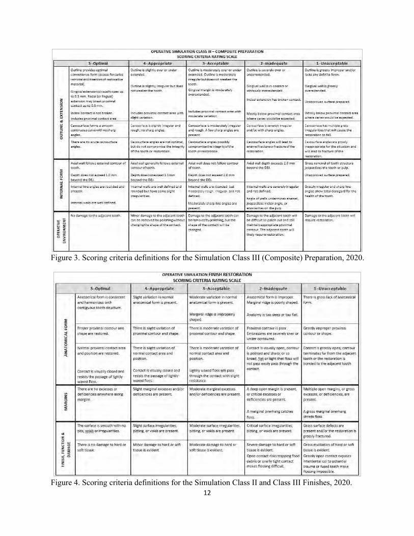
12
Figure 3. Scoring criteria definitions for the Simulation Class III (Composite) Preparation, 2020.
Figure 4. Scoring criteria definitions for the Simulation Class II and Class III Finishes, 2020.

13
Scoring and Results Reporting
Performance for each preparation and finish, is graded by three independent and
anonymous examiners who are calibrated to the scoring criteria prior to every examination. Each
preparation or finish is scored on the applicable criteria according to rating scales presented above.
Examiners are trained to assign a particular grade on the scale only when all aspects of
performance described for that level have been demonstrated. For example, if performance on the
criterion under review meets most aspects of the definition for a grade of “3” but does not quite
meet the standard for even one aspect of the definition, then the grade assigned will be a “2,” at
most. This holds for all six criteria per restoration.
The median of the three examiner grades is computed for each criterion and is weighted to
reflect the level of criticality relevant to minimally competent treatment, e.g., Outline and
Extension accounts for 46% of the preparation score and Operative Environment accounts for only
15%. The criterion weights are provided in Tables 2a and 2b.
Tables 2a and 2b. Operative Simulation Scoring Criteria and Weighting: Preparation, Finish
Preparation Criteria
and Weighting
Finish Criteria
and Weighting
Outline & Extension 46% Anatomical Form 36.5%
Internal Form 39% Margins 36.5%
Operative Environment 15% Finish, Function & Damage 27%
The mean of the preparation and finish scores is the restoration procedure score. The mean
of the two procedure scores, after any applicable penalties or deductions, is the final Operative
Simulation Section score.
The passing cut score on the Operative Simulation Section is 3.00, which reflects
minimally competent performance within the five-point rating scale for all criterion grades that
contribute to the final section score. Each performance level definition for a score of 3.00 on a
criterion has been worded to describe performance that would be deemed minimally competent
via consensus of the subject matter experts on the Operative section examination committee. While
methods of standard setting applied to selected-response assessment often rely on SMEs evaluating
each test question based on how each SME believes a minimally competent examinee would

14
perform, standard setting for many performance-based assessments involves defining minimum
expectations that can be observed directly in the candidate’s performance. The performance level
definitions (Figures 1 through 4), as developed by the examination committee, are critical to
guiding examiner grading. The definitions are used to describe examples of clinical performance
reviewed during examiner training and calibration, which provides performance benchmarks to
facilitate examiner adherence to the criteria and a high degree of examiner agreement.
While limitations on travel and group activity size due to COVID-19 remain in effect, the
grading of candidate performance will take place in grading sessions after the examination. While
this reduces the number of examiners traveling to and grading at the examination site, it also
prevents candidates from receiving onsite results immediately. Candidates and state licensing
boards will receive results as soon as possible after grading sessions are held. Results reports will
indicate clearly whether the Operative Examination was a simulation or involved the treatment of
a patient. As with all WREB examinations, results of all examination attempts, regardless of pass
or fail outcome, will be available to state licensing boards.
Examiner Training and Calibration
Most examiners are members or designees of their state boards. A small proportion (e.g.,
approximately twenty percent of examiners in 2019) are dental educators. All examiners must be
actively licensed and in good standing, with no license restrictions, and submit proof of license
renewal annually. Under social distancing restrictions, the only examiners that may be present at
the Operative Simulation Section may be the Chief Examiner and one or more Floor Examiners,
depending on the layout and size of the examination environment. There will not be any grading
examiners at the examination site unless social distancing and travel guidelines have been eased
enough to allow this. Under the current restrictions, grading examiners will grade candidate
performance in grading sessions, separate from the examination environment. Grading examiners
still will need to complete examiner self-assessments and calibration testing prior to grading.
Clinical examination scores are dependent upon the judgments of grading examiners. A
high degree of examiner agreement is critical to assessing candidate ability in a reliable and fair
manner. As with the conventional Operative Examination, scoring judgments on the Operative
Simulation Section are made by three independent examiners. The median of the three grades

15
assigned contributes to the candidate’s score. The median is more robust to extreme grades
assigned than the mean (i.e., conventional average).
Having multiple examiners helps to moderate the effects of varying levels of examiner
severity; however, it is essential that all examiners are trained and calibrated to an acceptable level
of agreement with respect to the scoring criteria for the examinations in which they participate.
Examiners must participate in orientation and calibration sessions that take place before every
examination or grading session. During calibration, examiners take assessments (tests) in which
they grade examples of clinical performance according to the grading criteria. Their judgments are
compared to scores that have been previously selected by the examination committees as
representative of the defined levels in the criteria. The examiner team completes calibration tests
until they each have demonstrated that they understand and can consistently apply WREB criteria
in their assessments. All calibration tests are reviewed regularly for content and psychometric
quality by WREB examination committees.
Examiners receive feedback on their performance after each examination. Examiners with
low percentages of agreement, high percentages of harshness or lenience, or erratic grading
patterns are counseled, remediated, and monitored to ensure increased understanding of criteria
definitions. Continued lack of agreement results in dismissal from the examination pool.
The two main approaches employed to evaluate examiner performance include a review of
examiner agreement which reflects the degree of exact and adjacent agreement and an estimation
of examiner severity employing a probabilistic statistical model which is designed to account for
and quantify potential sources of construct-irrelevant variance such as rater bias and error. With
three examiners there are multiple ways to define and track examiner agreement. WREB uses a
conservative computation of exact and adjacent agreement which involves comparing each
examiner rating, i.e., each individual grade assigned to a particular criterion, to the mean of the
other two raters’ grades assigned for the same criterion, within the same examination attempt.
Examiner ratings that may be adjacent to the rating of another rater may still be categorized as
harsh or lenient since agreement is defined as the rating falling within one scale point of the mean
of the other two ratings. Examiner severity is estimated using the Many-faceted Rasch Model
(Linacre, 1994; Rasch, 1960/1980) and allows examiner performance to be compared to the
performance of all other examiners within the examiner pool along a continuum of harshness to
lenience and provides statistical information regarding rater errors such as erratic grading or

16
grading that shows too little discernment among performance levels (e.g., assigning all or mostly
“3”s). Additional details regarding methods and results of examiner evaluation are provided in the
WREB Dental Examination Technical Report (WREB, 2019a)
Field Testing of the Operative Simulation Section: Overview
Two Operative Simulation field-tests were planned and conducted between March and
May of 2020. A total of 79 dental students from two dental schools participated; three students
attempted the examination twice resulting total of 82 attempts. These students planned in advance
to challenge the field test examination twice.
The planning of the field tests included the review and revision of the Operative scoring
criteria, creating a candidate guide for field test candidates, coordinating with each school to
produce social distancing and infection prevention protocols, and developing examiner training
and calibration materials.
One field test was conducted on March 30, 2020 at the University of Oklahoma with 20
dental students. A second field test was held on April 1 and 2, 2020 at the University of Utah with
59 dental students. WREB has already been conducting conventional clinical dental examinations
at these two schools and their campuses were reasonably accessible to WREB’s dental consultants,
given the limitations and recommendations regarding travel due to COVID-19. Oklahoma and
Utah are the states of residence of WREB’s two consulting SME dentists, who oversee
examination development and administration. The field test conducted at the University of
Oklahoma used a simulated tooth constructed of a harder material which generated student
concerns reflected in the post-examination candidate survey comments. The second field test,
conducted at University of Utah, employed the final choice of material which did not elicit these
concerns.
Initial Field Test Results: Faculty-graded
The performance of the 20 field test candidates who attempted the Operative Simulation at
the University of Oklahoma were initially graded by their faculty to partially fulfill program
competency requirements. The 20 scores based on the University of Oklahoma faculty grading
ranged from 2.94 to 4.37, with a mean score of 3.72 (SD = 0.41). Candidate scores (N = 57) from
the same university taking the WREB Operative section during the 2019 season ranged from 3.13

17
to 4.87, with a mean score of 3.90 (SD = 0.40). The field test results were not as high as the
examination results from 2019, but an independent samples t-test conducted to compare the results
indicated that the difference is not significant, with a value of t (df = 75; α = 0.05) = 1.67 and mean
difference of 0.17 (p = 0.10; 95% CI: -0.03, 0.38). The comparison is based on a small sample but
provides an initial indication of comparability. There was also no notable difference between mean
scores of the anterior tooth (3.73, SD = 0.51) and the posterior tooth (3.71, SD = 0.44) for the
faculty-graded teeth.
After the examination and the grading conducted by faculty, some of the teeth that had
been treated by the candidates at the University of Oklahoma field test were modified to reflect
specific descriptors in the scoring criteria. These modified teeth and examples of candidate
performance were then used in developing examiner training materials. The resulting preparations
and finished restorations were photographed and used as exemplars in examiner training and
calibration testing. The modified teeth will be graded along with the field-test performances from
the other field test examination site, but will also be analyzed separately, as they do not represent
the candidates’ original performance.
Treatment Times
Candidates were allowed up to four hours to complete the Operative Simulation Field Test.
The time spent preparing the preparations and the finishes was recorded for each field-test attempt
to determine if the initial time allotted was sufficient. The average total time used for the 82 field
test attempts was 2 hours, 10 minutes (130 minutes). The least amount of time needed was 1 hour,
22 minutes and the longest amount of time needed was 3 hours, 52 minutes. All but four candidates
(4.8%) completed their procedures in less than 3 hours and 30 minutes. The University of
Oklahoma site used more treatment time due to additional time needed for set-up between the
preparation and finish procedures. The need for this additional time was eliminated with the use
of a single tooth material for the second field test. The time allotted for the examination going
forward was reduced to 3 hours and 30 minutes. Table 3 shows the treatment times per field test
site.
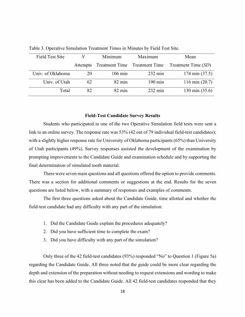
18
Table 3. Operative Simulation Treatment Times in Minutes by Field Test Site.
Field Test Site N
Attempts
Minimum
Treatment Time
Maximum
Treatment Time
Mean
Treatment Time (SD)
Univ. of Oklahoma 20 106 min 232 min 174 min (37.5)
Univ. of Utah 62 82 min 190 min 116 min (20.7)
Total 82 82 min 232 min 130 min (35.6)
Field-Test Candidate Survey Results
Students who participated in one of the two Operative Simulation field tests were sent a
link to an online survey. The response rate was 53% (42 out of 79 individual field-test candidates);
with a slightly higher response rate for University of Oklahoma participants (65%) than University
of Utah participants (49%). Survey responses assisted the development of the examination by
prompting improvements to the Candidate Guide and examination schedule and by supporting the
final determination of simulated tooth material.
There were seven main questions and all questions offered the option to provide comments.
There was a section for additional comments or suggestions at the end. Results for the seven
questions are listed below, with a summary of responses and examples of comments.
The first three questions asked about the Candidate Guide, time allotted and whether the
field-test candidate had any difficulty with any part of the simulation:
1. Did the Candidate Guide explain the procedures adequately?
2. Did you have sufficient time to complete the exam?
3. Did you have difficulty with any part of the simulation?
Only three of the 42 field-test candidates (93%) responded “No” to Question 1 (Figure 5a)
regarding the Candidate Guide. All three noted that the guide could be more clear regarding the
depth and extension of the preparation without needing to request extensions and wording to make
this clear has been added to the Candidate Guide. All 42 field-test candidates responded that they

19
had sufficient time to complete the examination (Figure 5b). Eight of the 42 respondents (19%)
expressed difficulty with part of the simulation (Figure 5c). In the optional comments, most of
these concerns were about the difficulty of adjacent teeth having differing degrees of hardness; all
were from field-test candidates at the University of Oklahoma, where a different tooth material
was tested. The material that was employed at the second field test did not elicit these concerns
and is the final choice of material planned for the Operative Simulation Section.
Figures 5a, b, c. Proportion of Yes or No responses to Field-Test Survey Questions 1, 2 and 3.
Question 4 asked about the level of challenge posed by the examination, overall.
4. Overall, was the exam easy, moderate, or difficult?”
Most respondents (37 of 42 or 88%) answered “Moderate” to Question 4 (Figure 6). Most
comments offered regarding Question 4 compared the simulated teeth to natural teeth, e.g., “Going
back to cutting on typodonts is always a readjustment! But definitely a valid test of hand skills.
Certain aspects are more difficult and certain aspects are less difficult compared to treating human
patients” and “The teeth were much softer, so probably required more dexterity than doing it on
an actual person but very doable.”
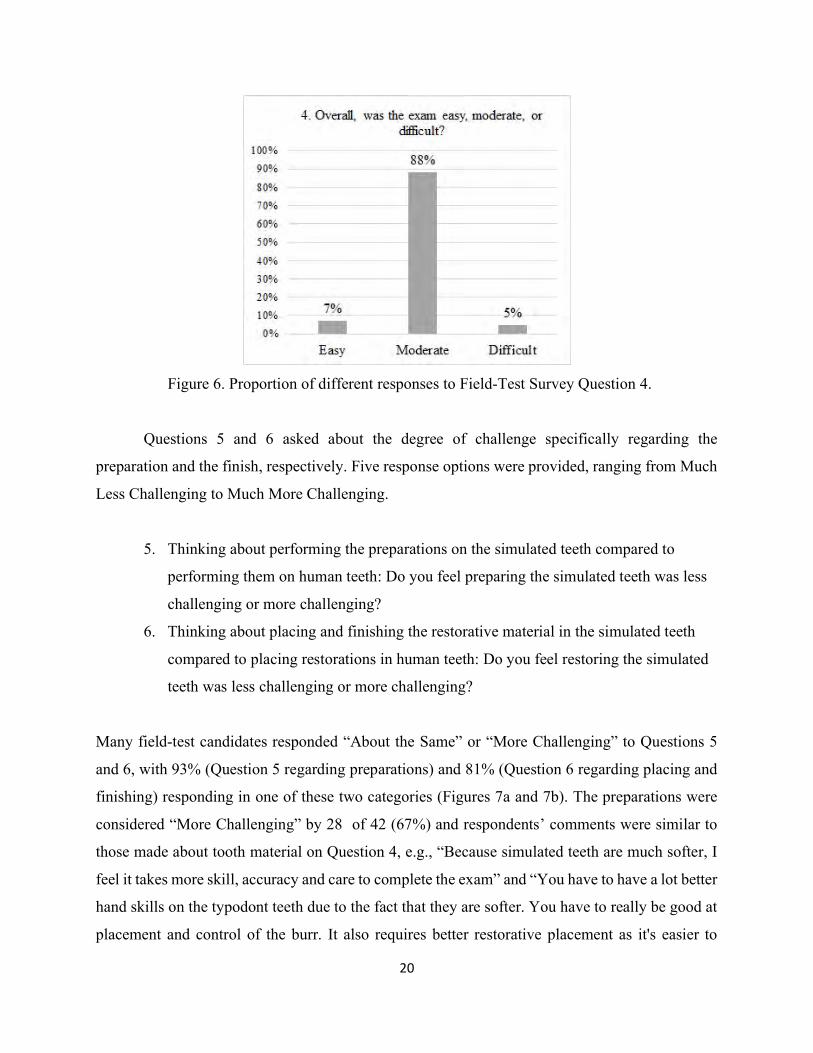
20
Figure 6. Proportion of different responses to Field-Test Survey Question 4.
Questions 5 and 6 asked about the degree of challenge specifically regarding the
preparation and the finish, respectively. Five response options were provided, ranging from Much
Less Challenging to Much More Challenging.
5. Thinking about performing the preparations on the simulated teeth compared to
performing them on human teeth: Do you feel preparing the simulated teeth was less
challenging or more challenging?
6. Thinking about placing and finishing the restorative material in the simulated teeth
compared to placing restorations in human teeth: Do you feel restoring the simulated
teeth was less challenging or more challenging?
Many field-test candidates responded “About the Same” or “More Challenging” to Questions 5
and 6, with 93% (Question 5 regarding preparations) and 81% (Question 6 regarding placing and
finishing) responding in one of these two categories (Figures 7a and 7b). The preparations were
considered “More Challenging” by 28 of 42 (67%) and respondents’ comments were similar to
those made about tooth material on Question 4, e.g., “Because simulated teeth are much softer, I
feel it takes more skill, accuracy and care to complete the exam” and “You have to have a lot better
hand skills on the typodont teeth due to the fact that they are softer. You have to really be good at
placement and control of the burr. It also requires better restorative placement as it's easier to

21
accidentally remove tooth while finishing and polishing.” An example comment from one of the
eleven (26%) respondents who selected “About the Same” stated, “More challenging due to the
lack of recent practice on teeth with this hardness, but less challenging due to known parameters
and no need for modifications.”
Nineteen of 42 (45%) respondents felt that the placing and finishing of the teeth was
“About the Same” but only a few offered comments, e.g., “Less challenging due to no need for
etching, more challenging from the difference in stability (possible loose screws, extremely tight
contacts, no wedging ability).” The source of the loose screws was identified and remedied prior
to the second field test. Most comments were associated with the fifteen (36%) responses of “More
Challenging,” and involved the tooth material, e.g., “I felt placing the material was the same but
polishing and removing flash was much more difficult on typodont teeth” and “Polishing
composite on real teeth is MUCH easier than polishing on typodont teeth.” The few comments that
accompanied the seven (17%) responses of “Less Challenging” reflected dryness and isolation,
e.g., “Obviously, there isn’t any saliva, so keeping a dry field is simple” and “Better isolation.”
Figures 7a, b. Proportion of different responses to Field-Test Survey Questions 5 and 6.

22
Question 7 asked about the ability to maintain social distancing at the examination.
7. How difficult was it for you to maintain social distancing during the examination?
Most field-test candidates (39 of 42 or 93%) responded that it was “Easy” to maintain social
distancing during the examination (Figure 8). All but one comment were associated with responses
of “Easy.” Examples include “Really strict and functional rules in place. Wasn’t a problem at all”
and “I was at least ten feet away from anyone else in the room at all times.” The other comment,
associated with a response of Moderate, stated, “During the announcement portion of the exam,
prior to the beginning, it was moderately difficult to maintain social distancing and adequately
hear the announcements and questions.” Plans have been implemented for additional information
to be provided early to candidates, allowing for questions by phone or email prior to the
examination to reduce the need for multiple announcements and possible reasons to encourage
crowding.
Figure 8. Proportion of different responses to Field-Test Survey Question 7.
Field-test candidates could offer additional comments or suggestions at the end of the
survey. Many comments were generally positive or expressed thanks, e.g., “Overall it was great!”
and several expressed their interest that this type of restorative examination be an acceptable option
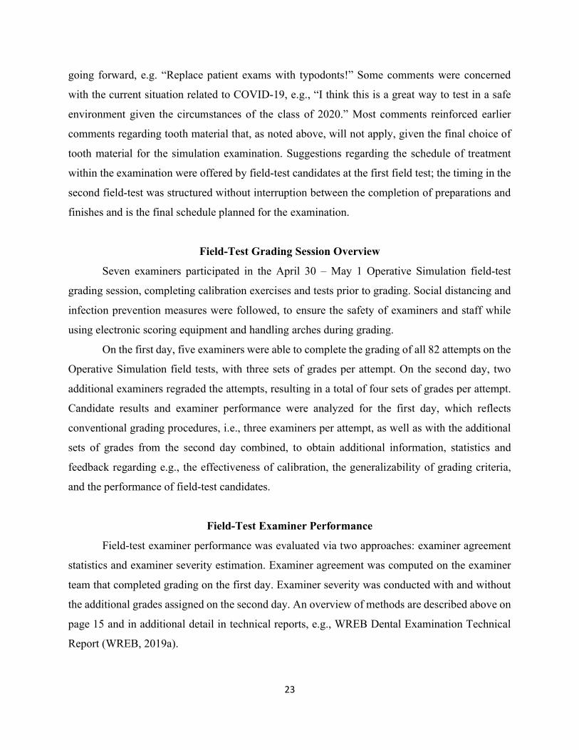
23
going forward, e.g. “Replace patient exams with typodonts!” Some comments were concerned
with the current situation related to COVID-19, e.g., “I think this is a great way to test in a safe
environment given the circumstances of the class of 2020.” Most comments reinforced earlier
comments regarding tooth material that, as noted above, will not apply, given the final choice of
tooth material for the simulation examination. Suggestions regarding the schedule of treatment
within the examination were offered by field-test candidates at the first field test; the timing in the
second field-test was structured without interruption between the completion of preparations and
finishes and is the final schedule planned for the examination.
Field-Test Grading Session Overview
Seven examiners participated in the April 30 – May 1 Operative Simulation field-test
grading session, completing calibration exercises and tests prior to grading. Social distancing and
infection prevention measures were followed, to ensure the safety of examiners and staff while
using electronic scoring equipment and handling arches during grading.
On the first day, five examiners were able to complete the grading of all 82 attempts on the
Operative Simulation field tests, with three sets of grades per attempt. On the second day, two
additional examiners regraded the attempts, resulting in a total of four sets of grades per attempt.
Candidate results and examiner performance were analyzed for the first day, which reflects
conventional grading procedures, i.e., three examiners per attempt, as well as with the additional
sets of grades from the second day combined, to obtain additional information, statistics and
feedback regarding e.g., the effectiveness of calibration, the generalizability of grading criteria,
and the performance of field-test candidates.
Field-Test Examiner Performance
Field-test examiner performance was evaluated via two approaches: examiner agreement
statistics and examiner severity estimation. Examiner agreement was computed on the examiner
team that completed grading on the first day. Examiner severity was conducted with and without
the additional grades assigned on the second day. An overview of methods are described above on
page 15 and in additional detail in technical reports, e.g., WREB Dental Examination Technical
Report (WREB, 2019a).

24
Percentages of agreement were computed for the three sets of grades assigned on the first
day of grading, as would be conducted for an actual examination after all three sets of grades per
attempt have been assigned. Over the past ten years, percentages of agreement for the standard
Operative Section have ranged from 88.4% to 89.9%, with comparatively balanced percentages of
harshness and lenience. Examiner agreement over the years reflects examiner grading teams that
have been selected for each examination based on their past examiner performance to ensure an
optimal balance of examiner severity level. While nearly all examiners perform within
recommended ranges of harshness and lenience percentages, to assign all the examiners that have
performed at one end of that continuum to a single examination could introduce a systematic bias.
The examiners who participated in the field-test grading session were scheduled based on location
and convenience, given the conditions posed by COVID-19. The field-test examiners also included
two relatively new examiners, who would not be assigned to the same examination under
conventional conditions. Despite these potential threats to optimal examiner team performance,
examiner agreement statistics for the field-test grading session were comparable to percentages of
agreement, harshness, and lenience for the standard Operative section in previous years. Table 4
provides examiner agreement percentages for the standard Operative Section from the 2019 season
and for the Operative Simulation field test grading session.
Table 4. Percentages of Examiner Agreement, Harshness, and Lenience: Standard Operative
Section and Operative Simulation Field Test
N Examiners % Harsh % Lenient % Agreement
Standard Operative Section
2019 Season 110 5.5% 5.3% 89.2%
Operative Simulation
Field Test Day 1 5 5.6% 5.7% 88.7%
Examiner severity estimated with the many-faceted Rasch model, is reported in Table 5,
which provides summaries of results in logit, i.e., log-odds, units. High negative logits reflect more
lenience and high positive logits reflect more harshness. For the standard Operative Section
examination, most examiners fall within one logit unit of the mean, i.e., between -1.00 and 1.00,
and within recommended ranges with respect to infit and outfit mean-square fit statistics, i.e.,

25
between 0.50 and 1.50. Examiner severity estimates for the first day of the Operative Simulation
field test and for all Operative Simulation field-test examiners reflect smaller ranges with no
outlying values. Additional details of the Many-faceted Rasch Model analyses are provided later
with the results of field-test candidate performance.
Table 5. Many-Faceted Rasch Model Examiner Severity Analysis Indicators in Logits: Standard
Operative Section and Operative Simulation Field Test (Number of examiners provided below
each header)
Indicator
Standard Operative Section 2019 Season
(NE = 110)
Operative Simulation Field Test Day 1
(NE = 5)
Operative Simulation Field Test All
(NE = 7)
Severity Measure Logit (Range)
-0.88 – 1.06 -0.41 – 0.44 -0.33 – 0.52
Standard Error (Range)
0.05 – 0.16 0.05 – 0.07 0.05 – 0.07
Severity Measure Logit Meana
0.0 0.0 0.0
Severity Measure Logit SD
0.42 0.33 0.31
Infit Mean-Square (Range)
0.54 – 1.77 0.71 – 1.25 0.66 – 1.38
Outfit Mean-Square (Range)
0.52 – 1.72 0.72 – 1.22 0.66 – 1.32
a Mean of examiner severity parameters constrained at 0.
Field-Test Examiner Survey Results
The seven examiners who participated in the Operative Simulation field test grading
session were sent a link to an online survey. The response rate was 100%. There were eight main
questions and all questions offered the option to provide comments. There was a section for
additional comments or suggestions at the end. Results for the eight questions are listed below,
with a summary of responses and examples of comments.
Examiners responded unanimously to the first five questions, which asked about materials,
instrumentation provided, difficulty of the grading tasks, as well as their understanding of, and
ability to follow, the social distancing protocol. Possible responses to the first five questions were

26
Yes or No, except for Question 3, with possible responses of Easy, Moderate, or Difficult. The
first five questions and the common responses are provided in Table 6.
Table 6. Operative Simulation Grading Session Field-Test Examiner Survey Questions 1 to 5
with Responses
Questions 1 to 5 Unanimous Response
1. Did the Candidate Guide and Examiner Manual adequately explain the simulation and grading procedures?
Yes, 100%
2. Were the social (physical) distancing instructions clear and easy to understand?
Yes, 100%
3. How difficult was it for you to maintain appropriate social (physical) distancing while serving as an examiner?
Easy, 100%
4. Did you have difficulty with any of the grading tasks? No, 100%
5. Was the instrumentation provided for your use, everything you needed?
Yes, 100%
Optional comments associated with the first five questions were positive, e.g., regarding
ability to maintain social distancing, (Question 3), “I felt very safe” and regarding grading tasks
(Question 4), “Calibration was well orchestrated and provided the preparation necessary for us as
examiners to perform efficiently and effectively. Nice job!”
Question 6 asked the field-test examiners about how well the calibration exercises prepared
them for grading. Figure 9 illustrates the percentages of each response. Five examiners (71%)
responded “Very well.” One commented, “It was my first time actually grading so it was very
helpful to me.” Two (29%) responded “Well enough” accompanied by the following two
comments, “Too detailed which sometimes can create more issues than being useful” and “This
was a new exam but we made do,” which suggest that continued review and refinement may be
useful. The criteria has already been evaluated and edited based on examiner feedback.

27
Figure 9. Proportion of different responses to Examiner Survey Question 6.
The grading criteria are nearly the same as the criteria used for the standard Operative
Section, except for the removal of a few items, such as caries, pulp exposure and rubber dam
isolation that do not apply for the Operative Simulation section. Question 7 asked the field-test
examiners how well the modified criteria work for the simulation. Figure 10 shows the percentages
of each response. Six examiners (86%) responded “Very well” or “Well enough,” evenly split
between the two responses. One examiner responded “Unsure.” Only one comment was offered,
“I think it’s easier to see mistakes on a manikin than in the mouth.”
Figure 10. Proportion of different responses to Examiner Survey Question 7.
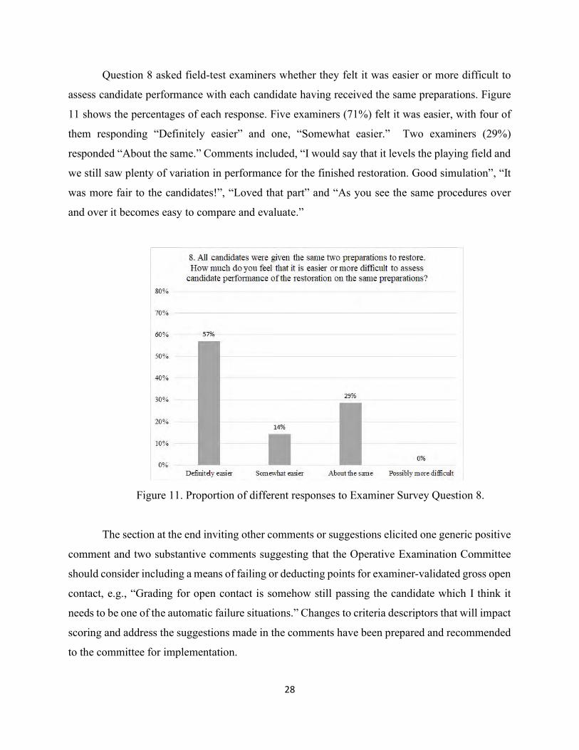
28
Question 8 asked field-test examiners whether they felt it was easier or more difficult to
assess candidate performance with each candidate having received the same preparations. Figure
11 shows the percentages of each response. Five examiners (71%) felt it was easier, with four of
them responding “Definitely easier” and one, “Somewhat easier.” Two examiners (29%)
responded “About the same.” Comments included, “I would say that it levels the playing field and
we still saw plenty of variation in performance for the finished restoration. Good simulation”, “It
was more fair to the candidates!”, “Loved that part” and “As you see the same procedures over
and over it becomes easy to compare and evaluate.”
Figure 11. Proportion of different responses to Examiner Survey Question 8.
The section at the end inviting other comments or suggestions elicited one generic positive
comment and two substantive comments suggesting that the Operative Examination Committee
should consider including a means of failing or deducting points for examiner-validated gross open
contact, e.g., “Grading for open contact is somehow still passing the candidate which I think it
needs to be one of the automatic failure situations.” Changes to criteria descriptors that will impact
scoring and address the suggestions made in the comments have been prepared and recommended
to the committee for implementation.

29
Field Test Results: Candidate Performance and Test Quality
Table 7 provides basic descriptive statistics for the raw and weighted means of medians
computed from the three sets of examiner grades for each criterion. Direct comparisons to the
standard Operative Section, particularly regarding criterion scores, are limited due to three factors.
One is that only 5.5% of procedures performed for the standard Operative Section in 2019 were
Class III procedures. All field-test attempts on the Operative Simulation Section included a Class
III procedure. Since 2018, most states are accepting the results of performance on one Class II
procedure if competence is demonstrated, so many candidates are completing Class II procedures.
Years of Operative Section data have shown that the Class III is slightly, but significantly, less
challenging than any Class II procedure and therefore, if completed, must be in combination with
a Class II procedure. The second limiting factor is that many arches completed in the first, smaller
field test, were modified to create additional exemplars of grading criteria performance levels
during the development calibration materials and some performance levels may not be distributed
within the sample in a comparable manner. The third factor is that the field-test host schools, which
were chosen for location and convenience, given the conditions posed by COVID-19 and their
students may not be a representative sample of all potential candidates.
Despite field-test limitations to direct comparison, three criteria and final scores (which
include point deductions from penalties and loss of all points due to critical errors) were highly
comparable. The slightly higher final score mean reflects a more negatively skewed distribution
in the field test data; the passing percentage is actually somewhat lower for the field test than the
standard Operative section in 2019. The significantly higher means of raw scores and some criteria
for the field-tests may be related to the difference in procedure type in the comparison, particularly
for Anatomical Form and Margins, which have traditionally scored significantly higher for the
Class III procedure. Recent additions, since the field-test, to the criterion definitions for Internal
Form related to grading examiner feedback are also expected to result in higher comparability.

30
Table 7. Grading Criteria and Section Scores for Standard Operative Section and Operative
Simulation Field Test: Means and Standard Deviations of Raw Unweighted Class II Median
Criterion Scores, Raw and Final Scores, with t-Tests. Included are t values, probability values (p),
effect size values (Cohen’s d) degrees of freedom (df), and alpha level (α), i.e., significance below
0.05. Number of procedures noted as Np, number of attempts noted as N.
Standard Operative
Section 2019
Np = 2,553a
Operative Simulation Field
Test 2020
Np = 164a
t-tests
df = 2,715; α = 0.05
Mean SD Mean SD
t value
p value
Cohen’s d b
Outline and Extension 3.63 0.75 3.65 0.85 -0.27 0.79 0.02
Internal Form 3.62 0.74 3.85 0.65 -3.90 <0.01 0.33
Operative Environment 4.27 0.67 4.19 0.76 1.42 0.16 0.11
Anatomical Form 3.60 0.70 3.99 0.81 -6.86 <0.01 0.52
Margins 3.65 0.66 3.99 0.72 -6.32 <0.01 0.49
Finish, Function, & Damage 3.94 0.59 3.88 0.85 1.23 0.22 0.08
N = 2,166 N = 82 df = 2,246
Overall Raw Score 3.74 0.46 3.88 0.44 -2.76 0.01 0.31
Overall Final Score
(with Penalties) 3.71 0.53 3.75 0.75 -0.69 0.49 0.06
a Only 5.5% of procedures performed in 2019 were Class III; 50% of Field test Procedures were Class III b Generally accepted interpretations of Cohen’s d effect size values are small, d = 0.2, medium, d = 0.5 and large, d = 0.8 (Cohen, 1988)
Table 8 provides field-test summary results from the many-faceted Rasch model (MFRM)
analysis for graded criteria in logit, i.e., log-odds, values, with results from the 2019 standard
Operative Section for reference. The MFRM analysis reported in Table 8 reflects the first day of
grading, with complete sets of three grades per examination attempt. Mean-square fit statistics and
discrimination parameter estimates are within suggested ranges. Since the criteria have multi-point

31
rating scales they were also assessed for category functioning, as well, in accordance with
Linacre’s (2002) rating scale guidelines to assess, e.g., that average parameter estimates of
candidate ability increase with each category scale point.
Table 8. Standard Operative Section and Operative Simulation Field Test: Many-Faceted Rasch
Model Criterion Analysis Indicators in Logits.
Standard Operative Section 2019
N = 2,166
Operative Simulation Field Test 2020
N = 82
Criterion Measure Logit (Range) -0.78 – 0.39 -0.37 – 0.43
Standard Error (Range) 0.02 – 0.02 0.08 – 0.10
Criterion Measure Logit Meana 0.0 0.0
Criterion Measure Logit SD 0.50 0.25
Many-Facet Point-Biserial rb (Range) 0.25 – 0.32 0.23 – 0.37
2pl Discrimination Estimatec (Range) 0.92 – 1.08 0.76 – 1.10
Infit Mean-Square (Range) 0.93 – 1.07 0.85 – 1.19
Outfit Mean-Square (Range) 0.92 – 1.08 0.85 – 1.21 a Mean of criterion parameters constrained at 0
b Correlation between observations and corresponding average observations, excluding current observation
c Estimate of discrimination parameter, as calculated for two-parameter logistic IRT model; Rasch (c.f., one-parameter IRT) model fit requires values close to 1.00 (i.e., between 0.5 to 1.5 logits)
Table 9 provides summary statistics for overall test functioning, with 2019 standard
Operative Section results for reference. The MFRM analysis reported in Table 9 also reflects the
first day complete sets of three grades per examination attempt. Results are highly comparable,
even with the large difference in sample size and limitations regarding comparisons noted earlier.
The reliability estimate for the Operative Simulation Field Test is quite high for a performance-
based assessment, at 0.91, which likely reflects the uniformity of the simulated teeth, in addition
to high levels of examiner agreement. An additional MFRM analysis was conducted including all
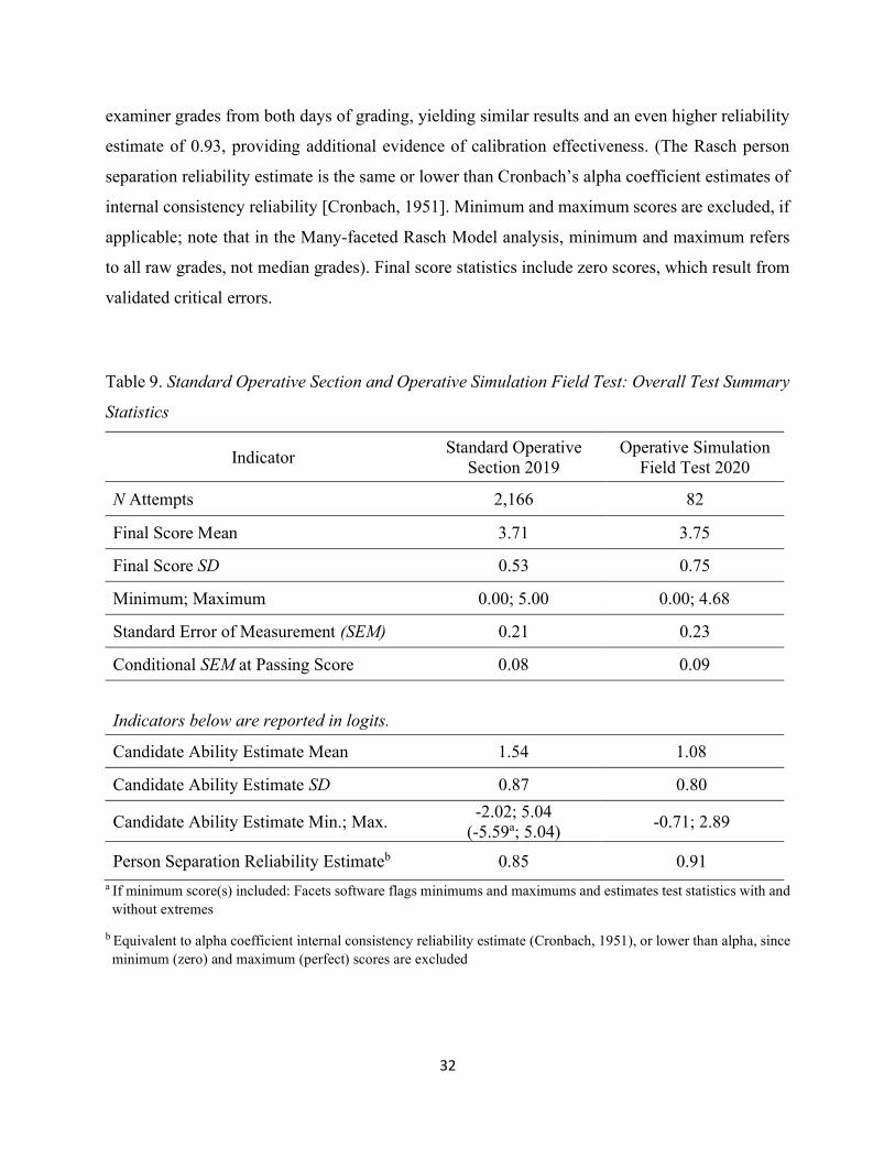
32
examiner grades from both days of grading, yielding similar results and an even higher reliability
estimate of 0.93, providing additional evidence of calibration effectiveness. (The Rasch person
separation reliability estimate is the same or lower than Cronbach’s alpha coefficient estimates of
internal consistency reliability [Cronbach, 1951]. Minimum and maximum scores are excluded, if
applicable; note that in the Many-faceted Rasch Model analysis, minimum and maximum refers
to all raw grades, not median grades). Final score statistics include zero scores, which result from
validated critical errors.
Table 9. Standard Operative Section and Operative Simulation Field Test: Overall Test Summary
Statistics
Indicator Standard Operative
Section 2019 Operative Simulation
Field Test 2020
N Attempts 2,166 82
Final Score Mean 3.71 3.75
Final Score SD 0.53 0.75
Minimum; Maximum 0.00; 5.00 0.00; 4.68
Standard Error of Measurement (SEM) 0.21 0.23
Conditional SEM at Passing Score 0.08 0.09
Indicators below are reported in logits.
Candidate Ability Estimate Mean 1.54 1.08
Candidate Ability Estimate SD 0.87 0.80
Candidate Ability Estimate Min.; Max. -2.02; 5.04
(-5.59a; 5.04) -0.71; 2.89
Person Separation Reliability Estimateb 0.85 0.91 a If minimum score(s) included: Facets software flags minimums and maximums and estimates test statistics with and without extremes
b Equivalent to alpha coefficient internal consistency reliability estimate (Cronbach, 1951), or lower than alpha, since minimum (zero) and maximum (perfect) scores are excluded

33
The percentage of candidates that scored at or above the passing cut score on the Operative
Simulation field tests was 92.7% (76 out of 82). The passing percentage for the second, larger field
test was lower than that of the first, due to penalties, including two attempts with validated critical
errors (e.g., treated the wrong tooth) that lost all points. Table 10 provides passing percentages for
the two Operative Simulation field tests, with the 2019 standard Operative Section passing
percentage for reference.
Table 10. Standard Operative Section and Operative Simulation Field Test: Passing Percentages
N Attempts
Passing
Count
Failing
Count
Passing
Percentage
Standard Operative Section
2019 Season 2,166 2,079 87 96.0%
Operative Simulation
Field Test 2020 - Total 82 76 6 92.7%
Field Test First Site
March 30, 2020 (U. of OK) 20 19 1 95.0%
Field Test Second Site
April 1-2, 2020 (U. of UT) 62 57 5 91.9%
34
REFERENCES
American Association of Dental Boards (2005). Guidance for Clinical Licensure Examinations in
Dentistry. Chicago, IL: AADB.
American Educational Research Association, American Psychological Association, & National
Council on Measurement in Education. (2014). Standards for Educational and
Psychological Testing. Washington, DC: American Educational Research Association.
Cohen, J. (1988). Statistical power analysis for the behavioral sciences (2nd ed.). Hillsdale, NJ: Lawrence Earlbaum Associates.
Cronbach, L. J. (1951). Coefficient alpha and the internal structure of tests. Psychometrika,16, 297-334.
Linacre, J. M., Engelhard, G., Jr., Tatum, D. S., & Myford, C. M. (1994). Measurement with
judges: Many-faceted conjoint measurement. International Journal of Educational
Research, 21(6), 569-577.
Linacre, J. M. (2002). Optimizing rating scale category effectiveness. Journal of Applied Measurement, 3(1), 85-106.
Rasch, G. (1960/1980). Probabilistic models for some intelligence and attainment tests.
Copenhagen: Danish Institute for Educational Research, 1960. Expanded edition, Chicago:
The University of Chicago Press, 1980.
Western Regional Examining Board. (2019a). WREB 2018 Dental Examination Technical Report.
Phoenix, AZ: WREB.
Western Regional Examining Board. (2019b). WREB Practice Analysis for General Dentist.
Phoenix, AZ: WREB.
Western Regional Examining Board. (2020 report in progress). WREB Dental Hygiene Practice
Analysis Report. Phoenix, AZ: WREB.

35
Sharon E. Osborn Popp, Ph.D.
WREB Testing Specialist/ Psychometrician
Copyright 2020 WREB - A National Dental and Dental Hygiene Testing Agency
23460 N. 19th Avenue, Suite 210 - Phoenix, Arizona 85027
phone: 602-209-5400 fax: 602-371-8131

June 9, 2020 Dr. Peter S. Katz, Chair Connecticut State Dental Commission 410 Capitol Avenue, MS #13PHO P. O. Box 340308 Hartford, CT 06134-0308 Dear Dr. Katz, The Joint Commission on National Dental Examinations (JCNDE) is aware of recent actions taken by the Connecticut State Dental Commission (CSDC) in accordance with Public Act 19-72 to consider alternative dental licensure examinations that do not require single encounter, procedure-based patient examinations. Representatives of the JCNDE would be pleased to speak with members of the CSDC to discuss the JCNDE’s new dental clinical examination—the Dental Licensure Objective Structured Clinical Examination (DLOSCE)—which will be available for use by US dental boards effective June 15, 2020. On April 2, 2020, the JCNDE released an announcement (attached) to all US dental boards concerning the launch of this new examination. The DLOSCE is a content-valid examination built specifically for clinical licensure purposes that assesses candidates’ clinical judgment and skills using sophisticated 3-D models, without the need to involve patients. On April 22, the JCNDE conducted a webinar on the DLOSCE specifically for state dental board members, and was pleased that representatives of 24 dental boards attended. The JCNDE website includes additional information about the DLOSCE which may be of interest to members of the CSDC, including the DLOSCE candidate guide, practice questions, and 3-D model tutorial. The JCNDE is confident that the DLOSCE is a strong examination that is well-suited for use in addressing the clinical examination licensure requirements of dental boards, and is particularly responsive to pressing needs in today’s challenging times. Representatives of the JCNDE, including myself, would welcome the opportunity to speak with the CSDC at its next meeting to further discuss acceptance of the DLOSCE for licensure purposes in Connecticut. Dr. David Waldschmidt, director of the JCNDE, would be pleased to work with Dental Commission staff and you regarding meeting arrangements. Sincerely,
Cataldo W. Leone, DMD, DMedSc, FACD, FICD, Chair Joint Commission on National Dental Examinations Cc: Mr. Jeffrey A. Kardys, Administrative Hearings Specialist, Board Liaison, Connecticut State Dental Commission Ms. Carol Dingeldey, Executive Director, Connecticut State Dental Association Dr. David Waldschmidt, Director, Joint Commission on National Dental Examinations

To: US Dental Boards, the Dental Education Community, Dental Licensure Candidates
From: Dr. Cataldo Leone, Chair of the Joint Commission on National Dental Examinations (JCNDE)
Date: April 2, 2020
Subject: Official Release of the JCNDE’s Dental Licensure Objective Structured Clinical Examination (DLOSCE)
The Joint Commission on National Dental Examinations (JCNDE) has a long and distinguished track record of providing valid and reliable high-stakes examinations for licensure purposes, to protect the public health. We continue to innovate in our efforts to serve the needs of those who use our examinations to inform their decisions. This innovation and drive for excellence is more important than ever given these uncertain times, the challenges that face all of us, and the implications of JCNDE examinations for stakeholders, communities of interest, and the public. In light of the preceding, I am pleased to announce that on March 31, 2020 the JCNDE approved a resolution to make its newest examination—the Dental Licensure Objective Structured Clinical Examination (DLOSCE)—available for use in 2020 by dental boards in the US. The DLOSCE is a content-valid examination built specifically for clinical licensure purposes that assesses candidates’ clinical judgment and skills using sophisticated 3-D models, without the need to involve patients. The JCNDE is targeting Monday, June 15, 2020 as the official date of release, pending test center availability. The DLOSCE is comprehensive in its assessment of clinical judgment, including content in the following areas: Restorative Dentistry; Prosthodontics; Oral Pathology, Pain Management and TMD; Periodontics; Oral Surgery; Endodontics; Orthodontics; Medical Emergencies; and Prescriptions. Diagnosis, Treatment Planning, and Occlusion are assessed across the aforementioned topic areas. DLOSCE content has been developed by teams of highly qualified subject matter experts, working together to build examination questions that are capable of accurately and reliably identifying those who possess the clinical skills necessary to safely practice dentistry. Use of the DLOSCE is supported by content validity arguments, the same type of validity evidence that is used to support the JCNDE’s other examination programs–the National Board Dental Examinations Parts I and II, the National Board Dental Hygiene Examination, and the Integrated National Board Dental Examination. The DLOSCE complements these other examination programs, advancing the work of the JCNDE as it pursues its vision to serve as the nation’s leading resource for supporting standards of oral healthcare professionals through valid, reliable and fair assessments for licensure and certification. The JCNDE understands that stakeholders and communities of interest will have many questions concerning the DLOSCE. With this in mind, the JCNDE will be conducting webinars in coming weeks, to share additional information in preparation for the release of this important new examination program. Each webinar will focus on a particular stakeholder group or community of interest (dental boards, dental educators, and students), answering questions and providing each group with information that is directly relevant to their specific needs. In the
meantime, updates on the DLOSCE will continue to be shared through the JCNDE’s DLOSCE webpage (ada.org/dlosce). In closing, the JCNDE would like to take this opportunity to thank dental boards for their continued reliance on the examinations of the JCNDE. The JCNDE is confident that the DLOSCE is a strong examination that is well-suited for use in addressing the clinical examination licensure requirements of each board, and is particularly responsive to their pressing needs in these challenging times. The JCNDE looks forward to working closely with all boards interested in accepting the DLOSCE for licensure purposes in 2020 and beyond.
The Joint Commission on National Dental Examinations
Cataldo W. Leone, DMD, DMedSc, FACD, FICD, Chair Kanthasamy Ragunanthan, DDS, MS, Vice Chair Joanna Allaire, RDH, MDH John D. Da Silva, DMD, MPH, ScM Roy L. Irons, DDS Michael E. King, DDS Frank A. Maggio, DDS Brandon Rensch, B.S. William F. Robinson, DDS R. Michael Sanders, DMD Mary A. Starsiak, DDS, FAGD, FACD, FICD, FADI, FPF Patrick J. Tepe, DDS Wesley Delaine Thomas, DMD Douglas C. Wilson, Ph.D. Joseph J. Zambon, DDS, PhD Laura Jeannie Binder, ASDA Student Observer James D. Stephens, DDS, ADA Board of Trustees Liaison
DLOSCE Steering Committee
W. Roy Thompson, DDS Chair Craig S. Armstrong, DDS David L. Carsten, DDS Edward J. Hebert, DDS Michael J. Kanellis, DDS, MS Cataldo W. Leone, DMD, DMedSc, FACD, FICD Frank W. Licari, DDS, MPH, MBA Prabu Raman, DDS, MICCMO, LVIM, FACD William F. Robinson, DDS Mark R. Stetzel, DDS