
OpenCommons@UConn - University of Connecticut
207
University of Connecticut OpenCommons@UConn Doctoral Dissertations University of Connecticut Graduate School 8-9-2019 Investigating the Role of Neuraminidase Activity in the Co-pathogenesis of Mycoplasma gallisepticum and Low Pathogenic Avian Influenza A Virus Jessica Canter University of Connecticut - Storrs, [email protected] Follow this and additional works at: hps://opencommons.uconn.edu/dissertations Recommended Citation Canter, Jessica, "Investigating the Role of Neuraminidase Activity in the Co-pathogenesis of Mycoplasma gallisepticum and Low Pathogenic Avian Influenza A Virus" (2019). Doctoral Dissertations. 2287. hps://opencommons.uconn.edu/dissertations/2287
-
Upload
khangminh22 -
Category
Documents
-
view
0 -
download
0
Transcript of OpenCommons@UConn - University of Connecticut
University of ConnecticutOpenCommons@UConn
Doctoral Dissertations University of Connecticut Graduate School
8-9-2019
Investigating the Role of Neuraminidase Activity inthe Co-pathogenesis of Mycoplasma gallisepticumand Low Pathogenic Avian Influenza A VirusJessica CanterUniversity of Connecticut - Storrs, [email protected]
Follow this and additional works at: https://opencommons.uconn.edu/dissertations
Recommended CitationCanter, Jessica, "Investigating the Role of Neuraminidase Activity in the Co-pathogenesis of Mycoplasma gallisepticum and LowPathogenic Avian Influenza A Virus" (2019). Doctoral Dissertations. 2287.https://opencommons.uconn.edu/dissertations/2287
Investigating the Role of Neuraminidase Activity in the Co-pathogenesis of
Mycoplasma gallisepticum and Low Pathogenic Avian Influenza A Virus
Jessica A. Canter, PhD
University of Connecticut, 2019
Abstract
The avian pathogen Mycoplasma gallisepticum, the etiologic agent of chronic
respiratory disease in chickens, exhibits enhanced pathogenesis in the presence of a
co-pathogen such as low-pathogenic avian influenza virus (LPAIV). M. gallisepticum
Rlow can utilize α-2,3 linked sialic acids, abundant in the avian respiratory tract, to bind
host cells in vitro. To further investigate the intricacies of this co-pathogenesis, chickens
were mono- or co-infected with either virulent M. gallisepticum strain Rlow, attenuated M.
gallisepticum neuraminidase mutant P1C5 and Mycoplasma specific lipoprotein A
mutant P1H9, or LPAIV H3N8 (A/duck/Ukraine/1963). These chickens were then
assessed for tracheal histopathology, pathogen load, and transcriptomic host response
to infection using RNA-sequencing. Chickens co-infected with M. gallisepticum Rlow
followed by H3N8 exhibited significantly more severe tracheal lesions and mucosal
thickening in response to infection than chickens infected with H3N8 alone. Viral load
was also significantly increased in this group over chickens who were infected first with
H3N8 and subsequently with M. gallisepticum Rlow. The attenuated M. gallisepticum
mutants P1C5 and P1H9, previously shown to be cleared 14 days post-infection, were
able to persist 6 to 7 days post-infection in the presence and absence of co-infection
Jessica A. Canter, University of Connecticut, 2019
with H3N8. P1H9 was able to persist to 14 days post-infection only in the presence of
H3N8. The transcriptional response to mono- and co-infection with M. gallisepticum and
LPAIV highlighted the involvement of differential expression of genes such as TLR4,
TLR15, TLR21, IL-1β, IRF4, MMP1, and MMP9. Pathway and gene ontology analysis of
these differentially expressed genes suggests that co-infection with virulent M.
gallisepticum and LPAIV induces a downregulation of ciliary activity in vivo and alters
the multiple immune-related signaling cascades. Although H3N8 is susceptible to
neuraminidase inhibition by oseltamivir in vitro, this antiviral treatment was not effective
in vivo at reducing H3N8 load in the trachea of co-infected chickens. These data
indicate that the co-pathogenesis of LPAIV and M. gallisepticum is not strictly
neuraminidase-dependent and warrants further experimental understanding.

Investigating the Role of Neuraminidase Activity in the Co-pathogenesis of
Mycoplasma gallisepticum and Low Pathogenic Avian Influenza A Virus
Jessica A. Canter
B.S., Towson University 2012
M.S., Towson University 2015
A Dissertation Submitted In Partial Fulfillment of the
Requirements for the Degree Of Doctor of Philosophy
At the University of Connecticut
August 2019

ii
Copyright Jessica A. Canter
2019

iii
APPROVAL PAGE
Doctor of Philosophy Dissertation
Investigating the Role of Neuraminidase Activity in the Co-pathogenesis of Mycoplasma gallisepticum and Low Pathogenic Avian Influenza A Virus
Presented by Jessica A. Canter, B.S., M.S.
Co-Major Advisor ______________________________________________________________
Steven J. Geary
Co-Major Advisor ______________________________________________________________
Steven M. Szczepanek
Associate Advisor _____________________________________________________________
Meghan A. May
Associate Advisor _____________________________________________________________
Dong-Hun Lee
University of Connecticut, 2019

iv
Acknowledgements
I would first like to thank my major advisors, Dr. Steven Geary an Dr. Steven
Szczepanek for their constant support and guidance throughout my degree process.
The members of my advisory committee, Dr. Meghan May and Dr. Dong-Hun Lee, also
deserve my sincerest thanks for their perspective on experimental design, data
analysis, and overall assistance.
I would also like to extend my gratitude to the members of the Geary Lab, current
and former, for not only giving me the tools to succeed scientifically, but the support of
friendship as well. Edan Tulman and Xiaofen Liao were instrumental in providing great
scientific and technical advice and a welcoming, collegial environment. My fellow
graduate students Katie Pflaum, Jessica Beaudet, and Arlind Mara were absolutely
essential in sharing their skills and experiences during experiments and steadfast
friendship. My thanks also go to my fellow graduate students of the Pathobiology and
Veterinary Sciences department here at UConn for their comradery and support as well
as the faculty and staff in the department for their helpful feedback during my education.
Finally, I would like to extend my deepest thanks to my parents, Donald and
Christine Canter, for their unwavering love and assistance throughout this process. The
two of them have always set a great example for the value of hard work. I truly owe all
of my successes to their love and support.

v
Table of Contents
Chapter 1 – Literature Review…………………………………………………………………1
Section 1: Broad Introduction to Mycoplasmas………………………………………………1
History and Nomenclature of Mycoplasmas………………………………………….1
General Mycoplasma Characteristics…………………………………………………2
Innate Immune Response to Mycoplasmas………………………………………….2
Mycoplasma Sialidases (Neuraminidases)…………………………………………..5
Sub-Section 1-1: Pathogenic Mycoplasmas………………………………………………….7
Mycoplasmas of Mammals……………………………………………………………..7
Humans…………………………………………………………………………..7
Swine……………………………………………………………………………..9
Bovids…………………………………………………………………………...10
Caprids………………………………………………………………………….11
Felines…………………………………………………………………………..13
Canines…………………………………………………………………………13
Rodents…………………………………………………………………………14
Mycoplasmas of Aquatic Vertebrates………………………………………………..14
Mycoplasmas of Avians……………………………………………………………….15
Spiroplasmas and Phytoplasmas of Plants, Insects, and Invertebrates…………17
Sub-Section 1-2: Mycoplasma gallisepticum……………………………………………….18
Significance…………………………………………………………………………….18
Virulence Factors………………………………………………………………………20
Host-Pathogen Interactions in the Respiratory Tract………………………………23

vi
Innate Immune Response………………………………………………………….…24
Section 2: Broad Introduction to Influenza Viruses………………………………………...29
History and Nomenclature of Influenza Viruses……………………………………29
Influenza A Virus Characteristics…………………………………………………….30
Viral Replication, Assembly, and Release…………………………………………..31
Antigenic Shift and Drift……………………………………………………………….33
Antiviral Drugs and Vaccines…………………………………………………………34
Sub-Section 2-1: Avian Influenza Viruses…………………………………………………..37
Pathobiology……………………………………………………………………………37
Significance and Control Measures………………………………………………….39
Host Innate Immune Response………………………………………………………42
Sub-Section 2-2: Influenza A Virus H3N8…………………………………………………..47
Mammalian H3N8……………………………………………………………………..47
Avian H3N8…………………………………………………………………………….48
Section 3: Viral and Bacterial Co-Infections of the Respiratory Tract……………………50
Mammalian Respiratory Co-Infections………………………………………………50
Avian Respiratory Co-Infections……………………………………………………..54
Co-Infection with M. gallisepticum and Low Pathogenic Avian Influenza Virus...55
References………………………………………………………………………....................59
Section 4: Hypothesis and Specific Aims………………………………………………….113
Chapter 2 - The Significance of Sialic Acids in Mycoplasma gallisepticum Adherence to
Host Cells and the Effect of the Viral Neuraminidase Inhibitor Oseltamivir on M.
gallisepticum Enzymatic Activity………………………………………...………………….115

vii
Methods……………………………………………………………………………….115
Results………………………………………………………………………………...118
Discussion…………………………………………………………………………….123
References……………………………………………………………………….......125
Chapter 3 – Transcriptional and Pathologic Host Responses to Co-infection with
Virulent or Attenuated Mycoplasma gallisepticum and Low Pathogenic Avian Influenza
A Virus in Chickens…………………………………………………………………………..127
Abstract………………………………………………………………………………..127
Introduction……………………………………………………………………………128
Methods……………………………………………………………………………….131
Results………………………………………………………………………………...137
Discussion…………………………………………………………………………….147
Acknowledgements…………………………………………………………………..153
References……………………………………………………………………….......154
Supplemental Data…………………………………………………………………..158
Chapter 4 - The Potential Contribution of M. gallisepticum Neuraminidase Activity to Co-
pathogenesis with LPAIV in vivo in the Presence of the Viral Neuraminidase Inhibitor
Oseltamivir…………………………………………………………...……………………….182
Methods……………………………………………………………………………….182
Results………………………………………………………………………………...183
Discussion…………………………………………………………………………….187
Chapter 5 - Future Directions……………...………………………………………………..191
References……………………………………………………………………...……............195

1
Chapter 1 – Literature Review
Section 1 – Broad Introduction to Mycoplasmas
History and Nomenclature of Mycoplasmas
In 1898 an infectious organism, described at the time as a “virus”, was isolated
from clinical samples taken from a cow stricken with contagious bovine
pleuropneumonia [1]. This organism was filterable but not entirely invisible and had also
become associated with mastitis, polyarthritis, and agalactia in goats and sheep [2].
Over the coming decades, there was little to no consensus on the nomenclature of
these agents of disease. The umbrella term of “pleuropneumonia-like organisms”
(PPLO) was employed to encompass this and related organisms [3].
It was not until 1955 that the term “Mycoplasma” became formally used to identify
these organisms within the order Mycoplasmatales [4] and the genus Mycoplasma [5].
In the following decade, a Subcommittee on the Taxonomy of Mycoplasma determined
that the class Mollicutes should thus be characterized by the lack of a true cell wall and
a plastic outer membrane [5,6]. It was around this same era that the study of
Mycoplasmas began in earnest with the determination that Eaton’s agent, the cause of
primary atypical pneumonia in humans, was classified as a Mycoplasma [8,9].

2
General Mycoplasma Characteristics
Although the lack of a classical prokaryotic cell wall became an integral aspect of
the identity of Mollicutes early on [6,7] there are other crucial elements to the unique
nature of Mycoplasmas. These organisms are among the smallest, self-replicating
prokaryotes with a highly reduced genome size ranging between 580 kB in the human
pathogen Mycoplasma genitalium [10] to 1380 kB in the bovine pathogen Mycoplasma
mycoides subsp. mycoides [11].
These small genomes are also comprised of a low guanine/cytosine content [11]
and are prone to mutation because of poor DNA polymerase proofreading [12]. Another
feature of Mycoplasmas are their limited biosynthetic abilities as a byproduct of their
reduced genomic size. This warrants the need for an intimate relationship between
Mycoplasmas and their host environment for the acquisition of molecules and nutrients
essential for life [11]. Although this host-microbial relationship is critical, it is not limited
by a narrow host organism range nor always a pathogen-host dynamic [11].
Innate Immune Response to Mycoplasmas
The innate host response to Mycoplasmas begins at the mucosal interface.
These mucosal barriers serve as the first line of defense in the host innate immune
system. Mycoplasmas of the respiratory tract, for instance, interact with cilia in the host
respiratory epithelium at sialo-oligosaccharide receptors [13]. Lipoproteins on the
surface of Mycoplasmas are sometimes involved in this interaction and are sensed by

3
pattern recognition receptors such as toll-like receptors (TLRs) and NOD-like receptors
(NLRs) [14].
These mycoplasma lipoproteins most notably are recognized by TLRs 1, 2, and 6
[14]. These TLRs all serve to recognize pathogen-associated molecular patterns
(PAMPs) in the form of bacterial lipoproteins [14]. These particular TLRs have a
concerted relationship in that TLR 2 interacts with TLRs 1 or 6 to bind both tri- and di-
acetylated lipoproteins [15,16]. These lipoproteins are key in Mycoplasmas such as
Mycoplasma pulmonis, which uses its family of Vsa lipoproteins in evasion of
phagocytosis by host alveolar macrophages [17,18].
Mycoplasma lipoproteins, such as those from Mycoplasma fermentans and
Mycoplasma salivarium are associated with activation of the inflammasome of the host
[19]. Mycoplasma genitalium is also recognized by TLRs 2 and 6 in the host, activating
NFκB and leading to downstream induction of interleukin (IL) 6, IL-7, IL-8, monocyte
chemoattractant protein 1 (MCP1) early in infection [20,21]. Other notable instances of
Mycoplasma lipoproteins involved in pathogenesis and immune evasion will be
discussed in later portions of this dissertation among a wide range of hosts.
The activation of the host inflammasome can also be mediated by extracellular
ATP production and macrophage stimulation, as seen in Mycoplasma hominis, which
subsequently leads to IL-1β production contributing to the pro-inflammatory immune
response [19,22]. The inflammasome can also be stimulated by Mycoplasma
pneumoniae exotoxin, community-acquired respiratory distress (CARDS) toxin, by its
ADP-ribosylation activity [23]. M. pneumoniae has also been shown to induce IL-8, IL-
1β, tumor necrosis factor (TNF) α, and regulated on activation, normal T cell expressed
4
and secreted (RANTES) [24]. Other pathogens, such a Mycoplasma hyopneumoniae
increase the expression of IL-5 and IL-13 in the host [25].
Mycoplasmas have also been documented to induce the release of neutrophil
extracellular traps (NETs). NETs are released by neutrophils in response to microbial
infection and consist of chromatin, cytoplasmic proteins, antimicrobial peptides, and
enzymes to neutralize microbial pathogens [26,27]. This release of NETs from the
cytoplasm of neutrophils is classified as NET-osis, or a variety of cellular death [26].
Infection with Mycoplasma bovis stimulates reactive oxygen species production and the
later release of these NETs [28]. Mycoplasmas can also inhibit cell cycle checkpoints by
modulating p53 and cellular apoptosis [29].
Another facet of host immunity relevant to Mycoplasma infection is the family of
matrix metalloproteinases (MMPs). These MMPs are produced by an array of immune
cells such as macrophages and T cells in response to cytokine stimulation and
contribute to tissue remodeling and inflammation mediation [30]. Mycoplasma hyorhinis,
for instance, activates MMP-2 with its antigen p37 which can contribute to
carcinogenesis [31]. MMP-2, as well as MMP-9 are increased in expression in response
to infection with Mycoplasma pulmonis in the respiratory tract [32]. Mycoplasma
pneumoniae also induces MMP-9 in human circulation during infection of the airway
[33]. The innate immune response to Mycoplasmas will be further described throughout
this dissertation with a focus on response to infection with Mycoplasma gallisepticum.

5
Mycoplasma Sialidases (Neuraminidases)
Sialidases, also referred to as neuraminidases, are glycosyl hydrolase enzymes
present in prokaryotes, eukaryotes, and viruses [34]. These sialidases release terminal
N-acetylneuraminate, also known as sialic acid, residues from glycoproteins, glycolipids
and polysaccharides [34]. Sialic acids are nine-carbon sugars that exist in four different
categories: N-acetylneruaminic acid (Neu5Ac, NANA) which is most relevant to
vertebrates, N-glycolneuraminic acid (Neu5Gc), deaminated neuraminic acid (KDN),
and neuraminic acid (Neu) [34]. These sialic acids can have an array of modifications at
the hydroxyl group position that can influence recognition by sialic acid binding proteins
[34]. Linkages of sialic acids to galactose molecules on the cell surface are commonly
seen in α(2,3)-, α(2,6)-, and α(2,8)- confirmations [34].
Bacteria can utilize sialidase activity to recognize host cells, colonize and
disseminate within the host, degrade the extracellular matrix and scavenge sialic acids
as nutrients [35,36,37,38]. Certain species, such as Streptococcus pneumoniae, have
specific sialidase activity for only α(2,3)-linked sialic acids [34]. Other organisms, such
as Actinobacter urefaciens utilize sialidase to hydrolyze α(2,3)-, α(2,6)-, and α(2,8)-
linked sialic acids [34]. In many bacterial genomes, the gene for sialidase is often
located in close proximity to genes encoding enzymes facilitating the transport and
intracellular metabolism of sialic acids and their byproducts for glycolysis [36].
Sialidase activity is frequently associated with bacterial virulence. For instance,
the sialidase gene nanA in Streptococcus pneumoniae is critical for virulence in the
nasopharyngeal tract of the host and both nanA and nanB sialidase genes are
associated with upper and lower respiratory tract infections and sepsis [39]. The first

6
Mycoplasma with reported sialidase activity was the avian pathogen Mycoplasma
gallisepticum [40,41,42]. The activity of sialidase varies widely among Mycoplasma
species [43,44].
Notably, another avian Mycoplasma pathogen, Mycoplasma synoviae, retains the
genes for sialic acid degradation and metabolism in its genome [45], whereas
Mycoplasma gallisepticum does not [46]. It is hypothesized that these genes were
transferred between these two species and that M. gallisepticum has lost the sialic acid
metabolism genes over time [47]. A more detailed discussion of sialidase in different
Mycoplasma species can be found in the subsequent review of pathogenic
Mycoplasmas and their hosts. There is evidence that sialidases from co-infecting
pathogens, i.e. a viral and bacterial pathogen, may have a synergistic relationship
contributing to the enhancement of disease. Instances of this pathogenic phenomenon
will also be discussed later in this dissertation.
7
Sub-Section 1-1: Pathogenic Mycoplasmas
Mycoplasmas of Mammals - Humans
Humans play host to multiple Mycoplasmal pathogens in both the respiratory and
reproductive tract. Mycoplasma pneumoniae, formerly known as Eaton’s agent [8] is
responsible for 4-8% of all community acquired bacterial pneumonia cases [48]. This
proportion can increase to 20-40% during an endemic and 70% within a closed
population such as a household or school [48]. Mycoplasma pneumoniae is readily
spread within a closed community, such as a family unit, and has an incubation period
of 23 days [49].
The atypical or “walking” pneumonia, caused by M. pneumonia is characterized
by tracheobronchitis, a cough either with or without sputum, and general malaise [50].
Infection with M. pneumoniae becomes even more serious in patients with chronic
asthma or chronic obstructive pulmonary disorder (COPD) due to dysregulation of host
inflammatory responses [51]. This pathogen can migrate to extrapulmonary sites in the
host causing encephalitis in 13% of cases, as well as stroke and psychological
disorders, dermatological pathology, gastrointestinal symptoms, carditis, and septic
arthritis [48,52]. Natural immunity to M. pneumoniae is not long-lived and a patient will
shed the pathogen over a chronic period [48].
One unique feature of M. pneumoniae is the presence of an endotoxin named
the community-acquired respiratory distress (CARDS) toxin [53,54]. This endotoxin has
ADP-ribosylation activity and its expression is induced when M. pneumoniae encounters
a host [54]. This association with the host respiratory tract requires the cytadherance

8
and gliding motility of M. pneumoniae mediated by the adhesin molecules P1, P90, P40
and P30 which assemble into an attachment organelle structure [55].
Another human respiratory pathogen is Mycoplasma amphoriforme which was
originally isolated in 1999 from a patient with x-linked agammaglobulinemia and chronic
bronchitis [56]. It has since been isolated from immunocompromised patients with
respiratory tract infections in Europe [56,57]. Similar to M. pneumoniae, M.
amphoriforme also utilizes an attachment organelle structure in cytadherance and has
gliding motility [58].
In addition to the respiratory tract, Mycoplasmas are also associated with disease
in the human urogenital tract. One such pathogen is Mycoplasma genitalium which was
first detected in 1981 in male patients with non-gonococcal urethritis [59]. M. genitalium
has since been associated with urethral inflammation and discharge in male patients
and cervicitis with chronic persistence in female patients [60]. The M. genitalium cell has
a flask-shaped morphology and has a similar attachment organelle to M. pneumoniae
that is mediated by the proteins MgpB and MgpC [61,62]. These attachment proteins
also experience genetic variation by recombination [63,64].
Another noteworthy urogenital pathogen is Mycoplasma hominis which is
associated with pelvic inflammatory disease (PID), bacterial vaginosis, spontaneous
abortion, and neonatal bacteremia and meningitis [65,66]. The pathobiology of M.
hominis is complicated by aberrant inflammatory cytokine signaling from pregnant
mother and fetal hosts which ultimately leads to pre-term birth and fetal disease in
affected patients [67]. M. hominis also possesses variable adherence associated (Vaa)
antigens which are phase variable and immunogenic in the human host [68].
9
Other relevant human Mycoplasma pathogens include Mycoplasma penetrans,
which employs an attachment organelle and gliding motility to cause disease in the
urogenital tract [69]. M. penetrans is strongly associated with disease in patients with
acquired immunodeficiency syndrome (AIDS) [70]. Lastly, Mycoplasma fermentans is a
urogeneital Mycoplasma implicated in genital tract infections, genital ulcers, and also
rheumatoid disease and respiratory infections [71,72].
Mycoplasmas of Mammals - Swine
There are two predominant Mycoplasma pathogens of porcine species:
Mycoplasma hyopnumoniae and Mycoplasma hyorhinis. The first pathogen, M.
hyopneumoniae, is responsible for enzootic pneumonia and a member of the porcine
respiratory disease complex (PRDC) [73]. This makes M. hyopneumoniae of great
relevance and concern to the agricultural industry. Infected pigs are susceptible to co-
infection with other respiratory pathogens, as will be discussed later in this review, and
can be exposed to M. hyopneumoniae through lactation [74]. M. hyopneumoniae uses
an array of cytadhesins (P97, P102, P159, and P146) to bind the pig respiratory tract
resulting in ciliostasis and ciliary loss in these animals [75,76].
Another Mycoplasma pathogen of swine is Mycoplasma hyorhinis which is
associated with polyserositis and polyarthritis in nursing piglets [Kobish and Friis, 1996].
This pathology results in reduced weight gain in piglets as they age and lameness,
making M. hyorhinis infection another agriculturally relevant Mycoplasma [77. M.
hyorhinis employs a family of variable lipoproteins (Vlp) such as VlpA, VlpB, and VlpC
which serve as variable surface antigens [77]. In addition, M. hyorhinis has been
10
recently associated with extracytoplasmic binding via P37, which can induce the
migration of cancer cells by n-terminal annexin A2, leading to tumor progression [78].
Mycoplasmas of Mammals - Bovids
In cattle, Mycoplasma bovis is a pathogen of great economic relevance to the
agricultural industry as it is the most common cause of Mycoplasma mastitis in cows
[79]. This pathogen is also an agent of chronic pneumonia and poly-arthritis syndrome
(CPPS) and bovine respiratory disease (BRD) in cattle [80,81]. Infection with M. bovis is
often associated with co-infection in immunosuppressed animals afflicted with viral
infections such as bovine diarrheal virus and bovine herpes virus 1 [82,83]. As with
other notable Mycoplasma pathogens, M. bovis also utilizes a family of variable surface
proteins (Vsp) that are involved in attachment and host-pathogen recognition [84].
There is also a group of closely related Mycoplasma species that infect bovine or
caprine hosts denoted as the Mycoplasma mycoides Mycoides cluster [85]. This cluster
consists of one bovine pathogen, M. mycoides subsp. mycoides Small Colony (SC), and
four caprine pathogens; M. mycoides subsp. capri (Mmc), M. capricolum subsp.
capricolum (Mcc), M. capricolum subsp. capripneumoniae (Mccp), and M. leachii (Ml)
[85,86]. Previously, M. mycoides subsp. mycoides Large Colony (LC) was a member of
this cluster, however it has since been classified as serovars within Mmc [86]. The
evolution of this cluster of pathogens coincides with the historical domestication of
ruminants by humans [87].

11
The first pathogen in this cluster, M. mycoides subsp. mycoides SC is the
causative agent of contagious bovine pleuropneumonia (CBPP) [87,88]. This disease is
classified by respiratory disease with extremely high mortality in infected animals as well
as weight loss, reduction in fertility, and crippling economic impact to the cattle industry
from quarantine and disease control measures [88]. In Africa, the Pan African Control of
Epizootics (PACE) has declared CBPP the second most important transboundary
disease in Africa, preceded only by Rinderpest [88]. M. mycoides subsp. mycoides SC
is the only bacterial pathogen to be classified as an OIE List A pathogen [89].
Infected cattle can be either symptomatic or asymptomatic carriers of the
disease, and this infection can transition to a chronic form over time [88]. The immune
response to M. mycoides subsp. mycoides SC is characterized by an overly robust
inflammatory response in the lung which sets the stage for mortality by respiratory
distress in as many as 30% of infected cattle [89]. This pathogen is equipped with
surface lipoproteins LppA, LppB, and LppQ as well as a family of phase-variable
surface proteins (Vmm), all of which are involved in the pathogenesis of CBPP [88,90].
Mycoplasmas of Mammals – Caprids
As mentioned previously, a portion of the Mycoplasma mycoides Mycoides
cluster are pathogens of caprine species. To begin, M. mycoides subsp. capri (Mmc) is
associated with systemic disease in caprine species known as mastitis, arthritis,
keratitis, pneumonia and septicemia (MAKePS) syndrome [91,92]. Another Mycoides
cluster pathogen, M. capricolum subsp. capricolum (Mcc), is responsible for contagious
12
agalactia and is the foremost cause of this disease in caprine species in France and
Spain and effects goat populations worldwide [93].
Lastly, M. capricolum subsp. capripneumoniae (Mccp) is the pathogen
responsible for contagious caprine pleuropneumonia (CCPP) in goats, sheep and wild
ruminants [92]. Mccp, formerly known as Mycoplasma biotype F38, induces a
serofibrinous pleupneumonia and ciliostasis in the respiratory tract with approximately
100% morbidity and mortality [94]. The disease process is often more acute in young
animals and chronic in older animals [94]. Currently, it can be detected in over 40
countries worldwide with the exception of those on the American continent [95]. As with
other members of the Mycoides cluster, Mccp is of great concern to the agricultural
industry causing over $507 million dollars in losses per year in endemic areas [95].
Outside of the Mycoides cluster, Mycoplasma agalactiae is a pathogen of sheep
and goats causing contagious agalactia [94]. M. agalactiae can be transmitted vertically
through milk feeding [94]. This pathogen also carries immunodominant surface
lipoproteins (Vpma) that are involved in pathogenesis in the host [96]. Lastly,
Mycoplasma ovipneumoniae is a common pathogen of sheep, effecting approximately
90% of sheep operations in the United States [97]. It is also the primary cause of
epizootics of pneumonia in bighorn sheep [98]. M. ovipneumoniae induces interstitial
pneumonia and is often detected in animals co-infected with pathogens such as
Mannheimia haemolytica [99].

13
Mycoplasmas of Mammals – Felines
Mycoplasma haemofelis is a hemotropic Mycoplasma, also known as a
Hemoplasma, that binds to feline red blood cells and can cause severe hemolytic
anemia in wild and domestic cats [100]. This infection state can be acute or can
transition to a chronic carrier state when infected cats receive antibiotic treatment and
later re-activate M. haemofelis infection [101]. Other feline pathogens include
Mycoplasma felis, which causes upper respiratory tract infections, and Mycoplasma
gatae, which is associated with arthritis and tenosynovitis [102,103].
Mycoplasmas of Mammals – Canines
Mycoplasma canis is an opportunistic pathogen of dogs which otherwise resides
as a commensal organism in the respiratory or urogenital tract [104,105]. It has been
found in the brains of dogs exhibiting granulomatous meningoencephalitis and
necrotizing meningoencephalitis [104]. Another pathogen, Mycoplasma cynos, is a
respiratory pathogen first isolated from a fatal infection in a puppy [106]. M. cynos
employs the hemagglutinin HapA in cytadherance to host cells [106]. In addition, both
M. canis and M. cynos are positive for sialidase activity as well as the other canine-
associated Mycoplasma edwardii, Mycoplasma spumans, and Mycoplasma molare
[107].
14
Mycoplasmas of Mammals – Rodents
The respiratory pathogen, Mycoplasma pulmonis, is often detected in animal
care facilities in rodents and is the etiologic agent of murine respiratory mycoplasmosis
(MRM) and arthritis [108]. This disease can be transmitted both vertically and
horizontally, although different strains of laboratory rodents have altered susceptibility to
M. pulmonis infection [108,109]. Recently, evidence suggests that IL-17A exacerbates
M. pulmonis disease in Balb/c mice [109].
Another rodent pathogen is Mycoplasma arthiditis which possesses the
Mycoplasma arthritidis-derived mitogen (MAM) superantigen [110]. M. arthriditis is
associated with arthritis, toxic shock and necrotizing fasciitis in rodent species
[111,112]. Additionally, Mycoplasma neurolyticum is associated with neurologic
pathology in young mice and rats and possesses weak sialidase activity [113].
Mycoplasmas of Aquatic Vertebrates
The host range of Mycoplasmas are not limited to mammalian species. For
instance, Mycoplasma agassizii is the causative agent of upper respiratory tract disease
(URTD) in Mojave Desert tortoises, North American tortoises and gopher tortoises
[114,115].
There are two other notable pathogenic Mycoplasmas of reptiles. The first of
which is Mycoplasma alligatoris which is associated with invasive, multisystemic
inflammatory disease in alligators and caimans [116,117]. This pathogen has both
hyaluronidase and sialidase activity and can scavenge N-acetylneuraminate for
15
catabolism [116,117,118]. M. alligatoris also induces apoptosis in host cells by
promoting CD95, or FasR, expression in fibroblasts [119].
A closely related pathogen of reptiles is Mycoplasma crocodyli, first isolated in
1997 from the joints and lungs of a crocodile [117,120]. In contrast to M. alligatoris, M.
crocodyli is associated with less invasive disease such as mild pneumonia or
polyarthritis [117]. Another key difference between these two pathogens is that M.
crocodyli is free of any sialidase or N-acetylneuraminate catabolism genes, although it
does have hyaluronidase activity [117]. This pathogen also has no identified adhesin or
variable surface antigen genes [117].
Another pathogen of aquatic hosts is Mycoplasma mobile which is responsible
for necrosis in the gill organs of freshwater fish [121]. Like some Mycoplasmas of
mammalian hosts, M. mobile possesses gliding motility and data suggests that M.
mobile has the fastest motility among the Mycoplasmas [122]. This pathogen also
employs mobile variable surface proteins (Mvsp) as variable surface antigens [123].
Mycoplasmas of Avians
There are many Mycoplasma pathogens of various avian species. Mycoplasma
meleagridis is a pathogen of turkeys which causes stunted growth, airsacculitis and
decreased egg production [124]. Similar to other pathogenic Mycoplasmas, M.
meleagridis is positive for sialidase activity [113]. Another sialidase positive pathogen is
Mycoplasma corogypsi which causes polyarthritis and tenosynovitis in black vultures
[113,125].
16
In waterfowl, Mycoplasma cloacale, Mycoplasma anatis, and Mycoplasma
anseris have been associated with airsacculitis, nervous system disease and
reproductive disease [126]. Mycoplasma gallinarum is responsible for fatty liver
hemorrhagic syndrome in commercial layer chickens and can also impact egg quality
[127]. Another avian pathogen with relevance to reproductive disease is Mycoplasma
iowae which can colonize avian embryos as well as reside within the gastrointestinal
tract [128]. The pathogenesis of M. iowae is controlled by exposure to atmospheric
oxygen impacting activity of its catalase, KatE [129]. Also of note, M. iowae carries a
homologous gene to the Mycoplasma pneumoniae CARDS toxin [130].
In wild birds, Mycoplasma sturnii is often isolated from conjunctival lesions
alongside the pathogen Mycoplasma gallisepticum, which will be discussed in greater
detail in a later section of this review [131,132]. However, M. sturnii does not cause
disease when used as the lone inoculum of experimentally infected house finches [132].
Lastly, Mycoplasma synoviae causes osteoarthritis, respiratory lesions and synovitis in
landfowl [133].
Infection with M. synoviae is often accompanied by co-infecting pathogens such
as Newcastle disease virus, infectious bronchitis virus, Escherichia coli, Mycoplasma
gallisepticum, or Mycoplasma meleagridis [134,135]. As with other notable
Mycoplasmas mentioned, M. synoviae is also positive for sialidase activity and
possesses the genes required for sialic acid catabolism which may have been
transferred from Mycoplasma gallisepticum [47,135].
17
Spiroplasmas and Phytoplasmas of Plants, Insects, and Invertebrates
Within the Mollicutes, there are also relevant pathogens such as Spiroplasmas
and Phytoplasmas. In the 1980’s, Spiroplasmas were first isolated from crops stricken
with citrus stubborn disease and the surfaces of plants [136]. These organisms are also
associated with arthropod and crustacean hosts, likely using them as a reservoir host
with horizontal transfer between these hosts occurring through interaction with infected
plants [137]. Notable example species of these Spiroplasmas include S. apis, S.
floricola, and S. citri [138].
Phytoplasmas, on the other hand, are limited to plant hosts and were first
characterized in 1967 from plants effected by Yellow’s disease [139]. Although
Phytoplasmas were initially implicated in plant disease, they can also result in
ornamental flora manifestation in host plants such as poinsettias [140]. These
pathogens are uncultivable outside of the host [138].

18
Sub-Section 1-2: Mycoplasma gallisepticum
Significance
Mycoplasma gallisepticum is an avian pathogen of great agricultural relevance
and will be the Mycoplasma of interest for this dissertation. This pathogen is an etiologic
agent of chronic respiratory disease (CRD) in chickens, which is characterized by
coughing, sneezing, respiratory rales, and nasal and ocular discharge [141,142,143].
The respiratory pathology of M. gallisepticum infection is composed of tracheal lesions,
airsacculitis, squamous cell metaplasia, ciliary loss in the respiratory epithelium, and
thickening of the tracheal mucosa [144].
Within a flock, M. gallisepticum is readily transmitted by horizontal transmission
via respiratory droplets or conjunctival exposure [145,146], as well as vertical
transmission via egg laying [147]. The easy spread of M. gallisepticum necessitates
culling of infected flocks to contain the infection, as well as losses in egg production and
egg quality [148,149]. It has been estimated that M. gallisepticum is responsible for
$588 million dollars in yearly economic losses in the broiler chicken industry and $132
million dollars yearly in the egg industry [150,151].
In addition to chickens, M. gallisepticum is a pathogen of other avian species.
This pathogen causes sinusitis in turkeys and conjunctivitis in wild passerines such as
the house finch [11,152]. Transmission of M. gallisepticum readily occurs within
populations of wild birds in wildlife rehabilitation facilities and is impacted by the density
of bird feeders in outdoor environments [153,154]. Notably, M. gallisepticum appears to
exhibit marked host adaptation over time. The M. gallisepticum strain isolated from

19
house finches in 1996 (Virginia 1994, VA94) induces severe pathology in chickens,
whereas a more house finch adapted isolate (Virginia 2013, VA13) is attenuated in the
chicken host [155].
Efforts to control M. gallisepticum infection began as early as the 1960’s when
the United States federal government instituted its National Poultry Improvement Plan
(NPIP) [156]. More recently, M. gallisepticum is among the top three avian pathogens of
concern in the United States as stated by the USDA Agricultural Research Service
(ARS) and Cooperative State Research, Education, and Extension Service (CSREES)
[157].
Over time there have been many iterations of vaccination strategies against M.
gallisepticum. Bacterins (inactive suspensions of whole M. gallisepticum cells) showed
promise early on, however further study suggested that this vaccine was not efficacious
in reducing bacterial load or infection with related M. gallisepticum strains
[158,159,160,161]. The addition of an adjuvant, such as chitosan, does appear to
enhance protection in layer hens [162].
Live attenuated M. gallisepticum vaccines are a much broader area of vaccine
research. The attenuated F strain of M. gallisepticum elicits a robust serological
response in vaccinated birds, however this response is not as strong as a natural
infection with virulent M. gallisepticum [163,164]. F strain has also been associated with
respiratory lesion formation in turkeys and young chickens which limit its appeal as a
vaccine [165]. Overall, vaccination through the ocular route is more efficacious than the
nasal route with the F strain vaccine MycoF [166].
20
The temperature-sensitive M. gallisepticum mutant ts-11 has been shown to be a
safer vaccine candidate than F strain, but with reduced immunogenicity [167]. Similarly,
the in vitro passage attenuated strain 6/85 is a fairly safe vaccine candidate but also is
less efficacious [168]. The systemic antibody response to ts-11 is acutely strong and
wanes over time, whereas the response to 6/85 is initially weak and slowly increases
over time 169]. In recent years two laboratory-generated attenuated M. gallisepticum
variants, GT5 and Mg7, have undergone study as promising vaccine candidates
[170,171,172,173].
Virulence Factors
There is an array of virulence factors that have been characterized in
Mycoplasma gallisepticum. The sequencing of the virulent Mycoplasma gallisepticum
strain Rlow genome was instrumental in outlining and describing some of these virulence
factors [46].
A subset of genes are lost after serial in vitro passage of the virulent, low
passage Rlow strain to its attenuated, high passage Rhigh strain. These genes are two
cytadhesins, GapA and CrmA, and one the high-affinity transporter HatA [174,175].
GapA and CrmA have homology to Mycoplasma pneumoniae adhesins [173,174].
Complementation of the attenuated Rhigh with both GapA and CrmA restores
cytadherance and virulence in vivo, whereas complementation with only GapA results in
an attenuated variant, GT5, which has diminished cytadherance properties [174,175].
Other adhesion related proteins include Hlp3 and PlpA, which also share homology with

21
adhesins of M. pneumoniae, and are capable of binding the gelatin and heparin binding
domains of fibronectin [176].
Metabolism-related factors also contribute to M. gallisepticum virulence. The
dihydrolipoamide dehydrogenase (lpd) gene within the pyruvate dehydrogenase
pathway contributes to the production of ATP in glycolysis [171]. Transposon
inactivation of lpd results in a metabolically weakened variant, Mg7, which is attenuated
in chickens and a promising vaccine candidate [171]. The ABC sugar transport
permease MalF is also a factor required for proper metabolism of M. gallisepticum and
transposon inactivation of MalF ablates M. gallisepticum persistence in vivo [177].
Conversely transposon mutants in the glycerol transport and hydrogen peroxide
production pathway, GlpO, GlpK, and GlpF, retain their virulent phenotype in vivo [178].
Some, but not all, M. gallisepticum vaccine strains do not produce hydrogen peroxide so
the role of this pathway in M. gallisepticum is not fully understood [178].
As mentioned previously in this dissertation, sialidases are known to be bacterial
virulence factors. Sialidase, or neuraminidase, activity had been described in some
strains of M. gallisepticum historically, but its role was not deeply explored [41,179]. The
M. gallisepticum Rlow gene MGA_0329 was identified as a sialidase with robust activity
[46,180]. Transposon insertional inactivation of this gene in three M. gallisepticum
mutants results in a loss of sialidase activity, persistence, and reduction of virulence in
vivo compared to virulent Rlow [180]. One such M. gallisepticum mutant, P1C5, was
complemented with MGA_0329 and this complementation successfully restored
sialidase activity but not virulence or persistence in chickens 14 days post-infection
22
[180]. These data suggest that the relationship between M. gallisepticum sialidase and
its virulence in the host is not yet fully elucidated.
Another virulence factor within M. gallisepticum Rlow, MGA_0674, has been
characterized as Mycoplasma-specific lipoprotein A (MslA) [181]. The mslA gene has
paralogues in Mycoplasma pneumoniae and homologs in other Mycoplasma species
[181]. MslA is immunogenic and differentially expressed between the virulent strain Rlow
and the attenuated F strain [181]. Transposon insertional inactivation of mslA yielded
the mutant P1H9, which cannot persist nor cause disease in chickens 14 days post-
infection [181]. Evidence indicates that MslA has polynucleotide binding activity for
ssDNA, dsDNA, and ssRNA and may use this binding activity to scavenge and utilize
these nucleotides [182].
A family of genes, the variable lipoprotein hemagglutinin A (vlhA) genes, have
been described in both chicken and passerine Mycoplasma gallisepticum strains
[46,183]. The expression of these vlhA genes appears to be coordinated, and changes
over time in the tracheae of infected chickens [155,184]. One vlhA in particular, vlhA
3.03, is the predominantly expressed vlhA in both virulent and attenuated M.
gallisepticum variants in vivo, early in infection [155,184]. However, transposon
inactivation of vlhA 3.03 does not diminish virulence and the expression pattern of the
remaining vlhA genes remains unchanged from its wild-type counterpart [185]. Two of
these VlhA’s, VlhA 1.07 and VlhA 4.01, alongside GapA and PlpA and others are
considered to be immunogenic, in vivo induced antigens in chickens [186]. Overall, the
role of VlhA expression in virulence of M. gallisepticum is not yet fully understood.
23
Host-Pathogen Interactions in the Respiratory Tract
The specific interactions between Mycoplasma gallisepticum and the host cell
are mediated by not only its primary adhesins mentioned previously, but the receptors
of the host cell. Sialoglycoconjugates serve as receptors for an array of Mycoplasmas
including M. pneumoniae, M. genitalium, M. synoviae and M. gallisepticum [11].
Specifically, M. gallisepticum can utilize these sialic acid moieties to bind human
erythrocytes and other eukaryotic cells in vitro [186,43]. This pathogen, as discussed
previously, can bind components of the extracellular matrix such as heparin, collagen
type IV, fibronectin, and plasminogen [188,189,176].
Cytadherance is crucial in the respiratory epithelium and navigation of the
mucociliary elevator is required for persistence. Other Mycoplasmas, such as M.
hyopneumoniae, colonize the cilia of the trachea and bronchi by adherence to
glycolipids in the pig epithelium [190]. This binding induces ciliostasis or stalling of the
ciliary movement in the respiratory epithelium, allowing M. hyopneumoniae to traffic to
the ciliary base [191]. M. pneumoniae exhibits a similar behavior of ciliary adherence
utilizing gliding motility [192].
Ciliostasis has also been documented in M. gallisepticum infection of the avian
respiratory tract. M. gallisepticum strain S6 and J1 induces ciliostasis in chicken
tracheal organ cultures [193,194,195]. Chicken embryos infected with M. gallisepticum
strain S6 also exhibit deciliation and erosion of the respiratory epithelial surface as early
as 5 days post-infection, and this same effect was seen as early as 6 hours post-

24
infection when embryonic tracheas were infected ex vivo [196]. This phenomenon has
also been described in turkeys infected with M. gallisepticum, which displayed
lymphocytic infiltration in the nasal submucosa and ciliary loss in the sinuses [197].
Innate Immune Response
Upon infection with Mycoplasma gallisepticum, the avian host employs pattern
recognition receptors (PRR) to recognize the invading pathogen. Chickens, the primary
host of interest for M. gallisepticum in this dissertation, have a suite of pattern
recognition receptors in the toll-like receptor (TLR) family that differ from their
mammalian counterparts.
One example is TLR15, which is unique to avian and reptile organisms [198].
TLR15 was originally characterized as a sensor of yeast-derived compounds inducing
the activation of NFκB signaling and subsequent IL-1β production [198]. Further study
revealed that the expression of TLR15 was upregulated in response to bacterial
infection [199,200,201] and may be directly induced by binding proteases during
infection [202].
Another TLR of interest is TLR21, which is functionally homologous to human
TLR9 [203]. This TLR recognizes CpG oligodeoxynucleotides, which are single-
stranded DNA molecules that are present in microbial genomes and serve as pathogen-
associated molecular patterns (PAMPs) to be recognized by PRRs during infection
[203]. In contrast, TLR4 exists in both mammals and chickens but with only 46% identity

25
common between them [204]. TLR4 is able to sense bacterial lipopolysaccharide (LPS)
during infection when associated with myeloid differentiation protein-2 (MD-2) [205].
TLR2 is also not unique to chickens and is associated with the sensing of
peptidoglycan and lipoproteins in association with TLRs 1 and 6, and polymorphisms of
TLR2 exist among various breeds of chickens who differ in their susceptibility to
bacterial infection [206]. Heterophils, the polymorphonuclear cell of avian species
similar to mammalian neutrophils, express TLR1/6/10, TLR2 types 1 and 2, TLR3,
TLR4, TLR5, and TLR7 in chickens [207].
Lipoproteins of the virulent Mycoplasma gallisepticum strain Rlow are recognized
by chicken TLR2 in cultured, primary tracheal epithelial cells leading to the increased
expression of pro-inflammatory genes related to NFκB signaling such as IL-1β, IL-6, IL-
8, IL-12p40, CCL20, NOS-2 [208]. These effects are mirrored in DF-1 chicken embryo
fibroblasts, in which both TLR2 and TLR6 upregulated in response to M. gallisepticum
infection as well as downstream inflammatory genes such as MyD88, NFκB, IL-2, IL-6,
and TNFα [209]. In chickens, M. gallisepticum infection induced increased expression of
genes such as lymphotactin, CXCL13, CXCL14, RANTES, IL-6, and MIP-1β [210].
Recently, next-generation sequencing has allowed for a global, transcriptomic
view of the tracheal immune response of chickens to infection with virulent M.
gallisepticum Rlow over time. Within the first 7 days after initial infection, a multitude of
immune signaling pathways were found to be upregulated in response to M.
gallisepticum including, but not limited to, TLR signaling, mitogen activated protein
kinase (MAPK) signaling, Jak-STAT signaling, and nucleotide oligomerization domain
(NOD)-like receptor signaling pathways [211]. The peak amount of differential gene

26
expression, both increased and decreased expression, occurred 3 days post-infection
[211]. Interestingly, TLR4 and TLR15 were the most abundantly expressed TLRs in
chickens across this 7-day time course of infection [211]. Metabolic pathways in the
chicken host were also significantly activated 1- and 3-days post-infection, likely due to
cellular stress in response to infection with virulent M. gallisepticum [211]. This cellular
stress also relates to the abundant expression of two matrix metalloproteinases (MMPs)
early in infection, MMP7 and MMP9, which are involved in extracellular matrix and
tissue remodeling [211].
Two attenuated variants of M. gallisepticum discussed earlier in this dissertation,
GT5 and Mg7, induced metabolic and immune-related genes to a much lower degree
than virulent Rlow in chickens during the first two days of infection [172]. The increases
in TLR4 and TLR15 were not recapitulated in the attenuated M. gallisepticum infected
chickens 1-day post-infection [172]. However, 2 days post-infection, the metabolic
mutant Mg7 induced an increase in TLR4 and TLR15 expression above that of GT5, but
not to the extent of virulent Rlow [172]. Interestingly, TLR21 expression was significantly
increased in Rlow and GT5 infected chickens and decreased in Mg7 infected chickens
both 1- and 2-days post-infection [Beaudet et al., 2019]. Induction of genes related to
the Nlrp3 inflammasome in response to cell stress were also induced in response to
infection with Rlow or Mg7, such as caspase 1 (CASP1) and IL-1β [172].
In another one of its primary avian hosts, the house finch, virulent M.
gallisepticum isolates Virginia 1994 (VA94) and North Carolina 2006 (NC06) induced
the expression of pro-inflammatory cytokines in the nictitating membrane and Harderian
gland [212]. Isolate NC06 was more virulent in house finches, and thus triggered a
27
stronger inflammatory immune response 3- to 6-days post-infection [212]. Notably, the
expression of IL-1β strongly correlated with the load of M. gallisepticum in the
conjunctival tissue of infected house finches [212].
Another growing area of research into the host response to M. gallisepticum
infection is the induction of microRNAs (miRNAs). These miRNAs are differentially
expressed during M. gallisepticum infection [213]. For instance, gga-miR-16-5p has
been shown to promote apoptosis in DF-1 cells infected with M. gallisepticum which
inhibits PI3K/Akt/NFκB signaling pathways [214]. In contrast, another miRNA, miR-
130b-2p activates this same pathway in response to M. gallisepticum infection in vitro
[215]. This differential effect on miRNA expression during infection may elude to
mechanisms by which M. gallisepticum dysregulates the host immune response.
The cell-mediated innate immune response to virulent M. gallisepticum Rlow
challenge is characterized by an abundance of aggregates of B cells, CD4+ and CD8+
lymphocytes infiltrating the lamina propria of the trachea of infected chickens with
expansion of lymphoplasmacytic and histiocytic infiltrates expanding in the tissue [216].
Chickens vaccinated with M. gallisepticum GT5 and subsequently challenged with
virulent Rlow yield lower numbers of these lymphocytes in the lamina propria of the
trachea but have a larger population of IgA and IgG-secreting plasma and B cells in the
trachea as early as 4 days post-challenge [216]. These same cells infiltrate not only the
tracheal mucosa, but the air sacs of M. gallisepticum infected chickens and turkeys
[217]. The pathologic manifestation of these responses results in the release of mucous
granules in the respiratory epithelium and exfoliation of both ciliated and non-ciliated
cells [218]. These epithelial cells lyse, and their cellular debris intermix with mucus to

28
form an exudate within the tracheal lumen and air sac [218]. Cells in the tracheal lumen
are hypertrophic and exhibit a loss of ciliated cells compounded by increased mucosal
thickness due to cellular infiltrates and edema in response to infection [218].

29
Section 2 – Broad Introduction to Influenza Viruses
History and Nomenclature of Influenza Viruses
The term “influenza” was originally used as a general descriptor for disease for a
majority of history [219]. Until 1918, Pfeiffer’s bacillus, now known as the bacterium
Haemophilus influenzae, was thought to be the cause of influenza before a filterable
agent was isolated from influenza patient sputum as the true etiologic agent [220]. This
filterable agent was used to reproduce disease in humans and ferrets, and thus
described as the influenza virus [220].
Influenza viruses were named as “mucin-reacting” viruses, or myxoviruses [221].
This naming was then adjusted to orthomyxovirus to differentiate them from
paramyxoviruses by differential structures [221]. Within this category, influenza viruses
were named in alphabetical order by the chronological order of their isolation (i.e.
Influenza A, B, C or D) [221].
Influenza A viruses can infect a range of hosts such as humans, pigs, horses,
dogs, marine mammals and birds [222]. This particular subtype of influenza virus will be
described in greater detail in the subsequent sections of this dissertation.
Influenza B viruses, first identified in 1940, primarily infect humans but can also
cause disease in seals, horses, dogs, and pigs [223,224,225]. This subtype of influenza
virus can cause more severe disease in immunocompromised humans and may be

30
responsible for annual epidemics, but not pandemics as with influenza A viruses
[222,224].
Similarly, influenza C viruses, isolated in 1947, can infect humans and induces
mild respiratory disease [226,227]. These viruses broadly circulate in areas across the
globe, primarily in children [224]. Influenza C viruses can also be isolated from pigs,
dogs, and camels with documented transmission between humans and pigs [224,228].
Most recently, influenza D viruses were identified in 2014 in swine, cattle and sheep
with cattle being the natural reservoir [224,229]. These viruses share a 50% homology
with influenza C viruses [224].
Influenza A Virus Characteristics
All influenza viruses are characterized by a segmented, negative-sense single
stranded RNA (ssRNA) genome which replicates via RNA-dependant RNA polymerase
activity [230,231]. The influenza A virus genome is contained in 8 RNA segments,
encoding between 10 and 15 viral proteins depending on the specific isolate [232]. The
influenza virion is spherical and particles range from 80 to 120 nanometers in diameter
[222]. The viral particle is surrounded by a lipid bilayer containing approximately 500
protein spike projections per virion [222].
These protein spikes consist of the transmembrane hemagglutinin (HA),
comprising 80% of the protein spikes, and the integral membrane protein
neuraminidase (NA) which makes up the remaining 20% [222]. To date, there are 18
characterized subtypes of HA and 11 subtypes of NA in influenza A viruses [233].

31
Subtypes of influenza virus are named by their antigenic type (A, B, C, or D), host of
origin, geographical origin, strain number, year of isolation, and combination of HA (H)
and NA (N) subtype [234]. Famous influenza A pandemics include the Spanish flu of
1918 (H1N1), Asian flu of 1957-1958 (H2N2), Hong Kong flu of 1968-1969 (H3N2), and
the Swine flu of 2009 (H1N1) [222].
Both HA and NA are crucial components of influenza virulence. Hemagglutinin
(HA) binds to host ligands containing N-acetylneuraminic acid, or sialic acids, and this
binding specificity determines tissue and species tropism of the virus [34,235]. For
instance, influenza A viruses of human hosts preferentially bind the terminal α(2,6)-
linked sialic acids abundant in the human upper respiratory epithelium and viruses of
avian hosts bind the α(2,3)-linked sialic acids of the avian respiratory and
gastrointestinal epithelium [34]. Porcines, however, can become infected with both
mammalian and avian strains of influenza A virus due to the abundance of both α(2,3)-
and (2,6)-linked sialic acids in their tracheae [34]. The implications of two cohabitating
influenza A viruses within a single host will be discussed in a later section of this
dissertation.
Viral Replication, Assembly, and Release
The HA protein is the mediator of fusion of influenza virus to the host cell
membrane. HA binds sialylated receptors on the host cell and is engulfed via
endocytosis into the host cell [236]. The virus then forms an endocytic vesicle which will
fuse with the endosomal compartment and release the virus into the endosome

32
[237,238]. Once within the endosome, the virus responds to the low pH environment by
a conformational change in the structure of HA to expose the fusion peptides in its
trimeric stem region [239]. This allows for a membrane fusion between the viral lipid
bilayer and the host endosome, as well as opening of the M2 ion channel [240].
Within the endosome, the virus will un-coat and export viral ribonuclear protein
(RNP) comprised of viral RNA, RNA-polymerase, and viral nucleoprotein (NP)
[241,242]. Viral RNA is trafficked to the host cell nucleus and new viral genome is
synthesized there utilizing host pre-mRNA caps before export [243,244]. The freshly
transcribed viral RNAs are transported into the host cell cytoplasm for translation on
host ribosomes [245]. Progeny virions are finally assembled at the membrane and
released by budding from the host cell mediated by NA and the matrix protein, M1
[245,246,247,147].
There are an array of proteins encoded by influenza A virus that contribute to
various stages of the viral life cycle. Polymerase basic protein 2 (PB2), is involved in the
transcription of viral genomic RNA [249]. Polymerase basic protein 1 (PB1) interact with
polymerase acidic protein (PA) in the elongation of viral RNA [250]. PB1-F2 targets host
mitochondria and trigger apoptosis [251]. PA also works to bind PB1 in the assembly of
viral RNPs alongside NP during replication [252,253].
The matrix protein (M1) is involved the assembly of progeny virions by binding to
viral RNP to associate with non-structural protein 2 (NS2), the viral nuclear export signal
[254,255]. Non-structural protein 1 (NS1), however has many functions including anti-
interferon activity and the regulation of viral RNA nuclear export [254,255]. The ion
channel protein, M2, mediates the uncoating of the viral capsid and controls the pH of

33
the host Golgi apparatus to prevent a premature change in HA confirmation
[240,256,257,258]. Lastly, influenza NA, as mentioned earlier with bacterial sialidases,
cleaves host sialic acids during viral maturation and budding from infected host cells
[259]. NA is a popular target for anti-influenza drug development and may work in
concert with bacterial NAs or sialidases during infection, as will be discussed later in this
dissertation.
Antigenic Shift and Drift
When a singular host cell is infected with multiple different influenza viruses, a
phenomenon known as antigenic shift can occur. Within the host cell, there is
reassortment of the segments of viral RNA which will then swap parts of the genome
between viruses when they assemble and bud from the host [260]. This reassortment
can lead to a broadening of tissue or host tropism and can be the start of an influenza
pandemic [224].
The most common hosts for these antigenic shifts are pigs, quails and bats due
to the abundance of both α(2,3)- and (2,6)-linked sialic acids to accommodate
mammalian and avian influenza A viruses [224]. Antigenic drift, however, is the gradual
accumulation of viral RNA mutations over time due to the error prone nature of influenza
RNA-dependent RNA polymerase [261]. These mutations lead to more subtle changes
in the virus which can be responsible for seasonal variations in the circulating influenza
A strains [261].

34
Antiviral Drugs and Vaccines
There are two main classes of anti-influenza dugs; M2 ion channel inhibitors and
neuraminidase inhibitors. M2 inhibitors are effective in both preventing infection with
influenza A virus and reducing the duration of infection by inhibiting viral replication
[262,263]. Examples of M2 inhibitors include amantadine (Symmetrel) and rimantadine
(Flumadine) [262]. Although initially effective, M2 inhibitors are no longer a
recommended course of treatment because of emerging resistance to the available
inhibitors [263].
The other class of anti-influenza drugs is neuraminidase (NA) inhibitors. One
such inhibitor is zanamivir (Relenza), which is an altered form of another NA inhibitor 2-
deoxy-2,3-dehydro-N-acetyl-neuraminic acid (DANA) to enhance specificity for viral NA
over host NA [34]. This drug has poor oral bioavailability and must be inhaled for
efficacy [264,265]. Perhaps the most popular NA inhibitor is oseltamivir (Tamiflu).
Oseltamivir is administered orally and is commonly stockpiled in anticipation of avian
influenza pandemics [265]. This drug was designed for competitive inhibition of the
active site of the viral NA [264,265]. The pro-drug form, oseltamivir phosphate, is
prescribed to patients and when ingested, undergoes an ester hydrolysis reaction in the
patient’s liver to its active form, oseltamivir carboxylate [266].
Neuraminidase inhibitors, such as oseltamivir and zanamivir, have been effective
in shortening the length of illness and viral shedding in human patients if given within
the first 24 to 48 hours of symptom onset [267]. Both zanamivir and oseltamivir are
available worldwide for human use [277]. Similar drugs, such as laninamivir (Inavir) are
35
available in Japan and peramivir (Rapiacta or Permaflu) are available in China, Japan,
South Korea and the United States [268]. Oseltamivir and zanamivir can be prescribed
as prophylactic measures, however this is not recommended for peramivir and
laninamivir [267].
This prophylactic use, however, can lead to the development of neuraminidase
inhibitor resistance in circulating influenza strains after prolonged use of sub-therapeutic
concentrations of drug [267]. Emerging resistance to these neuraminidase inhibitors is a
growing area of research to develop new drugs and drug targets. Recently, the World
Health Organization (WHO) analyzed 13,581 neuraminidase gene sequences from
public databases of influenza A virus strains from the 2016-2017 influenza season
[267]. Of the influenza viruses tested, 94% were sampled from the Western Pacific
region, North and South America, and Europe, whereas the remaining portion
represented the Eastern Mediterranean region, Africa, and South East Asian region
[269]. Only 0.5% of the neuraminidase sequences from these viruses displayed likely
neuraminidase inhibitor resistance, or reduced inhibition of activity, however this
proportion may grow over time and does not encompass the global influenza
distribution.
Vaccination for human influenza is a major public health endeavor, headlined by
the WHO. The FLUVACS clinical trial analyzed influenza vaccination strategies during
the 2007-2008 influenza season [270]. The live-attenuated nasal influenza vaccine,
FluMist, is efficacious but this does not correlate with an increase in HA or NA inhibition
titers in human patients [270]. The inactivated vaccine, Fluzone, however does correlate
increased efficacy with NA inhibition titers in vaccinated patients [270]. The most widely
36
used influenza vaccine is an inactivated, trivalent vaccine administered by injection
[270]. Another type of influenza vaccine, FluBlok made by Protein Sciences, is a
recombinant HA vaccine marketed to patients with an egg allergy, as its development
does not merit the use of embryonated chicken eggs with an up to 40% reduction in
influenza infection in elderly recipients [271]. These vaccines are carefully designed to
protect against the forecasted influenza strains of relevance for a given season, which
lasts 7 to 8 months [271].
Vaccination efforts also exist for cases of animal influenza and will be discussed
in more detail in subsequent sections of this dissertation. The World Organization for
Animal Health (OIE) and Food and Agriculture Organization of the United Nations (FAO)
collaborated to form a team of experts on animal influenza (OFFLU) as a means to
advise veterinary, scientific, and agricultural professionals on risk and management of
animal influenza infections [272]. In 2009, the U.S. Department of Agriculture (USDA)
formed a national swine influenza A virus surveillance system which has paved the way
for similar systems in European nations [273,274].
37
Sub-Section 2-1: Avian Influenza Viruses
Pathobiology
As mentioned earlier, a noteworthy group of hosts of influenza A virus are avian
species. Of the possible 16 HA and 9 NA subtypes of influenza a virus, most have been
found in waterfowl which serve as a common reservoir host for influenza [224]. These
strains of avian influenza A virus (AIV) preferentially bind α(2,3)-linked sialic acid
moieties present in the respiratory and gastrointestinal tract of galliform birds and
waterfowl [275]. Chickens, quails, partridges, turkeys, pheasants, ostriches, and mallard
ducks express both α(2,3)- and α(2,6)-linked sialic acids differentially in abundance
within their nasal cavities, tracheae, and lungs which contributes to the varied host and
tissue tropism of different AIV strains [276].
AIVs are categorized into two broad types based on their pathogenicity in avian
species; highly-pathogenic AIV (HPAIV) and low-pathogenic AIV (LPAIV). HPAIV has
been referred to as “fowl plague” throughout history before AIV was better understood
[277]. HPAIV primarily replicates in the avian respiratory tract and sheds readily from
the trachea [275]. This infection can spread readily to other internal organs such as
muscle, intestinal tract and central nervous system causing severe mortality [275].
HPAIV strains are typically limited to the H5 and H7 HA subtypes of influenza A
virus [278]. Notably, HPAIV strains that cause severe mortality in gallinaceous birds
often do not do the same in waterfowl and wild birds and vice versa [278]. These
HPAIVs are thought to have evolved from LPAIVs as they persist in a wild bird
population over time [278].

38
Gallinaceous birds infected with high mortality strains of HPAIV may exhibit
central nervous system lesions such as multifocal gliosis, neuronal degradation and
lymphocytic meningitis [279]. These lesions may be accompanied by heart lesions such
as diffuse lymphocytic perivascular cuffing, vacuolation of myocardial cells, as well as
general histiocytic infiltration, coagulative necrosis of the thymus, kidney and bursa,
fibrosis, catarrhal tracheitis, and pneumonia [279].
The majority of AIVs belong to the LPAIV category [280]. LPAIV replicates in the
intestinal tract of infected birds and is this shed from the cloaca for fecal-oral
transmission in a waterfowl [275]. These viruses, in contrast to HPAIVs, are limited to
the epithelial tissues in infected hosts and due to the subversive nature of disease,
transmission to poultry hosts may easily go unnoticed [275]. Lesions in LPAIV H5N2
(A/chicken/Pennsylvania/21525/83) and H4N8 (A/chicken/Alabama/7395/75) infected
chickens may present as a diffuse epithelial hyperplasia in the trachea with lymphocytic
infiltrates in the tracheal submucosa [279]. This may present clinically as general upper
respiratory signs, swelling of the head, and lethargy [280]. More severe cases may yield
diffuse lymphocytic pneumonia, necrosis of the spleen, bursa, thymus and kidneys, and
enteritis [279]. In waterfowl, the airway becomes infected with LPAIV when the birds
exhibit dabbling behavior in the water [275]. This results in a mild tracheitis and
pneumonia 2 days post-infection but is undetectable in the airway as soon as 3 to 4
days post-infection [275].
Certain LPAIVs, such as H9N2, are endemic in poultry populations in the Middle
East, North Africa, and Asia after circulating from China in 1994 [280]. The significance
of such LPAIV outbreaks will be discussed further in later sections of this dissertation.

39
However, most LPAIV infections often goes unnoticed due to the mild and often
subclinical nature of disease in most avian hosts [281]. In the field, LPAIV can be
suspected when populations of birds show a decrease in water and feed consumption
and minimal movement toward food or water [281]. Infected birds typically recover from
LPAIV infection in the respiratory tract if they do not have compounding health issues,
however effects on egg production can be severe in chickens and turkeys making the
disease of concern to agricultural professionals [281].
Significance and Control Measures
AIV is a pathogen of importance not only for animal health, but human health and
industrial practices as well. It has been estimated that a pandemic of AIV would initiate
an economic loss of between $100-200 billion dollars in the United States alone in 2004
currency values [282]. Asian nations experienced outbreaks of HPAIV in 2003 and 2004
which resulted in the direct loss of 44 million birds, accounting for around 17.5% of the
total poultry population in Vietnam [282]. This loss accounts for as much as 1.8% of the
gross domestic product of the entire nation, equating to approximately $76-450 million
United States dollars in 2004 currency values [281]. Such a pandemic would be
crippling in nations such as the United States or Brazil who account for almost 70% of
the global supply in the poultry trade [282].
These economic concerns are not limited to HPAIV isolates. For example, LPAIV
strain H9N2 was identified as the culprit of an outbreak in China between 1992 and
1994, leading to drastic economic losses resulting from losses in egg production and

40
poultry mortality [283]. LPAIV H9N2 was also responsible for a field outbreak in Korea in
1996 which lead to efforts to stamp out the disease in infected chickens [283].
Control measures for AIV are crucial in preventing outbreaks of disease in wild
and agricultural populations of birds. Although LPAIV does not carry the same stigma of
rapid mortality and severe disease as HPAIV, surveillance for this often subclinical,
underestimated pathogen is still of great importance [275]. LPAIVs of the H7 subtype in
particular can be readily transmitted between chickens and turkeys, so field surveillance
measures are critical in controlling spread between wild and domestic birds [284].
Vaccination efforts against AIV vary across the globe. Surprisingly, many
countries do not vaccinate against HPAIV. In the 1990s, some prophylactic use of
inactivated AIV vaccines were employed in Mexico and Pakistan to control outbreaks of
both HPAIV and LPAIV [272]. In the years following, inactivated virus vaccines have
been the recommended course of action over live-attenuated vaccines, regardless of
the subtype per OIE standards [272,285]. Overall, AIV vaccines are comprised of
inactivated virus in oil-emulsion, and only a minority of AIV vaccines remain live, viral
vectored vaccines [286]. In nations such as Egypt, Vietnam, China, Mexico, and
Indonesia, vaccination is targeted against H5, H7, and H9 AIVs to combat endemic
AIVs in those regions [287]. These vaccination protocols have also been useful in the
control of HPAIV to prevent nations from culling, or stamping out, populations of wild
and domestic birds to protect the food supply [281].
One concern, however, is that vaccination may prevent the manifestation of
clinical signs, but not infection, with HPAIV and may unintentionally contribute to the
spread of the virus to endemic status in a given region [288]. Targeted vaccine

41
strategies for HPAIV in specific geographic locations and host species is rare,
accounting for less than 1% of all AIV vaccinations [286]. Vaccination strategies for AIV
are primarily focused on poultry, however limited vaccinations effots have been
employed on captive birds such as zoo animals [286]. Wild birds, although often a
resevior for AIV, are not a target of vaccination efforts due to logistical concerns [286].
Vaccination against LPAIV is a growing effort due to the probability of the
mutation of a subclinical infection of LPAIV to HPAIV when circulating in the field [281].
In the 1970s, inactivated virus vaccines against H5 and H7 LPAIV strains have been
used in turkeys, as well as preventative vaccination against H5N1 AIV in outdoor poultry
and zoo birds in Europe [272].
Even in regions where vaccination is not supported, some poultry farmers will
expose pullets, or young chickens, to AIV to prevent a later outbreak that would cause
severe losses in production due to disease or eradication measures [289]. Evidence
also suggests that oseltamivir, the NA inhibitor used to treat human influenza infections,
is effective in reducing the load of H9N2 and H6N2 LPAIV in the tracheae and cecal
tonsils of infected chickens and ducks [291]. Prevention of LPAIV infection is also
crucial in that, when compounded by a secondary infection, disease severity can be
dramatically enhanced. These co-infection scenarios will be discussed in greater detail
in later sections of this dissertation.
Host Innate Immune Response

42
Upon infection with influenza virus, the host can sense and respond to this
invasion with an assortment of signaling cascades. For instance, innate sensors of
influenza virus infection include TLR3, TLR7, RIG-I, NLRs, and NLRP3 [292]. An
associated signaling molecule, MyD88, is a critical member of the signaling cascade to
recruit innate immune cells and to coordinate cytokine and chemokine production upon
viral infection [292]. One such class of innate immune cells are natural killer (NK) cells,
activated by TLR7 during influenza infection [293].
Although the host is equipped with an array of pattern recognition receptors to
respond to infection, influenza virus has adapted with mechanisms of immune evasion
and modulation. One such mechanism is the induction of suppressor of cytokine
signaling (SOCS) by influenza, which in turn inhibits TLR and antiviral IFN signaling
[294]. The viral protein NS1 is another facet of influenza immune evasion. One innate
response to influenza infection is signaling through the retinoic acid-inducible gene I
(RIG-I). This response also involves melanoma differentiation-associated gene 5
(MDA5), interferon-beta promoter stimulator 1 (IPS-1), and interferon-regulated factor 4
(IRF3), all contributing to an anti-influenza IFN-β response [295]. The NS1 protein of
influenza virus can negate this response by repressing RIG-I signaling and evading the
subsequent IFN-β activity [295].
Influenza infection can also trigger cell death in the host in a variety of ways.
Infection of a host cell with influenza can induce the expression of Fas and Fas-ligand
on the cell surface, which then triggers programmed cell death via apoptosis [296].
Apoptosis can also be induced during infection by the viral dysregulation of the host
type 1 interferon response [297]. In a human host, this dysregulation induces pulmonary

43
injury from alveolar macrophages expressing IFN-β, thus contributing to alveolar
epithelial damage [297]. This response triggers expression of TNF-related apoptosis-
inducing ligand (TRAIL), contributing to cell death in the host respiratory tract and a
subsequent manifestation of pneumonia [297]. Apoptosis can also be directly induced
by the influenza proteins PB1-F2 which target the host mitochondria and induce pore
formation [298].
Another mechanism of cell death induction in response to influenza infection is
the activation of the host inflammasome. To accomplish this, the host must recognize
the virus by activation of PRRs which will then signal with other members of the
inflammasome complex, such as NLRP and its adaptor Apoptosis-Associated Speck-
Like Protein Containing CARD (ASC) [298]. This adaptor then activates the maturation
of caspase-1 which cleaves pro-IL-1β and pro-IL-18 cytokines for secretion and
inflammatory action [298]. This caspase-1 activity is a facet of pyroptosis, or cell death
by inflammation [298]. The inflammatory cytokines IL-1β and IL-18 are also critical in
activation of the inflammasome [298]. This activation of ASC and caspase-1 signaling
serves as a primer for later CD8+ T cell responses to immunodominant influenza
antigens, and the downstream production of IgA by B cells [298].
The influenza porin protein M2 can also trigger NLRP3 activation of the
inflammasome by modulating the intracellular ion concentrations within the host cells
[298]. The NLRP3 inflammasome activation induces a TH17 and TH1 response in the
host, which promotes CD4+ T cell and memory T cell responses [298].
In the human respiratory tract, the induction of pro-inflammatory IL-6 and IFN-α
increases up to 2 days post-infection, and this increase correlates with climbing

44
influenza titers, increases in body temperature, mucous production, and clinical
symptom severity [299]. IL-6 is also abundant in the circulatory system at this time
[299]. At 3 to 4 days post-infection, TNF-α peaks in concentration in both the upper
respiratory tract and serum, followed by IL-8 four to six days post-infection both of which
correlating with decreasing viral loads and respiratory symptoms of infection [299]. In
humans with autosomal deficiencies in TLR3, the dysfunctional TLR3-dependent type I
and type III interferon response contributes to influenza virus infection progression to
pneumonitis [300].
The innate immune response to AIV is somewhat unique, and the response in
avian hosts yield even further intricacies to the innate response to influenza. For
instance, in contrast to LPAIV, HPAIV has tropism for endothelial cells in poultry due to
its trypsin-independent HA cleavage during replication [301]. Within the possible avian
hosts, the response to influenza infection varies even further. In ducks, infection with
HPAIV induces a more robust innate immune response overall compared to LPAIV, and
this response can produce inflammation in the brain [302]. HPAIV infection induces the
inhibition of the anti-inflammatory mediator STAT-3 by HPAIV H5N1 in chickens
[310,311]. This induces a unique pro-inflammatory cytokine storm in the chicken host,
which is not replicated in ducks during the same infection [310,311].
Although LPAIV typically replicates slower than HPAIV, it does induce a more
significant IFN-β and interferon stimulating gene (ISG) response [230]. These LPAIVs,
such as H9N2, also stimulate a weak IFN-α response but still contribute to increased
expression of interleukins, TLR activation, and RIG-I, Jak-STAT, and NF-κB signaling

45
[294]. HPAIVs, on the other hand, induce a drastic type 1 interferon response in the
lung during infection of an avian host [294].
In contrast to ducks, chickens do not possess RIG-I which contributes to a
differential susceptibility to HPAIV infection between ducks and chickens [275,294].
During LPAIV infection in mallards, RIG-I and myxovirus resistance gene (Mx) are
upregulated initially, then returning to a baseline level by 2 days post-infection [304]. In
place of RIG-I, chickens utilize MDA5 in its place to signal the same type 1 IFN
response via MAVS, IRF3 and IRF7 [294].
Another unique aspect of the avian response to AIV is the downstream signaling
of TLR3. In mammalian hosts, TLR3 recognizes dsRNA for the later activation of NFκB
and type 1 interferons [305]. In geese, TLR3 is upregulated in the lungs and brain
during AIV H5N1 infection alongside IL-6 and IFN-γ [305]. This type 1 interferon
response in mammals is mediated by IRF3, which is absent in chickens who instead
utilize IRF1 to regulate an IFN-β antiviral response [306]. TLR3 is also protective
against LPAIV H4N6 infection in chicken eggs [307].
Other relevant TLRs in AIV include TLR21, which is upregulated in response to
LPAIV H9N2 infection in geese [308]. In chickens pro-exposed to LPAIV H9N2 followed
by HPAIV H5N1, several TLRs are differentially expressed. TLR1, TLR2, TLR5 and
TLR7 are decreased in expression after the secondary infection with HPAIV in contrast
to TLR3 and TLR15 which are increased in expression [309]. Other genes of interest
include the downregulation of IFN-α, IFN-β, IFN-γ, IL-1β, IL-2, IL-4, IL-8 and TGF-β and
the upregulation of TNF-α [309]. Overall, the innate immune response to AIV is a

46
nuanced process made further complex by the viral evasion and modulation of the host
response.

47
Sub-Section 2-2: Influenza A Virus H3N8
Mammalian H3N8
The influenza A virus subtype H3N8 has been recently implicated in disease in
an array of mammalian species. In 2015, there was an outbreak of H3N8 equine
influenza in Malaysia [312]. Infected horses treated with oseltamivir phosphate had
reduced viral shedding and fever with an additional reduction in the likelihood of a
secondary bacterial pneumonia from Streptococcus equi subsp. zooepidemicus
infection [312]. It is hypothesized that this equine H3N8 was transmitted to canines,
resulting in the current H3N8 canine influenza with associated respiratory disease
[313,314].
This proposed introduction occurred in 2004, when H3N8 was first isolated in
greyhounds in the United States exhibiting respiratory disease [315]. Once established
in the canine host, H3N8 is easily horizontally transmitted between dogs in a closed
environment such as a shelter [316]. This canine H3N8 has experienced robust host-
adaptation and cannot be transmitted between dogs and avian species such as
chickens, turkeys, or ducks [317]. The respiratory disease caused by canine H3N8 is
variable in severity, however co-infection in dogs with other respiratory pathogens such
as S. equi subsp. zooepidemicus can be common and vaccination against canine H3N8
has been effective in reducing disease in co-infected dogs [318].
In 2011, an outbreak of H3N8 was detected in 162 New England harbor seals
with drastic mortality from pneumonia [320]. Unlike canine H3N8, the H3N8 isolated
48
from New England harbor seals retains the ability to bind α(2,3)-linked sialic acids
similarly to AIV which could indicate possibility for cross-species transmission [321].
Avian H3N8
In avian hosts, influenza A virus H3N8 is classified as a low pathogenic avian
influenza virus (LPAIV). This virus has been detected in areas across the globe in an
array of hosts. For instance, H3N8 was detected in wild birds on the central Peruvian
coast from 2006 to 2007 [322]. H3N8 and H4N6 are the most prevalent AIV strains
isolated in mallards in northwestern Minnesota in the United States between the years
of 2007 and 2016 [323].
In China, H3N8 accounts for 25% of AIV detected in domestic ducks and wild
birds among other hemagglutinin subtype 3 viruses and has the ability to cross into
chicken hosts [324]. H3N8 also co-circulates with H3N2 and a newly emerged H3N6 in
Chinese wild and domestic birds [325]. In Europe, oropharyngeal and cloacal H3N8
shedding is limited in passerine birds such as house sparrows and European starlings
[326].
H3N8 (A/Mallard/MN/199106/99) does not produce gross lesions in
experimentally infected mallards, however it does induce lymphocytic tracheitis and
laryngitis early in infection and can be shed up to four days after infection [326]. It has
also been documented that H3N8 infection can result in the decrease of α(2,3)-linked
sialic acids in the bursa of Fabricius and enterocytes of experimentally infected ducks
[326]. In young mallards, H3N8 is shed more abundantly than in older ducks [327].

49
Ducks that were previously infected with H3N8 are later protected from infection with
closely related strains of AIV [328,329,330].

50
Section 3 – Viral and Bacterial Co-Infections of the Respiratory Tract
Mammalian Respiratory Co-Infections
Concurrent infection with viral and bacterial pathogens in the respiratory tract are
a common phenomenon among humans and other mammals. These co-infections, also
referred to as superinfections, thrive in a compromised host and often lead to drastic
increases in disease severity and mortality. The underlying mechanisms of these co-
pathogen relationships include dysfunction of the lung and airway physiology, changes
in viral tropism, increased availability of receptors for pathogen attachment,
immunomodulation of the host, and increases in inflammatory signaling [331].
As mentioned earlier in this dissertation, Mycoplasmas infect a wide variety of
mammalian hosts and these infections can be compounded by viral co-infection.
Mycoplasma hyorhinis has been documented as a co-pathogen of porcine reproductive
and respiratory syndrome virus (PRRSV) in pigs, both contributing to more severe lung
lesions and the development of the porcine respiratory disease complex (PRDC) [332].
A similar dynamic exists between PRRSV and Mycoplasma hyopneumoniae
within the PRDC. Pigs co-infected with M. hyopneumoniae and PRRSV are protected
from exacerbation of disease from co-infection when given a vaccine against M.
hyopneumoniae [333,334,335]. M. hyopneumoniae and PRRSV are also co-pathogens
of porcine cirovirus type 2 (PCV2), the causative agent of postweaning multisystemic
wasting syndrome in pigs [336]. Co-infection with M. hyopneumoniae and PCV2
enhances viral replication in vitro [337].

51
In humans, there have been a number of cases of co-infection with Mycoplasma
pneumoniae and influenza virus. One such case was an elderly female patient in China
co-infected with M. pneumoniae and AIV H7N9 [338]. In this patient, M. pneumoniae
persisted to a more chronic state of 33 days after initial treatment, whereas H7N9 was
no longer detected after NA inhibitor treatment [338]. Another case related to a young
adult female patient co-infected with M. pneumoniae and influenza B virus [339]. This
patient experienced severe fever and respiratory signs for over 1 week after initial
treatment [339]. A recent survey of 445 patients suffering from acute febrile respiratory
syndrome in China demonstrated that co-infections with influenza virus and M.
pneumoniae were among the most commonly detected in this population [340]. In
these cases of influenza and M. pneumoniae co-infection, very little is understood about
the precise mechanistic relationship between the two pathogens.
Influenza co-infections are among the most commonly studied in the respiratory
tract among many hosts. For instance, canine influenza and Staphylococcus
pseudointermedus co-infection often causes severe respiratory disease in canines
[341]. A mouse model of this co-infection showed increases in both bacterial and viral
load in co-infected animals over their mono-infected counterparts [341]. Co-infected
mice also exhibited more severe histologic lesions in the brain, spleen, and lung as well
as increased expression of IFN-γ, IL-6, TNF-α, and lymphotactin [341].
Another frequent co-pathogen of influenza is Klebsiella oxytoca, often associated
with opportunistic infection of hospitalized patients during the influenza season [342]. In
a mouse model, co-infection with these pathogens significantly increased weight loss,
pulmonary inflammation, and mortality [342]. The pulmonary inflammatory response to
52
this co-infection was characterized by macrophage skewing to an inflammatory
phenotype, however this robust response did not effectively reduce the pathogen load in
co-infected mice [343].
Mice co-infected with influenza and methicillin-resistant Staphylococcus aureus
(MRSA) rely on an effective pro-inflammatory response to clear infection. Influenza
infection alone induces a strong type I interferon response which represses TH17
activation, and this signaling is controlled by the transcription factor STAT1. Mice
lacking STAT1 are significantly more susceptible to airway inflammation following
infection with influenza or MRSA but are protected against co-infection via reduced
MRSA colonization and mortality [343]. In mice lacking STAT2, another transcription
factor of type I interferons, a similar phenomenon exists during influenza and MRSA co-
infection as well as increased accumulation of M1 and M2 macrophages [344].
Perhaps the most widely studied co-pathogen relationship is between influenza
virus and Strepcococcus pneumoniae. These two pathogens each have an active
neuraminidase, and these viral and bacterial neuraminidases work synergistically during
infection [345]. When the neuraminidase inhibitor oseltamivir is used to disrupt viral
neuraminidase activity, the survival of co-infected animals significantly increases [345].
During co-infection, influenza neuraminidase cleaves sialic acid moieties on the surface
of host lung cells to expose S. pneumoniae receptors, and this enhanced colonization is
the underlying basis for the development of secondary bacterial pneumonia [345]. The
S. pneumoniae neuraminidase gene, nanA, enhances bacterial biofilm formation during
co-infection with influenza virus [346].
53
Host sialic acids are the primary host-derived metabolite used by S. pneumoniae
for growth and persistence in the host [347]. Infection with influenza increases the
availability of these sialic acids via sialylated mucins and the desialylation of host
glycoproteins [347]. S. pneumoniae mutants lacking the genes for sialic acid
metabolism do not exhibit enhanced growth during co-infection with influenza virus
compared to wild-type S. pneumoniae [347]. A sialidase, or neuraminidase, fusion
protein can be utilized as a broad-spectrum influenza therapeutic with significant
reduction in the development of secondary S. pneumoniae infection in mice co-infected
with influenza [348]. Perhaps most notably, S. pneumoniae neuraminidase activity is
able to rescue influenza replication in the presence of the viral neuraminidase inhibitors
zanamivir and DANA in vitro [349].
Co-infection with S. pneumoniae and influenza also induces increased activation
of TLR2, MyD88 and the NLRP3 inflammasome in mice [350]. Other key immune gene
modulation includes the upregulation of TGF-β, fibronectin, and integrins during
influenza infection of human epithelial cells which enhances S. pneumoniae attachment
to host cells [351]. TLR4 signaling is also key in the development of respiratory
pathology in rats infected with influenza [352]. Mice co-infected with influenza and S.
pneumoniae exhibited more severe lung pathology and mortality over mono-infected
mice, however this enhanced pathology was reversed when mice were treated with a
TLR4 agonist between influenza and S. pneumoniae infections [352]. Treatment with
this TLR4 agonist also reversed the suppression of IFN-β, CXCL1, and CXCL2 by
influenza infection, allowing for proper neutrophil activation and pathogen clearance
[352]. Another key inflammatory molecule is IL-17A, which reduces S. pneumoniae
54
colonization in mice but enhances respiratory inflammation when these mice are co-
infected with influenza virus [353].
Avian Respiratory Co-Infections
Viral and bacterial co-infections of the respiratory tract are also of great
relevance to avian species, and in light of the impact of AIV on avian populations these
co-infections are even more worthy of understanding. LPAIV H9N2 enhances
respiratory distress in birds co-infected with the foodborne pathogen Bacillus cereus
[354]. H9N2 also induces move severe airsacculitus in SPF birds co-infected with
bacterial pathogens such as Chlamydia psittaci, Ornithobacterium rhinotracheale, and
the fungal pathogen Aspergillus fumigatus which are all pathogens found in the Chinese
poultry industry [355].
Another prominent co-pathogen of influenza in avian species is Eschericia coli.
Turkeys co-infected with LPAIV H6N1 and E. coli exhibit enhanced disease severity in
terms of respiratory lesion development and mortality [356]. Similar impacts on
pathogenesis are seen in chickens co-infected with LPAIV H3N8 and E. coli or LPAIV
H9N2 and E. coli [357,358]. The order and timing of this co-infection impacts measures
of disease severity differently, however all combinations exacerbate disease over mono-
infected chickens [359]. Chickens infected first with LPAIV H9N2 followed by E. coli
yielded increased lung lesions up to 3 days post-infection compared to mono-infection,
with phagocytic infiltrates of the respiratory tract increasing with E. coli load [360]. Avian
macrophages infected with LPAIV H9N2 followed by a stimulation with E. coli

55
lipopolysaccharide increased the expression of IL-6, IL-1β, CXCL1, CXCL2, TLR4, and
MDA5 compared to macrophages infected with H9N2 alone [361].
Other relevant LPAIV co-pathogens include Staphylococcus aureus,
Haemophilus paragallinarum, and Ornithobacterium rhinotracheale, all of which
increase disease severity in chickens over mono-infected animals [362,363]. In wild
birds, AIV infection is commonly associated with pathogens such as Mycobacterium,
Salmonella, and Mycoplasma [364,365]. Vaccination of commercial layer hens with M.
synoviae leads to an increased replication of LPAIV H9N2 [366]. Mycoplasmas, such as
M. synoviae and M. gallisepticum, are also associated with co-infection with infectious
bronchitis virus (IBV) and avian metapneumovirus in poultry flocks [367]. Vaccination of
chickens with M. gallisepticum ts-11 and 6/85 also imparts some non-specific protection
against IBV infection, and this vaccination significantly decreased ciliary damage during
co-infection [368].
Co-Infection with M. gallisepticum and Low Pathogenic Avian Influenza Virus
Within the category of avian viral and bacterial co-infections, the synergistic
pathogenesis of Mycoplasma gallisepticum and low pathogenic avian influenza virus
(LPAIV) is the focus of this dissertation. In the literature, it has been established that
infection with both M. gallisepticum and LPAIV exacerbates disease both in vitro and in
vivo.
Stipkovits et al. demonstrated this co-infection relationship in vivo in two separate
publications using M. gallisepticum strain 1226 and LPAIV H3N8

56
(A/mallard/Hungary/19616/07) [367,368]. Both publications used an aerosol challenge
method to inoculate chickens with either M. gallisepticum 1226, LPAIV H3N8, or M.
gallisepticum followed by H3N8 7 days later [369,370]. After 8 days of observation
following the secondary infection, chickens were sacrificed in both published studies
[369,370].
In one publication, Stipkovits et al. observed that clinical signs, such as
respiratory rales and difficulty breathing, were more severe in co-infected chickens than
mono-infected chickens [369]. These co-infected chickens also exhibited a reduction in
weight gain compared to mono-infected chickens over the course of the experiment
[369]. Finally, isolation of M. gallisepticum 1226 was enhanced in sampling from co-
infected chickens in the respiratory and inner organs over M. gallisepticum mono-
infected chickens in this experiment [369].
In a similar publication, Stipkotivs et al. instead focused more on the pathologic
lesions induced by co-infection with M. gallisepticum and H3N8. Chickens co-infected
with these pathogens exhibited more severe gross pathologic lesions, such as
airsacculitis and peritonitis, when compared to chickens infected with either pathogen
alone [370]. Macroscopic lesions in the trachea, left and right thoracic air sacs, and
peritoneum were also enhanced in co-infected chickens over mono-infected chickens in
these experiments [370]. These data corresponded to more severe clinical scoring of
histopathologic lesions of tracheitis, bronchitis, and interstitial pneumonia in the co-
infected chickens of this publication [370]. This pathology was characterized by
increased tracheal and bronchial mucosal thickness as well as lymphocytic and
57
histiocytic infiltration accompanied by degeneration and metaplasia of the respiratory
epithelium [370].
To examine the co-pathogenesis relationship in vitro, Sid et al. utilized both
chicken and turkey tracheal organ cultures (TOCs) to model co-infection with M.
gallisepticum and LPAIV H9N2 (A/chicken/Saudi Arabia/CP7/1998) [371]. These TOCs
were first infected with M. gallisepticum followed by H9N2 either 24 or 72 hours after the
primary infection [371]. After 48 or 96 hours following the primary infection, data were
collected from all groupings of TOCs [371]. Isolation of M. gallisepticum was increased
in co-infected chicken and turkey TOCs, and H9N2 isolation was differentially affected
by co-infection across time points [371]. TOCs co-infected with both pathogens also
exhibited a larger proportion of apoptotic cells per tracheal ring compared to mono-
infected TOCs [371]. In turkey TOCs, IFN-α and IFN-β expression measured by qRT-
PCR were increased over mono-infected TOCs [371]. IFN-γ expression, however, was
suppressed in co-infected chicken TOCs when compared to mono-infection with H9N2
[371]. Most notably, co-infection with both pathogens significantly increased the degree
of ciliostasis in both chicken and turkey TOCs when compared to mono-infected TOCs
[371].
These scientific works illustrate potential contributing factors to M. gallisepticum
and LPAIV co-pathogenesis. However, there are still openings in our understanding of
the precise nature of the relationship between these pathogens and what mechanisms
underly their co-pathogenesis in the natural, avian host. Both M. gallisepticum and
LPAIV are of great relevance to the agricultural industry and merit further elucidation of
their synergistic infection to treat or prevent this phenomenon from occurring. On a

58
larger scale, the mechanisms of this particular co-pathogen dynamic may further inform
other co-infection scenarios in humans or other animals. The experimental hypothesis
and specific aims to address these openings in knowledge will be discussed in the
following chapters of this dissertation.
59
References
1) Nocard, E., and E. R. Roux. 1898. Le microbe de la peripneumonie. Ann. Inst.
Pasteur (Paris) 12:240-262.
2) Experiments in vaccination against contagious agalactia in sheep and goats,
A. Celi.i and D. de Blasi(Clin. Vet. [Milan], 2.9 (1906), No. 32, pp. 769-773).
3) Brown DR, Bradbury JM. 2014. The Contentious Taxonomy of Mollicutes, p. 1–
14. In Browning, GF, Citti, C (eds.), Mollicutes, 1st ed. Caister Academic Press,
Norfolk, UK.
4) FREUNDT, E. A. (1955). The classification of the pleuropneumonia group of
organisms (Borrelomycetales). Int. Bull. Bact. Nomen. Taxon. 5, 67.
5) Edward DG ff., Freundt EA. 1956. The Classification and Nomenclature of
Organisms of the Pleuropneumonia Group. Journal of General Microbiology
14:197–207.
6) Edward DG f., Freundt EA, Chanock RM, Fabricant J, Hayflick L, Lemcke RM,
Razin S, Somerson NL, Wittler RG. 1967. Recommendations on Nomenclature
of the Order Mycoplasmatales: Science 155:1694–1696.
7) Edward, D. G. "Mycoplasmas: their role in human disease." The Scientific basis
of medicine annual reviews (1967): 194-210.
8) Eaton M, Meiklejohn G, Van Herick W, Corey M. 1944. Studies on the etiology of
primary atypical pneumonia. J Exp Med 79:649–668.
9) Chanock RM, Dienes L, Eaton MD, Edward DG ff., Freundt EA, Hayflick L, Hers
JF ph., Jensen KE, Liu C, Marmion BP, Morton HE, Mufson MA, Smith PF,
Somerson NL, Taylor-Robinson D. 1963. Mycoplasma pneumoniae: Proposed

60
Nomenclature for Atypical Pneumonia Organism (Eaton Agent). Science
140:662–662.
10) Mushegian AR, Koonin E V. 1996. A minimal gene set for cellular life derived by
comparison of complete bacterial genomes. Proc Natl Acad Sci U S A 93:10268–
10273.
11) Razin S, Yogev D, Naot Y. 1998. Molecular biology and pathogenicity of
mycoplasmas. Microbiol Mol Biol Rev 62:1094–1156.
12) Woese CR, Maniloff J, Zablen LB. 1980. Phylogenetic analysis of the
mycoplasmas. Proc Natl Acad Sci U S A 77:494–498.
13) Loveless RW, Griffiths S, Fryer PR, Blauth C. 1992. Immunoelectron Microscopic
Studies Reveal Differences in Distribution of Sialo-Oligosaccharide Receptors for
Mycoplasma pneumoniae on the Epithelium of Human and Hamster Bronchi
60:9.
14) Kumar A, Yerneni LK. 2009. Semi-automated relative quantification of cell culture
contamination with mycoplasma by Photoshop-based image analysis on
immunofluorescence preparations. Biologicals 37:55–60.
15) Okusawa T, Fujita M, Nakamura J -i., Into T, Yasuda M, Yoshimura A, Hara Y,
Hasebe A, Golenbock DT, Morita M, Kuroki Y, Ogawa T, Shibata K -i. 2004.
Relationship between Structures and Biological Activities of Mycoplasmal
Diacylated Lipopeptides and Their Recognition by Toll-Like Receptors 2 and 6.
Infection and Immunity 72:1657–1665.
61
16) Shimizu T, Kida Y, Kuwano K. 2007. Triacylated lipoproteins derived from
Mycoplasma pneumoniae activate nuclear factor-kB through toll-like receptors 1
and 2. Immunology 121:473–483.
17) Denison AM, Clapper B, Dybvig K. 2005. Avoidance of the Host Immune System
through Phase Variation in Mycoplasma pulmonis. Infection and Immunity
73:2033–2039.
18) Shaw BM, Simmons WL, Dybvig K. 2012. The Vsa Shield of Mycoplasma
pulmonis Is Antiphagocytic. Infect Immun 80:704–709.
19) Into T, Fujita M, Okusawa T, Hasebe A, Morita M, Shibata KI. 2002. Synergic
effects of mycoplasmal lipopeptides and extracellular ATP on activation of
macrophages. Infect Immun 70:3586–3591.
20) McGowin CL, Liang M, Martin DH, Pyles RB. 2009. Mycoplasma genitalium-
encoded MG309 activates NF-κB via toll-like receptors 2 and 6 to elicit
proinflammatory cytokine secretion from human genital epithelial cells. Infect
Immun 77:1175–1181.
21) McGowin CL, Annan RS, Quayle AJ, Greene SJ, Ma L, Mancuso MM, Adegboye
D, Martin DH. 2012. Persistent Mycoplasma genitalium infection of human
endocervical epithelial cells elicits chronic inflammatory cytokine secretion. Infect
Immun 80:3842– 3849.
22) Hopfe M, Henrich B. 2008. OppA, the ecto-ATPase of Mycoplasma hominis
induces ATP release and cell death in HeLa cells. BMC Microbiol 8:55.

62
23) Bose S, Segovia JA, Somarajan SR, Chang TH, Kannan TR, Baseman JB. 2014.
ADP- Ribosylation of NLRP3 by Mycoplasma pneumoniae CARDS toxin
regulates inflammasome activity. MBio 5:1–11.
24) Chen Z, ShaoX,DouX,ZhangX,WangY,ZhuC,HaoC,FanM,JiW,YanY.2016. Role
of the Mycoplasma pneumoniae/interleukin-8/neutrophil axis in the pathogenesis
of pneumonia. PLoS One 11:1–12.
25) Rodríguez F, Batista M, Hernández JN, Afonso AM, Poveda JB. 2016.
Relationship Between Expression of Interleukin-5 and Interleukin-13 by Epithelial
Cells and Bronchiolar Changes in Pigs Infected with Mycoplasma
hyopneumoniae. J Comp Pathol 154:165–168.
26) Brinkmann V, Zychlinsky A. 2012. Neutrophil extracellular traps: Is immunity the
second function of chromatin? J Cell Biol 198:773–783.
27) Kaplan MJ, Radic M. 2012. Neutrophil Extracellular Traps: Double-Edged
Swords of Innate Immunity. JI 189:2689–2695.
28) Gondaira S, Higuchi H, Nishi K, Iwano H, Nagahata H. 2017. Mycoplasma bovis
escapes bovine neutrophil extracellular traps. Veterinary Microbiology 199:68–
73.
29) Borchsenius SN, Daks A, Fedorova O, Chernova O, Barlev NA. 2019. Effects of
mycoplasma infection on the host organism response via p53/NF-κB signaling:
BORCHSENIUS et al. J Cell Physiol 234:171–180.
30) Goetzl EJ, Banda MJ, Leppert D. Matrix metalloproteinases in immunity. 5.
31) Gong M, Meng L, Jiang B, Zhang J, Yang H, Wu J, Shou C. 2008. p37 from
Mycoplasma hyorhinis promotes cancer cell invasiveness and metastasis
63
through activation of MMP-2 and followed by phosphorylation of EGFR.
Molecular Cancer Therapeutics 7:530–537.
32) Baluk P, Raymond WW, Ator E, Coussens LM, McDonald DM, Caughey GH.
2004. Matrix metalloproteinase-2 and -9 expression increases in Mycoplasma -
infected airways but is not required for microvascular remodeling. American
Journal of Physiology-Lung Cellular and Molecular Physiology 287:L307–L317.
33) Puljiz I, Markotić A, Cvetko Krajinovic L, Gužvinec M, Polašek O, Kuzman I.
2012. Mycoplasma pneumoniae in adult community-acquired pneumonia
increases matrix metalloproteinase-9 serum level and induces its gene
expression in peripheral blood mononuclear cells. Med Sci Monit 18:CR500–
CR505.
34) Varki A, Cummings RD, Esko JD, et al., editors. Essentials of Glycobiology
[Internet]. 3rd edition. Cold Spring Harbor (NY): Cold Spring Harbor Laboratory
Press; 2015-2017.
35) Corfield T. 1992. Bacterial sialidases—roles in pathogenicity and nutrition.
Glycobiology 2:509–521.
36) Vimr E, Lichtensteiger C. 2002. To sialylate, or not to sialylate: that is the
question. Trends in Microbiology 10:254–257.
37) King SJ, Hippe KR, Weiser JN. 2006. Deglycosylation of human glycoconjugates
by the sequential activities of exoglycosidases expressed by Streptococcus
pneumoniae. Mol Microbiol 59:961–974.
38) Hunt ME, Brown DR. 2007. Role of sialidase in Mycoplasma alligatoris-induced
pulmonary fibroblast apoptosis. Veterinary Microbiology 121:73–82.

64
39) Manco S, Hernon F, Yesilkaya H, Paton JC, Andrew PW, Kadioglu A. 2006.
Pneumococcal Neuraminidases A and B Both Have Essential Roles during
Infection of the Respiratory Tract and Sepsis. Infection and Immunity 74:4014–
4020.
40) Roberts DH. 1967. Neuraminidase-like Enzyme present in Mycoplasma
gallisepticum. Nature 213:87–88.
41) Sethi KK, Muller HE. Neuraminidase activity in Mycoplasma gallisepticum. Infect
Immun. 1972;5:260–2
42) Muller HE, Sethi KK. Occurrence of neuraminidase in Mycoplasma
gallisepticum. Med Microbiol Immunol. 1972;157:1608.
43) Glasgow LR, Hill RL. 1980. Interaction of Mycoplasma gallisepticum with Sialyl
Glycoproteins. INFECT IMMUN 30:9.
44) Kahane I, Reisch-Saada A, Almagor M, Abeliuck P, Yatziv S. Glycosidase
activities of mycoplasmas. Zentralbl Bakteriol [Orig B] 1990;273:300–5
45) Vasconcelos ATR, Ferreira HB, Bizarro CV, Bonatto SL, Carvalho MO, Pinto PM,
Almeida DF, Almeida LGP, Almeida R, Alves-Filho L, Assuncao EN, Azevedo
VAC, Bogo MR, Brigido MM, Brocchi M, Burity HA, Camargo AA, Camargo SS,
Carepo MS, Carraro DM, Castro LA, Cavalcanti G, Chemale G, Collevatti RG,
Cunha CW, Dallagiovanna B, Dambros BP, Dellagostin OA, Falcao C, Fantinatti-
Garboggini F, Felipe MSS, Fiorentin L, Franco GR, Freitas NSA, Frıas D,
Grangeiro TB, Grisard EC, Guimaraes CT, Hungria M, Jardim SN, Krieger MA,
Laurino JP, Lima LFA, Lopes MI, Madeira HMF, Manfio GP, Maranhao AQ,
Martinkovics CT, Medeiros SRB, Moreira MAM, Neiva M, Ramalho-Neto CE,

65
Nicolas MF, Oliveira SC, Paixao RFC, Pedrosa FO, Pena SDJ, Pereira M,
Pereira-Ferrari L, Piffer I, Pinto LS, Potrich DP, Salim ACM, Santos FR, Schmitt
R, Schneider MPC, Schrank A, Schrank IS, Schuck AF, Seuanez HN, Silva DW,
Silva R, Silva SC, Soares CMA, Souza KRL, Souza RC, Staats CC, Steffens
MBR, Teixeira SMR, Urmenyi TP, Vainstein MH, Zuccherato LW, Simpson AJG,
Zaha A. 2005. Swine and Poultry Pathogens: the Complete Genome Sequences
of Two Strains of Mycoplasma hyopneumoniae and a Strain of Mycoplasma
synoviae. J BACTERIOL 187:10.
46) Papazisi L. 2003. The complete genome sequence of the avian pathogen
Mycoplasma gallisepticum strain Rlow. Microbiology 149:2307–2316.
47) May M, Brown DR. 2009. Diversifying and Stabilizing Selection of Sialidase and
N-Acetylneuraminate Catabolism in Mycoplasma synoviae. Journal of
Bacteriology 191:3588–3593.
48) Waites KB, Xiao L, Liu Y, Balish MF, Atkinson TP. 2017. Mycoplasma
pneumoniae from the Respiratory Tract and Beyond. Clin Microbiol Rev 30:747–
809.
49) Foy HM. 1993. Infections Caused by Mycoplasma pneumoniae and Possible
Carrier State in Different Populations of Patients. Clinical Infectious Diseases
17:S37–S46.
50) Atkinson TP, Balish MF, Waites KB. 2008. Epidemiology, clinical manifestations,
pathogenesis and laboratory detection of Mycoplasma pneumoniae infections:
Figure 1. FEMS Microbiol Rev 32:956–973.

66
51) Varma-Basil M, Dwivedi SKD, Kumar K, Pathak R, Rastogi R, Thukral SS, Shariff
M, Vijayan VK, Chhabra SK, Chaudhary R. 2009. Role of Mycoplasma
pneumoniae infection in acute exacerbations of chronic obstructive pulmonary
disease. Journal of Medical Microbiology 58:322–326.
52) Paz A, Potasman I. 2002. Mycoplasma-Associated Carditis. Cardiology 97:83–
88.
53) Kannan TR, Provenzano D, Wright JR, Baseman JB. 2005. Identification and
Characterization of Human Surfactant Protein A Binding Protein of Mycoplasma
pneumoniae. Infection and Immunity 73:2828–2834.
54) Kannan TR, Baseman JB. 2006. ADP-ribosylating and vacuolating cytotoxin of
Mycoplasma pneumoniae represents unique virulence determinant among
bacterial pathogens. Proceedings of the National Academy of Sciences
103:6724–6729.
55) Krause DC, Balish MF. 2001. Structure, function, and assembly of the terminal
organelle of Mycoplasma pneumoniae. FEMS Microbiology Letters 198:1–7.
56) Gillespie SH, Ling CL, Oravcova K, Pinheiro M, Wells L, Bryant JM, McHugh TD,
Bebear C, Webster D, Harris SR, Seth-Smith HMB, Thomson NR. 2015.
Genomic Investigations Unmask Mycoplasma amphoriforme, a New Respiratory
Pathogen. Clinical Infectious Diseases 60:381–388.
57) Ling CL, Oravcova K, Beattie TF, Creer DD, Dilworth P, Fulton NL, Hardie A,
Munro M, Pond M, Templeton K, Webster D, Workman S, McHugh TD, Gillespie
SH. 2014. Tools for Detection of Mycoplasma amphoriforme: a Primary
Respiratory Pathogen? Journal of Clinical Microbiology 52:1177–1181.

67
58) Hatchel JM. 2006. Ultrastructure and gliding motility of Mycoplasma
amphoriforme, a possible human respiratory pathogen. Microbiology 152:2181–
2189.
59) Tully JosephG, Cole RogerM, Taylor-Robinson D, Rose DavidL. 1981. A NEWLY
DISCOVERED MYCOPLASMA IN THE HUMAN UROGENITAL TRACT. The
Lancet 317:1288–1291.
60) Uusküla A, Kohl PK. 2002. Genital mycoplasmas, including Mycoplasma
genitalium, as sexually transmitted agents. Int J STD AIDS 13:79–85.
61) Hu PC, Horikawa M. A Mycoplasma genitalium Protein Resembling the
Mycoplasma pneumoniae Attachment Protein. INFECT IMMUN 6
62) Iverson-Cabral SL, Wood GE, Totten PA. 2015. Analysis of the Mycoplasma
genitalium MgpB Adhesin to Predict Membrane Topology, Investigate Antibody
Accessibility, Characterize Amino Acid Diversity, and Identify Functional and
Immunogenic Epitopes. PLoS ONE 10:e0138244.
63) Dallo SF, Baseman JB. 1991. Adhesin gene of Mycoplasma genitalium exists as
multiple copies. Microbial Pathogenesis 10:475–480.
64) Iverson-Cabral SL, Astete SG, Cohen CR, Totten PA. 2007. mgpB and mgpC
sequence diversity in Mycoplasma genitalium is generated by segmental
reciprocal recombination with repetitive chromosomal sequences. Mol Microbiol
66:55–73.
65) Waites KB, Schelonka RL, Xiao L, Grigsby PL, Novy MJ. 2009. Congenital and
opportunistic infections: Ureaplasma species and Mycoplasma hominis.
Seminars in Fetal and Neonatal Medicine 14:190–199.
68
66) Uusküla A, Kohl PK. 2002. Genital mycoplasmas, including Mycoplasma
genitalium , as sexually transmitted agents. Int J STD AIDS 13:79–85.
67) Dammann O, Allred EN, Genest DR, Kundsin RB, Leviton A. 2003. Antenatal
mycoplasma infection, the fetal inflammatory response and cerebral white matter
damage in very-low-birthweight infants. Paediatr Perinat Epidemiol 17:49–57.
68) Zhang Q, Wise KS. 1996. Molecular Basis of Size and Antigenic Variation of a
Mycoplasma hominis Adhesin Encoded by Divergent vaa Genes. INFECT
IMMUN 64:8.
69) Lo S, Tsai S, Benish, Shih J, Wear D, Wong D. 1991. Enhancement of HIV-1
cytocidal effects in CD4+ lymphocytes by the AIDS-associated mycoplasma.
Science 251:1074–1076.
70) Grau O, Slizewicz B, Tuppin P, Launay V, Bourgeois E, Sagot N, Moynier M,
Lafeuillade A, Bachelez H, Clauvel J-P, Blanchard A, Bahraoui E, Montagnier L.
1995. Association of Mycoplasma penetrans with Human Immunodeficiency
Virus Infection. Journal of Infectious Diseases 172:672–681.
71) Williams MH, Brostoff J, Roitt IM. 1970. POSSIBLE ROLE OF MYCOPLASMA
FERMENTANS IN PATHOGENESIS OF RHEUMATOID ARTHRITIS. The
Lancet 296:277–280.
72) Taylor-robinson, D ; Furr, Patricia M ; Hetherington, C. M. 1982. The
pathogenicity of a newly discovered human mycoplasma (strain G37) for the
genital tract of marmosets. Journal of Hygiene, 1982, Vol.89(3), pp.449-455
73) Thacker EL. 2001. Immunology of the Porcine Respiratory Disease Complex.
Veterinary Clinics of North America: Food Animal Practice 17:551–565.
69
74) Maes D, Sibila M, Kuhnert P, Segalés J, Haesebrouck F, Pieters M. 2018.
Update on Mycoplasma hyopneumoniae infections in pigs: Knowledge gaps for
improved disease control. Transbound Emerg Dis 65:110–124.
75) Minion FC, Adams C, Hsu T. 2000. R1 Region of P97 Mediates Adherence of
Mycoplasma hyopneumoniae to Swine Cilia. Infection and Immunity 68:3056–
3060.
76) Liu W, Zhou D, Yuan F, Liu Z, Duan Z, Yang K, Guo R, Li M, Li S, Fang L, Xiao
S, Tian Y. 2019. Surface proteins mhp390 (P68) contributes to cilium adherence
and mediates inflammation and apoptosis in Mycoplasma hyopneumoniae.
Microbial Pathogenesis 126:92–100.
77) Rosengarten R, Wise KS. 1991. The Vlp system of Mycoplasma hyorhinis:
combinatorial expression of distinct size variant lipoproteins generating high-
frequency surface antigenic variation. J Bacteriol 173:4782–4793.
78) Liu D, Hu Y, Guo Y, Zhu Z, Lu B, Wang X, Huang Y. 2017. Mycoplasma-
associated multidrug resistance of hepatocarcinoma cells requires the interaction
of P37 and Annexin A2. PLoS ONE 12:e0184578.
79) Hale HH, Plastridge WN, Stula EF. 1961. Streptococcus agalactiae reinfection
and eradication in a commercial heard. Cornell Vet. Apr;51:200-4.
80) Caswell JL, Archambault M. 2007. Mycoplasma bovis pneumonia in cattle. Anim
Health Res Rev 8:161–186.
81) Perez-Casal J, Prysliak T, Maina T, Suleman M, Jimbo S. 2017. Status of the
development of a vaccine against Mycoplasma bovis. Vaccine 35:2902–2907.
70
82) Haines DM, Martin KM, Clark EG, Jim GK, Janzen ED. 2001. The
immunohistochemical detection of Mycoplasma bovis and bovine viral diarrhea
virus in tissues of feedlot cattle with chronic, unresponsive respiratory disease
and/or arthritis 42:4.
83) Grissett GP, White BJ, Larson RL. 2015. Structured Literature Review of
Responses of Cattle to Viral and Bacterial Pathogens Causing Bovine
Respiratory Disease Complex. J Vet Intern Med 29:770–780.
84) Lysnyansky I, Sachse K, Rosenbusch R, Levisohn S, Yogev D. 1999. The vsp
Locus of Mycoplasma bovis: Gene Organization and Structural Features. J
BACTERIOL 18:8.
85) Cottew G.S., Breard A., Damassa A.J., Erno H., Leach R.H., Lefevre P.C.,
Rodwell A.W. & Smith G.R. (1987). Taxonomy of the Mycoplasma mycoides
cluster. Israel J. Med. Sci., 23, 632– 635.
86) Manso-Silvan L, Vilei EM, Sachse K, Djordjevic SP, Thiaucourt F, Frey J. 2009.
Mycoplasma leachii sp. nov. as a new species designation for Mycoplasma sp.
bovine group 7 of Leach, and reclassification of Mycoplasma mycoides subsp.
mycoides LC as a serovar of Mycoplasma mycoides subsp. capri.
INTERNATIONAL JOURNAL OF SYSTEMATIC AND EVOLUTIONARY
MICROBIOLOGY 59:1353–1358.
87) Fischer A, Shapiro B, Muriuki C, Heller M, Schnee C, Bongcam-Rudloff E, Vilei
EM, Frey J, Jores J. 2012. The Origin of the ‘Mycoplasma mycoides Cluster’
Coincides with Domestication of Ruminants. PLoS ONE 7:e36150.

71
88) Pilo P, Frey J, Vilei EM. 2007. Molecular mechanisms of pathogenicity of
Mycoplasma mycoides subsp. mycoides SC. The Veterinary Journal 174:513–
521.
89) Abdela N, Yune N. 2017. Seroprevalence and Distribution of Contagious Bovine
Pleuropneumonia in Ethiopia: Update and Critical Analysis of 20 Years (1996–
2016) Reports. Front Vet Sci 4:100.
90) Persson A, Jacobsson K, Frykberg L, Johansson K-E, Poumarat F. 2002.
Variable Surface Protein Vmm of Mycoplasma mycoides subsp. mycoides Small
Colony Type. Journal of Bacteriology 184:3712–3722.
91) Hernandez L, Lopez J, St-Jacques M, Ontiveros L, Acosta J, Handel K. 2006.
Mycoplasma mycoides subsp. capri associated with goat respiratory disease and
high flock mortality 47:4.
92) Thiaucourt F, Bolske G. 1996. Contagious caprine pleuropneumonia and other
pulmonary mycoplasmoses of sheep and goats: -EN- -FR- -ES-. Rev Sci Tech
OIE 15:1397–1414.
93) Tatay-Dualde J, Prats-van der Ham M, de la Fe C, Paterna A, Sánchez A,
Corrales JC, Contreras A, Tola S, Gómez-Martin Á. 2017. Antimicrobial
susceptibility and multilocus sequence typing of Mycoplasma capricolum subsp.
capricolum. PLoS ONE 12:e0174700.
94) Bergonier D, Berthelot X, Poumarat F. 1997. Contagious agalactia of small
ruminants: current knowledge concerning epidemiology, diagnosis and control.: -
EN- -FR- -ES-. Rev Sci Tech OIE 16:848–873.
72
95) Iqbal Yatoo Mohd, Raffiq Parray O, Tauseef Bashir S, Ahmed Bhat R,
Gopalakrishnan A, Karthik K, Dhama K, Vir Singh S. 2019. Contagious caprine
pleuropneumonia – a comprehensive review. Veterinary Quarterly 39:1–25.
96) Chopra-Dewasthaly R, Baumgartner M, Gamper E, Innerebner C, Zimmermann
M, Schilcher F, Tichy A, Winter P, Jechlinger W, Rosengarten R, Spergser J.
2012. Role of Vpma phase variation in Mycoplasma agalactiae pathogenesis.
FEMS Immunol Med Microbiol 66:307–322.
97) Besser TE, Levy J, Ackerman M, Nelson D, Manlove K, Potter KA, Busboom J,
Benson M. 2019. A pilot study of the effects of Mycoplasma ovipneumoniae
exposure on domestic lamb growth and performance. PLoS ONE 14:e0207420.
98) Besser TE, Frances Cassirer E, Highland MA, Wolff P, Justice-Allen A, Mansfield
K, Davis MA, Foreyt W. 2013. Bighorn sheep pneumonia: Sorting out the cause
of a polymicrobial disease. Preventive Veterinary Medicine 108:85–93.
99) Dassanayake RP, Shanthalingam S, Herndon CN, Subramaniam R, Lawrence
PK, Bavananthasivam J, Cassirer EF, Haldorson GJ, Foreyt WJ, Rurangirwa FR,
Knowles DP, Besser TE, Srikumaran S. 2010. Mycoplasma ovipneumoniae can
predispose bighorn sheep to fatal Mannheimia haemolytica pneumonia.
Veterinary Microbiology 145:354–359.
100) Barker E, Tasker S. 2013. Haemoplasmas: Lessons learnt from cats. New
Zealand Veterinary Journal 61:184–192.
101) Hicks CAE, Willi B, Riond B, Novacco M, Meli ML, Stokes CR, Helps CR,
Hofmann-Lehmann R, Tasker S. 2015. Protective Immunity against Infection with
Mycoplasma haemofelis. Clin Vaccine Immunol 22:108–118.

73
102) Nguyen, D., Barrs, V. R., Kelman, M., & Ward, M. P. (2018). Feline
upper respiratory tract infection and disease in Australia. Journal of Feline
Medicine and Surgery. https://doi.org/10.1177/1098612X18813248
103) Moise, N.S., Crissman, J.W., Fairbrother, J.F., Baldwin, C.,
1983.Mycoplasma gateae arthritis and tenosynovitis in cats: case report and
experimental reproduction of the disease. American Journal of Veterinary
Research 44, 16–21
104) Chalker VJ. 2005. Canine mycoplasmas. Research in Veterinary Science
79:1–8.
105) Michaels DL, Leibowitz JA, Azaiza MT, Shil PK, Shama SM, Kutish GF,
Distelhorst SL, Balish MF, May MA, Brown DR. 2016. Cellular Microbiology of
Mycoplasma canis. Infect Immun 84:1785–1795.
106) Kastelic S, Cizelj I, Narat M, Tozon N, Chalker VJ, Lysnyansky I, Spergser
J, Benčina D. 2015. Molecular characterisation of the Mycoplasma cynos
haemagglutinin HapA. Veterinary Microbiology 175:35–43.
107) May M, Brown DR. 2009. Secreted sialidase activity of canine
mycoplasmas. Veterinary Microbiology 137:380–383.
108) Chawla S, Jena S, Venkatsan B, Mahara K, Sahu N. 2017. Clinical,
pathological, and molecular investigation of Mycoplasma pulmonis-induced
murine respiratory mycoplasmosis in a rat (Rattus norvegicus) colony. Vet World
10:1378–1382.
74
109) Mize MT, Sun XL, Simecka JW. 2018. Interleukin-17A Exacerbates
Disease Severity in BALB/c Mice Susceptible to Lung Infection with Mycoplasma
pulmonis. Infect Immun 86:e00292-18, /iai/86/9/e00292-18.atom.
110) Shio MT, Hassan GS, Shah WA, Nadiri A, El Fakhry Y, Li H, Mourad W.
2014. Coexpression of TLR2 or TLR4 with HLA-DR Potentiates the
Superantigenic Activities of Mycoplasma arthritidis –Derived Mitogen. JI
192:2543–2550.
111) Davidson MK, Lindsey JR, Brown MB, Cassell GH, Boorman GA. 1983.
Natural Mycoplasma arthritidis infection in mice. Current Microbiology 8:205–208.
112) Mu H-H, Nourian MM, Jiang HH, Tran JW, Cole BC. 2014. Mycoplasma
superantigen initiates a TLR4-dependent Th17 cascade that enhances arthritis
after blocking B7-1 in M ycoplasma arthritidis -infected mice: Blocking B7-1
promotes Th17 and arthritis via MAM/TLR4. Cell Microbiol 16:896–911.
113) Berčič RL, Slavec B, Lavrič M, Narat M, Zorman-Rojs O, Dovč P, Benčina
D. 2008. A survey of avian Mycoplasma species for neuraminidase enzymatic
activity. Veterinary Microbiology 130:391–397.
114) Drake KK, Aiello CM, Bowen L, Lewison RL, Esque TC, Nussear KE,
Waters SC, Hudson PJ. 2019. Complex immune responses and molecular
reactions to pathogens and disease in a desert reptile ( Gopherus agassizii ).
Ecol Evol 9:2516–2534.
115) Goessling JM, Guyer C, Godwin JC, Hermann SM, Sandmeier FC, Smith
LL, Mendonça MT. 2019. Upper respiratory tract disease and associated

75
diagnostic tests of mycoplasmosis in Alabama populations of Gopher tortoises,
Gopherus polyphemus. PLoS ONE 14:e0214845.
116) Brown DR, Zacher LA, Farmerie WG. 2004. Spreading Factors of
Mycoplasma alligatoris, a Flesh-Eating Mycoplasma. Journal of Bacteriology
186:3922–3927.
117) Brown DR, Farmerie WG, May M, Benders GA, Durkin AS, Hlavinka K,
Hostetler J, Jackson J, Johnson J, Miller RH, Paralanov V, Radune D,
Szczypinski B, Glass JI. 2011. Genome Sequences of Mycoplasma alligatoris
A21JP2T and Mycoplasma crocodyli MP145T. Journal of Bacteriology 193:2892–
2893.
118) L. Michaels, Dina & Moneypenny, Craig & M. Shama, Suzanne &
Leibowitz, Jeffrey & May, Meghan & Glass, John & Brown, Daniel. (2018).
Sialidase and N-acetylneuraminate catabolism in nutrition of Mycoplasma
alligatoris. Microbiology. 165. 10.1099/mic.0.000739.
119) Hunt ME, Brown DR. 2005. Mycoplasma alligatoris Infection Promotes
CD95 (FasR) Expression and Apoptosis of Primary Cardiac Fibroblasts. Clinical
and Vaccine Immunology 12:1370–1377.
120) Kirchhoff, H., Mohan, K., Schmidt, R. & 7 other authors ( 1997; ).
Mycoplasma crocodyli sp. nov., a new species from crocodiles. Int J Syst
Bacteriol 47, 742–746.
121) Kirchhoff H, Rosengarten R. 1984. Isolation of a Motile Mycoplasma from
Fish. Microbiology 130:2439–2445.
76
122) Tanaka A, Nakane D, Mizutani M, Nishizaka T, Miyata M. 2016. Directed
Binding of Gliding Bacterium, Mycoplasma mobile , Shown by Detachment Force
and Bond Lifetime. mBio 7:e00455-16, /mbio/7/3/e00455-16.atom.
123) Adan-Kubo J, Yoshii S -h., Kono H, Miyata M. 2012. Molecular Structure
of Isolated MvspI, a Variable Surface Protein of the Fish Pathogen Mycoplasma
mobile. Journal of Bacteriology 194:3050–3057.
124) Yacoub E, Sirand-Pugnet P, Barré A, Blanchard A, Hubert C, Maurier F,
Bouilhol E, Ben Abdelmoumen Mardassi B. 2016. Genome Sequences of Two
Tunisian Field Strains of Avian Mycoplasma , M. meleagridis and M. gallinarum.
Genome Announc 4:e00558-16, /ga/4/3/e00558-16.atom.
125) Ruder MG, Feldman SH, Wünschmann A, McRuer DL. 2009. Association
of Mycoplasma corogypsi and Polyarthritis in a Black Vulture (Coragyps atratus)
in Virginia. Journal of Wildlife Diseases 45:808–816.
126) Grózner D, Forró B, Sulyok KM, Marton S, Kreizinger Z, Bányai K,
Gyuranecz M. 2018. Complete Genome Sequences of Mycoplasma anatis , M.
anseris , and M. cloacale Type Strains. Microbiol Resour Announc 7:e00939-18,
/mra/7/12/e00939-18.atom.
127) S. L. Branton, S. M D. Bearson, B. L. Bearson, W. R. Maslin, S. D. Collier,
J. D. Evans, D. M. Miles, and G. T. Pharr (2003) Mycoplasma gallinarum
Infection in Commercial Layers and Onset of Fatty Liver Hemorrhagic Syndrome.
Avian Diseases: April 2003, Vol. 47, No. 2, pp. 458-462.
128) Mirsalimi SM, Rosendal S, Julian RJ. 1989. Colonization of the Intestine of
Turkey Embryos Exposed to Mycoplasma iowae. Avian Diseases 33:310.

77
129) Pritchard RE, Prassinos AJ, Osborne JD, Raviv Z, Balish MF. 2014.
Reduction of Hydrogen Peroxide Accumulation and Toxicity by a Catalase from
Mycoplasma iowae. PLoS ONE 9:e105188.
130) Pritchard RE, Balish MF. 2015. Mycoplasma iowae: relationships among
oxygen, virulence, and protection from oxidative stress. Vet Res 46:36.
131) Forsyth MH, Tully JG, Hinckley L, Kruiningen HJV. 1996. Mycoplasma
stumi sp. nov., from the Conjunctiva of a European Starling (Sturnus vulgaris)
nternational Journal of Systematic and Evolutionary Microbiology 46: 716-719
132) David H. Ley, Nancy Anderson, Keila V. Dhondt, and André A. Dhondt
"Mycoplasma sturni from a California House Finch with Conjunctivitis Did Not
Cause Disease in Experimentally Infected House Finches," Journal of Wildlife
Diseases 46(3), 994-999, (1 July 2010). https://doi.org/10.7589/0090-3558-
46.3.994
133) Kerr KM, Olson NO. 1970. Pathology of Chickens Inoculated
Experimentally or Contact-Infected with Mycoplasma synoviae. Avian Diseases
14:291.
134) Kleven SH, King DD, Anderson DP. 1972. Airsacculitis in Broilers from
Mycoplasma synoviae: Effect on Air-Sac Lesions of Vaccinating with Infectious
Bronchitis and Newcastle Virus. Avian Diseases 16:915.
135) May M, Kleven SH, Brown DR. 2007. Sialidase Activity in Mycoplasma
Synoviae. Avian Diseases 51:829–833.
78
136) Davis RE, Worley JF. 1972. Motility in helical filaments produced by a
mycoplasmalike organism associated with corn stunt disease. Phytopathology
62:753.
137) Mello AFS, Wayadande AC, Yokomi RK, Fletcher J. 2009. Transmission
of Different Isolates of <I>Spiroplasma citri</I> to Carrot and Citrus by
<I>Circulifer tenellus</I> (Hemiptera: Cicadellidae). ec 102:1417–1422.
138) Gasparich GE. 2010. Spiroplasmas and phytoplasmas: Microbes
associated with plant hosts. Biologicals 38:193–203.
139) Shikata E, Matsumoto T. Mycoplasma or PLT Like Micro Organisms
Detected in Leaves of Sugarcane Plants Infected with White-Leaf Disease and
the Suppression of the Disease Symptoms by the Antibiotics of Tetracycline
Group 16.
140) Lee I-M, Gundersen-Rindal DE, Bertaccini A. 1998. Phytoplasma: Ecology
and Genomic Diversity. Phytopathology 88:1359–1366.
141) Adler HE. 1952. Mycoplasma, the cause of chronic respiratory disease.
Ann New York Acad Sci 703–712.
142) Chute HL, Cole CR. 1954. Lesions in Chicken Embryos Produced by
Pleuropneumonia-Like Organisms from Chronic Respiraory Disease of Chickens
and Infectious Sinusitis of Turkeys. Am J Vet Res 15:108–118.
143) Chu HP, K UP. 1975. Single and Mixed infections of avian infectious
bronchitis virus and Mycoplasma gallisepticum. Dev Biol Stand 28:101–114.
79
144) Nunoya T, Tajima M, Yagihashi T, Sannai S. 1987. Evaluation of
Respiratory Lesions in Chickens Induced by Mycoplasma gallisepticum. Jap J
Vet Sci 49:621– 629.
145) Feberwee A, Mekkes DR, Klinkenberg D, Vernooij JCM, Gielkens ALJ,
Stegeman JA. 2005. An experimental model to quantify horizontal transmission
of Mycoplasma gallisepticum. Avian Pathol 34:355–361.
146) McMullin P. 2014. A pocket guide to: Poultry Health and Disease.
147) Roberts DH, Mcdaniel JW. 1967. Mechanism of egg transmission of
mycoplasma gallisepticum. J Comp Path 77:439–442.
148) Rodriguez R, Kleven S. 1980. Evaluation of a Vaccine against
Mycoplasma gallisepticum in Commercial Broilers. Avian Dis 24:879–889.
149) Parker ATA, Branton SL, Jones MS, Peebles ED, Gerard PD, Burnham
MR, Maslin WR. 2002. Effects of an S6 Strain of Mycoplasma gallisepticum
Challenge before Beginning of Lay on Various Egg Characteristics in
Commercial Layers. Avian Dis 46:593–597.
150) Stipkovitis L, Kempf I. 1996. Mycoplasmoses in poultry. Rev Sci Tech
15:682–9.
151) Carpenter T, Mallinson E, Miller K, Gentry R, Schwartz L. 1981.
Vaccination with F-Strain Mycoplasma gallisepticum to Reduce Production
Losses in Layer Chickens. Avian Dis 25:404–409.
152) Ley DH, Berkhoff JE, McLaren JM. 1996. Mycoplasma gallisepticum
isolated from house finches (Carpodacus mexicanus) with conjunctivitis. Avian
Dis 40:480–483.
80
153) Krysta H. Rogers, David H. Ley, Leslie W. Woods "Mycoplasmosis of
House Finches (Haemorhous mexicanus) and California Scrub-Jays
(Aphelocoma californica) in a Wildlife Rehabilitation Facility with Probable
Nosocomial Transmission," Journal of Wildlife Diseases, 55(2), 494-498
154) Dana M. Hawley, Sahnzi C. Moyers, Johanel Caceres, Courtney
Youngbar & James S. Adelman (2018) Characterization of unilateral conjunctival
inoculation with Mycoplasma gallisepticum in house finches, Avian Pathology,
47:5, 526-530, DOI: 10.1080/03079457.2018.1495312
155) Pflaum K, Tulman ER, Beaudet J, Liao X, Dhondt KV, Dhondt AA, Hawley
DM, Ley DH, Kerr KM, Geary SJ. 2017. Attenuated phenotype of a recent house
finch-associated Mycoplasma gallisepticum isolate in domestic poultry. Infect
Immun 85:e00185-17. https://doi.org/10.1128/ IAI.00185-17.
156) Lancaster J, Fabricant J. 1988. The History of Avian Medicine in the
United States . IX . Events in the History of Avian Mycoplasmosis 1905-70. Avian
Dis 32:607–623.
157) 2005. National Poultry Improvement Plan.
158) Hildebrand D, Page D, Berg J. 1983. Mycoplasma gallisepticum ( MG ):
Laboratory and Field Studies Evaluating the Safety and Efficacy of an Inactivated
MG Bacterin. Avian Dis 27:792–802.
159) Talkington DF, Kleven SH. 1985. Evaluation of Protection against
Colonization of the Chicken Trachea Following Administration of Mycoplasma
gallisepticum Bacterin. Avian Dis 29:998–1003.
81
160) Yoder HW, Hopkins SR, Mitchell BW. 1984. Evaluation of Inactivated
Mycoplasma gallisepticum Oil-Emulsion Bacterins for Protection against
Airsacculitis in Broilers. Avian Dis 28:224–234.
161) Kleven S. 1985. Tracheal Populations of Mycoplasma gallisepticum after
Challenge of Bacterin-Vaccinated Chickens. Avian Dis 29:1012–1017.
162) A Limsatanun, J Sasipreeyajan, S Pakpinyo, Chitosan-
adjuvanted Mycoplasma gallisepticum bacterin via intraocular administration
enhances Mycoplasma gallisepticum protection in commercial layers, Poultry
Science, Volume 97, Issue 6, June 2018, Pages 1934–1940, (1)
163) Branton SL, Lott BD, Deaton JW, Hardin JM, Maslin WR. 1988. F Strain
Mycoplasma gallisepticum Vaccination of Post-Production-Peak Commercial
Leghorns and Its Effect on Egg and Eggshell Quality. Avian Dis 32:304–307.
164) Lin MY, Kleven SH. 1984. Evaluation of Attenuated Strains of
Mycoplasma gallisepticum as Vaccines in Young Chickens. Avian Dis 28:88–99.
165) Lin, M Y and Kleven SH. 1982. Pathogenicity of Two Strains of
Mycoplasma gallisepticum in Turkeys. Avian Dis 26:360–364.
166) Spencer A Leigh, Jeff D Evans, Stephanie D Collier, Scott L Branton, The
impact of vaccination route on Mycoplasma gallisepticum vaccine
efficacy, Poultry Science, Volume 97, Issue 9, September 2018, Pages 3072–
3075, https://doi.org/10.3382/ps/pey188
167) Bíró J, Povazsán J, Korösi L, Glávits R, Hufnagel L, Stipkovits L. 2005.
Safety and efficacy of Mycoplasma gallisepticum TS-11 vaccine for the protection
82
of layer pullets against challenge with virulent M. gallisepticum R-strain. Avian
Pathol 34:341–347.
168) Evans RD, Hafez YS, Orthel FW. 1992. Mycoplasma gallisepticum
Vaccination- Challenge : An Egg-Production Model. Avian Dis 36:956–963.
169) Amir H. Noormohammadi & Kevin G. Whithear (2019) Comparison of the
short-term and long-term efficacies of the Mycoplasma gallisepticum vaccines ts-
11 and 6/85, Avian Pathology, 48:3, 238-244, DOI:
10.1080/03079457.2019.1572103
170) Gates AE, Frasca S, Nyaoke A, Gorton TS, Silbart LK, Geary SJ. 2008.
Comparative assessment of a metabolically attenuated Mycoplasma
gallisepticum mutant as a live vaccine for the prevention of avian respiratory
mycoplasmosis. Vaccine 26:2010–2019.
171) Hudson P, Gorton TS, Papazisi L, Cecchini K, Frasca S, Geary SJ. 2006.
Identification of a virulence-associated determinant, dihydrolipoamide
dehydrogenase (lpd), in Mycoplasma gallisepticum through in vivo screening of
transposon mutants. Infect Immun 74:931–939.
172) Beaudet J, Tulman ER, Pflaum K, Canter JA, Silbart LK, Geary SJ. 2018.
Immunologic Pathways in Protective versus Maladaptive Host Responses to
Attenuated and Pathogenic Strains of Mycoplasma gallisepticum. Infect Immun
87:e00613-18, /iai/87/3/IAI.00613-18.atom.
173) Goh MS, Gorton TS, Forsyth MH, Troy KE, Geary SJ. 1998. Molecular
and biochemical analysis of a 105 kDa Mycoplasma gallisepticum cytadhesin
(GapA). Microbiology 144:2971–2978.

83
174) Papazisi L, Troy KE, Gorton TS, Liao X, Geary SJ. 2000. Analysis of
Cytadherence-Deficient, GapA-Negative Mycoplasma gallisepticum Strain R.
Infection and Immunity 68:6643–6649.
175) Papazisi L, Silbart LK, Frasca S, Rood D, Liao X, Gladd M, Javed MA,
Geary SJ. 2002. A modified live Mycoplasma gallisepticum vaccine to protect
chickens from respiratory disease. Vaccine 20:3709–3719.
176) May M, Papazisi L, Gorton TS, Geary SJ. 2006. Identification of
fibronectin- binding proteins in Mycoplasma gallisepticum strain R. Infect Immun
74:1777–1785.
177) Tseng CW, Kanci A, Citti C, Rosengarten R, Chiu CJ, Chen ZH, Geary SJ,
Browning GF, Markham PF. 2013. MalF is essential for persistence of
Mycoplasma gallisepticum in vivo. Microbiol (United Kingdom) 159:1459–1470.
178) Szczepanek SM, Boccaccio M, Pflaum K, Liao X, Geary SJ. 2014.
Hydrogen Peroxide Production from Glycerol Metabolism Is Dispensable for
Virulence of Mycoplasma gallisepticum in the Tracheas of Chickens. Infect
Immun 82:4915–4920.
179) Roberts DH. 1967. Neuraminidase-like Enzyme present in Mycoplasma
gallisepticum. Nature 213:87–88.
180) May M, Szczepanek SM, Frasca S, Gates AE, Demcovitz DL,
Moneypenny CG, Brown DR, Geary SJ. 2012. Effects of sialidase knockout and
complementation on virulence of Mycoplasma gallisepticum. Veterinary
Microbiology 157:91–95.
84
181) Szczepanek SM, Frasca S, Schumacher VL, Liao X, Padula M, Djordjevic
SP, Geary SJ. 2010. Identification of Lipoprotein MslA as a Neoteric Virulence
Factor of Mycoplasma gallisepticum. Infection and Immunity 78:3475–3483.
182) Masukagami Y, Tivendale KA, Mardani K, Ben-Barak I, Markham PF,
Browning GF. 2013. The Mycoplasma gallisepticum Virulence Factor Lipoprotein
MslA Is a Novel Polynucleotide Binding Protein. Infect Immun 81:3220–3226.
183) Tulman ER, Liao X, Szczepanek SM, Ley DH, Kutish GF, Geary SJ. 2012.
Extensive variation in surface lipoprotein gene content and genomic changes
associated with virulence during evolution of a novel North American house finch
epizootic strain of Mycoplasma gallisepticum. Microbiology 158:2073–2088.
184) Pflaum K, Tulman ER, Beaudet J, Liao X, Geary SJ. 2016. Global
Changes in Mycoplasma gallisepticum Phase-Variable Lipoprotein Gene vlhA
Expression during In Vivo Infection of the Natural Chicken Host. Infect Immun
84:351–355.
185) Pflaum K, Tulman ER, Beaudet J, Canter J, Geary SJ. 2018. Variable
Lipoprotein Hemagglutinin A Gene ( vlhA ) Expression in Variant Mycoplasma
gallisepticum Strains In Vivo. Infect Immun 86:e00524-18, /iai/86/11/e00524-
18.atom.
186) Ron M, Gorelick-Ashkenazi A, Levisohn S, Nir-Paz R, Geary SJ, Tulman
E, Lysnyansky I, Yogev D. 2015. Mycoplasma gallisepticum in vivo induced
antigens expressed during infection in chickens. Veterinary Microbiology
175:265–274.

85
187) Banai M, Kahane I, RAZINl S, Bredt W. 1978. Adherence of Mycoplasma
gallisepticum to Human Erythrocytes. INFECT IMMUN 21:8.
188) Jenkins C, Geary SJ, Gladd M, Djordjevic SP. 2007. The Mycoplasma
gallisepticum OsmC-like protein MG1142 resides on the cell surface and binds
heparin. Microbiology 153:1455–1463.
189) Fürnkranz U, Siebert-Gulle K, Rosengarten R, Szostak MP. 2013. Factors
influencing the cell adhesion and invasion capacity of Mycoplasma gallisepticum.
Acta Vet Scand 55:63.
190) Zhang Q, Young TF, Ross RF. 1994. Glycolipid Receptors for Attachment
of Mycoplasma hyopneumoniae to Porcine Respiratory Ciliated Cells. INFECT
IMMUN 62:7.
191) DEBEYt MC, Ross RF. 1994. Ciliostasis and Loss of Cilia Induced by
Mycoplasma hyopneumoniae in Porcine Tracheal Organ Cultures 62:7.
192) Prince OA, Krunkosky TM, Krause DC. 2014. In Vitro Spatial and
Temporal Analysis of Mycoplasma pneumoniae Colonization of Human Airway
Epithelium. Infect Immun 82:579–586.
193) Cherry JD, Taylor-Robinson D. 1971. Growth And Pathogenicity Studies
Of Mycoplasma Gallisepticum In Chicken TraCHeal Organ Cultures. Journal of
Medical Microbiology 4:441–449.
194) Araake M. 1982. Comparison of Ciliostasis by Mycoplasmas in Mouse and
Chicken Tracheal Organ Cultures. Microbiology and Immunology 26:1–14.

86
195) Abdul-Wahab OMS, Ross G, Bradbury JM. 1996. Pathogenicity and
Cytadherence of Mycoplasma imitans in Chicken and Duck Embryo Tracheal
Organ Cultures. INFECT IMMUN 64:6.
196) Lam KM. 2003. Scanning Electron Microscopic Studies of Mycoplasma
gallisepticum Infection in Embryonic Tracheae. Avian Diseases 47:193–196.
197) Vl S. Comparative study of Mycoplasma pulmonis and Mycoplasma
gallisepticum infections in turkey sinus. 1.
198) Boyd AC, Peroval MY, Hammond JA, Prickett MD, Young JR, Smith AL.
2012. TLR15 Is Unique to Avian and Reptilian Lineages and Recognizes a
Yeast-Derived Agonist. JI 189:4930–4938.
199) Higgs R, Cormican P, Cahalane S, Allan B, Lloyd AT, Meade K, James T,
Lynn DJ, Babiuk LA, O’Farrelly C. 2006. Induction of a Novel Chicken Toll-Like
Receptor following Salmonella enterica Serovar Typhimurium Infection. Infection
and Immunity 74:1692–1698.
200) Shaughnessy RG, Meade KG, Cahalane S, Allan B, Reiman C, Callanan
JJ, O’Farrelly C. 2009. Innate immune gene expression differentiates the early
avian intestinal response between Salmonella and Campylobacter. Veterinary
Immunology and Immunopathology 132:191–198.
201) Nerren JR, He H, Genovese K, Kogut MH. 2010. Expression of the avian-
specific toll-like receptor 15 in chicken heterophils is mediated by Gram-negative
and Gram-positive bacteria, but not TLR agonists. Veterinary Immunology and
Immunopathology 136:151–156.

87
202) Keestra AM, de Zoete MR, Bouwman LI, Vaezirad MM, van Putten JPM.
2013. Unique features of chicken Toll-like receptors. Developmental &
Comparative Immunology 41:316–323.
203) Brownlie R, Zhu J, Allan B, Mutwiri GK, Babiuk LA, Potter A, Griebel P.
2009. Chicken TLR21 acts as a functional homologue to mammalian TLR9 in the
recognition of CpG oligodeoxynucleotides. Molecular Immunology 46:3163–
3170.
204) Leveque G, Forgetta V, Morroll S, Smith AL, Bumstead N, Barrow P,
Loredo-Osti JC, Morgan K, Malo D. 2003. Allelic Variation in TLR4 Is Linked to
Susceptibility to Salmonella enterica Serovar Typhimurium Infection in Chickens.
Infection and Immunity 71:1116–1124.
205) Keestra AM, van Putten JPM. 2008. Unique Properties of the Chicken
TLR4/MD-2 Complex: Selective Lipopolysaccharide Activation of the MyD88-
Dependent Pathway. J Immunol 181:4354–4362.
206) Ruan WK, Wu YH, An J, Cui DF, Li HR, Zheng SJ. 2012. Toll-like receptor
2 type 1 and type 2 polymorphisms in different chicken breeds. Poultry Science
91:101–106.
207) Kogut M, Iqbal M, He H, Philbin V, Kaiser P, Smith A. 2005. Expression
and function of Toll-like receptors in chicken heterophils. Developmental &
Comparative Immunology 29:791–807.
208) Majumder S, Zappulla F, Silbart LK. 2014. Mycoplasma gallisepticum Lipid
Associated Membrane Proteins Up-regulate Inflammatory Genes in Chicken
88
Tracheal Epithelial Cells via TLR-2 Ligation through an NF-κB Dependent
Pathway. PLoS ONE 9:e112796.
209) Tian W, Zhao C, Hu Q, Sun J, Peng X. 2016. Roles of Toll-like receptors 2
and 6 in the inflammatory response to Mycoplasma gallisepticum infection in DF-
1 cells and in chicken embryos. Developmental & Comparative Immunology
59:39–47.
210) Mohammed J, Frasca S, Cecchini K, Rood D, Nyaoke AC, Geary SJ,
Silbart LK. 2007. Chemokine and cytokine gene expression profiles in chickens
inoculated with Mycoplasma gallisepticum strains Rlow or GT5. Vaccine
25:8611–8621.
211) Beaudet J, Tulman ER, Pflaum K, Liao X, Kutish GF, Szczepanek SM,
Silbart LK, Geary SJ. 2017. Transcriptional Profiling of the Chicken Tracheal
Response to Virulent Mycoplasma gallisepticum Strain R low. Infect Immun
85:e00343-17, /iai/85/10/e00343-17.atom.
212) Vinkler M, Leon AE, Kirkpatrick L, Dalloul RA, Hawley DM. 2018. Differing
House Finch Cytokine Expression Responses to Original and Evolved Isolates of
Mycoplasma gallisepticum. Front Immunol 9:13.
213) Zhao Y, Hou Y, Zhang K, Yuan B, Peng X. 2017. Identification of
differentially expressed miRNAs through high-throughput sequencing in the
chicken lung in response to Mycoplasma gallisepticum HS. Comparative
Biochemistry and Physiology Part D: Genomics and Proteomics 22:146–156.

89
214) Zhang K, Han Y, Wang Z, Zhao Y, Fu Y, Peng X. 2019. gga-miR-146c
Activates TLR6/MyD88/NF-κB Pathway through Targeting MMP16 to Prevent
Mycoplasma Gallisepticum (HS Strain) Infection in Chickens. Cells 8:501.
215) Yuan B, Zou M, Zhao Y, Zhang K, Sun Y, Peng X. 2018. Up-Regulation of
miR-130b-3p Activates the PTEN/PI3K/AKT/NF-κB Pathway to Defense against
Mycoplasma gallisepticum (HS Strain) Infection of Chicken. IJMS 19:2172.
216) Javed MA, Frasca S, Rood D, Cecchini K, Gladd M, Geary SJ, Silbart LK.
2005. Correlates of Immune Protection in Chickens Vaccinated with Mycoplasma
gallisepticum Strain GT5 following Challenge with Pathogenic M. gallisepticum
Strain Rlow. Infection and Immunity 73:5410–5419.
217) Lam KM. 2003. Scanning Electron Microscopic Studies of Mycoplasma
gallisepticum Infection in Embryonic Tracheae. Avian Diseases 47:193–196.
218) Wijesurendra DS, Kanci A, Tivendale KA, Devlin JM, Wawegama NK,
Bacci B, Noormohammadi AH, Markham PF, Browning GF. 2017. Immune
responses to vaccination and infection with Mycoplasma gallisepticum in turkeys.
Avian Pathology 46:464–473.
219) Dykstra MJ, Levisohn S, Fletcher OJ, Kleven SH. 1985. Evaluation of
cytopathologic changes induced in chicken tracheal epithelium by Mycoplasma
gallisepticum in vivo and in vitro. Am J Vet res. 46(1):116-122.
220) Creighton, C., 1891, A History of Epidemics in Britain, AD 664–1666
,Cambridge University Press, Cambridge.

90
221) Taubenberger JK, Hultin JV, Morens DM. 2007. Discovery and
characterization of the 1918 pandemic influenza virus in historical context.
Antiviral Therapy 11.
222) Kilbourne ED. 1987. Taxonomy and Comparative Virology of the Influenza
Viruses, p. 25–32. In Influenza. Springer US, Boston, MA.
223) Shors, Teri. Understanding Viruses. Jones Bartlett Learning, 2013.
224) Francis T. 1940. A NEW TYPE OF VIRUS FROM EPIDEMIC
INFLUENZA. Science 92:405–408.
225) Asha K, Kumar B. 2019. Emerging Influenza D Virus Threat: What We
Know so Far! JCM 8:192.
226) Osterhaus AD. 2000. Influenza B Virus in Seals. Science 288:1051–1053.
227) Taylor RM. 1949. Studies on Survival of Influenza Vitus Between
Epidemics and Antigenic Variants of the Virus 39:8.
228) Homma M, Ohyama S, Katagiri S. 1982. Age Distribution of the Antibody
to Type C Influenza Virus. Microbiology and Immunology 26:639–642.
229) Kimura H, Abiko C, Peng G, Muraki Y, Sugawara K, Hongo S, Kitame F,
Mizuta K, Numazaki Y, Suzuki H, Nakamura K. 1997. Interspecies transmission
of influenza C virus between humans and pigs. Virus Research 48:71–79.
230) Hause BM, Collin EA, Liu R, Huang B, Sheng Z, Lu W, Wang D, Nelson
EA, Li F. 2014. Characterization of a Novel Influenza Virus in Cattle and Swine:
Proposal for a New Genus in the Orthomyxoviridae Family. mBio 5:e00031-14.

91
231) Chow N-L, Simpson RW. 1971. RNA-Dependent RNA Polymerase Activity
Associated with Virions and Subviral Particles of Myxoviruses. Proceedings of
the National Academy of Sciences 68:752–756.
232) Shafiuddin M, Boon ACM. 2019. RNA Sequence Features Are at the Core
of Influenza A Virus Genome Packaging. Journal of Molecular Biology
S0022283619301494.
233) Baltimore D. 1971. Expression of Animal Virus Genomes. BACTERIOL
REV 35:7.
234) Wu NC, Wilson IA. 2017. A Perspective on the Structural and Functional
Constraints for Immune Evasion: Insights from Influenza Virus. Journal of
Molecular Biology 429:2694–2709.
235) WHO. 1980. A revision of the system of nomenclature for influenza
viruses: a WHO memorandum. Bull. World Health Organ. 58: 585-591.
236) Weis W, Brown JH, Cusack S, Paulson JC, Skehel JJ, Wiley DC. 1988.
Structure of the influenza virus haemagglutinin complexed with its receptor, sialic
acid. Nature 333:426–431.
237) De Vries, E., D. M. Tscherne, M. J. Wienholts, V. Cobos-Jiménez, F.
Scholte, A. García- Sastre, P. J. Rottier, and C. A. De Haan. 2011. Dissection of
the influenza A virus endocytic routes reveals macropinocytosis as an alternative
entry pathway. PLoS Pathog 7: e1001329.
238) Matlin, K. S., H. Reggio, A. Helenius, and K. Simons. 1981. Infectious
entry pathway of influenza virus in a canine kidney cell line. J. Cell Biol. 91: 601-
613.

92
239) Rust, M. J., M. Lakadamyali, F. Zhang, and X. Zhuang. 2004. Assembly of
endocytic machinery around individual influenza viruses during viral entry. Nat.
Struct. Mol. Biol. 11: 567- 573.
240) Doms, R. W., A. Helenius, and J. White. 1985. Membrane fusion activity of
the influenza virus hemagglutinin. The low pH-induced conformational change. J.
Biol. Chem. 260: 2973- 2981.
241) Pinto, L. H. and R. A. Lamb. 2006. The M2 proton channels of influenza A
and B viruses. J. Biol. Chem. 281: 8997-9000.
242) Martin, K. and A. Helenius. 1991. Transport of incoming influenza virus
nucleocapsids into the nucleus. J. Virol. 65: 232-244.
243) Zheng W, Tao YJ. 2013. Structure and assembly of the influenza A virus
ribonucleoprotein complex. FEBS Letters 587:1206–1214.
244) Shapiro, G. I., T. Gurney Jr, and R. M. Krug. 1987. Influenza virus gene
expression: control mechanisms at early and late times of infection and nuclear-
cytoplasmic transport of virus- specific RNAs. J. Virol. 61: 764-773.
245) O'Neill, R. E., J. Talon, and P. Palese. 1998. The influenza virus NEP
(NS2 protein) mediates the nuclear export of viral ribonucleoproteins. Embo J.
17: 288-296.
246) Rossman, J. S. and R. A. Lamb. 2011. Influenza virus assembly and
budding. Virology 411: 229-236.
247) Gomez-Puertas, P., C. Albo, E. Perez-Pastrana, A. Vivo, and A. Portela.
2000. Influenza virus matrix protein is the major driving force in virus budding. J.
Virol. 74: 11538-11547.
93
248) Chen, B. J., G. P. Leser, E. Morita, and R. A. Lamb. 2007. Influenza virus
hemagglutinin and neuraminidase, but not the matrix protein, are required for
assembly and budding of plasmid- derived virus-like particles. J. Virol. 81: 7111-
7123.
249) Ruigrok, R., F. Baudin, I. Petit, and W. Weissenhorn. 2001. Role of
influenza virus M1 protein in the viral budding process. 1219: 397-404.
250) Zhou, N. N., K. F. Shortridge, E. C. Claas, S. L. Krauss, and R. G.
Webster. 1999. Rapid evolution of H5N1 influenza viruses in chickens in Hong
Kong. J. Virol. 73: 3366-3374.
251) Yamada, H., R. Chounan, Y. Higashi, N. Kurihara, and H. Kido. 2004.
Mitochondrial targeting sequence of the influenza A virus PB1-F2 protein and its
function in mitochondria. FEBS Lett. 578: 331-336.
252) Wise, H. M., A. Foeglein, J. Sun, R. M. Dalton, S. Patel, W. Howard, E. C.
Anderson, W. S. Barclay, and P. Digard. 2009. A complicated message:
Identification of a novel PB1-related protein translated from influenza A virus
segment 2 mRNA. J. Virol. 83: 8021-8031.
253) Perez, D. R. and R. O. Donis. 2001. Functional analysis of PA binding by
influenza a virus PB1: effects on polymerase activity and viral infectivity. J. Virol.
75: 8127-8136.
254) Klumpp, K., R. W. Ruigrok, and F. Baudin. 1997. Roles of the influenza
virus polymerase and nucleoprotein in forming a functional RNP structure. Embo
J. 16: 1248-1257.
94
255) Qian, X. Y., F. Alonso-Caplen, and R. M. Krug. 1994. Two functional
domains of the influenza virus NS1 protein are required for regulation of nuclear
export of mRNA. J. Virol. 68: 2433-2441.
256) Guo, Z., L. Chen, H. Zeng, J. A. Gomez, J. Plowden, T. Fujita, J. M. Katz,
R. O. Donis, and S. Sambhara. 2007. NS1 protein of influenza A virus inhibits the
function of intracytoplasmic pathogen sensor, RIG-I. American Journal of
Respiratory Cell and Molecular Biology 36: 263- 269.
257) Ye, Z. P., R. Pal, J. W. Fox, and R. R. Wagner. 1987. Functional and
antigenic domains of the matrix (M1) protein of influenza A virus. J. Virol. 61:
239-246.
258) Yasuda, J., S. Nakada, A. Kato, T. Toyoda, and A. Ishihama. 1993.
Molecular assembly of influenza virus: association of the NS2 protein with virion
matrix. Virology 196: 249-255.
259) Rossman, J. S. and R. A. Lamb. 2011. Influenza virus assembly and
budding. Virology 411: 229-236.
260) Bos, T. J., A. R. Davis, and D. P. Nayak. 1984. NH2-terminal hydrophobic
region of influenza virus neuraminidase provides the signal function in
translocation. Proc. Natl. Acad. Sci. U. S. A. 81: 2327-2331.
261) Hope-Simpson RE, Golubev DB. 1987. A new concept of the epidemic
process of influenza A virus. Epidemiol Infect 99:5–54.
262) Dowdle WR. Epilogue. In: Selby P, ed. Influenza: virus, vaccines, strategy.
As New York: Academic Press, 1976:337–47.
95
263) Hay AJ, Wolstenholme AJ, Skehel JJ, Smith MH. 1985. The molecular
basis of the specific anti-influenza action of amantadine. The EMBO Journal
4:3021–3024.
264) Centers for Disease Control and Prevention. [Antiviral Agents for the
Treatment and Chemoprophylaxis of Influenza]. MMWR 2011;60(1).
265) Mendel DB, Tai CY, Escarpe PA, Li W, Sidwell RW, Huffman JH, Sweet
C, Jakeman KJ, Merson J, Lacy SA, Lew W, Williams MA, Zhang L, Chen MS,
Bischofberger N, Kim CU. 1998. Oral Administration of a Prodrug of the Influenza
Virus Neuraminidase Inhibitor GS 4071 Protects Mice and Ferrets against
Influenza Infection. Antimicrob Agents Chemother 42:640–646.
266) Colman PM. 1994. Influenza virus neuraminidase: Structure, antibodies,
and inhibitors. Protein Sci 3:1687–1696.
267) Davies BE. 2010. Pharmacokinetics of oseltamivir: an oral antiviral for the
treatment and prophylaxis of influenza in diverse populations. Journal of
Antimicrobial Chemotherapy 65:ii5–ii10.
268) Bassetti M, Castaldo N, Carnelutti A. 2019. Neuraminidase inhibitors as a
strategy for influenza treatment: pros, cons and future perspectives. Expert
Opinion on Pharmacotherapy 1–8.
269) Ison MG. 2017. Antiviral Treatments. Clinics in Chest Medicine 38:139–
153.
270) Lackenby A, Besselaar TG, Daniels RS, Fry A, Gregory V, Gubareva LV,
Huang W, Hurt AC, Leang S-K, Lee RTC, Lo J, Lollis L, Maurer-Stroh S, Odagiri
T, Pereyaslov D, Takashita E, Wang D, Zhang W, Meijer A. 2018. Global update
96
on the susceptibility of human influenza viruses to neuraminidase inhibitors and
status of novel antivirals, 2016–2017. Antiviral Research 157:38–46.
271) Gilbert PB, Fong Y, Juraska M, Carpp LN, Monto AS, Martin ET, Petrie
JG. 2019. HAI and NAI titer correlates of inactivated and live attenuated influenza
vaccine efficacy. BMC Infect Dis 19:453.
272) Houser K, Subbarao K. 2015. Influenza Vaccines: Challenges and
Solutions. Cell Host & Microbe 17:295–300.
273) Rajão DS, Pérez DR. 2018. Universal Vaccines and Vaccine Platforms to
Protect against Influenza Viruses in Humans and Agriculture. Front Microbiol
9:123.
274) Anderson TK, Nelson MI, Kitikoon P, Swenson SL, Korslund JA, Vincent
AL. 2013. Population dynamics of cocirculating swine influenza A viruses in the
United States from 2009 to 2012. Influenza Other Respi Viruses 7:42–51.
275) Simon G, Larsen LE, Dürrwald R, Foni E, Harder T, Van Reeth K,
Markowska-Daniel I, Reid SM, Dan A, Maldonado J, Huovilainen A, Billinis C,
Davidson I, Agüero M, Vila T, Hervé S, Breum SØ, Chiapponi C, Urbaniak K,
Kyriakis CS, ESNIP3 consortium, Brown IH, Loeffen W. 2014. European
Surveillance Network for Influenza in Pigs: Surveillance Programs, Diagnostic
Tools and Swine Influenza Virus Subtypes Identified in 14 European Countries
from 2010 to 2013. PLoS ONE 9:e115815.
276) Evseev D, Magor K. 2019. Innate Immune Responses to Avian Influenza
Viruses in Ducks and Chickens. Veterinary Sciences 6:5.

97
277) Costa T, Chaves AJ, Valle R, Darji A, van Riel D, Kuiken T, Majó N,
Ramis A. 2012. Distribution patterns of influenza virus receptors and viral
attachment patterns in the respiratory and intestinal tracts of seven avian
species. Vet Res 43:28.
278) Mo, I., M. Brugh, O. Fletcher, G. Rowland, and D. Swayne. 1997.
Comparative pathology of chickens experimentally inoculated with avian
influenza viruses of low and high pathogenicity. Avian Dis. 125-136.
279) David M. 2016. Highly pathogenic avian influenza: Challenges
encountered and measues for preventing its spread. Americas – OIE Regional
Commission.
280) Zhang P, Tang Y, Liu X, Liu W, Zhang X, Liu H, Peng D, Gao S, Wu Y,
Zhang L, Lu S, Liu X. 2009. A Novel Genotype H9N2 Influenza Virus Possessing
Human H5N1 Internal Genomes Has Been Circulating in Poultry in Eastern
China since 1998. Journal of Virology 83:8428–8438.
281) Pantin-Jackwood MJ, Suarez DL. 2013. Vaccination of domestic ducks
against H5N1 HPAI: A review. Virus Research 178:21–34.
282) Spackman E, Pantin-Jackwood MJ. 2014. Practical aspects of vaccination
of poultry against avian influenza virus. The Veterinary Journal 202:408–415.
283) McLeod A, Morgan N, Prakash A, Hinrichs J. 2005. Economic and social
impacts of avian influenza. FAO Emergency Centre for Transboundary Animal
Diseases.
98
284) Lee D, Fusaro A, Song C-S, Suarez DL, Swayne DE. 2016. Poultry
vaccination directed evolution of H9N2 low pathogenicity avian influenza viruses
in Korea. Virology 488:225–231.
285) Roy Chowdhury I, Yeddula S, Kim S-H. 2019. Pathogenicity and
Transmissibility of North American H7 Low Pathogenic Avian Influenza Viruses in
Chickens and Turkeys. Viruses 11:163.
286) Swayne DE, Spackman E, Pantin-Jackwood M. 2014. Success Factors for
Avian Influenza Vaccine Use in Poultry and Potential Impact at the Wild Bird–
Agricultural Interface. EcoHealth 11:94–108.
287) OIE Terrestrial Manual. 2015. Avian influenza (Infection with avian
influenza viruses). 2.3.4.
288) Domenech J, Dauphin G, Rushton J, McGrane J, Lubroth J, Tripodi A, et
al. 2009. Experiences with vaccination in countries endemically infected with
highly pathogenic avian influenza:the Food andAgriculture Organization
perspective. Rev Sci Tech 2009;28:293–305. Review.
289) Ellis TM, Leung CYHC, Chow MKW, Bissett LA, Wong W, Guan Y, Malik
Peiris JS. 2004. Vaccination of chickens against H5N1 avian influenza in the face
of an outbreak interrupts virus transmission. Avian Pathology 33:405–412.
290) Halvorson DA. 2002. The control of H5 or H7 mildly pathogenic avian
influenza: A role for inactivated vaccine. Avian Pathology 31:5–12.
291) Lee D-H, Lee Y-N, Park J-K, Yuk S-S, Lee J-W, Kim J-I, Han JS, Lee J-B,
Park S-Y, Choi I-S, Song C-S. Antiviral Efficacy of Oseltamivir Against Avian
Influenza Virus in Avian Species 3.
99
292) Teijaro JR, Walsh KB, Rice S, Rosen H, Oldstone MBA. 2014. Mapping
the innate signaling cascade essential for cytokine storm during influenza virus
infection. Proc Natl Acad Sci USA 111:3799–3804.
293) Stegemann-Koniszewski S, Behrens S, Boehme JD, Hochnadel I, Riese
P, Guzmán CA, Kröger A, Schreiber J, Gunzer M, Bruder D. 2018. Respiratory
Influenza A Virus Infection Triggers Local and Systemic Natural Killer Cell
Activation via Toll-Like Receptor 7. Front Immunol 9:245.
294) Ranaware PB, Mishra A, Vijayakumar P, Gandhale PN, Kumar H, Kulkarni
DD, Raut AA. 2016. Genome Wide Host Gene Expression Analysis in Chicken
Lungs Infected with Avian Influenza Viruses. PLOS ONE 16.
295) Opitz B, Rejaibi A, Dauber B, Eckhard J, Vinzing M, Schmeck B,
Hippenstiel S, Suttorp N, Wolff T. 2007. IFN? induction by influenza A virus is
mediated by RIG-I which is regulated by the viral NS1 protein. Cell Microbiol
9:930–938.
296) Fujimoto I, Takizawa T, Ohba Y, Nakanishi Y. Co-expression of Fas and
Fas-ligand on the surface of in¯uenza virus-infected cells 6.
297) Högner K, Wolff T, Pleschka S, Plog S, Gruber AD, Kalinke U, Walmrath
H-D, Bodner J, Gattenlöhner S, Lewe-Schlosser P, Matrosovich M, Seeger W,
Lohmeyer J, Herold S. 2013. Macrophage-expressed IFN-β Contributes to
Apoptotic Alveolar Epithelial Cell Injury in Severe Influenza Virus Pneumonia.
PLoS Pathog 9:e1003188.

100
298) Sarvestani ST, McAuley JL. 2017. The role of the NLRP3 inflammasome
in regulation of antiviral responses to influenza A virus infection. Antiviral
Research 148:32–42.
299) Hayden FG, Fritz R, Lobo MC, Alvord W, Strober W, Straus SE. 1998.
Local and systemic cytokine responses during experimental human influenza A
virus infection. Relation to symptom formation and host defense. J Clin Invest
101:643–649.
300) Lim HK, Huang SXL, Chen J, Kerner G, Gilliaux O, Bastard P, Dobbs K,
Hernandez N, Goudin N, Hasek ML, García Reino EJ, Lafaille FG, Lorenzo L,
Luthra P, Kochetkov T, Bigio B, Boucherit S, Rozenberg F, Vedrinne C, Keller
MD, Itan Y, García-Sastre A, Celard M, Orange JS, Ciancanelli MJ, Meyts I,
Zhang Q, Abel L, Notarangelo LD, Snoeck H-W, Casanova J-L, Zhang S-Y.
2019. Severe influenza pneumonitis in children with inherited TLR3 deficiency. J
Exp Med jem.20181621.
301) Lion A, Richard M, Esnault E, Kut E, Soubieux D, Guillory V, Germond M,
Blondeau C, Guabiraba R, Short KR, Marc D, Quéré P, Trapp S. 2018.
Productive replication of avian influenza viruses in chicken endothelial cells is
determined by hemagglutinin cleavability and is related to innate immune
escape. Virology 513:29–42.
302) Fleming-Canepa X, Aldridge JR, Canniff L, Kobewka M, Jax E, Webster
RG, Magor KE. 2019. Duck innate immune responses to high and low
pathogenicity H5 avian influenza viruses. Veterinary Microbiology 228:101–111.

101
303) Thube MM, Shil P, Kasbe R, Patil AA, Pawar SD, Mullick J. 2018.
Differences in Type I interferon response in human lung epithelial cells infected
by highly pathogenic H5N1 and low pathogenic H11N1 avian influenza viruses.
Virus Genes 54:414–423.
304) Helin AS, Wille M, Atterby C, Järhult JD, Waldenström J, Chapman JR.
2018. A rapid and transient innate immune response to avian influenza infection
in mallards. Molecular Immunology 95:64–72.
305) Yong Y-H, Liu S-F, Hua G-H, Jia R-M, Gooneratne R, Zhao Y-T, Liao M,
Ju X-H. 2018. Goose toll-like receptor 3 (TLR3) mediated IFN-γ and IL-6 in anti-
H5N1 avian influenza virus response. Veterinary Immunology and
Immunopathology 197:31–38.
306) Liu Y, Cheng Y, Shan W, Ma J, Wang H, Sun J, Yan Y. 2018. Chicken
interferon regulatory factor 1 (IRF1) involved in antiviral innate immunity via
regulating IFN-β production. Developmental & Comparative Immunology 88:77–
82.
307) Ahmed-Hassan H, Abdul-Cader MS, De Silva Senapathi U, Sabry MA,
Hamza E, Nagy E, Sharif S, Abdul-Careem MF. 2018. Potential mediators of in
ovo delivered double stranded (ds) RNA-induced innate response against low
pathogenic avian influenza virus infection. Virol J 15:43.
308) Qi Y, Yan B, Chen S, Chen H, Wang M, Jia R, Zhu D, Liu M, Liu F, Yang
Q, Sun K, Wu Y, Chen X, Jing B, Cheng A. 2016. CpG oligodeoxynucleotide-
specific goose TLR21 initiates an anti-viral immune response against NGVEV but
not AIV strain H9N2 infection. Immunobiology 221:454–461.

102
309) Dash SK, Kumar M, Kataria JM, Nagarajan S, Tosh C, Murugkar HV,
Rajukumar K, Kalaiyarasu S, Senthil Kumar D, Kulkarni DD. 2018. Induction
profiles of mRNA of toll like receptors and cytokines in chickens pre-exposed to
low pathogenic avian influenza H9N2 virus followed by challenge with highly
pathogenic avian influenza H5N1 virus. Microbial Pathogenesis 117:200–205.
310) Suzuki, K., H. Okada, T. Itoh, T. Tada, M. Mase, K. Nakamura, M. Kubo,
and K. Tsukamoto. 2009. Association of increased pathogenicity of Asian H5N1
highly pathogenic avian influenza viruses in chickens with highly efficient viral
replication accompanied by early destruction of innate immune responses. J.
Virol. 83: 7475-7486.
311) Kuchipudi, S. V., M. Tellabati, S. Sebastian, B. Z. Londt, C. Jansen, L.
Vervelde, S. M. Brookes, I. H. Brown, S. P. Dunham, and K. Chang. 2014. Highly
pathogenic avian influenza virus infection in chickens but not ducks is associated
with elevated host immune and pro- inflammatory responses. Vet. Res. 45: 118.
312) Toh X, Soh ML, Ng MK, Yap SC, Harith N, Fernandez CJ, Huangfu T.
2019. Isolation and characterization of equine influenza virus (H3N8) from an
equine influenza outbreak in Malaysia in 2015. Transbound Emerg Dis
tbed.13218.
313) Yamanaka T, Tsujimura K, Kondo T, Hobo S, Matsumura T. 2006.
Efficacy of Oseltamivir Phosphate to Horses Inoculated with Equine Influenza A
Virus. J Vet Med Sci 68:923–928.
314) Crawford PC. 2005. Transmission of Equine Influenza Virus to Dogs.
Science 310:482–485.

103
315) Von Grotthuss M and Rychlewshi L. 2006. Influenza mutation from equine
to canine. Science. 311 (5765), 1241-1242. DOI:
10.1126/science.311.5765.1241
316) Payungporn S, Crawford PC, Kouo TS, Chen L, Pompey J, Castleman
WL, Dubovi EJ, Katz JM, Donis RO. 2008. Influenza A Virus (H3N8) in Dogs with
Respiratory Disease, Florida. Emerg Infect Dis 14:902–908.
317) Pecoraro HL, Bennett S, Huyvaert KP, Spindel ME, Landolt GA. 2014.
Epidemiology and Ecology of H3N8 Canine Influenza Viruses in US Shelter
Dogs. J Vet Intern Med 28:311–318.
318) McKinley ET, Spackman E, Pantin-Jackwood MJ. 2010. The pathogenesis
of H3N8 canine influenza virus in chickens, turkeys and ducks: Canine influenza
in poultry. Influenza and Other Respiratory Viruses 4:353–356.
319) Larson LJ, Henningson J, Sharp P, Thiel B, Deshpande MS, Davis T,
Jayappa H, Wasmoen T, Lakshmanan N, Schultz RD. 2011. Efficacy of the
Canine Influenza Virus H3N8 Vaccine To Decrease Severity of Clinical Disease
after Cochallenge with Canine Influenza Virus and Streptococcus equi subsp.
zooepidemicus. Clin Vaccine Immunol 18:559–564.
320) Anthony SJ, St. Leger JA, Pugliares K, Ip HS, Chan JM, Carpenter ZW,
Navarrete-Macias I, Sanchez-Leon M, Saliki JT, Pedersen J, Karesh W, Daszak
P, Rabadan R, Rowles T, Lipkin WI. 2012. Emergence of Fatal Avian Influenza in
New England Harbor Seals. mBio 3:e00166-12.

104
321) Hussein ITM, Krammer F, Ma E, Estrin M, Viswanathan K, Stebbins NW,
Quinlan DS, Sasisekharan R, Runstadler J. 2016. New England harbor seal
H3N8 influenza virus retains avian-like receptor specificity. Sci Rep 6:21428.
322) Ghersi BM, Blazes DL, Icochea E, Gonzalez RI, Kochel T, Tinoco Y,
Sovero MM, Lindstrom S, Shu B, Klimov A, Gonzalez AE, Montgomery JM. 2009.
Avian Influenza in Wild Birds, Central Coast of Peru. Emerg Infect Dis 15:935–
938.
323) Hollander LP, Fojtik A, Kienzle-Dean C, Davis-Fields N, Poulson RL,
Davis B, Mowry C, Stallknecht DE. 2018. Prevalence of Influenza A Viruses in
Ducks Sampled in Northwestern Minnesota and Evidence for Predominance of
H3N8 and H4N6 Subtypes in Mallards, 2007–2016. Avian Diseases 63:126.
324) Choi J-G, Kang H-M, Kim M-C, Paek M-R, Kim H-R, Kim B-S, Kwon J-H,
Kim J-H, Lee Y-J. 2012. Genetic relationship of H3 subtype avian influenza
viruses isolated from domestic ducks and wild birds in Korea and their
pathogenic potential in chickens and ducks. Veterinary Microbiology 155:147–
157.
325) Li X, Yang J, Liu B, Jia Y, Guo J, Gao X, Weng S, Yang M, Wang L, Wang
L-F, Cui J, Chen H, Zhu Q. 2016. Co-circulation of H5N6, H3N2, H3N8 and
Emergence of Novel Reassortant H3N6 in a Local Community in Hunan Province
in China. Sci Rep 6:25549.
326) Nemeth NM, Thomas NO, Orahood DS, Anderson TD, Oesterle PT. 2010.
Shedding and serologic responses following primary and secondary inoculation
of house sparrows ( Passer domesticus ) and European starlings ( Sturnus
105
vulgaris ) with low-pathogenicity avian influenza virus. Avian Pathology 39:411–
418.
327) França M, Stallknecht DE, Poulson R, Brown J, Howerth EW. 2012. The
Pathogenesis of Low Pathogenic Avian Influenza in Mallards. Avian Diseases
56:976–980.
328) Costa TP, Brown JD, Howerth EW, Stallknecht DE. 2010. The Effect of
Age on Avian Influenza Viral Shedding in Mallards (Anas platyrhynchos). Avian
Diseases 54:581–585.
329) Latorre-Margalef N, Brown JD, Fojtik A, Poulson RL, Carter D, Franca M,
Stallknecht DE. 2017. Competition between influenza A virus subtypes through
heterosubtypic immunity modulates re-infection and antibody dynamics in the
mallard duck. PLoS Pathog 13:e1006419.
330) Segovia KM, Stallknecht DE, Kapczynski DR, Stabler L, Berghaus RD,
Fotjik A, Latorre-Margalef N, França MS. 2017. Adaptive Heterosubtypic
Immunity to Low Pathogenic Avian Influenza Viruses in Experimentally Infected
Mallards. PLoS ONE 12:e0170335.
331) McCullers JA. 2014. The co-pathogenesis of influenza viruses with
bacteria in the lung. Nat Rev Microbiol 12:252–262.
332) Lee J-A, Oh Y-R, Hwang M-A, Lee J-B, Park S-Y, Song C-S, Choi I-S, Lee
S-W. 2016. Mycoplasma hyorhinis is a potential pathogen of porcine respiratory
disease complex that aggravates pneumonia caused by porcine reproductive and
respiratory syndrome virus. Veterinary Immunology and Immunopathology
177:48–51.
106
333) Park S-J, Seo HW, Park C, Chae C. 2014. Interaction between single-
dose Mycoplasma hyopneumoniae and porcine reproductive and respiratory
syndrome virus vaccines on dually infected pigs. Research in Veterinary Science
96:516–522.
334) Bourry O, Fablet C, Simon G, Marois-Créhan C. 2015. Efficacy of
combined vaccination against Mycoplasma hyopneumoniae and porcine
reproductive and respiratory syndrome virus in dually infected pigs. Veterinary
Microbiology 180:230–236.
335) Park C, Kang I, Seo HW, Jeong J, Choi K, Chae C. Comparison of 2
commercial single-dose Mycoplasma hyopneumoniae vaccines and porcine
reproductive and respiratory syndrome virus (PRRSV) vaccines on pigs dually
infected with M. hyopneumoniae and PRRSV 12.
336) Dorr PM, Baker RB, Almond GW, Wayne SR, Gebreyes WA. 2007.
Epidemiologic assessment of porcine circovirus type 2 coinfection with other
pathogens in swine. Journal of the American Veterinary Medical Association
230:244–250.
337) Wang H, Feng Z, Wu Y, Wei Y, Gan Y, Hua L, Li B, Wang X, Liu M, Xiong
Q, Shao G. 2016. The effects of Mycoplasma hyopneumoniae on porcine
circovirus type 2 replication in vitro PK-15 cells. Research in Veterinary Science
105:56–61.
338) Luo H, Wang S, Yuan T, Liu J, Yao L, Pan X, Long X, Wu J, Shen F.
2018. Clinical characteristics from co-infection with avian influenza A H7N9 and
Mycoplasma pneumoniae: a case report. J Med Case Reports 12:77.
107
339) Shinozaki T, Sasahara K, Iwami E, Kuroda A, Matsuzaki T, Nakajima T,
Terashima T. 2018. A Case of Influenza B and Mycoplasma pneumoniae
Coinfection in an Adult. Case Reports in Infectious Diseases 2018:1–4.
340) LIU Gao Shan, LI Hong, ZHAO Sheng Cang, LU Rou Jian, NIU Pei Hua,
TAN Wen Jie. Viral and Bacterial Etiology of Acute Febrile Respiratory Syndrome
among Patients in Qinghai, China. Biomedical and Environmental Sciences,
2019, 32(6): 438-445. DOI: 10.3967/bes2019.058
341) Kalhoro DH, Gao S, Xie X, Liang S, Luo S, Zhao Y, Liu Y. 2016. Canine
influenza virus coinfection with Staphylococcus pseudintermedius enhances
bacterial colonization, virus load and clinical presentation in mice. BMC Vet Res
12:87.
342) Lee KM, Morris-Love J, Cabral DJ, Belenky P, Opal SM, Jamieson AM.
2018. Coinfection With Influenza A Virus and Klebsiella oxytoca: An
Underrecognized Impact on Host Resistance and Tolerance to Pulmonary
Infections. Front Immunol 9:2377.
343) Lee B, Gopal R, Manni ML, McHugh KJ, Mandalapu S, Robinson KM,
Alcorn JF. 2017. STAT1 Is Required for Suppression of Type 17 Immunity during
Influenza and Bacterial Superinfection. IH 1:81–91.
344) Gopal R, Lee B, McHugh KJ, Rich HE, Ramanan K, Mandalapu S, Clay
ME, Seger PJ, Enelow RI, Manni ML, Robinson KM, Rangel-Moreno J, Alcorn
JF. 2018. STAT2 Signaling Regulates Macrophage Phenotype During Influenza
and Bacterial Super-Infection. Front Immunol 9:2151.

108
345) McCullers JA, Bartmess KC. 2003. Role of Neuraminidase in Lethal
Synergism between Influenza Virus and Streptococcus pneumoniae. J INFECT
DIS 187:1000–1009.
346) Wren JT, Blevins LK, Pang B, Basu Roy A, Oliver MB, Reimche JL,
Wozniak JE, Alexander-Miller MA, Swords WE. 2017. Pneumococcal
Neuraminidase A (NanA) Promotes Biofilm Formation and Synergizes with
Influenza A Virus in Nasal Colonization and Middle Ear Infection. Infect Immun
85:e01044-16, /iai/85/4/e01044-16.atom.
347) Siegel SJ, Roche AM, Weiser JN. 2014. Influenza Promotes
Pneumococcal Growth during Coinfection by Providing Host Sialylated
Substrates as a Nutrient Source. Cell Host & Microbe 16:55–67.
348) Hedlund M, Aschenbrenner LM, Jensen K, Larson JL, Fang F. 2010.
Sialidase-Based Anti–Influenza Virus Therapy Protects against Secondary
Pneumococcal Infection. J INFECT DIS 201:1007–1015.
349) Nishikawa T, Shimizu K, Tanaka T, Kuroda K, Takayama T, Yamamoto T,
Hanada N, Hamada Y. 2012. Bacterial Neuraminidase Rescues Influenza Virus
Replication from Inhibition by a Neuraminidase Inhibitor. PLoS ONE 7:e45371.
350) Rodriguez AE, Bogart C, Gilbert CM, McCullers JA, Smith AM, Kanneganti
T-D, Lupfer CR. 2019. Enhanced IL-1β production is mediated by a TLR2-
MYD88-NLRP3 signaling axis during coinfection with influenza A virus and
Streptococcus pneumoniae. PLoS ONE 14:e0212236.
351) Li N, Ren A, Wang X, Fan X, Zhao Y, Gao GF, Cleary P, Wang B. 2015.
Influenza viral neuraminidase primes bacterial coinfection through TGF-β–

109
mediated expression of host cell receptors. Proc Natl Acad Sci USA 112:238–
243.
352) Shirey KA, Perkins DJ, Lai W, Zhang W, Fernando LR, Gusovsky F,
Blanco JCG, Vogel SN. 2019. Influenza “Trains” the Host for Enhanced
Susceptibility to Secondary Bacterial Infection. mBio 10:e00810-19,
/mbio/10/3/mBio.00810-19.atom.
353) Ambigapathy G, Schmit T, Mathur RK, Nookala S, Bahri S, Pirofski L,
Khan MN. 2019. Double-Edged Role of Interleukin 17A in Streptococcus
pneumoniae Pathogenesis During Influenza Virus Coinfection. The Journal of
Infectious Diseases jiz193.
354) Zhang Q, Zuo Z, Guo Y, Zhang T, Han Z, Huang S, Karama M, Saleemi
MK, Khan A, He C. 2019. Contaminated feed-borne Bacillus cereus aggravates
respiratory distress post avian influenza virus H9N2 infection by inducing
pneumonia. Sci Rep 9:7231.
355) Chu J, Zhang Q, Zuo Z, El-Ashram S, Guo Y, Zhao P, Huang S, He C,
Khan A. 2017. Co-infection of Chlamydia psittaci with H9N2, ORT and
Aspergillus fumigatus contributes to severe pneumonia and high mortality in SPF
chickens. Sci Rep 7:13997.
356) Umar S, Delverdier M, Delpont M, Belkasmi SFZ, Teillaud A, Bleuart C,
Pardo I, EL Houadfi M, Guérin J-L, Ducatez MF. 2018. Co-infection of turkeys
with Escherichia coli (O78) and H6N1 avian influenza virus. Avian Pathology
47:314–324.

110
357) Pu J, Fan YL, Wang Z, Ma B, Brown EG, Liu JH. 2012. Pathogenicity of
H3N8 influenza viruses isolated from domestic ducks in chickens with or without
Escherichia coli coinfections. Avian Dis. 56(3):597-600.
358) Jaleel S, Younus M, Idrees A, Arshad M, Khan AU, Ehtisham-ul-Haque S,
Zaheer MI, Tanweer M, Towakal F, Munibullah, Tipu MY, Sohail ML, Umar S.
2017. Pathological alterations in respiratory system during co-infection with low
pathogenic avian influenza virus (H9N2) and Escherichia coli in broiler chickens.
Journal of Veterinary Research 61:253–258.
359) Mosleh N, Dadras H, Asasi K, Taebipour MJ, Tohidifar SS, Farjanikish G.
2016. Evaluation of the timing of the Escherichia coli co-infection on
pathogenicity of H9N2 avian influenza virus in broiler chickens. Ir J Vet Res.
18(2):86-91.
360) Wang J, Li Y, Yin Y. 2018. Respiratory phagocytes are implicated in
enhanced colibacillosis in chickens co-infected with influenza virus H9N2 and
Escherichia coli. British Poultry Science 59:160–165.
361) Qi X, Liu C, Li R, Zhang H, Xu X, Wang J. 2017. Modulation of the innate
immune-related genes expression in H9N2 avian influenza virus-infected chicken
macrophage-like cells (HD11) in response to Escherichia coli LPS stimulation.
Research in Veterinary Science 111:36–42.
362) Kishida N, Sakoda Y, Eto M, Sunaga Y, Kida H. 2004. Co-infection of
Staphylococcus aureus or Haemophilus paragallinarum exacerbates H9N2
influenza A virus infection in chickens. Arch Virol 149:2095–2104.

111
363) Pan Q, Liu A, Zhang F, Ling Y, Ou C, Hou N, He C. 2012. Co-infection of
broilers with Ornithobacterium rhinotracheale and H9N2 avian influenza virus.
BMC Vet Res 8:104.
364) Torrontegi O, Alvarez V, Hurtado A, Sevilla IA, Höfle U, Barral M. 2018.
Naturally Avian Influenza Virus–Infected Wild Birds Are More Likely to Test
Positive for Mycobacterium spp. and Salmonella spp. Avian Diseases 63:131.
365) Roussan DA, Khawaldeh G, Shaheen IA. 2015. A survey of Mycoplasma
gallisepticum and Mycoplasma synovaie with avian influenza H9 subtype in
meat-type chicken in Jordan between 2011–2015. Poult Sci 94:1499–1503.
366) Umar S, Tanweer M, Iqbal M, Shahzad A, Hassan F, Usman M, Sarwar F,
Qadir H, Asif S, un-Nisa Q, Younus M, Ali A, Akbar M, Towakal F, Shah MA.
2017. Mycoplasma synoviae vaccine modifies virus shedding and immune
responses of avian influenza (H9N2) infection in commercial layers. Poultry
Science 96:3086–3095.
367) Sid H, Benachour K, Rautenschlein S. 2015. Co-infection with Multiple
Respiratory Pathogens Contributes to Increased Mortality Rates in Algerian
Poultry Flocks. Avian Diseases 59:440–446.
368) Bwala DG, Solomon P, Duncan N, Wandrag DBR, Abolnik C. 2018.
Assessment of Mycoplasma gallisepticum vaccine efficacy in a co-infection
challenge model with QX-like infectious bronchitis virus. Avian Pathology
47:261–270.
112
369) Stipkovits L, Egyed L, Palfi V, Beres A, Pitlik E, Somogyi M, Szathmary S,
Denes B. 2012. Effect of low-pathogenicity influenza virus H3N8 infection on
Mycoplasma gallisepticum infection of chickens. Avian Pathology 41:51–57.
370) Stipkovits L, Glavits R, Palfi V, Beres A, Egyed L, Denes B, Somogyi M,
Szathmary S. 2012. Pathologic Lesions Caused by Coinfection of Mycoplasma
gallisepticum and H3N8 Low Pathogenic Avian Influenza Virus in Chickens. Vet
Pathol 49:273–283.
371) Sid H, Hartmann S, Petersen H, Ryll M, Rautenschlein S. 2016.
Mycoplasma gallisepticum modifies the pathogenesis of influenza A virus in the
avian tracheal epithelium. International Journal of Medical Microbiology 306:174–
186.
113
Section 4 – Hypothesis and Specific Aims
Hypothesis
The synergistic pathogenesis of Mycoplasma gallisepticum and low-pathogenic
avian influenza virus (LPAIV) is contingent upon the shared bacterial sialidase and viral
neuraminidase activity. Co-infection of chickens with virulent or attenuated M.
gallisepticum and LPAIV will induce differential pathological and transcriptional
responses in the host at early time points compared to mono-infection. This co-infection
will allow for the chronic persistence of attenuated M. gallisepticum mutants in vivo.
Specific Aims
Aim 1: Characterize the significance of sialic acids in M. gallisepticum adherence to
host cells and effect of the viral neuraminidase inhibitor oseltamivir on M. gallisepticum
neuraminidase activity.
Sub-aim 1A: Determine the role of sialic acid moieties in the cytadherance of M.
gallisepticum to host cells.
Sub-aim 1B: Investigate the efficacy of oseltamivir on M. gallisepticum growth
and neuraminidase activity and LPAIV H3N8 neuraminidase activity.
Aim 2: Evaluate the co-pathogenesis of M. gallisepticum and LPAIV in vivo within the
natural chicken host.
Sub-aim 2A: Observe potential gross and histopathological differences in co-
pathogenesis determined by the order of infection with each co-pathogen as well
as a neuraminidase-negative M. gallisepticum mutant.

114
Sub-aim 2B: Utilize RNA-sequencing to characterize the chicken transcriptomic
response to mono- and co-infection with M. gallisepticum and LPAIV.
Sub-aim 2C: Observe differences in the chronic persistence in vivo of attenuated
M. gallisepticum mutants in a co-infection with LPAIV.
Aim 3: Evaluate the potential compensatory relationship between M. gallisepticum
neuraminidase and LPAIV neuraminidase in vivo in the presence of the viral
neuraminidase inhibitor oseltamivir.

115
Chapter 2 - The significance of sialic acids in Mycoplasma gallisepticum
adherence to host cells and effect of the viral neuraminidase inhibitor oseltamivir
on M. gallisepticum enzymatic activity.
Methods
-3H-Labeled Mycoplasma Binding Assay: To investigate the importance of sialic acid
moieties on M. gallisepticum binding to eukaryotic cells, M. gallisepticum Rlow were
grown in complete Hayflick’s medium with 10 µCi/mL of 3H-methyl thymidine (Perkin
Elmer) at 37°C to mid-log phase. MRC-5 human lung fibroblasts and Madin-Darby
Canine Kidney (MDCK) cell monolayers were cultivated in complete Eagle’s Minimal
Essential Medium (MEM) and seeded in 24-well tissue culture plates at a density of 1 x
105 cells per well in triplicate. These cell lines are appropriate for use because MRC-5
cells effectively bind M. gallisepticum in vitro and MDCK cells are the conventional host
cell for influenza A virus growth [1,2,3].
Wells of each cell type were pre-treated with Type VI neuraminidase (Sigma-
Aldrich) for 30 minutes at 37°C in 5% CO2 and washed. 3H-labeled M. gallisepticum Rlow
cells was then washed and used to inoculate MRC-5 and MDCK monolayers and
incubated for 1 hour at 37°C in 5% CO2 and washed again at the end of the incubation.
Bound M. gallisepticum and eukaryotic cells were then be lysed with 0.05 M NaOH and
collected for reading in duplicate for counts per minute on the Beckman LS 3801
scintillation counter. A Student’s T-Test was used to determine statistically significant

116
differences between neuraminidase treated and untreated control eukaryotic cell
monolayers in their ability to bind M. gallisepticum at a significant level of p < 0.05.
-Mycoplasma Growth Inhibition Assay: To evaluate any effect of oseltamivir on the
growth of Mycoplasma gallisepticum in vitro, cultures of M. gallisepticum Rlow were
grown to mid-log phase in Hayflick’s medium and resuspended to a concentration of 1 x
108 CFU/mL in fresh Hayflick’s medium. Oseltamivir carboxylate (Cayman Chemical)
was dissolved in PBS, sterile-filtered and added to M. gallisepticum Rlow at
concentrations of 0.1 µM, 1 µM, 10 µM, 100 µM and 1 mM. Tetracycline was then
added to M. gallisepticum Rlow at concentrations of 5 mg/L and 10 mg/L in Hayflick’s
medium as a control for antibiotic growth inhibition, as well as untreated M.
gallisepticum Rlow included as a positive growth control. All cultures were incubated
overnight at 37°C and growth was observed by color change in the medium compared
to un-inoculated control medium.
-Mycoplasma Neuraminidase Inhibition Assay: In order to determine the potential
effect of oseltamivir on M. gallisepticum neuraminidase activity in vitro, cultures of
Mycoplasma gallisepticum Rlow and Tn4001mod insertional mutant P1C5 were grown to
mid-log phase in Hayflick’s complete medium [4]. This mutant has been documented to
be negative for neuraminidase activity and is attenuated in vivo [4]. The mutant P1C5
was grown in this medium supplemented with 200µg/mL of gentamicin for Tn4001mod
transposon insertion maintenance. In addition, Mycoplasma sturnii UCMF was
cultivated in Hayflick’s complete medium for use as a neuraminidase negative

117
Mycoplasma in these assays [5]. Bacterial cells were then pelleted, washed, and
resuspended in 1x PBS at 1 x 10⁸ CFU/mL with concentrations of oseltamivir
carboxylate (Cayman Chemical) at 0.1 µM, 10 µM, 100 µM and 1 mM with untreated
suspensions of each culture included as a negative control and incubated for 30
minutes at 37°C. Each of these treatments were done in triplicate and seeded in a
black-walled 96-well plate.
After incubation, Mycoplasma cells were then incubated with a buffer of sodium
acetate and the fluorogenic substrate 20-(4-methy-lumbelliferyl)-a-D-N-acetylneuraminic
acid (MUAN) (Sigma-Aldrich) and read on a fluorescent microplate reader at 360 nm
excitation and 460 nm emission [6]. Fluorescence values were normalized by protein
content and background fluorescence seen in neuraminidase-negative Mycoplasma M.
sturnii UCMF. In order to determine statistically significant differences in neuraminidase
activity among treatments, a one-way ANOVA with a Tukey’s post-hoc test was utilized
at a significance threshold of p < 0.05.
-Viral Neuraminidase Inhibition Assay: Analysis of the neuraminidase inhibitor,
oseltamivir, and its ability to inhibit H3N8 (A/duck/Ukraine/1963) was performed in vitro
with adaptations from the methods described by Eichelberger et al. [7]. The active
metabolite form of oseltamivir, oseltamivir carboxylate (Cayman Chemical) was
incubated on washed MDCK cells mono-layers seeded in a 96-well plate at
concentrations ranging from 0 to 1000 nanomoles. These cell monolayers were then
infected with H3N8 (A/duck/Ukraine/1963) at an MOI of 0.075 and 0.02 and incubated
overnight at 37°C and 5% CO2. After incubation, 25 µl of the supernatant was collected
118
from each well and incubated with the fluorogenic substrate MUAN (Sigma-Aldrich) in a
black-walled 96-well plate for 1 hour at 37°C. After the addition of a stop solution of 0.1
M glycine and 25% ethanol, the plate was read at 355 nm excitation and 460 nm
emission on a fluorescent microplate reader. Statistically significant differences between
neuraminidase activity levels were calculated using a mixed effects one-way ANOVA
with Tukey’s post-hoc test and a significance threshold of p < 0.05.
Results
In the case of both MRC-5 and MDCK cell monolayers, type VI neuraminidase
pre-treatment significantly decreased 3H-labeled M. gallisepticum Rlow binding (p < 0.01
and p < 0.0001 respectively) (Figures 1A and 1B). This particular type VI neuraminidase
is derived from C. perfringens and preferentially cleaves α-2,3 linked sialic acids over α-
2,6 linked sialic acids (Sigma Aldrich). It can be surmised that these α-2,3 linked sialic
acid moieties on the eukaryotic host cells are important in M. gallisepticum Rlow
cytadherance. These α-2,3 linked sialic acids are the primary binding target for avian
influenza A viruses, which has great relevance in the context of an avian influenza virus
and M. gallisepticum co-infection scenario in an avian system [8].
Figure 1 – 3H-labeled M. gallisepticum binding to MRC-5 (Figure 1A) and MDCK
(Figure 1B) monolayers with and without neuraminidase (NA) treatment quantified by
counts per minute (CPM). Error bars represent SEM. Significant differences calculated

119
using an Unpaired T-test. Statistical significance markers ** indicate p < 0.01 and ****
indicate p < 0.0001.
At all concentrations of oseltamivir carboxylate tested (0.1 µM, 1 µM, 10 µM, 100
µM and 1 mM), there was no inhibition of M. gallisepticum Rlow growth as indicated by
Hayflick’s medium color change when compared to M. gallisepticum Rlow grown in the
absence of any antibiotic treatment (data not shown). This suggests that any effects on
M. gallisepticum Rlow neuraminidase activity in subsequent experiments using
oseltamivir can be attributed to effects on the neuraminidase enzyme and not the
growth kinetics of M. gallisepticum.
When incubated with oseltamivir carboxylate, there were no significant
differences between M. gallisepticum Rlow neuraminidase activity among un-treated
control Rlow and Rlow treated with 0.1 µM, 10 µM, 100 µM and 1 mM oseltamivir
carboxylate (Figure 2). These concentrations range above and below the concentration
Untreate
d
NA Treated
0
100000
200000
300000
M. gallisepticum Rlow Binding - MRC-5 Cells
Monolayers +/- Neuraminidase (NA) Treatment
Cou
nts
per M
inut
e (C
PM)
Untreate
d
NA Treated
0
20000
40000
60000
M. galliepticum Rlow Binding - MDCK Cells
Monolayers +/- Neuraminidase (NA) TreatmentC
ount
s pe
r Min
ute
(CPM
) ******
A B
120
of oseltamivir used commonly to inhibit neuraminidase activity of other bacterial
organisms, such as Streptococcus pneumoniae [9]. This indicates that oseltamivir
carboxylate does not have an inhibitory effect on the neuraminidase activity of M.
gallisepticum Rlow. In a co-infection environment with influenza A virus, oseltamivir
inhibition would likely act solely on the influenza virions and open the possibility for M.
gallisepticum neuraminidase activity to compensate or augment the neuraminidase
activity of influenza. Such a mechanism could contribute to the exacerbated
pathogenesis seen in models of M. gallisepticum and LPAIV co-infection.
Figure 2 – M. gallisepticum neuraminidase activity with and without treatment with
oseltamivir carboxylate (OC). No significant differences found using One-Way ANOVA
and Tukey’s Post-Hoc test.
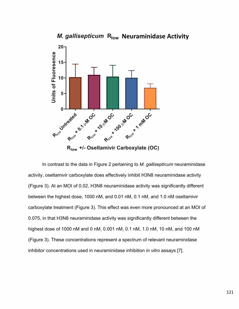
121
In contrast to the data in Figure 2 pertaining to M. gallisepticum neuraminidase
activity, oseltamivir carboxylate does effectively inhibit H3N8 neuraminidase activity
(Figure 3). At an MOI of 0.02, H3N8 neuraminidase activity was significantly different
between the highest dose, 1000 nM, and 0.01 nM, 0.1 nM, and 1.0 nM oseltamivir
carboxylate treatment (Figure 3). This effect was even more pronounced at an MOI of
0.075, in that H3N8 neuraminidase activity was significantly different between the
highest dose of 1000 nM and 0 nM, 0.001 nM, 0.1 nM, 1.0 nM, 10 nM, and 100 nM
(Figure 3). These concentrations represent a spectrum of relevant neuraminidase
inhibitor concentrations used in neuraminidase inhibition in vitro assays [7].
R low Untre
ated
R low + 0.
1 µM O
C
R low + 10
µM O
C
R low +
100 µ
M OC
R low +
1 mM O
C0
5
10
15
20
M. gallisepticum Rlow Sialidase Activity
Rlow +/- Oseltamivir Carboxylate (OC)
Uni
ts o
f Flu
ores
ence
Neuraminidase Activity
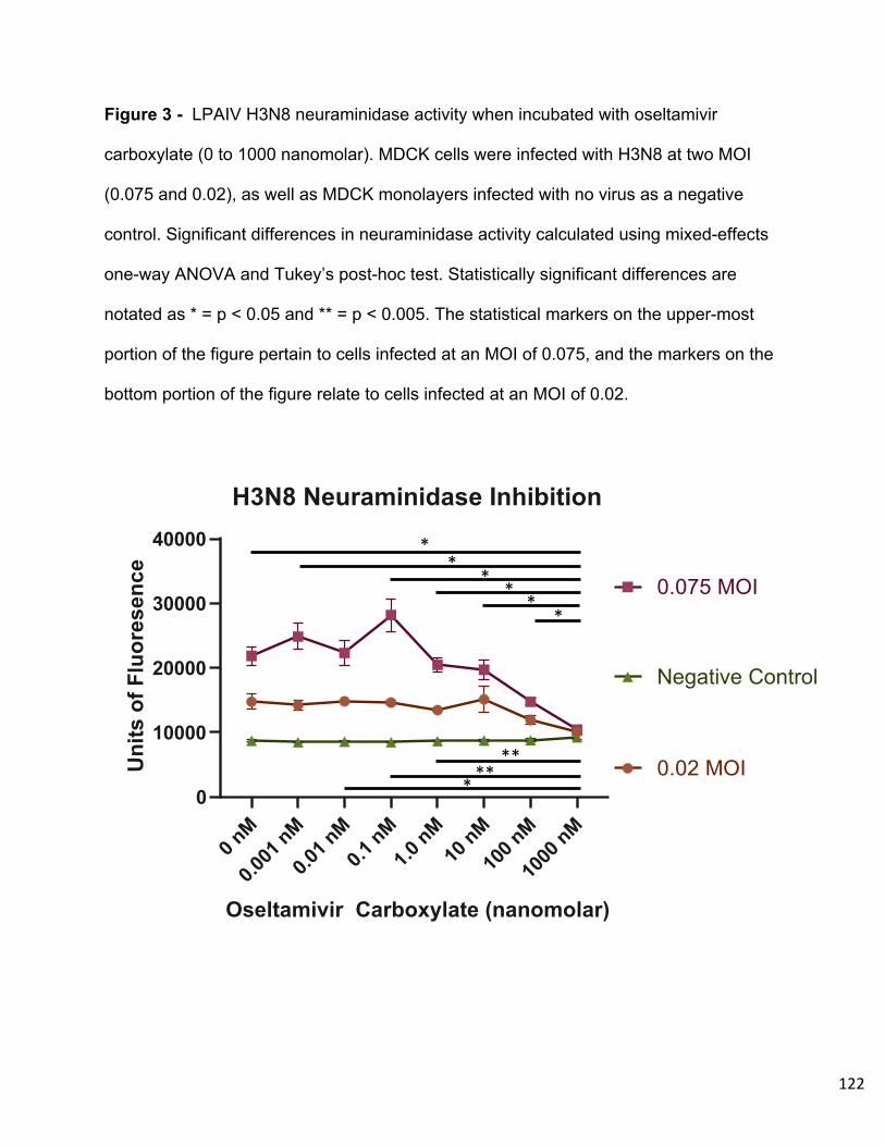
122
Figure 3 - LPAIV H3N8 neuraminidase activity when incubated with oseltamivir
carboxylate (0 to 1000 nanomolar). MDCK cells were infected with H3N8 at two MOI
(0.075 and 0.02), as well as MDCK monolayers infected with no virus as a negative
control. Significant differences in neuraminidase activity calculated using mixed-effects
one-way ANOVA and Tukey’s post-hoc test. Statistically significant differences are
notated as * = p < 0.05 and ** = p < 0.005. The statistical markers on the upper-most
portion of the figure pertain to cells infected at an MOI of 0.075, and the markers on the
bottom portion of the figure relate to cells infected at an MOI of 0.02.
0 nM
0.001
nM
0.01 nM
0.1nM
1.0nM
10nM
100 nM
1000
nM0
10000
20000
30000
40000
H3N8 Neuraminidase Inhibition
Oseltamivir Carboxylate (nanomolar)
Uni
tsof
Fluo
rese
nce 0.02 MOI
0.075 MOINegative Control
0 nM
0.001
nM
0.01 nM
0.1nM
1.0nM
10nM
100 nM
1000
nM0
10000
20000
30000
40000
H3N8 Neuraminidase Inhibition
Oseltamivir Carboxylate (nanomolar)
Units
ofFl
uore
senc
e 0.02 MOI0.075 MOINegative Control
0 nM
0.001
nM
0.01 nM
0.1nM
1.0nM
10nM
100 nM
1000
nM0
10000
20000
30000
40000
H3N8 Neuraminidase Inhibition
Oseltamivir Carboxylate (nanomolar)
Uni
tsof
Fluo
rese
nce 0.02 MOI
0.075 MOINegative Control
0 nM
0.001
nM
0.01 nM
0.1nM
1.0nM
10nM
100 nM
1000
nM0
10000
20000
30000
40000
H3N8 Neuraminidase Inhibition
Oseltamivir Carboxylate (nanomolar)
Units
ofFl
uore
senc
e 0.02 MOI0.075 MOINegative Control
*****
**
* * **

123
Discussion
The data presented above elude to possible factors contributing to the co-
pathogenesis of LPAIV and M. gallisepticum in terms of host cell tropism and possible
compensatory neuraminidase activity. The significant disruption in cytadherance to host
cells by M. gallisepticum in the absence of α-2,3 linked sialic acids is a novel finding. In
the context of a co-infection with LPAIV, this result holds even more significance in that
LPAIVs preferentially bind host tissues at α-2,3 linked sialic acid moieties common in
the avian respiratory and gastrointestinal tract [10].
A common host cell ligand could indicate potential competition between M.
gallisepticum and LPAIV for proximity to the host cell. However, the data from in vitro
and in vivo studies examining these co-pathogens indicates that there is more benefit to
replication and persistence when these two pathogens are present over a hampering of
one’s survival in favor of the other [11,12,13].
The shared importance of α-2,3 linked sialic acids in these co-pathogens
interaction with the host more likely indicates that these two pathogens may bind in
close proximity to one another on host tissues. This proximity facilitates an array of
potential mechanisms of mutual benefit during co-pathogenesis. These mechanisms
could include host metabolites, the availability of host cell receptors, or the shared
activity of enzymes such as the viral and bacterial neuraminidases.
As seen in other viral and bacterial co-pathogen relationships, neuraminidase
activity can be pivotal in the mutualistic dynamic during the disease process. For
instance, Streptococcus pneumoniae has potent neuraminidase activity that can rescue
124
influenza viral replication in the presence of viral neuraminidase inhibitors [14]. The
ability of M. gallisepticum and LPAIV to attach to host cells in potentially close-quarters
would allow for this same dynamic of neuraminidase activity compensation during
infection.
This hypothesis garners further merit in that M. gallisepticum Rlow is not
susceptible to oseltamivir carboxylate neuraminidase inhibition, whereas LPAIV H3N8
neuraminidase is susceptible to this inhibitor at a range of concentrations. In a case of
this co-infection in an avian population, neuraminidase inhibitor treatment could be
rendered useless if M. gallisepticum neuraminidase can effectively compensate for
LPAIV and further the progression of disease.
125
References
1. Cecchini KR, Gorton TS, Geary SJ. 2007. Transcriptional Responses of
Mycoplasma gallisepticum Strain R in Association with Eukaryotic Cells. Journal
of Bacteriology 189:5803–5807.
2. Balish AL, Katz JM, Klimov AI. 2013. Influenza: Propagation, Quantification, and
Storage. Current Protocols in Microbiology 29:15G.1.1-15G.1.24.
3. Eisfeld AJ, Neumann G, Kawaoka Y. 2014. Influenza A virus isolation, culture
and identification. Nat Protoc 9:2663–2681.
4. May M, Szczepanek SM, Frasca S, Gates AE, Demcovitz DL, Moneypenny CG,
Brown DR, Geary SJ. 2012. Effects of sialidase knockout and complementation
on virulence of Mycoplasma gallisepticum. Veterinary Microbiology 157:91–95.
5. Berčič RL, Slavec B, Lavrič M, Narat M, Zorman-Rojs O, Dovč P, Benčina D.
2008. A survey of avian Mycoplasma species for neuraminidase enzymatic
activity. Veterinary Microbiology 130:391–397.
6. May M, Kleven SH, Brown DR. 2007. Sialidase Activity in Mycoplasma Synoviae.
Avian Diseases 51:829–833.
7. Eichelberger MC, Hassantoufighi A, Wu M, Li M. 2008. Neuraminidase activity
provides a practical read-out for a high throughput influenza antiviral screening
assay. Virol J 5:109.
8. Jia N, Barclay WS, Roberts K, Yen H-L, Chan RWY, Lam AKY, Air G, Peiris
JSM, Dell A, Nicholls JM, Haslam SM. 2014. Glycomic Characterization of

126
Respiratory Tract Tissues of Ferrets: IMPLICATIONS FOR ITS USE IN
INFLUENZA VIRUS INFECTION STUDIES. J Biol Chem 289:28489–28504.
9. Grienke U, Richter M, Walther E, Hoffmann A, Kirchmair J, Makarov V,
Nietzsche S, Schmidtke M, Rollinger JM. 2016. Discovery of prenylated
flavonoids with dual activity against influenza virus and Streptococcus
pneumoniae. Sci Rep 6:27156.
10. Varki A, Cummings RD, Esko JD, et al., editors. Essentials of Glycobiology
[Internet]. 3rd edition. Cold Spring Harbor (NY): Cold Spring Harbor Laboratory
Press; 2015-2017.
11. Sid H, Hartmann S, Petersen H, Ryll M, Rautenschlein S. 2016. Mycoplasma
gallisepticum modifies the pathogenesis of influenza A virus in the avian tracheal
epithelium. International Journal of Medical Microbiology 306:174–186.
12. Stipkovits L, Egyed L, Palfi V, Beres A, Pitlik E, Somogyi M, Szathmary S, Denes
B. 2012. Effect of low-pathogenicity influenza virus H3N8 infection on
Mycoplasma gallisepticum infection of chickens. Avian Pathology 41:51–57.
13. Stipkovits L, Glavits R, Palfi V, Beres A, Egyed L, Denes B, Somogyi M,
Szathmary S. 2012. Pathologic Lesions Caused by Coinfection of Mycoplasma
gallisepticum and H3N8 Low Pathogenic Avian Influenza Virus in Chickens. Vet
Pathol 49:273–283.
14. Nishikawa T, Shimizu K, Tanaka T, Kuroda K, Takayama T, Yamamoto T,
Hanada N, Hamada Y. 2012. Bacterial Neuraminidase Rescues Influenza Virus
Replication from Inhibition by a Neuraminidase Inhibitor. PLoS ONE 7:e45371.
127
Chapter 3 – Transcriptional and pathological host responses to co-infection with
virulent or attenuated Mycoplasma gallisepticum and low pathogenic avian
influenza A virus in chickens.
Ammended Version Submitted to Infection and Immunity for Publication
(IAI00607-19)
Authors
J.A. Canter a, E.R. Tulman a, J. Beaudet a,b, D. Lee a, M. May c, S.M. Szczepanek a, and
S.J. Geary a*
Department of Pathobiology and Veterinary Sciences and the Center of Excellence for
Vaccine Research, University of Connecticut, Storrs, Connecticut, USA a; Department of
Allied Health Sciences, University of Connecticut, Storrs, Connecticut, USA b;
Department of Biomedical Sciences, University of New England College of Medicine,
Biddeford, Maine, USA c.
*Corresponding author
Abstract
The avian pathogen Mycoplasma gallisepticum, the etiologic agent of chronic
respiratory disease in chickens, exhibits enhanced pathogenesis in the presence of a
co-pathogen such as low-pathogenic avian influenza virus (LPAIV). To further
investigate the intricacies of this co-pathogenesis, chickens were mono- or co-infected
with either virulent M. gallisepticum strain Rlow, attenuated M. gallisepticum mutant

128
P1C5, or LPAIV H3N8 (A/duck/Ukraine/1963) and assessed for tracheal histopathology,
pathogen load, and transcriptomic host response to infection using RNA-sequencing.
Chickens co-infected with M. gallisepticum Rlow followed by H3N8 exhibited significantly
more severe tracheal lesions and mucosal thickening in response to infection than
chickens infected with H3N8 alone. Viral load was also significantly increased in this
group over chickens who were infected first with H3N8 and subsequently with M.
gallisepticum Rlow. The attenuated M. gallisepticum mutant P1C5, previously shown to
be cleared 14 days post-infection, was able to persist 6 days post-infection in the
presence and absence of co-infection with H3N8. The transcriptional response to mono-
and co-infection with M. gallisepticum and LPAIV highlighted the involvement of
differential expression of genes such as TLR4, TLR15, TLR21, IL-1β, IRF4, MMP1, and
MMP9. Pathway and gene ontology analysis of these differentially expressed genes
suggests that co-infection with virulent M. gallisepticum and LPAIV induces a
downregulation of ciliary activity in vivo and alters the multiple immune-related signaling
cascades. These data aid in the understanding of the relationship between M.
gallisepticum and LPAIV during co-pathogenesis in the natural host and may contribute
to the further understanding of co-pathogen infections of humans and other animals.
Introduction
The bacterial pathogen Mycoplasma gallisepticum causes significant avian
diseases such as sinusitis in turkeys, conjunctivitis in passerines, and chronic
respiratory disease in chickens [1,2]. Pathogenesis of M. gallisepticum in chickens

129
involves severe inflammation in components of the respiratory system, as well as
deleterious effects on egg production in these birds, making M. gallisepticum a
pathogen of great relevance and concern to the agricultural industry [1]. Infection of
chickens with virulent M. gallisepticum Rlow has been shown to induce a dysregulated
immune response in the chicken host with differential gene expression in the trachea
peaking 3 days post-infection over the first 7 days [3]. Toll-like receptor (TLR)
responses, most notably TLR4 and TLR15, were significantly upregulated in the trachea
during infection, as were IL-1β, MMP7, and an array of inflammatory cytokines and
chemokines [3]. An additional report demonstrated that the lipoproteins of M.
gallisepticum stimulate a TLR2-mediated response in primary chicken tracheal epithelial
cells [4].
Another avian pathogen of great importance is avian influenza A virus. Avian
influenza viruses occur in two main categories; highly-pathogenic avian influenza
viruses (HPAIV) and low-pathogenic avian influenza viruses (LPAIV) [5]. HPAIV causes
severe disease in a multitude of avian species with an extremely high mortality rate [6].
In contrast, LPAIV causes little-to-no disease in birds and, without rigorous surveillance,
could go unnoticed in wild or domestic populations of birds [6].
Co-infections consisting of M. gallisepticum and LPAIV in chickens exacerbates
disease compared to either pathogen in isolation]. Chickens challenged with both M.
gallisepticum strain 1226 and LPAIV H3N8 (A/mallard/Hungary/19616/07) showed
significantly more severe clinical signs and increased recovery of M. gallisepticum [7].
Co-infection with these two pathogens also yields significantly more severe clinical
scores of gross and histopathologic lesions of infected chickens [8]. Ex vivo

130
experiments with M. gallisepticum strain S6 and H9N2 (A/chicken/Saudi
Arabia/CP7/1998) in an avian tracheal organ culture system showed significant
differences in replication of M. gallisepticum and H9N2 between mono- and co-infected
tracheas [9]. Ciliostasis was also observed in these co-infected tracheal cultures at a
significantly higher rate and severity over mono-infected tracheas as well [9].
A possible mechanism underlying this co-pathogenesis phenomenon is the
common functionality of the glycosidic enzymes neuraminidase of influenza A virus, and
sialidase of M. gallisepticum. Influenza A virus utilizes neuraminidase to cleave N-
acetylneuraminic acids, or sialic acids, on host cells to release progeny virions during
infection. In M. gallisepticum, sialidase performs this same enzymatic reaction and
transposon insertional disruption of this gene in virulent M. gallisepticum Rlow results in
attenuation of the pathogen in vivo [10,11]. The true role of sialidase in the life cycle and
pathogenesis of M. gallisepticum is still yet to be elucidated. For the remainder of this
manuscript, both influenza A virus neuraminidase and Mycoplasma sialidase will be
referred to as neuraminidases for the sake of continuity.
Instances of influenza A virus co-infection with bacterial pathogens in humans,
such as Streptococcus pneumoniae, yield evidence that the neuraminidases of
influenza virus and S. pneumoniae work in concert during infection to enhance disease
[12,13]. In the presence of a viral neuraminidase inhibitor zanamivir, S. pneumoniae
neuraminidase can compensate for the hampered neuraminidase activity of influenza
virus to enhance viral replication and further co-pathogenesis in mice [14].
In the case of M. gallisepticum and LPAIV co-infection, there is strong evidence
that the presence of both pathogens enhances various markers of disease over

131
infection with either pathogen alone. However, the host-pathogen dynamics involved in
this co-pathogenesis relationship remain unexplored. It is our hypothesis that M.
gallisepticum neuraminidase works to support LPAIV neuraminidase during co-infection.
We also hypothesize that co-infection of chickens with Mycoplasma gallisepticum and
LPAIV H3N8 results in more severe disease than mono-infection, and that this
difference in pathogenesis will yield changes in pathologic lesions, pathogen load, and
transcriptional changes in the host. To examine this, we utilized an in vivo infection
model in chickens, a natural host of both M. gallisepticum and LPAIV. In addition, we
hypothesize that co-infection with LPAIV H3N8 would cultivate an environment to
sustain the chronic persistence of an attenuated M. gallisepticum mutant in vivo.
The co-pathogenesis of M. gallisepticum and LPAIV is of great relevance to the
agricultural industry for the prevention and treatment of viral and bacterial infections in
avian flocks [11]. Either pathogen alone may go un-detected in a population of birds for
a sufficient period of time to set the stage for severe losses due to later infection with
the other pathogen. The dynamics involved in this system of co-infection may also
prove beneficial in understanding the factors contributing to instances of viral and
bacterial co-pathogenesis in other animals and humans.
Methods
-Animals: For each study, four-week old female specific pathogen-free (SPF) White
Leghorn chickens (SPAFAS, North Franklin, CT, USA) were procured and separated
randomly into experimental groups for placement in HEPA-filtered housing isolators.
These chickens then acclimated in this habitat for a period of one week before the start

132
of the experiment. For this acclimation period, as well as the remainder of the
experiment, the chickens were maintained on non-medicated feed and water ad libitum.
These animal experiments were performed with approval from the University of
Connecticut Institutional Animal Care and Use Committee under protocol #A18-057.
-Mycoplasma gallisepticum and LPAIV Preparation: Influenza A virus isolate H3N8
(A/duck/Ukraine/1963) (BEI Resources) was used to inoculate 10-day old SPF
embryonated chicken eggs (SPAFAS, North Franklin, CT, USA) at 10-fold dilutions of
viral stock [15,16]. After 48 hours, allantoic fluid was collected from these eggs and viral
titer was determined by TCID50 on Madin Darby Canine Kidney (MDCK) cells using the
Reed-Meunch method [15,16]. Viral stocks were frozen at -80° C and diluted in
complete Hayflick’s medium to a concentration of 5 x 10⁶ TCID50/200 µL at the time of
use as an experimental inoculum.
Mycoplasma gallisepticum strain Rlow passage 17 was propagated from stock
cultures frozen at -80°C in complete Hayflick’s medium overnight at 37°C until mid-log
phase. The attenuated M. gallisepticum tn4001 mutants P1C5 and P1H9 were also
grown from frozen stock cultures in complete Hayflick’s medium with the addition of 150
µg/mL for the maintain the position of tn4001 transposon insertion [11,17]. Mycoplasma
cultures were quantified by optical density at 620 nm and then centrifuged for 10
minutes at 10,000 x g to pellet the cultures. Mycoplasma pellets were then resuspended
in fresh complete Hayflick’s medium to a density of 1 x 10⁸ CFU/200 µL to create the
experimental inoculum for each of the chicken infection experiments.

133
-Chicken Infection Experiments: 5 week old chickens were challenged intratracheally
by pipetting 200 µL of either 1 x 10⁸ CFU M. gallisepticum Rlow, 1 x 10⁸ CFU M.
gallisepticum mutant P1C5, 5 x 10⁶ TCID50 LPAIV H3N8, or mock-challenged with fresh
complete Hayflick’s medium according to the following schedule (n = 5 per group):
Experiment #1 Primary Infection (Day 0) Secondary Infection (Day 3)
Sacrifice (Day 6)
Mock (Hayflick’s medium) M. gallisepticum Rlow H3N8 M. gallisepticum Rlow M. gallisepticum Rlow H3N8 M. gallisepticum P1C5 H3N8 Mock (Hayflick’s medium) H3N8
In two companion challenge studies, additional set of chickens (n = 5 per group in
experiment #2, n = 10 in experiment #3) were challenged as described above in the
following groups:
Experiment #2 Primary Infection (Day 0)
Sacrifice (Day 6) M. gallisepticum Rlow M. gallisepticum P1C5 Mock (Hayflick’s medium)
Experiment #3 Primary Infection
(Day 0) Secondary Infection
(Day 3)
Sacrifice (Day 14)
M. gallisepticum P1C5 Mock (Hayflick’s medium) M. gallisepticum P1C5 H3N8 M. gallisepticum P1H9 Mock (Hayflick’s medium) Sacrifice (Day 7) M. gallisepticum P1H9 H3N8
Another attenuated mutant, mslA mutant P1H9, was incorporated into this model
of co-infection as a means to compare changes in M. gallisepticum mutant co-infection
over the course of disease and be able to attribute the possible changes in

134
pathogenesis to the specific relationship between P1C5 and LPAIV neuraminidase
activity [17]. The mutant P1C5 persists in the trachea of chickens 6 days post-infection,
however the previous studies utilizing the mutant P1H9 only examined Mycoplasma
recovery 14 days post-infection [17].
- Sample Collection, Mycoplasma Recovery and RNA Extraction: In each of the
above experiments, all chickens from each of the groups were humanely sacrificed by
cervical dislocation and the tracheas were excised. Three tracheal rings from each
chicken were taken from the proximal, middle, and distal portion of each trachea and
fixed in neutral buffered formalin for pathological assessments. An additional ring was
taken from the distal end of the trachea of each bird and placed into complete Hayflick’s
medium for mycoplasma recovery and incubated for 3 hours at 37°C. After incubation,
these cultures were filtered through 0.45 µm filter and diluted 10-fold for color changing
unit (CCU) quantification, as done previously [18]. Statistically significant differences
among groups for M. gallisepticum CCU recovery were calculated using using a
Kruskal-Wallis ANOVA On Ranks with Dunn’s Post-Hoc test (GraphPad Prism 8.0).
The remaining sections of excised tracheas from each bird were washed with 1
mL of TriZol (Zymo Research, Carlsbad, CA, USA) pipetted 4 times through the lumen
for RNA collection as described previously [18]. RNA was extracted from these TriZol
washes using the Zymo DirectZol RNA MiniPrep Kit (Zymo Research, Carlsbad, CA,
USA) and quantified using a Qubit 2.0 Fluorometer (ThermoFisher Scientific).

135
-Tracheal Thickness and Lesion Scoring: Tracheal rings were embedded and
sectioned for hematoxylin and eosin (H&E) staining as described previously [19]. These
rings were measured for tracheal thickness and lesions were scored by an AAVP board-
certified veterinary pathologist. These lesions were scored on a scale of 0 to 4, denoting
no inflammation, mild inflammation, moderate inflammation, marked inflammation, and
severe inflammation, respectively, as described previously [19]. Significant differences
among challenge groups for tracheal thickness measurements were calculated using a
one-way analysis of variance (ANOVA) and Tukey’s Post-Hoc test and differences in
tracheal lesion scores were calculated using a Kruskal-Wallis ANOVA On Ranks with
Dunn’s Post-Hoc test (GraphPad Prism 8.0).
-Viral Load Quantification: RNA collected from the tracheas of infected chickens in
experiment #1 was assayed using qRT-PCR of the influenza matrix gene in a 25 µL
reaction volume (Applied Biosystems 75000 Fast Real-Time PCR System) by the
Connecticut Veterinary Medical Diagnostic Laboratory. Cycling conditions for this
reaction were 45.0°C for 10 minutes, 95.0°C for 10 minutes, 94.0°C for 1 second, cycled
at this step 45 times before a final step at 60.0°C for 30 seconds. A standard curve of
H3N8 (A/duck/Ukraine/1963) RNA was used in 10-fold dilutions for calculation of viral
genomes per microliter.
-Illumina Sequencing: RNA extracted from tracheal washes of each bird from
experiment #1 were used as templates for cDNA library synthesis using the Illumina
TruSeq Stranded mRNA Library Preparation Kit (Illumina, San Diego, CA). Each library

136
was quantified and assessed for quality using the Agilent TapeStation 2200 (Agilent
Technologies). These libraries were then normalized and pooled for sequencing on the
Illumina NextSeq 500 (Illumina, San Diego, CA) with paired-end, 75 base pair reads
with 5 to 10 million reads per sample [3].
-RNA-seq Analysis: Read data from each bird sample in the Fastq format were
mapped to the Gallus gallus reference genome using the TopHat package and Bowtie2
engine to produce sequence alignments [20]. Alignment files were pooled by
experimental infection groups using Cufflinks to map to reference transcript files. Cuffdiff
was utilized to determine expression values and statistically significant differences of
these values between experimental infection groups. These expression values per
transcript are normalized within group via calculation of fragments per kilobase per exon
per million fragments mapped (FPKM). Benjamini-Hochberg analysis then generates a
p-value for statistical significance also accounting for a false discovery rate of <5% to
yield a q-value of significance. Fold change differences between experimental infection
groups were calculated by a log2 calculation of FPKM values from each group [20].
Significantly differentially expressed genes with greater than, or equal to, a positive or
negative 2 log2 fold change will be displayed for between-group comparisons.
-Pathway and Functional Gene Ontology Analysis: Significantly differentially
expressed genes (DEGs) were used as an input for analysis using the Database for
Annotation, Visualization and Integrated Discovery (DAVID) version 6.8 to assemble
137
these DEGs into biological pathways defined by the Kyoto Encyclopedia of Genes and
Genomes (KEGG) and functional gene ontologies using GO Term BP Direct [21].
Results
-Tracheal Thickness and Lesion Scores: Significant differences were found in the
tracheal mucosal thickness (p < 0.05) between Mock/H3N8 and M. gallisepticum
Rlow/H3N8 infected chickens in experiment #1 (One-Way ANOVA with Tukey’s Post-Hoc
test). Significant differences were observed in histopathologic tracheal lesion scores (p
< 0.05) between Mock/H3N8 and M. gallisepticum Rlow/H3N8 infected chickens of
experiment #1 (Kruskal-Wallis ANOVA On Ranks with Dunn’s Post-Hoc test) (Figures
1A and 1B). Although not statistically significant at this timepoint, tracheal thickness (p =
0.3901) exhibits a trend of increased severity in the H3N8/M. gallisepticum Rlow group
compared to the Mock/M. gallisepticum Rlow group (Figures 1A and 1B).
In experiment #2, significant differences exist between mock infected chickens
and Rlow infected chickens as well as mock infected and P1C5 infected chickens for
both tracheal thickness (Figure 1C) and tracheal lesion scores (Figure 1D). These data
indicate that although no lesions or live M. gallisepticum P1C5 were observed 14 days
post-infection in previous studies from our laboratory, this mutant appears to persist in
the chicken host 6 days post-infection and induces a moderate pathologic response in
the lumen of the trachea [11].
Figure 1 – Tracheal lesion scores (A) and thickness measurements (B) from M.
gallisepticum and H3N8 mono- and co-infected chickens. Statistically significant
138
differences among group tracheal lesion scores were calculated using a Kruskal-Wallis
ANOVA On Ranks with Dunn’s Post-Hoc test. Statistically significant differences among
group tracheal thickness measurements were calculated using a One-Way ANOVA with
Tukey’s Post-Hoc test. Significant differences are denoted using “a” and “b” indicating a
significant difference of p < 0.05. Tracheal histopathology (C and D) from experiment
#2. Statistically significant differences among tracheal thickness for experimental groups
were calculated using a One-Way ANOVA with Tukey’s Post-Hoc Test and are
indicated by ** = p < 0.05, *** = p < 0.0005. For tracheal lesion scores in experiment #2,
significant differences were calculated using a Kruskal-Wallis ANOVA On Ranks with
Dunn’s Post-Hoc test and using “a” and “b” indicating a significant difference of p < 0.01
and p <0.05.
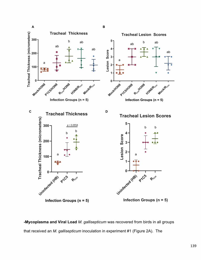
139
-Mycoplasma and Viral Load M. gallisepticum was recovered from birds in all groups
that received an M. gallisepticum inoculation in experiment #1 (Figure 2A). The
Mock/H3N
8
P1C5/H
3N8
R low/H3N
8
H3N8/R
low
Mock/R lo
w
0
1
2
3
4
5
Tracheal Lesion Scores
Infection Groups (n = 5)
Lesi
onSc
ore
Mock/H3N
8
P1C5/H
3N8
R low/H3N
8
H3N8/R
low
Mock/R lo
w
0
100
200
300
Tracheal Thickness
Infection Groups (n = 5)
Trac
heal
Thic
knes
s(m
icro
met
ers)
Uninfected
(HB)
P1C5
R low
0
1
2
3
4
5
Tracheal Lesion Scores
Infection Groups (n = 5)
Lesi
onSc
ore
Uninfected
(HB)
P1C5
R low
0
100
200
300
Tracheal Thickness
Infection Groups (n = 5)
Trac
heal
Thic
knes
s(m
icro
met
ers)
p = 0.0958
A B
C D
a
b abab
abab ab
ab
a
b
b
a
b
a
bb

140
bacterial loads recovered from H3N8/M. gallisepticum Rlow infected chickens were
significantly higher than the Mock/M. gallisepticum Rlow group (p < 0.05) (Kruskal-Wallis
ANOVA On Ranks with Dunn’s Post-Hoc test) (Figure 2A). Although tracheal pathology
did not differ significantly between the H3N8/M. gallisepticum Rlow group and Mock/M.
gallisepticum Rlow group (Figure 1A and 1B), M. gallisepticum was recovered from 80%
of chickens within the H3N8/M. gallisepticum Rlow group (Figure 2A), 20% of chickens in
the Mock/M. gallisepticum Rlow group and , 100% from chickens in the M. gallisepticum
Rlow/H3N8 group (Figure 2A).
In a similar vein, H3N8 was detected in chickens within all groups receiving an
inoculation with H3N8 in experiment #1, with the exception of chickens in the H3N8/M.
gallisepticum Rlow infection group (Figure 2B). H3N8 was detected in 80% of chickens in
the Mock/H3N8 group and the M. gallisepticum Rlow/H3N8 group, both having been
challenged with H3N8 for the final3 days of the experiment. H3N8 was not detected in
the H3N8/M. gallisepticum Rlow group after 6 days of H3N8 infection. H3N8 load was
significantly higher in the M. gallisepticum Rlow/H3N8 group over the H3N8/M.
gallisepticum Rlow group of experiment #1 (p < 0.01, One-Way ANOVA with Tukey’s
Post-Hoc test) (Figure 2B).
In experiment #2, M. gallisepticum was recovered from the tracheae of all M.
gallisepticum infected birds for both M. gallisepticum Rlow and P1C5 (Figure 2C). This
bacterial recovery follows a similar pattern to the tracheal histopathology displayed in
Figure 1, in that M. gallisepticum Rlow was the most abundant in the trachea at 6 days
post-infection and induced more severe tracheal lesions and thickening than the mutant
P1C5, which was recovered at a lesser abundance at that time (Figure 2C).

141
Experiment #3 demonstrated that M. gallisepticum was recovered from all M.
gallisepticum P1H9 infected chickens, with the exception of P1H9/Mock infected birds
sacrificed at 14 days post-infection (Figure 3). The difference in M. gallisepticum
recovery between these two groups is statistically significant (p < 0.001) (Kruskal-Wallis
ANOVA On Ranks with Dunn’s Post-Hoc test). P1H9 was previously shown to fail to
persist at 14 days post-infection [17], however these data show that this attenuated
mutant does survive in the chicken trachea at 7 days post-infection in 90% of infected
chickens. Persistence in the host trachea to 14 days post-infection was partially rescued
only in the presence of a co-infection with H3N8 in 40% of infected chickens (Figure
2D).
Figure 2 – Recovery of M. gallisepticum (A) and H3N8 (B) from the tracheas of mono-
and co-infected chickens of experiment #1. M. gallisepticum recovery (C) from the
tracheas of infected chickens of experiment #2 and experiment #3 (D). Bacterial
recovery was quantified using color changing unit (CCU) dilutions. Statistically
significant differences in M. gallisepticum recovery were determined using a Kruskal-
Wallis ANOVA On Ranks with Dunn’s Post-Hoc test and denoted using “a” and “b”
indicating a significant difference of p <0.05 (C). Significany differences in H3N8 load
were calculated using One-Way ANOVA with Tukey’s Post-Hoc test and denoted using
“a” and “b” indicating a significant difference of p < 0.05 (B).

142
-Significantly Differentially Expressed Gene (DEG) Analysis: CuffDiff differential
expression analysis between challenge groups of experiment #1 determined that there
are 260 significant DEGs increased in expression and 286 significant DEGs decreased
in expression between M. gallisepticum Rlow/H3N8 and Mock/H3N8 infected chickens.
Lists of DEGs between experimental groups of experiment #1 with a greater than 2 log2
fold change can be found in Supplemental Tables 10 through 14.
Mock/H
3N8
P1C5/H
3N8
R low/H
3N8
H3N8/R
low
0
2
4
6
8
Tracheal H3N8 Load
Infection Groups (n = 5)
Vira
lLoa
d(g
enom
es/m
icro
liter
)
P1C5/H
3N8
R low/H
3N8
H3N8/R
low
Mock/R low
100
101
102
103
104
105
106
107
Tracheal M. gallisepticum Recovery
Infection Groups (n = 5)
Log 1
0C
CU
Rec
over
y
P1C5 Rlow100
101
102
103
104
105
106
107
108
Infection Groups (n = 5)
Log 1
0C
CU
perm
illili
ter
Tracheal M. gallisepticum Recovery
A B
C D
p = 0.0554
ab
a
b
ab
P1H9/
Mock
(Day
7)
P1H9/
Mock
(Day
14)
P1H9/
H3N8
(Day
14)
P1C5/
Mock
(Day
14)
P1C5/
H3N8
(Day
14)
100
101
102
103
Tracheal M. gallisepticum Recovery
Infection Groups ( n = 10 )
Lo
g10
CC
UR
eco
very
a
b
ab
ab
ab
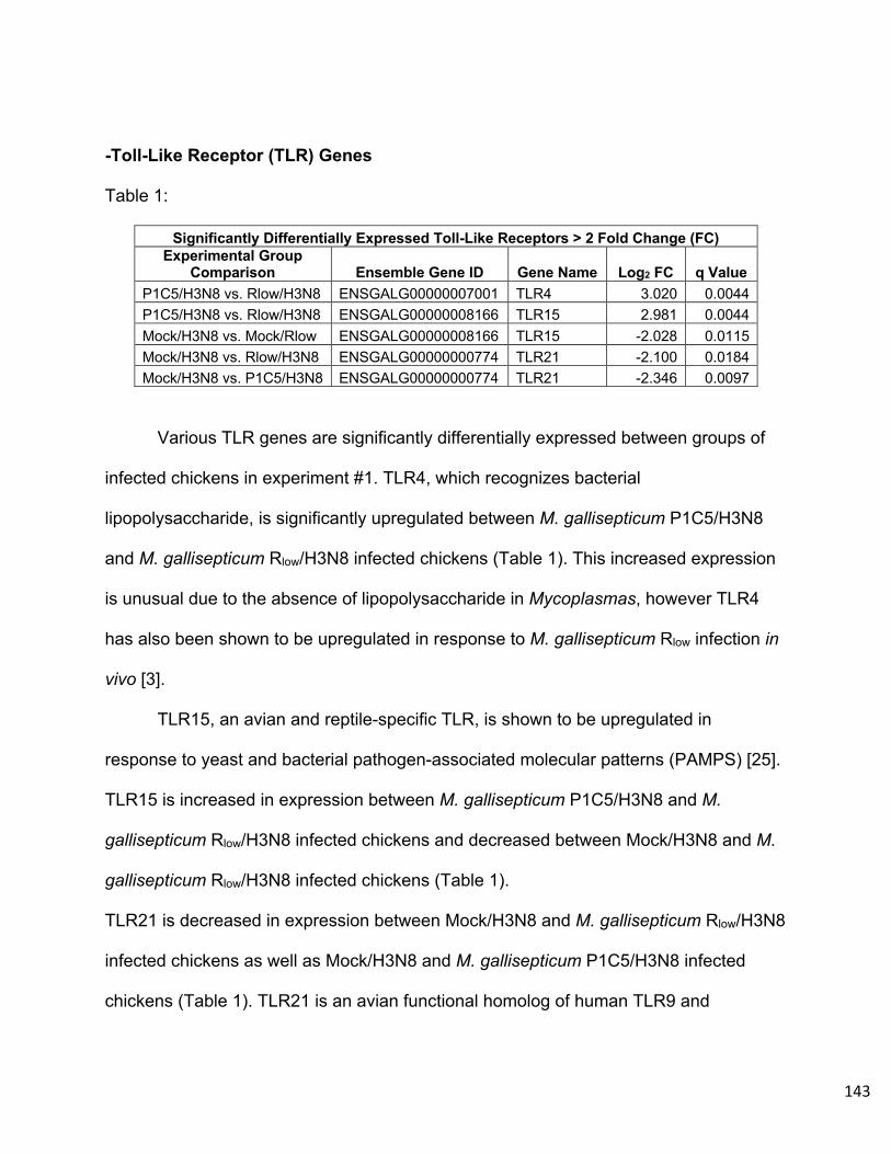
143
-Toll-Like Receptor (TLR) Genes
Table 1:
Significantly Differentially Expressed Toll-Like Receptors > 2 Fold Change (FC) Experimental Group
Comparison Ensemble Gene ID Gene Name Log2 FC q Value P1C5/H3N8 vs. Rlow/H3N8 ENSGALG00000007001 TLR4 3.020 0.0044 P1C5/H3N8 vs. Rlow/H3N8 ENSGALG00000008166 TLR15 2.981 0.0044 Mock/H3N8 vs. Mock/Rlow ENSGALG00000008166 TLR15 -2.028 0.0115 Mock/H3N8 vs. Rlow/H3N8 ENSGALG00000000774 TLR21 -2.100 0.0184 Mock/H3N8 vs. P1C5/H3N8 ENSGALG00000000774 TLR21 -2.346 0.0097
Various TLR genes are significantly differentially expressed between groups of
infected chickens in experiment #1. TLR4, which recognizes bacterial
lipopolysaccharide, is significantly upregulated between M. gallisepticum P1C5/H3N8
and M. gallisepticum Rlow/H3N8 infected chickens (Table 1). This increased expression
is unusual due to the absence of lipopolysaccharide in Mycoplasmas, however TLR4
has also been shown to be upregulated in response to M. gallisepticum Rlow infection in
vivo [3].
TLR15, an avian and reptile-specific TLR, is shown to be upregulated in
response to yeast and bacterial pathogen-associated molecular patterns (PAMPS) [25].
TLR15 is increased in expression between M. gallisepticum P1C5/H3N8 and M.
gallisepticum Rlow/H3N8 infected chickens and decreased between Mock/H3N8 and M.
gallisepticum Rlow/H3N8 infected chickens (Table 1).
TLR21 is decreased in expression between Mock/H3N8 and M. gallisepticum Rlow/H3N8
infected chickens as well as Mock/H3N8 and M. gallisepticum P1C5/H3N8 infected
chickens (Table 1). TLR21 is an avian functional homolog of human TLR9 and
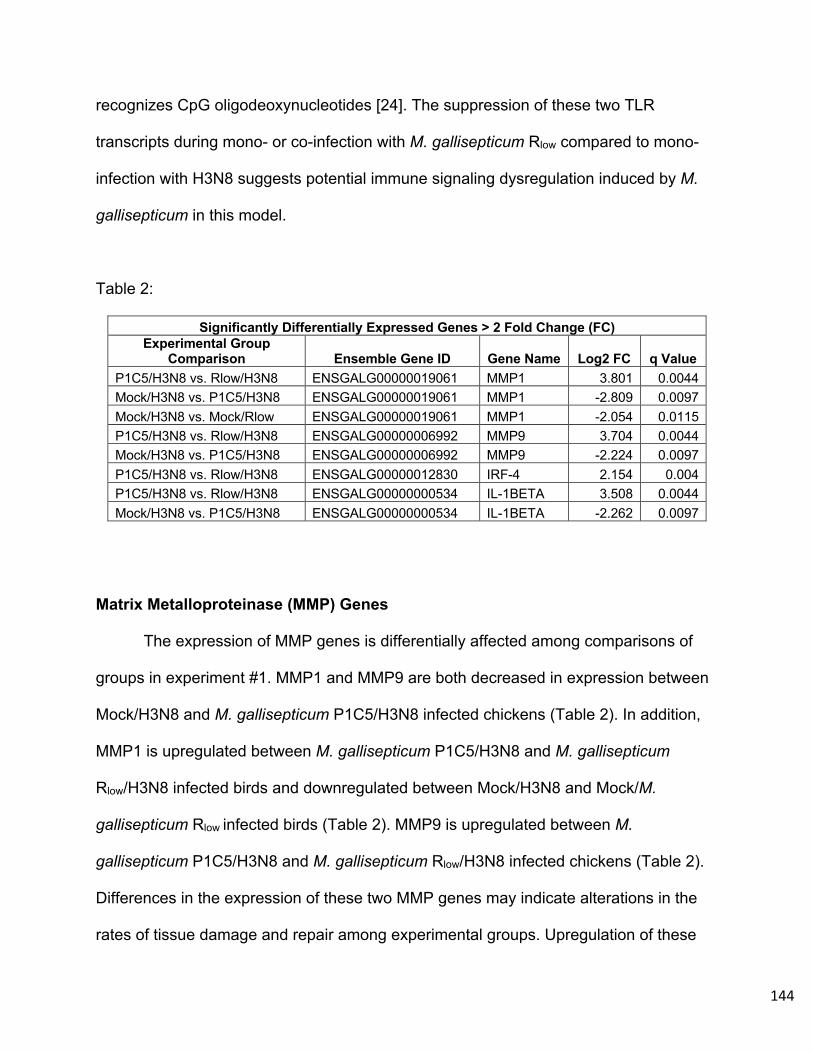
144
recognizes CpG oligodeoxynucleotides [24]. The suppression of these two TLR
transcripts during mono- or co-infection with M. gallisepticum Rlow compared to mono-
infection with H3N8 suggests potential immune signaling dysregulation induced by M.
gallisepticum in this model.
Table 2:
Significantly Differentially Expressed Genes > 2 Fold Change (FC) Experimental Group
Comparison Ensemble Gene ID Gene Name Log2 FC q Value P1C5/H3N8 vs. Rlow/H3N8 ENSGALG00000019061 MMP1 3.801 0.0044 Mock/H3N8 vs. P1C5/H3N8 ENSGALG00000019061 MMP1 -2.809 0.0097 Mock/H3N8 vs. Mock/Rlow ENSGALG00000019061 MMP1 -2.054 0.0115 P1C5/H3N8 vs. Rlow/H3N8 ENSGALG00000006992 MMP9 3.704 0.0044 Mock/H3N8 vs. P1C5/H3N8 ENSGALG00000006992 MMP9 -2.224 0.0097 P1C5/H3N8 vs. Rlow/H3N8 ENSGALG00000012830 IRF-4 2.154 0.004 P1C5/H3N8 vs. Rlow/H3N8 ENSGALG00000000534 IL-1BETA 3.508 0.0044 Mock/H3N8 vs. P1C5/H3N8 ENSGALG00000000534 IL-1BETA -2.262 0.0097
Matrix Metalloproteinase (MMP) Genes
The expression of MMP genes is differentially affected among comparisons of
groups in experiment #1. MMP1 and MMP9 are both decreased in expression between
Mock/H3N8 and M. gallisepticum P1C5/H3N8 infected chickens (Table 2). In addition,
MMP1 is upregulated between M. gallisepticum P1C5/H3N8 and M. gallisepticum
Rlow/H3N8 infected birds and downregulated between Mock/H3N8 and Mock/M.
gallisepticum Rlow infected birds (Table 2). MMP9 is upregulated between M.
gallisepticum P1C5/H3N8 and M. gallisepticum Rlow/H3N8 infected chickens (Table 2).
Differences in the expression of these two MMP genes may indicate alterations in the
rates of tissue damage and repair among experimental groups. Upregulation of these

145
MMPs in M. gallisepticum Rlow/H3N8 infected chickens associates with the enhanced
tracheal histopathology seen in Figure 1.
Interferon and Interleukin Genes
Interferon regulatory factor 4 (IRF4) is significantly upregulated between M.
gallisepticum P1C5/H3N8 and M. gallisepticum Rlow/H3N8 infected chickens (Table 2).
This indicates an activation of the type I interferon response to influenza virus in
chickens co-infected with virulent M. gallisepticum Rlow.
Interleukin 1 beta (IL-1β) is significantly upregulated between M. gallisepticum
P1C5/H3N8 and M. gallisepticum Rlow/H3N8 infected chickens but downregulated
between Mock/H3N8 and M. gallisepticum P1C5/H3N8 infected birds (Table 2). This
differential regulation of IL-1β expression could indicate an enhanced activation of the
NLRP3 inflammasome during co-infection with virulent M. gallisepticum Rlow and overall
increased inflammatory signaling unique to the presence of a pathogenic M.
gallisepticum.
-Functional Gene Ontology:
Between Mock/H3N8 and M. gallisepticum Rlow/H3N8 infected chickens these DEGs
can be grouped into an array of functional pathways. Significant DEGs decreased in
expression between these two challenge groups belong mostly to functional pathways
involved in cilium organization and macromolecule metabolic processes (Supplemental
Table 1). Significant DEGs increased in expression belong mainly to positive regulation
of the immune system processes (Supplemental Table 2). Between M. gallisepticum

146
P1C5/H3N8 and M. gallisepticum Rlow/H3N8 infected chickens, pathways increased in
expression belong foremost to response to lipopolysaccharide and signal transduction
(Supplemental Table 3), and pathways decreased in expression belong mainly to
cellular component assembly involved in morphogenesis, microtubule-based movement
and cilium organization (Supplemental Table 4).
-Pathway Analysis: KEGG pathway analysis of significant DEGs between
experimental groups illuminated key pathways differentially regulated during mono- and
co-infection. The toll-like receptor, phagosome, and cytokine-cytokine receptor
pathways are significantly downregulated between Mock/H3N8 and Mock/M.
gallisepticum Rlow infected chickens as well as between Mock/H3N8 and M.
gallisepticum P1C5/H3N8 infected chickens (Supplemental Tables 6 and 7). Metabolic
pathways are significantly downregulated between the groups stated above as well as
Mock/H3N8 and M. gallisepticum Rlow/H3N8 infected chickens and M. gallisepticum
P1C5/H3N8 and M. gallisepticum Rlow/H3N8 infected chickens (Supplemental Tables 8
and 9).
Notable upregulated pathways between M. gallisepticum P1C5/H3N8 and M.
gallisepticum Rlow/H3N8 infected chickens (Supplemental Table 9) and Mock/H3N8 and
M. gallisepticum Rlow/H3N8 infected chickens include the toll-like receptor, phagosome,
metabolic, apoptosis, RIG-I-like receptor signaling, focal adhesion, and ECM-receptor
interaction pathways (Supplemental Table 8).
The influenza A response pathway is significantly affected across multiple
comparisons. This pathway is significantly upregulated between M. gallisepticum

147
P1C5/H3N8 and M. gallisepticum Rlow/H3N8 infected chickens (SupplementalTable 9)
and Mock/H3N8 and M. gallisepticum Rlow/H3N8 infected chickens (Supplemental Table
8) and is downregulated between Mock/H3N8 and M. gallisepticum P1C5/H3N8
infected chickens (Supplemental Table 7). Regulation of the actin cytoskeleton is also
significantly upregulated between M. gallisepticum P1C5/H3N8 and M. gallisepticum
Rlow/H3N8 infected chickens (Supplemental Table 9) and Mock/H3N8 and M.
gallisepticum Rlow/H3N8 infected chickens (Supplemental Table 8).
Discussion
These data presented contribute to our further understanding of the co-
pathogenesis of Mycoplasma gallisepticum and LPAIV. the exacerbated disease
phenotype produced by these co-pathogens has been described using an ex vivo model
system and in vivo at a later time point of 15 days post-infection with alternate strains of
M. gallisepticum and LPAIV [7,8,9]. In our in vivo model examining this co-pathogenesis
at an early timepoint, tracheal histopathology was significantly more severe in M.
gallisepticum Rlow/H3N8 co-infected chickens compared to Mock/H3N8 mono-infected
chickens, both infected with H3N8 for a total of 3 days (Figure 1A and 1B).
In contrast, the inverse order of infection yielded no significant differences in
tracheal histopathology between H3N8/M. gallisepticum Rlow co-infected chickens and
Mock/M. gallisepticum Rlow mono-infected chickens both receiving M. gallisepticum Rlow
infection for 3 days (Figure 1A and 1B). Although we could not discern exacerbation of
pathologic lesions due to co-pathogenesis between these groups, the recovery of live
M. gallisepticum Rlow from the tracheas of H3N8/M. gallisepticum Rlow co-infected

148
chickens was notably higher than in Mock/M. gallisepticum Rlow mono-infected chickens
(Figure 2A). Previous in vivo studies by Stipkovits et al. yielded significant differences
between mono- and co-infected chickens in a similar model of M. gallisepticum and
H3N8 co-pathogenesis, however our model examines co-infection at a more acute
stage, 6 days post-infection, as seen in experiment #1 [7, 8]. The increased persistence
of M. gallisepticum Rlow in H3N8/M. gallisepticum Rlow co-infected chickens at 6 days
post-infection may contribute to more dramatic differences in tracheal pathology
between mono- and co-infected birds over a longer period.
Although previous works have utilized the transcriptomic approach to monitor M.
gallisepticum infection of chickens, our work furthers the use of these techniques in an
M. gallisepticum co-infection experiment in vivo [3,18,22,23]. This method of capturing
the global response to infection in the airway highlighted a number of critical host
responses during this co-infection model. One of these responses was the significant
downregulation of pathways contributing to ciliary activity and integrity in the trachea.
Although prior works have examined this ex vivo, the data presented here provides
evidence of deleterious impact on the ciliary mucosa function in the natural chicken host
at a transcript level [9]. This reduction of ciliary activity was shown between M.
gallisepticum P1C5/H3N8 and M. gallisepticum Rlow/H3N8 infected chickens as well as
Mock/H3N8 and M. gallisepticum Rlow/H3N8 infected chickens, indicating that there is a
unique relationship between co-infection with a virulent M. gallisepticum and LPAIV over
an attenuated M. gallisepticum, such as the mutant P1C5, or infection with LPAIV alone.
Loss of the integrity of the mucociliary elevator could heavily contribute to the

149
exacerbated co-pathogenesis by inhibiting pathogen clearing and promoting the
inflammatory immune response due to damage in the tracheal mucosa.
Another novel aspect of these experiments is the differential expression of TLR
genes in response to mono- and co-infection with M. gallisepticum and LPAIV. TLR15,
which is significantly decreased in expression in Mock/M. gallisepticum Rlow infected
chickens compared to Mock/H3N8 infected chickens (Table 1), has also been shown to
be abundantly expressed in response to M. gallisepticum Rlow infection in chickens [3].
Our current findings suggest that modulation in the expression of TLR15 remains critical
in response to M. gallisepticum, however it is more highly expressed in response to
LPAIV H3N8 alone. This could indicate that a TLR15-mediated response is more
relevant in response to influenza virus mono-infection and is not enhanced in the
presence of co-infection with M. gallisepticum.
TLR21 expression is suppressed during co-infection, in that between both
Mock/H3N8 and M. gallisepticum co-infection, TLR21 was significantly decreased in
expression (Table 1). This suppression of TLR21 during M. gallisepticum co-infection
negatively correlates with the significantly enhanced tracheal pathology and viral loads
in co-infected chickens (Figure 1A, 1B, and 2B). In ducks, TLR21 is upregulated in the
blood, spleen, bursa, and cecum in response to duck plague virus (DPV) and serves to
activate NFκB and the transcription of IL-6, IFNα, and IL-1β [24].
In chicken bone marrow macrophages, TLR21 and TLR4 co-stimulation induces
increases in pro-inflammatory genes such as IL-1β, IL-12p40, and IL-10 and nitric oxide
production [25]. Therefore, suppression of TLR21 signaling in response to CpG
deoxynucleotides and potential disruption of the association with TLR4 during co-

150
infection with Mycoplasma gallisepticum may also be a critical factor in our model of co-
pathogenesis.
Increased expression of IL-1β was also documented between M. gallisepticum
Rlow/H3N8 and M. gallisepticum P1C5/H3N8 infected experimental groups, indicating a
possible differential effect on activation of the NLRP3 inflammasome by IL-1β during co-
infection. Two MMP genes, MMP1 and MMP9, were also differentially expressed
among experimental groups. MMP1, was differentially expressed between Mock/H3N8
and Mock/M gallisepticum Rlow infected groups in experiment #1. These data can be
associated with the increased tracheal pathology seen in the Mock/M. gallisepticum Rlow
group over the Mock/H3N8 group, although this increase is not statistically significant
(Figure 1A and 1B). MMP7 was among the highest expressed genes in the trachea in
response to M. gallisepticum Rlow infection of chickens [3]. MMP1 and MMP7 have both
been implicated as biomarkers of human idiopathic pulmonary fibrosis and are
overexpressed in the lungs of patients with chronic pulmonary disease [26]. Therefore,
a precedent exists for these MMP genes being involved in tissue remodeling in
response to ongoing inflammatory challenge such as that induced by M. gallisepticum
infection and potential association with the manifestation of pathologic lesions in the
airway.
Functional pathway analysis also determined the differential regulation of the
response to influenza A virus pathway among comparisons between mono- and co-
infected chickens illustrates the importance of modulation of this specific response
mechanism. Co-infection with virulent M. gallisepticum appears to have a unique effect
on this response, as the pathway was upregulated M. gallisepticum P1C5/H3N8 and M.

151
gallisepticum Rlow/H3N8 infected chickens and Mock/H3N8 and M. gallisepticum
Rlow/H3N8 infected chickens and downregulated between Mock/H3N8 and M.
gallisepticum P1C5/H3N8 infected chickens. Although this influenza A response
pathway is increased in expression, it may not yield a productive immune response to
clear the virus during co-infection, as evidenced by the highest tracheal viral load being
present in the M. gallisepticum Rlow/H3N8 infected group of experiment #1 (Figure 2B).
A final novel finding of these experiments was the persistence of attenuated M.
gallisepticum mutants P1C5 6 days post-infection and P1H9 7 days post-infection in the
chicken trachea in experiments #2 and #3. Previously, these mutants have been shown
to be completely cleared from the birds at 14 days post-infection [11,17]. The ability of
these attenuated mutants to persist in the shorter term illustrates that the absence of a
functional neuraminidase gene and the Mycoplasma specific lipoprotein A (MslA) in M.
gallisepticum do not prohibit early colonization and replication in the chicken trachea.
The M. gallisepticum MslA mutant P1H9 was only able to persist to a chronic state at 14
days post-infection when in the presence of H3N8 co-infection. Although the M.
gallisepticum mutant P1C5 did persist at low titers 14 days post-infection in experiment
#3, this survival did not appear to be impacted by co-infection with H3N8. This contrast
to previous findings on the chronic persistence of P1C5 in vivo could be attributed to
experimental variation.
Co-infection with H3N8 likely induces changes in the host environment, such as
alterations in the immune response and changes in the tracheal architecture that
partially rescue the mutant phenotype to allow for the its persistence. The capacity of
LPAIV to rescue persistence of an attenuated mutant to levels of its parent strain
152
indicate that co-infection in populations of chickens infected with a sub-clinical M.
gallisepticum field isolate are still susceptible to potential deleterious effects of co-
pathogenesis. Furthermore, these findings have profound implications for potential
consequences of LPAIV outbreaks in flocks recently immunized with live, attenuated M.
gallisepticum vaccines (e.g., TS-11, 6/85).
Overall, our findings contribute to the understanding of what underlies the co-
pathogenesis of M. gallisepticum and LPAIV in chickens. This co-pathogen relationship
does not appear to be neuraminidase dependent as previously hypothesized. In
addition to impacting commercial flocks, these findings are relevant to backyard free
range flocks exposed to a primary infection with either M. gallisepticum or LPAIV
isolates of various virulence that may evade common clinical surveillance measures,
and could result in severe economic losses to farmers and increased loads of the
secondary pathogen if not properly addressed. Overall, our findings contribute to the
understanding of host-microbe interactions during the co-pathogenesis of M.
gallisepticum and LPAIV in chickens and indicate that the co-pathogenesis of M.
gallisepticum and LPAIV is a complex dynamic that warrants further experimental
analysis.
Acknowledgements
The authors thank Dr. Kirklyn Kerr for his expertise in veterinary pathology and
assistance during necropsy and Katherine Pflaum, Arlind Mara, and Tyler Gavitt for
assistance during the chicken experiments and for helpful experimental discussions. We
also thank the Connecticut Veterinary Medical Diagnostic Laboratory for assistance in
AIV testing. Funding for these experiments was generously provided by the Center of
153
Excellence for Vaccine Research (CEVR) and the United States Department of
Agriculture (USDA) Hatch grant program.
154
Refererences
1. Razin S, Yogev D, Naot Y. 1998. Molecular Biology and Pathogenicity of
Mycoplasmas. MICROBIOL MOL BIOL REV 62:63.
2. Ley DH, Berkhoff JE, McLaren JM. 1996. Mycoplasma gallisepticum isolated
from house finches (Carpodacus mexicanus) with conjunctivitis. Avian Dis
40:480–483.
3. Beaudet J, Tulman ER, Pflaum K, Liao X, Kutish GF, Szczepanek SM, Silbart
LK, Geary SJ. 2017. Transcriptional Profiling of the Chicken Tracheal Response
to Virulent Mycoplasma gallisepticum Strain R low. Infect Immun 85:e00343-17,
/iai/85/10/e00343-17.atom.
4. Majumder S, Zappulla F, Silbart LK. 2014. Mycoplasma gallisepticum Lipid
Associated Membrane Proteins Up-regulate Inflammatory Genes in Chicken
Tracheal Epithelial Cells via TLR-2 Ligation through an NF-κB Dependent
Pathway. PLoS ONE 9:e112796.
5. Capua I, Alexander DJ. 2004. Avian influenza: recent developments. Avian
Pathology 33:393–404.
6. Umar S, Guerin JL, Ducatez MF. 2017. Low Pathogenic Avian Influenza and
Coinfecting Pathogens: A Review of Experimental Infections in Avian Models.
Avian Diseases 61:3–15.
7. Stipkovits L, Egyed L, Palfi V, Beres A, Pitlik E, Somogyi M, Szathmary S, Denes
B. 2012. Effect of low-pathogenicity influenza virus H3N8 infection on
Mycoplasma gallisepticum infection of chickens. Avian Pathology 41:51–57.

155
8. Stipkovits L, Glavits R, Palfi V, Beres A, Egyed L, Denes B, Somogyi M,
Szathmary S. 2012. Pathologic Lesions Caused by Coinfection of Mycoplasma
gallisepticum and H3N8 Low Pathogenic Avian Influenza Virus in Chickens. Vet
Pathol 49:273–283.
9. Sid H, Hartmann S, Petersen H, Ryll M, Rautenschlein S. 2016. Mycoplasma
gallisepticum modifies the pathogenesis of influenza A virus in the avian tracheal
epithelium. International Journal of Medical Microbiology 306:174–186.
10. Sethi KK, Muller HE. Neuraminidase Activity in Mycoplasma gallisepticum 3.
11. May M, Szczepanek SM, Frasca S, Gates AE, Demcovitz DL, Moneypenny CG,
Brown DR, Geary SJ. 2012. Effects of sialidase knockout and complementation
on virulence of Mycoplasma gallisepticum. Veterinary Microbiology 157:91–95.
12. McCullers JA, Bartmess KC. 2003. Role of Neuraminidase in Lethal Synergism
between Influenza Virus and Streptococcus pneumoniae. J INFECT DIS
187:1000–1009.
13. Nishikawa T, Shimizu K, Tanaka T, Kuroda K, Takayama T, Yamamoto T,
Hanada N, Hamada Y. 2012. Bacterial Neuraminidase Rescues Influenza Virus
Replication from Inhibition by a Neuraminidase Inhibitor. PLoS ONE 7:e45371.
14. Abdelwhab E, Hafez H. 2012. Insight into Alternative Approaches for Control of
Avian Influenza in Poultry, with Emphasis on Highly Pathogenic H5N1. Viruses
4:3179–3208.
15. Balish AL, Katz JM, Klimov AI. 2013. Influenza: Propagation, Quantification, and
Storage. Current Protocols in Microbiology 29:15G.1.1-15G.1.24.

156
16. Eisfeld AJ, Neumann G, Kawaoka Y. 2014. Influenza A virus isolation, culture
and identification. Nat Protoc 9:2663–2681.
17. Szczepanek SM, Frasca S, Schumacher VL, Liao X, Padula M, Djordjevic SP,
Geary SJ. 2010. Identification of Lipoprotein MslA as a Neoteric Virulence Factor
of Mycoplasma gallisepticum. Infection and Immunity 78:3475–3483.
18. Pflaum K, Tulman ER, Beaudet J, Liao X, Geary SJ. 2016. Global Changes in
Mycoplasma gallisepticum Phase-Variable Lipoprotein Gene vlhA Expression
during In Vivo Infection of the Natural Chicken Host. Infect Immun 84:351–355.
19. Gates AE, Frasca S, Nyaoke A, Gorton TS, Silbart LK, Geary SJ. 2008.
Comparative assessment of a metabolically attenuated Mycoplasma
gallisepticum mutant as a live vaccine for the prevention of avian respiratory
mycoplasmosis. Vaccine 26:2010–2019.
20. Trapnell C, Roberts A, Goff L, Pertea G, Kim D, Kelley DR, Pimentel H,
Salzberg SL, Rinn JL, Pachter L. 2012. Differential gene and transcript
expression analysis of RNA-seq experiments with TopHat and Cufflinks. Nat
Protoc 7:562–578.
21. Huang DW, Sherman BT, Lempicki R a. 2009. Systematic and integrative
analysis of large gene lists using DAVID bioinformatics resources. Nat Protoc
4:44–57.
22. Pflaum K, Tulman ER, Beaudet J, Canter J, Geary SJ. 2018. Variable
Lipoprotein Hemagglutinin A Gene ( vlhA ) Expression in Variant Mycoplasma
gallisepticum Strains In Vivo. Infect Immun 86:e00524-18, /iai/86/11/e00524-
18.atom.

157
23. Beaudet J, Tulman ER, Pflaum K, Canter JA, Silbart LK, Geary SJ. 2018.
Immunologic Pathways in Protective versus Maladaptive Host Responses to
Attenuated and Pathogenic Strains of Mycoplasma gallisepticum. Infect Immun
87:e00613-18, /iai/87/3/IAI.00613-18.atom.
24. Cheng D, Wu X, Jia R, Wang M, Chen S, Liu M, Zhu D, Zhao X, Wu Y, Yang Q,
Zhang S, Zhang L, Liu Y, Yin Z, Jing B, Cheng A. 2019. CpG
oligodeoxynucleotide-specific duck TLR21 mediates activation of NF-κB
signaling pathway and plays an important role in the host defence of DPV
infection. Molecular Immunology 106:87–98.
25. Kim S, Kaiser P, Borowska D, Vervelde L. 2018. Synergistic effect of co-
stimulation of membrane and endosomal TLRs on chicken innate immune
responses. Veterinary Immunology and Immunopathology 199:15–21.
26. Rosas IO, Richards TJ, Konishi K, Zhang Y, Gibson K, Lokshin AE, Lindell KO,
Cisneros J, MacDonald SD, Pardo A, Sciurba F, Dauber J, Selman M, Gochuico
BR, Kaminski N. 2008. MMP1 and MMP7 as Potential Peripheral Blood
Biomarkers in Idiopathic Pulmonary Fibrosis. PLoS Med 5:e93.

158
Supplemental Data
Supplemental Table 1
Mock/H3N8 vs. R(low)/H3N8 Downregulated DEG DAVID Gene Ontology Term Count % P-Value Benjamini
cilium assembly 17 5.9 1.10E-13 4.90E-11 intraciliary transport 4 1.4 3.30E-04 6.90E-02 microtubule-based movement 6 2.1 6.20E-04 8.60E-02 cilium movement 4 1.4 1.10E-03 1.10E-01 neural tube patterning 3 1 1.50E-03 1.30E-01 nonmotile primary cilium assembly 4 1.4 3.40E-03 2.20E-01 intraciliary retrograde transport 3 1 3.80E-03 2.10E-01 epithelial cilium movement 3 1 5.20E-03 2.50E-01 inner dynein arm assembly 3 1 6.90E-03 2.80E-01 motile cilium assembly 3 1 6.90E-03 2.80E-01 sperm motility 4 1.4 8.30E-03 3.00E-01 cilium morphogenesis 4 1.4 1.00E-02 3.30E-01 fat cell differentiation 4 1.4 2.80E-02 6.50E-01 smoothened signaling pathway 4 1.4 3.40E-02 6.90E-01 multi-ciliated epithelial cell differentiation 2 0.7 4.80E-02 7.80E-01 cilium movement involved in cell motility 2 0.7 4.80E-02 7.80E-01 prostaglandin metabolic process 2 0.7 4.80E-02 7.80E-01 epithelial cilium movement involved in determination of left/right asymmetry 2 0.7 6.30E-02 8.50E-01 retina development in camera-type eye 3 1 7.10E-02 8.60E-01 determination of left/right symmetry 3 1 9.00E-02 9.10E-01 UTP biosynthetic process 2 0.7 1.20E-01 9.60E-01 CTP biosynthetic process 2 0.7 1.20E-01 9.60E-01 GTP biosynthetic process 2 0.7 1.20E-01 9.60E-01 ventricular system development 2 0.7 1.50E-01 9.80E-01 intracellular transport 2 0.7 1.50E-01 9.80E-01 protein localization to cilium 2 0.7 1.60E-01 9.80E-01 glycogen metabolic process 2 0.7 1.60E-01 9.80E-01 transcription from RNA polymerase III promoter 2 0.7 1.80E-01 9.80E-01 transcription from RNA polymerase I promoter 2 0.7 1.80E-01 9.80E-01 muscle cell differentiation 2 0.7 1.90E-01 9.90E-01 multicellular organism growth 3 1 2.00E-01 9.90E-01 small GTPase mediated signal transduction 5 1.7 2.40E-01 9.90E-01 positive regulation of macroautophagy 2 0.7 2.70E-01 1.00E+00 glycolytic process 2 0.7 2.80E-01 1.00E+00 tricarboxylic acid cycle 2 0.7 2.80E-01 1.00E+00 mitotic spindle assembly 2 0.7 3.00E-01 1.00E+00 lens development in camera-type eye 2 0.7 3.10E-01 1.00E+00 dorsal/ventral pattern formation 2 0.7 3.10E-01 1.00E+00

159
skeletal system morphogenesis 2 0.7 3.30E-01 1.00E+00 cytoplasmic microtubule organization 2 0.7 3.60E-01 1.00E+00 protein folding 3 1 3.60E-01 1.00E+00 homophilic cell adhesion via plasma membrane adhesion molecules 3 1 3.90E-01 1.00E+00 defense response to bacterium 2 0.7 4.20E-01 1.00E+00 cartilage development 2 0.7 4.30E-01 1.00E+00 heart looping 2 0.7 4.30E-01 1.00E+00 spermatid development 2 0.7 4.30E-01 1.00E+00 protein autophosphorylation 3 1 4.40E-01 1.00E+00 hematopoietic progenitor cell differentiation 2 0.7 4.70E-01 1.00E+00 heart development 3 1 4.70E-01 1.00E+00 male gonad development 2 0.7 4.80E-01 1.00E+00 kidney development 2 0.7 5.00E-01 1.00E+00 embryonic digit morphogenesis 2 0.7 5.10E-01 1.00E+00 positive regulation of sequence-specific DNA binding transcription factor activity 2 0.7 5.10E-01 1.00E+00 transforming growth factor beta receptor signaling pathway 2 0.7 5.20E-01 1.00E+00 Golgi organization 2 0.7 5.70E-01 1.00E+00 palate development 2 0.7 5.90E-01 1.00E+00 vesicle-mediated transport 2 0.7 6.10E-01 1.00E+00 brain development 2 0.7 6.30E-01 1.00E+00 intracellular signal transduction 4 1.4 6.60E-01 1.00E+00 mitophagy in response to mitochondrial depolarization 2 0.7 6.90E-01 1.00E+00 negative regulation of cell proliferation 3 1 7.00E-01 1.00E+00 visual perception 2 0.7 7.20E-01 1.00E+00 spermatogenesis 2 0.7 7.30E-01 1.00E+00 carbohydrate metabolic process 2 0.7 7.30E-01 1.00E+00 protein phosphorylation 2 0.7 7.40E-01 1.00E+00 cell migration 2 0.7 7.80E-01 1.00E+00 transcription, DNA-templated 6 2.1 7.90E-01 1.00E+00 transcription from RNA polymerase II promoter 2 0.7 7.90E-01 1.00E+00 regulation of transcription from RNA polymerase II promoter 3 1 8.10E-01 1.00E+00 positive regulation of transcription, DNA-templated 3 1 8.20E-01 1.00E+00 inflammatory response 2 0.7 8.70E-01 1.00E+00 cell adhesion 2 0.7 8.80E-01 1.00E+00 positive regulation of transcription from RNA polymerase II promoter 5 1.7 8.90E-01 1.00E+00 regulation of transcription, DNA-templated 4 1.4 9.40E-01 1.00E+00
Supplemental Table 2
Mock/H3N8 vs. R(low)/H3N8 Upregulated DEG DAVID Gene Ontology Term Count % P-Value Benjamini
immune response 16 6.2 8.80E-09 7.50E-06
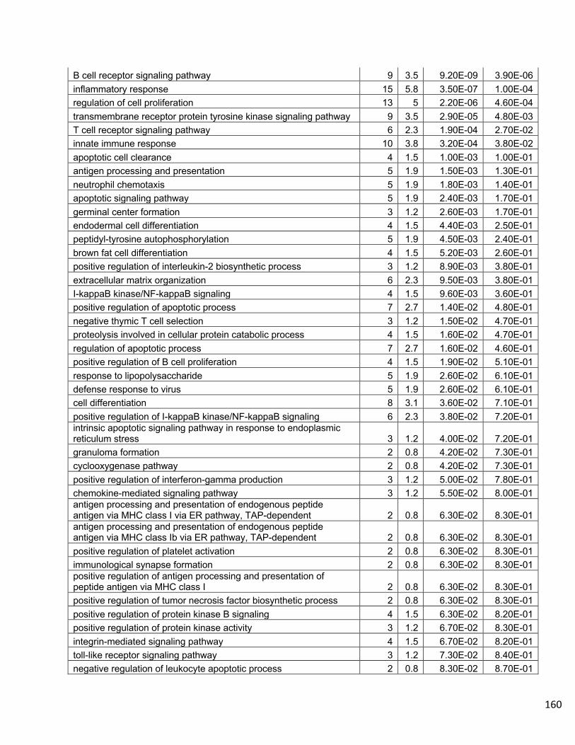
160
B cell receptor signaling pathway 9 3.5 9.20E-09 3.90E-06 inflammatory response 15 5.8 3.50E-07 1.00E-04 regulation of cell proliferation 13 5 2.20E-06 4.60E-04 transmembrane receptor protein tyrosine kinase signaling pathway 9 3.5 2.90E-05 4.80E-03 T cell receptor signaling pathway 6 2.3 1.90E-04 2.70E-02 innate immune response 10 3.8 3.20E-04 3.80E-02 apoptotic cell clearance 4 1.5 1.00E-03 1.00E-01 antigen processing and presentation 5 1.9 1.50E-03 1.30E-01 neutrophil chemotaxis 5 1.9 1.80E-03 1.40E-01 apoptotic signaling pathway 5 1.9 2.40E-03 1.70E-01 germinal center formation 3 1.2 2.60E-03 1.70E-01 endodermal cell differentiation 4 1.5 4.40E-03 2.50E-01 peptidyl-tyrosine autophosphorylation 5 1.9 4.50E-03 2.40E-01 brown fat cell differentiation 4 1.5 5.20E-03 2.60E-01 positive regulation of interleukin-2 biosynthetic process 3 1.2 8.90E-03 3.80E-01 extracellular matrix organization 6 2.3 9.50E-03 3.80E-01 I-kappaB kinase/NF-kappaB signaling 4 1.5 9.60E-03 3.60E-01 positive regulation of apoptotic process 7 2.7 1.40E-02 4.80E-01 negative thymic T cell selection 3 1.2 1.50E-02 4.70E-01 proteolysis involved in cellular protein catabolic process 4 1.5 1.60E-02 4.70E-01 regulation of apoptotic process 7 2.7 1.60E-02 4.60E-01 positive regulation of B cell proliferation 4 1.5 1.90E-02 5.10E-01 response to lipopolysaccharide 5 1.9 2.60E-02 6.10E-01 defense response to virus 5 1.9 2.60E-02 6.10E-01 cell differentiation 8 3.1 3.60E-02 7.10E-01 positive regulation of I-kappaB kinase/NF-kappaB signaling 6 2.3 3.80E-02 7.20E-01 intrinsic apoptotic signaling pathway in response to endoplasmic reticulum stress 3 1.2 4.00E-02 7.20E-01 granuloma formation 2 0.8 4.20E-02 7.30E-01 cyclooxygenase pathway 2 0.8 4.20E-02 7.30E-01 positive regulation of interferon-gamma production 3 1.2 5.00E-02 7.80E-01 chemokine-mediated signaling pathway 3 1.2 5.50E-02 8.00E-01 antigen processing and presentation of endogenous peptide antigen via MHC class I via ER pathway, TAP-dependent 2 0.8 6.30E-02 8.30E-01 antigen processing and presentation of endogenous peptide antigen via MHC class Ib via ER pathway, TAP-dependent 2 0.8 6.30E-02 8.30E-01 positive regulation of platelet activation 2 0.8 6.30E-02 8.30E-01 immunological synapse formation 2 0.8 6.30E-02 8.30E-01 positive regulation of antigen processing and presentation of peptide antigen via MHC class I 2 0.8 6.30E-02 8.30E-01 positive regulation of tumor necrosis factor biosynthetic process 2 0.8 6.30E-02 8.30E-01 positive regulation of protein kinase B signaling 4 1.5 6.30E-02 8.20E-01 positive regulation of protein kinase activity 3 1.2 6.70E-02 8.30E-01 integrin-mediated signaling pathway 4 1.5 6.70E-02 8.20E-01 toll-like receptor signaling pathway 3 1.2 7.30E-02 8.40E-01 negative regulation of leukocyte apoptotic process 2 0.8 8.30E-02 8.70E-01

161
cotranslational protein targeting to membrane 2 0.8 8.30E-02 8.70E-01 positive regulation of MHC class II biosynthetic process 2 0.8 8.30E-02 8.70E-01 dsRNA transport 2 0.8 8.30E-02 8.70E-01 antigen processing and presentation of exogenous protein antigen via MHC class Ib, TAP-dependent 2 0.8 8.30E-02 8.70E-01 protein N-linked glycosylation via asparagine 3 1.2 8.60E-02 8.70E-01 intrinsic apoptotic signaling pathway in response to DNA damage 3 1.2 8.60E-02 8.70E-01 chemotaxis 3 1.2 9.90E-02 9.00E-01 positive regulation of alpha-beta T cell proliferation 2 0.8 1.00E-01 9.00E-01 positive regulation of keratinocyte migration 2 0.8 1.00E-01 9.00E-01 toll-like receptor 3 signaling pathway 2 0.8 1.00E-01 9.00E-01 negative regulation of interleukin-17 production 2 0.8 1.00E-01 9.00E-01 secretion by cell 2 0.8 1.00E-01 9.00E-01 positive regulation of NF-kappaB transcription factor activity 4 1.5 1.00E-01 9.00E-01 positive regulation of MAP kinase activity 3 1.2 1.10E-01 9.00E-01 response to endoplasmic reticulum stress 3 1.2 1.10E-01 9.10E-01 cellular response to amino acid stimulus 3 1.2 1.20E-01 9.20E-01 complement activation, classical pathway 2 0.8 1.20E-01 9.20E-01 positive thymic T cell selection 2 0.8 1.20E-01 9.20E-01 regulation of cytokine secretion 2 0.8 1.20E-01 9.20E-01 negative regulation of protein phosphorylation 3 1.2 1.30E-01 9.20E-01 regulation of blood pressure 3 1.2 1.30E-01 9.20E-01 negative regulation of apoptotic process 7 2.7 1.30E-01 9.20E-01 activation of cysteine-type endopeptidase activity involved in apoptotic process 3 1.2 1.30E-01 9.20E-01 cell migration 5 1.9 1.40E-01 9.30E-01 immunoglobulin mediated immune response 2 0.8 1.40E-01 9.30E-01 activation of MAPKKK activity 2 0.8 1.40E-01 9.30E-01 regulation of osteoclast differentiation 2 0.8 1.40E-01 9.30E-01 adaptive immune response 3 1.2 1.40E-01 9.20E-01 regulation of protein localization 3 1.2 1.40E-01 9.20E-01 negative regulation of sequence-specific DNA binding transcription factor activity 3 1.2 1.50E-01 9.30E-01 positive regulation of GTPase activity 4 1.5 1.50E-01 9.30E-01 ossification 3 1.2 1.60E-01 9.30E-01 intracellular signal transduction 8 3.1 1.60E-01 9.30E-01 activation of NF-kappaB-inducing kinase activity 2 0.8 1.60E-01 9.30E-01 B cell activation 2 0.8 1.60E-01 9.30E-01 regulation of immune response 2 0.8 1.60E-01 9.30E-01 chaperone mediated protein folding requiring cofactor 2 0.8 1.60E-01 9.30E-01 negative regulation of B cell proliferation 2 0.8 1.60E-01 9.30E-01 positive regulation of calcium-mediated signaling 2 0.8 1.60E-01 9.30E-01 negative regulation of smoothened signaling pathway 2 0.8 1.60E-01 9.30E-01 myeloid dendritic cell differentiation 2 0.8 1.60E-01 9.30E-01 negative regulation of interferon-gamma production 2 0.8 1.60E-01 9.30E-01
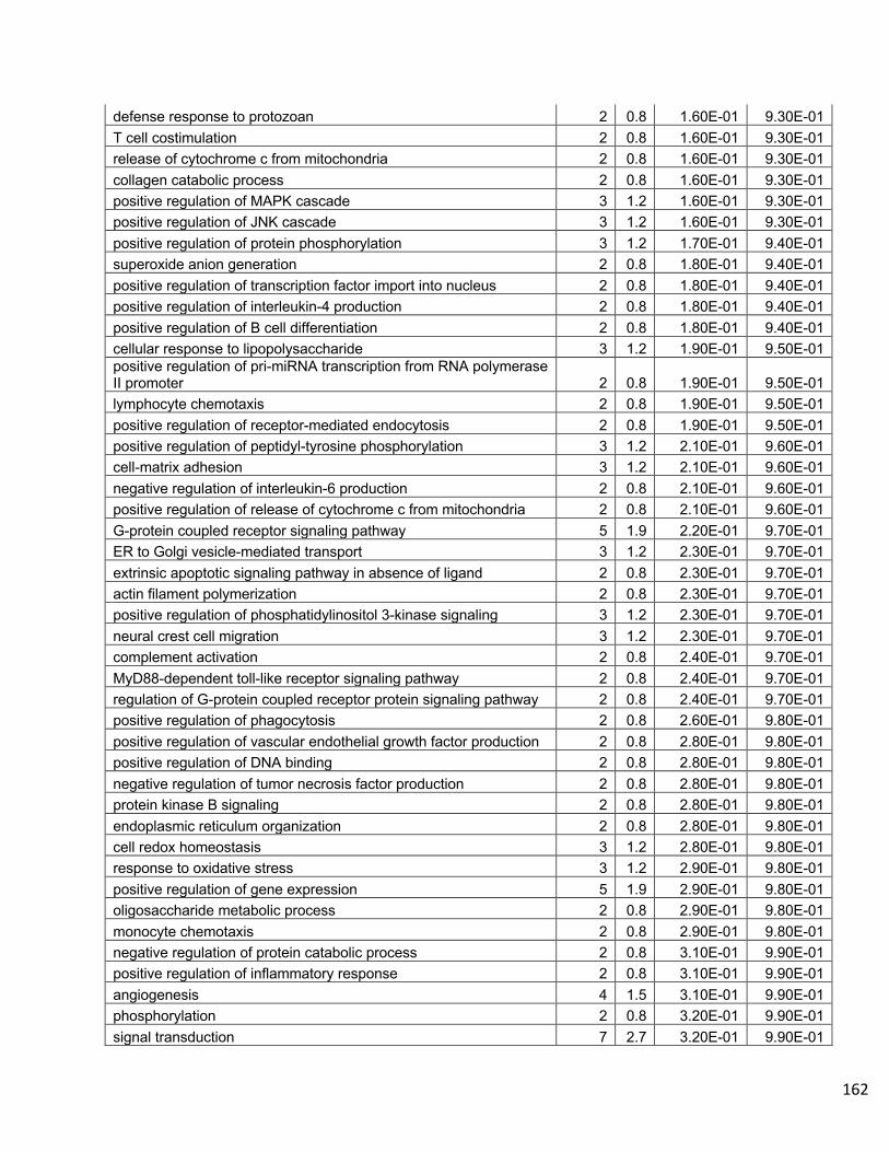
162
defense response to protozoan 2 0.8 1.60E-01 9.30E-01 T cell costimulation 2 0.8 1.60E-01 9.30E-01 release of cytochrome c from mitochondria 2 0.8 1.60E-01 9.30E-01 collagen catabolic process 2 0.8 1.60E-01 9.30E-01 positive regulation of MAPK cascade 3 1.2 1.60E-01 9.30E-01 positive regulation of JNK cascade 3 1.2 1.60E-01 9.30E-01 positive regulation of protein phosphorylation 3 1.2 1.70E-01 9.40E-01 superoxide anion generation 2 0.8 1.80E-01 9.40E-01 positive regulation of transcription factor import into nucleus 2 0.8 1.80E-01 9.40E-01 positive regulation of interleukin-4 production 2 0.8 1.80E-01 9.40E-01 positive regulation of B cell differentiation 2 0.8 1.80E-01 9.40E-01 cellular response to lipopolysaccharide 3 1.2 1.90E-01 9.50E-01 positive regulation of pri-miRNA transcription from RNA polymerase II promoter 2 0.8 1.90E-01 9.50E-01 lymphocyte chemotaxis 2 0.8 1.90E-01 9.50E-01 positive regulation of receptor-mediated endocytosis 2 0.8 1.90E-01 9.50E-01 positive regulation of peptidyl-tyrosine phosphorylation 3 1.2 2.10E-01 9.60E-01 cell-matrix adhesion 3 1.2 2.10E-01 9.60E-01 negative regulation of interleukin-6 production 2 0.8 2.10E-01 9.60E-01 positive regulation of release of cytochrome c from mitochondria 2 0.8 2.10E-01 9.60E-01 G-protein coupled receptor signaling pathway 5 1.9 2.20E-01 9.70E-01 ER to Golgi vesicle-mediated transport 3 1.2 2.30E-01 9.70E-01 extrinsic apoptotic signaling pathway in absence of ligand 2 0.8 2.30E-01 9.70E-01 actin filament polymerization 2 0.8 2.30E-01 9.70E-01 positive regulation of phosphatidylinositol 3-kinase signaling 3 1.2 2.30E-01 9.70E-01 neural crest cell migration 3 1.2 2.30E-01 9.70E-01 complement activation 2 0.8 2.40E-01 9.70E-01 MyD88-dependent toll-like receptor signaling pathway 2 0.8 2.40E-01 9.70E-01 regulation of G-protein coupled receptor protein signaling pathway 2 0.8 2.40E-01 9.70E-01 positive regulation of phagocytosis 2 0.8 2.60E-01 9.80E-01 positive regulation of vascular endothelial growth factor production 2 0.8 2.80E-01 9.80E-01 positive regulation of DNA binding 2 0.8 2.80E-01 9.80E-01 negative regulation of tumor necrosis factor production 2 0.8 2.80E-01 9.80E-01 protein kinase B signaling 2 0.8 2.80E-01 9.80E-01 endoplasmic reticulum organization 2 0.8 2.80E-01 9.80E-01 cell redox homeostasis 3 1.2 2.80E-01 9.80E-01 response to oxidative stress 3 1.2 2.90E-01 9.80E-01 positive regulation of gene expression 5 1.9 2.90E-01 9.80E-01 oligosaccharide metabolic process 2 0.8 2.90E-01 9.80E-01 monocyte chemotaxis 2 0.8 2.90E-01 9.80E-01 negative regulation of protein catabolic process 2 0.8 3.10E-01 9.90E-01 positive regulation of inflammatory response 2 0.8 3.10E-01 9.90E-01 angiogenesis 4 1.5 3.10E-01 9.90E-01 phosphorylation 2 0.8 3.20E-01 9.90E-01 signal transduction 7 2.7 3.20E-01 9.90E-01
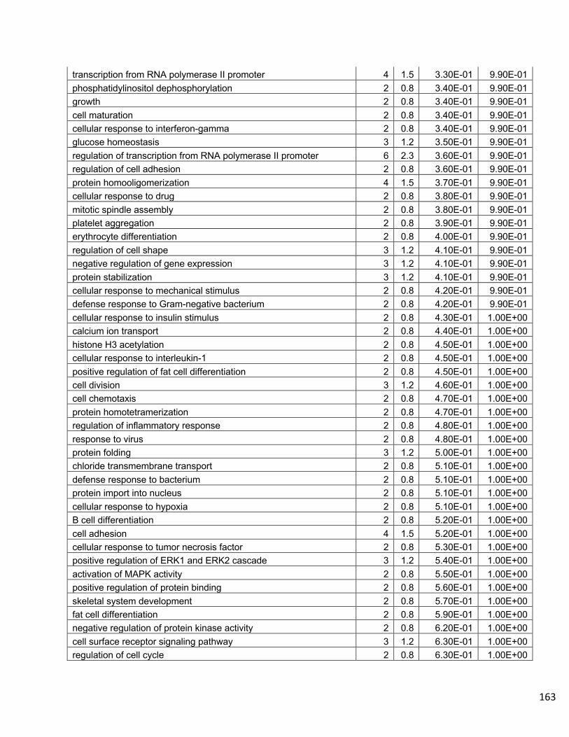
163
transcription from RNA polymerase II promoter 4 1.5 3.30E-01 9.90E-01 phosphatidylinositol dephosphorylation 2 0.8 3.40E-01 9.90E-01 growth 2 0.8 3.40E-01 9.90E-01 cell maturation 2 0.8 3.40E-01 9.90E-01 cellular response to interferon-gamma 2 0.8 3.40E-01 9.90E-01 glucose homeostasis 3 1.2 3.50E-01 9.90E-01 regulation of transcription from RNA polymerase II promoter 6 2.3 3.60E-01 9.90E-01 regulation of cell adhesion 2 0.8 3.60E-01 9.90E-01 protein homooligomerization 4 1.5 3.70E-01 9.90E-01 cellular response to drug 2 0.8 3.80E-01 9.90E-01 mitotic spindle assembly 2 0.8 3.80E-01 9.90E-01 platelet aggregation 2 0.8 3.90E-01 9.90E-01 erythrocyte differentiation 2 0.8 4.00E-01 9.90E-01 regulation of cell shape 3 1.2 4.10E-01 9.90E-01 negative regulation of gene expression 3 1.2 4.10E-01 9.90E-01 protein stabilization 3 1.2 4.10E-01 9.90E-01 cellular response to mechanical stimulus 2 0.8 4.20E-01 9.90E-01 defense response to Gram-negative bacterium 2 0.8 4.20E-01 9.90E-01 cellular response to insulin stimulus 2 0.8 4.30E-01 1.00E+00 calcium ion transport 2 0.8 4.40E-01 1.00E+00 histone H3 acetylation 2 0.8 4.50E-01 1.00E+00 cellular response to interleukin-1 2 0.8 4.50E-01 1.00E+00 positive regulation of fat cell differentiation 2 0.8 4.50E-01 1.00E+00 cell division 3 1.2 4.60E-01 1.00E+00 cell chemotaxis 2 0.8 4.70E-01 1.00E+00 protein homotetramerization 2 0.8 4.70E-01 1.00E+00 regulation of inflammatory response 2 0.8 4.80E-01 1.00E+00 response to virus 2 0.8 4.80E-01 1.00E+00 protein folding 3 1.2 5.00E-01 1.00E+00 chloride transmembrane transport 2 0.8 5.10E-01 1.00E+00 defense response to bacterium 2 0.8 5.10E-01 1.00E+00 protein import into nucleus 2 0.8 5.10E-01 1.00E+00 cellular response to hypoxia 2 0.8 5.10E-01 1.00E+00 B cell differentiation 2 0.8 5.20E-01 1.00E+00 cell adhesion 4 1.5 5.20E-01 1.00E+00 cellular response to tumor necrosis factor 2 0.8 5.30E-01 1.00E+00 positive regulation of ERK1 and ERK2 cascade 3 1.2 5.40E-01 1.00E+00 activation of MAPK activity 2 0.8 5.50E-01 1.00E+00 positive regulation of protein binding 2 0.8 5.60E-01 1.00E+00 skeletal system development 2 0.8 5.70E-01 1.00E+00 fat cell differentiation 2 0.8 5.90E-01 1.00E+00 negative regulation of protein kinase activity 2 0.8 6.20E-01 1.00E+00 cell surface receptor signaling pathway 3 1.2 6.30E-01 1.00E+00 regulation of cell cycle 2 0.8 6.30E-01 1.00E+00
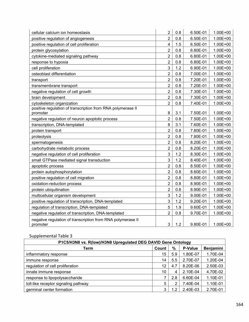
164
cellular calcium ion homeostasis 2 0.8 6.50E-01 1.00E+00 positive regulation of angiogenesis 2 0.8 6.50E-01 1.00E+00 positive regulation of cell proliferation 4 1.5 6.50E-01 1.00E+00 protein glycosylation 2 0.8 6.80E-01 1.00E+00 cytokine-mediated signaling pathway 2 0.8 6.80E-01 1.00E+00 response to hypoxia 2 0.8 6.80E-01 1.00E+00 cell proliferation 3 1.2 6.90E-01 1.00E+00 osteoblast differentiation 2 0.8 7.00E-01 1.00E+00 transport 2 0.8 7.20E-01 1.00E+00 transmembrane transport 2 0.8 7.20E-01 1.00E+00 negative regulation of cell growth 2 0.8 7.30E-01 1.00E+00 brain development 2 0.8 7.30E-01 1.00E+00 cytoskeleton organization 2 0.8 7.40E-01 1.00E+00 positive regulation of transcription from RNA polymerase II promoter 8 3.1 7.50E-01 1.00E+00 negative regulation of neuron apoptotic process 2 0.8 7.50E-01 1.00E+00 transcription, DNA-templated 8 3.1 7.60E-01 1.00E+00 protein transport 2 0.8 7.80E-01 1.00E+00 proteolysis 2 0.8 7.80E-01 1.00E+00 spermatogenesis 2 0.8 8.20E-01 1.00E+00 carbohydrate metabolic process 2 0.8 8.20E-01 1.00E+00 negative regulation of cell proliferation 3 1.2 8.30E-01 1.00E+00 small GTPase mediated signal transduction 3 1.2 8.40E-01 1.00E+00 apoptotic process 2 0.8 8.50E-01 1.00E+00 protein autophosphorylation 2 0.8 8.60E-01 1.00E+00 positive regulation of cell migration 2 0.8 8.80E-01 1.00E+00 oxidation-reduction process 2 0.8 8.90E-01 1.00E+00 protein ubiquitination 2 0.8 8.90E-01 1.00E+00 multicellular organism development 3 1.2 9.00E-01 1.00E+00 positive regulation of transcription, DNA-templated 3 1.2 9.20E-01 1.00E+00 regulation of transcription, DNA-templated 5 1.9 9.60E-01 1.00E+00 negative regulation of transcription, DNA-templated 2 0.8 9.70E-01 1.00E+00 negative regulation of transcription from RNA polymerase II promoter 3 1.2 9.80E-01 1.00E+00
Supplemental Table 3
P1C5/H3N8 vs. R(low)/H3N8 Upregulated DEG DAVID Gene Ontology Term Count % P-Value Benjamini
inflammatory response 15 5.9 1.80E-07 1.70E-04 immune response 14 5.5 2.70E-07 1.20E-04 regulation of cell proliferation 12 4.7 8.20E-06 2.50E-03 innate immune response 10 4 2.10E-04 4.70E-02 response to lipopolysaccharide 7 2.8 6.60E-04 1.10E-01 toll-like receptor signaling pathway 5 2 7.40E-04 1.10E-01 germinal center formation 3 1.2 2.40E-03 2.70E-01

165
endodermal cell differentiation 4 1.6 3.80E-03 3.50E-01 cellular response to hypoxia 5 2 4.20E-03 3.40E-01 defense response to virus 6 2.4 4.20E-03 3.20E-01 brown fat cell differentiation 4 1.6 4.50E-03 3.10E-01 regulation of cytokine secretion 3 1.2 5.80E-03 3.50E-01 transmembrane receptor protein tyrosine kinase signaling pathway 6 2.4 6.20E-03 3.50E-01 cell differentiation 9 3.6 9.10E-03 4.50E-01 positive regulation of apoptotic process 7 2.8 1.10E-02 5.00E-01 integrin-mediated signaling pathway 5 2 1.20E-02 4.90E-01 neutrophil chemotaxis 4 1.6 1.40E-02 5.20E-01 apoptotic cell clearance 3 1.2 1.60E-02 5.70E-01 positive regulation of pri-miRNA transcription from RNA polymerase II promoter 3 1.2 1.60E-02 5.70E-01 apoptotic signaling pathway 4 1.6 1.70E-02 5.50E-01 negative regulation of smooth muscle cell proliferation 3 1.2 2.00E-02 6.00E-01 peptide cross-linking 3 1.2 2.40E-02 6.40E-01 peptidyl-tyrosine autophosphorylation 4 1.6 2.60E-02 6.70E-01 MyD88-dependent toll-like receptor signaling pathway 3 1.2 2.70E-02 6.70E-01 positive regulation of axon extension 3 1.2 3.20E-02 7.00E-01 positive regulation of smooth muscle cell proliferation 3 1.2 3.60E-02 7.30E-01 protein kinase B signaling 3 1.2 3.60E-02 7.30E-01 cell migration 6 2.4 4.00E-02 7.60E-01 cyclooxygenase pathway 2 0.8 4.00E-02 7.50E-01 negative regulation of regulatory T cell differentiation 2 0.8 4.00E-02 7.50E-01 heme catabolic process 2 0.8 4.00E-02 7.50E-01 hypermethylation of CpG island 2 0.8 4.00E-02 7.50E-01 tolerance induction to lipopolysaccharide 2 0.8 4.00E-02 7.50E-01 granuloma formation 2 0.8 4.00E-02 7.50E-01 positive regulation of inflammatory response 3 1.2 4.50E-02 7.80E-01 positive regulation of macroautophagy 3 1.2 5.60E-02 8.30E-01 regulation of innate immune response 2 0.8 5.90E-02 8.40E-01 positive regulation of platelet activation 2 0.8 5.90E-02 8.40E-01 positive regulation of tumor necrosis factor biosynthetic process 2 0.8 5.90E-02 8.40E-01 B-1 B cell homeostasis 2 0.8 5.90E-02 8.40E-01 positive regulation of interleukin-12 biosynthetic process 2 0.8 5.90E-02 8.40E-01 negative regulation of B cell activation 2 0.8 5.90E-02 8.40E-01 I-kappaB kinase/NF-kappaB signaling 3 1.2 6.70E-02 8.70E-01 B cell receptor signaling pathway 3 1.2 7.20E-02 8.80E-01 platelet aggregation 3 1.2 7.80E-02 8.90E-01 secretion 2 0.8 7.90E-02 8.90E-01 detection of lipopolysaccharide 2 0.8 7.90E-02 8.90E-01 response to oxidative stress 4 1.6 8.00E-02 8.80E-01 antigen processing and presentation 3 1.2 8.40E-02 8.90E-01 cellular response to mechanical stimulus 3 1.2 9.00E-02 9.00E-01 proteolysis involved in cellular protein catabolic process 3 1.2 9.00E-02 9.00E-01

166
positive regulation of MAP kinase activity 3 1.2 9.60E-02 9.10E-01 secretion by cell 2 0.8 9.70E-02 9.10E-01 extracellular matrix disassembly 2 0.8 9.70E-02 9.10E-01 endoderm formation 2 0.8 9.70E-02 9.10E-01 negative regulation of interleukin-17 production 2 0.8 9.70E-02 9.10E-01 positive regulation of keratinocyte migration 2 0.8 9.70E-02 9.10E-01 positive regulation of I-kappaB kinase/NF-kappaB signaling 5 2 9.90E-02 9.10E-01 substrate adhesion-dependent cell spreading 3 1.2 1.00E-01 9.10E-01 negative regulation of apoptotic process 7 2.8 1.10E-01 9.20E-01 cellular response to amino acid stimulus 3 1.2 1.10E-01 9.10E-01 natural killer cell differentiation 2 0.8 1.20E-01 9.20E-01 cellular response to interferon-beta 2 0.8 1.20E-01 9.20E-01 negative regulation of interleukin-2 production 2 0.8 1.20E-01 9.20E-01 positive regulation of receptor biosynthetic process 2 0.8 1.20E-01 9.20E-01 regulation of blood pressure 3 1.2 1.20E-01 9.20E-01 positive regulation of gene expression 6 2.4 1.20E-01 9.10E-01 response to virus 3 1.2 1.20E-01 9.20E-01 regulation of inflammatory response 3 1.2 1.20E-01 9.20E-01 regulation of apoptotic process 5 2 1.30E-01 9.20E-01 regulation of I-kappaB kinase/NF-kappaB signaling 2 0.8 1.30E-01 9.30E-01 regulation of cell adhesion mediated by integrin 2 0.8 1.30E-01 9.30E-01 positive regulation of interleukin-2 biosynthetic process 2 0.8 1.30E-01 9.30E-01 DNA methylation involved in gamete generation 2 0.8 1.30E-01 9.30E-01 regulation of osteoclast differentiation 2 0.8 1.30E-01 9.30E-01 fatty acid transport 2 0.8 1.30E-01 9.30E-01 positive regulation of macrophage cytokine production 2 0.8 1.30E-01 9.30E-01 negative regulation of sequence-specific DNA binding transcription factor activity 3 1.2 1.40E-01 9.30E-01 ossification 3 1.2 1.40E-01 9.40E-01 positive regulation of MAPK cascade 3 1.2 1.50E-01 9.40E-01 release of cytochrome c from mitochondria 2 0.8 1.50E-01 9.40E-01 negative regulation of interferon-gamma production 2 0.8 1.50E-01 9.40E-01 myeloid dendritic cell differentiation 2 0.8 1.50E-01 9.40E-01 defense response to protozoan 2 0.8 1.50E-01 9.40E-01 chaperone mediated protein folding requiring cofactor 2 0.8 1.50E-01 9.40E-01 collagen catabolic process 2 0.8 1.50E-01 9.40E-01 deadenylation-dependent decapping of nuclear-transcribed mRNA 2 0.8 1.50E-01 9.40E-01 protein maturation 2 0.8 1.70E-01 9.50E-01 cell-substrate adhesion 2 0.8 1.70E-01 9.50E-01 signal peptide processing 2 0.8 1.70E-01 9.50E-01 superoxide anion generation 2 0.8 1.70E-01 9.50E-01 astrocyte development 2 0.8 1.80E-01 9.70E-01 positive regulation of phosphorylation 2 0.8 1.80E-01 9.70E-01 G-protein coupled receptor signaling pathway 5 2 2.00E-01 9.70E-01 fat cell differentiation 3 1.2 2.00E-01 9.70E-01
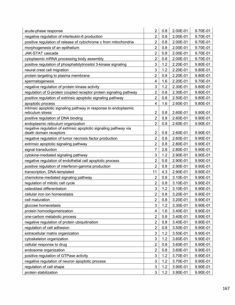
167
acute-phase response 2 0.8 2.00E-01 9.70E-01 negative regulation of interleukin-6 production 2 0.8 2.00E-01 9.70E-01 positive regulation of release of cytochrome c from mitochondria 2 0.8 2.00E-01 9.70E-01 morphogenesis of an epithelium 2 0.8 2.00E-01 9.70E-01 JAK-STAT cascade 2 0.8 2.00E-01 9.70E-01 cytoplasmic mRNA processing body assembly 2 0.8 2.00E-01 9.70E-01 positive regulation of phosphatidylinositol 3-kinase signaling 3 1.2 2.20E-01 9.80E-01 neural crest cell migration 3 1.2 2.20E-01 9.80E-01 protein targeting to plasma membrane 2 0.8 2.20E-01 9.80E-01 spermatogenesis 4 1.6 2.20E-01 9.70E-01 negative regulation of protein kinase activity 3 1.2 2.30E-01 9.80E-01 regulation of G-protein coupled receptor protein signaling pathway 2 0.8 2.30E-01 9.80E-01 positive regulation of extrinsic apoptotic signaling pathway 2 0.8 2.50E-01 9.80E-01 apoptotic process 4 1.6 2.60E-01 9.80E-01 intrinsic apoptotic signaling pathway in response to endoplasmic reticulum stress 2 0.8 2.60E-01 9.90E-01 positive regulation of DNA binding 2 0.8 2.60E-01 9.90E-01 endoplasmic reticulum organization 2 0.8 2.60E-01 9.90E-01 negative regulation of extrinsic apoptotic signaling pathway via death domain receptors 2 0.8 2.60E-01 9.90E-01 negative regulation of tumor necrosis factor production 2 0.8 2.60E-01 9.90E-01 extrinsic apoptotic signaling pathway 2 0.8 2.80E-01 9.90E-01 signal transduction 7 2.8 2.80E-01 9.90E-01 cytokine-mediated signaling pathway 3 1.2 2.90E-01 9.90E-01 negative regulation of endothelial cell apoptotic process 2 0.8 2.90E-01 9.90E-01 positive regulation of interferon-gamma production 2 0.8 2.90E-01 9.90E-01 transcription, DNA-templated 11 4.3 2.90E-01 9.90E-01 chemokine-mediated signaling pathway 2 0.8 3.10E-01 9.90E-01 regulation of mitotic cell cycle 2 0.8 3.10E-01 9.90E-01 osteoblast differentiation 3 1.2 3.10E-01 9.90E-01 cellular iron ion homeostasis 2 0.8 3.20E-01 9.90E-01 cell maturation 2 0.8 3.20E-01 9.90E-01 glucose homeostasis 3 1.2 3.30E-01 9.90E-01 protein homooligomerization 4 1.6 3.40E-01 9.90E-01 one-carbon metabolic process 2 0.8 3.40E-01 9.90E-01 negative regulation of protein ubiquitination 2 0.8 3.40E-01 9.90E-01 regulation of cell adhesion 2 0.8 3.50E-01 9.90E-01 extracellular matrix organization 3 1.2 3.50E-01 9.90E-01 cytoskeleton organization 3 1.2 3.60E-01 9.90E-01 cellular response to drug 2 0.8 3.60E-01 9.90E-01 endosome organization 2 0.8 3.60E-01 9.90E-01 positive regulation of GTPase activity 3 1.2 3.70E-01 9.90E-01 negative regulation of neuron apoptotic process 3 1.2 3.70E-01 9.90E-01 regulation of cell shape 3 1.2 3.90E-01 9.90E-01 protein stabilization 3 1.2 3.90E-01 9.90E-01
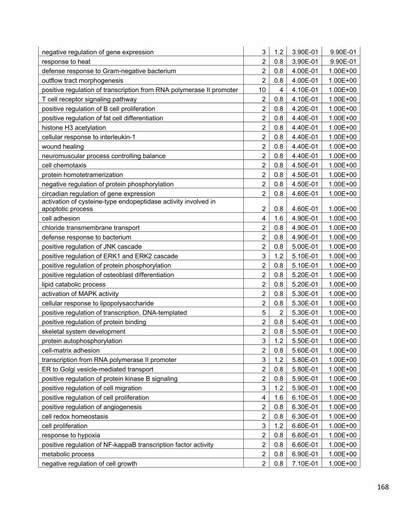
168
negative regulation of gene expression 3 1.2 3.90E-01 9.90E-01 response to heat 2 0.8 3.90E-01 9.90E-01 defense response to Gram-negative bacterium 2 0.8 4.00E-01 1.00E+00 outflow tract morphogenesis 2 0.8 4.00E-01 1.00E+00 positive regulation of transcription from RNA polymerase II promoter 10 4 4.10E-01 1.00E+00 T cell receptor signaling pathway 2 0.8 4.10E-01 1.00E+00 positive regulation of B cell proliferation 2 0.8 4.20E-01 1.00E+00 positive regulation of fat cell differentiation 2 0.8 4.40E-01 1.00E+00 histone H3 acetylation 2 0.8 4.40E-01 1.00E+00 cellular response to interleukin-1 2 0.8 4.40E-01 1.00E+00 wound healing 2 0.8 4.40E-01 1.00E+00 neuromuscular process controlling balance 2 0.8 4.40E-01 1.00E+00 cell chemotaxis 2 0.8 4.50E-01 1.00E+00 protein homotetramerization 2 0.8 4.50E-01 1.00E+00 negative regulation of protein phosphorylation 2 0.8 4.50E-01 1.00E+00 circadian regulation of gene expression 2 0.8 4.60E-01 1.00E+00 activation of cysteine-type endopeptidase activity involved in apoptotic process 2 0.8 4.60E-01 1.00E+00 cell adhesion 4 1.6 4.90E-01 1.00E+00 chloride transmembrane transport 2 0.8 4.90E-01 1.00E+00 defense response to bacterium 2 0.8 4.90E-01 1.00E+00 positive regulation of JNK cascade 2 0.8 5.00E-01 1.00E+00 positive regulation of ERK1 and ERK2 cascade 3 1.2 5.10E-01 1.00E+00 positive regulation of protein phosphorylation 2 0.8 5.10E-01 1.00E+00 positive regulation of osteoblast differentiation 2 0.8 5.20E-01 1.00E+00 lipid catabolic process 2 0.8 5.20E-01 1.00E+00 activation of MAPK activity 2 0.8 5.30E-01 1.00E+00 cellular response to lipopolysaccharide 2 0.8 5.30E-01 1.00E+00 positive regulation of transcription, DNA-templated 5 2 5.30E-01 1.00E+00 positive regulation of protein binding 2 0.8 5.40E-01 1.00E+00 skeletal system development 2 0.8 5.50E-01 1.00E+00 protein autophosphorylation 3 1.2 5.50E-01 1.00E+00 cell-matrix adhesion 2 0.8 5.60E-01 1.00E+00 transcription from RNA polymerase II promoter 3 1.2 5.80E-01 1.00E+00 ER to Golgi vesicle-mediated transport 2 0.8 5.80E-01 1.00E+00 positive regulation of protein kinase B signaling 2 0.8 5.90E-01 1.00E+00 positive regulation of cell migration 3 1.2 5.90E-01 1.00E+00 positive regulation of cell proliferation 4 1.6 6.10E-01 1.00E+00 positive regulation of angiogenesis 2 0.8 6.30E-01 1.00E+00 cell redox homeostasis 2 0.8 6.30E-01 1.00E+00 cell proliferation 3 1.2 6.60E-01 1.00E+00 response to hypoxia 2 0.8 6.60E-01 1.00E+00 positive regulation of NF-kappaB transcription factor activity 2 0.8 6.60E-01 1.00E+00 metabolic process 2 0.8 6.90E-01 1.00E+00 negative regulation of cell growth 2 0.8 7.10E-01 1.00E+00

169
regulation of transcription from RNA polymerase II promoter 4 1.6 7.30E-01 1.00E+00 proteolysis 2 0.8 7.60E-01 1.00E+00 protein transport 2 0.8 7.60E-01 1.00E+00 intracellular signal transduction 4 1.6 8.00E-01 1.00E+00 protein folding 2 0.8 8.00E-01 1.00E+00 negative regulation of transcription from RNA polymerase II promoter 5 2 8.00E-01 1.00E+00 cilium assembly 2 0.8 8.10E-01 1.00E+00 negative regulation of cell proliferation 3 1.2 8.10E-01 1.00E+00 negative regulation of transcription, DNA-templated 3 1.2 8.30E-01 1.00E+00 angiogenesis 2 0.8 8.50E-01 1.00E+00 protein ubiquitination involved in ubiquitin-dependent protein catabolic process 2 0.8 8.50E-01 1.00E+00 multicellular organism development 3 1.2 8.80E-01 1.00E+00 protein ubiquitination 2 0.8 8.80E-01 1.00E+00 regulation of transcription, DNA-templated 5 2 9.50E-01 1.00E+00
Supplemental Table 4
P1C5/H3N8 vs. R(low)/H3N8 Gene Downregulated DEG DAVID Gene Ontology Term Count % P-Value Benjamini
cilium assembly 6 5.7 1.60E-04 4.10E-02 inner dynein arm assembly 3 2.8 1.10E-03 1.40E-01 intraciliary transport 3 2.8 1.40E-03 1.20E-01 sperm motility 3 2.8 1.20E-02 5.60E-01 retina development in camera-type eye 3 2.8 1.30E-02 5.10E-01 determination of left/right symmetry 3 2.8 1.70E-02 5.40E-01 prostaglandin metabolic process 2 1.9 1.90E-02 5.30E-01 cilium movement involved in cell motility 2 1.9 1.90E-02 5.30E-01 neural tube patterning 2 1.9 2.60E-02 5.90E-01 intraciliary transport involved in cilium morphogenesis 2 1.9 3.80E-02 6.90E-01 intraciliary retrograde transport 2 1.9 3.80E-02 6.90E-01 epithelial cilium movement 2 1.9 4.40E-02 7.10E-01 motile cilium assembly 2 1.9 5.00E-02 7.20E-01 GTP biosynthetic process 2 1.9 5.00E-02 7.20E-01 CTP biosynthetic process 2 1.9 5.00E-02 7.20E-01 UTP biosynthetic process 2 1.9 5.00E-02 7.20E-01 aorta development 2 1.9 8.10E-02 8.50E-01 coronary vasculature development 2 1.9 9.30E-02 8.70E-01 lens development in camera-type eye 2 1.9 1.40E-01 9.40E-01 spermatid development 2 1.9 2.00E-01 9.80E-01 microtubule-based movement 2 1.9 2.40E-01 9.90E-01 Notch signaling pathway 2 1.9 2.60E-01 9.90E-01 Golgi organization 2 1.9 2.90E-01 9.90E-01 mitotic nuclear division 2 1.9 3.10E-01 9.90E-01

170
heart development 2 1.9 4.70E-01 1.00E+00 cell proliferation 2 1.9 5.10E-01 1.00E+00 positive regulation of transcription, DNA-templated 2 1.9 7.10E-01 1.00E+00 intracellular signal transduction 2 1.9 7.40E-01 1.00E+00 positive regulation of transcription from RNA polymerase II promoter 2 1.9 9.30E-01 1.00E+00
Supplemental Table 5
Mock/H3N8 vs. P1C5/H3N8 Downregulated DEG DAVID Gene Ontology Term Count % P-Value Benjamini
toll-like receptor signaling pathway 4 5.6 2.50E-04 8.70E-02 inflammatory response 6 8.3 8.50E-04 1.40E-01 MyD88-dependent toll-like receptor signaling pathway 3 4.2 2.60E-03 2.70E-01 tolerance induction to lipopolysaccharide 2 2.8 1.20E-02 6.60E-01 response to virus 3 4.2 1.40E-02 6.30E-01 smooth muscle cell migration 2 2.8 1.80E-02 6.60E-01 innate immune response 4 5.6 2.30E-02 7.00E-01 detection of lipopolysaccharide 2 2.8 2.40E-02 6.60E-01 positive regulation of keratinocyte migration 2 2.8 3.00E-02 7.00E-01 skeletal muscle contraction 2 2.8 3.50E-02 7.30E-01 positive regulation of chemokine production 2 2.8 4.10E-02 7.50E-01 positive regulation of wound healing 2 2.8 4.70E-02 7.70E-01 negative regulation of smooth muscle cell proliferation 2 2.8 6.40E-02 8.40E-01 negative regulation of interleukin-6 production 2 2.8 6.40E-02 8.40E-01 negative regulation of tumor necrosis factor production 2 2.8 8.60E-02 9.00E-01 cardiac muscle contraction 2 2.8 8.60E-02 9.00E-01 negative regulation of extrinsic apoptotic signaling pathway via death domain receptors 2 2.8 8.60E-02 9.00E-01 positive regulation of smooth muscle cell proliferation 2 2.8 8.60E-02 9.00E-01 endodermal cell differentiation 2 2.8 9.20E-02 9.00E-01 protein homotrimerization 2 2.8 9.20E-02 9.00E-01 positive regulation of I-kappaB kinase/NF-kappaB signaling 3 4.2 9.60E-02 9.00E-01 negative regulation of endothelial cell apoptotic process 2 2.8 9.70E-02 8.90E-01
Supplemental Table 6
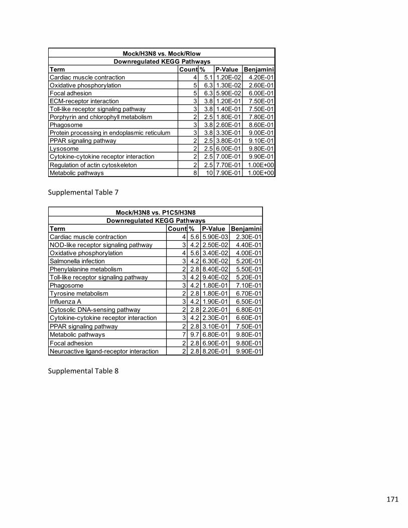
171
Supplemental Table 7
Supplemental Table 8
Term Count % P-Value BenjaminiCardiac muscle contraction 4 5.1 1.20E-02 4.20E-01Oxidative phosphorylation 5 6.3 1.30E-02 2.60E-01Focal adhesion 5 6.3 5.90E-02 6.00E-01ECM-receptor interaction 3 3.8 1.20E-01 7.50E-01Toll-like receptor signaling pathway 3 3.8 1.40E-01 7.50E-01Porphyrin and chlorophyll metabolism 2 2.5 1.80E-01 7.80E-01Phagosome 3 3.8 2.60E-01 8.60E-01Protein processing in endoplasmic reticulum 3 3.8 3.30E-01 9.00E-01PPAR signaling pathway 2 2.5 3.80E-01 9.10E-01Lysosome 2 2.5 6.00E-01 9.80E-01Cytokine-cytokine receptor interaction 2 2.5 7.00E-01 9.90E-01Regulation of actin cytoskeleton 2 2.5 7.70E-01 1.00E+00Metabolic pathways 8 10 7.90E-01 1.00E+00
Mock/H3N8 vs. Mock/RlowDownregulated KEGG Pathways
Term Count % P-Value BenjaminiCardiac muscle contraction 4 5.6 5.90E-03 2.30E-01NOD-like receptor signaling pathway 3 4.2 2.50E-02 4.40E-01Oxidative phosphorylation 4 5.6 3.40E-02 4.00E-01Salmonella infection 3 4.2 6.30E-02 5.20E-01Phenylalanine metabolism 2 2.8 8.40E-02 5.50E-01Toll-like receptor signaling pathway 3 4.2 9.40E-02 5.20E-01Phagosome 3 4.2 1.80E-01 7.10E-01Tyrosine metabolism 2 2.8 1.80E-01 6.70E-01Influenza A 3 4.2 1.90E-01 6.50E-01Cytosolic DNA-sensing pathway 2 2.8 2.20E-01 6.80E-01Cytokine-cytokine receptor interaction 3 4.2 2.30E-01 6.60E-01PPAR signaling pathway 2 2.8 3.10E-01 7.50E-01Metabolic pathways 7 9.7 6.80E-01 9.80E-01Focal adhesion 2 2.8 6.90E-01 9.80E-01Neuroactive ligand-receptor interaction 2 2.8 8.20E-01 9.90E-01
Mock/H3N8 vs. P1C5/H3N8Downregulated KEGG Pathways

172
Supplemental Table 9
Term Count % P-Value Benjamini Term Count % P-Value Benjamini
Phagosome 12 4.6 1.30E-04 1.00E-02 Cardiac muscle contraction 5 1.7 5.10E-03 3.00E-01Cell adhesion molecules (CAMs) 11 4.2 2.30E-04 9.30E-03 Purine metabolism 7 2.4 7.60E-03 2.30E-01Intestinal immune network for IgA production 6 2.3 7.20E-04 2.00E-02 Oxidative phosphorylation 6 2.1 1.10E-02 2.20E-01Lysosome 10 3.8 1.10E-03 2.20E-02 Metabolic pathways 21 7.3 1.40E-02 2.10E-01Jak-STAT signaling pathway 10 3.8 1.50E-03 2.40E-02 Biosynthesis of antibiotics 7 2.4 1.90E-02 2.30E-01Toll-like receptor signaling pathway 8 3.1 3.30E-03 4.50E-02 Pyrimidine metabolism 4 1.4 8.00E-02 6.20E-01Protein processing in endoplasmic reticulum 10 3.8 7.50E-03 8.40E-02 Drug metabolism - cytochrome P450 2 0.7 2.80E-01 9.60E-01Cytokine-cytokine receptor interaction 10 3.8 7.50E-03 8.40E-02 Metabolism of xenobiotics by cytochrome P450 2 0.7 3.10E-01 9.60E-01Influenza A 9 3.5 1.10E-02 1.00E-01 Cysteine and methionine metabolism 2 0.7 3.20E-01 9.50E-01Herpes simplex infection 8 3.1 5.00E-02 3.70E-01 Glycine, serine and threonine metabolism 2 0.7 3.40E-01 9.40E-01Nicotinate and nicotinamide metabolism 3 1.2 1.30E-01 6.90E-01 Adrenergic signaling in cardiomyocytes 3 1 3.80E-01 9.50E-01VEGF signaling pathway 4 1.5 1.50E-01 7.10E-01 Fatty acid metabolism 2 0.7 4.10E-01 9.50E-01Cytosolic DNA-sensing pathway 3 1.2 2.50E-01 8.60E-01 Valine, leucine and isoleucine degradation 2 0.7 4.10E-01 9.50E-01ECM-receptor interaction 4 1.5 2.50E-01 8.40E-01 Arachidonic acid metabolism 2 0.7 4.50E-01 9.60E-01Focal adhesion 7 2.7 2.70E-01 8.50E-01 PPAR signaling pathway 2 0.7 5.10E-01 9.70E-01Glycosaminoglycan degradation 2 0.8 3.50E-01 9.10E-01 ECM-receptor interaction 2 0.7 5.80E-01 9.80E-01Phosphatidylinositol signaling system 4 1.5 3.70E-01 9.10E-01 TGF-beta signaling pathway 2 0.7 5.90E-01 9.80E-01RIG-I-like receptor signaling pathway 3 1.2 3.80E-01 9.00E-01 Tight junction 2 0.7 6.00E-01 9.80E-01Protein export 2 0.8 4.10E-01 9.10E-01 Insulin resistance 2 0.7 6.80E-01 9.90E-01Porphyrin and chlorophyll metabolism 2 0.8 4.50E-01 9.30E-01 Vascular smooth muscle contraction 2 0.7 7.00E-01 9.90E-01Drug metabolism - other enzymes 2 0.8 4.70E-01 9.20E-01 Cell adhesion molecules (CAMs) 2 0.7 7.30E-01 9.90E-01Cysteine and methionine metabolism 2 0.8 5.50E-01 9.60E-01 Cell cycle 2 0.7 7.30E-01 9.90E-01ErbB signaling pathway 3 1.2 5.50E-01 9.50E-01 Insulin signaling pathway 2 0.7 7.50E-01 9.90E-01MAPK signaling pathway 6 2.3 5.70E-01 9.50E-01 FoxO signaling pathway 2 0.7 7.60E-01 9.90E-01ABC transporters 2 0.8 5.90E-01 9.50E-01 Calcium signaling pathway 2 0.7 8.60E-01 1.00E+00N-Glycan biosynthesis 2 0.8 6.70E-01 9.70E-01 Focal adhesion 2 0.7 8.90E-01 1.00E+00mTOR signaling pathway 2 0.8 7.00E-01 9.80E-01 Endocytosis 2 0.7 9.30E-01 1.00E+00Arachidonic acid metabolism 2 0.8 7.00E-01 9.70E-01 Neuroactive ligand-receptor interaction 2 0.7 9.60E-01 1.00E+00Apoptosis 2 0.8 7.20E-01 9.80E-01PPAR signaling pathway 2 0.8 7.60E-01 9.80E-01Insulin signaling pathway 3 1.2 7.70E-01 9.80E-01p53 signaling pathway 2 0.8 7.70E-01 9.80E-01Endocytosis 5 1.9 7.70E-01 9.80E-01FoxO signaling pathway 3 1.2 7.90E-01 9.80E-01Adipocytokine signaling pathway 2 0.8 7.90E-01 9.80E-01Regulation of actin cytoskeleton 4 1.5 8.00E-01 9.80E-01Inositol phosphate metabolism 2 0.8 8.10E-01 9.80E-01Progesterone-mediated oocyte maturation 2 0.8 8.40E-01 9.80E-01Purine metabolism 3 1.2 8.80E-01 9.90E-01Pyrimidine metabolism 2 0.8 8.80E-01 9.90E-01Calcium signaling pathway 3 1.2 9.00E-01 9.90E-01Insulin resistance 2 0.8 9.00E-01 9.90E-01Vascular smooth muscle contraction 2 0.8 9.10E-01 9.90E-01Wnt signaling pathway 2 0.8 9.50E-01 1.00E+00Metabolic pathways 18 6.9 9.80E-01 1.00E+00Neuroactive ligand-receptor interaction 2 0.8 1.00E+00 1.00E+00
Upregulated KEGG Pathways
Mock/H3N8 vs. Rlow/H3N8Mock/H3N8 vs. Rlow/H3N8
Downregulated KEGG Pathways
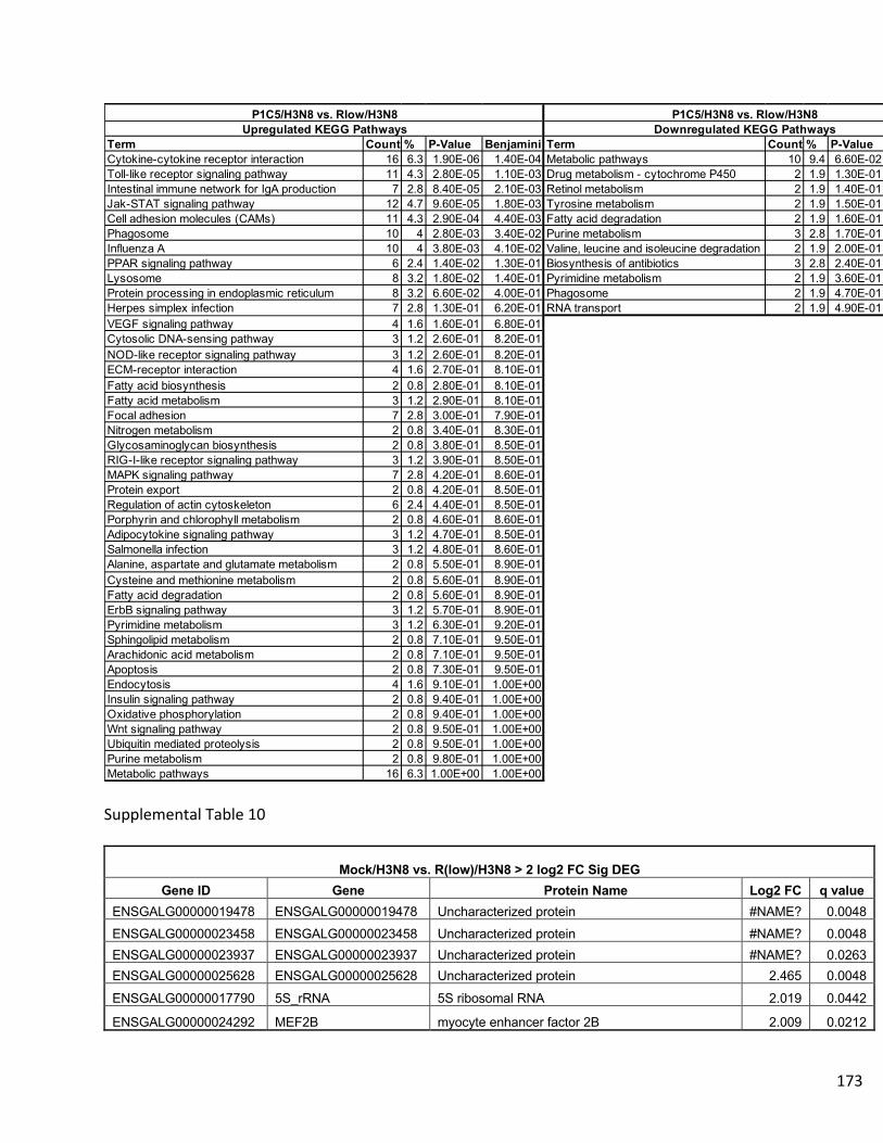
173
Supplemental Table 10
Mock/H3N8 vs. R(low)/H3N8 > 2 log2 FC Sig DEG
Gene ID Gene Protein Name Log2 FC q value ENSGALG00000019478 ENSGALG00000019478 Uncharacterized protein #NAME? 0.0048 ENSGALG00000023458 ENSGALG00000023458 Uncharacterized protein #NAME? 0.0048 ENSGALG00000023937 ENSGALG00000023937 Uncharacterized protein #NAME? 0.0263 ENSGALG00000025628 ENSGALG00000025628 Uncharacterized protein 2.465 0.0048
ENSGALG00000017790 5S_rRNA 5S ribosomal RNA 2.019 0.0442
ENSGALG00000024292 MEF2B myocyte enhancer factor 2B 2.009 0.0212
Term Count % P-Value Benjamini Term Count % P-Value BenjaminiCytokine-cytokine receptor interaction 16 6.3 1.90E-06 1.40E-04 Metabolic pathways 10 9.4 6.60E-02 9.00E-01Toll-like receptor signaling pathway 11 4.3 2.80E-05 1.10E-03 Drug metabolism - cytochrome P450 2 1.9 1.30E-01 9.10E-01Intestinal immune network for IgA production 7 2.8 8.40E-05 2.10E-03 Retinol metabolism 2 1.9 1.40E-01 8.20E-01Jak-STAT signaling pathway 12 4.7 9.60E-05 1.80E-03 Tyrosine metabolism 2 1.9 1.50E-01 7.30E-01Cell adhesion molecules (CAMs) 11 4.3 2.90E-04 4.40E-03 Fatty acid degradation 2 1.9 1.60E-01 6.70E-01Phagosome 10 4 2.80E-03 3.40E-02 Purine metabolism 3 2.8 1.70E-01 6.50E-01Influenza A 10 4 3.80E-03 4.10E-02 Valine, leucine and isoleucine degradation 2 1.9 2.00E-01 6.50E-01PPAR signaling pathway 6 2.4 1.40E-02 1.30E-01 Biosynthesis of antibiotics 3 2.8 2.40E-01 6.70E-01Lysosome 8 3.2 1.80E-02 1.40E-01 Pyrimidine metabolism 2 1.9 3.60E-01 8.00E-01Protein processing in endoplasmic reticulum 8 3.2 6.60E-02 4.00E-01 Phagosome 2 1.9 4.70E-01 8.80E-01Herpes simplex infection 7 2.8 1.30E-01 6.20E-01 RNA transport 2 1.9 4.90E-01 8.70E-01VEGF signaling pathway 4 1.6 1.60E-01 6.80E-01Cytosolic DNA-sensing pathway 3 1.2 2.60E-01 8.20E-01NOD-like receptor signaling pathway 3 1.2 2.60E-01 8.20E-01ECM-receptor interaction 4 1.6 2.70E-01 8.10E-01Fatty acid biosynthesis 2 0.8 2.80E-01 8.10E-01Fatty acid metabolism 3 1.2 2.90E-01 8.10E-01Focal adhesion 7 2.8 3.00E-01 7.90E-01Nitrogen metabolism 2 0.8 3.40E-01 8.30E-01Glycosaminoglycan biosynthesis 2 0.8 3.80E-01 8.50E-01RIG-I-like receptor signaling pathway 3 1.2 3.90E-01 8.50E-01MAPK signaling pathway 7 2.8 4.20E-01 8.60E-01Protein export 2 0.8 4.20E-01 8.50E-01Regulation of actin cytoskeleton 6 2.4 4.40E-01 8.50E-01Porphyrin and chlorophyll metabolism 2 0.8 4.60E-01 8.60E-01Adipocytokine signaling pathway 3 1.2 4.70E-01 8.50E-01Salmonella infection 3 1.2 4.80E-01 8.60E-01Alanine, aspartate and glutamate metabolism 2 0.8 5.50E-01 8.90E-01Cysteine and methionine metabolism 2 0.8 5.60E-01 8.90E-01Fatty acid degradation 2 0.8 5.60E-01 8.90E-01ErbB signaling pathway 3 1.2 5.70E-01 8.90E-01Pyrimidine metabolism 3 1.2 6.30E-01 9.20E-01Sphingolipid metabolism 2 0.8 7.10E-01 9.50E-01Arachidonic acid metabolism 2 0.8 7.10E-01 9.50E-01Apoptosis 2 0.8 7.30E-01 9.50E-01Endocytosis 4 1.6 9.10E-01 1.00E+00Insulin signaling pathway 2 0.8 9.40E-01 1.00E+00Oxidative phosphorylation 2 0.8 9.40E-01 1.00E+00Wnt signaling pathway 2 0.8 9.50E-01 1.00E+00Ubiquitin mediated proteolysis 2 0.8 9.50E-01 1.00E+00Purine metabolism 2 0.8 9.80E-01 1.00E+00Metabolic pathways 16 6.3 1.00E+00 1.00E+00
P1C5/H3N8 vs. Rlow/H3N8P1C5/H3N8 vs. Rlow/H3N8Downregulated KEGG PathwaysUpregulated KEGG Pathways

174
Mock/H3N8 vs. R(low)/H3N8 < -2 log2 FC Sig DEG Gene ID Gene Protein Name Log2 FC q value
ENSGALG00000012997 DNAH5 dynein, axonemal, heavy chain 5 -2.007 0.013 ENSGALG00000005500 HSBP1 heat shock factor-binding protein 1 -2.007 0.005
ENSGALG00000002553 PCDH1 Gallus gallus protocadherin 1 (PCDH1), mRNA -2.013 0.021 ENSGALG00000019164 CXORF30 -2.019 0.021
ENSGALG00000000451 ENSGALG00000000451 Uncharacterized protein -2.031 0.023 ENSGALG00000019240 GGA.46624 -2.037 0.005 ENSGALG00000013247 CCDC83 coiled-coil domain containing 83 -2.037 0.008 ENSGALG00000008109 CDHR3 cadherin related family member 3 -2.040 0.013 ENSGALG00000004121 GPR23 lysophosphatidic acid receptor 4 -2.084 0.025 ENSGALG00000015214 AKD1 -2.089 0.027 ENSGALG00000000774 TLR21 Toll-like receptor 21 precursor -2.100 0.018 ENSGALG00000005601 ENSGALG00000005601 Uncharacterized protein -2.104 0.005 ENSGALG00000008910 CERKL ceramide kinase like -2.110 0.005 ENSGALG00000010712 NDUFA4 -2.112 0.005
ENSGALG00000011277 PLEKHG7 pleckstrin homology and RhoGEF domain containing G7 -2.114 0.008
ENSGALG00000006048 FAM64A family with sequence similarity 64 member A -2.122 0.011
ENSGALG00000017226 CCDC67 coiled-coil domain containing 67 -2.129 0.005 ENSGALG00000000287 AGPAT1 -2.151 0.020 ENSGALG00000008879 CCDC39 -2.163 0.011 ENSGALG00000022869 TMEM200C -2.187 0.018 ENSGALG00000020920 ENSGALG00000020920 Uncharacterized protein -2.190 0.036 ENSGALG00000017190 KIAA1377 centrosomal protein 126kDa -2.201 0.028
ENSGALG00000000447 KCNA3 potassium voltage-gated channel subfamily A member 3 -2.230 0.013
ENSGALG00000008519 NR2C2 nuclear receptor subfamily 2 group C member 2 -2.248 0.022 ENSGALG00000003239 ENSGALG00000003239 Uncharacterized protein -2.258 0.005 ENSGALG00000017023 NEK5 NIMA-related kinase 5 -2.301 0.014
ENSGALG00000006841 POLR2L polymerase (RNA) II (DNA directed) polypeptide L, 7.6kDa -2.314 0.046
ENSGALG00000022714 C11ORF75 -2.333 0.045 ENSGALG00000003615 ENSGALG00000003615 Uncharacterized protein -2.339 0.014 ENSGALG00000015272 ENSGALG00000015272 Uncharacterized protein -2.356 0.015
ENSGALG00000011410 ENSGALG00000011410 Uncharacterized protein -2.395 0.014 ENSGALG00000015358 MYH15 myosin heavy chain, cardiac muscle isoform -2.440 0.035 ENSGALG00000009732 PCDH18 protocadherin 18 -2.445 0.005 ENSGALG00000015164 PCSK5 proprotein convertase subtilisin/kexin type 5 -2.468 0.008 ENSGALG00000022741 ENSGALG00000022741 Uncharacterized protein -2.473 0.005 ENSGALG00000009620 FSIP1 -2.507 0.048 ENSGALG00000016962 ENSGALG00000016962 Uncharacterized protein -2.537 0.005 ENSGALG00000007537 INCENP inner centromere protein -2.567 0.005 ENSGALG00000016281 DMD dystrophin -2.575 0.005 ENSGALG00000015329 ENSGALG00000015329 Uncharacterized protein -2.608 0.005
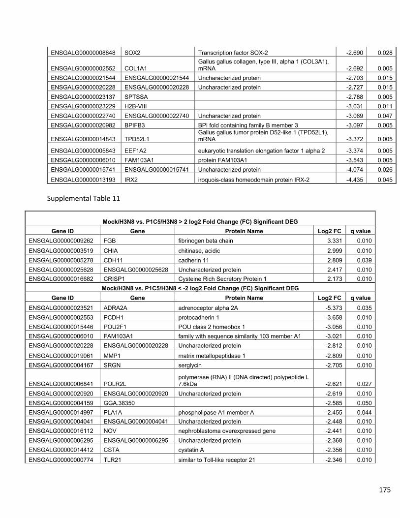
175
ENSGALG00000008848 SOX2 Transcription factor SOX-2 -2.690 0.028
ENSGALG00000002552 COL1A1 Gallus gallus collagen, type III, alpha 1 (COL3A1), mRNA -2.692 0.005
ENSGALG00000021544 ENSGALG00000021544 Uncharacterized protein -2.703 0.015 ENSGALG00000020228 ENSGALG00000020228 Uncharacterized protein -2.727 0.015 ENSGALG00000023137 SPTSSA -2.788 0.005 ENSGALG00000023229 H2B-VIII -3.031 0.011 ENSGALG00000022740 ENSGALG00000022740 Uncharacterized protein -3.069 0.047 ENSGALG00000020982 BPIFB3 BPI fold containing family B member 3 -3.097 0.005
ENSGALG00000014843 TPD52L1 Gallus gallus tumor protein D52-like 1 (TPD52L1), mRNA -3.372 0.005
ENSGALG00000005843 EEF1A2 eukaryotic translation elongation factor 1 alpha 2 -3.374 0.005 ENSGALG00000006010 FAM103A1 protein FAM103A1 -3.543 0.005 ENSGALG00000015741 ENSGALG00000015741 Uncharacterized protein -4.074 0.026
ENSGALG00000013193 IRX2 iroquois-class homeodomain protein IRX-2 -4.435 0.045 Supplemental Table 11
Mock/H3N8 vs. P1C5/H3N8 > 2 log2 Fold Change (FC) Significant DEG Gene ID Gene Protein Name Log2 FC q value
ENSGALG00000009262 FGB fibrinogen beta chain 3.331 0.010 ENSGALG00000003519 CHIA chitinase, acidic 2.999 0.010 ENSGALG00000005278 CDH11 cadherin 11 2.809 0.039 ENSGALG00000025628 ENSGALG00000025628 Uncharacterized protein 2.417 0.010 ENSGALG00000016682 CRISP1 Cysteine Rich Secretory Protein 1 2.173 0.010
Mock/H3N8 vs. P1C5/H3N8 < -2 log2 Fold Change (FC) Significant DEG Gene ID Gene Protein Name Log2 FC q value
ENSGALG00000023521 ADRA2A adrenoceptor alpha 2A -5.373 0.035 ENSGALG00000002553 PCDH1 protocadherin 1 -3.658 0.010 ENSGALG00000015446 POU2F1 POU class 2 homeobox 1 -3.056 0.010 ENSGALG00000006010 FAM103A1 family with sequence similarity 103 member A1 -3.021 0.010 ENSGALG00000020228 ENSGALG00000020228 Uncharacterized protein -2.812 0.010
ENSGALG00000019061 MMP1 matrix metallopeptidase 1 -2.809 0.010 ENSGALG00000004167 SRGN serglycin -2.705 0.010
ENSGALG00000006841 POLR2L polymerase (RNA) II (DNA directed) polypeptide L 7.6kDa -2.621 0.027
ENSGALG00000020920 ENSGALG00000020920 Uncharacterized protein -2.619 0.010 ENSGALG00000004159 GGA.38350 -2.585 0.050 ENSGALG00000014997 PLA1A phospholipase A1 member A -2.455 0.044 ENSGALG00000004041 ENSGALG00000004041 Uncharacterized protein -2.448 0.010 ENSGALG00000016112 NOV nephroblastoma overexpressed gene -2.441 0.010 ENSGALG00000006295 ENSGALG00000006295 Uncharacterized protein -2.368 0.010 ENSGALG00000014412 CSTA cystatin A -2.356 0.010 ENSGALG00000000774 TLR21 similar to Toll-like receptor 21 -2.346 0.010
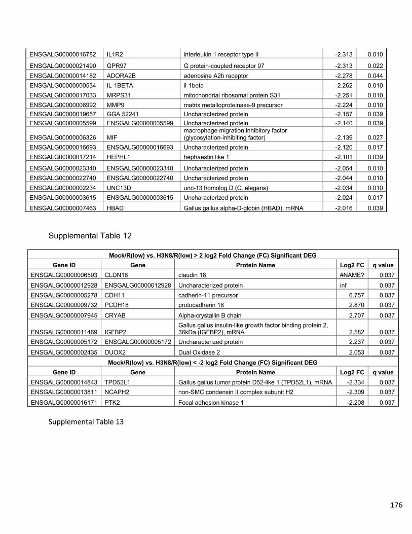
176
Supplemental Table 12
Mock/R(low) vs. H3N8/R(low) > 2 log2 Fold Change (FC) Significant DEG Gene ID Gene Protein Name Log2 FC q value
ENSGALG00000006593 CLDN18 claudin 18 #NAME? 0.037 ENSGALG00000012928 ENSGALG00000012928 Uncharacterized protein inf 0.037 ENSGALG00000005278 CDH11 cadherin-11 precursor 6.757 0.037 ENSGALG00000009732 PCDH18 protocadherin 18 2.870 0.037
ENSGALG00000007945 CRYAB Alpha-crystallin B chain 2.707 0.037
ENSGALG00000011469 IGFBP2 Gallus gallus insulin-like growth factor binding protein 2, 36kDa (IGFBP2), mRNA 2.582 0.037
ENSGALG00000005172 ENSGALG00000005172 Uncharacterized protein 2.237 0.037
ENSGALG00000002435 DUOX2 Dual Oxidase 2 2.053 0.037 Mock/R(low) vs. H3N8/R(low) < -2 log2 Fold Change (FC) Significant DEG
Gene ID Gene Protein Name Log2 FC q value ENSGALG00000014843 TPD52L1 Gallus gallus tumor protein D52-like 1 (TPD52L1), mRNA -2.334 0.037 ENSGALG00000013811 NCAPH2 non-SMC condensin II complex subunit H2 -2.309 0.037
ENSGALG00000016171 PTK2 Focal adhesion kinase 1 -2.208 0.037 Supplemental Table 13
ENSGALG00000016782 IL1R2 interleukin 1 receptor type II -2.313 0.010
ENSGALG00000021490 GPR97 G protein-coupled receptor 97 -2.313 0.022 ENSGALG00000014182 ADORA2B adenosine A2b receptor -2.278 0.044 ENSGALG00000000534 IL-1BETA il-1beta -2.262 0.010 ENSGALG00000017033 MRPS31 mitochondrial ribosomal protein S31 -2.251 0.010 ENSGALG00000006992 MMP9 matrix metalloproteinase-9 precursor -2.224 0.010 ENSGALG00000019657 GGA.52241 Uncharacterized protein -2.157 0.039 ENSGALG00000005599 ENSGALG00000005599 Uncharacterized protein -2.140 0.039
ENSGALG00000006326 MIF macrophage migration inhibitory factor (glycosylation-inhibiting factor) -2.139 0.027
ENSGALG00000016693 ENSGALG00000016693 Uncharacterized protein -2.120 0.017 ENSGALG00000017214 HEPHL1 hephaestin like 1 -2.101 0.039
ENSGALG00000023340 ENSGALG00000023340 Uncharacterized protein -2.054 0.010 ENSGALG00000022740 ENSGALG00000022740 Uncharacterized protein -2.044 0.010 ENSGALG00000002234 UNC13D unc-13 homolog D (C. elegans) -2.034 0.010 ENSGALG00000003615 ENSGALG00000003615 Uncharacterized protein -2.024 0.017
ENSGALG00000007463 HBAD Gallus gallus alpha-D-globin (HBAD), mRNA -2.016 0.039

177
P1C5/H3N8 vs. R(low)/H3N8 > 2 log2 Fold Change (FC) Significant DEG Gene ID Gene Protein Name Log2 FC q value
ENSGALG00000019061 MMP1 matrix metallopeptidase 1 3.801 0.004
ENSGALG00000006992 MMP9 matrix metallopeptidase 9 3.704 0.004 ENSGALG00000000534 IL-1BETA interleukin 1, beta (IL1B), mRNA 3.508 0.004 ENSGALG00000011668 K60 interleukin-8 precursor 3.338 0.004
ENSGALG00000002234 UNC13D unc-13 homolog D (C. elegans) 3.315 0.004 ENSGALG00000000081 IL4I1 L-amino-acid oxidase precursor 3.287 0.004 ENSGALG00000016782 IL1R2 interleukin 1 receptor, type II 3.253 0.004 ENSGALG00000004041 ENSGALG00000004041 Uncharacterized protein 3.208 0.004 ENSGALG00000002112 CSF3R granulocyte colony-stimulating factor receptor 3.190 0.004 ENSGALG00000005599 ENSGALG00000005599 Uncharacterized protein 3.164 0.047
ENSGALG00000004582 MYL2 myosin regulatory light chain 2A, cardiac muscle isoform 3.042 0.004
ENSGALG00000007001 TLR4 toll-like receptor 4 precursor 3.020 0.004
ENSGALG00000008166 TLR15 toll-like receptor 15 (TLR15), mRNA 2.981 0.004 ENSGALG00000002766 MLKL mixed lineage kinase domain-like 2.901 0.004
ENSGALG00000023818 ENSGALG00000023818 Uncharacterized protein 2.893 0.004
ENSGALG00000001905 DTX2 deltex 2, E3 ubiquitin ligase 2.875 0.004 ENSGALG00000004167 SRGN serglycin 2.873 0.014 ENSGALG00000021142 ENSGALG00000021142 Uncharacterized protein 2.869 0.004
ENSGALG00000015666 TNIP2 TNFAIP3-interacting protein 2 2.862 0.004 ENSGALG00000010892 ANTXR2 anthrax toxin receptor 2 2.820 0.004 ENSGALG00000017214 HEPHL1 hephaestin like 1 2.779 0.035 ENSGALG00000010362 SUCNR1 succinate receptor 1 2.773 0.004 ENSGALG00000016919 IRG1 immunoresponsive 1 homolog 2.742 0.004 ENSGALG00000014412 CSTA cystatin A 2.736 0.004
ENSGALG00000015446 POU2F1 POU domain, class 2, transcription factor 1 2.700 0.025 ENSGALG00000023370 ENSGALG00000023370 Uncharacterized protein 2.690 0.004 ENSGALG00000023933 G0S2 G0/G1switch 2 2.676 0.010 ENSGALG00000020316 IL13RA2 interleukin-13 receptor subunit alpha-2 precursor 2.658 0.004
ENSGALG00000000949 HBEGF Proheparin-binding EGF-like growth factor Heparin-binding EGF-like growth factor 2.653 0.004
ENSGALG00000011670 IL8 interleukin 8 [Gallus gallus] 2.651 0.008
ENSGALG00000011221 AGPAT9 Glycerol-3-phosphate acyltransferase 3 2.631 0.004
ENSGALG00000004060 RGS9 regulator of G-protein signaling 9 2.621 0.004 ENSGALG00000002383 CD72 CD72 antigen 2.612 0.004 ENSGALG00000005172 ENSGALG00000005172 Uncharacterized protein 2.590 0.004
ENSGALG00000005941 ENSGALG00000005941 Uncharacterized protein 2.583 0.041 ENSGALG00000019148 CSF2RA 2.556 0.008 ENSGALG00000023855 KRT14 keratin, type I cytoskeletal 14 2.554 0.004 ENSGALG00000021340 CA9 carbonic anhydrase IX 2.552 0.004
ENSGALG00000023622 AVD Avidin 2.551 0.004 ENSGALG00000006106 TNFRSF6B tumor necrosis factor receptor superfamily member 6b 2.549 0.004 ENSGALG00000002118 CO6 calcium-activated potassium channel subunit beta-1 2.545 0.004 ENSGALG00000016693 ENSGALG00000016693 Uncharacterized protein 2.518 0.020 ENSGALG00000022646 ENSGALG00000022646 Uncharacterized protein 2.515 0.025 ENSGALG00000005086 PLAU urokinase-type plasminogen activator preproprotein 2.463 0.004
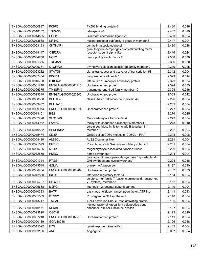
178
ENSGALG00000000837 FKBP6 FK506 binding protein 6 2.460 0.016 ENSGALG00000010152 TSPAN8 tetraspanin-8 2.452 0.029 ENSGALG00000014585 CCLI10 C-C motif chemokine ligand 26 2.450 0.004 ENSGALG00000013568 NR4A3 nuclear receptor subfamily 4 group A member 3 2.447 0.004 ENSGALG00000003123 CNTNAP1 contactin associated protein 1 2.430 0.028
ENSGALG00000019147 CSF2RA granulocyte-macrophage colony-stimulating factor receptor subunit alpha-like 2.416 0.024
ENSGALG00000004700 NCF2 neutrophil cytosolic factor 2 2.388 0.030 ENSGALG00000021355 TROJAN 2.386 0.030 ENSGALG00000000731 C1ORF38 thymocyte selection associated family member 2 2.349 0.025 ENSGALG00000003282 STAT5B signal transducer and activator of transcription 5B 2.342 0.004 ENSGALG00000007454 PDCD1 programmed cell death 1 2.329 0.014 ENSGALG00000016788 IL18RAP interleukin 18 receptor accessory protein 2.329 0.034 ENSGALG00000001710 ENSGALG00000001710 Uncharacterized protein 2.324 0.035 ENSGALG00000006375 TM4SF19 transmembrane 4 L6 family member 19 2.324 0.016 ENSGALG00000023340 ENSGALG00000023340 Uncharacterized protein 2.303 0.042 ENSGALG00000008308 BHLHE40 class E basic helix-loop-helix protein 40 2.296 0.004
ENSGALG00000003482 BHLHA15 2.283 0.004 ENSGALG00000000974 ENSGALG00000000974 Uncharacterized protein 2.277 0.034 ENSGALG00000013101 BG2 2.276 0.025 ENSGALG00000002728 SLC16A3 Monocarboxylate transporter 4 2.273 0.004 ENSGALG00000014962 FAM26F family with sequence similarity 26 member F 2.273 0.010
ENSGALG00000019552 SERPINB2 serpin peptidase inhibitor, clade B (ovalbumin), member 2 2.262 0.004
ENSGALG00000015474 CD80 Gallus gallus CD80 molecule (CD80), mRNA 2.243 0.008 ENSGALG00000005430 ALS2CL ALS2 C-terminal like 2.231 0.004 ENSGALG00000021573 PIK3R5 Phosphoinositide 3-kinase regulatory subunit 5 2.231 0.004 ENSGALG00000000736 MATK megakaryocyte-associated tyrosine kinase 2.229 0.004 ENSGALG00000012550 HMOX1 heme oxygenase 1 2.224 0.004
ENSGALG00000001314 PTGS1 prostaglandin-endoperoxide synthase 1 (prostaglandin G/H synthase and cyclooxygenase) 2.224 0.016
ENSGALG00000013548 GZMA granzyme A precursor 2.197 0.010 ENSGALG00000006204 ENSGALG00000006204 Uncharacterized protein 2.182 0.033 ENSGALG00000012830 IRF-4 interferon regulatory factor 4 2.154 0.004
ENSGALG00000005727 SLC7A3 solute carrier family 7 (cationic amino acid transporter, y+ system), member 3 2.152 0.004
ENSGALG00000005638 IL2RG interleukin 2 receptor subunit gamma 2.149 0.004 ENSGALG00000010323 BATF basic leucine zipper transcription factor, ATF-like 2.141 0.013 ENSGALG00000005069 PTGS2 Prostaglandin G/H synthase 2 2.140 0.004 ENSGALG00000013747 TAGAP T-cell activation RhoGTPase activating protein 2.135 0.004
ENSGALG00000010171 NFKBIE nuclear factor of kappa light polypeptide gene enhancer in B-cells inhibitor, epsilon 2.127 0.004
ENSGALG00000023820 OGCHI 2.123 0.020 ENSGALG00000007210 ENSGALG00000007210 Uncharacterized protein 2.111 0.004 ENSGALG00000000139 GGA.18046 2.106 0.018 ENSGALG00000015022 FYN tyrosine-protein kinase Fyn 2.102 0.004 ENSGALG00000003196 ANG Angiogenin 2.097 0.004

179
ENSGALG00000019251 LAG3 2.093 0.019 ENSGALG00000023634 ENSGALG00000023634 Uncharacterized protein 2.090 0.021 ENSGALG00000001076 LIMK1 LIM domain kinase 1 2.089 0.041 ENSGALG00000004243 ENSGALG00000004243 Uncharacterized protein 2.087 0.004 ENSGALG00000021616 MARCKSL1 MARCKS-related protein 2.082 0.008
ENSGALG00000016524 CAD carbamoyl-phosphate synthetase 2, aspartate transcarbamylase, and dihydroorotase 2.082 0.016
ENSGALG00000002540 RGS2 regulator of G-protein signaling 2 2.080 0.004 ENSGALG00000003578 FN1 fibronectin precursor 2.080 0.024 ENSGALG00000013698 EFHD2 EF-hand domain family member D2 2.072 0.010 ENSGALG00000009768 CCRN4L nocturnin 2.053 0.019 ENSGALG00000002343 ENSGALG00000002343 Uncharacterized protein 2.052 0.048 ENSGALG00000016988 KIAA0226L KIAA0226-like 2.046 0.004 ENSGALG00000002643 SELP selectin P 2.035 0.004 ENSGALG00000003103 MST1R macrophage-stimulating protein receptor precursor 2.027 0.004 ENSGALG00000013861 TNFAIP3 TNF alpha induced protein 3 2.010 0.004 ENSGALG00000002549 RGS1 regulator of G-protein signaling 1 2.001 0.004
ENSGALG00000012352 BLVRA biliverdin reductase A 2.000 0.008 P1C5/H3N8 vs. R(low)/H3N8 < -2 log2 Fold Change (FC) Significant DEG
Gene ID Gene Protein Name Log2 FC q value
ENSGALG00000009262 FGB Fibrinogen beta chain Fibrinopeptide B Fibrinogen beta chain -4.653 0.028
ENSGALG00000003519 CHIA -4.561 0.004 ENSGALG00000016962 ENSGALG00000016962 Uncharacterized protein -3.930 0.004 ENSGALG00000005278 CDH11 cadherin-11 precursor -3.808 0.004 ENSGALG00000015329 ENSGALG00000015329 Uncharacterized protein -3.752 0.004 ENSGALG00000016682 CRISP1 -3.376 0.004 ENSGALG00000002358 CDO1 cysteine dioxygenase type 1 -3.124 0.004 ENSGALG00000007945 CRYAB Alpha-crystallin B chain -2.933 0.045 ENSGALG00000003193 CRABP1 Cellular retinoic acid-binding protein 1 -2.910 0.004 ENSGALG00000007537 INCENP inner centromere protein -2.856 0.040 ENSGALG00000005843 EEF1A2 eukaryotic translation elongation factor 1 alpha 2 -2.840 0.004 ENSGALG00000006693 ENSGALG00000006693 Uncharacterized protein -2.772 0.004 ENSGALG00000014843 TPD52L1 tumor protein D52-like 1 (TPD52L1), mRNA -2.601 0.033
ENSGALG00000000184 SLC27A6 solute carrier family 27 (fatty acid transporter), member 6 -2.474 0.008
ENSGALG00000016292 COL21A1 collagen, type XXI, alpha 1 -2.368 0.014 ENSGALG00000023424 DMBT1 -2.344 0.004 ENSGALG00000005601 ENSGALG00000005601 Uncharacterized protein -2.256 0.004 ENSGALG00000010417 ENSGALG00000010417 Uncharacterized protein -2.243 0.042 ENSGALG00000006048 FAM64A family with sequence similarity 64 member A -2.223 0.004 ENSGALG00000020909 C20ORF85 chromosome 20 open reading frame 85 [Gallus gallus] -2.201 0.046 ENSGALG00000010769 HPGD 15-hydroxyprostaglandin dehydrogenase -2.154 0.004
ENSGALG00000011277 PLEKHG7 pleckstrin homology and RhoGEF domain containing G7 -2.133 0.034
ENSGALG00000009155 CHGB chromogranin B -2.089 0.008 ENSGALG00000020876 AOX2P aldehyde oxidase 2 pseudogene -2.075 0.004

180
Supplemental Table 14
ENSGALG00000012997 DNAH5 dynein, axonemal, heavy chain 5 -2.053 0.042 ENSGALG00000006217 S100B S100 calcium binding protein B -2.033 0.032
ENSGALG00000012250 GGA.45581 alcohol dehydrogenase 1C (class I), gamma polypeptide -2.017 0.040

181
Mock/H3N8 vs. Mock/R(low) > 2 log2 Fold Change (FC) Significant DEG Gene ID Gene Protein Name Log2 FC q value
ENSGALG00000009317 ENSGALG00000009317 Uncharacterized protein #NAME? 0.011
ENSGALG00000025284 SNORD89 Small nucleolar RNA SNORD89 #NAME? 0.011 Mock/H3N8 vs. Mock/R(low) < -2 log2 Fold Change (FC) Significant DEG
Gene ID Gene Protein Name Log2 FC q value ENSGALG00000014412 CSTA cystatin A -3.537 0.011 ENSGALG00000002553 PCDH1 Gallus gallus protocadherin 1 (PCDH1), mRNA -3.530 0.011 ENSGALG00000000379 ITGB3 integrin beta-3 precursor -3.136 0.011
ENSGALG00000017214 HEPHL1 hephaestin like 1 -3.087 0.011 ENSGALG00000015446 POU2F1 POU domain, class 2, transcription factor 1 -2.993 0.011 ENSGALG00000006010 FAM103A1 protein FAM103A1 -2.941 0.011
ENSGALG00000005278 CDH11 cadherin-11 precursor -2.860 0.045 ENSGALG00000023370 ENSGALG00000023370 Uncharacterized protein -2.850 0.011
ENSGALG00000016782 IL1R2 interleukin 1 receptor, type II -2.849 0.011 ENSGALG00000013548 GZMA granzyme A precursor -2.785 0.038 ENSGALG00000021142 ENSGALG00000021142 Uncharacterized protein -2.766 0.019
ENSGALG00000006054 CALCA calcitonin gene-related peptide isoform 3 preproprotein -2.704 0.041
ENSGALG00000004167 SRGN serglycin -2.575 0.011 ENSGALG00000020920 ENSGALG00000020920 Uncharacterized protein -2.526 0.026
ENSGALG00000006841 POLR2L polymerase (RNA) II (DNA directed) polypeptide L, 7.6kDa -2.454 0.029
ENSGALG00000005172 ENSGALG00000005172 Uncharacterized protein -2.438 0.011 ENSGALG00000023933 G0S2 G0/G1switch 2 -2.438 0.011 ENSGALG00000023340 ENSGALG00000023340 Uncharacterized protein -2.397 0.029 ENSGALG00000005599 ENSGALG00000005599 Uncharacterized protein -2.357 0.029 ENSGALG00000025698 5_8S_rRNA 5.8S ribosomal RNA -2.345 0.011
ENSGALG00000019147 CSF2RA granulocyte-macrophage colony-stimulating factor receptor subunit alpha-like -2.336 0.026
ENSGALG00000017033 MRPS31 mitochondrial ribosomal protein S31 -2.248 0.038 ENSGALG00000014201 PARVB parvin beta -2.229 0.011 ENSGALG00000003578 FN1 fibronectin precursor -2.155 0.011 ENSGALG00000019553 SERPINB10 Heterochromatin-associated protein MENT -2.151 0.049
ENSGALG00000006326 MIF macrophage migration inhibitory factor (glycosylation-inhibiting factor) -2.124 0.041
ENSGALG00000010362 SUCNR1 succinate receptor 1 -2.124 0.026
ENSGALG00000020228 ENSGALG00000020228 Uncharacterized protein -2.112 0.041 ENSGALG00000002383 CD72 CD72 antigen -2.091 0.049 ENSGALG00000019061 MMP1 matrix metallopeptidase 1 -2.054 0.011 ENSGALG00000008166 TLR15 Gallus gallus toll-like receptor 15 (TLR15), mRNA -2.028 0.011
182
Chapter 4 - The potential contribution of M. gallisepticum neuraminidase activity
to co-pathogenesis with LPAIV in vivo in the presence of the viral neuraminidase
inhibitor, oseltamivir.
Methods
-Chicken Infection Experiment: To study the potential role of neuraminidase activity in
the co-pathogenesis of M. gallisepticum and LPAIV, the viral neuraminidase inhibitor
oseltamivir phosphate was utilized in an in vivo co-infection model. This inhibitor was
used as a tool to observe any compensation by M. gallisepticum for the inhibited
neuraminidase activity of LPAIV. As described in previous chapters, 10 SPF White
Leghorn chickens per group were infected with combinations of M. gallisepticum Rlow,
the M. gallisepticum mutant P1C5, Hayflick’s medium as a mock infection, and H3N8
(A/duck/Ukraine/1963) as outlined in the table below.
All chickens were infected intratracheally with 200 µl of either 1 x 10⁸ CFU/mL of
M. gallisepticum Rlow, 5 x 10⁶ TCID50 of H3N8, or Hayflick’s medium as a mock
infection. All inoculum cultures of M. gallisepticum and H3N8 were diluted in fresh
Hayflick’s medium. Oseltamivir phosphate was administered at a dosage of 1 mg/kg per
day, divided into 0.5 mg/mg twice per day, at a 1:1 ratio in phosphate buffered saline
(PBS) [1].
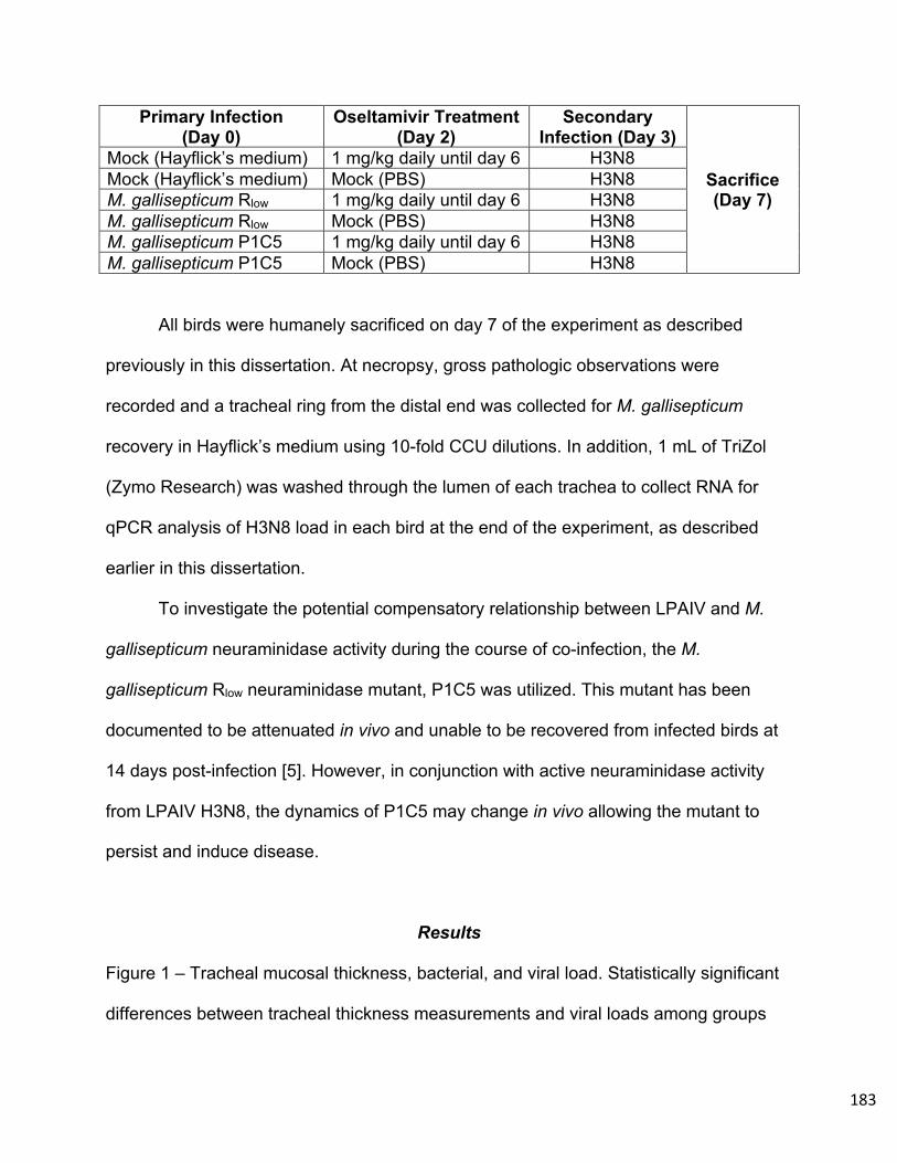
183
Primary Infection (Day 0)
Oseltamivir Treatment (Day 2)
Secondary Infection (Day 3)
Sacrifice (Day 7)
Mock (Hayflick’s medium) 1 mg/kg daily until day 6 H3N8 Mock (Hayflick’s medium) Mock (PBS) H3N8 M. gallisepticum Rlow 1 mg/kg daily until day 6 H3N8 M. gallisepticum Rlow Mock (PBS) H3N8 M. gallisepticum P1C5 1 mg/kg daily until day 6 H3N8 M. gallisepticum P1C5 Mock (PBS) H3N8
All birds were humanely sacrificed on day 7 of the experiment as described
previously in this dissertation. At necropsy, gross pathologic observations were
recorded and a tracheal ring from the distal end was collected for M. gallisepticum
recovery in Hayflick’s medium using 10-fold CCU dilutions. In addition, 1 mL of TriZol
(Zymo Research) was washed through the lumen of each trachea to collect RNA for
qPCR analysis of H3N8 load in each bird at the end of the experiment, as described
earlier in this dissertation.
To investigate the potential compensatory relationship between LPAIV and M.
gallisepticum neuraminidase activity during the course of co-infection, the M.
gallisepticum Rlow neuraminidase mutant, P1C5 was utilized. This mutant has been
documented to be attenuated in vivo and unable to be recovered from infected birds at
14 days post-infection [5]. However, in conjunction with active neuraminidase activity
from LPAIV H3N8, the dynamics of P1C5 may change in vivo allowing the mutant to
persist and induce disease.
Results
Figure 1 – Tracheal mucosal thickness, bacterial, and viral load. Statistically significant
differences between tracheal thickness measurements and viral loads among groups

184
were calculated using a one-way ANOVA with Tukey’s post-hoc test and a threshold for
significance at p < 0.05. Significant differences are indicated with **** = p < 0.0001, ** =
p < 0.01, and * = p < 0.05. Statistically significant differncees in M. gallisepticum
recovery among groups was calculated using a Kruskal-Wallis ANOVA On-Ranks with
Dunn’s Post Hoc test. No significant differences were found among experimental groups
for M. gallisepticum or H3N8 load.
185
Mock
/H3N
8+
PBS
Mock
/H3N
8+
Oselta
miv
ir
P1C5/
H3N8
+PBS
P1C5/
H3N8
+Ose
ltam
ivir
R low/H
3N8
+PBS
R low/H
3N8
+Ose
ltam
ivir
0
100
200
300
Tracheal Thickness
Infection Groups ( n = 10 )
Tra
chea
lTh
ickn
ess
(mic
rom
eter
s) **** ****
*
*
A B
Mock/H
3N8 +
PBS
Mock/H
3N8 +
Ose
ltam
ivir
P1C5/H
3N8 +
PBS
P1C5/H
3N8 +
Ose
ltam
ivir
R low/H
3N8 +
PBS
R low/H
3N8 +
Ose
ltam
ivir
10-9
10-8
10-7
10-6
10-5
10-4
10-3
10-2
Infection Groups ( n = 10 )
Nan
ogra
ms
per
Mic
rolit
er H
3N8
H3N8 Load Nanograms per MicroliterC
P1C5/H
3N8 + PBS
P1C5/H
3N8 + Ose
ltamivi
r
R low/H
3N8 + PBS
R low/H
3N8 + Ose
ltamivi
r100
101
102
103
104
105
106
Tracheal M. gallisepticum Recovery
Infection Groups ( n = 10 )
Log 1
0C
CU
perm
illili
ter
186
-Tracheal Histopathology: Significant differences in tracheal mucosal thickness exist
between multiple experimentally infected groups of chickens. As anticipated, tracheal
mucosal thickness is significantly increased between Mock/H3N8 + PBS and M.
gallisepticum Rlow/H3N8 + PBS infected birds (p < 0.01) due to enhanced disease due
to the presence of both M. gallisepticum and H3N8 infection (Figure 1A).
Similarly, a significant increase in tracheal thickness also exists between
Mock/H3N8 + Oseltamivir and M. gallisepticum Rlow/H3N8 + Oseltamivir infected
chickens (p < 0.0001) (Figure 1A). The difference between these groups is confounded
by the treatment with oseltamivir, which has been previously shown to effectively reduce
viral load in AIV infected chickens [1]. Although the M. gallisepticum Rlow/H3N8 +
Oseltamivir infected group contains both co-pathogens, the oseltamivir treatment did not
alter the pathologic response to this co-infection as originally hypothesized.
To expand upon this, M. gallisepticum Rlow/H3N8 + Oseltamivir infected chickens
in fact have significantly higher tracheal thickness measurements when compared to M.
gallisepticum Rlow/H3N8 + PBS infected chickens (p < 0.01) (Figure 1A). This same
phenomenon exists between M. gallisepticum P1C5/H3N8 + Oseltamivir and M.
gallisepticum P1C5H3N8 + PBS infected chickens (p < 0.05), in that oseltamivir
treatment appears to increase the tracheal thickness in response to infection (Figure
1A). A significant difference also exists between tracheal thicknesses of M.
gallisepticum Rlow/H3N8 + Oseltamivir and M. gallisepticum P1C5/H3N8 + Oseltamivir
infected chickens (p < 0.05) (Figure 1A).

187
-Tracheal Mycoplasma gallisepticum Recovery: Although no statistically significant
differences in M. gallisepticum recovery from the trachea emerge among experimentally
infected groups of chickens, noteworthy trends still exist (Figure 1B). M. gallisepticum
recovery was most abundant in M. gallisepticum Rlow/H3N8 + PBS infected chickens
(Figure 1B). The remaining experimental groups which received an M. gallisepticum
infection had similar levels of bacterial recovery from the trachea (Figure 1B).
-Tracheal LPAIV H3N8 Load: Similar to the recovery of M. gallisepticum from the
trachea (Figure 1B), no statistically significant differences exist between H3N8 load in
the trachea of experimentally infected groups of chickens. This is perhaps the most
notable finding in that, as mentioned previously, oseltamivir treatment has been
documented to significantly decrease AIV load in the tracheal tissue of infected
chickens [1]. The data presented here indicate that oseltamivir treatment had no effect
on the load of H3N8 in the tracheal lumen of experimentally infected chickens used in
this model (Figure 1C).
Discussion
The failure of oseltamivir treatment to combat H3N8 infection in this chicken
infection model was unexpected. The original hypothesis to be tested was that M.
gallisepticum neuraminidase activity could effectively compensate for LPAIV
neuraminidase in the presence of the inhibitor, oseltamivir. This relationship could also
work in reverse, in that LPAIV neuraminidase may enhance the pathogenesis of a
neuraminidase-deficient M. gallisepticum mutant, P1C5. However, the lack of

188
discernable difference in viral load between mock and oseltamivir treated, H3N8
infected chickens makes the appropriate testing of these hypotheses impossible.
Although the cause for the failure of oseltamivir to effectively reduce viral load in
this model is unknown, there are multiple possibilities that may contribute to the
explanation. Previously, Lee et al. utilized oseltamivir as a treatment for infection of
chickens with LPAIV strain H9N2 (A/ Chicken/Korea/310/01) [1]. The LPAIV isolate in
our model is of a different subtype; H3N8 (A/duck/Ukraine/1963). This difference could,
at least in part, explain the difference in sensitivity to oseltamivir treatment in vivo. This
H3N8 strain is, however, susceptible to oseltamivir carboxylate neuraminidase inhibition
in vitro, as shown in Chapter 2 of this dissertation.
Another potential factor of the outcome of this experiment is the difference in
original form of oseltamivir used between the two studies. Lee et al. utilized oseltamivir
from capsules which was later suspended in PBS, whereas our model incorporated
oseltamivir intended for oral suspension administration and these two drugs could have
originated from different suppliers due to geographic differences [1].
Additionally, although the dosages of oseltamivir used was consistent between
the two studies, the precise timing of administration could potentially contribute to
changes in active bioavailability of the drug in vivo. Finally, viral isolation may have
differed among tissues in our model. Sampling of other areas, such as ceacal tissues or
the lung, may have illuminated potential differences in viral load in animals from this
experiment.
Another interesting observation is the similarity between tracheal mucosal
thickness (Figure 1A) and viral load (Figure 1C). Although no statistically significant

189
differences exist between the amount of virus among experimental groups, the groups
with the most severe tracheal mucosal thickening are also the groups with the highest
viral titer in the trachea and vice versa (Figures 1A and 1C). Although M. gallisepticum
recovery was increased in M. gallisepticum Rlow infected birds over M. gallisepticum
P1C5 infected birds, the pattern seen between tracheal thickness and viral load does
not apply as seamlessly to M. gallisepticum recovery.
These data, as well as the data presented in chapter 3 of this dissertation,
indicate that the co-pathogen relationship between M. gallisepticum and LPAIV is not
neuraminidase-dependent as originally hypothesized. Instead, as indicated by the host
transcriptional response to mono- and co-infection, this pathobiological phenomenon is
much more complex.
Alterations in transcription of genes related to tracheal ciliary activity,
inflammatory immune signaling, and tissue remodeling all exist between mono- and co-
infected chickens and likely contribute to differences in disease manifestation. Although
some of these factors have been discussed in other publications, such as ciliostasis and
cytokine levels observed in M. gallisepticum and LPAIV tracheal explants [2], the data
presented in this dissertation are the first to examine these responses in the airway of
the natural host during co-infection.
Examples of relevant transcriptional responses include significant alterations in
TLR expression. TLR4 and TLR15 are increased in expression during co-infection with
M. gallisepticum Rlow and LPAIV H3N8 compared to mono-infected chickens, or
chickens instead co-infected with attenuated M. gallisepticum P1C5 (Chapter 3). These
two TLR genes were also abundantly expressed in the chicken trachea during infection
190
with M. gallisepticum Rlow alone over the course of the first 7 days of infection [3].
TLR21 is significantly decreased in expression in chickens co-infected with M.
gallisepticum Rlow or P1C5 and H3N8 over birds infected with H3N8 alone (Chapter 3).
This suppression of TLR21 signaling is a novel finding and correlates with increases in
tracheal histopathology in those infected chickens (Chapter 3). It is possible that, during
co-infection, TLR21 suppression prevents association with TLR4 signaling that
contributes to an appropriate immune response [4].
KEGG pathway analysis also indicates differential effects on signaling pathway
responses to co-infection. Noteworthy examples include differential effects on the
Influenza A response pathway, extracellular matrix receptor signaling, metabolic
pathways, phagocytosis, and TLR signaling (Chapter 3). Alterations in these processes
in response to co-infection all contribute to the understanding of the mechanisms of the
host response to co-infection with M. gallisepticum and LPAIV.
Another novel finding presented is the ability of attenuated M. gallisepticum
mutants P1C5 and P1H9 to persist 6 to 7 days post-infection in the chicken trachea.
These mutants were previously thought to be cleared early in infection due to their lack
of survival to 14 days post-infection [5,6]. However, examination of their survival at a
more acute time point suggests that both neuraminidase and MslA contribute to chronic
persistence in the host, however are not required for short-term survival in the chicken
airway. Co-infection with H3N8 has a more pronounced effect on chronic persistence of
the MslA mutant P1H9 to 14 days post-infection than the neuraminidase mutant P1C5.
This result contributes to the conclusion that the co-infection dynamic between these
pathogens is more complex than originally anticipated.

191
Chapter 5 - Future Directions
The experiments described within this dissertation provide multiple avenues for
future experimental exploration of M. gallisepticum and LPAIV co-pathogenesis. In
attempt to reconcile the lack of efficacy of oseltamivir treatment in our chicken model,
continued examination of dose, routes, and administration schedules can be performed
on chickens infected with H3N8 alone. Additional tissues from these animals can be
sampled to discern if other target tissues are more appropriate for changes in viral load
due to this oseltamivir treatment.
In a similar vein, alternate neuraminidase inhibitors can be employed to replace
oseltamivir in our model. Such inhibitors include zanamivir or DANA, as described in
chapter 1 of this dissertation. Alternate isolates of LPAIV can also be utilized in our
model. Our lab is in possession of another LPAIV isolate, H9N2
(A/turkey/Wisconsin/1/1966) (BEI Reagent Resources). This virus is of the same
subtype as the LPAIV used by Sid et al. in co-infection of tracheal organ cultures with
M. gallisepticum and LPAIV [2]. H9N2 is also growing in relevance due to its global
presence in poultry and zoonotic potential [7].
Another area of interest is the transcriptomic response in M. gallisepticum during
co-infection with LPAIV. Publications out of our laboratory have used RNA-sequencing
to find coordinated changes in the expression of genes by M. gallisepticum during early
time points of infection, and these coordinated changes may differ during a co-infection
scenario due to alterations in the host environment [8,9]. Genomic changes may also
occur in both M. gallisepticum and LPAIV in response to co-infection, and sequencing of

192
recovered isolates after co-infection may illuminate mutations relevant to adaptation to
that co-pathogen relationship over time.
There are also multiple avenues of co-pathogenesis in this model that have yet to
be explored. For instance, co-infection of alveolar cells in vitro with the 2009 pandemic
influenza virus, pdm2009, and methicillin-resistant Staphylococcus aureus contributes
to disruption in the barrier function of the alveolar cells [10]. This damage to a primary
host defense, along with alterations in host immune signaling, contribute to the severely
of disease. Although the avian respiratory tract is unique in its structure, a similar
phenomenon may be occurring during co-infection with M. gallisepticum and LPAIV in
other areas of the respiratory tract, such as the air sac.
The targeting of certain host immune molecules could also be a promising route
of further investigation. In mice, alterations in TGF-β and IFNγ in a model of allergic
airway disease effects the severity of co-infection of those mice with Streptococcus
pneumoniae and influenza A virus [11]. Another example is the inhibition of IL-10
signaling in reducing disease exacerbation during co-infection of mice with
Mycobacterium tuberculosis and influenza A virus [12].
Finally, TLR9 has been implicated in the co-pathogenesis of Staphylococcus
aureus and influenza A virus [13]. This relationship in particular, is noteworthy in that the
chicken TLR21, which is suppressed during co-infection with M. gallisepticum and
LPAIV (Chapter 3), is a homolog of human TLR9 [14]. Therefore, TLR21, among others,
could be a novel target host immune molecule for intervention of co-pathogenesis in our
model with M. gallisepticum and LPAIV.

193
Other facets of viral and bacterial co-pathogenesis such as alterations in the host
microbiome, biofilm formation, nutrient availability, and the availability of host receptors
also provide potential routes of investigation within our co-pathogenesis model [15].
Other economically relevant viral pathogens, such as infectious bronchitis virus (IBV)
could serve as viral co-pathogens of M. gallisepticum for other novel mechanisms of co-
infection in chickens [16].
Both M. gallisepticum and LPAIV are pathogens of great concern to the poultry
industry, with a foremost impact on small-scale backyard flocks. Farmers and hobbists
keeping these smaller flocks likely do not have access to the same surveillance and
prevention measures as large broiler or layer industry operations. This scenario has the
potential to become even more dangerous if the flock becomes infected by a virulent
field isolate of M. gallisepticum that can persist to a chronic state in chickens. In
addition, LPAIV isolates of sub-clinical pathogenicity could propagate within a flock
without being detected.
If both pathogens infect chickens within the flock, as demonstrated by the data
presented here, the resulting disease could be severe. By better understanding the
host-pathogen interations of this co-pathogenesis, such as the binding substrates and
host immunological response, we can aid in the development of prevention and
treatment efforts. This has the potential to greatly benefit the poultry industry, in that it
could prevent drastic economic losses to backyard poultry farmers due to exacerbated
disease during M. gallisepticum and LPAIV co-pathogenesis. In addition, these host-
pathogen dynamics could contribute to other bacterial and viral co-pathogen
relationships in other animals or humans. In summary, the data presented in this

194
dissertation contribute novel and interesting findings to the community of knowledge on
the co-pathogenesis of Mycoplasma gallisepticum and LPAIV.

195
References
1. Lee D-H, Lee Y-N, Park J-K, Yuk S-S, Lee J-W, Kim J-I, Han JS, Lee J-B, Park
S-Y, Choi I-S, Song C-S. Antiviral Efficacy of Oseltamivir Against Avian Influenza
Virus in Avian Species 3.
2. Sid H, Hartmann S, Petersen H, Ryll M, Rautenschlein S. 2016. Mycoplasma
gallisepticum modifies the pathogenesis of influenza A virus in the avian tracheal
epithelium. International Journal of Medical Microbiology 306:174–186.
3. Beaudet J, Tulman ER, Pflaum K, Liao X, Kutish GF, Szczepanek SM, Silbart
LK, Geary SJ. 2017. Transcriptional Profiling of the Chicken Tracheal Response
to Virulent Mycoplasma gallisepticum Strain R low. Infect Immun 85:e00343-17,
/iai/85/10/e00343-17.atom.
4. Kim S, Kaiser P, Borowska D, Vervelde L. 2018. Synergistic effect of co-
stimulation of membrane and endosomal TLRs on chicken innate immune
responses. Veterinary Immunology and Immunopathology 199:15–21.
5. May M, Szczepanek SM, Frasca S, Gates AE, Demcovitz DL, Moneypenny CG,
Brown DR, Geary SJ. 2012. Effects of sialidase knockout and complementation
on virulence of Mycoplasma gallisepticum. Veterinary Microbiology 157:91–95.
6. Szczepanek SM, Frasca S, Schumacher VL, Liao X, Padula M, Djordjevic SP,
Geary SJ. 2010. Identification of Lipoprotein MslA as a Neoteric Virulence Factor
of Mycoplasma gallisepticum. Infection and Immunity 78:3475–3483.
7. Peacock TP, James J, Sealy JE, Iqbal M. 2019. A Global Perspective on H9N2
Avian Influenza Virus. Viruses 11:620.

196
8. Pflaum K, Tulman ER, Beaudet J, Liao X, Geary SJ. 2016. Global Changes in
Mycoplasma gallisepticum Phase-Variable Lipoprotein Gene vlhA Expression
during In Vivo Infection of the Natural Chicken Host. Infect Immun 84:351–355.
9. Pflaum K, Tulman ER, Beaudet J, Canter J, Geary SJ. 2018. Variable
Lipoprotein Hemagglutinin A Gene ( vlhA ) Expression in Variant Mycoplasma
gallisepticum Strains In Vivo. Infect Immun 86:e00524-18, /iai/86/11/e00524-
18.atom.
10. Nickol ME, Ciric J, Falcinelli SD, Chertow DS, Kindrachuk J. 2019.
Characterization of Host and Bacterial Contributions to Lung Barrier Dysfunction
Following Co-infection with 2009 Pandemic Influenza and Methicillin Resistant
Staphylococcus aureus. Viruses. 11(116).
11. Roberts S, Salmon SL, Steiner DJ, Williams CM, Metzger DW, Furuya Y. 2019.
Allergic Airway Disease Prevents Lethal Synergy of Influenza A Virus-
Streptococcus pneumoniae Coinfection 10:17. mBio. 10:e01335-19. https://doi
.org/10.1128/mBio.01335-19.
12. Ring S, Eggers L, Behrends J, Wutkowski A, Schwudke D, Kröger A, Hierweger
AM, Hölscher C, Gabriel G, Schneider BE. 2019. Blocking IL-10 receptor
signaling ameliorates Mycobacterium tuberculosis infection during influenza-
induced exacerbation. JCI Insight 4:e126533.
13. Sun K, Metzger DW. 2019. Influenza and Staphylococcus aureus Coinfection:
TLR9 at Play. Trends in Microbiology 27:383–384.
14. Brownlie R, Zhu J, Allan B, Mutwiri GK, Babiuk LA, Potter A, Griebel P. 2009.
Chicken TLR21 acts as a functional homologue to mammalian TLR9 in the

197
recognition of CpG oligodeoxynucleotides. Molecular Immunology 46:3163–
3170.
15. Bakaletz LO. 2017. Viral–bacterial co-infections in the respiratory tract. Current
Opinion in Microbiology 35:30–35.
16. de Wit JJ, Cook JKA. 2019. Spotlight on avian pathology: infectious bronchitis
virus, Avian Pathology, DOI: 10.1080/03079457.2019.1617400