Connecticut State Health Information Technology Plan - CT.gov
Upload
khangminh22Category
view
3download
0
Amer. J. Bot. 77(1): 77-91. 1990.
COMPARATIVE ONTOGENY OF THE INFLORESCENCE AND FLOWER OF HAMAMELIS VIRGINIANA AND
LOROPETALUM CHINENSE (HAMAMELIDACEAE)'
Department of Plant Biology, University of New Hampshire, Durham, New Hampshire 03824
A B S T R A C T A comparative developmental study of the inflorescence and flower of Hamamelis L. (4-
merous) and Loropetalum (R. Br.) Oliv. (4-5 merous) was conducted to determine how de- velopment differs in these genera and between these genera and others of the family. Emphasis was placed on determining the types of floral appendages from which the similarly positioned nectaries of Hamamel~sand sterile phyllomes of Loropetalum have evolved. In Hamamelis virginiana L. and H. mollis Oliv. initiation of whorls of floral appendages occurred centripetally. Nectary primordia arose adaxial to the petals soon after the initiation of stamen primordia and before initiation of carpel primordia. In Loropetalum chinense (R. Br.) Oliv. floral appendages did not arise centripetally. Petals and stamens first arose on the adaxial portion, and then on the abaxial portion of the floral apex. The sterile floral appendages (sterile phyllomes of uncertain homology) were initiated adaxial to the petals after all other whorls of floral appendages had become well developed. In all three species, two crescent shaped carpel primordia arose opposite each other and became closely appressed at their margins. Postgenital fusion followed and a falsely bilocular, bicarpellate ovary was formed. Ovule position and development are described. The nectaries ofHamamelisand sterile phyllomes of Loropetalum rarely develop as staminodia, suggesting a staminodial origin. However, these whorls arise at markedly different times and are therefore probably not derived from the same whorl of organs in a common progenitor. This hypothesis seems probable when one considers that the seemingly least specialized genus of the tribe, Maingaya, bears whorls of both staminodia and sterile phyllomes inside its whorl of stamens.
THEHAMAMELIDOIDEAE is the largest of the six phylla Pursh of the Gulf States as a distinct subfamilies of the Hamamelidaceae and con- species or as a variety of H. virginiana (H. var. sists of five tribes and 23 genera (including the macrophylla (Pursh)) Nutt. (Sargent, 1920). intergeneric hybrid Sycoparrotia). The largest Hamamelis virginiana, the Common Witch- of these tribes is the Hamamelideae with ten Hazel of eastern North America, is the most genera, including Hamamelis L. and Loropeta- widely distributed species (see, e.g., Little, lum (R. Br.) Oliv. Genera of this tribe are con- 1977). Hamamelis vernalis, with its relatively centrated in eastern Asia (5 genera) and small flowers and stature is "confined to grav- Queensland, Australia (3 genera), but also oc- elly beds and rocky banks of streams in the cur in eastern North America, eastern and Interior Highlands of Missouri, Arkansas, and southeastern Africa, and Madagascar. eastern Oklahoma" (Bradford and Marsh,
Hamamelis is represented by six or seven 1977). Interestingly, H. virginiana and H. ver- species. Three or four species occur in North nalis are sympatric in a few areas and have America (Ernst, 1963; Standley, 1937), de- even been observed to flower simultar~eously pending on whether one recognizes H. macro- (Bradford and Marsh, 1977). The least known
' Received for publication 21 March 1989; revision ac- American species of Hamamelis is H. mexi- cana of the state of Nuevo Le6n, Mexico cepted July 1989.
This work represents a portion of a thesis submitted as (Standley, 1937). partial fulfillment of the requirements for a M.S. degree There are three Hamamelis species in east- in botany and was supported in part by UNH Central ern Asia (Sargent, 1890; Chang, 1979). Like University Research Grants, No. C86224 (to TM), No. H. virginiana in North America, H. mollis is980 (to ALB) and Agricultural Experiment Station Project widely distributed in eastern Asia, occurring H-301. Scientific contr. No. 1596 from the New Hamp- shire Agricultural Experiment Station. at midaltitudes in seven provinces of south-
We would like to thank Drs. G. E. Crow, W. R. Fager- central through southeastern China (Chang, berg, T. D. Lee, G. J. Anderson, and T. C. Philbrick for 1979). Other Hamamelis species ofeastern Asia helpful discussion and critical reading. are H. japonica of mountain forests in Japan Current address: University of Connecticut, Depart- ment of Ecology and Evolutionary Biology, Storrs, CT (Sargent, 1890) and H. subequalis of two small 06269-3043. areas of eastern China (Chang, 1979).

78 AMERICAN JOURNAL OF BOTANY [Vol. 77
The related genus Loropetalum is native to eastern Asia, where 3-4 species and one variety have been recognized. Loropetalum is distrib- uted from Japan and eastern China to the Khasia Hills of northeastern India (Chang, 1979; Kriissman, 1977). The best known of these is L. chinense, which occurs in Japan and in 12 contiguous' provinces of southeastern China (Creech, 1960; Chang, 1979; Chun, 1934). Mione (1 987) reviewed the species of Hamamelis and Loropetalum.
Loropetalum and Hamamelis were treated as congeners from the time of Loropetalum's first description by Robert Brown (1 8 18). Al- though Brown first described Loropetalum as H. chinensis, he made note of some differences which might justify its separation as a separate genus and suggested the name Loropetalum. Later, Oliver (1 862) raised the taxon to generic rank under this name.
Flowers of many genera of the Hamameli- daceae bear sterile appendages around the ovary. Determination of the type of floral ap- pendage from which these structures have evolved has been problematic. For conve-nience, the term staminodia will be used here to designate sterile floral appendages which are most certainly derived from stamens, i.e., ap- pendages which are morphologically similar to stamens but are sterile. The generalized term phyllome (sensu Esau, 1965) will be used to refer to sterile floral appendages which (except for putative atavisms) do not appear to be staminodial.
Previous studies of floral organogenesis in genera of the Hamamelidaceae have been made by Baillon (1 87 1-1 873, Fothergilla, Hama- melis), Shoemaker (1 905, Hamamelis), En- dress (1 967, Corylopsis; 1976, Fothergilla, Ma- tudaea), Wisniewski and Bogle (1982, Liquidambar), and Bogle (1 987, Rhodoleia).
In this study inflorescence and flower mor- phology and ontogeny of Hamamelis virgini- ana and H. mollis are compared with that of Loropetalum chinense. An objective of this comparison is to provide basic information on floral organogenesis of these genera that may be used to construct phylogenetic hypotheses for the species and genera of the tribe and fam- ily. In addition, we attempt to answer the ques- tion of whether or not the sterile floral ap- pendages which occur opposite the petals in both genera (nectaries in Hamamelis; sterile phyllomes in Loropetalum) are attributable to the same cycle of floral appendages in the com- mon ancestor.
MATERIALS InflorescenceAND METHODS-and flower buds in all stages of development
were collected for all three species. For Hama- melis virginiana, collections were primarily made from natural populations in Durham and Lee, NH. Material of H. mollis (and some ma- terial of H. virginiana) was collected from cul- tivated plants on the University ofNew Hamp- shire campus. Material of Loropetalum chinense was collected from a shrub purchased from Kingsville Nursery, Kingsville, Mary- land, and cultivated in the University of New Hampshire greenhouses. Material was collect- ed every other week from the fall of 1985 to the fall of 1986 and immediately fixed and stored in a solution of 5% f0rmalin-5~/0 glacial acetic acid-90% ethyl alcohol (70%). Addi- tional collections were made sporadically until late October of 1988.
For light microscopy most buds were dis- sected in 70% ethanol and observed with a dissecting stereomicroscope. Other buds were dissected, dehydrated in two or three changes of 100% ethanol and critical-point dried. This technique allowed observation of material which was too small for clear observation in alcohol, where refraction of light by the me- dium reduced resolution.
Specimens prepared for scanning electron microscopy (SEM) were dehydrated in two or three changes of 100% ethanol, critical-point dried for 8-10 min in a Samdri-790 using liq- uid carbon dioxide, mounted on stubs with conductive carbon paint, coated with about 200 A of 60/40 gold-palladium on a Technics Hummer V Sputter Coater and examined on an AMR Model 1000 Scanning Electron Mi- croscope at 20 kV.
Voucher specimens were deposited in the Hodgdon Herbarium (NHA) of the University ofNew Hampshire. Preserved material is stored in the Morphology Laboratory at the Univer- sity of New Hampshire.
RESULTS- Organography -In Hamamelis virginiana short, occasionally branched, axil- lary peduncles bear clusters or heads of three (1-5) flowers and a terminal, apical residuum (Fig. 1). In its diagrammatically expanded form the inflorescence is racemose and consists of two orders of axes (Fig. 3).
Flowers of Hamamelis virginiana are com- plete, 4-merous, perigynous, and functionally bisexual (Fig. 5, 25). At maturity a subtending bract (Fig. 1, estipulate B,) and two bracteoles (Fig. 1, b,) surround each flower. The sub- tending bract of each flower (Fig. 1, estipulate B,; Fig. 15, rB) is directed away from the in- florescence axis and encloses the two smaller bracteoles (Fig. 1, b,; Fig. 15, rb). Sepals, petals, stamens, and nectaries occur in alternating

Fig. 1-6. Diagrammatic representation of inflorescence and floral organization of Hamamelis virginiana and Lo-ropetalum chinense. 1. Inflorescence of H. virginiana to indicate the relationship of bracts (B,, B,), bracteoles (b,), flowers (large circles), apical protrusion (small circle) and the primary axis (A,). 2. Inflorescence of L. chinense, equivalent to A, or A, in Fig. 4, to indicate the relationship of bracts (B,, B,), flowers (circles) and primary axis (A,). Note absence of bracts subtending flowers. 3. An expanded inflorescence (modified raceme) of H. virginiana to indicate the relationship of the primary, secondary, and tertiary axes (A,-A,), bracts (B,, B,) and bracteoles (b,). 4. An expanded inflorescence (panicle) of L. chinense to indicate the relationship of the primary, secondary and tertiary axes (A,-A,) and bracts (B,- B,). Bracts subtending flowers present on A,, absent on A, and A,. 5,6. Floral diagrams of H. virginiana and L. chinense, respectively. Se, sepal; P, petal; S, stamen; N, nectary; SF, sterile phyllome-dots indicate possible fusion of sterile phyllome bodies.

' L I I x '(q) e!p~owud aloaiarq laraiel jo qaea uaamaq s a s p (vrI) xade p o y y -ZI -621 x ' imq panouxaJ 'a1 '(dv) uo!srulo~d p!de pm (9) saloa13arq aqi
uo dolanap 01 @bq samoq3g aiqlaic; '11 ' ~ 9 1 x .marq panowas fuo!snrio.~d p!de 'dv '(9) e!p~owud aloai3mq p a l l jo qxl e 01 asu sang saxe h!~.~al a a w aql jo q3e3 -01 -LLI x '(dv) uo!sruio~d p!de aqi punole (81 '1awq panowar) smarq h r a p u m 'aiqndgsa aqi 01 hqp a s p (fv) saxe A.nq.19~ (qlour .H) '6 ' O ~ E x .iamq panowal'a~ 'xade a3umarogn! aql jo laiua aqi ie s a s p (a) uo!swo~d p!de uv -9 '091 x 'aims an!tanpo~da~ aqi 01 uop!sumi aql aTaia?leqo (a) siaarq 1Clepum 'alepdvsa ' p a t e g ~ Apguanbas a a r q ~ .L .mrv!u.@~!n s p w v u r v ~ '21-L '@A

January 19901 MIONE AND BOGLE- FLOWERS OF HAMAMELIDACEAE 8 1
whorls (Fig. 5). Aestivation of the obtuse sepals is slightly imbricate with the median sepals enclosing the lateral sepals. Petals are generally yellow, strap-shaped, circinately coiled within the bud, and reflexed at anthesis. The short antesepalous stamens are bisporangiate (Shoe- maker, 1905); each sporangium dehisces lat- erally by means of an adaxially hinged valve (Fig. 25, V). The antepetalous, scalelike nec- taries are short, recurved (Fig. 25) and ap- pressed against the petals or nearly so. At ma- turity, small amounts of nectar are exuded from the adaxial surface of each nectary. The partly inferior, sealed ovary is falsely bilocular (Fig. 26, 27) with one anatropous, parietal, pen- dulous ovule inserted at the top of each locule (Fig. 28). A second ovule may abort early in development (Baillon, 187 1-1 873). Two ad- jacent, erect styles rise from the bicarpellate ovary (Fig. 24, 25, St). A year after flowering the woody, loculicidal capsule dehisces and forcibly ejects the seeds.
In a single mature flower collected in Mans- field, Connecticut, a putative atavism (rever- sion to a previous evolutionary state) was found: a staminodium with one completely formed, laterally borne, microsporangium within an adaxially hinged valve occurred in a position normally occupied by a nectary. Pollen of this staminodium was found to be completely formed but only 7% stainable (N = 320) with aniline blue in lactophenol (Hauser and Mor- rison, 1964). Aside from the presence of this microsporangium/lateral valve, the putative atavism was nectarylike in appearance.
In Loropetalum chinense three to eight tight- ly clustered flowers terminate a slender, axil- lary peduncle or vegetative branch (Fig. 2). Most often the inflorescence is a panicle con- sisting of two sequentially maturing racemes (Fig. 4). When a vegetative branch and an in- florescence depart from a common node, the inflorescence is not branched and can be char- acterized as a raceme.
Five to six (-7) alternate, subdistichous bracts occur on each peduncle of an axillary inflo- rescence (Fig. 2,4, B,). From proximal to distal portions of the peduncle, the internodes get progressively shorter and a transition occurs from obliquely based, apiculate bracts flanked by similarly shaped smaller stipules, to minute, linear bracts flanked by longer, linear stipules. The lowest bract is about 7 mm wide x 11 mm long while the highest is linear and about one mm long.
Below a terminal inflorescence, two or three (1-4) alternately arranged, stipulate bracts are transitional between stipulate leaves and the
flowers (Fig. 4). In a terminal inflorescence, one or rarely two minute, linear bracts may subtend a flower (Fig. 4,48). When two bracts are present they may be opposite each other or one may be directly above the other. The presence of these minute bracts does not seem to have an effect on the arrangement of flowers in the inflorescence.
Flowers of Loropetalum chinense are com- plete, 4- (70%) or 5- (30%) merous (N = 345) and functionally bisexual (Fig. 6) with a nearly inferior ovary. Sepals, petals, stamens, and sterile phyllomes occur in alternating whorls (Fig. 6). Aestivation of sepals is valvate. Flow- ers may have two or three adjacent sepals which are partially to entirely connate. Petals are white to cream-colored, strap-shaped, and circinate- ly coiled within the bud; at anthesis they re- main in close proximity to each other. The antesepalous stamens each bear a 0.5-0.75 mm long, produced connective at their apex. These hornlike projections are connivent toward the center of the flower and over the short styles. The anthers are tetrasporangiate. The two lat- erally facing valves of each pair of microspo- rangia are hinged at the outer margins with dehiscence occurring along a median, vertical suture. The sterile phyllomes are antepetalous, white, short, stout, fleshy bodies which are rel- atively minute compared to the other floral appendages (Fig. 6, 40, 41, 46, 47). We found sterile phyllomes to be either singly construct- ed, bilobed (rarely trilobed, Fig. 41) or to be composed of two distinctly separate units. Most often one, two, or even three sterile phyllomes of a flower were bilobed, or composed of two separate adjacent structures, while the remain- der were single structures. We saw no visible evidence of nectar secretion by these struc- tures. The bicarpellate, falsely bilocular ovary (Fig. 44) bears one parietal, pendent, anatro- pous ovule in each locule (Fig. 45, 46). A sec-ond ovule may abort during development (Bo- gle, 1968).
A putative atavism occurred in a few flowers of a single inflorescence of Loropetalum chi- nense: a staminodium occurred opposite one or more of the petals, in the position normally occupied by a sterile phyllome (Fig. 47). Both the number and morphology of the anomalous staminodia varied from flower to flower within the inflorescence. For example, in one flower a complete whorl of staminodia was present and in another flower only two staminodia were present. In flowers not having a complete whorl of staminodia, normal sterile phyllomes oc-curred where staminodia were not present (Fig. 47).
82 AMERICAN JOURNAL OF BOTANY pol. 77

83 January 19901 MIONE AND BOGLE- FLOWERS OF HAMAMELIDACEAE
Rarely, a petaloid staminodium occurred in the position normally occupied by a petal. Pet- aloid staminodia had only one laterally facing pair of microsporangia as opposed to the two laterally facing pairs of microsporangia of a normal stamen (Fig. 49). The presence of pol- len was not assessed in either of the types of staminodial-atavisms that occurred in Loro-petalum chinense.
Organogenesis-In H. virginiana, during early spring, vegetative shoot apices arise in the axils of some of the peduncle bracts of the previous year and in the leaf axils of the growth of the current year. Each vegetative apex which is axillary to a peduncle bract and some of the apices which are axillary to leaves of the year make a transition to the reproductive state. Each apex sequentially produces three spirally arranged stipulate bracts (Fig. 3, stipulate B,). When the peduncle (secondary axis) is very small these bracts, with their corresponding pairs of stipules, remain closely appressed to the secondary axis and may form three-parted sheathlike structures. These sheaths may pro- tect the apical meristem which, at this point, is making the transition to the reproductive state. The three stipulate bracts remain spirally arranged during elongation of the peduncle.
The transition from the vegetative to the reproductive state occurs in mid-May in Dur- ham and Lee, New Hampshire, and is char- acterized by the sequential production of three spirally arranged, estipulate bracts (Fig. 1, 3, estipulate B,; Fig. 7). Each inflorescence apex then produces a somewhat conical, central api- cal protrusion (Fig. 8). Three tertiary axes arise sequentially around the base of the apical pro- trusion (Fig. 9), each in the axil of one of the estipulate bracts. The plastochron between the initiation of the tertiary axes is quite short, occasionally approaching simultaneity. At this time the apical protrusion has become deter- minate (Fig. 9-1 1, 15).
Development of the flanks of each trans- versely oblong apex (tertiary axis) gives rise to
a pair of dome-shaped, lateral bracteole pri- mordia (Fig. 9, left; Fig. 10-12 and 15) that enlarge and cover the floral apex (Fig. 12). Be- neath the bracteoles, an oblong floral apex forms in line with the secondary axis (Fig. 12). Tri- chomes then develop on the apical residuum and the newly formed bracteoles (Fig. 10-1 2).
On the newly formed floral apex median se- pals are initiated first with the lower median sepal arising prior to the upper median sepal (Fig. 13, lower median Se on left). Lateral se- pals arise simultaneously after the median se- pals have become well developed (Fig. 14, 15). The first indication that sepal aestivation will become imbricate occurs when the median se- pals grow to be as wide as the floral apex plus the lateral sepals, while the lateral sepals grow only as wide as the floral apex (Fig. 16-1 8). At this stage the floral apex is flat, and somewhat square in outline.
Beneath the overtopping sepals, petal pri- mordia arise simultaneously in the corners of the floral apex (Fig. 16). When petal and sta- men primordia are initiated the lower median sepal is still slightly larger than the upper me- dian sepal. After all ofthe petal primordia have arisen, the stamen primordia arise alternate to them. Most often, simultaneous initiation of median stamens is followed by simultaneous initiation of lateral stamens (Fig. 17). Occa- sionally, lateral stamens arise before median stamens (Fig. 18). The nectary primordia are then initiated simultaneously, alternate to the stamens and opposite (adaxial to) the petals (Fig. 19-2 1).
The center of the floral apex becomes deeply concave after initiation of the nectaries and prior to the inception of carpel primordia (Fig. 19,20). Two crescent-shaped carpel primordia arise as mirror images of each other at the center of the floral apex (Fig. 21). The orien- tation of the carpel primordia is such that one is anterior and one is posterior. In other words, the carpel primordia are somewhat parallel to the median sepals. As the carpels grow upward, a centrally facing vertical cleft is retained on
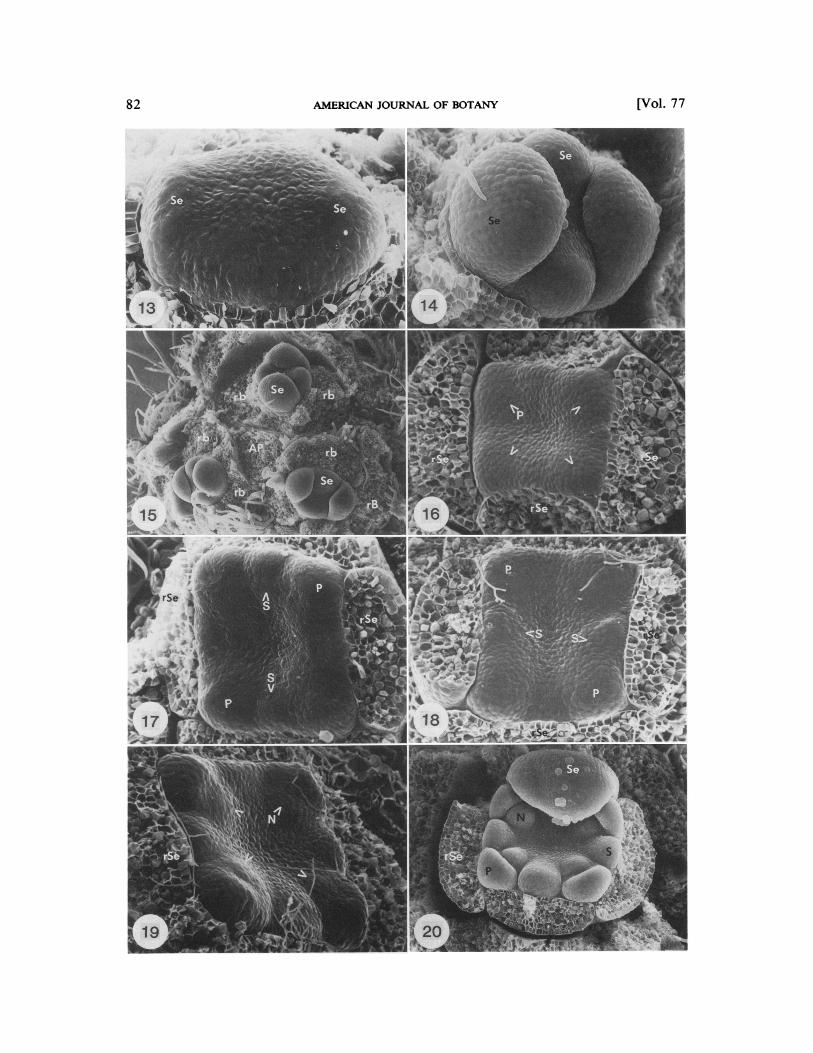
Fig. 13-20. Hamamelis vlrglniana. 13.Oblong floral apex develops in line with the secondary axis. Lower median sepal (left, Se) arises prior to the upper median sepal (right, Se). Floral apex turned 90" relative to Fig. 12. x 328. 14. Lateral sepals (Se) arise simultaneously (top and bottom) after median sepals (left and right) have enlarged. Lower median sepal (right) has remained larger than the upper median sepal (left). x 199. 15. Three floral buds are evenly distributed about the apical protrusion (AP). Se, sepal; rb, removed bracteole; rB, removed bract. x 82. 16. Petal (P, arrows) initiation occurs simultaneously at the comers of the floral apex, which is square in outline. Note that median sepals (left and right, rSe) are larger than lateral sepals (top and bottom). ~ 2 3 9 . 17. Median stamens (S) usually arise prior to lateral stamens. P, petal; rSe, removed sepal. x 244.18. Occasionally, lateral stamens (S) initiate prior to median stamens. P, petal. ~ 2 4 2 . 19. Nectary primordia (N, arrows) arise simultaneously, adaxial to petal primordia. rSe, removed sepal. x 240.20. The floral apex becomes deeply concave after nectaries (N) have become somewhat developed and prior to carpel initiation. S, stamen; P, petal; Se, sepal. ~ 2 2 6 .

84 AMERICAN JOURNAL OF BOTANY [Vol. 77

January 19901 MIONE AND BOGLE-FLOWERS OF HAMAMELIDACEAE 8 5
Fig. 29-33. Loropetalum chineme. 29. Apical meristem is small and low prior to the transition to the reproductive state. Pairs of stipules which flank the subdistichous bracts (B) have been removed. x 58. 30. Bracts (rB) and their flanking pairs of stipules have been removed to show that initially, the inflorescence apex expands where bracts and stipules are not closely appressed (arrows). x 105.31,32. Three to eight domelike floral apices (FA) arise simultaneously on the inflorescence apex. At the same time a subtending bract (B) arises abaxial to each flower. x 11 1, x 133. 33. Sepals (Se) arise in a seemingly random manner. A bract (rB) and its flanking pair of stipules have been removed. B, bract. x 102.
Fig. 21-28. Hamamelis virginiana. 21. Two crescent-shaped carpel primordia (C) are initiated parallel to the median sepals. N, nectary; S, stamen; P, petal; rSe, removed sepal. x 117.22. Petals (P) broaden and elongate, stamens (S) and nectaries enlarge, carpels (C) grow upward and trichomes develop around the ovary. x 65.23. Transverse, hand section. Carpel (C) margins become closely appressed and postgenitally fuse. x 284.24. Petal (P) elongation continues, stamen (S) valves begin to differentiate, nectaries (N) become broader distally and carpels elongate into styles (St). rP, removed petal. x 44.25. Petals (P) become circinately coiled while all other organs continue to differentiate. N, nectary; V, anther valve; St, style; S, stamen. x 3 1. 26. Transverse, hand section. A single ovule (0) arises in each carpel. Note the falsely bilocular ovary. x 257. 27. Transverse, hand section. Ovules (0) usually arise on opposite sides of the ovarian cavity. x 59. 28. Longitudinal, dissection. Integuments arise as girdling primordia with the inner integument arising prior to the outer integument (1). The outer integument ceases enlarging near the funiculus, and as a result becomes crescent shaped. x 135.

86 AMERICAN JOURNAL OF BOTANY [Vol. 77

87 January 19901 MIONE AND BOGLE- FLOWERS OF HAMAMELIDACEAE
each carpel from the original crescent shape of the primordium (Fig. 22-25). At the base of the carpels the margins become closely ap- pressed and postgenital fusion takes place re- sulting in the formation of the ovary. Here, the centrally facing vertical clefts give rise to the falsely bilocular ovary (Fig. 26, 27). Above the ovary each carpel elongates into a style (Fig. 24). The style retains some evidence ofits orig- inal crescent shape into late flower develop- ment (Fig. 24, 25).
Most often the two ovules arise on opposite sides of the ovarian cavity (one per carpel, diagonally across from each other as seen in transverse section, Fig. 26, 27). Less com-monly the two ovules arise on the same side of the ovarian cavity, one per carpel. During ovule development, integuments are initiated centrifugally (Fig. 28). Both integuments are initiated as girdling (completely circular) pri- mordia but, near the funiculus, the outer in- tegument ceases developing and becomes cres- cent shaped (Fig. 28, I). Both integuments eventually overtop the nucellus.
Late flower development is characterized by elongation of petals, differentiation of stamens and nectaries and profuse development of tri- chomes around the ovary (Fig. 25).
In Loropetalum chinense, development of inflorescences which terminate vegetative branches starts in June, but the majority of the axillary, vegetative apices make the transition to the reproductive state in late August or early September. Most often, an inflorescence which terminates a vegetative branch develops earlier than the axillary inflorescences on that branch.
Inflorescences that terminate vegetative branches are racemose in form (Fig. 4, A,). Inflorescences of lower axillary buds are usu- ally paniculate, consisting of two sequentially maturing racemes (Fig. 4, A,, A,). If a vege- tative branch departs form the node, however, the inflorescence is racemose in form. The pe- duncles of the axillary inflorescences elongate as the terminally borne flowers are nearing an- thesis. When two floral axes depart from a node (Fig. 4, A,, A,) both develop within the same
bud scales until the expansion of the first ma- turing axis causes the bud scales to open. A secondary bract may be lacking in the base of the tertiary axis (Fig. 4).
The transition of an apical meristem from the vegetative state (Fig. 29) to the reproduc- tive state initially involves expansion of the inflorescence apex where bracts and stipules have not been closely appressed (Fig. 30, ar- rows). The inflorescence apex then develops three to eight floral apices; at the same time bract primordia arise to subtend each floral apex (Fig. 31, 32). Occasionally, two bracts develop opposite each other on the periphery of the floral apex (Fig. 35). When this occurs, perhaps these bracts are best interpreted as the lower being the bract to which the flower is axillary and the upper a bracteole of the floral pedicel. On axillary inflorescences these bracts cease developing after their initiation (Fig. 4, lacking on A, and A,; Fig. 3 3 ,3 5; 36). On some flowers of inflorescences that terminate vege- tative branches, however, these bract primor- dia develop into minute, linear bracts which remain closely appressed to the flower (Fig. 4, A,; Fig. 48).
Sepal primordia (four or five) arise on each floral apex in a seemingly random manner (Fig. 33). By the time the sepals are well developed, stellate trichomes have grown on their surfaces and from between the flowers (Fig. 34-37). The floral apex is generally flat during sepal devel- opment (Fig. 33-36).
Prior to the initiation of petals and stamens the sepals grow to partially cover the slightly concave apex (Fig. 34). Petal and stamen ini- tiation is unidirectional (sensu Tucker, 1984). Petals and then stamen primordia are initiated first on the adaxial side of each floral apex (Fig. 35, 36). When petal and stamen primordia on the adaxial side of the floral apex have become domelike, petal and then stamen primordia are initiated on the abaxial side (Fig. 35,36). Adax- ial stamen primordia may be initiated at the same time as abaxial petal primordia. All petal and stamen primordia become roughly equal in size. At this stage the floral apex is somewhat
Fig. 34-41. Loropetalurn chinense. 34. Sepals (Se) grow to partially cover the floral apex. Stellate trichomes develop between flower buds and on sepals. ~ 6 7 . 35, 36. Petal (P) and then stamen primordia (S) first arise on the adaxial portion of the floral apex and then on the abaxial portion of the floral apex. B, bract; b, bracteole. x 134, x 111. 37. All petal (P) and stamen (S) primordia become approximately the same size. Floral apex is now slightly concave. Se, sepal. x 79. 38. Two crescent shaped carpel primordia (C) arise as mirror images of each other on the floral apex. P, petal; rS, removed stamen. x 235. 39. Carpel (C) margins become closely appressed and postgenitally fuse. P, petal; rS. removed stamen. x 71. 40, 41. At the time when the stigmatic lobes (SL) are developing on the styles, sterile phyllomes (SF) arise adaxial to the petals (P). Note the presence of an anomalous protruding ovule (Op) in Fig. 4 1. rS, removed stamen; rP, removed petal; S, stamen; P, petal. x 52, x 58 .

8 8 AMERICAN JOURNAL OF BOTANY [Vol. 77

January 19901 MIONE AND BOGLE-FLOWERS OF HAMAMELIDACEAE 8 9
concave (Fig. 37). Later, as carpels are being initiated, stamen connectives elongate and be- come connivent (Fig. 42).
Two crescent-shaped carpel primordia arise as mirror images of each other at the center of each floral apex (Fig. 38). When carpel initi- ation occurs, the petals and stamens are quite differentiated. The carpel margins become closely appressed; postgenital fusion follows (Fig. 39). More rapid upward growth occurs at the fold in each carpel giving rise to the styles (Fig. 40). The styles, in turn, give rise to the stigmatic lobes (Fig. 4 1, 45, 46).
Sterile phyllomes are the last whorl of ap- pendages to arise in the flowers of Loropetalum chinense. Sterile phyllomes arise centrifugal to the gynoecium, meaning that sterile phyllome primordia are initiated after and outside the carpel primordia (sensu Sattler, 1972). When the sterile phyllomes arise, the stamens are fully differentiated and the petals are longer than the stamens. Sterile phyllomes are initi- ated at about the time that the stigmatic lobes are beginning to develop on the styles (Fig. 40, 4 1, 47). Each sterile phyllome may arise as a single or bilobed (rarely trilobed, Fig. 4 1) pri- mordium.
During ovule development (Fig. 43-46), in- tegument development occurs as in Hama- melzs vzrginiana except that outer integuments do not become crescent-shaped.
DISCUSSION-Our observations on the floral organogenesis of Hamamelis virginiana are in general agreement with those of Baillon (1 87 1- 1 873) and Shoemaker (1 905). Baillon indicat- ed that median sepals are initiated sequen- tially; we add that the lower median sepal arises prior to the upper median sepal. Baillon found lateral sepals to be initiated sequentially. We have found lateral sepals to be initiated si- multaneously. Baillon described the stamens as being initiated simultaneously. In contrast, we have found that most often simultaneous
initiation of median stamens is followed by the simultaneous initiation of lateral stamens.
Sattler (1973) found six species, each in dif- ferent orders of flowering plants, to have discs initiated centrifugal to the gynoecium. On the assumption that the term disc does not imply affinity to any particular type of floral append- age (Schleiden, 1849), we are able to include the hamamelids with this condition (Corylop- sis, Liquidambar, Rhodoleia, and Loropeta- lum; see introduction for references) and report that at least 10 genera in families representing seven different orders exhibit centrifugal ini- tiation of discs (= sterile phyllomes in these genera).
Adjacent sepals of Loropetalum chinense were found to be partially to entirely connate in some flowers; this condition has not been reported previously. While our Hamamelis material was strictly 4-merous, Harms (1 930) observed occasional apical flowers which were 5-merous and Bogle (1968) observed a few trimerous flowers of H. virginiana. Loropeta- lum has been reported as 4-merous by all pre- vious workers (e.g., Hemsley, 1904; Chun, 1934; Chang, 1979). In contrast, the material we studied was 4- or 5- (rarely 6-) merous (Fig. 6).
In Loropetalum, petaloid staminodia were occasionally observed in the position that pet- als normally occupy. Similar observations have been made in several other genera of the family by Home (1 9 14, Corylopsis), Bogle (1 968, Dis- anthus, Exbucklandia, Rhodoleia), and En- dress (1 967, Corylopsis). These findings suggest that in the family Hamamelidaceae, petals and stamens are homologous.
The initiation of the nectaries of Hamamelis immediately after the stamens, the similar morphology of these appendages during early developmental stages, and the occurrence of a staminodial-nectary (a putative atavism) sug- gests that nectaries of Hamamelis are stami- nodial in origin. Baillon (1 87 1-1 873) also de-
Fig. 42-49. Loropetalum chinense. 42. While petals (P) elongate, stamen (S) connectives elongate and become connivent over the gynoecium. x 58. 43. Transverse, hand section. A single ovule (0 ) arises in each carpel. x 388. 44. Transverse, hand section. Ovules (0) most often arise on opposite sides of the ovarian cavity, x 102. 45. Longitudinal dissection. Integuments arise as girdling primordia with the inner integument (I) arising prior to the outer integument. When present, an anomalous protruding ovule (Op) arises from the opposite carpel margin from that bearing a normal, pendent ovule. Here, the protruding ovule (Op) arises from the far wall of the left carpel while the normal pendent ovule of that carpel (integuments facing viewer) is borne on the closer wall which has almost entirely been removed. The right carpel has almost entirely been removed. SL, stigmatic lobe; 0,ovule. x 119. 46. Longitudinal dissection. As in H. virginiana, the two ovules ( 0 ) are sometimes borne on the same side of the ovarian cavity. The integuments are then oriented in the same direction, in this case away from the viewer. SF, sterile phyllomes; SL, stigmatic lobes. x 56. 47. Extremely rare occurrence of a staminodium (Sto) adaxial to a petal (P). A sterile phyllome (SF) normally occurs in this position. x 64. 48. Bristlelike bracts (B, arrows) subtend some of the flowers of terminal inflorescences. x 64. 49. Rarely, a petaloid staminodium occurs in the position a petal normally occupies. x 26.

90 AMERICAN JOURNAL OF BOTANY [Vol. 77
scribed the stamens and nectaries as appearing similar during early development and went so far as to call the nectaries stamens ("Ctamines") during early developmental stages and "stam- inodes" during later developmental stages. The nearly identical vasculature of the stamens and nectaries in Hamamelis (Bogle, 1968; Mione, 1987) supports this conclusion.
In Loropetalum, the extremely rare occur- rence of staminodia in the position normally occupied by sterile phyllomes (Fig. 47) may be an atavism. In several other genera of the fam- ily appendages transitional between "stami- nodia" and stamens have been reported (Tong, 1930). These putative atavisms could be taken as evidence that in the Hamamelidaceae sterile phyllomes are derived from stamens.
The study of floral organogenesis in these two genera has revealed that in Hamamelis virginiana and H. mollis the nectaries arise immediately after the stamens while the sterile phyllomes of Loropetalum chinense arise after all other floral appendages have differentiated. This difference in time of appearance may be interpreted as evidence that although these whorls of appendages are similarly positioned, they have not evolved from the same whorl of stamens in the common progenitor.
In light of this conjecture, it is interesting to note that nectaries of Hamamelis and sterile phyllomes of Loropetalum have very different vasculatures (Bogle, 1968). In Hamamelis, each nectary is supplied by an adaxial branch of the petal bundle (petals and sterile appendages oc- cur on the same radii in both genera). In Lo-ropetalum, however, the bundle to the base of the sterile phyllome is derived from a hypan- thial vascular ring rather than directly from the petal bundle. The presence of such different vasculatures in Hamamelis and Loropetalum is incongruent with the idea that the sterile phyllomes of Loropetalum are derived from the same cycle of floral organs as the nectaries of Harnamelis, if one considers vasculature to be conservative. Furthermore, in the flowers of Maingaya and Dicoryphe (Bogle, 1984, 1988), which contain both staminodia and sterile phyllomes, the staminodia are supplied by branches of the petal trunk bundles, while the sterile phyllomes are supplied from a hy- panthial vascular ring, as in Loropetalum.
The sterile phyllomes of Loropetalum, like those of other Hamamelidaceae [Corylopsis ("Nektarschuppen," Endress, 1967), Liquid-ambar (Wisniewski and Bogle, 1982) and Rho-doleia (Bogle, 1987, 1989)l arise centrifugal to the gynoecium. This common time of initia- tion suggests that sterile phyllomes of these genera have evolved from a common whorl of
floral appendages in a progenitor. The rare re- version of sterile phyllomes to staminodia is perhaps the only evidence of a staminodial origin for the sterile phyllomes ofLoropetalum. If the sterile phyllomes are of staminodial or- igin, then if one considers the probable ho- mology of the sterile phyllomes of Loropet-alum with those of Corylopsis, Liquidambar, and Rhodoleia, a staminodial origin for the sterile phyllomes of all four of these genera must be considered.
The various arrangements of sterile phyl- lomes and staminodia in genera of the tribe Hamamelideae may be the result of loss and/ or retention of different whorls of appendages that have been retained in Maingaya (Bogle, 1984), the seemingly least specialized genus of the tribe Hamamelideae. Morphological evi- dence for this stems from the fact that flowers of Maingaya possess alternating whorls of five sepals, petals, stamens and staminodia and an innermost whorl of 10 sterile phyllomes. In species of Corylopsis, Tetrathyrium, Embo- lanthera, and Loropetalum possessing sterile phyllomes, these structures are two-parted or bilobed, indicating probable homology with the innermost whorl of 10 sterile phyllomes present in Maingaya. Regarding a different line of floral specialization (leading to loss of an innermost whorl of sterile phyllomes), some species of Dicoryphe possess a floral plan sim- ilar to that of Maingaya while other species of Dicoryphe lack sterile phyllomes but have ves- tigial vasculature which suggests the former presence of an innermost whorl of phyllomes (Bogle, 1984, and in preparation). Hamamelis possesses four individual staminodia and no evidence of an additional, inner whorl of phyl- lomes.
It is likely that progenitors of the subfamily Hamamelidoideae possessed several whorls (or a spiral with several cycles) of stamens which are represented as modern whorls of petals, stamens, staminodia and sterile phyllomes. This conjecture is supported by the aforemen- tioned evidence for a staminodial origin ofnec- taries and sterile phyllomes, homology of sta- mens and petals, and by the presence of more than one whorl of stamens in some genera of the subfamily Hamamelidoideae (e.g., Matu-daea, Bogle, 1968, 1970; Endress, 1976).
LITERATURE CITED
BAILLON,H. 1871-1873. Nouvelles notes sur les Hama- mClidCes. Adansonia 10: 120-1 37.
BOGLE, A. L. 1968. Floral vascular anatomy and the nature of the hamamelidaceous flower. Ph.D. disser- tation, University of Minnesota, St. Paul.
-. 1970. Floral morphology and vascular anatomy

January 19901 MIONE AND BOGLE- FLOWERS OF HAMAMELIDACEAE 9 1
of the Hamamelidaceae: The apetalous genera of Hamamelidoideae. J. Arnold Arbor. 5 1: 3 10-366.
-. 1984. Floral morphology and vascular anatomy of Maingaya oliv. (Hamamelidaceae, Hamamelidoi- deae, Hamamelideae). Amer. J. Bot. 7 l(5, part 2): 19.
-. 1987. Inflorescence and flower ontogeny in the pseudanthium of Rhodoleia (Hamamelidaceae). Amer. J . Bot. 74: 607-608.
-. 1988. Floral morphology and vascular anatomy of Dicoryphe Thouars. (Hamamelidaceae, Hama-melidoideae, Hamamelideae). Amer. J. Bot. 75(6, part 2): 18.
. 1989. The floral morphology, vascular anatomy and ontogeny of the Rhodoleioideae (Hamamelida- ceae) and their significance in relation to the lower Hamamelidaceae. In Evolution, systematics and fos- sil history of the Hamamelidae. Clarendon Press, Ox- ford.
BRADFORD,J. L., AND D. L. MARSH. 1977. Comparative studies of the Witch Hazels Harnamelis virginiana and H. vernalis. Proc. Arkansas Acad. Sci. 4 1 : 29-3 1.
BROWN,R. 1818. Characters and descriptions of three new species of plants. In C. Abel [ed.], Narrative of a journey in the interior ofChina, 374-379. Longman, London.
CHANG,H. T. 1979. Hamamelidaceae. Flora Reipublicae Popularis Sinicae 35(2): 36-1 16.
CHUN, W. Y. 1934. Contributions to the flora of Kwang- tung and South-eastern China. Sunyatsenia 1: 209- 3 17. [Hamamelidaceae, 36, 24 1-247.1
CREECH,J. L. 1960. On the distribution of Loropetalum chinense. Amer. Hort. Mag. 39: 236.
ENDRESS,P. K. 1967. Systematische Studie ueber die verwandtschaftlichen Beziehungen zwischen den Hamamelidaceen und Betulaceen. Bot. Jahrb. Syst. 87: 431-525.
-. 1976. Die Androeciumanlage bei polyandrischen Hamamelidaceen und ihre systematische Bedeutung. Bot. Jahrb. Syst. 97: 436-457.
ERNST,W. R. 1963. The genera of Hamamelidaceae and Platanaceae in the Southeastern United States. J. Ar-nold Arb. 44: 193-2 10.
ESAU,K. 1965. Plant anatomy. Ed. 2. John Wiley, New York.
HARMS,H. 1930. Hamamelidaceae. In A. Engler and K. Prantl [eds.], Die Natiirlichen Pflanzenfamilien. Ed. 2. 18a: 330-343.
HAUSER,E. J. P., AND J. P. MORRISON. 1964. The cy-
tochemical reduction of nitro blue tetrazolium as an index of pollen viability. Amer. J. Bot. 51: 748-752.
HEMSLEY,W. B. 1904. Loropetalum chinense. Bot. Mag. 130: 7979.
HORNE, A. S. 1914. A contribution to the study of the evolution of the flower with special reference to the Hamamelidaceae, Caprifoliaceae, and Cornaceae. Trans. Linn. Soc. London, Bot. Ser. 2, 8: 239-309.
KX~~SSMANN,1977. Manual of cultivated broad-leaved G. trees and shrubs. Vol. 2. Timber Press, Portland, OR. [Loropetalum,p. 257.1
LITTLE,E. J., JR. 1977. Atlas of United States trees, Vol. 4, Minor Eastern hardwoods. Misc. Publ. U.S. Dept. Agric. 1342.
MIONE, T. 1987. Comparative ontogeny of the inflores- cences and flowers of Harnamelis virginiana and Lo-ropetalum chinense (Hamamelidaceae). M.S. thesis, University of New Hampshire, Durham.
OLIVER,D. 1862. Note on Harnamelis and Loropetalum; with a description of a new Anisophyllea from Ma- lacca. Trans. Linn. Soc. London 23: 457-46 1.
SARGENT,C. S. 1890. The sylva of North America., Vol. 5: Hamamelidaceae-Sapotaceae [reprint ed. 19471. Peter Smith, New York.
-. 1920. Notes of North American trees. VI. J. Arnold Arbor. 1: 245-254.
SATTLER,R. 1972. Centrifugal primordium inception in floral development. Advances PI. Morph. 170-1 78.
-. 1973. Organogenesis of flowers. University of Toronto Press, Toronto.
SCHLEIDEN,M. J. 1849. Principles of scientific botany. (Trans. E. Lankester).
SHOEMAKER, 1905. On the development of Hama-D. M. melis virginiana. Bot. Gaz. (Crawfordsville) 39: 248- 266.
STANDLEY, 1937. Studies of American plants-VII. P. C. Publ. Field Mus. Bot. 17: 155-224.
TONG, K. 1930. Studien iiber die familie der Hama- melidaceae, mit besonderer bemcksichtigung der sys- tematik und entwicklungsgeschichte von Corylopsis. Bull. Dept. Biol. Coll. Sci. Sun Yatsen Univ. 2: 1-72.
TUCKER,S. C. 1984. Origin of symmetry in flowers. In R. A. White and W. C. Dickison [eds.], Contemporary problems in plant anatomy, 35 1-395. Academic Press, New York.
WISNIEWSKI,M., AND A. L. BOGLE. 1982. The ontogeny of the inflorescence and flower of Liquidambar sty- raciflua L. (Hamamelidaceae). Amer. J. Bot. 69: 1612- 1624.
Copyright © 2022 FDOKUMEN