based tool for country-wide standardized HIV confirmation and ...
Upload
jdkosajdfiasjsdCategory
view
1download
0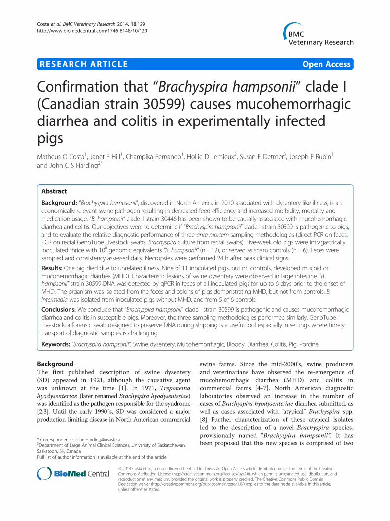
Costa et al. BMC Veterinary Research 2014, 10:129http://www.biomedcentral.com/1746-6148/10/129
RESEARCH ARTICLE Open Access
Confirmation that “Brachyspira hampsonii” clade I(Canadian strain 30599) causes mucohemorrhagicdiarrhea and colitis in experimentally infectedpigsMatheus O Costa1, Janet E Hill1, Champika Fernando1, Hollie D Lemieux2, Susan E Detmer3, Joseph E Rubin1
and John C S Harding2*
Abstract
Background: “Brachyspira hampsonii”, discovered in North America in 2010 associated with dysentery-like illness, is aneconomically relevant swine pathogen resulting in decreased feed efficiency and increased morbidity, mortality andmedication usage. “B. hampsonii” clade II strain 30446 has been shown to be causally associated with mucohemorrhagicdiarrhea and colitis. Our objectives were to determine if “Brachyspira hampsonii” clade I strain 30599 is pathogenic to pigs,and to evaluate the relative diagnostic performance of three ante mortem sampling methodologies (direct PCR on feces,PCR on rectal GenoTube Livestock swabs, Brachyspira culture from rectal swabs). Five-week old pigs were intragastricallyinoculated thrice with 108 genomic equivalents "B. hampsonii" (n = 12), or served as sham controls (n = 6). Feces weresampled and consistency assessed daily. Necropsies were performed 24 h after peak clinical signs.
Results: One pig died due to unrelated illness. Nine of 11 inoculated pigs, but no controls, developed mucoid ormucohemorrhagic diarrhea (MHD). Characteristic lesions of swine dysentery were observed in large intestine. “B.hampsonii” strain 30599 DNA was detected by qPCR in feces of all inoculated pigs for up to 6 days prior to the onset ofMHD. The organism was isolated from the feces and colons of pigs demonstrating MHD, but not from controls. B.intermedia was isolated from inoculated pigs without MHD, and from 5 of 6 controls.
Conclusions: We conclude that “Brachyspira hampsonii” clade I strain 30599 is pathogenic and causes mucohemorrhagicdiarrhea and colitis in susceptible pigs. Moreover, the three sampling methodologies performed similarly. GenoTubeLivestock, a forensic swab designed to preserve DNA during shipping is a useful tool especially in settings where timelytransport of diagnostic samples is challenging.
Keywords: “Brachyspira hampsonii”, Swine dysentery, Mucohemorrhagic, Bloody, Diarrhea, Colitis, Pig, Porcine
BackgroundThe first published description of swine dysentery(SD) appeared in 1921, although the causative agentwas unknown at the time [1]. In 1971, Treponemahyodysenteriae (later renamed Brachyspira hyodysenteriae)was identified as the pathogen responsible for the syndrome[2,3]. Until the early 1990′s, SD was considered a majorproduction-limiting disease in North American commercial
* Correspondence: [email protected] of Large Animal Clinical Sciences, University of Saskatchewan,Saskatoon, SK, CanadaFull list of author information is available at the end of the article
© 2014 Costa et al.; licensee BioMed Central LCommons Attribution License (http://creativecreproduction in any medium, provided the orDedication waiver (http://creativecommons.orunless otherwise stated.
swine farms. Since the mid-2000's, swine producersand veterinarians have observed the re-emergence ofmucohemorrhagic diarrhea (MHD) and colitis incommercial farms [4-7]. North American diagnosticlaboratories observed an increase in the number ofcases of Brachyspira hyodysenteriae diarrhea submitted, aswell as cases associated with “atypical” Brachyspira spp.[8]. Further characterization of these atypical isolatesled to the description of a novel Brachyspira species,provisionally named “Brachyspira hampsonii”. It hasbeen proposed that this new species is comprised of two
td. This is an Open Access article distributed under the terms of the Creativeommons.org/licenses/by/2.0), which permits unrestricted use, distribution, andiginal work is properly credited. The Creative Commons Public Domaing/publicdomain/zero/1.0/) applies to the data made available in this article,

Costa et al. BMC Veterinary Research 2014, 10:129 Page 2 of 12http://www.biomedcentral.com/1746-6148/10/129
different phylogenetic clades, sharing 96% sequenceidentity in the NADH oxidase (nox) gene sequence [9].Mucohemorrhagic diarrhea and colitis, indistinguishable
from SD has been experimentally reproduced in pigs with“B. hampsonii” clade II isolates from Canada and theUnited States [10,11]. Burrough et al. has also reproducedSD experimentally in mice [12] and in pigs [11] usingstrongly β-hemolytic strains initially identified as B.intermedia based on PCR [13], which have since beenconfirmed to be “B. hampsonii” clade I [14]. These datasuggests that clade I is pathogenic to pigs, but furtherstudies are required to confirm this, evaluate additionalstrains and to characterize the disease including patternsof shedding.The first Canadian diagnosis of “B. hampsonii” clade I
was in November 2011 from grow-finish pigs withbloody, mucoid diarrhea. This was a unique event,since all previous “B. hampsonii” cases diagnosed byour laboratory were of clade II. The isolate recoveredfrom this diagnostic case was designated “30599”.Since its first diagnosis in Canada, “B. hampsonii”clade I (strain 30599) has been identified, isolated orboth, in the absence of other Brachyspira spp., infeces or tissues from 39 cases of diarrhea from 15farms in western Canada. Given the distance betweenfarms and diagnostic laboratories in western Canada,this may be an underestimate of the number of farmsaffected by the organism. One of the main obstaclesto obtaining high quality diagnostic samples and reliableresults is the transit time required to ship samples to adiagnostic laboratory [15,16]. For PCR based diagnostics,sample quality may be improved by immediate preser-vation of target DNA in the sample and controllingthe growth of opportunistic organisms. A forensic swab(GenoTube Livestock; Prionics, Switzerland) capable ofrapidly drying the sample and providing minimal DNAloss after long-term storage without refrigeration maybe a potential tool for aiding the diagnosis of infectiousdiseases by PCR in biologic samples shipped long distancesto diagnostic laboratories.The objectives of this study were to determine the
pathogenicity of “Brachyspira hampsonii” clade I (strain30599) when inoculated in naïve pigs, and to investigatethe performance of GenoTube Livestock swabs for antemortem sampling of pigs for PCR-based detection of“Brachyspira hampsonii” clade I. In order to accomplishthese primary objectives, we also developed and validateda PCR assay for the detection and quantification of“Brachyspira hampsonii” clade I (strain 30599).
MethodsThis work was approved by the University of Saskatche-wan’s Animal Research Ethics Board and adhered to theCanadian Council on Animal Care guidelines for humane
animal use (permit #20110038) and ARRIVE Guidelines(Animal Research: Reporting In Vivo Experiments;Additional file 1).
Source of “B. hampsonii” strain 30599 inoculum“B. hampsonii” clade I strain 30599 was isolated from apig with mucoid diarrhea in Alberta, Canada in November,2011. PCR for B. hyodysenteriae [13], B. pilosicoli [10] and“B. hampsonii” clade II [10] performed on case tissuesreturned negative results. Sections of small and largeintestines tested negative for Salmonella (culture onbrilliant green agar following enrichment with selenitebroth), Lawsonia intracellularis (PCR) [17], and porcinecircovirus type 2 (PCV2, immunohistochemistry) [18] atthe Prairie Diagnostic Service Inc. (PDS), Saskatoon, SK.Culture of colon tissue on selective media resulted in areasof strong β-hemolysis, which were sub-cultured to obtainan isolate. Sequencing of the nox, cpn60 and 16S rRNAgenes from this isolate demonstrated that it was similar to,but phylogenetically distinct from “B. hampsonii” clade II.Whole genome sequencing of the isolate was performed(NCBI BioProject PRJNA188379) and confirmed itsaffiliation with what has recently been proposed as clade Iof “B. hampsonii” [9].
Experimental inoculationThe experimental inoculation was performed as describedpreviously (“Pure Broth Culture Inoculation” in [10]).Briefly, 18 five-week old pigs (16 male, 2 inadvertentlyfemale) were obtained from a commercial farm inSaskatchewan, Canada. No history of MHD or previousdiagnosis of Brachyspira spp. associated diarrhea was re-ported, and fecal samples collected from nursery (n = 20)and grow-finish (n = 10) pigs prior to the commencementof the study tested negative for Brachyspira spp. byculture and species-specific qPCR for B. hyodysenteriae, B.pilosicoli and “B. hampsonii” (data not shown). Uponarrival at the Animal Care Unit at the University ofSaskatchewan, the pigs were allocated randomly to control(CTRL, n = 6) and inoculated (INOC, n = 12) groupshoused in separate rooms with 3 or 2 pigs per pen,respectively. Group sizes were based on results of pastexperiments and provided an experimental power of 0.83based on the assumptions that 8/12 INOC and 0/6 CTRLpigs would develop MHD and alpha = 0.05. One femalepig was assigned to each group. Pigs were acclimated totheir new diet and room environment for 7 days prior toinoculation, and were offered ad libitum water andcommercially prepared, non-medicated, pelleted starterdiet (Whole Earth Pig Starter, Federated Cooperative Ltd.,Saskatoon, Canada) formulated with less than 15% soybeanmeal for the duration of the experiment.Pigs were sedated with 8 mg/kg azaperone IM
(Stresnil, Vetoquinol Canada Inc., Lavaltrie, Quebec)

Costa et al. BMC Veterinary Research 2014, 10:129 Page 3 of 12http://www.biomedcentral.com/1746-6148/10/129
then inoculated by gastric tube on three consecutive days(day (D) 0, 1 and 2). INOC received 10 mL of frozen JBSbroth containing 6.10 × 10⁸ (D0), 4.14 × 10⁸ (D1)and 4.24 × 10⁸ (D2) genome equivalents of “B. hampsonii”strain 30599, followed by 40 mL of PBS (0.1 M, pH 7).CTRL received 10 ml of sterile JBS broth followed by 40mL of PBS. For both groups, feed was removed 16 h priorto inoculation to increase gastric motility.Feces and rectal swabs were collected on day −8, −5, −2
and 0 prior to inoculation, and daily thereafter untilthe end of the experiment. Fecal consistency wasscored daily as: 0 = formed, normal; 1 = soft, wet cementconsistency; 2 = runny or watery diarrhea; 3 =mucoiddiarrhea; 4 =mucohemorrhagic diarrhea. Daily clinicalassessment was performed on all pigs. Rectal temperature,responsiveness to external stimuli (e.g. the presence ofresearcher in the pen) and skin colour were recorded.Body weight was measured on D0 and D8. INOC pigswere humanely euthanized by cranial captive bolt andexsanguination approximately 24 hours after the develop-ment of mucoid or mucohemorrhagic diarrhea. INOCpigs that did not demonstrate MHD were euthanized onD13; CTRL pigs on D14.
Pathological assessmentsA complete necropsy was performed and the gastrointes-tinal tract was completely removed from stomach to rec-tum, including mesenteric lymph nodes. Special attentionwas paid to the cecum and spiral colon which was com-pletely linearized and divided into thirds (proximal, apexand distal). The mucosal surfaces of the large intestineswere assessed for the presence of characteristic lesions ofSD including hyperemia, congestion, edema, necrosis,fibrin and mucus by a single pathologist (SED). A blindedhistologic evaluation of colon and cecum was performedby the same pathologist. Tissue samples were fixed in 10%buffered formalin for 24 hours and paraffin embedded. Allmicrosectioned tissues were stained with Hematoxylin-Eosin (H&E) and a serial section of the spiral colon wasalso Warthin-Faulkner (WF) silver stained. Lesions inthe spiral colon and cecum were scored based on theseverity of the inflammation and necrosis: 0 = no lesions;1 =minimal to mild necrosis of superficial enterocyteswith minimal inflammatory infiltrates; 2 = moderatenecrosis and attenuation of enterocytes with mild tomoderate inflammatory infiltrates; 3 = severe necrosis(erosion or ulceration present) with moderate inflammatoryinfiltrates predominantly consisting of neutrophils. Thepresence of Brachyspira-like organisms was also scoredfrom 0 to 3 in WF stained sections: 0 = no spirochetesobserved; 0.5 = a single gland contained a few spirochetes;1 = small numbers of spirochetes in multiple glands;2 = many spirochetes within several glands; 3 = manyspirochetes forming thick mats in numerous glands.
Microbiological assessmentsGastrointestinal tissues collected at termination werescreened for the presence of other relevant swine entericpathogens, including Lawsonia intracellularis (PCR, ileum),porcine circovirus type 2 (PCV2, immunohistochemistry,ileum and mesenteric lymph node) and Salmonella spp.(culture, ileum). All methods were the same as describedabove. PRRSv IgG antibody and RNA concentrationwere measured by ELISA (IDEXX PRRS X3, IDEXXLaboratories Inc., Westbrook, ME) and PCR (TetracoreInc., Rockville, MD), respectively, at PDS in sera collectedon D0 and at termination.
Quantification of “B. hampsonii” clade I strain 30599Details on the development and validation of the strain-specific qPCR assay are given in the Results. DNA forPCR was extracted from feces and colon tissue using theQIAmp DNA stool mini kit (Qiagen Inc., Toronto, ON),and from cultured bacteria and colon tissue usingDNEasy blood and tissue kit (Qiagen Inc.). SYBR greenreal-time qPCR detection of “B. hampsonii” strain 30599was performed on a Bio-Rad MyiQ thermocycler inreactions containing 1× SYBR Green Supermix (Bio-RadLaboratories (Canada) Ltd., Mississauga, ON), 400 nMeach primer JH0436 (5'-AAA GTG CCA CAG GCAATG TA-3′) and JH0437 (5′-TGC AAG ATT AGACGG AGC AA-3′)) and 2 μL of template DNA, in afinal volume of 25 μL. All qPCR reactions were run on aplate containing a no-template control and a standardcurve composed of target-containing plasmids at con-centrations of 100 to 107 copies/reaction. All reactionswere performed in duplicate. Thermocycling parametersincluded an initial denaturation (95°C for 3 min.),followed by 40 cycles of 95°C for 15 sec., 63°C for 15sec., 72°C for 15 sec., and a final extension at 72°C for 5min. A dissociation curve was subsequently performedfor 81 cycles at 0.5°C increments from 55°C to 95°C.Fluorescent signals were measured every cycle at theend of the annealing step and continuously during thedissociation curve data collection. All resulting data wasanalyzed using iQ5 Optical System Software (Bio-RadLaboratories (Canada) Ltd., Mississauga, ON).
Brachyspira cultureIsolation of Brachyspira spp. was performed on BJagar from feces and colonic mucosa, as previouslydescribed [10]. Presence of Brachyspira growth in zones ofβ-hemolysis was confirmed by dark-field microscopy, andgenus-specific PCR targeting the nox gene [19] followed byDNA sequencing using the amplification primers.
Comparison of ante mortem sampling techniquesThree ante mortem sampling and detection methodswere chosen for comparison: rectal swabs (CultureSwab

Costa et al. BMC Veterinary Research 2014, 10:129 Page 4 of 12http://www.biomedcentral.com/1746-6148/10/129
Liquid Stuart, BD Canada, Mississauga, ON) for cultureon selective agar, qPCR on DNA extracted from fecalsamples (200 mg samples, QIAmp DNA stool mini kit,Qiagen Inc.), and qPCR on DNA extracted from GenoTubeswabs. Samples were collected D-8, D-5, D-2 and D0 priorto inoculation, and every other day thereafter to D12 inINOC pigs. Targeted sampling was employed in order tosample pigs with different fecal scores. On each day,samples were simultaneously collected from pigs with nodiarrhea (n = 2), pigs with “wet-cement” or waterydiarrhea (n = 2) and pigs with mucoid and mucohe-morrhagic diarrhea (n = 2). If one or more categorywas not observed at the time of collection, pigs withthe closest available fecal score on that day were sampled.The pigs from which samples were collected wereblocked by pen.To simulate a field situation where samples would
be transported to the diagnostic laboratory over 24 h,culture swabs and feces were refrigerated at 4°C (to mimicshipment with ice packs), while GenoTube swabs werestored at room temperature for 24 h prior to processing.GenoTube swabs were expressed into 1.4 mL lysis buffer(buffer ASL, QIAmp DNA stool mini kit, Qiagen Inc.,Toronto, ON) in a 2 mL microcentrifuge tube by pressingthe swab head against the inside wall of the tube whiletwisting. To ensure consistent sample expression, swabswere turned exactly ten times each (5 clockwise and 5counter clockwise). Following expression, extractionproceeded according to the kit instructions. DNAextracts (2 μL each) from feces and GenoTube swabswere used as a template for “B. hampsonii” clade Ispecific qPCR analysis. Culture swabs were used toinoculate BJ agar plates.
Statistical analysisStatistical analysis was performed using SPSS v19.0(SPSS Inc., Chicago, IL) or Stata v13 (StatCorp, CollegeStation, TX). Group differences in the number of pigsdemonstrating MHD (fecal consistency score 4) wascompared using a Fisher’s exact test. The same test wasused to compare the presence or absence of specific gastro-intestinal lesions based on gross and histopathologicalassessments. Continuous variables including average dailygain (ADG) and the concentration of “B. hampsonii” strain30599 DNA detected by qPCR were non-parametricallydistributed and compared between groups using a Kruskal-Wallis Analysis of Variance. Spirochete scores in colonwere compared with a Mann–Whitney U test. To comparethe severity of gross lesions among proximal, apexand distal colon segments, a Wilcoxon matched-pairssigned-rank test was used after assigning negative,mild, moderate and severe lesion scores as 0, 1, 2, 3respectively. For all analyses, P < 0.05 was consideredas statistically significant.
ResultsDevelopment of “B. hampsonii” clade I strain 30599specific SYBR green PCR assayA strain-specific, SYBR green quantitative real time PCRassay was developed to quantify ”B. hampsonii“ strain30599 DNA. Primers JH0436 (5′-AAA GTG CCA CAGGCA ATG TA-3′) and JH0437 (5′-TGC AAG ATT AGACGG AGC AA-3′) were designed to target a 176 bpregion within a predicted open reading frame in the“B. hampsonii” strain 30599 genome encoding a hypo-thetical protein (Genbank accession WP_008726773).The target region lies within an approximately 15.8 kbregion of the genome identified as being unique to strain30599 in a comparison of the whole genome sequencesof clade I strain 30599 (BioProject PRJNA187424) andclade II strain 30446 (BioProject PRJNA169353). Thetarget sequence has no significant sequence identityto “B. hampsonii” clade II strain 30446, and is only90% identical at the nucleotide level to a sequencewithin the genome of B. intermedia PWS/A (ATCC51140T). No other significant sequence identities toother Brachyspira species were identified.An optimal annealing temperature of 63°C was deter-
mined, and a linear standard curve was obtained over arange of 100 to 107 target copies per PCR reaction using aten-fold dilution series of cloned target amplicon in plasmidpGEM T Easy as template.Analytical specificity was determined initially by applying
the primers to genomic DNA from B. hyodysenteriaeATCC 27164T, B. pilosicoli ATCC 51139T, B. intermediaATCC 51140T, B. innocens ATCC 29796T, B. murdochiiATCC 51284T, and “B. hampsonii” clade II strain 30446.No amplification was detected from any template otherthan “B. hampsonii” clade I strain 30599. Further validationof the analytical specificity of the “B. hampsonii” strain30599 specific qPCR involved its application to fecalsamples from previous clinical cases determined to bepositive for “B. hampsonii“ strain 30599 by amplificationand sequencing of the nox gene using previously publishedprimers (n = 25) [19], and fecal samples from healthy pigsfrom the source farm that were confirmed negative forBrachyspira by the same method (n = 30). All (30/30) ofthe negative samples were found negative by the strainspecific qPCR, and 24 of 25 of the positive samples werefound positive by the strain specific qPCR, demonstratingsignificant agreement between the methods.
Acclimation periodBrachyspira culture was performed on all pigs on fourdays during the pre-inoculation acclimation period. B.intermedia (97% nox gene sequence identity over 810 bp toB. intermedia ATCC 51140T) was isolated from one INOCand one CTRL pig on D-2, and from one INOC on D0. ”B.pulli” (98% nucleotide identity over 848 bp of nox to strain

Costa et al. BMC Veterinary Research 2014, 10:129 Page 5 of 12http://www.biomedcentral.com/1746-6148/10/129
AN304/04) was isolated from one INOC on D-2 and D0.“B. pulli” is the provisional name for a spirochete originallyidentified in birds [20], which has recently also been isolatedfrom pigs [21]. One CTRL had watery diarrhea on D-8 andD-7, immediately after arrival in the animal care facility, butBrachyspira was not isolated, and normal feces wereobserved during all other days pre-inoculation.
Clinical observationsOne INOC pig presented with tachypnea and dyspneaon D3 and D4, and for welfare reasons was euthanized
Figure 1 Fecal consistency scores, culture results and fecal concentratioFecal consistency scores (line, left ordinate: 0 = formed, normal; 1 = softor 4 = bloody diarrhea), semi-quantitative fecal culture score (red bars, lethan 10 colonies/2° streak; 3 = less than 10 colonies/3°streak; 4 = less than(triangles, right ordinate; upside down triangles indicate DNQ) are shownPig IDs are indicated in the upper left corner of each panel.
and necropsied on D5. Necropsy findings indicatedaspiration of feed at the tracheal bifurcation. Data fromthis pig were removed from all analyses.Post-inoculation fecal scores for INOC pigs are
summarized in Figure 1. Eight of 11 INOC pigs developedMHD (score 4) (Figure 2), and one INOC developedmucoid diarrhea (#22, score 3). Of the two remainingINOC pigs, one (#19) remained healthy throughoutthe study, and one (#670) had fecal consistency typical ofwet cement (score 1) on four post-inoculation days. Todistinguish these from the nine diarrheic INOC pigs, they
n of “Brachyspira hampsonii” strain 30599 following inoculation., wet cement consistency; 2 = runny or watery; 3 = mucoid diarrhea;ft ordinate: 0 = negative; 1 = less than 10 colonies/1° streak; 2 = less10 colonies/4° streak), and strain 30599 DNA concentration in feces. Days post-inoculation are shown on the abscissa. ND = no data.

Figure 2 Mucohemorrhagic diarrhea. Accumulation of loose and runny feces with mucus and frank blood. Pig #675, 11 days post inoculation,day of first observation of mucohemorrhagic diarrhea, 7th day of “B. hampsonii” strain 30599 shedding, feces culture positive (3+ stronglybeta-hemolytic “B. hampsonii” strain 30599) on this day, 1.86 × 10⁸ strain 30599 genomic equivalents/g. In inoculated pigs the severity ofbloody diarrhea ranged from that containing a few flecks of blood to diffusely hemorrhagic with copious blood and mucus as shown.
Costa et al. BMC Veterinary Research 2014, 10:129 Page 6 of 12http://www.biomedcentral.com/1746-6148/10/129
are referred to as “INOC (without MHD)”. No CTRL pigdemonstrated mucoid or bloody diarrhea after shaminoculation, although one had fecal consistency typical ofwet cement (score 1) on D6 and D9, and one had runnydiarrhea (score 2) on D4 and wet cement consistency onD8, but was normal on all other days. Mucohemorrhagicdiarrhea was significantly more frequent in INOC than inCTRL pigs (P = 0.002).On D0, one INOC pig (#672) unexpectedly presented
with runny diarrhea and fecal samples were submittedfor routine aerobic and anaerobic culture to PDS. Thesample was negative for Salmonella spp. but E. coli. wasisolated. PCR results indicated that the E.coli isolate waspositive for virulence factors AIDA-I and STb. Fecalconsistency from this pig scored 0 or 1 until D8, thenprogressed to MHD by D9. The median number of daysfrom first inoculation to the demonstration of mucoid ormucohemorrhagic diarrhea in INOC was 8, with theshortest and longest incubation periods at 7 and 11 daysrespectively. The onset of MHD was generally acute withaffected pigs progressing from relatively normal feces(score 0 or 1) to MHD within 2.4 days (range 1–4)(Figure 1).Average daily gain between D0 and D8 was numerically
decreased in INOC (359 ± 143 g/day) compared to CTRL(413 ± 123 g/day), but did not differ significantly betweengroups.
Serum from blood collected on D0 and at terminationwas PRRSv PCR and ELISA negative. Intestinal tissuesamples collected at termination tested negative forSalmonella sp., Lawsonia intracellularis and PCV2 byculture or PCR.
Brachyspira shedding patternsBrachyspira spp. isolates were recovered from feces from10 of 11 INOC pigs between D0 and D4. This was likelyassociated with pass-through of the inoculation dose.Eight of 11 were culture positive for “B. hampsonii” strain30599 solely, two of 11 for B. intermedia, one of 11 for “B.hampsonii” strain 30599 and B. pulli on different days(Figure 1). Between D5 and termination, “B. hampsonii”strain 30599 was consistently isolated in the feces andfrom the colon of all INOC pigs that developed MHD(Figure 1). B. intermedia was isolated from the feces andcolon of the two INOC pigs without MHD. B. intermediawas also isolated sporadically from the feces of 5 of 6 andfrom terminal colon in 3 of 6 CTRL pigs. The presence ofB. intermedia in feces was not associated with diarrhea.Inadvertently, an issue with the culture vessel on D9resulted in negative cultures from all pigs. Culturepositive isolates confirmed as “B. hampsonii” strain30599 had 99-100% sequence identity to the inoculumbased on sequencing of the nox gene. Any minordifferences in sequence are most likely explained by

Costa et al. BMC Veterinary Research 2014, 10:129 Page 7 of 12http://www.biomedcentral.com/1746-6148/10/129
PCR artefact since the Taq DNA polymerase used wasnot a proofreading enzyme.Quantifiable levels of “B. hampsonii” strain 30599 were
detected by qPCR beginning D4 in 11 of 11 INOCpigs (Figure 1). “B. hampsonii” strain 30559 was notdetected in CTRL pigs. In INOC pigs without MHD,quantifiable levels were detected on a single day in eachpig (D8 or D9). In INOC pigs with MHD, quantifiablelevels of “B. hampsonii” strain 30599 were detected overmultiple consecutive days preceding euthanasia. Onaverage, quantifiable levels were detected for four daysprior to the onset of MHD (range 2 to 6). The averageconcentration of “B. hampsonii” strain 30599 across alldays in INOC pigs with MHD was 8.05 (log 10 genomicequivalents/gram of feces).Between D1 and D4, “B. hampsonii” strain 30599 was
isolated by culture in 15 fecal samples of INOC pigswith MHD. In these same samples, the organism wasdetected by PCR 13 times. Of these, 3 samples hadquantifiable levels of DNA whereas the remaining 10had detectable but non-quantifiable levels (i.e. beyondthe lower limit of the linear portion of the standardcurve). From D5 to termination, “B. hampsonii” strain30599 was isolated by culture in 37 fecal samples ofINOC pigs with MHD, whereas DNA was detected inthe same samples 41 times, always at quantifiable levels.These data indicate that PCR and culture had similaroverall detection frequencies.
Pathological findingsA summary of gross pathological findings is presented inTable 1. Gross lesions in INOC pigs were confined to thelarge intestine, cecum and rectum. Cecum and rectum of
Table 1 Comparison of gross and histopathological lesion fre
P value
Description CTRL (n = 6) INOC (n = 11) INOC
Gross pathology frequency*
Proximal colon 1/6 8/11 8/9
Apex colon 0/6 9/11 9/9
Distal colon 0/6 7/11 7/9
Cecum 1/6 6/11 5/9
Rectum 0/6 5/11 5/9
Histopathologic lesion frequency**
Colonic inflammation 0/6 9/11 9/9
Colonic necrosis 0/6 9/11 9/9
Cecal inflammation 0/6 6/11 6/9
Cecal necrosis 0/6 3/11 3/9
Colonic spirochetes 0/6 8/11 8/9
*Gross lesions include mucosal edema, hyperemia, congestion, with multifocal to d**Histopathological lesions and colonic spirochete scores graded 0 to 3 reflecting nthan 1 were considered lesional.
INOC pigs displayed mild mucosal lesions, characterizedas hyperemia associated with mucoid and fibrinousexudation. No lesions were observed in CTRL or in thesmall intestine of INOC pigs. In the two INOC withoutMHD, no lesions were observed in the proximal, apex ordistal spiral colon; however, mild hyperemia of cecalmucosa was noted in one of these two pigs. Of the nineINOC with MHD, characteristic gross lesions of swinedysentery were observed consistently in spiral colon andcecum (Table 1). These lesions ranged in severity frommild, mucosal edema and hyperemia to severe, multifocalto coalescing, mucosal congestion with mucofibrinousexudate and focal areas of necrosis (Figure 3). Inproximal, apex and distal spiral colon, gross lesionswere significantly more frequent in INOC than inCTRL (P < 0.05 for all). In rectum, gross lesions weresignificantly more frequent in INOC with MHD thanin CTRL (P = 0.04). Typhlitis was not a remarkablefinding in INOC pigs, and lesion frequency did notdiffer between INOC and CTRL groups in this study.These results are contrary to previous descriptions ofinfections with B. hyodysenteriae or “B. hampsonii”clades I and II [2,10,11,22]. In INOC pigs with MHD,gross lesions were significantly more severe in theproximal and apex regions of the colon than in thedistal colon (P < 0.01).The frequency and severity of histopathologic findings
were consistent with clinical signs and macroscopiclesions (Table 1). A thick layer of mucus and neutrophils(Figure 4) was observed on the colonic mucosa in all butone INOC pig. The nine of eleven pigs with positivecultures had histologic scores of 2 to 3 in the colon. Bycontrast, five of six CTRL pigs demonstrated mats of
quency in CTRL and INOC groups
(with MHD) (n = 9) INOC vs. CTRL INOC (with MHD) vs. CTRL
0.04 0.01
<0.01 <0.001
0.02 <0.01
ns ns
0.08 0.04
<0.01 <0.001
<0.01 <0.001
0.04 0.001
ns 0.02
<0.01 0.001
iffuse mucofibrinonecrotic exudate.egative, mild, moderate and severe inflammation or necrosis. Scores greater

Figure 3 Mucohemorrhagic colitis. Congested mucosal surface of apex region of the spiral colon with thick layer of mucus and fibrin andoccasional foci of haemorrhage. Pig #29, 12 days post inoculation, 48 hours post onset of mucohemorrhagic diarrhea.
Figure 4 Brachyspira colitis. In a section of proximal colon (Pig #671) there is a focal area of epithelial necrosis surrounded in the laminapropria by a mixed inflammatory infiltrate composed primarily of degenerate neutrophils, and covered by a thick mat of mucous containingdegenerate neutrophils, necrotic epithelial cells, and bacterial colonies. Hematoxylin and Eosin. Bar = 200 μm.
Costa et al. BMC Veterinary Research 2014, 10:129 Page 8 of 12http://www.biomedcentral.com/1746-6148/10/129

Costa et al. BMC Veterinary Research 2014, 10:129 Page 9 of 12http://www.biomedcentral.com/1746-6148/10/129
Gram-negative rod-shaped bacteria with minimal tono mucus (histologic score of 1). The presence ofinflammation and superficial necrosis in the colon andcecum was higher in INOC than CTRL pigs (Table 1), aswere numbers of spirochetes visualized in colon usingsilver staining (Table 1, Figure 5). Inflammation andnecrosis were more severe in colon than cecum (P < 0.01).
Evaluation of ante mortem sampling techniquesOne-hundred and sixty-five samples (55 GenoTube swabs,55 culture swabs, 55 fecal samples) were collected. Sincethere were no diarrheic pigs on the majority of samplingdays, 78% of samples (43/55) were obtained from pigswith normal feces (score 0 or 1), whereas 9% (5/55) origi-nated from pigs with loose to watery feces (score 2) and13% (7/55) from pigs with mucoid or mucohemorrhagicdiarrhea (score 3 or 4). Forty-two percent (23/55) of thefecal samples were collected prior to inoculation.Culture on BJ media followed by sequencing of the
nox gene was set as the diagnostic gold standard fordetection of “B. hampsonii” clade I strain 30599. Acomparison of detection rate of “B. hampsonii” strain30599 across the three ante mortem sampling methodolo-gies is shown in Figure 6. Only 12 samples were collectedfrom pigs with runny, mucoid or mucohemorrhagicdiarrhea (score >1). Two of these were negative by culture,two were negative by GenoTube PCR, and one was
Figure 5 Brachyspira colitis. In the same section of proximal colon as Figlong, thin spiral-shaped bacteria. Warthin-Faulkner. Bar = 100 μm.
negative by PCR of fecal DNA extracts. Both PCRmethodologies detected “B. hampsonii” strain 30599in all samples of mucoid and mucohemorrhagic diarrhea(score 3 or 4). On the other hand, one sample ofmucohemorrhagic diarrhea collected on D8 was negativeby culture, but positive by both PCR methodologies. Thissample had 5.07 × 10⁷ genome equivalents/g of fecesbased on qPCR. While this dataset is small, biased towardsnegative test results, and limited to a single experiment,the results suggest these assays have similar detectioncapabilities.
DiscussionThe results of this experiment confirm that “B. hampsonii”clade I strain 30599 is pathogenic in susceptible pigs, andresults in clinical disease indistinguishable from swinedysentery associated with B. hyodysenteriae and “B.hampsonii” clade II. This finding is relevant for porkproducers in Canada and elsewhere whose herds experi-ence mucohemorrhagic diarrhea. To our knowledge this isthe first report of experimental reproduction of diseaseassociated with Canadian strain 30599. Moreover, aprevious report from Burrough et al. [11] pre-datedour present understanding based on nox sequences,that the strongly β-hemolyic strains of B. intermedia(KC35, EB106) used in these experiments were in fact“B. hampsonii” clade I. We hope the present research
ure 4, the tubular mucous-secreting glands contain large numbers of

Figure 6 Comparison of detection of “B. hampsonii” strain 30599by qPCR on DNA extracts from feces or GenoTube swabs, orculture on selective media. Contingency tables show the numbers ofsamples determined to be positive or negative for “B. hampsonii” strain30599 by (A) culture or qPCR on DNA extracts from GenoTube forensicswabs, and (B) culture or qPCR on DNA extracts from fecal samples.
Costa et al. BMC Veterinary Research 2014, 10:129 Page 10 of 12http://www.biomedcentral.com/1746-6148/10/129
will help to clarify any potential confusion that mayexist regarding the pathogenicity of this organism.A second substantial deliverable of this research is the
first description of a novel quantitative PCR assay specificfor “B. hampsonii” clade I. Based on a discriminatingregion of the genome, this SYBR-based PCR assay enablesquantification, and does not cross react with other knownBrachyspira species including “B. hampsonii” clade II.Three Brachyspira species are capable of causing bloody
diarrhea: B. hyodysenteriae, “B. hampsonii” clades I and II,and “B. suanatina”. B. hyodysenteriae is globally distributed,while “B. suanatina” is reported only in Europe. Although“B. hampsonii” was discovered and mainly recognized inNorth America, isolations of clade I in Europe have beenreported recently. One report details the isolation from twogilts that were imported from the Czech Republic toBelgium with soft watery feces within dilated largeintestines [14]. The second account describes the isolationfrom a group of 17 grower pigs demonstrating mildto moderate diarrhea imported from Belgium toGermany [23]. A third study described the first finding of“B. hampsonii” from feces of birds, more specificallygraylag geese and mallards in northwestern Spain [24].In the present study, the onset of mucoid or mucohe-
morrhagic diarrhea in INOC pigs occurred within 7–11days of inoculation, and was preceded by the fecal sheddingof high levels (105 to 108 genomic equivalents/gram offeces) of “B. hampsonii” strain 30599 for up to 6 days. Theprevalence of MHD was similar to that observed in the “B.hampsonii” clade II strain 30446 experimental reproduction[10] that used a similar animal model. It must be noted thatthis animal model does not require the feeding of 100%soybean meal prior to inoculation although a 16-hourperiod of fasting to enhance gastric motility is performedprior to each inoculation.Swine dysentery is a multifactorial disease, with clinical
expression dependent on individual host factors such
as the gut microbiota [25], environmental conditionsincluding diet composition [26,27], and presence of avirulent organism. Interestingly, two challenged pigsdid not develop diarrhea and from both, B. intermediawas isolated in feces and in colonic tissue collected attermination. B. intermedia is generally accepted to be a gutcommensal and non-pathogenic to pigs [28-30]. Whetheror not it played a role in preventing the development ofdiarrhea in these pigs is not known, but B. intermedia wasalso isolated in five control pigs, suggesting it was a normalinhabitant of the pigs used in this study. A wide range ofBrachyspira species can be isolated from healthy pigs fromcommercial farms [31] and are presumed to contribute tothe commensal microbiota. The isolation of species otherthan that contained in the inoculum should therefore notbe unexpected.One pig (#22) developed a mucoid diarrhea with no
evidence of blood prior to recovery over several days.This differed from pigs experimentally inoculated with“B. hampsonii” clade II, where 100% of the animals thatdisplayed diarrhea progressed to blood-stained feceswithin 8 days of inoculation [10]. Field veterinarians, uponsubmission of diagnostic samples in western Canada, haveoften communicated variation in clinical presentation,particularly diarrhea associated with “B. hampsonii” cladeI. We have further studies underway to help improve theunderstanding of the disease pathogenesis, and potentialdifferences in pathogenic mechanisms associated with thepathogenic Brachyspira species.Inoculated pigs with diarrhea had numerically lower
ADG compared to non-challenged controls. However,due to study design limitations, experimental powerwas low and we could not investigate long-term effects of“B. hampsonii” clade I on the ADG of pigs. There arenumerous reasons why ADG may be chronically depressedin surviving pigs, including any potential effects of “B.hampsonii” on the colonic mucosa and microbiota. Theseare relevant issues for the swine industry that are certainlyworthy of future research.The performance of qPCR on DNA extracted from
GenoTube swabs was found to be comparable to qPCRon DNA extracted from feces, or culture. Rectal swabsare an easy and quick tool, enabling efficient sampling oflarge numbers of pigs, with minimal stress to the pigsand collectors. Moreover, a large percentage of laboratoryerrors occur during the pre-analytical phase of diagnosis,prior to samples arriving at the diagnostic laboratory[15,32]. This, and the fact that GenoTube swabs preserveDNA eliminating the need for refrigeration, makes it apractical, yet reliable methodology for sampling pigs withBrachyspira-associated diarrhea. However, the methodologydoes have several important limitations including theinability to isolate by culture. In addition, the use ofrectal swabs precludes the ability to quantify target DNA in

Costa et al. BMC Veterinary Research 2014, 10:129 Page 11 of 12http://www.biomedcentral.com/1746-6148/10/129
feces. Quantification may be important diagnosticallysince “B. hampsonii” clade I or II can be detected atlow concentration (<105 genomic equivalents/gram) inhealthy, non-diarrheic weaner or finisher pigs, whereaslevels greater than 105 genomic equivalents/gram of fecesor tissues [4].
ConclusionsIn summary, recent reports describing the emergence of“Brachyspira hampsonii” in North America and Europeprovide evidence of the importance of this pathogen forthe global pork industry. Results of this study confirmthe pathogenicity of a Canadian “B. hampsonii” clade Istrain 30599 and describe the course of disease followingexperimental challenge. We confirmed that strain 30599caused mucohemorrhagic diarrhea indistinguishable fromswine dysentery associated with B. hyodysenteriae or “B.hampsonii” clade II strain 30446. All pigs with bloodydiarrhea were both qPCR and culture positive. Gross path-ology and histopathology revealed characteristic lesions thatwere more severe in the proximal and apex regions of thecolon, than the distal colon. Testing for other relevantpathogens, including PRRSv, L. intracellularis, PCV2 andSalmonella sp. were negative. Additionally, we evaluatedseveral ante mortem sampling techniques and confirmedthat a forensic swab, GenoTube Livestock, designed topreserve DNA during shipping may be a useful tool fordiagnostic and surveillance projects, especially in settingswhere timely transport of diagnostic samples is challenging.Further evaluation of GenoTube swabs in field situationsthat do not require Brachyspira isolation and DNAquantification is warranted.
Additional file
Additional file 1: Animal Research: Reporting In Vivo Experiments(ARRIVE) Checklist for present experiment.
AbbreviationsADG: Average daily gain; Bp: Base pair; CTRL: Group serving as control;D: Day; H & E: Hematoxylin and Eosin stain; INOC: Treatment groupinoculated with “B. hampsonii” clade I strain 30599; MHD: Mucohemorrhagicdiarrhea; PCR: Polymerase chain reaction; PCV2: Porcine circovirus type 2;PDS: Prairie Diagnostic Services, Inc., Saskatoon, Canada; PRRSv: Porcinereproductive and respiratory syndrome virus; SD: Swine dysentery;WF: Warthin Faulkner silver stain.
Competing interestsThe University of Saskatchewan has intellectual property applicationspertaining to the “B. hampsonii” clade II Canadian type strain (Brachyspira sp.sask30446) titled: “Diagnostic Method for Colitis” (PTC CA/2011/000828)submitted in July 2011 and “Isolated Brachyspira and methods andcompositions for expanding and isolating Brachyspira” (PCT WO/2013/010260)submitted in July 2012. Both are published and available online at:http://patentscope.wipo.int. JCSH, JER and JEH are named co-inventors.This does not alter the authors’ adherence to all policies pertaining to publication,interpretation of results, or sharing of materials and reagents.
Authors’ contributionsIn vivo work was conducted by HDL, MOC and JCSH. “Brachyspira hampsonii”strain 305996 was initially isolated and propagated for the trial by JER.Laboratory work was conducted by CF, HDL and JEH. MOC evaluated theante mortem sample methodologies. SED performed pathologicalassessments. JEH and JCSH are Principal Investigators. All authors read andapproved the final manuscript.
AcknowledgementsFunding for this research was provided by generous contributions of theCanadian Swine Health Board. The GenoTube Livestock swabs weregenerously donated by Prionics, Switzerland.
Author details1Department of Veterinary Microbiology, University of Saskatchewan, 52Campus Dr., S7N 5B4 Saskatoon, SK, Canada. 2Department of Large AnimalClinical Sciences, University of Saskatchewan, Saskatoon, SK, Canada.3Department of Veterinary Pathology, University of Saskatchewan, Saskatoon,SK, Canada.
Received: 26 January 2014 Accepted: 28 May 2014Published: 10 June 2014
References1. Whiting RA, Doyle LP, Spray RS: Swine dysentery. Purdue Univ Agric
Experiment Station Bull 1921, 257:1–15.2. Harris DL, Glock RD, Christensen CR, Kinyon JM: Inoculation of pigs with
Treponema hyodysenteriae (new species) and reproduction of thedisease. Vet Med Small Anim Clinic 1972, 67(1):61–64.
3. Taylor DJ, Alexander TJL: The production of dysentery in swine byfeeding cultures containing a spirochaete. Br Vet J 1971, 127(11):58–61.
4. Harding JCS, Chirino-Trejo M, Fernando C, Jacobson M, Forster Z, Hill JE:Detection of a novel Brachyspira species associated with haemorrhagic andnecrotizing colitis. In Proceedings of the Carlos Pijoan Symposium on SwineDysentery, 38th Allen D Leman Swine Conference; St. Paul, MN; 2011:27–32.
5. Harding JSC, Fernando C, Rubin J, Hill JE: Emergence of mucohaemorrhagicdiarrhea associated with “B. hampsonii” in western Canada. In Proceedings ofthe 6th Colonic Spirochaetal Infections of Animals and Humans. Guildford, UK:University of Surrey; 2013:61.
6. Harding JCS, Chirino-Trejo M, Fernando C, Jacobson M, Forster Z, Hill JE:Detection of a novel Brachyspira species associated with haemorrhagicand necrotizing colitis. In Proceedings of the Int Pig Vet Soc Congr.Vancouver, Canada: IPVS 2010; 2010:740.
7. Schwartz K: Brachyspira: what is happening in Iowa and why. InProceedings of the West Can Assoc Swine Vet Conf. Saskatoon, SK: WCASV;2011:25–33.
8. Clothier KA, Kinyon JM, Frana TS, Naberhaus N, Bower L, Strait EL, Schwartz K:Species characterization and minimum inhibitory concentration patterns ofBrachyspira species isolates from swine with clinical disease. J Vet DiagnostInvest 2011, 23(6):1140–1145.
9. Chander Y, Primus A, Oliveira S, Gebhart CJ: Phenotypic and molecularcharacterization of a novel strongly hemolytic Brachyspira species,provisionally designated “Brachyspira hampsonii”. J Vet Diagn Invest 2012,24(5):903–910.
10. Rubin JE, Costa MO, Hill JE, Kittrel HE, Fernando C, Huang Y, O’Connor B,Harding JCS: Reproduction of mucohaemorrhagic diarrhea, colitis andshedding indistinguishable from swine dysentery following experimentalinoculation with “Brachyspira hampsonii” strain 30446. PLoS ONE 2013,8(2):e57146.
11. Burrough ER, Strait EL, Kinyon JM, Bower LP, Madson DM, Wilberts BL,Schwartz KJ, Frana TS, Songer JG: Comparative virulence of clinicalBrachyspira spp. isolates in inoculated pigs. J Vet Diagn Invest 2012,24(6):1025–1034.
12. Burrough E, Strait E, Kinyon J, Bower L, Madson D, Schwartz K, Frana T,Songer JG: Comparison of atypical Brachyspira spp. clinical isolates andclassic strains in a mouse model of swine dysentery. Vet Microbiol 2012,160(3/4):387–394.
13. Song Y, Hampson DJ: Development of a multiplex qPCR for detectionand quantitation of pathogenic intestinal spirochaetes in the faeces ofpigs and chickens. Vet Microbiol 2009, 137(1/2):129–136.

Costa et al. BMC Veterinary Research 2014, 10:129 Page 12 of 12http://www.biomedcentral.com/1746-6148/10/129
14. Mahu M, De Jong E, De Pauw N, Vande Maele L, Vandenbroucke V,Vandersmissen T, Miry C, Pasmans F, Haesebrouck F, Martel A, Boyen F:First isolation of “Brachyspira hampsonii” from pigs in Europe. Vet Rec2014, 174:47. doi:10.1136/vr.101868.
15. Bonini P, Plebani M, Ceriotti F, Rubboli F: Errors in laboratory medicine.Clin Chem 2002, 48(5):691–698.
16. Waldmann KH, Wendt M, Amtsberg G: [Diagnostic examinations ofBrachyspira species and therapeutic strategy in swine dysentery]Untersuchungen zur Brachyspira-Diagnostik und Behandlungsstrategie beider Schweinedysenterie. Dtsch Tierarztl Wochenschr 2000, 107(12):486–489.
17. Jones GF, Ward GE, Murtaugh MP, Lin G, Gebhart CJ: Enhanced detectionof intracellular organism of swine proliferative enteritis, ileal symbiontintracellularis, in feces by polymerase chain reaction. J Clin Microbiol1993, 31(10):2611–2615.
18. Harding JCS, Baker CD, Tumber A, McIntosh KA, Parker SE, Middleton DM,Hill JE, Ellis JA, Krakowka S: Porcine circovirus-2 DNA concentrationdistinguishes wasting from nonwasting pigs and is correlated withlesion distribution, severity, and nucleocapsid staining intensity. J VetDiagnost Invest 2008, 20(3):274–282.
19. Rohde J, Rothkamp A, Gerlach GF: Differentiation of porcine Brachyspiraspecies by a novel nox PCR-based restriction fragment length polymorphismanalysis. J Clin Microbiol 2002, 40(7):2598–2600.
20. Stephens CP, Hampson D: Prevalence and disease association ofintestinal spirochaetes in chickens in eastern Australia. Avian Pathol 2010,28(5):447–454.
21. Osorio J, Carvajal A, Naharro G, Rubio P, La T, Phillips ND, Hampson DJ:Identification of weakly haemolytic Brachyspira isolates recovered frompigs with diarrhoea in Spain and Portugal and comparison with resultsfrom other countries. Res Vet Sci 2013, 95(3):861–869.
22. Jensen TK, Christensen AS, Boye M: Brachyspira murdochii colitis in pigs.Vet Pathol 2010, 47(2):334–338.
23. Rohde J, Habighorst-Blome K, Seehusen F: “Brachyspira hampsonii” clade Iisolated from Belgian pigs imported to Germany. Vet Microbiol 2014,168(2/4):432–435.
24. Martínez-Lobo FJ, Hidalgo Á, García M, Argüello H, Naharro G, Carvajal A,Rubio P: First Identification of “Brachyspira hampsonii” in Wild EuropeanWaterfowl. PLoS ONE 2013, 8(12):e82626.
25. Durmic Z, Pethick DW, Pluske JR, Hampson DJ: Changes in bacterialpopulations in the colon of pigs fed different sources of dietary fibre,and the development of swine dysentery after experimental infection.J Appl Microbiol 1998, 85:3.
26. Pluske JR, Pethick DW, Durmic Z, Hampson DJ, Mullan BP: Non-StarchPolysaccharides in Pig Diets and their Influence on Intestinal Microflora,Digestive Physiology and Enteric Disease. Nottingham, UK: NottinghamUniversity Press; 2001.
27. Hansen CF, Phillips ND, La T, Hernandez A, Mansfield J, Kim JC, Mullan BP,Hampson DJ, Pluske JR: Diets containing inulin but not lupins help toprevent swine dysentery in experimentally challenged pigs. J Anim Sci2010, 88:10.
28. Fellstrom C, Gunnarsson A: Phenotypical characterisation of intestinalspirochaetes isolated from pigs. Res Vet Sci 1995, 59(1):1–4.
29. Komarek V, Maderner A, Spergser J, Weissenbock H: Infections with weaklyhaemolytic Brachyspira species in pigs with miscellaneous chronicdiseases. Vet Microbiol 2009, 134(3/4):311–317.
30. Hampson DJ, Fellstrom C, Thomson JR: Swine Dysentery. In Diseases ofSwine. 9th edition. Edited by Straw BE, Zimmerman JJ, D’Allaire SD, Taylor DJ.Ames, IA: Blackwell Publishing; 2006:785–805.
31. Patterson AH, Rubin JE, Fernando C, Costa MO, Harding JCS, Hill JE: Fecalshedding of Brachyspira spp. on a farrow-to-finish swine farm with aclinical history of “ Brachyspira hampsonii”-associated colitis. BMC Vet Res2013, 9:137. doi:10.1186/1746-6148-1189-1137.
32. Plebani M, Carraro P: Mistakes in a stat laboratory: types and frequency.Clin Chem 1997, 43(8 Pt 1):1348–1351.
doi:10.1186/1746-6148-10-129Cite this article as: Costa et al.: Confirmation that “Brachyspirahampsonii” clade I (Canadian strain 30599) causes mucohemorrhagicdiarrhea and colitis in experimentally infected pigs. BMC VeterinaryResearch 2014 10:129.
Submit your next manuscript to BioMed Centraland take full advantage of:
• Convenient online submission
• Thorough peer review
• No space constraints or color figure charges
• Immediate publication on acceptance
• Inclusion in PubMed, CAS, Scopus and Google Scholar
• Research which is freely available for redistribution
Submit your manuscript at www.biomedcentral.com/submit
Copyright © 2022 FDOKUMEN